PROGRAMA DE PÓS-GRADUAÇÃO EM CIÊNCIAS BIOLÓGICAS (BIOLOGIA CELULAR E MOLECULAR) CARACTERIZAÇÃO MORFOLÓGICA DAS BRÂNQUIAS DO BIVALVE ENDÊMICO Diplodon expansus ANTES E APÓS EXPOSIÇÃO AO HERBICIDA ATRAZINA LARISSA ROSA NOGAROL Dissertação apresentada ao Instituto de Biociências do Câmpus de Rio Claro, Universidade Estadual Paulista, como parte dos requisitos para obtenção do título de Mestre em Ciências Biológicas (Biologia Celular e Molecular). Fevereiro - 2012

Welcome message from author
This document is posted to help you gain knowledge. Please leave a comment to let me know what you think about it! Share it to your friends and learn new things together.
Transcript
�������������� ��� ������� ��������������� ����
������������������������������ ������� �
PROGRAMA DE PÓS-GRADUAÇÃO EM CIÊNCIAS BIOLÓGICAS (BIOLOGIA CELULAR E MOLECULAR)
CARACTERIZAÇÃO MORFOLÓGICA DAS BRÂNQUIAS DO BIVALVE ENDÊMICO Diplodon expansus ANTES E APÓS
EXPOSIÇÃO AO HERBICIDA ATRAZINA
LARISSA ROSA NOGAROL
Dissertação apresentada ao Instituto de Biociências do Câmpus de Rio Claro, Universidade Estadual Paulista, como parte dos requisitos para obtenção do título de Mestre em Ciências Biológicas (Biologia Celular e Molecular).
Fevereiro - 2012
�������������� ��� ������� ��������������� ����
������������������������������ ������� �
PROGRAMA DE PÓS-GRADUAÇÃO EM CIÊNCIAS BIOLÓGICAS (BIOLOGIA CELULAR E MOLECULAR)
CARACTERIZAÇÃO MORFOLÓGICA DAS BRÂNQUIAS DO BIVALVE ENDÊMICO Diplodon expansus ANTES E APÓS
EXPOSIÇÃO AO HERBICIDA ATRAZINA
LARISSA ROSA NOGAROL
Dissertação apresentada ao Instituto de Biociências do Câmpus de Rio Claro, Universidade Estadual Paulista, como parte dos requisitos para obtenção do título de Mestre em Ciências Biológicas (Biologia Celular e Molecular).
Rio Claro Estado de São Paulo – Brasil
Fevereiro - 2012
Orientadora: Profa. Dra. Carmem S. Fontanetti Christofoletti
Co-orientadora: Profa. Dra. Ana Luiza Brossi-Garcia
Nogarol, Larissa Rosa Caracterização morfológica das brânquias do bivalveendêmico Diplodon expansus antes e após exposição aoherbicida atrazina. / Larissa Rosa Nogarol. - Rio Claro : [s.n.],2012 109 f. : il., figs., tabs.
Dissertação (mestrado) - Universidade Estadual Paulista,Instituto de Biociências de Rio Claro Orientador: Carmem Silvia Fontanetti Christofoletti Co-Orientador: Ana Luiza Brossi-Garcia
1. Anatomia comparada. 2. Mexilhão. 3. Pesticida. 4.Histologia. 5. Ultramorfologia. 6. Ultraestrutura. I. Título.
591.4N775c
Ficha Catalográfica elaborada pela STATI - Biblioteca da UNESPCampus de Rio Claro/SP
��
�
�
�
�
�
�
�
�
�
�
�
�
�
�
�
�
�
�
�
�
Ser feliz é extrair das pequenas coisas grandes emoções. É encontrar todos os dias motivos para sorrir, mesmo se não existirem grandes fatos. É rir de suas próprias tolices. É não desistir de quem se
ama, mesmo se houver decepções. É ter amigos para repartir as lágrimas e dividir as alegrias. É agradecer a Deus pelo espetáculo da vida...
Augusto Cury
�

���
�
�
�
�
�
�
�
�
�
�
�
�
�
�
�
�
�
�
�
�
�
�����
Aos meus amados pais, dedico.
�
����
�
AGRADECIMENTOS
Meu agradecimento especial é para DEUS, o responsável por iluminar meus caminhos e encher-me de força e coragem para continuar lutando pelos meus sonhos.
Gostaria de agradecer aos meus amados pais, José e Genesaré, por estarem sempre dispostos a me ajudar, dando-me apoio e incentivo principalmente nos momentos de dificuldades. A minha querida irmã Vanessa por sempre torcer pela minha vitória e felicidade.
À minha orientadora Profa. Dra. Carmem S. Fontanetti Christofoletti pelos ensinamentos durante minha iniciação científica e mestrado, aos técnicos e amigos Mônika Iamonte, Antônio Yabuki e Gerson de Souza por estarem sempre prontos a ajudar, à desenhista Cristiane Mileo pela ajuda com as ilustrações, ao Prof. Dr. José Augusto de Oliveira David, à Profa. Dra. Maria Izabel Camargo Mathias e ao Dr. Pablo Henrique Nunes pelas importantes sugestões neste trabalho, ao Prof. Dr. Wagner Eustáquio Paiva Avelar e à Profa. Dra. Cláudia Tasso Callil pelo auxílio com a identificação dos espécimes.
Meus sinceros agradecimentos aos meus amigos do grupo de pesquisa, Cintya Christofoletti, Janaína Pedro-Escher, Tamariz Pinheiro, Dânia Mazzeo, Raphael de Souza, Ana Claudia Marcato, Cristina de Sousa, Vanessa Merlini, Jorge Correia, Júlia Marinho,Annelise Francisco e Amanda Batista pela ajuda e pelos bons momentos que passamos juntos.
Agradeço, com muito carinho, aos meus amigos do programa de Pós-Graduação em Ciências Biológicas (Biologia Celular e Molecular) Franco Campos Pereira, Vlamir Bozzatto, Andrea Mendes, Edmara Nico e a nossa “agregada” Dhara Barbosa por terem feito parte da minha vida durante esses anos. Sucesso a todos nós. A vida nos reserva muitas felicidades.
Ao Marcos Perdiza pela ajuda durante as coletas e pela amizade.
À Profa. Dra. Ana Luiza Brossi Garcia pela ajuda durante o desenvolvimento desse projeto.
Agradeço à Fundação de Amparo à Pesquisa do Estado de São Paulo, FAPESP pelo apoio financeiro concedido para a realização desse trabalho.
�
�
�
�
�

���
�
SUMÁRIO
Página
1. RESUMO........................................................................................................................... 1
2. ABSTRACT....................................................................................................................... 2
3. INTRODUÇÃO................................................................................................................ 3
4. OBJETIVOS..................................................................................................................... 8
5. REVISÃO DE LITERATURA........................................................................................ 9
5.1. Bivalves da família Hyriidae: características gerais e estudos realizados..................... 9
5.2. A utilização de moluscos bivalves em estudos ecotoxicológicos................................... 14
5.3. �Herbicida atrazina e seus efeitos sobre a biota aquática................................................. 18
6. MATERIAL E MÉTODOS................................................................................................ 23
6.1. Coleta dos animais........................................................................................................... 23
6.2. Análise química da água................................................................................................... 23
6.3. Montagem dos bioensaios................................................................................................. 24
6.4. Histologia......................................................................................................................... 25
6.5. Histoquímica.................................................................................................................... 25
6.6. Microscopia Eletrônica de Varredura.............................................................................. 27
6.7. Microscopia Eletrônica de Transmissão.......................................................................... 27
6.8. Análise estatística............................................................................................................. 27
7. RESULTADOS................................................................................................................... 28
7.1. Capítulo 1......................................................................................................................... 29 Morphological and histochemical characterization of gill filaments of the Brazilian endemic bivalve Diplodon expansus (Küster, 1856) (Mollusca, Bivalvia, Hyriidae).
7.2. Capítulo 2........................................................................................................................ 50 Surface morphology of Diplodon expansus (Küster, 1856) (Mollusca, Bivalvia, Hyriidae) gill filaments after exposure to environmentally relevant concentrations of atrazine herbicide
��
�
7.3. Capítulo 3......................................................................................................................... 59 Histopathology of the gill filaments of the Brazilian endemic bivalve Diplodon expansus after exposure to the herbicide atrazine
8. CONSIDERAÇÕES FINAIS............................................................................................. 92 9. REFERÊNCIAS BIBLIOGRÁFICAS............................................................................... 94
�
�
�
�
�
�
�
�
�
Resumo� � ����
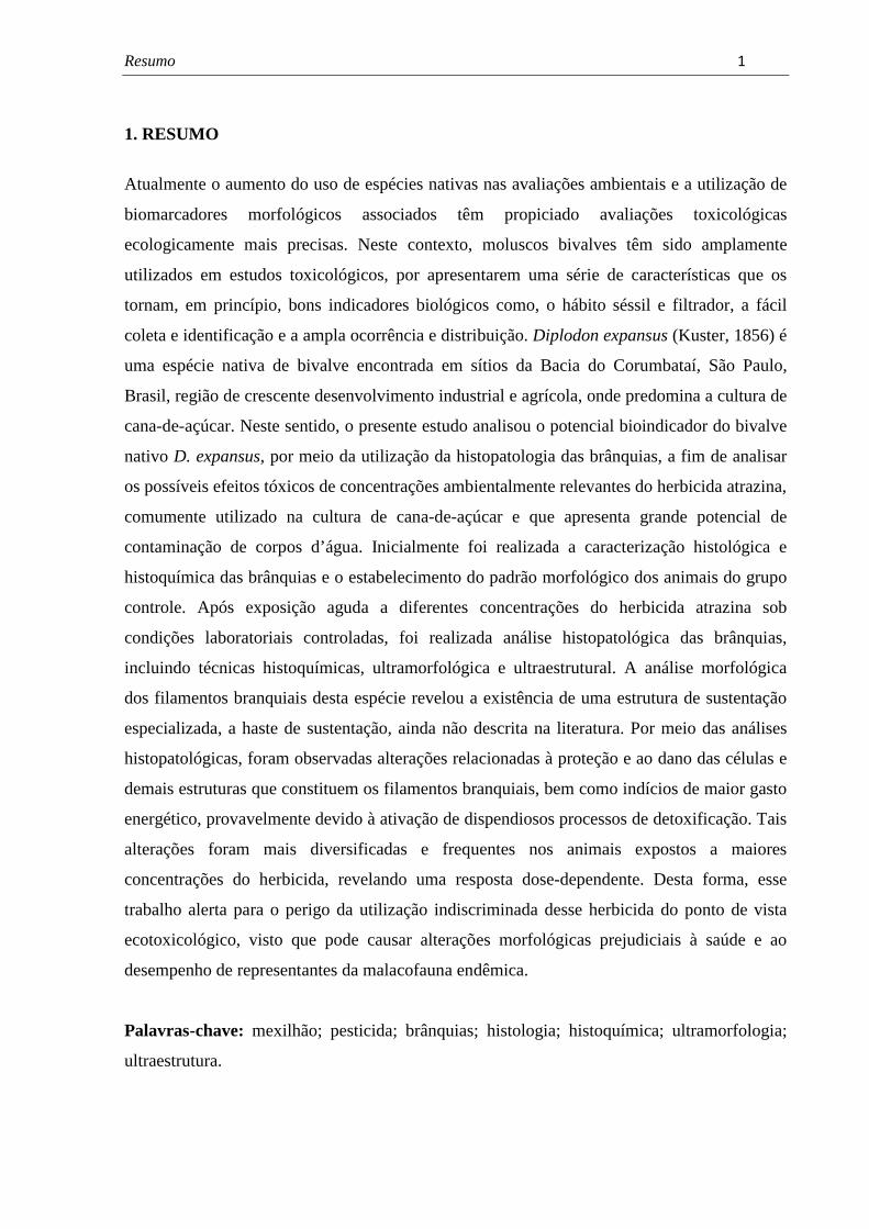
1. RESUMO
Atualmente o aumento do uso de espécies nativas nas avaliações ambientais e a utilização de
biomarcadores morfológicos associados têm propiciado avaliações toxicológicas
ecologicamente mais precisas. Neste contexto, moluscos bivalves têm sido amplamente
utilizados em estudos toxicológicos, por apresentarem uma série de características que os
tornam, em princípio, bons indicadores biológicos como, o hábito séssil e filtrador, a fácil
coleta e identificação e a ampla ocorrência e distribuição. Diplodon expansus (Kuster, 1856) é
uma espécie nativa de bivalve encontrada em sítios da Bacia do Corumbataí, São Paulo,
Brasil, região de crescente desenvolvimento industrial e agrícola, onde predomina a cultura de
cana-de-açúcar. Neste sentido, o presente estudo analisou o potencial bioindicador do bivalve
nativo D. expansus, por meio da utilização da histopatologia das brânquias, a fim de analisar
os possíveis efeitos tóxicos de concentrações ambientalmente relevantes do herbicida atrazina,
comumente utilizado na cultura de cana-de-açúcar e que apresenta grande potencial de
contaminação de corpos d’água. Inicialmente foi realizada a caracterização histológica e
histoquímica das brânquias e o estabelecimento do padrão morfológico dos animais do grupo
controle. Após exposição aguda a diferentes concentrações do herbicida atrazina sob
condições laboratoriais controladas, foi realizada análise histopatológica das brânquias,
incluindo técnicas histoquímicas, ultramorfológica e ultraestrutural. A análise morfológica
dos filamentos branquiais desta espécie revelou a existência de uma estrutura de sustentação
especializada, a haste de sustentação, ainda não descrita na literatura. Por meio das análises
histopatológicas, foram observadas alterações relacionadas à proteção e ao dano das células e
demais estruturas que constituem os filamentos branquiais, bem como indícios de maior gasto
energético, provavelmente devido à ativação de dispendiosos processos de detoxificação. Tais
alterações foram mais diversificadas e frequentes nos animais expostos a maiores
concentrações do herbicida, revelando uma resposta dose-dependente. Desta forma, esse
trabalho alerta para o perigo da utilização indiscriminada desse herbicida do ponto de vista
ecotoxicológico, visto que pode causar alterações morfológicas prejudiciais à saúde e ao
desempenho de representantes da malacofauna endêmica.
Palavras-chave: mexilhão; pesticida; brânquias; histologia; histoquímica; ultramorfologia;
ultraestrutura.
Abstract� � ����
2. ABSTRACT
Currently, the increased use of native species in environmental assessments and the use of
associated morphological biomarkers have provided more accurate ecotoxicological
assessments. In this context, bivalve molluscs have been widely used in toxicological studies,
by presenting a series of characteristics that make them in principle good biological
indicators, such as sessile and filter feeders habits, easy collection and identification and wide
occurrence and distribution. Diplodon expansus (Kuster, 1856) is a native species of mussel
found in sites of Corumbataí Basin, São Paulo, Brazil, a region of increasing industrial and
agricultural development, dominated by sugar cane culture. In this sense, the present study
examined the bioindicator potential of the native bivalve D. expansus through the use of gills
histopathology in order to analyze the possible toxic effects of environmentally relevant
concentrations of atrazine, commonly used in the crops of sugar cane and presents great
contamination potential of water bodies. First, it was performed to characterize the histology
and histochemistry of the gills and the establishment of the morphological pattern of the
control group. After acute exposure to different concentrations of atrazine herbicide under
controlled laboratory conditions, it was performed the histopathological analysis of the gills,
including histochemical, ultramorphological and ultrastructural techniques. Morphological
analyses of gill filaments of this species revealed the existence of a specialized support
structure, the skeletal rod, not yet described in literature. Histopathological analyses reveled
changes related to protection and damage of the cells and other structures that constitute the
gill filaments, as well as evidence of greater energy expenditure, probably due to the
activation of costly detoxification processes.�These changes were more diverse and frequent
in animals exposed to higher concentrations of the herbicide, revealing a dose-dependent
response. Thus, this work points out the danger of indiscriminate use of this herbicide under
an ecotoxicological point of view as it can cause morphological alterations that are adverse to
the health and performance of representatives of the endemic malacofauna.
Keywords: mussel; pesticide; gills; histology; histochemistry; ultramorfology; ultrastruture.
Introdução� � ����
3. INTRODUÇÃO
Durante as últimas décadas, a agricultura mundial cresceu em produtividade e área
cultivada acompanhada pelo uso intenso de agrotóxicos, que também sofreram grandes
evoluções. Muitas moléculas novas surgiram, com características físico-químicas que
propiciam funcionalidades diferenciadas e comportamentos ambientais distintos, com grandes
alterações nos perfis toxicológicos e ecotoxicológicos, fruto dos avanços tecnológicos e
pressões ambientalistas (ARMAS et al., 2005).
Ainda que os benefícios do uso de agrotóxicos sejam claros, muitos questionamentos
são feitos sobre a real necessidade de sua utilização, devido aos riscos que podem causar à
saúde do consumidor e ao meio ambiente. A prática mundial do uso de agrotóxicos, muitas
vezes indiscriminada e abusiva, vem trazendo preocupações às autoridades públicas e aos
envolvidos com saúde e sustentabilidade de recursos naturais, em consequência da
contaminação ambiental (UETA et al.,1998).
Embora a agricultura seja apenas uma das inúmeras fontes não pontuais de poluição de
corpos hídricos, geralmente é apontada como a maior contribuinte de todas as categorias de
poluentes (EDWIN, 1996). Os químicos utilizados na agricultura podem chegar aos rios
carregados pelas chuvas, por despejos industriais e urbanos e por assoreamento do solo
(YAMAGISHI et al., 1981; OHYAMA et al., 1986). Além disso, os agrotóxicos podem
alcançar os ambientes aquáticos por aplicações intencionais ou por deriva e escoamento
superficial, a partir de áreas que sofreram aplicações diretas desses produtos (EDWARDS,
1973).
O Brasil apresenta um dos maiores mercados na área de proteção de plantas, sendo a
agricultura praticada em nosso país ainda altamente dependente de insumos químicos, dentre
os quais se destacam os agrotóxicos. Segundo dados do Sindicato Nacional da Indústria de
Produtos para Defesa Agrícola – SINDAG, o mercado de defensivos agrícolas no Brasil
movimentou cerca de US$ 6,6 bilhões (R$ 12,9 bilhões), sendo o estado de São Paulo um dos
líderes em consumo, representando 15% do valor comercializado no país em 2009 (SINDAG,
2009).
Dentre os agrotóxicos, os herbicidas constituem o grupo mais empregado na
agricultura (aproximadamente 65%). Esses químicos têm como função controlar plantas
daninhas, um dos fatores redutores de produtividade, sem injuriar as culturas agrícolas.
Estima-se que as perdas ocasionadas nas culturas agrícolas, pela interferência das plantas
daninhas, no Brasil, estejam em torno de 20-30% (LORENZI, 1990).
Introdução� � ����
Dentre as culturas agrícolas presentes no estado de São Paulo, ocorre o destaque da
cana-de-açúcar que representa 25% do uso do solo rural do estado e 58% da produção
nacional de cana-de-açúcar (BRASIL, 2009). A cultura da cana-de-açúcar respondeu, em
2009, por 8% das vendas de agrotóxicos no Brasil, atrás somente da soja e do milho
(SINDAG, 2009).
O cultivo de cana-de-açúcar abrange quase a totalidade do território estadual, sendo a
região de Piracicaba (SP) considerada, por muitos anos, a maior produtora. No ano de 2006,
dos 40 Escritórios de Desenvolvimento Rural (EDRs) que compõem o estado, os de
Piracicaba e Limeira ocuparam o 10º e 11º lugar em produção de cana-de-açúcar,
respectivamente (SÃO PAULO, 2007).
Grande parte da região de Limeira (SP) e parte da região de Piracicaba (SP) são
abrangidas pela sub-bacia hidrográfica do rio Corumbataí, integrante da bacia hidrográfica do
rio Piracicaba, onde o cultivo de cana-de-açúcar é a principal atividade agrícola. Além disso,
o rio que atravessa esta bacia e que lhe empresta o nome é responsável pelo abastecimento de
vários municípios (ARMAS et al., 2005).
Armas et al. (2005) avaliaram o consumo total de agrotóxicos na sub-bacia do rio
Corumbataí no período de quatro anos (2000-2003) e concluíram que os herbicidas
representam a classe de agrotóxicos mais empregada na cultura da cana-de-açúcar. Dentre os
herbicidas, a atrazina ocupou posição de destaque representando 14,53% dos produtos
utilizados, perdendo somente para o glifosato com 19,88%.
A atrazina é um potencial contaminante de corpos hídricos e do solo devido as suas
propriedades químicas como, grande potencial de lixiviação, alta persistência no solo e
solubilidade moderada na água (EISLER, 1989). Parte deste herbicida é degradada no
ambiente por processos químicos e microbiológicos (SKIPPER et al., 1967) e parte é lixiviada
pela chuva e água de irrigação, atingindo águas superficiais (LUIZ et al., 2004)
Por meio de um estudo com os bivalves límnicos Anodontites trapesialis e Corbicula
fluminea, o herbicida atrazina mostrou-se altamente acumulativo, sendo que sua concentração
na massa visceral desses invertebrados alcançou níveis 30 vezes maiores que a água, onde se
encontravam expostos por 48 horas (JACOMINI et al., 2006).
Na ostra Crassostrea gigas, a exposição à atrazina em maiores concentrações
ambientais durante um curto período de exposição levou a mudanças moderadas na agregação
de hemócitos (AUFFRET; OUBELLA, 1997). Moraga e Tanguy (2000) observaram uma taxa
de mortalidade de aproximadamente 60-70% de espécimes adultos do mesmo animal, quando
expostos a concentrações de 100 e 200 µg/L deste herbicida depois de dois meses de
Introdução� � ���
experimento. O efeito negativo da exposição à atrazina no crescimento e desenvolvimento de
C. gigas juvenis também já foi relatado na literatura (ROBERT et al., 1986 apud BOUILLY
et al., 2004).
Segundo Jacomini et al. (2003), devido as suas propriedades físico-químicas, a
atrazina tem se mostrado relativamente estável no meio ambiente, tornando-se prejudicial à
biota. Os efeitos tóxicos deste herbicida em peixes e insetos aquáticos têm sido relatados por
alguns pesquisadores (DEWEY, 1986; EISLER, 1989). Além disso, estudos da
bioacumulação da atrazina em moluscos e peixes tem mostrado acumulação via brânquias e
sangue no fígado, cérebro, intestino e gônadas (GUNKEL; STREIT, 1980; PREEZ; VUREN,
1992).
Neste sentido, diferentes grupos de organismos são utilizados como bioindicadores da
avaliação de possíveis efeitos de contaminação natural ou de origem antropogênica. Em
ambientes aquáticos, moluscos bivalves têm se destacado nas últimas décadas como
bioindicadores de toxicidade de poluentes; muitos deles apresentam hábito séssil e filtrador, o
que é altamente desejado em estudos de bioacumulação de poluentes orgânicos e metais
(RITTSCHOF; McCLELLAN-GREEN, 2005).
Consequentemente, bivalves vem sendo amplamente utilizados em programas de
monitoramento biológico tanto em ambiente marinho (FARRINGTON, 1983; DE GREGORI
et al., 1994; MCCONNELL; HARREL, 1995) quanto em ambiente límnico (MANLY;
GEORGE, 1977; FOSTER; BATES, 1978; VILLAR et al., 1999). A maioria dos estudos
utilizando bivalves como bioindicadores foram realizados em regiões temperadas como
Europa e América do Norte, dando principal atenção às espécies locais como Mytilus
californianus, M. edulis e M. galloprovincialis (GREGORY et al., 2002).
Por outro lado, existe um crescente interesse neste tipo de análise nos países tropicais
e subtropicais, onde estas espécies não fazem parte da malacofauna local. Assim, uma série de
outras espécies tem sido utilizada como, por exemplo, o bivalve estuarino Mytella falcata
(DAVID; FONTANETTI, 2005; 2009; DAVID et al., 2008), o bivalve marinho Perna perna
(FERREIRA et al., 2004) e bivalves de água doce como Anodontites trapesialis
(TOMAZELLI et al., 2003; LOAYZA-MURO; ELIAS-LETTS, 2007).
Diplodon expansus (Kuster, 1856) é uma espécie nativa que vive em ambientes de
água corrente, geralmente em rios que drenam para o Atlântico, nos estados do Rio de Janeiro
e de São Paulo, ou para o alto rio Paraná, como o rio Tietê. Conhecida popularmente como
marisco de água doce, esta espécie também é encontrada na bacia hidrográfica do rio
Piraquara, PR (MEYER et al., 2010), no reservatório de Guarapiranga, nas nascentes do Tietê
Introdução� � ���
em São Paulo e em unidades de conservação como a Reserva Biológica (REBIO) da Serra do
Mar, SP (MANSUR; SANTOS, 2008).
Como os demais moluscos bivalves de água doce, D. expansus é um filtrador ativo,
desempenhando um papel de extrema importância no meio ambiente. Esses invertebrados
controlam a quantidade de fitoplâncton, detritos e partículas inorgânicas, promovendo o
aumento da penetração de luz para macrófitas submersas, das quais uma variedade de outros
animais depende (VAUGHN; HAKENKAMP, 2001 apud MEYER et al., 2010). Além disso,
integra a cadeia alimentar de vários vertebrados, entre os quais o homem. Pode ser utilizado
como indicador de condições ambientais ou como biomonitor de alterações ambientais ou de
poluição (MANSUR; SANTOS, 2008).
A espécie D. expansus foi estudada, até o momento, sob aspectos reprodutivos
(CURIAL; LANGE, 1974a, b; 1975) e ecológicos como densidade e biomassa de algumas
populações (HENRY; SIMÃO, 1984; 1985; HENRY; FILOSO, 1987). Recentemente, Meyer
et al. (2010) desenvolveram um estudo cujo objetivo foi analisar a distribuição de classes de
tamanho e a proporção sexual em D. expansus, na Área de Proteção Ambiental do Piraquara,
PR, a fim de contribuir com informações sobre os aspectos ecológicos e reprodutivos da
espécie.
Apenas para D. chilensis e D. fontaineanus foram desenvolvidos estudos
ecotoxicológicos visando à avaliação da contaminação de corpos hídricos e sedimento por
metais e herbicida (GUEVARA et al., 2004; 2005; JACOMINI et al.; 2011). Estudos voltados
para a análise do potencial bioindicador da espécie D. expansus em ambientes límnicos
impactados ainda não foram desenvolvidos.
A resposta biológica a agressões ambientais pode ser evidenciada em qualquer nível
de organização, desde ecossistemas até compartimentos subcelulares ou reações
intracelulares, passando por comunidades, populações, organismos, sistemas fisiológicos e
células (WALKER et al., 1997). Entretanto, toda resposta biológica se manifesta
primeiramente em nível bioquímico-celular (molecular) (DEPLEDGE, 1992), tornando as
técnicas que evidenciam respostas em níveis mais baixos de organização biológica mais
preventivas, ou seja, as alterações na estrutura e função dos ecossistemas passariam a ser
evitadas (NASCIMENTO et al., 2008).
Neste sentido, uma das abordagens mais promissoras para a detecção preventiva de
efeitos adversos é o uso de biomarcadores (NASCIMENTO et al., 2008). O uso mais comum
do termo biomarcadores é para indicadores bioquímicos, fisiológicos ou histológicos de
exposição a xenobióticos ou de efeitos de contaminantes químicos, em níveis suborgânico ou
Introdução� � ����
orgânico (HUGGETT et al., 1992). Desta forma, a fim de identificar os efeitos dos poluentes
nos seres vivos, uma variedade de testes foi desenvolvida, sendo que cada um deles apresenta
certa especificidade e pode analisar respostas diferentes nos diversos tecidos do organismo
estudado.
A análise histopatológica tem sido utilizada com sucesso em estudos ecotoxicológicos,
visto que fornece informações sobre a saúde geral dos animais e sobre modificações teciduais
específicas para os diferentes contaminantes (SUNILA, 1987; FONTANETTI et al., 2010). A
vantagem do uso da histopatologia como biomarcador está em sua localização intermediária
no que diz respeito ao nível de organização biológica (BERNET, 1999); alterações
histológicas aparecem como uma resposta de meio termo a estressores subletais.
Usualmente, bivalves adsorvem pesticidas através das brânquias e os distribuem via
hemolinfa para cada órgão, sendo semelhante o mecanismo para peixes (UNO et al., 2001).
Neste sentido, a estrutura das brânquias pode ser considerada adequada para análises
histopatológicas, uma vez que é formada por um epitélio simples, composto por uma
variedade de tipos celulares, onde facilmente se observa os efeitos de poluentes solúveis em
água (SUNILA, 1988).
Considerando a ausência de estudos que investiguem o potencial bioindicador do
bivalve nativo D. expansus, abundante numa região de grande influência de agrotóxicos
relacionados ao cultivo da cana-de-açúcar, e a escassez de informações precisas sobre os
mecanismos de ação e medidas de segurança para a aplicação dessas substâncias, o presente
projeto visou analisar os possíveis efeitos tóxicos de concentrações ambientalmente realistas
do herbicida atrazina no animal estudado por meio da análise histopatológica de suas
brânquias.
Objetivos� � ����
4. OBJETIVOS
O presente trabalho teve como objetivo investigar os efeitos subletais de
concentrações ambientalmente realistas do herbicida atrazina no bivalve límnico nativo D.
expansus, por meio de análises morfológicas das brânquias.
Os objetivos específicos desse trabalho foram:
1. Descrever a morfologia das brânquias de D. expansus e estabelecer o padrão
morfológico dos animais do grupo controle;
2. Expor exemplares de D. expansus a diferentes concentrações do herbicida atrazina, por
sete dias, a fim de analisar a resposta aguda desses animais;
3. Analisar a ultramorfologia das brânquias dos animais expostos, a fim de identificar
possíveis alterações na sua superfície por meio da microscopia eletrônica de varredura
(MEV);
4. Analisar histologicamente as brânquias dos animais expostos a fim de se verificar
possíveis alterações morfológicas;
5. Realizar testes histoquímicos para a detecção de proteínas e carboidratos com o intuito
de observar possíveis alterações na síntese de algum elemento, que posteriormente,
poderão indicar alterações fisiológicas;
6. Analisar por meio da ultraestrutura, possíveis alterações nas células que compõe os
tecidos das brânquias de D. expansus utilizando-se da microscopia eletrônica de
transmissão (MET);
7. Contribuir com informações sobre os potenciais efeitos do herbicida atrazina sobre o
meio ambiente e a malacofauna, além de fornecer subsídios que possam servir de
alerta para possíveis efeitos sobre o homem.

Revisão de Literatura� � �� �
5. REVISÃO DE LITERATURA
5.1 Bivalves da família Hyriidae: características gerais e estudos realizados
Moluscos bivalves não possuem cabeça, apresentam um único pé anexo a massa
visceral, dois pares de brânquias e os sexos separados, podendo ocorrer alguns casos de
hermafroditismo. Cada animal possui duas valvas que envolvem o corpo, cuja composição
inclui carbonato de cálcio (BOGAN, 2008).
A classe Bivalvia inclui moluscos marinhos ou de água doce como ostras, mexilhões,
vieiras e teredos. O nome comum para bivalve é marisco, sendo correto chamar qualquer
bivalve de marisco, mesmo que ele seja uma vieira ou uma ostra (RUPPERT et al., 2005).
Os bivalves límnicos desempenham o papel de filtradores em rios e lagos. Muitas
espécies são usualmente encontradas em densas agregações e filtram grandes quantidades de
algas, bactérias, partículas orgânicas, além de absorverem metais e grandes moléculas
orgânicas (BOGAN, 2008). Em concordância, Vaughn et al. (2008) afirmam que embora
moluscos bivalves tipicamente se alimentem de fitoplancton, evidências recentes indicam que
esses invertebrados também utilizam fontes alimentares alternativas como bactérias,
zooplancton, rotíferos e detritos, os quais variam consideravelmente em tamanho e qualidade.
Os bivalves límnicos são encontrados em três diferentes subclasses, separadas em
cinco ordens e divididas entre 19 famílias dentro da classe Bivalvia (DEATON;
GREENBERG, 1991). Segundo Bogan (2008), existem 19 famílias com 206 gêneros
reconhecidos como bivalves de água doce e um número de espécies estimado em 1026, sendo
que a maioria das famílias é representada por um a cinco gêneros.
Na América do Sul, as famílias Hyriidae, Mycetopodidae e Sphaeriidae representam a
maioria da diversidade, entretanto, seus representantes ainda continuam pouco conhecidos e
estudados, assim como grande parte da malacofauna da região neotropical (BOGAN, 2008).
Por outro lado, a diversidade de bivalves límnicos na Australásia é dominada pela família
Hyriidae, representada por oito gêneros e 28 espécies. Esses moluscos são encontrados na
Austrália, Tasmânia, Nova Zelândia, Nova Guiné e Ilhas Solomão (SMITH, 1992 apud
BOGAN, 2008).
A família Hyriidae é geralmente incluída na superfamília Unionoidea, embora estudos
recentes sugiram sua atribuição à superfamília Etherioidea (GRAF, 2000; WALKER et al.,
2001). A distribuição zoogeográfica dessa família reflete sua ocorrência gondwânica com
representantes na Australásia e América do Sul (MCMICHAEL; HISCOCK, 1958 apud
BYRNE, 1998). Baseado em análises de DNA, os gêneros da família Hyriidae da América do
Revisão de Literatura� � �����
Sul e Australásia formam grupos irmãos monofiléticos, cujas relações com outras famílias
unioniformes continuam incertas (HOEH et al.1999; GRAF, 2000).
A biologia dos bivalves límnicos pertencentes à família Unionidae na América do
Norte é mais compreendida devido ao amplo histórico de pesquisas quando comparada a
família Hyriidae no hemisfério sul (BYRNE, 1998). Diferentes pesquisadores afirmam que
pouco é conhecido sobre a ecologia populacional (WALKER et al., 2001) e aspectos
ultraestruturais (COLVILLE; LIM, 2003) das espécies australianas pertencentes a família
Hyriidae. Diante da falta de informações imprescindíveis para conservação desse grupo de
bivalves límnicos, Brainwood et al. (2008) utilizaram a geomorfologia como ferramenta de
avaliação dos fatores que determinam a distribuição de espécies de bivalves incluindo da
família Hyriidae, no rio Hawkerbury-Nepean, Austrália. Esse trabalho foi o primeiro a
relacionar a complexidade de habitat, quantitativamente com a densidade populacional e a
distribuição de classe de tamanhos de bivalves límnicos.
Alguns aspectos reprodutivos e morfológicos de hyriidios australianos foram descritos
em alguns estudos (JUPITER; BYRNE, 1997; BYRNE, 1998, 2000; COLVILLE; LIM,
2003). Byrne (1998) investigou a biologia reprodutiva de populações de Hyridella depressa
presentes em lagos e rios australianos que apresentam diferentes localizações e níveis de
influência antropogênica. Buscou-se compreender detalhes do ciclo gametogênico da espécie
por meio da análise histológica das gônadas e a influência dessas variáveis na reprodução
desses invertebrados.
Utilizando-se a microscopia de luz e eletrônica, os bivalves límnicos australianos
Velesunio ambiguus e H. depressa tiveram a morfologia dos seus palpos e manto analisada
por Colville e Lim (2003). Os resultados revelaram que os palpos e manto consistem em abas
de tecido margeadas por epitélio simples nas superfícies externa e interna. O tecido
interveniente consiste, principalmente, de tecido conjuntivo, musculatura, nervos e vasos de
hemolinfa com hemócitos. Os pesquisadores observaram também que ambos os moluscos
apresentam similaridades com os representantes das famílias Margaritiferidae e Unionidae,
particularmente em relação à ocorrência de grânulos mineralizados extracelulares.
Os grânulos de H. depressa capturam uma variedade de elementos do ambiente e
muitos estudos têm destacado sua aplicação no monitoramento da poluição por metais
(VESK; BYRNE, 1999; BYRNE, 2000). Desta forma, Colville e Lim (2003) afirmaram que a
literatura traz, principalmente, informações sobre a estrutura e composição desses grânulos
extracelulares devido a sua capacidade de acumular metais.
Revisão de Literatura� � �����
Vesk e Byrne (1999) compararam duas metodologias a fim de apontar a mais
adequada para preparação dos grânulos extracelulares do bivalve H. depressa em estudos
ecotoxicológicos. A análise dos dados obtidos revelou a necessidade da criopreparação das
amostras a fim de manter a distribuição e concentração dos elementos de interesse. Além
disso, os pesquisadores concluíram que o uso dos grânulos no biomonitoramento pode ser
particularmente promissor quando associado ao método de biópsia do manto, principalmente
em populações de moluscos encontrados em baixa densidade populacional.
Byrne (2000) descreveu a distribuição e estrutura dos grânulos de H. depressa por
meio da microscopia de luz e eletrônica. A caracterização dos elementos presentes nos
grânulos foi realizada utilizando-se manto criopreservado submetido à microanálise por raios-
X. A autora apontou possíveis funções desempenhadas pelos grânulos nesses invertebrados
como homeostase de cálcio, biomineralização, detoxicação de elementos potencialmente
danosos e depósito de cálcio para formação de conchas gloquídeas.
Os moluscos bivalves de água doce da superfamília Unionoidea apresentam uma larva
chamada gloquídio que geralmente parasita os peixes (MANSUR, 1999). Os gloquídios das
espécies sul-americanas (Hyriidae) possuem afinidades morfológicas com as espécies
australianas e são distintos das demais espécies de Unionoidea que vivem na região Holártica
(BONETTO et al., 1986). Entre os Hyriidae são conhecidos fundamentalmente dois tipos de
gloquídios: os portadores de dentes, que apresentam desenvolvimento parasitário no peixe,
onde se forma um cisto no qual o gloquídio completa seu desenvolvimento até pós-larva, e os
que não possuem dentes e apresentam um desenvolvimento completo, dentro dos ovos que
permanecem em bolsas incubadoras chamadas marsúpios, até a pós-larva (ORTMANN, 1921
apud MANSUR, 1999).
Diante do escasso conhecimento da morfologia e da fase parasitária de gloquídios da
América do Sul, Mansur (1999) realizou um trabalho com larvas da espécie Diplodon
martensi, o qual contribuiu com informações morfológicas e sistemáticas desse grupo de
bivalves de água doce.
Mansur e Anflor (1981) estudaram a morfologia interna de D. charruanus e D.
pilsbryi, espécies que habitam ambientes límnicos diferentes, como substrato de areia
grosseira com água corrente e fundo lodoso com águas tranquilas, respectivamente. Esse
estudo teve como principal objetivo estabelecer critérios diferenciais em nível de morfologia
interna na identificação dessas espécies.
A anatomia funcional de D. rotundus gratus e os principais aspectos ecológicos da
espécie foram descritos por Hebling e Penteado (1974). Exemplares foram coletados em
Revisão de Literatura� � �����
águas represadas do rio Tietê, nas proximidades da cidade de Barra Bonita, estado de São
Paulo, Brasil.
A análise de proporção de sexos em D. deodontus expansus foi realizada por Curial e
Lange (1974a), por meio de amostras coletadas em diferentes anos (1972, 1973 e 1974) e
meses (janeiro, agosto, outubro, novembro e dezembro) no rio Cerne, município de Campina
Grande do Sul, no estado do Paraná, Brasil. Não houve influência sazonal quanto a proporção
de sexos, ocorrendo 47,1% de machos, 51,5% de fêmeas e 1,3% de hermafroditas para o total
de 380 moluscos estudados.
Posteriormente, Curial e Lange (1974b) descreveram os casos de hermafroditismo
encontrados. Os pesquisadores afirmaram que entre os Unionacea, os sexos são em geral
separados e o hermafroditismo está limitado a poucos indivíduos. Nos exemplares analisados,
os espermatozóides e os óvulos são produzidos em ácinos distintos e as gônadas são
constituídas de ácinos ovarianos e testiculares localizados no tecido conjuntivo na massa
visceral. Provavelmente, o termo ácino foi utilizado pelos autores devido à semelhança entre
os ácinos glandulares e os folículos gametogênicos, os quais apresentam uma estrutura
ramificada arborescente como descrito para Anodontites trapesialis (CALLIL; MANSUR,
2007).
Os aspectos relativos à morfologia e ao desenvolvimento das gônadas em indivíduos
de D. deodontus expansus coletados em janeiro e julho de 1974 no Rio Cerne, Município de
Campina Grande do Sul, Paraná, Brasil foram analisados por Curial e Lange (1975). No
testículo do mesmo indivíduo, o aspecto histológico dos ácinos diferiu mesmo em ácinos
contíguos. O epitélio contém uma camada simples de espermatogônias reconhecíveis pelos
núcleos grandes. Entre as espermatogônias e o lúmen dos ácinos foi possível encontrar
quantidades variáveis de espermatócitos e espermátides. No mesmo ácino ocorrem zonas
onde predominam um desses dois tipos celulares. Espermatozóides em quantidade variável se
acumulavam no lúmen do ácino. No ovário, os ácinos continham ovócitos em diversos graus
de maturação. Grandes ovócitos podiam ser numerosos e preencher praticamente todo o
lúmen da maioria dos ácinos. Os pesquisadores observaram que a proliferação do epitélio
seminífero e a ovogênese mostraram maior intensidade no lote coletado em julho de 1974.
Informações sobre algumas características reprodutivas de D. expansus foram obtidas
por meio de exemplares coletados no rio Piraquara, Paraná, Brasil (MEYER et al., 2010). A
análise histológica das gônadas possibilitou a determinação de uma razão sexual 1:1, não
tendo sido identificado nenhum caso de hermafroditismo, caracterizando uma população
Revisão de Literatura� � �����
tipicamente dióica. Além disso, as análises qualitativas e quantitativas demonstram uma
gametogênese contínua, com picos de liberação larval no verão.
A espécie mais comum de bivalve límnico no Chile, D. chilensis, pertence à família
Hyriidae, cuja distribuição inclui ambientes lênticos e lóticos localizados em diversas bacias
hidrográficas (PARADA; PEREDO, 2002). Segundo Guevara et al. (2004), D. chilensis
também é considerado um dos membros mais comuns da fauna de invertebrados de água doce
da Patagônia, Argentina. Devido ao seu hábito filtrador e longevidade, esses moluscos podem
influenciar a abundância das comunidades fitoplanctônicas, a qualidade da água e o ciclo de
nutrientes (SOTO; MENA, 1999).
Na região da Patagônia, estudos envolvendo a biologia da espécie D. chilensis
revelaram que esses moluscos vivem enterrados em bancos de areia, absorvendo partículas
orgânicas presentes na água da interface do fundo através do sifão inalante. Na maioria dos
lagos, eles podem ser encontrados em bancos de areia de 2 a 50 metros de profundidade em
densidades populacionais consideráveis com pouco deslocamento vertical (BOTTINI, 1993).
Devido a essas características, tal espécie foi utilizada, com sucesso, em diferentes estudos
como um bioindicador de contaminação por metais em corpos d’água da Patagônia
(GUEVARA et al., 2004, 2005).
A distribuição de metais em lagos do Parque Nacional Nahuel Huapi, Patagônia,
Argentina foi avaliada por meio do bivalve D. chilensis, coletado em quatro lagos diferentes.
A glândula digestiva e os tecidos moles foram analisados separadamente, determinando-se a
concentração de potenciais poluentes como Ag, As, Cr, Hg, Sb e Se e outros nove elementos
de interesse (Ba, Br, Ca, Co, Cs, Fe, Na, Sr e Zn). Os pesquisadores observaram que alguns
elementos traço, como Ar e Cr, são encontrados em maiores concentrações nos tecidos de
moluscos coletados em lagos que sofrem maior interferência de atividades humanas
(GUEVARA et al., 2004).
Posteriormente, Guevara et al. (2005) avaliaram o impacto causado pela Ag na biota
de lagos também localizados no Parque Nacional Nahuel Huapi, Patagônia, Argentina, os
quais diferem no nível de influência humana. Para isso, os pesquisadores utilizaram a
glândula digestiva e os tecidos moles do bivalve nativo D. chilensis e o fígado e musculatura
de cinco espécies de peixes, sendo duas nativas e três exóticas. Os resultados revelaram que a
Ag em moluscos encontrava-se estreitamente relacionada com o sedimento onde se enterram
e a análise do fígado das diferentes espécies de peixes evidenciou a ocorrência da
biomagnificação da prata na cadeia alimentar, sendo os predadores de topo os mais afetados.
Revisão de Literatura� � �����
No norte do Brasil, bivalves límnicos da família Hyriidae como Paxydon
syrmatophorus são coletados no baixo Rio Tocantins por pescadores que vendem as conchas
para fabricação de botões feitos com a camada nacarada de madre pérola. Devido ao interesse
comercial, estudos sobre a biologia reprodutiva desses moluscos tornaram-se necessários, a
fim de elaborar estratégias de manejo adequadas para exploração (BEASLEY et al., 2000).
Beasley et al. (2000) realizaram análises mensais de cortes histológicos das gônadas e
inspecionaram as demibrânquias de fêmeas entre setembro de 1997 e agosto de 1998, a fim de
esclarecer aspectos fundamentais da biologia reprodutiva de uma população de P.
syrmatophorus. Os resultados revelaram que a gametogênese ocorre durante todo ano,
enquanto que a desova ocorre durante os meses da estação seca. Em termos de estratégias de
manejo, os autores acreditam ser necessária a manutenção de uma proporção de indivíduos
sexualmente maduros e de indivíduos pequenos com o intuito de permitir, respectivamente, a
reprodução e o recrutamento na população.
5.2. A utilização de moluscos bivalves em estudos ecotoxicológicos
Muitas substâncias químicas potencialmente danosas de origem antropogênica são
liberadas no ambiente aquático constantemente. Resíduos oriundos de atividades agrícolas e
efluentes de origem industrial e doméstica são lançados em mares e rios, contribuindo para a
contaminação dos ecossistemas aquáticos (ARIAS et al., 2006). Silva et al. (2003) alertaram
que contaminantes ambientais como, por exemplo, metais e agroquímicos podem afetar os
organismos de forma direta e induzir mutações, alterações morfológicas, distúrbios
fisiológicos e problemas de desenvolvimento.
Neste sentido, diferentes grupos de organismos são utilizados como bioindicadores na
avaliação dos possíveis efeitos da contaminação ambiental de origem antropogênica. Em
ambientes aquáticos, moluscos, vermes, esponjas, anfíbios e peixes têm sido utilizados como
biomonitores de toxicidade de poluentes de diferentes naturezas (ZAGATTO; BERTOLETTI,
2008).
Os moluscos bivalves são ecologicamente e economicamente importantes no
ecossistema aquático. Inúmeras características fazem desses invertebrados bioindicadores
apropriados na avaliação e no monitoramento da poluição da água e do sedimento como
ampla distribuição geográfica, fácil coleta, hábito séssil, tolerância a alterações ambientais e a
contaminantes (ZHOU et al., 2008). Uma característica fisiológica de grande relevância é sua
capacidade em filtrar grandes volumes de água (NAIMO, 1995), o que possibilita
considerável bioacumulação de contaminantes ambientais como metais e agrotóxicos. Por
Revisão de Literatura� � ����
fazerem parte da alimentação de diversos seres vivos, incluindo o homem, são também
utilizados em estudos de biomagnificação.
Na revisão realizada por Rittschof e McClellan-Green (2005), os autores apontaram os
moluscos, especialmente gastrópodes e bivalves, como bioindicadores modelo da toxicologia
ambiental e salientaram a importância em se desenvolver pesquisas multidisciplinares que
trabalhem com diferentes níveis de organização biológica. Diante disso, moluscos bivalves
marinhos, estuarinos e límnicos têm sido amplamente utilizados em estudos ecotoxicológicos,
tanto em bioensaios sob condições laboratoriais controladas, quanto em campo empregando-
se animais nativos e transplantados (RITTSCHOF; MCCLELLAN-GREEN, 2005;
BOLOGNESI; HAYASHI, 2011). Esses invertebrados vêm sendo utilizados, até mesmo, em
programas de biomonitoramento reconhecidos internacionalmente como o “Mussel Watch
Program”, iniciado na década de 80 nos Estados Unidos da América (RITTSCHOF;
MCCLELLAN-GREEN, 2005).
Vários parâmetros podem ser utilizados a fim de se avaliar os efeitos de contaminantes
ambientais em bivalves, como moleculares, bioquímicos, genotóxicos, mutagênicos,
morfológicos, fisiológicos e comportamentais. Neste sentido, uma grande variedade de
ensaios pode ser empregada, possibilitando a análise de respostas de estresse ambiental em
diferentes níveis da escala biológica.
Os principais órgãos dos bivalves utilizados em análises toxicológicas são as
brânquias (TÜRKMEN, CIMINLI, 2007; DAVID et al., 2008a) e a glândula digestiva
(MANTECCA et al., 2006; RIGONATO et al., 2005). Outras estruturas e órgãos que são
utilizados, com menor frequência, em estudos ecotoxicológicos são as valvas
(GUNDACKER, 2000; PROTASOWICKI ET AL., 2008), manto (GUNDACKER, 2000) e
gônadas (MANTECCA et al., 2006). Além disso, células que ganham destaque na avaliação
dos efeitos de agentes tóxicos são os hemócitos (PEREZ; FONTANETTI, 2011) e os
mucócitos localizados nos filamentos branquiais dos bivalves (DAVID; FONTANETTI,
2009).
O ensaio do cometa é utilizado em avaliações do potencial genotóxico de amostras
ambientais e xenobióticos isolados em diferentes organismos como peixes (SOUZA;
FONTANETTI, no prelo; VENTURA et al., 2008), bivalves (DAVID et al., 2008b;
FERNÁNDEZ-TAJES et al., 2011) e em culturas de células (MATSUMOTO et al., 2005).
Diferentes células dos moluscos bivalves podem ser submetidas a esse ensaio como, por
exemplo, os hemócitos e as células que compõem as brânquias e a glândula digestiva. Para o
bivalve límnico Corbicula fluminea, Rigonato et al. (2005) verificaram que a glândula
Revisão de Literatura� � ����
digestiva e os hemócitos presentes na hemolinfa do animal são mais indicados para a
realização do ensaio do cometa quando comparados com as brânquias. Resultados
semelhantes foram observados por David et al. (2008b) para o bivalve estuarino Mytella
falcata.
A avaliação do potencial mutagênico de xenobiontes em moluscos bivalves pode ser
realizada por meio do teste do micronúcleo. As células utilizadas nesse teste são usualmente
obtidas na hemolinfa (MANTECCA et al., 2006) ou nas brânquias (BARSIENE et al., 2008)
dos bivalves. Na revisão realizada por Bolognesi e Hayashi (2011), os autores discutiram a
ampla validação do teste do micronúcleo em bivalves e enumeraram uma série de estudos de
campo que utilizaram com sucesso esse teste em bivalves do gênero Mytilus.
Os níveis de atividade enzimática e a quantificação de proteínas relacionadas com
processos de metabolismo e detoxificação de xenobióticos recebem especial atenção em
estudos ecotoxicológicos que empregam parâmetros bioquímicos de avaliação (BOLDINA-
COSQUERIC et al., 2010). Na revisão de Sheehan e Power (1999), foram compiladas
algumas das principais enzimas utilizadas em estudos de biomonitoramento ambiental com
moluscos bivalves, destacando-se a glutatinona-S-transferase, metalotioneínas, catalase e
citocromo P-450.
A utilização de parâmetros químicos na ecotoxicologia aquática é bastante comum.
Alguns estudos são realizados em campo e buscam quantificar os níveis de xenobióticos em
amostras de água, de sedimento e nos tecidos de organismos como moluscos bivalves e peixes
(STORELLI; MARCOTRIGIANO, 2001; JACOMINI et al. 2011). Na literatura também são
encontrados estudos realizados em laboratório sob condições controladas, os quais visam
obter informações sobre o potencial de bioacumulação de metais (CHENEY et al., 2008),
pesticidas (JACOMINI et al., 2003; 2006) e hidrocarbonetos policíclicos aromáticos
(BIRDSALL et al., 2001) em diferentes espécies e órgãos de moluscos bivalves. Os resultados
obtidos geram informações sobre a bioviabilidade, o potencial de bioacumulação e
biomagnificação de diferentes xenobióticos, principalmente metais e agrotóxicos (BOENING,
1997).
A histopatologia ocupa lugar de destaque nos estudos ecotoxicológicos, visto que
alterações morfológicas podem levar a prejuízos na função dos órgãos analisados e,
consequentemente, na saúde e sobrevivência do organismo (SUNILA, 1987; FONTANETTI
et al., 2010). As alterações morfológicas nos tecidos e células dos bivalves vêm sendo
usualmente analisadas por meio da histologia de rotina (DAVID et al., 2008a; ZUPAN;
KALAFATIC, 2003), microscopia eletrônica de varredura (DAVID; FONTANETTI, 2005;
Revisão de Literatura� � �����
GREGORY et al., 1999; NOGAROL et al., 2011) e de transmissão (BIGAS et al., 2001;
GREGORY et al., 2002).
Em estuários, o gênero Mytella recebe especial atenção quanto ao seu uso como
bioindicador devido à possibilidade de avaliação integrada da água e substratos. Desta forma,
David e Fontanetti (2005) utilizaram a microscopia eletrônica de varredura para investigar a
superfície dos filamentos branquiais de M. falcata e compará-los à estrutura das brânquias de
espécimes coletados em locais que apresentavam diferentes níveis de influência de atividades
antropogênicas no estuário de Santos, São Paulo, Brasil. Os resultados revelaram a ausência
de alterações morfológicas, entretanto, nos animais coletados em um dos locais de grande
influência de atividades poluidoras foi encontrado acúmulo de muco da superfície frontal dos
filamentos. Os autores sugeriram que tais resultados refletem uma adaptação desses moluscos
a exposição crônica a poluentes.
Dando continuidade ao estudo, David et al. (2008a) utilizaram as técnicas de
histologia, histoquímica e ultraestrutura para analisar possíveis alterações nas brânquias de M.
falcata. Os animais coletados nas regiões mais poluídas do estuário de Santos apresentaram
alterações histopatológicas como: destacamento do epitélio na região intermediária, mudanças
morfológicas do epitélio, processo inflamatório e aumento no número de células mucosas. Os
autores sugeriram que tais alterações constituem uma tentativa de evitar a entrada de
poluentes através dos filamentos branquiais para todo o organismo.
O estuário Thermaikos, localizado ao norte da Grécia, apresenta um conhecido
gradiente de contaminação e também foi alvo de estudos de caráter ecotoxicológico.
Domouhtsidou e Dimitriadis (2004) analisaram as alterações morfológicas dos palpos,
brânquias e epitélio do intestino posterior de espécimes do bivalve marinho Mytilus
galloprovincialis coletados em seis pontos ao longo do estuário. As alterações observadas
incluíram destacamento das células epiteliais dos filamentos branquiais e espaços
extracelulares dilatados nos palpos e intestino. Os autores ressaltaram que tais alterações
encontram-se possivelmente relacionadas ao grau de poluição dos pontos analisados.
Na literatura é possível observar que uma das principais espécies utilizadas em estudos
ecotoxicológicos em ambientes límnicos é o bivalve exótico Corbicula fluminea de origem
asiática. Esses invertebrados foram utilizados principalmente na avaliação do potencial de
bioacumulação e toxicidade de alguns metais essenciais e não essenciais (GRANEY JR et al.
1983; VILLAR et al., 1999; ) e pesticidas (BASACK et al., 1998; JACOMINI et al., 2006) em
laboratório e campo. Adam-Guillermin et al. (2009) empregaram essa espécie exótica num
trabalho cujo objetivo foi avaliar a bioacumulação e os padrões de toxicidade do selênio, um
Revisão de Literatura� � �����
não-metal cujas concentrações essenciais e tóxicas possuem um estreito intervalo. Os autores
utilizaram a histopatologia das brânquias como um dos parâmetros de avaliação da toxicidade
do selênio orgânico. Por meio da microscopia eletrônica de transmissão, foi sugerido que as
mitocôndrias são o primeiro alvo para citotoxicidade do selênio na forma orgânica como
selenometionina (SeMet), a qual causou alterações morfológicas nas membranas externas e
cristas mitocondriais.
Outra espécie exótica de bivalve límnico que se destaca em estudos ecotoxicológicos é
Dreissena polymorpha, comumente chamado de mexilhão zebra. Mantecca et al. (2006)
investigaram os efeitos histopatológicos nas gônadas, intestino e glândula digestiva desses
moluscos expostos por sete e 14 dias a diferentes concentrações do herbicida paraquat,
simultaneamente com a indução de micronúcleos nos hemócitos. A partir dos resultados
obtidos, os autores concluíram que o herbicida paraquat apresenta propriedades altamente
citotóxicas e genotóxicas no bivalve estudado, sendo que os danos nas gônadas podem causar
riscos ao sucesso reprodutivo da espécie.
5.3. Herbicida atrazina e seus efeitos sobre a biota aquática
A biota aquática está constantemente exposta a um grande número de substâncias
tóxicas oriundas de diversas fontes de emissão. Os principais contaminantes de origem
agrícola são os resíduos de fertilizantes e agrotóxicos. Esses produtos, quando aplicados sobre
o campo de cultivo, podem atingir os corpos d’água diretamente, através da água da chuva e
da irrigação, ou indiretamente através da percolação no solo, chegando aos lençóis freáticos
(ARIAS et al., 2007).
Dentre os inúmeros agrotóxicos aplicados em áreas agrícolas, alguns herbicidas
requerem grande atenção devido ao seu grande potencial de contaminação de águas
superficiais e subterrâneas (ZUPAN; KALAFATIC, 2003; MANTECCA et at., 2006;
JACOMINI et al., 2006). Os herbicidas triazínicos apresentam amplo potencial de
contaminação de diferentes compartimentos ambientais em virtude de suas características tais
como: alto potencial de escoamento e lixiviação e elevada persistência nos solos, hidrólise
lenta, baixa pressão de vapor, solubilidade baixa em água, adsorção moderada à matéria
orgânica e à argila (EISLER, 1989).
A atrazina é uma substância que pertence ao grupo dos triazínicos cujas características
físico-químicas e uso indiscriminado a tornou o maior contaminante das águas subterrâneas
nos Estados Unidos. No Canadá, resíduos de atrazina são encontrados até mesmo em água de
poços (GRISOLIA, 2005). Estudos mostraram que parte deste herbicida é degradada no
Revisão de Literatura� � ��� �
ambiente por processos químicos e microbiológicos (SKIPPER et al., 1967) e parte é lixiviada
pela chuva e água de irrigação, atingindo águas superficiais (LUIZ et al., 2004). Devido ao
seu alto grau de solubilidade na água e grande estabilidade, a atrazina pode manter-se no
ambiente aquático por longos períodos e causar danos à biota (YANG et al., 2010).
Atrazina é o nome comum de 2-cloro-4-etilamino-6-isopropilamino-s-triazina
(fórmula química: C8H14ClN5) estando entre os mais importantes herbicidas seletivos
utilizados para pré e pós-emergência no controle de ervas daninhas. Esse herbicida é utilizado
principalmente em culturas comerciais de cana-de-açúcar, sorgo e milho. É classificado como
muito perigoso para o meio ambiente (classe III), altamente persistente e, possivelmente,
carcinogênico para humanos.
Nas últimas décadas, os cultivos extensivos de soja e milho, que demandam grande
uso de agrotóxicos, foram intensificados nos cerrados brasileiros. Diante disso, Laabs et al.
(2000) realizaram um estudo para a verificação do potencial de lixiviação dos agrotóxicos
mais comumente utilizados nessas culturas, como o herbicida atrazina em solos tropicais da
região de Cuiabá, Estado do Mato Grosso, Brasil. Observou-se maior mobilidade dos
herbicidas no subsolo quando comparados com os inseticidas, além disso, dentre os
agrotóxicos testados, a atrazina apresentou um dos maiores padrões de mobilidade. Verificou-
se também que o potencial de lixiviação desse herbicida em climas tropicais é maior que em
climas temperados, em razão das chuvas torrenciais que ocorrem nos trópicos, aumentando a
percolação que acarreta dissipação vertical dos agrotóxicos.
No trabalho realizado por Ward e Ballantine (1985), foram obtidas respostas agudas e
crônicas de invertebrados como embriões de ostras, camarões e copépodos além do peixe
Cyprinodon variegatus encontrados em estuários a fim de avaliar o potencial impacto da
atrazina em ambientes aquáticos. Diante dos resultados obtidos, os autores afirmaram que a
toxicidade da atrazina em organismos encontrados em estuários é muito similar a encontrada
previamente em organismos límnicos. Por meio da determinação da DL50 dos diferentes
organismos submetidos a diferentes concentrações de atrazina, os autores propõem uma
concentração “segura” desse herbicida de 9 µg/L. Portanto, os autores acreditam que
concentrações ambientais menores ou iguais a 2 µg/L de atrazina não devem causar efeitos
adversos em invertebrados aquáticos e peixes. Em concordância com esses dados, a resolução
CONAMA no 357 (2005) estabelece a concentração máxima de atrazina de 2 µg/L em corpos
de água doce.
Revisão de Literatura� � �����
Segundo Jacomini et al. (2006), os efeitos ecotoxicológicos desse herbicida em
organismos aquáticos continuam pouco conhecidos. Desta forma, o potencial de
bioacumulação desse herbicida em diferentes órgãos do bivalve límnico Anodontites
trapesialis foi investigado. Os resultados apontaram o manto mais sifão, massa visceral e o pé
mais músculos, como principais órgãos de acúmulo de atrazina.
Zupan e Kalafatic (2003) realizaram um estudo dos efeitos de diferentes concentrações
de atrazina (3, 50, 500 e 5000 µg/L) no bivalve límnico Dreissena polymorpha, avaliando-se
mortalidade e alterações morfológicas no hepatopâncreas, brânquias e gônadas. Foi
constatado que os danos causados no hepatopâncreas e gônadas estão relacionados com a
concentração e tempo de exposição dos animais ao herbicida. O hepatopâncreas e as gônadas
sofreram maiores alterações histopatológicas quando os animais foram expostos por períodos
mais longos a altas concentrações do químico, causando maiores taxas de mortalidade. Nas
gônadas, a alteração mais evidente foi necrose do tecido conjuntivo. Os autores não
encontraram alterações nas brânquias dos animais ao final do experimento. Entretanto,
estudos com bivalves estuarinos (DAVID; FONTANETTI, 2005, 2009; DAVID et al., 2008a)
e peixes (BIAGINI et al., 2009) confirmaram a sensibilidade deste órgão a muitos agentes
potencialmente tóxicos encontrados no ambiente.
Mudanças comportamentais do bivalve límnico Elliptio complanata foram avaliadas,
expondo os animais a diferentes concentrações do herbicida atrazina por um curto período
(FLYNN; SPELLMAN, 2009). Em condições normais simuladas pelo controle negativo,
esses animais tenderam a se agregar. Entretanto, a exposição ao herbicida por 72 horas
diminuiu a agregação dos animais. Como a agregação desses moluscos pode estar relacionada
com sua reprodução, as pesquisadoras concluíram que até mesmo baixas concentrações do
herbicida podem causar consequências ecológicas às populações dessa espécie.
O efeito imunotoxicológico de doses ambientalmente relevantes do herbicida atrazina
(10, 23, 50, 100 µg/L) foi analisado no gastrópodo Lymnaea stagnalis (RUSSO; LAGADIC,
2004). Os resultados revelaram que todas as concentrações testadas induziram aumento
significativo no número de hemócitos circulantes, sem nenhuma relação evidente entre
concentração de exposição e resposta observada. Outro estudo revelou alterações
histopatológicas no rim do gastrópodo Physa acuta exposto por 10 dias a atrazina na
concentração 100 µg/L como a perda da integridade citoplasmática e posterior lise. Após o
período de descontaminação ou recuperação, não foram evidenciados sinais de reversibilidade
(ROSÉS et al., 1999).
Revisão de Literatura� � �����
Silvestre et al. (2002) afirmaram que a atrazina pode alterar a capacidade
osmoregulatória de peixes e caranguejos, porém os mecanismos envolvidos ainda são pouco
compreendidos. Desta forma, esses autores realizaram um trabalho cujo objetivo foi
compreender o efeito do herbicida na osmoregulação do caranguejo Eriocheir sinensis,
animal modelo em estudos fisiológicos, devido a sua capacidade de habitar tanto regiões de
água doce como salina. Os pesquisadores concluíram que a homeostase do caranguejo não foi
afetada a níveis consideráveis por esse poluente durante o período de duas semanas de
exposição.
Estudos revelaram que peixes podem acumular o herbicida atrazina em seus tecidos
como os do fígado, brânquias, sangue, cérebro ou músculo (GUNKEL; STREIT, 1980;
PREEZ; VAN VUREN, 1992). Outros autores também confirmaram a capacidade desse
herbicida em causar danos genotóxicos (VENTURA et al., 2008) e alterações endócrinas
(MOORE; WARING, 1998), comportamentais (STEINBERG et al., 1995) e imunológicas
(KREUTZ et al., 2010) em peixes.
Os estudos com anfíbios demonstraram claramente a fragilidade dos ecossistemas
aquáticos às contaminações por agrotóxicos (GRISOLIA, 2005). O herbicida atrazina não é
considerado altamente tóxico para rãs em concentrações ambientalmente relevantes (DIANA
et al, 2000;. COADY et al, 2005), entretanto parece contribuir com o declínio populacional de
anuros pois interfere no desenvolvimento gonadal normal desses animais (HAYES et al.,
2002; 2003). Um dos casos mais conhecidos é o desenvolvimento de hermafroditismo e
desmasculinação em sapos após exposição a baixas doses do herbicida atrazina (GILBERT,
2006; HAYES et al., 2002). Segundo Hayes et al (2002), esse herbicida parece induzir a ação
da enzima aromatase, a qual é capaz de converter testosterona em estrógeno.
Diante dos possíveis efeitos desse herbicida sobre populações de anfíbios, Murphy et
al. (2006) coletaram três espécies de anuros de locais com e sem prática agrícola no estado de
Michigan, Estados Unidos. O objetivo desse estudo foi determinar a incidência de ovócitos
testiculares e hermafroditismo nesses animais e avaliar possíveis correlações entre as
concentrações obtidas de atrazina na água e tais alterações. Hermafroditismo foi utilizado em
casos de indivíduos que possuíam tecidos gonadais femininos e masculinos em um ou ambas
as gônadas, enquanto que o termo ovócitos testiculares foi utilizado para descrever casos em
que um ou mais ovócitos ocorrem em testículos de sapos machos.
Foram realizadas coletas de animais jovens e adultos nos verões de 2002 e 2003.
Nesse período as concentrações de atrazina obtidas não ultrapassaram 2µg/L na maioria das
regiões agrícolas. Poucos indivíduos hermafroditas foram encontrados tanto em locais com
Revisão de Literatura� � �����
quanto em locais sem prática agrícola. Por outro lado, ovócitos testiculares foram encontrados
em sapos machos na maioria dos locais, sem diferença significativa entre regiões agrícolas e
não agrícolas. Os autores concluíram que as concentrações de atrazina não se encontravam
correlacionadas de forma significativa à incidência de hermafroditismo e ovócitos testiculares.
Materiais e Métodos� � �����
6. MATERIAIS E MÉTODOS
6.1. Coleta dos animais
Espécimes de D. expansus (peso médio ± DP = 14,15g ± 7,8; comprimento médio ±
DP= 4,29cm ± 0,84) foram coletados em abril de 2010 no Ribeirão Claro, município de Rio
Claro (S 22º24’33.1’’; O 47º32’25.1’’), São Paulo, Brasil. Amostras de água do local foram
coletadas seguindo recomendações específicas (CETESB, 1987; COGERH, 2001) e
imediatamente encaminhadas para quantificação de metais e agrotóxicos.
No laboratório, os animais foram aclimatados em aquários com capacidade de 30 litros
com água de poço artesiano por três dias, temperatura ± 25º C, aeração constante e ciclo
claro/escuro de 12 horas. Este procedimento foi adotado a fim de evitar a influência do
estresse da coleta e do transporte dos animais.
6.2. Análise química da água
Após a coleta das amostras de água, os frascos foram acondicionados em caixas
térmicas com gelo reciclável e encaminhados ao laboratório TASQA Serviços Analíticos
Ltda. (Paulínia, São Paulo, Brasil), onde foram realizadas as análises químicas.
Os parâmetros orgânicos (atrazina, glifosato, 2,4-D) e inorgânicos (alumínio
dissolvido, cádmio total, chumbo total, cobre dissolvido, ferro dissolvido, manganês total,
níquel total, prata total, zinco total) estabelecidos na resolução do Conselho Nacional do Meio
Ambiente (CONAMA, 2005) foram utilizados a fim de se avaliar a qualidade das amostras de
água.
A análise dos resíduos de atrazina e 2,4-D foi realizada aplicando-se o método EPA
8270D para determinação da concentração de compostos orgânicos semivoláteis e para o
glifosato, o método EPA 547 foi aplicado.
Para a quantificação dos metais, as amostras de água foram submetidas ao método
SM21 3120B, utilizado na quantificação de elementos por espectrofotometria de emissão
atômica em plasma de argônio indutivamente acoplado, em extratos aquosos, e similar ao
método de quantificação EPA 6010B, após digestão ácida em sistema fechado com
aquecimento por microondas pelos métodos EPA 3015 para amostras líquidas e EPA 3052
para amostras sólidas.
As concentrações de metais e agrotóxicos obtidas nas análises químicas das amostras
de água do local de coleta dos animais foram comparadas com os valores máximos
apresentados para corpos de água doce presentes na resolução do Conselho Nacional do Meio
Materiais e Métodos� � �����
Ambiente (CONAMA, 2005). As águas doces do território brasileiro são classificadas,
segundo a qualidade requerida para os seus usos preponderantes. Desta forma, neste trabalho
foram utilizados os valores para as classes I e II, as quais podem ser utilizadas para consumo
humano e proteção das comunidades aquáticas.
Os padrões de qualidade das águas determinados nesta resolução estabelecem limites
individuais para cada substância em cada classe e determina as concentrações máximas
permitidas. Concentrações acima do permitido representam comprometimento na qualidade
da água, para seus usos preponderantes.
6.3. Montagem dos bioensaios
Seis aquários com capacidade de 10 litros foram utilizados: um aquário para o grupo
controle, contendo somente água do poço artesiano e os outros cinco, com diferentes
concentrações de atrazina (2, 6.25, 12.5, 25 e 50 µg/L) obtidas diluindo-se a solução estoque
(0.5g/L) na água. A maior concentração utilizada (50 µg/L) consiste no dobro da solução
indicada para uso agrícola e a menor (2 µg/L), a máxima concentração permitida em corpos
de água doce pela legislação brasileira, conforme a resolução CONAMA no 357 (2005).
Foram utilizadas tais concentrações para se avaliar os efeitos da aplicação indiscriminada de
agrotóxicos e o efeito da diluição desse herbicida nos corpos hídricos, aproximando-se de
concentrações ambientalmente realistas. Os bioensaios foram denominados da seguinte
forma:
Controle: 8L de água do poço artesiano
Tratamento 1: 8L de água do poço artesiano + 16 µg de atrazina (2 µg/L)
Tratamento 2: 8L de água do poço artesiano + 50 µg de atrazina (6,25 µg/L)
Tratamento 3: 8L de água do poço artesiano + 100 µg de atrazina (12,5 µg/L)
Tratamento 4: 8L de água do poço artesiano + 200 µg de atrazina (25 µg/L)
Tratamento 5: 8L de água do poço artesiano + 400 µg de atrazina (50 µg/L)
Os aquários permaneceram continuamente aerados por uma bomba de ar. Após o
período de aclimatação, os animais foram distribuídos randomicamente, sendo que em cada
aquário foram expostos cinco animais por sete dias a fim de se obter a resposta aguda de
exposição. Este tempo de exposição foi utilizado em trabalhos anteriores envolvendo atrazina
e bivalves límnicos (ZUPAN; KALAFATIC, 2003; JACOMINI et al., 2006).
As condições experimentais consistiram em um sistema semi-estático em que 100%
do volume d’água foi trocado a cada 24 horas, seguida da adição da solução estoque de
Materiais e Métodos� � ����
herbicida recém preparada em água destilada, com a intenção de manter a concentração das
soluções-teste. A temperatura foi mantida entre 23 – 25oC durante o período de exposição.
Após sete dias de exposição, os moluscos foram anestesiados por choque térmico e
tiveram pequenos fragmentos das mesmas regiões de suas brânquias retiradas e fixadas.
6.4. Histologia
6.4.1. Inclusão em resina (Historesina)
O material foi fixado em solução aquosa de Bouin por 24 horas; em seguida foi
colocado em solução tampão fosfato de sódio pH=7 e mantido na geladeira. Posteriormente, o
material foi desidratado em soluções de etanol 70, 80, 90 e 95%, durante 20 minutos cada
banho. Na sequência, foi transferido para solução de resina Leica na ausência de catalisador,
durante 24 horas em geladeira. Posteriormente, o material foi transferido para moldes
plásticos previamente preenchidos com resina contendo catalisador.
Após a polimerização da historesina, os blocos foram seccionados com 5 µm,
utilizando-se o micrótomo Leica RM 2245 com navalhas de vidro; os cortes foram hidratados
em banho histológico e recolhidos em lâminas. Após secagem, os cortes foram corados com
hematoxilina de Harris por 10 minutos e lavadas em água corrente por 5 minutos para a
reação; em seguida foram coradas com eosina aquosa por 5 minutos e lavadas em água. Após
secagem, os cortes foram diafanizados em xilol e as lâminas montadas com bálsamo do
Canadá. Posteriormente, as secções foram analisadas e fotografadas em fotomicroscópio
Leica.
6.5. Histoquímica
O material foi fixado e processado de acordo com os procedimentos da rotina
histológica descritos previamente. Posteriormente, os testes histoquímicos foram aplicados
nas secções histológicas a fim de se detectar a presença de diferentes compostos.
6.5.1. Técnica do azul de bromofenol para detecção de proteínas totais (PEARSE, 1985)
Os cortes, recolhidos em lâminas de vidro, foram corados com solução de azul de
bromofenol à temperatura ambiente durante 1 hora, sendo em seguida lavados em solução
aquosa de ácido acético 0.5%, durante 5 minutos. Em seguida, os cortes foram secos,
diafanizados em xilol e as lâminas montadas em bálsamo do Canadá. Posteriormente, foram
examinadas e fotografadas em fotomicroscópio Leica.
Materiais e Métodos� � ����
6.5.2. Técnica simultânea do PAS/azul de Alcian para detecção de polissacarídeos neutros e
ácidos (JUNQUEIRA; JUNQUEIRA, 1983)
As secções histológicas foram coradas com azul de Alcian 1% pH 2,5 durante 30
minutos. Em seguida, as lâminas contendo as secções foram lavadas em água destilada e
passadas em ácido periódico 1% durante 5 minutos. Posteriormente, foram submetidas ao
reativo de Schiff no escuro por 30 minutos e, em seguida, lavadas em água corrente durante
10 minutos. Na sequência, foram secas, diafanizadas em xilol e montadas em bálsamo do
Canadá, para posterior observação e registro em fotomicroscópio Leica. Foi realizada também
a contagem de hemócitos infiltrados no epitélio e mucócitos presentes em 50 filamentos
branquiais de cada indivíduo analisado. Esses dados foram submetidos à análise estatística.
6.5.3. Método de von Kossa para detecção de cálcio (JUNQUEIRA; JUNQUEIRA, 1983)
As secções foram imersas em nitrato de prata por 20 minutos, lavadas em água
corrente e transferidas para revelador D-72 por 2 horas, imersas em fixador F-5 por 30
minutos e posteriormente lavadas em água corrente. As lâminas foram secas, diafanizadas em
xilol e montadas em bálsamo do Canadá.
6.5.4. Técnica do picrocirius para detecção de colágeno total (JUNQUEIRA; JUNQUEIRA,
1983, adaptado)
Os cortes foram colocados previamente por 15 minutos na estufa a 60º C.
Posteriormente, os cortes foram submetidos à solução picrosirius a 60º C por 60 minutos
numa estufa a 60º C. Em seguida, os cortes foram lavados em água destilada em três banhos.
As lâminas foram montadas com bálsamo do Canadá, secas em estufa e levadas ao
fotomicroscopio Leica para análise.
6.5.5. Técnica do tricômico de Mallory para detecção de colágeno total (JUNQUEIRA;
JUNQUEIRA, 1983)
As secções histológicas foram submetidas ao lugol por 4 minutos. Posteriormente,
foram transferidas para solução de hipossulfito aquoso 5% e ali mantidas por 3 minutos. Na
sequência, os cortes foram lavados em água corrente por 20 minutos e, em seguida, corados
com hematoxilina de Harris por 10 minutos e lavados em água corrente por 5 minutos para a
reação. As lâminas, contendo as secções histológicas, foram colocadas em câmara úmida e
submetidas ao corante de Mallory por 15 minutos a 37°C. Em seguida, os cortes foram
Materiais e Métodos� � �����
lavados em água destilada em três banhos. As lâminas foram montadas com bálsamo do
Canadá, secas em estufa e levadas ao fotomicroscopio Leica para análise.
6.6. Microscopia Eletrônica de Varredura
Fragmentos das brânquias foram fixados em solução Karnovsky (KARNOVSKY,
1965) por 2 horas e desidratadas em uma série de concentrações crescentes de acetona.
Posteriormente, o material foi levado ao ponto crítico (Balzer CPD 030), fixado em suportes
metálicos e coberto com ouro utilizando-se Sputtering Balzer SCD 050. O material foi
analisado e fotografado no microscópio eletrônico de varredura Philips, operado em 12 Kv.
6.7. Microscopia Eletrônica de Transmissão
Pequenos fragmentos de brânquias foram fixados em glutaraldeído 2,5% em tampão
cacodilato de sódio 0,1M a 4ºC, lavados em tampão cacodilato de sódio e pós-fixados em
tetróxido de ósmio 1% por 2 horas. O material foi novamente lavado no mesmo tampão,
colocado em álcool 10% por 15 minutos e contrastado com acetato de uranila 2% em álcool
10% por 3 horas. Em seguida, foi desidratado em série crescente de acetona, submetido à
solução resina: acetona (1:1) por 12 horas, embebido em resina Epon-araldite contendo
catalisador por 24 horas e levado a estufa a 70ºC por 24 horas para polimerização da resina. O
material foi seccionado em ultramicrótomo Sorvall–Porter Blum e as secções ultrafinas foram
coletadas em grades de cobre e contrastadas com acetato de uranila e citrato de chumbo
durante 45 e 10 minutos, respectivamente. O material presente nas grades de cobre foi
observado e fotografado ao microscópio eletrônico de transmissão Phillips CM 100, operado
a 80 kV.
6.8. Análise estatística
O número de hemócitos infiltrados no epitélio e mucócitos foram contabilizados em
50 filamentos branquiais por indivíduo (cinco indivíduos para cada bioensaio). Durante a
contagem dos mucócitos, foram diferenciadas as células íntegras das rompidas.
Os resultados foram estatisticamente analisados utilizando-se o software SYSTAT 11
e o teste análise de variância (ANOVA). Os dados foram previamente analisados para
verificar normalidade utilizando-se o teste a priori Shapiro-Wilk (SW), sendo considerados
dados com distribuição normal aqueles que apresentaram p>0.05.
Resultados� � �����
7. RESULTADOS
Os resultados obtidos no presente trabalho são apresentados na forma de capítulos,
sendo que cada um contém um artigo publicado ou submetido em periódico internacional e
especializado. Os artigos seguem a formatação estipulada pelas revistas a que foram
submetidos.
CAPÍTULO 1
Título do artigo: Morphological and histochemical characterization of gill filaments of the
Brazilian endemic bivalve Diplodon expansus (Küster, 1856) (Mollusca, Bivalvia, Hyriidae).
Autores: Larissa Rosa Nogarol, Ana Luíza Brossi-Garcia, José Augusto de Oliveira David e
Carmem Silvia Fontanetti.
Submetido ao periódico Tissue and Cell (2011).
CAPÍTULO 2
Título do artigo: Surface morphology of Diplodon expansus (Küster, 1856) (Mollusca,
Bivalvia, Hyriidae) gill filaments after exposure to environmentally relevant concentrations of
atrazine herbicide.
Autores: Larissa Rosa Nogarol, Ana Luíza Brossi-Garcia e Carmem Silvia Fontanetti.
Publicado no periódico Microscopy Research and Technique, 2011.
DOI 10.1002/jemt.21130
CAPÍTULO 3
Título do artigo: Histopathology of the gill filaments of the Brazilian endemic bivalve
Diplodon expansus after exposure to the herbicide atrazine.
Autores: Larissa Rosa Nogarol, Ana Luíza Brossi-Garcia e Carmem Silvia Fontanetti.
Submetido ao periódico Ecotoxicology and Environmental Safety (2012).
Capítulo 1� � � � �
� �
7.1. CAPÍTULO 1
Morphological and histochemical characterization of gill filaments of the
Brazilian endemic bivalve Diplodon expansus (Küster, 1856) (Mollusca,
Bivalvia, Hyriidae)
Larissa Rosa Nogarol1; Ana Luiza Brossi-Garcia1; José Augusto de Oliveira David2 &
Carmem Silvia Fontanetti11*
1 UNESP, São Paulo State University, Av. 24A, no 1515, CEP 13506-900, Rio Claro, SP,
Brazil. 2 UFES, Federal University of Espírito Santo, Alto Universitário, s/n°, CEP 29500-000,
Alegre, ES, Brazil.
Diplodon expansus gill filaments: morphology and histochemistry
* Corresponding author
E-mail address: [email protected]
Tel.: +55 19 35264139; fax: +55 19 35264135.
Artigo submetido ao periódico Tissue and Cell.
Capítulo 1� � � ���
� �
Resumo
O presente trabalho traz a descrição morfológica e a caracterização histoquímica dos
filamentos branquiais do molusco bivalve endêmico brasileiro D. expansus, visando ampliar o
conhecimento morfológico desta espécie e estabelecer a estrutura das brânquias que servirão
como controle em estudos histopatológicos aplicados ao biomonitoramento. Os filamentos
branquiais são divididos em três zonas: frontal, intermediária e abfrontal. Ocorrem pontes de
tecido que interconectam os filamentos branquiais (junções interfilamentares) e as lamelas
(junções interlamelares). No centro do filamento, hemócitos circulam pelo vaso de hemolinfa
o qual é internamente revestido por endotélio. A superfície frontal dos filamentos é coberta
por cílios, a superfície lateral exibe dutos aqüíferos e a superfície abfrontal apresenta células
ciliadas e não ciliadas. O epitélio dos filamentos é constituído por células ciliadas, não
ciliadas absortivas e mucócitos. A sustentação dos filamentos é feita pela haste e alça de
sustentação. Com base nas informações obtidas, os filamentos branquiais da espécie estudada
apresentam algumas características peculiares ainda não relatadas detalhadamente na
literatura como a presença simultânea da alça e haste de sustentação. Por outro lado, a
constituição geral do filamento é semelhante àquelas descritas tanto para bivalves marinhos
como límnicos e parecem ser adequadas para aplicação em estudos ecotoxicológicos.
Palavras-chave: molusco; ctenidia; microscopia eletrônica de varredura; histologia.
Capítulo 1� � � ���
� �
Abstract
The present study presents the morphological description and histochemical characterization
of gill filaments of the Brazilian endemic bivalve Diplodon expansus, aiming to broaden the
morphological knowledge of this species and establish the structure of the gills that will serve
as control in histopathological studies applied to biomonitoring. The gill filaments are divided
into three zones: frontal, intermediate and abfrontal. In the center of the filament, haemocytes
circulate through the haemolymph vessel, which is internally lined by endothelium. The
frontal surface of the filament is covered with cilia, the lateral surface exhibits aquifer ducts
and the abfrontal surface presents ciliated and non-ciliated cells. The epithelium of the
filaments is constituted by ciliated cells, non-ciliated absorptive cells and mucocytes. The
support of the filaments is made by two specialized structures called skeletal rod and skeletal
loop. Based on the obtained information, the gill filaments of the studied species preset some
peculiar characteristics that are not yet reported in detail in the literature such as the
simultaneous presence of skeletal rod and skeletal loop. On the other hand, the general
constitution of the filament is similar to those described for both marine and limnic bivalves
and seems to be suitable for ecotoxicological studies.
Keywords: mussel; ctenidia; scanning electron microscopy; histology.
Capítulo 1� � � ���
� �
1. Introduction
The interest in studying bivalve molluscs has increased considerably due to their
population decline in several regions of the world and also due to their potential use as
bioindicators in different impacted ecosystems (Colville and Lim, 2003).
The data available in the literature reveal the ability of these molluscs in accumulating
pollutants of several chemical natures in their tissues (Alyakrinskaya, 2003), which can lead
to the development of histopathologies as observed by Gregory et al. (2002) for the marine
bivalve Perna perna exposed to mercury. Thus, the morphology of target organs such as gills
of estuarine, marine and limnic bivalves has been studied in order to be used as indicators of
environmental pollution, especially in regions under the influence of different pollutant
sources (David and Fontanetti, 2005; David et al., 2008; Gregory and George, 2000; Gregory
et al., 1996; Lemaire-Gony and Boudou, 1997).
However, most part of the ecotoxicological studies in freshwater bodies use exotic
species such as Corbicula fluminea (Peltier et al., 2008; Villar et al., 1999), Dreissena
polymorpha (Mantecca et al., 2006; Zupan and Kalafatic, 2003) and Limnoperna fortunei
(Vilela et al., 2006), which preclude the performance of more accurate toxicological
assessments under an ecological point of view. Besides, the responses of native Brazilian
malacofauna exposed to potential toxic substances commonly found in freshwater bodies are
little known.
The limnic malacofauna in Brazil has about 155 species of bivalve molluscs, which
belongs mainly to four families: Hyriidae, Mycetopodidae, Sphaeridae and Corbiculidae. The
first two have a wide geographical distribution, occurring in very varied habitats, such as
lakes, marginal lagoons and reservoirs in most of the hydrographic basins of the South
American continent, while the others are of a more restricted occurrence (Avelar, 1999).
The Brazilian endemic species D. expansus (Küster, 1856), popularly known as
freshwater mussel, belongs to the Hyriidae family (Mansur and Santos, 2008), whose
distribution is restricted to South America and Australia (Avelar and Cunha, 2009).
Individuals of this species generally occur in rivers that drain into the Atlantic, in the states of
São Paulo and Rio de Janeiro, or into the upper Paraná River, as the Tietê River (Mansur and
Santos, 2008). The species D. expansus has been studied, so far, on reproductive (Curial and
Lange, 1974 a,b; 1975) and ecological aspects, such as density and biomass of some
populations and their role in the decomposition process in some ecosystems (Henry and
Simão, 1984; Henry and Filoso, 1985; 1987).
Capítulo 1� � � ���
� �
In order to contribute to morphological information of the Brazilian species D.
expansus, for conservation purposes and for its use as model in ecotoxicological studies in
impacted limnic environments, this study aimed to characterize its gills, organs that are in
direct contact with environmental contaminants such as metals and agrochemical residues.
2. Material and methods
2.1. Collection and acclimation
Specimens of D. expansus (average weight ± SD = 13.42g ± 6.64; mean length ± SD=
4.28cm ± 0.81) were collected in April 2010 in Ribeirão Claro stream, municipality of Rio
Claro (S 22º24’33.1’’; O 47º32’25.1’’), São Paulo, Brazil. A collection site that does not
suffer direct influence of industrial and agriculture practices was chosen and, therefore, can be
considered a little impacted region.
In the laboratory, the animals were acclimated for three days in aquariums with
capacity of 30 litres containing artesian well water, temperature ± 25º C, constant aeration and
light/dark cycle of 12 hours. This procedure was adopted in order to minimize the influence of
the stress of collection and transport of the animals.
The freshwater bivalve molluscs of the Hyriidae family, in which the species D.
expanus belongs to, present larvae called glochidium that remain sheltered in the demibranch
until they reach the maturity (Mansur, 1999). In this study, only animals that did not present
marsupial gills sheltering larvae were used.
After acclimation, the molluscs were anesthetized by heat shock and small fragments
of their gills were removed and fixed in different solutions.
2.2. Histology and histochemistry
The gill fragments were fixed in aqueous Bouin solution for 24 hours and then
submitted to phosphate buffer solution 0.1M pH 7.4. In order to remove picric acid residues,
the material was washed three times with the same buffer solution and then dehydrated in
ethanol 70, 80, 90 and 95%, during 20 minutes each bath and embedded in resin for 24 h at
4°C. Subsequently, the material was transferred to plastic molds containing Leica resin for
inclusion. After resin polymerization, the blocks were sectioned with 5 µm thick, using Leica
RM2245 microtome with glass knives; the sections were hydrated in histological bath and
collected on microscope slides.
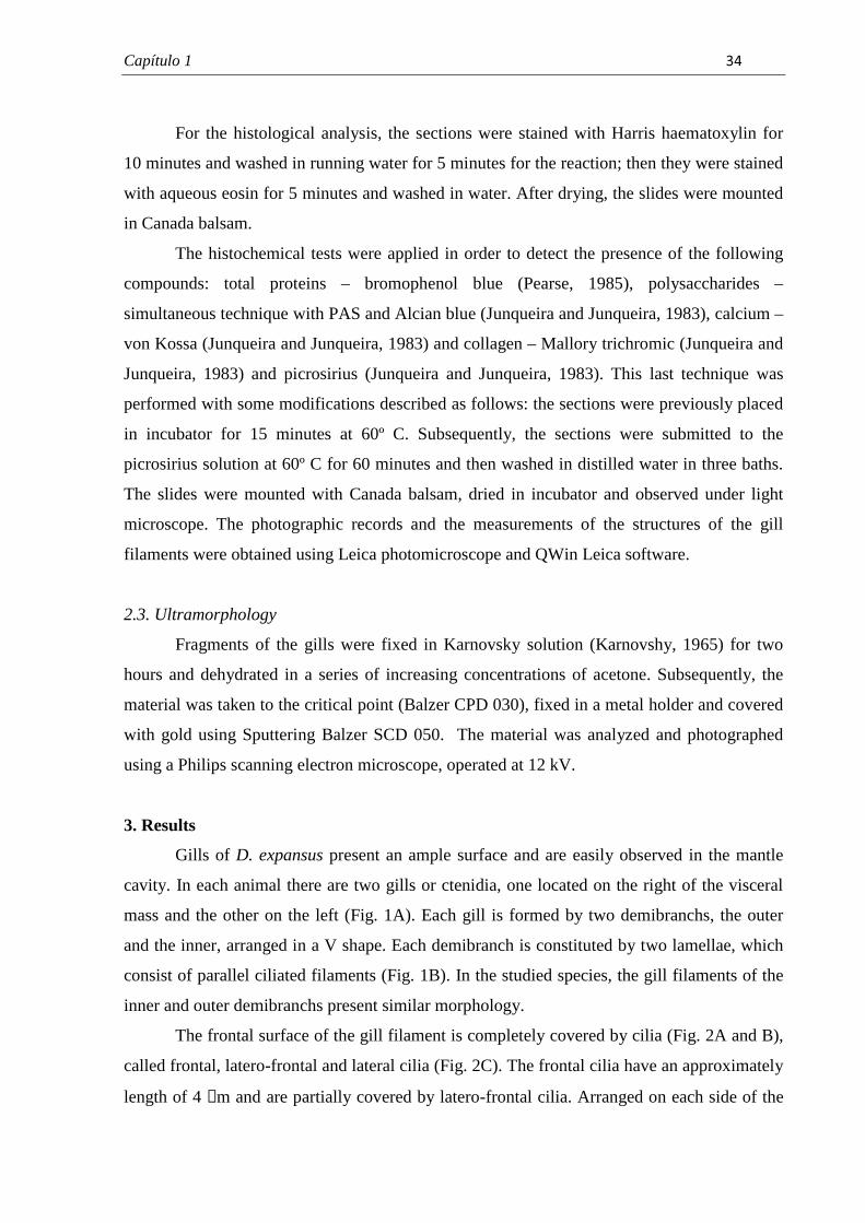
Capítulo 1� � � ���
� �
For the histological analysis, the sections were stained with Harris haematoxylin for
10 minutes and washed in running water for 5 minutes for the reaction; then they were stained
with aqueous eosin for 5 minutes and washed in water. After drying, the slides were mounted
in Canada balsam.
The histochemical tests were applied in order to detect the presence of the following
compounds: total proteins – bromophenol blue (Pearse, 1985), polysaccharides –
simultaneous technique with PAS and Alcian blue (Junqueira and Junqueira, 1983), calcium –
von Kossa (Junqueira and Junqueira, 1983) and collagen – Mallory trichromic (Junqueira and
Junqueira, 1983) and picrosirius (Junqueira and Junqueira, 1983). This last technique was
performed with some modifications described as follows: the sections were previously placed
in incubator for 15 minutes at 60º C. Subsequently, the sections were submitted to the
picrosirius solution at 60º C for 60 minutes and then washed in distilled water in three baths.
The slides were mounted with Canada balsam, dried in incubator and observed under light
microscope. The photographic records and the measurements of the structures of the gill
filaments were obtained using Leica photomicroscope and QWin Leica software.
2.3. Ultramorphology
Fragments of the gills were fixed in Karnovsky solution (Karnovshy, 1965) for two
hours and dehydrated in a series of increasing concentrations of acetone. Subsequently, the
material was taken to the critical point (Balzer CPD 030), fixed in a metal holder and covered
with gold using Sputtering Balzer SCD 050. The material was analyzed and photographed
using a Philips scanning electron microscope, operated at 12 kV.
3. Results
Gills of D. expansus present an ample surface and are easily observed in the mantle
cavity. In each animal there are two gills or ctenidia, one located on the right of the visceral
mass and the other on the left (Fig. 1A). Each gill is formed by two demibranchs, the outer
and the inner, arranged in a V shape. Each demibranch is constituted by two lamellae, which
consist of parallel ciliated filaments (Fig. 1B). In the studied species, the gill filaments of the
inner and outer demibranchs present similar morphology.
The frontal surface of the gill filament is completely covered by cilia (Fig. 2A and B),
called frontal, latero-frontal and lateral cilia (Fig. 2C). The frontal cilia have an approximately
length of 4 µm and are partially covered by latero-frontal cilia. Arranged on each side of the
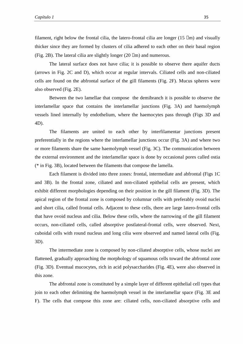
Capítulo 1� � � ��
� �
filament, right below the frontal cilia, the latero-frontal cilia are longer (15 µm) and visually
thicker since they are formed by clusters of cilia adhered to each other on their basal region
(Fig. 2B). The lateral cilia are slightly longer (20 µm) and numerous.
The lateral surface does not have cilia; it is possible to observe there aquifer ducts
(arrows in Fig. 2C and D), which occur at regular intervals. Ciliated cells and non-ciliated
cells are found on the abfrontal surface of the gill filaments (Fig. 2F). Mucus spheres were
also observed (Fig. 2E).
Between the two lamellae that compose the demibranch it is possible to observe the
interlamellar space that contains the interlamellar junctions (Fig. 3A) and haemolymph
vessels lined internally by endothelium, where the haemocytes pass through (Figs 3D and
4D).
The filaments are united to each other by interfilamentar junctions present
preferentially in the regions where the interlamellar junctions occur (Fig. 3A) and where two
or more filaments share the same haemolymph vessel (Fig. 3C). The communication between
the external environment and the interlamellar space is done by occasional pores called ostia
(* in Fig. 3B), located between the filaments that compose the lamella.
Each filament is divided into three zones: frontal, intermediate and abfrontal (Figs 1C
and 3B). In the frontal zone, ciliated and non-ciliated epithelial cells are present, which
exhibit different morphologies depending on their position in the gill filament (Fig. 3D). The
apical region of the frontal zone is composed by columnar cells with preferably ovoid nuclei
and short cilia, called frontal cells. Adjacent to these cells, there are large latero-frontal cells
that have ovoid nucleus and cilia. Below these cells, where the narrowing of the gill filament
occurs, non-ciliated cells, called absorptive postlateral-frontal cells, were observed. Next,
cuboidal cells with round nucleus and long cilia were observed and named lateral cells (Fig.
3D).
The intermediate zone is composed by non-ciliated absorptive cells, whose nuclei are
flattened, gradually approaching the morphology of squamous cells toward the abfrontal zone
(Fig. 3D). Eventual mucocytes, rich in acid polysaccharides (Fig. 4E), were also observed in
this zone.
The abfrontal zone is constituted by a simple layer of different epithelial cell types that
join to each other delimiting the haemolymph vessel in the interlamellar space (Fig. 3E and
F). The cells that compose this zone are: ciliated cells, non-ciliated absorptive cells and
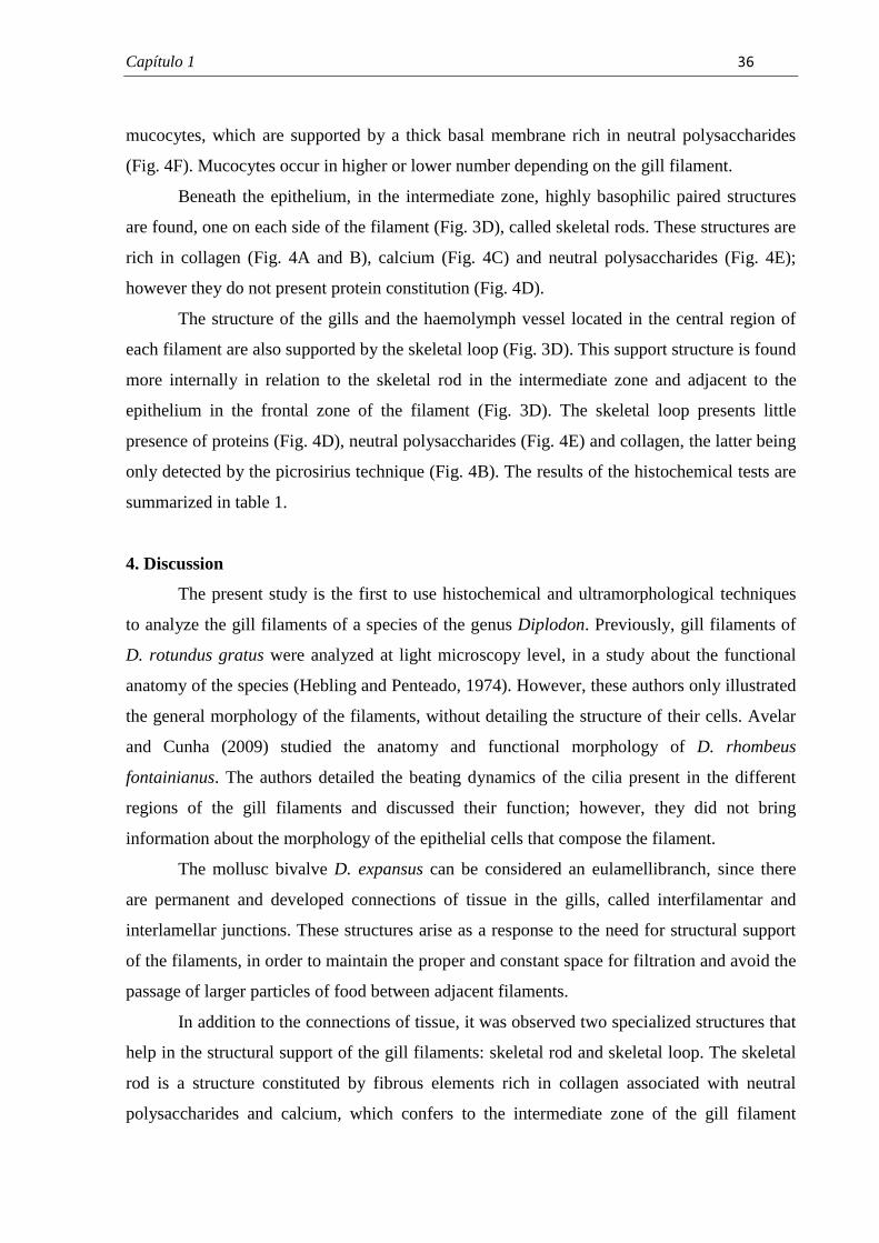
Capítulo 1� � � ��
� �
mucocytes, which are supported by a thick basal membrane rich in neutral polysaccharides
(Fig. 4F). Mucocytes occur in higher or lower number depending on the gill filament.
Beneath the epithelium, in the intermediate zone, highly basophilic paired structures
are found, one on each side of the filament (Fig. 3D), called skeletal rods. These structures are
rich in collagen (Fig. 4A and B), calcium (Fig. 4C) and neutral polysaccharides (Fig. 4E);
however they do not present protein constitution (Fig. 4D).
The structure of the gills and the haemolymph vessel located in the central region of
each filament are also supported by the skeletal loop (Fig. 3D). This support structure is found
more internally in relation to the skeletal rod in the intermediate zone and adjacent to the
epithelium in the frontal zone of the filament (Fig. 3D). The skeletal loop presents little
presence of proteins (Fig. 4D), neutral polysaccharides (Fig. 4E) and collagen, the latter being
only detected by the picrosirius technique (Fig. 4B). The results of the histochemical tests are
summarized in table 1.
4. Discussion
The present study is the first to use histochemical and ultramorphological techniques
to analyze the gill filaments of a species of the genus Diplodon. Previously, gill filaments of
D. rotundus gratus were analyzed at light microscopy level, in a study about the functional
anatomy of the species (Hebling and Penteado, 1974). However, these authors only illustrated
the general morphology of the filaments, without detailing the structure of their cells. Avelar
and Cunha (2009) studied the anatomy and functional morphology of D. rhombeus
fontainianus. The authors detailed the beating dynamics of the cilia present in the different
regions of the gill filaments and discussed their function; however, they did not bring
information about the morphology of the epithelial cells that compose the filament.
The mollusc bivalve D. expansus can be considered an eulamellibranch, since there
are permanent and developed connections of tissue in the gills, called interfilamentar and
interlamellar junctions. These structures arise as a response to the need for structural support
of the filaments, in order to maintain the proper and constant space for filtration and avoid the
passage of larger particles of food between adjacent filaments.
In addition to the connections of tissue, it was observed two specialized structures that
help in the structural support of the gill filaments: skeletal rod and skeletal loop. The skeletal
rod is a structure constituted by fibrous elements rich in collagen associated with neutral
polysaccharides and calcium, which confers to the intermediate zone of the gill filament
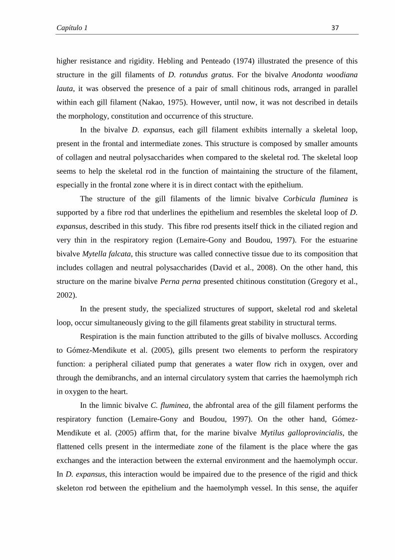
Capítulo 1� � � ���
� �
higher resistance and rigidity. Hebling and Penteado (1974) illustrated the presence of this
structure in the gill filaments of D. rotundus gratus. For the bivalve Anodonta woodiana
lauta, it was observed the presence of a pair of small chitinous rods, arranged in parallel
within each gill filament (Nakao, 1975). However, until now, it was not described in details
the morphology, constitution and occurrence of this structure.
In the bivalve D. expansus, each gill filament exhibits internally a skeletal loop,
present in the frontal and intermediate zones. This structure is composed by smaller amounts
of collagen and neutral polysaccharides when compared to the skeletal rod. The skeletal loop
seems to help the skeletal rod in the function of maintaining the structure of the filament,
especially in the frontal zone where it is in direct contact with the epithelium.
The structure of the gill filaments of the limnic bivalve Corbicula fluminea is
supported by a fibre rod that underlines the epithelium and resembles the skeletal loop of D.
expansus, described in this study. This fibre rod presents itself thick in the ciliated region and
very thin in the respiratory region (Lemaire-Gony and Boudou, 1997). For the estuarine
bivalve Mytella falcata, this structure was called connective tissue due to its composition that
includes collagen and neutral polysaccharides (David et al., 2008). On the other hand, this
structure on the marine bivalve Perna perna presented chitinous constitution (Gregory et al.,
2002).
In the present study, the specialized structures of support, skeletal rod and skeletal
loop, occur simultaneously giving to the gill filaments great stability in structural terms.
Respiration is the main function attributed to the gills of bivalve molluscs. According
to Gómez-Mendikute et al. (2005), gills present two elements to perform the respiratory
function: a peripheral ciliated pump that generates a water flow rich in oxygen, over and
through the demibranchs, and an internal circulatory system that carries the haemolymph rich
in oxygen to the heart.
In the limnic bivalve C. fluminea, the abfrontal area of the gill filament performs the
respiratory function (Lemaire-Gony and Boudou, 1997). On the other hand, Gómez-
Mendikute et al. (2005) affirm that, for the marine bivalve Mytilus galloprovincialis, the
flattened cells present in the intermediate zone of the filament is the place where the gas
exchanges and the interaction between the external environment and the haemolymph occur.
In D. expansus, this interaction would be impaired due to the presence of the rigid and thick
skeleton rod between the epithelium and the haemolymph vessel. In this sense, the aquifer
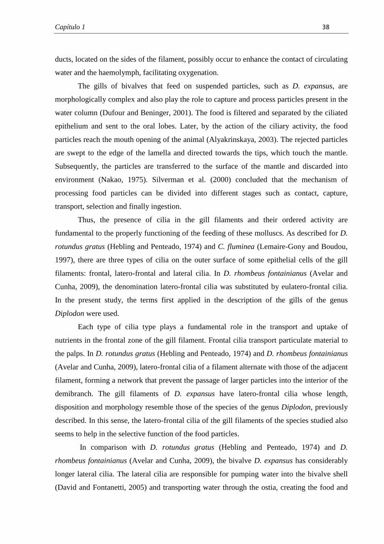
Capítulo 1� � � ���
� �
ducts, located on the sides of the filament, possibly occur to enhance the contact of circulating
water and the haemolymph, facilitating oxygenation.
The gills of bivalves that feed on suspended particles, such as D. expansus, are
morphologically complex and also play the role to capture and process particles present in the
water column (Dufour and Beninger, 2001). The food is filtered and separated by the ciliated
epithelium and sent to the oral lobes. Later, by the action of the ciliary activity, the food
particles reach the mouth opening of the animal (Alyakrinskaya, 2003). The rejected particles
are swept to the edge of the lamella and directed towards the tips, which touch the mantle.
Subsequently, the particles are transferred to the surface of the mantle and discarded into
environment (Nakao, 1975). Silverman et al. (2000) concluded that the mechanism of
processing food particles can be divided into different stages such as contact, capture,
transport, selection and finally ingestion.
Thus, the presence of cilia in the gill filaments and their ordered activity are
fundamental to the properly functioning of the feeding of these molluscs. As described for D.
rotundus gratus (Hebling and Penteado, 1974) and C. fluminea (Lemaire-Gony and Boudou,
1997), there are three types of cilia on the outer surface of some epithelial cells of the gill
filaments: frontal, latero-frontal and lateral cilia. In D. rhombeus fontainianus (Avelar and
Cunha, 2009), the denomination latero-frontal cilia was substituted by eulatero-frontal cilia.
In the present study, the terms first applied in the description of the gills of the genus
Diplodon were used.
Each type of cilia type plays a fundamental role in the transport and uptake of
nutrients in the frontal zone of the gill filament. Frontal cilia transport particulate material to
the palps. In D. rotundus gratus (Hebling and Penteado, 1974) and D. rhombeus fontainianus
(Avelar and Cunha, 2009), latero-frontal cilia of a filament alternate with those of the adjacent
filament, forming a network that prevent the passage of larger particles into the interior of the
demibranch. The gill filaments of D. expansus have latero-frontal cilia whose length,
disposition and morphology resemble those of the species of the genus Diplodon, previously
described. In this sense, the latero-frontal cilia of the gill filaments of the species studied also
seems to help in the selective function of the food particles.
In comparison with D. rotundus gratus (Hebling and Penteado, 1974) and D.
rhombeus fontainianus (Avelar and Cunha, 2009), the bivalve D. expansus has considerably
longer lateral cilia. The lateral cilia are responsible for pumping water into the bivalve shell
(David and Fontanetti, 2005) and transporting water through the ostia, creating the food and

Capítulo 1� � � � �
� �
respiratory flow (Jorgensen, 1976). Thus, the food and respiratory functions may be
optimized due to the presence of longer lateral cilia in the species studied.
The abfrontal surface of the gill filaments is not directly involved with feeding
process, since the capture of food particles occurs on the frontal surface of the epithelium
(Dufour and Beninger, 2001). However, some elements related to the processing of
alimentary particles, such as cilia and mucus, are present in the abfrontal surface of the gills
of D. expansus.
The cilia are scarce and the mucocytes rich in acid polysaccharides are more frequent
in the abfrontal zone of the gill filament of the species studied, similar situation that is found
for the eulamellibranch bivalve Spisula solidissima (Dufour and Beninger, 2001). In D.
expansus, the cilia found in the abfrontal zone of the filaments do not seem to be related to the
creation of water flow, since such cilia are short and in little number when compared to the
lateral cilia that admittedly perform this function. Possibly these structures appear as vestigial
along the evolution of bivalves (Dufour and Beninger, 2001) or plays the sensorial function,
as previously discussed by Atkins (1936).
According to Beninger et al. (1997), the presence of mucocytes in the abfrontal zone
of eulamellibranch bivalves, such as D. expansus, occurs due to the need of a great lubrication
of this region in order to diminish the friction between water and epithelium, since the water
pumped through the gill filaments is directed to the abfrontal chambers (present in the
interlamellar space) of reduced volume. In limninc bivalves, gills secrete mainly acid
mucosubstances, which help in the transport of food particles and in the formation of a
protective and lubricant mucous layer (Kale and Patil, 1977). The mucus of acid nature like
those found in the mucocytes of D. expansus is highly viscous and considered a good
lubricant, since it is not easily hydrated or removed from the epithelium (Faillard and
Schauer, 1972; Hunt, 1970). Therefore, it is possible that the mucus secreted in the abfrontal
zone of D. expansus acts as an effective lubricant, considerably reducing the attrition between
water and epithelium.
In the intermediate zone of the gill filaments of D. expansus, mucocytes rich in acid
polysaccharides were also found. In the marine bivalve Mytilus galloprovincialis, the
mucocytes rich in acid polysaccharides occurred in the frontal zone of the filament, while
mucocytes rich in neutral polysaccharides were observed in the abfrontal zone (Gómez-
Mendikute et al., 2005). In the estuarine bivalve Mytella falcata, mucocytes rich in neutral
polysaccharides were often observed in the abfrontal zone of the filament. Such cells were
Capítulo 1� � � ���
� �
also observed in the frontal zone near the intermediate zone of the filament (David et al.,
2008). In animals exposed to pollutants, the authors found that the increase in the number of
mucocytes occurred preferentially in the frontal zone of the filament (David and Fontanetti,
2009).
In the gill filament of the marine bivalve Mya arenaria, the arrangement of the
mucocytes rich in acid polysaccharides occurs similarly to the observed in D. expansus,
however, such cells are less abundant in the abfrontal zone of the marine bivalve (Beninger et
al., 1997). The authors affirm that it is unlikely that the mucous secreted in the intermediate
zone be transported toward the frontal zone, since the lateral cilia beat in opposite direction
and are separated from the latero-frontal cilia by non-ciliated cells. Since residues of mucous
are found in the frontal zone of the filaments, the authors pointed out the need to conduct
further investigations in order to verify the possible existence of a glandular system integrated
to these mucocytes that would make possible the direct secretion of mucus on the frontal zone
of the filament.
The information obtained in this study showed that the gills of the Brazilian endemic
bivalve D. expansus are typical of eulamellibranch bivalves and are suitable for
histopathological studies applied on the effects of water pollutants. Important peculiarities
never related in detail before showed the morphological and histochemical differences in
bivalve species gills such as the constitution of the structure supporting the haemolymph
vessel and the simultaneous presence of two specialized structures that play an important role
in the structural maintenance of the gills, the skeletal rod and skeletal loop. Ultrastructural
studies will be carried out in order to provide morphological detailing of the structures and
cells that compose the gill filaments of the bivalve.
Acknowledgments
This research has been supported by FAPESP (Fundação de Amparo a Pesquisa do Estado de
São Paulo) Grant n. 2009/12489-9. The authors thank Dr. Wagner Eustáquio Paiva Avelar
and Dr. Cláudia Tasso Callil for the specimens identification; Cristiane M. Mileo for the
illustrations; Mônika Iamonte, Antonio Teruyoshi Yabuki and Gerson Mello de Souza for the
technical support; Marcos Perdiza for the support during the collection of the animals; and the
biologists Cintya Aparecida Christofoletti, Raphael Bastão de Souza, and Ana Claudia de
Castro Marcato for their help during the experiments.
Capítulo 1� � � ���
� �
References
Alyakrinskaya, I.O., 2003. Tissue hemoglobins in Bivalvia (Mollusca). Biology Bulletin, 30,
617-626.
Atkins, D., 1936. On the ciliary mechanisms and interrelationships of lamellibranchs. I. New
observations on sorting mechanisms. Q.J. Microsc. Sci. 79, 181-308.
Avelar, W.E.P., 1999. Moluscos Bivalves, in: Ismael, D., Valentin, W.C., Matsumara-
Tundisi, T., Rocha, O. (Eds.), Biodiversidade do Estado de São Paulo, Brasil: Invertebrados
de água doce, pp. 65-68. Fundação de Amparo à pesquisa do Estado de São Paulo (FAPESP),
São Paulo.
Avelar, W.E.P., Cunha, A.D., 2009. The anatomy and functional morphology of Diplodon
rhombeus fontainianus (Orbigny, 1835) (Mollusca, Bivalvia, Hyriidae). Braz. J. Biol. 69,
1153-1163.
Beninger, P.G., Dufour, S.C., Bourque, J., 1997. Particle processing mechanisms of the
eulamellibranch bivalves Spisula solidissima and Mya arenaria. Mar. Ecol. Prog. Ser. 150,
157-169.
Colville, A.E., Lim, R.P., 2003. Microscopic structure of the mantle and palps in the
freshwater mussels Velesunio ambiguus and Hyridella depressa (Bivalvia: Hyriidae).
Molluscan Research, 23, 1-20.
Curial, O., Lange, R.R., 1974a. Hermafroditismo em Diplodon delotundus expansus.
Arquivos de Biologia e Tecnologia, 17, 109-110.
Curial, O., Lange, R.R., 1974b. Observações sobre a proporção de sexos em Diplodon
deodontus expansus. Arquivos de Biologia e Tecnologia, 17, 111-112.
Curial, O., Lange, R.R., 1975. Variações histológicas sazonais das gônadas do Diplodon
delodontus expansus (Küster) (Mollusca, Unionidade). Arquivos de Biologia e Tecnologia,
18, 65-67.
Capítulo 1� � � ���
� �
David, J.A.O., Fontanetti, C.S., 2005. Surface morphology of Mytella falcata gill filaments
from three regions of the Santos estuary. Brazilian Journal of Morphological Sciences, 22,
203-210.
David, J.A.O., Fontanetti, C.S., 2009. The role of mucus in Mytella falcata (Orbigny, 1842)
gills from polluted environments. Water Air Soil Pollut. 203, 261–266.
David, J.A.O., Salaroli, R.B., Fontanetti, C.S., 2008. Fine structure of Mytella falcata
(Bivalvia) gill filaments. Micron, 39, 329-336.
Dufour, S.C., Beninger, P.G., 2001. A functional interpretation of cilia and mucocyte
distributions on the abfrontal surface of bivalve gills. Mar. Biol. 138, 295–309.
Faillard, H., Schauer, R., 1972. Glycoproteins as lubrificans, protective agents, carries,
structural proteins and as participants in other functions, in: Gottschalk, A. (Ed.),
Glycoproteins. Their composition, structure and function, part B, vol. 5, pp. 1246-1267.
Elsevier, Amsterdam.
Gómez-Mendikute, A., Elizondo, M., Venier, P., Cajaraville, M. P., 2005. Characterization of
mussel gill cells in vivo and in vitro. Cell Tissue Res. 321, 131-140.
Gregory, M.A., George, R.C., 2000. The structure and surface morphology of the gill
filaments in the brown mussel Perna perna Afr. Zool. 35, 121-129.
Gregory, M.A., George, R.C., McClurg, T.P., 1996. The architecture and fine structure of gill
filaments in the brown mussel Perna perna. S. Afr. J. Zool. 31, 193-206.
Gregory, M.A., Marshall, D.J., George, R.C., Anandraj, A., McClurg, T.P., 2002.
Correlations between metal uptake in the soft tissue of Perna perna and gill filament
pathology after exposure to mercury. Mar. Pollut. Bull. 45, 114-125.
Hebling, N.J., Penteado, A.M.G., 1974. Anatomia functional de Diplodon rotundus gratus
Wagner, 1827 (Mollusca, Bivalvia). Rev. Brasil. Biol. 34, 67-80.
Capítulo 1� � � ���
� �
Henry, R., Simão, S.A., 1984. Evaluation of density and biomass of a bivalve population
(Diplodon delodontus expansus) (Küster, 1856) in a small tropical reservoir. Revue D’
Hydrobiologie Trocale, 17, 309-318.
Henry, R., Filoso, S., 1985. Spatial distribution of a bivalve population (Diplodon delodontus
expansus) (Küster, 1856) in a small tropical reservoir. Rev. Brasil. Biol. 45, 407-415.
Henry, R., Filoso, S., 1987. Nutrient Release Through Decomposition of Diplodon
Delodontus Expansus (Mollusca, Bivalvia) and Some Considerations of its Effects on the
Ecology of a Small Tropical Reservoir. Rev. Brasil. Biol. 47, 479-486.
Hunt, S., 1970. Polysaccharides-protein complexes in invertebrates. Academic Press, London.
Jorgensen, C.B., 1976. Comparative studies on the function of gills in suspension feeding
bivalves, with special reference to effects of serotonin. Biol. Bull. 151, 331-343.
Junqueira, L.C.U., Junqueira, M.M.S., 1983. Técnicas Básicas de Citologia e Histologia.
Livraria Editora Santos, São Paulo.
Kale, G.S., Patil, V.Y., 1977. Histochemical studies on the mucosubstances in the gill of
freshwater mussels, Parreysia corrugata var. nagpoorensis (Lea). Acta Histochem. 59, 1-7.
Karnovsky, M.J., 1965. A formaldehyde-glutaraldehyde fixative at high osmolarity for use in
electron microscopy. J. Cell Biol. 11, 137-140.
Lemaire-Gony, S., Boudou, A., 1997. Mantle and gill fine structure in freshwater Asiatic clam
Corbicula fluminea (Müller). Annls Limnol. 33, 163-178.
Mansur, M.C.D., 1999. Gloquídio de Diplodon martensi (Ihering) (Mollusca, Bivalvia,
Hyriidae) e seu ciclo parasitário. Rev. Bras. Zool. 16, 185-194.
Capítulo 1� � � ���
� �
Mansur, M.C.D., Santos, C.P., 2008. Invertebrados Aquáticos: Diplodon Expansus, in:
Machado, A.B.M., Drummond, G.M., PAGLIA, A.P. (Eds.), Livro Vermelho da Fauna
Ameaçada de Extinção, pp. 197-198. Fundação Bioversitas, Belo Horizonte.
Mantecca, P., Vailati, G., Bacchetta, R., 2006. Histological changes and micronucleous
inductions in the Zebra mussel Dreissena polymorpha after Paraquat exposure. Histol.
Histopathol. 21, 829-840.
Nakao, T., 1975. The fine structure and innervation of gill lamellae in Anodonta. Cell Tiss.
Res. 157, 239-254.
Pearse, A.G.E., 1985. Histochemistry: Theoretical and Applied. Churchill, London.
Peltier, G. L., Meyer, J.L., Jagoe, C.H., William, A., Hopkins, W.A., 2008. Using trace
element concentrations in Corbicula fluminea to identify potential sources of contamination
in an urban river. Environmental Pollution, 154, 283-290.
Silverman, H., Lynn, J.W., Dietz, T.H., 2000. In vitro studies of particle capture and transport
in suspension-feeding bivalves. Limnol. Oceanogr. 45, 1199-1203.
Vilela, I.V., Oliveira, I.M., Silva, J., Henriques, J.A.P., 2006. DNA damage and repair in
haemolynph cells of golden mussel (Limnoperna fortunei) exposed to environmental
contaminants. Mutat. Res. 605, 78-86.
Villar, C., Stripeikis, J., D´Huicque, L., Tudino, M., Troccoli, O., Bonetto, C., 1999. Cd, Cu,
and Zn concentrations in sediments and the invasive bivalves Limnoperna fortunei and
Corbicula fluminea at the Rio de La Plata basin, Argentina. Hydrobiologia, 416, 41-49.
Zupan, I., Kalafatic, M., 2003. Histological effects of low atrazine contamination on Zebra
Mussel (Dreissena polymorpha Pallas). Bull. Environ. Contam. Toxicol. 70, 688-695.
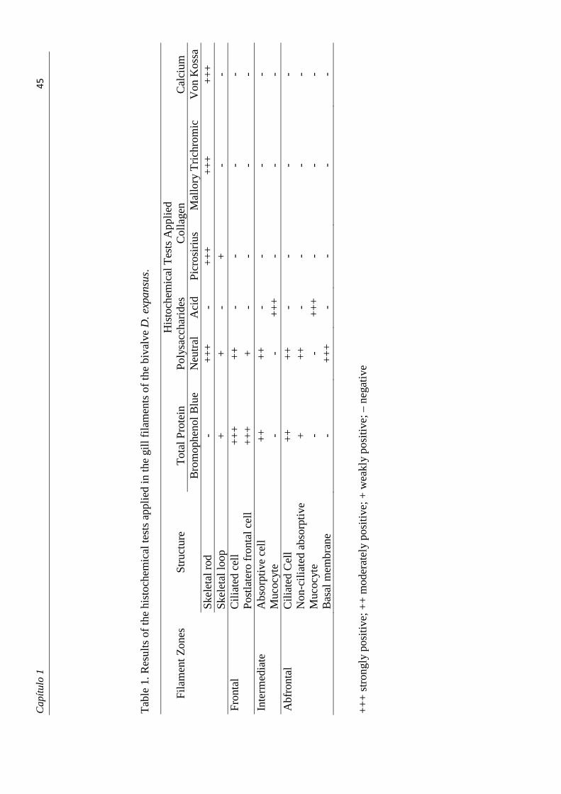
Cap
ítul
o 1�
��
��
��
Tab
le 1
. Res
ults
of
the
hist
oche
mic
al te
sts
appl
ied
in th
e gi
ll fi
lam
ents
of
the
biva
lve
D. e
xpan
sus.
His
toch
emic
al T
ests
App
lied
Fi
lam
ent Z
ones
St
ruct
ure
Tot
al P
rote
in
Pol
ysac
char
ides
C
olla
gen
Cal
cium
B
rom
ophe
nol B
lue
Neu
tral
A
cid
Picr
osir
ius
Mal
lory
Tri
chro
mic
V
on K
ossa
Ske
leta
l rod
-
++
+
- +
++
+
++
+
++
Skel
etal
loop
+
+
-
+
- -
Fron
tal
Cil
iate
d ce
ll
++
+
++
-
- -
-
Pos
tlate
ro f
ront
al c
ell
++
+
+
- -
- -
Inte
rmed
iate
A
bsor
ptiv
e ce
ll
++
+
+
- -
- -
M
ucoc
yte
- -
++
+
- -
- A
bfro
ntal
C
iliat
ed C
ell
++
+
+
- -
- -
N
on-c
ilia
ted
abso
rpti
ve
+
++
-
- -
-
Muc
ocyt
e
- -
++
+
- -
- B
asal
mem
bran
e
- +
++
-
- -
-
++
+ s
tron
gly
posi
tive;
++
mod
erat
ely
posi
tive;
+ w
eakl
y po
sitiv
e; –
neg
ativ
e
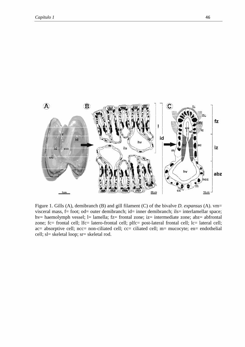
Capítulo 1� � ��
� �
Figure 1. Gills (A), demibranch (B) and gill filament (C) of the bivalve D. expansus (A). vm= visceral mass, f= foot; od= outer demibranch; id= inner demibranch; ils= interlamellar space; hv= haemolymph vessel; l= lamella; fz= frontal zone; iz= intermediate zone; abz= abfrontal zone; fc= frontal cell; lfc= latero-frontal cell; plfc= post-lateral frontal cell; lc= lateral cell; ac= absorptive cell; ncc= non-ciliated cell; cc= ciliated cell; m= mucocyte; en= endothelial cell; sl= skeletal loop; sr= skeletal rod.
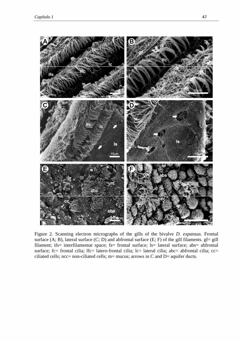
Capítulo 1� � ���
� �
Figure 2. Scanning electron micrographs of the gills of the bivalve D. expansus. Frontal surface (A; B), lateral surface (C; D) and abfrontal surface (E; F) of the gill filaments. gf= gill filament; ifs= interfilamentar space; fs= frontal surface; ls= lateral surface; abs= abfrontal surface; fc= frontal cilia; lfc= latero-frontal cilia; lc= lateral cilia; abc= abfrontal cilia; cc= ciliated cells; ncc= non-ciliated cells; m= mucus; arrows in C and D= aquifer ducts.
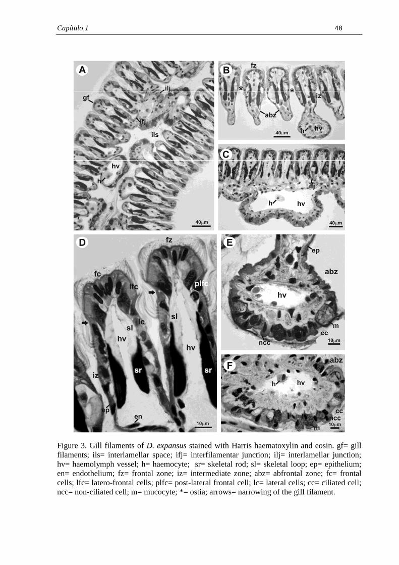
Capítulo 1� � ���
� �
Figure 3. Gill filaments of D. expansus stained with Harris haematoxylin and eosin. gf= gill filaments; ils= interlamellar space; ifj= interfilamentar junction; ilj= interlamellar junction; hv= haemolymph vessel; h= haemocyte; sr= skeletal rod; sl= skeletal loop; ep= epithelium; en= endothelium; fz= frontal zone; iz= intermediate zone; abz= abfrontal zone; fc= frontal cells; lfc= latero-frontal cells; plfc= post-lateral frontal cell; lc= lateral cells; cc= ciliated cell; ncc= non-ciliated cell; m= mucocyte; *= ostia; arrows= narrowing of the gill filament.
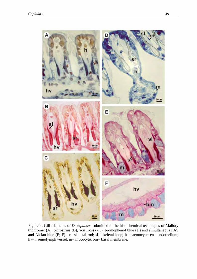
Capítulo 1� � � �
� �
Figure 4. Gill filaments of D. expansus submitted to the histochemical techniques of Mallory trichromic (A), picrosirius (B), von Kossa (C), bromophenol blue (D) and simultaneous PAS and Alcian blue (E; F). sr= skeletal rod; sl= skeletal loop; h= haemocyte; en= endothelium; hv= haemolymph vessel; m= mucocyte; bm= basal membrane.
Capítulo 2� � ��
7.2. CAPÍTULO 2
Surface morphology of Diplodon expansus (Küster, 1856) (Mollusca,
Bivalvia, Hyriidae) gill filaments after exposure to environmentally relevant
concentrations of atrazine herbicide
Larissa Rosa Nogarol; Ana Luiza Brossi-Garcia & Carmem Silvia Fontanetti1*
UNESP, São Paulo State University, Av. 24A, no 1515, CEP 13506-900, Rio Claro, SP,
Brazil.
* Corresponding author
E-mail address: [email protected]
Tel.: +55 19 35264139; fax: +55 19 35264135.
Artigo publicado no periódico Microscopy Research and Technique, 2011.
DOI 10.1002/jemt.21130�
�
�
�
�
Capítulo 2� � ��
Resumo
A espécie endêmica brasileira Diplodon expansus (Küster, 1856) é encontrada em corpos de
água doce no sudeste do país, em regiões de grande influência antropogênica com destaque
para a agricultura. Um dos principais defensivos agrícolas utilizados na região de ocorrência
da espécie é o herbicida atrazina, o qual possui grande potencial de contaminação dos
ambientes aquáticos. Assim, esse estudo buscou investigar as possíveis alterações na
superfície dos filamentos branquiais do bivalve, após exposição aguda a diferentes
concentrações do herbicida. As principais alterações observadas foram o acúmulo do muco na
superfície frontal dos filamentos, desorganização dos cílios na superfície frontal e perda dos
cílios das células frontais. Como observado em outros estudos, o aumento da secreção de
muco na região frontal dos filamentos agiu como um mecanismo de proteção do filamento. A
desorganização dos cílios da superfície frontal dos filamentos e a perda dos cílios das células
frontais poderão causar prejuízos funcionais das brânquias, comprometendo a captação de
partículas alimentares e a respiração.
Palavras-chave: molusco; pesticida; MEV; ctenidia.
�
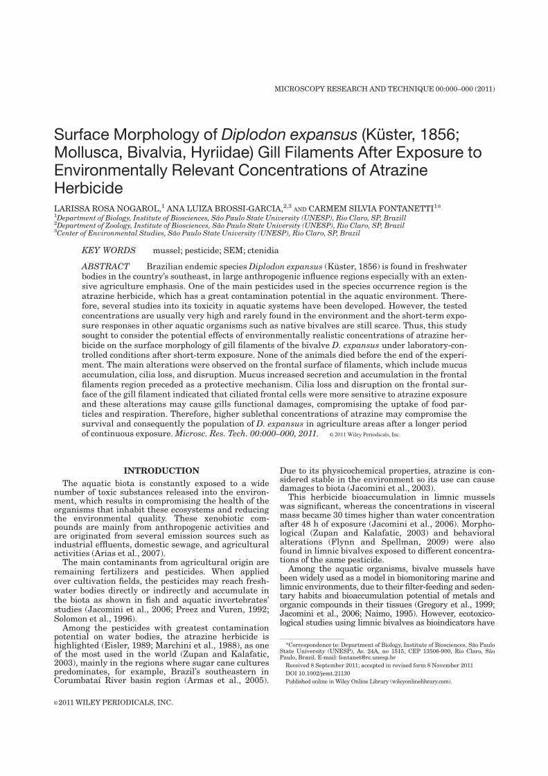
Surface Morphology of Diplodon expansus (Kuster, 1856;Mollusca, Bivalvia, Hyriidae) Gill Filaments After Exposure toEnvironmentally Relevant Concentrations of AtrazineHerbicideLARISSA ROSA NOGAROL,1 ANA LUIZA BROSSI-GARCIA,2,3 AND CARMEM SILVIA FONTANETTI1*1Department of Biology, Institute of Biosciences, Sao Paulo State University (UNESP), Rio Claro, SP, Brazill2Department of Zoology, Institute of Biosciences, Sao Paulo State University (UNESP), Rio Claro, SP, Brazil3Center of Environmental Studies, Sao Paulo State University (UNESP), Rio Claro, SP, Brazil
KEY WORDS mussel; pesticide; SEM; ctenidia
ABSTRACT Brazilian endemic speciesDiplodon expansus (Kuster, 1856) is found in freshwaterbodies in the country’s southeast, in large anthropogenic influence regions especially with an exten-sive agriculture emphasis. One of the main pesticides used in the species occurrence region is theatrazine herbicide, which has a great contamination potential in the aquatic environment. There-fore, several studies into its toxicity in aquatic systems have been developed. However, the testedconcentrations are usually very high and rarely found in the environment and the short-term expo-sure responses in other aquatic organisms such as native bivalves are still scarce. Thus, this studysought to consider the potential effects of environmentally realistic concentrations of atrazine her-bicide on the surface morphology of gill filaments of the bivalve D. expansus under laboratory-con-trolled conditions after short-term exposure. None of the animals died before the end of the experi-ment. The main alterations were observed on the frontal surface of filaments, which include mucusaccumulation, cilia loss, and disruption. Mucus increased secretion and accumulation in the frontalfilaments region preceded as a protective mechanism. Cilia loss and disruption on the frontal sur-face of the gill filament indicated that ciliated frontal cells were more sensitive to atrazine exposureand these alterations may cause gills functional damages, compromising the uptake of food par-ticles and respiration. Therefore, higher sublethal concentrations of atrazine may compromise thesurvival and consequently the population of D. expansus in agriculture areas after a longer periodof continuous exposure.Microsc. Res. Tech. 00:000–000, 2011. VVC 2011 Wiley Periodicals, Inc.
INTRODUCTION
The aquatic biota is constantly exposed to a widenumber of toxic substances released into the environ-ment, which results in compromising the health of theorganisms that inhabit these ecosystems and reducingthe environmental quality. These xenobiotic com-pounds are mainly from anthropogenic activities andare originated from several emission sources such asindustrial effluents, domestic sewage, and agriculturalactivities (Arias et al., 2007).The main contaminants from agricultural origin are
remaining fertilizers and pesticides. When appliedover cultivation fields, the pesticides may reach fresh-water bodies directly or indirectly and accumulate inthe biota as shown in fish and aquatic invertebrates’studies (Jacomini et al., 2006; Preez and Vuren, 1992;Solomon et al., 1996).Among the pesticides with greatest contamination
potential on water bodies, the atrazine herbicide ishighlighted (Eisler, 1989; Marchini et al., 1988), as oneof the most used in the world (Zupan and Kalafatic,2003), mainly in the regions where sugar cane culturespredominates, for example, Brazil’s southeastern inCorumbataı River basin region (Armas et al., 2005).
Due to its physicochemical properties, atrazine is con-sidered stable in the environment so its use can causedamages to biota (Jacomini et al., 2003).This herbicide bioaccumulation in limnic mussels
was significant, whereas the concentrations in visceralmass became 30 times higher than water concentrationafter 48 h of exposure (Jacomini et al., 2006). Morpho-logical (Zupan and Kalafatic, 2003) and behavioralalterations (Flynn and Spellman, 2009) were alsofound in limnic bivalves exposed to different concentra-tions of the same pesticide.Among the aquatic organisms, bivalve mussels have
been widely used as a model in biomonitoring marine andlimnic environments, due to their filter-feeding and seden-tary habits and bioaccumulation potential of metals andorganic compounds in their tissues (Gregory et al., 1999;Jacomini et al., 2006; Naimo, 1995). However, ecotoxico-logical studies using limnic bivalves as bioindicators have
*Correspondence to: Department of Biology, Institute of Biosciences, Sao PauloState University (UNESP), Av. 24A, no 1515, CEP 13506-900, Rio Claro, SaoPaulo, Brazil. E-mail: [email protected]
Received 8 September 2011; accepted in revised form 8 November 2011
DOI 10.1002/jemt.21130
Published online inWiley Online Library (wileyonlinelibrary.com).
VVC 2011 WILEY PERIODICALS, INC.
MICROSCOPY RESEARCH AND TECHNIQUE 00:000–000 (2011)
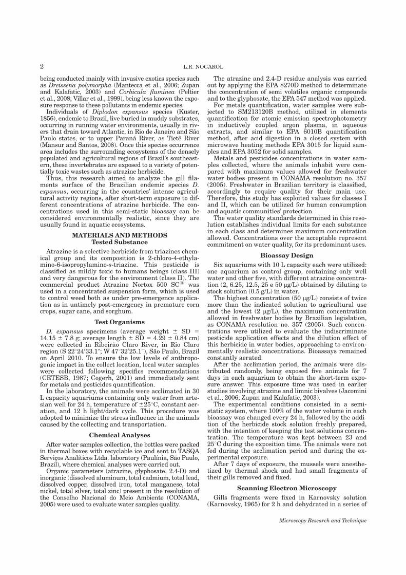
being conducted mainly with invasive exotics species suchas Dreissena polymorpha (Mantecca et al., 2006; Zupanand Kalafatic, 2003) and Corbicula fluminea (Peltieret al., 2008; Villar et al., 1999), being less known the expo-sure response to these pollutants in endemic species.Individuals of Diplodon expansus species (Kuster,
1856), endemic to Brazil, live buried inmuddy substrates,occurring in running water environments, usually in riv-ers that drain toward Atlantic, in Rio de Janeiro and SaoPaulo states, or to upper Parana River, as Tiete River(Mansur and Santos, 2008). Once this species occurrencearea includes the surrounding ecosystems of the denselypopulated and agricultural regions of Brazil’s southeast-ern, these invertebrates are exposed to a variety of poten-tially toxic wastes such as atrazine herbicide.Thus, this research aimed to analyze the gill fila-
ments surface of the Brazilian endemic species D.expansus, occurring in the countries’ intense agricul-tural activity regions, after short-term exposure to dif-ferent concentrations of atrazine herbicide. The con-centrations used in this semi-static bioassay can beconsidered environmentally realistic, since they areusually found in aquatic ecosystems.
MATERIALS AND METHODSTested Substance
Atrazine is a selective herbicide from triazines chem-ical group and its composition is 2-chloro-4-ethyla-mino-6-isopropylamino-s-triazine. This pesticide isclassified as mildly toxic to humans beings (class III)and very dangerous for the environment (class II). Thecommercial product Atrazine Nortox 500 SC1 wasused in a concentrated suspension form, which is usedto control weed both as under pre-emergence applica-tion as in untimely post-emergency in premature corncrops, sugar cane, and sorghum.
Test Organisms
D. expansus specimens (average weight 6 SD 514.15 6 7.8 g; average length 6 SD 5 4.29 6 0.84 cm)were collected in Ribeirao Claro River, in Rio Claroregion (S 22824033.1@; W 47832025.1@), Sao Paulo, Brazilon April 2010. To ensure the low levels of anthropo-genic impact in the collect location, local water sampleswere collected following specifics recommendations(CETESB, 1987; Cogerh, 2001) and immediately sentfor metals and pesticides quantification.In the laboratory, the animals were acclimated in 30
L capacity aquariums containing only water from arte-sian well for 24 h, temperature of 6258C, constant aer-ation, and 12 h light/dark cycle. This procedure wasadopted to minimize the stress influence in the animalscaused by the collecting and transportation.
Chemical Analyses
After water samples collection, the bottles were packedin thermal boxes with recyclable ice and sent to TASQAServicos Analıticos Ltda. laboratory (Paulınia, Sao Paulo,Brazil), where chemical analyses were carried out.Organic parameters (atrazine, glyphosate, 2.4-D) and
inorganic (dissolved aluminum, total cadmium, total lead,dissolved copper, dissolved iron, total manganese, totalnickel, total silver, total zinc) present in the resolution ofthe Conselho Nacional do Meio Ambiente (CONAMA,2005) were used to evaluate water samples quality.
The atrazine and 2.4-D residue analysis was carriedout by applying the EPA 8270D method to determinatethe concentration of semi volatiles organic compoundsand to the glyphosate, the EPA 547 method was applied.For metals quantification, water samples were sub-
jected to SM213120B method, utilized in elementsquantification for atomic emission spectrophotometryin inductively coupled argon plasma, in aqueousextracts, and similar to EPA 6010B quantificationmethod, after acid digestion in a closed system withmicrowave heating methods EPA 3015 for liquid sam-ples and EPA 3052 for solid samples.Metals and pesticides concentrations in water sam-
ples collected, where the animals inhabit were com-pared with maximum values allowed for freshwaterwater bodies present in CONAMA resolution no. 357(2005). Freshwater in Brazilian territory is classified,accordingly to require quality for their main use.Therefore, this study has exploited values for classes Iand II, which can be utilized for human consumptionand aquatic communities’ protection.The water quality standards determined in this reso-
lution establishes individual limits for each substancein each class and determines maximum concentrationallowed. Concentrations over the acceptable representcommitment on water quality, for its predominant uses.
Bioassay Design
Six aquariums with 10 L capacity each were utilized:one aquarium as control group, containing only wellwater and other five, with different atrazine concentra-tion (2, 6.25, 12.5, 25 e 50 lg/L) obtained by diluting tostock solution (0.5 g/L) in water.The highest concentration (50 lg/L) consists of twice
more than the indicated solution to agricultural useand the lowest (2 lg/L), the maximum concentrationallowed in freshwater bodies by Brazilian legislation,as CONAMA resolution no. 357 (2005). Such concen-trations were utilized to evaluate the indiscriminatepesticide application effects and the dilution effect ofthis herbicide in water bodies, approaching to environ-mentally realistic concentrations. Bioassays remainedconstantly aerated.After the acclimation period, the animals were dis-
tributed randomly, being exposed five animals for 7days in each aquarium to obtain the short-term expo-sure answer. This exposure time was used in earlierstudies involving atrazine and limnic bivalves (Jacominiet al., 2006; Zupan and Kalafatic, 2003).The experimental conditions consisted in a semi-
static system, where 100% of the water volume in eachbioassay was changed every 24 h, followed by the addi-tion of the herbicide stock solution freshly prepared,with the intention of keeping the test solutions concen-tration. The temperature was kept between 23 and258C during the exposition time. The animals were notfed during the acclimation period and during the ex-perimental exposure.After 7 days of exposure, the mussels were anesthe-
tized by thermal shock and had small fragments oftheir gills removed and fixed.
Scanning Electron Microscopy
Gills fragments were fixed in Karnovsky solution(Karnovsky, 1965) for 2 h and dehydrated in a series of
Microscopy Research and Technique
2 L.R. NOGAROL
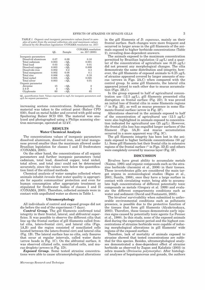
increasing acetone concentrations. Subsequently, thematerial was taken to the critical point (Balzer CPD030), fixed on metal holder and covered with gold usingSputtering Balzer SCD 050. The material was ana-lyzed and photographed using a Philips scanning elec-tron microscope, operated at 12 kV.
RESULTSWater Chemical Analysis
The concentrations values of inorganic parametersdissolved aluminum, dissolved iron, and total manga-nese proved smaller than the maximum allowed underBrazilian legislation for classes I and II freshwaters(CONAMA, 2005).On the other hand, the concentrations of all organic
parameters and further inorganic parameters (totalcadmium, total lead, dissolved copper, total nickel,total silver, and total zinc) could not be determinedaccurately, since the values were below the quantifica-tion limit of the utilized methods.Chemical analysis of water samples collected where
animals inhabit reveals that water quality is appropri-ate for aquatic communities’ protection and even forhuman consumption after appropriate treatment asstipulated for freshwater bodies of classes I and II(CONAMA, 2005). Therefore, collected animals were incontact with unpolluted water as shown in Table 1.
Ultramorphology
All individuals of control and exposed groups did notdie before the end of the experiment (7 days).Control Group. The gill filaments confirmed total
integrity in their frontal, lateral, and abfrontral super-ficies. It was possible to observe the different cilia thatline up the frontal surface of gill filaments called fron-tal cilia, latero-frontal cirri, and lateral cilia (Figs.1A,B) and the region consisted of nonciliated cellslocated between the latero-frontal cirri and lateral cilia(Fig. 1B). The lateral surface has no cilia, only fissuresthat occur at regular intervals, called water ducts(arrow heads in Fig. 1C). On the abfrontal surface, itwas observed ciliated cells, nonciliated cells, and mu-cus droplets (arrows in Fig. 1D).Exposed Group. Some tested atrazine concentra-
tions were able to cause ultramorphological alterations
in the gill filaments of D. expansus, mainly on theirfrontal surface. Such changes were more frequent andoccurred in larger areas in the gill filaments of the ani-mals exposed to higher herbicide concentrations (Table2), revealing dose-dependent answers.The animals exposed to the maximum concentration
permitted by Brazilian legislation (2 lg/L) and a quar-ter of the concentration of agriculture use (6.25 lg/L)did not present any morphological changes. The ciliademonstrate the same distribution and integrity, how-ever, the gill filaments of exposed animals to 6.25 lg/Lof atrazine appeared covered by larger amounts of mu-cus (arrows in Figs. 2A,C) when compared with thecontrol group. In some gill filaments, the lateral ciliaappeared joined to each other due to mucus accumula-tion (Figs. 2B,C).In the group exposed to half of agricultural concen-
tration use (12.5 lg/L), gill filaments presented ciliadisruption on frontal surface (Fig. 2D). It was provedan initial loss of frontal cilia in some filaments regions(* in Fig. 2E), as well as mucus presence in some fila-ments frontal surface (arrow in Fig. 2F).Alterations observed in the animals exposed to half
of the concentration of agricultural use (12.5 lg/L)were also highlighted in animals exposed to concentra-tion indicated for agricultural use (25 lg/L). However,the frontal cilia loss has engulfed larger surfaces of thefilament (Figs. 3A,B) and mucus accumulationoccurred in a more apparent way (Fig. 3C).The gill filaments integrity has been lost in the ani-
mals exposed to higher herbicide concentrations (50 lg/L). Some gill filaments lost their frontal cilia in extensiveregions of the frontal surface (* in Figs. 3D,E) and otherswere completely covered by mucous layer (Fig. 3F).
DISCUSSION
Bivalves have great ability to accumulate metals(Naimo, 1995) and organic compounds such as the atra-zine herbicide (Jacomini et al., 2006) in their tissues.These invertebrates gills are considered the main tar-get organs in ecotoxicological studies (Bigas et al.,2001; Sunila, 1988), once they have a large surface incontact with circulating water, being able to accumu-late high concentrations of different potentially toxiccompounds as metals (Gregory et al. 1999) and evalu-ate the different compartments conditions such aswater and sediment (David and Fontanetti, 2005).The bivalves’ survivability when submitted to unfav-
orable environmental conditions such as pollutantspresence, is possible due to the protective function ofthe tissues that form gill filaments (Alyakrinskaya,2003). Therefore, these tissues demonstrate early inju-ries signs caused by potentially toxic agents (Le Pennecet al., 1988). In this study, none of the exposed animalsdied during the experiment period and the highest con-centrations of atrazine herbicide were capable of induc-ing morphological alterations in gill filaments’ wideregions of the exposed surface.Therefore, lack of mortality of animals exposed to
atrazine showed that tested concentrations are suble-thal for this species. Besides, ultramorphological analy-ses demonstrated a dose-dependent effect of atrazineherbicide as observed by Zupan and Kalafatic (2003) inzebra mussels (Dreissena polymorpha). After histologi-cal analyses of hepatopancreas and gonads, the authors
TABLE 1. Organic and inorganic parameters values found in sam-ples of water from the animal collection site and maximum valuesallowed by the Brazilian legislation (CONAMA resolution no. 357)
QL SampleCONAMA resolution
no. 357 (2005)
Inorganic parametersDissolvid aluminum 0.07 0.09 0.10Total cadmium 0.003 <QL 0.001Total lead 0.03 <QL 0.01Dissolved copper 0.003 <QL 0.009Dissolvido iron 0.002 0.2 0.3Total manganese 0.002 0.08 0.1Total zinc 0.006 <QL 0.18Total nickel 0.005 <QL 0.025Total silver 0.003 <QL 0.01
Organic parameterAtrazine 15 <QL 22.4-D 2 <QL 4Glyphosate 60 <QL 65
QL, quantification limit. Values expressed in mg/L for inorganic parameters andlg/L for organic parameters.
Microscopy Research and Technique
3EFFECTS OF ATRAZINE ON BIVALVE GILLS
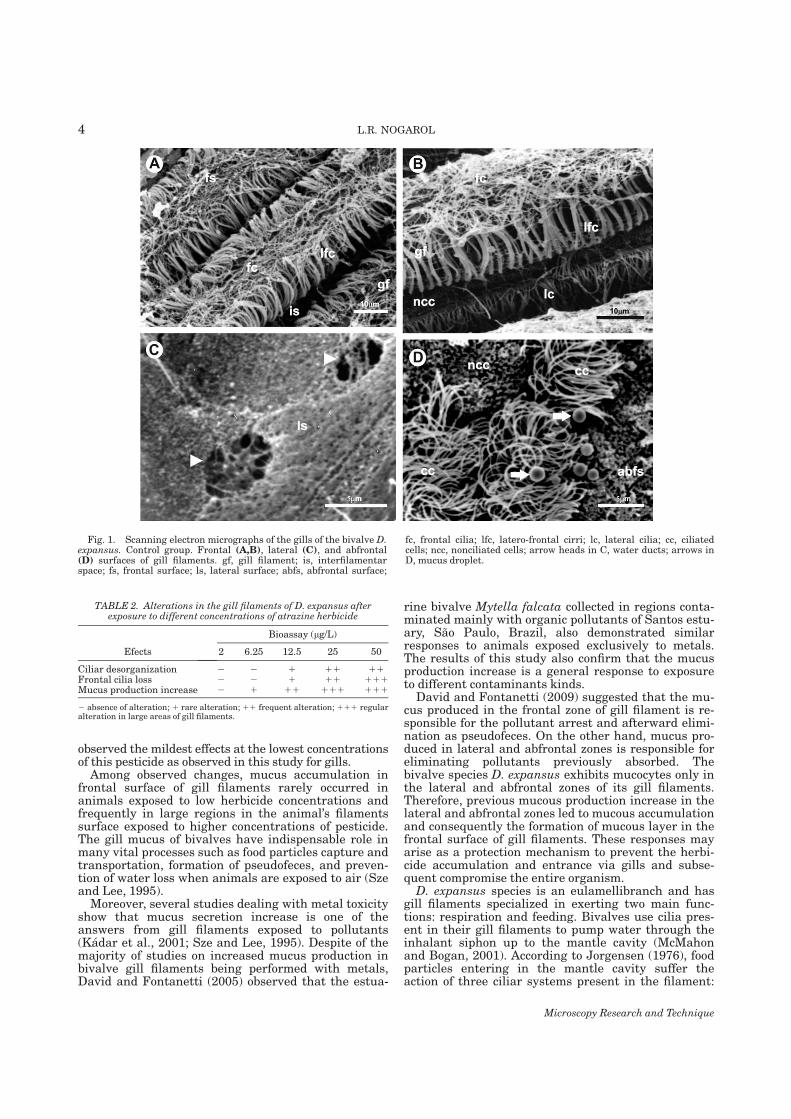
observed the mildest effects at the lowest concentrationsof this pesticide as observed in this study for gills.Among observed changes, mucus accumulation in
frontal surface of gill filaments rarely occurred inanimals exposed to low herbicide concentrations andfrequently in large regions in the animal’s filamentssurface exposed to higher concentrations of pesticide.The gill mucus of bivalves have indispensable role inmany vital processes such as food particles capture andtransportation, formation of pseudofeces, and preven-tion of water loss when animals are exposed to air (Szeand Lee, 1995).Moreover, several studies dealing with metal toxicity
show that mucus secretion increase is one of theanswers from gill filaments exposed to pollutants(Kadar et al., 2001; Sze and Lee, 1995). Despite of themajority of studies on increased mucus production inbivalve gill filaments being performed with metals,David and Fontanetti (2005) observed that the estua-
rine bivalve Mytella falcata collected in regions conta-minated mainly with organic pollutants of Santos estu-ary, Sao Paulo, Brazil, also demonstrated similarresponses to animals exposed exclusively to metals.The results of this study also confirm that the mucusproduction increase is a general response to exposureto different contaminants kinds.David and Fontanetti (2009) suggested that the mu-
cus produced in the frontal zone of gill filament is re-sponsible for the pollutant arrest and afterward elimi-nation as pseudofeces. On the other hand, mucus pro-duced in lateral and abfrontal zones is responsible foreliminating pollutants previously absorbed. Thebivalve species D. expansus exhibits mucocytes only inthe lateral and abfrontal zones of its gill filaments.Therefore, previous mucous production increase in thelateral and abfrontal zones led to mucous accumulationand consequently the formation of mucous layer in thefrontal surface of gill filaments. These responses mayarise as a protection mechanism to prevent the herbi-cide accumulation and entrance via gills and subse-quent compromise the entire organism.D. expansus species is an eulamellibranch and has
gill filaments specialized in exerting two main func-tions: respiration and feeding. Bivalves use cilia pres-ent in their gill filaments to pump water through theinhalant siphon up to the mantle cavity (McMahonand Bogan, 2001). According to Jorgensen (1976), foodparticles entering in the mantle cavity suffer theaction of three ciliar systems present in the filament:
TABLE 2. Alterations in the gill filaments of D. expansus afterexposure to different concentrations of atrazine herbicide
Efects
Bioassay (lg/L)
2 6.25 12.5 25 50
Ciliar desorganization 2 2 1 11 11Frontal cilia loss 2 2 1 11 111Mucus production increase 2 1 11 111 111
2 absence of alteration; 1 rare alteration; 11 frequent alteration; 111 regularalteration in large areas of gill filaments.
Fig. 1. Scanning electron micrographs of the gills of the bivalve D.expansus. Control group. Frontal (A,B), lateral (C), and abfrontal(D) surfaces of gill filaments. gf, gill filament; is, interfilamentarspace; fs, frontal surface; ls, lateral surface; abfs, abfrontal surface;
fc, frontal cilia; lfc, latero-frontal cirri; lc, lateral cilia; cc, ciliatedcells; ncc, nonciliated cells; arrow heads in C, water ducts; arrows inD, mucus droplet.
Microscopy Research and Technique
4 L.R. NOGAROL
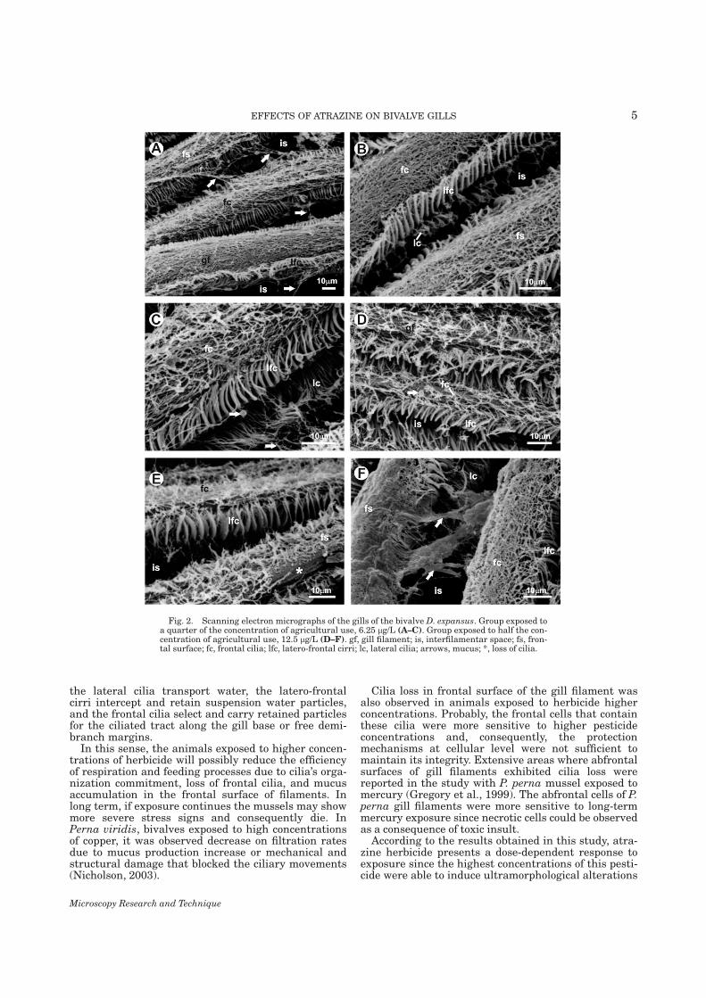
the lateral cilia transport water, the latero-frontalcirri intercept and retain suspension water particles,and the frontal cilia select and carry retained particlesfor the ciliated tract along the gill base or free demi-branch margins.In this sense, the animals exposed to higher concen-
trations of herbicide will possibly reduce the efficiencyof respiration and feeding processes due to cilia’s orga-nization commitment, loss of frontal cilia, and mucusaccumulation in the frontal surface of filaments. Inlong term, if exposure continues the mussels may showmore severe stress signs and consequently die. InPerna viridis, bivalves exposed to high concentrationsof copper, it was observed decrease on filtration ratesdue to mucus production increase or mechanical andstructural damage that blocked the ciliary movements(Nicholson, 2003).
Cilia loss in frontal surface of the gill filament wasalso observed in animals exposed to herbicide higherconcentrations. Probably, the frontal cells that containthese cilia were more sensitive to higher pesticideconcentrations and, consequently, the protectionmechanisms at cellular level were not sufficient tomaintain its integrity. Extensive areas where abfrontalsurfaces of gill filaments exhibited cilia loss werereported in the study with P. perna mussel exposed tomercury (Gregory et al., 1999). The abfrontal cells of P.perna gill filaments were more sensitive to long-termmercury exposure since necrotic cells could be observedas a consequence of toxic insult.According to the results obtained in this study, atra-
zine herbicide presents a dose-dependent response toexposure since the highest concentrations of this pesti-cide were able to induce ultramorphological alterations
Fig. 2. Scanning electron micrographs of the gills of the bivalve D. expansus. Group exposed toa quarter of the concentration of agricultural use, 6.25 lg/L (A–C). Group exposed to half the con-centration of agricultural use, 12.5 lg/L (D–F). gf, gill filament; is, interfilamentar space; fs, fron-tal surface; fc, frontal cilia; lfc, latero-frontal cirri; lc, lateral cilia; arrows, mucus; *, loss of cilia.
Microscopy Research and Technique
5EFFECTS OF ATRAZINE ON BIVALVE GILLS
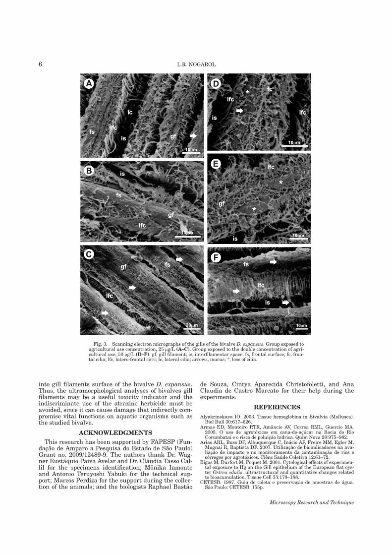
into gill filaments surface of the bivalve D. expansus.Thus, the ultramorphological analyses of bivalves gillfilaments may be a useful toxicity indicator and theindiscriminate use of the atrazine herbicide must beavoided, since it can cause damage that indirectly com-promise vital functions on aquatic organisms such asthe studied bivalve.
ACKNOWLEDGMENTS
This research has been supported by FAPESP (Fun-dacao de Amparo a Pesquisa do Estado de Sao Paulo)Grant no. 2009/12489-9. The authors thank Dr. Wag-ner Eustaquio Paiva Avelar and Dr. Claudia Tasso Cal-lil for the specimens identification; Monika Iamonteand Antonio Teruyoshi Yabuki for the technical sup-port; Marcos Perdiza for the support during the collec-tion of the animals; and the biologists Raphael Bastao
de Souza, Cintya Aparecida Christofoletti, and AnaClaudia de Castro Marcato for their help during theexperiments.
REFERENCES
Alyakrinskaya IO. 2003. Tissue hemoglobins in Bivalvia (Mollusca).Biol Bull 30:617–626.
Armas ED, Monteiro RTR, Amancio AV, Correa RML, Guercio MA.2005. O uso de agrotoxicos em cana-de-acucar na Bacia do RioCorumbataı e o risco de poluicao hıdrica. Quim Nova 28:975–982.
Arias ARL, Buss DF, Albuquerque C, Inacio AF, Freire MM, Egler M,Mugnai R, Baptista DF. 2007. Utilizacao de bioindicadores na ava-liacao de impacto e no monitoramento da contaminacao de rios ecorregos por agrotoxicos. Cienc Saude Coletiva 12:61–72.
Bigas M, Durfort M, Poquet M. 2001. Cytological effects of experimen-tal exposure to Hg on the Gill epithelium of the European flat oys-ter Ostrea edulis: ultrastructural and quantitative changes relatedto bioacumulation. Tissue Cell 33:178–188.
CETESB. 1987. Guia de coleta e preservacao de amostras de agua.Sao Paulo: CETESB. 155p.
Fig. 3. Scanning electron micrographs of the gills of the bivalve D. expansus. Group exposed toagricultural use concentration, 25 lg/L (A–C). Group exposed to the double concentration of agri-cultural use, 50 lg/L (D–F). gf, gill filament; is, interfilamentar space; fs, frontal surface; fc, fron-tal cilia; lfc, latero-frontal cirri; lc, lateral cilia; arrows, mucus; *, loss of cilia.
Microscopy Research and Technique
6 L.R. NOGAROL
Cogerh. 2001. Recomendacoes e cuidados na coleta de amostras deagua. Informe tecnico n8.02. Fortaleza: Cogerh, p 20.
CONAMA. 2005. Conselho Nacional do Meio Ambiente/Ministerio doMeio Ambiente. Resolucao No. 357 de 17 de macro de 2005. Avail-able at: http://www.mma.gov.br/port/conama/legiano1.cfm?codlegitipo=3&ano52005. (Accessed on March, 2010).
David JAO, Fontanetti CS. 2005. Surface morphology of Mytella fal-cata gill filaments from three regions of the Santos estuary. Braz JMorphol Sci 22:203–210.
David JAO, Fontanetti CS. 2009. The role of mucus inMytella falcata(Orbigny, 1842) gills from polluted environments. Water Air SoilPollut 200:1–6.
Eisler R. 1989. Atrazine hazards to fish, wildlife and invertebrates: Asynoptic review. US Fish Wildl Serv Biol Rep 85:1–55.
Flynn K, Spellman T. 2009. Environmental levels of atrazinedecreases patial aggregation in the freshwater mussel, Elliptiocomplanata. Ecotoxicol Environ Saf 72:1228–1233.
Gregory MA, George RC, Marshall DJ, Anandraj A, McClurg TP. 1999.The effects of mercury exposure on the surface morphology of gill fila-ments in Perna perna (Mollusca: Bivalvia). Mar Pollut Bull 39:116–121.
Jacomini AE, Bonato PS, Avelar WEP. 2003. HPLC method for theanalysis of atrazine in freshwater bivalves. J Liquid ChromatogrRelat Technol 26:1885–1894.
Jacomini AE, Avelar WEP, Martines AS, Bonato PS. 2006. Bioaccu-mulation of atrazine in freshwater bivalves Anodontites trapesialis(Lamarck, 1819) and Corbicula fluminea (Muller, 1774). Arch Envi-ron Contam Toxicol 51:387–391.
Jorgensen CB. 1976. Comparative studies on the function of gills insuspension feeding bivalves, with special reference to effects ofserotonine. Biol Bull 151:331–343.
Kadar E, Salanki J, Jugdaohsingh R, Powell JJ, McCrohan CR, WhiteKN. 2001. Avoidance responses to aluminum in the freshwaterbivalve Anodonta cygnea. Aquat Toxicol 55:137–148.
Karnovsky MJ. 1965. A formaldehyde–glutaraldehyde fixative at highosmolarity for use in electron microscopy. J Cell Biol 11:137–140.
Le Pennec H, Beninger PG, Herry A. 1988. New observations of thegills of Placopecten magellanicus (Mollusca: Bivalvia) and implica-tions for nutrition. Mar Biol 98:229–237.
Mansur MCD, Santos CP. 2008. Invertebrados aquaticos: Diplodonexpansus. In: Machado ABM, Drummond GM, Paglia AP, editors.Livro Vermelho da Fauna Ameacada de Extincao. Belo Horizonte:Fundacao Bioversitas. pp197–198.
Mantecca P, Vailati G, Bacchetta R. 2006. Histological changes andmicronucleous inductions in the Zebra mussel Dreissena polymor-pha after Paraquat exposure. Histol Histopathol 21:829–840.
Marchini S, Passerini L, Cesareo D, Tosato ML. 1988. Herbicidal tria-zines: Acute toxicity on Daphnia, fish and plants and analysis of itsrelationships with structural factors. Ecotoxicol Environ Saf16:148–157.
McMahon RF, Bogan AE. 2001. Mollusca: Bivalvia. In: Trorp JH,Covish AP, editors. Ecology and classification of North Americanfreshwater invertebrates. San Diego: Academic Press. pp 331–429.
Naimo TJ. 1995. A review of the effects of heavy metals on freshwatermussels. Ecotoxicology 4:341–362.
Nicholson S. 2003. Cardiac and branchial physiology associated withcopper accumulation and detoxication in the mytilid mussel Pernaviridis (L.). J Exp Mar Biol Ecol 295:157–171.
Peltier GL, Meyer JL, Jagoe CH, William A, Hopkins WA. 2008. Usingtrace element concentrations in Corbicula fluminea to identifypotential sources of contamination in an urban river. Environ Pol-lut 154:283–290.
Preez HH, Vuren JHJ. 1992. Bioaconcentration of atrazine in thebanded tilapia, Tilapia sparrmanii. Comp Biochem Physiol101:651–655.
Solomon KR, Baker DB, Richards RP, Dixon KR, Klaine SJ, LaPoint TW, Kendall R J, Weiss-Kopf CP, Giddings JM, Giesy JP,Hall LW, Jr, Williams ,WM. 1996. Ecological risk assessment ofatrazine in North American surface waters. Environ ToxicolChem 15:31–76.
Sunila ,I. 1988. Acute histological responses of the gill of the mussel,Mytilus edulis, to exposure by environmental pollutants. J Inver-tebr Pathol 52:137–141.
Sze PWC, Lee SY. 1995. The potential role of mucus in the depurationof copper from the mussels Perna viridis (L.) and Septifer virgatus(Wiegmann). Mar Pollut Bull 31:390–393.
Villar C, Stripeikis J, D’Huicque L, Tudino M, Troccoli O, Bonetto C.1999. Cd, Cu and Zn concentrations in sediments and the invasivebivalves Limnoperna fortunei and Corbicula fluminea at the Rio dela Plata basin, Argentina. Hydrobiologia 416:41–49.
Zupan I, Kalafatic M. 2003. Histological effects of low atrazine con-tamination on Zebra Mussel (Dreissena polymorpha Pallas). BullEnviron Contam Toxicol 70:688–695.
Microscopy Research and Technique
7EFFECTS OF ATRAZINE ON BIVALVE GILLS

Capítulo 3� � � �
�
7.3. CAPÍTULO 3
Histopathology of the gill filaments of the Brazilian endemic bivalve
Diplodon expansus after exposure to the herbicide atrazine
Larissa Rosa Nogarol; Ana Luiza Brossi-Garcia & Carmem Silvia Fontanetti*
UNESP, São Paulo State University, Av. 24A, no. 1515, CEP 13506-900, Rio Claro, SP,
Brazil.
*Corresponding author
E-mail address: [email protected]
Full postal address: Department of Biology, Institute of Biosciences, São Paulo State
University (UNESP), Av. 24 A, no. 1515, CP 199, 13506-900 Rio Claro, SP, Brazil
Tel.: +55 19 35264139; fax: +55 19 35264135
Submetido ao periódico Ecotoxicology and Environmental Safety.
Capítulo 3� � � ��
�
Resumo
Devido ao hábito séssil e filtrador e grande potencial de bioacumulação de metais e
compostos orgânicos que apresentam, os moluscos bivalves vêm sendo amplamente utilizados
em estudos de campo e laboratório que visam avaliar o potencial tóxico de inúmeros
compostos lançados em rios, mares e estuários. Entretanto, a maioria dos estudos
ecotoxicológicos utiliza espécies exóticas invasoras nas suas avaliações, sendo menos
conhecidas as respostas de espécies nativas. A espécie endêmica brasileira Diplodon expansus
ocorre em rios próximos a regiões de intensa atividade agrícola do país; o herbicida atrazina é
hoje um dos principais resíduos encontrados em corpos d’água do território brasileiro. Desta
forma, buscou-se investigar a toxicidade de diferentes concentrações desse herbicida em um
representante da malacofauna nativa brasileira, por meio de análises histológicas,
histoquímicas e ultraestruturais de seus filamentos branquiais. As células que compõem as
zonas frontal e intermediária dos filamentos branquiais foram as mais afetadas pelo herbicida,
sendo observadas respostas relacionadas ao dano, proteção e aumento do gasto energético. As
respostas tissulares e celulares observadas revelaram-se dose-dependentes, sendo que as
maiores concentrações do herbicida foram capazes de causar alterações severas em amplas
regiões das brânquias. Provavelmente, a persistência das alterações observadas causará
problemas funcionais graves nas brânquias desses animais, afetando negativamente seu
desempenho e saúde.
Palavras-chave: histoquímica; MET; MEV; agrotóxicos; ctenidia.
Capítulo 3� � � ��
�
Abstract
Due to their benthic filter-feeding habits and high bioaccumulation potential of metals and
organic compounds, bivalve mollusks have been widely used in studies in the field and
laboratory to assess the toxic effects of several compounds discharged in rivers, oceans and
estuaries. However, most ecotoxicological studies use invasive exotic species, while the
response of native species is poorly known. The Brazilian endemic species Diplodon
expansus occurs in rivers near areas of intense agricultural activity, and atrazine is currently
one of the main residues of pesticides found in water bodies of the Brazilian territory. This
study was aimed at examining the toxicity of different atrazine concentrations to a Brazilian
native mollusk by analyzing the histology, histochemistry, and ultrastructure of its gill
filaments. The cells that comprise the frontal and intermediary regions of gill filaments were
the most affected by the herbicide and responses associated to damage, protection, and
increase in energy use were observed. The response of tissues and cells were dose-dependent.
Higher concentrations of herbicide caused more severe alterations in large areas of the gills.
The persistence of the alterations observed might result in severe functional problems in the
gills of these animals, negatively affecting their performance and health.
Keywords: morphology; histochemistry; ultrastructure; agrochemical; mussel.
Capítulo 3� � � ��
�
1. Introduction
The herbicide atrazine has been associated to several environmental problems, as it
tends to dissipate in the environment, contaminating the soil and surface and ground waters
(Sanches et al., 2003). This herbicide is one of the most widely used pesticides worldwide
(Zupan and Kalafatic, 2003), especially in regions with a predominance of sugarcane
plantations, such as in southeast Brazil in the Corumbataí River Basin (Armas et al., 2005).
Because of its physical-chemical proprieties, atrazine is a stable compound in the environment
(Jacomini et al., 2003; Yang et al., 2010), increasing the chances of exposure of the biota
(Solomon et al., 2008) and bioaccumulation in the tissues of aquatic invertebrates, such as
bivalve mollusks (Jacomini et al., 2006).
Bivalve mollusks exhibit several characteristics that make them good sentinel
organisms, such as benthic filter-feeding habits, wide distribution, easily collected (Depledge,
1998; Gregory et al., 2002) and high bioaccumulation potential of metals and organic
compounds (Gregory et al., 1999; Jacomini et al., 2006; Naimo, 1995). Thus, these
invertebrates have been broadly used in ecotoxicological studies in marine, estuarine, and
freshwater habitats.
Most ecotoxicological studies using bivalve mollusks in the biomonitoring of
disturbed aquatic habitats or bioassays are focused on North America and Europe, especially
using species of the genus Mytilus in marine habitats, as pointed out by Gregory et al. (2002)
and David and Fontanetti (2005). In freshwater habitats, exotic invasive species from Asia
have been used, such as Dreissena polymorpha (Mantecca et al., 2006; Zupan and Kalafatic,
2003) and Corbicula fluminea (Peltier et al., 2008; Simon et al., 2011).
In order to improve the accuracy of ecological assays, species commonly found in the
southern hemisphere have recently received more attention in the assessment of disturbed
habitats in these areas, such as those conducted by Brazilian and South-African research
groups using the marine bivalve Perna perna (Abessa et al., 2005; Gregory et al., 1999;
Gregory et al., 2002), the estuarine bivalve Mytella falcata (David and Fontanetti, 2005, 2009;
David et al., 2008a), and the freshwater bivalves Anadontites trapezialis (Jacomini et al.,
2003, 2006) and Diplodon fontaineanus (Jacomini et al., 2011).
The impact of pollutants in aquatic organisms can be assessed in different levels of
biological organization, including histopathology (Akaishi et al., 2007). Histopatological
analysis can provide information on the general health of the animal and changes in the
tissues caused by the exposure to toxic agents (Fontanetti et al., 2010; Sunila, 1987). These
Capítulo 3� � � ��
�
alterations may have an ecological significance, due to adverse or impaired effects on growth,
reproduction and performance of organisms exposed to xenobiotics (Sunila, 1987).
Among the main target organs used to examine the effects of pollutants in mollusks,
gills are considered adequate for ecological assessments due to their large surface area in
contact with the surrounding water contaminated by xenobiotics (Bigas et al., 2001). In
general, gill filaments of bivalve mollusks consist of epithelial and endothelial cells, and
mucocytes (David et al., 2008b; Sunila, 1986). The morphology of gill filaments allows this
organ to play different roles, such as respiration, transport of food particles and formation of a
protective and lubricant layer of mucus on the epithelial surface (Dufour and Beninger, 2001;
Gómez-Mendikute et al., 2005).
Therefore, this study was aimed at examining the histology, histochemistry, and
ultrastructure of the gill filaments of the Brazilian endemic species D. expansus, which occurs
in areas of intense agricultural activity in southeast Brazil, after acute exposure to the
herbicide atrazine in environmentally realistic exposure concentrations.
2. Material and methods
2.1 Tested compound
Atrazine (2-chloro-4-ethylamino-6-isopropylamino-s-triazine) is a selective herbicide
of the triazine group. This herbicide is classified as moderately toxic to humans (class III) and
highly hazardous for the environment (class II). The concentrated suspension of the
commercial product Atrazine Nortox 500 SC ® is used in the control of weeds for pre-
emergent and early post-emergent applications in corn, sugarcane and sorghum crops.
�
2.2 Collection of animals
Specimens of D. expansus (average body weight ± SD = 14.15g ± 7.8; average shell
length ± DP= 4.29cm ± 0.84) were collected in April 2010 in Ribeirão Claro, municipality of
Rio Claro (S 22º24’33.1’’; O 47º32’25.1’’), São Paulo, Brazil. In order to ensure low
contamination levels at the collection site, water samples were collected according to
CETESB (1987) and COGERH (2001) and metal and pesticide levels were quantified.
In the laboratory, animals were acclimated for three days in 30-L aquaria filled with
water from an artesian well at 25º C, constant aeration and 12-h photoperiod. This procedure
was used to minimize stress caused by collection procedures and transport of animals.
Capítulo 3� � � ��
�
2.3 Chemical analysis
After collection, water samples were transported in coolers with reusable ice packs to
TASQA Serviços Analíticos Ltda. (Paulínia, São Paulo, Brazil), where chemical analysis
were carried out.
Organic parameters (atrazine, glyphosate, 2,4-D) and inorganic compounds
(aluminum, total cadmium, total lead, dissolved copper, dissolved iron, total manganese, total
nickel, total silver, total zinc) established by the National Environment Council (Conselho
Nacional do Meio Ambiente - CONAMA, 2005) were used to evaluate the quality of water
samples.
Residues of atrazine and 2,4-D were quantified by the concentration of semivolatile
organic compounds with the EPA 8270D method. For glyphosate, the EPA 547 method was
used.
The concentration of metals in water samples was obtained based on the quantification
of elements by atomic emission spectrophotometry in inductively coupled argon plasma in
aqueous extract, according to the SM21 3120B method, similar to the EPA 6010B
quantification method, after acid digestion in closed system using microwave heating,
following EPA 3015 for liquid samples and EPA 3052 for solid samples.
The concentrations of metals and pesticides in water samples from the collection site
were compared with the maximum values allowed for freshwater bodies reported by the
National Environment Council (CONAMA, 2005). Freshwater in the Brazilian territory is
classified based on the quality required for a given use. In this study, class I and II levels, for
human consumption and protection of aquatic communities, were used.
The water quality patterns determined by the National Environment Council
(CONAMA, 2005) establish individual limits for each compound in each class and the
maximum concentrations allowed. Concentrations above the limits indicate compromised
water quality for its corresponding uses.
2.4 Bioassay design
Six 10-L aquaria were used: one aquarium as the control group containing water from
an artesian well and five aquaria containing different concentrations of atrazine (2, 6.25, 12.5,
25 and 50 µg/L) obtained by diluting a stock solution (0.5g/L) in water. The highest
concentration tested (50 µg/L) is twice that indicated for agricultural use, while the lowest
concentration (2 µg/L) is the maximum allowed in freshwater bodies by Brazilian laws
Capítulo 3� � � �
�
according to the CONAMA resolution 357 (2005). These concentrations were used to
evaluate the effects of indiscriminate use of pesticides and the effect of dilution of atrazine in
water bodies, similar to environmentally realistic concentrations. The bioassays were as
follows:
Control: 8L of artesian well water
Treatment 1: 8L of artesian well water + 16 µg of atrazine (2 µg/L)
Treatment 2: 8L of artesian well water + 50 µg of atrazine (6.25 µg/L)
Treatment 3: 8L of artesian well water + 100 µg of atrazine (12.5 µg/L)
Treatment 4: 8L of artesian well water + 200 µg of atrazine (25 µg/L)
Treatment 5: 8L of artesian well water + 400 µg of atrazine (50 µg/L)
The water in aquaria was aerated continuously by an air pump. After acclimation, five
animals were randomly distributed per aquarium. Animals were exposed for seven days in
order to evaluate an acute response. This time of exposure has been used in previous studies
with atrazine and freshwater bivalves (Jacomini et al., 2006; Zupan and Kalafatic, 2003).
The experimental conditions consisted of a semi-static system with 100% of the
volume changed every 24 hours, followed by addition of a freshly prepared stock solution of
atrazine with distilled water, to maintain the concentration of the herbicide. The temperature
was maintained between 23 – 25oC during the exposure period.
After seven days of exposure, mollusks were anesthetized by thermal chock and small
fragments of gills were removed and fixed.
2.5 Histology and histochemistry
The material was fixed in aqueous Bouin solution for 24 hours; immersed in a sodium
phosphate buffer solution (pH=7) and maintained in the refrigerator. Samples were then
dehydrated in a crescent series of ethanol (70, 80, 90 and 95%,) for 20 minutes each bath, and
processed for inclusion in historesin.
Blocs were sectioned at 5 µm with a Leica RM 2245 microtome and glass knifes.
Sections were hydrated in histological bath and placed in slides. After drying, sections were
stained with Harris hematoxylin for 10 minutes and rinsed under tap water for 5 minutes,
stained with aqueous eosin for 5 minutes and rinsed with water. After drying, slides were
mounted with Canada balsam.
Histochemical methods were carried out to detect the presence of the following
compounds: total proteins – bromophenol blue (Pearse, 1985), and polysaccharides –
Capítulo 3� � � �
�
simultaneously staining with PAS (periodic acid - Schiff) and Alcian blue (Junqueira and
Junqueira, 1983). Slides were mounted with Canada balsam, dried and examined under a light
microscope.
2.6 Ultrastructure
Small gill fragments were fixed with 2.5% glutaraldehyde in 0.1 M sodium cacodylate
buffer at 4ºC, rinsed with sodium cacodylate buffer and post-fixed with 1% osmium tetroxide
for 2 hours. The material was rinsed once more with the same buffer, immersed in 10%
ethanol for 15 minutes and contrasted with 2% uranyl acetate in 10% ethanol for 4 hours. The
samples were then dehydrated in a crescent series of acetone, immersed in resin:acetone
solution (1:1) for 12 hours, embedded in Epon-araldite resin with catalyzer for 24 hours and
maintained in an oven at 70ºC for 24 h for resin polymerization. The material was sectioned
with an ultramicrotome, sections were placed on copper screen and contrasted with uranyl
acetate and lead citrate for later examination under electron transmission microscope Philips
CM 100, operated at 80 kV.
2.7 Statistical analysis
Hemocytes infiltrated in the epithelium and mucocytes were counted in 50 gill
filaments per individual (5 individuals for each bioassay). Mucocytes were classified as intact
or damaged.
The results were statistically analyzed using analysis of variance (ANOVA) with the
software SYSTAT 11. Normality was verified previously with the a priori Shapiro-Wilk test
(SW) with significance set at p>0.05.
3. Results
3.1 Water analysis
The concentrations of inorganic compounds, dissolved aluminum, dissolved iron and
total manganese were lower than the maximum allowed by Brazilian laws for freshwater
classes I and II (CONAMA, 2005).
However, the concentrations of all organic compounds and the remaining inorganic
parameters (total cadmium, total lead, dissolved copper, total nickel, total silver, and total
zinc) could not be determined accurately, as they were below the limits of quantification (LQ)
for the method used (Table 1).
Capítulo 3� � � ��
�
The general chemical analysis indicated that the water from the collection site is
adequate for the conservation of aquatic communities and human consumption after
treatment, as determined for freshwater class I and II (CONAMA, 2005). Therefore, animals
collected were not exposed to polluted water.
3.2 Histology and histochemistry
3.2.1 Control group
All animals of the control group survived the seven-day experiment and their gill
filaments exhibited intact cells and other associated structures. As described for other
eulamellibranch bivalves, gill filaments were united by tissue connections termed
interfilamentar and interlamellar junctions. The interfilamentar junctions unite adjacent gill
filaments while interlamellar junctions are located in the interlamellar space (Fig. 1A).
Occasional pores termed ostia were observed connecting the interlamellar space and the
external environment (* in Fig. 1A, B).
The gill filaments of the bivalve D. expansus consist of a single layer of different cell
types (ciliated and nonciliated cells and mucocytes) and endothelial cells that line them
internally. The frontal zone of gill filaments comprises cells with cilia termed frontal, latero-
frontal, and lateral cilia. Still in this zone, nonciliated absorptive cells, termed postlateral-
frontal cells, are present. The intermediary zone consists of absorptive nonciliated cells with
flattened nuclei, gradually resembling squamous cells toward the abfrontal zone. In the
abfrontal zone, ciliated and nonciliated cells are present (Fig. 1B).
Supporting structures of gill filaments, termed skeletal loop and skeletal rod are also
observed. The skeletal loop extends from the frontal to the intermediary zone (Fig. 1B) and is
weakly stained for neutral polysaccharides (Fig. 1C) and proteins (Fig. 1E). The skeletal rod
is found in the intermediary zone and is strongly stained for neutral polysaccharides (Fig. 1C,
D) but negative for proteins (Fig. 1E).
Mucocytes were observed in intermediary (Fig. 1C) and abfrontal (Fig. 1D) zones.
Hemocytes moderately stained for neutral polysaccharides and proteins moved freely in the
hemolymph vessels and rarely infiltrated the gill filament epithelium (Fig. 1C).
3.2.2 Exposed groups
All animals exposed to different concentrations of atrazine survived the seven-day
experiments. All concentrations of atrazine induced histopatological alterations in the gill
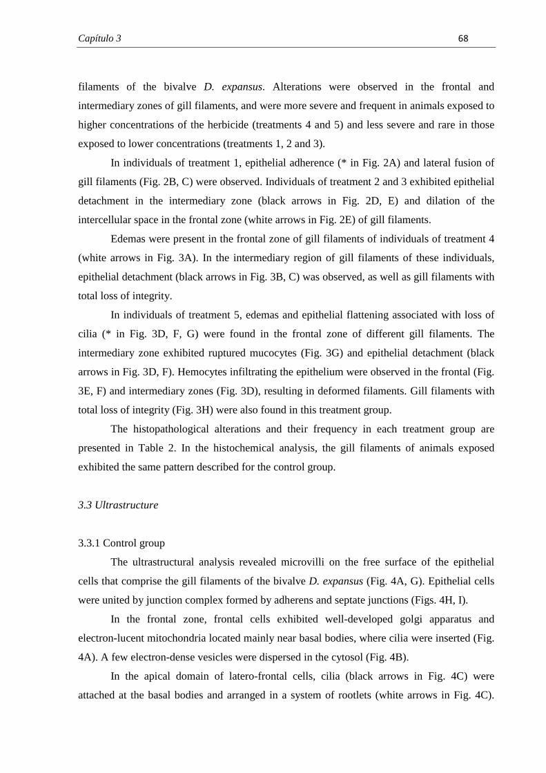
Capítulo 3� � � ��
�
filaments of the bivalve D. expansus. Alterations were observed in the frontal and
intermediary zones of gill filaments, and were more severe and frequent in animals exposed to
higher concentrations of the herbicide (treatments 4 and 5) and less severe and rare in those
exposed to lower concentrations (treatments 1, 2 and 3).
In individuals of treatment 1, epithelial adherence (* in Fig. 2A) and lateral fusion of
gill filaments (Fig. 2B, C) were observed. Individuals of treatment 2 and 3 exhibited epithelial
detachment in the intermediary zone (black arrows in Fig. 2D, E) and dilation of the
intercellular space in the frontal zone (white arrows in Fig. 2E) of gill filaments.
Edemas were present in the frontal zone of gill filaments of individuals of treatment 4
(white arrows in Fig. 3A). In the intermediary region of gill filaments of these individuals,
epithelial detachment (black arrows in Fig. 3B, C) was observed, as well as gill filaments with
total loss of integrity.
In individuals of treatment 5, edemas and epithelial flattening associated with loss of
cilia (* in Fig. 3D, F, G) were found in the frontal zone of different gill filaments. The
intermediary zone exhibited ruptured mucocytes (Fig. 3G) and epithelial detachment (black
arrows in Fig. 3D, F). Hemocytes infiltrating the epithelium were observed in the frontal (Fig.
3E, F) and intermediary zones (Fig. 3D), resulting in deformed filaments. Gill filaments with
total loss of integrity (Fig. 3H) were also found in this treatment group.
The histopathological alterations and their frequency in each treatment group are
presented in Table 2. In the histochemical analysis, the gill filaments of animals exposed
exhibited the same pattern described for the control group.
3.3 Ultrastructure
3.3.1 Control group
The ultrastructural analysis revealed microvilli on the free surface of the epithelial
cells that comprise the gill filaments of the bivalve D. expansus (Fig. 4A, G). Epithelial cells
were united by junction complex formed by adherens and septate junctions (Figs. 4H, I).
In the frontal zone, frontal cells exhibited well-developed golgi apparatus and
electron-lucent mitochondria located mainly near basal bodies, where cilia were inserted (Fig.
4A). A few electron-dense vesicles were dispersed in the cytosol (Fig. 4B).
In the apical domain of latero-frontal cells, cilia (black arrows in Fig. 4C) were
attached at the basal bodies and arranged in a system of rootlets (white arrows in Fig. 4C).
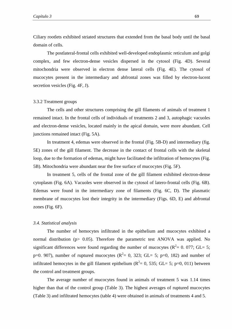
Capítulo 3� � � �
�
Ciliary rootlets exhibited striated structures that extended from the basal body until the basal
domain of cells.
The postlateral-frontal cells exhibited well-developed endoplasmic reticulum and golgi
complex, and few electron-dense vesicles dispersed in the cytosol (Fig. 4D). Several
mitochondria were observed in electron dense lateral cells (Fig. 4E). The cytosol of
mucocytes present in the intermediary and abfrontal zones was filled by electron-lucent
secretion vesicles (Fig. 4F, J).
3.3.2 Treatment groups
The cells and other structures comprising the gill filaments of animals of treatment 1
remained intact. In the frontal cells of individuals of treatments 2 and 3, autophagic vacuoles
and electron-dense vesicles, located mainly in the apical domain, were more abundant. Cell
junctions remained intact (Fig. 5A).
In treatment 4, edemas were observed in the frontal (Fig. 5B-D) and intermediary (fig.
5E) zones of the gill filament. The decrease in the contact of frontal cells with the skeletal
loop, due to the formation of edemas, might have facilitated the infiltration of hemocytes (Fig.
5B). Mitochondria were abundant near the free surface of mucocytes (Fig. 5F).
In treatment 5, cells of the frontal zone of the gill filament exhibited electron-dense
cytoplasm (Fig. 6A). Vacuoles were observed in the cytosol of latero-frontal cells (Fig. 6B).
Edemas were found in the intermediary zone of filaments (Fig. 6C, D). The plasmatic
membrane of mucocytes lost their integrity in the intermediary (Figs. 6D, E) and abfrontal
zones (Fig. 6F).
3.4. Statistical analysis
The number of hemocytes infiltrated in the epithelium and mucocytes exhibited a
normal distribution (p> 0.05). Therefore the parametric test ANOVA was applied. No
significant differences were found regarding the number of mucocytes (R2= 0. 077; GL= 5;
p=0. 907), number of ruptured mucocytes (R2= 0, 323; GL= 5; p=0, 182) and number of
infiltrated hemocytes in the gill filament epithelium (R2= 0, 535; GL= 5; p=0, 011) between
the control and treatment groups.
The average number of mucocytes found in animals of treatment 5 was 1.14 times
higher than that of the control group (Table 3). The highest averages of ruptured mucocytes
(Table 3) and infiltrated hemocytes (table 4) were obtained in animals of treatments 4 and 5.

Capítulo 3� � � ���
�
4. Discussion
The exposure of the bivalve D. expansus to atrazine induced significant alterations in
the morphology of the gill filaments. The level and frequency of damage was dose dependent.
Lower concentrations of the herbicides caused few morphological changes, while higher
concentrations resulted in extensive altered areas of the gill filaments of D. expansus. Similar
results were obtained by Yang et al. (2010) based on a morphological analysis of gills of the
fish Gobiocypris rarus exposed to environmentally relevant concentrations of this herbicide.
According to these authors, the lesions were more severe as the concentration of atrazine
increased, with histopathological alterations found in individuals exposed to concentrations
equal or above 10 µg/L.
The exposure to environmental pollutants can induce two main types of alterations at
the tissue and cell level: changes that indicate damage and those that show the presence and
action of protection or defense mechanisms (Parashar and Banerjee, 2002; Triebskorn et al.,
1996). Defense mechanisms require considerable amounts of energy to be continuously
active. Thus, responses associated to higher energy use are also observed under
environmentally stressful conditions (Triebskorn et al., 1996).
Alterations indicating damage were characterized by epithelial detachment, dilation of
the intercellular space, edemas, cytoplasmic condensation and vacuolation. The main defense
alterations observed were filament fusion, infiltration of hemocytes in the epithelium, and
increase in the number of autophagic vacuoles. The response associated to energy use was the
increase in mitochondria in mucocytes.
The fusion of gill filaments has been reported in bivalve mollusks exposed to toxic
agents (Akaishi et al., 2007; Domouhtsidou and Dimitriadis, 2000; Sunila, 1986, 1987). The
corresponding response in fish is the fusion of secondary lamellae, commonly observed in
individuals exposed to different pollutants, including the herbicide atrazine (Olurin et al.,
2006; Yang et al., 2011; Yasser and Naser, 2011).
Filament fusion can occur in the intermediary and abfrontal zones of gill filaments of
bivalve mollusks. Filament fusion in the intermediary zone is usually termed lateral fusion
(Domouhtsidou and Dimitriadis, 2000) while that located in the abfrontal zone is termed
lamellar fusion (Akaishi et al., 2007; Sunila, 1986). In bivalve mollusks, some authors believe
that this alteration is the result of errors during the regeneration of the gill epithelium (Akaishi
et al., 2007; Sunila, 1986).
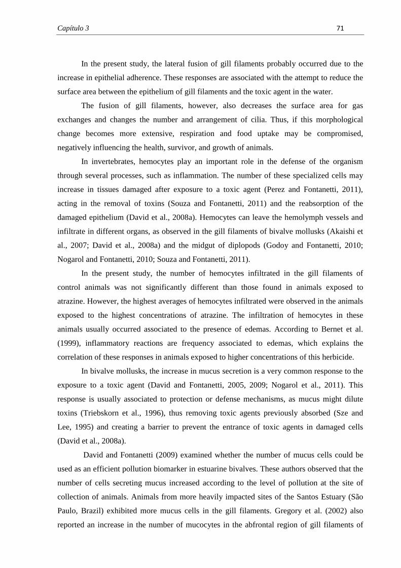
Capítulo 3� � � ���
�
In the present study, the lateral fusion of gill filaments probably occurred due to the
increase in epithelial adherence. These responses are associated with the attempt to reduce the
surface area between the epithelium of gill filaments and the toxic agent in the water.
The fusion of gill filaments, however, also decreases the surface area for gas
exchanges and changes the number and arrangement of cilia. Thus, if this morphological
change becomes more extensive, respiration and food uptake may be compromised,
negatively influencing the health, survivor, and growth of animals.
In invertebrates, hemocytes play an important role in the defense of the organism
through several processes, such as inflammation. The number of these specialized cells may
increase in tissues damaged after exposure to a toxic agent (Perez and Fontanetti, 2011),
acting in the removal of toxins (Souza and Fontanetti, 2011) and the reabsorption of the
damaged epithelium (David et al., 2008a). Hemocytes can leave the hemolymph vessels and
infiltrate in different organs, as observed in the gill filaments of bivalve mollusks (Akaishi et
al., 2007; David et al., 2008a) and the midgut of diplopods (Godoy and Fontanetti, 2010;
Nogarol and Fontanetti, 2010; Souza and Fontanetti, 2011).
In the present study, the number of hemocytes infiltrated in the gill filaments of
control animals was not significantly different than those found in animals exposed to
atrazine. However, the highest averages of hemocytes infiltrated were observed in the animals
exposed to the highest concentrations of atrazine. The infiltration of hemocytes in these
animals usually occurred associated to the presence of edemas. According to Bernet et al.
(1999), inflammatory reactions are frequency associated to edemas, which explains the
correlation of these responses in animals exposed to higher concentrations of this herbicide.
In bivalve mollusks, the increase in mucus secretion is a very common response to the
exposure to a toxic agent (David and Fontanetti, 2005, 2009; Nogarol et al., 2011). This
response is usually associated to protection or defense mechanisms, as mucus might dilute
toxins (Triebskorn et al., 1996), thus removing toxic agents previously absorbed (Sze and
Lee, 1995) and creating a barrier to prevent the entrance of toxic agents in damaged cells
(David et al., 2008a).
David and Fontanetti (2009) examined whether the number of mucus cells could be
used as an efficient pollution biomarker in estuarine bivalves. These authors observed that the
number of cells secreting mucus increased according to the level of pollution at the site of
collection of animals. Animals from more heavily impacted sites of the Santos Estuary (São
Paulo, Brazil) exhibited more mucus cells in the gill filaments. Gregory et al. (2002) also
reported an increase in the number of mucocytes in the abfrontal region of gill filaments of
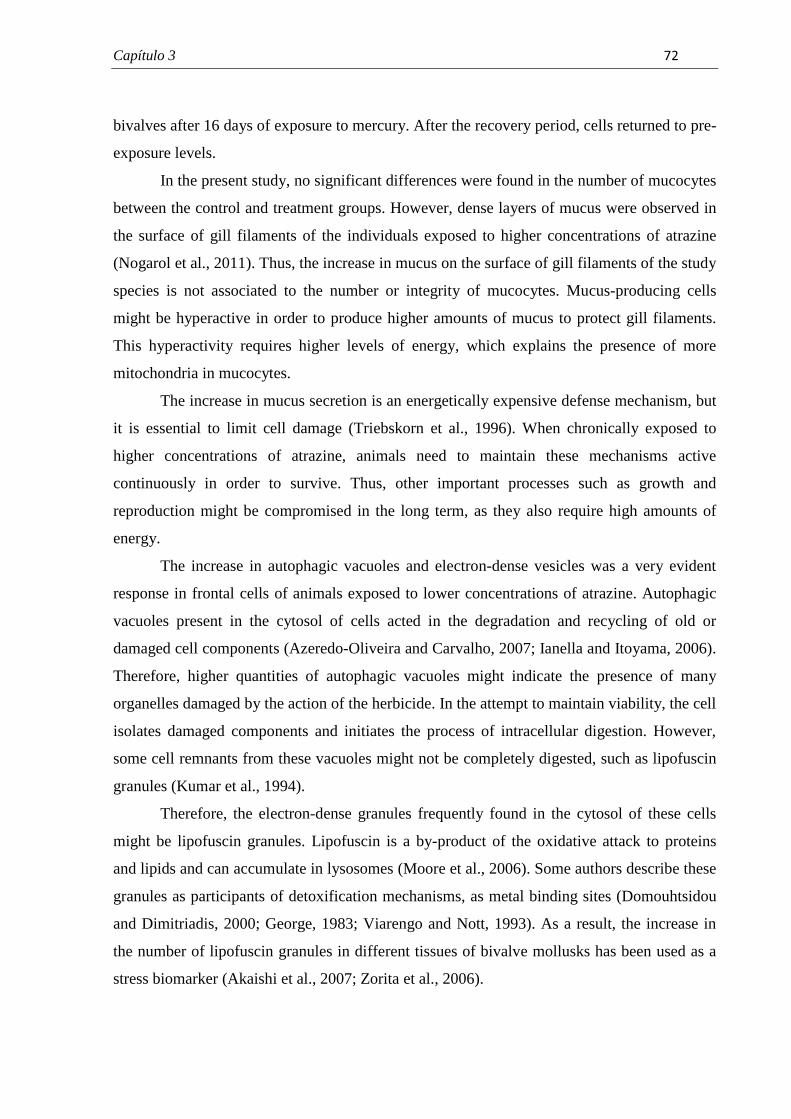
Capítulo 3� � � ���
�
bivalves after 16 days of exposure to mercury. After the recovery period, cells returned to pre-
exposure levels.
In the present study, no significant differences were found in the number of mucocytes
between the control and treatment groups. However, dense layers of mucus were observed in
the surface of gill filaments of the individuals exposed to higher concentrations of atrazine
(Nogarol et al., 2011). Thus, the increase in mucus on the surface of gill filaments of the study
species is not associated to the number or integrity of mucocytes. Mucus-producing cells
might be hyperactive in order to produce higher amounts of mucus to protect gill filaments.
This hyperactivity requires higher levels of energy, which explains the presence of more
mitochondria in mucocytes.
The increase in mucus secretion is an energetically expensive defense mechanism, but
it is essential to limit cell damage (Triebskorn et al., 1996). When chronically exposed to
higher concentrations of atrazine, animals need to maintain these mechanisms active
continuously in order to survive. Thus, other important processes such as growth and
reproduction might be compromised in the long term, as they also require high amounts of
energy.
The increase in autophagic vacuoles and electron-dense vesicles was a very evident
response in frontal cells of animals exposed to lower concentrations of atrazine. Autophagic
vacuoles present in the cytosol of cells acted in the degradation and recycling of old or
damaged cell components (Azeredo-Oliveira and Carvalho, 2007; Ianella and Itoyama, 2006).
Therefore, higher quantities of autophagic vacuoles might indicate the presence of many
organelles damaged by the action of the herbicide. In the attempt to maintain viability, the cell
isolates damaged components and initiates the process of intracellular digestion. However,
some cell remnants from these vacuoles might not be completely digested, such as lipofuscin
granules (Kumar et al., 1994).
Therefore, the electron-dense granules frequently found in the cytosol of these cells
might be lipofuscin granules. Lipofuscin is a by-product of the oxidative attack to proteins
and lipids and can accumulate in lysosomes (Moore et al., 2006). Some authors describe these
granules as participants of detoxification mechanisms, as metal binding sites (Domouhtsidou
and Dimitriadis, 2000; George, 1983; Viarengo and Nott, 1993). As a result, the increase in
the number of lipofuscin granules in different tissues of bivalve mollusks has been used as a
stress biomarker (Akaishi et al., 2007; Zorita et al., 2006).
Capítulo 3� � � ���
�
In some cases, such as exposure to higher concentrations of toxic agents, defense
mechanisms are insufficient to maintain the integrity of cells and tissues, resulting in
responses indicating damage.
One of these responses found in the epithelial cells that comprise the gill filaments of
the bivalve D. expansus was the dilation of the intercellular space. The same response was
observed in the gill filaments of bivalves exposed to mercury (Bigas et al., 2001) and in the
midgut epithelium of diplopods exposed to soil contaminated with metals and polycyclic
hydrocarbons (Souza and Fontanetti, 2011).
Kumar et al. (1994) reported that intercellular space dilation is associated to
modifications in the plasmatic membrane as a result of the loss of stability of junction
complexes. However, in animals from treatments 2 and 3, the ultrastructure analysis indicated
that cell junctions were intact.
Sunila (1988) exposed the bivalve Mytilus edulis to different environmental pollutants
such as metals and pesticides. The organic toxic agents induced shrinkage and separation of
the plasmatic membrane of cells of gill filaments. Thus, intercellular space dilation in animals
exposed to atrazine might be a consequence of cell atrophy. This was supported by the
increased number of autophagic vacuoles in the cytosol of these cells, as previously described,
which is frequently observed in cells undergoing atrophy (Kumar et al., 1994).
Edemas were present in the frontal and intermediary zones of gill filaments exposed to
higher concentrations of atrazine. The term edema is defined as the accumulation of abnormal
quantities of liquid in the intercellular spaces of the tissue (Kumar et al., 1996), due to
circulatory disturbances (Bernet et al., 1999). This alteration has been observed in gill
filaments of bivalves exposed to metals (Gregory et al., 2002) and gills of fish exposed to
atrazine (Jayachandran and Pugazhendy, 2009; Yang et al., 2010).
The presence of edemas in fish is of minor relevance, since the lesion is easy reverted
after the exposure to the toxic agent (Bernet et al., 1999). Apparently edemas are also easily
reversible in bivalve mollusks. However, further studies are needed to examine the resilience
of different bivalve species to exposure to various toxic agents.
Cytoplasmic condensation was also observed in animals exposed to higher
concentrations of atrazine. A similar response was also reported for the midgut of diplopods
(Triebskorn et al., 1991) and the hepatopancreas of isopods (Köhler et al., 1996) after
exposure to different metals. This response is usually accompanied by a reduction in the
number of organelles and changes in cell osmolarity (Köhler et al., 1996), and may result in a
Capítulo 3� � � ���
�
decrease in cell volume and consequently dilation of the intercellular space (Triebskorn et al.,
1991).
In the presence study, latero-frontal cells exhibited condensed cytoplasm with
vacuoles. These responses indicate that these cells sustained irreversible damage and are
undergoing cell death. Cytoplasmic vacuolation is a typical response of necrotic cells after
severe stress, such as the exposure to a toxic agent (Mello and Castilho, 2007). Cytoplasm
condensation suggests that the cytoplasmic components may be damaged and non-viable.
The filtering of particles present in the circulating water is one of the main roles of
latero-frontal cells (Gregory et al., 2002). These cells exhibit specialized cilia that act as nets
to capture food particles (Silverman et al., 1996). Therefore, damage to these cells can
decrease the filtering rate and consequently food intake, as observed for the mollusk Perna
perna exposed to mercury for two days (Gregory et al., 2002).
The flattening of frontal cells and loss of cilia were observed in animals exposed to the
highest concentration of atrazine. The exposure to mercury for two and four days induced
similar responses in the bivalve P. perna (Gregory et al., 2002). The authors reported that
throughout the days of exposure, alterations became less frequent and after the recovery
period, were no longer observed.
Nogarol et al. (2011) observed disorganization and loss of frontal cilia of gill filaments
of D. expansus exposed to the same conditions described in the present study. These authors
suggested a higher sensitivity of frontal ciliated cells, as confirmed in our study by the
observation of flat frontal cells and edemas.
The detachment of the epithelium occurred in the intermediary surface of gills of
animals exposed to different concentrations of atrazine, more frequently in individuals
exposed to higher concentrations. This response was observed in different areas of gill
filaments of bivalve mollusks to different environmental contaminants (David et al., 2008a;
Domouhtsidou and Dimitriadis, 2004; Sunila, 1988). In fishes, the detachment of the gill
epithelium is considered a regressive alteration, thus indicating a functional reduction of the
organ (Bernet et al., 1999). Because of the respiratory role of cells present in the intermediary
surface of the filament, gas exchanges might be compromised as well as the health of animals
especially those exposed to higher concentrations of atrazine.
Capítulo 3� � � ��
�
5. Conclusions
Our findings clearly indicate that sublethal concentrations of atrazine have adverse
effects on the gill filaments of the study species. This herbicide apparently induces dose-
dependent alterations. Some of these alterations might result in severe functional impairment,
compromising the health and performance of animals exposed for long periods to this
herbicide.
Acknowledgments
This study was conducted with financial support of FAPESP (The State of São Paulo
Research Foundation), Grant number 2009/12489-9. The authors thank. Dr. Wagner
Eustáquio Paiva Avelar and Dr. Cláudia Tasso Callil for the identification of species, Mônika
Iamonte, Antonio Teruyoshi Yabuki and Gerson Mello de Souza for technical support,
Marcos Perdiza for assistance in the collection of animals, Dr. Tamaris Gimenez Pinheiro for
assistance with statistical analysis, the biologists Raphael Bastão de Souza, Ana Claudia de
Castro Marcato and Dr. Cintya Aparecida Christofoletti for assistance during experiments.
References
Abessa, D.M.S., Zaroni, L.P., Sousa, E.C.P.M., Garparro, M.R., Pereira, C.D.S., Rachid,
B.R.F., Depledge, M., King, R.S., 2005. Physiological and cellular responses in two
population of the mussel Perna perna collected at different sites from the coast of São Paulo,
Brazil. Braz. Arch. Biol. Technol. 48, 217-225.
Akaishi, F.M., St-Jean, S.D., Bishay, F., Clarke, J., Rabitto, I.S., Ribeiro, C.A.O., 2007.
Immunological responses, histopathological finding and disease resistance of blue mussel
(Mytilus edulis) exposed to treated and untreated municipal wastewater. Aquatic Toxicology
82, 1-14.
Armas, E.D., Monteiro, R.T.R., Amâncio, A. V., Correa, R.M.L., Guercio, M.A., 2005. O uso
de agrotóxicos em cana-de-açúcar na Bacia do Rio Corumbataí e o risco de poluição hídrica.
Quim. Nova 28, 975-982.
Capítulo 3� � � ��
�
Azeredo-Oliveira, M.T.V., Carvalho, H. F., 2007. Lisossomos, in: Carvalho, H. F., Recco-
Pimentel, S.M. (Eds.), A Célula. Ed. Manole, Barueri, pp. 199-210.
Bernet, D., Schmidt, H., Meier, W., Burkhardt-Holm, P., Wahli, T., 1999. Histopathology in
fish: proposal for a protocol to assess aquatic pollution. J. Fish Dis. 22, 25–34.
Bigas, M., Durfort, M., Poquet, M., 2001. Cytological effects of experimental exposure to Hg
on the gill epithelium of the European flat oyster Ostrea edulis: ultrastructural and
quantitative changes related to bioaccumulation. Tissue & Cell 33, 178-188.
CETESB - Companhia Ambiental do Estado de São Paulo, 1987. Guia de coleta e
preservação de amostras de água. CETESB, São Paulo.
COGERH - Companhia de Gestão dos Recursos Hídricos, 2001. Recomendações e cuidados
na coleta de amostras de água. Informe técnico n.02. COGERH, Fortaleza.
CONAMA, 2005. Conselho Nacional do Meio Ambiente/Ministério do Meio Ambiente.
Resolução No. 357 de 17 de março de 2005. Disponível em
<http://www.mma.gov.br/port/conama/legiano1.cfm?codlegitipo=3&ano=2005> Acesso em
novembro de 2010.
David, J.A.O., Fontanetti, C.S., 2005. Surface morphology of Mytella falcata gill filaments
from three regions of the Santos estuary. Braz. J. Morphol. Sci. 22, 203-210.
David, J.A.O., Fontanetti, C.S., 2009. The role of mucus in Mytella falcata (Orbigny, 1842)
gills from polluted environments. Water Air Soil Pollut. 200, 1-6.
David, J.A.O., Salaroli, R.B., Fontanetti, C.S., 2008a. The significance of changes in Mytella
falcata (Orbigny, 1842) gill filaments chronically exposed to polluted environments. Micron
39, 1293-1299.
David, J.A.O., Salaroli, R.B., Fontanetti, C.S., 2008b. Fine structure of Mytella falcata
(Bivalvia) gill filaments. Micron 39, 329-336.
Capítulo 3� � � ���
�
Depledge, M.H., 1998. The ecotoxicological significance of genotoxicity in marine
invertebrates. Mutat. Res. 399, 109-122.
Domouhtsidou, G.P., Dimitriadis, V.K., 2000. Ultrastructural Localization of Heavy Metals
(Hg, Ag, Pb, and Cu) in Gills and Digestive Gland of Mussels, Mytilus galloprovincialis (L.).
Arch. Environ. Contam. Toxicol. 38, 472–478.
Domouhtsidou, G.P., Dimitriadis, V.K., 2004. Lysosomal, tissue and cellular alterations in the
gills, palps and intestine of the mussels Mytilus galloprovincialis, in relation to pollution.
Mar. Biol. 145, 109–120.
Dufour, S.C., Beninger, P.G., 2001. A functional interpretation of cilia and mucocyte
distributions on the abfrontal surface of bivalve gills. Mar. Biol. 138, 295–309.
Fontanetti, C.S., Chistofoletti, C.A., Pinheiro, T.G., Souza, T.S., Pedro-Escher, J., 2010.
Microscopy as a tool in toxicological evaluations, in: Méndez-Vilas, A., Días, J. (Eds.),
Microscopy: Science, Technology, Applications and Education. Formatex Research Center,
Badajoz, pp. 1001-1007.
George, S.G., 1983. Heavy metal detoxification in the mussel Mytilus edulis. Composition of
Cd-containing kidney granules (tertiary lysosomes). Comp. Biochem. Physiol. 76C, 53-57.
Godoy, J.A.P., Fontanetti, C.S., 2010. Diplopods as bioindicators of soils: analysis of midgut
of individuals maintained in substract containing sewage sludge. Water Air Soil Pollut. 210,
389–398.
Gómez-Mendikute, A., Elizondo, M., Venier, P., Cajaraville, M. P., 2005. Characterization of
mussel gill cells in vivo and in vitro. Cell Tissue Res. 321, 131-140.
Gregory, M.A., George, R.C., Marshal, D.J., Anandraj, A., Mcclurg, T.P., 1999. The effects
of mercury exposure on the surface morphology of gill filaments in Perna perna (Mollusca:
Bivalvia). Mar. Pollut. Bull. 39, 116-121.
Capítulo 3� � � ���
�
Gregory, M.A., Marshal, D.J., George, R.C., Anandraj, A., Cclurg, T.P., 2002. Correlations
between metal uptake in the soft tissue of Perna perna and gill filament pathology after
exposure to mercury. Mar. Pollut. Bull. 45, 114-125.
Ianella, P., Itoyama, M. M., 2006. Morte Celular. Rev. Unorp 5, 63–77.
Jacomini, A.E., Bonato, P.S., Avelar, W.E.P., 2003. HPLC method for the analysis of atrazine
in freshwater bivalves. J Liq Chrom Relat Tech 26, 1885-1894.
Jacomini, A.E., Avelar, W.E.P., Martinês, A.S., Bonato, P.S., 2006. Bioaccumulation of
Atrazine in Freshwater Bivalves Anodontites trapesialis (Lamarck, 1819) and Corbicula
fluminea (Müller, 1774). Arch. Environ. Contam. Toxicol. 51, 387–391.
Jacomini, A.E., Camargo, P.B., Avelar, W.E.P., Bonato, P.S., 2011. Assessment of Ametryn
Contamination in River Water, River Sediment, and Mollusk Bivalves in São Paulo State,
Brazil. Arch. Environ. Contam. Toxicol. 60, 452–461.
Jayachandran, K., Pugazhendy, K., 2009. Histopathological Changes in the Gill of Labeo
rohita (Hamilton) Fingerlings Exposed to Atrazine. Am-Euras. J. Sci. Res. 4, 219-221.
Junqueira, L.C., Junqueira, L.M.M.S., 1983. Técnicas Básicas de Citologia e Histologia.
Livraria Editora Santos, São Paulo.
Köhler, H.R., Hüttenrauch, K., Berkus, M., Gräff, S., Alberti, G., 1996. Cellular
hepatopancreatic reactions in Porcellio scaber (Isopoda) as biomarkers for the evaluation of
heavy metal toxicity in soils. Appl. Soil Ecol. 3, 1–15.
Kumar, V., Cotran, R.S., Robbins, S.L., 1994. Patologia básica, fifth ed., Guanabara Koogan,
Rio de Janeiro.
Mantecca, P., Vailati, G., Bacchetta, R., 2006. Histological changes and micronucleous
inductions in the Zebra mussel Dreissena polymorpha after Paraquat exposure. Histol.
Histopathol. 21, 829-840.
Capítulo 3� � � � �
�
Mello, M. L. S., Castilho, R.F., 2007. Morte Celular, in: Carvalho, H. F.; Recco-Pimentel,
S.M. (Eds.), A Célula. 2. ed. Ed. Manole, Barueri, pp. 364-373.
Moore, M.N., Allen, J.I., Somerfield, P., 2006. Autophagy: role in surviving environmental
stress. Mar. Environ. Res. 62, 420-425.
Naimo, T.J., 1995. A review of the effects of heavy metals on freshwater mussels.
Ecotoxicology 4, 341-362.
Nogarol, L.R., Fontanetti, C.S., 2010. Acute and subchronic exposure of diplopods to
substrate containing sewage mud: Tissular responses of the midgut. Micron 41, 239–246.
Nogarol, L.R., Brossi-Garcia, A.L., Fontanetti, C.S., 2011. Surface Morphology of Diplodon
expansus (Küster, 1856) (Mollusca, Bivalvia, Hyriidae) Gill Filaments After Exposure to
Environmentally Relevant Concentrations of Atrazine Herbicide. Microsc. Res. Tech. DOI
10.1002/jemt.21130.
Olurin, K.B., Olojo, E.A.A., Mbaka, G.O., Akindele, A.T., 2006. Histopathological responses
of the gill and liver tissues of Clarias gariepinus fingerlings to the herbicide, glyphosate. Afr.
J. Biotechnol. 5, 2480-2487.
Parashar, R.S., Banerjee, T.K., 2002. Toxic impact of lethal concentration of lead nitrate on
the gills of air-breathing catfish Heteropneustes fossilis (Bloch). Vet. Arch. 72, 167–183.
Pearse, A.G.E., 1985. Histochemical: Theoretical and Applied. Churchill 530p.
Peltier. G. L.; Meyer, J.L.; Jagoe, C.H, William, A., Hopkins, W.A., 2008. Using trace
element concentrations in Corbicula fluminea to identify potential sources of contamination
in an urban river. Environ. Pollut. 154, 283-290.
Perez, D.G., Fontanetti, C.S., 2011. Hemocitical responses to environmental stress in
invertebrates: a review. Environ. Monit. Assess. 177, 437-447.
Capítulo 3� � � ���
�
Sanches, S.M., Silva, C.H.T.P., Campos, S.X., Vieira, E.M., 2003. Pesticidas e seus
respectivos riscos associados à contaminação da água. Pesticidas: R. Ecotoxicol. e Meio
Ambiente, 13, 53-58.
Silverman, H., Lynn, J.W., Dietz, T.H., 1996. Particle capture by the gills of Dreissena
polymorpha: structure and function of laterofrontal cirri. Biol. Bull. 191, 42–54.
Simon, O., Floriani, M., Cavalie, I., Camilleri, V., Adam, C., Gilbin, R., Garnier-Laplace, J.,
2011. Internal distribution of uranium and associated genotoxic damages in the chronically
exposed bivalve Corbicula fluminea. J. Environ. Radioact. 102, 766- 773.
Solomon, K.R.; Carr, J. A.; Du Preez, L.H.; Giesy, J. P.; Kendall, R.J; Smith, E. E.; Der
Kraak, G.J.V., 2008. Effects of Atrazine on Fish, Amphibians, and Aquatic Reptiles: A
Critical Review. Crit. Rev. Toxicol. 38, 721–772.
Souza, T.S., Fontanetti, C.S., 2011. Morphological biomarkers in the Rhinocricus padbergi
midgut exposed to contaminated soil. Ecotoxicol. Environ. Safe. 74, 10-18.
Sunila, I., 1986. Chronic histopathological effects of short-term copper and cadmium
exposure on the gill of the mussel, Mytilus edulis. J. Invertebr. Pathol. 47, 125-142.
Sunila, I., 1987. Histopathology of mussels (Mytilus edulis L.) from the Tvärminne area, the
Gulf of Finland (Baltic Sea). Ann. Zool. Fennici, 24, 55-69.
Sunila, I., 1988. Acute Histological Responses of the Gill of the Mussel, Mytilus edulis, to
Exposure by Environmental Pollutants. J. Invertebr. Pathol. 52, 137-141.
Sze, P.W.C., Lee, S.Y. 1995. The Potential Role of Mucus in the Depuration of Copper from
the Mussels Perna viridis (L.) and Septifer virgatus (Wiegmann). Mar. Pollut. Bull. 31, 390-
393.
Triebskorn, R., Köhler, H.R., Zanh, T., Vogt, G., Ludwing, M., Rumpf, S., Kratzmann, M.,
Alberti, G., Storch, V., 1991. Invertebrate cells as targets for hazardous substances. Ziet. Für
Angewandte Zool. 78, 277–287.
Capítulo 3� � � ���
�
Triebskorn, R., Henderson, I. F., Martin, A., Köhler, H. R., 1996. Slugs as targets or non-
target organisms for environmental chemicals. Slug & Snail Pests in Agriculture. BCPC
Symposiun Proceedings, 66, 65–72.
Viarengo, A., Nott, J.A., 1993. Mechanisms of heavy metal cation homeostasis in marine
invertebrastes. Comp. Biochem. Physiol. 104C, 355-372.
Yang, L., Zha, J., Li, W., Li, Z., Wang, Z., 2010. Atrazine affects kidney and adrenal
hormones (AHs) related genes expressions of rare minnow (Gobiocypris rarus). Aquat.
Toxicol. 97, 204-211.
Yasser, A.G., Naser, M.D., 2011. Impact of pollutants on fish collected from different parts of
Shatt Al-Arab River: a histopathological study. Environ Monit Assess. 181, 175-182.
Zorita, I., Ortiz-Zarragoitia, M., Soto, M., Cajaraville, M.P., 2006. Biomarkers in mussels
from a copper site gradient (Visnes, Norway): an integrated biochemical, histochemical and
histological study. Aquat. Toxicol. 78, 109-116.
Zupan, I., Kalafatic, M., 2003. Histological effects of low atrazine contamination on Zebra
Mussel (Dreissena polymorpha Pallas). Bull. Environ. Contam. Toxicol. 70, 688-695.
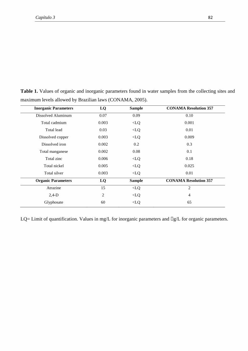
Capítulo 3� � � ���
�
Table 1. Values of organic and inorganic parameters found in water samples from the collecting sites and
maximum levels allowed by Brazilian laws (CONAMA, 2005).
Inorganic Parameters LQ Sample CONAMA Resolution 357
Dissolved Aluminum 0.07 0.09 0.10
Total cadmium 0.003 <LQ 0.001
Total lead 0.03 <LQ 0.01
Dissolved copper 0.003 <LQ 0.009
Dissolved iron 0.002 0.2 0.3
Total manganese 0.002 0.08 0.1
Total zinc 0.006 <LQ 0.18
Total nickel 0.005 <LQ 0.025
Total silver 0.003 <LQ 0.01
Organic Parameters LQ Sample CONAMA Resolution 357
Atrazine 15 <LQ 2
2,4-D 2 <LQ 4
Glyphosate 60 <LQ 65
�
LQ= Limit of quantification. Values in mg/L for inorganic parameters and µg/L for organic parameters.
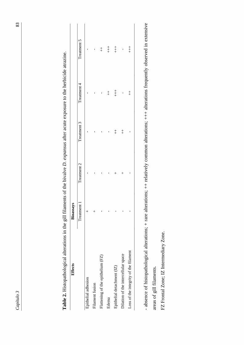
Cap
ítul
o 3�
����
� Tab
le 2
. His
topa
thol
ogic
al a
lter
atio
ns in
the
gill
fila
men
ts o
f th
e bi
valv
e D
. exp
ansu
s af
ter
acut
e ex
posu
re to
the
herb
icid
e at
razi
ne.
Eff
ects
B
ioas
says
T
reat
men
t 1
Tre
atm
ent 2
T
reat
men
t 3
Tre
atm
ent 4
T
reat
men
t 5
Epi
thel
ial a
dhes
ion
+
- -
- -
Fila
men
t fus
ion
+
- -
- -
Flat
teni
ng o
f th
e ep
ithel
ium
(F
Z)
- -
- -
++
Ede
ma
- -
- +
+
++
+
Epi
thel
ial d
etac
hmen
t (IZ
) -
+
++
+
++
+
++
Dil
atio
n of
the
inte
rcel
lula
r sp
ace
- +
+
+
- -
Los
s of
the
inte
grit
y of
the
fila
men
t -
- -
++
+
++
� - ab
senc
e of
his
topa
thol
ogic
al a
ltera
tions
; +
rar
e al
tera
tion
s; +
+ r
elat
ivel
y co
mm
on a
lter
atio
ns;
++
+ a
lter
atio
ns f
requ
entl
y ob
serv
ed i
n ex
tens
ive
area
s of
gil
l fil
amen
ts.
FZ F
ront
al Z
one;
IZ
Int
erm
edia
ry Z
one.
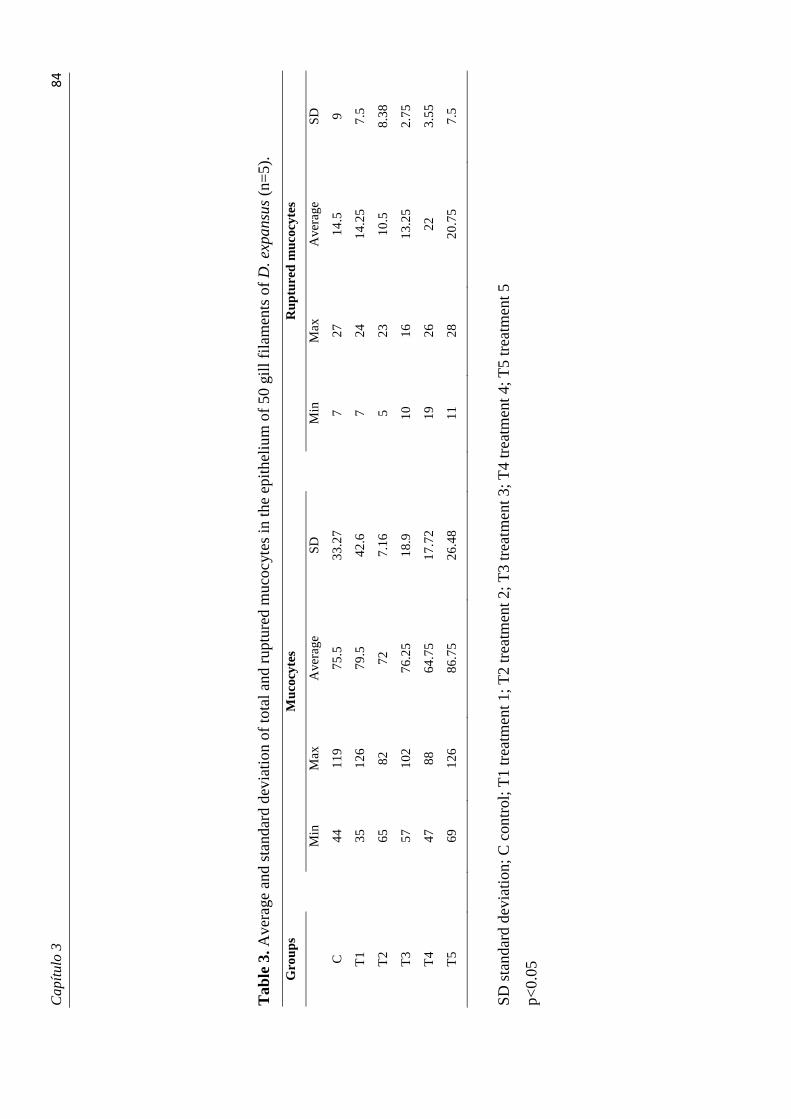
Cap
ítul
o 3�
����
� Tab
le 3
. Ave
rage
and
sta
ndar
d de
viat
ion
of to
tal a
nd r
uptu
red
muc
ocyt
es in
the
epith
eliu
m o
f 50
gill
fila
men
ts o
f D
. exp
ansu
s (n
=5)
.
Gro
ups
M
ucoc
ytes
Rup
ture
d m
ucoc
ytes
Min
M
ax
Ave
rage
SD
Min
M
ax
Ave
rage
SD
C
44
11
9 75
.5
33.2
7
7 27
14
.5
9
T1
35
12
6 79
.5
42.6
7 24
14
.25
7.5
T2
65
82
72
7.
16
5
23
10.5
8.
38
T3
57
10
2 76
.25
18.9
10
16
13.2
5 2.
75
T4
47
88
64
.75
17.7
2
19
26
22
3.55
T5
69
12
6 86
.75
26.4
8
11
28
20.7
5 7.
5
� SD s
tand
ard
devi
atio
n; C
con
trol
; T1
trea
tmen
t 1; T
2 tr
eatm
ent 2
; T3
trea
tmen
t 3; T
4 tr
eatm
ent 4
; T5
trea
tmen
t 5
p<0.
05
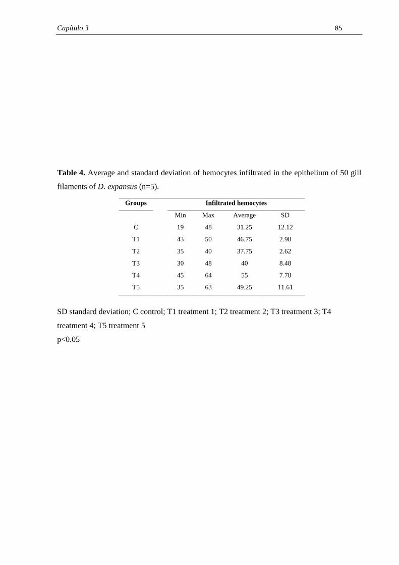
Capítulo 3� � ��
Table 4. Average and standard deviation of hemocytes infiltrated in the epithelium of 50 gill
filaments of D. expansus (n=5).
SD standard deviation; C control; T1 treatment 1; T2 treatment 2; T3 treatment 3; T4
treatment 4; T5 treatment 5
p<0.05
�
Groups Infiltrated hemocytes
Min Max Average SD
C 19 48 31.25 12.12
T1 43 50 46.75 2.98
T2 35 40 37.75 2.62
T3 30 48 40 8.48
T4 45 64 55 7.78
T5 35 63 49.25 11.61
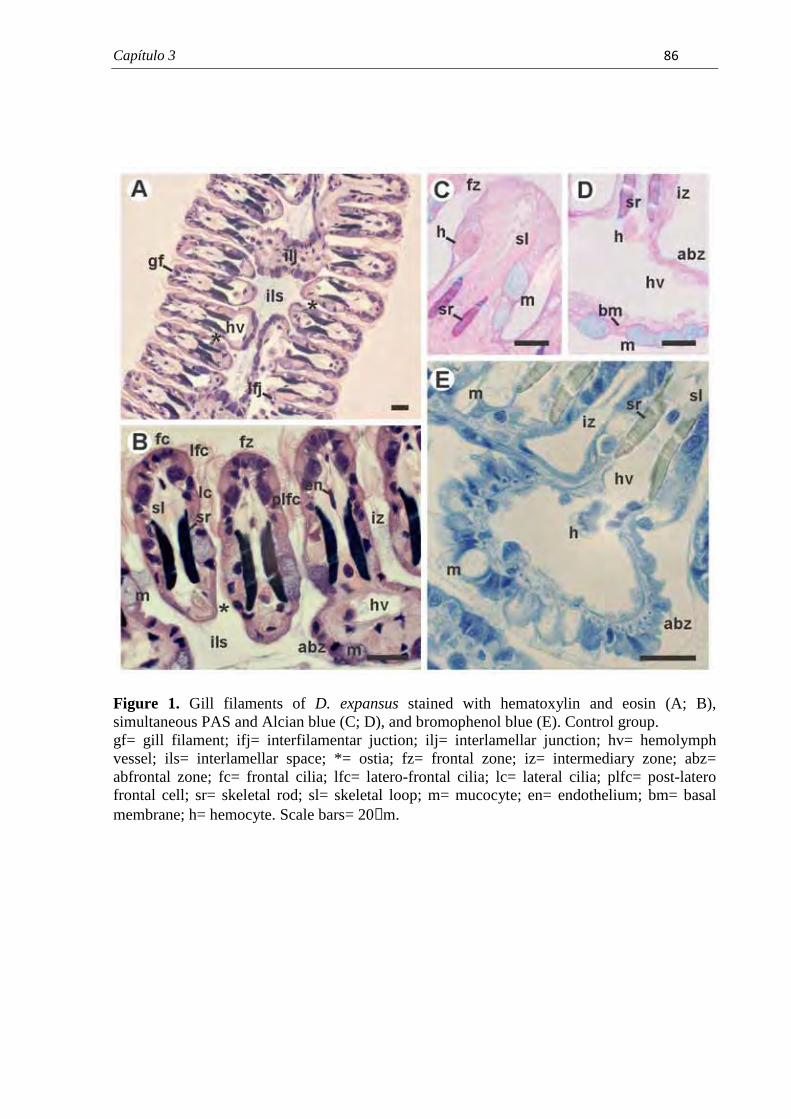
Capítulo 3� � � ��
Figure 1. Gill filaments of D. expansus stained with hematoxylin and eosin (A; B), simultaneous PAS and Alcian blue (C; D), and bromophenol blue (E). Control group. gf= gill filament; ifj= interfilamentar juction; ilj= interlamellar junction; hv= hemolymph vessel; ils= interlamellar space; *= ostia; fz= frontal zone; iz= intermediary zone; abz= abfrontal zone; fc= frontal cilia; lfc= latero-frontal cilia; lc= lateral cilia; plfc= post-latero frontal cell; sr= skeletal rod; sl= skeletal loop; m= mucocyte; en= endothelium; bm= basal membrane; h= hemocyte. Scale bars= 20µm.
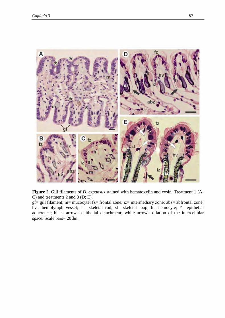
Capítulo 3� � � ���
Figure 2. Gill filaments of D. expansus stained with hematoxylin and eosin. Treatment 1 (A-C) and treatments 2 and 3 (D; E). gf= gill filament; m= mucocyte; fz= frontal zone; iz= intermediary zone; abz= abfrontal zone; hv= hemolymph vessel; sr= skeletal rod; sl= skeletal loop; h= hemocyte; *= epithelial adherence; black arrow= epithelial detachment; white arrow= dilation of the intercellular space. Scale bars= 20µm.
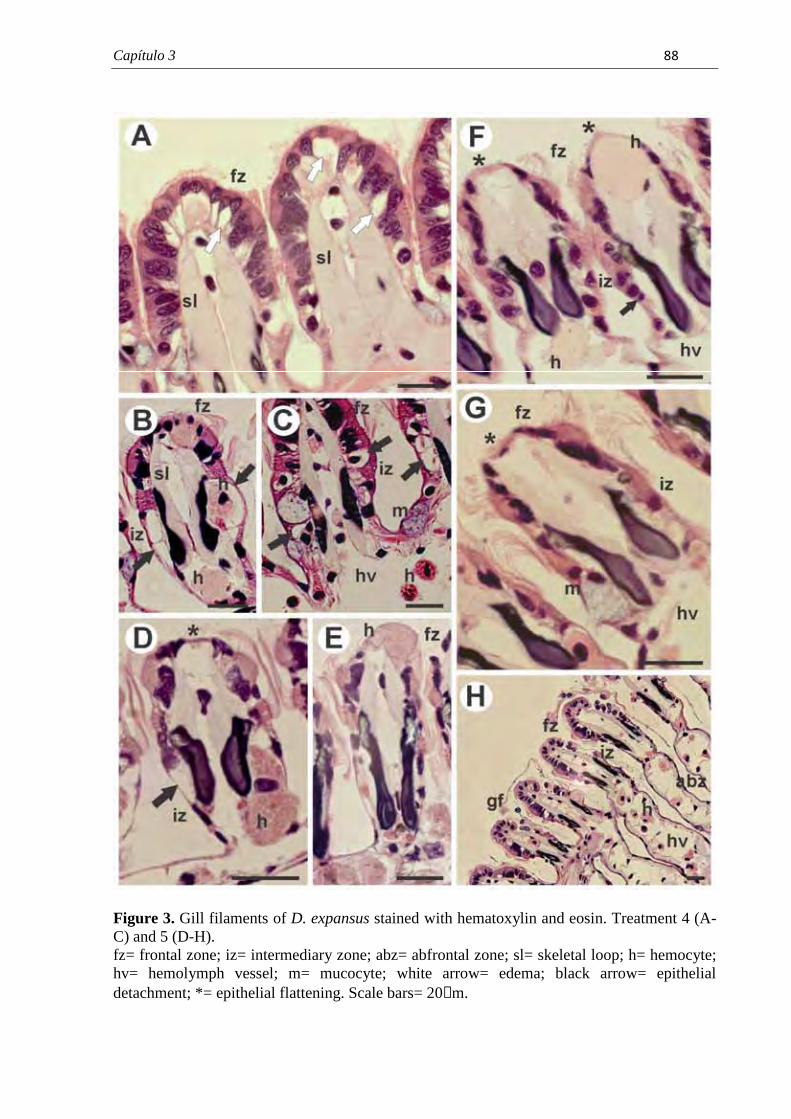
Capítulo 3� � � ���
Figure 3. Gill filaments of D. expansus stained with hematoxylin and eosin. Treatment 4 (A-C) and 5 (D-H). fz= frontal zone; iz= intermediary zone; abz= abfrontal zone; sl= skeletal loop; h= hemocyte; hv= hemolymph vessel; m= mucocyte; white arrow= edema; black arrow= epithelial detachment; *= epithelial flattening. Scale bars= 20µm.
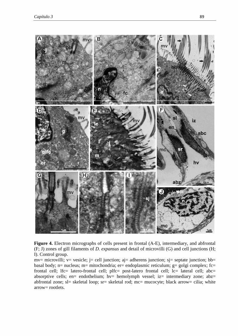
Capítulo 3� � � � �
Figure 4. Electron micrographs of cells present in frontal (A-E), intermediary, and abfrontal (F; J) zones of gill filaments of D. expansus and detail of microvilli (G) and cell junctions (H; I). Control group. mv= microvilli; v= vesicle; j= cell junction; aj= adherens junction; sj= septate junction; bb= basal body; n= nucleus; m= mitochondria; er= endoplasmic reticulum; g= golgi complex; fc= frontal cell; lfc= latero-frontal cell; plfc= post-latero frontal cell; lc= lateral cell; abc= absorptive cells; en= endothelium; hv= hemolymph vessel; iz= intermediary zone; abz= abfrontal zone; sl= skeletal loop; sr= skeletal rod; mc= mucocyte; black arrow= cilia; white arrow= rootlets.
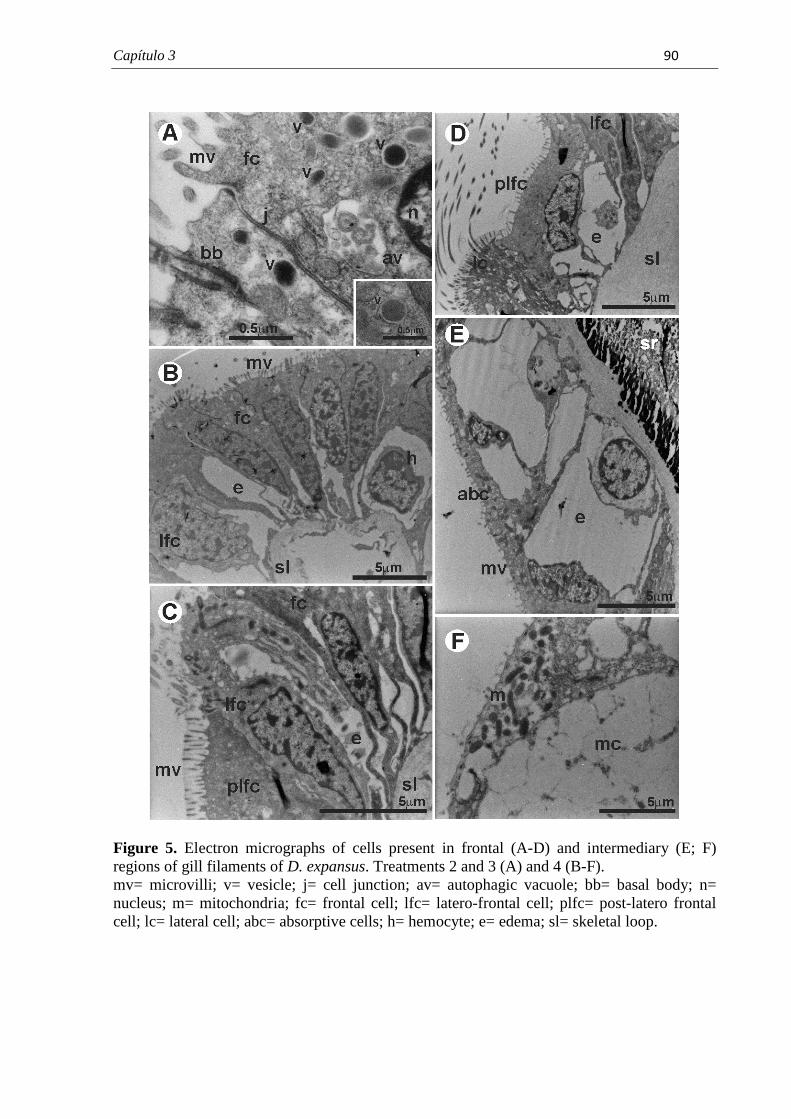
Capítulo 3� � � ��
Figure 5. Electron micrographs of cells present in frontal (A-D) and intermediary (E; F) regions of gill filaments of D. expansus. Treatments 2 and 3 (A) and 4 (B-F). mv= microvilli; v= vesicle; j= cell junction; av= autophagic vacuole; bb= basal body; n= nucleus; m= mitochondria; fc= frontal cell; lfc= latero-frontal cell; plfc= post-latero frontal cell; lc= lateral cell; abc= absorptive cells; h= hemocyte; e= edema; sl= skeletal loop.
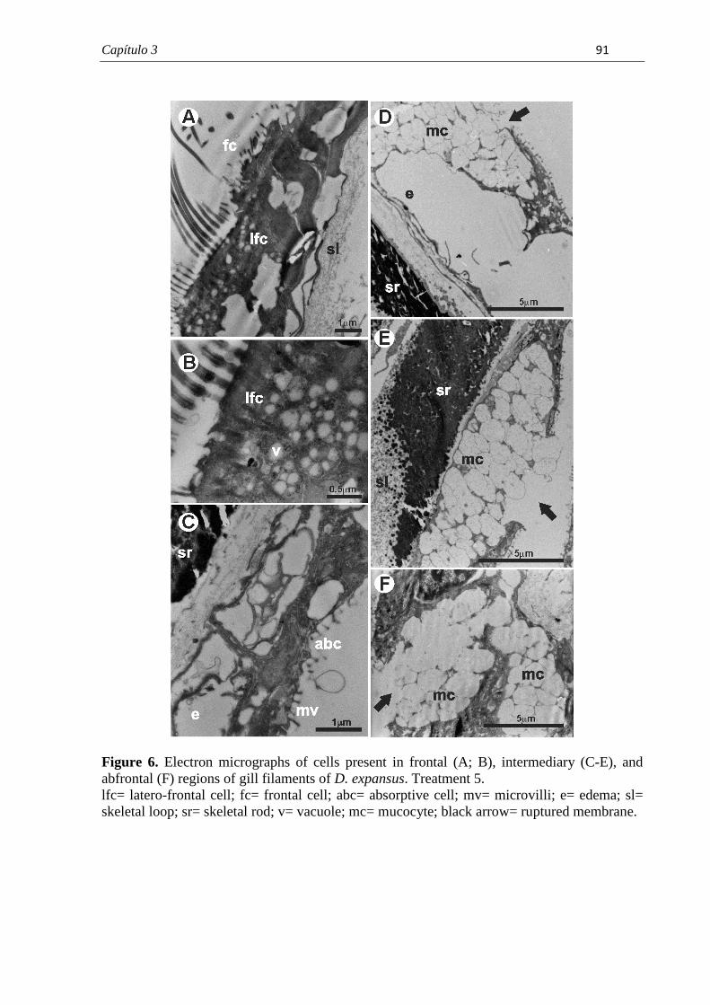
Capítulo 3� � � ��
Figure 6. Electron micrographs of cells present in frontal (A; B), intermediary (C-E), and abfrontal (F) regions of gill filaments of D. expansus. Treatment 5. lfc= latero-frontal cell; fc= frontal cell; abc= absorptive cell; mv= microvilli; e= edema; sl= skeletal loop; sr= skeletal rod; v= vacuole; mc= mucocyte; black arrow= ruptured membrane.�
Considerações Finais� � ��
�
8. CONSIDERAÇÕES FINAIS
Devido a suas características físico-químicas e ampla utilização, o herbicida atrazina é
um dos principais contaminantes de águas superficiais e subterrâneas em todo o mundo. Essa
é uma problemática enfrentada por muitos países como os Estados Unidos da América e que
também tem se agravado no Brasil diante da expansão das culturas agrícolas como, por
exemplo, de cana-de-açúcar e milho. Desta forma, torna-se necessário o monitoramento
desses ecossistemas que sofrem a influência dessas fontes de contaminação provenientes das
atividades agrícolas. A utilização de parâmetros biológicos é fundamental para se avaliar os
impactos causados no ecossistema, utilizando-se principalmente representantes da fauna
endêmica, como potenciais espécies bioindicadoras. Neste sentido, esse trabalho buscou
avaliar os possíveis efeitos tóxicos de concentrações ambientalmente relevantes do herbicida
atrazina em um representante da malacofauna endêmica, utilizando-se parâmetros
morfológicos sob condições laboratoriais controladas. A partir dos resultados obtidos nesse
trabalho foi possível concluir:
• Os filamentos branquiais da espécie estudada apresentam morfologia similar à de
outros bivalves eulamelibrânquios. Entretanto, foi observada a presença simultânea de
duas estruturas de sustentação especializadas chamadas hastes e alça de sustentação,
ainda não descritas detalhadamente na literatura.
• A espécie D. expansus mostrou-se sensível para avaliar a toxicidade do herbicida
atrazina e, possivelmente, possa ser utilizada para avaliação da qualidade de corpos
d’água sob influência de diferentes fontes de poluição.
• As brânquias foram sensíveis à presença do herbicida atrazina. Esse órgão apresentou
respostas tissulares relacionadas com proteção ou defesa, ocorrência de danos e maior
gasto energético.
• As alterações histopatológicas foram mais diversificadas e frequentes nos animais
expostos a maiores concentrações do herbicida, revelando uma resposta dose-
dependente;
Considerações Finais� � ��
�
• A concentração máxima de atrazina permitida em corpo d’água doce pela legislação
brasileira não causou alterações histopatológicas significativas no bivalve estudado
após exposição aguda. Entretanto, existe a necessidade de se avaliar os efeitos dessa
concentração de herbicida em níveis mais baixos da escala biológica, por exemplo, a
nível molecular e bioquímico.
• Em corpos d’água próximos a áreas agrícolas, as maiores concentrações de herbicida
são usualmente encontradas logo após a aplicação no solo, seguida de chuvas e
irrigação. No trabalho realizado, essa situação seria comparável a uma exposição por
um curto período (exposição aguda) as maiores concentrações testadas. Essas
concentrações causaram alterações histopatológicas significativas nas brânquias dos
animais estudados, cuja permanência e extensão comprometeriam o desempenho e
saúde dos animais. Desta forma, seria interessante realizar trabalhos futuros que
avaliem se essas alterações são reversíveis ou permanentes diante da diminuição das
concentrações do herbicida, submetendo os animais a um período de recuperação.
Referências Bibliográficas� � ��
�
9. REFERÊNCIAS BIBLIOGRÁFICAS
ADAM-GUILLERMIN, C.; FOURNIER, E.; FLORIANI, M.; CAMILLERI, V.;
MASSABUAU, G.C.; GARNIER-LAPLACE, J. Biodynamics, Subcellular Partitioning, and
Ultrastructural Effects of Organic Selenium in a Freshwater Bivalve. Environ. Sci. Technol.,
v. 43, p. 2112–2117, 2009.
ARIAS, A.R.L.; BUSS, D.F; ALBUQUERQUE, C.; INÁCIO, A.F.; FREIRE, M.M.;
EGLER, M.; MUGNAI, R.; BAPTISTA, D.F. Utilização de bioindicadores na avaliação de
impacto e no monitoramento da contaminação de rios e córregos por agrotóxicos. Ciência &
Saúde Coletiva, v.12, n.1, p.61-72, 2007.
ARMAS, E.D.; MONTEIRO, R.T.R.; AMÂNCIO, A. V.; CORREA, R.M.L.; GUERCIO,
M.A. O uso de agrotóxicos em cana-de-açúcar na Bacia do Rio Corumbataí e o risco de
poluição hídrica. Quim. Nova, v. 28, n.6, p. 975-982, 2005.
AUFFRET, M.; OUBELLA, R. Hemocyte aggregation in the oyster Crassostrea gigas: in
vitro measurement and experimental modulation by xenobiotics. Comp Biochem Physiol A,
v. 118, p. 705– 712, 1997.
BARŠIEN�, J.; ANDREIK�NAIT�, L.; GARNAGA, G.; RYBAKOVAS, A. Genotoxic and
cytotoxic effects in the bivalve mollusks Macoma balthica and Mytilus edulis from the Baltic
Sea. Ekologija, v. 54, n.1, p.44–50, 2008.
BASACK, S.B.; ONETO, M.L.; FUCHS, J.S.; WOOD, E.J.; KESTEN, E.M. Esterases of
Corbicula fluminea as Biomarkers of Exposure to Organophosphorus Pesticides. Bull.
Environ. Contam. Toxicol., v. 61, p. 569-576, 1998.
BEASLEY, C.R.; TÚRY, E.; VALE, W.G.; TAGLIARO, C.H. Reproductive cycle,
management and conservation of Paxyodon syrmatophorus (Bivalvia; Hyriidae) from the
Tocantins River, Brazil. J. Moll. Stud., v. 66, p. 393-402, 2000.
Referências Bibliográficas� � �
�
BERNET, D.; SCHMIDT, H.; MEIER, W.; BURKHARDT-HOLM, P.; WAHLI, T.
Histopathology in fish: proposal of a protocol to assess aquatic pollution. J. Fish Dis.,
Oxford, v. 22, p. 25-34, 1999.
BIAGINI, F. R.; DAVID, J. A. O. ; FONTANETTI, C.S. The use of histological,
histochemical and ultramorphological techniques to detect gill alterations in Oreochromis
niloticus reared in treated polluted waters. Micron (Oxford), v. 40, p. 839-844, 2009.
BIGAS, M.; DURFORT, M.; POQUET, M. Cytological effects of experimental exposure to
Hg on the gill epithelium of the European flat oyster Ostrea edulis: ultrastructural and
quantitative changes related to bioaccumulation. Tissue & Cell, v. 33, n. 2, p. 178-188, 2001.
BIRDSALL, K.; KUKOR, J.J.; CHENEY, M.A. Uptake of polycyclic aromatic hydrocarbon
compounds by the gills of the bivalve mollusk Elliptio complanata. Environmental
Toxicology and Chemistry, v. 20, n. 2, p. 309–316, 2001.
BOENING, D.W. An evaluation of bivalves as biomonitors of heavy metals pollution in
marine waters. Environmental Monitoring and Assessment, v. 55, p. 459–470, 1999.
BOGAN, A.E. Global diversity of freshwater mussels (Mollusca, Bivalvia) in freshwater.
Hydrobiologia, v. 595, p. 139–147, 2008.
BOLDINA-COSQUERIC, I.; AMIARD, J.C.; AMIARD-TRIQUET, C.; DEDOURGE-
GEFFARD, O.; MÉTAIS, I.; MOUNEYRAC, C.; MOUTEL, B.; A, BERTHET, B.
Biochemical, physiological and behavioural markers in the endobenthic bivalve Scrobicularia
plana as tools for the assessment of estuarine sediment quality. Ecotoxicology and
Environmental Safety, v. 73, p. 1733–1741, 2010.
BOLOGNESI, C.; HAYASHI, M. Micronucleus assay in aquatic animals. Mutagenesis, v.
26, n. 1, p. 205–213, 2011.
BONETTO, A.A.; TASSARA, M.P.; RUMI, A. Australis n. subgen. de Diplodon spix
(Bivalvia, Unionacea) y posibles relaciones con Hyriidae australianos. Bol. Soc. Biol., v.57,
55-61, 1986.
Referências Bibliográficas� � �
�
BOTTINI, M.A. Composición y distribuición de la asociación de moluscos bentónicos en la
laguna La Patagua, Península de Quetrihu, Degree Final Work Report, Centro Regional
Universitário Bariloche, Universidade Nacional del Comahue, Argentina. 1993.
BOUILLY, K.; MCCOMBIE, H.; LEITÃO, A.; LAPÉGUE, S. Persistence of atrazine impact
on aneuploidy in Pacific oysters, Crassostrea gigas. Marine Biology, v. 145, p. 699–705,
2004.
BRAINWOOD, M.; BURGIN, S.; BYRNE, M. The role of geomorphology in substratum
patch selection by freshwater mussels in the Hawkesbury- Nepean River (New Sputh Wales)
Australia. Aquatic Conservation: Marine and Freshwater Ecosystems, v.18, p.1285–1301,
2008.
BRASIL. Ministério do Planejamento, Orçamento e Gestão. Instituto Brasileiro de Geografia
e Estatística; Anuário Estatístico do Brasil, IBGE: Rio de Janeiro, v.69, 2009.
BYRNE, M. Reproduction of river and lake populations of Hyridella depressa (Unionacea:
Hyriidae) in New South Wales: implications for their conservation. Hydrobiologia, v. 389, p.
29–43, 1998.
BYRNE, M. Calcium concretions in the interstitial tissues of the Australian freshwater
mussels Hyridella depressa (Hyriidae). In: HARPER, E.M.; TAYLOR; J.D; CRAME, J.A.
(Orgs.). The Evolutionary Biology of the Bivalvia. London: Geological Society, 2000.
p.329-337.
CALLIL, C.T.; MANSUR, M. C. D. Gametogênese e dinâmica da reprodução de Anodontites
trapesialis (Lamarck) (Unionoida, Mycetopodidae) no lago Baía do Poço, planície de
inundação do rio Cuiabá, Mato Grosso, Brasil. Revista Brasileira de Zoologia, v. 24, n. 3., p.
825-840, 2007.
CETESB - Companhia de Tecnologia de Saneamento Ambiental. Guia de coleta e
preservação de amostras de água. 1ª ed. São Paulo: CETESB, 1987. 155p.
Referências Bibliográficas� � ��
�
CHENEY, M.A.; KEIL, D.; QIAN, S. Uptake and effect of mercury on amino acid losses
from the gills of the bivalve mollusks Mytilus californianus and Anodonta californiensis.
Journal of Colloid and Interface Science, v. 320, p. 369–375, 2008.
COADY, K.K.; MURPHY, M.B.; VILLENEUVE, D.L.; HECKER, M.; JONES, P.D.;
CARR, J.A.; SOLOMON, K.; SMITH, E.; VAN DER KRAAK, G.; KENDALL, R.J.;
GIESY, J.P. Effects of atrazine on metamorphosis, growth, laryngeal and gonadal
development, aromatase activity, and sex steroid concentrations in Xenopus laevis.
Ecotoxicol. Environ. Saf., v. 62, p.160–173, 2005.
COGERH - Companhia de Gestão dos Recursos Hídricos. Recomendações e cuidados na
coleta de amostras de água. Informe técnico n.02. 1ª ed. Fortaleza: COGERH, 2001. 20p.
COLVILLE, A.E.; LIM, R.P. Microscopic structure of the mantle and palps in the freshwater
mussels Velesunio ambiguus and Hyridella depressa (Bivalvia: Hyriidae). Molluscan
Research, v. 23, p. 1-20, 2003.
CONAMA - Conselho Nacional do Meio Ambiente/Ministério do Meio Ambiente, 2005.
Resolução No. 357 de 17 de março de 2005. Disponível em
<http://www.mma.gov.br/port/conama/legiano1.cfm?codlegitipo=3&ano=2005>Acesso em
novembro de 2010.
CURIAL, O.; LANGE, R.R. Observações sobre a proporção de sexos em Diplodon deodontus
expansus. Arquivos de Biologia e Tecnologia, v. 17, n. 2, p. 111-112, 1974a.
CURIAL, O.; LANGE, R.R. Hermafroditismo em Diplodon delotundus expansus. Arquivos
de Biologia e Tecnologia, v. 17, n. 2, p. 109-110, 1974b.
CURIAL, O.; LANGE, R.R. Variações histológicas sazonais das gônadas do Diplodon
delodontus expansus (Küster) (Mollusca, Unionidade). Arquivos de Biologia e Tecnologia,
v. 18, p. 65-67, 1975.
Referências Bibliográficas� � ��
�
DAVID, J. A. O.; FONTANETTI, C.S. Surface morphology of Mytella falcata gill filaments
from three regions of the Santos estuary. Brazilian Journal of Morphological Sciences,
Campinas, v. 22, n. 4, p. 203-210, 2005.
DAVID, J. A. O.; FONTANETTI, C.S. The role of mucus in Mytella falcata (Orbigny, 1842)
gills from polluted environments. Water, Air, and Soil Pollution, v. 200, p. 1-6, 2009.
DAVID, J. A. O.; SALAROLI, R. B.; FONTANETTI, C.S. The significance of changes in
Mytella falcata (Orbigny, 1842) gill filaments chronically exposed to polluted environments.
Micron (Oxford), v. 39, p. 1293-1299, 2008a.
DAVID, J. A. O.; HOSHINA, M.; FONTANETTI, C.S. DNA damages in Mytella falcata
(Mytiloida, Mytilidae) cells: a new tool for biomonitoring studies in tropical estuarine
ecosystems. Naturalia (São José do Rio Preto), v. 31, p. 2, 2008b.
DE GREGORI, I.; DELGADO, D.; PINOCHET, H.; GRAS, N.; MUÑOZ, L.; BRHUN, C. e
NAVARRETE, G. Cadmium, lead, copper and mercury levels in fresh and canned bivalve
mussels Tagelus dombeii (Navajuela) and Semelle solida (Almeja) from the Chilean coast.
Sci. Total Environ., v.148, p. 1-10, 1994.
DEPLEDGE, M. H. Conceptual paradigms in marine ecotoxicology. In: BJORNSTAD, E.;
HAGERMAN, L.; JENSEN, K. JENSEN, K. Proceedings of the 21th Baltic Marine
biologists Symposium. Olsen & Olsen, Fredensborg, Denmark. 1992.
DEWEY, S.L. Effects of the herbicide atrazine on aquatic insect community structure and
emergence. Ecology, v. 67, p.148–162, 1986.
DIANA, S.G.; RESETARITS JR. W.J.; SCHAEFFER, D.J.; BECKMEN, K.B.; BEASLEY,
V. Effects of atrazine on amphibian growth and survival in artificial aquatic communities.
Environ. Toxicol. Chem., v.19, p. 2961–2967, 2000.
DOMOUHTSIDOU, G.P.; DIMITRIADIS, V.K. Lysosomal, tissue and cellular alterations in
the gills, palps and intestine of the mussels Mytilus galloprovincialis, in relation to pollution.
Marine Biology, v.145, p. 109–120, 2004.
Referências Bibliográficas� � �
�
DU PREEZ, H.H., VAN VUREN, J.H.J. Bioconcentration of atrazine in the banded tilapia,
Tilapia sparrmanii. Comp. Biochem. Physiol., v.101C, p.651–655, 1992.
EDWARDS, C. A. Persistent pesticides in the environment. 2. ed. U.S.A.: CRC Press,
1973. 170p.
EDWIN, D. O. Control of water pollution from agriculture. Irrigation and Drainage, New
York, v.55, p. 1-101. 1996.
EISLER, R. Atrazine hazards to fish, wildlife and invertebrates: a synoptic review. U.S. Fish
Wildl. Serv. Biol. Rep. v. 85, p. 1–55, 1989.
FERNÁNDEZ-TAJES, J.; FLÓREZ, F.; PEREIRA, S.; RÁBADE, T.; LAFFON, B.;
MÉNDEZ, J. Use of three bivalve species for biomonitoring a polluted estuarine
environment. Environ Monit Assess., v. 177, p. 289–300, 2011.
FARRINGTON, J. W. Bivalves as sentinels of coastal chemical pollution: The Mussel (and
the oyster) Watch. Oceans, v.20, n.2, p.18-29, 1983.
FERREIRA, A.G., MACHADO, A.L.S., ZALMON, I,R. Temporal and Spatial Variation on
Heavy Metal Concentrations in the bivalve Perna perna (LINNAEUS, 1758) on the Northern
Coast of Rio de Janeiro State, Brazil. Brazilian Archives of biology and technology, v. 47,
n. 2, p. 319-327, 2004.
FLYNN, K.; SPELLMAN, T. Environmental levels of atrazine decreases patial aggregation in
the freshwater mussel, Elliptio complanata. Ecotoxicology and Environmental Safety, v.
72, p.1228– 1233, 2009.
FONTANETTI, C.S.; CHISTOFOLETTI, C.A.; PINHEIRO, T.G.; SOUZA, T.S.; PEDRO-
ESCHER, J. Microscopy as a tool in toxicological evaluations. In: MÉNDEZ VILAS, A.;
DÍAS, J. (Eds.). Microscopy: Science, Technology, Applications and Education.
FORMATEX, 2010. p. 1001-1007.
Referências Bibliográficas� � ����
�
FOSTER, R.B.; BATES, J.M.; Use of freshwater mussels to monitor point source industrial
discharges. Environ. Sci. Technol., v. 12, n.8, p. 958-962, 1978.
GILBERT, S.F. Developmental Biology. 8.ed. Sunderland: Sinauer Associates Inc., 2006.
817p.
GRAF, D. L. The Etheriidae revisited: a phylogenetic analysis of hyriid relationships
(Mollusca: Bivalvia: Paleoheterodonta: Unionoida). Occasional papers of the Museum of
Zoology, The University of Michigan Number 729: 1–21. 2000.
GRANEY JR., R.L.; CHERRY, D.S.; CAIRNS JR., J. Heavy metal indicator potential of the
Asiatic clam (Corbicula fluminea) in artificial stream systems. Hydrobiologia, v.102, p.81-
88, 1983.
GREGORY, M.A.; GEORGE, R.C.; MARSHALL, D.J.; ANANDRAJ, A.; MCCLURG, T.P.
The effects of mercury exposure on the surface morphology of gill filaments in Perna perna
(Mollusca: Bivalvia). Mar. Pollut. Bull., v. 39, p. 116-121, 1999.
GREGORY, M.A.; MARSHAL, D.J.; GEORGE, R.C.; ANANDRAJ, A.; CCLURG, T.P.
Correlations between metal uptake in the soft tissue of Perna perna and gill filament
pathology after exposure to mercury. Mar. Pollut. Bull., v. 45, p. 114-125, 2002.
GRISOLIA, C. K. Agrotóxicos: Mutações, Câncer e Reprodução. Brasília: Editora
Universidade de Brasília, 2005. 392p.
GUEVARA, S.R., BUBACH, D., VIGLIANO P., LIPPOLT, G., ARRIBÉRE, M. Heavy
metal and other trace elements in native mussel Diplodon chilensis from Norhern Patagonia
Lakes, Argentina. Biologica Trace Element Research, v. 102, p.245-263, 2004.
GUEVARA, S.R.; ARRIBÉRE, M.; BUBACH, D., VIGLIANO P., RIZZO, A., ALONSO,
M.; SNACHEZ, R. Silver contamination on abiotic and biotic compartments of Nahuel Huapi
National Park lakes, Patagonia, Argentina. Science of the Total Environment, v. 336, p.
119-134, 2005.
Referências Bibliográficas� � ����
�
GUNDACKER, C. Comparison of heavy metal bioaccumulation in freshwater mollusks of
urban river habitats in Vienna. Environmental Pollution, v.110, p. 61-71, 2000.
GUNKEL, G.; STREIT, B. Mechanisms of bioaccumulation of a herbicide (atrazine,S-
triazine) in a freshwater mollusk (Ancylus fluviatilis Müll.) and a fish (Coregonus fera
Jurine). Water Res., v.14, p.1573–1584, 1980.
HAYES, T.; COLLINS, A.; LEE, M.; MENDOZA, M.; NORIEGA, N.; STUART, A.;
VONK, A. Hermaphroditic, demasculinized frogs after exposure to the herbicide, atrazine, at
low ecologically relevant doses. Proc. Natl. Acad. Sci. U.S.A., v. 99, p.5476–5480, 2002.
HAYES, T.; HASTON, K.; TSUI, M.; HOANG, A.; HAEFFELE, C.; VONK, A. Atrazine-
induced hermaphroditism at 0.1 PPB in American leopard frogs (Rana pipiens): laboratory
and field evidence. Environ. Health Persp., v. 111, n.4, p. 568–575, 2003.
HEBLING, N.J.; PENTEADO, A.M.G. Anatomia functional de Diplodon rotundus gratus
Wagner, 1827 (Mollusca, Bivalvia). Rev. Brasil. Biol., v. 34, n.1, p. 67-80, 1974.
HENRY, R; SIMÃO, S.A. Evaluation of density and biomass of a bivalve population
(Diplodon delodontus expansus) (Küster, 1856) in a small tropical reservoir. Revue D’
Hydrobiologie Trocale, v.17, p.309-318, 1984.
HENRY, R., FILOSO, S. Spatial distribution of a bivalve population (Diplodon delodontus
expansus) (KÜSTER, 1865) in a small tropical reservoir. Rev. Brasil. Biol., v. 45, n. 4, p.
407-415, 1985.
HENRY, R., FILOSO, S. Nutrient Release Through Decomposition of Diplodon delodontus
Expansus (Mollusca, Bivalvia) and Some Considerations of its Effects on the Ecology of a
Small Tropical Reservoir. Rev. Brasil. Biol., v. 47, n. 4, p. 479-486, 1987.
HOEH, W. R.; BOGAN, A.E.; CUMMINGS, K.S.; GUTTMAN, S.E. Evolutionary
relationships among the higher taxa of freshwater mussels (Bivalvia: Unionoida): inferences
on phylogeny and character evolution from analyses of DNA sequence Data. Malacological
Review, v. 31, p.111–130, 1999.
Referências Bibliográficas� � ����
�
HUGGETT, R.J.; KIMERLE, R.A.; MEHRLE, PM.; BERGMAN, H.L. Biomarkers:
Biochemical, Physiological and Histological Markers of Anthropogenic. Stress. Flórida:
Lewis Publishers Boca Raton, 1992.
JACOMINI, A.E.; BONATO, P.S.; AVELAR, W.E.P. HPLC method for the analysis of
atrazine in freshwater bivalves. Journal of liquid chromatography and related
technologies, v. 26, n. 12, p. 1885-1894, 2003.
JACOMINI, A.E.; AVELAR, W.E.P.; MARTINÊS, A.S.; BONATO, P.S. Bioaccumulation
of Atrazine in Freshwater Bivalves Anodontites trapesialis (Lamarck, 1819) and Corbicula
fluminea (Müller, 1774). Arch. Environ. Contam. Toxicol., v. 51, p. 387–391, 2006.
JACOMINI, A.E.; CAMARDO, P.B.; AVELAR, W.E.P.; BONATO, P.S. Assessment of
Ametryn Contamination in River Water, River Sediment, and Mollusk Bivalves in São Paulo
State, Brazil. Arch. Environ. Contam. Toxicol., v. 60, p. 452–461, 2011.
JUNQUEIRA, L.C.; JUNQUEIRA, L.M.M.S. Técnicas Básicas de Citologia e Histologia.
São Paulo: Livraria Editora Santos, 1983.123p.
JUPITER, S. D.; BYRNE, M. Light and scanning electron microscopy of the embryos and
glochidia larvae of the Australian freshwater bivalve Hyridella depressa (Hyriidae). Invert.
Repro. Dev., v.32, p.177–186, 1997.
KARNOVSKY, M.J. A formaldehyde-glutaraldehyde fixative at high osmolarity for use in
electron microscopy. J. Cell Biol., v. 11, p. 137-140, 1965.
KREUTZ, L.C.; BARCELLOS, L. J. G.; MARTENINGHE, A.; SANTOS, E.D.; ZANATTA,
R. Exposure to sublethal concentration of glyphosate or atrazine-based herbicides alters the
phagocytic function and increases the susceptibility of silver catfish fingerlings (Rhamdia
quelen) to Aeromonas hydrophila challenge. Fish & Shellfish Immunology, v.29, p.694-697,
2010.

Referências Bibliográficas� � ����
�
LAABS, V.; AMELUNG, W.; PINTO, A.; ALTSTAEDT, A.; ZECH, W. Leaching and
degradation of corn and soybean pesticides in an Oxisol of the Brazilian Cerrados.
Chemosphere, v. 41, p 1441-1449, 2000.
LOAYZA-MURO, R.; ELIAS-LETTS, R. Responses of the mussel Anodontites trapesialis
(Unionidae) to environmental stressors: Effect of pH, temperature and metals on filtration
rate. Environmental Pollution, v.149, p. 209-215, 2007.
LORENZI, H. Manual de identificação e controle de plantas daninhas. Nova Odessa:
Editora Plantarum. 1990.
LUIZ, A.I.B.; NEVES, M.C.; DYNIA, J.F. Implicações na qualidade das águas
subterrâneas nas atividades agrícolas na região metropolitana de Campinas, SP.
Jaguariúna: Embrapa Meio Ambiente. 2004. 33p.
MANLY, R E GEORGE , W. O. The occurrence of some heavy metals in populations of
freshwater mussel Anodonta anatina from the river Thames. Environ. Pollut., v. 14, p. 139-
154, 1977.
MANSUR, M.C.D. Gloquídio de Diplodon martensi (Ihering) (Mollusca, Bivalvia, Hyriidae)
e seu ciclo parasitário. Revista Brasileira de Zoologia, v.16, p.185-194, 1999.
MANSUR, M.C.D.; ANFLOR, L. M.; Diferenças morfológicas entre Diplodon charruanus
ORBIGNY, 1835 e D. pilsbryi MARSHALL, 1928 (Bivalvia, Hyriidae). Iheringia. Ser. Zool.,
v. 60, p.101-116, 1981.
MANSUR, M.C.D.; SANTOS, C. P. Invertebrados Aquáticos: Diplodon Expansus. In:
MACHADO, A.B.M.; DRUMMOND, G.M.; PAGLIA, A.P (Eds). Livro Vermelho da
Fauna Ameaçada de Extinção, Belo Horizonte: Fundação Bioversitas. 2008.
MANTECCA, P.; VAILATI, G.; BACCHETTA, R. Histological changes and micronucleous
inductions in the Zebra mussel Dreissena polymorpha after Paraquat exposure. Histology and
histopathology, v. 21, p.829-840, 2006.
Referências Bibliográficas� � ����
�
MATSUMOTO, S.T.; RIGONATO, J.; MANTOVANI, M.S; MARIN-MORALES, M.A.
Evaluation of the gentotoxic potencial due to the action of an effluent contaminated with
chromium, by the comet assay in CHO-K1 cultures. Caryologia, v. 58, n.1, p. 40-46, 2005.
MCCONNELL, M. A.; HARREL, R. C. The estuarine clam Rangia cuneata (Gray) as a
biomonitor of heavy metals under laboratory and field conditions. Am. Malacol. Bull., v. 11,
n. 2, p.1991-2001, 1995.
MEYER, A.A.N.; OLIVEIRA, E.; MARTIM, J. Classes de comprimento e proporção sexual
em Diplodon expansus (Mollusca, Bivalvia, Hyriidae) no rio Piraquara, Paraná, Brasil.
Iheringia, Sér. Zool., v. 100, n.4, p.329-335, 2010.
MOORE, A.; WARING, C.P. Mechanistic effects of a triazine pesticide on reproductive
endocrine function in mature Atlantic salmon (Salmo salar L.) parr. Pest. Biochem. Physiol.,
v.62, p.41–50, 1998.
MURPHY, M.B.; HECKER, M.; COADY, K.K.; TOMPSETT, A.R.; JONES, P.D.; DU
PREEZ, L.H.; EVERSON, G.J.; SOLOMON, K.R.; CARR, J.A.; SMITH, E.E.; KENDALL,
R.J.; VAN DER KRAAK, G.; GIESY, J.P. Atrazine concentrations, gonadal gross
morphology and histology in ranid frogs collected in Michigan agricultural areas. Aquatic
Toxicology, v.76, p.230–245, 2006.
NAIMO, T.J. A review of the effects of heavy metals on freshwater mussels. Ecotoxicology,
v.4, p.341-362, 1995.
NASCIMENTO, I.A.; PEREIRA, S.A.; LEITE, M.B.N.L. Biomarcadores como instrumentos
preventivos de poluição. In: ZAGATTO, P.A.; BERTOLETTI, E. (Eds). Ecotoxicologia
Aquática: Princípios e aplicações. 2 ed. São Carlos: RiMa. 2008.
NESKOVIC, N.K.; ELEZOVIC, I.; KARAN, V.; POLEKSIC, V.; BUDIMIR, M. Acute and
subacute toxicity of atrazine to carp (Cyprimus carpio). Ecotoxicology and Environmental
Safety, v.25, p.173-182, 1993.
Referências Bibliográficas� � ���
�
NOGAROL, L.R.; BROSSI-GARCIA, A.L.; FONTANETTI, C.S. Surface Morphology of
Diplodon expansus (Küster, 1856) (Mollusca, Bivalvia, Hyriidae) Gill Filaments After
Exposure to Environmentally Relevant Concentrations of Atrazine Herbicide. Microscopy
Research and Technique, 2011. DOI 10.1002/jemt.21130.
OHYAMA, T.; JIN, K.; KATAH, Y.; CHIPA, Y.; INOVE, K. 1,3,5-trichloro-2-(4-
nitrophenoxy) benzene (CNP) in water, sediments, and shellfish of the Ishikari river. Bull.
Environ. Contam. Toxicol., New York, v. 37, p 344-349, 1986.
PARADA, E.; PEREDO, S. Estado actual de la taxonomía de bivalvos dulceacuícolas
chilenos: progresos y conflictos. Rev. Chil. Hist. Nat., v. 75, n. 4, p. 691–701, 2002.
PEARSE, A.G.E. Histochemistry: Theoretical and Applied. 4 th ed., London: J&A.
Churchill, v.2, 1985. 998p.
PEREZ, D.G.; FONTANETTI, C.S. Hemocitical responses to environmental stress in
invertebrates: a review. Environ Monit Assess, v.177, p. 437-447, 2011.
PREEZ, H.H.; VUREN, J.H.J. Bioconcentration of atrazine in the banded tilapia, Tilapia
sparrmanii. Comp. Biochem. Physiol., v. 101, p. 651–655, 1992.
PROTASOWICKI, M.; DURAL, M.; JAREMEK, J. Trace metals in the shells of blue
mussels (Mytilus edulis) from the Poland coast of Baltic sea. Environ Monit Assess, v.141,
p. 329–337, 2008.
RIGONATO, J; MANTOVANI, M. S.; JORDÃO, B.Q. Comet assay comparison of different
Corbicula fluminea (Mollusca) tissues for the detection of genotoxicity. Genetics and
Molecular Biology, v.28, n.3, p.464-468, 2005.
RITTSCHOF, D.; MCCLELLAN-GREEN, P. Molluscs as multidisciplinary models in
environment toxicology. Mar. Pollut. Bull., Oxford, v. 50, p. 369-373, 2005.
Referências Bibliográficas� � ���
�
ROSÉS, N.; POQUET, M.; MUNÕZ, I. Behavioural and Histological Effects of Atrazine on
Freshwater Molluscs (Physa acuta Drap. and Ancylus fluviatilis Müll. Gastropoda). J. Appl.
Toxicol., v.19, p. 351–356, 1999.
RUPPERT, E.E.; FOX, R.S.; BARNES, R.D. Zoologia dos invertebrados: uma abordagem
funcional e evolutiva. 7. ed. São Paulo: Rocca, 2005.
RUSSO, J.; LAGADIC, L. Effects of environmental concentrations of atrazine on hemocyte
density and phagocytic activity in the pond snail Lymnaea stagnalis (Gastropoda, Pulmonata).
Environmental Pollution, v. 127, p. 303–311, 2004.
SÃO PAULO (Estado). Secretaria de Agricultura e Abastecimento. Agência Paulista de
Tecnologia dos Agronegócios; Informações estatísticas da agricultura: anuário IEA 2002,
Instituto de Economia Agrícola: São Paulo, 2007.
SEGNINI, M.I.B.; MEDINA, J.; MARCANO, S.; FINOL, H.J. Alteraciones ultraestructurales
de los hepatocitos de Caquetaia kraussii (PISCES: CICHLIDAE) debido a la atrazina. Acta
Microscópica, v.18, n. 2, p.81-84, 2009.
SILVA, J.; HEUSER, V.; ANDRADE, V. Biomonitoramento Ambiental, 167-178. In:
SILVA, J.; ERDTMANN, B.; HENRIQUES, J.A.P. Genética Toxicológica. Porto Alegre:
Alcance, 2003.
SILVESTRE, F.; TRAUSCH, G.; SPANÒ, L.; DEVOS, P. Effects of atrazine on
osmoregulation in the Chinese mitten crab, Eriocheir sinensis. Comparative Biochemistry
and Physiology Part C, v. 132, p.385–390, 2002.
Sindicato Nacional da Indústria de Produtos para Defesa Agrícola – SINDAG. MENTEN,
J.O. M; SAMPAIO, I. A.: MOREIRA, H.; FLORES, D.; MENTEN, M. O setor de defensivos
agrícolas no Brasil, 2009. Disponível em <http://www.sindag.com.br/dados_mercado.php>.
Acesso em: 20 de janeiro de 2011.
SKIPPER, H.D.; GILMOUR, C.M.; FURTICK, W.T. Microbial versus chemical degradation
of atrazine in soils. Soil Sci. Soc. Am. Proc., v. 31, p. 653–656, 1967.

Referências Bibliográficas� � ����
�
SHEEHAN, D.; POWER, A. Effects of seasonality on xenobiotic and antioxidant defence
mechanisms of bivalve mollusks. Comparative Biochemistry and Physiology Part C, v.
123, p. 193–199, 1999.
SOTO, D.; MENA, G. Filter feeding by the freshwater mussel, Diplodon chilensis, as a
biocontrol of salmon farming eutrophication. Aquaculture, v.171, p.65–81, 1999.
SOUZA, T. S.; FONTANETTI, C.S. DNA damage of erythrocytes of Oreochromis niloticus,
after acute exposure to river water receiving efluent from an oil refinery. Journal of the
Brazilian Society of Ecotoxicology-JBSE, no prelo.
STEINBERG, C.E.W.; LORENZ, R.; SPIESER, O.H. Effect of atrazine on swimming
behavior of zebrafish, Brachydanio rerio. Water Res., v.29, p.981–985, 1995.
STORELLI, M.M.; MARCOTRIGIANO, G.O. Heavy Metal Monitoring in Fish, Bivalve
Molluscs, Water, and Sediments from Varano Lagoon, Italy. Bull. Environ. Contam.
Toxicol., v. 66, p. 365–370, 2001.
SUNILA, I. Histopathology of mussels (Mytilus edulis L.) from the Tvärminne area, the Gulf
of Filand (Baltic Sea). Ann. Zool. Fenn., v. 24, p. 55-69, 1987.
SUNILA, I. Acute histological responses of the gill of the mussel, Mytilus edulis, to exposure
by environmental pollutants. J. Invertebr. Pathol., v.52, p. 137-141, 1988.
SZE, P.W.C.; LEE, S.Y. The Potential Role of Mucus in the Depuration of Copper from the
Mussels Perna viridis (L.) and Septifer virgatus (Wiegnmnn). Marine Pollution Bulletin,
v.31, n. 4-12, p. 390-393, 1995.
TOMA ZELLI, A.C.; MARTINELLI, L.A.; AVELAR, W.E.P.; DE CAMARGO, P.B.;
FOSTIER, A.H.; FERRAZ, E.S.B., KRUG, F.J.SANTOS-JUNIOR, D. Biomonitoring of Pb
and Cd in Two Impacted Watersheds in Southeast Brazil, Using the Freshwater Mussel
Anodontites trapesialis (Lamarck, 1891) (Bivalvia:Mycetopodidae) as a Biological Monitor.
Brazilian Archives of Biology and Technology, v.46, n.4, p.673-684, 2003.
Referências Bibliográficas� � ����
�
TÜRKMEN, M.; CIMINLI, C. Determination of metals in fish and mussel species by
inductively coupled plasma-atomic emission spectrometry. Food Chemistry, v.103, p. 670–
675, 2007.
UETA, J.; PEREIRA, N. L.; SHUHAMA, I. K.; CERDEIRA, A. L. Biodegradação de
herbicidas e biorremediação. Microrganismos degradadores do herbicida atrazina.
Biotecnologia, Madri, v. 1, p. 10-13, 1998.
UNO, S.; SHIRAISHI, H.; HATAKEYAMA, S.; OTSUKI, A. Uptake and depuration
kinetics and BCFs of several pesticides in three species of shellfish (Corbicula leana,
Corbicula japonica, and Cipangopludina chinensis): comparison between field and laboratory
experiment. Aquat Toxicol, v. 39, p. 23–43, 1997.
USHEVA, N.; ODINTSOVA, N.A. Hyperplastic growth of mucous cells in the mantle of the
mussel Modiolus kurilensis from a heavily polluted area of Amursky Bay, Sea of Japan. Dis
Aquat Org, v. 33, p. 235-238, 1998.
VAUGHN, C.C.; NICHOLS, S.J.; SPOONER, D.E. Community and food web ecology of
freshwater mussels. Journal of the North American Benthological Society, v.27, p. 409–
423, 2008.
VENTURA, B.C.; ANGELIS, D.F.; MARIN-MORALES, M.A. Mutagenic and genotoxic
effects of the Atrazine herbicide in Oreochromis niloticus (Perciformes, Cichlidae) detected
by the micronuclei test and the comet assay. Pesticide Biochemistry and Physiology, v.90,
p.42–51, 2008.
VESK, P.A.; BYRNE, M. Metal levels in tissue granules of the freshwater bivalve Hyridella
depressa (Unionida) for biomonitoring: the importance of cryopreparation. The Science of
the Total Environment, v.225, p.219-229, 1999.
VILLAR, C.; STRIPEIKIS, J.; D´HUICQUE, L.; TUDINO, M.; TROCCOLI, O.;
BONETTO, C. Cd, Cu, and Zn concentrations in sediments and the invasive bivalves
Limnoperna fortunei and Corbicula fluminea at the Rio de La Plata basin, Argentina.
Hydrobiologia, v. 416, p. 41-49, 1999.
Referências Bibliográficas� � �� �
�
YAMAGISHI, T.; MIYAZAKI, K;, AKIYAMA, K.; MORITA, M.; NAKAGAWA, J.;
HORII, S.; KANEKO, S. Polychlorinated dibenzo-p-dioxins and dibenzofurans in
commercial diphenyl ether herbicides, and in freshwater fish collected from the application
area. Chemosphere, Oxford, v.10, p. 1137–1144, 1981.
WALKER, C.H.; HOPKIN, S.P.; SIBLY, R.M.; PEAKALL, D.B. Principles of
Ecotoxicology. Taylor & Francis Eds., 1997. 321 p.
WALKER, K.F.; BYRNE, M.; HICKEY, C.W.; ROPER, D.S. Freshwater mussels (Hyriidae)
of Australasia. In: BAUER, G.; WÄCHTLER, K. (Orgs.). Ecology and Evolution of the
Freshwater Mussels Unionoida. Springer-Verlag: Heidelberg, 2001. p.5-31.
WARD, G.S.; BALANTINE, L. Acute and chronic toxicity of atrazine to estuarine fauna.
Estuaries, v.8, n.1, p.22-27, 1985.
YANG, L.; ZHA, J.; LI, W.; LI, Z.; WANG, Z. Atrazine affects kidney and adrenal hormones
(AHs) related genes expressions of rare minnow (Gobiocypris rarus). Aquatic Toxicology, v.
97, p. 204-211, 2010.
ZAGATTO, P.A.; BERTOLETTI, E. Ecotoxicologia aquática: princípios e aplicações. 2.ed.
São Carlos: RiMa, 2008. 486p.
ZHOU, Q.; ZHANG, J.; FU, J. SHI, J, JIANG, G. Biomonitoring: an appealing tool for
assessment of metal pollution in the aquatic ecosystem. Analytica Chimica Acta, v. 606, p.
135-150, 2008.
ZUPAN, I; KALAFATIC, M. Histological effects of low atrazine contamination on Zebra
Mussel (Dreissena polymorpha Pallas). Bull. Environ. Contam. Toxicol., v. 70, p. 688-695,
2003.
Related Documents