Citation: Cao, H.; Qiao, S.; Qin, H.; Jandt, K.D. Antibacterial Designs for Implantable Medical Devices: Evolutions and Challenges. J. Funct. Biomater. 2022, 13, 86. https:// doi.org/10.3390/jfb13030086 Academic Editor: James Kit-hon Tsoi Received: 22 May 2022 Accepted: 17 June 2022 Published: 21 June 2022 Publisher’s Note: MDPI stays neutral with regard to jurisdictional claims in published maps and institutional affil- iations. Copyright: © 2022 by the authors. Licensee MDPI, Basel, Switzerland. This article is an open access article distributed under the terms and conditions of the Creative Commons Attribution (CC BY) license (https:// creativecommons.org/licenses/by/ 4.0/). Journal of Functional Biomaterials Review Antibacterial Designs for Implantable Medical Devices: Evolutions and Challenges Huiliang Cao 1,2,3, * , Shichong Qiao 4,5,6, * , Hui Qin 7, * and Klaus D. Jandt 3,8,9, * 1 Interfacial Electrochemistry and Biomaterials, School of Materials Science and Engineering, East China University of Science and Technology, Shanghai 200237, China 2 Lab of Low-Dimensional Materials Chemistry, Key Laboratory for Ultrafine Materials of Ministry of Education, East China University of Science & Technology, Shanghai 200237, China 3 Chair of Materials Science, Otto Schott Institute of Materials Research (OSIM), Friedrich Schiller University Jena, 07743 Jena, Germany 4 Department of Implant Dentistry, Shanghai Ninth People’s Hospital, College of Stomatology, Shanghai Jiao Tong University School of Medicine, Shanghai 200011, China 5 National Clinical Research Center for Oral Diseases, Shanghai 200011, China 6 Shanghai Key Laboratory of Stomatology & Shanghai Research Institute of Stomatology, Shanghai 200011, China 7 Department of Orthopaedics, Shanghai Jiaotong University Affiliated Sixth People’s Hospital, Shanghai 200233, China 8 Jena Center for Soft Matter (JCSM), Friedrich Schiller University Jena, 07743 Jena, Germany 9 Jena School for Microbial Communication (JSMC), Neugasse 23, 07743 Jena, Germany * Correspondence: [email protected] (H.C.); [email protected] (S.Q.); [email protected] (H.Q.); [email protected] (K.D.J.) Abstract: The uses of implantable medical devices are safer and more common since sterilization methods and techniques were established a century ago; however, device-associated infections (DAIs) are still frequent and becoming a leading complication as the number of medical device implantations keeps increasing. This urges the world to develop instructive prevention and treatment strategies for DAIs, boosting the studies on the design of antibacterial surfaces. Every year, studies associated with DAIs yield thousands of publications, which here are categorized into four groups, i.e., antibacterial surfaces with long-term efficacy, cell-selective capability, tailored responsiveness, and immune- instructive actions. These innovations are promising in advancing the solution to DAIs; whereas most of these are normally quite preliminary “proof of concept” studies lacking exact clinical scopes. To help identify the flaws of our current antibacterial designs, clinical features of DAIs are highlighted. These include unpredictable onset, site-specific incidence, and possibly involving multiple and resistant pathogenic strains. The key point we delivered is antibacterial designs should meet the specific requirements of the primary functions defined by the “intended use” of an implantable medical device. This review intends to help comprehend the complex relationship between the device, pathogens, and the host, and figure out future directions for improving the quality of antibacterial designs and promoting clinical translations. Keywords: implantable antibacterial surfaces; polymicrobial infections; surface modification; biocompatibility; tissue integration; bacterial charging; cell-selective surfaces; antibiotic resistance; antimicrobials; protein adsorption 1. Introduction It was estimated that over 500,000 types of medical devices, such as dental implants, vascular graft/endograft, orthopedic prosthetics, catheters, etc., are currently marketing globally for medical applications [1]. Every year, there are about 10,000,000 dental implants and more than 1,000,000 cardiovascular electronic devices inserted around the world [2,3]. It has been estimated that 100 million urinary catheters are sold worldwide each year [4]. As the population of the aged increases, procedures for implantable medical devices are J. Funct. Biomater. 2022, 13, 86. https://doi.org/10.3390/jfb13030086 https://www.mdpi.com/journal/jfb

Welcome message from author
This document is posted to help you gain knowledge. Please leave a comment to let me know what you think about it! Share it to your friends and learn new things together.
Transcript
Citation: Cao, H.; Qiao, S.; Qin, H.;
Jandt, K.D. Antibacterial Designs for
Implantable Medical Devices:
Evolutions and Challenges. J. Funct.
Biomater. 2022, 13, 86. https://
doi.org/10.3390/jfb13030086
Academic Editor: James Kit-hon Tsoi
Received: 22 May 2022
Accepted: 17 June 2022
Published: 21 June 2022
Publisher’s Note: MDPI stays neutral
with regard to jurisdictional claims in
published maps and institutional affil-
iations.
Copyright: © 2022 by the authors.
Licensee MDPI, Basel, Switzerland.
This article is an open access article
distributed under the terms and
conditions of the Creative Commons
Attribution (CC BY) license (https://
creativecommons.org/licenses/by/
4.0/).
Journal of
Functional
Biomaterials
Review
Antibacterial Designs for Implantable Medical Devices:Evolutions and ChallengesHuiliang Cao 1,2,3,* , Shichong Qiao 4,5,6,* , Hui Qin 7,* and Klaus D. Jandt 3,8,9,*
1 Interfacial Electrochemistry and Biomaterials, School of Materials Science and Engineering,East China University of Science and Technology, Shanghai 200237, China
2 Lab of Low-Dimensional Materials Chemistry, Key Laboratory for Ultrafine Materials of Ministry ofEducation, East China University of Science & Technology, Shanghai 200237, China
3 Chair of Materials Science, Otto Schott Institute of Materials Research (OSIM), Friedrich Schiller UniversityJena, 07743 Jena, Germany
4 Department of Implant Dentistry, Shanghai Ninth People’s Hospital, College of Stomatology,Shanghai Jiao Tong University School of Medicine, Shanghai 200011, China
5 National Clinical Research Center for Oral Diseases, Shanghai 200011, China6 Shanghai Key Laboratory of Stomatology & Shanghai Research Institute of Stomatology, Shanghai 200011, China7 Department of Orthopaedics, Shanghai Jiaotong University Affiliated Sixth People’s Hospital,
Shanghai 200233, China8 Jena Center for Soft Matter (JCSM), Friedrich Schiller University Jena, 07743 Jena, Germany9 Jena School for Microbial Communication (JSMC), Neugasse 23, 07743 Jena, Germany* Correspondence: [email protected] (H.C.); [email protected] (S.Q.);
[email protected] (H.Q.); [email protected] (K.D.J.)
Abstract: The uses of implantable medical devices are safer and more common since sterilizationmethods and techniques were established a century ago; however, device-associated infections (DAIs)are still frequent and becoming a leading complication as the number of medical device implantationskeeps increasing. This urges the world to develop instructive prevention and treatment strategies forDAIs, boosting the studies on the design of antibacterial surfaces. Every year, studies associated withDAIs yield thousands of publications, which here are categorized into four groups, i.e., antibacterialsurfaces with long-term efficacy, cell-selective capability, tailored responsiveness, and immune-instructive actions. These innovations are promising in advancing the solution to DAIs; whereas mostof these are normally quite preliminary “proof of concept” studies lacking exact clinical scopes. To helpidentify the flaws of our current antibacterial designs, clinical features of DAIs are highlighted. Theseinclude unpredictable onset, site-specific incidence, and possibly involving multiple and resistantpathogenic strains. The key point we delivered is antibacterial designs should meet the specificrequirements of the primary functions defined by the “intended use” of an implantable medical device.This review intends to help comprehend the complex relationship between the device, pathogens,and the host, and figure out future directions for improving the quality of antibacterial designs andpromoting clinical translations.
Keywords: implantable antibacterial surfaces; polymicrobial infections; surface modification;biocompatibility; tissue integration; bacterial charging; cell-selective surfaces; antibiotic resistance;antimicrobials; protein adsorption
1. Introduction
It was estimated that over 500,000 types of medical devices, such as dental implants,vascular graft/endograft, orthopedic prosthetics, catheters, etc., are currently marketingglobally for medical applications [1]. Every year, there are about 10,000,000 dental implantsand more than 1,000,000 cardiovascular electronic devices inserted around the world [2,3].It has been estimated that 100 million urinary catheters are sold worldwide each year [4].As the population of the aged increases, procedures for implantable medical devices are
J. Funct. Biomater. 2022, 13, 86. https://doi.org/10.3390/jfb13030086 https://www.mdpi.com/journal/jfb
J. Funct. Biomater. 2022, 13, 86 2 of 35
expected to increase rapidly in the coming years. In the United States of America (USA), theprimary total knee arthroplasty (TKA) is going to grow by 85%, to 1.26 million proceduresby 2030 [5]. In Germany, by 2040, the total number of TKA is expected to increase by45% to over 244,000 procedures; and the incidence rate of total hip arthroplasty (THA)is projected to increase to 437 per 100,000 inhabitants [6]. In the United Kingdom, thevolume of hip and knee joint replacement is expected to increase by almost 40% by 2060 [7].Bacterial infections are one of the most frequent and severe complications associatedwith the clinical application of implantable medical devices [1]. It was reported thatdevice-associated infections (DAIs), including ventilator-associated pneumonia, catheter-associated urinary tract infection, and central-catheter-associated bloodstream infection),accounted for approximately 26% of all healthcare-associated infections (HAIs) in theUSA [8]. The annual number of HAIs in European Union countries is about 3.2 million,including 37,000 registered mortalities [9]. The financial burden for the treatment of a DAIis also extraordinarily high. For instance, the average revision costs in the USA for infectedhip and knee arthroplasty were approximately USD 80 and 60 thousand, respectively [10].Additionally, by 2030, the estimated combined annual hospital costs related to arthroplastyinfection will rise to USD 1.85 billion in the USA alone [11]. This urges the world to developinstructive prevention and treatment strategies for DAIs.
Accordingly, fundamental research on the development of various antibacterial sur-faces has dramatically increased in recent years. Screening for “antibacterial surface” or“antibacterial coating” in the topic of the articles included in the Web of Science (www.webofscience.com; accessed on 14 February 2022) can hit more than 50,000 records betweenthe years 1996 and 2021. Around 80% of these records were published during the lastdecade (between 2012 and 2021), and over 67% of them were published during the last fiveyears (between 2017 and 2021), identifying a boom in developing antibacterial surfacesor coatings. Developing antibacterial surfaces for implantable medical devices also iscurrently a hot direction among the Chinese communities focusing on biomaterials scienceand engineering. Typical designs published in the first half of 2022 include copper-bearingtitanium [12], surface charge and wettability control in lysozyme [13], light-activatablecarbon monoxide gas generation by triiron dodecacarbonyl loaded polydopamine [14],clickable peptide engineered surface [15], calcium-doped titanium targeting blood proteinadsorption [16], puncture and ROS (reactive oxygen species) release by nanorod zinc ox-ide patterns [17], light-stimulated ROS generation by rare-earth elements-doped titaniumdioxide coating [18], on-demand antibiotics release by responsive polymers [19,20], andbacteriophage-modified alginate hydrogels [21]. This trend demonstrates that the academiccommunity has already realized the urgency of solving the DAI problem, whereas only alimited number of these innovations have entered clinical applications or clinical studiesaround the world. A very small number of registered records (we found merely eight)concerning antibacterial surfaces were found in ClinicalTrials.gov (accessed on 22 May 2022)by searching for “device infection” in the “Condition or disease” field. As shown in Table 1,silver in metallic or ionic forms is the most popular active ingredient in developing antibac-terial medical devices. Currently, a handful of antibacterial surfaces have been brandedfor clinical uses, which are commonly silver-based and normally custom-made (availableon request). These include Acticoat using magnetron sputtering synthesized nanosilvercoatings for wound care [22], MUTARS prosthesis reducing infections by electroplating ametallic-silver surface, METS prosthesis acting against pathogenic bacteria by absorptionof ionic silver to anodized titanium implants [23], PorAg prosthesis taking advantage of acontrolled electrochemical reaction (do not directly release silver ions) in a titanium-silveralloy for disinfection [23], and PROtect nails administrating gentamicin for prevention ofinfections in complex open fractures [24]. These commercial promotions have set examplesfor the development of antibacterial surfaces for implantable medical devices (here we cointhem “implantable antibacterial surfaces”); however, it is still a challenge to improve the qual-ity and efficiency of translational research over those “antibacterial surface” or “antibacterialcoating” reports.
J. Funct. Biomater. 2022, 13, 86 3 of 35
Table 1. Antibacterial surface registered for clinical studies *.
Active Ingredients Devices Phase Locations First Posted
Silver coating Intravenous catheters Not applicable United States 25 August 2009Antibiotics (minocyclineand rifampin)
Antibacterial envelope for a cardiac implantableelectronic device Not applicable United States 7 January 2010
Silver-based coating Urinary catheter Not applicable United States 10 September 2012
Ionic silver Wound dressings for a cardiac implantableelectronic device Phase 4 United States 24 May 2016
Silver-dopedhydroxyapatite coating
Orthopedic implants (hip joint prostheses,intramedullary nails, and externalfixator implants)
Not applicable Turkey 17 November 2017
Gold-silver-palladiumcoating
Invasive devices (endotracheal tube, centralvenous catheter, and urinary catheter) Phase 1, 2 Brazil 11 March 2019
Iodine Barrier dressing for a cardiac implantableelectronic device Not applicable Canada 19 October 2020
Antibiotic (gentamycin) Platform wound device Phase 4 United States 15 February 2021
* Data were obtained by searching for “device infection” in the “Condition or disease” field of the registered clinicalstudies conducted around the world on ClinicalTrials.gov (plus manual exclusion, as of 31 March 2022).
Herein we firstly analyze the cases associated with device-associated infections (DAIs)by highlighting the clinical features and challenges in DAIs prevention and treatment, thenpresent the state-of-art research by identifying the evolutions in developing antibacterialsurfaces for implantable medical devices, i.e., implantable antibacterial surfaces and, finally,illuminate the flaws in reporting of the findings in fundamental researchers to advancethe development and translation of innovative designs against bacterial infections andpromote the success of implantable medical devices.
2. Clinical Features of Device-Associated Infections2.1. Site-Specific Incidence
Infection is a common and frequent complication associated with all types of biomed-ical materials, despite the infection rate varying greatly among different intended usesof various implantable devices (Table 2) [25–61]. Orthopedic implants, such as the ankle,hip, knee, elbow, shoulder, and finger joint prosthetics, are made of metals (titanium al-loys, stainless steel, cobalt-chromium alloy, etc.) and are expected to serve long periods(>10 years) in patients’ bodies. Infections of these devices are extremely troublesome [1].Ankle arthroplasty has higher infection rates (2.4–8.9%) than hip (0.4–2.4%) and knee (1–2%)arthroplasty, although they are normally made of the same materials (Table 2). This isremarkably related to wound dehiscence (or prolonged drainage) developed due to the frailsoft tissue surrounding ankles and increased chance of delayed wound healing followingankle arthroplasty [26,62]. The infection situation will be even more serious in revisioncases. For example, the incidence of infection for primary hip and knee arthroplasty isaround 2% (Table 2), yet this will be possibly as high as 12% and 22% for revision hip andknee arthroplasty, respectively [63]. Moreover, the number of infection cases is expectedto increase progressively because the number of arthroplasty surgeries is going to growin the coming years. In Taiwan, China, for instance, a total of 728 hip and knee infectioncases were recorded in 2013 and this number was expected to increase markedly to over3500 by 2035 [10]. Not only these metallic implants are connected to bacterial infection,but also polymer devices are susceptible to this complication (Table 2). Examples includebreast implants, vascular graft/endograft, cardiovascular electronic devices, and cochlearimplants, which are made of silicone, polytetrafluoroethylene, plastics, or Teflon, and haveinfection incidence high up to 10.2% [32], 6% [33], 7% [37], and 8% [40], respectively. Addi-tionally, the DAIs may occur due to the device design. As in brain stimulation implants, thebattery of the pulse generator should be replaced typically every 2 years, and such multiplereplacements increase the risk of DAIs [46]. Furthermore, the incidence of infection ishighly determined by the site a device is placed in. As shown in Table 2, the infection ratesin urinary catheters (up to 13.7 cases per 1000 catheter-days), cerebrospinal fluid shunts(27%), internal fixation devices (32%), and dental implants (47%) are high. This is because
J. Funct. Biomater. 2022, 13, 86 4 of 35
these devices are highly challenged by bacterial adhesion and biofilm formation duringtheir insertion and the subsequent service period. For example, urinary catheters provideroutes for the entry of pathogenic bacteria, increasing the risk of acquiring infections [51].Investigations of the bacterial sources in infected shunts also demonstrate that a major-ity of harmful microbes gained entry from the skin of the patients themselves [64]. Therisk of complications in fixation of fractures is highly in connection to the low blood sup-ply and elder people are susceptible to infection [59]. Additionally, there are more than500 bacterial species associated with commensals or pathogens within the oral cavity [65].This situation makes the prevention of infections in dental implants extremely complicated.The reported incidence rates for dental implants serving of over 3 and 5 years are 9.25%and 9.6%, respectively, and this rate for implants with service periods of over 10 years is upto 26% [61]. More importantly, the prevalence of the pathogenic strains is also associatedwith specific anatomical locations. Although Staphylococcus spp. is the most prevalentmicrobe associated with all types of bacterial infections, other pathogens can be involvedin specific sites. Gram-negative microbes are involved in 10–40%, 20%, and 35–55% ofvertebral, trauma/fracture, and foot/ankle-related infections [66]. Additionally, 15–30%,20–30%, and 30–80% of polymicrobial infections occur in vertebral, trauma/fracture, andfoot/ankle, respectively [66]. Different bacterial strains may have different metabolismsand pathogenic mechanisms that require specifically tailored treatments. This is especiallycritical to cure infections involving multiple pathogenic strains; as a result, developing anall-around antibacterial solution for all medical devices is hardly possible.
Table 2. Incidence of typical device-associated infections.
Device Materials Incidence Reference
Ankle arthroplasty Metals (titanium alloys), Ceramic, Polyethylene 2.4–8.9% [25,26]
Hip arthroplastyMetals (titanium alloys, stainless steel), Ceramics(alumina, zirconia), Polymers (polyethylene,polyetheretherketone), Composites
0.4–2.4% [10,27,28]
Knee arthroplasty Metals (titanium alloys, cobalt-chromium alloy), Ceramics(zirconia, titanium nitride), Polymers (polyethylene,) 1–2% [10,29]
Breast implants Silicone 1–10.2% [30–32]Vascular graft/endograft Polytetrafluoroethylene, Polyethylene Terephthalate, Nitinol 0.16–6% [33]Cardiovascular electronic devices Plastic polymers, Titanium, Teflon, Gold, Copper 0.9–7% [34–38]Cochlear implant Teflon, Platinum-iridium alloy, Silicone, Titanium, Ceramics 1–8% [39–43]Brain stimulation implant Stainless steel, Platinum, Titanium oxide, Iridium oxide 2–10% [44–46]
Urinary catheters * Natural rubber, Polyisoprene, Polymer ethylene vinyl acetate,Polytetrafluoroethylene, Hydrogel
0.1–13.7 casesper 1000 catheter-days [47–52]
Cerebrospinal fluid shunts Silicone rubber 1.9–27% [53–57]Internal fixation devices Stainless steel, Cobalt-chromium alloys, Titanium alloys 7–32% [58,59]Dental implants Titanium, Ceramics (zirconia, alumina) 6–47% [60,61]
* The incidence of catheter-associated urinary tract infection is typically expressed as the number of infectionsper 1000 urinary catheter-days [52].
2.2. The Unpredictable Onset
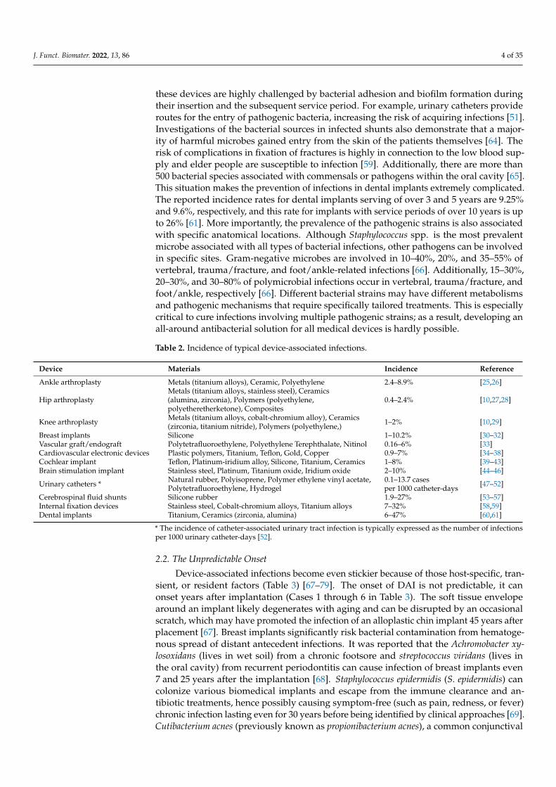
Device-associated infections become even stickier because of those host-specific, tran-sient, or resident factors (Table 3) [67–79]. The onset of DAI is not predictable, it canonset years after implantation (Cases 1 through 6 in Table 3). The soft tissue envelopearound an implant likely degenerates with aging and can be disrupted by an occasionalscratch, which may have promoted the infection of an alloplastic chin implant 45 years afterplacement [67]. Breast implants significantly risk bacterial contamination from hematoge-nous spread of distant antecedent infections. It was reported that the Achromobacter xy-losoxidans (lives in wet soil) from a chronic footsore and streptococcus viridans (lives inthe oral cavity) from recurrent periodontitis can cause infection of breast implants even7 and 25 years after the implantation [68]. Staphylococcus epidermidis (S. epidermidis) cancolonize various biomedical implants and escape from the immune clearance and an-tibiotic treatments, hence possibly causing symptom-free (such as pain, redness, or fever)chronic infection lasting even for 30 years before being identified by clinical approaches [69].Cutibacterium acnes (previously known as propionibacterium acnes), a common conjunctival
J. Funct. Biomater. 2022, 13, 86 5 of 35
inhabitant, are slow-growing, anaerobic Gram-positive rods, and can manifest severalyears or even decades before leading to late infections in orbital implants made of sili-cone or tantalum [70,71]. The sources of the pathogens of the DAIs can be host-specific(Cases 7 through 9 in Table 3). DAIs can be initiated by acute illness (e.g., diarrhea devel-oped during a holiday journey [31]), penetration of contaminated water during partici-pating in outdoor activities [45], or even when the patients play with their pets (bacterialcontamination from zoonotic sources) [72]. Moreover, the occurrence of DAIs is commonlyassociated with a compromised immune system in the hosts (Cases 10 and 11 in Table 3).Methotrexate, a folate antagonist, can affect neutrophil chemotaxis and induce apoptosis ofT cells and reactivation of opportunistic pathogens; hence chronic treatment of rheumatoidarthritis with this kind of drug significantly increases the risk of infections around thebattery for brain stimulation [73]. Nocardia nova is a common environmental pathogen andrarely affects immunocompetent hosts; however, this species successfully colonized a tibiaimplant placed in an immunocompetent patient [74]. Listeria monocytogenes, a commonorganism associated with unpasteurized dairy products (e.g., deli meats and unpasteurizedcheeses), can induce a periprosthetic joint infection in a patient with a history of diabetesmellitus, asthma, and psoriatic arthritis [75]. Anaerobiospirillum succiniciproducens, a com-mon settler in the gastrointestinal tract of cats and dogs, can induce a prosthetic hip jointinfection in an immunocompromised patient [76]. DAIs are normally initiated by bacterialseeding and as a result tissue integration will be impaired quickly; however, some casesfailed to identify any organism by cultures [77,78] and tissue integration was intact afterbeing infected [79]. These situations add difficulties to the prevention, diagnosis, andtreatment of DAIs.
Table 3. Representative cases showing the latent period of DAIs.
Case Devices Latent Period(Post Insertion) Pathogens Causes Reference
1 Alloplastic chinimplant 45 years / After scratching herself (soft tissue
degeneration due to aging)[67]
2 Breast implant Seven years Achromobacter xylosoxidans (apathogen that lives inwet soil)
Development of a chronic footsore(hematogenous spread from distantbacterial infection sites)
[68]
3 Breast implant 25 years Streptococcus viridans (apathogen that lives in the oralcavity)
After extensive dental treatment(hematogenous spread from distantbacterial infection sites)
[68]
4 Alloplastic implant 30 years Staphylococcus epidermidis
Bacterial contamination yearsbefore identifying the infection (asymptom-free chronic infection; thepathogen escaped immuneclearance and antibiotic treatments)
[69]
5 Orbital implant 30 years Cutibacterium acnes(previously known asPropionibacterium acnes)
Bacterial contamination during theprimary implantation (the pathogencan manifest for several decades)
[70]
6 Orbital implant26 years (implantexposure 10 years beforethe presentationwas documented)
Propionibacterium acnes(renamed Cutibacterium acnes)
Bacterial contamination during theprimary implantation or implantexposure during scleral patchgraft repair
[71]
7 Breast Implant Five months Salmonella serogroup CHematogenous seeding due todeveloping of diarrhea during aholiday travel
[31]
8 Generator for brainstimulation
Four months Multispecies including therare Cupriavidus pauculusspecies (an environmentalPathogen in “water”)
Penetration of contaminated waterduring participating inoutdoor activities
[45]
9 Breast implant Seven monthsPasteurella canis (a pathogennormally lives in theoropharyngeal commensalflora of cats and dogs)
Bacterial contamination from apatient-owned cat [72]
10 Battery for brainstimulation
Two cases (Two yearsor 10 years)
Staphylococcus aureus Chronic treatment of rheumatoidarthritis with methotrexate
[73]
J. Funct. Biomater. 2022, 13, 86 6 of 35
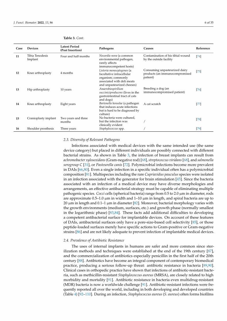
Table 3. Cont.
Case Devices Latent Period(Post Insertion) Pathogens Causes Reference
11 Tibia TenodesisImplant
Four and half months Nocardia nova (a commonenvironmental pathogen,rarely affectsimmunocompetent hosts)
Contamination of his tibial woundby the outside facility
[74]
12 Knee arthroplasty 4 monthsListeria monocytogenes (afacultative intracellularorganism; commonlyassociated with deli meatsand unpasteurized cheeses)
Consuming unpasteurized dairyproducts (an immunocompromisedpatient)
[75]
13 Hip arthroplasty 10 years Anaerobiospirillumsucciniciproducens (lives in thegastrointestinal tract of catsand dogs)
Breeding a dog (animmunocompromised patient)
[76]
14 Knee arthroplasty Eight years Bartonella henselae (a pathogenthat induces acute infectionsbut is hard to be diagnosed byculture)
A cat scratch [77]
15 Cranioplasty implant Two years and threemonths
No bacteria were cultured,but the infection wasclinically evident
/[78]
16 Shoulder prosthesis Three years Staphylococcus spp. / [79]
2.3. Diversity of Relevant Pathogens
Infections associated with medical devices with the same intended use (the samedevice category) but placed in different individuals are possibly connected with differentbacterial strains. As shown in Table 3, the infection of breast implants can result fromachromobacter xylosoxidans (Gram-negative rod) [68], streptococcus viridans [68], and salmonellaserogroup C [31], or Pasteurella canis [72]. Polymicrobial infections become more prevalentin DAIs [66,80]. Even a single infection in a specific individual often has a polymicrobialcomposition [81]. Multispecies including the rare Cupriavidus pauculus species were isolatedin an infection associated with the generator for brain stimulation [45]. Since the bacteriaassociated with an infection of a medical device may have diverse morphologies andarrangements, an effective antibacterial strategy must be capable of eliminating multiplepathogenic species. Cocci cells (spherical bacteria) range from 0.5 to 2.0 µm in diameter, rodsare approximate 0.5–1.0 µm in width and 1–10 µm in length, and spiral bacteria are up to20 µm in length and 0.1–1 µm in diameter [82]. Moreover, bacterial morphology varies withthe growth environments (medium, surfaces, etc.) and growth phase (normally smallestin the logarithmic phase) [83,84]. These facts add additional difficulties to developinga competent antibacterial surface for implantable devices. On account of these featuresof DAIs, antibacterial surfaces only have a pore-size-based cell selectivity [85], or thosepeptide-loaded surfaces merely have specific actions to Gram-positive or Gram-negativestrains [86] and are not likely adequate to prevent infection of implantable medical devices.
2.4. Prevalence of Antibiotic Resistance
The uses of internal implants in humans are safer and more common since ster-ilization methods and techniques were established at the end of the 19th century [87],and the commercialization of antibiotics especially penicillin in the first half of the 20thcentury [88]. Antibiotics have become an integral component of contemporary biomedicalpractice, producing a serious follow-up threat: antibiotic resistance in bacteria [89,90].Clinical cases in orthopedic practice have shown that infections of antibiotic-resistant bacte-ria, such as methicillin-resistant Staphylococcus aureus (MRSA), are closely related to highmorbidity and mortality [91]. Antibiotic resistance in bacteria even multidrug-resistant(MDR) bacteria is now a worldwide challenge [91]. Antibiotic-resistant infections were fre-quently reported all over the world, including in both developing and developed countries(Table 4) [92–110]. During an infection, Staphylococcus aureus (S. aureus) often forms biofilms
J. Funct. Biomater. 2022, 13, 86 7 of 35
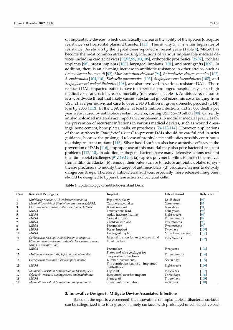
on implantable devices, which dramatically increases the ability of the species to acquireresistance via horizontal plasmid transfer [111]. This is why S. aureus has high rates ofresistance. As shown by the typical cases reported in recent years (Table 4), MRSA hasbecome the most common strain causing infections of various implantable medical de-vices, including cardiac devices [93,95,99,103,106], orthopedic prosthetics [96,97], cochlearimplants [98], breast implants [100], laryngeal implants [101], and stent grafts [109]. Inaddition, there is an alarming increase in antibiotic resistance in other strains, such asAcinetobacter baumannii [92], Mycobacterium chelonae [94], Enterobacter cloacae complex [102],S. epidermidis [104,110], Klebsiella pneumoniae [105], Staphylococcus haemolyticus [107], andStaphylococcal endophthalmitis [108], are also involved in various resistant DAIs. Thoseresistant DAIs impacted patients have to experience prolonged hospital stays, bear highmedical costs, and risk increased mortality (references in Table 4). Antibiotic recalcitranceis a worldwide threat that likely causes substantial global economic costs ranging fromUSD 21,832 per individual case to over USD 3 trillion in gross domestic product (GDP)loss by 2050 [112]. In the USA alone, at least 2 million infections and 23,000 deaths peryear were caused by antibiotic-resistant bacteria, costing USD 55–70 billion [90]. Currently,antibiotic-loaded materials are important complements to modular medical practices forthe prevention of recurrent infections in various medical devices, such as wound dress-ings, bone cement, bone plates, nails, or prostheses [24,113,114]. However, applicationsof these surfaces in “uninfected tissues” to prevent DAIs should be careful and in strictguidance, because the prolonged release of prophylactic antibiotics possibly contributesto arising resistant mutants [115]. Silver-based surfaces also have attractive efficacy in theprevention of DAIs [116], improper use of this material may also pose bacterial-resistantproblems [117,118]. In addition, pathogenic bacteria have many defensive actions resistantto antimicrobial challenges [91,119,120]: (a) express polymer biofilms to protect themselvesfrom antibiotic attacks; (b) remodel their outer surface to reduce antibiotic uptake; (c) syn-thesize precursors to modify the target of antimicrobials; (d) produce enzymes to detoxifydangerous drugs. Therefore, antibacterial surfaces, especially those release-killing ones,should be designed to bypass these actions of bacterial cells.
Table 4. Epidemiology of antibiotic-resistant DAIs.
Case Resistant Pathogens Implant Latent Period Reference
1 Multidrug-resistant Acinetobacter baumannii Hip arthroplasty 12–25 days [92]2 Methicillin-resistant Staphylococcus aureus (MRSA) Cardiac pacemaker Nine years [93]3 Clarithromycin-resistant Mycobacterium chelonae Breast implant Four days [94]4 MRSA Transvenous lead Four years [95]5 MRSA Ankle fracture fixation Eight weeks [96]6 MRSA Cranial implant Three months [97]7 MRSA Cochlear implant Five months [98]8 MRSA Pacemaker Two months [99]9 MRSA Breast Implant Two days [100]10 MRSA Laryngeal implant More than one year [101]
11 Carbapenem-resistant Acinetobacter baumannii;Fluoroquinolone-resistant Enterobacter cloacae complex(AmpC overexpression)
Internal fixation for an open proximaltibial fracture
Two months [102]
12 MRSA Pacemaker Two years [103]
13 Multidrug-resistant Staphylococcus epidermidis Plates and wire cerclages forperiprosthetic fractures Three months [104]
14 Carbapenem-resistant Klebsiella pneumoniae Lumbar instruments, Seven days [105]
15 MRSA The ventricular lead of an implanteddefibrillator Eight weeks [106]
16 Methicillin-resistant Staphylococcus haemolyticus Hip joint Two years [107]17 Ofloxacin-resistant staphylococcal endophthalmitis Intravitreal ozurdex implant Three days [108]18 MRSA Stent graft Three days [109]19 Methicillin-resistant Staphylococcus epidermidis Spinal instrumentation 7–88 days [110]
3. Innovative Designs to Mitigate Device-Associated Infections
Based on the reports we screened, the innovations of implantable antibacterial surfacescan be categorized into four groups, namely surfaces with prolonged or cell-selective bac-
J. Funct. Biomater. 2022, 13, 86 8 of 35
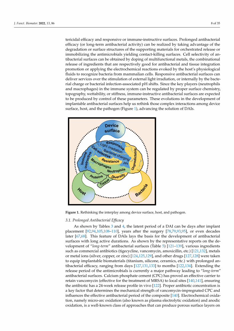
tericidal efficacy and responsive or immune-instructive surfaces. Prolonged antibacterialefficacy (or long-term antibacterial activity) can be realized by taking advantage of thedegradation or surface structures of the supporting materials for orchestrated release orimmobilizing the antimicrobials yielding contact-killing surfaces. Cell selectivity of an-tibacterial surfaces can be obtained by doping of multifunctional metals, the combinationalrelease of ingredients that are respectively good for antibacterial and tissue integrationpromotion or applying the electrochemical reactions evoked by the host’s physiologicalfluids to recognize bacteria from mammalian cells. Responsive antibacterial surfaces candeliver services over the stimulation of external light irradiation, or internally by the bacte-rial charge or bacterial infection-associated pH shifts. Since the key players (neutrophilsand macrophages) in the immune system can be regulated by proper surface chemistry,topography, wettability, or stiffness, immune-instructive antibacterial surfaces are expectedto be produced by control of these parameters. These evolutions in the development ofimplantable antibacterial surfaces help us rethink those complex interactions among devicesurface, host, and the pathogen (Figure 1), advancing the solution of DAIs.
Figure 1. Rethinking the interplay among device surface, host, and pathogen.
3.1. Prolonged Antibacterial Efficacy
As shown by Tables 3 and 4, the latent period of a DAI can be days after implantplacement [92,94,105,108–110], years after the surgery [78,79,93,95], or even decadeslater [67,68]. This feature of DAIs lays the basis for the development of antibacterialsurfaces with long active durations. As shown by the representative reports on the de-velopment of “long-term” antibacterial surfaces (Table 5) [121–139], various ingredientssuch as commercial antibiotics (tigecycline, vancomycin, amoxicillin, etc.) [121,132], metalsor metal ions (silver, copper, or zinc) [124,125,129], and other drugs [127,128] were takento equip implantable biomaterials (titanium, silicone, ceramics, etc.) with prolonged an-tibacterial efficacy, ranging from days [127,131,133] to months [122,134]. Extending therelease period of the antimicrobials is currently a major pathway leading to “long-term”antibacterial surfaces. Calcium phosphate cement (CPC) has proved an effective carrier toretain vancomycin (effective for the treatment of MRSA) to local sites [140,141], ensuringthe antibiotic has a 24-week release profile in vivo [122]. Proper antibiotic concentration isa key factor that determines the mechanical strength of vancomycin-impregnated CPC andinfluences the effective antibacterial period of the composite [140]. Electrochemical oxida-tion, namely micro-arc oxidation (also known as plasma electrolytic oxidation) and anodicoxidation, is a well-known class of approaches that can produce porous surface layers on
J. Funct. Biomater. 2022, 13, 86 9 of 35
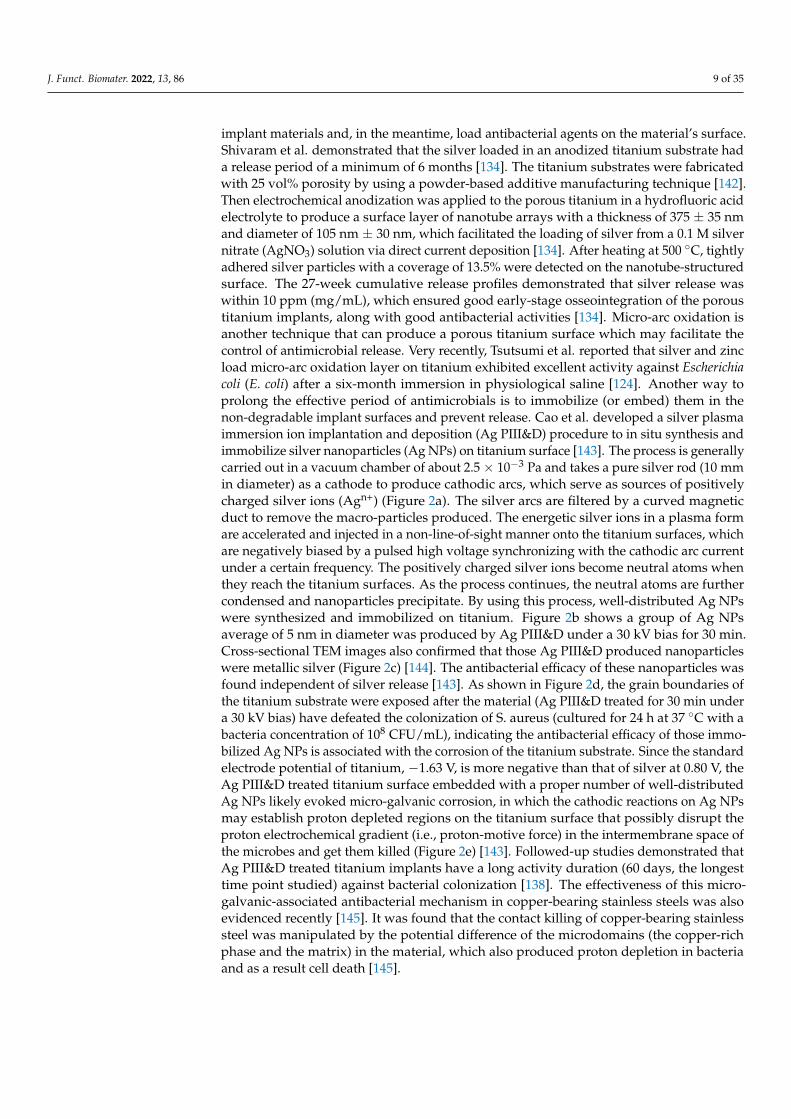
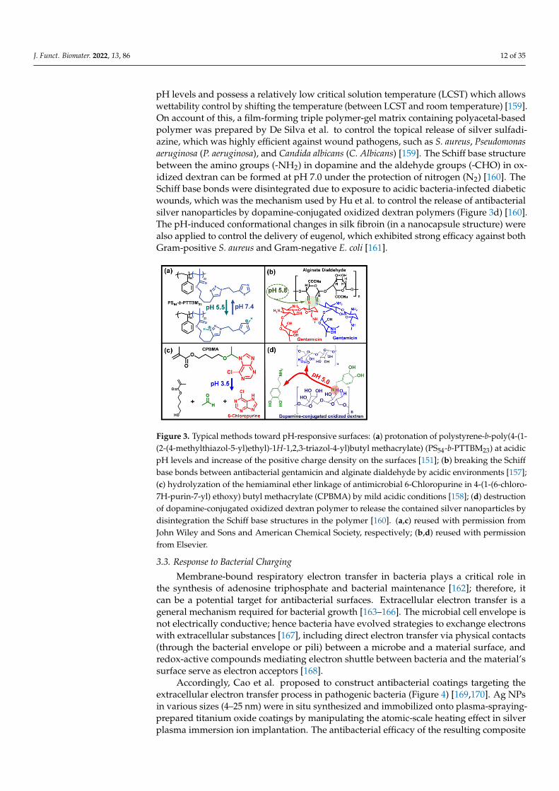
implant materials and, in the meantime, load antibacterial agents on the material’s surface.Shivaram et al. demonstrated that the silver loaded in an anodized titanium substrate hada release period of a minimum of 6 months [134]. The titanium substrates were fabricatedwith 25 vol% porosity by using a powder-based additive manufacturing technique [142].Then electrochemical anodization was applied to the porous titanium in a hydrofluoric acidelectrolyte to produce a surface layer of nanotube arrays with a thickness of 375 ± 35 nmand diameter of 105 nm ± 30 nm, which facilitated the loading of silver from a 0.1 M silvernitrate (AgNO3) solution via direct current deposition [134]. After heating at 500 ◦C, tightlyadhered silver particles with a coverage of 13.5% were detected on the nanotube-structuredsurface. The 27-week cumulative release profiles demonstrated that silver release waswithin 10 ppm (mg/mL), which ensured good early-stage osseointegration of the poroustitanium implants, along with good antibacterial activities [134]. Micro-arc oxidation isanother technique that can produce a porous titanium surface which may facilitate thecontrol of antimicrobial release. Very recently, Tsutsumi et al. reported that silver and zincload micro-arc oxidation layer on titanium exhibited excellent activity against Escherichiacoli (E. coli) after a six-month immersion in physiological saline [124]. Another way toprolong the effective period of antimicrobials is to immobilize (or embed) them in thenon-degradable implant surfaces and prevent release. Cao et al. developed a silver plasmaimmersion ion implantation and deposition (Ag PIII&D) procedure to in situ synthesis andimmobilize silver nanoparticles (Ag NPs) on titanium surface [143]. The process is generallycarried out in a vacuum chamber of about 2.5× 10⁻³ Pa and takes a pure silver rod (10 mmin diameter) as a cathode to produce cathodic arcs, which serve as sources of positivelycharged silver ions (Agn+) (Figure 2a). The silver arcs are filtered by a curved magneticduct to remove the macro-particles produced. The energetic silver ions in a plasma formare accelerated and injected in a non-line-of-sight manner onto the titanium surfaces, whichare negatively biased by a pulsed high voltage synchronizing with the cathodic arc currentunder a certain frequency. The positively charged silver ions become neutral atoms whenthey reach the titanium surfaces. As the process continues, the neutral atoms are furthercondensed and nanoparticles precipitate. By using this process, well-distributed Ag NPswere synthesized and immobilized on titanium. Figure 2b shows a group of Ag NPsaverage of 5 nm in diameter was produced by Ag PIII&D under a 30 kV bias for 30 min.Cross-sectional TEM images also confirmed that those Ag PIII&D produced nanoparticleswere metallic silver (Figure 2c) [144]. The antibacterial efficacy of these nanoparticles wasfound independent of silver release [143]. As shown in Figure 2d, the grain boundaries ofthe titanium substrate were exposed after the material (Ag PIII&D treated for 30 min undera 30 kV bias) have defeated the colonization of S. aureus (cultured for 24 h at 37 ◦C with abacteria concentration of 108 CFU/mL), indicating the antibacterial efficacy of those immo-bilized Ag NPs is associated with the corrosion of the titanium substrate. Since the standardelectrode potential of titanium, −1.63 V, is more negative than that of silver at 0.80 V, theAg PIII&D treated titanium surface embedded with a proper number of well-distributedAg NPs likely evoked micro-galvanic corrosion, in which the cathodic reactions on Ag NPsmay establish proton depleted regions on the titanium surface that possibly disrupt theproton electrochemical gradient (i.e., proton-motive force) in the intermembrane space ofthe microbes and get them killed (Figure 2e) [143]. Followed-up studies demonstrated thatAg PIII&D treated titanium implants have a long activity duration (60 days, the longesttime point studied) against bacterial colonization [138]. The effectiveness of this micro-galvanic-associated antibacterial mechanism in copper-bearing stainless steels was alsoevidenced recently [145]. It was found that the contact killing of copper-bearing stainlesssteel was manipulated by the potential difference of the microdomains (the copper-richphase and the matrix) in the material, which also produced proton depletion in bacteriaand as a result cell death [145].
J. Funct. Biomater. 2022, 13, 86 10 of 35
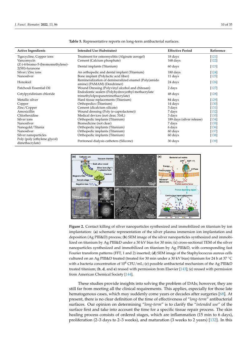
Table 5. Representative reports on long-term antibacterial surfaces.
Active Ingredients Intended Use (Substrates) Effective Period Reference
Tigecycline, Copper ions Treatment for osteomyelitis (Alginate aerogel) 18 days [121]Vancomycin Cement (Calcium phosphate) 168 days [122](Z-)-4-bromo-5-(bromomethylene)-2(5H)-furanone Dental implants (Titanium) 60 days [123]
Silver/Zinc ions An orthopedic and dental implant (Titanium) 180 days [124]Nanosilver Bone implant (Polylactic acid fiber) 11 days [125]
Honokiol Remineralization of demineralized enamel (Poly(amidoamine) (PAMAM) (Dendrimer) 24 days [126]
Patchouli Essential Oil Wound Dressing (Polyvinyl alcohol and chitosan) 2 days [127]
Cetylpyridinium chloride Endodontic sealers (Polyhydroxyethyl methacrylatetrimethylolpropanetrimethacrylate) 48 days [128]
Metallic silver Hard tissue replacements (Titanium) 84 days [129]Copper Orthopedics (Titanium) 14 days [130]Zinc/Copper Cement (dicalcium silicate) 3 days [131]Amoxicillin Wound dressing (Poly (e-caprolactone)) 7 days [132]Chlorhexidine Medical devices (not clear, 316L) 3 days [133]Silver ions Orthopedic implants (Titanium) 189 days (silver release) [134]Nanosilver Biomedicine (not clear) 7 days [135]Nanogold/Titania Orthopedic implants (Titanium) 6 days [136]Nanosilver Orthopedic implants (Titanium) 60 days [137]Silver nanoparticles Orthopedic implants (Titanium) 60 days [138]Poly (poly (ethylene glycol)dimethacrylate) Peritoneal dialysis catheters (Silicone) 30 days [139]
Figure 2. Contact killing of silver nanoparticles synthesized and immobilized on titanium by ionimplantation: (a) schematic representation of the silver plasma immersion ion implantation anddeposition (Ag PIII&D) process; (b) SEM image of the silver nanoparticles synthesized and immobi-lized on titanium by Ag PIII&D under a 30 kV bias for 30 min; (c) cross-sectional TEM of the silvernanoparticles synthesized and immobilized on titanium by Ag PIII&D, with corresponding fastFourier transform patterns (FFT, 1 and 2) inserted; (d) SEM image of the Staphylococcus aureus cellscultured on an Ag PIII&D treated (treated for 30 min under a 30 kV bias) titanium for 24 h at 37 ◦Cwith a bacteria concentration of 108 CFU/mL; (e) possible antibacterial mechanism of the Ag PIII&Dtreated titanium; (b, d, and e) reused with permission from Elsevier [143]; (c) reused with permissionfrom American Chemical Society [144].
These studies provide insights into solving the problem of DAIs; however, they arestill far from meeting all the clinical requirements. This applies, especially for those latehematogenous cases, which may suddenly come years or decades after surgeries [68]. Atpresent, there is no clear definition of the time of effectiveness of “long-term” antibacterialsurfaces. Our opinion on determining “long-term” is to clarify the “intended use” of thesurface first and take into account the time for a specific tissue repair process. The skinhealing process consists of ordered stages, which are inflammation (15 min to 6 days),proliferation (2–3 days to 2–3 weeks), and maturation (3 weeks to 2 years) [132]. In this

J. Funct. Biomater. 2022, 13, 86 11 of 35
respect, we believe that the effective period for a “long-term” antibacterial wound dressingshould exceed 3 weeks. Generally, the healing of a bone fracture involves three distinct butoverlapping stages, i.e., the early inflammatory period (a few hours to days), the repairingperiod (weeks), and the late remodeling period (months to years) [146,147]. As a result, thetypical time for a new bone to achieve adequate strength is 3 to 6 months [146]. Therefore,we believe the effective period for “long-term” antibacterial bone implants should exceed3 months, and this time required for elder patients should be longer because their bonehealing process is relatively slow. Although studies on “long-term” antibacterial surfacesare increasing these years, most of the reports did not consider the “matching” problembetween antibacterial duration and tissue integration, yielding barriers to translationalresearch. Since the incidence of DAI is site-specific, this aspect should be considered infuture studies.
3.2. Response to pH Shifts
It is known that the pH shift is a common phenomenon of bacterial infections [148–150],laying the basis for the development of pH-responsive antibacterial surfaces. The antibac-terial activity of pH-responsive films or coatings can be triggered by the protonation ordeprotonation of their ionic groups. The thiazole and triazole groups, for example, in poly-mer PS54-b-PTTBM23 (on porous polystyrene surfaces) can be protonated under acidic pHlevels, which increased the positive charge density on the materials surface to act againstbacterial adhesion (Figure 3a) [151]. In addition, the killed bacteria can be further removedby increasing the pH levels (pH 7.4, for instance), which induced deprotonation of thethiazole and triazole groups in the materials [151]. Normally, pH-responsive surfaces aredesigned for the controllable release of antibacterial agents by manipulating the materials’pH-associated swelling or shrinking processes. By shifting the environmental pH, theprotons of the carboxyl repeat units in poly(methacrylic acid) can be removed to make thematerial swell, which can control the release of antibacterial drugs [152]. In this manner,Wei et al. developed a pH-responsive surface capable of loading bacteriolytic lysozymeat acidic pH levels and releasing it under neutral or basic pH [152]. A pH-sensitive fi-brous membrane was also developed to control the release of antibacterial gatifloxacinhydrochloride and silver nanoparticles [153]. The backbone (hydrophobic)-attached aminogroups (weak basic moieties) of chitosan adapt to a deprotonated state above pH 6 whilebecoming protonated and positively charged at low pH, demonstrating a pH-dependentextension of the colloid chains and consequently swelling of the material [154]. Accordingly,chitosan was crosslinked with hydroxypropyl methylcellulose and 2-hydroxypropyl-β-cyclodextrin to produce a superabsorbent hydrogel for controllable delivery of antibacterial3,4-dihydroxy cinnamic acid in response to pH changes [154]. Similarly, the structure ofkeratin hydrogel was reorganized by manipulating the protonation and deprotonationprocess of carboxyl groups in the material, yielding pH-dependent shrinking and swellingat low and high pH levels, respectively [155]. This behavior of the keratin hydrogel wastaken to control the release of biocidal zinc oxide nanoplates in a pH-dependent manner,which can be a potential therapy response to a bacteria-contaminated media with biasedpH and treatment of chronic wounds [156].
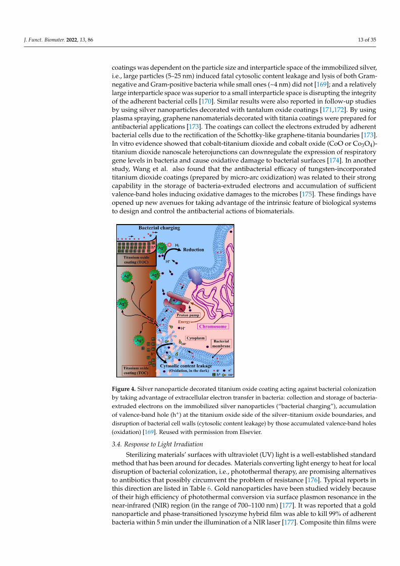
In addition, acid-labile bonds can also be used to program the release of antibacte-rial agents. Antibacterial gentamicin was conjugated with an alginate dialdehyde Schiffbase reaction between the aldehyde groups (-CHO) and amino groups (-NH2) from thepolymer, and was released due to the acidic environment triggered the disintegrationof the Schiff base bonds (Figure 3b) [157]. Similarly, antimicrobial 6-Chloropurine wasconjugated to 4-(vinyloxy) butyl methacrylate (VBMA) to produce 4-(1-(6-chloro-7H-purin-7-yl) ethoxy) butyl methacrylate (CPBMA), which contained a hemiaminal ether linkagecan be hydrolyzed in mildly acidic conditions and allowed the controllable release of theantibacterial agent (Figure 3c) [158]. Moreover, pH-induced material structural evolutions,such as degradation, disintegration, and conformational changes, are also applied for thecontrollable delivery of biocides. Polyacetal-based polymers are degradable under acidic
J. Funct. Biomater. 2022, 13, 86 12 of 35
pH levels and possess a relatively low critical solution temperature (LCST) which allowswettability control by shifting the temperature (between LCST and room temperature) [159].On account of this, a film-forming triple polymer-gel matrix containing polyacetal-basedpolymer was prepared by De Silva et al. to control the topical release of silver sulfadi-azine, which was highly efficient against wound pathogens, such as S. aureus, Pseudomonasaeruginosa (P. aeruginosa), and Candida albicans (C. Albicans) [159]. The Schiff base structurebetween the amino groups (-NH2) in dopamine and the aldehyde groups (-CHO) in ox-idized dextran can be formed at pH 7.0 under the protection of nitrogen (N2) [160]. TheSchiff base bonds were disintegrated due to exposure to acidic bacteria-infected diabeticwounds, which was the mechanism used by Hu et al. to control the release of antibacterialsilver nanoparticles by dopamine-conjugated oxidized dextran polymers (Figure 3d) [160].The pH-induced conformational changes in silk fibroin (in a nanocapsule structure) werealso applied to control the delivery of eugenol, which exhibited strong efficacy against bothGram-positive S. aureus and Gram-negative E. coli [161].
Figure 3. Typical methods toward pH-responsive surfaces: (a) protonation of polystyrene-b-poly(4-(1-(2-(4-methylthiazol-5-yl)ethyl)-1H-1,2,3-triazol-4-yl)butyl methacrylate) (PS54-b-PTTBM23) at acidicpH levels and increase of the positive charge density on the surfaces [151]; (b) breaking the Schiffbase bonds between antibacterial gentamicin and alginate dialdehyde by acidic environments [157];(c) hydrolyzation of the hemiaminal ether linkage of antimicrobial 6-Chloropurine in 4-(1-(6-chloro-7H-purin-7-yl) ethoxy) butyl methacrylate (CPBMA) by mild acidic conditions [158]; (d) destructionof dopamine-conjugated oxidized dextran polymer to release the contained silver nanoparticles bydisintegration the Schiff base structures in the polymer [160]. (a,c) reused with permission fromJohn Wiley and Sons and American Chemical Society, respectively; (b,d) reused with permissionfrom Elsevier.
3.3. Response to Bacterial Charging
Membrane-bound respiratory electron transfer in bacteria plays a critical role inthe synthesis of adenosine triphosphate and bacterial maintenance [162]; therefore, itcan be a potential target for antibacterial surfaces. Extracellular electron transfer is ageneral mechanism required for bacterial growth [163–166]. The microbial cell envelope isnot electrically conductive; hence bacteria have evolved strategies to exchange electronswith extracellular substances [167], including direct electron transfer via physical contacts(through the bacterial envelope or pili) between a microbe and a material surface, andredox-active compounds mediating electron shuttle between bacteria and the material’ssurface serve as electron acceptors [168].
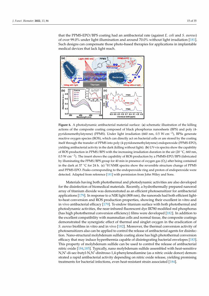
Accordingly, Cao et al. proposed to construct antibacterial coatings targeting theextracellular electron transfer process in pathogenic bacteria (Figure 4) [169,170]. Ag NPsin various sizes (4–25 nm) were in situ synthesized and immobilized onto plasma-spraying-prepared titanium oxide coatings by manipulating the atomic-scale heating effect in silverplasma immersion ion implantation. The antibacterial efficacy of the resulting composite
J. Funct. Biomater. 2022, 13, 86 13 of 35
coatings was dependent on the particle size and interparticle space of the immobilized silver,i.e., large particles (5–25 nm) induced fatal cytosolic content leakage and lysis of both Gram-negative and Gram-positive bacteria while small ones (~4 nm) did not [169]; and a relativelylarge interparticle space was superior to a small interparticle space is disrupting the integrityof the adherent bacterial cells [170]. Similar results were also reported in follow-up studiesby using silver nanoparticles decorated with tantalum oxide coatings [171,172]. By usingplasma spraying, graphene nanomaterials decorated with titania coatings were prepared forantibacterial applications [173]. The coatings can collect the electrons extruded by adherentbacterial cells due to the rectification of the Schottky-like graphene-titania boundaries [173].In vitro evidence showed that cobalt-titanium dioxide and cobalt oxide (CoO or Co3O4)-titanium dioxide nanoscale heterojunctions can downregulate the expression of respiratorygene levels in bacteria and cause oxidative damage to bacterial surfaces [174]. In anotherstudy, Wang et al. also found that the antibacterial efficacy of tungsten-incorporatedtitanium dioxide coatings (prepared by micro-arc oxidization) was related to their strongcapability in the storage of bacteria-extruded electrons and accumulation of sufficientvalence-band holes inducing oxidative damages to the microbes [175]. These findings haveopened up new avenues for taking advantage of the intrinsic feature of biological systemsto design and control the antibacterial actions of biomaterials.
Figure 4. Silver nanoparticle decorated titanium oxide coating acting against bacterial colonizationby taking advantage of extracellular electron transfer in bacteria: collection and storage of bacteria-extruded electrons on the immobilized silver nanoparticles (“bacterial charging”), accumulationof valence-band hole (h+) at the titanium oxide side of the silver–titanium oxide boundaries, anddisruption of bacterial cell walls (cytosolic content leakage) by those accumulated valence-band holes(oxidation) [169]. Reused with permission from Elsevier.
3.4. Response to Light Irradiation
Sterilizing materials’ surfaces with ultraviolet (UV) light is a well-established standardmethod that has been around for decades. Materials converting light energy to heat for localdisruption of bacterial colonization, i.e., photothermal therapy, are promising alternativesto antibiotics that possibly circumvent the problem of resistance [176]. Typical reports inthis direction are listed in Table 6. Gold nanoparticles have been studied widely becauseof their high efficiency of photothermal conversion via surface plasmon resonance in thenear-infrared (NIR) region (in the range of 700–1100 nm) [177]. It was reported that a goldnanoparticle and phase-transitioned lysozyme hybrid film was able to kill 99% of adherentbacteria within 5 min under the illumination of a NIR laser [177]. Composite thin films were

J. Funct. Biomater. 2022, 13, 86 14 of 35
produced by coordinative assembly of tannic acid (TA) and iron ions (Fe3+) and yieldedAu-TA/Fe (Figure 5a; the support can be other materials, rather than gold) [178]. Thesefilms exhibited high absorption and efficient light-to-heat convention under NIR irradiation(Figure 5b), as a result, they had efficient photothermal killing effects that disrupted 99%of adherent microbes, including both Gram-negative E. coli and Gram-positive MRSAstrains (Figure 5c).
Figure 5. A photothermal antibacterial surface: (a) schematic illustration of the coordinated assemblyof tannic acid (TA) and Fe3+ ions (iron chloride hexahydrate) on gold (can be other materials),yielding the Au-TA/Fe; (b) near-infrared (NIR) irradiation (808 nm, 2.2 W·cm−2) induced temperaturechanges on the material surface immersed in phosphate-buffered saline (PBS), with correspondingthermal images inserted; (c) SEM images of adherent bacteria (E. coli or MRSA) on materials surfacewith/without NIR irradiation (5 min), together with the typical photographs of bacterial coloniesre-cultured from materials surface of different processing histories. Adapted from reference [178]with permission from the American Chemical Society.
Materials absorbing light energy to produce reactive oxygen species (ROS) for oxidiza-tion of bacterial cell walls, yielding photodynamic therapies [179], are also developed forpotential biomedical applications. Quantum dots are sensitive to blue light illuminationand capable of producing singlet oxygen, which is a powerful ROS able to disintegratebacterial cell walls [180]. It was reported that hydrophobic carbon quantum dots incorpo-rated into various polymer matrices in form of thin films had significant activity againstS. aureus under blue light irradiation [180]. An antibacterial coating composed of blackphosphorus nanosheets (BPS) and poly (4-pyridonemethylstyrene) (PPMS) was coated ona titanium surface (PPMS/BPS) (Figure 6a) [181]. Under the stimulation of visible light(660 nm, 0.5 W·cm−2), the contained photosensitizer BPS released ROS (singlet oxygen),which was evidenced by the gradual decrease of UV absorption at 415 nm as the irra-diation duration was prolonged to 50 min (Figure 6b). This was monitored by using 1,3-diphenylisobenzofuran, which reacts with singlet oxygen to decrease the absorptionaround 415 nm [181]. In addition, the coating was good at the storage of the illumination-generated ROS via transforming PPMS to poly (4-pyridonemethylstyrene) endoperoxide(PPMS-EPO) (Figure 6a). The stored ROS can be released in the dark, mediating the “killingwithout light” capability of the coating. By illuminating in presence of oxygen gas (O2) for40 min, the PPMS/BPS group was transferred into PMMS-EPO/BPS, which was able torelease ROS even after being contained in the dark at 37 ◦C for 24 h (the insert in Figure 6b).A reverse transformation between pyridone and endoperoxide was evidenced by the aris-ing 1H nuclear magnetic resonance (1H NMR) peaks corresponding to the endoperoxidering and the proton of endoperoxide in PMMS-EPO (Figure 6c). Tan et al. demonstrated
J. Funct. Biomater. 2022, 13, 86 15 of 35
that the PPMS-EPO/BPS coating had an antibacterial rate (against E. coli and S. aureus)of over 99.0% under light illumination and around 70.0% without light irradiation [181].Such designs can compensate those photo-based therapies for applications in implantablemedical devices that lack light reach.
Figure 6. A photodynamic antibacterial material surface: (a) schematic illustration of the killingactions of the composite coating composed of black phosphorus nanosheets (BPS) and poly (4-pyridonemethylstyrene) (PPMS). Under light irradiation (660 nm, 0.5 W·cm−2), BPSs generatereactive oxygen species (ROS), which can directly act on bacterial cells or are stored by the coatingitself through the transfer of PPMS into poly (4-pyridonemethylstyrene) endoperoxide (PPMS-EPO),yielding antibacterial activity in the dark (killing without light). (b) UV-vis spectra show the capabilityof ROS production in PPMS/BPS with the increasing irradiation duration in the air (20 ◦C, 660 nm,0.5 W·cm−2). The insert shows the capability of ROS production by a PMMS-EPO/BPS (fabricatedby illuminating the PPMS/BPS group for 40 min in presence of oxygen gas (O2) after being containedin the dark at 37 ◦C for 24 h. (c) 1H NMR spectra show the reversible structure change of PPMSand PPMS-EPO. Peaks corresponding to the endoperoxide ring and proton of endoperoxide weredetected. Adapted from reference [181] with permission from John Wiley and Sons.
Materials having both photothermal and photodynamic activities are also developedfor the disinfection of biomedical materials. Recently, a hydrothermally prepared nanorodarray of titanium dioxide was demonstrated as an efficient photosensitizer for antibacterialapplications [179]. In response to a NIR light (808 nm), the nanorods had both efficient light-to-heat conversion and ROS production properties, showing their excellent in vitro andin vivo antibacterial efficacy [179]. To endow titanium surface with both photothermal andphotodynamic activities, the near-infrared fluorescent dye IR780 modified red phosphorus(has high photothermal conversion efficiency) films were developed [182]. In addition tothe excellent compatibility with mammalian cells and normal tissue, the composite coatingsdemonstrated the synergistic effect of thermal and singlet oxygen in the eradication ofS. aureus biofilms in vitro and in vivo [182]. Moreover, the thermal conversion activity ofphotosensitizers also can be applied to control the release of antibacterial agents for disinfec-tion. Nano-structural molybdenum sulfide coating alone has high photothermal conversionefficacy that may induce hyperthermia capable of disintegrating bacterial envelopes [183].This property of molybdenum sulfide can be used to control the release of antibacterialnitric oxide [184,185]. Typically, nano molybdenum sulfide assembled with heat-sensitiveN,N′-di-sec-butyl-N,N′-dinitroso-1,4-phenylenediamine (as a nitric oxide donor) demon-strated a rapid antibacterial activity depending on nitric oxide release, yielding promisingtreatments for bacterial infections, even heat-resistant strain associated [184].
J. Funct. Biomater. 2022, 13, 86 16 of 35
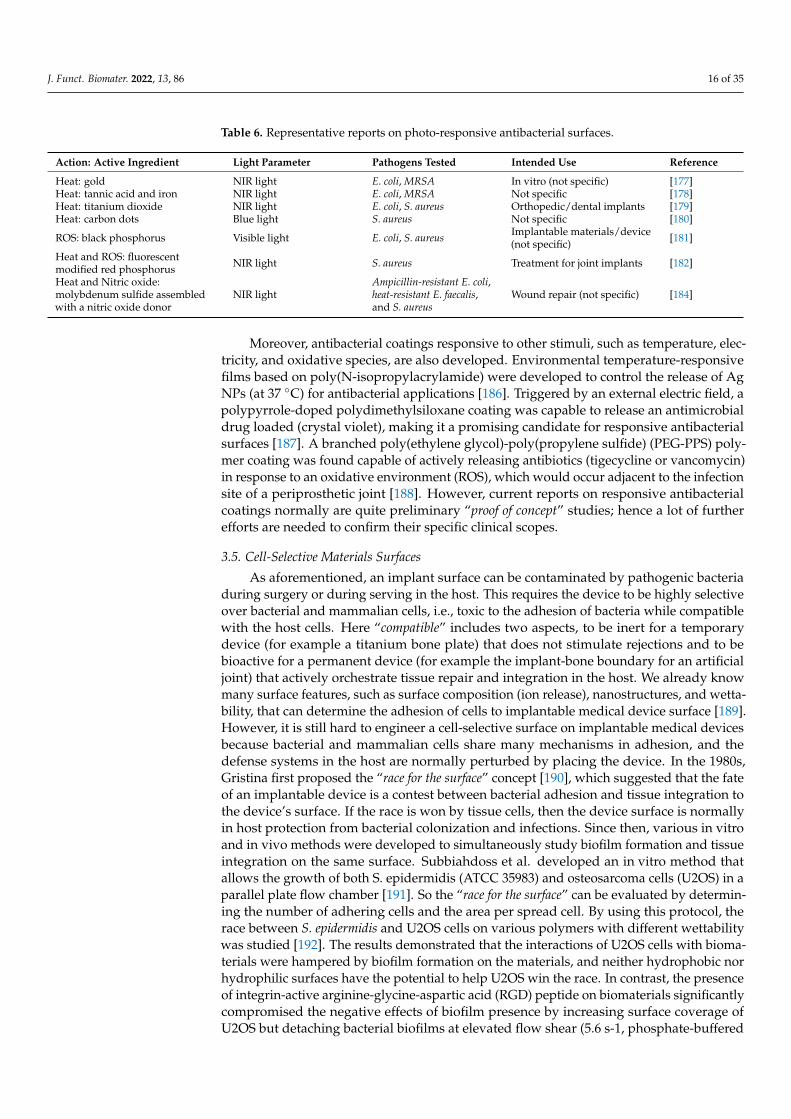
Table 6. Representative reports on photo-responsive antibacterial surfaces.
Action: Active Ingredient Light Parameter Pathogens Tested Intended Use Reference
Heat: gold NIR light E. coli, MRSA In vitro (not specific) [177]Heat: tannic acid and iron NIR light E. coli, MRSA Not specific [178]Heat: titanium dioxide NIR light E. coli, S. aureus Orthopedic/dental implants [179]Heat: carbon dots Blue light S. aureus Not specific [180]
ROS: black phosphorus Visible light E. coli, S. aureus Implantable materials/device(not specific) [181]
Heat and ROS: fluorescentmodified red phosphorus NIR light S. aureus Treatment for joint implants [182]
Heat and Nitric oxide:molybdenum sulfide assembledwith a nitric oxide donor
NIR lightAmpicillin-resistant E. coli,heat-resistant E. faecalis,and S. aureus
Wound repair (not specific) [184]
Moreover, antibacterial coatings responsive to other stimuli, such as temperature, elec-tricity, and oxidative species, are also developed. Environmental temperature-responsivefilms based on poly(N-isopropylacrylamide) were developed to control the release of AgNPs (at 37 ◦C) for antibacterial applications [186]. Triggered by an external electric field, apolypyrrole-doped polydimethylsiloxane coating was capable to release an antimicrobialdrug loaded (crystal violet), making it a promising candidate for responsive antibacterialsurfaces [187]. A branched poly(ethylene glycol)-poly(propylene sulfide) (PEG-PPS) poly-mer coating was found capable of actively releasing antibiotics (tigecycline or vancomycin)in response to an oxidative environment (ROS), which would occur adjacent to the infectionsite of a periprosthetic joint [188]. However, current reports on responsive antibacterialcoatings normally are quite preliminary “proof of concept” studies; hence a lot of furtherefforts are needed to confirm their specific clinical scopes.
3.5. Cell-Selective Materials Surfaces
As aforementioned, an implant surface can be contaminated by pathogenic bacteriaduring surgery or during serving in the host. This requires the device to be highly selectiveover bacterial and mammalian cells, i.e., toxic to the adhesion of bacteria while compatiblewith the host cells. Here “compatible” includes two aspects, to be inert for a temporarydevice (for example a titanium bone plate) that does not stimulate rejections and to bebioactive for a permanent device (for example the implant-bone boundary for an artificialjoint) that actively orchestrate tissue repair and integration in the host. We already knowmany surface features, such as surface composition (ion release), nanostructures, and wetta-bility, that can determine the adhesion of cells to implantable medical device surface [189].However, it is still hard to engineer a cell-selective surface on implantable medical devicesbecause bacterial and mammalian cells share many mechanisms in adhesion, and thedefense systems in the host are normally perturbed by placing the device. In the 1980s,Gristina first proposed the “race for the surface” concept [190], which suggested that the fateof an implantable device is a contest between bacterial adhesion and tissue integration tothe device’s surface. If the race is won by tissue cells, then the device surface is normallyin host protection from bacterial colonization and infections. Since then, various in vitroand in vivo methods were developed to simultaneously study biofilm formation and tissueintegration on the same surface. Subbiahdoss et al. developed an in vitro method thatallows the growth of both S. epidermidis (ATCC 35983) and osteosarcoma cells (U2OS) in aparallel plate flow chamber [191]. So the “race for the surface” can be evaluated by determin-ing the number of adhering cells and the area per spread cell. By using this protocol, therace between S. epidermidis and U2OS cells on various polymers with different wettabilitywas studied [192]. The results demonstrated that the interactions of U2OS cells with bioma-terials were hampered by biofilm formation on the materials, and neither hydrophobic norhydrophilic surfaces have the potential to help U2OS win the race. In contrast, the presenceof integrin-active arginine-glycine-aspartic acid (RGD) peptide on biomaterials significantlycompromised the negative effects of biofilm presence by increasing surface coverage ofU2OS but detaching bacterial biofilms at elevated flow shear (5.6 s-1, phosphate-buffered

J. Funct. Biomater. 2022, 13, 86 17 of 35
saline) [193]. The competition for a poly(methylmethacrylate) surface between U2OS cellsand highly virulent S. aureus or P. aeruginosa in presence of murine macrophages was alsodescribed by the research group [194]. The presence of S. aureus decreased the adherence ofhuman osteogenic sarcoma (SaOS-2) or primary osteoblast (hOB) cells to the surfaces oftitanium, polydimethylsiloxane, and polystyrene; on the other hand, the presence of eithertype of these human cells was also associated with a reduction of bacterial colonization tothe material’s surface [195]. Martinez-Perez et al. described an in vitro approach for thestudy of the adherence of S. aureus and S. epidermidis in the presence of pre-osteoblasticcells (MC3T3-E1), which can be used for assessing the effects of surface coatings withantibacterial potentials [196]. By using a bilateral intramedullary rat model and injectingbacteria (S. aureus) into the tail vein after implant placement, the temporal interplay be-tween host-cell adhesion and bacterial colonization was examined [197]. To determine theeffects of hematogenous spreading of bacteria on infection subcutaneous implants (afterhealing of the implantation wound), rats were intravenously injected either with S. aureus,S. epidermidis, or P. aeruginosa 4 weeks after subcutaneous implantation of various bioma-terials, including silicone rubber, polyethylene, polypropylene, poly(tetrafluoroethylene),poly(ethylene terephthalate), poly(methyl methacrylate), polyurethane, or glass [198]. Theresults demonstrated that late hematogenous infection of subcutaneous biomaterials doesnot occur in the rat, hence those reported late infections in humans were likely caused byperioperatively introduced pathogens [198], which is worthy of further studies. Reportscovering biofilm formation on biomaterials surfaces lack often the differentiation betweenbiofilm reducing and biofilm inhibitory effects [199]. Hence, the current biofilm method-ologies used for judging the antibacterial effects of implant surfaces need to be criticallyrevisited and if necessary revised and standardized. These reports show the significance ofconstructing cell-selective surfaces for implantable medical devices, as well as methods toevaluate the property.
Metallic ingredients are performing multiple functions in humans that can be buildingblocks leading to single-element-release mediated cell-selective surfaces. Zinc is knownas a stimulus to the osteogenic function of bone cells and also an inhibitor of bacterialgrowth. Accordingly, zinc was loaded onto various titanium surfaces by using micro-arcoxidation [200], hydrothermal treatment [201], and ion implantation [202], demonstratingexcellent antimicrobial and osteogenic properties. It is known that antimicrobial cobaltions can induce hypoxia-like conditions [203]. By applying this feature of cobalt ions,antibacterial wound dressings with excellent capability for the promotion of angiogenesisand epithelialization were fabricated by Shi et al. [203]. Copper, in addition to its broad-spectrum bactericidal activity, was found to be capable of promoting osteoblast proliferationand bone formation [204]. It was reported that proper control of the content and release ofcopper in Ti-Cu alloys are capable of balancing the antibacterial and osteogenic propertiesof the metal implants [204].
The combinational release of antimicrobials and tissue-integration promoters is an-other pathway extensively studied to develop cell-selective implants. Low-molecular-weight polyethyleneimine is a cationic antimicrobial agent, and alendronate is a stimulusfor new bone formation and osteointegration improvement [205]. They can be covalentlyconjugated onto ethanediamine-functionalized poly (glycidyl methacrylate) to constructtitanium implants of both antibacterial and osteogenic properties [205]. Silver and hy-droxyapatite are two typical materials with antibacterial and osteogenic properties, re-spectively. Recently, Fazel et al. developed a duplex process that sequentially employedmicro-arc oxidation and hydrothermal treatment to decorate Ag NPs and hydroxyapatitenanocrystals on the surface of a porous Ti6Al4V substrate (fabricated by using selectivelaser melting), creating surfaces of both osteogenic and antibacterial properties [206]. Bac-terial infection in burn wounds is common and fatal [207]. Many works have been doneto develop wound dressings preventing bacterial infection and promoting wound heal-ing. Porphyrin photosensitizer sinoporphyrin sodium has two macrocycles that showhigh efficiency against pathogenic bacteria via the production of ROS [208]. This dimeric
J. Funct. Biomater. 2022, 13, 86 18 of 35
photosensitizer was chosen to work with fibroblast growth factor in a carboxymethylchitosan-sodium alginate matrix that has successfully suppressed the growth of bacteriaand simultaneously accelerated the healing of bacteria-contaminated burn wounds in miceunder mild photoirradiation (30 J·cm−2, 5 min) [208]. Due to inferior vascularization, poorre-epithelialization, and increased infection risk, treatment of diabetic wounds is consider-ably challenging and becomes a focus of cell-selective surfaces. The (11-mercaptoundecyl)-N,N,N-trimethylammonium (MTA) contains a quaternary ammonium cation that interactsstrongly with the negatively charged cell membrane of microbes [209]. Together withthe vascular endothelial growth factor, MTA can be conjugated into gold nanoparticlesto produce dual-functional (antimicrobial and proangiogenic) dressings for treatments ofdiabetic wounds [209]. Ag NPs and pro-angiogenic deferoxamine were encapsulated in apH-responsive hydrogel developed via double-crosslinking between chitosan quaternaryammonium salt and oxidized dextran-dopamine, achieving pH-sensitive feature in drugrelease and accelerated healing of infected diabetic wounds [160]. The antibacterial efficacyof hydroxypropyltrimethyl ammonium chloride chitosan is related to the substitutiondegree of its quaternary ammonium group [210]. This chitosan derivative cooperates withmagnesium ions (magnesium chloride) in calcium alginate, yielding an antibacterial andangiogenic dressing for the treatment of infected diabetic wounds [210].
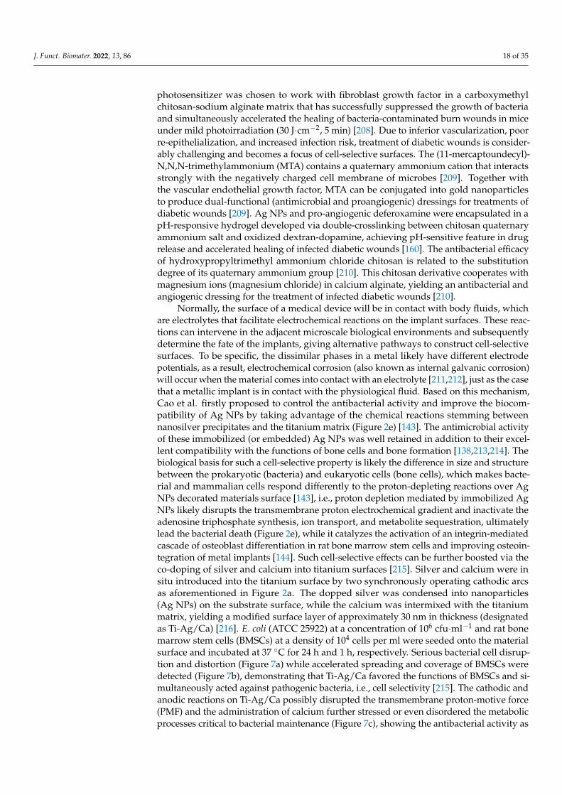
Normally, the surface of a medical device will be in contact with body fluids, whichare electrolytes that facilitate electrochemical reactions on the implant surfaces. These reac-tions can intervene in the adjacent microscale biological environments and subsequentlydetermine the fate of the implants, giving alternative pathways to construct cell-selectivesurfaces. To be specific, the dissimilar phases in a metal likely have different electrodepotentials, as a result, electrochemical corrosion (also known as internal galvanic corrosion)will occur when the material comes into contact with an electrolyte [211,212], just as the casethat a metallic implant is in contact with the physiological fluid. Based on this mechanism,Cao et al. firstly proposed to control the antibacterial activity and improve the biocom-patibility of Ag NPs by taking advantage of the chemical reactions stemming betweennanosilver precipitates and the titanium matrix (Figure 2e) [143]. The antimicrobial activityof these immobilized (or embedded) Ag NPs was well retained in addition to their excel-lent compatibility with the functions of bone cells and bone formation [138,213,214]. Thebiological basis for such a cell-selective property is likely the difference in size and structurebetween the prokaryotic (bacteria) and eukaryotic cells (bone cells), which makes bacte-rial and mammalian cells respond differently to the proton-depleting reactions over AgNPs decorated materials surface [143], i.e., proton depletion mediated by immobilized AgNPs likely disrupts the transmembrane proton electrochemical gradient and inactivate theadenosine triphosphate synthesis, ion transport, and metabolite sequestration, ultimatelylead the bacterial death (Figure 2e), while it catalyzes the activation of an integrin-mediatedcascade of osteoblast differentiation in rat bone marrow stem cells and improving osteoin-tegration of metal implants [144]. Such cell-selective effects can be further boosted via theco-doping of silver and calcium into titanium surfaces [215]. Silver and calcium were insitu introduced into the titanium surface by two synchronously operating cathodic arcsas aforementioned in Figure 2a. The dopped silver was condensed into nanoparticles(Ag NPs) on the substrate surface, while the calcium was intermixed with the titaniummatrix, yielding a modified surface layer of approximately 30 nm in thickness (designatedas Ti-Ag/Ca) [216]. E. coli (ATCC 25922) at a concentration of 106 cfu·ml−1 and rat bonemarrow stem cells (BMSCs) at a density of 104 cells per ml were seeded onto the materialsurface and incubated at 37 ◦C for 24 h and 1 h, respectively. Serious bacterial cell disrup-tion and distortion (Figure 7a) while accelerated spreading and coverage of BMSCs weredetected (Figure 7b), demonstrating that Ti-Ag/Ca favored the functions of BMSCs and si-multaneously acted against pathogenic bacteria, i.e., cell selectivity [215]. The cathodic andanodic reactions on Ti-Ag/Ca possibly disrupted the transmembrane proton-motive force(PMF) and the administration of calcium further stressed or even disordered the metabolicprocesses critical to bacterial maintenance (Figure 7c), showing the antibacterial activity as

J. Funct. Biomater. 2022, 13, 86 19 of 35
shown by Figure 7a. Additionally, the electrochemical reactions on Ti-Ag/Ca also possiblyaccelerated the proton extrusion of sodium-proton exchanger 1 (NHE1) and the calciuminflux of sodium-calcium exchanger 1 (NCX1) in BMSCs (Figure 7d), showing enhancedmembrane bleb nucleation, growth, and retraction as shown in Figure 7b. Moreover, thegrowth and retraction of membrane blebs can modulate cellular mechanics and promotethe osteogenic differentiation of BMSCs, which is good to improve the osseointegration oftitanium [215]. Such antibacterial surfaces are promising for orthopedic devices and dentalimplants, which are intended to be integrated into bone tissues.
Figure 7. A cell-selective titanium surface: (a) SEM surface morphology of the microbes (E. coli) cul-tured for 24 h on titanium doped with both calcium and silver (Ti-Ag/Ca), with a high magnificationimage, inserted; (b) typical morphology of rat bone marrow stem cells (BMSCs) cultured for 1 hon Ti-Ag/Ca, with a high magnification image inserted; (c,d) potential mechanism underlying theactions of Ti-Ag/Ca on microbes and mammalian cells, respectively [215]. Reused with permissionfrom the Royal Society of Chemistry.
In addition, topographical nanostructures, such as nanopillars [216–218], nanosheets [219],nanorod [220,221], and nano-roughness [222,223], with the efficacy of physical sterilizationrepresent an innovative pathway toward antibacterial surfaces. Among these designs,nanopillars received the most attention because their puncture-based biocidal actions arematerial composition and bacterial species independent. The bactericidal mechanismsof such structures were already highlighted in a very recent review [224]. Additionally,there are several studies that demonstrated that titanium nanopillars with proper diameter,spacing, and height yield cell-selective surfaces. Hasan et al. fabricated nanopillars ona commercially pure titanium substrate (the surface appears black) using a reactive ionetching process [225]. They found that titanium nanopillars of about 1 µm in height havemaximal bactericidal efficiency without compromising attachment and proliferation ofhuman mesenchymal stem cells. Similarly, Ganjian et al. reported that titanium nanopillarswith diameters of about 26 nm and lengths of about 1.1 µm have biocidal activity againstStaphylococcus aureus and Escherichia coli, but murine preosteoblasts (MC3T3-E1) canattach and spread well [226]. Additionally, Modaresifar et al. recently concluded that theheight and spatial organization are key factors contributing to the cell selectivity of the tita-nium nanopillars between MC3T3-E1 and staphylococcus aureus [227]. These studies showthat black titanium with nanopillars is a very promising material surface for orthopedicimplants. However, further efforts are looking forward to confirming the cell selectivity ofsuch a design in vivo.

J. Funct. Biomater. 2022, 13, 86 20 of 35
3.6. Immune-Instructive Materials Surfaces
It is acknowledged that the high susceptibility of biomaterials to infections is not onlyrelated to bacterial contamination but also owed to the undesirable host responses whichcompromise the intrinsic immune capability in bacterial clearance [228,229]. The researchof Zimmerli et al. revealed that 100 colony-forming units (CFU) of S. aureus were sufficientto cause infection to 95% of subcutaneous implants in pigs, whereas 105-fold higher CFUdid not produce any infection in the same subcutaneous model without alien implants [230].Southwood et al. found that the surgical site of an arthroplasty implant became infectedby contamination with 50 CFU of S. aureus. This concentration was 200 times lower thanthat causing infection in a surgical site without any foreign device [231]. Given this, theinherent immunomodulatory effects of implantable devices and their interactions withthe host’s immunity system should be highlighted in developing advanced anti-infectivebiomaterials [232]. The immune system is generally divided into innate and adaptivearms, which cooperatively protect the host from bacterial infections [233]. The nonspecificnature of the former indicates its modulation potential yielding a broad spectrum againstpathogenic bacteria [234]. The neutrophils and macrophages are key players mediating theinnate immune response of the host at the cellular level. They have multiple actions againstpathogenic bacteria, such as the production of reactive oxygen species (ROS) and reactivenitrogen species (nitric oxide), the release of granule proteins and neutrophil extracellulartraps (NETs), cytokine expression of interleukins, and phagocytosis [235–238]; therefore,they are especially concerning to the biomaterial community.
The behaviors of neutrophil adhesion to biomaterials are determined by surfacechemistry [239], topography [240], wettability [241], and stiffness [242]. Polystyrene andwoven Dacron or Silastic induced neutrophil release of granule antibacterial products(are cationic peptides known as defensins) to create an environment hostile to phagocytickilling by neutrophils [243]. Polytetrafluoroethylene and Dacron promoted the produc-tion of ROS and mediated premature neutrophil death while polystyrene did not [239].Furthermore, after being manually scratched with forceps, the polystyrene also inducedthe production of more reactive oxygen intermediates and rapid neutrophil death [240].A rapid decrease in expression of L-selectin was detected within 16 min of neutrophiladhesion to titanium, and Fc gamma III receptor (CD16) expression dominated the ini-tial adhesion (within 30 min) [244]. Human neutrophils rapidly adhered to sandblastedlarge-grit acid-etched titanium surfaces and released NETs [245], which are efficient actionsof neutrophils limiting pathogenic spreading in a microbe-size-dependent manner [246].Rough-hydrophilic titanium surfaces (produced by sandblasting and acid etching, andstored in a nitrogen environment) decreased the production of pro-inflammatory cytokinesand enzymes as well as the formation of extracellular traps in adherent neutrophils [241]. Inaddition to the release of reactive oxygen radicals, neutrophils possibly enhance nitric oxideproduction in response to acute and chronic inflammation. The reaction of nitric oxideand reactive oxygen species readily generates peroxynitrite, which is a potent cytotoxicmediator [247]. It was demonstrated adherent neutrophils on polyethylene oxide-modifiedpolyurethane produced lower amounts of nitric oxide; however, peroxynitrite formationdid occur upon bacterial stimulation (S. epidermidis). This indicated that biomaterialscan compromise neutrophil generation of nitric oxide, possibly diminishing the bacterialclearance capacity of the immune system and increasing the risk of DAIs [248]. Surfacestructures and stiffness have modulatory effects on macrophages. Surface-topography-induced changes in macrophages were examined in terms of polymer parallel gratings [248].Such gratings, particularly of larger size, affected the adhesion, morphology, and cytokinesecretion of macrophages. Adherent macrophages on both micro- and nanostructuredsilicon dioxide films did not increase the production of interleukin IL-6 or alter membranemobility but had significantly greater phagocytic capacity than those on flat surfaces [249].Macrophages are master regulators orchestrating host immune responses to biomaterials.Micropatterned surfaces (microgrooves/ridges and micropillars) did induce distinct geneexpression profiles in human macrophages [250]. To be specific, micropillars (5–10 µm
J. Funct. Biomater. 2022, 13, 86 21 of 35
in diameter) were dominant in driving macrophage attachment to a polystyrene chip,and pillar size and spacing were critical in priming anti-inflammatory phenotype [251].Hotchkiss et al. studied the surface roughness and wettability effects of titanium onmacrophage activation and cytokine production. They found that smooth titanium inducedinflammatory macrophage (M1) activation to express high levels of interleukins IL-1β, IL-6,and tumor necrosis factor alpha (TNF-α), while hydrophilic and rough titanium inducedanti-inflammatory macrophage (M2) activation to release high levels of interleukins IL-4and IL-10 [252]. Poly(ethylene glycol) based hydrogels with lower stiffness induced mildmacrophage activation and a more typical mild foreign body reaction [253]. Similar re-sults were found on polyacrylamide gels, i.e., stiff gels (323 kPa) prime pro-inflammatorymacrophages with impaired phagocytosis while soft (11 kPa) and medium-stiff (88 kPa)gels prime anti-inflammatory and highly phagocytic phenotype [254]. These results wereconsistent with that reported by Previtera et al., who demonstrated that the production ofpro-inflammatory mediators by macrophages was mechanically regulatable, namely stiffsubstrates enhanced proinflammation [255].
The immunomodulatory effects of antimicrobials attract extensive attention in additionto their biocidal activities. Silver-based coatings are commonly proposed for antibacterialapplications; however, Croes et al. demonstrated that the electroplated silver on poroustitanium was cytotoxic to neutrophils via releasing an excessive amount of silver ions anddiminishing neutrophil phagocytic activity [256]. Diffusive Ag NPs were able to rapidlypenetrate inside neutrophils and induce atypical cell death [257], which possibly inhibitedneutrophil reactive oxygen production and subsequently impair the antibacterial efficacyof the innate immune system [258]. Other studies concluded that the immunomodulatoryefficacy of diffusive Ag NPs was correlated with their ability to release silver ions [259].Moreover, the immunomodulatory effects of engineered nanomaterials are chemistry-dependent. It was demonstrated that macrophages responded to diffusive nanoparticles bysignificantly increasing the generation of IL-6, nuclear translocation of nuclear factor-kappaB, induction of cyclooxygenase-2, and expression of TNF-α, with maximum prominentsuch pro-inflammatory responses detected in cells treated by diffusive Ag NPs, followedby aluminum, carbon black, and carbon-coated Ag NPs [260]. Moreover, zinc and copperare branded immunomodulatory and antimicrobial activities. Zinc plays multiple rolesin the innate immune system, and zinc deficiency normally reduces the chemotaxis ofneutrophils and the phagocytosis of macrophages [261]. Zinc oxide nanoparticles werecoated on titanium by magnetron sputtering. This surface improved the antibacterialefficacy of macrophages and neutrophils in terms of phagocytosis and inflammatory cy-tokine secretion [262]. Copper-doped titanium oxide coatings (fabricated by micro-arcoxidation technique) induced high levels of inducible nitric oxide synthase activity andIL-6 release but low levels of IL-4 and IL-10 in macrophages to prime M1 phenotype, whichexhibited enhanced phagocytosis and antibacterial efficacy [263]. Copper nanoparticlescoated with polyetheretherketone (with a porous microstructure produced by sulfonation)by magnetron sputtering technique also were capable of polarizing macrophages to apro-inflammatory phenotype with improved phagocytic ability toward MRSA [264].
This evidence gave us a comprehensive understanding of the biological actions ofvarious silver-, zinc-, and copper-based materials, which are extensively concerned withdeveloping antibacterial surfaces for medical devices [116,265]. However, they are normallypassive studies that just demonstrate the immune-interfering actions of synthetic antibacte-rial materials, rather than active studies directly taking advantage of immunomodulatorybiomaterials to construct antibacterial activity. In this respect, the immunomodulatoryeffects of essential metals, such as magnesium and calcium, should be appreciated. Itwas reported that high extracellular magnesium concentration can attenuate neutrophilactivation by inhibiting the generation of superoxide radicals [266]. Calcium also playsa critical role in the regulation of pro-inflammatory functions of neutrophils, such as therelease of superoxide anions, secretion of cytokine, formation of NETs, and phagocyto-sis [267]. Previously, we fabricated calcium-doped titanium (designated as Ti-Ca) by using

J. Funct. Biomater. 2022, 13, 86 22 of 35
a calcium plasma immersion ion implantation technique [268], which is similar to thatshown in Figure 2a (just replace the silver cathode with pure calcium, and treat for 90 minwith a 30 kV bias). Although in vitro tests demonstrated Ti-Ca was poor against bacterialcolonization, the Ti-Ca implants survived the challenge of MRSA (ATCC 43300) in thetibia of rabbits and promoted osseointegration of titanium, while the pure titanium control(Ti) failed [256]. Very recently, we found that the locally delivered calcium by titaniumcan react with carboxy-terminal regions of the Aα chains and influence their interactionwith the N-termini of Bβ chains in fibrinogen (a blood protein), which facilitates the expo-sure of the protein’s antimicrobial motifs, showing the surprising antimicrobial efficacyof calcium-doped titanium (Figure 8) [16]. This finding validates that the antibacterialsurfaces can address the functions of the host rather than targeting directly the pathogenicbacteria, breaking the existing paradigm on minimizing DAIs. Fibrinogen adsorption isan essential process in the intrinsic immune responses of the human body to implantationoperations, the aforementioned effect of calcium-doped titanium on fibrinogen adsorptionbrings further insight into the design of immunomodulatory biomaterials. In addition,the increased release ratio of magnesium/calcium from a magnesium alloy was foundto prime the M2 phenotype of macrophages [269]. Since magnesium serves as a naturalcalcium antagonist [270], such synergistic effects of magnesium and calcium indicate afruitful direction for the development of immunomodulatory antibacterial surfaces.
Figure 8. An antibacterial surface targeting the adsorption of fibrinogen: the calcium released bytitanium turns the intramolecular interactions between αC regions and the amino-terminal of Bβchains, and subsequently contributes to the exposure of the antibacterial peptide in fibrinogen. TheGly-His-Arg-Pro (Gly: glycine; His: histidine; Pro: proline; Arg: arginine) are the start sequences ofthe antibacterial peptide Bβ15–42 which locates at the N-terminal end of the β chain [16]. Reusedwith permission from the Royal Society of Chemistry.
4. Directions to Improve the Quality of Antibacterial Reports
As demonstrated in Section 3, tremendous antibacterial designs have been proposedto treat DAIs. Despite a large number of studies carried out systematical in vitro andin vivo tests, clinical translation of these designs is limited. All the devices have their own“intended use”, which defines a primary function of an implantable medical device. Forexample, the primary functions of a wound dressing and dental implant are to help woundhealing and promote osseointegration, respectively. Since not all the implantable medicaldevices are bound to a bacterial infection, antibacterial function, in our opinion, shall serveas a property secondary to those primary ones. This requires that the design, synthesis,and evaluation of implantable antibacterial surfaces should fit the needs of a specific“intended use”, which will help to clarify the exact biological environment that the material
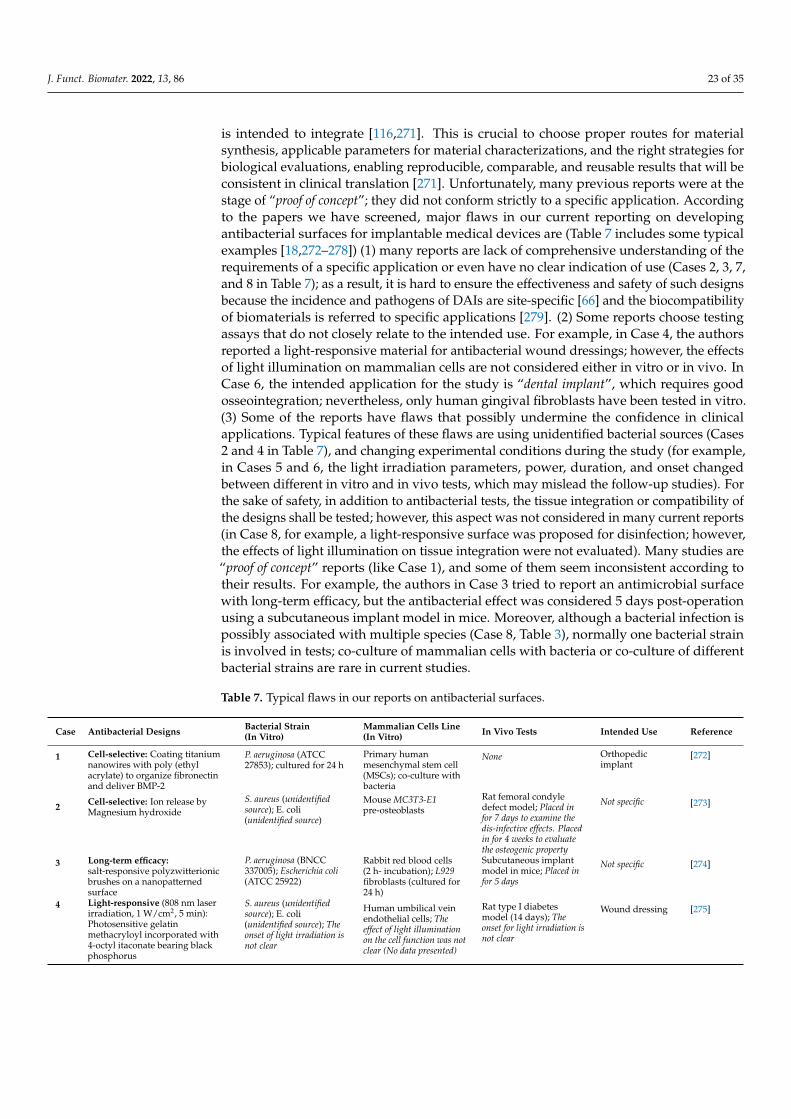
J. Funct. Biomater. 2022, 13, 86 23 of 35
is intended to integrate [116,271]. This is crucial to choose proper routes for materialsynthesis, applicable parameters for material characterizations, and the right strategies forbiological evaluations, enabling reproducible, comparable, and reusable results that will beconsistent in clinical translation [271]. Unfortunately, many previous reports were at thestage of “proof of concept”; they did not conform strictly to a specific application. Accordingto the papers we have screened, major flaws in our current reporting on developingantibacterial surfaces for implantable medical devices are (Table 7 includes some typicalexamples [18,272–278]) (1) many reports are lack of comprehensive understanding of therequirements of a specific application or even have no clear indication of use (Cases 2, 3, 7,and 8 in Table 7); as a result, it is hard to ensure the effectiveness and safety of such designsbecause the incidence and pathogens of DAIs are site-specific [66] and the biocompatibilityof biomaterials is referred to specific applications [279]. (2) Some reports choose testingassays that do not closely relate to the intended use. For example, in Case 4, the authorsreported a light-responsive material for antibacterial wound dressings; however, the effectsof light illumination on mammalian cells are not considered either in vitro or in vivo. InCase 6, the intended application for the study is “dental implant”, which requires goodosseointegration; nevertheless, only human gingival fibroblasts have been tested in vitro.(3) Some of the reports have flaws that possibly undermine the confidence in clinicalapplications. Typical features of these flaws are using unidentified bacterial sources (Cases2 and 4 in Table 7), and changing experimental conditions during the study (for example,in Cases 5 and 6, the light irradiation parameters, power, duration, and onset changedbetween different in vitro and in vivo tests, which may mislead the follow-up studies). Forthe sake of safety, in addition to antibacterial tests, the tissue integration or compatibility ofthe designs shall be tested; however, this aspect was not considered in many current reports(in Case 8, for example, a light-responsive surface was proposed for disinfection; however,the effects of light illumination on tissue integration were not evaluated). Many studies are“proof of concept” reports (like Case 1), and some of them seem inconsistent according totheir results. For example, the authors in Case 3 tried to report an antimicrobial surfacewith long-term efficacy, but the antibacterial effect was considered 5 days post-operationusing a subcutaneous implant model in mice. Moreover, although a bacterial infection ispossibly associated with multiple species (Case 8, Table 3), normally one bacterial strainis involved in tests; co-culture of mammalian cells with bacteria or co-culture of differentbacterial strains are rare in current studies.
Table 7. Typical flaws in our reports on antibacterial surfaces.
Case Antibacterial Designs Bacterial Strain(In Vitro)
Mammalian Cells Line(In Vitro) In Vivo Tests Intended Use Reference
1 Cell-selective: Coating titaniumnanowires with poly (ethylacrylate) to organize fibronectinand deliver BMP-2
P. aeruginosa (ATCC27853); cultured for 24 h
Primary humanmesenchymal stem cell(MSCs); co-culture withbacteria
None Orthopedicimplant
[272]
2 Cell-selective: Ion release byMagnesium hydroxide
S. aureus (unidentifiedsource); E. coli(unidentified source)
Mouse MC3T3-E1pre-osteoblasts
Rat femoral condyledefect model; Placed infor 7 days to examine thedis-infective effects. Placedin for 4 weeks to evaluatethe osteogenic property
Not specific [273]
3 Long-term efficacy:salt-responsive polyzwitterionicbrushes on a nanopatternedsurface
P. aeruginosa (BNCC337005); Escherichia coli(ATCC 25922)
Rabbit red blood cells(2 h- incubation); L929fibroblasts (cultured for24 h)
Subcutaneous implantmodel in mice; Placed infor 5 days
Not specific [274]
4 Light-responsive (808 nm laserirradiation, 1 W/cm2, 5 min):Photosensitive gelatinmethacryloyl incorporated with4-octyl itaconate bearing blackphosphorus
S. aureus (unidentifiedsource); E. coli(unidentified source); Theonset of light irradiation isnot clear
Human umbilical veinendothelial cells; Theeffect of light illuminationon the cell function was notclear (No data presented)
Rat type I diabetesmodel (14 days); Theonset for light irradiation isnot clear
Wound dressing [275]
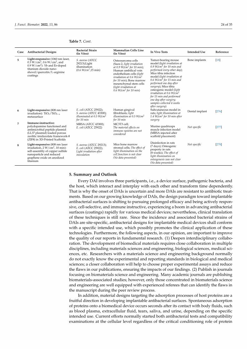
J. Funct. Biomater. 2022, 13, 86 24 of 35
Table 7. Cont.
Case Antibacterial Designs Bacterial Strain(In Vitro)
Mammalian Cells Line(In Vitro) In Vivo Tests Intended Use Reference
5 Light-responsive (1060 nm laser,0.3 W/cm2, 0.6 W/cm2, and0.9 W/cm2): Yb and Er-dopedtitanium dioxide nano-shovel/quercetin/L-argininecoatings
S. aureus (ATCC29213);Lightillumination(0.6 W/cm2,15 min)
Osteosarcoma cells(Saos-2, light irradiationat 0.9 W/cm2 for 10 min);Human umbilical veinendothelium cells (lightirradiation at 0.6 W/cm2
for 10 min); Bone marrowmesenchymal stem cells(light irradiation at0.6 W/cm2 for 10 min)
Tumor-bearing mousemodel (light irradiation at0.9 W/cm2 for 10 min andperformed every other day);Mice tibia infectionmodel (light irradiation at0.6 W/cm2 for 15 min andperformed one day aftersurgery); Mice tibiaosteogenic model (lightirradiation at 0.6 W/cm2
for 15 min and performedone day after surgery;samples collected 4 weeksafter surgery)
Bone implants [18]
6 Light-responsive (808 nm laserirradiation): TiO2/TiO2−xmetasurface
E. coli (ATCC 25922);S. aureus (ATCC 43300);illuminated at 0.5 W/cm2
for 10 min
Human gingivalfibroblasts; lightillumination at 0.5 W/cm2
for 10 min
Subcutaneous model inrats; light illumination at1.4 W/cm2 for 10 min aftersurgery
Dental implant [276]
7 Immune-instructive:polydopamine functioned andantimicrobial peptide plasmid(LL37 plasmid) loaded porouszeolitic imidazolate framework-8(ZIF8) in 3D-Printed Scaffolds
MRSA (ATCC 43300);E. coli (ATCC 25922)
MC3T3 cell;The material effects onimmune systems are notconsidered
Murine quadricepsmuscle infection model(MRSA injected afterscaffold placement)
Not specific [277]
8 Light-responsive (808 nm laserirradiation, 2 W/cm2, 10 min):self-assembly of copper sulfidenanoparticle and reducedgraphene oxide on anodizedtitanium
S. aureus (ATCC 29213);E. coli (ATCC 25922);Light irradiation afterinoculation
Mice bone marrowstromal cells; The effect oflight illumination on thecell function is not clear(No data presented)
Disinfection in rats(7 days); Osteogenicproperty in rats(8 weeks); The effect oflight illumination onosteogenesis was not clear(No data presented)
Not specific [278]
5. Summary and Outlook
Every DAI involves three participants, i.e., a device surface, pathogenic bacteria, andthe host, which interact and interplay with each other and transform time dependently.That is why the onset of DAIs is uncertain and more DAIs are resistant to antibiotic treat-ments. Based on our growing knowledge of DAIs, the design paradigm toward implantableantibacterial surfaces is shifting to pursuing prolonged efficacy and being actively respon-sive, cell-selective, and immune instructive, experiencing a boom in advancing antibacterialsurfaces (coatings) rapidly for various medical devices; nevertheless, clinical translationof these techniques is still rare. Since the incidence and associated bacterial strains ofDAIs are site-specific, antibacterial designs for implantable medical devices shall conformwith a specific intended use, which possibly promotes the clinical application of thesetechnologies. Furthermore, the following aspects, in our opinion, are important to improvethe quality of our reports in fundamental research. (1) Deepen interdisciplinary collabo-ration. The development of biomedical materials requires close collaboration in multipledisciplines, including materials sciences and engineering, biological sciences, medical sci-ences, etc. Researchers with a materials science and engineering background normallydo not exactly know the experimental and reporting standards in biological and medicalsciences; a closer collaboration will help to choose proper experimental assays and reducethe flaws in our publications, ensuring the impacts of our findings. (2) Publish in journalsfocusing on biomaterials science and engineering. Many academic journals are publishingbiomaterials-associated studies; however, only those concentrated in biomaterials scienceand engineering are well equipped with experienced referees that can identify the flaws inthe manuscript during the peer review process.
In addition, material designs targeting the adsorption processes of host proteins are afruitful direction in developing implantable antibacterial surfaces. Spontaneous adsorptionof proteins onto a biomedical device occurs seconds after its contact with body fluids, suchas blood plasma, extracellular fluid, tears, saliva, and urine, depending on the specificintended use. Current efforts normally started both antibacterial tests and compatibilityexaminations at the cellular level regardless of the critical conditioning role of protein
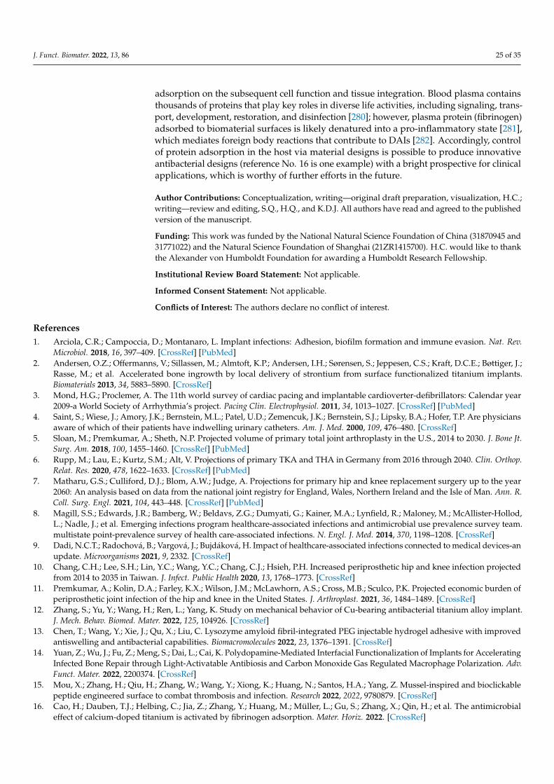
J. Funct. Biomater. 2022, 13, 86 25 of 35
adsorption on the subsequent cell function and tissue integration. Blood plasma containsthousands of proteins that play key roles in diverse life activities, including signaling, trans-port, development, restoration, and disinfection [280]; however, plasma protein (fibrinogen)adsorbed to biomaterial surfaces is likely denatured into a pro-inflammatory state [281],which mediates foreign body reactions that contribute to DAIs [282]. Accordingly, controlof protein adsorption in the host via material designs is possible to produce innovativeantibacterial designs (reference No. 16 is one example) with a bright prospective for clinicalapplications, which is worthy of further efforts in the future.
Author Contributions: Conceptualization, writing—original draft preparation, visualization, H.C.;writing—review and editing, S.Q., H.Q., and K.D.J. All authors have read and agreed to the publishedversion of the manuscript.
Funding: This work was funded by the National Natural Science Foundation of China (31870945 and31771022) and the Natural Science Foundation of Shanghai (21ZR1415700). H.C. would like to thankthe Alexander von Humboldt Foundation for awarding a Humboldt Research Fellowship.
Institutional Review Board Statement: Not applicable.
Informed Consent Statement: Not applicable.
Conflicts of Interest: The authors declare no conflict of interest.
References1. Arciola, C.R.; Campoccia, D.; Montanaro, L. Implant infections: Adhesion, biofilm formation and immune evasion. Nat. Rev.
Microbiol. 2018, 16, 397–409. [CrossRef] [PubMed]2. Andersen, O.Z.; Offermanns, V.; Sillassen, M.; Almtoft, K.P.; Andersen, I.H.; Sørensen, S.; Jeppesen, C.S.; Kraft, D.C.E.; Bøttiger, J.;
Rasse, M.; et al. Accelerated bone ingrowth by local delivery of strontium from surface functionalized titanium implants.Biomaterials 2013, 34, 5883–5890. [CrossRef]
3. Mond, H.G.; Proclemer, A. The 11th world survey of cardiac pacing and implantable cardioverter-defibrillators: Calendar year2009-a World Society of Arrhythmia’s project. Pacing Clin. Electrophysiol. 2011, 34, 1013–1027. [CrossRef] [PubMed]
4. Saint, S.; Wiese, J.; Amory, J.K.; Bernstein, M.L.; Patel, U.D.; Zemencuk, J.K.; Bernstein, S.J.; Lipsky, B.A.; Hofer, T.P. Are physiciansaware of which of their patients have indwelling urinary catheters. Am. J. Med. 2000, 109, 476–480. [CrossRef]
5. Sloan, M.; Premkumar, A.; Sheth, N.P. Projected volume of primary total joint arthroplasty in the U.S., 2014 to 2030. J. Bone Jt.Surg. Am. 2018, 100, 1455–1460. [CrossRef] [PubMed]
6. Rupp, M.; Lau, E.; Kurtz, S.M.; Alt, V. Projections of primary TKA and THA in Germany from 2016 through 2040. Clin. Orthop.Relat. Res. 2020, 478, 1622–1633. [CrossRef] [PubMed]
7. Matharu, G.S.; Culliford, D.J.; Blom, A.W.; Judge, A. Projections for primary hip and knee replacement surgery up to the year2060: An analysis based on data from the national joint registry for England, Wales, Northern Ireland and the Isle of Man. Ann. R.Coll. Surg. Engl. 2021, 104, 443–448. [CrossRef] [PubMed]
8. Magill, S.S.; Edwards, J.R.; Bamberg, W.; Beldavs, Z.G.; Dumyati, G.; Kainer, M.A.; Lynfield, R.; Maloney, M.; McAllister-Hollod,L.; Nadle, J.; et al. Emerging infections program healthcare-associated infections and antimicrobial use prevalence survey team.multistate point-prevalence survey of health care-associated infections. N. Engl. J. Med. 2014, 370, 1198–1208. [CrossRef]
9. Dadi, N.C.T.; Radochová, B.; Vargová, J.; Bujdáková, H. Impact of healthcare-associated infections connected to medical devices-anupdate. Microorganisms 2021, 9, 2332. [CrossRef]
10. Chang, C.H.; Lee, S.H.; Lin, Y.C.; Wang, Y.C.; Chang, C.J.; Hsieh, P.H. Increased periprosthetic hip and knee infection projectedfrom 2014 to 2035 in Taiwan. J. Infect. Public Health 2020, 13, 1768–1773. [CrossRef]
11. Premkumar, A.; Kolin, D.A.; Farley, K.X.; Wilson, J.M.; McLawhorn, A.S.; Cross, M.B.; Sculco, P.K. Projected economic burden ofperiprosthetic joint infection of the hip and knee in the United States. J. Arthroplast. 2021, 36, 1484–1489. [CrossRef]
12. Zhang, S.; Yu, Y.; Wang, H.; Ren, L.; Yang, K. Study on mechanical behavior of Cu-bearing antibacterial titanium alloy implant.J. Mech. Behav. Biomed. Mater. 2022, 125, 104926. [CrossRef]
13. Chen, T.; Wang, Y.; Xie, J.; Qu, X.; Liu, C. Lysozyme amyloid fibril-integrated PEG injectable hydrogel adhesive with improvedantiswelling and antibacterial capabilities. Biomacromolecules 2022, 23, 1376–1391. [CrossRef]
14. Yuan, Z.; Wu, J.; Fu, Z.; Meng, S.; Dai, L.; Cai, K. Polydopamine-Mediated Interfacial Functionalization of Implants for AcceleratingInfected Bone Repair through Light-Activatable Antibiosis and Carbon Monoxide Gas Regulated Macrophage Polarization. Adv.Funct. Mater. 2022, 2200374. [CrossRef]
15. Mou, X.; Zhang, H.; Qiu, H.; Zhang, W.; Wang, Y.; Xiong, K.; Huang, N.; Santos, H.A.; Yang, Z. Mussel-inspired and bioclickablepeptide engineered surface to combat thrombosis and infection. Research 2022, 2022, 9780879. [CrossRef]
16. Cao, H.; Dauben, T.J.; Helbing, C.; Jia, Z.; Zhang, Y.; Huang, M.; Müller, L.; Gu, S.; Zhang, X.; Qin, H.; et al. The antimicrobialeffect of calcium-doped titanium is activated by fibrinogen adsorption. Mater. Horiz. 2022. [CrossRef]
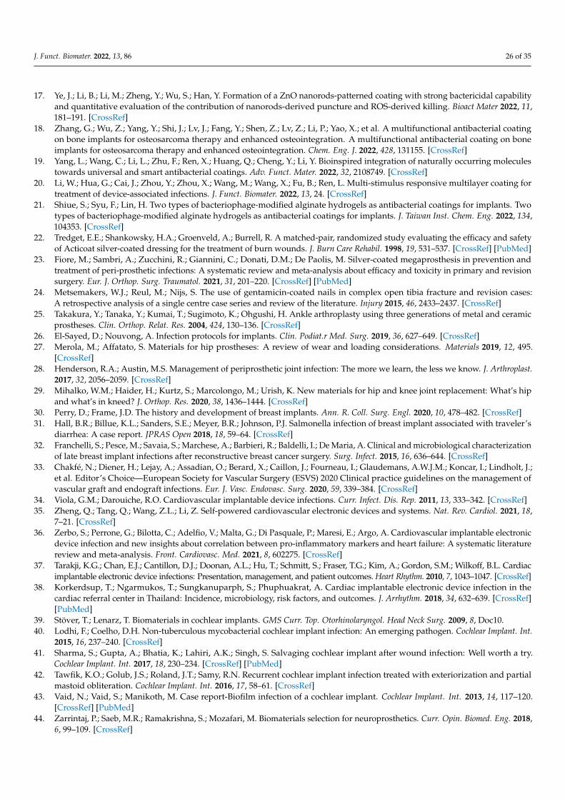
J. Funct. Biomater. 2022, 13, 86 26 of 35
17. Ye, J.; Li, B.; Li, M.; Zheng, Y.; Wu, S.; Han, Y. Formation of a ZnO nanorods-patterned coating with strong bactericidal capabilityand quantitative evaluation of the contribution of nanorods-derived puncture and ROS-derived killing. Bioact Mater 2022, 11,181–191. [CrossRef]
18. Zhang, G.; Wu, Z.; Yang, Y.; Shi, J.; Lv, J.; Fang, Y.; Shen, Z.; Lv, Z.; Li, P.; Yao, X.; et al. A multifunctional antibacterial coatingon bone implants for osteosarcoma therapy and enhanced osteointegration. A multifunctional antibacterial coating on boneimplants for osteosarcoma therapy and enhanced osteointegration. Chem. Eng. J. 2022, 428, 131155. [CrossRef]
19. Yang, L.; Wang, C.; Li, L.; Zhu, F.; Ren, X.; Huang, Q.; Cheng, Y.; Li, Y. Bioinspired integration of naturally occurring moleculestowards universal and smart antibacterial coatings. Adv. Funct. Mater. 2022, 32, 2108749. [CrossRef]
20. Li, W.; Hua, G.; Cai, J.; Zhou, Y.; Zhou, X.; Wang, M.; Wang, X.; Fu, B.; Ren, L. Multi-stimulus responsive multilayer coating fortreatment of device-associated infections. J. Funct. Biomater. 2022, 13, 24. [CrossRef]
21. Shiue, S.; Syu, F.; Lin, H. Two types of bacteriophage-modified alginate hydrogels as antibacterial coatings for implants. Twotypes of bacteriophage-modified alginate hydrogels as antibacterial coatings for implants. J. Taiwan Inst. Chem. Eng. 2022, 134,104353. [CrossRef]
22. Tredget, E.E.; Shankowsky, H.A.; Groenveld, A.; Burrell, R. A matched-pair, randomized study evaluating the efficacy and safetyof Acticoat silver-coated dressing for the treatment of burn wounds. J. Burn Care Rehabil. 1998, 19, 531–537. [CrossRef] [PubMed]
23. Fiore, M.; Sambri, A.; Zucchini, R.; Giannini, C.; Donati, D.M.; De Paolis, M. Silver-coated megaprosthesis in prevention andtreatment of peri-prosthetic infections: A systematic review and meta-analysis about efficacy and toxicity in primary and revisionsurgery. Eur. J. Orthop. Surg. Traumatol. 2021, 31, 201–220. [CrossRef] [PubMed]
24. Metsemakers, W.J.; Reul, M.; Nijs, S. The use of gentamicin-coated nails in complex open tibia fracture and revision cases:A retrospective analysis of a single centre case series and review of the literature. Injury 2015, 46, 2433–2437. [CrossRef]
25. Takakura, Y.; Tanaka, Y.; Kumai, T.; Sugimoto, K.; Ohgushi, H. Ankle arthroplasty using three generations of metal and ceramicprostheses. Clin. Orthop. Relat. Res. 2004, 424, 130–136. [CrossRef]
26. El-Sayed, D.; Nouvong, A. Infection protocols for implants. Clin. Podiat.r Med. Surg. 2019, 36, 627–649. [CrossRef]27. Merola, M.; Affatato, S. Materials for hip prostheses: A review of wear and loading considerations. Materials 2019, 12, 495.
[CrossRef]28. Henderson, R.A.; Austin, M.S. Management of periprosthetic joint infection: The more we learn, the less we know. J. Arthroplast.
2017, 32, 2056–2059. [CrossRef]29. Mihalko, W.M.; Haider, H.; Kurtz, S.; Marcolongo, M.; Urish, K. New materials for hip and knee joint replacement: What’s hip
and what’s in kneed? J. Orthop. Res. 2020, 38, 1436–1444. [CrossRef]30. Perry, D.; Frame, J.D. The history and development of breast implants. Ann. R. Coll. Surg. Engl. 2020, 10, 478–482. [CrossRef]31. Hall, B.R.; Billue, K.L.; Sanders, S.E.; Meyer, B.R.; Johnson, P.J. Salmonella infection of breast implant associated with traveler’s
diarrhea: A case report. JPRAS Open 2018, 18, 59–64. [CrossRef]32. Franchelli, S.; Pesce, M.; Savaia, S.; Marchese, A.; Barbieri, R.; Baldelli, I.; De Maria, A. Clinical and microbiological characterization
of late breast implant infections after reconstructive breast cancer surgery. Surg. Infect. 2015, 16, 636–644. [CrossRef]33. Chakfé, N.; Diener, H.; Lejay, A.; Assadian, O.; Berard, X.; Caillon, J.; Fourneau, I.; Glaudemans, A.W.J.M.; Koncar, I.; Lindholt, J.;
et al. Editor’s Choice—European Society for Vascular Surgery (ESVS) 2020 Clinical practice guidelines on the management ofvascular graft and endograft infections. Eur. J. Vasc. Endovasc. Surg. 2020, 59, 339–384. [CrossRef]
34. Viola, G.M.; Darouiche, R.O. Cardiovascular implantable device infections. Curr. Infect. Dis. Rep. 2011, 13, 333–342. [CrossRef]35. Zheng, Q.; Tang, Q.; Wang, Z.L.; Li, Z. Self-powered cardiovascular electronic devices and systems. Nat. Rev. Cardiol. 2021, 18,
7–21. [CrossRef]36. Zerbo, S.; Perrone, G.; Bilotta, C.; Adelfio, V.; Malta, G.; Di Pasquale, P.; Maresi, E.; Argo, A. Cardiovascular implantable electronic
device infection and new insights about correlation between pro-inflammatory markers and heart failure: A systematic literaturereview and meta-analysis. Front. Cardiovasc. Med. 2021, 8, 602275. [CrossRef]
37. Tarakji, K.G.; Chan, E.J.; Cantillon, D.J.; Doonan, A.L.; Hu, T.; Schmitt, S.; Fraser, T.G.; Kim, A.; Gordon, S.M.; Wilkoff, B.L. Cardiacimplantable electronic device infections: Presentation, management, and patient outcomes. Heart Rhythm. 2010, 7, 1043–1047. [CrossRef]
38. Korkerdsup, T.; Ngarmukos, T.; Sungkanuparph, S.; Phuphuakrat, A. Cardiac implantable electronic device infection in thecardiac referral center in Thailand: Incidence, microbiology, risk factors, and outcomes. J. Arrhythm. 2018, 34, 632–639. [CrossRef][PubMed]
39. Stöver, T.; Lenarz, T. Biomaterials in cochlear implants. GMS Curr. Top. Otorhinolaryngol. Head Neck Surg. 2009, 8, Doc10.40. Lodhi, F.; Coelho, D.H. Non-tuberculous mycobacterial cochlear implant infection: An emerging pathogen. Cochlear Implant. Int.
2015, 16, 237–240. [CrossRef]41. Sharma, S.; Gupta, A.; Bhatia, K.; Lahiri, A.K.; Singh, S. Salvaging cochlear implant after wound infection: Well worth a try.
Cochlear Implant. Int. 2017, 18, 230–234. [CrossRef] [PubMed]42. Tawfik, K.O.; Golub, J.S.; Roland, J.T.; Samy, R.N. Recurrent cochlear implant infection treated with exteriorization and partial
mastoid obliteration. Cochlear Implant. Int. 2016, 17, 58–61. [CrossRef]43. Vaid, N.; Vaid, S.; Manikoth, M. Case report-Biofilm infection of a cochlear implant. Cochlear Implant. Int. 2013, 14, 117–120.
[CrossRef] [PubMed]44. Zarrintaj, P.; Saeb, M.R.; Ramakrishna, S.; Mozafari, M. Biomaterials selection for neuroprosthetics. Curr. Opin. Biomed. Eng. 2018,
6, 99–109. [CrossRef]

J. Funct. Biomater. 2022, 13, 86 27 of 35
45. Shenai, M.B.; Falconer, R.; Rogers, S. A cupriavidus pauculus infection in a patient with a deep brain stimulation implant. Cureus2019, 11, e6104. [CrossRef]
46. Wei, Z.; Gordon, C.R.; Bergey, G.K.; Sacks, J.M.; Anderson, W.S. Implant site infection and bone flap osteomyelitis associated withthe neuropace responsive neurostimulation system. World Neurosurg. 2016, 88, 687.e1–687.e6. [CrossRef]
47. Lawrence, E.L.; Turner, I.G. Materials for urinary catheters: A review of their history and development in the UK. Med. Eng. Phys.2005, 27, 443–453. [CrossRef] [PubMed]
48. Huang, W.C.; Wann, S.R.; Lin, S.L.; Kunin, C.M.; Kung, M.H.; Lin, C.H.; Hsu, C.W.; Liu, C.P.; Lee, S.S.; Liu, Y.C.; et al. Catheter-associated urinary tract infections in intensive care units can be reduced by prompting physicians to remove unnecessarycatheters. Infect. Control Hosp. Epidemiol. 2004, 25, 974–978. [CrossRef]
49. Lo, E.; Nicolle, L.E.; Coffin, S.E.; Gould, C.; Maragakis, L.L.; Meddings, J.; Pegues, D.A.; Pettis, A.M.; Saint, S.; Yokoe, D.S.Strategies to prevent catheter-associated urinary tract infections in acute care hospitals: 2014 update. Infect. Control Hosp.Epidemiol. 2014, 35, 464–479. [CrossRef]
50. Luzum, M.; Sebolt, J.; Chopra, V. Catheter-associated urinary tract infection, clostridioides difficile colitis, central line-associatedbloodstream infection, and methicillin-resistant staphylococcus aureus. Med. Clin. North Am. 2020, 104, 663–679. [CrossRef]
51. Li, F.; Song, M.; Xu, L.; Deng, B.; Zhu, S.; Li, X. Risk factors for catheter-associated urinary tract infection among hospitalizedpatients: A systematic review and meta-analysis of observational studies. J. Adv. Nurs. 2019, 75, 517–527. [CrossRef]
52. Shuman, E.K.; Chenoweth, C.E. Urinary catheter-associated infections. Infect. Dis. Clin. North Am. 2018, 32, 885–897. [CrossRef]53. Del Bigio, M.R. Biological reactions to cerebrospinal fluid shunt devices: A review of the cellular pathology. Neurosurgery 1998, 42,
319–326. [CrossRef]54. Canadian Nosocomial Infection Surveillance Program. Device-associated infections in Canadian acute-care hospitals from 2009
to 2018. Can. Commun. Dis. Rep. 2020, 46, 387–397. [CrossRef]55. Shibamura-Fujiogi, M.; Ormsby, J.; Breibart, M.; Warf, B.; Priebe, G.P.; Soriano, S.G.; Sandora, T.J.; Yuki, K. Risk factors for
pediatric surgical site infection following neurosurgical procedures for hydrocephalus: A retrospective single-center cohort study.BMC Anesthesiol. 2021, 21, 124. [CrossRef]
56. Benachinmardi, K.K.; Ravikumar, R.; Indiradevi, B. Role of biofilm in cerebrospinal fluid shunt infections: A study at tertiaryneurocare center from South India. J. Neurosci. Rural. Pract. 2017, 8, 335–341. [CrossRef]
57. Fernández-Méndez, R.; Richards, H.K.; Seeley, H.M.; Pickard, J.D.; Joannides, A.J. UKSR collaborators, Current epidemiology ofcerebrospinal fluid shunt surgery in the UK and Ireland (2004–2013). J. Neurol. Neurosurg. Psychiatry 2019, 90, 747–754. [CrossRef]
58. Deshmukh, R.M.; Kulkarni, S.S. A review on biomaterials in orthopedic bone plate application. International J. Curr. Eng. Technol.2015, 5, 2587–2591.
59. Toro-Aguilera, Á.; Zuriarrain, S.W.; Masdeu, M.G.; Sayol, R.R.; Billi, A.M.; Carrera, I.; de Caso, J. Risk factors for infection infixation of distal tibia fractures. Injury 2021, 52 (Suppl. 4), S104–S108. [CrossRef]
60. Guillaume, B. Dental implants: A review. Morphologie 2016, 100, 189–198. [CrossRef]61. Neely, A.L.; Maalhagh-Fard, A. Successful management of early peri-implant infection and bone loss using a multidisciplinary
treatment approach. Clin. Adv. Periodontics 2018, 8, 5–10. [CrossRef]62. Patton, D.; Kiewiet, N.; Brage, M. Infected total ankle arthroplasty: Risk factors and treatment options. Foot Ankle Int. 2015, 36,
626–634. [CrossRef] [PubMed]63. Gbejuade, H.O.; Lovering, A.M.; Webb, J.C. The role of microbial biofilms in prosthetic joint infections. Acta Orthop. 2015, 86,
147–158. [CrossRef] [PubMed]64. Bayston, R.; Lari, J. A study of the sources of infection in colonised shunts. Dev. Med. Child Neurol. 1974, 16, 16–22. [CrossRef]
[PubMed]65. Reynolds-Campbell, G.; Nicholson, A.; Thoms-Rodriguez, C.A. Oral bacterial infections: Diagnosis and management. Dent. Clin.
N. Am. 2017, 61, 305–318. [CrossRef] [PubMed]66. Masters, E.A.; Ricciardi, B.F.; de Mesy Bentley, K.L.; Moriarty, T.F.; Schwarz, E.M.; Muthukrishnan, G. Skeletal infections:
Microbial pathogenesis, immunity and clinical management. Nat. Rev. Microbiol. 2022, 20, 385–400. [CrossRef]67. Bain, C.J.; Odili, J. Late infection of an alloplastic chin implant masquerading as squamous cell carcinoma. J. Plast. Reconstr.
Aesthet. Surg. 2012, 65, e151–e152. [CrossRef]68. Chang, J.; Lee, G.W. Late hematogenous bacterial infections of breast implants: Two case reports of unique bacterial infections.
Ann. Plast. Surg. 2011, 67, 14–16. [CrossRef]69. Beidas, O.E.; Rabb, C.H.; Sawan, K.T.; Tan, B.K. The pseudomeningocoele that wasn’t: Case report of an adult who presented
with a late infection of an implant. J. Plast. Reconstr. Aesthet. Surg. 2011, 64, 1228–1231. [CrossRef]70. Vichitvejpaisal, P.; Dalvin, L.A.; Lally, S.E.; Shields, C.L. Delayed implant infection with Cutibacterium acnes (Propionibacterium
acnes) 30 years after silicone sheet orbital floor implant. Orbit 2020, 39, 139–142. [CrossRef]71. Coden, D.J.; Hornblass, A. Propionibacterium acnes orbital abscess. Arch. Ophthalmol. 1990, 108, 481. [CrossRef]72. Hannouille, J.; Belgrado, J.P.; Vankerchove, S.; Vandermeeren, L. Breast implant infection with pasteurella canis: First case-report.
JPRAS Open 2019, 21, 86–88. [CrossRef]73. Oses, M.; Ordás, C.M.; Feliz, C.; Del Val, J.; Ayerbe, J.; García-Ruiz, P.J. Disease-modifying anti-rheumatic drugs as a risk factor for
delayed DBS implant infection. Parkinsonism Relat. Disord. 2018, 55, 143–144. [CrossRef]
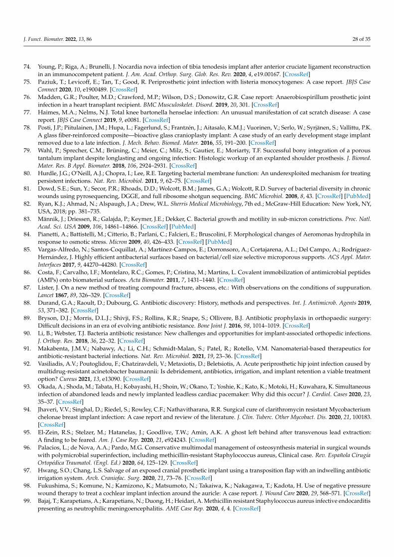
J. Funct. Biomater. 2022, 13, 86 28 of 35
74. Young, P.; Riga, A.; Brunelli, J. Nocardia nova infection of tibia tenodesis implant after anterior cruciate ligament reconstructionin an immunocompetent patient. J. Am. Acad. Orthop. Surg. Glob. Res. Rev. 2020, 4, e19.00167. [CrossRef]
75. Paziuk, T.; Levicoff, E.; Tan, T.; Good, R. Periprosthetic joint infection with listeria monocytogenes: A case report. JBJS CaseConnect 2020, 10, e1900489. [CrossRef]
76. Madden, G.R.; Poulter, M.D.; Crawford, M.P.; Wilson, D.S.; Donowitz, G.R. Case report: Anaerobiospirillum prosthetic jointinfection in a heart transplant recipient. BMC Musculoskelet. Disord. 2019, 20, 301. [CrossRef]
77. Haimes, M.A.; Nelms, N.J. Total knee bartonella henselae infection: An unusual manifestation of cat scratch disease: A casereport. JBJS Case Connect 2019, 9, e0081. [CrossRef]
78. Posti, J.P.; Piitulainen, J.M.; Hupa, L.; Fagerlund, S.; Frantzén, J.; Aitasalo, K.M.J.; Vuorinen, V.; Serlo, W.; Syrjänen, S.; Vallittu, P.K.A glass fiber-reinforced composite—bioactive glass cranioplasty implant: A case study of an early development stage implantremoved due to a late infection. J. Mech. Behav. Biomed. Mater. 2016, 55, 191–200. [CrossRef]
79. Wahl, P.; Sprecher, C.M.; Brüning, C.; Meier, C.; Milz, S.; Gautier, E.; Moriarty, T.F. Successful bony integration of a poroustantalum implant despite longlasting and ongoing infection: Histologic workup of an explanted shoulder prosthesis. J. Biomed.Mater. Res. B Appl. Biomater. 2018, 106, 2924–2931. [CrossRef]
80. Hurdle, J.G.; O’Neill, A.J.; Chopra, I.; Lee, R.E. Targeting bacterial membrane function: An underexploited mechanism for treatingpersistent infections. Nat. Rev. Microbiol. 2011, 9, 62–75. [CrossRef]
81. Dowd, S.E.; Sun, Y.; Secor, P.R.; Rhoads, D.D.; Wolcott, B.M.; James, G.A.; Wolcott, R.D. Survey of bacterial diversity in chronicwounds using pyrosequencing, DGGE, and full ribosome shotgun sequencing. BMC Microbiol. 2008, 8, 43. [CrossRef] [PubMed]
82. Ryan, K.J.; Ahmad, N.; Alspaugh, J.A.; Drew, W.L. Sherris Medical Microbiology, 7th ed.; McGraw-Hill Education: New York, NY,USA, 2018; pp. 381–735.
83. Männik, J.; Driessen, R.; Galajda, P.; Keymer, J.E.; Dekker, C. Bacterial growth and motility in sub-micron constrictions. Proc. Natl.Acad. Sci. USA 2009, 106, 14861–14866. [CrossRef] [PubMed]
84. Pianetti, A.; Battistelli, M.; Citterio, B.; Parlani, C.; Falcieri, E.; Bruscolini, F. Morphological changes of Aeromonas hydrophila inresponse to osmotic stress. Micron 2009, 40, 426–433. [CrossRef] [PubMed]
85. Vargas-Alfredo, N.; Santos-Coquillat, A.; Martínez-Campos, E.; Dorronsoro, A.; Cortajarena, A.L.; Del Campo, A.; Rodríguez-Hernández, J. Highly efficient antibacterial surfaces based on bacterial/cell size selective microporous supports. ACS Appl. Mater.Interfaces 2017, 9, 44270–44280. [CrossRef]
86. Costa, F.; Carvalho, I.F.; Montelaro, R.C.; Gomes, P.; Cristina, M.; Martins, L. Covalent immobilization of antimicrobial peptides(AMPs) onto biomaterial surfaces. Acta Biomater. 2011, 7, 1431–1440. [CrossRef]
87. Lister, J. On a new method of treating compound fracture, abscess, etc.: With observations on the conditions of suppuration.Lancet 1867, 89, 326–329. [CrossRef]
88. Durand, G.A.; Raoult, D.; Dubourg, G. Antibiotic discovery: History, methods and perspectives. Int. J. Antimicrob. Agents 2019,53, 371–382. [CrossRef]
89. Bryson, D.J.; Morris, D.L.J.; Shivji, F.S.; Rollins, K.R.; Snape, S.; Ollivere, B.J. Antibiotic prophylaxis in orthopaedic surgery:Difficult decisions in an era of evolving antibiotic resistance. Bone Joint J. 2016, 98, 1014–1019. [CrossRef]
90. Li, B.; Webster, T.J. Bacteria antibiotic resistance: New challenges and opportunities for implant-associated orthopedic infections.J. Orthop. Res. 2018, 36, 22–32. [CrossRef]
91. Makabenta, J.M.V.; Nabawy, A.; Li, C.H.; Schmidt-Malan, S.; Patel, R.; Rotello, V.M. Nanomaterial-based therapeutics forantibiotic-resistant bacterial infections. Nat. Rev. Microbiol. 2021, 19, 23–36. [CrossRef]
92. Vasiliadis, A.V.; Poutoglidou, F.; Chatziravdeli, V.; Metaxiotis, D.; Beletsiotis, A. Acute periprosthetic hip joint infection caused bymultidrug-resistant acinetobacter baumannii: Is debridement, antibiotics, irrigation, and implant retention a viable treatmentoption? Cureus 2021, 13, e13090. [CrossRef]
93. Okada, A.; Shoda, M.; Tabata, H.; Kobayashi, H.; Shoin, W.; Okano, T.; Yoshie, K.; Kato, K.; Motoki, H.; Kuwahara, K. Simultaneousinfection of abandoned leads and newly implanted leadless cardiac pacemaker: Why did this occur? J. Cardiol. Cases 2020, 23,35–37. [CrossRef]
94. Jhaveri, V.V.; Singhal, D.; Riedel, S.; Rowley, C.F.; Nathavitharana, R.R. Surgical cure of clarithromycin resistant Mycobacteriumchelonae breast implant infection: A case report and review of the literature. J. Clin. Tuberc. Other Mycobact. Dis. 2020, 21, 100183.[CrossRef]
95. El-Zein, R.S.; Stelzer, M.; Hatanelas, J.; Goodlive, T.W.; Amin, A.K. A ghost left behind after transvenous lead extraction:A finding to be feared. Am. J. Case Rep. 2020, 21, e924243. [CrossRef]
96. Palacios, L.; de Nova, A.A.; Pardo, M.G. Conservative multimodal management of osteosynthesis material in surgical woundswith polymicrobial superinfection, including methicillin-resistant Staphylococcus aureus, Clinical case. Rev. Española CirugíaOrtopédica Traumatol. (Engl. Ed.) 2020, 64, 125–129. [CrossRef]
97. Hwang, S.O.; Chang, L.S. Salvage of an exposed cranial prosthetic implant using a transposition flap with an indwelling antibioticirrigation system. Arch. Craniofac. Surg. 2020, 21, 73–76. [CrossRef]
98. Fukushima, S.; Komune, N.; Kamizono, K.; Matsumoto, N.; Takaiwa, K.; Nakagawa, T.; Kadota, H. Use of negative pressurewound therapy to treat a cochlear implant infection around the auricle: A case report. J. Wound Care 2020, 29, 568–571. [CrossRef]
99. Bajaj, T.; Karapetians, A.; Karapetians, N.; Duong, H.; Heidari, A. Methicillin resistant Staphylococcus aureus infective endocarditispresenting as neutrophilic meningoencephalitis. AME Case Rep. 2020, 4, 4. [CrossRef]

J. Funct. Biomater. 2022, 13, 86 29 of 35
100. Hisanaga, K.; Kadota, H.; Fukushima, S.; Inatomi, Y.; Shimamoto, R.; Kamizono, K.; Hanada, M.; Yoshida, S. Toxic shocksyndrome caused by staphylococcal infection after breast implant surgery: A case report and literature review. Ann. Plast. Surg.2019, 83, 359–362. [CrossRef]
101. Meleca, J.B.; Bryson, P.C. Delayed laryngeal implant infection and laryngocutaneous fistula after medialization laryngoplasty.Am. J. Otolaryngol. 2019, 40, 462–464. [CrossRef]
102. Siebenbürger, G.; Grabein, B.; Schenck, T.; Kammerlander, C.; Böcker, W.; Zeckey, C. Eradication of acinetobacter bauman-nii/enterobacter cloacae complex in an open proximal tibial fracture and closed drop foot correction with a multidisciplinaryapproach using the taylor spatial frame®: A case report. Eur. J. Med. Res. 2019, 24, 2. [CrossRef] [PubMed]
103. Nozoe, M.; Yoshida, D.; Nagatomo, D.; Suematsu, N.; Kubota, T.; Okabe, M.; Yamamoto, Y. Successful percutaneous retrieval of amicra transcatheter pacing system at 8 weeks after implantation. J. Arrhythm. 2018, 34, 653–655. [CrossRef] [PubMed]
104. Bonacker, J.; Darowski, M.; Haar, P.; Westphal, T.; Bergschmidt, P. Periprosthetic tibial fracture with nonunion and ascendingprosthetic joint infection: A case report of an individual treatment strategy. J. Orthop. Case Rep. 2018, 8, 3–8. [PubMed]
105. Rico-Nieto, A.; Moreno-Ramos, F.; Fernández-Baillo, N. Lumbar arthrodesis infection by multi-resistant Klebsiella pneumoniae,successfully treated with implant retention and ceftazidime/avibactam. Rev. Española Cirugía Ortopédica Traumatol. (Engl. Ed.)2018, 62, 471–473. [CrossRef] [PubMed]
106. Vaidya, G.N.; Deam, A.G. Simultaneous suction debulking of lead vegetation prior to percutaneous lead extraction. J. Cardiol.Cases. 2018, 18, 17–19. [CrossRef] [PubMed]
107. Sebastian, S.; Malhotra, R.; Pande, A.; Gautam, D.; Xess, I.; Dhawan, B. Staged reimplantation of a total hip prosthesis after co-infection with candida tropicalis and staphylococcus haemolyticus: A case report. Mycopathologia 2018, 183, 579–584. [CrossRef][PubMed]
108. Mahalingam, P.; Topiwalla, T.T.; Ganesan, G. Drug-resistant coagulase-negative staphylococcal endophthalmitis followingdexamethasone intravitreal implant. Indian J. Ophthalmol. 2017, 65, 634–636. [CrossRef]
109. Gharacholou, S.M.; Dworak, M.; Dababneh, A.S.; Palraj, R.V.; Roskos, M.C.; Chapman, S.C. Acute infection of viabahn stent graftin the popliteal artery. J. Vasc. Surg. Cases Innov. Tech. 2017, 3, 69–73. [CrossRef]
110. Takizawa, T.; Tsutsumimoto, T.; Yui, M.; Misawa, H. Surgical site infections caused by methicillin-resistant staphylococcusepidermidis after spinal instrumentation surgery. Spine 2017, 42, 525–530. [CrossRef]
111. Savage, V.J.; Chopra, I.; O’Neill, A.J. Staphylococcus aureus biofilms promote horizontal transfer of antibiotic resistance.Antimicrob. Agents Chemother. 2013, 57, 1968–1970. [CrossRef]
112. Naylor, N.R.; Atun, R.; Zhu, N.; Kulasabanathan, K.; Silva, S.; Chatterjee, A.; Knight, G.M.; Robotham, J.V. Estimating the burdenof antimicrobial resistance: A systematic literature review. Antimicrob. Resist. Infect. Control 2018, 7, 58. [CrossRef]
113. Zilberman, M.; Elsner, J.J. Antibiotic-eluting medical devices for various applications. J. Control Release 2008, 130, 202–215.[CrossRef]
114. Freischmidt, H.; Armbruster, J.; Reiter, G.; Grützner, P.A.; Helbig, L.; Guehring, T. Individualized techniques of implant coatingwith an antibiotic-loaded, hydroxyapatite/calcium sulphate bone graft substitute. Ther. Clin. Risk Manag. 2020, 16, 689–694.[CrossRef] [PubMed]
115. Campoccia, D.; Montanaro, L.; Speziale, P.; Arciola, C.R. Antibiotic-loaded biomaterials and the risks for the spread of antibioticresistance following their prophylactic and therapeutic clinical use. Biomaterials 2010, 31, 6363–6377. [CrossRef]
116. Cao, H.; Qin, H.; Li, Y.; Jandt, K.D. The action-networks of nanosilver: Bridging the gap between material and biology. Adv.Healthc. Mater. 2021, 26, e2100619. [CrossRef]
117. Percival, S.L.; Bowler, P.G.; Russell, D. Bacterial resistance to silver in wound care. J. Hosp. Infect. 2005, 60, 1–7. [CrossRef]118. Panácek, A.; Kvítek, L.; Smékalová, M.; Vecerová, R.; Kolár, M.; Röderová, M.; Dycka, F.; Šebela, M.; Prucek, R.; Tomanec, O.; et al.
Bacterial resistance to silver nanoparticles and how to overcome it. Nat. Nanotechnol. 2018, 13, 65–71. [CrossRef]119. Nadeem, S.F.; Gohar, U.F.; Tahir, S.F.; Mukhtar, H.; Pornpukdeewattana, S.; Nukthamna, P.; Moula Ali, A.M.; Bavisetty, S.C.B.;
Massa, S. Antimicrobial resistance: More than 70 years of war between humans and bacteria. Crit. Rev. Microbiol. 2020, 46,578–599. [CrossRef]
120. Ciofu, O.; Moser, C.; Jensen, P.Ø.; Høiby, N. Tolerance and resistance of microbial biofilms. Nat. Rev. Microbiol. 2022. [CrossRef]121. Wu, X.X.; Zhang, Y.; Hu, T.; Li, W.X.; Li, Z.L.; Hu, H.J.; Zhu, S.R.; Chen, W.Z.; Zhou, C.S.; Jiang, G.B. Long-term antibacterial
composite via alginate aerogel sustained release of antibiotics and Cu used for bone tissue bacteria infection. Int. J. Biol. Macromol.2021, 167, 1211–1220. [CrossRef]
122. Mukai, M.; Uchida, K.; Sugo, K.; Nakasu, M.; Nakajima, T.; Takata, K.; Takaso, M.; Urabe, K. Long-term antibacterial activity ofvancomycin from calcium phosphate cement in vivo. Biomed. Mater. Eng. 2022, 33, 41–50. [CrossRef] [PubMed]
123. Cheng, Y.; Mei, S.; Kong, X.; Liu, X.; Gao, B.; Chen, B.; Wu, J. Long-term antibacterial activity of a composite coating on titaniumfor dental implant application. J. Biomater. Appl. 2021, 35, 643–654. [CrossRef] [PubMed]
124. Tsutsumi, H.; Tsutsumi, Y.; Shimabukuro, M.; Manaka, T.; Chen, P.; Ashida, M.; Ishikawa, K.; Katayama, H.; Hanaw, T.Investigation of the long-term antibacterial properties of titanium by two-step micro-arc oxidation treatment. Coatings 2021,11, 798. [CrossRef]
125. Liu, F.; Cheng, X.; Xiao, L.; Wang, Q.; Yan, K.; Su, Z.; Wang, L.; Ma, C.; Wang, Y. Inside-outside Ag nanoparticles-loaded polylacticacid electrospun fiber for long-term antibacterial and bone regeneration. Int. J. Biol. Macromol. 2021, 167, 1338–1348. [CrossRef]
J. Funct. Biomater. 2022, 13, 86 30 of 35
126. Tao, S.; Yang, X.; Liao, L.; Yang, J.; Liang, K.; Zeng, S.; Zhou, J.; Zhang, M.; Li, J. A novel anticaries agent, honokiol-loadedpoly(amido amine) dendrimer, for simultaneous long-term antibacterial treatment and remineralization of demineralized enamel.Dent. Mater. 2021, 37, 1337–1349. [CrossRef]
127. Jia, J.; Duan, S.; Zhou, X.; Sun, L.; Qin, C.; Li, M.; Ge, F. Long-term antibacterial film nanocomposite incorporated with patchouliessential oil prepared by supercritical co 2 cyclic impregnation for wound dressing. Molecules 2021, 26, 5005. [CrossRef]
128. Kitagawa, H.; Kitagawa, R.; Tsuboi, R.; Hirose, N.; Thongthai, P.; Sakai, H.; Ueda, M.; Ono, S.; Sasaki, J.; Ooya, T.; et al.Development of endodontic sealers containing antimicrobial-loaded polymer particles with long-term antibacterial effects. Dent.Mater. 2021, 37, 1248–1259. [CrossRef]
129. Wan, R.; Chu, S.; Wang, X.; Lei, L.; Tang, H.; Hu, G.; Dong, L.; Li, D.; Gu, H. Study on the osteogenesis of rat mesenchymalstem cells and the long-term antibacterial activity of Staphylococcus epidermidis on the surface of silver-rich TiN/Ag modifiedtitanium alloy. J. Biomed. Mater. Res. B Appl. Biomater. 2020, 108, 3008–3021. [CrossRef]
130. Liang, T.; Wang, Y.; Zeng, L.; Liu, Y.; Qiao, L.; Zhang, S.; Zhao, R.; Li, G.; Zhang, R.; Xiang, J.; et al. Copper-doped 3D porouscoating developed on Ti-6Al-4V alloys and its in vitro long-term antibacterial ability. Appl. Surf. Sci. 2020, 509, 144717. [CrossRef]
131. Zhang, F.; Zhou, M.; Gu, W.; Shen, Z.; Ma, X.; Lu, F.; Yang, X.; Zheng, Y.; Gou, Z. Zinc-/copper-substituted dicalcium silicatecement: Advanced biomaterials with enhanced osteogenesis and long-term antibacterial properties. J. Mater. Chem. B. 2020, 8,1060–1070. [CrossRef]
132. Yu, H.; Chen, X.; Cai, J.; Ye, D.; Wu, Y.; Liu, P. Dual controlled release nanomicelle-in-nanofiber system for long-term antibacterialmedical dressings. J. Biomater. Sci. Polym. Ed. 2019, 30, 64–76. [CrossRef] [PubMed]
133. Daud, N.M.; Masri, N.A.; Malek, N.A.N.N.; Razak, S.I.A.; Saidin, S. Long-term antibacterial and stable chlorhexidine-polydopamine coating on stainless steel 316L. Prog. Org. Coat. 2018, 122, 147–153. [CrossRef]
134. Shivaram, A.; Bose, S.; Bandyopadhyay, A. Understanding long-term silver release from surface modified porous titaniumimplants. Acta Biomater. 2017, 58, 550–560. [CrossRef]
135. Zhao, R.; Lv, M.; Li, Y.; Sun, M.; Kong, W.; Wang, L.; Song, S.; Fan, C.; Jia, L.; Qiu, S.; et al. Stable nanocomposite based onpegylated and silver nanoparticles loaded graphene oxide for long-term antibacterial activity. ACS Appl. Mater. Interfaces 2017, 9,15328–15341. [CrossRef]
136. Wang, G.; Feng, H.; Jin, W.; Gao, A.; Peng, X.; Li, W.; Wu, H.; Li, Z.; Chu, P.K. Long-term antibacterial characteristics andcytocompatibility of titania nanotubes loaded with Au nanoparticles without photocatalytic effects. Appl. Surf. Sci. 2017, 414,230–237. [CrossRef]
137. Uhm, S.; Kwon, J.; Song, D.; Lee, E.; Jeong, W.; Oh, S.; Kim, K.; Choi, E.H.; Kim, K. Long-Term antibacterial performance andbioactivity of plasma-engineered Ag-NPs/TiO2. J. Biomed. Nanotechnol. 2016, 12, 1890–1906. [CrossRef]
138. Qin, H.; Cao, H.; Zhao, Y.; Zhu, C.; Cheng, T.; Wang, Q.; Peng, X.; Cheng, M.; Wang, J.; Jin, G.; et al. In vitro and in vivoanti-biofilm effects of silver nanoparticles immobilized on titanium. Biomaterials 2014, 35, 9114–9125. [CrossRef]
139. Li, M.; Neoh, K.G.; Xu, L.Q.; Wang, R.; Kang, E.; Lau, T.; Olszyna, D.P.; Chiong, E. Surface modification of silicone for biomedicalapplications requiring long-term antibacterial, antifouling, and hemocompatible properties. Langmuir 2012, 28, 16408–16422.[CrossRef]
140. Urabe, K.; Naruse, K.; Hattori, H.; Hirano, M.; Uchida, K.; Onuma, K.; Park, H.J.; Itoman, M. In vitro comparison of elutioncharacteristics of vancomycin from calcium phosphate cement and polymethylmethacrylate. J. Orthop. Sci. 2009, 14, 784–793.[CrossRef]
141. Uchida, K.; Sugo, K.; Nakajima, T.; Nakawaki, M.; Takano, S.; Nagura, N.; Takaso, M.; Urabe, K. In vivo release of vancomycinfrom calcium phosphate cement. Biomed. Res. Int. 2018, 2018, 4560647. [CrossRef]
142. Bandyopadhyay, A.; Shivaram, A.; Tarafder, S.; Sahasrabudhe, H.; Banerjee, D.; Bose, S. In vivo response of laser processed poroustitanium implants for load-bearing implants. Ann. Biomed. Eng. 2017, 45, 249–260. [CrossRef] [PubMed]
143. Cao, H.; Liu, X.; Meng, F.; Chu, P.K. Biological actions of silver nanoparticles embedded in titanium controlled by micro-galvaniceffects. Biomaterials 2011, 32, 693–705. [CrossRef] [PubMed]
144. Cao, H.; Zhang, W.; Meng, F.; Guo, J.; Wang, D.; Qian, S.; Jiang, X.; Liu, X.; Chu, P.K. Osteogenesis catalyzed by titanium-supportedsilver nanoparticles. ACS Appl. Mater. Interfaces 2017, 9, 5149–5157. [CrossRef] [PubMed]
145. Zhang, X.; Yang, C.; Yang, K. Contact killing of Cu-bearing stainless steel based on charge transfer caused by the microdomainpotential difference. ACS Appl. Mater. Interfaces 2020, 12, 361–372. [CrossRef]
146. Kalfas, I.H. Principles of bone healing. Neurosurg. Focus 2001, 10, E1. [CrossRef]147. Marsell, R.; Einhorn, T.A. The biology of fracture healing. Injury 2011, 42, 551–555. [CrossRef]148. Ono, S.; Imai, R.; Ida, Y.; Shibata, D.; Komiya, T.; Matsumura, H. Increased wound pH as an indicator of local wound infection in
second degree burns. Burns 2015, 41, 820–824. [CrossRef]149. Sawyer, R.G.; Spengler, M.D.; Adams, R.B.; Pruett, T.L. The peritoneal environment during infection. The effect of monomicrobial
and polymicrobial bacteria on pO2 and pH. Ann. Surg. 1991, 213, 253–260. [CrossRef]150. Stassen, W.N.; McCullough, A.J.; Bacon, B.R.; Gutnik, S.H.; Wadiwala, I.M.; McLaren, C.; Kalhan, S.C.; Tavill, A.S. Immediate
diagnostic criteria for bacterial infection of ascitic fluid. Evaluation of ascitic fluid polymorphonuclear leukocyte count, pH, andlactate concentration, alone and in combination. Gastroenterology 1986, 90, 1247–1254. [CrossRef]
151. Del Campo, A.; Echeverría, C.; Martín, M.S.; Cuervo-Rodríguez, R.; Fernández-García, M.; Muñoz-Bonilla, A. Porous microstruc-tured surfaces with ph-triggered antibacterial properties. Macromol. Biosci. 2019, 19, 1900127. [CrossRef]
J. Funct. Biomater. 2022, 13, 86 31 of 35
152. Wei, T.; Yu, Q.; Zhan, W.; Chen, H. A smart antibacterial surface for the on-demand killing and releasing of bacteria. Adv. Healthc.Mater. 2016, 5, 449–456. [CrossRef]
153. Wei, Z.; Yang, J.; Long, S.; Zhang, G.; Wang, X. Smart and in-situ formation electrospun fibrous membrane for the control ofantimicrobial efficacy. Smart Mater. Med. 2021, 2, 87–95. [CrossRef]
154. Pinho, E.; Machado, S.; Soares, G. Smart hydrogel for the ph-selective drug delivery of antimicrobial compounds. Macromol.Symp. 2019, 385, 1800182. [CrossRef]
155. Ramos, M.L.P.; González, J.A.; Fabian, L.; Pérez, C.J.; Villanueva, M.E.; Copello, G.J. Sustainable and smart keratin hydrogel withpH-sensitive swelling and enhanced mechanical properties. Mater. Sci. Eng. C Mater. Biol. Appl. 2017, 78, 619–626. [CrossRef]
156. Villanueva, M.E.; Cuestas, M.L.; Pérez, C.J.; Dall Orto, V.C.; Copello, G.J. Smart release of antimicrobial ZnO nanoplates from apH-responsive keratin hydrogel. J. Colloid. Interface Sci. 2019, 536, 372–380. [CrossRef]
157. Tao, B.; Deng, Y.; Song, L.; Ma, W.; Qian, Y.; Lin, C.; Yuan, Z.; Lu, L.; Chen, M.; Yang, X.; et al. BMP2-loaded titania nanotubescoating with pH-responsive multilayers for bacterial infections inhibition and osteogenic activity improvement. Colloids Surf. BBiointerfaces 2019, 177, 242–252. [CrossRef]
158. Phoungtawee, P.; Seidi, F.; Treetong, A.; Warin, C.; Klamchuen, A.; Crespy, D. Polymers with hemiaminal ether linkages forph-responsive antibacterial materials. ACS Macro. Lett. 2021, 10, 365–369. [CrossRef]
159. De Silva, C.C.; Israni, N.; Zanwar, A.; Jagtap, A.; Leophairatana, P.; Koberstein, J.T.; Modak, S.M. “Smart” polymer enhances theefficacy of topical antimicrobial agents. Burns 2019, 45, 1418–1429. [CrossRef]
160. Hu, C.; Long, L.; Cao, J.; Zhang, S.; Wang, Y. Dual-crosslinked mussel-inspired smart hydrogels with enhanced antibacterial andangiogenic properties for chronic infected diabetic wound treatment via pH-responsive quick cargo release. Chem. Eng. 2021,411, 128564. [CrossRef]
161. Quartinello, F.; Tallian, C.; Auer, J.; Schön, H.; Vielnascher, R.; Weinberger, S.; Wieland, K.; Weihs, A.M.; Herrero-Rollett, A.; Lendl,B.; et al. Smart textiles in wound care: Functionalization of cotton/PET blends with antimicrobial nanocapsules. J. Mater. Chem. B.2019, 7, 6592–6603. [CrossRef]
162. Kaila, V.R.I.; Wikström, M. Architecture of bacterial respiratory chains. Nat. Rev. Microbiol. 2021, 19, 319–330. [CrossRef]163. Reguera, G.; McCarthy, K.D.; Mehta, T.; Nicoll, J.S.; Tuominen, M.T.; Lovley, D.R. Extracellular electron transfer via microbial
nanowires. Nature 2005, 435, 1098–1101. [CrossRef]164. Harris, H.W.; El-Naggar, M.Y.; Bretschger, O.; Ward, M.J.; Romine, M.F.; Obraztsova, A.Y.; Nealson, K.H. Electrokinesis is a
microbial behavior that requires extracellular electron transport. Proc. Natl. Acad. Sci. USA 2010, 107, 326–331. [CrossRef][PubMed]
165. Kato, S.; Hashimoto, K.; Watanabe, K. Microbial interspecies electron transfer via electric currents through conductive minerals.Proc. Natl. Acad. Sci. USA 2012, 109, 10042–10046. [CrossRef] [PubMed]
166. Light, S.H.; Méheust, R.; Ferrell, J.L.; Cho, J.; Deng, D.; Agostoni, M.; Iavarone, A.T.; Banfield, J.F.; D’Orazio, S.E.F.; Portnoy, D.A.Extracellular electron transfer powers flavinylated extracellular reductases in Gram-positive bacteria. Proc. Natl. Acad. Sci. USA2019, 116, 26892–26899. [CrossRef] [PubMed]
167. Shi, L.; Dong, H.; Reguera, G.; Beyenal, H.; Lu, A.; Liu, J.; Yu, H.; Fredrickson, J.K. Extracellular electron transfer mechanismsbetween microorganisms and minerals. Nat. Rev. Microbiol. 2016, 14, 651–662. [CrossRef] [PubMed]
168. Pankratova, G.; Hederstedt, L.; Gorton, L. Extracellular electron transfer features of Gram-positive bacteria. Anal. Chim. Acta2019, 1076, 32–47. [CrossRef] [PubMed]
169. Cao, H.; Qiao, Y.; Liu, X.; Lu, T.; Cui, T.; Meng, F.; Chu, P.K. Electron storage mediated dark antibacterial action of bound silvernanoparticles: Smaller is not always better. Acta. Biomater. 2013, 9, 5100–5110. [CrossRef]
170. Cao, H.; Qiao, Y.; Meng, F.; Liu, X. Spacing-dependent antimicrobial efficacy of immobilized silver nanoparticles. J. Phys. Chem.Lett. 2014, 5, 743–748. [CrossRef]
171. Wang, M.; Cao, H.; Meng, F.; Zhao, X.; Ping, Y.; Lü, X.; Liu, X. Schottky barrier dependent antimicrobial efficacy of silvernanoparticles. Mater. Lett. 2016, 179, 1–4. [CrossRef]
172. Cao, H.; Meng, F.; Liu, X. Antimicrobial activity of tantalum oxide coatings decorated with Ag nanoparticles. J. Vac. Sci. Technol.A 2016, 34, 04C102. [CrossRef]
173. Yang, M.; Liu, H.; Qiu, C.; Iatsunskyi, I.; Coy, E.; Moya, S.; Wang, Z.; Wu, W.; Zhao, X.; Wang, G. Electron transfer correlatedantibacterial activity of biocompatible graphene Nanosheets-TiO2 coatings. Carbon 2020, 166, 350–360. [CrossRef]
174. Li, J.; Wang, J.; Wang, D.; Guo, G.; Yeung, K.W.K.; Zhang, X.; Liu, X. Band gap engineering of titania film through cobalt regulationfor oxidative damage of bacterial respiration and viability. ACS Appl. Mater. Interfaces 2017, 9, 27475–27490. [CrossRef] [PubMed]
175. Wang, R.; Zhou, T.; Liu, J.; Zhang, X.; Long, F.; Liu, L. Bilayer microstructure of antibacterial TiO2 coating on Ti6Al4V fabricatedvia micro-arc oxidation in W-containing electrolytes. Surf. Coat. Technol. 2021, 413, 127094. [CrossRef]
176. Ray, P.C.; Khan, S.A.; Singh, A.K.; Senapati, D.; Fan, Z. Nanomaterials for targeted detection and photothermal killing of bacteria.Chem. Soc. Rev. 2012, 41, 3193–3209. [CrossRef] [PubMed]
177. Qu, Y.; Wei, T.; Zhao, J.; Jiang, S.; Yang, P.; Yu, Q.; Chen, H. Regenerable smart antibacterial surfaces: Full removal of killedbacteria via a sequential degradable layer. J. Mater. Chem. B. 2018, 6, 3946–3955. [CrossRef]
178. Wang, Y.; Wei, T.; Qu, Y.; Zhou, Y.; Zheng, Y.; Huang, C.; Zhang, Y.; Yu, Q.; Chen, H. Smart, photothermally activated, antibacterialsurfaces with thermally triggered bacteria-releasing properties. ACS Appl. Mater. Interfaces 2020, 12, 21283–21291. [CrossRef]
J. Funct. Biomater. 2022, 13, 86 32 of 35
179. Zhang, X.; Zhang, G.; Chai, M.; Yao, X.; Chen, W.; Chu, P.K. Synergistic antibacterial activity of physical-chemical multi-mechanismby TiO2 nanorod arrays for safe biofilm eradication on implant. Bioact. Mater. 2020, 6, 12–25. [CrossRef]
180. Kovácová, M.; Kleinová, A.; Vajd’ák, J.; Humpolícek, P.; Kubát, P.; Bodík, M.; Markovic, Z.; Špitálský, Z. Photodynamic-activesmart biocompatible material for an antibacterial surface coating. J. Photochem. Photobiol. B. 2020, 211, 112012. [CrossRef]
181. Tan, L.; Li, J.; Liu, X.; Cui, Z.; Yang, X.; Yeung, K.W.K.; Pan, H.; Zheng, Y.; Wang, X.; Wu, S. In situ disinfection through photoinspired radical oxygen species storage and thermal-triggered release from black phosphorous with strengthened chemicalstability. Small 2018, 14, 1703197. [CrossRef]
182. Tan, L.; Li, J.; Liu, X.; Cui, Z.; Yang, X.; Zhu, S.; Li, Z.; Yuan, X.; Zheng, Y.; Yeung, K.W.K.; et al. Rapid biofilm eradication on boneimplants using red phosphorus and near-infrared light. Adv. Mater. 2018, 30, e1801808. [CrossRef]
183. Yuan, Z.; Tao, B.; He, Y.; Liu, J.; Lin, C.; Shen, X.; Ding, Y.; Yu, Y.; Mu, C.; Liu, P.; et al. Biocompatible MoS2/PDA-RGD coating ontitanium implant with antibacterial property via intrinsic ROS-independent oxidative stress and NIR irradiation. Biomaterials2019, 217, 119290. [CrossRef]
184. Gao, Q.; Zhang, X.; Yin, W.; Ma, D.; Xie, C.; Zheng, L.; Dong, X.; Mei, L.; Yu, J.; Wang, C.; et al. Functionalized MoS2 nanovehiclewith near-infrared laser-mediated nitric oxide release and photothermal activities for advanced bacteria-infected wound therapy.Small 2018, 14, e1802290. [CrossRef]
185. Liu, L.; Pan, X.; Liu, S.; Hu, Y.; Ma, D. Near-infrared light-triggered nitric oxide release combined with low-temperaturephotothermal therapy for synergetic antibacterial and antifungal. Smart Mater. Med. 2021, 2, 302–313. [CrossRef]
186. Yang, H.; Li, G.; Stansbury, J.W.; Zhu, X.; Wang, X.; Nie, J. Smart antibacterial surface made by photopolymerization. ACS Appl.Mater. Interfaces 2016, 8, 28047–28054. [CrossRef]
187. Dekanovský, L.; Elashnikov, R.; Kubiková, M.; Vokatá, B.; Švorcík, V.; Lyutakov, O. Dual-action flexible antimicrobial material:Switchable self-cleaning, antifouling, and smart drug release. Adv. Funct. Mater. 2019, 29, 1901880. [CrossRef]
188. Stavrakis, A.I.; Zhu, S.; Hegde, V.; Loftin, A.H.; Ashbaugh, A.G.; Niska, J.A.; Miller, L.S.; Segura, T.; Bernthal, N.M. In vivoefficacy of a “smart” antimicrobial implant coating. J. Bone Joint Surg. Am. 2016, 98, 1183–1189. [CrossRef]
189. Cao, H.; Liu, X. Plasma sprayed ceramic coatings for osseointegration. Int. J. Appl. Ceram. Technol. 2013, 10, 1–10. [CrossRef]190. Gristina, A.G. Biomaterial-centered infection: Microbial adhesion versus tissue integration. Science 1987, 237, 1588–1595.
[CrossRef]191. Subbiahdoss, G.; Kuijer, R.; Grijpma, D.W.; van der Mei, H.C.; Busscher, H.J. Microbial biofilm growth vs. tissue integration: “the
race for the surface” experimentally studied. Acta Biomater. 2009, 5, 1399–1404. [CrossRef]192. Subbiahdoss, G.; Grijpma, D.W.; van der Mei, H.C.; Busscher, H.J.; Kuijer, R. Microbial biofilm growth vs. tissue integration on
biomaterials with different wettabilities and a polymer-brush coating. J. Biomed. Mater. Res. A 2010, 94, 533–538. [PubMed]193. Subbiahdoss, G.; Pidhatika, B.; Coullerez, G.; Charnley, M.; Kuijer, R.; van der Mei, H.C.; Textor, M.; Busscher, H.J. Bacterial
biofilm formation versus mammalian cell growth on titanium-based mono- and bi-functional coating. Eur. Cells Mater. 2010, 19,205–213. [CrossRef] [PubMed]
194. Subbiahdoss, G.; Fernandez, I.C.; Domingues, J.F.; Kuijer, R.; van der Mei, H.C.; Busscher, H.J. In vitro interactions betweenbacteria, osteoblast-like cells and macrophages in the pathogenesis of biomaterial-associated infections. PLoS ONE 2011, 6, e24827.[CrossRef] [PubMed]
195. Perez-Tanoira, R.; Han, X.; Soininen, A.; Aarnisalo, A.A.; Tiainen, V.M.; Eklund, K.K.; Esteban, J.; Kinnari, T.J. Competitivecolonization of prosthetic surfaces by staphylococcus aureus and human cells. J. Biomed. Mater. Res. A 2017, 105, 62–72. [CrossRef][PubMed]
196. Martinez-Perez, M.; Perez-Jorge, C.; Lozano, D.; Portal-Nuñez, S.; Perez-Tanoira, R.; Conde, A.; Arenas, M.A.; Hernandez-Lopez,J.M.; de Damborenea, J.J.; Gomez-Barrena, E.; et al. Evaluation of bacterial adherence of clinical isolates of Staphylococcus sp.using a competitive model: An in vitro approach to the “race for the surface” theory. Bone Joint Res. 2017, 6, 315–322. [CrossRef][PubMed]
197. Shiels, S.M.; Mangum, L.H.; Wenke, J.C. Revisiting the “race for the surface” in a pre-clinical model of implant infection. Eur. CellsMater. 2020, 39, 77–95. [CrossRef] [PubMed]
198. Gottenbos, B.; Klatter, F.; Van Der Mei, H.C.; Busscher, H.J.; Nieuwenhuis, P. Late hematogenous infection of subcutaneousimplants in rats. Clin. Diagn. Lab. Immunol. 2001, 8, 980–983. [CrossRef] [PubMed]
199. Thieme, L.; Hartung, A.; Tramm, K.; Klinger-Strobel, M.; Jandt, K.D.; Makarewicz, O.; Pletz, M.W. MBEC Versus MBIC: The lackof differentiation between biofilm reducing and inhibitory effects as a current problem in biofilm methodology. Biol. Proced.Online 2019, 21, 18. [CrossRef]
200. Hu, H.; Zhang, W.; Qiao, Y.; Jiang, X.; Liu, X.; Ding., C. Antibacterial activity and increased bone marrow stem cell functions ofZn-incorporated TiO2 coatings on titanium. Acta Biomater. 2012, 8, 904–915. [CrossRef]
201. Huo, K.; Zhang, X.; Wang, H.; Zhao, L.; Liu, X.; Chu, P.K. Osteogenic activity and antibacterial effects on titanium surfacesmodified with Zn-incorporated nanotube arrays. Biomaterials 2013, 34, 3467–3478. [CrossRef]
202. Jin, G.; Cao, H.; Qiao, Y.; Meng, F.; Zhu, H.; Liu, X. Osteogenic activity and antibacterial effect of zinc ion implanted titanium.Colloids Surf. B Biointerfaces 2014, 117, 158–165. [CrossRef]
203. Shi, Q.; Luo, X.; Huang, Z.; Midgley, A.C.; Wang, B.; Liu, R.; Zhi, D.; Wei, T.; Zhou, X.; Qiao, M.; et al. Cobalt-mediatedmulti-functional dressings promote bacteria-infected wound healing. Acta Biomater. 2019, 86, 465–479. [CrossRef]
J. Funct. Biomater. 2022, 13, 86 33 of 35
204. Zhang, W.; Zhang, S.; Liu, H.; Ren, L.; Wang, Q.; Zhang, Y. Effects of surface roughening on antibacterial and osteogenic propertiesof Ti-Cu alloys with different Cu contents. J. Mater. Sci. Technol. 2021, 88, 158–167. [CrossRef]
205. Sun, Y.; Zhao, Y.; Zeng, Q.; Wu, Y.; Hu, Y.; Duan, S.; Tang, Z.; Xu, F. Dual-functional implants with antibacterial andosteointegration-promoting performances. ACS Appl. Mater. Interfaces 2019, 11, 36449–36457. [CrossRef]
206. Fazel, M.; Salimijazi, H.R.; Shamanian, M.; Minneboo, M.; Modaresifar, K.; van Hengel, I.A.J.; Fratila-Apachitei, L.E.; Apachitei, I.;Zadpoor, A.A. Osteogenic and antibacterial surfaces on additively manufactured porous Ti-6Al-4V implants: Combining silvernanoparticles with hydrothermally synthesized HA nanocrystals. Mater. Sci. Eng. C 2021, 120, 111745. [CrossRef]
207. Wang, Y.; Beekman, J.; Hew, J.; Jackson, S.; Issler-Fisher, A.C.; Parungao, R.; Lajevardi, S.S.; Li, Z.; Maitz, P.K.M. Burn injury:Challenges and advances in burn wound healing, infection, pain and scarring. Adv. Drug Deliv. Rev. 2018, 123, 3–17. [CrossRef]
208. Mai, B.; Jia, M.; Liu, S.; Sheng, Z.; Li, M.; Gao, Y.; Wang, X.; Liu, Q.; Wang, P. Smart hydrogel-based DVDMS/bFGF nanohybridsfor antibacterial phototherapy with multiple damaging sites and accelerated wound healing. ACS Appl. Mater. Interfaces 2020, 12,10156–10169. [CrossRef]
209. Wei, S.; Chang, L.; Huang, C.; Chang, H. Dual-functional gold nanoparticles with antimicrobial and proangiogenic activitiesimprove the healing of multidrug-resistant bacteria-infected wounds in diabetic mice. Biomater. Sci. 2019, 7, 4482–4490. [CrossRef]
210. Wang, M.; Yang, Y.; Yuan, K.; Yang, S.; Tang, T. Dual-functional hybrid quaternized chitosan/Mg/alginate dressing withantibacterial and angiogenic potential for diabetic wound healing. J. Orthop. Translat. 2021, 30, 6–15. [CrossRef]
211. Song, G.; Atrens, A. Corrosion mechanisms of magnesium alloys. Adv. Eng. Mater. 1999, 1, 11–33. [CrossRef]212. Deshpande, K.B. Numerical modeling of micro-galvanic corrosion. Electrochim. Acta 2011, 56, 1737–1745. [CrossRef]213. Qin, H.; Cao, H.; Zhao, Y.; Jin, G.; Cheng, M.; Wang, J.; Jiang, Y.; An, Z.; Zhang, X.; Liu, X. Antimicrobial and osteogenic properties
of silver-ion-implanted stainless steel. ACS Appl. Mater. Interfaces 2015, 7, 10785–10794. [CrossRef] [PubMed]214. Qiao, S.; Cao, H.; Zhao, X.; Lo, H.; Zhuang, L.; Gu, Y.; Shi, J.; Liu, X.; Lai, H. Ag-plasma modification enhances bone apposition
around titanium dental implants: An animal study in Labrador dogs. Int. J. Nanomed. 2015, 10, 653–664.215. Cao, H.; Tang, K.; Liu, X. Bifunctional galvanics mediated selective toxicity on titanium. Mater. Horiz. 2018, 5, 264–267. [CrossRef]216. Hazell, G.; May, P.W.; Taylor, P.; Nobbs, A.H.; Welch, C.C.; Su, B. Studies of black silicon and black diamond as materials for
antibacterial surfaces. Biomater. Sci. 2018, 6, 1424–1432. [CrossRef] [PubMed]217. Ivanova, E.P.; Hasan, J.; Webb, H.K.; Gervinskas, G.; Juodkazis, S.; Truong, V.K.; Wu, A.H.F.; Lamb, R.N.; Baulin, V.A.; Watson, G.S.;
et al. Bactericidal activity of black silicon. Nat. Commun. 2013, 4, 2838. [CrossRef] [PubMed]218. Roy, A.; Chatterjee, K. Bactericidal anisotropic nanostructures on titanium fabricated by maskless dry etching. ACS Appl. Nano
Mater. 2022, 5, 4447–4461. [CrossRef]219. Tang, K.; Wang, L.; Geng, H.; Qiu, J.; Cao, H.; Liu, X. Molybdenum disulfide (MoS2) nanosheets vertically coated on titanium for
disinfection in the dark. Arab. J. Chem. 2020, 13, 1612–1623. [CrossRef]220. Luo, Q.; Cao, H.; Wang, L.; Ma, X.; Liu, X. ZnO@ZnS nanorod-array coated titanium: Good to fibroblasts but bad to bacteria. J.
Colloids Interface Sci. 2020, 579, 50–60. [CrossRef]221. Li, J.; Tan, L.; Liu, X.; Cui, Z.; Yang, X.; Yeung, K.W.K.; Chu, P.K.; Wu, S. Balancing bacteria-osteoblast competition through
selective physical puncture and biofunctionalization of ZnO/Polydopamine/Arginine-Glycine-Aspartic Acid-Cysteine nanorods.ACS Nano 2017, 11, 11250–11263. [CrossRef]
222. Lüdecke, C.; Roth, M.; Yu, W.; Horn, U.; Bossert, J.; Jandt, K.D. Nanorough titanium surfaces reduce adhesion of Escherichia coliand Staphylococcus aureus via nano adhesion points. Colloids Surf. B Biointerfaces 2016, 145, 617–625. [CrossRef]
223. Dauben, T.J.; Dewald, C.; Firkowska-Boden, I.; Helbing, C.; Peisker, H.; Roth, M.; Bossert, J.; Jandt, K.D. Quantifying therelationship between surfaces’ nano-contact point density and adhesion force of Candida albicans. Colloids Surf. B Biointerfaces.2020, 194, 111177. [CrossRef]
224. Hawi, S.; Goel, S.; Kumar, V.; Pearce, O.; Ayre, W.N.; Ivanova, E.P. Critical review of nanopillar-based mechanobactericidalsystems. ACS Appl. Nano Mater. 2022, 5, 1–17. [CrossRef]
225. Hasan, J.; Jain, S.; Chatterjee, K. Nanoscale topography on black titanium imparts multi-biofunctional properties for orthopedicapplications. Sci. Rep. 2017, 7, 41118. [CrossRef]
226. Ganjian, M.; Modaresifar, K.; Zhang, H.; Hagedoorn, P.; Fratila-Apachitei, L.E.; Zadpoor, A.A. Reactive ion etching for fabricationof biofunctional titanium nanostructures. Sci. Rep. 2019, 9, 18815. [CrossRef]
227. Modaresifar, K.; Ganjian, M.; Angeloni, L.; Minneboo, M.; Ghatkesar, M.K.; Hagedoorn, P.; Fratila-Apachitei, L.E.; Zadpoor, A.A.On the use of black Ti as a bone substituting biomaterial: Behind the scenes of dual-functionality. Small 2021, 17, e2100706.[CrossRef]
228. Zimmerli, W.; Lew, P.D.; Waldvogel, F.A. Pathogenesis of foreign body infection. Evidence for a local granulocyte defect. J. Clin.Invest. 1984, 73, 1191–1200. [CrossRef]
229. Zimmerli, W.; Sendi, P. Pathogenesis of implant-associated infection: The role of the host. Semin. Immunopathol. 2011, 33, 295–306.[CrossRef]
230. Zimmerli, W.; Waldvogel, F.A.; Vaudaux, P.; Nydegger, U.E. Pathogenesis of foreign body infection: Description and characteristicsof an animal model. J. Infect. Dis. 1982, 146, 487–497. [CrossRef]
231. Southwood, R.T.; Rice, J.L.; McDonald, P.J.; Hakendorf, P.H.; Rozenbilds, M.A. Infection in experimental hip arthroplasties. J.Bone Joint Surg. Br. 1985, 67, 229–231. [CrossRef]

J. Funct. Biomater. 2022, 13, 86 34 of 35
232. Yavari, S.A.; Castenmiller, S.M.; van Strijp, J.A.G.; Croes, M. Combating implant infections: Shifting focus from bacteria to host.Adv. Mater. 2020, 32, e2002962. [CrossRef] [PubMed]
233. Medzhitov, R. Recognition of microorganisms and activation of the immune response. Nature 2007, 449, 819–826. [CrossRef][PubMed]
234. Hancock, R.; Nijnik, A.; Philpott, D. Modulating immunity as a therapy for bacterial infections. Nat. Rev. Microbiol. 2012, 10,243–254. [CrossRef] [PubMed]
235. Fang, F. Antimicrobial reactive oxygen and nitrogen species: Concepts and controversies. Nat. Rev. Microbiol. 2004, 2, 820–832.[CrossRef] [PubMed]
236. Papayannopoulos, V. Neutrophil extracellular traps in immunity and disease. Nat. Rev. Immunol. 2018, 18, 134–147. [CrossRef][PubMed]
237. Russell, D.G.; Huang, L.; VanderVen, B.C. Immunometabolism at the interface between macrophages and pathogens. Nat. Rev.Immunol. 2019, 19, 291–304. [CrossRef]
238. André, A.C.; Laborde, M.; Marteyn, B.S. The battle for oxygen during bacterial and fungal infections. Trends. Microbiol. 2022, 30,643–653. [CrossRef]
239. Nadzam, G.S.; De La Cruz, C.; Greco, R.S.; Haimovich, B. Neutrophil adhesion to vascular prosthetic surfaces triggers nonapop-totic cell death. Ann. Surg. 2000, 231, 587–599. [CrossRef]
240. Chang, S.; Popowich, Y.; Greco, R.S.; Haimovich, B. Neutrophil survival on biomaterials is determined by surface topography. J.Vasc. Surg. 2003, 37, 1082–1090. [CrossRef]
241. Abaricia, J.O.; Shah, A.H.; Musselman, R.M.; Olivares-Navarrete, R. Hydrophilic titanium surfaces reduce neutrophil inflamma-tory response and NETosis. Biomater. Sci. 2020, 8, 2289–2299. [CrossRef]
242. Erpenbeck, L.; Gruhn, A.L.; Kudryasheva, G.; Günay, G.; Meyer, D.; Busse, J.; Neubert, E.; Schön, M.P.; Rehfeldt, F.; Kruss, S.Effect of adhesion and substrate elasticity on neutrophil extracellular trap formation. Front. Immunol. 2019, 10, 2320. [CrossRef]
243. Kaplan, S.S.; Heine, R.P.; Simmons, R.L. Defensins impair phagocytic killing by neutrophils in biomaterial-related infection. Infect.Immun. 1999, 67, 1640–1645. [CrossRef]
244. Eriksson, C.; Nygren, H. Adhesion receptors of polymorphonuclear granulocytes on titanium in contact with whole blood. J. Lab.Clin. Med. 2001, 137, 56–63. [CrossRef]
245. Vitkov, L.; Krautgartner, W.; Obermayer, A.; Stoiber, W.; Hannig, M.; Klappacher, M.; Hartl, D. The initial inflammatory responseto bioactive implants is characterized by NETosis. PLoS ONE 2015, 10, e0121359. [CrossRef]
246. Branzk, N.; Lubojemska, A.; Hardison, S.E.; Wang, Q.; Gutierrez, M.G.; Brown, G.D.; Papayannopoulos, V. Neutrophils sensemicrobe size and selectively release neutrophil extracellular traps in response to large pathogens. Nat. Immunol. 2014, 15,1017–1025. [CrossRef] [PubMed]
247. Patel, J.D.; Krupka, T.; Anderson, J.M. iNOS-mediated generation of reactive oxygen and nitrogen species by biomaterial-adherentneutrophils. J. Biomed. Mater. Res A 2007, 80, 381–390. [CrossRef]
248. Chen, S.; Jones, J.A.; Xu, Y.; Low, H.; Anderson, J.M.; Leong, K.W. Characterization of topographical effects on macrophagebehavior in a foreign body response model. Biomaterials 2010, 31, 3479–3491. [CrossRef]
249. Makaremi, S.; Luu, H.; Boyle, J.P.; Zhu, Y.; Cerson, C.; Bowdish, D.M.E.; Moran-Mirabal, J.M. The topography of silica filmsmodulates primary macrophage morphology and function. Adv. Mater. Interfaces 2019, 6, 1900677. [CrossRef]
250. Singh, S.; Awuah, D.; Rostam, H.M.; Emes, R.D.; Kandola, N.K.; Onion, D.; Htwe, S.S.; Rajchagool, B.; Cha, B.; Kim, D.; et al.Unbiased analysis of the impact of micropatterned biomaterials on macrophage behavior provides insights beyond predefinedpolarization states. ACS Biomater. Sci. Eng. 2017, 3, 969–978. [CrossRef]
251. Vassey, M.J.; Figueredo, G.P.; Scurr, D.J.; Vasilevich, A.S.; Vermeulen, S.; Carlier, A.; Luckett, J.; Beijer, N.R.M.; Williams, P.;Winkler, D.A.; et al. Immune modulation by design: Using topography to control human monocyte attachment and macrophagedifferentiation. Adv Sci 2020, 7, 1903392. [CrossRef]
252. Hotchkiss, K.M.; Reddy, G.B.; Hyzy, S.L.; Schwartz, Z.; Boyan, B.D.; Olivares-Navarrete, R. Titanium surface characteristics,including topography and wettability, alter macrophage activation. Acta Biomater. 2016, 31, 425–434. [CrossRef] [PubMed]
253. Blakney, A.K.; Swartzlander, M.D.; Bryant, S.J. The effects of substrate stiffness on the in vitro activation of macrophages andin vivo host response to poly(ethylene glycol)-based hydrogels. J. Biomed. Mater. Res. A 2012, 100, 1375–1386. [CrossRef][PubMed]
254. Sridharan, R.; Cavanagh, B.; Cameron, A.R.; Kelly, D.J.; O’Brien, F.J. Material stiffness influences the polarization state, functionand migration mode of macrophages. Acta Biomater. 2019, 89, 47–59. [CrossRef] [PubMed]
255. Previtera, M.L.; Sengupta, A. Substrate stiffness regulates proinflammatory mediator production through tlr4 activity inmacrophages. PLoS ONE 2015, 10, e0145813. [CrossRef]
256. Croes, M.; Bakhshandeh, S.; van Hengel, I.A.J.; Lietaert, K.; van Kessel, K.P.M.; Pouran, B.; van der Wal, B.C.H.; Vogely, H.C.;Van Hecke, W.; Fluit, A.C.; et al. Antibacterial and immunogenic behavior of silver coatings on additively manufactured poroustitanium. Acta Biomater. 2018, 81, 315–327. [CrossRef]
257. Liz, R.; Simard, J.; Leonardi, L.B.A.; Girard, D. Silver nanoparticles rapidly induce atypical human neutrophil cell death by aprocess involving inflammatory caspases and reactive oxygen species and induce neutrophil extracellular traps release upon celladhesion. Int. Immunopharmacol. 2015, 28, 616–625. [CrossRef]
J. Funct. Biomater. 2022, 13, 86 35 of 35
258. Huang, M.; Ye, K.; Hu, T.; Liu, K.; You, M.; Wang, L.; Qin, H. Silver nanoparticles attenuate the antimicrobial activity of the innateimmune system by inhibiting neutrophil-mediated phagocytosis and reactive oxygen species production. Int. J. Nanomed. 2021,16, 1345–1360. [CrossRef]
259. Haase, H.; Fahmi, A.; Mahltig, B. Impact of silver nanoparticles and silver ions on innate immune cells. J. Biomed. Nanotechnol.2014, 10, 1146–1156. [CrossRef]
260. Nishanth, R.P.; Jyotsna, R.G.; Schlager, J.J.; Hussain, S.M.; Reddanna, P. Inflammatory responses of RAW 264.7 macrophages uponexposure to nanoparticles: Role of ROS-NFκB signaling pathway. Nanotoxicology 2011, 5, 502–516. [CrossRef]
261. Kim, B.; Lee, W. Regulatory role of zinc in immune cell signaling. Mol. Cells 2021, 44, 335–341. [CrossRef]262. Wang, J.; Zhou, H.; Guo, G.; Tan, J.; Wang, Q.; Tang, J.; Liu, W.; Shen, H.; Li, J.; Zhang, X. Enhanced anti-infective efficacy of zno
nanoreservoirs through a combination of intrinsic anti-biofilm activity and reinforced innate defense. ACS Appl. Mater. Interfaces2017, 9, 33609–33623. [CrossRef]
263. Huang, Q.; Li, X.; Elkhooly, T.A.; Liu, X.; Zhang, R.; Wu, H.; Feng, Q.; Li, Y. The Cu-containing TiO2 coatings with modulatoryeffects on macrophage polarization and bactericidal capacity prepared by micro-arc oxidation on titanium substrates. ColloidsSurf. B Biointerfaces 2018, 170, 242–250. [CrossRef]
264. Liu, W.; Li, J.; Cheng, M.; Wang, Q.; Qian, Y.; Yeung, K.W.K.; Chu, P.K.; Zhang, X. A surface-engineered polyetheretherketonebiomaterial implant with direct and immunoregulatory antibacterial activity against methicillin-resistant Staphylococcus aureus.Biomaterials 2019, 208, 8–20. [CrossRef]
265. Lemire, J.; Harrison, J.; Turner, R. Antimicrobial activity of metals: Mechanisms, molecular targets and applications. Nat. Rev.Microbiol. 2013, 11, 371–384. [CrossRef]
266. Bussière, F.I.; Gueux, E.; Rock, E.; Girardeau, J.; Tridon, A.; Mazur, A.; Rayssiguier, Y. Increased phagocytosis and production ofreactive oxygen species by neutrophils during magnesium deficiency in rats and inhibition by high magnesium concentration. Br.J. Nutr. 2002, 87, 107–113. [CrossRef]
267. Hann, J.; Bueb, J.-L.; Tolle, F.; Bréchard, S. Calcium signaling and regulation of neutrophil functions: Still a long way to go. J.Leukoc. Biol. 2020, 107, 285–297. [CrossRef]
268. Cao, H.; Qin, H.; Zhao, Y.; Jin, G.; Lu, T.; Meng, F.; Zhang, X.; Liu, X. Nano-thick calcium oxide armed titanium: Boosts bone cellsagainst methicillin-resistant Staphylococcus aureus. Sci. Rep. 2016, 6, 21761. [CrossRef]
269. Hou, Y.; Witte, F.; Li, J.; Guan, S. The increased ratio of Mg2+/Ca2+ from degrading magnesium alloys directs macrophage fate forfunctionalized growth of endothelial cells. Smart Mater. Med. 2022, 3, 188–198. [CrossRef]
270. Iseri, L.T.; French, J.H. Magnesium: Nature’s physiologic calcium blocker. Am. Heart J. 1984, 108, 188–193. [CrossRef]271. Cao, H.; Liu, X.; Jandt, K.D. The action-network of nanomaterials in On the issue of transparency and reproducibility in
nanomedicine. Nat. Nanotechnol. 2019, 14, 629–635.272. Damiati, L.A.; Tsimbouri, M.P.; Hernandez, V.; Jayawarna, V.; Ginty, M.; Childs, P.; Xiao, Y.; Burgess, K.; Wells, J.; Sprott, M.R.;
et al. Materials-driven fibronectin assembly on nanoscale topography enhances mesenchymal stem cell adhesion, protecting cellsfrom bacterial virulence factors and preventing biofilm formation. Biomaterials 2022, 280, 121263. [CrossRef] [PubMed]
273. Yuan, B.; Chen, H.; Zhao, R.; Deng, X.; Chen, G.; Yang, X.; Xiao, Z.; Aurora, A.; Iulia, B.A.; Zhang, K.; et al. Construction of amagnesium hydroxide/graphene oxide/hydroxyapatite composite coating on Mg–Ca–Zn–Ag alloy to inhibit bacterial infectionand promote bone regeneration. Bioact. Mater. 2022, 18, 354–367. [CrossRef] [PubMed]
274. Liu, Z.; Yi, Y.; Song, L.; Chen, Y.; Tian, L.; Zhao, J.; Ren, L. Biocompatible mechano-bactericidal nanopatterned surfaces withsalt-responsive bacterial release. Acta Biomater. 2022, 141, 198–208. [CrossRef] [PubMed]
275. Ding, Q.; Sun, T.; Su, W.; Jing, X.; Ye, B.; Su, Y.; Zeng, L.; Qu, Y.; Yang, X.; Wu, Y.; et al. Bioinspired multifunctional blackphosphorus hydrogel with antibacterial and antioxidant properties: A stepwise countermeasure for diabetic skin wound healing.Adv. Healthc. Mater. 2022, 11, 2102791. [CrossRef]
276. Yang, M.; Qiu, S.; Coy, E.; Li, S.; Załeski, K.; Zhang, Y.; Pan, H.; Wang, G. NIR-Responsive TiO2 Biometasurfaces: Toward in situphotodynamic antibacterial therapy for biomedical implants. Adv. Mater. 2022, 34, 2106314. [CrossRef]
277. Qu, X.; Wang, M.; Wang, M.; Tang, H.; Zhang, S.; Yang, H.; Yuan, W.; Wang, Y.; Yang, J.; Yue, B. Multi-mode antibacterial strategiesenabled by gene-transfection and immunomodulatory nanoparticles in 3D-printed scaffolds for synergistic exogenous andendogenous treatment of infections. Adv. Mater. 2022, 34, 2200096. [CrossRef]
278. Zhang, Z.; Wang, Y.; Teng, W.; Zhou, X.; Ye, Y.; Zhou, H.; Sun, H.; Wang, F.; Liu, A.; Lin, P.; et al. An orthobiologics-free strategyfor synergistic photocatalytic antibacterial and osseointegration. Biomaterials 2021, 274, 120853. [CrossRef]
279. Williams, D.F. There is no such thing as a biocompatible material. Biomaterials 2014, 35, 10009–10014. [CrossRef]280. Sun, B.B.; Maranville, J.C.; Peters, J.E.; Stacey, D.; Staley, J.R.; Blackshaw, J.; Burgess, S.; Jiang, T.; Paige, E.; Surendran, P.; et al.
Genomic atlas of the human plasma proteome. Nature 2018, 558, 73–79. [CrossRef]281. Hu, W.J.; Eaton, J.W.; Ugarova, T.P.; Tang, L. Molecular basis of biomaterial-mediated foreign body reactions. Blood 2001, 98,
1231–1238. [CrossRef]282. Takahashi, J.; Ebara, S.; Kamimura, M.; Kinoshita, T.; Misawa, H.; Shimogata, M.; Tozuka, M.; Takaoka, K. Pro-inflammatory and
anti-inflammatory cytokine increases after spinal instrumentation surgery. J. Spinal Disord. Tech. 2002, 15, 294–300. [CrossRef][PubMed]
Related Documents