Abstracts 001 SLEEP AND DEVELOPMENT IN INFANTS AND TODDLERS JODI MINDELL The Children’s Hospital of Philadelphia, Philadelphia, USA Limited studies have been conducted on sleep and development in young children. The majority of previous studies have looked at sleep architecture rather than sleep patterns and sleep problems. Research will be presented on the relationship between sleep and development, including cognitive, motor, and socioemotional development, in a pro- spective longitudinal study of infants and toddlers. Measures completed included the Brief Infant Sleep Questionnaire, Bayley Scales of Infant Development-III (cognitive, language, and motor development), and the Infant-Toddler Social and Emotional Assessment (competence, internal- izing, and externalizing). Findings regarding the relationships among sleep and multiple aspects of development, both concurrently and lon- gitudinally, will be presented. 003 SLEEP AND DEPRESSED MOOD AT THE TRANSITION TO UNIVERSITY: ROLE OF GENOTYPE MARY CARSKADON 1 The Alpert Medical School of Brown University, Providence, USA, 2 E.P Bradley Hospital, Providence, USA, 3 University of South Australia, Adelaide, Australia The typical transition from high school to college in US universities involves moving from home into a new city and new housing situation with novel social experiences and academic expectations. This major life transition is often accompanied by altered sleep patterns and may also involve emergence of depressed mood symptoms. First year students ages 18 or older (N = 1154, mean age 18.6, 59% female) were recruited to provide daily reports of sleep patterns for the first ∼9 weeks of the first semester, followed by an “outcome survey” that included the Center for Epidemiological Studies Depression scale (CES-D) as a measure of depressed mood. Participants provided cheek cells for DNA extraction; blood was collected for epigenetic analyses. Several candidate genes were explored in subsets of the sample based on sleep and mood phenotypes. Serotonin transporter variable number tandem repeat (5-HTTPLPR in SLC6A4) and PER3 were related to depressed mood symptoms in the context of sleep patterns. Furthermore, preliminary data indicate that sleep quantity is associated with differences in DNA methlyation. Thus “sleep exposure” may modify the gene X mood interaction. 005 TESTING A COGNITIVE VULNERABILITY MODEL ON SLEEP AND MOOD IN ADOLESCENTS UNDER RESTRICTED AND EXTENDED SLEEP OPPORTUNITIES BEI BEI 1,2 , JOSHUA WILEY 3,4 , NICHOLAS ALLEN 2 , JOHN TRINDER 2 1 Monash University, Melbourne, VIC, Australia, 2 University of Melbourne, Melbourne, VIC, Australia, 3 University of California Los Angeles, Los Angeles, CA, USA, 4 Elkhart Group Ltd., Columbia City, IN, USA Introduction: It is well established that for adolescents, school days are associated with sleep restriction, and insufficient sleep has been linked to mood disturbances. In this longitudinal study over school terms with naturalistically restricted, and vacations with extended sleep opportu- nities, a cognitive model of the relationships among objective sleep, subjective sleep, and negative mood was tested, with sleep-specific (i.e., dysfunctional beliefs and attitudes about sleep) and global (i.e., dys- functional attitudes) cognitive vulnerabilities as moderators. Methods: 146 adolescents (47.3% male) aged 16.2 ± 1.0 years (M ± SD) from the general community wore an actigraph continuously for four weeks: the last week of a school term (Time-E), the following two-week vacation (Time-V), and the first week of the next term (Time- S). Sociodemographic information and cognitive vulnerabilities were assessed at Time-E. Subjective sleep, symptoms of depression, anxiety, and academic stress were measured at all three time points. The hypoth- esized model was tested using Bayesian path analysis with follow-up sensitivity analysis on directionality. Results: Controlling for academic stress and sex, subjective sleep quality mediated the relationship between objective sleep and negative mood at all time points. During extended (Time-V), but not restricted (Time-E and Time-S) sleep opportunity, this mediation was moderated by global cognitive vulnerability, with the indirect effects stronger with higher vulnerability. Further, at Time-E and Time-V, but not Time-S, greater sleep-specific and global cognitive vulnerabilities were associ- ated with poorer subjective sleep quality and mood, respectively. Conclusions: Results highlighted the importance of subjective sleep perception in the development of sleep-related mood problems, and supported the role of cognitive vulnerabilities as potential mechanisms in the relationships between objective sleep, subjective sleep, and nega- tive mood. Adolescents with higher cognitive vulnerability are more susceptible to perceived poor sleep and sleep-related mood problems. These findings have practical implications for interventions that aim to improve adolescents’ sleep and psychological wellbeing. Sleep and Biological Rhythms 2014; 12 (Suppl. 1): 1–85 doi:10.1111/sbr.12082 1 © 2014 The Authors Sleep and Biological Rhythms © 2014 Japanese Society of Sleep Research

Welcome message from author
This document is posted to help you gain knowledge. Please leave a comment to let me know what you think about it! Share it to your friends and learn new things together.
Transcript
Abstracts
001
SLEEP AND DEVELOPMENT IN INFANTSAND TODDLERSJODI MINDELLThe Children’s Hospital of Philadelphia, Philadelphia, USA
Limited studies have been conducted on sleep and development inyoung children. The majority of previous studies have looked at sleeparchitecture rather than sleep patterns and sleep problems. Research willbe presented on the relationship between sleep and development,including cognitive, motor, and socioemotional development, in a pro-spective longitudinal study of infants and toddlers. Measures completedincluded the Brief Infant Sleep Questionnaire, Bayley Scales of InfantDevelopment-III (cognitive, language, and motor development), and theInfant-Toddler Social and Emotional Assessment (competence, internal-izing, and externalizing). Findings regarding the relationships amongsleep and multiple aspects of development, both concurrently and lon-gitudinally, will be presented.
003
SLEEP AND DEPRESSED MOOD ATTHE TRANSITION TO UNIVERSITY:ROLE OF GENOTYPEMARY CARSKADON1The Alpert Medical School of Brown University, Providence, USA, 2E.PBradley Hospital, Providence, USA, 3University of South Australia,Adelaide, Australia
The typical transition from high school to college in US universitiesinvolves moving from home into a new city and new housing situationwith novel social experiences and academic expectations. This major lifetransition is often accompanied by altered sleep patterns and may alsoinvolve emergence of depressed mood symptoms. First year studentsages 18 or older (N = 1154, mean age 18.6, 59% female) were recruitedto provide daily reports of sleep patterns for the first ∼9 weeks of thefirst semester, followed by an “outcome survey” that included the Centerfor Epidemiological Studies Depression scale (CES-D) as a measure ofdepressed mood. Participants provided cheek cells for DNA extraction;blood was collected for epigenetic analyses. Several candidate geneswere explored in subsets of the sample based on sleep and moodphenotypes. Serotonin transporter variable number tandem repeat(5-HTTPLPR in SLC6A4) and PER3 were related to depressed moodsymptoms in the context of sleep patterns. Furthermore, preliminarydata indicate that sleep quantity is associated with differences in DNAmethlyation. Thus “sleep exposure” may modify the gene X moodinteraction.
005
TESTING A COGNITIVE VULNERABILITY MODELON SLEEP AND MOOD IN ADOLESCENTSUNDER RESTRICTED AND EXTENDEDSLEEP OPPORTUNITIESBEI BEI1,2, JOSHUA WILEY3,4, NICHOLAS ALLEN2,JOHN TRINDER2
1Monash University, Melbourne, VIC, Australia, 2University of Melbourne,Melbourne, VIC, Australia, 3University of California Los Angeles, LosAngeles, CA, USA, 4Elkhart Group Ltd., Columbia City, IN, USA
Introduction: It is well established that for adolescents, school days areassociated with sleep restriction, and insufficient sleep has been linkedto mood disturbances. In this longitudinal study over school terms withnaturalistically restricted, and vacations with extended sleep opportu-nities, a cognitive model of the relationships among objective sleep,subjective sleep, and negative mood was tested, with sleep-specific (i.e.,dysfunctional beliefs and attitudes about sleep) and global (i.e., dys-functional attitudes) cognitive vulnerabilities as moderators.Methods: 146 adolescents (47.3% male) aged 16.2 ± 1.0 years(M ± SD) from the general community wore an actigraph continuouslyfor four weeks: the last week of a school term (Time-E), the followingtwo-week vacation (Time-V), and the first week of the next term (Time-S). Sociodemographic information and cognitive vulnerabilities wereassessed at Time-E. Subjective sleep, symptoms of depression, anxiety,and academic stress were measured at all three time points. The hypoth-esized model was tested using Bayesian path analysis with follow-upsensitivity analysis on directionality.Results: Controlling for academic stress and sex, subjective sleepquality mediated the relationship between objective sleep and negativemood at all time points. During extended (Time-V), but not restricted(Time-E and Time-S) sleep opportunity, this mediation was moderatedby global cognitive vulnerability, with the indirect effects stronger withhigher vulnerability. Further, at Time-E and Time-V, but not Time-S,greater sleep-specific and global cognitive vulnerabilities were associ-ated with poorer subjective sleep quality and mood, respectively.Conclusions: Results highlighted the importance of subjective sleepperception in the development of sleep-related mood problems, andsupported the role of cognitive vulnerabilities as potential mechanismsin the relationships between objective sleep, subjective sleep, and nega-tive mood. Adolescents with higher cognitive vulnerability are moresusceptible to perceived poor sleep and sleep-related mood problems.These findings have practical implications for interventions that aim toimprove adolescents’ sleep and psychological wellbeing.
bs_bs_banner
Sleep and Biological Rhythms 2014; 12 (Suppl. 1): 1–85 doi:10.1111/sbr.12082
1© 2014 The AuthorsSleep and Biological Rhythms © 2014 Japanese Society of Sleep Research
006
AIRWAY PHYSIOLOGY CHANGES MOVING FROMTHE LATERAL TO SUPINE SLEEPING POSITIONIN OSA PATIENTSSIMON JOOSTEN1,2, BRADLEY EDWARDS4, ANDREW WELLMAN4,ANTHONY TURTON1, THILINI SAMARASINGHE2,ELIZABETH SKUZA2, PHILIP BERGER2, GARUN HAMILTON1,3
1Monash Lung and Sleep, Clayton, Victoria, Australia, 2Ritchie Centre,Clayton, Victoria, Australia, 3Southern Clinical School, Clayton, Victoria,Australia, 4Harvard Medical School, Boston, MA, USA
Introduction: Supine compared to lateral sleep is associated with moresevere obstructive sleep apnoea (OSA). Some patients experience OSAexclusively in supine sleep. The factors that lead to improvement in OSAin lateral sleep may include more favourable airway anatomy, improvedgenioglossus action, lower loop gain and higher arousal threshold. Howthese traits interact with each other with body position change isunknown and we hypothesize that worsening of airway anatomy is themajor determinant of more severe OSA when lying supine, mediated inpart by lower lung volume.Methods: Using a technique described by Wellman et al. (JAP2013), wephenotyped the four known physiological traits contributing to airwayobstruction in the supine and lateral position in 20 subjects with severeOSA. In addition, we performed a subgroup analysis of patients withsupine predominant OSA, who experience the majority of their respira-tory events in the supine sleeping position.Results: When moving lateral to supine, severe OSA patients demon-strated a significant worsening in both passive and active airwayanatomy, (Passive V0 3.56 ± 2.94 to 0.33 ± 0.76, p < 0.001; Active V0(4.71 ± 3.08 to 1.10 ± 1.97, p < 0.001). Lung volume expressed as apercentage of the seated value (87% to 79%, p = 0.011) was alsoreduced. There were no significant changes overall in loop gain, arousalthreshold or upper airway gain. A sub-group with Supine PredominantOSA (supine AHI to non-supine AHI ratio of ≥4:1, n = 7) were com-pared to the remaining, non-positional OSA patients (n = 13). Theydisplayed significant differences in Lateral Upper Airway Gain(2.14 ± 2.53 to −0.32 ± 2.48, p = 0.049) and a strong trend to a differ-ence in Lateral Active V0 (6.30 ± 1.97 to 3.85 ± 3.28, p = 0.052).Conclusion: In severe OSA patients the worsening of OSA in the supineposition is due to a greater anatomical predisposition to collapse (worsepassive anatomy) and a lessening of the ability to stiffen and dilate theairway (worse active anatomy). Patients with supine predominant OSAhave a greater ability to stiffen and dilate the airway when lateral,compared to those with non-positional OSA. The reason for theimproved lateral airway function in this patient group requires furtherinvestigation.
007
CPAP USE IMPROVES SEXUAL FUNCTION INMEN WITH OSA AND ERECTILE DYSFUNCTION(ED): A RANDOMISED CONTROLLED STUDYKERRI MELEHAN1,2, CAMILLA HOYOS3, BRENDON YEE1,3,SHAMUS O’MEAGHER1, GARUN HAMILTON4, ROB MCLACHLAN5,MARTIN NG1,6, RON GRUNSTEIN1,3, PETER LIU7
1Royal Prince Alfred Hospital, Sydney, Australia, 2University of Sydney,Sydney, Australia, 3Woolcock Institute of Medical Research, Sydney,Australia, 4Monash Medical Centre, Melbourne, Australia, 5Prince Henry’sInstitute of Medical Research, Melbourne, Australia, 6Heart ResearchInstitute, Sydney, Australia, 7Harbor-UCLA Medical Centre and LosAngeles Biomedical Research Institute, Los Angeles, USA
Introduction: Erectile Dysfunction (ED) is common in men with OSA.Uncontrolled studies suggest CPAP improves erectile function & PDE-5inhibitor treatment for ED may worsen OSA, however no randomisedcontrolled studies exist. This placebo controlled study investigated theeffects of CPAP and a low daily dose of Vardenafil on sexual function.Methods: 61 men with OSA (AHI > 20) and ED were randomised to 12weeks of CPAP or sham CPAP, as well as 10 mg daily Vardenafil orplacebo in a factorial design study. Sexual function was assessed usingInternational Index of Erectile Function questionnaire & sleep relatederections were monitored using Rigiscan (subset n = 35). Assessments ofquality of life, treatment satisfaction, hormone levels, as well as vascularfunction using pulse arterial tonometry, pulse wave analysis and flowmediated dilatation were performed. A sub-analysis was also completedfor participants who adhered to CPAP or sham CPAP for more than 4hours per night (n = 20).Results: CPAP improved overall sexual satisfaction (p = 0.04),increased the number of sleep related erections (p < 0.01), improvedarterial stiffness with reduced PAT and PWA augmentation indices(p = 0.008 & p = 0.0004), and increased time to reflection, (p = 0.047)but did not change testosterone levels. Participants adherent to CPAPhad increased erectile function (p = 0.04) and sexual desire (p = 0.03)compared to those adherent to sham and had improvements in self-esteem, sleepiness, vigilance, vitality, social function, mental health, hadreductions in depression and stress and were more satisfied with treat-ment compared to sham (all p < 0.05). Vardenafil improved sleeprelated erection quality (p = 0.01), self-esteem & relationship satisfac-tion (p = 0.04), reduced distress due to sexual dysfunction (p = 0.007)but did not change any parameter of vascular function or hormonelevels. Vardenafil did not worsen OSA.Conclusion: The use of CPAP not only improves traditional quality of lifemeasurements, but also improves sexual satisfaction and function, par-ticularly in those who are adherent to CPAP. This may be due to changesin vascular function, as well as mental health parameters. Vardenafil, at alow dose does not worsen OSA, and improves some parameters of sexualfunction but has no beneficial effect on vascular function.
008
PERFORMANCE AND FATIGUE AFTER WAKINGFROM 10 MIN AND 30 MIN NIGHT-TIME NAPSCASSIE HILDITCH1, STEPHANIE CENTOFANTI1,JILLIAN DORRIAN1, HANS VAN DONGEN2, SIOBHAN BANKS1
1Centre for Sleep Research, University of South Australia, Adelaide, SA,Australia, 2Sleep and Performance Research Center, Washington StateUniversity, Spokane, WA, USA
Objectives: Short (≤30 min) day-time naps have been found to improvealertness without significant sleep inertia (SI). It is unknown whether
Sleep, Science and Research
2 © 2014 The AuthorsSleep and Biological Rhythms © 2014 Japanese Society of Sleep Research
this holds true for short night-time naps as well. This study examined SIafter 10 min and 30 min naps during a simulated night shift.Methods: Thirty healthy subjects (21–35 y; 18F) participated in a 3-daylaboratory study including one baseline sleep opportunity (2200 h–0700 h) and one experimental night involving randomisation to one ofthree conditions: total sleep deprivation (NO-NAP), a 10 min nap (10-NAP), or a 30 min nap (30-NAP). Nap opportunities ended at 0400 h.A Samn-Perelli Fatigue Scale (fatigue) and 3-minute psychomotor vigi-lance test (PVT) were undertaken pre-nap (0300 h) and at 2, 17, 32 and47 min post-nap. Fatigue and PVT mean reciprocal reaction times(MRRT) were analysed with mixed-effects ANOVA.Results: There was a significant time effect (p = 0.001) and time*condition interaction (p = 0.010) for subjective fatigue. In the NO-NAPcondition, fatigue increased across the measurement period. In the10-NAP condition, there was a decrease in fatigue at 2 min post-napcompared to pre-nap, then fatigue increased steadily but remained belowNO-NAP levels through to 47 min post-nap. In the 30-NAP condition,fatigue did not change between pre- and 2 min post-nap and remainedrelatively stable across subsequent testing points, such that it was belowNO-NAP at 47 min post-nap. For PVT MRRT, effects of time (p < 0.001),condition (p = 0.032), and their interaction (p = 0.002) were significant.In the NO-NAP condition, performance deteriorated steadily. In the10-NAP condition, there was a small reduction in performance at 2 minpost-nap compared to pre-nap, but then performance stabilised and wasat least as good as the NO-NAP condition at 47 min post-nap. In the30-NAP condition, there was considerable degradation of performancepost-nap compared to pre-nap, and performance remained below the10-NAP condition. through to 47 min post-nap.Conclusions: These results suggest that, during a night of total sleepdeprivation, a 10 min nap ending at 0400 h helps to mitigate perfor-mance impairment for at least 47 min post-nap, with minimal SI uponawakening. A 30 min nap, however, despite no change in subjectivefatigue pre- to post-nap, resulted in significant performance impairmentafter waking.
009
EFFECT OF SLEEP DISORDERED BREATHINGAND INSPIRED CARBON DIOXIDE ON WORKAND COST OF BREATHING DURING SLEEP INHUMANS WITH HEART FAILUREKIRK KEE1,2, SCOTT SANDS3,4, CHRISTOPHERSTUART-ANDREWS1, BRADLEY EDWARDS3,4, ELIZABETH SKUZA3,TEANAU ROEBUCK1, BRUCE THOMPSON1,2, GARUN HAMILTON3,PHILIP BERGER3, MATTHEW NAUGHTON1,2
1Department of Allergy, Immunology and Respiratory Medicine, The Alfred,Melbourne, Australia, 2Faculty of Medicine, Monash University,Melbourne, Australia, 3The Ritchie Centre, MIMR-PHI, Melbourne,Australia, 4Division of Sleep Medicine, Brigham and Women’s Hospital andHarvard Medical School, Boston, USA
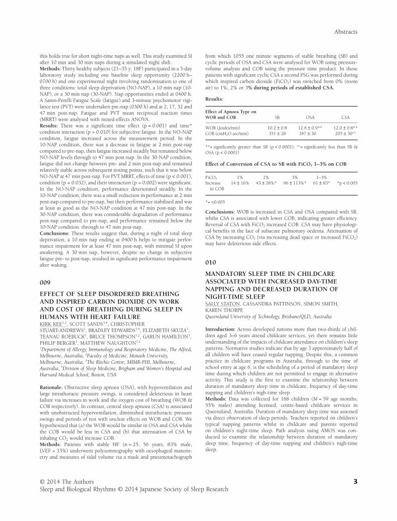
Rationale: Obstructive sleep apnoea (OSA), with hypoventilation andlarge intrathoracic pressure swings, is considered deleterious in heartfailure via increases in work and the oxygen cost of breathing (WOB &COB respectively). In contrast, central sleep apnoea (CSA) is associatedwith unobstructed hyperventilation, diminished intrathoracic pressureswings and periods of rest with unclear effects on WOB and COB. Wehypothesized that (a) the WOB would be similar in OSA and CSA whilstthe COB would be less in CSA and (b) that attenuation of CSA byinhaling CO2 would increase COB.Methods: Patients with stable HF (n = 25, 56 years, 83% male,LVEF = 33%) underwent polysomnography with oesophageal manom-etry and measures of tidal volume via a mask and pneumotachograph
from which 1055 one minute segments of stable breathing (SB) andcyclic periods of OSA and CSA were analysed for WOB using pressure-volume analysis and COB using the pressure time product. In thosepatients with significant cyclic CSA a second PSG was performed duringwhich inspired carbon dioxide (FiCO2) was switched from 0% (roomair) to 1%, 2% or 3% during periods of established CSA.
Results:
Effect of Apnoea Type onWOB and COB SB OSA CSA
WOB (Joules/min) 10.2 ± 0.8 12.6 ± 0.9** 12.0 ± 0.8**COB (cmH2O.sec/min) 351 ± 28 347 ± 30 205 ± 30∧∧
**= significantly greater than SB (p < 0.0001); ∧∧= significantly less than SB &OSA (p < 0.0001)
Effect of Conversion of CSA to SB with FiCO2 1–3% on COB
FiCO2 1% 2% 3% 1–3%Increase
in COB14 ± 16% 43 ± 28%* 96 ± 113%* 61 ± 83* *p < 0.005
*= <0.005
Conclusions: WOB is increased in CSA and OSA compared with SB,whilst CSA is associated with lower COB, indicating greater efficiency.Reversal of CSA with FiCO2 increased COB. CSA may have physiologi-cal benefits in the face of subacute pulmonary oedema. Attenuation ofCSA by increasing CO2 (via increasing dead space or increased FiCO2)may have deleterious side effects.
010
MANDATORY SLEEP TIME IN CHILDCAREASSOCIATED WITH INCREASED DAY-TIMENAPPING AND DECREASED DURATION OFNIGHT-TIME SLEEPSALLY STATON, CASSANDRA PATTINSON, SIMON SMITH,KAREN THORPEQueensland University of Technology, Brisbane/QLD, Australia
Introduction: Across developed nations more than two-thirds of chil-dren aged 3–6 years attend childcare services, yet there remains littleunderstanding of the impacts of childcare attendance on children’s sleeppatterns. Normative studies indicate that by age 3 approximately half ofall children will have ceased regular napping. Despite this, a commonpractice in childcare programs in Australia, through to the time ofschool entry at age 6, is the scheduling of a period of mandatory sleeptime during which children are not permitted to engage in alternativeactivity. This study is the first to examine the relationship betweenduration of mandatory sleep time in childcare, frequency of day-timenapping and children’s nigh-time sleep.Methods: Data was collected for 168 children (M = 59 age months;55% males) attending licensed, centre-based childcare services inQueensland, Australia. Duration of mandatory sleep time was assessedvia direct observation of sleep periods. Teachers reported on children’stypical napping patterns whilst in childcare and parents reportedon children’s night-time sleep. Path analysis using AMOS was con-ducted to examine the relationship between duration of mandatorysleep time, frequency of day-time napping and children’s nigh-timesleep.
Abstracts
3© 2014 The AuthorsSleep and Biological Rhythms © 2014 Japanese Society of Sleep Research
Results: The average duration of mandatory sleep time in childcare was56 minutes (range = 0–145 minutes). Path analyses showed a significantindirect path between mandatory sleep time and duration of night-timesleep, through increased napping in childcare (p = .001). Childrenexposed to longer duration of mandatory sleep time in childcare hadsignificantly shorter duration of night-time sleep and this relationshipwas mediated by increased frequency of napping in childcare settings.This relationship stayed significant after adjusting for potential con-founding variables; age, gender, family income, parental education,childcare quality, days/week of attendance and service type.Discussion: This study is the first to show a relationship betweenobserved duration of mandatory sleep time in childcare settings andchildren’s duration of night-time sleep. These effects are important asreduced night-time sleep in children has been found to be a significantrisk factor for reduced academic performance, behaviour difficulties andpoorer physical health. Implications for future research and sleep prac-tice in childcare are discussed.
012
THE PHARYNGEAL HYDROSTAT MODEL OF THEUPPER AIRWAYKRISTINA KAIRAITIS1,2
1Ludwig Engel Centre for Respiratory Research, Westmead MillenniumInstitute, Westmead, Australia, 2University of Sydney at WestmeadHospital, Westmead, Australia
The pharynx is a complex muscular structure with little bony supportthat performs the competing function of deglutition and respiration.The pharynx is commonly modelled as a collapsible tube enclosed in achamber surrounded by a single pressure, or Starling resistor. Thismodel, although useful, ignores the complexities of pharyngeal anatomyand behaviour. An alternative model of pharyngeal behaviour is toconsider upper airway luminal size and shape as being determined bythe distribution of tissues around the pharyngeal lumen. Peri-pharyngeal tissue distribution is in turn a consequence of the distribu-tion of pressures in the tissues surrounding the pharyngeal airway,or upper airway extraluminal tissue pressure (ETP), which alterstransmural (intraluminal-extraluminal) pressure surrounding theairway. This principle underlies the ability of a number of other mus-cular organs such as the tongue, elephant trunk, and octopus tentaclesto achieve movement and shape change in the absence of bony support.Previously, measuring ETP laterally and anteriorly in the sub-mucosaltissues of an anaesthetised rabbit model, we have demonstrated thatETP fluctuates with respiration, is non-uniformly distributed and het-erogeneously altered by neuro-mechanical influences such as mandibu-lar advancement, tracheal traction, lung volume, head and neck positionand upper airway dilator muscle contraction. More recently, measuringETP at 6 sites whilst simultaneously measuring upper airway lumengeometry via CT imaging, we have further demonstrated that ETP isnon-uniformly distributed axially and longitudinally, and that, inresponse to graded caudal tracheal displacement ETP decreases non-uniformly both along and around the pharyngeal airway. Graded caudaltracheal displacement also resulted in non-uniform changes in upperairway geometry. These findings further support the model of thepharynx as a muscular hydrostat. The applications of this alternativemodel are relevant in understanding how therapies such as ahypoglossal neural stimulation or mandibular advancement may workas a therapy for sleep apnoea.
Supported by NHMRC Health Professional Fellowship 1013234
013
DYNAMIC UPPER AIRWAY PATENCY: THEINFLUENCE OF OSA, BMI AND POSTURELYNNE BILSTONNeuroscience Research Australia, Randwick, NSW, Australia
Introduction: Maintaining upper airway patency when supine relies ona delicate balance between airway pressure and forces in the airwaywalls. Posture, obesity and obstructive sleep apnoea (OSA) can influ-ence upper airway patency. Active contraction of airway dilator musclescan help to oppose negative pressure in the airway during breathing, butthe relationship between activity of dilator muscles and airway dilationis not well understood.Methods: Dynamic tagged MRI was used to track the motion of theupper airway musculature during breathing while supine. Healthy sub-jects across a broad range of ages and BMI, and OSA patients werestudied. The influence of head and jaw posture was also investigated.Results: In healthy subjects, active airway dilation occurs during inspi-ration. Healthy, but overweight or obese subjects whose airways weresmaller in cross section had larger active dilations of the airway than leansubjects. In contrast, OSA patients tended either not to dilate the airwaysignificantly, or to have paradoxical motion, where one part of theairway dilates, but another narrows. In healthy subjects, jaw openingtended to increase the degree of airway dilation.Conclusions: Healthy subjects with narrow airways can overcome theirincreased risk of airway collapse by actively dilating the airway duringinspiration, but this mechanism appears to fail in OSA patients.
014
FACTORS PERPETUATING UPPER AIRWAYOBSTRUCTIONPETER CATCHESIDE1,2
1Adelaide Institute for Sleep Health, Daw Park, SA, Australia, 2FlindersUniversity, Bedford Park, SA, Australia
Obstructive sleep apnoea (OSA) was traditionally viewed as an upperairway anatomical problem inherited or acquired via obesity and/oraging effects causing upper airway obstruction during sleep whenmuscle tone is diminished. However, this view is too simplistic andignores major respiratory and neuromuscular control, and arousal influ-ences on upper airway function. The traditional view also fails to explainseveral known features of OSA, such as normal airway collapsibility insome patients, treatment emergent central sleep apnoea indicatingunstable respiratory control, and profound sleep stage effects on OSAseverity supporting that arousal factors are also important. Conse-quently, frequent airway obstruction in sleep is now understood toreflect a combination of factors including increased airway collapsibility,ineffective airway muscles/compensatory recruitment, unstable respira-tory control and/or exaggerated arousal responses, with variable deficitsbetween patients contributing to different OSA “phenotypes”, and sleepstage effects further modulating OSA severity. By better understandingand tailoring treatments to specific deficits in individual patients itshould be possible to improve long-term treatment outcomes beyondthose of constant positive airway pressure as first-line treatment, whichis well known to fail in many patients due to poor treatment toleranceand adherence.
Airway obstruction likely arises when neural drive to upper airwaystiffening and dilating muscles becomes insufficient to prevent airwaycollapse, which is then difficult to recover through respiratory andupper airway muscle compensatory responses without also initiatingarousal, brief hyperventilation and recurring obstruction. In patients
Sleep, Science and Research
4 © 2014 The AuthorsSleep and Biological Rhythms © 2014 Japanese Society of Sleep Research
with unfavourable airway anatomy and easily collapsed airways anabrupt loss of muscle tone at sleep onset is likely to be the main factorprecipitating obstruction. However, non-anatomical factors includingrespiratory compensation and arousal propensity/responses and theirinteractions must also play a role in perpetuating cyclical upper airwayobstruction. In at least some patients, and potentially in many, non-anatomical factors could play a more important role than abnormalanatomy in perpetuating cyclical obstruction during sleep.
018
ACTIGRAPHY FOR OVERNIGHT SLEEP ANDDAYTIME NAPPING IN INFANTS ANDPRE-SCHOOL AGED CHILDRENBARBARA GALLAND, KIM MEREDITH-JONESUniversity of Otago, Dunedin, New Zealand
The actigraphy field lacks established standards for paediatric sleepmeasurement. Whilst this also applies to the adult field, paediatric sleeppresents unique challenges, particularly in the very young with oftenfragmented overnight sleep, and several sleep periods over 24-hours.Although many PSG validation studies have been conducted for over-night sleep across the paediatric age range, citing good sensitivity (sleepagreement) but poorer specificity (wake agreement), few consider24-hour validation. The requirement for wired and connected EEGlimits 24-hour validation, but developments in EEG metrics withinwireless sensing headsets may advance this area. Video data, at leastcould be used for 24-hour validation with headgear and fixed videocamera systems. Accurate identification of daytime naps is difficult as noguidelines exist for daytime naps and thus non-validated night-timerules have been used. Automatic scoring accepting a minimum of 30minutes of sleep to calculate sleep-wake summary parameters precludesnaps less than 30 minutes from being identified. Without accurate sleepdiaries, non-wear time during the day can be misinterpreted as naps, ascan events such as riding in the car with little movement. Parentaladherence to complete 24-hour sleep-wake diaries or logs of accuratesleep onset and offset times is made difficult by the requirement for thisto continue over several days. Data will be presented on the use of thecount-scaled algorithm for identifying naps from 24-hour actigraphydata collected in infants aged 6, 12, 24 and 42 months. The algorithmis integrated into a MATLAB™ script programmed to detect sleep onset(night time sleep) and sleep offset (morning wake) and all sleep andwake epochs over 24 hours as computed and compared to a sleep–wakethreshold. Although validation with PSG is still required, agreementswith parental diaries in respect of nap timing and nap duration will bepresented, including the value of applying nap-screening rules to thedata.
019
THE VALIDITY OF ACTIGRAPHY TO MEASURESLEEP IN CHILDREN AND ADOLESCENTSMICHELLE SHORTUniversity of South Australia, Adelaide, Australia
Activity monitors are portable devices that use continuously recordedmovement data to infer sleep and wake. They have the advantage ofbeing lightweight, portable and non-invasive. Actigraphy provides anobjective estimate of sleep and wake without the time and cost associ-ated with the gold standard measure of sleep, polysomnography. Inaddition, multiple nights of sleep can be measured within the individ-uals’ home environment and later downloaded for analysis, making
these devices particularly useful for estimating the sleep patterns ofchildren and adolescents in their normative environment over longerperiods of time.
Despite these many advantages, actigraphy is not without limitations.Scoring algorithms may need to be refined to better account for norma-tive developmental changes to sleep motor activity across childhood andadolescence. Specifically, there may be important developmentalchanges to sleep that affect the validity of actigraphically-estimated sleepand wake in this age group. Current data from children and adolescentsindicate that actigraphic scoring algorithms tend to over-estimate wakeafter sleep onset and correspondingly under-estimate sleep. Moreworryingly, there may be significant inter-individual variance in thedegree to which actigraphic estimates of wake and sleep correspond toactual wake and sleep, with the degree of correspondence varyingdepending on characteristics such as age, sex and pubertal status.
Supported by University of South Australia Divisional Research Perfor-mance Fund and ARC grant # DP0881261
021
ORAL APPLIANCES – PAST, PRESENT ANDTHE FUTUREMARIE MARKLUNDInstitution of Odontology, Umea, Sweden
Oral appliances (OAs) for the treatment of obstructive sleep apnea(OSA) and snoring were introduced 30 years ago after ideas fromorthodontic treatment methods. This was just shortly after the inventionof continuous positive airway pressure (CPAP). Today, OA therapy isused in many countries worldwide.
This presentation shortly overviews the development of OA therapyfrom start until today’s popular treatment method based on results frommany randomized controlled studies. There is strong evidence for treat-ment effects on sleep apneas from OAs. In addition, recent studiesindicate that there are clear health benefits on the negative medicalconsequences of untreated OSA such as increased blood pressure.Symptoms are reduced and quality of life is increased from OAs, butthese effects are more unclear. This presentation will include new find-ings from a randomized controlled trial about the symptomatic effectsfrom OAs in patients with primary indications for this treatment(AHI < 30). OAs have been found effective in the longer term in patientswho continue treatment, while a certain proportion of the patients willdiscontinue treatment because of side effects and reduced efficacy.Future aspects on the long term effectiveness of OAs will be considered.
In conclusion, OA has developed from an orthodontic device into anattractive method for patients with snoring and sleep apnea. OA is nowthe main alternative to CPAP. More knowledge is, however, neededabout the overall health benefits from OA treatment.
022
ORAL APPLIANCE THERAPY FOR SLEEPDISORDERED BREATHING IN CHILDRENJOACHIM NGIAMHornsby Dental Clinic, Hornsby, Australia
Children with sleep disordered breathing (SDB) manifest a spectrum ofabnormal breathing ranging from simple snoring, upper airway resist-ance syndrome to obstructive sleep apnea syndrome (OSAS). SDB canaffect a child’s growth and craniofacial development leading to neuro-cognitive deficits and cardiovascular sequelae. In infants, symptomsmay include noisy and obstructed breathing whereas in pre-school aged
Abstracts
5© 2014 The AuthorsSleep and Biological Rhythms © 2014 Japanese Society of Sleep Research
children, snoring and mouth breathing may be observed. School-agedchildren with SDB may exhibit behavioural problems, orthodontic andcraniofacial abnormalities. A narrow upper airway with maxillary con-striction and retrusion and/or mandibular retrusion is oftenphenotypical of paediatric OSAS. Specific craniofacial morphologicalfeatures may include a hyperdivergent growth pattern with increasedcranio-mandibular, intermaxillary and mandibular plane angles withincreased lower anterior facial heights.
In recent times there has been an increase in studies evaluating theuse of oral appliances (OA) and rapid maxillary expanders (RME) forpaediatric SDB. Moreover, OAs including jaw repositioning appliances,functional orthopaedic appliances and RMEs have been proposed astreatment alternatives in the management of paediatric SDB. RMEis a dento-facial orthopaedic procedure routinely used to open themidpalatal suture. Although mainly used to correct dental and skeletaldiscrepancies, concomitant benefits include increases in nasopharyngealairway dimensions and improvement in nasal respiration. Some studiesalso document improvements in tongue posture, mode of respirationand the apnoea hypopnea index in children with obstructive sleepapnoea (OSA).
OA therapy and RME are emerging as valid treatment alternatives forpaediatric OSA. A dental and orthodontic assessment should be encour-aged in children with SDB. Timely dental and orthodontic interventionmay promote nasal respiration and prevent obstruction of the upperairway, potentially changing the natural history of paediatric SDB.
024
SCORING OXYGEN DESATURATION EVENTS:DO WE HAVE A STANDARD ANDRELIABLE METHODOLOGY?THOMAS J CHURCHWARD1Department of Respiratory and Sleep Medicine, Austin Hospital,Heidelberg, Australia, 2Institute for Breathing and Sleep, Heidelberg,Australia
Oxygen desaturation, as observed during polysomnography, is associ-ated with adverse outcomes and is a significant element of conventionaldiagnostic criteria. Starting with a brief reflection on the basic physiol-ogy, this talk will then shed light on the use of oximetry to measureoxygenation and subsequently why it is important to detect, describeand quantify hypoxia. Examples of desaturation patterns seen in aclinical setting will then be used to highlight the strengths and weak-nesses of current methodology and suggest tactics to further improvethis methodology.
025
TRANSCUTANEOUS CARBON DIOXIDEMONITORING IN THE SLEEP LABNICOLE C VERGINIS1Melbourne Children’s Sleep Centre, Melbourne, Australia, 2MonashHealth, Melbourne, Australia
The methodology and underlying physiology of transcutaneous carbondioxide monitoring will be discussed briefly. Current recommendationsfor the use of carbon dioxide monitoring during polysomnography inadult and paediatric patients will be reviewed. Examples of clinical casesdemonstrating the utility of this monitoring for patients with variousclinical conditions and in various clinical scenarios will be presented.Limitations of the technology including common problems with its useand some tips for troubleshooting will also be discussed.
026
RESPIRATORY MEASURES: OH THE STRAIN,THE EFFORT!ANGELA CAMPBELLUniversity of Otago, Wellington, New Zealand
Obtaining an accurate measure of respiratory effort during sleep is keyto good diagnostic outcomes for any suspected sleep related breathingdisorder.
While the AASM have recommended technology requirements for themeasurement of respiratory effort during sleep, the type of signal thatcan be measured and available technologies are plentiful. This talk willfocus on pros and cons of different respiratory effort measurementtechnologies using specific cases to illustrate the points and discusspossible future devices/technologies which may be available to us.
027
SLEEP MEDICINE AND THE LAW:INTRODUCTORY OVERVIEWPETER BUCHANANWoolcock Institute of Medical Research, Glebe, Australia
This symposium intends to explore the nexus between the specialitypractice of sleep medicine and the law in the particular areas thatinvolve automatomistic behaviours encountered in sleep medicine.
Automatistic behaviours range from the simplistic (e.g. an elicited deeptendon reflex) to the complex and complicated, such as exemplified bythe parasomnia state of sleepwalking. Automatistic behaviour has a par-ticular definition in law, and there are forensic situations where themedical interpretation of such behaviours and the legally acceptedunderstanding of automatistic behaviour must face off, and hopefullyreconcile, in the pursuit and delivery of justice.
Legal aspects of the definition and application of automatism will bepresented and discussed by our invited legal contributor, Mrs HayleyCormann, Senior Associate at the legal firm of Clayton Utz Perth.
Sleep physicians and other health care providers are frequentlyinvolved in the process of advising authorities regarding medical aspectsof the licencing of drivers, both private and commercial, when a sleepdisorder is or may be suspected to be relevant to safe driving. Dr MarkHoward will review the Australian context of assessing fitness to drivefrom the sleep medicine perspective. In extreme cases of “sleepy driving”causing harm including death, automatistic behaviour may be invoked asboth providing some understanding of such egregious behaviour andalso as a potential legal defence against criminal charge.
Violent behaviours can occur during and/or arising from sleep, ormay be alleged to so arise, in forensic cases involving a range of suchviolent behaviours and including violent assaults, sexual assaults andrape, and homicide. Sleep medicine experts may be drawn into forensiclegal cases that can result from such violent behaviours and be asked bycourts to provide expert legal reports and opinions and present thoseunder legal examination in courts, to aid in the process of justicedelivery. Dr Dev Banerjee is such a sleep medicine expert, who has hadextensive experience in the UK providing reports and opinions inmatters of this kind, and will be distilling some of his experience andinsights with us today.
Today’s speakers will combine in a panel discussion to address ques-tions from the floor and present further case examples.
Sleep, Science and Research
6 © 2014 The AuthorsSleep and Biological Rhythms © 2014 Japanese Society of Sleep Research
028
AUTOMATISM IN LAW: WHAT IT IS AND HOWIT RELATES TO SLEEP DISORDERS ANDCRIMINAL ACTSHAYLEY CORMANNClayton Utz, Perth, Australia
The concept of “automatism” is increasingly used in the legal context, todeny the actions of a person alleged of criminal behaviours as being‘voluntary’, in an attempt to escape culpability. Automatism is not,however, necessarily best categorised as a “defence” to those behaviours.Instead, it can perhaps be better described as an assertion that theoffence cannot be established because there is no “actus reus”, being theexistence of the ‘action or conduct’ necessary to constitute the wronghaving occurred. That is, the current model of criminal responsibilityrequires that a serious offence be constituted by both a physical and afault element, in the absence of a defence. Put in other words, in theAustralian legal context, before the physical act, (or, the actus reus), canbe described as an ‘act’, there must be some operation of the will. So,does it follow that the physical movements of a person who is asleepshould not be regarded as ‘acts’ at all and, more particularly, are not actsfor the purpose of assigning criminal responsibility? In law, the answerwill often be ‘yes’, and the successful operation of automatism in a legalcontext will be to deny the accused is criminally responsible for hisactions. So, how does this concept affect sleep medicine practice andhow is it currently applied? In Australia, legal authority suggests manydrivers may escape blame in cases of motor-vehicle accidents causingfatalities, because the view adopted in the High Court has been toconclude that where sleep is not voluntary, people will not be culpablefor acts caused while asleep. In this view, perhaps controversially andpuzzling to many, an accident caused by a sleeping driver does not meanthe driver had sufficient warning to stop driving. Arguably, the courtdoes qualify the application of automatism in this context, by requiringthe decision-maker to look at the period immediately before the personfell asleep and, not only must it be sufficiently contemporaneous withthe time of impact, the driving during that period must, in a practicalsense, be the cause of impact and of death. How does this sit withcurrent medical-scientific concepts of sleep and consciousness? Is ourlegal system on the wrong track? And how does this principle apply inthe context of a person accused of other serious criminal offences ofsexual assault or even murder?
These issues will form the basis for topical discussion and presenta-tion on 9 October 2014.
030
A CASE-BASED REVIEW OF SLEEP-RELATEDCRIMINAL BEHAVIOURS, TREATMENT& OUTCOMESDEV BANERJEE1Woolcock Institute of Medical Research, Glebe, Australia, 2University ofSydney, Camperdown, Australia
Parasomnia- regarded as undesirable physical & behavioural phenom-ena occurring during sleep, but the clinical features of sleepwalkingremain varied & can range from behaviour typical of either night terrors,confusional arousals as well as purely walking around the bedroom andthe house. It has been recognised that some patients may have a com-bination of sleepwalking & night terrors. Those who sleepwalk pre-dominantly may bolt upright and run, some may talk or shout at thesame time. Most of the actions are simple, crude even. The behaviours
that are carried out by a sleepwalker is regarded as not in keeping withsophisticated cognitive behaviours such as laying down memory, havingintent, planning & social interaction. Violent behaviour directed againstanother individual appears to follow direct provocation by or closeproximity to another individual. Sleepwalkers generally do not seek outtheir victims but rather victims sought out or were encountered by thesleepwalker. Certainly this is an area of much legal debate & there havebeen cases of acquittal on the basis of sleepwalking being the legaldefence. Men are more likely to commits serious violent acts and drugand alcohol abuse is not uncommon. There is a growing awareness ofabnormal sexual behaviour emerging during sleep and as such, termssuch as “sleepsex” or “sexsomnia” have been coined to describe theseactivities. There have been case reports of sleepwalkers convicted forindecent exposure. Behaviours specific to parasomnias include sexualvocalisation/ talking / shouting, masturbation, fondling another person,sexual intercourse with or without orgasm, agitated or assaultive sexualbehaviour. Fondling and sexual intercourse are the most common activ-ities and memory recall is absent. It was rare to exhibit “sexsomnia”without a history of other parasomnias. There are very few cases ofreported sleep related sexual activity during PSG in the literature.Medico-legal experts need to determine the predisposition, priming andtriggers factors in legal cases. It is now recognised that sleep specialistsare being asked more often to provide opinions and judgements on legalcases of possible sleepwalking related violence / sexual offences defenceand are based on a case of automatism. How exactly to approachsuch as legal case in the sleep lab continues to be debated in the sleepmedical field.
031
HYPOPNOEAS: PRECIPITATING ANDTERMINATING FACTORSPETER CATCHESIDE1,2
1Adelaide Institute for Sleep Health, Daw Park, SA, Australia, 2FlindersUniversity, Bedford Park, SA, Australia
Hypopnoeas are a major feature of obstructive sleep apnoea and accountfor the majority of events within the apnoea hypopnea index, andoccasional respiratory events occurring during sleep in healthy individ-uals on most nights. Most hypopnoeas reflect Starling resistor-likebehaviour of the upper airway, where partial collapse dynamically limitsor “chokes” airflow irrespective of downstream inspiratory pressure,sometimes with “negative effort dependence” where airflow worsensdespite augmenting inspiratory effort. Hypopneas can also reflect tran-sient periods of low ventilatory drive following hyperventilation fromarousal and/or unstable breathing control without clear obstruction.However, most hypopnoeas are obstructive events precipitated whenairway stiffening and dilator muscle activity becomes insufficient tooppose airway collapsing forces from unfavourable airway complianceand geometry, intraluminal pressure, and gravitational effects on tissuesaround the airway. The two main factors initiating hypopnoeas in OSAare an abnormally narrowed and collapsible airway, combined with anabrupt loss of upper airway muscle tone at sleep onset and the return tosleep post-arousal. Once initiated impeded breathing and blood gasdisturbances progressively stimulate mechano- and chemo-reflexes toaugment inspiratory effort and upper airway muscle activity. However,flow limitation is inherently difficult to reverse and these mechanismsare typically ineffective in terminating obstructed airflow withoutelevated breathing effort also triggering arousal. Arousal produces briefbut intense cardio-respiratory reflex and upper airway muscle activationthat facilitates airway re-opening and rapid recovery of blood gas dis-turbances via hyperventilation. In healthy individuals this appears toprovide a very efficient mechanism for rapidly reversing flow-limited
Abstracts
7© 2014 The AuthorsSleep and Biological Rhythms © 2014 Japanese Society of Sleep Research
and highly inefficient breathing that would otherwise be sustainedwithout arousal. However, in OSA patients low breathing effort whensleep resumes may exacerbate the propensity for airway collapse andongoing cyclical obstruction and arousal. Airflow limitation duringhypopnoeas also underpins snoring, which can be prolonged beyondconventionally scored hypopneas, annoy others and cause prolongedperiods of high breathing effort that may interfere with sleep and con-tribute to adverse cardiovascular outcomes.
032
ARE WORSE OUTCOMES ASSOCIATED WITHAPNOEAS VERSUS HYPOPNOEAS, GREATERDEGREE OF DESATURATION, HYPOPNOEASWITH AROUSAL?CRAIG PHILLIPSWoolcock Institute of Medical Research, Glebe, Australia
Numerous cross-sectional and longitudinal studies have established astrong association between Obstructive Sleep Apnoea (OSA) and poorcardiovascular and neurocognitive outcomes. Randomised trials suggestthat the association between OSA and these adverse outcomes is causalwith physiological disturbances including intermittent hypoxia andsleep fragmentation secondary to arousals as key upstream mechanisms.
The severity of Obstructive Sleep Apnoea (OSA) is measured usingthe apnoea hypopnoea index (AHI), a metric that represents the totalnumber of apnoeas and hypopnoeas per hour of sleep. Over the pastone to two decades, the field of sleep medicine has witnessed a continualredefining of the AHI. This is predominantly due to an ongoing debateabout the definition of the hypopnoea. This has in turn resulted in a lackof consistency between studies in what constitutes mild, moderate orsevere OSA and how OSA severity then impacts on adverse healthoutcomes. Today’s discussion will highlight the studies that haveattempted to ascertain the relative contribution of apnoeas versushypopnoeas and hypoxia versus arousal to poor health outcomes.
033
UPPER AIRWAY OBSTRUCTION IN CHILDREN –HOW DOES IT DIFFER FROM ADULTS?YVONNE PAMULAAdelaide Women’s & Children’s Hospital, South Australia, Australia
Sleep-related upper airway obstruction (UAO) is a common disorder ofchildhood and is described as a spectrum of severity ranging fromprimary snoring at the mild end to obstructive sleep apnoea syndromeat the severe end. The presentation, pathophysiology and clinical fea-tures of paediatric OSAS is different to that observed in adults. Inparticular the pattern of obstruction as documented by overnightpolysomnography (PSG) has shown some interesting differencesbetween adults and children. For example, in children prolongedperiods of partial upper airway obstruction are more commonlyobserved than episodes of frank obstructive apnoea, with different pat-terns of oxygen desaturation and arousal between adults and children.Furthermore unlike adults, obstructive events in children are oftendifferentially distributed in REM sleep and overall sleep architecture isgenerally preserved. A more patent upper airway during sleep andhigher arousal thresholds in children, and impaired ventilatory drive,the presence of co-morbidities and longer disease duration in adults, arepotential explanations for these differences. While hypopnoeic breath-ing in children may appear to be a milder phenotype compared to
adults, significant clinical sequela are still observed, although these donot correlate strongly with standard PSG measures. It is likely thatcurrent PSG technology does not sufficiently describe or measure thespectrum of paediatric upper airway obstruction.
035
LIMBS THAT GO TWITCH IN THE NIGHTDAVID BERLOWITZ1,2
1Institute for Breathing and Sleep. Austin Health, Melbourne, Vic,Australia, 2University of Melbourne, Melbourne, Vic, Australia
Spinal cord injury (SCI) is a catastrophic disability that typically resultsin severe and permanent disability. Alongside the obvious primarydamage, SCI patients also suffer from a high rate of secondary compli-cations and disorders, including a high prevalence of periodic leg move-ments (PLMs) during sleep (De Mello, 2004). The prevalence of PLMsduring sleep is reportedly 6% for the able-bodied population (Esteves etal, 2004) yet as high as 75% in people with SCI (Tellers et al, 2011).
While the exact origin and pathophysiology of PLMs in the able-bodied remains unclear, subcortical regions in the substantia nigra, thebrainstem, thalamus, red nucleus, and cerebellum have all been impli-cated in PLMs pathogenesis. Further, it is speculated that PLMs manifestnot only because of abnormalities in these regions but also becauseassociated suppression of descending inhibitory (pyramidal) pathwaysallow for expression of the movement disorder. It is this second mecha-nism, a failure of descending inhibition, which is strongly implicated inSCI. In the able-bodied, PLMs display varying degrees of sleep-statedependency but typically PLMs are not observed in REM sleep. Peoplewith SCI and PLMs typically display the same degree and pattern of themovements throughout REM and occasionally even during wakefulness.
Despite these differences in aetiologies, it appears that PLMs in SCImay be treated with similar agents as are used in the able-bodied.Exercise, dopaminergic and opioid agents all appear to be efficaciousalthough the data in SCI are not conclusive. While exercise has beendemonstrated to have some efficacy in SCI, there are obvious limitationson the ability to exercise the limbs attributable to the SCI per se,especially in those with higher lesions. Functional electrical stimulation(FES) is a way of externally activating skeletal muscles to produce grossmotor movements such as cycling in people with SCI who wouldnormally not be able to cycle. Research is currently underway toexamine the role of FES cycling in the management of PLMs after SCI.
036
CIRCADIAN RHYTHM DISTURBANCES INSPINAL CORD INJURYJO SPONGInstitute for Breathing and Sleep, Heidelberg, Victoria, Australia
Injuries that result in tetraplegia are uncommon but devastating to theperson, their family and the community. In addition to the severephysical disability, complete tetraplegia has been found to result in theabolition of melatonin production, a hormone which plays a major rolein the timing of circadian rhythms like the sleep-wake cycle. The dailymelatonin rhythm is regulated by the suprachiasmatic nucleus (SCN,‘circadian clock’) which sends signals to the pineal gland via a circuitousroute through other hypothalamic and brain stem nuclei, the spinal cordand peripheral sympathetic neurons from the superior cervical ganglion(SCG). The fibres to the SCG are routed along with those of the auto-nomic nervous system and it is this anatomical connection which is cutin complete tetraplegia.
Sleep, Science and Research
8 © 2014 The AuthorsSleep and Biological Rhythms © 2014 Japanese Society of Sleep Research
Sleep disorders and impaired temperature regulation are extremelycommon complications in people with tetraplegia and it is highly likelythat these complications are related to the melatonin dysfunction. Peoplewith tetraplegia have been found to have a core temperature circadianphase which is an average of six hours shorter than the able-bodied, andno evening increase in melatonin. Further, the administration of exog-enous melatonin two hours prior to sleep has been found to increasesubjective sleep duration by 45 minutes in this population.
This session will present research evidence that melatonin, sleep andtemperature is disrupted in people with complete tetraplegia. It will alsopresent recent research findings of the effect of exogenous melatonin onobjective and subjective sleep in this group. The possibility of a phasedysynchrony between the SCN and melatonin in tetraplegia will also bediscussed.
039
CPAP TREATMENT IN GERIATRIC PATIENTSSONIA ANCOLI-ISRAELUniversity of California, San Diego, USA
It is well documented that obstructive sleep apnea (OSA) is well treatedwith PAP. There have been many studies on the effect of treating OSA onoutcomes other than apnea-related symptoms, such as heart failure,hypertension, diabetes, etc. However, few of these studies have beenconducted on older adults (defined as >65 years) or in frail elderly.Based on a 10-year PUBMED search, 74 abstracts were pulled on PAPtreatment of obstructive or central sleep apnea. All 74 papers werereviewed. Of these, 20 met the criteria of participants with a mean ageof 65 years and these 20 were reviewed and evaluated. The majority ofthe studies (n = 15) evaluated populations with medical co-morbiditiesmore prevalent in older adults, e.g., dementia, Parkinson’s disease,stroke, heart failure, and the main outcomes were the effect of PAPtreatment on either apnea or on some aspect of that co-morbidity. Theevidence levels of these studies ranges from high at 1++ (one study) tolow at 2- (11 publications) and only 10 were randomized controlledtrials; the rest were observational in nature. The Jadad score for 13publications were in the 0–3 suggesting a higher risk of bias with sevenscoring 4–5 suggesting a lower risk and a higher quality study. Samplesizes ranged from a handful to over 4000.
The majority of studies of PAP treatment in the elderly primarilyshowed improvement in variables of sleep apnea (e.g., AHI, daytimesleepiness) as well as consequences of sleep apnea includingcomorbidites and psychosocial. One could conclude based on thesestudies that PAP should be used routinely for the treatment of sleep apneain older persons and in frail elderly, particularly those with heart diseaseand/or stroke based on an expert consensus level. Additional randomizedcontrolled trials are need in patients with AD and PD as well as other frailelderly.
040
THE EFFECTS OF DENTURES ON SLEEP ANDBREATHING IN THE ELDERLYMARC BRAEMUniversity of Antwerp, Antwerp, Belgium
Introduction: Epidemiological data show that 18% of individuals over60 years old are edentulous. Also, activity of upper airway musclesduring sleep decreases with ageing thereby increasing the risk forobstructive sleep apnea (OSA) in the elderly.Mechanism of action (MOA): Edentulism might act by modifyinganatomy and function of the pharyngeal airway and of tongue and by
favouring inflammatory edema. Furthermore, a cephalometric studysuggested a reduction in the upper airway space when edentulouspatients removed their complete dentures.Effects of dentures: It is found that patients had worse apnea-hyponeaindex (AHI) and decreased antero-posterior oropharyngeal wall distancewhen examined without their dentures. From cephalometric analysis itseems that wearing dentures induces changes in the position of thetongue, of the jaw and of the pharyngeal airway space that can favourthe reduction of apnea episodes as is reported on in case reports. Recentpublications indicate that AHI was aggravated when patients slept withtheir dentures in place, especially mild OSA patients showed a consist-ent worsening of their AHI with dentures while no significant changeswere noted in moderate to severe OSA patients. Another study reportsthat wearing the dentures without any mandibular advancement did notsignificantly improve polysomnographic parameters as compared tobaseline without dentures. Furthermore, wearing complete dentureswhile sleeping has no effect on AHI sleep quality measured by PSQIscores or daytime sleepiness measured using the Epworth SleepinessScale (ESS), and sometimes an adverse effect on ESS. Results from 306patients showed statistical significant association between the risk forOSA and an inadequate vertical dimension (VD).Effects of Oral Appliances (OA): Once mandibular advancement(OAm) is applied using the dentures or using overdentures, uprightcephalometry showed an increase in the upper airway dimensions.Conclusions: One might expect a similar MOA on AHI by applyingmandibular advancement therapy in elderly wearing complete dentures.Obviously, retention will become a major issue so implant retainedmandibular advancement appliance could well be the treatment ofchoice for edentulous OSA patients.
041
TREATING GERIATRIC SLEEP APNOEA PATIENTSWITH ORAL APPLIANCESSUSANNE SCHWARTINGKiel, Germany
Objectives: Mandibular advancement devices are a therapy option formild to moderate obstructive sleep apnoea (OSA) and in severe OSAwith intolerance of continuous positive airway pressure (CPAP). Theiruse is recommended in international sleep medicine guidelines.Patients and Methods: Elderly patients often have a reduced number ofteeth and therefore wear partial or full dentures. Geriatric patientsadditionally can present disabilities (reduced visus, limited dexterity)compromising the use of oral appliances.Results: Geriatric OSA patients intolerant of CPAP can be effectivelytreated with mandibular advancement devices. Implant retained oralappliances and special device constructions can be a solution.Conclusions: Even in geriatric OSA patients with less teeth oredentulous the treatment with a mandibular advancement deviceshould be considered if they are not compliant with CPAP.
042
THE COMPLEXITIES OF DEFININGOPTIMAL SLEEPBARBARA GALLAND1, SARAH BLUNDEN2
1University of Otago, Dunedin, New Zealand, 2CQ University, Adelaide,SA, Australia
Experimental data in adults on the effects of both acute and cumulativepartial sleep deprivation consistently point out that sleep restriction has
Abstracts
9© 2014 The AuthorsSleep and Biological Rhythms © 2014 Japanese Society of Sleep Research
substantial negative effects on daytime sleepiness, motor and cognitiveperformance and mood, as well as on some metabolic, hormonal andimmunological variables. In children, prospective and cross-sectionalstudies and sleep restriction studies, similarly show that short sleepduration is associated with a wide range of negative physical, social,emotional and cognitive outcomes including poor concentration,impaired performance at work or school, increased risk of obesity andmetabolic syndrome, depression, suicide ideation and injuries. Further-more, there is evidence in a number of experimental studies in bothchildren and adolescents that cognitive and behavioural outcomesimprove if restricted sleep is restored or extended. Together, this evi-dence suggests that there is an optimal sleep need, which raises animportant question. What defines optimal sleep? Is it about sleepquality, sleep continuity, sleep architecture, sleep/wake rhythms, circa-dian preference? Or is it about the length and quantity of sleep? Ifoptimal sleep is a quantity, then is it constant? Does it differ betweenindividuals? What is the most important outcome? Is it task dependentand therefore does it show a dose repose relationship or is there a cost(less sleep)/ benefit (increased non-sleep leisure activity) ratio relation-ship? Or is sleep optional to a certain degree, and is this need for sleepdriven by the subjective notion of sleepiness rather than evidence ofperformance deficits? Could “optimal sleep” be therefore socially con-structed and culturally different? These and other questions will drivediscussion about the complexities of defining optimal sleep for bothadults and children.
045
HOW MUCH SLEEP DO WE NEED? INFORMINGTHIS QUESTION WITH ADULT ANDPAEDIATRIC DATASARAH BLUNDENAppleton Institute of Behavioural Science, Adelaide, Australia
How much sleep is needed for optimal health and performance? This isa question that remains unanswered. In most cases optimal sleep foroptimal performance on specific tasks has been estimated based onsleep extension and restriction studies but ofteh fails to include themyriad of factors that also impact on this interrelationship such as age,gender, race, culture, the task at hand, and an individual’s position inboth sleep–alert and morningness–eveningness continuums. This paperwill firstly discuss the concept of optimal sleep, how to define it, how toassess the theoretical and empirical factors impacting optimal sleep andhow these relate to research efforts.
Data will be presented from specific international sleep extension/restriction studies in adults and children undertaken by the presentersto demonstrate how changes to sleep duration can impact performanceon specific cognitive tasks and physiological function. Results of sys-tematic reviews and meta-analyses of these studies and others in bothadults and children will be presented. It is hoped that this symposiumwill inform our understanding of the relationship between performanceand sleep, in the hope that we may have a better understanding aboutthe importance of sleep quantity for optimal functioning and to under-stand if current sleep guidelines reflect this understanding.
046
GENDER DIFFERENCES IN UPPER AIRWAYPHYSIOLOGY AND COLLAPSIBILITYDANNY ECKERTNeuroscience Research Australia (NeuRA) and the University of New SouthWales, Randwick, Sydney, NSW, Australia
Obstructive sleep apnoea (OSA) is 2–3 times more common in menversus women. When matched for BMI, OSA is also more severe in menthan in women. Thus, there are clear differences in the manifestationand the mechanisms underpinning OSA between the sexes. Recentdetailed upper airway physiology studies have highlighted the hetero-geneity of OSA pathogenesis. In addition to anatomical factors that leadto a narrow or highly collapsible upper airway, there are several keynon-anatomical traits or phenotypes that importantly contribute to OSApathogenesis. Non-anatomical contributors include: ineffective upperairway dilator muscles during sleep, waking up too easily (a low res-piratory arousal threshold), and respiratory control instability (highloop gain).
Two studies have clearly shown that upper airway collapsibilityduring sleep (Pcrit), is increased in men versus women, even whengroups are matched for BMI. Men tend to have longer upper airways andincreased fat deposition around the airway which could contribute toincreased upper airway collapsibility. An early study showed increasedupper airway muscle activity in women versus men but subsequentstudies have shown no difference. While overall respiratory controlstability (loop gain) does not appear to differ between men and women,other respiratory control factors including differences in the apnoeathreshold and the ventilatory response to arousal may place men morevulnerable breathing instability during sleep. No published studies havesystematically examined the respiratory arousal threshold between thesexes. Data from our recent detailed phenotype study in which wemeasured key anatomical and non-anatomical traits contributing toOSA in a group of 75 individuals with and without OSA (Eckert et al,AJRCCM, 2013) are consistent with prior physiology studies in thisarea. Specifically, upper airway collapsibility is increased in men versuswomen by ∼3 cmH2O but the other non-anatomical traits, including therespiratory arousal threshold, are similar between men and women.
In summary, this presentation summarises what is known aboutdifferences in the predisposition to upper airway collapse between menand women. It will also highlight gaps in our understanding and oppor-tunities for future research.
Support: NHMRC
048
IS TEEN SLEEP GENDERED? THE INFLUENCEOF BIOLOGY, FAMILY ANDPSYCHOSOCIAL FACTORSMICHELLE SHORTUniversity of South Australia, Adelaide, Australia
During adolescence, multiple converging biological and psychosocialfactors impact upon sleep. Puberty-related changes to sleep homeostaticand circadian regulatory systems occur alongside psychosocial changes,such as increased academic pressure, more social activity, electronicmedia use and greater autonomy. These factors are permissive of phasedelay and insufficient sleep and contribute to the stereotypical adoles-cent sleep pattern of truncated sleep during the school week due to theneed to rise early for school, followed by a later timed and longer sleepon the weekend.
Sleep, Science and Research
10 © 2014 The AuthorsSleep and Biological Rhythms © 2014 Japanese Society of Sleep Research

While the characteristic sleep patterns of adolescents are well docu-mented, the limited work examining sex differences in adolescent sleephas yielded relatively few meaningful differences. However, while thesleep patterns of male and female adolescents may be similar, it ispremature to conclude that factors influencing sleep operate to the samedegree, or with the same time course, or with the same trajectory forboth males and females. Indeed, it is possible that while sleep patternsper se may not be significantly different, that the factors impacting onand predicting sleep patterns may differ. Data collected from Australianadolescents in both home-based and laboratory-based studies suggestthat profiles of risk and protective factors vary between male and femaleadolescents. Sex differences in risk taking, self-regulation of sleep pat-terns, parent-regulation of sleep patterns, pubertal timing, electronicmedia use, employment and sport participation will be discussed withreference to their impact on sleep. In addition, the possibility that theconsequences of insufficient sleep may be felt and expressed differentlybetween males and females will be examined.
Supported by University of South Australia Divisional Research Perfor-mance Fund and ARC grant # DP0881261
049
BODY FAT DISTRIBUTION – GENDER EFFECTSON SLEEP APNOEALAILA SIMPSONUniversity of Western Australia, Perth, Australia
Sex ratios from prevalence studies suggest that for every woman withOSA there are up to 3 men. When men and women are matched forbody weight, men have a higher AHI which is due to a greater propor-tion of apnoeic than hypopneic events. The sex hormones testosterone,oestrogen and progesterone are known to contribute to chemical controlof breathing. Testosterone is known to induce and exacerbate OSA.Oestrogen may be protective against the development of OSA. OSA ismore prevalent in post-menopausal women than pre-menopausalwomen and hormone replacement therapy attenuates the risk of OSA.Sex hormones are also implicated in fat distribution. Female sex resultsin of a more favourable pattern of distribution of excess fat. Specifically,women tend to distribute fat peripherally around the hips, buttocks,and thighs, whereas men tend to distribute excess fat more centrally onthe abdomen and neck. As a result, although women may have propor-tionally greater fat mass than men, they have less mechanical loading ontheir upper airway. This lecture will summarise research investigatinghow gender and body fat distribution relate to OSA.
050
HYPER-AROUSAL MODEL OF INSOMNIADAVID CUNNINGTONMelbourne Sleep Disorders Centre, Melbourne, Australia
There is increasing basic science and clinical data suggesting that formany people with insomnia, one of the main features is hyper-arousal.Recognising this is important as it directs the treatment approachtowards strategies targeted at reducing arousal. Most medications act bypromoting or enhancing the sleep centres via GABA agonist action, butin hyper-arousal, medications that reduce waking drive may be moreappropriate. Some components of psychology-based treatment dealbetter with hyper-arousal than others, and meta-cognitive techniquessuch as mindfulness have been shown to reduce arousal in other con-ditions such as anxiety, stress and pain, so may have a role in hyper-arousal in insomnia.
053
OCCURRENCE OF UNDIAGNOSEDOBSTRUCTIVE SLEEP APNEA AND INSOMNIAIN HYPERTENSIVE PRIMARY CAREPATIENTS – ASSOCIATION WITH SLEEPCOMPLAINTS, DEPRESSIVE SYMPTOMSAND SELF-RATED HEALTHANDERS BROSTRÖM1, MARTIN ULANDER2,3, OLASUNNERGREN3,4, EVA SVABORG2,3, PETER JOHANSSON5,6
1Department of Nursing Science, School of Health Sciences, JönköpingUniversity, Sweden., Jönköping, Sweden, 2Department of ClinicalNeurophysiology, Linköping University Hospital, Linköping, Sweden,Linköping, Sweden, 3Department of Clinical and Experimental Medicine,Division of Clinical Neurophysiology, Faculty of Health Sciences LinköpingUniversity, Linköping, Sweden., Linköping, Sweden, 4Ear-, Nose- andThroat clinic, Ryhov County Hospital, Jönköping, Sweden., Jönköping,Sweden, 5Department of Cardiology, Linköping University Hospital,S-58185 Linköping, Sweden., Linköping, Sweden, 6Department of Medicineand Health Sciences, Division of Cardiovascular Medicine, Faculty ofHealth Sciences Linköping University, Linköping, Sweden., Norrköping,Sweden
Objectives: To describe how prevalent undiagnosed obstructive sleepapnea (OSA) and insomnia is in hypertensive primary care patients, andexplore its association to clinical symptoms and self-rated health.Methods: Cross-sectional design including 394 hypertensive patients(52.5 % women, mean age 57.8 years, SD 6.7 years) who had noprevious clinical diagnose of OSA or insomnia. All patients were,however, on pharmacological treatment for hypertension. Data weregathered with clinical examinations, polygraphy and scales for OSAsymptoms (Berlin Sleep Apnea Questionnaire), daytime sleepiness(Epworth Sleepiness Scale) and insomnia (Minimal Insomnia SymptomsScale). The first question on SF-36 was used to asses self-rated health.Polygraphy recordings were scored according to AASM criteria. OSAand insomnia was defined as AHI ≥ 5 together with at least moderateinsomnia (Minimal Insomnia Symptoms Scale score ≥7).Results: Undiagnosed mild, moderate and severe OSA occurred among29%, 16% and 14% of the patients. The frequency of OSA with insom-nia was 32% in the whole sample. 39%, 29% and 20% of those withmild, moderate and severe OSA had OSA with insomnia. AHI, satura-tion time <90%, HDL cholesterol and total sleep time was higher(p < 0.001) among those with OSA and insomnia. However, sleep suf-ficiency index, depressive symptoms and excessive daytime sleepinessdid not differ. Medication with angiotensin converting enzyme inhibi-tors (p < 0.05) and hypnotics (p < 0.01) was more common amongpatients with OSA and insomnia. Linear regression showed that OSAwith insomnia (p < 0.05), depressive symptoms (p < 0.001) and obesity(p < 0.001) were significantly associated with self-rated health. Addinggender, diabetes and diagnose of ischemic heart disease to the model didnot change the results.Conclusion: The occurrence of undiagnosed OSA was high amonghypertensive primary care patients. Insomnia was frequently reportedby patients of different severity levels, even those with mild and mod-erate OSA, and does together with depressive symptoms and obesityhave a negative impact on self-rated health.
Abstracts
11© 2014 The AuthorsSleep and Biological Rhythms © 2014 Japanese Society of Sleep Research
054
THE RELATIONSHIP BETWEENGASTROESOPHAGEAL REFLUX AND BODYFAT DISTRIBUTIONVIJEYADEZMI GANASAN1, KELLY SHEPHERD1,2,JAMES OCKELFORD1, HOOI EE3, RICHARD HOLLOWAY4,DAVID HILLMAN1,2, PETER EASTWOOD1,2
1The University of Western Australia, Perth, Western Australia,Australia, 2West Australian Sleep Disorders Research Institute, Perth,Western Australia, Australia, 3Sir Charles Gairdner Hospital, Perth,Western Australia, Australia, 4Royal Adelaide Hospital, Adelaide, SouthAustralia, Australia
Introduction: The increase in prevalence of obesity has been paralleledby an increase in the prevalence of gastroesophageal reflux (GOR)disease. GOR can occur during the day and night. Distribution of bodyfat appears to be an important factor in the severity of GOR, however itis unknown whether such an association is equally important fordaytime and nighttime GOR. This study investigated the relationshipbetween severity of GOR (during wake and sleep) and body composi-tion and fat distribution.Methods: Twenty-four subjects (14 male & 10 female, mean age43.5 ± 14.4 years) were recruited from the Department of Gastroenter-ology at Sir Charles Gairdner Hospital. Subjects underwent 24-hourpH/impedance monitoring to assess GOR, in-laboratory polysomno-graphy [PSG] to determine the sleep period, and DEXA scans to assessbody composition (% body fat, %BF) and fat distribution (abdominal fatpercentage, ABF%). BMI and waist-to-hip ratio [W : H] were also meas-ured. Reflux severity was measured using the total number of refluxevents (TRE), bolus exposure time (BET) and proximal reflux events(PRE). Data were analysed for both the group as a whole and separated bygender.Results: The average BMI of the subjects was 26.7 ± 5.3 kg.m2. Duringthe total study time (i.e. 24 hour monitoring, wake + sleep) PRE wassignificantly related to %ABF in all subjects (n = 20, r2 = 0.29,p = 0.01,), and in females (n = 8) with BMI (r2 = 0.82, p < 0.01), WHR(r2 = 0.66, p = 0.01), %BF (r2 = 0.88, p < 0.01) and %ABF (r2 = 0.85,p < 0.01)). During sleep BET was significantly related to %ABF in malesonly (n = 12, r2 = 0.38, p = 0.03) but tended to be related to %ABF in allsubjects (n = 20, r2 = 0.16, p = 0.08). During wakefulness there were nosignificant relationships noted between TRE, PRE or BET and anymeasure of body fat.Discussion: This study showed that severity of GOR, particularlyduring sleep, is related to both general and abdominal obesity and toabdominal obesity males. This study highlights the potential roles ofobesity and sleep in exacerbating GOR severity in men and women.
055
THE EFFECT OF OBSTRUCTIVE SLEEPAPNOEA ON SLEEP-RELATEDGASTROESOPHAGEAL REFLUXKELLY SHEPHERD, WILLIAM ORR1. Lynn Health Science Institute, Oklahoma City, Oklahoma, Australia
Introduction: Gastroesophageal reflux (GER) symptoms and events areextremely common in individuals with obstructive sleep apnoea (OSA).The role of obesity vs. upper airway obstruction in the high prevalenceof GER in OSA patients remains unclear. The present study investigatedthe additional risk of OSA on the presence and severity of GER in obeseindividuals.
Methods: Ten obese individuals with moderate to severe OSA (apnoea-hypopnea index 2: 15 events per hour) and 10 obese individualswithout OSA (apnoea-hypopnea index <5 events per hour) underwent24-hour oesophageal pH-impedance monitoring and polysomnography.Reflux events were classified as distal vs. proximal (migrating >15 cmabove the lower oesophageal sphincter) or acidic (pH < 4) vs. non-acidic. For analysis, the study was divided into a wake period, a sleepperiod (time after sleep onset) and the combined 24-hour period.Results: There were no significant differences in age or BMI between thetwo groups; however there was a significantly greater proportion ofmales in the obese with OSA group (100% vs. 40%). There were nodifferences between groups in any pH or impedance variable, includingacid contact time, during the sleep period. Over the 24-hour period,however, the number of GER events was significantly greater in the OSAgroup in the distal oesophagus (55 ± 21 vs. 38 ± 13, p = 0.05) and theproximal oesophagus (32 ± 17 vs. 17 ± 12, p = 0.03). Likewise theaverage duration of GER events was significantly longer in the OSAgroup (5 ± 6 vs.1 ± 0.5 min, p = 0.02) during the 24-hour period.Conclusion: There were no differences in GER measures betweengroups during the sleep period, arguing against a significant role of OSAin the occurrence of GER during sleep. GER during sleep in thesesubjects is most likely secondary to obesity. However, OSA was associ-ated with an increase in number and duration of GER events over a24-hour period.
056
DO CLINICAL SLEEP MEASURES PREDICTVULNERABILITY TO VIGILANCE IMPAIRMENTFOLLOWING SLEEP LOSS IN PATIENTSWITH OSA?HAIDER NAQVI1,4, HANNAH OPENSHAW1, ANDREW VAKULIN1,3,DAVID STEVENS1, ANGELA D’ROZARIO1,4, KEITH WONG1,4,DOUG MCEVOY2,3, RON GRUNSTEIN1,4
1Woolcock Institute of Medical Research and CIRUS, University of Sydney,Sydney, NSW, Australia, 2Adelaide Institute for Sleep Health, RepatriationGeneral Hospital, Adelaide, SA, Australia, 3School of Medicine, FlindersUniversity, Adelaide, SA, Australia, 4Department of Respiratory and SleepMedicine, RPAH, Sydney Local Health District, Sydney, NSW, Australia
Objectives: Clinical measures of obstructive sleep apnoea (OSA) relatepoorly to daytime vigilance performance. However, many OSA patientsare unimpaired during the day and may only show vigilance decrementsfollowing sleep loss. This study determined whether clinical OSA meas-ures explain vigilance impairment vulnerability in OSA patients exposedto sleep loss.Methods: Thirty OSA patients (Age 48.6 ± 9.4 yrs, BMI 32.4 ± 6.2 kg/m2, AHI 33.9 ± 24.5 events/hr), underwent polysomnography (PSG)followed by 28 hrs of extended wakefulness. Vigilance was assessedevery 2 hrs using 4 choice reaction time task (4CRT) with reaction speed(RS) and accuracy averaged for 3 daytime (1200–1800) and night-time(0000–0600) assessments. Absolute and percentage change from day tonight performance was used to quantify individual vigilance vulnerabil-ity to sleep loss. Bivariate correlations and linear regression were usedfor analysis.Results: RS and accuracy deteriorated from day to night (p < 0.001 andp = 0.035 respectively). No relationship was observed between sleepstudy measures and daytime 4CRT performance. Study variables (AHI,oxygen desaturation index, % time <90% SaO2, arousal index) nega-tively correlated with night-time RS, accuracy and percentage change inaccuracy (all p < 0.05). Linear regression models controlling for age,BMI and ESS showed AHI was a significant but not a strong predictor(r2 = 0.25) of day to night change in 4CRT accuracy.
Sleep, Science and Research
12 © 2014 The AuthorsSleep and Biological Rhythms © 2014 Japanese Society of Sleep Research
Conclusion: Sleep study metrics from baseline PSG, most notably AHI,predict subsequent night-time vigilance impairment in OSA. Howeverthe clinical significance of this relationship is limited. Additionalbiomarkers warrant further examination to better explain individualvigilance vulnerability to sleep loss in OSA patients.
057
25 YEARS OF DIAGNOSTIC AND CPAP SERVICESAT A LARGE METROPOLITAN SOUTHAUSTRALIAN HOSPITALEMILY VIIRET1, SONYA JOHNSTON1, RATOMIR ANTIC1,CHIEN-LI HOLMES-LIEW1,2, DIEN DANG1,2, AENEAS YEO1,2
1Royal Adelaide Hospital, Adelaide, SA, Australia, 2University of Adelaide,Adelaide, SA, Australia
Introduction: Our sleep disorders service has been providing a publicdiagnostic and treatment service for Sleep Apnoea since 1990 to bothmetropolitan Adelaide and several country centres. We investigatedsleep study referral and CPAP prescription epidemiological patterns atour centre.Aim: To assess sleep study referrals as well as CPAP prescription, com-pliance, return and mortality rates for our patients since commencementof the sleep disorders service and to assess trends in these parametersover time.Method: Sleep laboratory polysomnograms (PSG) between 1998 andApril 2014 were retrospectively analysed for PSG type, patient age, BMI,gender and Apnoea Hypopnea Index (AHI). A database of all patientscommenced on CPAP between 1990 to April 2014 was also analysed forage, sex, CPAP start date and return date. Mortality rate and average ageat death were determined by using the Births, Deaths and Marriagesregistry to cross-reference our database.Results: Of the 11481 patients who had sleep studies, 65% were obese,90% were overweight and 53% had moderate to severe OSA (AHI > 15).Mean age at referral was 52 years (median 53). There was no correlationbetween Epworth Sleepiness Scale and AHI. CPAP prescription progres-sively increased every year with 370 machines issued in 2013 comparedto 42 in 1995 whereas the rate of CPAP returns has decreased from 34%(n = 43) in 1995 to 9%(n = 38) in 2013. This returns trend coincidedwith the implementation of a comprehensive nursing assessment andfollow-up service in 2004. 13% (n = 720) of patients have died over thisperiod. Mortality rate was higher with earlier CPAP start date but age atdeath remained similar over time. More men (74%) have been pre-scribed CPAP than women (26%) but more women have been pre-scribed CPAP over time. To date 64% (n = 3682) of patients retain theirmachines but our database could not determine certain CPAP compli-ance in these patients.Conclusion: Our service has diagnosed and treated a large number ofpatients with obstructive sleep apnoea with decreasing CPAP returnsrate in the context of increasing CPAP prescription.
058
MODAFINIL AND ARMODAFINIL INOBSTRUCTIVE SLEEP APNOEA. A SYSTEMATICREVIEW AND META-ANALYSISJULIA CHAPMAN1, ANDREW VAKULIN1,2, BRENDON YEE1,3,NATHANIEL MARSHALL1,4
1NHMRC Centre of Research Excellence, NeuroSleep, Woolcock Institute ofMedical Research, University of Sydney, Sydney, Australia, 2AdelaideInstitute for Sleep Health, Flinders University, Adelaide, Australia, 3RoyalPrince Alfred Hospital, Sydney, Australia, 4University of Sydney NursingSchool, Sydney, Australia
Introduction: Modafinil is a wakefulness promoter used to treat exces-sive daytime sleepiness in narcolepsy, obstructive sleep apnoea (OSA)and shift work sleep disorder. In 2011 the European Medicines Agency(EMA) reviewed two clinical trials of modafinil in OSA patients withresidual daytime sleepiness despite CPAP treatment and subsequentlyremoved this indication. More studies have been conducted on modafiniland its R-enantiomer armodafinil than were included in the EMA review.This meta-analysis aims to collate all data on modafinil/armodafinil inOSA to assess whether they improve daytime sleepiness.Methods: We searched Scopus, Pubmed, the Cochrane Register, recentsleep conference abstracts and the US, European and Australian/NewZealand clinical trial registries for every clinical trial conducted testingmodafinil/armodafinil in patients with OSA. Identified articles weresystematically assessed by two independent researchers until the sampleincluded only randomised, placebo-controlled trials of modafinil/armodafinil in adult OSA lasting ≥2 weeks where sleepiness was anoutcome. Where data was unavailable from published reports, research-ers or sponsors were contacted directly for data clarification whererequired. Data was meta-analysed using RevMan software Version 5.2.Results: 245 articles were identified via the systematic search and theindependent reviewers agreed that 13 met inclusion criteria. Meta-analysis indicated that modafinil/armodafinil caused a 2.3 pointimprovement over placebo in the Epworth Sleepiness Score (95%CI1.7–3.0, I2 = 65%, 10 trials) and a 3.4 minute improvement in theMaintenance of Wakefulness Test (95%CI 2.4–4.4, I2 = 27%, 6 trials).Discussion: Modafinil and Armodafinil improve subjective and objec-tive daytime sleepiness in patients with OSA.
059
CONTINUOUS MONITORING OF HEADPOSITION DURING SLEEP – VALIDATIONOF A NOVEL DEVICEJENNIFER WALSH1,2, PHILLIP TERRILL3, CRAIG FREAKLEY3,KATHLEEN MADDISON1,2, DAVID HILLMAN1,PETER EASTWOOD1,2
1West Australian Sleep Disorders Institute, Department of PulmonaryPhysiology, Sir Charles Gairdner Hospital, WA, Australia, 2Centre for SleepScience, School of Anatomy, Physiology & Human Biology, University ofWestern Australia, WA, Australia, 3Electrical Engineering and MedicalElectronics, The School of Information Technology and ElectricalEngineering, The University of Queensland, QLD, Australia
Background: Head posture significantly influences collapsibility of theupper airway during anaesthesia such that flexion significantlyincreases, and extension significantly decreases collapsibility. It is likelythat head posture also influences upper airway collapsibility, and there-fore the severity of OSA, during sleep. To date there is a lack of suitableequipment for measuring head posture continuously during sleep. The
Abstracts
13© 2014 The AuthorsSleep and Biological Rhythms © 2014 Japanese Society of Sleep Research
aim of this study was to develop, validate and test a device capable ofaccurately measuring head position during sleep.Methods: A novel device was developed whereby accelerometerslocated on the chest and forehead are used to measure the inclinationrelative to the earth, and subsequently, the relative angle between headand torso. This relative head position was measured with standardgoniometer and our head position device in awake, supine individualsat variable degrees of flexion and extension. The degree of flexion orextension relative to head neutral (line between the infraorbital rim andtragus of the ear perpendicular to the bed/horizontal) was recorded forboth devices. Correlation and Bland-Altman analyses were used toidentify agreement between devices.Results: Measurements were obtained in a minimum of 6 different headpositions (range 6–8) in 4 individuals. Head position measured by ourdevice correlated highly with head positioned measured by goniometer(r2 = 0.98). Bland-Altman analyses demonstrated similarity betweendevices; mean bias between head position measured by our device andhead position measured by goniometer was −0.1 ± 1.4 degrees. Nosystematic bias was observed.Conclusion: Head angle measured by our novel head posture devicewas highly correlated with head angle measured by goniometer. Use ofsuch a device to quantify changes in head posture during sleep willenable the potentially important role of head posture in the pathogen-esis of OSA to be defined. This device interfaces with the analogue inputof standard polysomnography hardware and can therefore be applied toboth scientific and clinical studies.
060
CHRONIC COUGH AND OSA IN PATIENTSUNDERGOING POLYSOMNOGRAPHY: A CROSSSECTIONAL STUDYAMIRTHAN VINAY1, KATE GALLOWAY1, CAMERON STEWART1,VIJAYA SUNDARARAJAN1,2, ANDREW KYOONG1
1St.Vincent’s Hospital, Victoria, Australia, 2University of Melbourne,Victoria, Australia
Background and Aim: Obstructive Sleep Apnoea (OSA) has been iden-tified as a possible cause of chronic cough. Furthermore, there havebeen several case reports of patients with OSA whose chronic cough hasresponded well to CPAP therapy. Our aim was to evaluate the associa-tion between OSA and chronic cough in patients undergoingpolysomnography.Methods: Patients referred to a sleep laboratory for polysomnographyover a 12 month period were invited to participate in the study. Patientquestionnaires were administered to assess for presence of chroniccough (defined as ≥2 months duration), cough severity, smoking history,physician diagnosed chronic lung disease, likelihood of active gastro-oesophageal reflux disease (using the Richter Scale/Acid Test), upperairway cough syndrome and medications. Body mass index, neck cir-cumference and standard polysomnographic variables were also calcu-lated. Logistic regression analysis was also performed to identifypredictors of chronic cough in this population.Results: A total of 342 subjects were included for analysis (mean age50 yrs, 212 males). 65 subjects (19%) had a chronic cough. OSA waspresent in 226 subjects (66%), of whom 88 (39%) had severe OSA. Therewas no significant association between chronic cough and OSA at anyseverity (p = 0.742). Active GORD (both with and without antirefluxmedications) was the only independent predictor of chronic cough onlogistic regression analysis (p = 0.000 and p = 0.002 respectively).Conclusion: OSA does not appear to be associated with chronic coughat any severity, however active GORD was found to be an independentpredictor.
061
THE EFFECT OF EXTENDED WAKEFULNESS ONPOSTURAL CONTROL IN OBSTRUCTIVE SLEEPAPNEA PATIENTSCHRISTOPHER GORDON1,2, HANNAH OPENSHAW1,CHRISTOPHER MILLER1, RON GRUNSTEIN1,ANDREW VAKULIN1,3
1NHMRC Centre for Integrated Research and Understanding of Sleep,Woolcock Institute of Medical Research, Sydney, NSW, Australia, 2SydneyNursing School, University of Sydney, Sydney, NSW, Australia, 3AdelaideInstitute for Sleep Health, Repatriation General Hospital, Adelaide, SA,Australia
Introduction: In healthy individuals, postural control has been shownto be impaired during sleep deprivation and may be a risk factor forfalls. However, little is known about postural control of patients withobstructive sleep apnea (OSA). We investigated the effect of extendedwakefulness on postural control in patients with OSA.Methods: Thirty-four patients with OSA (mean age 49.0 y; 28 males;AHI 36.4; ESS 9.5) underwent a 28-h extended wakefulness challenge(EWC). Repeat measures of postural tasks consisting of standing uprightfor 60 s with eyes open followed by eyes closed were conducted usinga force platform across the challenge (10 trials approx. every 2.5 h).Postural control was quantified using centre of pressure (CoP) in theanterior-posterior and mediolateral directions for range (minimum-maximal position) and average velocity (mean of CoP movements).Sleepiness (Karolinska sleepiness scale) and reaction time (Four-choicereaction test: msec) were also measured repeatedly throughout theEWC. Mixed effects models were used to examine postural control,subjective sleepiness and reaction time changes during the EWC.Results: Subjective sleepiness and mean reaction time increased pro-gressively from baseline after 18-h of the challenge (both p < 0.001)with greater nocturnal inter-individual variability in reaction time.There was a significant effect of time on all posture measures (p < 0.01)with significant increases during eyes closed compared to eyes openedin the anterior-posterior range especially towards the end of the chal-lenge (p < 0.01). Average velocity was greater when the eyes were closedacross the whole challenge (p < 0.001). The CoP anterior-posteriorrange was elevated compared to the mediolateral range, irrespective ofeyes open/closed.Discussion: These preliminary results show that in OSA patients withincreased sleep pressure, CoP in the anterior-posterior axis is challengedwhen the eyes are closed in contrast with the similar range ofmediolateral axis deviations in both eye states. Further investigations areongoing to determine the influence of circadian postural control in thispopulation.
063
AUTOBIOGRAPHICAL MEMORY BIAS INPATIENTS WITH OBSTRUCTIVE SLEEP APNOEAMELINDA JACKSON, V VIEN LEE, SHERIN KANGEN,RACHEL PICKERSGILL, JOHN TRINDERUniversity of Melbourne, VIC, Australia
Introduction: Obstructive Sleep Apnoea (OSA) is associated withhigher rates of depression and mood disturbance, and memory impair-ments. Recent studies have linked overgeneral autobiographicalmemory (AM) with the course of depression in depressed OSA patients.It is unclear whether overgeneral AM is specifically linked to depressionin OSA patients, or whether it is also observed in OSA patients withoutdepression, as a result of their sleep disturbance. This study aimed to
Sleep, Science and Research
14 © 2014 The AuthorsSleep and Biological Rhythms © 2014 Japanese Society of Sleep Research
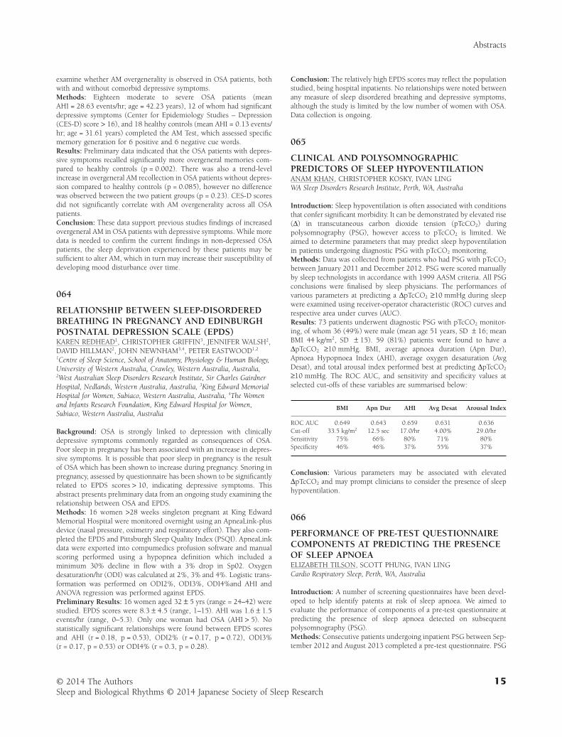
examine whether AM overgenerality is observed in OSA patients, bothwith and without comorbid depressive symptoms.Methods: Eighteen moderate to severe OSA patients (meanAHI = 28.63 events/hr; age = 42.23 years), 12 of whom had significantdepressive symptoms (Center for Epidemiology Studies – Depression(CES-D) score > 16), and 18 healthy controls (mean AHI = 0.13 events/hr; age = 31.61 years) completed the AM Test, which assessed specificmemory generation for 6 positive and 6 negative cue words.Results: Preliminary data indicated that the OSA patients with depres-sive symptoms recalled significantly more overgeneral memories com-pared to healthy controls (p = 0.002). There was also a trend-levelincrease in overgeneral AM recollection in OSA patients without depres-sion compared to healthy controls (p = 0.085), however no differencewas observed between the two patient groups (p = 0.23). CES-D scoresdid not significantly correlate with AM overgenerality across all OSApatients.Conclusion: These data support previous studies findings of increasedovergeneral AM in OSA patients with depressive symptoms. While moredata is needed to confirm the current findings in non-depressed OSApatients, the sleep deprivation experienced by these patients may besufficient to alter AM, which in turn may increase their susceptibility ofdeveloping mood disturbance over time.
064
RELATIONSHIP BETWEEN SLEEP-DISORDEREDBREATHING IN PREGNANCY AND EDINBURGHPOSTNATAL DEPRESSION SCALE (EPDS)KAREN REDHEAD1, CHRISTOPHER GRIFFIN3, JENNIFER WALSH2,DAVID HILLMAN2, JOHN NEWNHAM3,4, PETER EASTWOOD1,2
1Centre of Sleep Science, School of Anatomy, Physiology & Human Biology,University of Western Australia, Crawley, Western Australia, Australia,2West Australian Sleep Disorders Research Institute, Sir Charles GairdnerHospital, Nedlands, Western Australia, Australia, 3King Edward MemorialHospital for Women, Subiaco, Western Australia, Australia, 4The Womenand Infants Research Foundation, King Edward Hospital for Women,Subiaco, Western Australia, Australia
Background: OSA is strongly linked to depression with clinicallydepressive symptoms commonly regarded as consequences of OSA.Poor sleep in pregnancy has been associated with an increase in depres-sive symptoms. It is possible that poor sleep in pregnancy is the resultof OSA which has been shown to increase during pregnancy. Snoring inpregnancy, assessed by questionnaire has been shown to be significantlyrelated to EPDS scores > 10, indicating depressive symptoms. Thisabstract presents preliminary data from an ongoing study examining therelationship between OSA and EPDS.Methods: 16 women >28 weeks singleton pregnant at King EdwardMemorial Hospital were monitored overnight using an ApneaLink-plusdevice (nasal pressure, oximetry and respiratory effort). They also com-pleted the EPDS and Pittsburgh Sleep Quality Index (PSQI). ApneaLinkdata were exported into compumedics profusion software and manualscoring performed using a hypopnea definition which included aminimum 30% decline in flow with a 3% drop in Sp02. Oxygendesaturation/hr (ODI) was calculated at 2%, 3% and 4%. Logistic trans-formation was performed on ODI2%, ODI3%, ODI4%and AHI andANOVA regression was performed against EPDS.Preliminary Results: 16 women aged 32 ± 5 yrs (range = 24–42) werestudied. EPDS scores were 8.3 ± 4.5 (range, 1–15). AHI was 1.6 ± 1.5events/hr (range, 0–5.3). Only one woman had OSA (AHI > 5). Nostatistically significant relationships were found between EPDS scoresand AHI (r = 0.18, p = 0.53), ODI2% (r = 0.17, p = 0.72), ODI3%(r = 0.17, p = 0.53) or ODI4% (r = 0.3, p = 0.28).
Conclusion: The relatively high EPDS scores may reflect the populationstudied, being hospital inpatients. No relationships were noted betweenany measure of sleep disordered breathing and depressive symptoms,although the study is limited by the low number of women with OSA.Data collection is ongoing.
065
CLINICAL AND POLYSOMNOGRAPHICPREDICTORS OF SLEEP HYPOVENTILATIONANAM KHAN, CHRISTOPHER KOSKY, IVAN LINGWA Sleep Disorders Research Institute, Perth, WA, Australia
Introduction: Sleep hypoventilation is often associated with conditionsthat confer significant morbidity. It can be demonstrated by elevated rise(Δ) in transcutaneous carbon dioxide tension (pTcCO2) duringpolysomnography (PSG), however access to pTcCO2 is limited. Weaimed to determine parameters that may predict sleep hypoventilationin patients undergoing diagnostic PSG with pTcCO2 monitoring.Methods: Data was collected from patients who had PSG with pTcCO2
between January 2011 and December 2012. PSG were scored manuallyby sleep technologists in accordance with 1999 AASM criteria. All PSGconclusions were finalised by sleep physicians. The performances ofvarious parameters at predicting a ΔpTcCO2 ≥10 mmHg during sleepwere examined using receiver-operator characteristic (ROC) curves andrespective area under curves (AUC).Results: 73 patients underwent diagnostic PSG with pTcCO2 monitor-ing, of whom 36 (49%) were male (mean age 51 years, SD ± 16; meanBMI 44 kg/m2, SD ± 15). 59 (81%) patients were found to have aΔpTcCO2 ≥10 mmHg. BMI, average apnoea duration (Apn Dur),Apnoea Hypopnoea Index (AHI), average oxygen desaturation (AvgDesat), and total arousal index performed best at predicting ΔpTcCO2
≥10 mmHg. The ROC AUC, and sensitivity and specificity values atselected cut-offs of these variables are summarised below:
BMI Apn Dur AHI Avg Desat Arousal Index
ROC AUC 0.649 0.643 0.659 0.631 0.636Cut-off 33.5 kg/m2 12.5 sec 17.0/hr 4.00% 29.0/hrSensitivity 75% 66% 80% 71% 80%Specificity 46% 46% 37% 55% 37%
Conclusion: Various parameters may be associated with elevatedΔpTcCO2 and may prompt clinicians to consider the presence of sleephypoventilation.
066
PERFORMANCE OF PRE-TEST QUESTIONNAIRECOMPONENTS AT PREDICTING THE PRESENCEOF SLEEP APNOEAELIZABETH TILSON, SCOTT PHUNG, IVAN LINGCardio Respiratory Sleep, Perth, WA, Australia
Introduction: A number of screening questionnaires have been devel-oped to help identify patents at risk of sleep apnoea. We aimed toevaluate the performance of components of a pre-test questionnaire atpredicting the presence of sleep apnoea detected on subsequentpolysomnography (PSG).Methods: Consecutive patients undergoing inpatient PSG between Sep-tember 2012 and August 2013 completed a pre-test questionnaire. PSG
Abstracts
15© 2014 The AuthorsSleep and Biological Rhythms © 2014 Japanese Society of Sleep Research
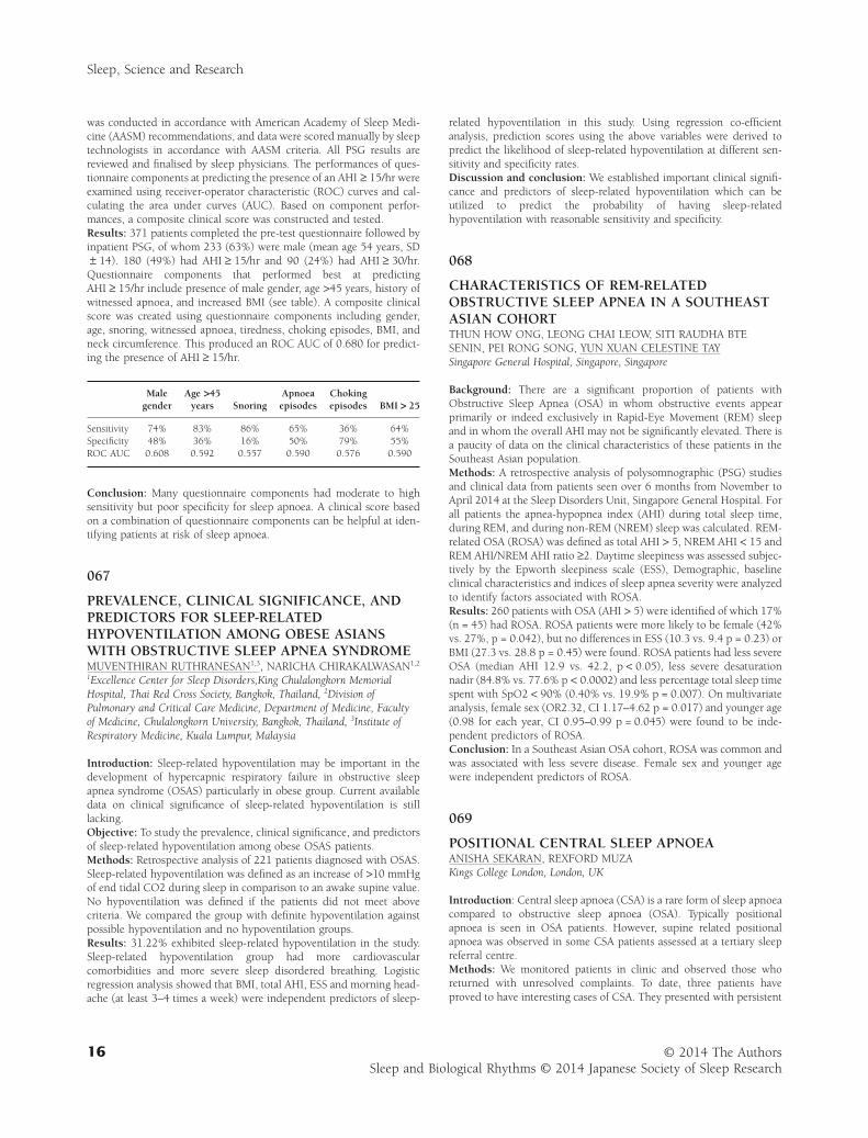
was conducted in accordance with American Academy of Sleep Medi-cine (AASM) recommendations, and data were scored manually by sleeptechnologists in accordance with AASM criteria. All PSG results arereviewed and finalised by sleep physicians. The performances of ques-tionnaire components at predicting the presence of an AHI ≥ 15/hr wereexamined using receiver-operator characteristic (ROC) curves and cal-culating the area under curves (AUC). Based on component perfor-mances, a composite clinical score was constructed and tested.Results: 371 patients completed the pre-test questionnaire followed byinpatient PSG, of whom 233 (63%) were male (mean age 54 years, SD± 14). 180 (49%) had AHI ≥ 15/hr and 90 (24%) had AHI ≥ 30/hr.Questionnaire components that performed best at predictingAHI ≥ 15/hr include presence of male gender, age >45 years, history ofwitnessed apnoea, and increased BMI (see table). A composite clinicalscore was created using questionnaire components including gender,age, snoring, witnessed apnoea, tiredness, choking episodes, BMI, andneck circumference. This produced an ROC AUC of 0.680 for predict-ing the presence of AHI ≥ 15/hr.
Malegender
Age >45years Snoring
Apnoeaepisodes
Chokingepisodes BMI > 25
Sensitivity 74% 83% 86% 65% 36% 64%Specificity 48% 36% 16% 50% 79% 55%ROC AUC 0.608 0.592 0.557 0.590 0.576 0.590
Conclusion: Many questionnaire components had moderate to highsensitivity but poor specificity for sleep apnoea. A clinical score basedon a combination of questionnaire components can be helpful at iden-tifying patients at risk of sleep apnoea.
067
PREVALENCE, CLINICAL SIGNIFICANCE, ANDPREDICTORS FOR SLEEP-RELATEDHYPOVENTILATION AMONG OBESE ASIANSWITH OBSTRUCTIVE SLEEP APNEA SYNDROMEMUVENTHIRAN RUTHRANESAN1,3, NARICHA CHIRAKALWASAN1,2
1Excellence Center for Sleep Disorders,King Chulalongkorn MemorialHospital, Thai Red Cross Society, Bangkok, Thailand, 2Division ofPulmonary and Critical Care Medicine, Department of Medicine, Facultyof Medicine, Chulalongkorn University, Bangkok, Thailand, 3Institute ofRespiratory Medicine, Kuala Lumpur, Malaysia
Introduction: Sleep-related hypoventilation may be important in thedevelopment of hypercapnic respiratory failure in obstructive sleepapnea syndrome (OSAS) particularly in obese group. Current availabledata on clinical significance of sleep-related hypoventilation is stilllacking.Objective: To study the prevalence, clinical significance, and predictorsof sleep-related hypoventilation among obese OSAS patients.Methods: Retrospective analysis of 221 patients diagnosed with OSAS.Sleep-related hypoventilation was defined as an increase of >10 mmHgof end tidal CO2 during sleep in comparison to an awake supine value.No hypoventilation was defined if the patients did not meet abovecriteria. We compared the group with definite hypoventilation againstpossible hypoventilation and no hypoventilation groups.Results: 31.22% exhibited sleep-related hypoventilation in the study.Sleep-related hypoventilation group had more cardiovascularcomorbidities and more severe sleep disordered breathing. Logisticregression analysis showed that BMI, total AHI, ESS and morning head-ache (at least 3–4 times a week) were independent predictors of sleep-
related hypoventilation in this study. Using regression co-efficientanalysis, prediction scores using the above variables were derived topredict the likelihood of sleep-related hypoventilation at different sen-sitivity and specificity rates.Discussion and conclusion: We established important clinical signifi-cance and predictors of sleep-related hypoventilation which can beutilized to predict the probability of having sleep-relatedhypoventilation with reasonable sensitivity and specificity.
068
CHARACTERISTICS OF REM-RELATEDOBSTRUCTIVE SLEEP APNEA IN A SOUTHEASTASIAN COHORTTHUN HOW ONG, LEONG CHAI LEOW, SITI RAUDHA BTESENIN, PEI RONG SONG, YUN XUAN CELESTINE TAYSingapore General Hospital, Singapore, Singapore
Background: There are a significant proportion of patients withObstructive Sleep Apnea (OSA) in whom obstructive events appearprimarily or indeed exclusively in Rapid-Eye Movement (REM) sleepand in whom the overall AHI may not be significantly elevated. There isa paucity of data on the clinical characteristics of these patients in theSoutheast Asian population.Methods: A retrospective analysis of polysomnographic (PSG) studiesand clinical data from patients seen over 6 months from November toApril 2014 at the Sleep Disorders Unit, Singapore General Hospital. Forall patients the apnea-hypopnea index (AHI) during total sleep time,during REM, and during non-REM (NREM) sleep was calculated. REM-related OSA (ROSA) was defined as total AHI > 5, NREM AHI < 15 andREM AHI/NREM AHI ratio ≥2. Daytime sleepiness was assessed subjec-tively by the Epworth sleepiness scale (ESS), Demographic, baselineclinical characteristics and indices of sleep apnea severity were analyzedto identify factors associated with ROSA.Results: 260 patients with OSA (AHI > 5) were identified of which 17%(n = 45) had ROSA. ROSA patients were more likely to be female (42%vs. 27%, p = 0.042), but no differences in ESS (10.3 vs. 9.4 p = 0.23) orBMI (27.3 vs. 28.8 p = 0.45) were found. ROSA patients had less severeOSA (median AHI 12.9 vs. 42.2, p < 0.05), less severe desaturationnadir (84.8% vs. 77.6% p < 0.0002) and less percentage total sleep timespent with SpO2 < 90% (0.40% vs. 19.9% p = 0.007). On multivariateanalysis, female sex (OR2.32, CI 1.17–4.62 p = 0.017) and younger age(0.98 for each year, CI 0.95–0.99 p = 0.045) were found to be inde-pendent predictors of ROSA.Conclusion: In a Southeast Asian OSA cohort, ROSA was common andwas associated with less severe disease. Female sex and younger agewere independent predictors of ROSA.
069
POSITIONAL CENTRAL SLEEP APNOEAANISHA SEKARAN, REXFORD MUZAKings College London, London, UK
Introduction: Central sleep apnoea (CSA) is a rare form of sleep apnoeacompared to obstructive sleep apnoea (OSA). Typically positionalapnoea is seen in OSA patients. However, supine related positionalapnoea was observed in some CSA patients assessed at a tertiary sleepreferral centre.Methods: We monitored patients in clinic and observed those whoreturned with unresolved complaints. To date, three patients haveproved to have interesting cases of CSA. They presented with persistent
Sleep, Science and Research
16 © 2014 The AuthorsSleep and Biological Rhythms © 2014 Japanese Society of Sleep Research
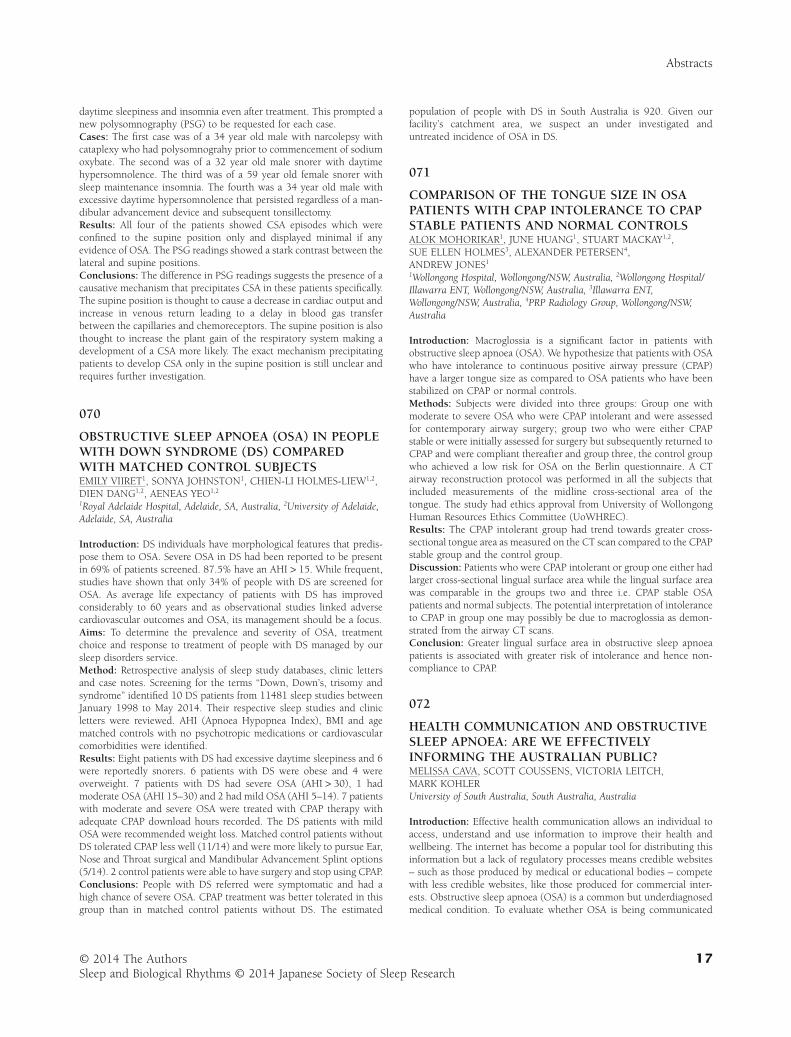
daytime sleepiness and insomnia even after treatment. This prompted anew polysomnography (PSG) to be requested for each case.Cases: The first case was of a 34 year old male with narcolepsy withcataplexy who had polysomnograhy prior to commencement of sodiumoxybate. The second was of a 32 year old male snorer with daytimehypersomnolence. The third was of a 59 year old female snorer withsleep maintenance insomnia. The fourth was a 34 year old male withexcessive daytime hypersomnolence that persisted regardless of a man-dibular advancement device and subsequent tonsillectomy.Results: All four of the patients showed CSA episodes which wereconfined to the supine position only and displayed minimal if anyevidence of OSA. The PSG readings showed a stark contrast between thelateral and supine positions.Conclusions: The difference in PSG readings suggests the presence of acausative mechanism that precipitates CSA in these patients specifically.The supine position is thought to cause a decrease in cardiac output andincrease in venous return leading to a delay in blood gas transferbetween the capillaries and chemoreceptors. The supine position is alsothought to increase the plant gain of the respiratory system making adevelopment of a CSA more likely. The exact mechanism precipitatingpatients to develop CSA only in the supine position is still unclear andrequires further investigation.
070
OBSTRUCTIVE SLEEP APNOEA (OSA) IN PEOPLEWITH DOWN SYNDROME (DS) COMPAREDWITH MATCHED CONTROL SUBJECTSEMILY VIIRET1, SONYA JOHNSTON1, CHIEN-LI HOLMES-LIEW1,2,DIEN DANG1,2, AENEAS YEO1,2
1Royal Adelaide Hospital, Adelaide, SA, Australia, 2University of Adelaide,Adelaide, SA, Australia
Introduction: DS individuals have morphological features that predis-pose them to OSA. Severe OSA in DS had been reported to be presentin 69% of patients screened. 87.5% have an AHI > 15. While frequent,studies have shown that only 34% of people with DS are screened forOSA. As average life expectancy of patients with DS has improvedconsiderably to 60 years and as observational studies linked adversecardiovascular outcomes and OSA, its management should be a focus.Aims: To determine the prevalence and severity of OSA, treatmentchoice and response to treatment of people with DS managed by oursleep disorders service.Method: Retrospective analysis of sleep study databases, clinic lettersand case notes. Screening for the terms “Down, Down’s, trisomy andsyndrome” identified 10 DS patients from 11481 sleep studies betweenJanuary 1998 to May 2014. Their respective sleep studies and clinicletters were reviewed. AHI (Apnoea Hypopnea Index), BMI and agematched controls with no psychotropic medications or cardiovascularcomorbidities were identified.Results: Eight patients with DS had excessive daytime sleepiness and 6were reportedly snorers. 6 patients with DS were obese and 4 wereoverweight. 7 patients with DS had severe OSA (AHI > 30), 1 hadmoderate OSA (AHI 15–30) and 2 had mild OSA (AHI 5–14). 7 patientswith moderate and severe OSA were treated with CPAP therapy withadequate CPAP download hours recorded. The DS patients with mildOSA were recommended weight loss. Matched control patients withoutDS tolerated CPAP less well (11/14) and were more likely to pursue Ear,Nose and Throat surgical and Mandibular Advancement Splint options(5/14). 2 control patients were able to have surgery and stop using CPAP.Conclusions: People with DS referred were symptomatic and had ahigh chance of severe OSA. CPAP treatment was better tolerated in thisgroup than in matched control patients without DS. The estimated
population of people with DS in South Australia is 920. Given ourfacility’s catchment area, we suspect an under investigated anduntreated incidence of OSA in DS.
071
COMPARISON OF THE TONGUE SIZE IN OSAPATIENTS WITH CPAP INTOLERANCE TO CPAPSTABLE PATIENTS AND NORMAL CONTROLSALOK MOHORIKAR1, JUNE HUANG1, STUART MACKAY1,2,SUE ELLEN HOLMES3, ALEXANDER PETERSEN4,ANDREW JONES1
1Wollongong Hospital, Wollongong/NSW, Australia, 2Wollongong Hospital/Illawarra ENT, Wollongong/NSW, Australia, 3Illawarra ENT,Wollongong/NSW, Australia, 4PRP Radiology Group, Wollongong/NSW,Australia
Introduction: Macroglossia is a significant factor in patients withobstructive sleep apnoea (OSA). We hypothesize that patients with OSAwho have intolerance to continuous positive airway pressure (CPAP)have a larger tongue size as compared to OSA patients who have beenstabilized on CPAP or normal controls.Methods: Subjects were divided into three groups: Group one withmoderate to severe OSA who were CPAP intolerant and were assessedfor contemporary airway surgery; group two who were either CPAPstable or were initially assessed for surgery but subsequently returned toCPAP and were compliant thereafter and group three, the control groupwho achieved a low risk for OSA on the Berlin questionnaire. A CTairway reconstruction protocol was performed in all the subjects thatincluded measurements of the midline cross-sectional area of thetongue. The study had ethics approval from University of WollongongHuman Resources Ethics Committee (UoWHREC).Results: The CPAP intolerant group had trend towards greater cross-sectional tongue area as measured on the CT scan compared to the CPAPstable group and the control group.Discussion: Patients who were CPAP intolerant or group one either hadlarger cross-sectional lingual surface area while the lingual surface areawas comparable in the groups two and three i.e. CPAP stable OSApatients and normal subjects. The potential interpretation of intoleranceto CPAP in group one may possibly be due to macroglossia as demon-strated from the airway CT scans.Conclusion: Greater lingual surface area in obstructive sleep apnoeapatients is associated with greater risk of intolerance and hence non-compliance to CPAP.
072
HEALTH COMMUNICATION AND OBSTRUCTIVESLEEP APNOEA: ARE WE EFFECTIVELYINFORMING THE AUSTRALIAN PUBLIC?MELISSA CAVA, SCOTT COUSSENS, VICTORIA LEITCH,MARK KOHLERUniversity of South Australia, South Australia, Australia
Introduction: Effective health communication allows an individual toaccess, understand and use information to improve their health andwellbeing. The internet has become a popular tool for distributing thisinformation but a lack of regulatory processes means credible websites– such as those produced by medical or educational bodies – competewith less credible websites, like those produced for commercial inter-ests. Obstructive sleep apnoea (OSA) is a common but underdiagnosedmedical condition. To evaluate whether OSA is being communicated
Abstracts
17© 2014 The AuthorsSleep and Biological Rhythms © 2014 Japanese Society of Sleep Research
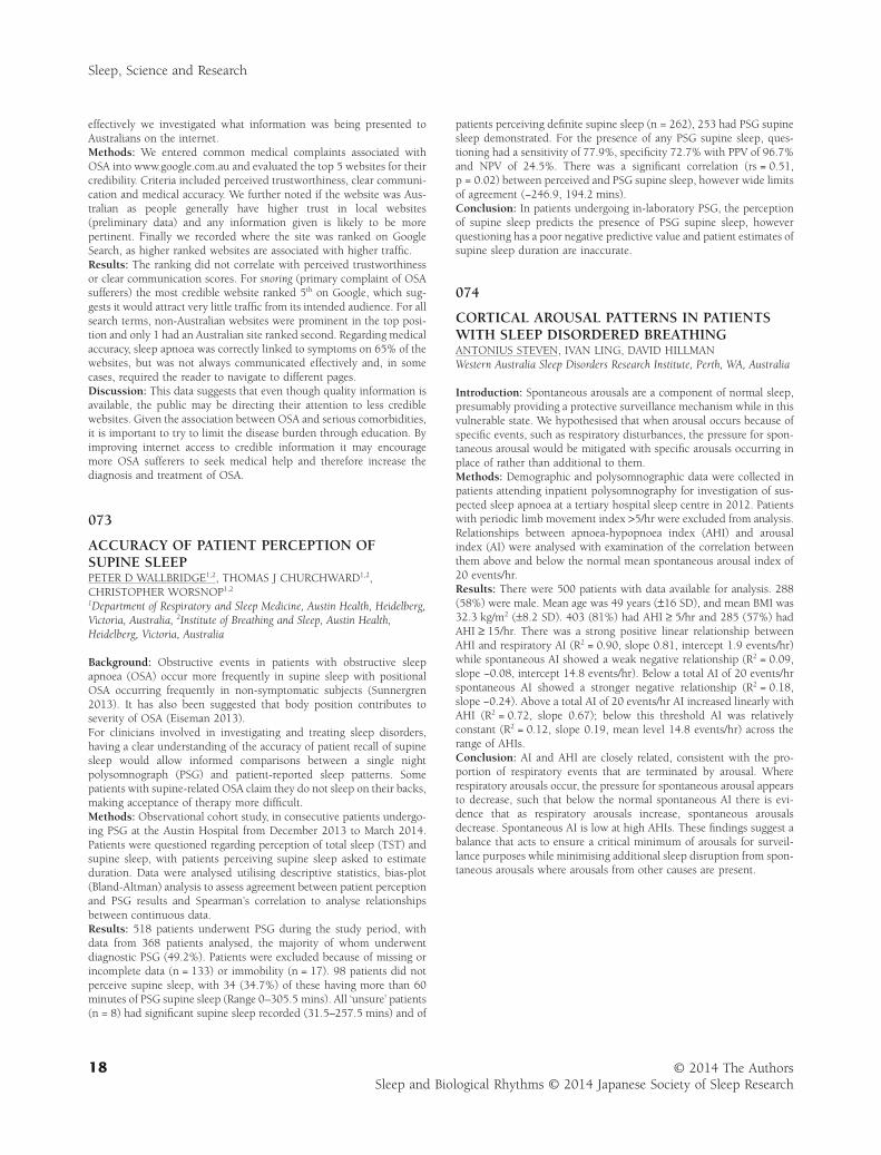
effectively we investigated what information was being presented toAustralians on the internet.Methods: We entered common medical complaints associated withOSA into www.google.com.au and evaluated the top 5 websites for theircredibility. Criteria included perceived trustworthiness, clear communi-cation and medical accuracy. We further noted if the website was Aus-tralian as people generally have higher trust in local websites(preliminary data) and any information given is likely to be morepertinent. Finally we recorded where the site was ranked on GoogleSearch, as higher ranked websites are associated with higher traffic.Results: The ranking did not correlate with perceived trustworthinessor clear communication scores. For snoring (primary complaint of OSAsufferers) the most credible website ranked 5th on Google, which sug-gests it would attract very little traffic from its intended audience. For allsearch terms, non-Australian websites were prominent in the top posi-tion and only 1 had an Australian site ranked second. Regarding medicalaccuracy, sleep apnoea was correctly linked to symptoms on 65% of thewebsites, but was not always communicated effectively and, in somecases, required the reader to navigate to different pages.Discussion: This data suggests that even though quality information isavailable, the public may be directing their attention to less crediblewebsites. Given the association between OSA and serious comorbidities,it is important to try to limit the disease burden through education. Byimproving internet access to credible information it may encouragemore OSA sufferers to seek medical help and therefore increase thediagnosis and treatment of OSA.
073
ACCURACY OF PATIENT PERCEPTION OFSUPINE SLEEPPETER D WALLBRIDGE1,2, THOMAS J CHURCHWARD1,2,CHRISTOPHER WORSNOP1,2
1Department of Respiratory and Sleep Medicine, Austin Health, Heidelberg,Victoria, Australia, 2Institute of Breathing and Sleep, Austin Health,Heidelberg, Victoria, Australia
Background: Obstructive events in patients with obstructive sleepapnoea (OSA) occur more frequently in supine sleep with positionalOSA occurring frequently in non-symptomatic subjects (Sunnergren2013). It has also been suggested that body position contributes toseverity of OSA (Eiseman 2013).For clinicians involved in investigating and treating sleep disorders,having a clear understanding of the accuracy of patient recall of supinesleep would allow informed comparisons between a single nightpolysomnograph (PSG) and patient-reported sleep patterns. Somepatients with supine-related OSA claim they do not sleep on their backs,making acceptance of therapy more difficult.Methods: Observational cohort study, in consecutive patients undergo-ing PSG at the Austin Hospital from December 2013 to March 2014.Patients were questioned regarding perception of total sleep (TST) andsupine sleep, with patients perceiving supine sleep asked to estimateduration. Data were analysed utilising descriptive statistics, bias-plot(Bland-Altman) analysis to assess agreement between patient perceptionand PSG results and Spearman’s correlation to analyse relationshipsbetween continuous data.Results: 518 patients underwent PSG during the study period, withdata from 368 patients analysed, the majority of whom underwentdiagnostic PSG (49.2%). Patients were excluded because of missing orincomplete data (n = 133) or immobility (n = 17). 98 patients did notperceive supine sleep, with 34 (34.7%) of these having more than 60minutes of PSG supine sleep (Range 0–305.5 mins). All ‘unsure’ patients(n = 8) had significant supine sleep recorded (31.5–257.5 mins) and of
patients perceiving definite supine sleep (n = 262), 253 had PSG supinesleep demonstrated. For the presence of any PSG supine sleep, ques-tioning had a sensitivity of 77.9%, specificity 72.7% with PPV of 96.7%and NPV of 24.5%. There was a significant correlation (rs = 0.51,p = 0.02) between perceived and PSG supine sleep, however wide limitsof agreement (−246.9, 194.2 mins).Conclusion: In patients undergoing in-laboratory PSG, the perceptionof supine sleep predicts the presence of PSG supine sleep, howeverquestioning has a poor negative predictive value and patient estimates ofsupine sleep duration are inaccurate.
074
CORTICAL AROUSAL PATTERNS IN PATIENTSWITH SLEEP DISORDERED BREATHINGANTONIUS STEVEN, IVAN LING, DAVID HILLMANWestern Australia Sleep Disorders Research Institute, Perth, WA, Australia
Introduction: Spontaneous arousals are a component of normal sleep,presumably providing a protective surveillance mechanism while in thisvulnerable state. We hypothesised that when arousal occurs because ofspecific events, such as respiratory disturbances, the pressure for spon-taneous arousal would be mitigated with specific arousals occurring inplace of rather than additional to them.Methods: Demographic and polysomnographic data were collected inpatients attending inpatient polysomnography for investigation of sus-pected sleep apnoea at a tertiary hospital sleep centre in 2012. Patientswith periodic limb movement index >5/hr were excluded from analysis.Relationships between apnoea-hypopnoea index (AHI) and arousalindex (AI) were analysed with examination of the correlation betweenthem above and below the normal mean spontaneous arousal index of20 events/hr.Results: There were 500 patients with data available for analysis. 288(58%) were male. Mean age was 49 years (±16 SD), and mean BMI was32.3 kg/m2 (±8.2 SD). 403 (81%) had AHI ≥ 5/hr and 285 (57%) hadAHI ≥ 15/hr. There was a strong positive linear relationship betweenAHI and respiratory AI (R2 = 0.90, slope 0.81, intercept 1.9 events/hr)while spontaneous AI showed a weak negative relationship (R2 = 0.09,slope −0.08, intercept 14.8 events/hr). Below a total AI of 20 events/hrspontaneous AI showed a stronger negative relationship (R2 = 0.18,slope −0.24). Above a total AI of 20 events/hr AI increased linearly withAHI (R2 = 0.72, slope 0.67); below this threshold AI was relativelyconstant (R2 = 0.12, slope 0.19, mean level 14.8 events/hr) across therange of AHIs.Conclusion: AI and AHI are closely related, consistent with the pro-portion of respiratory events that are terminated by arousal. Whererespiratory arousals occur, the pressure for spontaneous arousal appearsto decrease, such that below the normal spontaneous AI there is evi-dence that as respiratory arousals increase, spontaneous arousalsdecrease. Spontaneous AI is low at high AHIs. These findings suggest abalance that acts to ensure a critical minimum of arousals for surveil-lance purposes while minimising additional sleep disruption from spon-taneous arousals where arousals from other causes are present.
Sleep, Science and Research
18 © 2014 The AuthorsSleep and Biological Rhythms © 2014 Japanese Society of Sleep Research

075
THE RELATIONSHIP BETWEEN THEAPNOEA-HYPOPNOEA INDEX (AHI) AND THERESPIRATORY DISTURBANCE INDEX (RDI) WITHRESPECT TO OBSTRUCTIVE SLEEP APNOEAAND ANTHROPOMETRIC PREDICTORSSHAVITA KUCKREJA, LYNNETTE REID-PRICE, ANNE MARIESOUTHCOTT, LATA JAYARAMWestern Health, Melbourne, Australia
Background: The Apnoea-Hypopnoea Index (AHI) is often used inter-changeably with the Respiratory Disturbance Index (RDI)(which meas-ures respiratory effort related arousals plus AHI) in sleep studies as ascoring system for the severity of obstructive sleep apnoea(OSA).Empirically, the RDI correlates well in severe OSA and less so in mild tomoderate OSA. We hypothesised that the relationship is variable in mildto moderate OSA, and that the variability may be explained by physicalfactors (anthromopmetric predictors), and overnight oxygenation.Aim: To assess the relationship between AHI and RDI in our patientpopulation with OSA, and second, to identify aspects of body compo-sition and overnight oxygenation that explain the difference betweenthe 2 scoring systems.Methods: Retrospective study examining 511 in-laboratory diagnosticsleep studies performed at our Sleep Laboratory from January 1st 2013to December 31st 2013 for AHI, RDI, height, weight, BMI, neck cir-cumference, average and lowest oxygen saturation.Results: Baseline characteristics: 48% men; Age: mean (standard devia-tion):51(16) years; BMI: 32.5(7.9); AHI: 20(27) events/hr; RDI:20(21)events/hr. Average overnight oxygen saturation: 93.8(8.7)%;minimum oxygen saturation: 85.5(9.0)%. The correlation between AHIand RDI was 0.94(p = 0.00). Average overnight oxygen saturation con-tributed significantly to the variability between AHI and RDI. Whenusing standardised criteria to classify severity of OSA, the RDI increasedthe classification of severity of OSA by 1 category (i.e. normal to mild,mild to moderate and moderate to severe) compared with AHI in 30%of those with a normal AHI; 31% of those with mild OSA; 9% withmoderate OSA and in no cases of severe OSA. The difference in absoluteevents/hr between the AHI and RDI, however, increased with severity ofOSA: percentage of cases with greater than 5 unit difference betweenAHI and RDI was 17% in those with a normal AHI; 32% with mild OSA,36% with moderate OSA and 60% with severe OSA.Conclusions: The RDI correlates well with the AHI in our population.The classification of severity of OSA is increased in a proportion of ourpatients with no or with mild OSA using the RDI rather than the AHI;and this may have implications for treatment.
076
THE EFFECT OF AROUSAL AND SUBSEQUENTHYPOCAPNIA ON GENIOGLOSSUS MUSCLEACTIVITY IN OBSTRUCTIVE SLEEP APNEAJENNIFER CORI1,2, THERESE THORNTON1,2, PETER ROCHFORD2,FERGAL O’DONOGHUE1,2, JOHN TRINDER1, AMY JORDAN1,2
1The University of Melbourne, Parkville, VIC, Australia, 2Institute forBreathing and Sleep, Heidelberg, VIC, Australia
Introduction: Hypocapnia induced by a large ventilatory response toarousal is believed to predispose to further airway collapse in obstruc-tive sleep apnea (OSA) by reducing upper airway dilator muscle activityat return to sleep. However, prior studies have not observed reduceddilator muscle activity following arousal. These studies were limited asmany did not investigate arousals terminating respiratory events, or
measure CO2. As CO2 is important in the arousal model, we aimed toclarify whether reduced CO2 following arousal is associated withreduced dilator muscle activity on return to sleep in OSA patients.Methods: Data was obtained in 16 of 22 mild to severe OSA patientswho slept untreated and instrumented with standard sleeppolysomnography, partial pressure of end-tidal CO2(PetCO2), intramus-cular genioglossus electrodes(EMGgg) and an epiglottic pressurecatheter(Pepi). Post-study, NREM respiratory arousals were categorisedby PetCO2 on the last breath of arousal, immediately before return tosleep, relative to waking baseline PetCO2. Arousals 0–3 mmHg belowparticipant’s waking baseline CO2 were classified ‘low’. Arousals0–3 mmHg above participant’s waking baseline CO2 were classified‘high’. Muscle and respiratory data at return to sleep were comparedusing a two-way repeated measures ANOVA(CO2 condition-low vs.high)x(Breath-first five breaths of return to sleep)Results: There were 376 arousals in the low CO2 condition and 236arousals in the high CO2 condition. By design PetCO2 on the last breathof arousal was significantly less for the low CO2 condition than thehigh CO2 condition (38.7 ± 2.7 vs. 41.4 ± 2.7 mmHg, p < .001). CO2
condition effects were observed for Peak EMGgg with activity signifi-cantly elevated across the breaths during return to sleep followingarousal for the low CO2 condition, compared to the high CO2 condition.Breath effects were observed only for Vi and Pepi Nadir with bothvariables declining across the breaths of return to sleep followingarousal.Conclusion: If these results persist in the complete dataset, low CO2 atarousal may not cause reduced dilator muscle activity on return to sleepas predicted by the arousal model.
077
COMPARISON OF THE QUALITY OF HOMESLEEP STUDY SET UP IN SLEEP LAB VS SET BYPATIENT AT HOMEALAN LUCAS, KATJA MCGUIRE, ANTONIO AMBROGETTISleep Medicine, Charlestown, NSW, Australia
Introduction: Home studies (level 3) for the assessment of snoring andsleep apnea have become common. As the home studies are not super-vised the quality of the acquisition data is a potential problem. Onepossible source of artefacts is the poor applications of EEG, EOG, EMGand respiratory derivations (abdominal-thoracic belt, nasal cannula andoximetry). We assessed the quality of home PSG acquisition in patientsset up in the sleep unit in the evening by a sleep scientist versus patientswho were given detailed instruction early in day and who set themselvesup before going to bed.Method: Forty two home recordings set up by the sleep scientist in theevenings were compared to 37 studies where the patients set up them-selves at bedtime, using Embletta™. Each study was evaluated by visualinspection of each hour of recording or part of thereof (30 seconds epochby epoch) for four parameters (EEG, EOG, EMG and Respiratory) asexcellent (no artefacts), good (some artefacts but able to be interpreted)and unsatisfactory (not be able to score). The overall study were classi-fied as good if all the derivations were scorable, fair if a derivation wasnot scorable but assessment was still possible (e.g. abdominal belt notusable but thoracic belt, oxi and nasal cannula available), poor (Staging,or respiratory not scorable) and failure (data not usable at all)Results: In the self-set up group there were 18 men and 19 women, themean age 51.4 (range 16–76). Twenty four men and 18 women, meanage 52 (range 17–76) in the in lab set up group. 84% and 76% of thestudies set up at home and by the scientist respectively were of goodquality. There were three failures in the in lab set up all due to thepatients removing the leads at home and try to re-apply later.
Abstracts
19© 2014 The AuthorsSleep and Biological Rhythms © 2014 Japanese Society of Sleep Research
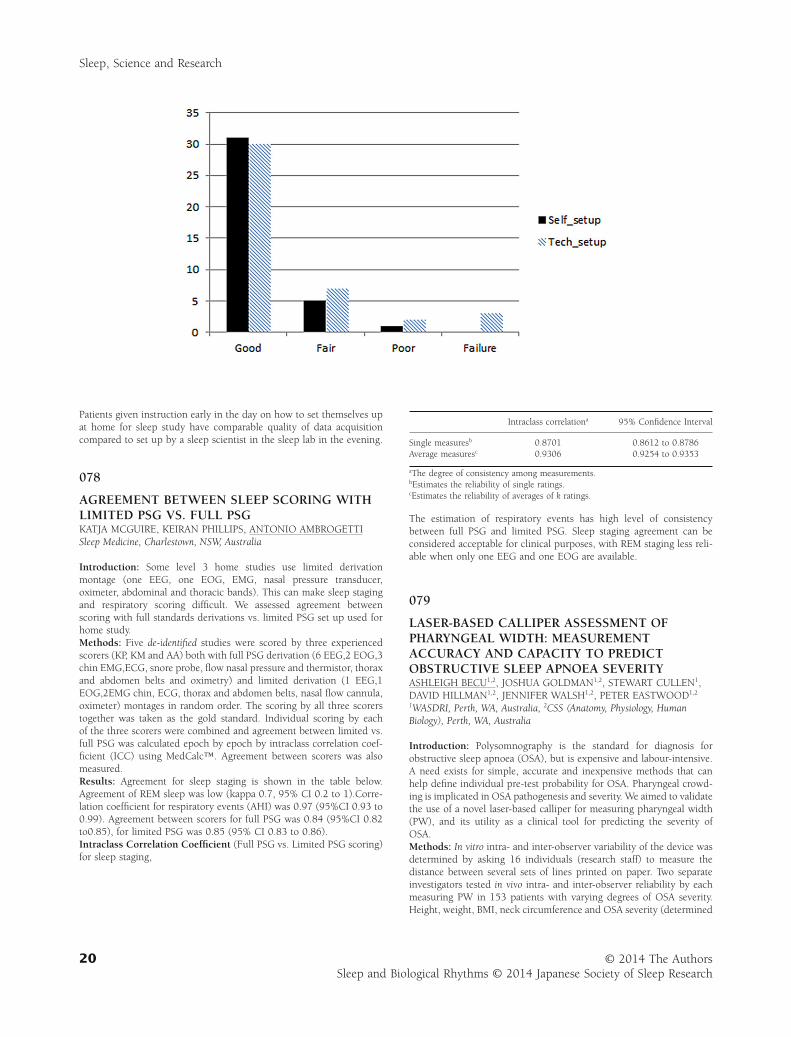
Patients given instruction early in the day on how to set themselves upat home for sleep study have comparable quality of data acquisitioncompared to set up by a sleep scientist in the sleep lab in the evening.
078
AGREEMENT BETWEEN SLEEP SCORING WITHLIMITED PSG VS. FULL PSGKATJA MCGUIRE, KEIRAN PHILLIPS, ANTONIO AMBROGETTISleep Medicine, Charlestown, NSW, Australia
Introduction: Some level 3 home studies use limited derivationmontage (one EEG, one EOG, EMG, nasal pressure transducer,oximeter, abdominal and thoracic bands). This can make sleep stagingand respiratory scoring difficult. We assessed agreement betweenscoring with full standards derivations vs. limited PSG set up used forhome study.Methods: Five de-identified studies were scored by three experiencedscorers (KP, KM and AA) both with full PSG derivation (6 EEG,2 EOG,3chin EMG,ECG, snore probe, flow nasal pressure and thermistor, thoraxand abdomen belts and oximetry) and limited derivation (1 EEG,1EOG,2EMG chin, ECG, thorax and abdomen belts, nasal flow cannula,oximeter) montages in random order. The scoring by all three scorerstogether was taken as the gold standard. Individual scoring by eachof the three scorers were combined and agreement between limited vs.full PSG was calculated epoch by epoch by intraclass correlation coef-ficient (ICC) using MedCalc™. Agreement between scorers was alsomeasured.Results: Agreement for sleep staging is shown in the table below.Agreement of REM sleep was low (kappa 0.7, 95% CI 0.2 to 1).Corre-lation coefficient for respiratory events (AHI) was 0.97 (95%CI 0.93 to0.99). Agreement between scorers for full PSG was 0.84 (95%CI 0.82to0.85), for limited PSG was 0.85 (95% CI 0.83 to 0.86).Intraclass Correlation Coefficient (Full PSG vs. Limited PSG scoring)for sleep staging,
Intraclass correlationa 95% Confidence Interval
Single measuresb 0.8701 0.8612 to 0.8786Average measuresc 0.9306 0.9254 to 0.9353
aThe degree of consistency among measurements.bEstimates the reliability of single ratings.cEstimates the reliability of averages of k ratings.
The estimation of respiratory events has high level of consistencybetween full PSG and limited PSG. Sleep staging agreement can beconsidered acceptable for clinical purposes, with REM staging less reli-able when only one EEG and one EOG are available.
079
LASER-BASED CALLIPER ASSESSMENT OFPHARYNGEAL WIDTH: MEASUREMENTACCURACY AND CAPACITY TO PREDICTOBSTRUCTIVE SLEEP APNOEA SEVERITYASHLEIGH BECU1,2, JOSHUA GOLDMAN1,2, STEWART CULLEN1,DAVID HILLMAN1,2, JENNIFER WALSH1,2, PETER EASTWOOD1,2
1WASDRI, Perth, WA, Australia, 2CSS (Anatomy, Physiology, HumanBiology), Perth, WA, Australia
Introduction: Polysomnography is the standard for diagnosis forobstructive sleep apnoea (OSA), but is expensive and labour-intensive.A need exists for simple, accurate and inexpensive methods that canhelp define individual pre-test probability for OSA. Pharyngeal crowd-ing is implicated in OSA pathogenesis and severity. We aimed to validatethe use of a novel laser-based calliper for measuring pharyngeal width(PW), and its utility as a clinical tool for predicting the severity ofOSA.Methods: In vitro intra- and inter-observer variability of the device wasdetermined by asking 16 individuals (research staff) to measure thedistance between several sets of lines printed on paper. Two separateinvestigators tested in vivo intra- and inter-observer reliability by eachmeasuring PW in 153 patients with varying degrees of OSA severity.Height, weight, BMI, neck circumference and OSA severity (determined
Sleep, Science and Research
20 © 2014 The AuthorsSleep and Biological Rhythms © 2014 Japanese Society of Sleep Research
by overnight PSG, and quantified by the apnoea-hypopnea index (AHI))were also obtained from each patient.Results: There was no significant difference between in vitro intra- orinter-observer measurements t(15) = 0.53, p = 0.61; t(15) = −1.57,p = 0.14 respectively. There was no significant difference between invivo intra- and inter-observer measurements F(1.72, 70.41) = 1.93,p = 0.16, partial n2 = 0.05; F(1.66, 68.07) = 0.58, p = 0.53 partialn2 = 0.01 respectively. Of the 153 patients, 82 (54%) were male, meanage 52 ± 13 years, mean BMI 32.4 ± 5.9 kg/m2 and mean AHI 35.8± 26.9/hr. PW was able to be obtained in 128 patients, and ranged from7.84 to 34.27 mm (mean 19.0, SD ± 6.0). A significant relationshipbetween PW and AHI was found: r = −0.22; p = 0.02; and PWexplained a significant proportion of variance in AHI: = 0.049,F(1,107) = 5.47, p = 0.02. With stepwise multiple linear regression, asignificant model emerged to predict AHI: F(4,104) = 12.29, p < 0.01,R2 = 0.32. The following formula was used to predict AHI: logAHI = −3.67 + (2.22 log neck circumference, cm) − (0.016 PW,mm) + (0.01 age, yrs) + (0.01 height, m).Conclusion: Laser-based calliper assessment of PW combined withother clinical measurements may be a useful clinical screening tool fordetermining OSA severity.
080
SNORING DURING PREGNANCY:QUESTIONNAIRE RESULTS VERSUS OBJECTIVEMEASUREMENT (AHI).ANNETTE ROBERTSON1,3, PAMELA JOHNSON2,ANNEMARIE HENNESSY1,3, COLIN SULLIVAN2
1University of Western Sydney, Campbelltown NSW, Australia, 2Universityof Sydney, Sydney NSW, Australia, 3SWSLDH, Campbelltown NSW,Australia
Introduction: Snoring is common during pregnancy, particularly in thelast trimester. Most investigations of snoring during pregnancy haveutilised questionnaires to determine the presence and severity ofsnoring. This study administered questionnaires and recorded snoringduring sleep in 202 women in order to validate the use of questionnairesagainst objective measurement of actual snoring.Aim: To determine the efficacy of questionnaires to determine thepresence of snoring in pregnant women including AHI.Methods: Berlin questionnaires were administered to 202 pregnantwomen. One question asked “Do you snore?” Snoring was then rec-orded in the woman’s home using a SonoMat™ (Home sleep test)device, a thin mattress in which sensors measure and record sound andmovement from the participant and the ambient sound. AHI weredetermined. The results from the questionnaire were compared to rec-orded snoring and AHI resultsResults: 124 women answered “Yes” to the question “Do you snore?”and 78 women answered “No” and “I don’t know” for score purposewere included in “No”. The average gestation was 28 ± 7 weeks. Therewere no significant differences between the percentages of snoring rec-orded in <20% or >20% of night spent snoring.
Recordedsnoring
Questionnaire responsesto “Do you snore?” Quiescent time
Yes % No % minutes
<20% 64.5 69.2 260–660 P = 0.0079≥20% 35.5 30.8 60–460
Conclusion: This study has demonstrated that questionnaire results arenot a reliable method of determining whether snoring is present, and
overnight monitoring should be used to rule our sleep disorderedbreathing, particularly when hypertension is present.
081
A COMPARISON OF SLEEP STAGES AND SLEEPDISORDERED BREATHING IDENTIFICATIONBETWEEN A PORTABLE MONITORING DEVICEAND IN-LAB POLYSOMNOGRAPHY (PSG)ANDREW PERKINS1, MAI TRAN1,2, JACKIE ERITAIA1
1Royal Melbourne Hospital, Melbourne, Australia, 2Swinburne Universityof Technology, Melbourne, Australia
Introduction: Portable sleep monitoring offers great benefit by allowingthe flexibility of conducting sleep studies outside of dedicated labora-tories, either with inpatients at hospitals or at patients’ homes. Thisstudy compares the accuracy of one device, the iTamar Watch-PAT 200(ITAMAR Medical LTD, Caesarea, Israel) with a simultaneous attendedin-lab full PSGMethods: 18 Patients referred to a metropolitan tertiary hospital sleepdisorders unit for a diagnostic study were consecutively selected on thebasis of investigator availability, (8M, 10F, aged 30–69 years, M = 47.8,SD = 12.2, BMI ESS). Each participant had a full PSG & simultaneousrecording with the Watch-PAT device. The PSG results were scored &staged by Lab staff & compared with the automatically generated resultsfrom the device.Result: Adequate data was obtained for only 13 patients, Altman-BlandComparisons between the two methods showed no significant differ-ence between the two methods for the duration of the combined stagesN1&N2 (as is inherent to the watch-Pat) (M = −3.442, 95%,CI = −11042–4.16, t-0.99, p = 0.34) & in slow wave sleep (M = −2.45,95% CI = −7.69–2.79, t = −1.02, p = 0.33). A significant difference wasfound in total sleep time duration, with the Watch-PAT overestimatingthe total sleep time by on average just over an hour (M = 63.37 min,95%CI = 12.11–114.63, t = 2.69, p < .05) & by almost 8 min in REM(M = 7.69 min, 95%CI = 2.91–12.46 min, t3.51, p < 0.005). A signifi-cant difference was found between mean SpO2 values, with theWatchPat underestimating by 1% (M = −1.02, 95%CI = −1.7–0.7),t = −4.89, p < 0.001) however the SpO2 Nadir did not differ signifi-cantly (M = −2.9, 95%CI-7.4–1.5, t = −1.44, p = 0.176). Comparisonsalso showed the WatchPat significantly overestimated the AHI by 7.4/hr(M = 7.434, 95%CI-3.48–11.39, t = 4.10, p < .005) and the RDI by9.7/hr (M = 9.965, 95%CI 5.51–13.88, t = 5.04, p < .001)Discussion: These results suggest the WatchPat may not be the firstchoice for a portable sleep study alternative. More work needs to bedone to identify why this pattern of results occurred.
082
UTILITY OF ABG VS VBG IN RESPIRATORYFAILURE: EXPLORING CLINICIAN OPINIONSDINITHI SAMARATUNGA, JAMES DOUGLAS, STEPHEN FAHEYThe Prince Charles Hospital, Brisbane, Queensland, Australia
Previous literature about the clinical utility of venous gases has oftendirectly compared VBG results to that of arterial blood gases (ABG).These studies described poor correlation of pCO2 and pO2 results butsome correlation between arterial and venous pH. Given this data, it isinteresting to note a continuing source of conflict between Respiratoryand Emergency departments as to the role of these tests in patients withrespiratory failure. The aim of this study is to assess why Respiratory and
Abstracts
21© 2014 The AuthorsSleep and Biological Rhythms © 2014 Japanese Society of Sleep Research
Emergency department doctors choose a particular test and whatoutcome this test has on treatment choices.Methods: A cross-sectional survey has been administered via online orpaper questionnaire to The Prince Charles Hospital Emergency andRespiratory department doctors. The survey is based on a clinical sce-nario, followed by options regarding choice of investigation and reasonsfor this. Disadvantages of each test are also examined, in particularperceived pain, other complications and failure rates with ABG sam-pling. Finally, the survey reviews what impact ABG vs VBG choice hason treatment.Results: Based on preliminary results, Respiratory department clini-cians were more likely to choose an ABG to investigate respiratoryfailure than a VBG, and chose this to be superior in terms of accuracy ofinformation provided. Venous blood gas testing was noted to be inac-curate regarding pCO2, pO2 and to lead to a doubling up of procedures.Regarding complications of performing an ABG, most respiratorydoctors felt there was moderate pain associated with the procedure andall used local anaesthetic on a regular basis. 70% had strong personalconfidence in obtaining the ABG sample and 77% were taught how toperform an ABG during residency. 88.9% of respiratory doctors notedthey would perform an ABG in the setting of an abnormal VBG resultsuggestive of respiratory acidosis and mild respiratory distress, whereasthe remaining 11% felt that they would commence non-invasive venti-lation immediately. This will be compared to emergency departmentresponses.Conclusions: Respiratory doctors rely on arterial blood gas results foraccuracy of information regarding pCO2, pO2 and pH in respiratoryfailure and feel that the associated complication rate with performing anABG is minimal.
083
AUTOMATED DETECTION OF SLEEPDISORDERED BREATHING IN PATIENTSUNDERGOING HOLTER MONITORINGSCOTT PHUNG1, KATE BOULTON1, CHRISTOPHER HUMMEL1,PHILIP CURRIE2, IVAN LING1
1Cardio Respiratory Sleep, Perth, WA, Australia, 2Cardiovascular Services,Perth, WA, Australia
Introduction: Obstructive sleep apnoea (OSA) is associated withcardiac arrhythmias, including atrial fibrillation, complex ventricularectopy, and bradycardia. Holter monitoring is commonly used in theevaluation of arrhythmias. We aimed to evaluate the performance ofautomated processing of ECG signals at predicting the presence of OSAin patients undergoing Holter monitoring at a private sleep and cardi-ology service.Methods: Data were collected on all patients who had Holter monitor-ing (Spacelabs Healthcare Lifecard CF) and also polysomnography(PSG) between May 2012 and May 2014. Patients who were receivingOSA treatment were excluded. Automated analysis of Holter data wasperformed via software (Spacelabs Healthcare Pathfinder SL, Ver. 1.7.1)to derive an estimated AHI (eAHI). PSG was conducted in accordancewith American Academy of Sleep Medicine (AASM) recommendations,and data were scored manually by sleep technologists in accordancewith AASM criteria. All PSG results are reviewed and finalised by sleepphysicians. The performance of eAHI at predicting PSG derived AHI ≥5/hr and ≥15/hr was examined.Results: 152 patients completed PSG and Holter recording, of whom 83(55%) were male (mean age 63 years, SD ± 15). 124 (82%) hadAHI ≥ 5/hr and 79 (52%) had AHI ≥ 15/hr. The mean differencebetween eAHI and AHI was 3.2/hr (SD ± 20.1). AHI correlated weaklywith eAHI (r = 0.33). The areas under receiver-operator characteristic
curves of eAHI for the detection of AHI ≥ 5/hr and ≥15/hr were 0.719and 0.682 respectively. Sensitivity, specificity and positive predictivevalues (PPV) for various eAHI cut-offs are summarised below:
AHI ≥ 5/hr AHI ≥ 15/hr
eAHI Sensitivity Specificity PPV Sensitivity Specificity PPV
≥5/hr 89% 46% 88% 89% 25% 56%≥10/hr 77% 54% 88% 85% 42% 61%≥15/hr 68% 61% 88% 75% 51% 62%
Conclusion: Software estimated AHI derived from ECG signals inpatients undergoing Holter monitoring may be useful for identifyingpatients who have OSA.
084
DRUG INDUCED NATURAL SLEEP –DEXMEDETOMIDINE VERSUS PROPOFOLJENNIFER WALSH1,2, KATHLEEN MADDISON1,2,KELLY SHEPHERD1,2, BRAD LAWTHER3, PETER PLATT3,PETER EASTWOOD1,2, DAVID HILLMAN1
1West Australian Sleep Disorders Institute, Department of PulmonaryPhysiology, Sir Charles Gairdner Hospital, WA, Australia, 2Centre for SleepScience, School of Anatomy, Physiology & Human Biology, University ofWestern Australia, WA, Australia, 3Department of Anaesthesia, Sir CharlesGairdner Hospital, WA, WA, Australia
Background: Propofol sedation/anaesthesia is associated with respira-tory depression, muscle atonia and absent arousal responses, therebyproviding a useful model in which to identify influences on upperairway collapsibility and to stimulate sleep for the investigation of sleepdisorders or potential therapies for them (ie. ‘sleep’ endoscopy).Dexmedetomidine is a relatively new drug which inhibits central wake-promoting pathways, essentially producing a state which appears akinto natural sleep, with intact respiratory drive and arousal mechanisms.However, its effects on airway collapsibility are unknown. This studycompared upper airway collapsibility and arousability duringdexmedetomidine-induced sleep to that during propofol-inducedsedation.Methods: Upper airway collapsibility was derived from discrete5-breath reductions in applied nasal pressure to induce varying degreesof flow limitation (pharyngeal critical pressure; Pcrit). The observersassessment of sedation score (OASS) was used to identify depth ofsedation/arousability, where 1 is non-responsive and 5 is readily respon-sive to verbal command. Pcrit and OASS were assessed in 3 individuals(2 OSA, 1 control) during dexmedetomidine-induced sleep (1.0 μg/kg/h) initially which was immediately followed by propofol-inducedsedation (≤1.0 μg/mL/kg).Results: Pcrit was similar during dexmedetomidine-induced sleep andpropofol-induced sedation (0.1 ± 4.7 vs −0.1 ± 5.5 cmH2O, respec-tively; p = NS). OASS remained 5 (readily arousable) duringdexmedetomidine and 1 (no response to verbal or physical stimuli)during propofol sedation.Conclusion: At this modest dose of dexmedetomidine upper airwaycollapsibility is similar to that during propofol-induced sedation despitearousal responses being present (i.e. similar to that of natural sleep),suggesting that dexmedetomidine may induce a physiological conditionmore similar to natural sleep than that of propofol sedation. Datacollection is ongoing.
Sleep, Science and Research
22 © 2014 The AuthorsSleep and Biological Rhythms © 2014 Japanese Society of Sleep Research
085
DOES POOR QUALITY SLEEP WORSEN THEACCURACY OF ACTIGRAPHY?JAMES A SLATER1, LEON M STRAKER2, JENNIFER H WALSH1,3,PETER R EASTWOOD1,3
1University of Western Australia, Perth, WA, Australia, 2Curtin University,Perth, WA, Australia, 3West Australian Sleep Disorders Research Institute(WASDRI), Perth, WA, Australia
Introduction: Wrist actigraphy is widely used to measure sleep inhealthy individuals. Compared to polysomnography (PSG), the accu-racy of actigraphy to detect sleep is good (accuracy approx. 84%),mainly due to its strong capability to detect periods of sleep (sensitivityapprox. 90%) despite its poor capability to detect wake periods duringsleep (specificity approx. 48%). Given the low overall specificity it ispossible that the accuracy of actigraphy is influenced by individualdifferences in sleep quality.Methods: 100 healthy young adults young adults (53 males, 47 females,23 ± 0 years, BMI 26 ± 6 kg/m2) from the Western Australian PregnancyCohort (Raine) Study underwent a single night of simultaneous PSG andwrist actigraphy (GTX3+). Arousal index (number of arousals of ≥3seconds, AASM 2012 definition) and awakening index (number oftransitions from sleep epoch to wake epoch) were obtained from PSG.Sensitivity, specificity and accuracy were assessed by epoch-by-epochcomparison of actigraphy to PSG. Regression and correlation analyseswere undertaken between sensitivity, specificity and accuracy comparedto each individual’s arousal index and awakening index.Results: PSG-derived awakening index was 4 ± 1 events/hr (range 2 to8) and arousal index was 10 ± 6 events/hr (range 0 to 34%). Overallaccuracy, sensitivity and specificity were 84 ± 8% (range 49 to 97%),90 ± 8% (range 48 to 100%), and 44 ± 20% (range 0 to 83%) respec-tively. Individual measures of accuracy were inversely correlated toawakening index (r = −0.53, p < 0.05) but not arousal index. Individualmeasures of sensitivity were inversely correlated to awakening index(r = −0.44, p < 0.05) but not arousal index. Individual measures ofspecificity were correlated to awakening index (r = 0.22, p < 0.05) butnot arousal index.Summary: With increasing number of overnight awakenings: (i) thecapacity of actigraphy to detect periods of sleep decreases; (ii) its capac-ity to accurately detect periods of wake increases; and (ii) the overallaccuracy of actigraphy decreases. In contrast, the accuracy of actigraphyis not affected by brief arousals from sleep.Conclusions: Care should be taken where using actigraphy in adultswho are otherwise healthy but who have poor sleep quality.
086
DO WE KNOW OUR SLEEP POSTURE AND DOESSLEEP POSTURE INFLUENCE SPINALSYMPTOMS?DOUG CARY1,3, MICHELE STERLING2, ROGER COLLISON1,KATHY BRIFFA1
1Curtin University, WA, Australia, 2University of Queensland, QLD,Australia, 3AAP Education, WA, Australia
Question: Is there a difference between perceived and actual sleeppostures? Is there a relationship between sleep posture and complaintsof morning symptoms?Design: Cross sectional observational pilot study.Participants: Fifteen sleeping adults were filmed using infrared videotechniques. Recordings were viewed to determine actual time spent ineach of four postures; prone, supine, supported side-lying and 3⁄4 prone.
Outcome measures: Participants completed a pre-sleep questionnairein which they nominated the percentage of time they spent in eachposture and the number of mornings per week they experienced painand stiffness.Results: ICC (95% CI) were calculated, comparing actual and predictedsleep posture; supine = 0.824 (0.482,0.940), supported side lying0.521 (−0.526,0.842), and 3⁄4 prone 0.485 (−0.629,0.830) and proneICC = 0.370, 95% CI = (−0.437,0.764). Combining 3⁄4 prone and sup-ported side lying into one side lying group, resulted in 0.742 (0.230,0.914). The time spent in different sleeping postures (expressed as apercentage of the time spent asleep) did not differ significantly accord-ing the level of morning symptoms (ANOVA p > 0.43). However, rawdata trends indicate participants with morning symptoms (neck, back orboth) spent more time in prone and less in supine and those withoutmorning symptoms spent less time in 3⁄4 prone and more in supportedside lying.Conclusion: Participants were able to reliably predict the percentage ofthe night they spent in supine but none of the other three sleepingpostures. These data suggest that self-report may not be an adequatemeans of assessing whether sleep posture as an aggravating factor formorning pain and stiffness. In this small sample there was suggestionthat sleeping in the prone and 3⁄4 prone positions may be associated withwaking with morning symptoms, however the associations were notsignificant and should be further examined in a larger sample.
087
PERFORMANCE OF AUTOMATED DETECTIONOF SLEEP DISORDERED BREATHING USINGHOLTER MONITORING IN PATIENTSUNDERGOING DIAGNOSTICPOLYSOMNOGRAPHYIVAN LING1, KATE BOULTON1, CHRISTOPHER HUMMEL1,PHILIP CURRIE2, SCOTT PHUNG1
1Cardio Respiratory Sleep, Perth, WA, Australia, 2Cardiovascular Services,Perth, WA, Australia
Introduction: Obstructive sleep apnoea (OSA) is associated withcardiac arrhythmias, including atrial fibrillation, complex ventricularectopy, and bradycardia. Holter monitoring is commonly used in theevaluation of arrhythmias. We aimed to evaluate the performance ofautomated processing of ECG signals at detecting OSA in patientsundergoing inpatient polysomnography (PSG) at a private sleep andcardiology service.Methods: Consecutive patients undergoing PSG between January andApril 2014 had concurrent Holter monitoring performed (SpacelabsHealthcare Lifecard CF). Automated analysis was performed via soft-ware (Spacelabs Healthcare Pathfinder SL, Ver. 1.7.1) to derive anestimated AHI (eAHI). PSG was conducted in accordance with Ameri-can Academy of Sleep Medicine (AASM) recommendations, and datawere scored manually by sleep technologists in accordance with 2012AASM criteria. All PSG results are reviewed and finalised by sleepphysicians. The performance of eAHI at detecting PSG derivedAHI ≥ 5/hr and ≥15/hr were examined.Results: 204 patients completed PSG and Holter recording, of whom122 (60%) were male (mean age 54 years, SD ± 14). 164 (80%) hadAHI ≥ 5/hr and 95 (46%) had AHI ≥ 15/hr. The mean differencebetween eAHI and AHI was 6.5/hr (SD ± 15.0) with Holter tending tounderestimate AHI. AHI correlated moderately with eAHI (r = 0.64,p < 0.01). The areas under receiver-operator characteristic curves ofeAHI for the detection of AHI ≥ 5/hr and ≥15/hr were 0.614 and 0.700respectively. Sensitivity and specificity values for various eAHI cut-offsare summarised below:
Abstracts
23© 2014 The AuthorsSleep and Biological Rhythms © 2014 Japanese Society of Sleep Research
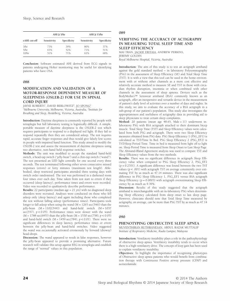
eAHI cut-off
AHI ≥ 5/hr AHI ≥ 15/hr
Sensitivity Specificity Sensitivity Specificity
3/hr 73% 39% 80% 37%5/hr 63% 52% 71% 51%10/hr 51% 71% 62% 68%
Conclusion: Software estimated AHI derived from ECG signals inpatients undergoing Holter monitoring may be useful for identifyingpatients who have OSA.
088
MODIFICATION AND VALIDATION OF AMOTOR-RESPONSE DEPENDENT MEASURE OFSLEEPINESS (OSLER-2) FOR USE IN SPINALCORD INJURYJAYNE ROBERTS1, DAVID BERLOWITZ2, JO SPONG2
1Melbourne University, Melbourne, Victoria, Australia, 2Institute forBreathing and Sleep, Heidelberg, Victoria, Australia
Introduction: Daytime sleepiness is commonly reported by people withtetraplegia but full laboratory testing is logistically difficult. A simple,portable measure of daytime sleepiness is the OSLER-2 test whichrequires participants to respond to a displayed red light. If they fail torespond repeatedly then they are considered asleep. The test requiresrapid, accurate finger responses and thus would give falsely low scoresin people with impaired hand function. This study aimed to modify theOSLER-2 test and assess the measurement of daytime sleepiness usingtwo alternative, non-hand held response switches.Methods: The unit was modified to accept the standard hand-heldswitch, a head-tap switch (“jelly-bean”) and a chin-tap switch (“wand”).The test presented an LED light centrally for one second every threeseconds. The test terminated following either seven consecutive missedresponses (errors) or forty minutes (maximum test length). Able-bodied, sleep restricted participants attended three testing days withswitch order randomised. The test was performed in a darkened roomfour times over each day. Time taken from test start to errors if theyoccurred (sleep latency), performance times and errors were recorded.Video was recorded to qualitatively describe performance.Results: 22 participants (median age = 21 yrs) with no diagnosed sleepdisorders were recruited. Analyses were conducted on those who fellasleep only (sleep latency) and again including those who completedthe test without falling asleep (performance times). Participants tooklonger to fall asleep when using the wand (M = 1265 sec(546)) than thejelly-bean (M = 1102(544)) and hand-held switch (M = 1037sec(557), p < 0.05). Performance times were slower with the wand(M = 1788 sec(695)) than the jelly-bean (M = 1530 sec(758); p < 0.05)and hand-held switch (M = 1459 sec(784); p < 0.01). There were nosignificant differences in sleep latency, performance times or errorsbetween the jelly-bean and hand-held switches. Video suggestedthe wand was occasionally activated erroneously by forward (drowsy)head-drops.Discussion: The wand appeared to result in false responses, howeverthe jelly-bean appeared to provide a promising alternative. Futureresearch will validate this setup against EEG in tetraplegia and establishthe range of “normal” values in this population.
089
VERIFYING THE ACCURACY OF ACTIGRAPHYIN MEASURING TOTAL SLEEP TIME ANDSLEEP EFFICIENCYMAI TRAN, JACKIE ERITAIA, ANDREW PERKINS,JEREMY GOLDINRoyal Melbourne Hospital, Victoria, Australia
Introduction: The aim of this study is to test an actigraph armbandagainst the gold standard method – in laboratory Polysomnography(PSG) in the assessment of Sleep Efficiency (SE) and Total Sleep Time(TST). It is with a view that this tool can be used in the home environ-ment with or without other channels as a more cost effective andrelatively accurate method to measure SE and TST in those with circa-dian rhythm disruption, insomnia or when combined with otherchannels in the assessment of sleep apnoea. Devices such as theBodyMedia’s™ Senswear armband (BSA) commonly known as anactigraph, offer an inexpensive and versatile device in the measurementof patient’s daily level of activities over a number of days and nights. Inthis study, we aim to evaluate the accuracy of a BSA actigraph in asub-group of our patient’s population. This study also investigates theappropriateness and usefulness of actigraphy data in providing aid tosleep physicians to treat certain sleep complaints.Method: 20 patients (mean age 48.65, Male = 11) underwent in-laboratory PSG with BSA actigraph attached to their dominant bicepmuscle. Total Sleep Time (TST) and Sleep Efficiency values were calcu-lated from both PSG and actigraph. There were two Sleep Efficiencymeasures obtained from PSG data. PSG Sleep Efficiency 1 (PSG_EF1) iscalculated as TST/Time In Bed. PSG Sleep Efficiency 2 (PSG_EF2) isTST/Sleep Period Time. Time in bed is measured from light off to lighton. Sleep Period Time is measured from Sleep Onset to Last Sleep Page.The Altmand-Bland Agreement analysis was used to compare TST andSleep Efficiency values from the two methods.Results: There was no significant difference in actigraph Sleep Effi-ciency value when compared to PSG Sleep Efficiency 2, PSG_EF2(p = 0.2701). A significant difference was found between the two TSTvalues (p < .001) with actigraph TST on average consistently over esti-mating TST by as much as 47.14 minutes. There was also significantdifference in PSG Sleep Efficiency 1, PSG_EF1 versus BSA actigraphSleep Efficiency (p = 0.0003) with actigraph overestimating Sleep Effi-ciency by as much as 9.39%.Discussion: Results of this study suggested that the actigrapharmband is interchangeable with an in-laboratory PSG when determin-ing Sleep efficiency calculated from sleep onset to last sleep page.However, clinicians should note that Total Sleep Time measured byactigraphy, on average, can be more than PSG TST by as much as 47.14minutes.
090
PHENOTYPING OBSTRUCTIVE SLEEP APNEAMUVENTHIRAN RUTHRANESAN, ABDUL RAZAK MUTTALIFInstitute of Respiratory Medicine, Kuala Lumpur, Malaysia
Introduction: Ventilatory instability plays a role in the pathophysiologyof obstructive sleep apnea. Ventilatory instability tends to occur whenthere is a high ventilatory drive. The concept of loop gain has been usedto explain ventilatory instability.Objectives: To highlight the importance of recognising phenotypesof Obstructive sleep apnea patients who would benefit from combina-tion therapy with Continuous Positive airway pressure (CPAP) andacetazolamide.
Sleep, Science and Research
24 © 2014 The AuthorsSleep and Biological Rhythms © 2014 Japanese Society of Sleep Research
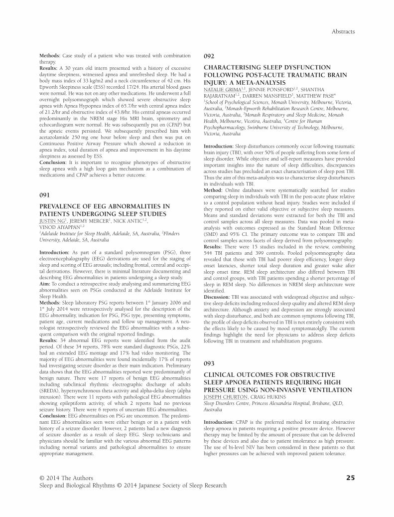
Methods: Case study of a patient who was treated with combinationtherapy.Results: A 30 years old intern presented with a history of excessivedaytime sleepiness, witnessed apnea and unrefreshed sleep. He had abody mass index of 33 kg/m2 and a neck circumference of 42 cm. HisEpworth Sleepiness scale (ESS) recorded 17/24. His arterial blood gaseswere normal. He was not on any other medications. He underwent a fullovernight polysomnograph which showed severe obstructive sleepapnea with Apnea Hypopnea index of 65.7/hr with central apnea indexof 21.2/hr and obstructive index of 43.8/hr. His central apneas occurredpredominantly in the NREM stage His MRI brain, spirometry andechocardiogram were normal. He was subsequently put on (CPAP) butthe apneic events persisted. We subsequently prescribed him withacetazolamide 250 mg one hour before sleep and then was put onContinuous Positive Airway Pressure which showed a reduction inapnea index, total duration of apnea and improvement in his daytimesleepiness as assessed by ESS.Conclusion: It is important to recognise phenotypes of obstructivesleep apnea with a high loop gain mechanism as a combination ofmedications and CPAP achieves a better outcome.
091
PREVALENCE OF EEG ABNORMALITIES INPATIENTS UNDERGOING SLEEP STUDIESJUSTIN NG1, JEREMY MERCER1, NICK ANTIC1,2,VINOD AIYAPPAN1,2
1Adelaide Institute for Sleep Health, Adelaide, SA, Australia, 2FlindersUniversity, Adelaide, SA, Australia
Introduction: As part of a standard polysomnogram (PSG), threeelectroencephalography (EEG) derivations are used for the staging ofsleep and scoring of EEG arousals; including frontal, central and occipi-tal derivations. However, there is minimal literature documenting anddescribing EEG abnormalities in patients undergoing a sleep study.Aim: To conduct a retrospective study analysing and summarizing EEGabnormalities seen on PSGs conducted at the Adelaide Institute forSleep Health.Methods: Sleep laboratory PSG reports between 1st January 2006 and1st July 2014 were retrospectively analysed for the description of theEEG abnormality, indication for PSG, PSG type, presenting symptoms,patient age, current medications and follow up management. A neu-rologist retrospectively reviewed the EEG abnormalities with a subse-quent comparison with the original reported findings.Results: 34 abnormal EEG reports were identified from the auditperiod. Of these 34 reports, 78% were standard diagnostic PSGs, 22%had an extended EEG montage and 17% had video monitoring. Themajority of EEG abnormalities were found incidentally. 17% of reportshad investigating seizure disorder as their main indication. Preliminarydata shows that the EEG abnormalities reported were predominantly ofbenign nature. There were 17 reports of benign EEG abnormalitiesincluding subclinical rhythmic electrographic discharge of adults(SREDA), hypersynchronous theta activity and alpha-delta sleep (alphaintrusion). There were 11 reports with pathological EEG abnormalitiesshowing epileptiform activity, of which 2 reports had no previousseizure history. There were 6 reports of uncertain EEG abnormalities.Conclusion: EEG abnormalities on PSG are uncommon. The predomi-nant EEG abnormalities seen were either benign or in a patient withhistory of a seizure disorder. However, 2 patients had a new diagnosisof seizure disorder as a result of sleep EEG. Sleep technicians andphysicians should be familiar with the various abnormal EEG patternsincluding normal variants and pathological abnormalities to ensureappropriate management.
092
CHARACTERISING SLEEP DYSFUNCTIONFOLLOWING POST-ACUTE TRAUMATIC BRAININJURY: A META-ANALYSISNATALIE GRIMA1,2, JENNIE PONSFORD1,2, SHANTHARAJARATNAM1,2, DARREN MANSFIELD3, MATTHEW PASE4
1School of Psychological Sciences, Monash University, Melbourne, Victoria,Australia, 2Monash-Epworth Rehabilitation Research Centre, Melbourne,Victoria, Australia, 3Monash Respiratory and Sleep Medicine, MonashHealth, Melbourne, Vicotira, Australia, 4Centre for HumanPsychopharmacology, Swinburne University of Technology, Melbourne,Victoria, Australia
Introduction: Sleep disturbances commonly occur following traumaticbrain injury (TBI), with over 50% of people suffering from some form ofsleep disorder. While objective and self-report measures have providedimportant insights into the nature of sleep difficulties, discrepanciesacross studies has precluded an exact characterisation of sleep post TBI.Thus the aim of this meta-analysis was to characterise sleep disturbancesin individuals with TBI.Method: Online databases were systematically searched for studiescomparing sleep in individuals with TBI in the post-acute phase relativeto a control population without head injury. Studies were included ifthey reported on either valid objective or subjective sleep measures.Means and standard deviations were extracted for both the TBI andcontrol samples across all sleep measures. Data was pooled in meta-analysis with outcomes expressed as the Standard Mean Difference(SMD) and 95% CI. The primary outcome was to compare TBI andcontrol samples across facets of sleep derived from polysomnography.Results: There were 15 studies included in the review, combining544 TBI patients and 399 controls. Pooled polysomnography datarevealed that those with TBI had poorer sleep efficiency, longer sleeponset latencies, shorter total sleep duration and greater wake aftersleep onset time. REM sleep architecture also differed between TBIand control groups, with TBI patients spending a shorter percentage ofsleep in REM sleep. No differences in NREM sleep architecture wereidentified.Discussion: TBI was associated with widespread objective and subjec-tive sleep deficits including reduced sleep quality and altered REM sleeparchitecture. Although anxiety and depression are strongly associatedwith sleep disturbance, and both are common symptoms following TBI,the profile of sleep deficits observed in TBI is not entirely consistent withthe effects likely to be caused by mood symptomatolglly. The currentfindings highlight the need for physicians to address sleep deficitsfollowing TBI in treatment and rehabilitation programs.
093
CLINICAL OUTCOMES FOR OBSTRUCTIVESLEEP APNOEA PATIENTS REQUIRING HIGHPRESSURE USING NON-INVASIVE VENTILATIONJOSEPH CHURTON, CRAIG HUKINSSleep Disorders Centre, Princess Alexandria Hospital, Brisbane, QLD,Australia
Introduction: CPAP is the preferred method for treating obstructivesleep apnoea in patients requiring a positive pressure device. Howevertherapy may be limited by the amount of pressure that can be deliveredby these devices and also due to patient intolerance as high pressure.The use of bi-level NIV has been considered in these patients so thathigher pressures can be achieved with improved patient tolerance.
Abstracts
25© 2014 The AuthorsSleep and Biological Rhythms © 2014 Japanese Society of Sleep Research
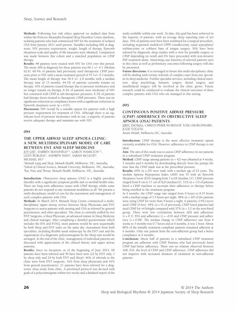
Methods: Following low risk ethics approval we audited data fromwithin the Princess Alexandra Hospital Sleep Disorders Centre database,isolating patients who had commenced NIV for the treatment of isolatedOSA from January 2011 until present. Variables including AHI at diag-nosis, NIV pressure requirement, weight, length of therapy, Epworthsleepiness scale and quality of life measures were obtained. Comparisonwas made between this cohort and a matched population on CPAPtherapy.Results: 48 patients were treated with NIV for OSA over this period.The mean AHI at diagnosis for these patients was 66.1 +/− 6.1 (Median62.6). 60.4% of patients had previously used therapeutic CPAP pres-sures prior to NIV with a mean treatment period of 55.7+/− 9.3 months.The mean length of therapy was 18.4 +/− 2.8 months with a mediantherapy time of 13 months. 64.3% of patients currently remain ontherapy. 10% of patients ceased therapy due to pressure intolerance andno longer remain on therapy. 6.3% of patients were intolerant of NIVbut continued with CPAP at sub therapeutic pressures. 6.3% of patientshad therapy down titrated to therapeutic CPAP pressures. There was nosignificant reduction in compliance hours with a significant reduction inEpworth sleepiness score (p = 0.01).Discussion: NIV would be a suitable option for patients with a highpressure requirement for treatment of OSA. Although there is an sig-nificant level of pressure intolerance with its use, a majority of patientsreceive adequate therapy and maintain use with NIV.
094
THE UPPER AIRWAY SLEEP APNOEA CLINIC:A NEW, MULTIDISCIPLINARY MODEL OF CAREBETWEEN ENT AND SLEEP MEDICINEJOY LEE1, DARREN MANSFIELD1,2, GARUN HAMILTON1,2,GLEN BURGESS3, ANDREW HAYS3, SARAH MCLEOD3,MICHAEL HO1
1Monash Lung and Sleep, Monash Health, Melbourne, VIC, Australia,2School of Clinical Sciences, Monash University, Melbourne, VIC, Australia,3Ear, Nose and Throat, Monash Health, Melbourne, VIC, Australia
Introduction: Obstructive sleep apnoea (OSA) is a highly prevalentdisorder with a significant symptom profile and co-morbid health risks.There are long term adherence issues with CPAP therapy whilst somepatients do not respond to any treatment modalities at all. We present amulti-disciplinary model of care for the assessment and management ofsuch complex patients with OSA.Methods: In March 2014, Monash Sleep Centre commenced a multi-disciplinary upper airway service between Sleep Physicians and ENTSurgeons to assess patients with snoring and OSA as referred by generalpractitioners, and other specialists. The clinic is currently staffed by twoENT Surgeons, a Sleep Physician, an advanced trainee in Sleep Medicineand clinical manager. After completing a detailed questionnaire whichincludes the ESS and FOSQ, most patients would be seen sequentiallyby both Sleep and ENT units on the same day. Assessment from bothspecialties, including flexible nasal endoscopy by the ENT unit and theorganisation of a diagnostic polysomnogram by the Sleep unit would bearranged. At the end of the clinic, management of individual patients arediscussed with appreciation of the clinical history and upper airwayanatomy.Results: Since its inception, as of the beginning of June 2014, 60patients have been referred and 50 have been seen (22 by ENT only, 1by sleep only and 24 by both ENT and Sleep). 46% of referrals to theclinic were from ENT surgeons, 16% from sleep physicians and 42%from general practitioners). 21 patients have been referred for a diag-nostic sleep study from clinic. A prioritised protocol was devised withgoals of a polysomnogram within two weeks and a finalised report of the
study available within one week. To date, this goal has been achieved inthe majority of patients, with an average sleep reporting time of tendays. 76% of patients seen have been waitlisted for a surgical procedure,including sequential multilevel UPPP, tonsillectomy, nasal septoplasty/turbinectomy or coblator base of tongue surgery. 36% have beenreferred by diagnostic sleep studies with a view for possible surgery orCPAP depending on result and 6% have proceeded with non-invasivePAP treatment alone. Interesting case histories of selected patients seenin this clinic as well as preliminary outcomes following surgery will alsobe presented.Future directions: It is envisaged in future this multi-disciplinary clinicwill be dealing with tertiary referrals of complex cases from any special-ist in sleep medicine. Further specialist services, including clinical nutri-tion, sleep psychology, bariatric surgery, dental surgery, andmaxillofacial surgery will be involved as the clinic grows. Futureresearch could be conducted to evaluate the clinical outcomes of thesecomplex patients with OSA who are managed in this setting.
095
CONTINUOUS POSITIVE AIRWAY PRESSURE(CPAP) ADHERENCE IN OBSTRUCTIVE SLEEPAPNOEA (OSA) PATIENTSJIBIN THOMAS, CHRISTOPHER WORSNOP, TOM CHURCHWARD,JULIE TOLSONAustin Helath, Melbourne,VIC, Australia
Introduction: CPAP therapy is the most effective treatment optioncurrently available for OSA. However, adherence to CPAP therapy is notideal.Aim: The aim of this study was to assess CPAP adherence in our patientsin a subsidised CPAP treatment program.Method: CPAP usage among patients (n = 42) was obtained at 4 weeks,3 months and 6 months by downloading directly from the pumps thetime that the CPAP mask was at the prescribed pressure.Results: 69% (n = 29) were male with a median age of 63 years. Themedian Apnoea Hypopnoea Index (AHI) was 35 with an EpworthSleepiness Score (ESS) ranging from 1 to18 (median 11). CPAP pressureranged from 8 cm to 17 cm of H20 (median12). 31% (n = 13) of patientshired a CPAP machine to ascertain their adherence to therapy beforebeing enrolled in the treatment program.At 6 months, the CPAP usage rate ranged from 0 hours to 8.25 hourswith a median usage of 3.5 hours per night. 48% (n = 20) of the patientswere using CPAP for more than 4 hours a night. 6 patients (14%) neverused CPAP (0 hrs). 69% (n = 9) of previously CPAP hired patients hadused CPAP for >4 h/night compared with 37% (n = 11) in the non-hiredgroup. There were low correlations between AHI and adherence(r = 0.1), ESS and adherence (r = −0.6) and CPAP pressure and adher-ence (r = 0.08). The median change in CPAP adherence rate from 4weeks to 3 months was 0.7 hours and at 6 months, it was 1 hour. About80% of the initially treatment compliant patients remained adherent at6 months. Only one patient from the non-adherent group had a bettercompliance at 6 months.Conclusion: About half of patients in a subsidised CPAP treatmentprogram are adherent with CPAP. Patients who had previously hiredCPAP had better adherence. There was no relation observed betweenAHI, ESS, the level of CPAP and CPAP adherence. CPAP adherence didnot improve with increased duration of treatment in non-adherentpatients.
Sleep, Science and Research
26 © 2014 The AuthorsSleep and Biological Rhythms © 2014 Japanese Society of Sleep Research
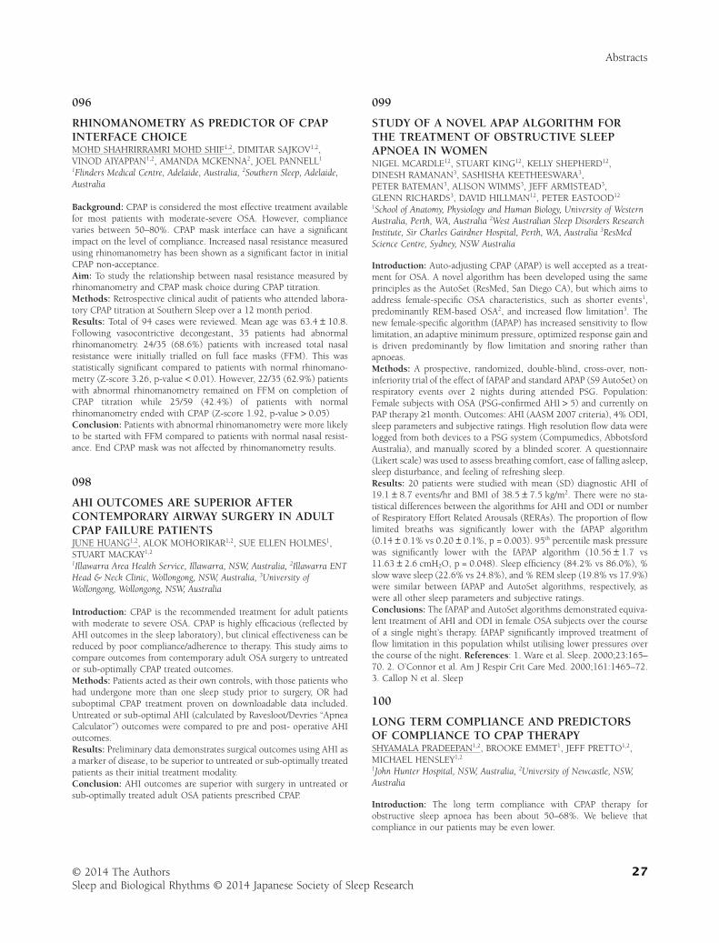
096
RHINOMANOMETRY AS PREDICTOR OF CPAPINTERFACE CHOICEMOHD SHAHRIRRAMRI MOHD SHIF1,2, DIMITAR SAJKOV1,2,VINOD AIYAPPAN1,2, AMANDA MCKENNA2, JOEL PANNELL1
1Flinders Medical Centre, Adelaide, Australia, 2Southern Sleep, Adelaide,Australia
Background: CPAP is considered the most effective treatment availablefor most patients with moderate-severe OSA. However, compliancevaries between 50–80%. CPAP mask interface can have a significantimpact on the level of compliance. Increased nasal resistance measuredusing rhinomanometry has been shown as a significant factor in initialCPAP non-acceptance.Aim: To study the relationship between nasal resistance measured byrhinomanometry and CPAP mask choice during CPAP titration.Methods: Retrospective clinical audit of patients who attended labora-tory CPAP titration at Southern Sleep over a 12 month period.Results: Total of 94 cases were reviewed. Mean age was 63.4 ± 10.8.Following vasocontrictive decongestant, 35 patients had abnormalrhinomanometry. 24/35 (68.6%) patients with increased total nasalresistance were initially trialled on full face masks (FFM). This wasstatistically significant compared to patients with normal rhinomano-metry (Z-score 3.26, p-value < 0.01). However, 22/35 (62.9%) patientswith abnormal rhinomanometry remained on FFM on completion ofCPAP titration while 25/59 (42.4%) of patients with normalrhinomanometry ended with CPAP (Z-score 1.92, p-value > 0.05)Conclusion: Patients with abnormal rhinomanometry were more likelyto be started with FFM compared to patients with normal nasal resist-ance. End CPAP mask was not affected by rhinomanometry results.
098
AHI OUTCOMES ARE SUPERIOR AFTERCONTEMPORARY AIRWAY SURGERY IN ADULTCPAP FAILURE PATIENTSJUNE HUANG1,2, ALOK MOHORIKAR1,2, SUE ELLEN HOLMES1,STUART MACKAY1,2
1Illawarra Area Health Service, Illawarra, NSW, Australia, 2Illawarra ENTHead & Neck Clinic, Wollongong, NSW, Australia, 3University ofWollongong, Wollongong, NSW, Australia
Introduction: CPAP is the recommended treatment for adult patientswith moderate to severe OSA. CPAP is highly efficacious (reflected byAHI outcomes in the sleep laboratory), but clinical effectiveness can bereduced by poor compliance/adherence to therapy. This study aims tocompare outcomes from contemporary adult OSA surgery to untreatedor sub-optimally CPAP treated outcomes.Methods: Patients acted as their own controls, with those patients whohad undergone more than one sleep study prior to surgery, OR hadsuboptimal CPAP treatment proven on downloadable data included.Untreated or sub-optimal AHI (calculated by Ravesloot/Devries “ApneaCalculator”) outcomes were compared to pre and post- operative AHIoutcomes.Results: Preliminary data demonstrates surgical outcomes using AHI asa marker of disease, to be superior to untreated or sub-optimally treatedpatients as their initial treatment modality.Conclusion: AHI outcomes are superior with surgery in untreated orsub-optimally treated adult OSA patients prescribed CPAP.
099
STUDY OF A NOVEL APAP ALGORITHM FORTHE TREATMENT OF OBSTRUCTIVE SLEEPAPNOEA IN WOMENNIGEL MCARDLE12, STUART KING12, KELLY SHEPHERD12,DINESH RAMANAN3, SASHISHA KEETHEESWARA3,PETER BATEMAN3, ALISON WIMMS3, JEFF ARMISTEAD3,GLENN RICHARDS3, DAVID HILLMAN12, PETER EASTOOD12
1School of Anatomy, Physiology and Human Biology, University of WesternAustralia, Perth, WA, Australia 2West Australian Sleep Disorders ResearchInstitute, Sir Charles Gairdner Hospital, Perth, WA, Australia 3ResMedScience Centre, Sydney, NSW Australia
Introduction: Auto-adjusting CPAP (APAP) is well accepted as a treat-ment for OSA. A novel algorithm has been developed using the sameprinciples as the AutoSet (ResMed, San Diego CA), but which aims toaddress female-specific OSA characteristics, such as shorter events1,predominantly REM-based OSA2, and increased flow limitation3. Thenew female-specific algorithm (fAPAP) has increased sensitivity to flowlimitation, an adaptive minimum pressure, optimized response gain andis driven predominantly by flow limitation and snoring rather thanapnoeas.Methods: A prospective, randomized, double-blind, cross-over, non-inferiority trial of the effect of fAPAP and standard APAP (S9 AutoSet) onrespiratory events over 2 nights during attended PSG. Population:Female subjects with OSA (PSG-confirmed AHI > 5) and currently onPAP therapy ≥1 month. Outcomes: AHI (AASM 2007 criteria), 4% ODI,sleep parameters and subjective ratings. High resolution flow data werelogged from both devices to a PSG system (Compumedics, AbbotsfordAustralia), and manually scored by a blinded scorer. A questionnaire(Likert scale) was used to assess breathing comfort, ease of falling asleep,sleep disturbance, and feeling of refreshing sleep.Results: 20 patients were studied with mean (SD) diagnostic AHI of19.1 ± 8.7 events/hr and BMI of 38.5 ± 7.5 kg/m2. There were no sta-tistical differences between the algorithms for AHI and ODI or numberof Respiratory Effort Related Arousals (RERAs). The proportion of flowlimited breaths was significantly lower with the fAPAP algorithm(0.14 ± 0.1% vs 0.20 ± 0.1%, p = 0.003). 95th percentile mask pressurewas significantly lower with the fAPAP algorithm (10.56 ± 1.7 vs11.63 ± 2.6 cmH2O, p = 0.048). Sleep efficiency (84.2% vs 86.0%), %slow wave sleep (22.6% vs 24.8%), and % REM sleep (19.8% vs 17.9%)were similar between fAPAP and AutoSet algorithms, respectively, aswere all other sleep parameters and subjective ratings.Conclusions: The fAPAP and AutoSet algorithms demonstrated equiva-lent treatment of AHI and ODI in female OSA subjects over the courseof a single night’s therapy. fAPAP significantly improved treatment offlow limitation in this population whilst utilising lower pressures overthe course of the night. References: 1. Ware et al. Sleep. 2000;23:165–70. 2. O’Connor et al. Am J Respir Crit Care Med. 2000;161:1465–72.3. Callop N et al. Sleep
100
LONG TERM COMPLIANCE AND PREDICTORSOF COMPLIANCE TO CPAP THERAPYSHYAMALA PRADEEPAN1,2, BROOKE EMMET1, JEFF PRETTO1,2,MICHAEL HENSLEY1,2
1John Hunter Hospital, NSW, Australia, 2University of Newcastle, NSW,Australia
Introduction: The long term compliance with CPAP therapy forobstructive sleep apnoea has been about 50–68%. We believe thatcompliance in our patients may be even lower.
Abstracts
27© 2014 The AuthorsSleep and Biological Rhythms © 2014 Japanese Society of Sleep Research
Aims: 1.To investigate CPAP compliance after 4 or more years oftreatment2. Identify factors influencing patient compliance to CPAP therapy.Methods: Patients who commenced CPAP therapy in the year of 2008–2009 (4–5 yrs after CPAP commencement) were identified using thesleep disorders patient data base. Consenting patients were contacted byletters and telephone calls to complete a questionnaire regarding theirCPAP usage. A second reminder was sent to them after 3 months. Thenon-responders were phoned with the existing numbers that our database had. Compliance was measured by self-reported compliance of atleast 4 hours for 5 days of the week.Results: 469 patients were identified to be commenced on CPAP. 30patients were deceased by that time. 23 patients did not want to par-ticipate. 261(63%) patients were managed to be contacted. 132(50.6%)patients reported to be compliant. 129(49.45) reported to be non-compliant. Average apnoea hypopnoea index (AHI) of Compliant andNon-Compliant patients were 58 and 38 respectively. The averageSPO2 < 90 was 56 minutes in Compliant patients compared to 21minutes in Non-compliant patients. Information was sparse in the Non-compliant with regards to employment, marital status and who paid forthe CPAP machine, but overall Compliant patients had more secondaryand tertiary education and being married compared to Non-compliantpatients. 40% of the compliant patients had CPAP provided by thehospital and the 60% bought their own machine.Summary and Conclusions: Overall long term CPAP in our populationis about 50% and lower compared to published previous studies. CPAP,although is an effective treatment, is not well-tolerated. Efforts may haveto be directed at more targeted, user friendly treatment options.
101
IMPROVEMENTS OF QUALITY OF LIFEBETWEEN CPAP AND APAP TREATMENT INOBSTRUCTIVE SLEEP APNEAEN-TING CHANG, HSIU-MEI WANGBuddhist Tzu Chi General Hospital, Hualien, Taiwan
Background: Obstructive sleep apnea (OSA) is a sleep disorder withrepeated nocturnal desaturation and sleep fragmentation. It can lead topoor quality of life. Our goal was to investigate improvements of qualityof life (SF-36) between 3- month treatment of CPAP or APAP of OSApatients.Method: This was a prospective, randomized and crossover study. Therewere 19 patients (Male/ female: 18/1) suffering from OSA with defini-tion of apnea/hypopnea index (AHI) >15/hr. The studied subjectswere randomised divided into APAP or CPAP group, with 3 months laterthe two group crossover. All subjects received CPAP or APAP treatmentover than 4 hours per day during treatment. SF-36 was completedbefore and after 3-month CPAP or APAP treatment in each studiedsubjects.Result: Nineteen subjects with OSA had severe OSA (mean AHI was59.7 ± 23.9/hr) and the mean optimal pressure of CPAP titration was8.7 ± 1.5 mmHg. Comparing to APAP treatment group, CPAP grouphad higher mean pressure 8.7 ± 1.4 mmHg as well as lower AHI0.6 ± 1.4 than APAP group. No difference between compliances in APAPor CPAP group. After APAP or CPAP treatment, both treatment groupshad significant improvement in general health and vatality comparing tothose before treatment. However, there were no significant improve-ment in each components of SF-36 between 3-month APAP or CPAPgroup.Conclusion: In our study, CPAP had higher pressure and lower AHIthan APAP group. Both CPAP and APAP group can improved generalhealth and vatality in OSA patients.
102
ASSESSMENT OF A NEW NASAL PILLOWS MASKIN PATIENTS RECEIVING CPAPALISA PHAM, SAHISHA KETHEESWARAN, JEFF ARMITSTEADResMed Science Centre, Sydney, NSW, Australia
Study Objectives: Mask side effects such as pressure sores, leakage andclaustrophobia can all limit adherence to CPAP therapy. These may bereduced with the use of nasal pillows. Nasal pillows have also beenshown to be as efficacious as nasal masks and a suitable alternative forpatients receiving high CPAP pressures. Although nasal pillows are aneffective option, factors such as noise can affect patient acceptance totherapy and can disturb partners. As technological development con-tinues, newer generations of masks such as ResMed AirFit P10 nasalpillows aim to address issues such as noise and comfort.Aims: This study was conducted to assess the comfort, seal, stability andpatient preference of the AirFit P10 nasal pillows mask (ResMed Ltd,Bella Vista, Australia) compared with the ResMed Swift FX nasal pillowsmask.Methods: 21 OSA patients, established on CPAP therapy (≥6 months)were enrolled into the study. Baseline objective device data from the last7 consecutive nights on participants’ current Swift FX mask were down-loaded. Participants then began using the AirFit P10 mask at home for7 consecutive nights in a prospective open label trial. Objective devicedata was obtained via device download. Also, subjective feedback wascollected via an 11-point Likert scale (0 = major issue, 5 = ok, and10 = highly positive).Results: 21 subjects (18 males, 3 females) completed the study. Therewas a statistically significant difference in objective average daily CPAPusage hours favouring the AirFit P10 over the Swift FX (7.4 ± 1.1 vs.6.7 ± 1.4 (Means ± SD), p = 0.001) (hr/night). AHI, 95th percentilePressure and 95th percentile Leak were found to be equivalent. Subjec-tive ratings for mask seal, stability, facial marks and overall performancewere significantly higher (better) for AirFit P10 compared with Swift FX.Median Likert scale scores for AirFit P10 vs. Swift FX were as follows:Seal 9 vs. 8, Stability 10 vs. 8, Facial marks 10 vs. 8, Overall Perfor-mance 10 vs. 8, respectively. 76% of participants preferred AirFit P10,19% preferred Swift FX and 5% found no difference between the twomasks.Conclusion: New technology continues to improve nasal pillowsmasks, with new generation interfaces such as the AirFit P10 maskoffering a number of potential comfort advantages.
103
DEVELOPMENT OF A MODEL OF CARE FORAUSTRALIAN COMMUNITY PHARMACIESPROVIDING OBSTRUCTIVE SLEEPAPNEA SERVICESCARISSA HANES1,3, KEITH WONG2,4, BANDANA SAINI1,2
1University of Sydney, Sydney, NSW, Australia, 2Woolcock Institute ofMedical Research, Sydney, NSW, Australia, 3NHMRC Centre for IntegratedResearch and Understanding of Sleep (CIRUS), Sydney, NSW, Australia,4Royal Prince Alfred Hospital, Camperdown, NSW, Australia
Introduction: The supply of continuous positive airway pressure(CPAP) therapy is unregulated in Australia and a variety of providervenues and practice models exist. Some community pharmacies haveundertaken a role in providing CPAP and obstructive sleep apnea(OSA)-related services, including home diagnostic studies. Our previ-ous research identified a need to define best practice and standardise
Sleep, Science and Research
28 © 2014 The AuthorsSleep and Biological Rhythms © 2014 Japanese Society of Sleep Research
OSA service provision in pharmacies. The aim of this study was todevelop a stakeholder-led model of care for OSA services in communitypharmacies.Methods: The study was conducted in three phases: 1.) A qualitativephase involving telephone interviews with pharmacy-based CPAP prac-titioners. 2.) A focus group with broader stakeholder input, includingrepresentatives from peak professional sleep and pharmacy bodies,medical practitioners, patients and industry. 3.) An online modifiedDelphi study with a joint stakeholder panel to develop consensus-defined practice guidelines.Results: Telephone interviews with 22 pharmacy staff identified currentdiagnostic and treatment pathways in the community, and found adesire for professional training programmes amongst CPAP practition-ers. The 9-member focus group obtained input from key stakeholderson best practice models for diagnostic pathways. Both medical andethical concerns were raised over certain pathways operating in thecommunity. Following this, a panel of 35 stakeholders participated inthe Delphi study, culminating in 51 criteria achieving consensus forinclusion in the final model of care. These consensus-defined criteriahave been distributed to peak professional bodies for translation into apolicy document for profession-wide dissemination.Discussion: Key outputs from this project include future collaborationbetween peak sleep and pharmacy bodies to develop practice guidelinesand professional training programmes. It is important to establish aframework for medically appropriate sleep apnea services, and to stand-ardise care for patients accessing OSA treatment through pharmacies.Once measures of quality have been established, patient outcomes ulti-mately need to be measured to facilitate continuous practice improve-ment. Future initiatives are required to determine the quality of OSAservices in other provider settings.
104
FACTORS DETERMINING THE ACCEPTANCE OFCPAP THERAPYANTONIUS STEVEN, IVAN LING, GAVIN STURDY,ASHLEIGH BECU, THOMAS ANSELL, BHAJAN SINGHWestern Australia Sleep Disorders Research Institute, Perth, WesternAustralia, Australia
Introduction: Continuous positive airway pressure (CPAP) therapy isthe most effective treatment for obstructive sleep apnoea (OSA) butCPAP acceptance is variable. The aim of this study was to identifypatient characteristics and polysomnographic parameters that are asso-ciated with CPAP acceptance.Methods: We examined the characteristics and polysomnographic dataof patients who completed a home trial of CPAP therapy and attended asleep physician and technologist CPAP therapy clinic over a 41⁄2 monthperiod between September 2013 and January 2014. Patient character-istics included body mass index (BMI), age and gender. Polysomno-graphic data included apnoea-hypopnoea index (AHI), mean and nadiroxygen saturation (SpO2), oxygen desaturation index (ODI), time spentat SpO2, <90%, and arousal index. Acceptance of CPAP at the end of theCPAP trial was recorded. Characteristics of patients who accepted andrejected CPAP were compared using unpaired t-tests.Results: Of the 78 patients who completed CPAP trial, 47 (60%)accepted, 18 (23%) rejected and 13 (17%) were undecided about longterm CPAP therapy. Characteristics of patients who accepted CPAPtherapy were different from those who rejected CPAP therapy: they wereof higher BMI (35.7 vs. 29.7, p < 0.02) and were older (54 vs. 45 years,p < 0.05). Higher AHI (45.7 vs. 27.4 events/hr, p < 0.02), greater timespent at SpO2 < 90% (52 vs. 19 minutes, p = 0.01), lower nadir SpO2
(78% vs. 85%, p < 0.02), and higher ODI 4% (31 vs. 19 events/hr,
p = 0.03) were associated with greater CPAP acceptance. There was atrend towards a significant difference in ODI 3% (40 vs. 26 events/hr,p = 0.06) between the two groups. Total arousal index and respiratoryarousal index were not associated with CPAP acceptance. CPAP intoler-ance and mask discomfort were the most common reasons for CPAPrejection.Conclusion: CPAP therapy acceptance was associated with older age,higher BMI, more severe OSA, and more severe indices of hypoxaemia(time spent at SpO2 < 90%,nadir SpO2, and ODI 4%).
105
COMPARISON OF OSA AND CPAP THERAPYREQUIREMENTS IN CHINESE VERSUSNON-CHINESE POPULATIONYEE TUNG (ERIC) KUO1, DENISE O’DRISCOLL1,2,FRANK THIEN1,2, ALAN YOUNG1,2
1Eastern Health, Melbourne, Australia, 2Monash University, Melbourne,Australia
Introduction: Obstructive sleep apnoea (OSA) is strongly associatedwith central obesity. However, despite a lower mean body mass index(BMI) in Asian populations, there is a similar prevalence of OSA. Con-tinuous positive airway pressure (CPAP) is currently the most effectivetreatment for severe OSA. The aim of this study is to determine whetherChinese patients with severe OSA have a lower BMI and CPAP require-ment compared to non-Chinese patients.Methods: Retrospective case-control study utilising the Eastern HealthSleep database between January 2011 and June 2014. Diagnosticpolysomnography (PSG), in-lab CPAP implementation PSG and homeCPAP implementation studies of Chinese patients with severe OSA (totalAHI > 30 events/h) and non-Chinese patients matched for age, genderand total AHI were analysed. Patients with unclear ethnicity based onname, obesity hypoventilation syndrome, non-OSA sleep disorders orsignificant central apnoea were excluded. Demographic, polysomno-graphic and CPAP data were compared between groups using pairedt-tests or Wilcoxon Signed rank tests depending on normality. Data arepresented as mean ± SEM or median (IQR) where appropriate.Results: A total of 60 patients (54 male) were included in the study: 30Chinese (C) and 30 non-Chinese (NC). As the subjects were matched forage and total AHI, there was no significant difference between groups(ageC:55.6 ± 2.5 y, ageNC = 52 ± 3.1 y. AHIC = 57.7 ± 3.4 events/h,AHINC = 57.8 ± 3.5 events/h. p > 0.05 for both). However, the Chinesegroup had a significantly lower supine NREM AHI (62.7 ± 4.2 vs.78.4 ± 6.3 events/h, p < 0.05). BMI was significantly lower in theChinese group (median 27.6 (IQR 25.0–31.3) versus 33.1 (IQR 30.7–37.3) kg/m2; P < 0.001). There was no significant difference in optimalCPAP between the two groups (CPAPC = 12.0 ± 0.6 cmH2O, CPAPNC =11.2 ± 0.5 cmH2O, p > 0.05).Discussion: Chinese patients with severe OSA have a lower proportionof supine-related events and lower BMI, yet have the same CPAPrequirements as non-Chinese patients. These data indicate that factorsother than obesity, such as craniofacial anatomy, play a role in thepathogenesis of OSA and response to treatment. Further research iswarranted to better characterise the OSA phenotype in Chinese patients.
Abstracts
29© 2014 The AuthorsSleep and Biological Rhythms © 2014 Japanese Society of Sleep Research
106
CPAP ADHERENCE IN PATIENTS WITHOBSTRUCTIVE SLEEP APNEA SYNDROME –CRITICAL SITUATIONS INFLUENCING THETREATMENT INITIATION FROM THEPERSONNEL’S PERSPECTIVESUSANNE KARLSSON1,2, MARIA ELFSTRÖM1,2, OLASUNNERGREN2,3, BENGT FRIDLUND1, MARTIN ULANDER3,4,ANDERS BROSTRÖM1,3
1Department of Nursing Science, School of Health Sciences, JönköpingUniversity, Sweden., Jönköping, Sweden, 2Ear-, Nose- and Throat clinic,Ryhov County Hospital, Jönköping, Sweden., Jönköping, Sweden,3Department of Clinical Neurophysiology, Linköping University Hospital,Linköping, Sweden, Linköping, Sweden, 4Department of Clinical andExperimental Medicine, Division of Clinical Neurophysiology, Faculty ofHealth Sciences Linköping University, Linköping, Sweden., Linköping,Sweden
Background: Care interventions such as encouragement, education andcognitive behavioural therapy can affect adherence to continuous posi-tive airway pressure (CPAP) treatment in patients with obstructive sleepapnea syndrome (OSAS) positively. However, CPAP adherence is oftendescribed as a clinical problem. Currently there are no studies describ-ing critical situations during the initiation of treatment from the per-spective of the personnel.Aim: The aim was to describe situations influencing the initiation ofCPAP in patients with OSAS from a personnel perspective.Materials and methods: A qualitative approach using critical incidenttechnique was used. Data were collected through semi-structured inter-views. Thirty one healthcare personnel were strategically selected fromsixteen centres in Sweden.Results: Motivation, a prepared patient, communicational aspects andparticipation of family were described as pedagogical circumstances.External conditions, practical experience, the patient’s state of healthand adaption to the mask were described as practical circumstances.The personnel handled the situations in a theoretical, practical and/or anemotional way.Conclusions: A better understanding of situations creating barriers orbeing facilitators, as well as ways to handle these situations, can be usedto develop the role of healthcare personell during the initiation processin order to increase for CPAP adherence.
107
CRANIOFACIAL ASSESSMENT OF ORALAPPLIANCE TREATMENT RESPONDERS ANDNON-RESPONDERS USING CONE BEAMCOMPUTED TOMOGRAPHY (CBCT)KATE SUTHERLAND1,3, ANEL BLIGNAUT4, OYKU DALCI4,ANDREW CHAN1,2, ALI DARENDELILER3, PETER CISTULLI1,2
1Department of Respiratory and Sleep Medicine, Royal North ShoreHospital, Sydney, NSW, Australia, 2Discipline of Sleep Medicine, SydneyMedical School, University of Sydney, Sydney, NSW, Australia, 3WoolcockInstitute of Medical Research, University of Sydney, Sydney, NSW,Australia, 4Department of Orthodontics, Sydney Dental Hospital,University of Sydney, Sydney, NSW, Australia
Oral appliances are an effective treatment for OSA in the majority ofpatients; however others do not show adequate therapeutic response.Craniofacial characteristics, assessed by lateral cephalometry, have pre-viously been associated with treatment outcome including cranial base
and mandible angle, hyoid bone position and pharyngeal airway area.Cone beam computed tomography (CBCT) is an imaging modalitywhich is becoming more widely available and allows 3D assessment ofthe airway space and craniofacial skeleton. We aimed to determinewhether 3D craniofacial and airway measurements derived from CBCTscans differ between oral appliance treatment responders and non-responders.Method: OSA patients (AHI.10/hr) undergoing oral appliance treatmenthad a supine CBCT scan of the head and neck. Oral appliance treatmentoutcome was determined by polysomnography after titration tomaximal comfortable limit of mandibular advancement. Treatmentresponse was defined as a post-treatment AHI < 10 with a 50% reduc-tion from baseline. Image analysis was performed to obtain measure-ments of the upper airway (volume, cross-sectional area and lateral/anteroposterior diameters), mandible and maxilla, and mid-sagittalmeasurements (SNA, SNB, face height, hyoid position, tissue area,airway length).Results: Image analysis is complete in 57 patients (27 responders) age53.9 ± 10.9 years, BMI 29.5 ± 5.0 kgm2. There was no difference inupper airway volume between responders and non-responders(11.1 ± 4.7 vs. 10.7 ± 5.0 cm3, p = 0.8) although a more lateral ellipti-cal shape of the minimum oropharyngeal area was seen in responders(2.1 ± 0.7 vs. 1.7 ± 0.6 cm, p = 0.023). There was no significant differ-ence in maxillary width/depth or mandibular width, length or anglesbetween responders and non-responders. However responders showeda smaller mid-sagittal upper airway soft tissue area (35.5 ± 4.4 vs.38.0 ± 3.6 cm2, p = 0.023).Conclusions: 3D airway and skeletal measurements assessed usingCBCT did not overtly differ between oral appliance treatment respond-ers and non-responders. We aim to further assess whether these detailedmeasures of craniofacial structure may help discriminate responders andnon-responders in consideration with other characteristics of upperairway structure and function.
108
THE DEGREE OF NASAL FLOW FOLLOWINGNASAL SPRAY IS NOT ASSOCIATED WITHSUBJECTIVE PERCEPTION OF NASALCONGESTIONSAUL GILBERT1, MARIA ZERELLA1, MICHAEL BOWES2,ALISON TEARE2, MICHAEL CHIA1,2
1Western Sleep, Western Hospital, Henley Beach, SA, Australia., 2Sleep SA,The Memorial Hospital, N Adelaide, SA, Australia
Introduction: Vasoconstrictive nasal decongestants, in conjunctionwith anterior rhinomanometry, are routinely used to determine if apatient’s nasal congestion is variable or fixed. This is a useful tool tofacilitate selection of the most suitable type of mask for CPAP therapy. Asa patient’s perception of their nasal congestion can also influence thetype of mask selected, the present study aims to determine whetherpatients’ perception of the effectiveness of a vasoconstrictive nasaldecongestant is associated with an objective measure of vasomotorreactivity, as determined via rhinomanometry.Method: While currently data being collected, preliminary dataobtained from 33 patients (20 male, 13 female) are presented. Allpatients underwent anterior rhinomanometry to determine vasomotorreactivity (the difference in nasal airflow, as indicated by total inspira-tory flow at 150 kPa, before and after vasoconstrictive nasal decongest-ant spray). Subjective perceptions of the percentage improvement innasal flow following this nasal decongestant were also recorded. Datawere subjected to linear regression analysis and to Sensitivity & Speci-ficity analyses, with ROC analyses also performed.
Sleep, Science and Research
30 © 2014 The AuthorsSleep and Biological Rhythms © 2014 Japanese Society of Sleep Research
Results: There was no significant (p > 0.05) relationship between thevasomotor reactivity as measured by rhinomanometry and the patients’subjective impression of the change in nasal congestion following nasaldecongestant spray, with a correlation coefficient (R) of 0.1 and a coef-ficient of determination (R2) of 0.01. The percent improvement insubjective nasal congestion that gave the optimal specificity and sensi-tivity to predict a moderate improvement in objective vasomotor reac-tivity (of 30%) was 23%. This cut off yielded a specificity of only 25%but a sensitivity of 84%. However, when subjected to ROC analysis, anon significant AUC of 0.52 was achieved.Discussion: As expected, patients’ perception of their nasal congest-ion is not a reliable measure of the objective effectiveness of avasoconstrictive nasal decongestant to improve nasal flow. As such,care should be taken to explain to a patient that their perception oftheir nasal state may not be the best predictor of their actual nasalcongestion. Moreover, that a nasal mask should not automatically bediscounted simply because the patient feels that their airway is nottotally unobstructed.
109
A MODEST SUPINE-AVOIDANCE CONDITIONINGEFFECT FOLLOWING POSITIONAL THERAPYIN SNORERSSOPHIE HAMILTON1,3, JEREMY MERCER1, NICK ANTIC1,2,DOUG MCEVOY1,2, EMILY PEAKE1, MELANIE HEATH1,2,MAREE BARNES4, STEPHEN QUINN2, PETER CATCHESIDE1,2
1Adelaide Institute for Sleep Health, Daw Park, SA, Australia, 2FlindersUniversity, Bedford Park, SA, Australia, 3University of Adelaide, Adelaide,SA, Australia, 4Institute for Breathing and Sleep, Heidelberg, VIC,Australia
Introduction: Patient self-report acceptance and use of discomfortbased supine-avoidance treatment for supine-predominant OSA and/orsnoring are poor. However, some patients claim to learn to avoid supinesleep and not require ongoing treatment, suggesting clinically usefulsupine-avoidance conditioning effects may be possible. To test for con-ditioning effects this study examined the change in overnight supine-time following supine-avoidance treatment as part of a largerrandomised controlled trial of supine-avoidance treatment for supine-predominant snorers.Methods: A device worn on the chest each night was used to recordposture with or without an active supine vibration alarm throughout7-weeks of the study protocol. Following a 1 week baseline of posturerecording, 15 self or bed-partner reported habitual supine-mainlysnorers were randomised to 2 weeks of active and 2 weeks of inactivesupine-avoidance treatment in random order, followed by 2 weeks ofongoing posture recording with no supine-alarm. At the end of activeand inactive treatment sleep and snoring assessments were conductedusing full in-home sleep studies with microphone-based snoring fre-quency and intensity measures. Mixed model analysis was used toexamine treatment, order and time effects on the % of each night spentsupine. Differences between sleep study nights in sleep study outcomeswere assessed via Student’s paired t-tests.Results: 3 participants randomised to receive inactive treatment firstwithdrew prior to the first sleep study, leaving 8 participants completingactive then inactive treatment, and 4 completing inactive then activetreatment. There was a marked reduction in supine-time during activeversus inactive treatment (mean ± SEM 2.5 ± 0.5% vs 36.8 ± 3.2%,p < 0.001) and an order by time effect showing those who receivedinactive treatment first showed a sustained reduction post-treatment(baseline-post 17.4 ± 4.8%, p = 0.001), with a trend for a smaller reduc-tion in the other group (baseline-post 5.0 ± 2.5%, p = 0.053). Sleep
and snoring frequency and intensity outcomes were not differentbetween active versus inactive treatment.Discussion: These preliminary findings suggest a modest supine-avoidance conditioning effect following 2 weeks of active supine-avoidance treatment. The clinical utility and persistence of thiseffect and the influence of pre-treatment duration warrants furtherinvestigation.
110
CLASSIFICATION OF MANDIBULARADVANCEMENT APPLIANCESTIMOTHY GIBBSQueensland, Australia
The use of Mandibular Advancement Appliances (MAA) and CPAP arethe two treatment options for assisting patients with Obstructive SleepApnea with many patients preferring the use of MAA over CPAP.
The goal of the MAA is to advance the mandible to prevent thecollapse of the pharyngeal airway as the patient sleeps. Several studies ofthe long term use of MAA have reported changes in the patient’s toothand skeletal arrangement (up to 75–85% of patients had changes over 7years). There are many different designs concepts and types of MAAavailable and there are few reviews in the literature comparing theeffectiveness and long term dental stability of the different types ofappliances. Differences in the types of MAA include methods of con-struction, the type of attachment to teeth, the method of connectionbetween the jaws, adjustability for titration and control of the airway,tooth and dental arch stability, incidence of Temporomandibular Dys-function, patient and laboratory cost, ease of patient use.
The classification divides the MAA into 4 classes with each classhaving a set of defined features that separate the type of appliance fromthe other classes. Class I: Single arch appliance, non adjustable, poortooth and arch stability, lowest cost; Class II Bimaxillary appliance, nonadjustable, dental arches rigidly connected, improved tooth and dentalarch stability, low cost; Class III: Bimaxilaary appliance, non adjustable,allowing independent movement of the mandible; improved tooth andarch stability, higher cost; Class IV: Bimaxillary appliance, adjustable,allowing independent movement of the mandible, high tooth and dentalarch stability, highest cost.
The adoption of the classification would allow researchers and clini-cians to compare the effectiveness of the different types of appliances andgenerate recommendation for the most appropriate appliance to use.
111
INFLUENCE OF MAXILLARY WIDTH ANDOBESITY MEASURES ON OSA SEVERITYANNA MOHAMMADIEH, KATE SUTHERLAND, PETER CISTULLIRoyal North Shore Hospital
Introduction: OSA patients have narrower maxillary arches when com-pared to non-apnoeic controls. We hypothesised that maxillary archwidth in patients referred for mandibular advancement splint (MAS)therapy would correlate with OSA severity.Methods: Patients were recruited as part of a larger trial examiningpredictive phenotypes for response to mandibular advancement splint(MAS) therapy. Patient characteristics including age, BMI, facial widthand AHI were prospectively collected. Maxillary arch width wasassessed by inter-tooth measurements (inter-canine, inter-premolar andinter-molar) derived from cone beam computerised tomography(CBCT) scans. Facial width was defined as the distance between the left
Abstracts
31© 2014 The AuthorsSleep and Biological Rhythms © 2014 Japanese Society of Sleep Research
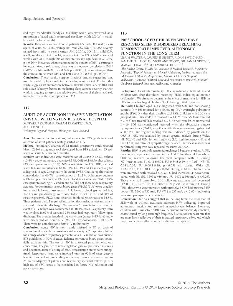
and right mandibular condyles. Maxillary width was expressed as aproportion of facial width (corrected maxillary width (CMW) = maxil-lary width / facial width).Results: Data was examined on 35 patients (27 male, 8 female; meanage 51.6 years, SD 11.6). Average BMI was 28.7 (SD 4.7). OSA severityranged from mild to severe (mean AHI 26.5/hr, SD 17.1; mild OSAn = 9, moderate OSA n = 14, severe OSA n = 12). CMW correlatedweakly with AHI, though this was not statistically significant (r = 0.235,p = 0.204). However, when examined in the context of BMI, a surrogatefor upper airway soft tissue, there was a moderate correlation (BMI /CMW correlates with AHI, r = 0.468, p = 0.008). This was stronger thanthe correlation between AHI and BMI alone (r = 0.341, p = 0.045).Conclusion: These results support previous studies suggesting thatmaxillary width plays a role in the development of OSA. Further, thisstudy suggests an interaction between skeletal (maxillary width) andsoft tissue (obesity) factors in mediating sleep apnoea severity. Furtherwork is ongoing to assess the relative contribution of skeletal and softtissue factors in the development of OSA.
112
AUDIT OF ACUTE NON INVASIVE VENTILATION(NIV) AT WELLINGTON REGIONAL HOSPITALSANKARAN KANNAMANGALAM RAMAKRISHNAN,ALISTER NEILLWellington Regional Hospital, Wellington, New Zealand
Aim: To assess the indications, adherence to BTS guidelines andoutcome of adult patients acutely started on NIV.Method: Preliminary analysis of 12 month prospective study (startedMarch 2014) using audit tool developed from BTS guidelines. 33 epi-sodes of acute NIV use in 4 months.Results: NIV indications were exacerbation of COPD (51.5%), asthma(15.6%), acute pulmonary oedema (6.1%), OHS (9.1%), kyphoscoliosis(3%) and pneumonia (6.1%). NIV was initiated in ED, HDU, medicalward, ICU and ambulance in 84.8%, 3%, 6%, 3% and 3% respectively fora diagnosis of type 2 respiratory failure in 29/33. Chest x ray showed noconsolidation in 66.7%, consolidation in 21.2%, pulmonary oedema6.1% and pneumothorax 6.1% cases. Blood gases were sampled in 97%cases prior to starting NIV and in one half did not show acute respiratoryacidosis. Predominantly venous blood gases (VBGs) (71%) were used forinitial and follow-up assessment. A follow-up blood gas at 1–2 hrs,4–6 hrs and pre-discharge were collected in 45.5%, 36.4% and 50% ofcases respectively. FiO2 is not recorded in blood gas requests in 15.8%.Three patients died, 1 required intubation (for cardiac arrest) and otherssurvived to hospital discharge. Management/ resuscitation status in theevent of NIV failure was documented in 48.5% cases. Respiratory teamwas involved in 60% of cases and 75% cases had respiratory follow-up atdischarge. The average length of stay was 6 days (range 1–23 days) and 4were discharged on home NIV (MND-1, Kyphoscoliosis-1, OHS -2).There were no complications from NIV in this study.Conclusion: Acute NIV is now mainly initiated in ED on basis ofvenous blood gas with inconsistent evidence of type 2 respiratory failurefor a range of acute respiratory presentations. NIV initiation was outsideBTS guidelines in 50% of cases. Reliance on venous blood gases poten-tially explains this. The use of NIV in untreated pneumothorax wasconcerning. The practice of repeating blood gases at prescribed intervalsand documentation of ceiling of care / resuscitation status were subop-timal. Respiratory team were involved only in 60% of cases despitehospital protocol recommending respiratory team involvement within24 hours. Majority of patients had respiratory specialist follow-up. EDshigh use of VBG needs to be considered in future NIV education andpolicy revisions.
113
PRESCHOOL-AGED CHILDREN WHO HAVERESOLVED SLEEP DISORDERED BREATHINGDEMONSTRATE IMPROVED AUTONOMICFUNCTION IN THE LONG TERMLISA M WALTER1,2, LAUREN E NISBET1, AIDAN J WEICHARD1,SAMANTHA L HOLLIS1, VICKI ANDERSON4, GILLIAN M NIXON1,3,MARGOT J DAVEY1,3, ROSEMARY SC HORNE1,2
1The Ritchie Centre, MIMR-PHI Institute of Medical Research, Melbourne,Australia, 2Dept of Paediatrics, Monash University, Melbourne, Australia,3Melbourne Children’s Sleep Centre, Monash Children’s Hospital,Melbourne, Australia, 4Critical Care and Neuroscience Research, MurdochChildren’s Research Institute, Melbourne, Australia
Background: Heart rate variability (HRV) is reduced in both adults andchildren with sleep disordered breathing (SDB), indicating autonomicdysfunction. We aimed to determine the effect of treatment for SDB onHRV in preschool-aged children 3 y following initial diagnosis.Methods: Children aged 3–5 y diagnosed with SDB and non-snoringcontrols (n = 14) returned for a follow-up (FU) overnight polysomnography (PSG) 3 y after their baseline (BL) PSG. Children with SDB weregrouped into: 1) treated/SDB resolved n = 14; 2) treated/SDB unresolvedn = 7; 3) not treated/SDB resolved n = 8; 4) not treated/SDB unresolvedn = 10. SDB was considered resolved when the obstructive apnoeahypopnoea index (OAHI) was ≤1 event/h, there was no snoring detectedat the PSG and regular snoring was not indicated by parents on theOSA-18. HRV was analysed by power spectral analysis during Wake,N1, N2, N3 and REM, for low frequency (LF), high frequency (HF) andthe LF/HF, indicative of sympathovagal balance. Statistical analysis wasperformed using two-way repeated measures ANOVA.Results: HRV in controls remained unchanged between studies. At FU,there was a significant increase in the LF/HF for the children whoseSDB had resolved following treatment compared with BL, duringN2 (mean ± sem, BL 0.42 ± 0.05, FU 0.84 ± 0.19, p = 0.01), N3 (BL0.34 ± 0.03, FU 0.60 ± 0.10, p < 0.05) and during Wake (BL1.02 ± 0.10, FU 1.40 ± 1.6, p = 0.06). During REM the children whowere untreated with resolved SDB at FU had increased LF power com-pared with BL (BL 1345 ± 446 ms2, FU 1676 ± 346 ms2, p < 0.05).Those who had unresolved SDB following treatment had decreasedLF/HF (BL, 2.42 ± 0.45, FU 0.88 ± 0.18, p < 0.05) during N1. DuringREM, those who were untreated with unresolved SDB had increased HFpower (BL 2666 ± 935 ms2, FU 4718 ± 632 ms2, p < 0.05), indicatingincreased parasympathetic activity.Conclusion: Our data suggest that in the long term, the resolution ofSDB with or without treatment increases HRV, indicating improvedautonomic function and restored sympathovagal balance. However,children with unresolved SDB have persistent autonomic dysfunction,characterised by long-term high frequency fluctuations in heart rate thatare most likely reflective of their increased respiratory effort and whichmay have adverse effects on the cardiovascular system.
Sleep, Science and Research
32 © 2014 The AuthorsSleep and Biological Rhythms © 2014 Japanese Society of Sleep Research
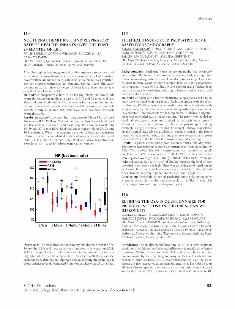
114
NOCTURNAL HEART RATE AND RESPIRATORYRATE OF HEALTHY INFANTS OVER THE FIRST24 MONTHS OF LIFEPHILIP TERRILL1, GORDON WILLIAMS2, MAGGIE YUILL2,CAROLYN DAKIN2
1The University of Queensland, Brisbane, Queensland, Australia, 2TheMater Children’s Hospital, Brisbane, Queensland, Australia
Aim: Overnight polysomnogram and cardio-respiratory studies are usedto investigate a range of disorders in neonates and infants. Unfortunatelyhowever there are limited sleep-state stratified reference data available,even for simple measures such as heart and respiratory rate. This studypresents nocturnal reference ranges of heart rate and respiratory rateover the first 24 months of life.Methods: A prospective cohort of 34 healthy infants underwent fullovernight polysomnography at 2 weeks, 3, 6,12 and 24 months of age.Mean and standard deviation of instantaneous heart rate and respiratoryrate were calculated for each 30 s epoch, and the mean values for eachvariable during REM, non-REM and wake were calculated for eachovernight study.Results: As expected, the mean heart rate decreased from 132, 136 and162 in non-REM, REM and Wake respectively at 2-weeks to 94, 100 and125 beats/min at 24-months; and mean respiratory rate decreased from34, 29 and 27 in non-REM, REM and Wake respectively to 20, 21 and24 breaths/min. Whilst the standard deviation of heart rate remainedrelatively stable, the standard deviation of respiratory rate decreasedfrom 3.3, 6.7 and 9.0 in non-REM, REM and Wake respectively at2-weeks to 1.3, 1.7 and 4.5 breaths/min at 24-months.
Discussion: The mean heart and respiratory rate decrease over the first24 months of life, and these values vary significantly between non-REM,REM and wake. A similar reduction is seen in the variability of respira-tory rate, which may be a signature of increased ventilatory stability.Such reference data has an important role in allowing the pathologicalcharacteristics to be differentiated from normal physiological variability.
115
TELEHEALTH-SUPPORTED PAEDIATRIC HOMEBASED POLYSOMNOGRAPHYAMANDA MUKUSHI1, FIONA TABART1,2, ANNE-MARIE GIBSON1,2,MARY ROBERTS1,2, SUSAN JURY1, PENELOPE BRYANT1,SARATH RANGANATHAN1,2, AMANDA GRIFFITHS1,2
1The Royal Children’s Hospital, Melbourne, Victoria, Australia, 2MurdochChildren’s Research Institute, Melbourne, Victoria, Australia
Background/aim: Paediatric home polysomnography has previouslybeen technically limited. If electrodes are not replaced, missing infor-mation reduces diagnostic capacity.Home sleep studies are preferable forchildren and families for reasons of comfort, familiarity and convenienceWe proposed the use of live Sleep Nurse support using Telehealth toimprove diagnostic capabilities and support families during home basedpaediatric sleep studies.Methods: Children with a history obstructive sleep apnoea aged 6 to 18years were recruited from outpatients. Exclusion criteria were psychiat-ric disorder, ADHD, autism or other medical conditions interfering withsleep or cooperation. The patients were set up with a portable SomtePSG monitor (Compumedics) by the Sleep Nurse. A Telehealth appoint-ment was scheduled just prior to bedtime. The parent was guided tocheck all technical aspects and assisted to re-attach loose sensors/electrodes. Parents were advised to check the patient upon wakingovernight using a checklist provided. Overnight Telehealth assistancevia the hospital sleep lab was available if needed. Hospital in the Homenurses visited families the next morning to remove electrodes and returnthe Somte PSG to the hospital for downloading and reporting.Results: 14 patients were studied from December 2013 until May 2014.The service was reported as more convenient than hospital studies by93%. The pre-bed Telehealth consultation was reported as good/excellent by 100% of respondents. In 4/14 (29%) families, electrodeswere replaced overnight and 1 family utilised Telehealth for overnighttechnical assistance. 13/14 (93%) of families reported the level of careprovided in this service as high. There was some degree of signal loss in6/14 cases, but an acceptable diagnosis was achieved in 12/14 (86%) ofcases. Two studies were repeated due to significant signal loss.Conclusion: Telehealth-supported paediatric home polysomnographyis readily accessible, helpful and acceptable to families. It may alsoreduce signal loss and improve diagnostic yield.
116
REFINING THE OSA-18 QUESTIONNAIRE FORPREDICTION OF OSA IN CHILDREN- CAN WEIMPROVE IT?GILLIAN M NIXON1,2, NATASCHA CIKOR1, KATHY ROWE3,4,MARGOT J DAVEY2, ROSEMARY SC HORNE1, LISA M WALTER1
1The Ritchie Centre, MIMR-PHI Institute of Medical Research, Melbourne,Australia, 2Melbourne Children’s Sleep Centre, Monash Children’s Hospital,Melbourne, Australia, 3Murdoch Children’s Research Institute, University ofMelbourne, Melbourne, Australia, 4Department of General Medicine, RoyalChildren’s Hospital, Melbourne, Australia
Introduction: Sleep disordered breathing (SDB) is a very commoncondition in childhood and adenotonsillectomy is usually an effectivetreatment. Waiting times for both ENT and sleep clinics and forpolysomnography are very long in many centres and strategies areneeded to prioritise these lists to ensure that children with the worstdisease are given expedited assessment and treatment. The OSA-18 is an18 item disease specific questionnaire that has only been validatedagainst daytime nap PSG. It uses a 7-point Likert scale (min score 18,
Abstracts
33© 2014 The AuthorsSleep and Biological Rhythms © 2014 Japanese Society of Sleep Research
max score 126) and includes 5 sub-scales of 3 or 4 questions each.Previous studies have shown a poor correlation between the totalOSA-18 score and OSA severity, reducing its utility as a waiting listtriage tool.Aim: To assess the utility of the OSA-18 to predict severity of OSA,including the contribution of individual questions rather than simplythe score as a whole.Methods: OSA-18 questionnaires were analysed for 198 children (40%female; 2–12 y) with symptoms of OSA and without significantco-morbidity (SDB group), and 52 non snoring controls. Linear regres-sion was used to examine the relationship between total OSA-18 scoreand individual question scores and the obstructive apnoea hypopnoeaindex (OAHI).Results: Total OSA-18 score was not correlated with OAHI in the SDBgroup (p = 0.68). Within those with OSA (OAHI > 1/h), only the “emo-tional symptoms” sub-scale score was significantly correlated withOAHI, and that was weak and in a negative direction (r = −0.2,p < 0.01). In children aged 2–6 y, the questions about loud snoring(Q1), breathing pauses (Q2), choking sounds (Q3), mouth breathing(Q5) and parental concern (Q16) were correlated with OAHI. In thoseaged 6–12 y, only loud snoring (Q1) and interference with parentalactivities (Q17) were correlated with OAHI. When the SDB group wascategorised into primary snoring (OAHI < 1 event/h), mild OSA (1–5events/h) and moderate/severe OSA (>5 events/h), the total OSA-18score, all sub-scales and all individual question scores were significantlydifferent for all categories when compared to the controls.Discussion: Some questions on the OSA-18 perform better than othersas predictors of severity of OSA. These results will allow us to developa weighted OSA-18 scoring system for waiting list triage, which we willpresent in more detail.
117
RATES OF SLEEP DISORDERS AMONG HIGHSCHOOL STUDENTS IN NEW ZEALANDCHINTHAKA SAMARANAYAKE1, GARETH ROBERTS2,BRUCE ARROLL2, ANTONIO FERNANDO2
1Auckland City Hospital, Auckland, New Zealand, 2The University ofAuckland, Auckland, New Zealand
Introduction: Sleep disorders impact greatly on the quality of life aswell as the social and academic performance of adolescents. The aim ofthis study is to determine the rate of different types of sleep disorders inNew Zealand high school students.Method: The Auckland Sleep Questionnaire was administered to allyear 12 and 13 students at six high schools in the North Island. Schoolswere chosen to reflect a range of ethnicities and deciles. The question-naire distinguishes between more common types of sleep disorders. Therates of sleep symptoms, medical conditions affecting sleep, depression(PHQ > 9) and anxiety (GAD > 8), substance use, parasomnias wereassessed. The rate of delayed sleep phase disorder (DSPD) and primaryinsomnia were also calculated.Results: Total of 1388 students participated in this study (response rate69.9%). The median age was 17years (range 14–23), and females rep-resented 43.5% of the study population. A total of 37.1% of studentsreported having a significant sleep problem (symptoms lasting morethan one month affecting day to day life). Depression and anxiety wasthe biggest contributing factor for reported sleep problem and werepresent in 51.7% and 44.8% of students reporting a sleep problemrespectively. A moderate correlation was observed between sleep prob-lems and depression (r = 0.34, p = 0.01), and sleep problems andanxiety (r = 0.31, p < 0.01). Medical conditions contributed to sleepsymptoms in 15% of these students. Obstructive sleep apnoea symp-
toms were found in 3.9% and parasomnias were identified in 9.3% ofthe students who reported having a significant sleep problem. DSPD wasfound in 13.3% and primary insomnia was present in 17.2% of thestudents who reported having a significant sleep problem. Problemalcohol use and drug use was more common in students with sleepsymptoms (12.2% and 5.5% respectively)Discussion: A significant number of high school students are sufferingfrom significant sleep symptoms. Depression and anxiety, substance use,parasomnias and circadian rhythm disorders can greatly contribute tosleep difficulties in this population group and screening for these con-ditions in students presenting to primary and other health care servicesis important.
118
SLEEP DISTURBANCE AND ASTHMA INADOLESCENTS AND YOUNG ADULTS:CAN ONE PREDICT THE OTHER?MARK GARDEN1,3, SADASIVAM SURESH1,2,MICHAEL O’CALLAGHAN3,1
1Mater Children’s Hospital, South Brisbane, Qld, Australia, 2MaterResearch, University of Queensland, Brisbane, Qld, Australia, 3School ofMedicine, University of Queensland, Brisbane, Qld, Australia
Paediatric asthma is the most common chronic respiratory disease inAustralia. Sleep disturbances are common in asthmatics with poor sleepquality and excessive daytime sleepiness. Although the causal directionof this relationship is unknown, improvement in one could benefit theseverity of the other.Aim: The aims of this study were to confirm, using a single birth cohort,the nature and strength of association between sleep quality, snoring andasthma in the adolescent and young adult population, and to identify towhat extent asthma, sleep quality and snoring at 14 years independentlypredicted themselves or each other at 21 years.Methods: Data from a 21-year follow-up of mothers and their childrenrecruited into the Mater-University of Queensland Study of Pregnancy(n = 7223). Complete asthma and sleep information (questionnairedata) was available for 5015 participants at 14 years and 3527 at 21years, with 3237 participants at both 14 and 21 years. The cross sec-tional association between asthma, sleep quality and snoring was ana-lysed at both 14 years and 21 years. Predictive analysis examined thelongitudinal relationship between asthma, sleep quality and snoring at14 and 21 years. Cross tabulation with the χ2 test was used for categori-cal data & binary logistic regression was used to calculate odds ratiosusing SPSS.Results: Sleep disturbance and snoring were associated with asthma at14 years [odds ratio 1.51(1.25–183), 1.73 (1.49–2.01)]. Similar asso-ciation was noted at 21 years, albeit only significant in females. Thisassociation persisted in females when analysed for asthma severity vari-ables. Asthma, sleep quality and snoring at 14 strongly predicted them-selves at the age of 21[p < 0.001]. In addition, snoring and sleepdisturbance at 14 predicted asthma at 21 and asthma at 14 predictedsnoring at 21 years, especially in females[p < 0.001].Discussion: The predictive relationship seen between sleep and snoringat 14 years and asthma at 21 years allows a possible opportunity toaddress these issues at a younger age and reduce the incidence of earlyadult-onset asthma in females. Similarly aggressive management ofasthma in adolescence may lead to a reduction in snoring and poten-tially associated sleep disordered breathing.
Sleep, Science and Research
34 © 2014 The AuthorsSleep and Biological Rhythms © 2014 Japanese Society of Sleep Research
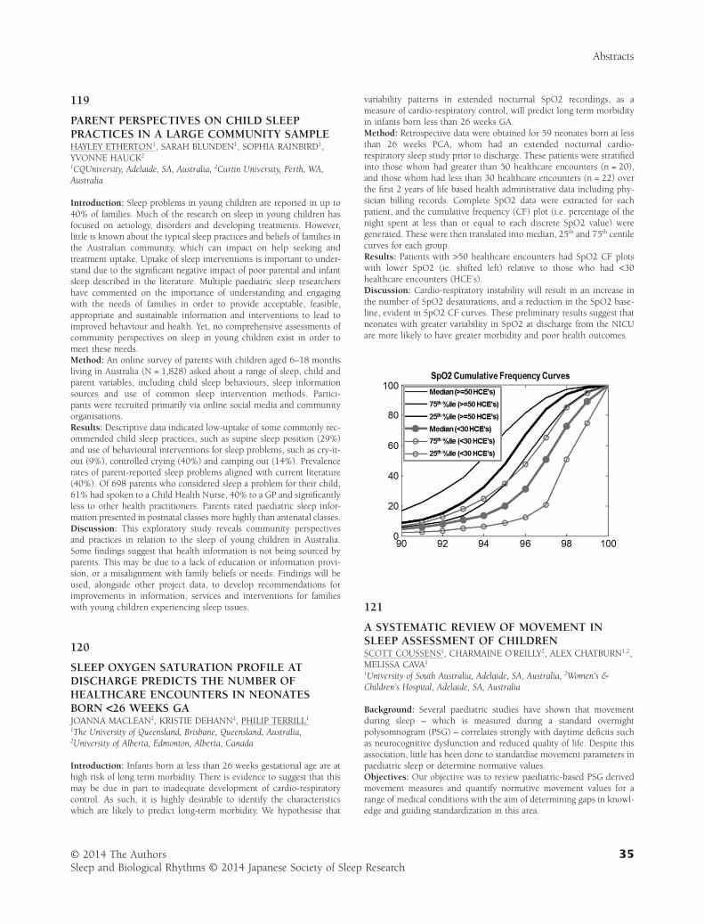
119
PARENT PERSPECTIVES ON CHILD SLEEPPRACTICES IN A LARGE COMMUNITY SAMPLEHAYLEY ETHERTON1, SARAH BLUNDEN1, SOPHIA RAINBIRD1,YVONNE HAUCK2
1CQUniversity, Adelaide, SA, Australia, 2Curtin University, Perth, WA,Australia
Introduction: Sleep problems in young children are reported in up to40% of families. Much of the research on sleep in young children hasfocused on aetiology, disorders and developing treatments. However,little is known about the typical sleep practices and beliefs of families inthe Australian community, which can impact on help seeking andtreatment uptake. Uptake of sleep interventions is important to under-stand due to the significant negative impact of poor parental and infantsleep described in the literature. Multiple paediatric sleep researchershave commented on the importance of understanding and engagingwith the needs of families in order to provide acceptable, feasible,appropriate and sustainable information and interventions to lead toimproved behaviour and health. Yet, no comprehensive assessments ofcommunity perspectives on sleep in young children exist in order tomeet these needs.Method: An online survey of parents with children aged 6–18 monthsliving in Australia (N = 1,828) asked about a range of sleep, child andparent variables, including child sleep behaviours, sleep informationsources and use of common sleep intervention methods. Partici-pants were recruited primarily via online social media and communityorganisations.Results: Descriptive data indicated low-uptake of some commonly rec-ommended child sleep practices, such as supine sleep position (29%)and use of behavioural interventions for sleep problems, such as cry-it-out (9%), controlled crying (40%) and camping out (14%). Prevalencerates of parent-reported sleep problems aligned with current literature(40%). Of 698 parents who considered sleep a problem for their child,61% had spoken to a Child Health Nurse, 40% to a GP and significantlyless to other health practitioners. Parents rated paediatric sleep infor-mation presented in postnatal classes more highly than antenatal classes.Discussion: This exploratory study reveals community perspectivesand practices in relation to the sleep of young children in Australia.Some findings suggest that health information is not being sourced byparents. This may be due to a lack of education or information provi-sion, or a misalignment with family beliefs or needs. Findings will beused, alongside other project data, to develop recommendations forimprovements in information, services and interventions for familieswith young children experiencing sleep issues.
120
SLEEP OXYGEN SATURATION PROFILE ATDISCHARGE PREDICTS THE NUMBER OFHEALTHCARE ENCOUNTERS IN NEONATESBORN <26 WEEKS GAJOANNA MACLEAN2, KRISTIE DEHANN2, PHILIP TERRILL1
1The University of Queensland, Brisbane, Queensland, Australia,2University of Alberta, Edmonton, Alberta, Canada
Introduction: Infants born at less than 26 weeks gestational age are athigh risk of long term morbidity. There is evidence to suggest that thismay be due in part to inadequate development of cardio-respiratorycontrol. As such, it is highly desirable to identify the characteristicswhich are likely to predict long-term morbidity. We hypothesise that
variability patterns in extended nocturnal SpO2 recordings, as ameasure of cardio-respiratory control, will predict long term morbidityin infants born less than 26 weeks GA.Method: Retrospective data were obtained for 59 neonates born at lessthan 26 weeks PCA, whom had an extended nocturnal cardio-respiratory sleep study prior to discharge. These patients were stratifiedinto those whom had greater than 50 healthcare encounters (n = 20),and those whom had less than 30 healthcare encounters (n = 22) overthe first 2 years of life based health administrative data including phy-sician billing records. Complete SpO2 data were extracted for eachpatient, and the cumulative frequency (CF) plot (i.e. percentage of thenight spent at less than or equal to each discrete SpO2 value) weregenerated. These were then translated into median, 25th and 75th centilecurves for each group.Results: Patients with >50 healthcare encounters had SpO2 CF plotswith lower SpO2 (ie. shifted left) relative to those who had <30healthcare encounters (HCE’s).Discussion: Cardio-respiratory instability will result in an increase inthe number of SpO2 desaturations, and a reduction in the SpO2 base-line, evident in SpO2 CF curves. These preliminary results suggest thatneonates with greater variability in SpO2 at discharge from the NICUare more likely to have greater morbidity and poor health outcomes.
121
A SYSTEMATIC REVIEW OF MOVEMENT INSLEEP ASSESSMENT OF CHILDRENSCOTT COUSSENS1, CHARMAINE O’REILLY2, ALEX CHATBURN1,2,MELISSA CAVA1
1University of South Australia, Adelaide, SA, Australia, 2Women’s &Children’s Hospital, Adelaide, SA, Australia
Background: Several paediatric studies have shown that movementduring sleep – which is measured during a standard overnightpolysomnogram (PSG) – correlates strongly with daytime deficits suchas neurocognitive dysfunction and reduced quality of life. Despite thisassociation, little has been done to standardise movement parameters inpaediatric sleep or determine normative values.Objectives: Our objective was to review paediatric-based PSG derivedmovement measures and quantify normative movement values for arange of medical conditions with the aim of determining gaps in knowl-edge and guiding standardization in this area.
Abstracts
35© 2014 The AuthorsSleep and Biological Rhythms © 2014 Japanese Society of Sleep Research
Data sources: We performed a systematic review of the current litera-ture (from 1992 – current) using PubMed and studies cited by returnedarticles not index in Pubmed. Both clinical and research studies wereincluded if they measured movement during objectively measured sleepin subjects aged 0–18 years.Study Appraisal and Synthesis Methods: Four experienced sleeptechnicians reviewed the articles for adherence to inclusion criteriautilising the PRISMA guidelines. Study appraisal was performed using amodified version of the Down’s and Black appraisal method.Results: Our initial screening identified 648 papers, and after initialexclusion 62 papers were retained for full reading. These were thor-oughly analysed and their movement data was extracted and collated.Movement was found to be correlated with disease severity in severaldisease groupsUnfortunately, further synthesis was restricted as thenumber of each of the different movement measure definitions for eachof the disease groups was limited.Limitations: Many studies did not report movement data, even thoughit was in fact measured. Additionally, many studies used different defi-nitions of movement in sleep making comparisons between studiesdifficult.Conclusions and implications of key findings: Further study isrequired to investigate the most useful movement measures appropriateto each disease group.
122
USE OF BI-LEVEL POSITIVE AIRWAY SUPPORT(BPAP) WITH AVERAGE VOLUME ASSUREDPRESSURE SUPPORT (AVAPS) IN SLEEP TOSUCCESSFULLY MANAGE SEVERESLEEP-RELATED RESPIRATORY INSUFFICIENCY:CASE PRESENTATIONSNATALIE GENTIN1,2, BRUCE WILLIAMSON1,2,GANESH THAMBIPILLAY1,2, ARTHUR TENG1,2
1Sydney Children’s Hospital, NSW, Australia, 2University of New SouthWales, NSW, Australia
Average volume assured pressure support (AVAPS) is a feature onBi-level positive airway pressure (BPAP) machines which delivers aconsistent pre-set target volume by automatically adjusting the pressuresupport within a pre-determined range. Therefore, varying pressurerequirements within a period of sleep can be provided. This allowspotentially lower settings with a guaranteed volume delivery.
There have been some studies looking at AVAPS in adults which haveshown improved sleep quality and normalisation of TcCO2 with equiva-lent compliance. There are almost no studies of AVAPS in children.
We present three cases of paediatric patients with nocturnalhypoventilation who were better managed on BPAP with the AVAPSfeature than with conventional BPAP. One patient has a myopathy, thesecond a chromosomal disorder and the third a brainstem tumour.These cases highlight the benefits of AVAPS.
All patients had a diagnostic polysomnogram (PSG), a PSG on con-ventional BPAP and a PSG on AVAPS. With AVAPS, our patients receivedan appropriate tidal volume with each breath, but without using highpressure settings throughout the night. The automatically adjusted pres-sure allowed lower pressures to be given in non REM sleep and thenhigher pressures to achieve the same tidal volume in REM. The AVAPS-feature was well tolerated by the patients.
AVAPS is a potentially useful feature on BPAP machines which can beused in paediatric patients with nocturnal hypoventilation. Furtherstudies are warranted to assess this as an important addition tothe armamentarium of non-invasive ventilation therapies available tochildren.
124
SYMPTOMS OF OBSTRUCTIVE SLEEP APNOEAIN CHILDREN WITH A PARENT DIAGNOSEDWITH OBSTRUCTIVE SLEEP APNOEASHELLEY MATHER1, ANGELA CAMPBELL1, DAWN ELDER2
1WellSleep, Otago University Wellington, Wellington, New Zealand, 2DeptPaediatrics, Otago University Wellington, Wellington, New Zealand
Untreated paediatric obstructive sleep apnoea (OSA) is associated witha range of detrimental academic and health outcomes. Recognising thepresence of OSA in children early so that they can receive timelytreatment is therefore of the upmost importance. Known risk factors forpaediatric OSA include enlarged tonsils and adenoids, craniofacialabnormalities, neuromuscular co-morbidities and obesity. Genetic pre-disposition has also been reported. We hypothesised that children of aparent diagnosed with OSA would be more likely to have symptoms andphysical characteristics of OSA than age and gender matched controls.Aim: To document symptoms of OSA in children whose parents havebeen diagnosed with OSA and compare them to age and sex matchedcontrols.Method: A case-control study. We recruited 25 children who had aparent diagnosed with OSA and 25 age and gender matched controlsfrom the community whose parents scored as low risk for OSA (usingadjusted Flemons score). The following measures were compared:OSA-18 questionnaire, measures of height and weight and mallampatiscore.Results: There was a significant difference between the case and controlgroups on the OSA-18 questionnaire. Average score (SD) was36.5 ± 18.1 for cases and 28.1 ± 9.1 for controls, resulting in an averagedifference of 8.4 (p = 0.018). Twenty percent (n = 5) of cases scored >60indicating a significant impact on quality of life. 56% of the cases grouphad a mallampati score of either III or IV compared to only 11% in thecontrol group. There was a significant difference in BMIz betweengroups with the cases group averaging 0.56 ± 1.07 and the controlgroup −0.15 ± 0.67 (p value = 0.002).Conclusion: Children of parents with OSA are more likely to havesymptoms of OSA and physical findings that are associated with anincreased risk of obstruction than age matched controls. Further assess-ment will be required to determine whether a definite diagnosis of OSAcan be made in these children.Funded by: Maurice and Phyllis Paykel Trust
125
PRESCHOOL CHILDREN WITH SLEEPDISORDERED BREATHING EXHIBIT INCREASINGSURGES IN HR WITH INCREASING DURATIONSOF REPETITIVE RESPIRATORY EVENT EPISODESLISA M WALTER1,2, AIDAN J WEICHARD1, GILLIAN M NIXON1,3,MARGOT J DAVEY1,3, ROSEMARY SC HORNE1,2
1The Ritchie Centre, MIMR-PHI Institute of Medical Research, Melbourne,Australia, 2Dept of Paediatrics, Monash University, Melbourne, Australia,3Melbourne Children’s Sleep Centre, Melbourne, Australia
Background: Following a respiratory event there is a surge in heart rate(HR), and this has been linked to the adverse cardiovascular conse-quences of obstructive sleep apnoea (OSA). Previous analyses of this HRsurge have been limited to isolated events, however children frequentlyhave episodes of repetitive respiratory events. We aimed to determinethe effect of increasing numbers of respiratory events in each repetitiveepisode, the effect of increasing numbers of episodes of repetitive events
Sleep, Science and Research
36 © 2014 The AuthorsSleep and Biological Rhythms © 2014 Japanese Society of Sleep Research
and the duration of the episodes, on the surge in HR that occurs at thetermination of each respiratory event in the episode.Methods: Data from 11 children (3–5 years) diagnosed with SDB byovernight polysomnography (PSG). Events were considered repetitive ifthere was <25 s between two or more events. Each event was dividedequally into an early and a late event phase. Linear regressions wereperformed for the % change in HR from the late event phase to the phasebetween successive events and the number of events in each episode,the number of episodes per child and the duration of the episodes. Theeffect of episode duration was analysed for episodes separated intodurations of ≤50 s, ≤100 s, ≤200 s, ≤300 s, ≤400 s and >400 s using theKruskal-Wallis and Mann-Whitney posthoc tests.Results: A total of 114 episodes of repetitive respiratory events whichincluded 520 individual events were analysed. The number of respira-tory events in an episode and the duration were correlated with %change in HR (r2 = 0.03, p = 0.001 for both). There was no correlationbetween the number of episodes per child and % change in HR. The %change in HR during episodes with durations of ≤50 s, ≤100 s and≤200 s were significantly lower compared with durations of ≤300 s,≤400 s and >400 s (median 23%, 26%, 29%, vs 38%, 38%, and 36%,respectively, p < 0.05 for all).Conclusion: our results suggest that for episodes of repetitive respira-tory events, the surge in HR that occurs between successive eventsincreases significantly for the initial 300 s of the episode and thenremains at that level. This study highlights the adverse cardiovascularconsequences of repetitive respiratory events. Further analysis isongoing to confirm these results.
126
EVALUATION OF RESPIRATORY MUSCLESTRENGTH IN CHILDREN WITHPOLYSOMNOGRAPHY (PSG) DIAGNOSEDOBSTRUCTIVE SLEEP APNOEA (OSA)JASNEEK CHAWLA, ANDREW COATES, KIARA SCLIP,CAROLYN DAKINMater Children’s Hospital, Brisbane, Australia
Background: Maximal inspiratory pressure (MIP), Maximal expiratorypressure (MEP) and Sniff nasal inspiratory pressure (SNIP) are non-invasive respiratory muscle tests currently used predominantly in chil-dren with underlying neuromuscular disorders to monitor musclestrength. To our knowledge the respiratory muscle strength of childrenwith obstructive sleep apnoea has not been previously evaluated. It ispossible that due to the prolonged periods of increased effort experi-enced by children with Obstructive Sleep Apnoea (OSA) during sleeprespiratory muscle strength may differ in this population.Aim: To determine if children with Polysomnography (PSG) diagnosedOSA have increased respiratory muscle strength compared to publishedvalues for healthy children and compared to children with normalPSG-no OSA.Methods: A prospective study of all children aged ≥6 yrs, withoutadditional comorbidity that attended a tertiary sleep clinic and under-went PSG for evaluation of OSA was conducted. Children undertook aseries of respiratory muscle tests in the lab either before or after PSGevaluation. Patients with a negative PSG study for OSA were used asinternal controls.Discussion: Results of respiratory muscle tests in patients with PSGdiagnosed OSA compared with reference values and internal controlswill be presented. These tests may have significant clinical benefit in thispopulation, with the potential to develop as a screening tool for thedetection of OSA in children.
128
RELATIONSHIP BETWEEN SLEEP DEFICIENCYAND POOR BEHAVIOUR IN CHILDREN WITHAUTISM SPECTRUM DISORDERSIMONNE COHEN1, KIM CORNISH1, SHANTHA RAJARATNAM1,2,STEVEN LOCKLEY1,2, RUSSELL CONDUIT1
1Monash University, Melbourne, Australia, 2Harvard Medical School,Boston, USA
Although there is reason to believe that serious sleep problems arecommon in children with Autism Spectrum Disorders (ASD), and thatpoor sleep exacerbates problematic daytime behaviour, such problemshave received very little attention in both research and clinical prac-tice. To date, the relationship between sleep and behavioural problemsin ASD remains limited to investigations based on small sample sizes,and children with High functioning autism. Fortunately, we haveaccess to a large cohort of low functioning ASD patients, which willallow us to carefully examine these relationships. This data has beencollected retrospectively from two residential facilities in Boston (USA)which specialize in clinical interventions for individuals with autismranging from 7–21 years of age. As apart of routine clinical care, thesefacilities conduct frequent assessments of sleep and behaviour in allindividuals. Sleep-awake behaviour is assessed by continuous clinicalobservations every 30 minutes from 21:00–7:00h (∼110,000 observa-tions) and up to 40 behavioural parameters are rated by caretakersevery hour during the day (8:00–21:00h). This is a remarkably com-prehensive, high quality dataset that extends for the past 5 years andincludes data on more than 250 children. A preliminary analysis ofthe data found that the average sleep-duration was 8.2 ± 0.9 h/nightin 5–10 year old and 8.1 ± 1 h/ night in 11–17 year olds. 76% ofsleep-time in the children <11 years did not meet the recommendedminimum of 10 hours per day, and 18% of sleep-time in the 11–17year age group did not meet the National Sleep Foundation (NSF)recommended minimum sleep-durations. From this data we can con-clude that a large proportion of children with ASD do not meet theNSF- recommended minimum sleep-duration for age. In addition,92% of these children engage in aggressive behaviour (e.g., hitting,biting, hair pulling), 70% engage in property destruction (e.g., throw-ing objects, destroying property) and 53% engage in dangerous behav-iours (e.g., bolting). From this data, we will be examining therelationships between the sleep parameters (e.g., total sleep time, sleepefficiency) and the aforementioned challenging behaviours. Theseresults will provide the foundation for future randomized clinical trialsto test the efficacy of interventional measures to improve sleep andbehaviour in children with ASD.
129
EVALUATION OF THE BENEFITS OF PLAYTHERAPY WITH CPAP INITIATION ANDCOMPLIANCENATALIE GENTIN1,2, NICOLA MOONEY1,GANESH THAMBIPILLAY1,2
1Sydney Children’s Hospital, NSW, Australia, 2University of New SouthWales, NSW, Australia
CPAP is a clinically proven effective therapy for obstructive sleepapnoea. Unfortunately, children are often averse to the initial and con-tinued use of CPAP. Play therapy has been used in many differentconditions and situations to improve patient and family acceptance withmedical therapy and procedures.
Abstracts
37© 2014 The AuthorsSleep and Biological Rhythms © 2014 Japanese Society of Sleep Research
Our department has started incorporating the Play Therapy Team inthe CPAP initiation process and we have found that this has improvedthe initial use of CPAP, ongoing compliance and overall patient andfamily experiences.
The aim of this study was to objectively assess whether there has beena benefit of play therapy with CPAP initiation and ongoing compliance.All patients who were initiated on CPAP from January 2014 to July 2014were identified. A survey was sent to the carers of the patients. Thirty-four surveys were sent and 11 were returned. Of these, a play therapistwas involved in 4 cases.
From parental reports, all 4 patients who had play therapy involve-ment were fully compliant with therapy after a week compared with 5of the 7 patients who did not have play therapy involvement.
These preliminary results show high patient and family satisfactionwith our CPAP initiation practices. In patients with whom the playtherapist was involved, the feedback was very positive.
This confirms our subjective view that play therapy is beneficial forCPAP acceptance and compliance. We will use this information toincrease the role of Play Therapy in CPAP initiation. We plan to pro-spectively collect more data on longer term compliance using machinedownloads.
130
A DESCRIPTION OF CENTRAL SLEEP APNOEAIN A COHORT OF CHILDREN UNDER ONE YEAROF AGEJOHN WONG1, NITIN GUPTA2,3, YVONNE PAMULA1,JAMES MARTIN1, DECLAN KENNEDY1,3
1Women’s and Children’s Hospital, Adelaide, South Australia, Australia,2Sydney Children’s Hospital, Sydney, New South Wales, Australia,3University of Adelaide, Adelaide, South Australia, Australia
Introduction: The epidemiology and pathophysiology of central sleepapnoea in term infants is poorly understood. Our aim was to describethe polysomographic (PSG) features of central apnoea in a group ofinfants presenting at a single centre with either an Apparent Life Threat-ening Events (ALTE), apnoeic/cyanotic episodes or oxygen desaturationduring overnight monitoring in the neonatal period.Methods: Retrospective PSG analysis was undertaken of term infantsless than 12 months of age who presented to the Women’s and Chi-ldren’s Hospital, Adelaide with ALTE, apnoeic/cyanotic episodes orabnormal overnight oximetry Exclusion criteria included infants bornpre-term or those with known neurological impairment. Scoring ofsleep studies were based upon current American Academy of SleepMedicine guidelines.Results: Twenty-eight children (12 males and 16 females) underwentPSG. The mode of presentation included: ALTE (n = 5), cyanotic orapnoeic episodes (n = 19), frequent desaturation on cardiorespiratorymonitoring (n = 3) and presentation after apnoea monitor alarm (n = 1).The median age of patients at PSG was 9.5 days (range: 1–183 days). Ofthe 28 PSG’s performed the mean total sleep time was 6 hours (range:4–8.4 hours). The median REM-sleep central apnoea index was 15.9/hour (range: 9–99.7/hour). The mean nadir oxygen desaturation was82.3% (range: 79–90%).Conclusion: Significant central sleep apnoea was identified in thiscohort of term infants. We plan to perform further analysis of this cohortto better understand these sleep study findings.
131
A RANDOMISED CONTROLLED TRIAL USINGOVERNIGHT VIDEO IN THE HOME TO COMPAREA CULTURALLY DERIVED SLEEP DEVICE(WAHAKURA) WITH A STANDARD BASSINETSALLY BADDOCK1, SHEILA WILLIAMS2, DAVID TIPENE-LEACH2,ANGELINE TANGIORA2, RAYMOND JONES2, BARRY TAYLOR2
1Otago Polytechnic, Dunedin, New Zealand, 2University of Otago,Dunedin, New Zealand
In New Zealand, the Maori SUDI rate is 5 times that of non Maori, nonPacific (2.34 deaths per 1000 live births vs 0.52). Many deaths areassociated with bedsharing and maternal smoking. These behaviourshave proved resistant to change. The wahakura (flax bassinet) isdesigned from a cultural perspective as a potentially safer sleep place. Todate there has been no assessment of the safety of this traditionalsleeping device.Aim: to compare wahakura with bassinets as sleep options acceptable toMaori families. Methods: 200 participants were recruited, through mid-wifery practices supporting mainly Maori families in deprived areas, andwere randomized to receive a wahakura (W) or a bassinet (B) duringpregnancy. Baseline data was collected during pregnancy and then ques-tionnaires on sleep practices, breastfeeding and maternal and infantwell-being were completed at 1, 3 and 6 mths. At 1 month, overnightvideosomnography, oximetry and temperature recordings of 80 bassinetand 79 wahakura babies were completed. This paper reports on theresults of overnight videosomnography.Results: Participants were relatively high risk with 67% identifying asMaori, 55% residing in areas with a NZ deprivation score of 9–10 (10being most deprived) and 55% reporting some smoking in pregnancy,Intention to treat analysis was used. Mean study time (B:9.9 vs 10.1 h),mean asleep time (B:6.7 vs W:6.7), head covered time (B:0.45 vs0.43 h), and breastfeeding (B:0.38 vs W:0.44) were not significantlydifferent. Similarly, the number of head coverings (mean, B:2.1 vsW:1.5, p = 0.4) and breastfeeding events (B:3.2 vs W:3.9, p = 0.3) werenot different between groups. On the study night babies slept in abassinet (mean time B:6.7 h vs W:1.9 hr, p < 0.0001), wahakura underadult bed-covers (B:0 vs W:0.4 h), wahakura on bed-covers (B:0 vsW:1.8 h), and wahakura off the bed (B:0 vs 2 h), in mother’s bed(B:1.7 h vs 2.1 h, p = .48) and in a cot (B:0.4 vs W:0.5, p = .74).Conclusion: This study suggests it is as reasonable to provide awahakura as it is to provide a bassinet as a safe sleep intervention in thispopulation. The findings show no difference between the behaviours forthe two options. Further pragmatic analysis will provide greater under-standing of behaviour while sleeping in the wahakura.
132
CONSTRUCTING A COMPOSITE MEASURE OFDISEASE SEVERITY IN CHILDREN WITHSLEEP-RELATED UPPER AIRWAY OBSTRUCTIONSCOTT COUSSENS1,2, MELISSA CAVA1, YVONNE PAMULA3,DAVID SAINT2, MARK KOHLER1
1University of South Australia, Adelaide, SA, Australia, 2University ofAdeliade, Adelaide, SA, Australia, 3Women’s & Children’s Hospital,Adelaide, SA, Australia
Background: Upper airway obstruction (UAO) is typically diagnosedusing a single polysomnogram-derived index of breathing disturbance(respiratory disturbance index; RDI). However, RDI is an incompletemeasure of UAO disease severity in children, correlating poorly with the
Sleep, Science and Research
38 © 2014 The AuthorsSleep and Biological Rhythms © 2014 Japanese Society of Sleep Research
associated daytime deficits, including neurocognitive dysfunction andbehavioural problems, reduced school performance and reduced qualityof life. A comprehensive measure of disease severity has yet to bedeveloped. Several inherent problems contribute to such a develop-ment, including the variability of 1) the nature, symptoms and severityof the disease, 2) the adaptive responses to the disease, 3) age andassociated developmental changes, and 4) the lack of well-defined andstandardised outcome measures to compare candidate indices against.We hypothesised that a composite index of various known and novelindices of sleep quality that take into account these factors wouldprovide a more sensitive measure of disease severity and better predictdaytime morbidity.Methods: Data from 92 primary school aged children (48 controls and44 with UAO) were utilised for this study. Subjects underwent two testsessions six months apart that included an overnight polysomnogramand a battery of cognitive and behavioural tests. Using an existing modelas a template, developed for adults with obstructive sleep apnoea syn-drome, a model of disease severity was built specifically for childrenwith UAO. By analysing the correlational and mediating effect of varioussleep variables between RDI and selected outcome measures, additionalelements that could further enhance the new disease severity modelwere identified. A new composite index utilising various modellingtechniques was subsequently constructed and we compared the dis-criminative and predictive ability of this index to other known indices ofdisease severity (e.g. AHI, RDI, sleep pressure score).Results: The composite index accurately discriminated between chil-dren with UAO and matched controls (p < 0.001). The composite indexalso predicted subjective performance scores (e.g. self-report quality oflife) and objective outcome measure deficits (e.g. SpO2 nadir) betterthan any of the tradition indices alone.Discussion: A composite index may be a useful tool in the diagnosis ofchildren suspected of having UAO, particularly those with milder formsof the disease who may be given reduced priority for treatment orconsidered not at risk of significant daytime deficits based on traditionalmethods.
133
REM-RELATED BRADYARRHYTHMIA SYNDROME:A CASE REPORTJORDAN CUNNINGHAM1,2, ROBYN O’SULLIVAN1,2
1Greenslopes Private Hospital, Brisbane, Queensland, Australia, 2Universityof Queensland, Brisbane, Queensland, Australia
Introduction: Cardiac arrhythmias in Obstructive Sleep Apnoea are awell-recognised consequence of autonomic instability due to apnoeaand hypoxia. In contrast, REM-related bradyarrhythmia syndrome is ararely reported condition characterised by bradycardia, transient sinusarrest, and 3rd degree atrioventricular block during phasic REM in theabsence, or despite adequate treatment, of sleep-disordered breathing orunderlying cardiac abnormality. We present such a case, and reviewphysiology and management.Case Report: A 55 year old man presenting with mild daytime dizzinesswas discovered on Holter to have severe nocturnal bradycardia, andsinus pauses up to 12 seconds. Past history included treated AcuteLymphocytic Leukaemia 21 years previously. CT coronary angiographyshowed mild coronary artery disease only. LV size and function werenormal.
Diagnostic polysomnography was performed, revealing moderatepositional obstructive sleep apnoea (RDI 19.4 events/hour overall,37.7 supine, 7.4 lateral, nadir SpO2 90%), and bradycardia (HR34–48 bpm), with numerous REM-related sinus pauses up to 7 secondsin length, without definite association with events or airflow limitation.
CPAP titration polysomnography found persistence of the bradycardiadespite effective control of flow limitation and obstructive events. Thiswas further confirmed on Holter after a six week trial of CPAP.
Sleep apnoea was subsequently managed with side-lying. The rhythmdisturbance is currently being managed conservatively.Discussion: REM-related bradyarrhythmia syndrome likely reflects anincrease in vagal tone, or an inappropriate reduction in sympathetictone, during phasic REM – more usually associated with increasedsympathetic tone. There is disagreement in the literature as to optimaltreatment – whether anticholinergic, pacing, or conservative. Thisunusual pathophysiology and its prognostic implications and manage-ment are explored.
134
SLEEP DISORDERS AMONG TERTIARYSTUDENTS: A CROSS SECTIONAL STUDYCHINTHAKA SAMARANAYAKE1, BRUCE ARROLL2,ANTONIO FERNANDO0
1Auckland City Hospital, Auckland, New Zealand, 2The University ofAuckland, Auckland, New Zealand
Introduction: Young adults, particularly university students, areincreasingly recognised as a population group that is greatly affected bysleep difficulties. However the rates of these conditions in the NewZealand population are poorly understood. The aim of this study was todetermine the rates of sleep disorders and associated depression andanxiety among university students in Auckland.Method: Auckland Sleep Questionnaire (ASQ) was administered toundergraduate students from five faculties of The University of Auck-land. The questionnaire distinguishes between more common types ofsleep disorders. The rates of sleep symptoms, medical conditions affect-ing sleep, depression (PHQ > 9) and anxiety (GAD > 8), substance use,parasomnias were assessed. The prevalence of delayed sleep phase dis-order (DSPD) and primary insomnia were also calculated.Results: A total of 1933 students were invited to participate and 66.8%completed the questionnaire. The responded students included 575(62.0%) medical, 208 (75.4%) health sciences, 170 (69.1%) nursing,136 (69.4%) law, 108 (65.1%) engineering and 95 (77.9%) architecturestudents. The median age was 20 years (range 16–38) and womenrepresented 63.9% of the total group. A total of 39.4% of the studentssurveyed reported having significant sleep symptoms lasting longer thanone month. Clinically significant depression and anxiety was found in30.8% (95%CI 26% to 34%) and 31.0% (95% CI 27% to 35%) of thesestudents respectively. Delayed sleep phase disorder was found in 24.9%of students and parasomnias were reported by 12.4%. Symptoms ofobstructive sleep apnoea were reported by 2.4% of students. Problem-atic alcohol and drug use was common among students with a sleepproblem: 17.5% and 4.9% respectively. There was no statistically sig-nificant difference in the rate of reported sleep problem in the differentcourse groups. Maori and Pacific Island students had higher rates ofsleep symptoms (54% and 39.1% respectively) compared to otherethnicitiesDiscussion: A large number of university students are suffering fromsignificant sleep symptoms. Mood disorders, substance use, and circa-dian rhythm disorders can greatly contribute to sleep difficulties in thispopulation group.
Abstracts
39© 2014 The AuthorsSleep and Biological Rhythms © 2014 Japanese Society of Sleep Research
135
IMPLICATIONS OF POOR SLEEP SYMPTOMS INSOUTH SUDANESE AUSTRALIANS FORPOST-TRAUMATIC STRESS DISORDER (PTSD)DOROTHY BRUCK, SANTINO ATEM DENG, BICHOK WAN KOT,MICHELE GROSSMANVictoria University, Melbourne, Victoria, Australia
Background: Cross-cultural counselling theory suggests that peoplefrom non-Western backgrounds preferentially focus on physical symp-toms in their consultations with medical practitioners and may havelittle mental health awareness (e.g. Sue & Sue, 2012). Given the closerelationship between sleep difficulties and both mood problems andPTSD, sleep difficulties can be a valuable indicator of mental healthproblems in individuals from non-Western societies. While we knowthat early morning awakenings are a key indicator of depression, little isknown about which specific sleep difficulties may be the best indicatorsfor PTSD, apart from nightmares. The aim of this study was to deter-mine which types of sleep related symptoms have the strongest predic-tive relationship with PTSD severity in a sample of South Sudaneseformer refugees.Method: A survey was developed which included 14 sleep-related itemsand the PSQI Addendum for PTSD. The survey was translated intoDinka, Nuer and Arabic and administered individually within urbanand regional Victoria by PhD students from the South Sudanese com-munity in Australia (n = 117, aged 16–60 years).Results: Problems were prevalent with 22% meeting the criteria forsevere clinical insomnia and 39% meeting the criteria for PTSD. Twosleep-related compound variables were created by averaging the scoresfrom items within each of two factors from a factor analysis. Onecompound variable, Disrupted Sleep, contained three questions pertain-ing to troubles falling asleep, waking a lot or waking too early. A secondcompound variable, Waking Sequela, consisted of four questions relatingto daytime sleepiness, fatigue/exhaustion, waking unrefreshed and rest-less legs. While the one- to-one relationships between the two compoundsleep variables and PTSD were significant, when Disrupted Sleep andWaking Sequela were both entered into the regression Disrupted Sleepbecame non-significant. Thus Waking Sequela fully mediated DisruptedSleep in its relationship to PTSD symptoms.Conclusions: Daytime symptoms arising from poor sleep play animportant mediating role in governing the relationship between PTSDand disrupted sleep. Thus asking about the waking consequences ofsleep difficulties (e.g. sleepiness, exhaustion) is much more importantthan the specific nature of the sleep difficulties per se in helping toidentify the possible presence of PTSD symptoms.
136
SLEEP DURATION AND ETHNICITY INA LARGE POPULATION BASED SAMPLEOF ADULT CANADIANSSUTAPA MUKHERJEE1,3, MANDEEP SINGH1, WOGANEE FILATE1,MATT KOWGIER1, LYLE PALMER2
1University of Toronto, Toronto, Ontario, Canada, 2University of Adelaide,Adelaide, SA, Australia, 3Women’s College Hospital and Research Institute,Toronto, Ontario, Canada
Background: It is unknown whether ethnic differences exist in theCanadian general population. We hypothesized that ethnic differencesin sleep duration exist amongst the general population of adult residentsof Ontario.
Methods: We conducted a cross-sectional study using data from theOntario Health Study (OHS) from a sample of adult residents ofOntario, Canada. Participants provided medical history, socio-demographic characteristics and sleep duration. Self-reported sleepduration was recorded based on response to the question: “On average,how many hours per day do you usually sleep, including naps?” Using amultiple linear regression model, differences in sleep duration betweenethnic groups were examined after controlling for age, body mass indexand prior physician diagnosis of sleep apnea.Results: Of the 167,518 respondents with complete data, 136 753(82%, n(%)) identified themselves as White, 8437(5%) as Mixed (>1ethnicity), 6448(4%) as Chinese, 5829(3.5%) as South-Asian,2314(1.4%) as Black, 1238(0.7%) as Latin American Hispanic,1233(0.7%) as East Asian, 1093(0.6%) as Filipino, 1055(0.6%) as Abo-riginal, 707(0.4%) as South East Asian, 662(0.4%) as West Asian,438(0.3%) as Korean and 345(0.2%) as Japanese. Mean sleep durationof the entire cohort was 7.39 hours (maximum(max) 22.98, minimum(min)1.17). The mean sleep duration for females (7.4 hours; max20.5,min 1.18) was longer than males (mean7.28; max 22.98,min 1.17)(P < 0.01). In the multivariate model for males, compared to the Whitegroup as reference (mean = 7.53 hours), mean sleep duration wasshorter in the Filipino, Japanese, Black, Arab, Chinese, groups by 28.8,23.4, 21, 7.2 and 6, minutes, respectively (p < 0.05). In the multivariatemodel for females, compared to the White group as reference(mean = 7.53 hours), mean sleep duration was shorter in the Filipino,Black, Japanese, Korean, Chinese, South-East Asian, South-Asian, andMixed, and groups by 28.2, 26.4, 16.8, 14.4, 10.2, 10.2, 6 and 4.2,minutes, respectively (p < 0.05).Conclusion: In a large cohort of the Canadian adult general population,ethnic and gender differences exist in self-reported sleep duration.These may be due to environmental or genetic influences or a combi-nation of both.
137
TO DRUG OR NOT TO DRUG: AN EXPLORATIONOF INSOMNIA TREATMENT PREFERENCESAMONG THE TREATMENT-NAÏVE ANDTREATMENT-EXPERIENCEDJANET M.Y. CHEUNG1,2, DELWYN J. BARTLETT2,TRACEY-LEA LABA3, CAROL L. ARMOUR2, BANDANA SAINI1,2
1Faculty of Pharmacy, The University of Sydney, Sydney, NSW, Australia,2Centre for Integrated Research and Understanding of Sleep (CIRUS), TheWoolcock Institute of Medical Research, Sydney, NSW, Australia, 3TheGeorge Institute for Global Health, Sydney, NSW, Australia
Introduction: Concordance between patient preferences and prescribedtreatment is important for optimising clinical outcomes. However, littleis known about the upstream decision-making process that patientsundertake to derive their treatment choices or the extent to whichtreatment choices differ across the heterogeneous insomnia patientpopulation. The current study explores and maps out the treatmentpreferences among insomnia patients from a general community sampleand a specialist clinic population.Method: 51 semi-structured interviews were conducted with insomniapatients recruited from general community venues (n = 29) and special-ist sleep/psychology clinics (n = 22). Interviews explored treatmentexperiences, treatment preferences and included a choice-makingtask about treatment. Each interview was audio recorded and tran-scribed verbatim. Initial ‘concept seeds’ were generated usingLeximancer© software followed by ‘Framework Analysis’ to identifyemergent themes.
Sleep, Science and Research
40 © 2014 The AuthorsSleep and Biological Rhythms © 2014 Japanese Society of Sleep Research
Results: Regardless of the venue of care, comparable treatment beliefswere observed across both patient populations. Community and clinicpatients described similar experiences in the uptake and ineffectivenessof treatments including passive sleep-inducing strategies and variouspharmacological and/or herbal sleep aids. When asked to choosebetween medication or effort-intensive behavioural therapy, participantsacross both groups viewed behavioural therapy more favourably. Manyrelated their choice to a desire for ‘natural sleep’ and ‘getting to the rootof the problem’, particularly among longstanding medication usersrecruited from the community setting. By contrast, when asked toconceptualise an ideal insomnia treatment, participant responsesshowed an overlapping interest in the core elements of pharmacological,behavioural and alternative health principles rather than a preference forone treatment over another.Discussion and Conclusion: This is the first study explicitly exploringtreatment preferences between two distinct insomnia patient popula-tions and the treatment factors that influence choice. Participantresponses suggest that the provision of structured combination treat-ment programs could potentially appeal to insomnia patients acrossdifferent clinical settings.
138
RELATIONSHIP BETWEEN REPETITIVENEGATIVE THOUGHT, INSOMNIA AND OSA-RISKIN OLDER ADULTSSHERILYN TAN1, MICHELLE OLAITHE1, LARA BERTOLINO1,EDWARD WATKINS2, ROMOLA BUCKS1
1University of Western Australia, School of Psychology, Perth, WesternAustralia, Australia, 2University of Exeter, School of Psychology, Exeter, UK
Background: An increased tendency to ruminate and worry (RepetitiveNegative Thought; RNT) is related to more insomnia symptoms.However, this direct relationship between RNT and insomnia may beconfounded by other highly prevalent sleep disorders such as, Obstruc-tive Sleep Apnoea (OSA). The present study explored OSA-risk as apossible moderator of the relationship between RNT and insomnia inadults over the age of 45 years.Methods: A community sample of 200 participants (M = 65.5 yrs,SD = 11.4, 65.5% female) completed self-report measures of rumination(Rumination Response Scale; RRS), worry (Penn State Worry Question-naire; PSWQ), insomnia (Insomnia Severity Index; ISI) and OSA-risk(Berlin Questionnaire; BQ). RNT was a composite factor, created usingscores on the RRS and the PSWQ.Results: A Structural Equation Model (SEM) of the relationshipsbetween RNT, OSA-risk and insomnia symptoms demonstrated thatincreased RNT and OSA-risk predicted increased insomnia symptomseverity. OSA-risk significantly moderated this relationship. When thesample was split by gender, OSA-risk and RNT were significant, inde-pendent predictors of insomnia symptom severity in females. However,in males only in those not at risk of OSA was greater RNT related tomore severe insomnia symptoms. In males at risk of OSA, RNT is nolonger associated with insomnia symptom severity.Discussion: RNT and OSA are risk factors for insomnia in older adults,however this relationship is different for males and females. Such find-ings have implications for the management of insomnia in males andfemales.
139
THE CONSTRUCTION AND VALIDATIONOF THE RESTLESS LEGS SYNDROMECOGNITIONS QUESTIONNAIREYIN LING SIM2, KEN GREENWOOD2,3, DAVID CUNNINGTON1,JOHN SWIECA1
1Melbourne Sleep Disorders Centre, Melbourne, Australia, 2RMIT,Bundoora, Australia, 3Edith Cowan University, Perth, Australia
Introduction: The Restless Legs Syndrome (RLS) is a chronic sensori-motor sleep disorder, associated with impairment in quality of life andpsychosocial functioning. This study was designed to explore RestlessLegs Syndrome (RLS)-related cognitions and develop a RLS CognitionsQuestionnaire (RLSCQ) to better understand key thought themes ofpeople with RLS. This may inform the development of non-pharmacologic interventions as treatment options in the RLS patientpopulation.Methods: Twenty-seven patients with severe RLS from the MelbourneSleep Disorders Centre (MSDC) in Victoria, Australia, agreed to partici-pate in a structured telephone interview, in order to identify RLS-relatedcognitions for the development of the initial RLSCQ. 349 participantswith RLS recruited worldwide through MSDC and the RLS FoundationInc., an American-based organization, completed a questionnaire con-sisting of the initial RLSCQ and the International RLS Study Grouprating scale (Walters et al., 2003) to analyse the psychometric propertiesof the RLSCQ.Results: The responses from the telephone interviews were examinedand 57 items describing RLS-related cognitions formed the initialRLSCQ. Factor analysis revealed five factors (Cronbach’s α of eachfactor was greater than .60): 1) psychosocial impact; 2) perseverance; 3),medication persistence; 4) helplessness; and 5) unbearability. The finalquestionnaire contained 25 items. The RLSCQ factor scores correlatedsignificantly with the International RLS Study Group (IRLSSG) ratingscale, and in particular, items on quality of life and RLS severity.Discussion: The development of the RLSCQ has shown that there arecommon, mainly negative, cognitions in patients with severe RLS. Thisstudy showed that these cognitions correlated well with the IRLSSGrating scale, suggesting that this widely used scale may be largely meas-uring the distress associated with RLS and other negative cognitions.Finally, these results reflect the potential utility of psychologicalmethods, modifying unhelpful thoughts and reducing emotionalarousal, to improve coping with RLS and thereby reducing symptomseverity. Future research will include intervention studies, to assess thechange in RLSCQ, with both behavioural and pharmacologic therapies.
140
AUDITORY DISRUPTION OF SLEEP CONTINUITYAND THE SUBSEQUENT EFFECT ON SLEEPDEPENDENT MOTOR SKILL LEARNINGSHANE LANDRY, CLARE ANDERSON, RUSSELL CONDUITMonash University, VIC, Australia
Objectives: Intervening sleep benefits consolidation of motor skilllearning, except in patients with Obstructive Sleep Apnoea (OSA). Thecurrent study aimed to measure both the impact of fragmenting sleepcontinuity, as well as manipulation of stimulation during wakefulness onmemory consolidation.Methods: Participants (n = 10, recruitment ongoing) completed theKarolinska Sleepiness Scale, Psychomotor Vigilance Task and theSequential Finger Tapping Task (a sleep dependent motor sequence
Abstracts
41© 2014 The AuthorsSleep and Biological Rhythms © 2014 Japanese Society of Sleep Research
learning task) at 10.00–12:00 and were retested 7 hours later at 17:00–19:00. Earlier, at 13.30 all participants had (1) a 30 min NREM nap (2)45 mins of quiet wakefulness (left in quiet room without stimulation),(3) 30 min NREM nap fragmented by audio tone induced arousals, or(4) 45 mins of wakefulness with access to TV, movies, internet etc.Participants completed 2 conditions in a randomized order separated byat least 1 week.Results: Motor skill performance remained stable with non-significantincreases in typing speed following nap (1.37%) and fragmented napconditions (2.6%). Performance decreased following quiet wake(−3.2%) and more notably following active wake (−8.1%) conditions. Apreliminary 2 × 2 within-between subjects ANOVA (sleep v quiet wake,fragmented sleep v active wake) shows a significant main effect of sleep(F = 5.85, p = 0.02). No significant interaction effect was found(p = .427).Conclusions: These preliminary results suggest sleep related motor skillimprovements are robust to induced disruption of sleep continuity.Further investigation into the manipulation of stimulation during wake-fulness is forthcoming.
141
SLEEP QUANTITY AND QUALITY DURINGPLANNED BURN OPERATIONSGRACE VINCENT1,3, BRAD AISBETT1,3, SALLY FERGUSON2,3
1Centre for Physical Activity and Nutrition Research, School of Exerciseand Nutrition Sciences, Deakin University, Burwood, Victoria, Australia,2The Appleton Institute, Central Queensland University, Wayville, SouthAustralia, Australia, 3Bushfire Co-Operative Research Centre, EastMelbourne, Victoria, Australia
Introduction: Planned burning is an important part of Australia’sbushfire management designed to maintain biodiversity and to reducethe risk posed by bushfires to people and property. During plannedburn operations firefighters regularly work day or night shifts of 10–12hours over several days. Since this work may require considerable travelfrom a firefighter’s home location, sleep occurs in temporary accommo-dation, which can lead to inadequate sleep. However, the existing evi-dence surrounding sleep during planned burn operations is largelyanecdotal. Therefore, the aim of this study was to determine firefighters’sleep quantity and quality during multi-day planned burn operations.Methods: Twenty firefighters were recruited from across Australia’s stateland management agencies. Sleep was measured objectively using wristactigraphy for 3-days pre-, during and 3-days post-planned burn. Allmeasured variables compared two conditions: 1) sleep post-plannedburning, and 2) sleep at home. Activity monitors were set to sample in1-min epochs, with a sensitivity of <40 counts per epoch to distinguishbetween sleep and wake states. Total sleep time (hours), latency(minutes) and efficiency (%) were recorded and analysed. Participantsalso completed sleep diaries to subjectively report pre- and post-sleepfatigue levels (7-point Likert scale).Results: Preliminary results indicate that firefighters obtained 6.7 ± 1.1hours sleep while on planned burn deployment, compared to 8.1 ± 1.3hours at home. Sleep efficiency was significantly less during plannedburn operations (83.4%) compared to at home (86.5%). Both pre- andpost-sleep fatigue was significantly greater post-planned burn (pre:4.1 ± 0.4 and post: 3.9 ± 0.7) compared to at home (pre: 3.7 ± 0.4 andpost: 3.2 ± 0.6). The number of times woken was significantly greaterduring planned burn operations (3.5 ± 1.2) compared to at home(1.2 ± 0.9). There were no significant differences in sleep latencybetween conditions.Conclusion: This study is the first empirical investigation to provide adetailed characterisation of the sleep quantity and quality of firefighters
during planned burning operations. This work will inform fire agenciesand policy makers when evaluating and devising appropriate fatiguemanagement strategies.
142
THE PHYSIOLOGICAL STRESS RESPONSE TOAN EMERGENCY ALARM AND MOBILISATION:IS TIME OF DAY OR NIGHT IMPORTANT?SARAH HALL1, BRAD AISBETT1, JAMIE TAIT1, ANNE TURNER1,SALLY FERGUSON2, LUANA MAIN1
1Centre for Physical Activity and Nutrition Research, School of Exerciseand Nutrition Sciences, Deakin University, Burwood, Victoria, Australia,2The Appleton Institute, Central Queensland University, Wayville, SouthAustralia, Australia
Introduction: Emergency service personnel (e.g. police, firefighters andparamedics), may be required to respond to alarms at any time of theday or night. The response to has been implicated in the high numberof adverse coronary heart disease related deaths in United States fire-fighters. However, there are few studies investigating the stress responseto such alarms. The purpose of the current study was to investigate theacute sympatho-adrenal medullary (SAM) system and hypothalamo-pituitary adrenal (HPA) axis response to an alarm and mobilisationduring the day, and when woken at night, and secondly, to investigatewhether the magnitude of these responses differ.Methods: Sixteen healthy males aged 25 ± 4 years (mean ± SD) spentfour consecutive days and nights in a sleep laboratory. When an alarmsounded (1558 h for day conditions or 0358 h for night conditions),participants were required to mobilise immediately. Saliva samples forcortisol analysis were collected 0, 15, 30, 45, 60, 90 and 120 min postmobilisation, and at corresponding times in control conditions, (noalarm and no mobilization). Heart rate was measured continuouslythroughout the study.Results: Heart rate was higher in the day (p < 0.001) and night(p < 0.001) alarm and mobilisation conditions compared to the respec-tive control conditions. There was no difference in the magnitude ofdifference in heart rate between the day conditions compared to thenight conditions. There was no difference in saliva cortisol between dayalarm and day control conditions. Saliva cortisol was higher (p < 0.001)in the 2-h sampling period following the night alarm and mobilisationcompared to the night control condition. The magnitude of difference incortisol between night conditions was greater (p < 0.001) than the mag-nitude of difference between the day conditions.Conclusion: This study suggests there is a SAM system response to dayand night alarm and mobilisations but only a HPA axis response atnight. The augmented heart rate response to the day and night alarmssubstantiates findings of previous studies. The greater cortisol responseat night compared to during the day may relate to an increased sensi-tivity to noise, and therefore the sound of an alarm, when asleep versusawake.
143
SCREENING FOR OSA RISK IN REMOTEMINING EMPLOYEESRHONDA RUSSOSleep for Health and Safety, Sydney, NSW, Australia
Introduction: Sleep disordered breathing (SDB) and Obstructive SleepApnea (OSA) has been shown to be independently associated with anincrease in work-related injuries and fatigue-related incidents. This
Sleep, Science and Research
42 © 2014 The AuthorsSleep and Biological Rhythms © 2014 Japanese Society of Sleep Research
study examines the methodology used to assess the incidence of mod-erate to severe OSA risk in remote Australian mining employees January2012 to January 2014.Methods: Employees who met inclusion criteria, completed on site,co-morbidity and ESS questionnaires and an overnight/dayApneaLink™ (Level 4) test which recorded airflow, oximetry and pulserate. Data was manually analyzed by sleep technologists and reported onby sleep physicians.Results: A total of 575 employees from remote sites throughout Aus-tralia were studied. The average scores were, AGE:39; BMI:29.7; ESS:8;AHI:12; ODI:12; Lowest SpO2::86% and the OSA risk based on AHIwas; Normal (<5):44%; Mild (5<15):31%; Moderate 15<30):14%;Severe (≥30):11%.Males n = 505: Average AGE:40; BMI:30; ESS:8; AHI:13; ODI:13;Lowest SpO2::85%OSA risk (AHI): Normal (<5):40%; Mild (5<15):33%; Moderate(15<30):15%; Severe (≥30):12%Females n = 70: Average AGE:37; BMI:29; ESS:8; AHI:4; ODI:5; LowestSpO2::88%.OSA risk (AHI): Normal (<5):76%; Mild (5<15):19%; Moderate(15<30):3%; Severe (≥30):3%Self reported excessive sleepiness (ESS > 10) was 36% overall andobesity was observed in 43% (BMI ≥ 30). A lack of correlation betweenESS and AHI is well recognised and this was consistent with the resultof no association found in this study (r = 0.05).Conclusion: Sleep apnea is a significant and unrecognized problem inremote mining employees. Almost one third of male mining employeeshave a moderate to severe risk of OSA. Screening for OSA risk inemployees plays an important role in providing prompt and inexpensiveaccess to sleep health support and clinical follow up.
144
OBJECTIVE MEASURES OF SLEEP ANDSUBJECTIVE SYMPTOMS IN PATIENTSEVALUATED FOR CHRONIC INSOMNIAAND GROUPED BY THREE DIFFERENTDEPRESSION CRITERIADANIEL LEVENDOWSKI2, PHILIP WESTBROOK2, JOHN SWIECA1,DAVID CUNNINGTON1
1Melbourne Sleep Disorders Centre, Melbourne, Australia, 2AdvancedBrain Monitoring, Carlsbad, USA
Introduction: Patients evaluated for chronic insomnia may also presentwith different measures of depression. Some may be acutely depressedbased on an elevated questionnaire response (i.e., PHQ9), others mayhave been previously diagnosed with a depression (Dx) and some maybe taking anti-depressant medications (ADRx) for depression, anxiety orsleep issues. This retrospective analysis evaluates differences amongpatients when grouped by each of these criteria.Methods: Two nights of frontopolar EEG were acquired and auto-stagedfrom 38 patients with the Sleep Profiler™ system (ABM, Carlsbad CA).Questionnaire responses were obtained prior to the sleep studies. Thepercent sleep by stage, latencies, WASO, sleep efficiency, and awaken-ings (based on 30- and 60-sec windows) after manual editing of theauto-staging. Sympathetic arousal indices (SAI) were calculated forNREM and REM sleep based on the sum of a six beat-per-minuteincrease (decrease) compared to the previous (subsequent) 10th beatdivided by associated sleep times. The data were stratified by PHQ9(>10) or no-PHQ9, Dx or no-Dx,and ADRx or no-ANRx with n = 18 ineach of the depression groups and n = 22 in the non-depression groups.Bonferroni-corrected t-tests which assumed equal variances were usedto assess group differences.
Results: In this cohort, 29%(11) reported no-PHQ9, no-Dx andno-ADrx, while 18%(7) were categorized as depressed by all threemeasures. Of the seven patients with only one affirmative response, fourwere based on PHQ9. Of the 13 patients with two of three positivedepression conditions, 9 reported ADRx and 10 reported Dx, respec-tively. PHQ9 patients reported significantly greater symptoms ofdaytime somnolence (ESS), insomnia severity (ISI) and anxiety (GAD-7)but no consistent, significant patterns of abnormal sleep. Dx patientsshowed significantly greater heart rate variability based on the NREMSAI and greater mean heart rate in both nights, but no symptoms orsleep differences. ADRx patients showed significantly poorer sleep effi-ciency, and increased 30-and 60-sec awakenings in both nights, but nodifferences in symptoms or sympathetic arousals.Conclusion: Significant different were observed for the PHQ9, Dx andADRx sorted groups, but none of metrics which characterized eachdepression condition overlapped.
145
DOES GENERAL REPETITIVE NEGATIVETHINKING PREDICT INSOMNIA SEVERITY?EXPLORING PSYCHOLOGICAL DISTRESSAS A MEDIATORLARA BERTOLINO1, MICHELLE OLAITHE1, SHERILYN TAN1,EDWARD WATKINS2, ROMOLA BUCKS1
1University Of Western Australia, School of Psychology, Perth, WesternAustralia, Australia, 2University of Exeter, School of Psychology, Exeter, UK
Background: Harvey’s (2002) cognitive model of insomnia suggeststhat Repetitive Negative Thinking (RNT) about sleep and the conse-quences of sleep loss leads to psychological distress which, among otherfactors, leads to insomnia. The current study tested whether thispathway held true for general, rather than specifically sleep-relatedRNT, and sought to establish any age or gender differences in theserelationships.Methods: We used mediation analysis conducted using StructuralEquation Modelling (community sample, N = 483, age 18 to 91 years).RNT was a latent variable of the Ruminative Response Scale’s Reflectionand Brooding subscales, and the Penn State Worry Questionnaire total.Psychological distress was measured using the Depression Anxiety andStress Scale (DASS-21). Insomnia Severity was measured using theInsomnia Severity Index (ISI). The model explored the direct pathwaybetween RNT and ISI, and also the indirect pathway through psycho-logical distress. Two multi-group moderated mediations were then con-ducted, comparing the mediation model for older and younger adults,and for males and females.Results: Heightened general RNT significantly predicted more psycho-logical distress, and more severe insomnia. In women, RNT predictedinsomnia entirely through its impact on psychological distress.However, in men, psychological distress was not related to insomniaseverity, and as such, psychological distress did not mediate the rela-tionship between RNT and insomnia severity. Although older adultsexhibited less RNT and less psychological distress, age did not moderatethe pathways between these factors and insomnia severity.Discussion: General RNT, not just that which is sleep-related, is animportant contributing factor to insomnia. Psychological distress isequally important in the relationship between RNT and insomnia forolder and younger adults. By comparison, psychological distress plays adifferent role in the relationship between RNT and insomnia for menand women. Such information about gender differences, if replicated,has the potential to inform formulation and treatment strategies forindividuals with insomnia.
Abstracts
43© 2014 The AuthorsSleep and Biological Rhythms © 2014 Japanese Society of Sleep Research
146
ASSESSING SLEEP IN HOSPITALISED PATIENTS –WHAT TOOLS CAN NURSES USE?LYNN HOEY1,2, PAUL FULBROOK1,2, JAMES DOUGLAS1
1The Prince Charles Hospital, Brisbane, Australia, 2Australian CatholicUniveristy, Brisbane Campus, Australia
Background: Sleep is a dynamic component of human life and health.In hospitalised patients, nurses are strategically placed to promote sleepand sleep health. Nursing assessment of inpatient sleep should thereforeextend beyond referencing sleep in conversational comment and bepredicated on effective, reliable and valid methods of evaluation. Cur-rently, standardised clinical assessment of patient sleep does not occur inhospitalised patients. An effective and efficient sleep assessment toolcould be useful to clinicians, enabling specific nursing interventions tobe tailored to individual patients.Objective: The aim of this paper was to examine the literature on sleepmeasurement of hospitalised patients to identify and evaluate any suit-able subjective sleep assessment tools could be routinely employed withhospitalised patients.Method: A review of existing literature was undertaken to identify andevaluate subjective sleep measurement tools.Results: Initial searching identified 402 articles. Of these, ten met thecriteria for review. The ten papers reported on three subjective sleepmeasurement scales: the Richards-Campbell Sleep Questionnaire; the StMary’s Hospital Sleep Questionnaire; and the Verran Snyder-HalpernSleep Scale. The Richards-Campbell Sleep Questionnaire is brief andeasy to use. In specific samples, its items correlate with domains reflect-ing sleep quality and has shown excellent internal consistency. Equivo-cal results and scoring challenges were found with the St Mary’sHospital Sleep Questionnaire. The Verran Snyder-Halpern Sleep Scalecaptured sleep disturbance and total sleep time, but time-to-complete ismore burdensome than the Richards-Campbell Sleep Questionnaire.Conclusions: Current use of sleep assessment tools by nurses in theacute hospital setting is limited to research activities. Few subjectivesleep scales exist that could be utilised in clinical settings. Although theRichards-Campbell Sleep Questionnaire was developed for use in theintensive care setting, it holds greatest potential for standardised clinicaluse due to its brevity and ease of use by patient and clinician. Furtherresearch is required however, as the Richards-Campbell Sleep Question-naire has yet to be validated for use with general hospital inpatients.
147
DELAYED SLEEP ONSET IN DEPRESSEDYOUNG PEOPLENICHOLAS GLOZIER1, BRIDIANNE O’DEA1, PATRICK DMCGORRY2, CHRISTOS PANTELLIS3, G.PAUL AMMINGER2,DANIEL F HERMENS1, ROSEMARY PURCELL3,ELIZABETH SCOTT1, IAN B HICKIE1
1Brain & Mind Research Institute, University of Sydney, Sydney, NSW,Australia, 2Orygen Youth Health Research Centre, Melbourne, Victoria,Australia, 3University of Melbourne, Melbourne, Victoria, Australia
Background: The circadian abnormality of delayed sleep phase hasbeen suggested to characterise a subgroup of depressed young adultswith different risk factors and course of illness. We aim to assess theprevalence and factors, particularly substance use, associated with suchdelay in a large help-seeking cohort of young people with mental healthproblems.Methods: From a consecutively recruited sample of 802 help-seekingyoung people, 305 (38%) had at least moderate depressive symptoms
(QIDS-C16 > 10), sleep data and did not have a chronic severe mentalillness. Demographic and clinical characteristics were evaluated throughself report and clinical interview. Delayed sleep phase was defined as asleep onset between the hours of 02:00 a.m.–06:00 a.m. and the char-acteristics of this group were compared to normal phase sleepers.Results: Delayed sleep onset was reported amongst 18% (n = 56/305)of the depressed group compared to 11% of the non-depressed youngpeople. Amongst the depressed group, delayed sleep onset was associ-ated with tobacco, alcohol and cannabis misuse and short sleep duration(M: 5.8 hrs vs. M: 7.8 hrs). There were no differences in demographicfactors, personality traits or symptoms. Tobacco smoking was verycommon: In logistic regression analyses only tobacco use (OR 2.28,95% CI: 1.04–5.01) was associated with delayed sleep onset. There wasno interaction with age.Conclusions: Delayed sleep onset was twice as common in depressedyoung people as the general population and young people with othermental health problems, and is a potential marker for a subgroup ofmood disorders. Those with delayed sleep onset were not more severelydepressed but had short sleep duration, a risk for chronic psychologicalill health, and higher levels of tobacco use. Nicotine use was common inthis group, has biological evidence as a sleep disrupter, and requiresspecifically addressing in this population.
148
DEVELOPMENT OF SLEEP RELATEDEDUCATION RESOURCES FOR THE COMMUNITYANGELA CAMPBELL, ALISTER NEILLWellSleep, Otago University Wellington, Wellington, New Zealand
Background and Aims: A focus group run with community basedMaori CPAP users was undertaken to determine what sleep resourcesthey would like to see available at a community level. Following this huiand a meeting of sleep health professionals it was decided to develop ahealthy ‘sleep tips’ information sheet to update current written GoodSleep Habits and to produce a DVD suitable for first time CPAP usersshowing patients’ journeys on the CPAP treatment pathway.Methods: Healthy sleep tips were adapted from the commonly usedGood Sleep Habits and appropriate pictures and designs put together,this was done as a poster and brochure format. The material usescommonly used Maori words but is designed for a wide range of people.The DVD was produced using two CPAP users from the Gisborne regionand an introduction was added explaining CPAP.Results: Posters and brochures have been developed and are nowundergoing public and health professional review. The DVD has beensent to a number of sleep services and feedback from both sleep servicestaff and patients has been positive.Conclusion: Following the determination that there was a significantlack of basic sleep education material available to public and healthprofessionals, a range of resources have been produced. The poster andbrochures have been distributed free of charge and feedback obtainedhas been positive from both health professionals and the public. TheDVD is available free of charge to patients, it is hoped that this will assistwith CPAP treatment compliance for those who are struggling withtreatment.
Sleep, Science and Research
44 © 2014 The AuthorsSleep and Biological Rhythms © 2014 Japanese Society of Sleep Research
149
DAYTIME SLEEPINESS SIGNIFICANTLYINFLUENCES SUSTAINED ATTENTION IN OLDERADULTS WITH MILD COGNITIVE IMPAIRMENTANNA SCOVELLE1, SHARON NAISMITH2, ZOE TERPENING2,SIMON LEWIS2, CLARE ANDERSON1
1Monash University, Victoria, Australia, 2Healthy Brain Ageing Clinic,University of Sydney, New South Wales, Australia
Introduction: Age-related cognitive dysfunction is not uniform, butpreferentially affects attention outcomes. In addition, sleep disturbancesare often a primary complaint of older adults with and without mildcognitive impairment (MCI). As attentional slowing is the hallmark ofthe sleep deprived state, poor attentional outcomes in older adults maybe due to age-related cognitive dysfunction and/or sleep problems.Therefore, our aim was to examine (i) attention profiles of older adultswith and without MCI and (ii) assess the influence of sleep outcomes onattention deficits.Method: Seventy-six older adults with (n = 36; Mage = 66.9 ± 11.1) andwithout MCI (n = 40; Mage = 65.1 ± 8.8) wore actiwatches and com-pleted sleep diaries for two weeks prior to attending the sleep laboratoryfor attention testing. Here, participants completed a task of sustainedattention (Psychomotor Vigilance Task; PVT) at 6, 3, and 1.5 hours priorto habitual sleep onset. Participants also completed self-report measuresof sleep quality (PSQI), daytime sleepiness (ESS), and dysfunctionalbeliefs and attitudes about sleep (DBAS).Results: Participants with MCI experienced significantly more lapses inattention (RT ≥ 500 ms) at all time points, compared to participantswithout MCI (p < .05). Between group differences in RTs were reportedat each probability percentile (i.e.10th, 20th, . . . . 90th). For the MCIgroup, cumulative distribution analyses identified overall individualdifferences in attentional capacity, plus individual differences in (a)information processing speed; (b) attention lapsing; and (c) global defi-cits. While there were no significant differences in age, years of educa-tion, sleep quality, or daytime sleepiness, between overall “good” and“bad” performers, higher levels of daytime sleepiness were reported forindividuals exhibiting global deficits. This was not evident for attentionlapsing or information processing.Discussion: Older adults with MCI were significantly impaired on a10-minute sustained attention task, compared to older adults withoutMCI. For those with MCI, daytime sleepiness was associated withincreased global deficits on the PVT. These results suggest (i) MCI leadsto poorer outcomes on a sustained attention task; and (ii) daytimesleepiness may contribute to some of the deficits observed.
150
AWARENESS OF DROWSY DRIVING IN YOUNGADULTS: EFFECTS OF AN EDUCATIONALINTERVENTION PROGRAMSUZANNE FTOUNI, EMILY LI, CLARE ANDERSONMonash University, Clayton, Victoria, Australia
Sleepiness has been identified as a major cause of motor vehicle accidents,and is the most preventable and identifiable cause of motor vehiclecrashes worldwide. While young adults are at the highest risk of asleep-related vehicle accidents, it is currently unknown whether this isdue to inexperienced driving, being over-represented on the roads duringthe night-time hours, a lack of understanding of drowsy driving, or acombination of these factors. The study aimed to evaluate the level ofawareness of the causes, characteristics and countermeasures of drowsy
driving, and evaluate the effectiveness of a drowsy driving educationprogram on improving individuals’ awareness of drowsy driving.
Forty-seven healthy, young adults undertook a short questionnairedesigned to evaluate their knowledge of drowsy driving. Each individualwas then exposed to a drowsy driving awareness program, lastingapproximately 15 minutes in duration. The questionnaire was thenre-administered to evaluate any change in awareness.
On average, individuals underestimated the causes and consequencesassociated with drowsy driving. They also reported employing commoncountermeasures to stay awake while driving, such as rolling down thewindow, though they did not perceive these to be ‘signs of drowsiness’.Following an education program, awareness of the causes and conse-quences associated with drowsiness changed, and there was an increasein the proportion of individuals perceiving ‘behavioural modifications’as signs of drowsy driving. There was an increase in the proportion ofindividuals who would opt to stop their car and either drink caffeine ortake a nap, and a decrease in the utilisation of measures that do notalleviate drowsiness, such as turning up the music.
The results of the study expose a lack of awareness in young peopleof the signs of drowsy driving and measures taken to alleviate drowsi-ness. Our results also indicate that an education program designed toeducate young drivers on the causes, characteristics and countermeas-ures of drowsy driving may make them more aware of the importance ofsleep prior to a journey; more aware of the signs of drowsiness whiledriving; and may be more likely to employ countermeasures that will bemore effective at alleviating drowsy driving risk.
151
OCULAR MEASURES OF SLEEPINESS AREINCREASED IN NIGHT SHIFT WORKERS TESTEDAT AN ADVERSE CIRCADIAN PHASESUZANNE FTOUNI1, TRACEY L SLETTEN1, CHRISTIAN LNICHOLAS2, DAVID J KENNAWAY3, STEVEN W LOCKLEY1,4,SHANTHA MW RAJARATNAM1,4
1Monash University, Clayton, Victoria, Australia, 2The University ofMelbourne, Melbourne, Victoria, Australia, 3University of Adelaide,Adelaide, South Australia, Australia, 4Brigham and Women’s Hospital;Harvard Medical School, Boston, Massachusetts, USA
The study examined the effect of adverse circadian phase on ocularmeasures of sleepiness and neurobehavioural performance in shiftworkers undergoing a simulated night shift. Twenty-two shift workers(mean age = 33.4, SD = 11.8 years) were tested at approximately thebeginning (2000 h) and the end (0555 h) of a simulated night shift in thelaboratory. At the time point corresponding to the end of the simulatedshift, 14 participants were classified as being at an adverse circadianphase, defined as within three hours of the 6-sulfatoxymelatonin (aMT6s)peak, and eight were classified as not being at an adverse circadian phase.Participants completed the Karolinska Sleepiness Scale (KSS) and theauditory psychomotor vigilance task (aPVT). Waking electroencephalo-graphy (EEG) was recorded and infrared reflectance oculography wasused to collect ocular measures of sleepiness; positive and negativeamplitude/velocity ratio (PosAVR, NegAVR), mean blink total duration(BTD), the percentage of eye closure (%TEC), and a composite score ofsleepiness levels (Johns Drowsiness Scale; JDS). Participants who weretested at an adverse circadian phase displayed higher levels of sleepinesson ocular measures (%TEC, BTD, PosAVR, JDS), objective sleepiness(EEG delta power frequency band), subjective ratings of sleepiness, andneurobehavioural performance, compared to those who were not at anadverse circadian phase. The study demonstrated that objective ocularmeasures of sleepiness are sensitive to circadian rhythm misalignment inshift workers.
Abstracts
45© 2014 The AuthorsSleep and Biological Rhythms © 2014 Japanese Society of Sleep Research
152
CHRONOTHERAPY WORKSHOP IMPROVESAWARENESS AND ATTITUDES IN FINAL YEARPHARMACY STUDENTSGAGANDEEP KAUR1, CRAIG PHILLIPS2, KEITH WONG2,BANDANA SAINI1
1The University of Sydney, Sydney, NWS, Australia, 2Woolcock Institute ofMedical Research, Sydney, NSW, Australia
Introduction: Significant developments in the field of circadianrhythms and chronotherapy have occurred during the last decade, andare important for frontline professionals to be aware of. However aprevious survey conducted by our team indicated “knowledge” gaps inpharmacy students’ awareness about chronotherapeutic principles.Objective: The aim of this study was to develop, implement and evalu-ate a targeted educational intervention on chronotherapeutic principlesfor final year undergraduate pharmacy students.Method: A chronotherapy focussed educational workshop and lecturewere designed based on the principles of adult learning. It was thenimplemented in a cohort of fourth year pharmacy students enrolled in aProfessional Pharmacy subject at the University of Sydney in Semester 2,2013. A survey measuring attitudes towards and awareness about prin-ciples of chronotherapeutic application were administered pre and postthe educational workshop. Data from the completed survey was collatedand analyzed statistically using SPSS software version 22 using pairedsample t-testsResult: Matched responses from 159 final year pharmacy students wereobtained. The pre educational intervention mean total awareness andattitude scores were 6.5 (SD 1.95) % and 27.3 (SD 3.9)%, respectively.The post-educational intervention mean total awareness score improvedto 10.2 (SD 1.80)%, and the attitude score to 31.4(SD 3.4)%. Theimprovement in both awareness and attitudes post-education were sta-tistically significant (p < 0.001). Students offered positive and construc-tive feedback, and recommended for the course to be offered to ongoingcohorts.Conclusion These findings indicate the significant role of up-to-dateeducational interventions and professional training in the area ofchronotherapeutic principles to improve current chronotherapy relatedknowledge and practice standards.
153
MULTIMODAL PHENOTYPING FOR PREDICTIONOF ORAL APPLIANCE TREATMENT OUTCOMEIN OBSTRUCTIVE SLEEP APNOEAKATE SUTHERLAND1,2, ANDREW CHAN1,2, PETER SINGH1,3,JOACHIM NGIAM1,2, OYKU DALCI4, ALI DARENDELILER4,PETER CISTULLI4
1Department or Respiratory and Sleep Medicine, Royal North ShoreHospital, Sydney, NSW, Australia, 2Discipline of Sleep Medicine, SydneyMedical School, University of Sydney, Sydney, NSW, Australia, 3WoolcockInstitute of Medical Research, University of Sydney, Sydney, NSW,Australia, 4Department of Orthodontics, Sydney Dental Hospital,University of Sydney, Sydney, NSW, Australia
Effectiveness of oral appliance (OA) therapy in OSA treatment is vari-able with suboptimal response in around one third of patients. Cur-rently there is no validated clinical prediction method. Previouslyproposed predictors include aspects of craniofacial structure, airwayfunction, obesity levels, age and gender. However evidence suggestssingle assessments are inadequate for reliable prediction. We hypoth-
esised that multiple assessments capturing different patient and upperairway characteristics may be necessary. We aim to use a range of simple,office-based tests with a previous role in prediction to phenotype OSApatients in order to develop a comprehensive prediction model for OAtreatment response.Methods: OSA patients (AHI > 10/hr) were recruited for OA treatment(Mandibular Advancement Splint). Before commencing treatmentpatients underwent phenotyping by anthropometry, nasendoscopy, pos-terior rhinomanometry, facial photography and spirometry. Treatmentoutcome was confirmed by polysomnography with response definedas post-treatment AHI < 10 with 50% reduction from baseline. Ininitial analysis nasal resistance and maxillary-mandibular relationshipangle, velopharyngeal airway collapse with Muller manoeuvre andmidinspiratory flow (MIF50) variables were compared between respond-ers and non-responders using the independent t-test.Results: 103 patients (43% female) have completed the protocol.Responders (n = 57) and non-responders (n = 47) did not significantlydiffer in baseline AHI (26.0 ± 14.8 vs. 32.2 ± 19.1 hr−1, p = 0.07) orgender (p = 0.4). Responders were less obese (BMI 28.2 ± 5.3 vs.31.3 ± 4.9 kgm2, p = 0.003) with a smaller neck circumference(39.1 ± 3.3 vs. 40.6 ± 3.8 cm, p = 0.047) than non-responders. Therewas no difference between responders and non-responders in nasalresistance (0.28 ± 0.09 vs. 0.28 ± 0.12 Pa/cm3/s at 75 Pa), MIF50(5.3 ± 2.4 vs. 5.3 ± 2.3 L/s), maxillary-mandibular relationship angle(7.5 ± 3.4 vs. 7.2 ± 2.8°) or degree of velopharyngeal collapse.Conclusions: In preliminary analysis we did not show a difference inphenotypic measurements between OA treatment responders and non-responders. We next aim to assess whether combinations of differentphenotypic measures are able to predict oral appliance treatmentresponse using data mining analysis techniques.
154
DO SYMPATHETIC ACTIVITY MARKERS PREDICTWEIGHT LOSS WITHIN A RANDOMISEDCONTROLLED TRIAL OF WEIGHTMAINTENANCE THERAPY FOLLOWING VERYLOW ENERGY DIET IN TREATED ANDUNTREATED SLEEP APNEA PATIENTS?ELIZABETH CAYANAN1,2, NATHANIEL MARSHALL1,2,YASMINA DJAVADKHANI1,3, KEITH WONG1,3, BRENDON YEE1,3,CAMILLA HOYOS1,2, RONALD GRUNSTEIN1,3
1Woolcock Institute of Medical Research, University of Sydney, Sydney,NSW, Australia, 2Centre for Integrated Research and Understanding ofSleep, Sydney, NSW, Australia, 3Department of Respiratory and SleepMedicine, RPAH, Sydney Local Health District, Sydney, NSW, Australia
Aim: To test whether obstructive sleep apnea (OSA) patients who areeither mechanically treated (MT) or not mechanically treated (nMT) canlose weight on a Very Low Energy Diet (VLED) and maintain this weightloss for at least 6 months as predicted by markers of sympathetic activity(SA). ACTRN12613000191796Methods: We recruited equal numbers of obese adult OSA patients(BMI ≥ 30 kg/m2, AHI ≥ 5/hr) both on and not on mechanical treat-ment. All patients received 2 months of a VLED before randomisation toone of two maintenance diets for an additional four months (StandardAustralian hypocaloric diet vs. High protein + low glycaemic indexdiet). Clinical outcomes and markers of SA (day, night and 24-hoururinary norepinephrine (NE) and epinephrine (E)) were measured atbaseline, 2 and 6 months. Sleep studies were undertaken in all patientsat baseline to quantify heart rate variability as two markers of SA andwere repeated in the nMT group only at two months. Outcomes were
Sleep, Science and Research
46 © 2014 The AuthorsSleep and Biological Rhythms © 2014 Japanese Society of Sleep Research
reported mean ± SD and mean change (95% CI) based on mixed modelanalysis of variance.Results: We recruited 24 MT (weight = 115.2 ± 18.0 kg, age =51.9 ± 7.2years, 18 males) and 18 nMT patients (Weight = 110.5 ±21.26 kg, age = 46.3 ± 10.5 years; AHI = 29.4 ± 24 events/h; 14 males).Only 3 patients were lost to follow-up before 6 months (2 patients couldnot tolerate VLED therapy). Two month weight loss in MT was 13.2 kg(11.1, 15.2) and in nMT was 12.6 kg (10.0, 15.1). Six months weightloss in MT was 13.4 kg (11.3, 15.5) and in nMT 13.5 kg (10.9, 16.0) allp < 0.0001. Patients did not regain weight between 2 and 6 months(further pooled weight loss across MT and nMT groups of 0.6 kg (−1.03,2.14) p = 0.5). OSA severity was reduced in the nMT group at 2 months(AHI: −12.5 events/hr (−20.9, −4.0, p = 0.006). Triglycerides andfasting glucose improved across all patients (p < 0.005). Only baselinedaytime NE predicted weight loss at 2 and 6 months (r = 0.37, r = 0.32,both p < 0.05)Conclusion: Patients with OSA, regardless of whether mechanicallytreated or not, can lose a clinically significant amount of weight throughVLED and maintain this loss for at least 6 months via maintenance diets.This weight loss is associated with significant improvements in mostcardiometabolic risk factors associated with OSA. Only one of eightsympathetic activity variables tested (baseline daytime NE) modestlypredicted weight loss.
155
USE OF THE STOP-BANG QUESTIONNAIRE IN APRE-OPERATIVE POPULATION UNDERGOINGSURGERY AT WESTMEAD HOSPITAL TO SCREENFOR OBSTRUCTIVE SLEEP APNOEA ANDPOST-OPERATIVE COMPLICATIONSBRIAN CHUONG1, GILES MILLER3, PETER WU1, SHARON LEE1,4,RICHARD HALLIWELL3, PETER KLINEBERG3,2,JOHN WHEATLEY1,4
1Department of Respiratory and Sleep medicine, WestmeadHospital, Westmead, Australia, 2University of Sydney at WestmeadHospital, Westmead, Australia, 3Department of Anaesthesia,Westmead Hospital, Westmead, Australia, 4Ludwig Engel Centre forRespiratory Research, Westmead, Australia
Background: Obstructive sleep apnoea (OSA) is frequently un-diagnosed before elective surgery and may predispose patients toperi-operative complications. Screening questionnaires such as theSTOP-Bang allow stratification of patients at risk of suspected OSA, andmay assist with prioritisation of patients who require polysomnographyprior to surgery. We applied the STOP-Bang questionnaire in a pre-operative population (day-only surgery excluded) to determine theprevalence of suspected OSA and prospectively assessed its associationwith subsequent peri-operative complications.Methods: In a prospective observational cohort study, successivepatients attending selected pre-operative anaesthetic clinics (and not onany treatment for OSA), were asked to complete the STOP-Bang Ques-tionnaire (loud Snoring, Tiredness, Observed apnoea, high blood Pres-sure, Body mass index, Age, Neck circumference, Gender). Recruitedpatients received standard perioperative care, and were followed upduring their surgical admission for post-operative cardiac and respira-tory complications. We categorised patients as being at high risk of OSAusing a STOP-Bang score of ≥4, which has a reported probability for atleast mild OSA of 73%.Results: We recruited 86 subjects (53 male; aged 58 ± 19 years(mean ± SD); BMI 28.7 ± 6.1 kg/m2). The majority underwent gastro-intestinal surgery (60%), using general anaesthesia (94%). Nine subjects
(10.5%) reported a diagnosis of OSA and two subjects (2.3%) werecurrently using CPAP therapy. In the remaining 84 subjects, STOP-Bangclassified 41 as being at high risk of OSA (49% of subjects). Post-operatively, subjects with a STOP-Bang score ≥4 had a greater incidenceof serious pulmonary complications (OR 7.7, 95%CI 1.0–100,p = 0.049), but no significant increase in incidence of cardiac compli-cations (OR 2.2, 95% CI 0.4–12.8; p = 0.43).Conclusion: In a group of patients attending a pre-operative anaestheticclinic, 50% of patients are at increased risk for OSA based on theSTOP-Bang questionnaire. In addition, this high risk group has a greaterincidence of serious post-operative pulmonary complications comparedto patients with a low STOP-Bang score. We conclude that the STOP-Bang questionnaire is a useful screening tool to pre-operatively identifypotential OSA patients who are at increased risk of post-operative pul-monary complications.
156
PREDICTORS OF OBSTRUCTIVE SLEEP APNOEASEVERITY IN PATIENTS ENROLLED IN THESLEEP APNOEA CARDIOVASCULAR ENDPOINT“SAVE” TRIALKHIN HNIN1,2, NICK A ANTIC1,2, CRAIG S ANDERSON3,EMMA HEELEY3, CHING LI CHAI-COETZER1,2,RICHARD J WOODMAN2, NS ZHONG4, YM LUO4, XL ZHANG6,R CHEN7, ZH LIU5, BL DU8, G LORENZI-FILHO9, O MEDIANO10,F BARBE11, RD MCEVOY1
1Adelaide Institute for Sleep Health, Adelaide, South Australia, Australia,2Flinders University, Adelaide, South Australia, Australia, 3The GeorgeInstitute for Global Health, Sydney, New South Wales, Australia, 4The FirstAffiliated Hospital of Guangzhou Medical College, Guangzhou, China,5Fuwai Hospital, Beijing, China, 6The First Affiliated Hospital of NanjingMedical University, Nanjing, China, 7The Second Affiliated Hospital ofSoochow University, Soochow, China, 8Hejian Municipal People’s Hospital,Hejian, China, 9Incor Hospital, Sao Paulo, Brazil, 10Hospital Universiitariode Guadalajara, Guadalajara, Spain, 11Hospital Universitari Arnau deVilanova-Santa María, IRB Lleida, Lleida, Catalonia, Spain
Aim: The Sleep Apnoea cardioVascular Endpoints (SAVE) study is amulticentre randomised controlled trial to determine whether the addi-tion of continuous positive airway pressure (CPAP) to standard medicaltreatment will reduce cardiovascular (CV) events in patients with coex-isting CVdisease and moderate-to-severe Obstructive sleep apnoea(OSA). The aim of this study was to determine the patient characteristicsthat determine the severity of OSA in the SAVE cohort.Methods: The trial commenced in 2008 and 2717 patients (age 45–75years, oxygen desaturation index (ODI ≥ 4%) ≥12/hr, established cer-ebrovascular or coronary artery disease) were randomised across 7countries (Australia, Brazil, China, India, New Zealand, Spain and USA)until recruitment was complete in December 2013. OSA severity wasmeasured by ODI which was log transformed for the regression analysis.Variables that were significant in univariate analyses (p < 0.05) wereincluded in a multivariate model.Results: The 2717 randomised patients had the following characteris-tics: Asian 1722(63.4%), Caucasian/European 678 (25%), Indian 126(4.6%), Hispanic/Latino 113 (4.2%) other ethno-racial group 78,(2.8%); BMI (mean ± SD) 28.6 ± 4.5; waist circumference 100.9 ±12.2 cm; neck circumference 40.7 ± 4.1 cm; ODI 28.2 ± 14.3 and ESS7.4 ± 3.6. Univariate analyses showed significant associations betweenODI and age, sex, race/ethnicity, BMI, neck circumference, hip waistratio, snoring severity, type of CV disease (cerebrovascular, coronaryartery disease, or both). On multivariate analysis, the variables that wereindependently associated with ODI were BMI (coefficient 0.02 ± 0.00),
Abstracts
47© 2014 The AuthorsSleep and Biological Rhythms © 2014 Japanese Society of Sleep Research
race/ethnicity [Asian (reference), Caucasian/European (−0.21 ± .03),Indian (−0.09 ± .05), Hispanic/Latino (−0.28 ± .05), Other (−0.27 ±.06)], neck circumference (0.01 ± 0.00), hip waist ratio (−0.33 ± 0.1)and snoring severity (0.13 ± 0.4).Conclusion: The patient characteristics that were predictive ofincreased OSA severity in the SAVE cohort were Asian race, higher BMIand neck circumference, abdominal obesity, and increased snoringseverity. Age, gender and type of cardiovascular disease were not inde-pendently associated with OSA severity.Conflict of interest: NHMRC, Philips Respironics, ResMed and Fisher &Paykel
Medical Research Council (Project grant 1006501)
157
POLYSOMNOGRAPHIC FINDINGS IN MOTORNEURON DISEASE PATIENTS WITHRESPIRATORY SYMPTOMSKARINA FALLAND1, LESLEY HOWARD1, JOHN WHEATLEY1,2,PETER WU1
1Westmead Hospital, NSW, Australia, 2Sydney University, NSW, Australia
Introduction: Motor Neuron Disease (MND) patients frequentlydevelop respiratory and upper airway muscle weakness as a complica-tion of their disease progression, leading to both respiratory failure andupper airway obstruction. Polysomnography (PSG) is often performedto identify nocturnal respiratory failure and obstructive sleep apnoea inorder to institute appropriate therapies. There are few studies reportingthe prevalence of sleep disordered breathing (SDB), sleep disturbanceand nocturnal respiratory failure in MND patients referred for evalu-ation of respiratory symptoms.Methods: A retrospective review of diagnostic PSGs (including dia-phragm EMG and transcutaneous CO2 levels) in a cohort of patientsreferred from a specialist MND clinic for investigation of respiratorysymptoms over the period 2003 to 2014. All PSGs were analysed andscored according to standard criteria. In addition, all studies were exam-ined for the presence of respiratory muscle weakness and sleephypoventilation.Findings: 59 MND subjects were reviewed: 41 male; age = 61.2 ± 11.8(mean ± SD) years; body mass index = 25.8 ± 7.3 kg/m2. Sleep effi-ciency was reduced (57.7 ± 21.5%) and REM sleep time (13.1 ± 8.5%total sleep time) was low. The arousal index showed significant sleepdisruption (30.7 ± 18.8 arousals/hour) and visual inspection of thesleep hypnograms frequently demonstrated sleep fragmentation,periods of wakefulness and poorly consolidated sleep. The respiratorydisturbance index (23.7 ± 21.7events/hour) demonstrated moderateSDB. Average awake oxygen saturation was normal (95 ± 2%). Evidenceof respiratory muscle weakness was observed in 40 patients (68% ofgroup), as demonstrated by a reduction in amplitude of diaphragmEMG signal or expiratory EMG activation indicative of the use ofabdominal muscles to assist respiration. Hypercapnic respiratory failurewas demonstrated in 17 patients (29% of group).Conclusion: There is a high prevalence of disturbed sleep, SDB, res-piratory muscle weakness and respiratory failure in MND patientsreferred for investigation of respiratory symptoms. A full diagnostic PSGwith a reliable diaphragm EMG signal and measure of CO2 levels isessential in accurate diagnosis of causes of sleep disruption, SDB andnocturnal respiratory failure in MND patients.
158
QUANTITATIVE SLEEP EEG PARAMETERSIN PARKINSON’S DISEASE:A CASE-CONTROL STUDYANNA MULLINS1, SIMON LEWIS2, NATHANIAL MARSHALL3,ANGELA D’ROSARIO1
1Woolcock Institute of Medical Research, Sydney, Australia, 2Brain andMind Research Institute, University of Sydney, Sydney, Australia,3University of Sydney Nursing School, Sydney, Australia
Introduction: Sleep disturbance in Parkinson’s Disease (PD) is multi-faceted and has significant, negative impact on the quality of life forsuffers and their caregivers. However Polysomnographic (PSG) indicesof sleep quality do not adequately reflect the subjective complaints ofsleep disruption. Traditional PSG measures are semi-qualitative inter-pretations of the underlying electrical activity of the brain and do notfully utilise all of the information available in the EEG trace. Also, thesestandard methods are more difficult to apply in the assessment of sleepin patients with neurodegeneration. In contrast quantitative EEG(qEEG) measures during sleep are a more objective measure and maycapture fundamental neuronal activity. We hypothesised that qEEGmay be able to demonstrate differences between patients with PDand healthy older adults and offer insights into the pathophysiologyat play.Methods: This study assessed 46 patients with PD and 26 healthy olderadults from the Brain and Mind Research Institute at the University ofSydney. After an adaptation night PSG, a second additional sleeppolysomnographic EEG recording with satisfactory data quality wasscored according to traditional Rechtschaffen and Kales criteria and theC3-A2 derivation was analysed by our qEEG program (Kim et al ClinNeurophysiol 2009).Results: Conventional sleep architecture measures did not differbetween cases and controls (all p > 0.1) except for Time in Bed (TIB)and REM Latency (both p = 0.02). Significant differences (p < 0.05)were found in the Absolute Low Delta (0.25–1 Hz) power (μV2) duringNREM sleep. Absolute Sigma Power differences during NREM sleepapproached significance.Discussion: Whilst traditional scoring methods assessing the PSG maybe insensitive to changes associated with neurodegeneration, qEEGappears to offer promise as a novel technique to improve understanding.Sigma is approximated with spindle frequency and spindle counts arethought to reduce with neurodegeneration but further analysis anddevelopment of the qEEG tool is required to investigate these findingsmore fully. Furthermore, PD is heterogeneous disease and confounderssuch as medication and disease duration will warrant consideration infuture investigations of sleep microstructure.
Sleep, Science and Research
48 © 2014 The AuthorsSleep and Biological Rhythms © 2014 Japanese Society of Sleep Research
159
CPAP USE, WEIGHT CHANGE AND METABOLICOUTCOMES: A PATIENT-LEVEL META-ANALYSISOF DATA FROM 3 RANDOMISEDCONTROLLED TRIALSSWATI MURUGAN1, CAMILLA HOYOS1, KERRI MELEHAN1,2,ELIZABETH CAYANAN1, KEITH K WONG1,2, BRENDON J YEE1,2,CRAIG L PHILLIPS1,3, PETER Y LIU4, RONALD R GRUNSTEIN1,2,NATHANIEL MARSHALL1,5
1NHMRC Centre for Integrated Research and Understanding ofSleep,Woolcock Institute of Medical Research,Sydney Medical School,University of Sydney, NSW, Australia, 2Royal Prince Alfred Hospital, NSW,Australia, 3Royal North Shore Hospital, NSW, Australia, 4Harbor-UCLAMedical Center and Los Angeles Biomedical Research Institute, Los Angeles,USA, 5Sydney Nursing School, NSW, Australia
Background: Recent data has shown that CPAP increases weight in adose-dependent manner compared to sham CPAP. However it notknown whether this weight gain is associated with metabolic dysfunc-tion in a dose-dependent manner.Methods: Patient-level meta-analysis using the first arm of threerandomised sham-controlled trials was performed to test whetherpatients gained weight in a dose-dependent manner. Metabolic markers(fasting glucose, insulin and insulin resistance [HOMA]) were alsotested to indirectly determine whether weight changes might be due toincreases in fat mass. Mixed model analysis of variance was used toquantify the effects of CPAP, dose (hours/night), their interaction andregression to the mean.Results: 82 and 79 participants received CPAP and sham respectively.High use (>20 hrs/week) was associated with greater weight gain acrossboth treatments (difference 0.96 kg; 95%CI 0.0 to 1.9, p = 0.04). CPAPincreased weight compare to sham irrespective of dose however thedifference did not reach significance (0.83 kg; −0.08 to 1.7, p = 0.07).High use of CPAP increased weight more than high use sham (1.4 kg;0.04 to 2.9, p = 0.05) and more than low CPAP use (1.6 kg; 0.4 to 2.8,p = 0.01). There was no difference between high and low sham users(0.36 kg; −1.0 to 1.7 NS). Neither treatment alone, CPAP dose alonenor the combined effect of both influenced glucose, insulin or insulinresistance.Conclusion: High use CPAP increases weight compared to high usesham and low use CPAP without any subsequent changes in metabolicdysfunction. It is possible an increase in lean muscle mass rather than fatis the cause of the weight gain which may not necessarily have adetrimental effect on health.
160
TESTING FOR REVERSE CAUSALITY IN THE‘SLEEP CAUSES OBESITY’ HYPOTHESIS: DOESDELIBERATE WEIGHT LOSS CAUSE CHANGESIN ACTIGRAPHICALLY-MEASURED SLEEPDURATION INSIDE A CLINICAL TRIAL OFWEIGHT LOSS FOR OBESE MODERATE-SEVERESLEEP APNEA?ALESSANDRA BOOTH1,2, JULIA CHAPMAN1,3,ELIZABETH MACHAN1,3, KEITH WONG1,4, BRENDON YEE1,4,RON GRUNSTEIN1,4, NATHANIEL MARSHALL1,5
1The Woolcock Institute of Medical Research, University of Sydney, Sydney,NSW, Australia, 2Exeter Medical School, Exeter, Devon, UK, 3Centre forIntegrated Research and Understanding of Sleep, Sydney, NSW, Australia,4Department of Respiratory and Sleep Medicine, RPAH, Sydney LocalHealth District, Sydney, NSW, Australia, 5Sydney Nursing School,University of Sydney, Sydney, NSW, Australia
Introduction: Too much or too little sleep is regularly reported to be adriver of the obesity epidemic. But in many studies neither weight, sleepduration nor sleep apnea are objectively measured. Longitudinal studiesthat report sleep duration as a risk factor for incident weight gain oftenreport much weaker associations than cross-sectional studies. Oneexplanation for this is reverse causality: it might be obesity that causesperturbations in sleep. Another weakness in this literature is the lack ofexperimental or quasi-experimental manipulation of either weight orsleep being used to influence the other. We hypothesised that obesesleep apnea patients in a weight loss trial (ACTRN12611000847910)would show an association between weight deliberately lost throughdietitian assisted lifestyle modification and either sleep duration orchanges in sleep duration.Methods: We analysed the anthropometric and sleep duration data fromthe first 62 patients (85% males, means: age 52, AHI 45.8/h, ESS 10,BMI 33.6 kg/m2) to complete 3 months of dietary intervention (thegreatest amount of expected weight lost coupled with objective meas-urements of both weight and sleep). Morning fasting weight was meas-ured with a calibrated scale (Wedderburn DS-530). Sleep duration wascalculated via wrist-worn actigraphy (Actiwatch 2, Phillips Respironics)and associated software (Actiware v6.0). To be analysed patients had towear the actiwatch for at least one day at both 0 and 3 months. Wecalculated average sleep duration from the major sleep bout (n = 47)and in noon-to-noon days (n = 43) (with SDs to quantify stability).Simple correlations and scatterplots were used to test whether weightloss changed sleep duration or sleep variability (the SD).Results: Weight loss at 3 months (mean:5.2 kg ± 4.3) did not correlatevisually or statistically with changes in sleep duration or stability (allr < 0.15, all p > 0.3). Weight loss also did not correlate with observedsleep duration at 3 months (all r < 0.1, all p > 0.75).Discussion: In this select patient sample objectively phenotyped forsleep, sleep apnea and obesity we found no evidence that weight changeinfluences sleep duration or stability. As such we found no evidence forreverse causality where deliberate weight change markedly influencessleep habits.
Abstracts
49© 2014 The AuthorsSleep and Biological Rhythms © 2014 Japanese Society of Sleep Research
161
SLOW WAVE ACTIVITY DURING SLEEP AS AMARKER OF IMPAIRED VIGILANCE AFTERSLEEP LOSS IN OBSTRUCTIVE SLEEP APNOEAANDREW VAKULIN1,2, ANGELA D’ROZARIO1,5, HAIDER NAQVI1,HANNAH OPENSHAW1, DAVID STEVENS1, KEITH WONG1,4,JONG-WON KIM1,6, R DOUG MCEVOY2,3, RONALD GRUNSTEIN1,5
1Woolcock Institute of Medical Research and CIRUS, University of Sydney,Sydney, NSW, Australia, 2Adelaide Institute for Sleep Health, RepatriationGeneral Hospital, Adelaide, SA, Australia, 3School of Medicine, FlindersUniversity, Adelaide, SA, Australia, 4Sydney Medical School, University ofSydney, Sydney, NSW, Australia, 5Department of Respiratory and SleepMedicine, Royal Prince Alfred Hospital, Sydney Local Health District,Sydney, NSW, Australia, 6School of Physics, University of Sydney, Sydney,NSW, Australia
Objectives: Slow wave activity (SWA, 0.5–4.5 Hz) during NREM sleepis a marker of homeostatic sleep pressure but has not been related tovigilance impairment in obstructive sleep apnoea (OSA). We investi-gated the relationship between SWA and vulnerability to vigilanceimpairment after extended wakefulness in patients with untreated OSA.Methods: Thirty OSA patients (Age 48.6 ± 9.4 yrs, BMI 32.4 ± 6.2 kg/m2, AHI 33.9 ± 24.5 events/h), underwent overnight polysomnography(PSG) followed by 28-h awake. Power spectral analysis was applied toall-night EEG data and SWA in NREM and slow wave sleep (SWS) wascalculated. Vigilance (4-choice reaction time task) was assessed every2-h during wake. Reciprocal reaction time (RRT) and accuracy wereaveraged for 3 daytime (12:00–18:00) and 3 night-time (00:00–06:00)assessments. Change in performance from day-to-night was used toquantify vulnerability to sleep loss. Paired t-tests, bivariate correlationsand linear regression were used for analysis.Results: RRT and accuracy on the vigilance task deteriorated after sleeploss (day vs night assessments, p < 0.05). Slower reaction speed (RRT) atnight-time was related to higher SWA during NREM (r = −0.405,p = 0.032) and SWS (r = −0.509, p = 0.006). Day-to-night change inRRT was also related to SWA during NREM (r = 0.472, p = 0.01) andSWS (r = 0.493, p = 0.008). Linear regression models controlling forage, AHI, BMI and ESS showed that SWA explained 37% and 24% ofnight-time RRT and change in RRT, respectively.Conclusion: Greater SWA during baseline sleep in OSA patients wasassociated with subsequent vigilance impairment following extendedwakefulness. Quantifying SWA during PSG may be useful in identifyingpatients at risk of vigilance failure.
162
ASSESSING UPPER AIRWAY COLLAPSIBILITYDURING SLEEP AND GENERAL ANAESTHESIA –COMPARISON OF TWO TECHNIQUESKATHLEEN MADDISON1,2, JEREMY ONG1, GABBY TOUYZ1,PETER PLATT3, DAVID HILLMAN1, PETER EASTWOOD1,2,JENNIFER WALSH1,2
1West Australian Sleep Disorders Institute, Department of PulmonaryPhysiology, Sir Charles Gairdner Hospital, WA, Australia, 2Centre for SleepScience, School of Anatomy, Physiology & Human Biology, University ofWestern Australia, WA, Australia, 3Department of Anaesthesia, Sir CharlesGairdner Hospital, WA, Australia
Background: The pharyngeal critical pressure (Pcrit) is a widely usedmeasure of upper airway (UA) collapsibility, but is difficult to obtainduring sleep as multiple pressure drop sequences are required over
several minutes without change in state (arousals) or body or headposture. The pharyngeal closing pressure (Pclose) is another measure ofUA collapsibility with potentially broader clinical applicability thanPcrit due to its simplicity and speed of measurement. This study com-pared Pcrit and Pclose during sleep and during general anaesthesia, thelatter providing a highly controlled state, devoid of potentially con-founding changes in posture and neurogenic influences includingarousals.Methods: Pcrit was determined from pressure-flow relationshipsderived from decreases in applied UA pressure that elicited variabledegrees of inspiratory flow-limitation. Pclose was identified as the pres-sure at which nasal pressure diverged from oesophageal or epiglotticpressure in response to a 3–5 breath period of UA occlusion. Sequentialassessments of Pclose and Pcrit were obtained during constant sleepstage in 15 individuals with OSA (mean AHI = 49 ± 27 events/hr) andduring propofol anaesthesia in a separate group of 7 individuals with(mean AHI = 26 ± 11 events/hr) and 6 without OSA.Results: Analysis of mean Pcrit/pclose comparisons from a total of 46measurements during N2 or N3 sleep revealed a small but clinicallyinsignificant difference between Pcrit and Pclose measurements(−0.2 ± 3.1 vs −1.5 ± 1.7 cmH2O, respectively; p < 0.05) where Pclosetended to underestimate Pcrit, particularly at positive Pcrit values.However, a moderate correlation between the measurements wasobserved (r2 = 0.29, p < 0.001) and a positive Pclose was highly dis-criminatory for the most collapsible airways (most positive Pcrit values).Assessments performed under anaesthesia also indicate that Pcloseslightly underestimates Pcrit, both in individuals with OSA (−0.8 ± 1.6vs 1.6 ± 3.0 cmH2O; p < 0.05) and without (−1.6 ± 1.4 vs0.9 ± 2.8 cmH2O; p < 0.05).Conclusion: The data from these studies indicates that the Pcrit andPclose measures of UA collapsibility taken during both anaesthesia andN2 and N3 sleep provide similar results and the simplified Pclosetechnique may be particularly discriminatory for identifying those withthe most collapsible airways.
163
RESPIRATORY SENSATION DURINGWAKEFULNESS IS RELATED TO THERESPIRATORY AROUSAL THRESHOLD DURINGSLEEP IN OBSTRUCTIVE SLEEP APNOEAMICHAEL KELLER1, LAUREN FISHER2, JAYNE CARBERRY2,JANE BUTLER2,3, DAVID MCKENZIE2,4, DANNY ECKERT2,3
1Prince of Wales Hospital, Respiratory and Sleep Department, Sydney,NSW, Australia, 2Neuroscience Research Australia (NeuRA), Sydney, NSW,Australia, 3School of Medical Sciences, University of New South Wales,Sydney, NSW, Australia, 4Prince of Wales Clinical School, University ofNew South Wales, Sydney, NSW, Australia
Introduction: The respiratory arousal threshold (propensity for awak-ening to respiratory stimuli) can contribute to obstructive sleep apnoea(OSA) pathogenesis. Negative intrathoracic pressure is believed to bethe key trigger for respiratory-induced arousal. Some OSA patients havea low arousal threshold (0 to −15 cmH2O) whereas others have veryhigh arousal thresholds (<−30 cmH2O). It is not known if respiratorysensation measured during wakefulness is related to the propensity forawakening to respiratory stimuli during sleep. Accordingly, this studyaimed to determine if respiratory sensation during wakefulness isrelated to the respiratory arousal threshold during sleep. Ultimately, ifsimple respiratory sensation measures could be used to identify OSApatients with a low arousal threshold, this would be helpful in informingfuture intervention studies aimed at increasing the arousal threshold asa novel therapy in the appropriately selected patients.
Sleep, Science and Research
50 © 2014 The AuthorsSleep and Biological Rhythms © 2014 Japanese Society of Sleep Research
Methods: To date, 10 untreated OSA patients of varying severity (AHI:11–51 events/h) have been instrumented with EEG electrodes, an epi-glottic pressure catheter (to quantify the arousal threshold in cmH20),and a nasal mask. 5 inspiratory externally applied resistive loads tobreathing of varying magnitude were delivered, for a single breath vianasal mask, during wakefulness. Each load was presented 3 times.Participants were asked to rate the size of each load using a modifiedBorg scale. Load magnitude estimation was defined as the slope of therelationship between nadir mask pressure and Borg scores. The respira-tory arousal threshold was quantified as the nadir epiglottic pressureimmediately prior to arousal during naturally occurring respiratoryevents.Results: There was a significant linear relationship between respiratoryload magnitude estimation measured during wakefulness and the res-piratory arousal threshold during sleep (r2 = 0.48, p = 0.037). Whenthese two variables were compared using an exponential model, the r2
increased to 0.62.Conclusion: These preliminary findings suggest that respiratory sensa-tion measured during wakefulness is related to the propensity for awak-ening to respiratory stimuli during sleep in OSA patients. These findingssuggest that a simple wakefulness measure of respiratory sensation maybe a useful tool to estimate the respiratory arousal threshold duringsleep.
Support: NHMRC (1042493)
164
EEG ACTIVATION IN OSA PATIENTS TREATEDWITH MODAFINIL AND PLACEBO DURINGACUTE CPAP WITHDRAWALDAVID WANG1,2, XIAO XUE BAI1,3, SHAUN WILLIAMS1,SHU CHENG HUA3, JONG WON KIM1, NATHANIEL MARSHALL1,ANGELA D’ROZARIO1, RON GRUNSTEIN1,2
1Woolcock Institute of Medical Research, Sydney University, Sydney,Australia, 2Royal Prince Alfred Hospital, Sydney, Australia, 3The FirstHospital of Jilin University, Chang Chun, China
Rationale: We have previously demonstrated that modafinil can attenu-ate the neurobehavioural impairment observed with acute CPAP with-drawal compared with placebo. In the present study, we examined thechanges of EEG biomarkers with modafinil/placebo intervention whichmay correlate with neurobehavioural improvement in OSA patientsduring CPAP withdrawal.Methods: This is a randomized double-blind placebo-controlled crosso-ver study. Stable CPAP users (n = 23 OSA men) were monitored for 3consecutive nights and the 3 following days. CPAP was used for the firstnight and then withdrawn for two subsequent nights. Each morningafter the two CPAP withdrawal nights, patients received either 200 mgmodafinil or placebo. After a 5-week washout, the procedure repeatedwith the crossover drug. Karolinska Drowsiness Test (KDT) (awake EEGmeasurement with eyes open and closed), Psychomotor Vigilance Task(PVT) and Driving Simulator Performance were assessed bi-hourlybetween 08:00 and 20:00 during the day following CPAP treatment andthe first day after the CPAP withdrawal night. These tests were alsoperformed bi-hourly between 08:00 and 14:00 in the second day afterthe CPAP withdrawal.Results: Compared to placebo, modafinil significantly increased awakeEEG activation (faster EEG frequency) with increased Alpha/Delta (A/D)ratio (p < 0.0001) and Fast ratio = (Alpha + Beta)/(Delta + Theta)(p < 0.0001) across the two days of CPAP withdrawal. The same patternpresented in both eyes-open and eyes-closed periods (p < 0.005 for all).The change of A/D ratio significantly correlated with the changes of
driving simulator response time (p = 0.015), steering variation(p = 0.002), and PVT reaction time (p = 0.006). In contrast, the indi-vidual EEG bands power of Alpha, Beta, Theta and Delta did not showconsistent pattern of changes with the drug intervention andneurobehavioural tests.Conclusions: Modafinil administration during short-term cessation ofCPAP in OSA patients significantly increased awake EEG activationwhich correlated to neurobehavioural improvement. This study furthersupports our previous finding that modafinil could be considered as ashort-term treatment option in OSA patients who undergo acute CPAPwithdrawal to prevent neurobehavioural impairment.
165
SLOW WAVE ACTIVITY IS ASSOCIATED WITHEXECUTIVE FUNCTION DEFICITS IN CHILDRENWITH SLEEP DISORDERED BREATHINGSARAH BIGGS1,2, CHLOE LAPPIN1,4, JESSICA GOODIN1,5,GILLIAN NIXON1,3, MARGOT DAVEY1,3, ROSEMARY HORNE1,2
1The Ritchie Centre, MIMR-PHI, Melbourne, Australia, 2Department ofPaediatrics, Monash University, Melbourne, Australia, 3MelbourneChildren’s Sleep Centre, Monash Health, Melbourne, Australia,4Department of Physiology, Monash University, Melbourne, Australia,5School of Psychological Sciences, Monash University, Melbourne, Australia
Introduction: Our recent research has shown that slow wave activity(SWA), a marker of both homeostatic drive and cortical maturation, isattenuated in children with sleep disordered breathing (SDB). Thisstudy aimed to determine if SWA was predictive of executive dysfunc-tion in children with SDB, thereby providing evidence for a potentialmechanistic pathway linking SDB and cognitive dysfunction in children.Methods: Children aged 5–12 years (N = 40), referred to the Mel-bourne Children’s Sleep Centre for diagnosis of SDB, were recruited(mean OAHI 6.1 ± 8.1 events/h). Executive function domains ofworking memory, spatial planning, information processing and sus-tained attention were assessed using the Cambridge NeuropsychologicalTest Automated Battery (CANTAB). Power spectral analysis, based onFast Fourier Transform, was conducted to determine SWA across thefirst four non-REM periods of sleep, defined as consecutive epochs ofN2 or N3 lasting >15 minutes which are terminated by 5 minutes ofwake or 1 epoch of REM. CANTAB scores were compared to standard-ised norms. Correlation analysis determined the relationship betweenSWA and executive functioning.Results: SWA significantly reduced across the four NREM periods andwas not related to severity of SDB. Children with SDB had a mean scoreunder the 25th percentile for planning accuracy, speed of mental pro-cessing, and task efficiency (number of errors). Planning accuracy wasnegatively correlated with SWA in final NREM period (r = −0.36,p < 0.05). Speed of mental processing (r = 0.56, p = 0.01) and sustainedattention (mean reaction time latency: r = 0.45, p < 0.01; maximumreaction time latency: r = 0.36, p < 0.05) were positively correlated withSWA in the first NREM period. SWA was not related to task efficiency.Conclusion: SWA is associated with executive dysfunction in childrenwith SDB. High SWA at the beginning of the night, the quantitativemeasure of sleepiness, is related to decreased processing speed andsustained attention. High SWA at the end of the night is related toreduced planning accuracy, suggesting that impaired dissipation inter-feres with the integrity of neural connections. These results provideevidence that SWA is one possible mechanism linking SDB with cogni-tive dysfunction.
Abstracts
51© 2014 The AuthorsSleep and Biological Rhythms © 2014 Japanese Society of Sleep Research
166
PRESCHOOL CHILDREN WITH SLEEPDISORDERED BREATHING HAVE MOREREPETITIVE RESPIRATORY EVENTS COMPAREDTO ISOLATED RESPIRATORY EVENTS,CONCOMITANT WITH INCREASED HEARTRATE DURING SLEEPLISA M WALTER1,2, AIDAN J WEICHARD1, GILLIAN M NIXON1,3,MARGOT J DAVEY1,3, ROSEMARY SC HORNE1,2
1The Ritchie Centre, MIMR-PHI Institute of Medical Research, Melbourne,Australia, 2Dept of Paediatrics, Monash University, Melbourne, Australia,3Melbourne Children’s Sleep Centre, Monash Children’s Hospital,Melbourne, Australia
Background: Periodic respiratory events have been analysed in infantsand adults, however studies have not investigated episodes of repetitiverespiratory events in children. Following a respiratory event there is asurge in heart rate (HR), and this has been linked to the adversecardiovascular consequences of obstructive sleep apnoea (OSA). Weaimed to quantify the number of respiratory events that were in episodesof repetitive events compared to isolated events and to investigate thecardiovascular impact of repetitive respiratory events on mean HRduring sleep in preschool children.Methods: Children (3–5 y) diagnosed with OSA by overnightpolysomnography (PSG) were studied. Events were considered repeti-tive if there was <25 s between two or more events. A ratio of thenumber of repetitive respiratory events within an episode of repetitiverespiratory events (RRE) to the number of events that were isolatedthroughout the night was calculated and Pearson Correlations used todetermine correlations between this ratio, and the mean HR for eachsubject during wake, N1, N2, N3 and REM.Results: Data are reported from 21 children. The number of RRE rangedfrom 4 to 152/child, and the number of episodes of RRE/child rangedfrom 2 to 28. Isolated events ranged from 3 to 42/child. Seventeenchildren had more RRE compared with isolated events (mean 60 RRE vs20 isolated events), one had an equal number (21 of both) and three hadmore isolated events (mean 25 isolated events vs 16 RRE). The RRE/isolated events ratio ranged from 0.31 to 17, and was positively corre-lated with HR during N2 (Pearson Correlation = 0.5, p = 0.02), N3 (0.5,p = 0.04), and REM (0.5, p = 0.02). 64% of children had more obstruc-tive than central events within episodes of RRE, 32% had more centralevents and 4% had the same percentage of obstructive and centralevents.Conclusion: Our data suggest that in the majority of preschool-agedchildren respiratory events do not occur in isolation but more com-monly occur in close repetition as a mixture of obstructive and centralevents. A higher number of repetitive respiratory to isolated eventscorrelated with a higher mean HR during N2, N3 and REM sleep. Wespeculate that the effect of repetitive respiratory events is more detri-mental to the cardiovascular system than the effect of isolated events.
167
A RANDOMIZED CONTROLLED TRIAL TOINVESTIGATE INFANT SLEEP RISKS ANDBENEFITS WHEN USING ACULTURALLY-APPROPRIATE SLEEP DEVICECOMPARED TO A STANDARD BASSINETBARRY TAYLOR1, DAVID TIPENE-LEACH1, SALLY BADDOCK2,EMILY MACLEOD1, SHEILA WILLIAMS1, ANGELINE TANGIORA1,RAYMOND JONES1
1Duneidn School of Medicine, University of Otago, Dunedin, New Zealand,2School of Midwifery, Otago Polytechnic, Dunedin, New Zealand
Sudden Unexpected Death in Infancy [SUDI], is the biggest singlecomponent of post neonatal death in the developed world. Unfortu-nately, like many indigenous groups, Maori have an elevated risk forSUDI. The Maori community developed a culturally-appropriate sleepdevice (wahakura) as an alternative to direct bedsharing. However, thereis currently no research showing what happens when a significantnumber of babies use this device.Aim: To compare the wahakura with a standard bassinet on recogniseddangers in sleep environments (bedsharing, infant sleep position,hypoxia and hyperthermia), other possible consequences (maternalsleep quality and quantity, maternal fatigue) and to investigate thepotential benefits of a culturally-sensitive sleep device for promotingbreastfeeding.Methods: 200 mainly Maori participants were recruited through mid-wifery practices in deprived areas, and randomized to receive awahakura or a bassinet during pregnancy. At 1, 3 and 6 mths, motherscompleted questionnaires regarding their use of the allocated or othersleep devices, bedsharing, infant sleep position, maternal sleep qualityand quantity, maternal fatigue, and breastfeeding. Infrared video, infantHbO2 saturation, heart rate and peripheral temperature were recordedovernight at 1 month of age.Results: Participants were relatively high risk with 29% of mothersbeing single, 59% in the most deprived quintile, 75% of Maori ethnicityand 43.6% had some smoking in pregnancy. Intention to treat analysiswas used. Infant sleep position was not different between groups (80.9vs 82.8% supine, p = .636). Maternal sleep quantity (p = .835) andquality (p = .25) was also not different with 59% of each group describ-ing enough sleep at 1 month and good or very good sleep quality in95%. The numbers of mothers fully breastfeeding were also equivalentacross groups (p = .374) with 48, 39 and 15% fully breast feeding at 1,3 and 6 mths.Conclusion: Preliminary analyses show that the safety and benefits ofthe wahakura are at least equivalent to the currently recommendedbassinet. The wahakura appears to represent a safe, culturally-appropriate sleep device to offer to parents. Further analysis of the videodata, including a pragmatic analysis will increase understanding of therisks and benefits to mothers and infants when using the wahakura.
Sleep, Science and Research
52 © 2014 The AuthorsSleep and Biological Rhythms © 2014 Japanese Society of Sleep Research
168
A PREDICTION MODEL FOR THE PRESENCE OFMODERATE/SEVERE OBSTRUCTIVE SLEEPAPNOEA IN CHILDHOOD BASED ON CLINICALFINDINGS AND OXIMETRYGILLIAN M NIXON1,3, SANJEEVA RANASINHA2,AIDAN WEICHARD1, MARGOT J DAVEY3, ROSEMARY SC HORNE1
1The Ritchie Centre, MIMR-PHI Institute of Medical Research, Melbourne,Australia, 2School of Public Health and Preventive Medicine, MonashUniversity, Melbourne, Australia, 3Melbourne Children’s Sleep Centre,Monash Children’s Hospital, Melbourne, Australia
Introduction: Formally defining the presence of obstructive sleepapnoea (OSA) in a snoring child requires polysomnography (PSG), a testwith limited availability. In this study we aimed to examine whichcombination of clinical factors, oximetry and actigraphy measuresprovide the most effective model for prediction of moderate-severe OSA(MS OSA) for possible use as a tool outside specialist centres.Methods: Children referred for PSG for possible OSA were recruitedprospectively and underwent clinical investigations including: OSA-18questionnaire, physical examination, and home overnight oximetry(Masimo Radical 2-sec averaging time) with concurrent actigraphy. Vari-ables assessed for inclusion in the predictive model were: age, gender,OSA-18 (total, sub-scale and individual question scores), BMI z-score,tonsil size, Mallampati score, Friedman pharyngeal score, Angle’s dentalmalocclusion score, oximetry parameters (SpO2 nadir, dips below90%/h, dips of 4%/h, pulse rate (PR) standard deviation and PRincreases of 15 bpm/h), and movement fragmentation index fromactigraphy. The primary outcome variable dichotomised the obstructiverespiratory disturbance index from PSG into <5/h (primary snoring/mild OSA) and ≥5/h (MS OSA). Stepwise forward elimination logisticregression was used to define significant variables (p > 0.1) and a modelwas then developed to predict MS OSA.Results: 89 children (38F; mean ± SD age 5.5 ± 2.5 y) with full PSGresults were included. 37 (42%) had MS OSA. OSA-18 Q16 (parentconcerned child not getting enough air), dental malocclusion score,SpO2 nadir and PR increases of 15 bpm/h were identified as significantpredictor variables and the model was developed on these parameters,dividing SpO2 nadir and PRI15 into tertiles and OSA-18 Q16 scores intotwo categories. ROC analysis gave an area under the curve of 0.88 usingthis model, indicating very good discrimination. 81% of children werecorrectly classified, with sensitivity 82%, specificity 81%, positive pre-dictive value 77% and negative predictive value 85%.Conclusion: We have developed a clinical risk score for havingmoderate-severe OSA that has very good discrimination properties andcorrectly classifies 81% of children referred for PSG.
169
PRETERM INFANTS EXHIBIT GREATERVARIABILITY IN CEREBROVASCULAR CONTROLCOMPARED TO TERM INFANTSKARINNA FYFE1,2, ALEXSANDRIA ODOI1, STEPHANIEYIALLOUROU1,2, FLORA WONG1,2, ADRIAN WALKER1,ROSEMARY HORNE1,2
1The Ritchie Centre, MIMR-PHI Institute, Monash University, Melbourne,Victoria, Australia, 2The Departmemt of Paediatrics, Monash University,Melbourne, Victoria, Australia
Background: Sudden Infant Death Syndrome (SIDS) remains an impor-tant cause of infant death, particularly amongst infants born preterm.
Prone sleeping is the major risk factor for SIDS and it has recently beenshown that prone sleeping alters cerebrovascular control in term infants.As preterm infants are at greater risk for SIDS, we hypothesised thatcerebrovascular control would be reduced in preterm infants in theprone position and in preterm compared to term infants.Methods: 35 preterm (mean gestation 31.2 ± 0.4 wk) and 17 term(mean gestation 40.1 ± 0.3 wk) infants. Infants underwent daytimepolysomnography at 2–4 weeks, 2–3 months and 5–6 months post-term corrected age (CA). Infants slept both prone and supine and werepresented with cardiovascular challenges in the form of 15° head-up tilts(HUT). Cerebral tissue oxygenation index (TOI) was recorded (NIRO-200 spectrophotometer, Hamamatsu Photonics KK, Japan) and meanarterial pressure (MAP) was recorded using a Finometer™ cuff(Finapress Medical Systems, Amsterdam, The Netherlands). The per-centage change for cerebral TOI and MAP following each HUT, with thebaseline being the 30 beats preceding each HUT was calculated. Theeffect of sleep position and preterm birth on baseline data was deter-mined using two-way analysis of variance (ANOVA) with Student-Newman Keuls post-hoc analysis.Results: Preterm infants had lower baseline MAP compared to terminfants in both sleep states in the prone position at 2–3 months CA(p < 0.05 for both). Cerebral TOI was significantly lower in pretermcompared to term infants in both sleep states and positions at 2–4 weeksCA (p < 0.01 for all) and in both sleep states in the prone position at 2–3months CA (p < 0.05 for both). In the prone position TOI increasedfollowing the HUT, in contrast to no change seen in the supine position.The response of TOI to HUT was similar but more variable in pretermcompared to term infants.Conclusions: Cerebrovascular control is altered in the prone positionin preterm infants. Whilst overall the response to HUT was similarbetween term and preterm infants, greater variability in preterm infantssuggests persistent immaturity of cerebrovascular control which maycontribute to their increased risk of SIDS.
170
DOES THE MULTIPLE SLEEP LATENCYTEST (MSLT) CHANGE WITH CLINICALIMPROVEMENT IN CHILDRENWITH NARCOLEPSY-CATAPLEXY ONDRUG TREATMENT?NATALIE GENTIN1,2, BRUCE WILLIAMSON1,2, ARTHUR TENG1,2
1Sydney Children’s Hospital, NSW, Australia, 2University of New SouthWales, NSW, Australia
Narcolepsy is a life-long debilitating neurologic syndrome characterisedby excessive daytime sleepiness and cataplexy. The standard diagnostictest is a multiple sleep latency test (MSLT). The current standard ofmedical treatment involves wake-promotion and suppression of theabnormal intrusion of REM sleep elements into wakefulness.
In adults, the Maintenance of Wakefulness Test (MWT) is often usedto assess the level of wakefulness on treatment. In children no normativedata is available for MWT. Response to therapy is assessed clinically andoccasionally by improvement in MSLT. In our practice, it is not clear thatthe MSLT improves on treatment despite obvious clinical improvement.
The primary aim of this study was to determine whether there is achange in the MSLT in children with narcolepsy on seemingly effectivemedical therapy. The secondary aims were to assess whether the MSLTresult change is consistent with the clinical response and to compare theMSLT and clinical changes with different medical therapies.
Eleven patients with confirmed narcolepsy who were managed in ourhospital over the last 10 years were identified. Clinical notes, sleepiness
Abstracts
53© 2014 The AuthorsSleep and Biological Rhythms © 2014 Japanese Society of Sleep Research

scales and medications were reviewed. All patients were HLA-DQB1*0602 positive. All 11 improved clinically on medication. Theaverage MSLT result pre therapy was 2.6 minutes (0.125–5.4) and posttherapy was 5.4 minutes (0.125–20). Two MSLT results were normal ontherapy. Four results improved but 5 were actually worse. Two olderchildren went on to have a normal MWT.
These results indicate that the MSLT does not necessarily improve onstandard narcolepsy therapy. This highlights the need for considerationof the appropriate test of response to medication.
171
MOTIVATED TO SLEEP-IN: A RANDOMISEDCONTROLLED TRIAL OF MOTIVATIONALSCHOOL-BASED SLEEP INTERVENTIONSFOR ADOLESCENTSNERALIE CAIN1, DANIEL BONNAR1, LYNETTE MOSELEY1,ANNE-MARIE COUGHLIN1, MICHELLE SHORT2,MICHAEL GRADISAR1
1Flinders University, Adelaide, SA, Australia, 2Centre for Sleep Research,University of South Australia, Adelaide, SA, Australia
Delayed sleep timing is a common issue among adolescents. The schoolclassroom has been considered by many to be a promising arena for thedissemination of sleep education to assist adolescents, particularly thosewith delayed sleep timing, to improve their sleep habits. However,previous research suggests that improvements in sleep knowledge maynot translate into actual behaviour change. The likely influence of lowintrinsic motivation led to the development of a Sleep EducationProgram (SEP) based on principles of Motivational Interviewing. Thecurrent study aimed to further evaluate and extend this program in arandomised controlled trial, with the addition of bright light therapy(BLT; using “Re-Timers” portable green light LED glasses) and parentalinvolvement (PI; via weekly brief YouTube videos for parents).
Participants were 193 adolescents (age = 16.3 ± 0.4 yrs, 79%f) from6 schools. Three intervention groups (i.e., SEP + BLT, SEP + PI,SEP + BLT + PI) were compared to a control group who attendedclasses-as-usual (with no sleep information).
Students in all three intervention groups showed improvements inschool-night and weekend sleep onset time (school-night: d = .49–.53;weekend: d = .26–.37) and total sleep time (school-night: d = .35–.58;weekend: d = .24–.27). These students also experienced a reduction indepression symptoms (d = .25–.42). For a subgroup of students whowere identified as having delayed sleep timing, these effects were evenstronger (e.g., school-night sleep onset time: d = .81–1.0; school nighttotal sleep time: d = .83–1.3). Interestingly, however, there was nochange in weekend out-of-bed time for students in any group, despitethis being a specific target of the intervention.
Students who participated in the 4-week school-based sleep inter-vention achieved some meaningful improvements in sleep behaviour.Interestingly, though, these improvements were similar for all threeintervention groups. Consistent with our previous work, it remainsdifficult to motivate adolescents to get up earlier on weekends, which isa core component in the treatment of delayed sleep timing.
172
DOES NAPPING ON NIGHT-SHIFT BENEFITDRIVING PERFORMANCE ON THECOMMUTE HOME?STEPHANIE A. CENTOFANTI1, CASSIE J. HILDITCH1,JILLIAN DORRIAN1, HANS P.A. VAN DONGEN2, SIOBHAN BANKS1
1Centre for Sleep Research, University of South Australia, Adelaide, SouthAustralia, Australia, 2Sleep and Performance Research Center, WashingtonState University, Spokane, WA, USA
Objectives: Road accidents and near-misses are common on thecommute home from night-shift, with a heightened risk of fatigue-related accidents on long-distance drives. Whether night-time naps mayact as a countermeasure to unsafe driving has not been systematicallystudied. We investigated whether a short, night-time nap with an addi-tional nap before the commute home reduces risk on a simulateddriving task.Methods: Thirty healthy subjects (21–35 y; 18F) participated in a 3-daylaboratory study, including one baseline night (2200 h–0700 h sleepopportunity) and one sleep deprivation night with random assignmentto: a 10-minute nap ending at 0400 h plus a 10-minute nap ending at0700 h; a 30-minute nap ending at 0400 h; or no napping. A 40-minuteYork driving task was administered at 0700 h to simulate the commutehome. Mixed model analyses of variance were conducted to determinedifferences in percentage of time spent in a “safe zone” (within 10 km/hof the speed limit and within 0.838 m of the centre of the lane) betweennap conditions and across time-on-task (eight 5-minute time bins).Results: No significant differences between nap conditions were found.There was a significant main effect of time-on-task (p < 0.001), withpercentage of time in the safe zone significantly decreasing after the first5-minutes of the drive. Time spent in the safe zone during the last 20minutes of the drive (M 57.3% SE 4.0%) was significantly reducedcompared to the first 20 minutes of the drive (M 70.9% SE 4.0%).Conclusions: Regardless of napping condition, percentage of timespent in the safe zone significantly decreased over a 40-minute drivingtask. Additionally, there was a progressive increase in driving outside ofthe safe zone as time-on-task increased, suggesting that drivers wereat greater risk of road accidents towards the end of the simulatedcommute. These data indicate that the recovery benefits of a 10-minutemorning nap immediately before the commute home were insuffi-cient to significantly improve safe driving compared to a 30-minutenight-time nap or no napping.
Sleep, Science and Research
54 © 2014 The AuthorsSleep and Biological Rhythms © 2014 Japanese Society of Sleep Research
173
RANDOMISED CONTROLLED TRIAL OF THEEFFICACY OF A BLUE-ENRICHED LIGHTINTERVENTION TO IMPROVE ALERTNESSAND NEUROBEHAVIOURAL PERFORMANCEIN NIGHT SHIFTWORKERSTRACEY L SLETTEN1, SUZANNE FTOUNI1, CHRISTIAN LNICHOLAS2, MICHELLE MAGEE1, RON GRUNSTEIN3,SALLY FERGUSON4, DAVID J KENNAWAY5, DARREN O’BRIEN3,STEVEN W LOCKLEY1,6, SHANTHA MW RAJARATNAM1,6
1School of Psychological Sciences, Monash University, Victoria, Australia,2Melbourne School of Psychological Sciences, The University of Melbourne,Victoria, Australia, 3Woolcock Institute of Medical Research, University ofSydney, New South Wales, Australia, 4Central Queensland University,Appleton Institute, South Australia, Australia, 5Research Centre forReproductive Health, University of Adelaide, South Australia, Australia,6Division of Sleep Medicine, Brigham and Women’s Hospital and HarvardMedical School, Boston, MA, USA
Night shift workers often experience high levels of sleepiness due tomisalignment of circadian phase and acute and cumulative sleep loss.Light can reset circadian rhythms and has direct alerting effects. Theseresponses are most sensitive to short wavelength (blue) visible light.This trial examined the efficacy of blue-enriched white light to improvealertness and neurobehavioural performance in night workers. Partici-pants were 71 night shiftworkers (42 male; 32.8 ± 10.5 y) with shifts≤12 h duration and ≥6 h worked between 2200 and 0800 h. Partici-pants were free of unstable medical or psychiatric conditions, and majorvisual impairments including colour blindness. Habitual sleep-wakepatterns were monitored for 1–3 weeks during their usual work sched-ules via diaries and actigraphy. Urine was collected for 48 h to examinethe 6-sulphatoxymelatonin (aMT6s) rhythm as a marker of circadianphase. After at least two consecutive night shifts in the field, workersattended a simulated night shift in the laboratory with hourly assess-ments of subjective sleepiness (Karolinska Sleepiness Scale, KSS),neurobehavioural performance (Psychomotor Vigilance Task, PVT),polysomnography and mood. Following baseline assessment in back-ground lighting (4,000 K; 150 lux at eye level) participants wererandomised to either continued background white light (4,000 K,150 lux; n = 36) or blue-enriched white light (17,000 K, 150 lux;n = 35) between 2300 h and 0700 h. Subjective and objective sleepinessmeasures increased across the night shift regardless of light conditionincluding PVT mean reaction time, PVT lapses, KSS, % EOG epochscontaining slow eye moments (p < 0.05). Assessment relative to circa-dian phase showed a distinct circadian rhythm in sleepiness during theshift. Exposure to blue-enriched light was not associated with significantimprovements in PVT performance, or EOG correlates of alertness.During the biological night (aMT6s acrophase ± 6 h), however, blue-enriched light was associated with improved subjective alertness(F1,19 = 4.61, p = 0.032). This applied study suggests that further refine-ment of blue-enriched lighting interventions is required to optimise itsimplementation as a sleepiness countermeasure in operational settings.
Supported by NHMRC grant # 545871 and Philips Lighting.
174
FEEDBACK BLUNTING: A NOVEL MECHANISMUNDERLYING DEFICITS IN REAL-WORLDDECISION MAKING DUE TO SLEEP LOSSMELINDA JACKSON1,2, PAUL WHITNEY3, JOHN HINSON3,HANS VAN DONGEN2
1The University of Melbourne, Melbourne, Australia, 2Sleep andPerformance Research Center, Spokane, USA, 3Department of Psychology,Washington State University, Pullman, USA
Introduction: We investigated the effects of total sleep deprivation(TSD) on decision making, and specifically on the updating of decision-relevant information based on feedback.Methods: 27 healthy subjects (22–40 y) completed an in-laboratorystudy involving two baseline days, randomization to 62 h TSD (n = 13)or control (n = 14), and two recovery days. A go/no-go paradigm wasadministered at 48 h intervals: during baseline, after 51 h TSD / controland after recovery. The task required speeded responses to two numericstimuli while withholding responses to two other numeric stimuli.Subjects did not know in advance which stimuli were in the go (response)and no-go (withhold) sets. After a random number of trials the mappingof go and no-go stimuli was reversed without warning. They were toacquire the mapping of go and no-go stimuli through feedback on theirchoice accuracy. Skin conductance response (SCR) was recordedthroughout the task. SCR amplitude provided an index of affectivereactions to information processing. A second study examined the effectsof simulated attentional lapses on task performance in 40 non-sleepdeprived subjects (18–25 y), who were randomized to perform the taskeither with feedback on all trials or with feedback omitted on 20%randomly selected trials.Results: After 51 h TSD, pre-reversal performance improved less thanin well-rested controls (P ≤ 0.01). Moreover, after reversal, the TSDgroup no longer differentiated the go and no-go stimuli (P > 0.05).Following recovery sleep, task performance in the TSD group improved,but not to the level of controls (P < 0.05). SCR amplitudes revealed thatsleep loss-induced deficits in task performance were associated withblunting of affective reactions to choice accuracy feedback (P < 0.05). Inthe second study, omitting feedback on 20% of trials did not producesignificant declines in performance (P > 0.25), indicating that simulatedattentional lapses could not adequately explain the task performancedeficits induced by TSD.Conclusion: Sleep loss was associated with blunted affective reactionsto choice accuracy feedback and degraded ability to utilize feedback toimprove decision making. This finding reveals a novel mechanism forhow sleep loss may cause deficits in decision making.
Abstracts
55© 2014 The AuthorsSleep and Biological Rhythms © 2014 Japanese Society of Sleep Research
175
CHANGES IN INFLAMMATORY CYTOKINEPRODUCTION ARE ASSOCIATED WITHPERFORMANCE IMPAIRMENTS INDUCED BY49 HRS OF SUSTAINED WAKEFULNESSMAJA PAJCIN1, GEMMA PAECH1,2, CHRIS DELLA VEDOVA1,CRYSTAL GRANT2, KATIE TOOLEY3, GARY KAMIMORI4,JASON WHITE1, SIOBHAN BANKS2
1School of Pharmacy and Medical Sciences, University of South Australia,Adelaide, South Australia, Australia, 2Centre for Sleep Research, Universityof South Australia, Adelaide, South Australia, Australia, 3Defence Scienceand Technology Organisation, Edinburgh, South Australia, Australia,4Walter Reed Army Institute of Research, Silver Spring, Maryland, USA
Introduction: The detrimental effects of sleep loss induced fatigue andperformance impairments are well established. As a subjectivesymptom, individual interpretation of fatigue does not always equate toobjective measures of performance. There are few methods available toobjectively detect fatigue and most are not optimised for field settings.Biological markers of fatigue are promising for the prediction, detectionand management of operator fatigue. Cytokines are chemical messen-gers primarily involved in regulation of the immune response, but manyalso have neuromodulatory activity and alter complex behaviours,including sleep. This study assessed the utility of cytokines as biologicalmarkers to detect fatigue related performance impairments during asleep deprivation (SD) protocol.Methods: Twenty-four healthy adults were randomly assigned to eithera caffeine (n = 12) or placebo (n = 12) condition. Participants werescheduled to one baseline sleep followed by two nights (49 h) of wake-fulness. Each participants were given 4 × 200 mg of caffeine or placebogum. Blood was collected and performance assessed every 3 h duringSD using a 3 min PVT and the digit symbol substitution test (DSST).Plasma levels of IL-6, IL-8 and IL-10 were analysed using commerciallyavailable ELISA kits. Spearman correlations were performed to comparechanges in performance with changes in the expression of inflammatorycytokines.Results: As duration of wakefulness increased, PVT (number of lapsesand reaction time) and DSST (number correct) performance declined inboth groups. However, performance deteriorated more rapidly in theplacebo compared to the caffeine condition. There was a positive cor-relation in the caffeine group between IL-6 and PVT lapses (ρ = 0.7,p < 0.05), and a negative correlation between IL-6 and reaction time(RT) (ρ = −0.8, p < 0.05). In the placebo group, a negative correlationwas observed between IL-8 and RT (ρ = −0.8, p = < 0.05), and therewas also a negative correlation observed between IL-10 and numbercorrect in the DSST (ρ = −0.7, p = < 0.05).Conclusion: Results indicate a relationship between inflammatorycytokine production and changes in performance during sleep depriva-tion. Further analysis is underway assessing additional markers andmeasures of performance. However, this study provides preliminaryevidence for the potential of inflammatory cytokines to be used asbiomarkers of fatigue.
176
CIRCADIAN RHYTHM ANALYSIS OF VOLUNTARYRUNNING WHEEL ACTIVITY IN DIVERSEMOUSE STRAINS: A PLATFORM FORIDENTIFYING MODIFYING GENESCONTROLLING REST AND ACTIVITY PATTERNSADRIANA MESSINEO1, NAOMI ROGERS2, JIAJIA MA1,JORDAN BOUTILIER1, ELYSHIA MCNAMARA1, ROYSTON ONG1,RAMESH RAM1, MUNISH MEHTA1, NIGEL LAING1,GRANT MORAHAN1, KRISTEN NOWAK1
1Harry Perkins Institute of Medical Research, Nedlands, Western Australia,Australia, 2The University of Sydney, Sydney, New South Wales, Australia
Analysis of voluntary running wheel activity in mice may be a usefulmeasure to identify circadian and sleep-wake phenotypes. We havecollected voluntary running wheel activity data from >50 strains of micebelonging to the ‘Collaborative Cross’ (aka The Gene Mine). The GeneMine is a mouse reference population derived from eight geneticallydiverse founder strains. It is designed specifically for complex traitanalysis and the identification of quantitative trait loci (QTL). Thegenome of each offspring strain is known, and is a mosaic of chromo-somal segments inherited randomly from the founders. Our mice cohortcomprised both male and female Gene Mine mice at two different ages (6weeks and 6 to 9 months of age). In addition, we have included knownmouse models of neuromuscular disease for comparison. During each24-hour period, mice were exposed to 9 hours of darkness and 15 hoursof light, and were maintained at 22oC. We recorded real-time activitylevels on voluntary running wheels in 1-minute intervals and determinedthe activity period for each strain. From the rest-activity data we derivedthe following variables: distance run per 24-hours; maximum speed;average speed; time spent on the voluntary running wheels per 24-hours– to determine duration, pattern, amplitude and phase of activity; andtime spent at rest per 24-hours; over a 6-day consecutive period. Ourresults indicate marked variation in rest-activity patterns among themouse strains for each cohort. Preliminary analyses reveal significantvariability in the rest-activity and circadian variables. These differencesindicate specific strain differences in the circadian and sleep-wake vari-ables and overall phenotypes. We are now using the genetic informationfor each strain to map QTLs for the various phenotypes measured.Identification of specific genes responsible for determining circadian andsleep-wake activity will further our understanding of the role that thesegenes and subsequent circadian phenotypes play on a range of disorderswhere disturbances to the circadian system play a significant role, forexample depression, dementia and psychosis. These advances in under-standing may allow for the development of specific tests to identify at riskindividuals and suggest where early interventions may be beneficial toreduce the impact of some disorders.
177
COPING STRATEGIES MEDIATE THE EFFECTSOF INSOMNIA SYMPTOMS ON MATERNALDEPRESSION DURING PREGNANCY ANDFOLLOWING CHILDBIRTHLIORA KEMPLER1,2, DELWYN BARTLETT1,2, LOUISE SHARPE1
1University of Sydney, Sydney, NSW, Australia, 2Woolcock Institute ofMedical Research, Sydney, NSW, Australia, 3CIRUS (Centre for IntegratedResearch and Understanding of Sleep), Sydney, NSW, Australia
Objectives: During pregnancy and as a new mother sleep deprivation isknown to impair mood. We investigated the factors that may mediate
Sleep, Science and Research
56 © 2014 The AuthorsSleep and Biological Rhythms © 2014 Japanese Society of Sleep Research
this relationship to give mothers with poor sleep more options to helpimprove their mood.Method: 184 women expecting their first babies were recruited into thestudy. Participants (18+) had no history of major depression. Outcomevariables included depression (Edinburgh Postnatal Depression Scale:EPDS), sleep (Insomnia Severity Index: ISI and MultidimensionalAssessment of Fatigue: MAF), coping strategies (Brief COPE) and self-efficacy (Self-efficacy Scale: SES). These were examined using onlinequestionnaires during the third trimester of pregnancy and 6 weekspostnatally.Results: During pregnancy, insomnia and fatigue explained 32% of thevariance in depression (p < .05). Certain coping strategies includingdistraction, denial, behavioural disengagement, venting and self-blamecollectively mediated the relationship between insomnia and depression(z = 4.316, p < 0.001) as well as fatigue and depression (z = 3.88,p < .001). At six weeks postpartum, coping strategies including emo-tional support, instrumental support, venting, positive reframing, plan-ning, acceptance and self-blame mediated the effect of insomnia(z = 2.09, p < 0.05) but not fatigue on depression. Self-efficacy inde-pendently predicted depression both during pregnancy (p < .01) andthe early postpartum (p < .01).Conclusions: Although insomnia and fatigue were consistently relatedto lower mood both during pregnancy and postnatally, coping strategieswere important mediators of these relationships. Coping strategiesshould be considered as a possible approach in preventing poor moodin first time expectant mums.
178
DEPRESSIVE SYMPTOMS IN LATEPREGNANCY ARE ASSOCIATED WITH SLEEPDURATION, QUALITY, AND SYMPTOMS OFSLEEP DISORDERSLEIGH SIGNAL1, SARAH-JANE PAINE1, BRONWYN SWEENEY1,DIANE MULLER1, MONIQUE PRISTON1, ALEXANDER SMITH1,MARK HUTHWAITE1, KATHRYN LEE3, PHILIPPA GANDER1
1Massey University, Wellington, New Zealand, 2Otago University,Wellington, New Zealand, 3University of California, San Francisco, USA
Introduction: It is well established that sleep duration and qualitydecline in late pregnancy and that there is an increase in the occurrenceof certain sleep disorders. There has, however, been limited researchfocused on the possible association with depression during this lifestage, which was the aim of the present study.Methods: Women participating in the E Moe, Mama: Sleep and MaternalHealth in Aotearoa/New Zealand study provided demographic informa-tion and completed questions on sleep, symptoms of sleep disordersand the Edinburgh Postnatal Depression Scale (EPDS) between 35–37weeks gestation. Scores on the EPDS were dichotomised as ≥13 vs <13and logistic regression analyses were used to examine the associationbetween sleep (duration, quality, snoring, breathing pauses, leg twitch-ing, restless legs, insomnia) and significant depressive symptoms. Allmodels included ethnicity, age, socioeconomic position, parity, priorhistory of depression, life stress, relationship status and sleep prior topregnancy (with the exception of symptoms of restless legs).Results: Data were obtained from 376 Maori (mean age = 28 y,SD = 6 y) and 713 non-Maori women (mean age = 32 y, SD = 5 y). Ofthose women, 17.8% scored ≥13 on the EPDS; 9.7% slept ≤5 hrs pernight; 50.1% had <3 good nights of sleep in the past week; 21.9%frequently snored, 5.3% had breathing pauses, 13.6% had frequent legtwitching/jerking, 16.4% had restless legs and 80.5% reported insomniasymptoms. Logistic regression analyses showed that ≤5 hrs sleep pernight (OR 3.7, 95% CI 2.0–7.2), <3 good nights of sleep a week (OR
1.5, 95% CI 1.1–2.1), frequent snoring (OR 2.1, 95% CI 1.4–3.2),frequent leg twitching (OR 2.7, 95% CI 1.6–4.5), and insomnia symp-toms (OR 4.8, 95% CI 2.3–10.2) were independently associated with anEPDS score ≥13. A history of depression, life stress and relationshipstatus were also consistently associated with depressive symptoms butethnicity, age, socioeconomic position and parity were not.Discussion: Changes in sleep and symptoms of sleep disorders wereindependent predictors of significant depressive symptoms in late preg-nancy, suggesting that maternity health care providers should be iden-tifying women with sleep problems and referring them for furtherassessment and treatment.
179
EEG CHARACTERISTICS OF WAKE AND NREMSLEEP IN PATIENTS EVALUATED FOR CHRONICINSOMNIA AND EITHER ON OR OFFANTI-DEPRESSANT MEDICATIONSDANIEL LEVENDOWSKI2, PHILIP WESTBROOK2, JOHN SWIECA1,DAVID CUNNINGTON0
1Melbourne Sleep Disorders Centre, Melbourne, Australia, 2AdvancedBrain Monitoring, Carlsbad, USA
Introduction: The transition from wakefulness to non-REM sleep ischaracterized by persistent elevated metabolic activity in thefrontoparietial regions and thalamus in depressed patients. This studyinvestigates sleep patterns in patients evaluated for chronic insomniawhile taking anti-depressant (AD) medications (Meds) compared tothose not taking AD medications (No-Meds).Methods: Two nights of frontopolar EEG were acquired and auto-stagedfrom 38 patients with the Sleep Profiler™ system (ABM, Carlsbad CA).Sleep diaries were obtained each night. Awakenings based on 30- and60-sec windows, sleep efficiency, total and percent time sleep by stagewere computed. The average power spectra densities (PSD) were aver-aged for delta, theta, alpha, sigma, beta and EMG bands for wake,NREM (stages N1/N2), SWS (stage N3) and REM stages after manualediting of the auto-staging. Data were stratified by use of AD medication.Bonferroni-corrected t-tests which assumed equal variances were usedto identify group differences.Results: No differences were noted in the demographic characteristicsbetween the Meds (n = 18) and No-Meds (n = 20) groups. The Medsgroup exhibited significantly greater wake time, reduced sleep efficiencyand increased 30- and 60-sec awakenings/hour on both nights as com-pared to the non-meds group. WASO compromised 75% of wake timein each group and on each night. Meds patients exhibited increasedalpha and sigma activity during Stage Wake on Nights 1 and 2. DuringNREM sleep elevated alpha and EMG activity were observed on eachnight. Paradoxically, patients taking anti-depressants reported signifi-cantly fewer awakenings per night, on average 2.2 fewer awakenings onNight 1 and 1.6 fewer awakenings on Night 2.Discussion: During NREM sleep, the increased alpha activity suggestpatients were not sleeping as deeply and explains the compromisedsleep quality. The increased EMG during NREM is likely attributed tothe AD medications. The elevated alpha/sigma activity during primarilyWASO in the Meds group is more difficult to explain. It is possible thatuse of the AD medication results in a relaxed wake state that compro-mises the patient’s ability to accurately recall brief awakenings and/orrecognize their compromised sleep quality.
Abstracts
57© 2014 The AuthorsSleep and Biological Rhythms © 2014 Japanese Society of Sleep Research
180
EFFICACY OF COGNITIVE BEHAVIOURALTHERAPY FOR INSOMNIA: A SYSTEMATICREVIEW AND META-ANALYSISJAMES TRAUER1,2, MARY QIAN1,4, JOSEPH DOYLE2,3,SHANTHA RAJARATNUM5, DAVID CUNNINGTON1
1Melbourne Sleep Disorders Centre, Melbourne, Australia, 2BurnetInstitute, Centre for Population Health, Melbourne, Australia, 3Universityof Melbourne, Peter Doherty Institute, Parkville, Australia, 4WesternHealth, Footscray, Australia, 5Monash University, School of PsychologicalSciences, Clayton, Australia
Introduction: Insomnia is a very common disorder with significantconsequences. Whilst hypnotics are a treatment option, and widely usedin community practice, there are problems with hypnotic use includingdependence and tolerance. The non-drug treatment, cognitive behav-ioural therapy for insomnia (CBTi) has been shown to be effective inindividual RCTs, but utilisation and uptake of this treatment remain low.By performing this study we planned to clearly quantify the effects ofCBTi.Methods: We performed a systematic review and meta-analysis inaccordance with the PRISMA guidelines using methods from theCochrane Handbook for Systematic Reviews of Interventions. Eligiblestudies were randomised controlled trials comparing CBTi to a controlintervention in adults with chronic insomnia. CBTi was defined asmultimodality treatment with at least three components. DatabasesMedline, Embase, PsycINFO, CINAHL, Cochrane Library and PubMedClinical Queries were searched from inception to Sept 1, 2012.Results: The search yielded 194 references, with 72 considered poten-tially appropriate for inclusion and 17 studies meeting all inclusioncriteria. Subjective total sleep time (TST) improved compared to controlby 5.8 minutes post treatment, 35.6 minutes at early follow-up and 38.1minutes at late follow-up. Subjective wake after sleep onset (WASO)also improved by 21.7 minutes post treatment, 39.0 minutes at earlyfollow-up and 18.4 minutes at late follow-up. Subjective sleep onsetlatency improved by 19.2 minutes post-treatment compared to control.Polysomnographic measurements of TST, WASO and SOL showedsimilar improvements to subjective measures.Discussion: This systematic review and meta-analysis shows that CBTiis an effective treatment for chronic insomnia. The effect sizes are similarto those seen in hypnotic studies of both benzodiazepines and benzo-diazepine receptor agonists, but unlike hypnotics, the effects are sus-tained after treatment even at late follow-up. Given this, CBTi should beconsidered in patients with chronic insomnia, either as an adjunct oralternative to treatment with hypnotic medication.
181
ONLINE INSOMNIA TREATMENT FOR PATIENTSWAITING FOR TREATMENT FROM A PUBLICHOSPITAL INSOMNIA CLINICHAILEY MEAKLIM1,4, JO ABBOTT1, GERARD KENNEDY3,4,GREGORY MURRAY1, BRITT KLEIN2, IMOGEN REHM2
1Swinburne University of Technology, VIC, Australia, 2FederationUniversity, VIC, Australia, 3Victoria University, VIC, Australia, 4Institutefor Breathing and Sleep, Austin Health, VIC, Australia
Introduction: Online cognitive behavioural therapy for insomnia(CBT-I) programs have the potential to provide increased access totreatment for people with insomnia. These programs have been found tobe effective in treating insomnia in research trials, yet researchers have
generally recruited non-clinical samples and excluded participants withother health, psychiatric and sleep disorder co-morbidities. Howevermany people with insomnia, especially those from clinical samples, haveco-morbidities. The present study investigates the participation rate ofpatients from a public hospital insomnia clinic in trialling a free, onlineCBT-I program, Sleep-e.Method: Participants were recruited from a public hospital insomniaclinic. Patients referred to the clinic were contacted by phone and askedto participate in a free trial of Sleep-e whilst they were waiting for theirclinic appointment. Participation had no impact on waiting time. Exclu-sion criteria were minimal, and the study allowed for health, psychiatricand sleep disorder co-morbidities. Participants who consented weregiven access to the Sleep-e program and therapist assistance via email.Results: Of the 43 participants contacted, only 6 commenced theSleep-e program. Twenty nine participants declined to participate orwere excluded. The main reasons given were no access or unable to usethe internet/computer, preference to be seen in person, no interest inparticipating, and lack of time. There were also many patients who werenot contactable by phone. A further 8 participants consented on thephone but never commenced the program, with 3 citing computer/internet issues and the remaining 5 not providing a reason.Discussion: Online CBT-I interventions are effective in reducing insom-nia symptoms but there are challenges to engaging patients from apublic hospital insomnia clinic. If these clinics are to utilise CBT-Iinterventions effectively, future research should be conducted on waysof engaging patients to try online interventions and improving patientscomputer/internet skills
182
CHRONOTYPE AND IMPROVED SLEEP QUALITYINDEPENDENTLY PREDICT DEPRESSIVESYMPTOM REDUCTION AFTER A GROUP CBTFOR INSOMNIA INTERVENTIONBEI BEI1, SHANTHAKUMAR RAJARATNAM1, RACHEL MANBER2
1Monash University, Melbourne, VIC, Australia, 2Stanford University, PaloAlto, CA, USA
Introduction: Poor sleep quality and evening preference have beenassociated with symptoms of depression. Cognitive BehaviouralTherapy for Insomnia (CBTI) has been shown to improve both sleep anddepressive symptoms, but predictors of depression outcome have notbeen well examined. This study investigated how chronotype (i.e.,morningness-eveningness) and changes in sleep quality were related tochanges in depressive symptoms after a CBTI group intervention amonginsomnia outpatients at the Stanford Sleep Disorder Clinic.Methods: Included in this study were 419 adults (43.20% males,age M ± SD = 48.14 ± 14.02) who completed the Morningness-Eveningness Composite Scale (MECS) and participated in the interven-tion up to the second last session. All patients were assessed by sleepspecialists as having clinically significant insomnia symptoms. Thegroup intervention was consisted of an initial evaluation and six sessionsover nine weeks. Sleep quality was assessed via sleep efficiency (SE)extracted from daily sleep diaries. Depressive symptoms were assessedusing the Beck Depression Inventory (BDI) prior to (Baseline), and atthe end (End) of intervention. Multilevel Structural Equation Modelingwas used to examine how MECS and changes in SE were related tochanges in BDI.Results: From Baseline to End, SE increased and BDI decreased signifi-cantly (p-values < .001). Controlling for BDI and SE at Baseline, moremorning preference (p < .05), and greater improvement in SE (p < .01)significantly and uniquely predicted greater reduction in BDI fromBaseline to End. In addition, lower Baseline SE was associated with
Sleep, Science and Research
58 © 2014 The AuthorsSleep and Biological Rhythms © 2014 Japanese Society of Sleep Research
significantly greater increase in SE (p < .001), but MECS did not predictthe change in SE (p = .35).Conclusion: In an insomnia outpatient sample, sleep quality anddepressive symptoms improved significantly after a CBTI group inter-vention. Morning circadian preference and greater improvement insleep efficiency were independent predictors of greater reduction indepressive symptoms. This suggests that chronotype and sleep havedistinct relationships with mood, and that therapeutically targeting bothmight improve outcome on depressive symptoms. Those with eveningpreference were less responsive for mood improvement after CBTI, thustargeted therapies may be warranted for this subgroup.
183
COMPARING INSPIRATORY MOVEMENT OFGENIOGLOSSUS IN QUADRIPLEGIC ANDABLE-BODIED OBSTRUCTIVE SLEEPAPNOEA PATIENTSALICE HATT1, ELIZABETH BROWN1,3, BONNE LEE1,3,DAVID BERLOWITZ4, FERGAL O’DONOGHUE4,HAILEY MEAKLIM4, PETER CISTULLI5,6, LYNNE BILSTON1,2
1NeuRA, Randwick, NSW, Australia, 2University of New South Wales,Randwick, NSW, Australia, 3Prince of Wales Hospital, Randwick, NSW,Australia, 4Institute for Breathing and Sleep, Austin Health, Heidelberg,VIC, Australia, 5Royal North Shore Hospital, St Leonards, NSW, Australia,6University of Sydney, Camperdown, NSW, Australia
Obstructive sleep apnoea (OSA) is much more prevalent in the quadri-plegic community than the able-bodied, developing just weeks afterinjury for unknown reasons. Anterior contraction of genioglossus (GG)precedes inspiration in healthy subjects to dilate the airway, howeverthis pattern of dilation is known to be altered in able-bodied OSApatients. In this study, magnetic resonance imaging (MRI) was used tocompare GG movement during inspiration in patients with chronicquadriplegia and OSA (SCI-OSA), able-bodied OSA patients approxi-mately matched for gender, age and OSA severity (AB-OSA), and controlsubjects without OSA (AB-CTRL).
The upper airway of six SCI-OSA patients (2F, 50.0 ± 10.9 years), sixAB-OSA patients (2F, 53.0 ± 10.9 years) and seven male AB-CTRL sub-jects (40.1 ± 15.3 years) were studied using 3-T MRI (Philips Achieva TX,Best, The Netherlands). Anterior-posterior GG movement was assessedawake, at the beginning of inspiration in the sagittal plane using ‘tagged’MRI. The maximal anterior displacement of five points in the posteriorGG was assessed using Matlab and the harmonic phase method.
Anterior contraction of GG at the nasopharyngeal level was measuredin both the AB-CTRL and SCI-OSA groups (mean movements of0.52 ± 1.04 mm and 0.85 ± 1.47 mm respectively), which was notevident in the AB-OSA group (−0.02 ± 0.37 mm).
Similarly, anterior contraction of GG at the oropharyngeal level wasmeasured in all AB-CTRL and SCI-OSA subjects (1.10 ± 0.90 mm and1.90 ± 1.59 mm respectively), while AB-OSA patients exhibited avariety of movement patterns ranging from exaggerated movement, tovery little and/or counterproductive posterior movement (mean0.48 ± 0.87 mm). GG movement was significantly greater in the SCI-OSA group than the AB-OSA group at this level (t = 1.92, p = 0.042,independent-samples t-test, equal variances).
This is the first study to investigate GG movement during inspirationin quadriplegic OSA patients and to compare this with movement inable-bodied OSA patients and controls. Our results suggest that thereare differences in the dilation of the airway in quadriplegic and able-bodied OSA patients potentially indicative of a different mechanism ofdisease.
184
VENTILATORY EFFORT VERSUS OUTPUT INOBSTRUCTIVE SLEEP APNOEA ASSESSEDVIA THE RESPIRATORY SYSTEM EQUATIONOF MOTIONPETER CATCHESIDE1,2, KAREN REYNOLDS2, DANIEL STADLER1,DOUG MCEVOY1,2
1Adelaide Institute for Sleep Health, Daw Park, SA, Australia, 2FlindersUniversity, Bedford Park, SA, Australia, 3University of Adelaide, Adelaide,SA, Australia
Introduction: Centrally co-ordinated ventilatory effort plays a key rolein driving breathing, upper airway muscle activity and respiratory-related arousal from sleep. Insufficient ventilatory effort to compensatefor reduced airway tone at sleep onset and the return to sleep afterarousal could be important in obstructive sleep apnoea (OSA). However,conventional diaphragm EMG and oesophageal /airway pressure meas-ures are difficult to compare to achieved ventilation, and to interpretand compare between groups. This study examined ventilatory effortchanges in OSA using a new analytical method.Methods: Previously recorded airflow (mask and pneumotachograph)and oesophageal pressure (Poes) from 6 males with severe OSA(mean ± SD age 44 ± 9 yr, BMI 35 ± 5 kg/m2, AHI 77 ± 29 /hr) and 5age-matched normal-weight males were fit to the respiratory systemequation of motion to estimate respiratory system resistance (Rrs) andelastance (Ers) during 30-sec periods of stable breathing in wake. Are-arranged form of the equation was used to derive attempted flow andminute ventilation (“effort”) expected from Poes had the airwayremained open with average control Rrs and Ers values over the firstsleep onset period, hypopnoea, apnoea, and 30-sec period of stablebreathing in sleep. Comparisons between groups and periods wereexamined via mixed effects models.Results: The equation of motion produced good Poes fits during wake(r2 0.92 ± 0.07). Estimated flow closely tracked unobstructed flow andhighlighted periods of obstruction. Ventilation was not differentbetween groups during wake or stable N2, but estimated effort washigher during wake in OSA patients vs controls (18 ± 7 vs 8 ± 2 l/min,p = 0.012) and substantially higher in stable N2 sleep (38 ± 17 vs13 ± 5 l/min, p = 0.007). Effort immediately preceding obstructiveevents was transiently reduced and similar to wake levels.Discussion: Preliminary data support that OSA patients require sub-stantially augmented ventilatory effort to compensate for lost upperairway tone in sleep, making transitions from periods of low ventilatoryeffort to stable breathing difficult to achieve without initiating cyclicalobstruction and arousal. Further evaluation is required, but this methodshows major promise for helping to clarify the role of breathing effortchanges in OSA, and for evaluating OSA phenotypic traits assessed fromventilatory responses in sleep.
185
IMPACT OF ETHNICITY ON RATES OF SLEEPDISORDERS AMONG ADOLESCENTS ANDYOUNG ADULTS IN NEW ZEALANDCHINTHAKA SAMARANAYAKE1, GARETH ROBERTS2,BRUCE ARROLL2, ANTONIO FERNANDO2
1Auckland City Hospital, Auckland, New Zealand, 2The University ofAuckland, Auckland, New Zealand
Introduction: Studies on ethnic disparities in the cases of sleep ordersare rare, particularly in young adults. The aim of this study was to
Abstracts
59© 2014 The AuthorsSleep and Biological Rhythms © 2014 Japanese Society of Sleep Research
determine the rates of sleep disorders among different ethnicities in agroup of adolescents and young adults in New Zealand.Method: The Auckland Sleep Questionnaire was administered to allyear 12 and 13 students at six high schools in the North Island and fivefaculties of The University of Auckland. Schools and faculties werechosen to reflect a range of ethnicities. The questionnaire distinguishesbetween more common types of sleep disorders. Rates of sleep symp-toms, medical conditions affecting sleep, depression (PHQ > 9) andanxiety (GAD > 8), substance use, parasomnias, delayed sleep phasedisorder (DSPD) and primary insomnia were also calculated for studentsfrom different ethnicities.Results: Total of 3919 students were invited to participate and 68.4%completed the questionnaire. The median age was 19 years (range14–38). The ethnicity make of the students were: 34.0% New ZealandEuropean (NZE), 17.9% Maori, 6.4% Pacific Island, 29.7% Asian and12% other. A significant sleep problem lasting for more than one monthwas reported by 43.6% (95% CI 41.72% to 45.48%) students. Sleepdisorders were more common among Maori students with 67.5%reporting significant sleep symptoms, compared to 44% of NZ Euro-pean, 39% Pacific, 34.7% Asian and 29.3% other. In terms of specificcause of insomnia, alcohol problems as a cause of insomnia was morecommon in Maori (22.5%) and Pacific Island (17.3%) students com-pared to other ethnicities (10.8% in NZ European, 6.1% in Asian and11.5% in other). Obstructive sleep apnoea symptoms were morecommon in Pacific students 7.5%, compared to 2.5% in Maori, 4.5% inNZE, 3.7% in Asian and 2.5% in other. No significant difference wasobserved in the other specific causes of insomnia between the differentethnicity groups.Discussion: Higher rates of significant sleep symptoms were found inMaori compared to other students in this surveyed population. Multiplefactors contribute to the observed results, and higher rates of alcoholand drug use found among the Maori students may be playing a role.
186
OBSTRUCTIVE SLEEP APNOEA IN THENORTHERN TERRITORY INDIGENOUSAUSTRALIANS- AN INSIGHT INTO CLOSINGTHE GAPANUK KRUAVIT, REBECCA PEARSON, SAMANTHA WINDLER,CSABA VARGA, HANNAH WORLEY, THOMAS MCELROY,DELMA SWAN, SUBASH HERAGANAHALLYRoyal Darwin Hospital, Darwin, NT, Australia
Introduction: The burden of chronic diseases including cardiovasculardisease, diabetes, renal and respiratory disorders has contributed to asubstantial rise in morbidity and mortality among the Aboriginal andTorres Strait Islander peoples in the Northern Territory of Australia.Appropriate interventions can prevent these adverse consequences andprovide positive health outcomes. Obstructive sleep apnoea (OSA) isbeing increasingly recognised as an important contributor to the mor-bidity, mortality and other health-related consequences. Despite higherprevalence of other chronic diseases in Aboriginal and Torres StraitIslander peoples, there is no published data on the prevalence of OSA.Method: In this study we evaluated the clinical characteristics andseverity of OSA in Aboriginal and Torres Strait Islander peoples whowere referred for sleep study between 2011 and 2012. Clinical anddemographic data were collected. All studies were performed as ambu-latory home based sleep studies by using Philips Respironics’ Alice PDxportable diagnostic recording device. Sleep scoring was performed inaccordance with the American Academy of Sleep Medicine’s Manual forthe Scoring of Sleep and Associated Events (2007).
Results: There were a total of 124 patients. There were 59 (48%) maleand 65 (52%) female patients with a mean age of 49 ± 14 years. Seventypatients (56%) were from urban Darwin (<80 Km) and 54 (44%) fromremote communities (>80 km). Data were available for 102 patients foranalysis. There were 66 patients (75%) with a Body Mass Index (BMI) inthe obese range (>30 kg/m2). Hypertension, diabetes and heart diseasewere present in 56.0%, 43.1% and 40.5% respectively. The proportionsof patients with normal, mild, moderate and severe OSA as per AHIscores were 14%, 25%, 18% and 43% respectively. Continuous positiveairway pressure (CPAP) was initiated on 54 patients.Conclusion: This study is the first comprehensive assessment of thesleep health profile of Indigenous Australians in the Northern Territoryand 43% of these patients were noted to have severe OSA. Severe OSAis contributing to overall health burden along with other chronic dis-eases. The task of following up Indigenous patients in the NorthernTerritory is complicated due to the remoteness, lack of service acces-sibility, resources and cultural factors. Strategies should be further inves-tigated into closing the gap.
187
DIET, ACTIVITY AND SLEEP: EXPLORINGTHE NEXUS BETWEEN LIFESTYLE ANDSLEEP APNOEAJOY LEE1, DENISE O’DRISCOLL3,4, MAXINE BONHAM2,MICHAEL TAN1, ANTHONY TURTON1, BENJAMIN CHOSICH4,KAIN XU4, HELEN TRUBY2, GARUN HAMILTON1,4
1Monash Medical Centre, Melbourne, Australia, 2Department of Nutritionand Dietetics, Monash University, Melbourne, Australia, 3Sleep Centre,Eastern health, Box Hill Hospital, Melbourne, Australia, 4School of ClinicalSciences, Monash University, Melbourne, Australia
Introduction: Sleep restriction has been associated with poor dietaryhabits and is an increased risk factor for obesity. Whether obstructivesleep apnoea (OSA) confers a similar risk is uncertain. Comorbiddepression and anxiety are common and are also well known to affecteating and physical activity patterns. This study aimed to compare theeating behaviours, appetite, mood, and levels of physical activity amongthose with and without OSA.Methods: Data was prospectively collected from patients presentingover a 12 month period for a diagnostic sleep study. Anthropometricbody measurements were collected using a body stat BIA, and patientswere asked to complete medical, lifestyle, sleep, depression, anxiety andphysical activity questionnaires prior to their sleep study. Eating habitswere further analysed by way of the EPIC 24 hr food recall diary. Sleephas been analysed via a full in-lab polysomnogram. Patients withoutOSA were compared to patients with OSA.Results: 50 patients have been recruited to the study. 30 patients (60%)had OSA (taken as an apnoea-hypopnoea index of >5/hour) and 20patients (40%) had a normal AHI (≤5/hour). 73% of subjects with OSAwere male. Patients with OSA were older, with an average age of 48 yearscompared to 38 years. There was no difference in Epworth sleepinessscore (8 and 7 respectively). There was no significant difference inaverage weight between the two groups, (82.5 kg versus 90.7 kg) orbody mass index (28.97 vs 30.7). Patients without OSA tended to havehigher depression and anxiety scores as measured by the PHQ-9 andGAD-7, respectively. Patients with OSA had a significantly higher totaland saturated fat intake compared to patients without. The total andsaturated fat questionnaire was used to assess this intake, with a score of>70 indicative of an urgent need to change the fat intake in the diet anda score between 50–70 suggesting that step I dietary recommendationsare required. The average score of patients with OSA was 53.6, com-pared to 24 in patients without. Patients with OSA also tended to have
Sleep, Science and Research
60 © 2014 The AuthorsSleep and Biological Rhythms © 2014 Japanese Society of Sleep Research
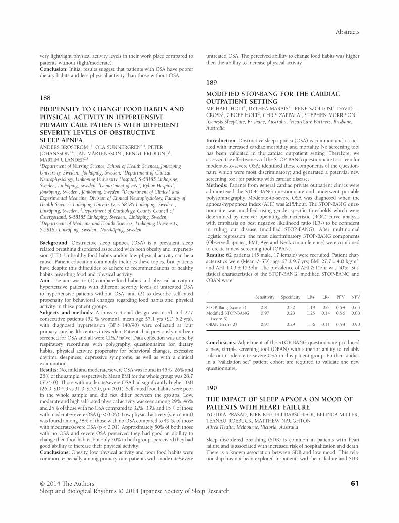
very light/light physical activity levels in their work place compared topatients without (light/moderate).Conclusion: Initial results suggest that patients with OSA have poorerdietary habits and less physical activity than those without OSA.
188
PROPENSITY TO CHANGE FOOD HABITS ANDPHYSICAL ACTIVITY IN HYPERTENSIVEPRIMARY CARE PATIENTS WITH DIFFERENTSEVERITY LEVELS OF OBSTRUCTIVESLEEP APNEAANDERS BROSTRÖM1,2, OLA SUNNERGREN3,4, PETERJOHANSSON4,6, JAN MÅRTENSSON1, BENGT FRIDLUND1,MARTIN ULANDER2,4
1Department of Nursing Science, School of Health Sciences, JönköpingUniversity, Sweden., Jönköping, Sweden, 2Department of ClinicalNeurophysiology, Linköping University Hospital, S-58185 Linköping,Sweden, Linköping, Sweden, 3Department of ENT, Ryhov Hospital,Jönköping, Sweden., Jönköping, Sweden, 4Department of Clinical andExperimental Medicine, Division of Clinical Neurophysiology, Faculty ofHealth Sciences Linköping University, S-58185 Linköping, Sweden.,Linköping, Sweden, 5Department of Cardiology, County Council ofÖstergötland, S-58185 Linköping, Sweden., Linköping, Sweden,6Department of Medicine and Health Sciences, Linköping University,S-58185 Linköping, Sweden., Norrköping, Sweden
Background: Obstructive sleep apnoea (OSA) is a prevalent sleeprelated breathing disordered associated with both obesity and hyperten-sion (HT). Unhealthy food habits and/or low physical activity can be acause. Patient education commonly includes these topics, but patientshave despite this difficulties to adhere to recommendations of healthyhabits regarding food and physical activity.Aim: The aim was to (1) compare food habits and physical activity inhypertensive patients with different severity levels of untreated OSAto hypertensive patients without OSA, and (2) to describe self-ratedpropensity for behavioral changes regarding food habits and physicalactivity in these patient groups.Subjects and methods: A cross-sectional design was used and 277consecutive patients (52 % women), mean age 57.1 yrs (SD 6.2 yrs),with diagnosed hypertension (BP > 140/90) were collected at fourprimary care health centres in Sweden. Patients had previously not beenscreened for OSA and all were CPAP naïve. Data collection was done byrespiratory recordings with polygraphy, questionnaires for dietaryhabits, physical activity, propensity for behavioral changes, excessivedaytime sleepiness, depressive symptoms, as well as with a clinicalexamination.Results: No, mild and moderate/severe OSA was found in 45%, 26% and28% of the sample, respectively. Mean BMI for the whole group was 28.7(SD 5.0). Those with moderate/severe OSA had significantly higher BMI(26.9, SD 4.3 vs 31.0, SD 5.0, p < 0.01). Self-rated food habits were poorin the whole sample and did not differ between the groups. Low,moderate and high self-rated physical activity was seen among 29%, 46%and 25% of those with no OSA compared to 32%, 33% and 15% of thosewith moderate/severe OSA (p < 0.05). Low physical activety (step count)was found among 28% of those with no OSA compared to 49 % of thosewith moderate/severe OSA (p < 0.01). Approximately 50% of both thosewith no OSA and severe OSA perceived they had good an abillity tochange their food habits, but only 30% in both groups perceived they hadgood abillity to increase their physical activity.Conclusions: Obesity, low physical activity and poor food habits werecommon, especially among primary care patients with moderate/severe
untreated OSA. The perceived abillity to change food habits was higherthen the abillity to increase physical activity.
189
MODIFIED STOP-BANG FOR THE CARDIACOUTPATIENT SETTINGMICHAEL HOLT1, DYTHEA MARAIS1, IRENE SZOLLOSI1, DAVIDCROSS2, GEOFF HOLT2, CHRIS ZAPPALA1, STEPHEN MORRISON1
1Genesis SleepCare, Brisbane, Australia, 2HeartCare Partners, Brisbane,Australia
Introduction: Obstructive sleep apnoea (OSA) is common and associ-ated with increased cardiac morbidity and mortality. No screening toolhas been validated in the cardiac outpatient setting. Therefore, weassessed the effectiveness of the STOP-BANG questionnaire to screen formoderate-to-severe OSA; identified those components of the question-naire which were most discriminatory; and generated a potential newscreening tool for patients with cardiac disease.Methods: Patients from general cardiac private outpatient clinics wereadministered the STOP-BANG questionnaire and underwent portablepolysomnography. Moderate-to-severe OSA was diagnosed when theapnoea-hypopnoea index (AHI) was ≥15/hour. The STOP-BANG ques-tionnaire was modified using gender-specific thresholds which weredetermined by receiver operating characteristic (ROC) curve analysiswith emphasis on best negative likelihood ratio (LR-) to be confidentin ruling out disease (modified STOP-BANG). After multinomiallogistic regression, the most discriminatory STOP-BANG components(Observed apnoea, BMI, Age and Neck circumference) were combinedto create a new screening tool (OBAN).Results: 62 patients (45 male, 17 female) were recruited. Patient char-acteristics were (Mean+/−SD): age 67 ± 9.7 yrs; BMI 27.7 ± 4.0 kg/m2;and AHI 19.3 ± 15.9/hr. The prevalence of AHI ≥ 15/hr was 50%. Sta-tistical characteristics of the STOP-BANG, modified STOP-BANG andOBAN were:
Sensitivity Specificity LR+ LR- PPV NPV
STOP-Bang (score 3) 0.81 0.32 1.19 0.6 0.54 0.63Modified STOP-BANG
(score 3)0.97 0.23 1.25 0.14 0.56 0.88
OBAN (score 2) 0.97 0.29 1.36 0.11 0.58 0.90
Conclusions: Adjustment of the STOP-BANG questionnaire produceda new, simple screening tool (OBAN) with superior ability to reliablyrule out moderate-to-severe OSA in this patient group. Further studiesin a “validation set” patient cohort are required to validate the newquestionnaire.
190
THE IMPACT OF SLEEP APNOEA ON MOOD OFPATIENTS WITH HEART FAILUREJYOTIKA PRASAD, KIRK KEE, ELI DABSCHECK, BELINDA MILLER,TEANAU ROEBUCK, MATTHEW NAUGHTONAlfred Health, Melbourne, Victoria, Australia
Sleep disordered breathing (SDB) is common in patients with heartfailure and is associated with increased risk of hospitalization and death.There is a known association between SDB and low mood. This rela-tionship has not been explored in patients with heart failure and SDB.
Abstracts
61© 2014 The AuthorsSleep and Biological Rhythms © 2014 Japanese Society of Sleep Research
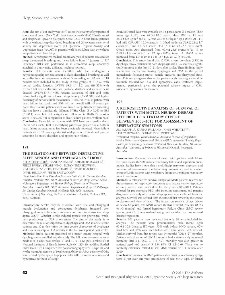
Aim: The aim of our study was to: (i) assess the severity of symptoms ofshortness of breath (New York Heart Association (NYHA) Classification)and sleepiness (Epworth Sleepiness Score (ESS) in heart failure patientswith or without sleep disordered breathing and (ii) to assess severity ofanxiety and depression scores (14 Question Hospital Anxiety andDepression Scale (HADS)) in patients with heart failure with or withoutsleep disordered breathing.Method: A retrospective medical record analysis of patients assessed forsleep disordered breathing and heart failure from 1st January to 31st
December 2013 was performed at an accredited sleep laboratoryattached to a university-affiliated teaching hospital.Results: 119 patients were identified who had undergone apolysomnography for assessment of sleep disordered breathing as wellas cardiac function assessment with an Echocardiogram. 83 out of 119patients were included in this study in two groups of (i) 63% withnormal cardiac function (LVEF% 64.9 +/− 2.2) and (ii) 37% withreduced left ventricular function (systolic, diastolic and valvular heartdisease) (LVEF%33.7+/−4.8). Patients suspected of SDB and heartfailure had a significantly longer sleep latency (P < 0.004) and higherfrequency of periodic limb movements (P < 0.05). 84% of patient withheart failure had confirmed SDB with an overall AHI > 5 events perhour. Heart failure patients with confirmed sleep disordered breathingdid not have a significantly different NYHA Class (P > 0.05) or ESS(P > 0.1) score but had a significant difference in HADs Depressionscore (P < 0.007) in comparison to heart failure patients without SDB.Conclusion: Heart failure patients with SDB have poor quality sleep.ESS is not a useful tool in identifying patients at greater risk of SDB inheart failure population as has been previously reported. Heart failurepatients with SDB have a greater risk of depression. This should promptscreening for mood disorders and appropriate referral.
191
THE RELATIONSHIP BETWEEN OBSTRUCTIVESLEEP APNOEA AND DYSPHAGIA IN STROKEKELLY SHEPHERD1,2, VANESSA BAKER1, AMHAD BAMAGOOS2,REECE PARRY2, STUART KING2, ROBYN TRZASKOWSKI3,KIM BROOKES3, GERALDINE HARRIS3, DAVID BLACKER4,DAVID HILLMAN1, PETER EASTWOOD1,2
1West Australian Sleep Disorders Research Institute, Sir Charles GairdnerHospital, Nedlands WA, 6009, Australia, 2Centre for Sleep Science, Schoolof Anatomy, Physiology and Human Biology, University of WesternAustralia, Crawley WA, 6009, Australia, 3Department of Speech Pathology,Sir Charles Gairdner Hospital, Nedlands WA, 6009, Australia,4Department of Neurology, Sir Charles Gairdner Hospital, Nedlands WA,6009, Australia
Introduction: Stroke may be associated with oral and pharyngealmuscle dysfunction and consequent dysphagia. Impaired oro-pharyngeal muscle function can also contribute to obstructive sleepapnea (OSA). Whether stroke-induced muscle oro-pharyngeal weak-ness predisposes to OSA is uncertain. The aim of this study is todetermine the relationship between dysphagia and OSA in acute strokepatients and ii) to determine the time-course of recovery of dysphagiaand its relationship to OSA severity in the 2–3 week period post stroke.Methods: Stroke patients presented to a major tertiary hospital withdysphagia were enrolled into the study. The following assessments weremade at 0–5 days post stroke(T1) and 10–21 days post stroke(T2): i)National Institutes of Health Stroke Scale (NIHSS); ii) modified BarthelIndex (mBI); iii) Comprehensive polysomnography (PSG)(Type II); andiv) the Mann Assessment of Swallowing Ability (MASA). Severity of OSAwas defined by the apnea hypopnea index (AHI: number of apneas andhypopneas per hour of sleep).
Results: Paired data were available on 15 participants (11 males). Theirmean age (±SD) was 67.3 ± 10.0 years. Mean BMI at T1 was28.3 ± 6.6 kg.m−2 and at T2 was 28.0 ± 5.9 kg.m−2 (p > 0.05). At T1 1had mild OSA (AHI 13.3 events.hr−1), 3 had moderate OSA (26.8 ± 1.7events.hr−1) and 10 had severe OSA (AHI 64.3 ± 22.5 events.hr−1).Group mean AHI decreased from 49.4 ± 28.8 events.hr−1at T1 to28.9 ± 24.0 events.hr−1 at T2 (p < 0.05)(Figure 1). MASA scoresimproved from 134 ± 19 at T1 to 167 ± 20 at T2 (p < 0.05).Conclusion: This study found that: i) OSA is very prevalent (93%) indysphagic stroke patients; ii) both dysphagia and OSA severities signifi-cantly improve in the first 10–21 days after stroke. These findings implya common mechanism linking dysphagia and OSA in the periodimmediately following stroke, namely impaired oro-pharyngeal func-tion. This study suggests that stroke patients with dysphagia should beroutinely assessed for OSA and appropriate early treatment imple-mented, particularly given the potential adverse impact of OSA-associated hypoxemia on recovery.
192
A RETROSPECTIVE ANALYSIS OF SURVIVAL OFPATIENTS WITH MOTOR NEURON DISEASEREFERRED TO A TERTIARY CENTREBETWEEN 2000–2013 FOR ASSESSMENT OFRESPIRATORY SYMPTOMSALI PARAPPIL1, KARINA FALLAND1, JOHN WHEATLEY1,3,LESLEY HOWARD1, SUHAIL DOI2, PETER WU1
1Westmead Hospital, Westmead/NSW, Australia, 2School of PopulationHealth University of Queensland, Brisbane/QLD, Australia, 3Ludwig EngelCentre for Respiratory Research, Westmead Millenium Institute, Westmead,Australia, 4University of Sydney at Westmead Hospital, Westmead,Australia
Introduction: Common causes of death with patients with MotorNeuron Disease (MND) include ventilatory failure and aspiration pneu-monia. Studies have shown that a multidisciplinary team approach andinitiation of non-invasive ventilation may improve survival in a selectgroup of MND patients with ventilatory failure or significant respiratorymuscle weakness.Methods: A retrospective survival analysis of MND patients referred forthe assessment of respiratory symptoms to a tertiary centre respiratory& sleep service was undertaken for the years 2000–2013. Patientsreferred for pre-operative PEG tube insertion assessment, and patientsdiagnosed with only obstructive sleep apnoea were excluded from theanalysis. Survival was defined from the time of first review by the serviceto documented time of death. The impact on survival of age (aboveor below 60 years), sex, MND variant (bulbar or limb), NIV use (0, ≤3or >3 months) and formal Respiratory Failure Clinic (RFC) review(pre or post 2010) was analysed using multivariable Cox proportionalhazards regression.Results: 102 patients were reviewed but only 78 were included foranalysis. The patients were predominantly male (62%), aged61.4 ± 10.8 (mean ± SD) years, 53% with bulbar MND variant, 60%used NIV, and 46% were seen before 2010 (pre formal RFC review).Median survival from first review was 14 months (IQR 5–27 months).Patients with duration of NIV ≤ 3 months had a significantly increasedmortality (HR 3.1, 95% CI 1.4–7.2). Mortality was also greater inpatients aged >60 years (HR 1.9, 95% CI 1.1–3.4). There was nomortality difference related to sex, MND variant or RFC review after2010.Conclusion: Survival in MND patients after onset of respiratory symp-toms is just over one year, irrespective of sex, MND type, or formal
Sleep, Science and Research
62 © 2014 The AuthorsSleep and Biological Rhythms © 2014 Japanese Society of Sleep Research
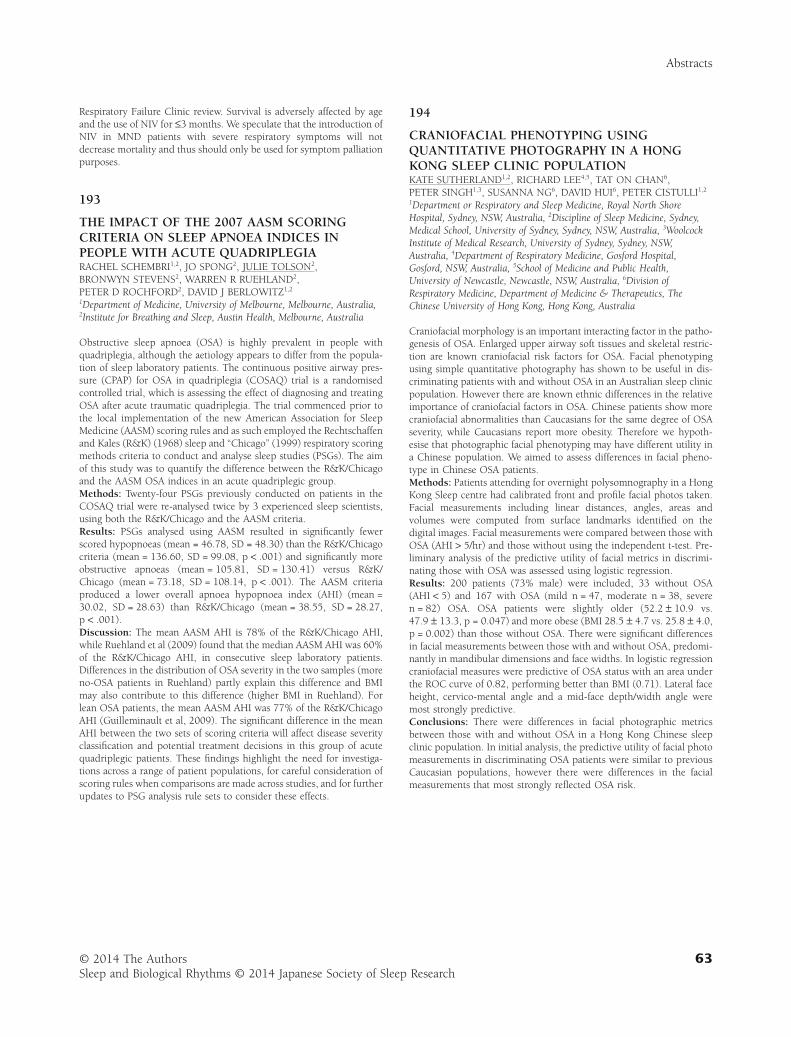
Respiratory Failure Clinic review. Survival is adversely affected by ageand the use of NIV for ≤3 months. We speculate that the introduction ofNIV in MND patients with severe respiratory symptoms will notdecrease mortality and thus should only be used for symptom palliationpurposes.
193
THE IMPACT OF THE 2007 AASM SCORINGCRITERIA ON SLEEP APNOEA INDICES INPEOPLE WITH ACUTE QUADRIPLEGIARACHEL SCHEMBRI1,2, JO SPONG2, JULIE TOLSON2,BRONWYN STEVENS2, WARREN R RUEHLAND2,PETER D ROCHFORD2, DAVID J BERLOWITZ1,2
1Department of Medicine, University of Melbourne, Melbourne, Australia,2Institute for Breathing and Sleep, Austin Health, Melbourne, Australia
Obstructive sleep apnoea (OSA) is highly prevalent in people withquadriplegia, although the aetiology appears to differ from the popula-tion of sleep laboratory patients. The continuous positive airway pres-sure (CPAP) for OSA in quadriplegia (COSAQ) trial is a randomisedcontrolled trial, which is assessing the effect of diagnosing and treatingOSA after acute traumatic quadriplegia. The trial commenced prior tothe local implementation of the new American Association for SleepMedicine (AASM) scoring rules and as such employed the Rechtschaffenand Kales (R&K) (1968) sleep and “Chicago” (1999) respiratory scoringmethods criteria to conduct and analyse sleep studies (PSGs). The aimof this study was to quantify the difference between the R&K/Chicagoand the AASM OSA indices in an acute quadriplegic group.Methods: Twenty-four PSGs previously conducted on patients in theCOSAQ trial were re-analysed twice by 3 experienced sleep scientists,using both the R&K/Chicago and the AASM criteria.Results: PSGs analysed using AASM resulted in significantly fewerscored hypopnoeas (mean = 46.78, SD = 48.30) than the R&K/Chicagocriteria (mean = 136.60, SD = 99.08, p < .001) and significantly moreobstructive apnoeas (mean = 105.81, SD = 130.41) versus R&K/Chicago (mean = 73.18, SD = 108.14, p < .001). The AASM criteriaproduced a lower overall apnoea hypopnoea index (AHI) (mean =30.02, SD = 28.63) than R&K/Chicago (mean = 38.55, SD = 28.27,p < .001).Discussion: The mean AASM AHI is 78% of the R&K/Chicago AHI,while Ruehland et al (2009) found that the median AASM AHI was 60%of the R&K/Chicago AHI, in consecutive sleep laboratory patients.Differences in the distribution of OSA severity in the two samples (moreno-OSA patients in Ruehland) partly explain this difference and BMImay also contribute to this difference (higher BMI in Ruehland). Forlean OSA patients, the mean AASM AHI was 77% of the R&K/ChicagoAHI (Guilleminault et al, 2009). The significant difference in the meanAHI between the two sets of scoring criteria will affect disease severityclassification and potential treatment decisions in this group of acutequadriplegic patients. These findings highlight the need for investiga-tions across a range of patient populations, for careful consideration ofscoring rules when comparisons are made across studies, and for furtherupdates to PSG analysis rule sets to consider these effects.
194
CRANIOFACIAL PHENOTYPING USINGQUANTITATIVE PHOTOGRAPHY IN A HONGKONG SLEEP CLINIC POPULATIONKATE SUTHERLAND1,2, RICHARD LEE4,5, TAT ON CHAN6,PETER SINGH1,3, SUSANNA NG6, DAVID HUI6, PETER CISTULLI1,2
1Department or Respiratory and Sleep Medicine, Royal North ShoreHospital, Sydney, NSW, Australia, 2Discipline of Sleep Medicine, Sydney,Medical School, University of Sydney, Sydney, NSW, Australia, 3WoolcockInstitute of Medical Research, University of Sydney, Sydney, NSW,Australia, 4Department of Respiratory Medicine, Gosford Hospital,Gosford, NSW, Australia, 5School of Medicine and Public Health,University of Newcastle, Newcastle, NSW, Australia, 6Division ofRespiratory Medicine, Department of Medicine & Therapeutics, TheChinese University of Hong Kong, Hong Kong, Australia
Craniofacial morphology is an important interacting factor in the patho-genesis of OSA. Enlarged upper airway soft tissues and skeletal restric-tion are known craniofacial risk factors for OSA. Facial phenotypingusing simple quantitative photography has shown to be useful in dis-criminating patients with and without OSA in an Australian sleep clinicpopulation. However there are known ethnic differences in the relativeimportance of craniofacial factors in OSA. Chinese patients show morecraniofacial abnormalities than Caucasians for the same degree of OSAseverity, while Caucasians report more obesity. Therefore we hypoth-esise that photographic facial phenotyping may have different utility ina Chinese population. We aimed to assess differences in facial pheno-type in Chinese OSA patients.Methods: Patients attending for overnight polysomnography in a HongKong Sleep centre had calibrated front and profile facial photos taken.Facial measurements including linear distances, angles, areas andvolumes were computed from surface landmarks identified on thedigital images. Facial measurements were compared between those withOSA (AHI > 5/hr) and those without using the independent t-test. Pre-liminary analysis of the predictive utility of facial metrics in discrimi-nating those with OSA was assessed using logistic regression.Results: 200 patients (73% male) were included, 33 without OSA(AHI < 5) and 167 with OSA (mild n = 47, moderate n = 38, severen = 82) OSA. OSA patients were slightly older (52.2 ± 10.9 vs.47.9 ± 13.3, p = 0.047) and more obese (BMI 28.5 ± 4.7 vs. 25.8 ± 4.0,p = 0.002) than those without OSA. There were significant differencesin facial measurements between those with and without OSA, predomi-nantly in mandibular dimensions and face widths. In logistic regressioncraniofacial measures were predictive of OSA status with an area underthe ROC curve of 0.82, performing better than BMI (0.71). Lateral faceheight, cervico-mental angle and a mid-face depth/width angle weremost strongly predictive.Conclusions: There were differences in facial photographic metricsbetween those with and without OSA in a Hong Kong Chinese sleepclinic population. In initial analysis, the predictive utility of facial photomeasurements in discriminating OSA patients were similar to previousCaucasian populations, however there were differences in the facialmeasurements that most strongly reflected OSA risk.
Abstracts
63© 2014 The AuthorsSleep and Biological Rhythms © 2014 Japanese Society of Sleep Research
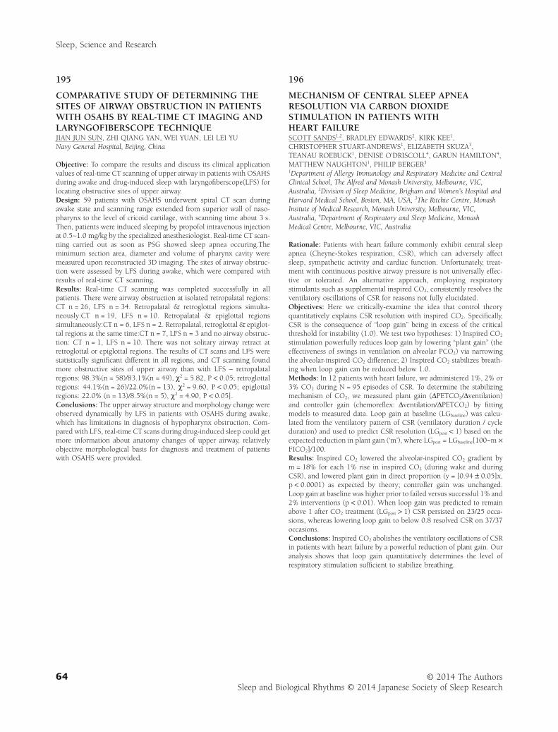
195
COMPARATIVE STUDY OF DETERMINING THESITES OF AIRWAY OBSTRUCTION IN PATIENTSWITH OSAHS BY REAL-TIME CT IMAGING ANDLARYNGOFIBERSCOPE TECHNIQUEJIAN JUN SUN, ZHI QIANG YAN, WEI YUAN, LEI LEI YUNavy General Hospital, Beijing, China
Objective: To compare the results and discuss its clinical applicationvalues of real-time CT scanning of upper airway in patients with OSAHSduring awake and drug-induced sleep with laryngofiberscope(LFS) forlocating obstructive sites of upper airway.Design: 59 patients with OSAHS underwent spiral CT scan duringawake state and scanning range extended from superior wall of naso-pharynx to the level of cricoid cartilage, with scanning time about 3 s.Then, patients were induced sleeping by propofol intravenous injectionat 0.5∼1.0 mg/kg by the specialized anesthesiologist. Real-time CT scan-ning carried out as soon as PSG showed sleep apnea occuring.Theminimum section area, diameter and volume of pharynx cavity weremeasured upon reconstructed 3D imaging. The sites of airway obstruc-tion were assessed by LFS during awake, which were compared withresults of real-time CT scanning.Results: Real-time CT scanning was completed successfully in allpatients. There were airway obstruction at isolated retropalatal regions:CT n = 26, LFS n = 34. Retropalatal & retroglottal regions simulta-neously:CT n = 19, LFS n = 10. Retropalatal & epiglottal regionssimultaneously:CT n = 6, LFS n = 2. Retropalatal, retroglottal & epiglot-tal regions at the same time:CT n = 7, LFS n = 3 and no airway obstruc-tion: CT n = 1, LFS n = 10. There was not solitary airway retract atretroglottal or epiglottal regions. The results of CT scans and LFS werestatistically significant different in all regions, and CT scanning foundmore obstructive sites of upper airway than with LFS – retropalatalregions: 98.3%(n = 58)/83.1%(n = 49), χ2 = 5.82, P < 0.05; retroglottalregions: 44.1%(n = 26)/22.0%(n = 13), χ2 = 9.60, P < 0.05; epiglottalregions: 22.0% (n = 13)/8.5%(n = 5), χ2 = 4.90, P < 0.05].Conclusions: The upper airway structure and morphology change wereobserved dynamically by LFS in patients with OSAHS during awake,which has limitations in diagnosis of hypopharynx obstruction. Com-pared with LFS, real-time CT scans during drug-induced sleep could getmore information about anatomy changes of upper airway, relativelyobjective morphological basis for diagnosis and treatment of patientswith OSAHS were provided.
196
MECHANISM OF CENTRAL SLEEP APNEARESOLUTION VIA CARBON DIOXIDESTIMULATION IN PATIENTS WITHHEART FAILURESCOTT SANDS1,2, BRADLEY EDWARDS2, KIRK KEE1,CHRISTOPHER STUART-ANDREWS1, ELIZABETH SKUZA3,TEANAU ROEBUCK1, DENISE O’DRISCOLL4, GARUN HAMILTON4,MATTHEW NAUGHTON1, PHILIP BERGER3
1Department of Allergy Immunology and Respiratory Medicine and CentralClinical School, The Alfred and Monash University, Melbourne, VIC,Australia, 2Division of Sleep Medicine, Brigham and Women’s Hospital andHarvard Medical School, Boston, MA, USA, 3The Ritchie Centre, MonashInsitute of Medical Research, Monash University, Melbourne, VIC,Australia, 4Department of Respiratory and Sleep Medicine, MonashMedical Centre, Melbourne, VIC, Australia
Rationale: Patients with heart failure commonly exhibit central sleepapnea (Cheyne-Stokes respiration, CSR), which can adversely affectsleep, sympathetic activity and cardiac function. Unfortunately, treat-ment with continuous positive airway pressure is not universally effec-tive or tolerated. An alternative approach, employing respiratorystimulants such as supplemental inspired CO2, consistently resolves theventilatory oscillations of CSR for reasons not fully elucidated.Objectives: Here we critically-examine the idea that control theoryquantitatively explains CSR resolution with inspired CO2. Specifically,CSR is the consequence of “loop gain” being in excess of the criticalthreshold for instability (1.0). We test two hypotheses: 1) Inspired CO2
stimulation powerfully reduces loop gain by lowering “plant gain” (theeffectiveness of swings in ventilation on alveolar PCO2) via narrowingthe alveolar-inspired CO2 difference; 2) Inspired CO2 stabilizes breath-ing when loop gain can be reduced below 1.0.Methods: In 12 patients with heart failure, we administered 1%, 2% or3% CO2 during N = 95 episodes of CSR. To determine the stabilizingmechanism of CO2, we measured plant gain (ΔPETCO2/Δventilation)and controller gain (chemoreflex: Δventilation/ΔPETCO2) by fittingmodels to measured data. Loop gain at baseline (LGbaseline) was calcu-lated from the ventilatory pattern of CSR (ventilatory duration / cycleduration) and used to predict CSR resolution (LGpost < 1) based on theexpected reduction in plant gain (‘m’), where LGpost = LGbaseline[100−m ×FICO2]/100.Results: Inspired CO2 lowered the alveolar-inspired CO2 gradient bym = 18% for each 1% rise in inspired CO2 (during wake and duringCSR), and lowered plant gain in direct proportion (y = [0.94 ± 0.05]x,p < 0.0001) as expected by theory; controller gain was unchanged.Loop gain at baseline was higher prior to failed versus successful 1% and2% interventions (p < 0.01). When loop gain was predicted to remainabove 1 after CO2 treatment (LGpost > 1) CSR persisted on 23/25 occa-sions, whereas lowering loop gain to below 0.8 resolved CSR on 37/37occasions.Conclusions: Inspired CO2 abolishes the ventilatory oscillations of CSRin patients with heart failure by a powerful reduction of plant gain. Ouranalysis shows that loop gain quantitatively determines the level ofrespiratory stimulation sufficient to stabilize breathing.
Sleep, Science and Research
64 © 2014 The AuthorsSleep and Biological Rhythms © 2014 Japanese Society of Sleep Research
197
HEART RATE VARIABILITY DURING TREATMENTWITH TWO AUTO-TITRATING CPAP DEVICESANTHONY TURTON1, EUGENE ZILBERG3, GARUN HAMILTON1,2
1Monash Health, Clayton, Victoria, Australia, 2Monash University,Clayton, Australia, 3Compumedics Limited, Abbotsford, Australia
Introduction: Measures of Heart Rate Variability (HRV) have beenshown to be poorer for patients treated with auto-titrating CPAP (APAP)devices when compared to fixed pressure CPAP devices. Several studieshave found APAP devices inferior to CPAP in reducing cardiovascularrisk factors associated with Obstructive Sleep Apnea. The aim of thecurrent study was to compare measures of HRV during treatment withthe Compumedics SPAP device and the Resmed Autoset S8 device.Methods: 30 patients (12 female, age 51.5(2.5)years, BMI 33.6(1.5)kg/m2, mean (SEM)) recently diagnosed with OSA (Apnea HypopneaIndex(AHI) 41.6(6.0)events/h) and naïve to CPAP were studied. Eachpatient was randomised to undergo treatment on either the SPAP or S8during polysomnography (PSG), followed by a repeat study one weeklater with the alternate device. Standard PSG variables, side effects andpressure statistics were recorded. Spectral analysis of HRV estimatedvery low frequency (VLF – 0.003 to 0.04 Hz), low frequency (LF – 0.04to 0.15 Hz) and high frequency (HF – 0.15 to 0.4 Hz) power and LF/HFratio by sleep stage. Linear mixed models were used to evaluate thedifferences between HRV measures.Results: Reduction in LF/HF ratio was observed during treatment withthe SPAP device, across all sleep stages compared to the S8: 0.78(.12),t = 6.54, p < 0.001 during NREM and 1.57(.57), t = 2.77, p = 0.006during REM. The observed improvements were consistent withincreased HF power during SPAP treatment: 90.10(11.45), t = 7.87,p < 0.001 msec2 during NREM, 76.08(14.034), t = 5.42, p <0.001 msec2 during REM.Discussion: Reduced LF/HF ratio is interpreted as improved parasym-pathetic activity, or sympathovagal balance. Treatment with the SPAPdevice resulted in reduced LF/HF ratio compared to the S8 deviceduring NREM and REM sleep. Increased HF power is considered reflec-tive of increased parasympathetic vagal activity. Higher HF power in theHRV was observed during treatment with the SPAP device compared tothe S8 device. HRV measures suggest improved parasympathetic activityduring treatment with the Compumedics SPAP device when comparedwith the ResMed S8 device.Conclusion: HRV measures suggest improved parasympathetic activityduring treatment with the Compumedics SPAP device when comparedwith the ResMed S8 device.
198
THE PREDICTIVE ROLE OF APAP TITRATIONDOWNLOAD DATA FOR EARLY (WEEK 4) CPAPTHERAPY OUTCOMESNIGEL MCARDLE1,2, BHAJAN SINGH2, CHRISTINE MAGUIRE2,SARAH MUTCH2, DAVID HILLMAN2
1University of Western Australia,, Nedlands WA, Australia, 2Sir CharlesGairdner Hospital:, Perth, Western Australia, Australia
Introduction: Titration of continuous positive airway pressure (CPAP)for treatment of obstructive sleep apnoea (OSA) increasingly uses auto-matic positive airway pressure (APAP) devices to determine a fixedpressure for long-term use. There are a paucity of data on the usefulnessof the APAP download obtained during the initial establishment ofCPAP. We hypothesized that data from the initial night of APAP titrationwould predict important outcomes after 4 weeks of CPAP treatment.
Methods: We utilized data from a randomized controlled trial compar-ing manual polysomnographic (PSG) titration with APAP titration todetermine a fixed CPAP pressure for long-term use among moderate tosevere OSA (AHI > 15/hr) with sleepiness (ESS > 8). Only thoserandomized to APAP titration are investigated in this report (n = 166).We sought predictors of clinically important outcomes after 4 weeks ofCPAP use. Predictors were sought from baseline demographic (age,gender, BMI, ESS), co-morbidities (e.g., history of depression), diagnos-tic PSG and titration night APAP download data (APAP use, residualAHI, 95th centile and median pressure, 95th centile and median leak).Results: 166 patients consented to APAP titration (in-laboratory, n = 80;home, n = 86). Participants were usually male (n = 110), withmean ± SD age (51 ± 11 yr), BMI (33 ± 5 kg/m2), ESS (13 ± 4) and AHI(44 ± 21 events/hr). 26 participants abandoned therapy by week 4 andindependent predictors were titration night APAP use and history ofhypercholesterolaemia (r = 0.364, adjusted r2 = 0.122, p < 0.001).Those continuing CPAP at week 4 had average nightly use of4.7 ± 2.1 hrs/night and the independent predictor was titration APAPuse (r = 0.434, r2 = 0.189, p < 0.001). ESS at week 4 was 8 ± 4 and theindependent predictors were baseline ESS and download median leak(r = 0.397, adjusted r2 = 0.145, p < 0.001). PSG using CPAP was onlyamong per protocol participants (n = 128). The AHI during CPAP atweek 4 was 10.6 ± 4 events/hr and independent predictors were down-load 95th centile leak, download AHI and diagnostic PSG arousal index(r = 0.416, adjusted r2 = 0.153, p < 0.001).Discussion: APAP download data from the first titration night indepen-dently predicts abandoning CPAP within 4 weeks and week 4 averagenightly use, ESS and PSG AHI (on treatment), in those accepting CPAP.
199
MECHANISM OF BIPHASIC INSPIRATORYWAVEFORMS IN NIVJOSEPH CHURTON, BRETT DUCE, CRAIG HUKINSSleep Disorders Centre, Princess Alexandria Hospital, Brisbane, QLD,Australia
Introduction: Biphasic inspiratory waveforms or double triggering canbe observed during NIV ventilation and can result in significant patientdiscomfort and reduced efficacy of ventilation. The underlying causesfor these biphasic waveforms are uncertain, as there are a constellationof variables that contribute to the triggering and cycling of the ventilator.Whether it is a single factor or a combination of these variables thatresult in double triggering is also unknown. We aimed to explore thepossible ventilator, patient and interface variables that may contribute tothese waveforms.Methods: An Ingmar Medical ASL 5000 breathing simulator in combi-nation with a physiological upper airway manikin was used with eithera Resmed S9 VPAP TX or Phillips Respironics Omnilab advanced plussystems and Resmed Quattro Medium face mask to deliver NIV. TheResmed device was set with medium trigger and cycle sensitivity.The Phillips Respironics used the usual Auto-trak setting. These are thedefault settings for these devices. The mask and apparatus were modi-fied to deliver variables of both continuous mask leak and mask dis-placement. Other device variables included EPAP and pressure support,respiratory muscle effort and respiratory rate. The simulator was set toresemble a restricted lung with airway resistance of 5 cm H2O/L/sec andcompliance of 25 mls/cm H2O.Results: With the Phillips Respironics device, high inspiratory muscleeffort appeared to significantly increase the rate of biphasic waveformsin isolation. Other variables including increased respiratory rate,increased continuous mask leak and increased mask displacement led toincrease rates of biphasic waveforms generally with an additive effect.
Abstracts
65© 2014 The AuthorsSleep and Biological Rhythms © 2014 Japanese Society of Sleep Research
The Resmed NIV device did not display biphasic waveforms with anyvariable change, however there was an increased rate of failure to triggerwith low respiratory rate and low respiratory muscle effort.Discussion: Differences in proprietary algorithms between the devicesresults in different responses to alterations in patient factors includingrespiratory rate and effort, interface and ventilator settings. With thePhillips Respironics device biphasic inspiratory waveforms with higherrespiratory effort and lower respiratory frequency were observed. Ourdata suggest that laboratory titrations should be performed on thespecific device planned for home use.
200
USABILITY TESTING OF A NEW MIXEDMODE VENTILATORSAHISHA KETHEESWARAN, ETIENNE VESCHAMBRE,DION MARTIN, JUNE MENDOZA, JULIA LORD, ALISON WIMMSResMed Science Centre, Sydney, NSW, Australia
Aim: Medical devices which are overly complex or counter-intuitiveduring operation can compromise patient safety through user error1.The aim of this test was to evaluate the usability of a new mixed modelife support ventilator (Astral, ResMed Ltd).Methods: Participants were asked to complete several realistic task-based scenarios which evaluated the ventilator core operating functions,such as ‘setting up the ventilator for ventilation with leak circuit on anadult patient’. A total of 307 different tasks were identified for evalu-ation, with each task tested by a minimum of 8 participants, or 15participants for safety critical tasks. Participants were also asked torespond to ‘emergency’ situations during which ventilator alarms andpatient conditions were simulated and assessed. Successful task com-pletion and user errors were recorded. Participants rated ease of com-pleting tasks using an 11-point Likert scale.Results: 90 participants (55 clinicians, 27 lay operators, 8 biomedicaltechnicians) completed the evaluation. Participants’ ages ranged from 18to >70 years, and participants’ levels of experience ranged from noexperience to >20 years. All safety critical tasks and core ventilator taskswere successfully completed by participants. Areas of difficulty centredprimarily around circuit set-up. Areas which participants rated ashaving the highest usability (mean usability score: 10/10) were: con-ducting alarms tests; changing alarm volume; and identifying the acti-vation of the remote alarm.Conclusion: During this evaluation the Astral ventilator was ableto be used safely, as intended, with strong usability ratings from partici-pants concerning the safety-related tasks of testing and activatingalarms.References: 1. FDA. Do it by design: an introduction to human factorsin medical devices. 1996:ii.
201
DEVELOPMENT OF THE USABILITY OF A NEWPAP SYSTEMRHYS THOMPSON, JAMIE WEHBEH, SAHISHA KETHEESWARAN,JEFF ARMITSTEADResMed Science Centre, Sydney, NSW, Australia
Introduction: The perceptual ease of the initial adaptation process isimportant to the long term adherence to PAP users to therapy. Anemerging area of focus for PAP system manufacturers is the user experi-ence of patients, of which the usability of PAP systems is a significantpart.
Method: 178 individuals (including current PAP users, non-PAP usersand healthcare professionals) participated across a period of two years,culminating in a usability trial of the completed PAP system (AirSense10, ResMed Ltd., Bella Vista), including 15 current PAP users and 5non-PAP users as participants. Primary factors assessed within theseusability trials included the ability of participants to perform tasks(objectively assessed) and the ease with which participants were able toperform tasks (subjectively rated by participants). Participants were notprovided with any orientation to or demonstration of the system prior toparticipation, however were provided with a user guide to refer to attheir discretion.Results: The average success rate of task performance without assis-tance was 98.3%, including tasks related to non-standard accessories(ie. Oximetry module), with 15/20 participants able to perform 100% oftasks without assistance. The average median task performance easerating was 9.75/10, with 19/24 tasks receiving a median rating fromparticipants of 10/10. Areas of greater challenge in task performanceincluded connection of the heated tubing to the PAP device (Successrate = 80%), related primarily to correct orientation of this tubing, andconnection of the finger sensor cable to the oximetry module (Ease = 7/10). None of the participants carried out any behaviour with the systemwhich could lead to a situation hazardous to their safety.Conclusion: The process of repeated evaluation and assessment has ledto the development of a PAP system which is intuitive and easy to use.It is intended that this usability will enhance the user experience ofpatients, particularly in the early transition to PAP therapy use.
202
CALF EXERCISE WHILE SITTINGREDUCES SNORINGBHAJAN SINGH1,2, AZADEH YADOLLAHI3, HISHAM ALSHAER3,OWEN LYONS3, DOUGLAS BRADLEY3
1West Australian Sleep Disorders Research Institute, WA, Australia, 2SirCharles Gairdner Hospital, WA, Australia, 3University of Toronto, ON,Canada
Background: Overnight rostral fluid shift from the legs to the neckcorrelates with the severity of obstructive sleep apnea (OSA) and timespent sitting. Sitting causes gravity-dependent fluid accumulation in thelegs that is counteracted by walking. However most office workers arerequired to sit for long periods and walking to preventing fluid accu-mulation in the legs is impractical. We hypothesized that sitting wouldcause fluid accumulation below the knees that could be attenuated bycalf exercise while sitting and this would reduce snoring.Methods: In 11 healthy non-obese subjects (8 men, age 50.8 ± 8.3 yrs,BMI 25.0 ± 3.3 kg/m2) we measured leg fluid volume (LFV) usingbioelectrical impedance and leg volume (LV) below both knees whilesitting for 4 hours during the day, Mallampati score, and snoring usinga portable ApneaDx device. Subjects were studied twice, a week apart,in a randomised double cross-over design: control – no calf exercise,intervention – near continuous calf muscle contractions against a pedalresistance (Step-It). Paired t-tests were used to analyze changes in LFVand LV and the effect of the intervention.Findings: Sitting for 4 hours was associated with a progressive increasein LFV and LV by 134.2 ± 54.5 ml and 247.9 ± 73.8 ml without exer-cise (both p < 0.0001), and 97.8 ± 38.6 ml and 115.3 ± 82.2 ml withcalf exercise respectively. Calf exercise reduced the change in LFV andLV (both p < 0.001), and reduced snoring in subjects with a Mallampatiscore of 4 (p = 0.006, Mann Whitney).Conclusions: Sitting for 4 hours results in substantial fluid accumula-tion in the legs of healthy subjects that could serve as a reservoir for fluiddisplacement to the neck during sleep. Calf exercise while sitting
Sleep, Science and Research
66 © 2014 The AuthorsSleep and Biological Rhythms © 2014 Japanese Society of Sleep Research
reduces the accumulation of fluid in the legs while sitting and reducessnoring in non-obese subjects with upper airway narrowing.
203
RETROSPECTIVE AUDIT OF THE EFFICACYOF THE MANDIBULAR ADVANCEMENTSPLINT (MAS)ROSIANNA WEE1,2
1Canberra Sleep Clinic, Canberra, ACT, Australia, 2The CanberraHospital, Canberra, ACT, Australia
Objectives: To check on the effectiveness of the MAS in patientsreferred from the Canberra Sleep Clinic.Method: Retrospective audit of all pre-MAS and post-MAS diagnosticsleep studies in patients who had a post-MAS study at the unit over a 12month period.Results: 13 patients had a post-MAS study during the 12 months. AHIduring the initial diagnostic study ranged from 11.0–120.6/hr, average27.9/hr, 3 patients had severe OSA, 7 patients had moderate OSA, and3 patients had mild OSA. AHI during post-MAS studies ranged from0.5–48.2/hr, average AHI 12.3/hr, 1 patient had residual severe OSA, 2patients had residual moderate OSA, 7 patients had residual mild OSA,3 (23%) patients had no residual OSA. 8 patients (61.5%) had AHIreduced by >50%, 1 patient had an increased AHI post-MAS, 4 patientshad reduced AHI but not >50%. Of the 3 patients with severe OSA intheir pre-MAS studies, 1 still had severe OSA post-MAS but severity<50% compared to their pre-MAS study, 1 had mild OSA post-MAS(severity was also reduced by 50%), 1 had no OSA post-MAS.Conclusion: The MAS has been successful in reducing the severity ofOSA in 61.5% of our patients. The MAS has been effective even inpatients with severe OSA.
204
CONTINUOUS POSITIVE AIRWAY PRESSURE(CPAP) ADHERENCE OF PATIENTSCOMMENCING THERAPY AT A PRIVATE CLINICWITH INTENSIVE SUPPORT AND CHOICE OFEQUIPMENT MANUFACTURERSHELEN GILBERT, ANDREW MAIRAbout Sleep Clinics, Adelaide, SA, Australia
Four hours of adherence to Continuous Positive Airway Pressure(CPAP) therapy per day is generally accepted as the minimum require-ment for adequate treatment of obstructive sleep apnoea (OSA) andcompliance with associated legal safety standards. This study aimed toassess the effectiveness of a private clinical model delivering CPAPtherapy incorporating intensive contact and free choice of 3 CPAPequipment brands and also the relationship between pre-treatmentApnoea Hypopnoea Index(AHI), age, sex & adherence.Objectives: To examine (i)CPAP treatment adherence (>4 hours/day) ofpatients with OSA at 4 weeks and 6 months after initiation of CPAPtherapy; (ii) the relationship between pre-treatment Apnoea HypopnoeaIndex (AHI), age and gender and CPAP adherence; (iii) CPAP equipment(machine and mask) choices after trial therapy.Setting: 182 consecutive patients in a private sleep therapy service(CPAP, mandibular advancement splints, psychology, lifestyle interven-tion) with 2 clinics in metropolitan area.Design: Retrospective analysis of clinical records and choice of CPAPequipment.
Methods: CPAP adherence at 4 weeks and 6 months after initiation ofCPAP trial were compared across age, sex & AHI (mild, mod, severe).CPAP equipment choices were compared for machine type (auto, fixed,ASV), and mask type (full-face, nasal, pillows).Results: (i) 74% of all patients were adherent 4 weeks after commenc-ing their trial. This decreased to 52% adherence at 6 months. Of patientswho were adherent at 4 weeks, 62% were adherent at 6 months. 23% ofnon-adherent patients at 4 weeks became adherent at 6 months. (ii) Nosignificant differences in adherence were found across age, sex or pre-treatment AHI. (iii) Equipment choice of CPAP machines was 50%automatic, 45% fixed, and 5% ASV; of masks 39% full-face, 36% nasal,and 25% pillows.Conclusion: A significant proportion of patients commencing CPAPtherapy are able to achieve adherence after 4 weeks and 6 months withintensive clinical support and free choice of equipment. Persistence withpatients who were initially non-adherent is warranted as a significantproportion of them are also able to achieve adherence after 6 months.Patients demonstrate a wide range of equipment preferences whenoffered choice of CPAP machines and masks.
205
USING A CHINSTRAP FOR MOUTH LEAK: HELPOR HINDRANCE?MATTHEW LEONG, LYNN HOEYThe Prince Charles Hospital, Brisbane, Australia
Continuous positive airway (CPAP) is currently accepted as the mosteffective treatment option for moderate to severe Obstructive SleepApnoea (OSA). Treatment most commonly involves pneumatic splintingof the intermittent collapsing upper airway via a nasal or oronasalinterface. Despite this, adherence to CPAP remains problematic. Use ofan oronasal interface may contribute to poor compliance to therapy anda nasal modality is thus preferred. However, mouth leaks with nasalCPAP can occur, representing a risk for reduced adherence to therapy. Acommon course of action is to minimize mouth leaks by application ofa chinstrap. We report on a case where chinstrap application was det-rimental to desired outcomes.
A 52 year-old man with diagnosed severe obstructive sleep apnoeaunderwent an attended Type I Continuous Positive Airway Pressuretitration polysomnography, using a nasal interface, at a tertiary sleepcentre. Pressure was titrated between 5–9.0 cmH2O to establish upperairway patency. Upper airway obstruction was controlled at a pressure of9.0 cmH2O; however, mouth leaks were problematic, resulting in cor-tical arousal. Two different chin restraints were employed on separateoccasions in an attempt to minimise mouth leak. However, independentof body position, head position and sleep stage; an apnea-hypopnoeaindex of 60/hr was evident upon adding a chinstrap compared to anindex of 1.5/hr without a chinstrap at the same pressure. This casehighlights that the common course of action to minimise mouth leakwith the addition of a chin restraint may be detrimental and contributeto upper airway obstruction.
Abstracts
67© 2014 The AuthorsSleep and Biological Rhythms © 2014 Japanese Society of Sleep Research
206
FACTORS INFLUENCING CONTINUOUSPOSITIVE AIRWAY PRESSURE THERAPYACCEPTANCE AT A PHYSICIAN ANDTECHNOLOGIST THERAPY CLINICGAVIN STURDY, THOMAS ANSELL, ASHLEIGH BECU,LIAM COFFEY, IVAN LINGWA Sleep Disorders Research Institute, Perth, WA, Australia
Introduction: Continuous positive airway pressure (CPAP) therapy isthe gold standard treatment for obstructive sleep apnoea (OSA) but hasvariable acceptance rates. We aimed to evaluate differences betweenpatients who accept and reject CPAP therapy at a physician and tech-nologist CPAP therapy clinic to determine factors that may influenceCPAP acceptance.Methods: Data collected on consecutive patients attending a CPAPtherapy clinic from January to February 2014 included pre- and post-treatment apnoea-hypopnoea indices (AHI) and Epworth sleepinessscores (ESS), CPAP titration method, average usage (hours), symptomresponse (recorded as complete, partial, or nil), and acceptance at theend of therapy trial. In patients who accepted therapy, machine type andCPAP pressure was noted. Characteristics between patients whoaccepted and rejected CPAP were compared.Results: 57 patients completed a trial of CPAP therapy of minimum 3weeks, of whom 43 (75%) accepted and 14 (25%) rejected CPAPtherapy. The majority (79%) had CPAP titration conducted at homeusing auto-titration CPAP. Patients who accepted therapy differed sig-nificantly from those who rejected therapy in average hours of CPAPusage (7.0 vs 3.0, p < 0.001) and had significantly higher rates ofcomplete symptom response (67% vs 7%, p < 0.001). There was a trendtoward a difference in pre-treatment AHI (40.1 vs 30.7/hr, p = 0.11) andimprovement in ESS (5.0 vs 3.3, p = 0.17). There was no significantdifference in post-treatment AHI (4.2 vs 5.1/hr), and absolute ESS(pre-treatment 11.0 vs 9.8, post-treatment 5.9 vs 6.9). Amongst patientswho rejected therapy, the most common reasons cited were mask dis-comfort (4/14), dryness (2/14), and noise (2/14). Amongst patients whoaccepted therapy, most obtained fixed-pressure CPAP (90%) with anaverage prescribed CPAP pressure of 10.8 cmH2O (SD ± 1.2).Conclusion: Hours of CPAP usage and subjective symptom responsepredicted short term acceptance of CPAP therapy in this population.There was a trend toward higher pre-treatment AHI and improvementin ESS in those who accepted CPAP therapy. The most common reasoncited for rejection of CPAP therapy was mask discomfort.
207
BREATH SHAPE AS A CLINICAL MARKERIN PAEDIATRIC SLEEP-RELATEDBREATHING DISORDERSCHLOE PARSLEY1,2, SADASIVAM SURESH1,2, PHILIP TERRILL2
1Mater Children’s Hospital, South Brisbane, QLD, Australia, 2University ofQueensland, St. Lucia, QLD, Australia
Aim: Duchenne Muscular Dystrophy (DMD) and Prader-Willi syn-drome (PWS) are complex congenital disorders which manifest as pae-diatric sleep related breathing disorders (SRBD). Due to the slowprogression of pathological onset, and lack of correlation with daytimeclinical features, these patients are often only first treated when theirpathology reaches a crisis point – often at the acute stage. This studyaims to determine if monitoring of respiratory shape during sleep canreveal more subtle changes with the progression of the disorder, which
could then be monitored non-invasively with commercial home-useequipment.Methods: A retrospective cohort of 14 DMD and 11 PWS patientsunderwent three full overnight polysomnograms over a three to five yearaged 2–12 at first study and 6–17 at final study. Nasal air pressure datawere extracted for three 10 minute periods of artefact free non-REMsleep for each study, and inspiratory and expiratory phases of the nasalpressure curves were segmented and normalized for amplitude and timeto minimize instrumentation and age effects. The variables kurtosis,skewness and variance (measurements of curve shape) were calculatedfor both inspiratory and expiratory segments. Mean values of eachvariable were calculated for each study and the Friedman test for groupdata applied (with each segment a repeated measure) to determinewhether changes with study number were statistically significant.Results: There were significant changes in respiratory curve shape inDMD patients with study progression as measured by expiratory kur-tosis increasing from 2.0 to 2.3 (p = 0.003), expiratory variance increas-ing from 0.07 to 0.08 (p = 0.03) and inspiratory variance increasingfrom 0.06 to 0.07 (p = 0.01) between the first and third studies respec-tively. Changes were also observed in PWS patients as measured by anincrease in inspiratory variance from 0.06 to 0.07 (p = 0.001) betweenthe first and third studies respectively.Discussion: Linear indices of breath shape are able to quantify keychanges in the breathing patterns of DMD and PWS patients as theirdisease progresses over time. Such variables may be useful in monitor-ing the clinical status of such patients, assisting clinicians with respect tothe decision to treat and measuring treatment response.
208
GESTATIONAL AGE AT BIRTH AFFECTS THEMATURATION OF BAROREFLEX SENSITIVITYKARINNA FYFE1,2, STEPHANIE YIALLOUROU1,2, FLORA WONG1,2,ALEXSANDRIA ODOI1, ADRIAN WALKER1, ROSEMARY HORNE1,2
1The Ritchie Centre, MIMR-PHI Institute, Monash University, Melbourne,Victoria, Australia, 2Department of Paediatrics, Monash University,Melbourne, Victoria, Australia
Background: Preterm birth is associated with increased risk of theSudden Infant Death Syndrome (SIDS) and cardiovascular disease inadulthood, which may be due to impaired cardiovascular control. Thebaroreflex is the primary mechanism by which autonomic control ofblood pressure (BP) occurs. Baroreflex sensitivity (BRS) has been shownto be altered by preterm birth. In term infants it is reduced in the proneposition, the major risk factor for SIDS. To date, the effect of pronesleeping on BRS in preterm infants has not been assessed. Therefore, weaimed to determine the effect of prone sleeping on BRS in preterminfants born across a range of gestational ages.Methods: Daytime polysomnography was performed at 2–4 weeks, 2–3months and 5–6 months post-term age on 21 very preterm (meangestation 29.4 ± 0.3 wk), 14 preterm (mean gestation 33.1 ± 0.3 wk)and 17 term (mean gestation 40.1 ± 0.3 wk) infants. Blood pressure wasmeasured using a Finometer™ cuff (Finapress Medical Systems,Amsterdam, The Netherlands). Data were recorded in both the supineand prone positions. BRS was calculated using cross spectral analysis ofspontaneous fluctuations in BP and heart period (HP). The effects ofsleep state and position were determined using two-way analysis ofvariance (ANOVA) within very preterm, preterm and term infantgroups. The effects of post-term age and gestational age at birth (verypreterm, preterm or term) were determined using two-way ANOVAwithin each sleep state and position.Results: Systolic BP was lower in very preterm (p < 0.01) and preterm(p < 0.05) infants compared to term infants in QS and lower in very
Sleep, Science and Research
68 © 2014 The AuthorsSleep and Biological Rhythms © 2014 Japanese Society of Sleep Research
preterm compared to term infants in AS (p < 0.05) in the prone positionat 2–3 months CA. There was no effect of gestational age at birthon systolic BP at any other age. There was no effect of gestationalage at birth on HP at any age. BRS was lower in the prone positionin very preterm infants at 2–4 weeks in AS (p < 0.05). Maturationof BRS was delayed in very preterm compared to preterm and terminfants.Conclusions: Maturation of BRS after term-equivalent age is altered invery preterm infants. Although this is unlikely to contribute to increasedSIDS risk, it may predispose very preterm infants to cardiovasculardisease in adulthood.
209
LONG-TERM CARDIOVASCULAR IMPAIRMENTIN PRESCHOOL-AGED CHILDREN WITHUNRESOLVED SLEEP DISORDERED BREATHINGLISA M WALTER1,2, LAUREN E NISBET1, AIDAN J WEICHARD1,SAMANTHA L HOLLIS1, VICKI ANDERSON4, GILLIAN M NIXON1,3,MARGOT J DAVEY1,3, ROSEMARY SC HORNE1,2
1The Ritchie Centre, MIMR-PHI Institute of Medical Research, Melbourne,Australia, 2Dept of Paediatrics, Monash University, Melbourne, Australia,3Melbourne Children’s Sleep Centre, Monash Children’s Hospital,Melbourne, Australia, 4Critical Care and Neuroscience Research, MurdochChildren’s Research Institute, Melbourne, Australia
Background: To date, studies have not identified the long-term cardio-vascular outcomes of treatment of sleep disordered breathing (SDB) inpreschool-aged children. We aimed to investigate both the effect oftreatment and resolution of obstructive sleep apnoea (OSA) on heartrate (HR) and pulse transit time (PTT) as a surrogate measure of bloodpressure change, in preschool-aged children 3 y following initialdiagnosis.Methods: Children aged 3–5 y diagnosed with SDB and non-snoringcontrols returned for follow-up overnight polysomnography (PSG) 3 yfollowing their baseline PSG. The children with SDB were grouped into:1) treated and SDB resolved n = 14; 2) treated and SDB unresolvedn = 7; 3) not treated and SDB resolved n = 8; 4) not treated and SDBunresolved n = 10. SDB was considered resolved when the obstructiveapnoea hypopnoea index (OAHI) was ≤1 event/h, there was no snoringdetected at the PSG and regular snoring was not indicated by parents onthe OSA-18. Statistical analysis was performed using two-way repeatedmeasures ANOVA.Results: We are presenting data from 39 children with SDB and 14controls. Children whose SDB had resolved whether they were treatedor not, and control children, had a significant decrease in HR atfollow-up compared with baseline during both wake (control,mean ± sem 103 ± 3 vs 94 ± 3 bpm; treated, 101 ± 3 vs 87 ± 3 bpm;not treated 94 ± 3 vs 84 ± 4 bpm p < 0.01 for all) and sleep (control,89 ± 2 vs 82 ± 2 bpm p < 0.01; treated, 87 ± 2 vs 76 ± 3 bpm,p < 0.001; not treated, 81 ± 3 vs 73 ± 3 bpm, p < 0.05). In contrast, HRwas not decreased at follow-up in the children whose SDB remainedunresolved, whether they were treated or not, during either wake orsleep no difference in PTT was identified between studies for any groupduring either wake or sleep.Conclusion: Our data suggest that in preschool children 3 years fol-lowing their original study, concomitant with the resolution of SDB, HRfollowed the normal physiological pattern of decreasing significantlywith increasing age, similar to the non-snoring control children.However, in children whose SDB remained unresolved, HR remained atbaseline levels. As elevated HR has adverse consequences for the car-diovascular system, further research with a larger cohort of children isneeded to confirm these findings.
210
CAN NECK AND WAIST CIRCUMFERENCE BEUSED AS INDICATORS OF PAEDIATRICOBSTRUCTIVE SLEEP APNOEA SEVERITY?LAUREN NISBET1, LISA WALTER1, MARGOT DAVEY1,2,GILLIAN NIXON1,2, ROSEMARY HORNE1
1The Ritchie Centre, MIMR-PHI Institute of Medical Research, Melbourne,Victoria, Australia, 2Melbourne Children’s Sleep Centre, MonashChildren’s, Melbourne, Victoria, Australia
Introduction: Obesity is a major cause of obstructive sleep apnoea(OSA) in older children and adolescents. Neck circumference (NC)percentiles have recently been proposed as a screening tool for OSA inCanadian children aged 6–17 years (Katz et al, 2014). However therelationship between anthropometric variables and OSA severity has notbeen investigated in preschool age children, when OSA is most preva-lent and is largely due to adenotonsillar hypertrophy. This study aimedto characterise such a relationship in Australian children aged 3–5 and7–12 years.Methods: 192 preschool (3–5 y) and 53 school-age children (7–12 y)underwent overnight polysomnography plus height, weight, NC, hipand waist circumference (WC) measurements. Subjects were groupedaccording to OSA severity (Controls, no snoring, obstructive apnoeahypopnoea index (OAHI) ≤ 1 event/h; Primary Snoring (PS), OAHI ≤ 1;Mild OSA, OAHI > 1–5; Moderate-Severe (MS) OSA, OAHI > 5). Bodymass index (BMI) z-scores were calculated. NC and WC percentileswere calculated for age and sex based on established norms (>6 years)or cohort percentiles (<6 years; no established norms available) andproportions of subjects >90th percentile within each group were com-pared using chi-square analysis.Results: School-age children with MS OSA had a higher waist-to-hipratio than other groups (p < 0.05). There were no group differences inother anthropometric measurements. OAHI was correlated with BMIz-score (r = 0.4, p < 0.01), WC (r = 0.3, p < 0.05) and waist-to-hip ratio(r = 0.4, p < 0.01). The proportion of subjects in each group with NCand WC >90th percentile increased with OSA severity (Control 23%, PS40%, Mild OSA 60%, MS OSA 71%, p = 0.08; Control 23%, PS 33%,Mild OSA 40%, MS OSA 71%; p = 0.1 respectively). In preschool chil-dren, OAHI was not correlated with any anthropometric variable. Therewere no group differences in anthropometric variables or proportions ofsubjects with NC and WC >90th percentile.Conclusion: Anthropometric variables relate to OSA severity in school-aged, but not in preschool aged children. These findings are largelyunderpinned by the contrasting causes of OSA in each age group.The use of NC and WC as a screening tool for OSA is likely to be agedependent.
211
DOES SLEEP LOSS ACCUMULATE OVER ANENTIRE SCHOOL TERM? OBJECTIVE DATA INADOLESCENT BOYSSARAH BLUNDEN1, LARISSA CLARKSON1, BREE DREW1,SIOBHAN BANKS2, AMELIA SEARLE3, TIM OLDS2
1Central Queensland University, Adelaide, South Australia, Australia,2University of South Australia, Adelaide, South Australia, Australia,3University of Adelaide, Adelaide, South Australia, Australia
Introduction: Evidence suggests adolescents experience less sleepduring the school week compared to weekends or school holidays,where there is usually ‘catch-up’ sleep. These patterns, studied overshort time periods, are known to be detrimental to daytime function.
Abstracts
69© 2014 The AuthorsSleep and Biological Rhythms © 2014 Japanese Society of Sleep Research
However, it is unknown what happens to these sleep patterns during anentire 10-week school term. Does relative weekly sleep loss accumulateover the school term? This study aims to assess what happens to ado-lescent sleep over an entire school term and, in so doing, inform thesleep loss debate in adolescents.Method: Fifty14–16 year old males from 5 randomly selected schoolscompleted baseline (i.e.school holidays) and weekly measures of theirsleep (daily Stanford Sleepiness Scale and sleep diary, 24 hour wristactivity monitors) for 12 weeks (one week of school holidays, 10 weeksof school term and another week of school holidays). Mixed model andtime series analyses were/are being undertaken and are ongoing.Results: To date (n = 27) data show that compared to baseline, nightlysleep duration reduced significantly by an average of 45 minutes inweek one of school term. Over the term, this deficit reduced to anaverage daily sleep 20 minutes shorter than at baseline. Participantsspent less time in bed on school nights than on the weekends (p < 0.05),but sleep loss was minimised because they also went to sleep earlier(p < 0.05). Week bedtimes generally advanced across the school termbut sleep loss was held in check by weekend catch-up sleep resulting inno cumulative sleep loss across the 10 weeks. Sleepiness scoresremained stable across the term.Discussion: These findings confirm that average daily sleep duration isconsistently reduced across the school week compared to holidays andacross the school week compared to weekends. Therefore relative to pre-and post-term sleep, in-term sleep was consistently shorter, and hencestudents are likely accumulating sleep loss during the term. However,stable sleepiness scores suggests that these participants may be copingwith reduced sleep across the term and perhaps that ‘catch up’ sleep onthe weekends is sufficient for maintenance. These data make a signifi-cant contribution to the debate concerning adolescent sleep loss. Dataanalyses are ongoing.Acknowledgements: This study is financially supported by BeyondBlue.
212
PHOTOGRAPHIC ASSESSMENT OFCRANIOFACIAL PHENOTYPE IN CHILDRENWITH OSA: A FEASIBILITY STUDYJOACHIM NGIAM1,4, AIDAN WEICHARD3, KATE SUTHERLAND1,PARMINDER SINGH1, MARGOT DAVEY2, ROSEMARY HORNE3,PETER CISTULLI1, GILLIAN NIXON2,3
1Department of Respiratory and Sleep Medicine,Royal North ShoreHospital, Sydney, NSW, Australia, 2Melbourne Children’s Sleep Centre,Melbourne, Victoria, Australia, 3Ritchie Centre, MIMRA-PHI Institute ofMedical Research, Melbourne, Victoria, Australia, 4Phd candidate, TheUniversity of Sydney, Sydney, NSW, Australia
Introduction: Craniofacial structure plays an important role in thepathogenesis of Obstructive Sleep Apnoea (OSA). Previous radiographicimaging studies have demonstrated a range of craniofacial abnormalitiesthat are associated with the presence and severity of OSA, in both adultsand children. We have reported a novel photographic method for quan-tifying craniofacial phenotype in adults with OSA, and this may have arole in screening patients for OSA risk. The aim of this study was todetermine the feasibility of taking standardised facial photographs inchildren, as a precursor to determining the clinical utility of the tech-nique for predicting the presence and severity of OSA.Methods: Parents of children were approached for permission to taketheir child’s photo as an optional part of a larger study occurring at a sitedistant to the photographic analysis site. The photographic techniqueinvolved the use of a consumer digital camera (IXUS 60, Canon Inc.,Japan). Photographers followed a written protocol, but were not spe-
cifically trained in taking photos for craniofacial phenotype. Calibrationmarkers were placed on the face, and frontal and profile photographswere taken according to a standardised protocol. The quality of thephoto in terms of its suitability for subsequent detailed cephalometricphoto analysis was rated according to a pre-defined scale (categoricalscale: 1-perfect, 2-good, 3-adequate, 4-borderline, 5-poor).Results: Parents of a total of 106 children (42%F; median age 5.1 y,range 1.8–14.6 y) consented to the main study. Photographs were suc-cessfully obtained in 41 children (44%F; median age 4.2 y, range2–14.6 y). 54 children were not present at the time of consent and sophotos could not be obtained. 6 parents specifically did not giveconsent. No photos were obtained in 5 children whose parents did giveconsent, due to poor co-operation of the child. The mean quality ratingof these photos was very good at 1.9 ± 1.1 (mean ± sem), and 78% ofphotos were rated as either “good” or “perfect”.Conclusion: This pilot study demonstrates the feasibility of takingcraniofacial photographs in children presenting to a sleep clinic, acrossa wide age spectrum. This will permit exploration of the clinical utilityof this technique in quantifying craniofacial phenotype as a risk factorfor OSA in children.
213
SLEEP AND WAKE ASSESSMENT WITH AMOVEMENT DETECTOR: IMPROVINGACTIGRAPHY FOR CHILDREN WITHFRAGMENTED SLEEPMARNIE LAMPRECHT1, CHLOE PARSLEY2, SADASIVAM SURESH2,ANDREW P BRADLEY1, PHILIP I TERRILL1
1School of Information Technology and Electrical Engineering, University ofQueensland, Brisbane, QLD, Australia, 2Department of Respiratory andSleep Medicine, Mater Children’s Hospital, Brisbane, QLD, Australia
Background: Actigraphy is an effective, non-invasive tool for assessingsleep and wakefulness. However, there are significant limitations whenused with patients exhibiting fragmented sleep. Specifically, actigraphyis unable to distinguish restless sleep from wake, and restful wake fromsleep, resulting in poor specificity. Commercial actigraphs representmovement as a summarised ‘activity count’ per epoch (typically 30 s).However, summarising movement in this way limits the ability to iden-tify specific movements. Therefore, we hypothesise that (1) movementsduring sleep are characteristically different to those during wake; and(2) detecting sleep and wakefulness on a movement-by-movement basismay improve the performance of actigraphy in sleep assessment.Methods: Twenty-five children with suspected OSA were studied sim-ultaneously with full overnight polysomnogram and tri-axialaccelerometry of the left finger and wrist, chest, left ankle and great toe.Movements during periods of wake and sleep were manually labelledfrom eight children (ages 6–15 years, 4M) and time-frequency analysiswas applied to identify variables which differentiate sleep and wakemovements. These variables were then applied using regression to theremaining 17 children (ages 6–16 years, 13M) to classify the movementsas having occurred during sleep or wake. The predicted sleep stageswere then compared to the manually scored polysomnogram.Results: There were distinctly different time-frequency characteristicsof movements that occur during sleep and wake. In summary, highfrequency movements preceded positional changes during sleep andthat movements during sleep were typically longer. Classifying sleepand wake using these characteristics on a movement-by-movementbasis achieved a Kappa agreement of 0.46(0.18), sensitivity of93.6(3.7)%, and specificity of 50.7(19.3)%.
Sleep, Science and Research
70 © 2014 The AuthorsSleep and Biological Rhythms © 2014 Japanese Society of Sleep Research
Discussion: Detecting sleep and wakefulness on a movement-by-movement basis, instead of conventional epochs, is likely to signifi-cantly improve the performance of actigraphy in sleep assessment forpatients with fragmented sleep. Although specificity was modest, thismethod did not attempt to identify ‘restful wake’ (as typically imple-mented using the Sadeh or Cole algorithms that consider surroundingepochs). As such, the performance is likely to improve as such tech-niques are adapted to this novel analysis.
214
COMPARISON OF COGNITIVE DEVELOPMENTAND SLEEP IN TODDLERS AND PRESCHOOLAGED CHILDREN WITH SLEEP DISORDEREDBREATHING OR BEHAVIOURAL INSOMNIAOF CHILDHOODRACHAEL SPOONER1, MARK KOHLER1,2, KURT LUSHINGTON1,2,HANNAH KEAGE1, DECLAN KENNEDY2,3, SARAH BLUNDEN4,DAVID WABNITZ2, MARK SCHEMBRI2
1University of South Australia, Adelaide, SA, Australia, 2Women’s andChildren’s Hospital, Adelaide, SA, Australia, 3Adelaide University,Adelaide, SA, Australia, 4Central Queensland University, Adelaide, SA,Australia
Objective: To compare cognition and sleep in toddlers and preschoolaged children with sleep disordered breathing (SDB) or behaviouralinsomnia of childhood (BIC) to healthy children.Design: Toddlers and preschool aged children (12–67 months) whohad been referred for the clinical evaluation of SDB (n = 20) or BIC(n = 13) and a comparative sample of non-snoring healthy sleepers(controls; n = 77) were recruited from the community. Children under-went cognitive assessment (Mullen’s Scale of Early Learning), a sleepproblems questionnaire (Pediatric Sleep Problem Survey Instrument)and underwent home-based pediatric sleep monitoring for one week(Actigraphy and Sleep Diary).Results: Children with SDB performed significantly worse than bothcontrols and children with BIC on receptive (visual and language-based)cognitive tasks, but groups were comparative on objective measures ofsleep. To test the predictive contributions of sleep problems (sleepdisordered breathing, morning tiredness and restless sleep were foundto be significantly increased in the SDB group) on cognitive dysfunction,a stepwise regression was conducted. Analysis revealed that sleep dis-ordered breathing scores were significantly predictive of early compositeperformance (Adjusted R2 = .035, F (1, 107) = 4.873, p = .029),however morning tiredness and restless sleep did not explain anyunique variance in cognitive performance (p > .10).Conclusions: Receptive (visual and linguistic) deficits were evident inyoung children with SDB but not BIC. This deficit could not be attrib-uted to any difference on objective movement based assessments ofsleep disturbance, or attributed to co-morbid sleep problems beyondSDB symptoms.
215
CAN THE USE OF HOME NOCTURNALOXIMETRY ASSIST IN THE PRIORITISATIONOF POLYSOMNOGRAMS?NATALIE GENTIN1,2, CLAIRE BLACKBURN1, ARTHUR TENG1,2
1Sydney Children’s Hospital, NSW, Australia, 2University of New SouthWales, NSW, Australia
The gold standard test for assessment of sleep disordered breathing inchildren is an overnight polysomnogram (PSG). This test requires an
overnight hospital stay and the use of specialised equipment and staff.The current waiting time for a PSG at Sydney Children’s Hospital is 3–6months, with many Australian centres experiencing even longer waitingtimes.
The Sydney Children’s Hospital Network has a department whichacts as a ‘hospital in the home’- Community Acute Post Acute Care(CAPAC). Following assessment by a sleep physician, overnight down-loadable oximetry can be done by a CAPAC nurse in the home within1–2 weeks.
The aim of this study was to assess the effect of home overnightdownloadable oximetry on the prioritisation of in-hospital PSG’s. Wereviewed all patients who had oximetry done by CAPAC from January2014 to July 2014. Standard equipment was used: Masimo Radical-7®oximeter using a 2-second averaging time.
There were 17 episodes of home oximetry done in 16 patients. Themain indication for home oximetry was to exclude significant sleepdisordered breathing. The average time from booking to testing was11.9 days (1–27).
The outcomes of these tests were: an urgent PSG in 1 patient (con-firmed severe infantile OSA), routine PSG in 4 patients, no PSG requiredin 4 patients, intervention without PSG in 6 patients (cessation of CPAPtherapy in 4, adenotonsillectomy in 1 and CPAP pressure increase in 1).In 1 case, the oximetry data was poor and needed repeating. In total, 10patients were managed without a PSG and 1 required an urgent PSG.
These results from a single tertiary centre show that home oximetry isa useful tool to assist with the allocation of the scarce resource of PSG’sand in the management of selected patients.
216
UTILITY OF POSTURAL CHANGES INSPIROMETRY TO DETECT SLEEP DISORDEREDBREATHING IN CHILDREN WITHNEUROMUSCULAR DISEASESCHETAN PANDIT12, BRENDON KENNEDY1, KAREN WATERS12,HELEN YOUNG12, KRISTI JONES12, DOMINIC FITZGERALD12
1The Children’s Hospital at Westmead, Sydney, NSW, Australia, 2Universityof Sydney, Sydney, Australia
Background: Children with neuromuscular diseases (NMD) have pro-gressive loss of muscle strength which can lead to respiratory failure.Postural change in spirometry has been previously used in adult patientsto detect diaphragmatic dysfunction, sleep disordered breathing andearly respiratory failure. Postural spirometry changes in children withNMD have been advocated in guidelines for the assessment of NMD butnot extensively studied.Aim: To explore the relationship between postural changes in forcedvital capacity [FVC] and polysomnography (PSG) in the assessment ofearly sleep disordered breathing in children with NMD.Method: Prospective cross sectional study where children with NMDperformed spirometry in sitting (sit) and supine (sup) position. PSGperformed within 6 months of spirometry. Spirometry and PSG reportedaccording to standardized guidelinesResults: Of 33 children who performed spirometry, 21 were able toperform acceptable supine spirometry. Underlying diagnoses wereheterogeneous, with majority having Duchenne Muscular dystrophy(n = 10). Mean age was 11.5 years (range 8 to 17 yrs) with 11 males and9 females. Mean FEV1sit and FVCsit were 83.7% (SD ± 22.4) and79.5%(SD ± 28.4) respectively.SpO2 mean 96% [92 to 98%] and SpO2nadir was 87% [84 to 96%]. Mean% ΔFVC (sit – sup) in these childrenwas −6.7% (range +6 to −28%). PSG data was available on 20 children.Mean total AHI 7.3/hr (0.3 to 29), obstructive AHI 4.0/hr (0.2 to 10),REM AHI 12.6/hr (0.1 to 34.7). Children were categorized on the basis
Abstracts
71© 2014 The AuthorsSleep and Biological Rhythms © 2014 Japanese Society of Sleep Research
of ΔFVC (sit-sup) into <7.5% and >7.5%. Total AHI and REM AHI didnot differ between the 2 groups (p = 0.18)Conclusion: In this cohort study, children with NMD with mild restric-tive lung disease had mild to moderate obstructive sleep apnoea whichwas not able to be predicted by the degree of postural change in FVC.
217
SLEEP DISORDERED BREATHING (SDB) INCHILDREN WITH OBESITYRUBINA KASSIM, HELEN HEUSSLER, MARGARET HARRIS,GARY LEONGMater Children’s Hospital, Brisbane, Queensland, Australia
Introduction: Children with obesity are at risk of Sleep DisorderedBreathing (SDB) both obstructive sleep apnoea (OSA) andhypoventilation. Approximately 60% of obese children have OSA ascompared to 1–3% of the “healthy” paediatric population. Early recog-nition and treatment of SDB in obesity could help reduce the risks ofdeveloping cardiovascular and metabolic problems. It is not clearlyunderstood what factors predict SDB difficulties in this population. Ifpredictive factors are identifiable and can be used to prioritise servicesand develop guidance for early intervention in this population, theremay be significant long term benefits for adult health.Methods: A retrospective chart review was undertaken of children withobesity that were referred from Endocrinology to the Sleep Clinic forassessment. Inclusion criteria were children with obesity classed aseither overweight (BMI > 85th centile) or obese (BMI > 95th centile).Data collected included relevant sleep and medical history, clinicalexamination findings (including anthropometric measurements), bio-chemical markers including hormonal and inflammatory markers, andpolysomnography (PSG) results. Statistical analysis includes descriptivedata, with bivariate analysis to identify factors that may identify single orgroups of factors that predict high risk of OSA in this population.Results: Approximately 60 children with an age range between 6–12years that were referred during the relevant time period had completedata. All children were seen in a tertiary children’s sleep clinic and thenreferred for investigation on clinical grounds. Some children hadoximetry only and did not go on to full PSG if oximetry was normal. Thedata collected will be analysed to see if there are any significant differ-ences in various parameters between the obese children who have SDBand those who do not in those who had PSG. This data will help todetermine whether any clinical or biochemical markers are likely to bepredictive of SDB in children with obesity. Results to follow.Discussion: Based on final results we expect to be able to identify agroup of factors that can then be tested in a prospective randomisedstudy for development of guidelines for prioritising PSGs and interven-tions in this population.
218
SLEEP ISSUES FOR CHILDREN WITH CEREBRALPALSY: WHY ARE CHILDREN AND THEIRPARENTS AWAKE AT NIGHT? A PILOT STUDYSACHA PETERSEN1,2, DINAH REDDIHOUGH2,1, FIONA NEWALL1,3
1Royal Children’s Hospital, Melbourne, Victoria, Australia, 2MurdochChildrens Research Insititute, Melbourne, Victoria, Australia, 3University ofMelbourne, Melbourne, Victoria, Australia
Introduction: Children with cerebral palsy (CP) and their caregiversoften report poor quality sleep. A proportion of sleep problems can belinked to physical care needs associated with CP; the need for reposi-
tioning, pain and hygiene. Simple but effective changes can be made tocare regimes that may reduce sleep disturbance. However, sleep issuesand the subsequent sleep solutions are frequently overlooked. The aimof this pilot study is to identify why children with CP and their parents/caregivers are awake at night-time.Methods: Parents of children with severe CP aged between 6 and 10years were asked to complete a Time Use Diary (TUD) of their child’snight from 6pm until 9am for one weekend night and one weekdaynight.Results: Eight families returned the study documents, yielding a total of16 nights of TUD data. Two children slept uninterrupted for bothnights. Two children experienced 11 awakenings over the two nights.The remaining 4 children woke 0–2 times across both nights. Wakingtime ranged from 15 minutes to 4 hours. Reasons for children wakingincluded; need for repositioning or comforting, distress, pain, boredomor restlessness.Discussion: Children with CP and their parents are awake often and forlong periods overnight. The TUD provided rich data in regards to nighttime awakenings for children with CP and their parents. This data canbe used to design sleep interventions to address problematic sleep forthis cohort. This study needs to be refined and repeated on a larger scalein order to gain greater breadth of information and validity.
219
SLEEP AND WEIGHT STATUS OF AUSTRALIANCHILDREN: THE EFFECTS OF DAY, NIGHT ANDTOTAL SLEEPCASSANDRA PATTINSON1, SIMON SMITH2, SALLY STATON1,KAREN THORPE1
1School of Psychology and Counselling, Queensland University ofTechnology, Brisbane, Queensland, Australia, 2Centre for Accident Researchand Road Safety-Queensland (CARRS-Q), Queensland University ofTechnology, Brisbane, Queensland, Australia
Introduction: Sleep is a cornerstone of physical and mental health.Strong and consistent associations between paediatric sleep quality(duration, variation, and timing) and obesity have been documented,but the underlying mechanisms of this association are not understood.Napping, night-sleep, timing and sleep duration have each been shownto have an influence over different cognitive and hormonal processes. Assuch, identifying the sleep parameters which underpins the associationbetween sleep and weight status in children, will lead us closer to theunderlying mechanisms that may be involved. This study aimed toclarify the role of each sleep parameter on the relative risk of paediatricoverweight/obesity.Method: We present data from the E4Kids study, a large, longitudinalstudy of Australian children. Parents (N = 1,095) reported on theirchild’s typical sleep patterns, including bed and wake times, as well astheir napping frequency and duration. Parents also provided demo-graphic information, alongside child, family and environmental charac-teristics that have been shown to influence both sleep and obesityoutcomes for young children. Anthropometric data (height, weightand waist circumference) for each child was collected directly byfieldworkers using WHO standard protocols. Body mass index (BMI)was calculated using the Centre for Disease Control’s (CDC) sex-specificBMI-for-age SAS statistical program. CDC guidelines were used to clas-sify children into: average, overweight (≥85th percentile) and obesecategories (≥95th percentile).Results: Logistic regression analysis was used to determine the oddsratio of being overweight/obese in relation to the following sleep param-eters: total sleep duration, bed-time, wake-time, napping duration,napping frequency and ratio of day : night sleep duration. Each analysis
Sleep, Science and Research
72 © 2014 The AuthorsSleep and Biological Rhythms © 2014 Japanese Society of Sleep Research
controlled for child factors (age, gender, temperament, perinatal adver-sity), family factors (parent age, parental control, SES and education)and environmental factors (childcare attendance, media use and familystress).Discussion: Our results provide an insight into the sleep parametersthat influence a child’s subsequent risk of overweight/obesity. By iden-tifying the sleep parameters that are most influential in the associationbetween sleep and obesity we are one step closer to isolating the mecha-nism by which sleep may be associated to child weight status.
220
A CASE REPORT – NOCTURNAL TONGUEBITING IN TWO YOUNG SIBLINGSPAYAL MANDALIYA, ROB SMITH, BRADLEY MARTIN,HELEN CORNWELL, BRUCE WHITEHEADJohn Hunter Children’s Hospital, Newcastle,NSW, Australia
Nocturnal tongue biting can be a diagnostic dilemma especially in thepaediatric age group. We present two siblings who presented withnocturnal tongue biting. They were ultimately diagnosed withfaciomandibular myoclonus (FMM) based on video-polysomnography.FMM is an uncommon diagnosis in paediatrics,but can be missed andlead to inappropriate management. We present the differential diagnosisand approach as well as management of this condition.
221
THE MEDIATING ROLE OF PRE-SLEEP AROUSALIN THE RELATIONSHIP BETWEEN LIFE STRESSAND SLEEP ONSET LATENCY IN RESTRICTEDAND EXTENDED SLEEP OPPORTUNITIESIN ADOLESCENTSBEI BEI1,2, NICHOLAS ALLEN2, JOHN TRINDER2
1Monash University, Melbourne, VIC, Australia, 2University of Melbourne,Melbourne, VIC, Australia
Introduction: It is well established that for adolescents, school days areassociated with restricted, and non-school days with extended sleepopportunity. In adults, cognitive pre-sleep arousal (e.g., bedtimethoughts and worries) has been shown to mediate the relationshipbetween life stress and insomnia symptoms. This study examined themediating roles of cognitive and somatic pre-sleep arousals (PSAcog andPSAsom) in the relationship between life stress and objective/subjectivesleep onset latency (SOL) among adolescent during restricted andextended sleep opportunities.Methods: 146 adolescents (47.3% male) aged 16.2 ± 1.0 years(M ± SD) from the general community wore an actigraph continuouslyfor four weeks: the last week of a school term (Time-E), the followingtwo-week vacation (Time-V), and the first week of the next term (Time-S). At all time points, life stress was measured by the Inventory ofHigh-School Students’ Recent Life Experiences, PSAcog and PSAsomwere measured by the Pre-Sleep Arousal Scale, and subjective SOL wasextracted from the Pittsburgh Sleep Quality Index. Multiple group pathanalysis was used to examine the mediating roles of PSAcog andPSAsom in the relationship between life stress and objective/subjectiveSOL.Results: Time-E and Time-S were associated with restricted sleepopportunity and Time-V with extended sleep opportunity. HigherPSAcog was associated with significantly longer subjective SOL at alltime points, as well as significantly longer objective SOL at Time-V (but
not Time-E or Time-S). PSAcog significantly mediated the relationshipbetween life stress and subjective SOL at all time points, as well as therelationship between life stress and objective SOL at Time-V (but notTime-E or Time-S). PSAsom did not share a significant relationship witheither life stress or objective/subjective SOL.Conclusion: In adolescents, cognitive but not somatic pre-sleep arousalwas related to sleep onset problems. Cognitive pre-sleep arousal mightplay an aetiological role in stress-related sleep onset difficulties bymediating the effects of life stress on SOL, particularly when sleepopportunity was unconstrained. Therapeutic interventions that aim toreduce excessive bedtime thoughts and worries might reduce andprotect against stress-related sleep onset problems in adolescents.
222
SLEEP MISPERCEPTION IN INSOMNIA: ANEXPLORATORY ASSESSMENT OF CHANGES INTHE SUBJECTIVE ESTIMATION OF SLEEPCOMPARED TO ACTIGRAPHY THROUGH5-WEEKS OF SLEEP RESTRICTION THERAPYSYLVIA PIETKIEWICZ1, SIMON KYLE2, CHRISTOPHER GORDON1,3,DELWYN BARTLETT1, RON GRUNSTEIN1, CHRISTOPHER MILLER1
1Woolcock Institute of Medical Research, University of Sydney, Sydney,Australia, 2School of Psychological Sciences, University of Manchester,Manchester, UK, 3Sydney Nursing School, University of Sydney, Sydney,Australia
Sleep misperception, where subjective estimations of sleep are under-estimated compared to objective total sleep time (TST) is common ininsomnia and has been found to reduce during sleep restriction therapy(SRT). Changes from baseline remain to be profiled. This study inves-tigates sleep misperception prior to and during SRT forPsychophysiological Insomnia (PI). Eight participants with PI (meanage = 46; 6 female) underwent two weeks of baseline study and 5 weeksof SRT. Daily discrepancies between subjectively-defined TST (sleepdiaries) and objectively-defined TST (actigraphy) were analysed througha misperception index (MI) [(objective TST – subjective TST)/objectiveTST]. Results ranged from −1 to +1, positive values signify underesti-mation of sleep. It was hypothesised that sleep misperception wouldimprove with increases in subjective TST. Insomnia Severity Index (ISI)subjectively quantified treatment response and improved with the inter-vention (mean (SD) = 18.2 (2.8), 8.4 = (4.8) p < .01). Actigraphic andsleep diary data was collected in 5 participants. Mixed effects modelsfound MI scores decreased significantly between baseline and week 5(mean (95% CI): 0.21 (0.14, 0.28) vs −0.01 (−0.76, 0.56), p < .01).Improvements were found for subjective TST ((min): 338.4 (281.4,395.4) vs. 373.2 (316.3, 430.2), p < .05). SRT reduced objective TST inthe first week compared to baseline (421.8 (373.0, 470.5) vs. 335.91(287.1, 384.8), p < .01). TST then increased between weeks 1–5(335.91 (287.1, 384.8) vs. 369.4 (320.5, 418.3), p < .01) but did notrecover to the number of minutes at baseline. Although subjectiveunderestimation of sleep was reduced, objective sleep did not fullyrecover within the 5 week time period. However, the MI change is avaluable clinical outcome as it enables individuals to estimate their sleepmore accurately.
Abstracts
73© 2014 The AuthorsSleep and Biological Rhythms © 2014 Japanese Society of Sleep Research
223
PSYCHOLOGICAL FACTORS CONTRIBUTING TOTHE MAINTENANCE OF INSOMNIA IN PEOPLEWITH PSYCHOSISVIVIAN CHIU1,2, ROBERT HARVEY1,3, NANCY SLOAN1,4,MELISSA REE5, FLAVIE WATERS1,2
1Clinical Research Centre, North Metropolitan Health Service, Perth, WA,Australia, 2University of Western Australia, Perth, WA, Australia, 3CurtinUniversity, Perth, WA, Australia, 4University of California, Santa Cruz,USA, 5The Marian Centre, Perth, WA, Australia
Introduction: Insomnia is a prominent feature of psychotic disorderssuch as schizophrenia and it radically impacts on clinical health andfunctional outcomes. The maintaining factors of insomnia in psychosisare poorly understood. Studies in the general population show that poorsleep habits, metacognitive beliefs, and thought control strategies con-tribute to insomnia by preventing individuals from re-establishing anormal sleep pattern. We investigated: (1) sleep knowledge and habits;(2) beliefs about sleep; and (3) types of thought control strategies usedby psychotic patients with and without insomnia, compared to com-munity adults with insomnia.Methods: Psychotic patients with (n = 25) and without (n = 30) insom-nia and community adults with (n = 25) and without insomnia (n = 41)were compared on the Insomnia Severity Index, Sleep Hygiene Knowl-edge, Dysfunctional Beliefs and Attitudes about Sleep, Thought ControlQuestionnaire, and Patient Health Questionnaire.Results: Patients with insomnia had a similar profile to healthy controlswith insomnia on global sleep measures. However there were some keydifferences: (i) Patients identified the causes of insomnia to be outside oftheir control (due to illness, medications, etc), whereas controls identi-fied behavioural factors that were amenable to change; (ii) patients hadpoorer knowledge about smoking, exercise and timing of sleep; (iii)Worry and thought control difficulties were prominent features contrib-uting to poor sleep in patients. In addition, patients without insomniaalso showed elevated dysfunctional beliefs about sleep.Discussion: This study shows commonalities in psychological mecha-nisms underlying insomnia in both healthy and psychosis groups.Therefore, psychological therapies that include sleep education andcognitive methods may be useful for treating insomnia in psychosis.However, sleep interventions should be adapted to address the uniquebeliefs and cognitive style of patients which favour worry and rumina-tive thoughts. Finally, interventions should also include patients withoutinsomnia who show cognitive beliefs that make them vulnerable todeveloping insomnia.
224
EXPLORING THE ASSOCIATION BETWEENMATERNAL MOOD AND SELF-REPORTS OFSLEEP DURING THE PERINATAL PERIODSOLEDAD COO1, JEANNETTE MILGROM1,2, PETER KUPPENS3,PAULINE COX4, JOHN TRINDER1
1Melbourne School of Psychological Sciences, The University of Melbourne,Melbourne, Victoria, Australia, 2Parent-Infant Research Institute andClinical and Health Psychology Department, Austin Health, Victoria,Australia, 3Department of Psychology, University of Leuven, Leuven,Belgium, 4Maternal and Child Health Service, City of Whittlesea,Melbourne, Victoria, Australia
Objective: The aim of this study was to explore the psychologicalmechanisms involved in the close association between maternal mood
and self-reports of sleep quality during the perinatal period, using thetheoretical framework of appraisal theory of emotions.Background: Self-reports of poor sleep reflect, and are consistent with,low maternal mood during the perinatal period, yet the psychologicalmechanisms underlying this phenomenon have not been sufficientlyaddressed. Appraisal theory of emotions suggests that emotional experi-ences are the outcome of how individuals judge their circumstances andprevious research has identified particular appraisal dimensions thattrigger maternal distress. Identifying the appraisals underlying the sub-jective perception of poor sleep quality could help to clarify whichappraisal dimensions are related to the association between subjectivesleep quality and maternal moodMethods: One hundred and twenty two pregnant women completedmeasures of sleep, mood and appraisal during the third trimester ofgestation, within 7–10 days after childbirth and at 10–12 weeks post-partum. Questionnaires included the Pittsburgh Sleep Quality Index(PSQI), the Edinburgh Postnatal Depression Scale (EPDS) and anappraisal questionnaire. Additional demographic and obstetric informa-tion was obtained from the participants. Correlational and regressionanalyses were used to explore the associations between sleep reports andappraisals.Results: Overall, the participants reported low to moderate levels ofdistress. Self-reports of poor sleep quality, impaired daytime dysfunctiondue to poor sleep, and the global PSQI score were associated with a lowperceived ability to cope practically and emotionally with motherhood-related issues, as well as with negative expectations about the future.Conclusions: Appraisal dimensions associated with self-reports of poorsleep quality are similar to those related to maternal distress, as identi-fied by previous research. This finding contributes to a better under-standing of the association between self-reports of sleep and maternalmood, and has implications for practice and intervention.
225
MALE SOUTH SUDANESE AUSTRALIANS ARE ATHIGH RISK FOR SLEEP AND PSYCHOLOGICALDIFFICULTIESDOROTHY BRUCK, BICHOK WAN KOT, SANTINO ATEM DENG,MICHELE GROSSMANVictoria University, Melbourne, Australia, Australia
Background: Women in Western countries are more likely to reportinsomnia (e.g. Zhang & Wing, 2006) and mood disorders (e.g. Kuehner,2003) than men. Further, a meta-analysis found women were morelikely to meet the criteria for Post-Traumatic Stress Disorder (PTSD)than men (Tolin & Foa 2006). However, it is unknown whether suchsex differences in reported sleep difficulties and PTSD symptomsalso apply to those from non-Western countries who have settled inAustralia.Methods: The questionnaire included 14 sleep related items, the K10(psychological distress) and the PSQI-Addendum for PTSD symptoms.It was administered individually in Dinka, Nuer, English or Arabicacross Victoria by PhD students from the South Sudanese community.The final sample (n = 117, aged 16–60 years, 53% male) was broadlyrepresentative of the age profile of adult South Sudanese Australians(SSA) and the average length of time of living in Australia was 10 years.Over two thirds of SSA males had completed a TAFE/Technical/Apprentice or University qualification and about 30% were seekingwork. Comparisons were made with a national Australian sleep surveyreport (Hillman and Lack, 2013) and, for psychological distress, theVictorian Population Survey (2010).Results: SSA males were about five times more likely to be classifiedwith ‘severe clinical insomnia’ than both males in the broad Australian
Sleep, Science and Research
74 © 2014 The AuthorsSleep and Biological Rhythms © 2014 Japanese Society of Sleep Research
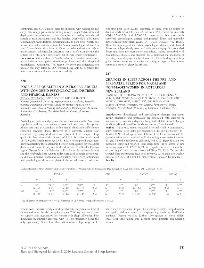
community and SSA females. Rates for difficulty with waking up tooearly, restless legs, pauses in breathing in sleep, fatigue/exhaustion anddaytime sleepiness were two to four times that reported by both a broadsample of male Australians and SSA females. Over 40% of SSA malesreported significant daytime impairments due to poor sleep. Nearly onein five SSA males met the criteria for ‘severe psychological distress’, arate 10 times higher than found in Victorian males and twice as high asin SSA females. Of particular concern is that 57% of SSA males met thecriteria for PTSD, a rate three times that of their female counterparts.Conclusion: Males within the South Sudanese Australian communityreport hitherto unrecognised significant problems with their sleep andpsychological adjustment. The reason for these sex differences areunclear but may relate to SSA women being able to negotiate theuncertainties of resettlement more successfully.
226
POOR SLEEP QUALITY IN AUSTRALIAN ADULTSWITH COMORBID PSYCHOLOGICAL DISTRESSAND PHYSICAL ILLNESSJESSICA PATERSON1, DAVID SCOTT3, BRENDA HAPPELL2
1Central Queensland University, Appleton Institute, Adelaide, Australia,2Central Queensland University, Centre for Mental Health NursingInnovation and School of Nursing and Midwifery, Rockhampton, Australia,3University of Melbourne, NorthWest Academic Centre, Melbourne,Australia
Psychological distress and physical illness are common in the Australianpopulation and are independently associated with sleep disruption.Almost one-third of Australians with a mental illness also suffer from acomorbid physical illness. However, it is currently unclear howcomorbid psychological distress and physical illness impact sleepquality in Australian adults. A total of 1,818 Australian adults aged18–91 y (56% female; mean age 55.3 y ± 12.8 y) completed a question-naire investigating the relationship between sleep quality, psychologicaldistress and comorbid physical health disorders. The Kessler Psycho-logical Distress Scale, the Behavioural Risk Factor Surveillance Systemand the Pittsburgh Sleep Quality Index were used to assess psychologi-cal distress, physical health and sleep quality, respectively. Participantswith psychological distress or physical illness had increased odds for
reporting poor sleep quality, compared to those with no illness ordistress (odds ratios [ORs] = 2.22, for both; 95% confidence intervals[CIs] = 3.54–10.36 and 1.53–3.23, respectively), but those withcomorbid psychological distress and physical illness had markedlyhigher odds for poor sleep quality (OR = 11.99, 95%CI = 7.90–18.20).These findings suggest that while psychological distress and physicalillness are independently associated with poor sleep quality, comorbidillness may have the most deleterious effects. Indeed, comorbidity ofpsychological distress and physical illness increased the likelihood ofreporting poor sleep quality by eleven fold. These findings may helpguide holistic treatment strategies and mitigate negative health out-comes as a result of sleep disturbance.
227
CHANGES IN SLEEP ACROSS THE PRE- ANDPERI-NATAL PERIOD FOR MAORI ANDNON-MAORI WOMEN IN AOTEAROA/NEW ZEALANDDIANE MULLER1, BRONWYN SWEENEY1, T LEIGH SIGNAL1,SARAH-JANE PAINE1, MONIQUE PRISTON1, ALEXANDER SMITH1,MARK HUTHWAITE2, KATHY LEE3, PHILIPPA GANDER1
1Massey University, Wellington, New Zealand, 2University of Otago,Wellington, New Zealand, 3University of California, California, USA
Introduction: Physiological and psychological changes that occurduring pregnancy and postnatally are associated with changes inwomen’s sleep quantity and quality. Long-itudinal data on such changesin Maori (M) and non-Maori (nM) women in NZ are limited.Method: The E Moe, Mama: Maternal Sleep and Health in Aotearoa/NZstudy collected sleep data: pre-pregnancy (T1); late pregnancy (35–37 wks) (T2); 4–6 wks post-natal (T3); and 11–13 wks post-natal (T4).Questionnaires were completed at T2 (including retrospective items forT1) and T4 and a brief phone call conducted at T3. Sleep duration wasmeasured using self-reported total sleep time (TST) across 24 hrsincluding naps at T1, T2, T3 & T4. Sleep quality included the numberof good night’s sleep across a week (GNS) at T1, T2 & T4, and theGeneral Sleep Disturbance Scale Total Score (GSDS-T) and Sleep Qualitysubscale (GSDS-Q) at T2 & T4 (higher values = greater disturbance).Results:
Median (Range) of Sleep Quantity and Quality Variables for Women who Participated in Data Collection at All Time-points (M = 255; nM = 535)
TST (hrs) GNS GSDS-T GSDS-Q
T1* T2 T3 T4 T1 T2 T4 T2 T4 T2 T4
M 8.0 7.0** 8.0** 7.8** 5 2** 4** 62 45*** 4 3***(2–16) (3–15) (2–16) (3–17) (0–7) (0–7) (0–7) (19–100) (2–98) (0–7) (0–7)
nM 8.0 7.0** 8.0** 7.0** 5 3** 4** 62 46*** 4 3***(6–13) (3–14) (3–17) (4–12) (0–7) (0–7) (0–7) (17–99) (7–92) (1–7) (0–6)
*Sig. difference by ethnicity <.05; **Sig. difference to T1 < .001; ***Sig. difference to T2 < .001
Discussion: Univariate analyses indicate that late pregnancy is a time ofshorter and more disturbed sleep for women. This may be a crucial timefor support and intervention for women with sleep difficulties. Fewdifferences by ethnicity emerged, with TST pre-pregnancy being theonly significantly different variable. Maori women slept longer at T1
which may be explained, in part, by a younger sample. Sleep durationand quality did not return to pre-pregnancy levels by 11–13 wkspostnatal. Results warrant further investigation of sleep differ-ences over time taking into account other possible confoundingfactors.
Abstracts
75© 2014 The AuthorsSleep and Biological Rhythms © 2014 Japanese Society of Sleep Research
228
THE RELATIONSHIP BETWEENGASTROESOPHAGEAL REFLUX ANDSLEEP QUALITYJAMES OCKELFORD2, DEZMI GANASAN2, HOOI EE3,RICHARD HOLLOWAY4, DAVID HILLMAN1,2,PETER EASTWOOD1,2, KELLY SHEPHERD1,2
1West Australian Sleep Disorders Research Institute, Sir Charles GairdnerHospital, Nedlands, Western Australia, Australia, 2School of Anatomy andHuman Biology, University of Western Australia, Crawley, WesternAustralia, Australia, 3Department of Gastroenterology, Sir CharlesGairdner Hospital, Nedlands, Western Australia, Australia, 4Department ofGastroenterology and Hepatology, Royal Adelaide Hospital, Adelaide, SouthAustralia, Australia
Introduction: Epidemiological studies show a strong associationbetween night-time symptoms of gastroesophageal reflux (GOR) andsubjective sleep disturbance. Few studies, however, have used objectivemeasures of GOR and sleep quality to investigate this association. Theaim of this study was to investigate the relationship between sleep-related GOR and sleep quality.Methods: Participants were recruited from the Department of Gastroen-terology at Sir Charles Gairdner Hospital after having undergone endos-copy. Exclusion criteria included gastroesophageal conditions aside fromGOR and/or a large hiatus hernia (>5 cm). Participants underwent24-hour pH-impedance monitoring and overnight polysomnography.Reflux severity was assessed by the number of reflux events between sleeponset and final awakening. Presence and severity of obstructive sleepapnoea (OSA) was assessed using the apnoea-hypopnoea index (AHI).Sleep quality was assessed using sleep efficiency, number of arousals perhour of sleep (arousal index) and number of spontaneous arousals perhour of sleep (spontaneous arousal index).Results: Complete data were available from 19 individuals (11 males) –12 had endoscopically defined oesophageal changes due to reflux. Meanage was 43.5 ± 14.4 years and BMI was 26.9 ± 5.4 kg.m−2. 11 individ-uals were found to have OSA (an AHI > 5 events/hr). The mean numberof reflux events during the sleep period was 3.5 ± 4.3 events. Age, BMI,arousal index and AHI were significantly higher in the OSA group thanin the non-OSA group. The number of sleep reflux events per hour ofsleep tended to be associated with a decrease in sleep efficiency(r2 = 0.33, p = 0.06) and spontaneous arousal index (r2 = 0.33,p = 0.06) in the OSA group. In the non-OSA group, an increase in thenumber of sleep reflux events per hour of sleep tended to be associatedwith arousal (r2 = 0.32, p = 0.1) and spontaneous arousal index(r2 = 0.44, p = 0.07).Discussion: The main finding of this study was that the frequency ofGOR events during the sleep period tended to be related to measures ofsleep quality and fragmentation in both OSA and non-OSA groups.
229
GENDER DIFFERENCES IN ASSOCIATIONBETWEEN SELF-REPORTED SLEEP DURATIONAND PSYCHOLOGICAL DISTRESS INHOME-DWELLING ELDERLYMARTIN ULANDER1, ANDERS BROSTRÖM1,2, ÅKE WAHLIN2,PETER JOHANSSON3
1Dept of Clinical Neurophysiology, Linköping University Hospital,Linköping, Sweden, 2Dept of Nursing, School of Health Sciences, JönköpingUniversity, Jönköping, Sweden, 3Dept of Cardiology, Faculty of HealthSciences, Linköping University, Linköping, Sweden
Background: Short as well as long sleep have been associated toincreased morbidity and mortality. Altered sleep patterns are also com-monly found in depression. Our aim was to examine whetherassociatons between sleep duration and psychological distress includingdepression, anxiety and insomnia complaints differed between men andwomen.Method: Self-report data regarding sleep duration and sleep-specificcomplaints such as various forms of insomnia, depressive symptomsand anxiety symptoms (Hospital Anxiety and Depression), daytimesleepiness (Epworth Sleepiness Scale) and vitality (from Short Form 36)were collected from 675 home-dwelling elderly (52% women, meanage ± SD 77.7 ± 3.8 years). Statistical analyses were performed in SPSSand R. Subjects were divided into short sleepers (i.e., ≤6 hours) averagesleepers (i.e., 7–8 hours) and long sleepers (≥9 hours). Between-groupcomparisons were made with Kruskal-Wallis and Mann-Whitney tests.Results: 36.7% of the respondents were short sleepers, 53.7% wereaverage sleepers and 9.7% were long sleepers respectively. Compared toaverage sleepers, short sleepers of both genders complained more aboutproblems initiating sleep, maintaining sleep or non-restorative sleep.Long-sleeping men, but not women, complained more about non-restorative sleep than average sleepers. No differences were found forany of the other insomnia symptoms among long sleepers compared toaverage sleepers. Short-sleeping men reported more depressive symp-toms and more daytime sleepiness than those sleeping 7–8 hours. Inwomen, no correlation was seen between sleep duration and depressivesymptoms or daytime sleepiness. Short-sleeping women and long-sleeping men scored significantly lower on the vitality subscale of SF36than average sleepers of the same gender. No associations were foundbetween anxiety and sleep durationConclusions: Self-reported sleep duration is related to psychologicalsymptoms in both men and women, but the associations are not thesame. Short sleep in men were, for example, related to depressivesymptoms. These results might indicate that depression might havedifferent symptom profiles in men and women.
230
SLEEP PROBLEMS ASSOCIATED WITH SOCIALDRUG USE: A HIDDEN HARM?ROWAN OGEIL1,2, JAMES PHILLIPS3, SHANTHA RAJARATNAM1,JILLIAN BROADBEAR4
1Monash University, Clayton, Australia, 2Turning Point, Eastern Health,Fitzroy, Australia, 3Auckland University of Technology, Auckland, NewZealand, 4Spectrum, Eastern Health, Ringwood, Australia
Alcohol and cannabis are the most commonly used social drugs in manycountries including Australia and New Zealand. Use of these drugs isassociated with health risks and an increased potential for harmfuloutcomes. In clinical settings, sleep disturbances are often reported by
Sleep, Science and Research
76 © 2014 The AuthorsSleep and Biological Rhythms © 2014 Japanese Society of Sleep Research
users of these drugs. To date however, interactions between more mod-erate, social use of these drugs and sleep disturbances are unknown. Theaim of this study was to measure subjective sleep quality and excessivedaytime sleepiness in social drug users, to determine whether problemalcohol and/or cannabis use was associated with poorer sleep outcomes.Self-identified social drug users (n = 248) with a mean age of 26 yearscompleted the Pittsburgh Sleep Quality Index (PSQI) and the EpworthSleepiness Scale (ESS), and questionnaires assessing alcohol (AlcoholUse Disorders Identification Test) and cannabis (Marijuana ScreeningInventory) use online. Poorer sleep quality was associated with problemalcohol or cannabis use, with adverse effects on sleep compounded byco-morbid problem drug use with mean scores >8 on the PSQI for thisco-morbid group. Sleep complaints are common in social drug users,particularly those who have problems using these drugs, but these maybe overlooked when their overall physical and mental health areassessed. Given that poor sleep is associated with decrements in cogni-tion, mood and health, sleep problems likely increase their risk ofsubsequent injury and/or poor health outcomes.
231
ENHANCING CBT FOR CHRONIC INSOMNIA:AN RCT OF ADDITIVE COMPONENTS OFMINDFULNESS OR COGNITIVE THERAPYMEIYIN WONG1, MELISSA REE2, CHRISTOPHER LEE1
1Murdoch University, WA, Australia, 2The Marian Centre, WA, Australia
Cognitive Behavioural Therapy (CBT) for primary insomnia is wellestablished as the gold standard treatment. Research into maximizingtreatment effectiveness and better understanding the modification ofunderlying psychological processes is important. In an RCT with 64participants, Wong et al (2012) found that additional sessions of eitherCognitive Therapy (CT: Harvey, 2002) or Mindfulness Based Therapy(MBT: Segal, Williams & Teasdale, 2002) significantly enhanced out-comes following a standard CBT for insomnia module on both selfreport and objective measures of sleep. For example, the effect sizereduction on the Insomnia Severity Index was 3.1 for both MBT and CT.In terms of clinical significance, the ISI means achieved after CT (5.74)and MBT (6.69) suggested that on average, participants had moved intothe good sleeper range. Both MBT and CT appear to be promisingadjuncts to standard CBT and it may be that switching treatment modal-ity after 4 sessions of CBT enhances outcome beyond continuing withfurther CBT sessions
232
SLEEP AND COGNITION IN PAROXYSMALDISORDERS: CASES ADMITTED FOREPILEPSY MONITORINGJESSICA PATTI1,2, MICHAEL SALING1, TERENCE O’BRIEN2,DENNIS VELAKOULIS2
1The University of Melbourne, Parkville, Victoria, Australia, 2The RoyalMelbourne Hospital, Parkville, Victoria, Australia
Sleep disordered breathing (SDB) has been identified as being morecommon in people with epilepsy than in the general population and isalso associated with reduced cognitive functioning. The aim of thepresent study was to look at the relative contribution of SDB and seizurevariables to cognitive variation in a cohort of patients admitted forseizure monitoring. An exploratory aim was to investigate the potentialinfluence of mood on the relationship between sleep and cognition.
Direct neuropsychological measures of cognition were used concur-rently with electrophysiological measures of sleep (PSG) and otherscreening tests of mood symptoms (HADS), global cognition (NUCOG)and subjective sleep quality (PSQI). Information regarding seizure vari-ables (number of antiepileptic drugs, duration of syndrome and seizurefrequency) was gathered by clinical interviews. Using hierarchicalregression analyses and accounting for variance explained by seizurevariables, it was found that AHI explained some unique variance inperformance on NUCOG in a sub-group of patients without a diagnosisof epilepsy. AHI and PSQI both contributed unique variance to perfor-mances in select cognitive domains when measured by aneuropsychological test battery. Measures of mood symptoms also con-tributed to variation in certain cognitive domains. Conclusion: Whenobjective and direct physiological and neuropsychological measureswere used to explore the hypothesis in question, there was some evi-dence to suggest that sleep disordered breathing contributes uniquevariance in cognitive performances, over and above that of seizure-related variables. This relationship is of clinical significance as sleepdisorders are often modifiable and such findings highlight an avenuethrough which already affected brain networks may be alleviated of theadditional load of sleep disturbance.
233
DIFFICULTIES IN TREATING SLEEP PROBLEMSREPORTED BY COUNSELLORS WORKING WITHSURVIVORS OF SEXUAL ASSAULTJESSICA DARVELL, DOROTHY BRUCK, GERARD KENNEDYVictoria University, Melbourne, Victoria, Australia
Objectives: This study sought to determine common sleep problemsencountered by counsellors working with survivors of sexual assault,and which problems were the most difficult to treat.Methods: Four focus groups (total n = 29) were conducted with coun-sellors working with survivors of sexual assault. The focus groups wereconducted to gather information about the counsellors’ understanding,experiences, knowledge and problems encountered with regard tohelping survivors of sexual assault with sleep difficulties. The counsel-lors also completed a short rating scale designed to examine theirconfidence in treating sleep disorders. Focus group transcript data wereanalysed using thematic analyses, and rating scale data were subjected tofrequency analyses.Results: Counsellors consistently agreed that sleep difficulties were aprominent issue for survivors of sexual assault. Common problemsimpacting sleep included nightmares, hyper-vigilance, safety concerns,medication-related difficulties with falling asleep and frequent waking,and restless limbs. Counsellors described experiencing the most diffi-culty with treating problematic jerking of limbs, snorting, snoring orgasping for air, sleepwalking, and delayed sleep phase issues. Many ofthe treatment difficulties encountered by counsellors were attributed toresistance and reluctance on the client’s behalf to persist with sugges-tions due to an increase in anxiety, or re-experience of their priortrauma.Conclusions: Counsellors consistently reported a lack of confidence in,and knowledge of, treating sleep breathing related disorders, restlesslimbs, and any of the more complex sleep difficulties not readily treatedwith sleep hygiene, psycho-education or relaxation techniques.
Abstracts
77© 2014 The AuthorsSleep and Biological Rhythms © 2014 Japanese Society of Sleep Research
234
RETROSPECTIVE APPLICATION OF THE ICSD-3DIAGNOSTIC CRITERIA FOR NARCOLEPSYJOSEPH CHURTON, KELLI RIXON, CONCHITA REGO,BRETT DUCE, CRAIG HUKINSSleep Disorders Centre, Princess Alexandra Hospital, Brisbane, QLD,Australia
Introduction: The International Classification of Sleep Disorders 2nd
Edition (ICSD-2) criteria for the diagnosis of narcolepsy with cataplexyincluded a Mean Sleep Latency (MSL) on Multiple Sleep LatencyTest(MSLT) of less than 8 minutes, with at least 2 Sleep Onset REM Periods(SOREMPs) during the MSLT. The International Classification of SleepDisorders 3rd Edition (ICSD-3) criteria still includes a MSL on MSLT ofless than 8 minutes, with at least 2 SOREMPs. However, SOREMPs fromthe previous night’s Polysomnography (PSG) may replace one MSLTSOREMP.Methods: All MSLTs undertaken at the Sleep Disorders Centre, PrincessAlexandra Hospital, between 2008–2014 were included. The results ofthe MSLT were then reassessed against the ICSD-3 criteria for thediagnosis of narcolepsy.Results: Ninety-four MSLTs were performed between 2008 and 2014.Results were reviewed for 64 of these MSLTs. Twenty-six patientsachieved a MSL of <8 minutes. Seven of those 26 cases were positive forat least 1 SOREMP. Of the 26 cases, 9 patients were diagnosed withIdiopathic Hypersomnia, 1 diagnosed with Narcolepsy with cataplexy, 7diagnosed with Narcolepsy without cataplexy. Upon examination oftheir previous night PSG, only one patient displayed a REM latency of<15 minutes. This was the patient diagnosed with Narcolepsy withcataplexy. The median REM latency for the <8 minutes MSL group was106.5. This compared with a median REM latency of 124.8 minutes forthose patients with a MSL > 8 minutes. The PSG sleep latency and MSLTMSL were similar in the >8 minutes MSL group (13.5 vs 13.3 minutesrespectively. For the group with a MSLT MSL <8 minutes the PSG sleeplatency was slightly higher (5.2 minutes MSLT vs 7.0 minutes PSG).Subjectively sleepiness as measured by the Epworth Sleepiness Scale(ESS) was higher in the MSL < 8 minutes group compared to theMSL > 8 minutes group (16.0 vs 14.5).Discussion: Based on our limited data, the application of the ICSD-3changes would not impact upon the clinical diagnosis of patients sus-pected of narcolepsy.
235
ANXIETY AND DEPRESSION SYMPTOMS INPATIENTS BEING REFERRED FOR ANIN-LABORATORY POLYSOMNOGRAPHYSANJEEVAN MURUGANANDAN1, MELINDA LEE JACKSON2,TOM CHURCHWARD1, JULIE TOLSON1,CHRISTOPHER WORSNOP1
1Austin Hospital, Melbourne, Victoria, Australia, 2University of Melbourne,Melbourne, Victoria, Australia
Introduction: Depression and anxiety often co-exist with sleep disor-ders, with growing evidence indicating that sleep problems may causeor contribute to depressive disorders. Both depression and anxiety areassociated with reduced quality of life as well as increased morbidity.Previous studies have demonstrated higher rates of depression inobstructive sleep apnoea (OSA); however there is less evidence aboutthe prevalence of anxiety in this population.Aim: The purpose of this study was to determine the prevalence ofdepression and anxiety symptoms in patients being referred for a diag-
nostic sleep study and to examine whether the severity of depressionand anxiety symptoms are associated with the degree of somnolence andphysiological sleep measurements.Methods: Consecutive patients undergoing an overnight polysomno-graphy in the sleep laboratory were assessed using the Epworth Sleepi-ness Scale (ESS) and the Hospital Anxiety and Depression Scale (HADS)for anxiety and depressive symptoms.Results: 96 consecutive patients (mean age 50.6 years, 39 women) havebeen studied.80.2% had an AHI ≥ 5 and 35% had AHI ≥ 30. On the HADS ques-tionnaire, 25.3% and 10.3% of patients exhibited significant anxiety anddepressive symptoms respectively by scoring more than 11 on thesescales. Both anxiety (r = 0.28; p = 0.006) and depression (r = 0.32;p = 0.002) were significantly associated with ESS. Depression was alsosignificantly associated with BMI (r = 0.33; p = 0.002). There were nosignificant associations between HADS scores and PSG variables.Conclusion: Depression and anxiety symptoms are highly prevalentin patients with OSA, and appear to be related to sleepiness ratherthan AHI.
236
NEUROBEHAVIOURAL EFFECTS OF ‘CATCH-UP’SLEEP IN MEN WITH LIFESTYLE DRIVEN,CHRONIC, INTERMITTENT SLEEP RESTRICTIONROO KILLICK1, CAMILLA HOYOS1, KERRI MELEHAN1,3,DELWYN BARTLETT1, KEITH WONG1,3, TRACEY SLETTEN4,SHANTHA RAJARATNAM1,4, RON GRUNSTEIN1,3, PETER LIU1,2
1CIRUS, Woolcock Institute of Medical Research, University of Sydney,Sydney, Australia, 2Los Angeles Biomedical Research Institute atHarbor-UCLA Medical Center and David Geffen School of Medicine,University of California Los Angeles, Los Angeles, USA, 3Royal PrinceAlfred Hospital, Sydney, Australia, 4School of Psychological Sciences,Monash University, Melbourne, Australia
Introduction: Chronic, intermittent sleep restriction is common inmodern society and the neurobehavioural consequences of chronicsleep loss are increasingly recognised.Methods: 19 men (mean ± SEM age 28.6 ± 2.0years, BMI 26.0 ±0.8 kg/m2) with at least 6 months’ history (5.1 ± 0.9years) of lifestyledriven, restricted sleep during the working week (6 h12 min ±7 min/night) with regular weekend ‘catch up’ sleep (weekend sleep8 h30 min ± 9 min/night) completed an in-laboratory, randomised,cross-over study comprising 2 of 3 conditions, stratified by age. Con-ditions were 2 weekend nights of ‘catch-up’ sleep (10 h time-in-bed(TIB)), sustained sleep restriction (6 h TIB) or 10 h TIB with slow wavesleep (SWS) suppression using acoustic stimuli. Reported sleep wasrecorded at screening and before each study visit by two weeks ofactigraphy. A neurocognitive test battery comprising N-back, Stroop,Tower of London and Psychomotor Vigilance Task (PVT) was performedon Saturday and Sunday mornings two hours after wake time. Subjec-tive sleepiness was assessed by Karolinska Sleepiness Scale at frequenttime-points over both days. Driving simulation (AusEd) was performedat 2100 h both evenings.Results: Compared to sustained sleep restriction, ‘catch-up’ sleepimproved subjective sleepiness (p < 0.0001) and measures of vigilancetesting in PVT (all p ≤ 0.005), 1-back accuracy (p = 0.01) and drivingsimulator braking response time (p = 0.009). Similar changes were seenin 2-back (p = 0.03), 3-back (p = 0.002) and Stroop colour (p = 0.01)response times when examining the younger men exclusively. SWSsuppression did not alter parameters consistently compared to ‘catch-up’ sleep or sustained sleep restriction, however certain PVT responseswere slower compared to ‘catch-up’ sleep (p ≤ 0.03), despite no differ-ence in subjective sleepiness.
Sleep, Science and Research
78 © 2014 The AuthorsSleep and Biological Rhythms © 2014 Japanese Society of Sleep Research
Conclusion: ‘Catch-up’ sleep provided improvements compared tosustained sleep restriction in subjective sleepiness and measuresof objective vigilance and reaction time tasks across several neuro-cognitive domains, in this cohort of men with chronic, intermittentsleep restriction.
237
THE EFFECT OF HYPNOTICS ON AUDITORYAROUSAL THRESHOLDS IN OLDER ADULTSCHRIS LYKIARDOPOULOS, DOROTHY BRUCK, MICHELLE BALLVictoria University, Melbourne, Victoria, Australia
The ability to respond quickly from sleep to emergency signals isassumed, however previous research has found that being under theinfluence of alcohol affects a person’s ability to respond to their smokealarm, even at very low levels. Relatively little is known about the impacthypnotics have on the response to emergency signals, such as smokealarms, in older adults.Methods: The repeated measures experiment involved monitoring thesleep patterns and arousal thresholds of 12 participants (10 males andtwo females aged between 65 to 80 years with an average age of 71) intheir homes over 11 nights, modulating hypnotic intake (using theirusual hypnotic) and alarm presentation (the current 3100 Hz sine wavecompared with the 520 Hz square wave). The alarms were presentedwhen the drug was at peak concentration.Results: Hypnotic ingestion increased arousal thresholds by approxi-mately 9 dBA. More than double the proportion of participants sleptthrough the 3100 Hz sine wave at 75 dBA when under the influence oftheir hypnotic compared to the no hypnotic condition (17% cf. 8%respectively). The 520 Hz square wave significantly outperformed thestandard smoke alarm (3100 Hz sine wave).Conclusions: The findings suggest that hypnotics significantly compro-mise arousal thresholds amongst older adults. Together with the previ-ous literature, the findings present a challenge to the effectiveness ofexisting fire detection and alerting technology amongst vulnerablegroups and support the continued adoption of the 520 Hz square waveas the signal in smoke alarms.
238
THE IMPACT OF SLEEP RESTRICTION ONACUTE INFLAMMATORY STRESS RESPONSES TOSIMULATED PHYSICAL FIREFIGHTING WORKALEXANDER WOLKOW1,2, BRAD AISBETT1,2, SALLY FERGUSON3,LUANA C MAIN1
1Deakin University, Burwood, Vic, Australia, 2Bushfire Co-OperativeResearch Centre, East Melbourne, Vic, Australia, 3CQUniversity, Wayville,SA, Australia
Introduction: Firefighters are exposed to extended periods of physicalwork and restricted sleep in the line of duty, yet the combined impactthese occupational stressors have on firefighters’ acute inflammatorystress response is poorly understood. Evidence indicates that a shift ininflammatory cytokine levels are associated with an increased risk ofnegative health outcomes (e.g., cardiovascular disease, metabolic syn-drome). This study investigated the effect restricted sleep has on fir-efighter’s inflammatory cytokine levels during a simulated three-day andtwo-night fire ground deployment.Methods: Firefighters completed multiple days of simulated physicalfirefighting work separated by either an 8-h (Control condition; n = 18;
39 ± 16 yr; bedtime 2200-0600 h) or 4-h sleep opportunity (Sleeprestriction condition; n = 17; 39 ± 15 yr; bedtime 0200-0600 h)between days. During each work day, participants in both conditionscompleted multiple work circuits that comprised simulated physicalfirefighting tasks. Performance of these tasks was self-paced. Bloodsamples were collected four times a day (i.e., 0630, 1200, 1800, 2100)and plasma levels of pro- (IL-6, IL-8, IL-1β, TNF-α) and anti-inflammatory (IL-4, IL-10) cytokine levels were measured. Linear mixedmodels were used for the analysis.Results: There was significant interaction effect of sample time andcondition for IL-6 (P < 0.05). The increase in IL-6 over the three dayswas greater in the control condition. No significant changes were dem-onstrated for any other cytokine levels.Conclusion: The different rise in IL-6 between the conditions could berelated to various factors (e.g., physical work intensity, muscle glycogenlevels). However, further research is required to determine which factorsare contributing to the change in IL-6 between the conditions. Further-more, these findings suggest that regardless of the amount of sleepbetween shifts, consecutive days of physical firefighting work can elicitan inflammatory stress response for IL-6. Chronic increases in inflam-matory cytokine levels such as IL-6 are known to be associated with thedevelopment of negative health outcomes (e.g., metabolic syndrome)and therefore, these findings could have implications for managing thehealth of firefighters deployed to several large multiday fires over thecourse of a fire season.
239
OCULAR INDICATORS OF SLEEPINESS INSHIFT WORKERSSUZANNE FTOUNI1, TRACEY L SLETTEN1,STEVEN W LOCKLEY1,2, SHANTHA MW RAJARATNAM1,2
1Monash University, Clayton, Victoria, Australia, 2Brigham and Women’sHospital; Harvard Medical School, Boston, Massachusetts, Australia
Driver sleepiness is one of the most common causes of preventablemotor vehicle collisions (MVC). The shift working population is over-represented in sleepiness-related MVCs. This increased risk is in-partdue to the interactive effects of circadian misalignment and sleep loss,which can result in excessive sleepiness and neurobehavioural perfor-mance impairment, particularly after night work. Effective interventionstrategies to counteract the deleterious effects of shift work as well asreal-time, objective measures of sleepiness are critical to maintainingsafety and productivity in shift workers. Objective methods are beingdeveloped to monitor sleepiness levels in operational settings. Thesemeasures include infrared oculography, which utilises eye blink charac-teristics to quantify physiological sleepiness. We conducted threestudies to assess the utility of ocular measures in assessing sleepinessand their associations with performance impairment. Study oneassessed the utility of oculometrics as objective indicators of sleepinessin relation to validated measures of performance, vigilance, and subjec-tive sleepiness. Ocular indicators of sleepiness paralleled performanceimpairment and self-rated sleepiness levels, demonstrating their poten-tial to detect sleepiness-related attentional lapses. Study two examinedthe response of ocular measures of sleepiness and attentional perfor-mance in night shift workers exposed to a simulated laboratory nightshift at an adverse circadian phase. Participants tested at an adversecircadian phase at the end of their shift displayed higher levels ofoculometric sleepiness, subjective and objective sleepiness, as well asneurobehavioural performance, compared to those who were not at anadverse circadian phase. Study three examined the association betweenoculometrics and unsafe driving events during real-world commutes toand from night shift work. Under real-world driving conditions, shift
Abstracts
79© 2014 The AuthorsSleep and Biological Rhythms © 2014 Japanese Society of Sleep Research
working nurses experience high levels of drowsiness as indicated byocular measures, which are associated with impaired driving perfor-mance following night shift work. The findings demonstrate the poten-tial utility of oculometrics to assess sleepiness in healthy individuals andshift workers in laboratory and field settings, and demonstrate theapplication of oculometrics in identifying performance impairment as aresult of sleepiness and circadian misalignment.
240
ALCOHOL USE AND SLEEP IN SHIFTWORKERSJILLIAN DORRIAN1, GEORGINA HEATH1, CHARLI SARGENT3,WILL MCINTOSH1, SIOBHAN BANKS1, ALISON COATES2
1Centre for Sleep Research, School of Psychology, Social Work and SocialPolicy, University of South Australia, Adelaide, SA, Australia, 2NutritionalPhysiology Research Centre, Sansom Institute for Health Research,University of South Australia, Adelaide, SA, Australia, 3Appleton Institute,Central Queensland University, Adelaide, SA, Australia
Introduction: Since they often sleep at biologically difficult times,shiftworkers may use alcohol to promote sleep. While alcohol hassedative properties, reducing sleep onset latency, it disrupts architecture.The negative effects of alcohol, particularly at high doses, are wideranging. This study examined alcohol intake, shift type, sleep andtiredness in a group of Australian shiftworkers.Methods: Shiftworkers (n = 118, age = 43.4 ± 9.9 y, 68%male) fromprinting, postal, nursing and oil industries, including 12 h-rotating(n = 29), 8 h-rotating (n = 29), morning (n = 33) and night (n = 27)workers, completed the Cancer Council Dietary Questionnaire, record-ing frequency and amount of alcohol consumed over the preceding year.They also completed a modified version of the Standard ShiftworkIndex, including questions on shift schedule, sleep duration, tiredness,gender and age.Results: Average alcohol consumption was 9.6 ± 13.1 standard drinksper week, which was not significantly different (F2,207 = 1.9, p = 0.14)to a previously reported large representative sample of shiftworkers(8.3 ± 11.0, n = 355) and those on standard schedules (7.4 ± 11.0,n = 1735) from the Household Income and Labour Dynamics Survey(2006/2007). More than three quarters of shiftworkers reported drink-ing ≥4 drinks in 24 h (exceeding guidelines for injury risk, NHMRC2009) and 28% reported ≥12 drinks in 24 h. One in five shiftworkersreported >14 drinks per week (exceeding guidelines for lifetime harm,<2 drinks/day, NHMRC 2009). One in six reported using alcohol asa sleep aid between shifts at least sometimes. Controlling for genderand age, there were significant differences between shift types insleep (F3,110 = 9.2, p < 0.001), maximum drinks consumed in 24 h(F3,116 = 2.7, p = 0.04) and tiredness (F3,114 = 5.3, p = 0.002), but notstandard drinks per week (F3,116 = 0.7, p = 0.50). Those on 12 h-rotat-ing shifts had less sleep, higher maximum alcohol intake and increasedtiredness at work.Discussion: Data suggest that average alcohol consumption is nothigher among shiftworkers than in the overall Australian population.However, the majority of shiftworkers exceed guidelines for injury risk.This supports the suggestion that shiftworkers are likely to binge drink.Alcohol use in shiftworkers may put pressure on already vulnerablephysiological systems. Younger, male workers and those on long, rotat-ing shifts may be at particular risk.
241
HIGH DOSE CAFFEINE DOES NOT HAVE ANADDITIVE EFFECT ON IMPAIRED GLUCOSECONTROL DURING EXTENDED WAKEFULNESSCRYSTAL GRANT1, GEMMA PAECH1, CHRIS DELLA VEDOVA2,MAJA PAJCIN2, JILL DORRIAN1, ALISON COATES3, SIOBHANBANKS1
1Centre for Sleep Research, University of South Australia, Adelaide, SA,Australia, 2School of Pharmacy and Medical Sciences, University of SouthAustralia, Adelaide, SA, Australia, 3Nutritional Physiology ResearchCentre, University of South Australia, Adelaide, SA, Australia
Introduction: Shift workers regularly experience sleep loss, resulting inhigh sleepiness and impaired performance. Sleep loss also leads toimpaired postprandial glucose response. Caffeine is an effective counter-measure for the performance impairments associated with sleep loss,however studies have shown caffeine increases postprandial glucoseresponse. The aim of this study was to determine the additive effect ofcaffeine on impaired glucose response to a breakfast meal duringextended wakefulness.Methods: Participants with normal glucose control were randomlyassigned to either a caffeine (n = 10, 4F, 22.5 ± 3.3 y, 21.7 ± 1.5 kg/m2)or placebo condition (n = 11, 5F, 22.5 ± 2.5 y, 22.3 ± 2.1 kg/m2).Habitual caffeine consumers defined as ≥1 caffeine drinks/ day. Thein-lab protocol included one baseline night (22:00–08:00), 49 hextended wakefulness, and a recovery sleep episode (10:00–19:00). At01:00, 03:00, 05:00 and 07:00 during each night of wake participantswere given 200 mg of caffeine or placebo gum. Meal timing (08:00baseline, 07:10 subsequent mornings) and composition (break-fast ≈ 1611 KJ; 16% protein, ≈73% CHO and 3% fat) were controlledthroughout the study. Interstitial glucose was recorded at 5 min intervalsfor 2 h post meal consumption. Linear mixed models were used toassess the effect of caffeine on glucose response to breakfast.Results: Extended wakefulness resulted in increased interstitial glucoseresponse to breakfast with higher glucose at 24 h (6.5 mmol/L ± 0.5 mmol/L, P < 0.001) and 48 h (7.1 mmol/L ± 0.5 mmol/L,P < 0.001) relative to baseline (6.1 mmol/L ± 0.5 mmol/L). There wasno significant difference between caffeine and placebo conditions(P = 0.743). Non-habitual caffeine consumers followed a linear increasein interstitial glucose. Habitual caffeine consumers showed increases ininterstitial glucose during extended wake, but did not follow a linearpattern by day.Conclusions: Caffeine does not have an additive effect over and abovethe effects of extended wakefulness on postprandial glucose response.Results suggest the use of caffeine as a fatigue countermeasure in shiftworking populations does not further impair glucose metabolism.Further research into the differing profiles of habitual and non-habitualcaffeine consumers is needed.
242
ACUTE AND CHRONIC ALCOHOLCONSUMPTION DISTURBS SLEEPARCHITECTURE IN LATE ADOLESCENCECAITLYN G GOURLAY1, HAYLEY C AYTON1, JULIA KM CHAN1,ABBIE COUCHMAN1, IAN M COLRAIN2,1, JOHN TRINDER1,CHRISTIAN L NICHOLAS1
1Melbourne School of Psychological Sciences, The University of Melbourne,Vic, Australia, 2SRI International, California, USA
Introduction: Acute and chronic alcohol consumption is assoc-iated with significant sleep disturbance. Notably, alcohol dependent
Sleep, Science and Research
80 © 2014 The AuthorsSleep and Biological Rhythms © 2014 Japanese Society of Sleep Research
individuals show marked reductions in slow wave sleep (SWS). Acutealcohol ingestion in healthy individuals is associated with consistentdose dependent alterations to sleep stage distribution, and sleep disrup-tion. Late adolescents are a developing cohort particularly at risk ofheavy or ‘binge’ drinking behaviour. The current study examined theeffects of a high dose of pre-bedtime alcohol on sleep in heavy and lightdrinking late adolescents.Method: Ten heavy drinking (HD: 19.6 ± 1.1 yrs) and 9 light drinking(LD: 19.8 ± 1.2 yrs) late adolescents completed laboratory PSG undertwo conditions: pre bedtime alcohol (dosed to 0.1% peak BAC; meanBAC at lights out 0.081 ± 0.02%), and placebo beverage (0.0% BAC).Participants abstained from alcohol for 48 hours prior to testing. Datawere evaluated across the first four sleep cycles. HD and LD groups didnot differ in BAC at lights out (alcohol condition, 0.087 ± 0.02% vs.0.074 ± 0.02%, p > .05). By design, groups differed on drinking historyin the previous month (HD: 125.9 ± 88.2 vs. LD: 13.1 ± 8.5 standarddrinks, p = .003), but did not differ in age, BMI or age drinkingcommenced (p > .05).Results: HD showed less SWS (27.06 ± 6.6% vs. 39.05 ± 6.6%,p < .001) and more REM sleep (21.52 ± 2.9% vs. 18.37 ± 2.9%,p = .032) than LD across sleep cycles. Significantly less REM sleepoccurred after alcohol (p < .01). Alcohol did not increase SWS in LD(p > .05), but did increase SWS in HD (alcohol condition by groupinteraction, p = .016). Both groups exhibited the usual sleep cyclerelated reductions in SWS (p = .001) and increases in REM sleep(p = .001). No differences were observed between groups for sleep onsetlatency, time in bed or total sleep time (p > .05).Discussion: Findings suggest that despite relatively short drinking his-tories, HD late adolescents show similar reductions in SWS to those seenin long-term alcohol dependence. These SWS deficits were partiallyreversed by acute alcohol administration. It is unclear whether thesedifferences in sleep precede or result from heavy alcohol use in this agegroup. There appear to be important interactions between chronicand acute alcohol consumption in adolescence on sleep that have notpreviously been considered.
243
SLEEP IN PARKINSON’S DISEASE: MEASURINGCHANGE OVER TIME USING THE PARKINSON’SDISEASE SLEEP SCALEMARIA PUSHPANATHAN1,3, ROMOLA BUCKS1,3,CAITLIN TIMMS2,3, MEGHAN THOMAS1,3, NATALIE GASSON2,3,ANDREA LOFTUS2,3
1University of Western Australia, Perth, Western Australia, Australia,2Curtin University, Perth, Western Australia, Australia, 3ParkCCollaborative, Perth, Western Australia, Australia
Sleep disruption is one of the earliest symptoms to manifest in Parkin-son’s disease (PD), and it is disrupted in a number of ways. There is animmense range of sleep disturbance experienced in PD, and the ways inwhich sleep is disturbed typically change as the disease progresses. Forexample, REM sleep behaviour disorder is often observed in the pro-dromal period (before motor symptoms emerge), while night time visualhallucinations are more common much later when neurodegeneration ismore advanced.
The Parkinson’s Disease Sleep Scale (PDSS) is a brief, 15 item, ques-tionnaire measure that requires a patient to rate how frequently theyexperience the most common types of sleep disturbance in PD. As sleepdisturbance in this population is multifactorial and can lead to otherproblems (cognitive decline, poor quality of life), we believe that this isa good measure for use in longitudinal research. However, little isknown about the factor structure of the PDSS, either in a cross-sectionalsample or in a cohort study.
Previous research has tended to focus on a single facet of sleepdisturbance, or to measure ‘sleep quality’ generally. This study aims toexamine whether the factor structure of the PDSS remains stable overtime, or whether it changes as the disease course progresses in ourcohort. This information is useful for evaluating the utility of the PDSSas a research measure; if the underlying factor structure of the PDSS isdifferent in early PD relative to late PD, research design will need to takethis into account when looking for associations with other non-motorsymptoms.
Although analysis of our wave 1 data demonstrate that the PDSSgenerates factor scores that are associated with specific neuro-psychological deficits; preliminary analyses of our longitudinal datasuggest that measurement invariance is an issue with this measure, assleep problems evolve and become more complex as PD progresses. Incohort studies using the PDSS, we need to consider how best to measurethe relationship between sleep dysfunction and other non-motor symp-toms when the factors measured by this scale are strongly influenced bysample characteristics.
244
EXPLORATORY FACTOR ANALYSIS OF THEPARKINSON’S DISEASE SLEEP SCALE: IS THEREAN ASSOCIATION BETWEEN SLEEP DISORDERAND COGNITIVE DYSFUNCTION INPARKINSON’S DISEASE?MARIA PUSHPANATHAN1,3, ANDREA LOFTUS2,3,CAITLIN TIMMS2,3, MEGHAN THOMAS1,3, NATALIE GASSON2,3,ROMOLA BUCKS1,3
1University of Western Australia, Perth, Western Australia, Australia,2Curtin University, Perth, Western Australia, Australia, 3ParkCCollaborative, Perth, Western Australia, Australia
Sleep disorder is one of the earliest symptoms of Parkinson’s disease(PD) emerging up to ten years before motor symptoms first appear.People with PD experience insomnia, sleep fragmentation, diminishedslow wave sleep, sleep related breathing disorders, nocturia, and REMsleep behaviour disorder. One questionnaire, the Parkinson’s diseasesleep scale (PDSS) is designed to measure the range of sleep symptoma-tology commonly experienced in PD.
A second significant non-motor symptom of PD is cognitive dysfunc-tion, particularly in the domains of memory and executive function.Cognitive dysfunction significantly impacts on quality of life and pre-dicts nursing home placement in this population. It is well-establishedthat disrupted sleep has neuropsychological consequences in otherwisehealthy people, however, studies that have endeavoured to establish thisrelationship in PD have had mixed results, perhaps because they tend tofocus on a single facet of sleep disturbance, when sleep disturbance inPD is multifactorial.
The present study is the first to use the PDSS; which is a compre-hensive (albeit brief) measure of sleep disruption in PD and examinewhether factors underlying this scale are associated with poor perfor-mance on a range of neuropsychological tests, focussing on sub-domains of memory and executive function.
We found that in our cohort, scores on the PDSS had five underlyingfactors: i) overall sleep quality, initiation and maintenance, ii) nocturia,iii) symptoms of REM sleep behaviour disorder and sleep related breath-ing disorders iv) motor symptoms interfering with sleep (restless legs,dystonia, medication wearing off) and v) difficulty falling back to sleepat night and daytime sleep attacks. Each factor (excepting nocturia) wasassociated with deficits in specific tests of memory of executive functionwhen the effects of age, premorbid IQ, disease severity (UPDRS score)and medication were controlled for. This would suggest that the PDSS is
Abstracts
81© 2014 The AuthorsSleep and Biological Rhythms © 2014 Japanese Society of Sleep Research
useful for detecting different types of sleep disorder in PD. Further,particular types of sleep disorder in PD are associated with discrete,dissociable cognitive deficits.
245
DO INDIVIDUAL DIFFERENCES AFFECTPERFORMANCE DURING SHIFT WORK?ALEXANDRA AGOSTINI1, CASSIE HILDITCH1,STEPHANIE CENTOFANTI1, MICHELLE SHORT1,2
1University of South Australia, Adelaide, South Australia, Australia,2Bushfire Cooperative Research Centre, Melbourne, Victoria, Australia
Objectives: Shift work is associated with a number of negative conse-quences, with arguably the most dangerous being performance decre-ments. In order to minimise the number of shift work-related accidentsand optimise productivity, researchers have begun investigating theindividual factors that predict shift work performance. This studyassessed the effect of age, chronotype and neuroticism on performanceduring a simulated shift work schedule.Methods: Twenty-four participants undertook a nine-day laboratorystudy. Each participant was randomly assigned to one of three shift workschedules: a six-hour early, six-hour late or eight-hour roster. Partici-pants’ performance was measured using the Psychomotor Vigilance Task(PVT) and the Digit Symbol Substitution Test (DSST).Results: Mixed model analyses revealed no significant main effects ofage, chronotype or neuroticism. No significant interactions were foundbetween any predictors, shift type, or day for PVT lapses or PVT fastest10% of reaction time. There was a significant main effect of bothchronotype and age on DSST performance, with younger adults andmore evening chronotypes performing better. There were also signifi-cant interactions between age, chronotype and neuroticism and shifttype on DSST performance.Conclusions: These findings suggest that there may be a more compli-cated relationship between individual factors and some aspects of shiftworkers’ cognitive performance, with the effect of individual differenceson performance varying according to the specific type of shift schedulebeing used.
246
INTRODUCTION TO THE ASIA PACIFICPAEDIATRIC SLEEP ALLIANCE (APPSA)DANIEL GOH1Khoo Teck Puat-National University Children’s Medical Institute,Singapore, Singapore, 2Yong Loo Lin School of Medicine, Singapore,Singapore, 3National University Hospital, Singapore, Singapore, 4AseanPaediatric Federation, Singapore, Singapore, 5Singapore Paediatric Society,Singapore, Singapore
APPSA is a workgroup in paediatric sleep with the focus on improvingthe understanding and management of sleep and sleep-related disordersin children across the Asia Pacific region. It is made up of a group ofphysicians with special interest and expertise in the field of sleep in thebaby and child.What do we do?• Research – to better understand sleep habits and practices, factors
affecting sleep and sleep problems in children• Contribute to literature on Sleep in childhood through peer-reviewed
journal publications• Education to the public and medical professionals to promote
awareness and understanding of sleep in children through lectures,
presentations, CME programmes, participation in paediatric andsleep conferences, and other activities.
The alliance has the objective of expanding the scientific understandingof sleep, and sharing/spreading this knowledge to paediatricians andmothers via publications and educational programmes, with the ulti-mate goal of improving the quality of sleep in our children.
247
CHILD AND MATERNAL SLEEP:A WORLD-WIDE PERSPECTIVEJODI MINDELLThe Children’s Hospital of Philadelphia, Philadelphia, USA
Prior research has indicated that sleep is determined by both biologicaland cultural determinants, and it is the interaction between these twofactors that affect cultural and developmental norms. Findings will bepresented from two large-scale cross-cultural studies of almost 40,000families conducted by the Asia-Pacific Pediatric Sleep Alliance. Thesetwo studies investigated sleep patterns and sleep problems of youngchildren (ages 0–6 years) and their mothers. The results indicate sig-nificant cross-cultural differences in sleep patterns, sleeping arrange-ments, and sleep problems. Overall, children from predominantly-Asiancountries had significantly later bedtimes, shorter nighttime sleep, andincreased parental perception of sleep problems, compared with thosefrom predominantly-Caucasian countries. In contrast, mothers inpredominantly-Asian countries/regions reported later bedtimes butsleeping better and longer than mothers from predominantly-Caucasiancountries, which is dissimilar to the cross-cultural findings of youngchildren. For mothers, psychosocial factors were found to be the bestpredictors of poor sleep, irrespective of culture. Further studiesare needed to understand the underlying bases and impact of thesefindings.
249
SLEEP-RELATED BREATHING DISORDER ANDITS RELATED PROBLEMS – THE ASIAPACIFIC ASPECTALBERT LI1,2
1Prince of Wales Hospital, Sydney, Australia, 2The Chinese University ofHong Kong, Shatin, Hong Kong
The term sleep-related breathing disorder refers to a spectrum of breath-ing anomalies ranging from chronic or habitual snoring to upper airwayresistance syndrome (UARS) to frank obstructive sleep apnoea (OSA). Inpaediatrics, much research has been carried out in OSA and consistentfindings suggest that if it is not treated a variety of important sequelaecan result. Recently attention has shifted towards the other end of thedisorder spectrum. There is accumulating evidence to support thathabitual snoring, which is snoring but without apnoeas, hypopnoeasor gaseous exchange abnormalities is also associated with clinicallyrelevant complications, namely elevated blood pressure, abnormalendothelial function and neurocognitive deficit. In this talk, the pre-senter will share with the audience the current scientific literature onhabitual snoring and its known complications from an Asia Pacificperspective.
Sleep, Science and Research
82 © 2014 The AuthorsSleep and Biological Rhythms © 2014 Japanese Society of Sleep Research
250
CHOICE OF PROCEDURE IN OSA SURGERYRICHARD LEWISRoyal Perth Hospital, Perth, Australia
It is often asked of surgeons and non surgeons alike, “how does onedecide which procedure to do to which patient?” This talk will touch onthe evolution of surgery in OSA, and give some insights into the think-ing behind choice of surgical procedure in OSA patients.
251
OUTCOME MEASURES IN OSA SURGERY –BEWARE NUMERICAL ENSLAVEMENTSTUART MACKAYIllawarra ENT Head & Neck Clinic, Woollongong, Australia
This talk will seek to outline the need to re-focus outcome measures incontemporary airway surgery for obstructive sleep apnoea, in order topromote the importance of quality of life and other measures, aside fromsimply polysomnographic results.
Analogies to other health related outcomes in different disciplines willbe drawn, to emphasise the significance of subjective and objectivemeasures of improvement and “success”.
The use of AHI and other measurement tools and how they should beviewed in context of symptom relief will be highlighted.
At completion, audience input and participation will be encouraged.
252
OVERVIEW OF OSA IN THE ELDERLY(COMMUNITY PREVALENCE)SONIA ANCOLI-ISRAELUniversity of California, San Diego, USA
It has been known for over 30 years that the prevalence of sleep apneaincreases with age. Some of the first studies in the early 1980s showeda prevalence of 25% of apnea index >10 (before hypopneas had beendefined). When records were rescored to include hypopneas, the preva-lence of having an AHI > 10 was 70% in men, 56% in women, com-pared to the Wisconsin cohort sample of younger adults of 15% in menand 5% in women. In a more recent study of close to 3000 older men(MrOs; mean age 76 y). 26.4% had RDI ≥ 15 and 60% had RDI ≥ 5. Theprevalence of RDI > 15 increased with increasing age quartile(p = 0.005), from 22.8% for those <72 y to 30.1% for those ≥80 y.Multivariable logistic regression models demonstrated that each of thefollowing were independently associated with sleep apnea: age, obesity,race, snoring, sleepiness, hypertension, cardiovascular disease, andheart failure. The prevalence also increased with increasing BMI (16.5%for BMI < 24.6 and 38.5% for BMI > 29.4. It is unclear why the preva-lence increases, as most older adults are not obese, but one reason maybe the changes in airway anatomy that occur with age such as thelengthening of the soft palate, the increased size of the pharyngeal fatpads, the change in the shape of the of the bony structures aroundpharyngeal airway and the decreased response of genioglosus muscle tonegative pressure stimulation. So, regardless of the definition used forsleep apnea, a high prevalence of is observed in older men. There arequalitatively similar associations, as reported for middle aged popula-tions, between sleep disorders and snoring, obesity, and comorbidities.Prevalence estimates are almost three times as high as for WisconsinCohort Study (30 to 60 y) and are similar to men in the SHHS (mean age
65 y). Consistency of estimates across studies in older men, and differ-ences in estimates made in younger populations, strongly supports thevulnerability of older men, and likely older women, to sleep apnea.
253
EFFECTS OF AGEING ON UPPER AIRWAYPHYSIOLOGY: IMPLICATIONS FOR UPPERAIRWAY COLLAPSEDANNY ECKERTNeuroscience Research Australia (NeuRA) and the University of New SouthWales, Randwick, Sydney, NSW, Australia
Ageing is an important risk factor for the development of obstructivesleep apnoea (OSA). However, the underlying mechanisms by whichageing increases the propensity for upper airway collapse during sleepremains incompletely understood. In addition impaired upper airwayanatomy/increased upper airway collapsibility, recent findings indicatethat there are other key non-anatomical traits or phenotypes that con-tribute to OSA pathogenesis. Non-anatomical contributors include:ineffective upper airway dilator muscles during sleep, waking up tooeasily (a low respiratory arousal threshold), and respiratory controlinstability (high loop gain).
Few studies have systematically examined the role of ageing on thesekey phenotypic traits. However, the existing data suggest that upperairway collapsibility (anatomy) is impaired with age as are upper airwayreflexes during wakefulness and neural control of dilator muscles atsleep onset. Data from our recent detailed sleep apnoea phenotype studyin which we measured key anatomical and non-anatomical traits con-tributing to OSA in a group of 75 individuals with and without OSA(Eckert et al, AJRCCM, 2013) shows a weak but statistically significantcorrelation between increasing age and upper airway collapsibilityduring sleep as measured by the passive critical closing pressure (Pcrit)technique (r = 0.27, p = 0.02). Increasing age was also weakly associ-ated with the respiratory arousal threshold such that increasing age wasassociated with more pronounced negative epiglottic pressure prior torespiratory load-induced arousal (r = 0.25, P = 0.03). There were nosignificant relationships between age and respiratory control (loop gain)or between age and genioglossus muscle responsiveness during non-REM sleep in this cohort.
In summary, this presentation focuses on recent findings and sum-marises what we currently know about the effects of ageing on the keyanatomical and non-anatomical causes of OSA. It will also highlightgaps in our understanding and opportunities for future research.
Support: NHMRC
255
CARDIOVASCULAR RISK OF OBSTRUCTIVESLEEP APNOEA IN THE ELDERLY:DOES IT EXIST?DENISE O’DRISCOLL1,2
1Eastern Health, Victoria, Australia, 2Monash University, Victoria,Australia
Obstructive sleep apnoea (OSA) is increasingly recognised as a riskfactor for cardiovascular morbidity and mortality. Untreated moderate/severe OSA has been shown to promote hypertension, coronary arterydisease, heart failure, cardiac arrhythmias, stroke and, not surprisingly,premature cardiovascular death. The mechanisms responsible forincreased cardiovascular risk in OSA are multifactorial including
Abstracts
83© 2014 The AuthorsSleep and Biological Rhythms © 2014 Japanese Society of Sleep Research
increased sympathetic activation, endothelial dysfunction and systemicinflammation, resulting from the intermittent hypoxia, hypercapnia andincreased arousals associated with obstructive apnoeas and hypopnoeas.
However, recent longitudinal population-based studies suggest thatthe risk of cardiovascular disease arising from OSA is not uniform acrossthe population, with the elderly potentially being spared OSA-associated cardiovascular morbidity and mortality. Specifically, data sug-gests that OSA related risk of hypertension, cerebrovascular risk andall-cause mortality may reduce in individuals beyond the age of 60–70years. Although there are no clear reasons for the lack of associationbetween OSA and cardiovascular morbidity in the elderly demonstratedin these studies, pre-conditioning and compensatory mechanisms inolder individuals to resist the negative effects of intermittent hypoxiahave been suggested. Conversely other studies have shown thatuntreated OSA, even at an advanced age, is associated with greatercardio- and cerebrovascular risk, and that CPAP treatment significantlyreduces this risk.
In the light of this controversy, epidemiological and clinical studiesinvestigating OSA-related cardiovascular risk in older individuals will beoutlined and discussed. The evidence suggests that compared to youngor middle aged adults, OSA in older adults may be distinct in its impacton clinical outcomes. Given the increased prevalence of OSA in theelderly and the rapidly aging Australian population, defining the phe-notype of OSA in aging warrants further research.
257
MECHANISTIC PATHWAYS LINKING OSA WITHMETABOLIC DYSFUNCTIONCHRIS O’DONNELLUniversity of Pittsburgh, Pittsburgh, USA
The field of sleep medicine has invested considerable effort in deter-mining whether obstructive sleep apnea (OSA) causes metabolic dys-function. Multiple clinical studies describe an association between OSAand a core feature of metabolic dysfunction – insulin resistance.However the confounding effects of obesity and the insidious nature ofthe development of insulin resistance and diabetes have limited theattribution of causality, as well as defining mechanisms, due to OSA. Alarge body of researchers are focussed on determining the mechanismsthat cause insulin resistance, but few have examined a role for OSA. Yetthere is considerable biologic plausibility for OSA to negatively impactinsulin sensitivity through pathways activated by hypoxic stress anddisruption of sleep. Oxygenation and sleep are such basic fundamentalneeds that their disruption will have multiple systemic and tissue-specific effects. From a systemic and integrative perspective, hypoxiaand sleep fragmentation can lead to up-regulation of counter-regulatoryhormones including cortisol and glucagon, hyperactivation of the sym-pathetic neural axis, and stimulation of the hypothalamic-pituitary-adrenal axis; effects that are more likely susceptible to the nighttimeperiod of OSA. In contrast, much recent work in insulin resistance hasfocused on the ‘lipotoxic’ pathway with the insidious development ofectopic fat, hyperlipidemia, reactive oxygen species, and inflammatorycytokines leading to activation of pro-inflammatory and stress responsesin metabolic cells and tissues. However, many of the features oflipotoxicity are known responses to hypoxia and sleep fragmentation,suggesting there may be an underlying state of chronic metabolic dis-turbance that characterizes OSA. Although it is difficult to separate outthe impact of OSA per se on insulin resistance from the co-morbidcondition of obesity, there is mounting evidence from clinical interven-tion studies and reductionist approaches in animal models, and evencell culture, that brief and intermittent periods of hypoxic stress anddisruption of sleep negatively impact insulin sensitivity.
264
SHOULD WE BE WORRIED ABOUT PAEDIATRICCENTRAL SLEEP APNOEA?ROSEMARY HORNEThe Ritchie Centre, Clayton, Australia
Introduction: Central apnoeas (CA) are common in healthy infants andchildren. The central apnoea (CAI) index is defined as the number ofcentral apnoeas per hour of sleep. Population based studies in childrenhave reported CAI of between 0–6 (Traeger et al., 2005; Montgomery-Downs et al., 2006; Verhulst et al., 2007; Scholle et al., 2011) with theincidence falling with age (Montgomery-Downs et al., 2006; Scholle etal., 2011). A CAI of >1 event/h is considered diagnostic for central sleepapnoea (Iber et al., 2007). In infants, particularly those born preterm,CAs are very common with apnoea of prematurity occurring in morethan 85% of infants born prior to 34 weeks of gestation and this isinversely related to gestation at birth (Henderson-Smart 1981). CAsoccurring in a repetitive pattern of more than 3 events are termedperiodic breathing. Periodic breathing is also more common in infantsborn preterm than those born at term and the incidence decreases withincreasing postnatal age. It is believed that this decrease in incidence isdue, at least in part, by maturation of the central nervous system. CAsin infants and children are usually considered benign because theyoccur in healthy infants and children and are usually not associated withsignificant oxygen desaturation.Results: Studies have however shown that movement-induced CAswere more common in children with obstructive sleep apnoea com-pared to non snoring control children and were associated with signifi-cant increases in heart rate and blood pressure (O’Driscoll et al., 2009).In both term and preterm infants periodic breathing is associated withcyclical changes in cerebral oxygenation and cerebral blood volume(Jenni et al., 1994; Urlesberger et al., 2000). Recent studies by our grouphave shown that in some preterm infants who have been dischargedfrom hospital and followed longitudinally significant falls in cerebraloxygenation continue until 6 months post-term corrected age.Conclusion: Central apnoeas are common in infants and children andconventionally are considered benign, however recent evidence suggeststhat they may have a role in the adverse consequences of sleep disor-dered breathing in children. In infants there are few longitudinal studiesassessing the impact of persistent CA on developmental outcomes andmore studies are required to elucidate these.
265
FUTURE CHALLENGES OF ORAL APPLIANCETHERAPY IN THE MANAGEMENT OFOBSTRUCTIVE SLEEP APNOEAMARIE MARKLUNDInstitution of Odontology, Umea, Sweden
Oral appliances (OAs) constitute the primary non-CPAP therapy for thetreatment of patients with obstructive sleep apnoea (OSA) and snoring.These devices have clear effects on OSA and its health consequences.Despite this, there is insufficient knowledge about the importance ofdevice design, how to find the optimal mandibular positioning and thelonger term outcomes including avoiding side effects.
This session will discuss the influence of design details of OAs onefficacy and side effects. Preliminary data of the importance of OAdesign on the stability of the device and side effects will be overviewed.Secondly, the titration procedure will be discussed regarding the startingpositioning and possibilities to advance the mandible in relation to the
Sleep, Science and Research
84 © 2014 The AuthorsSleep and Biological Rhythms © 2014 Japanese Society of Sleep Research
degree of vertical change. Finally, the longer term outcomes and theneed for follow-up will be discussed.
In conclusion, OA is effective and simple to use. Some of the futurechallenges of this therapy will concern construction details of thedevice, the titration procedure and the longer term outcome andfollow-up in order to increase the quality of OA treatment comparedwith CPAP.
266
CLINICAL MANIFESTATION OF NARCOLEPTICPATIENTS WITH FAMILY HISTORYYAN HANPeking University People’s Hospital, Beijing, China
Background: Children with neuromuscular diseases (NMD) have pro-gressive loss of muscle strength which can lead to respiratory failure.Postural change in spirometry has been previously used in adult patientsto detect diaphragmatic dysfunction, sleep disordered breathing andearly respiratory failure. Postural spirometry changes in children withNMD have been advocated in guidelines for the assessment of NMD butnot extensively studied.Aim: To explore the relationship between postural changes in forcedvital capacity [FVC] and polysomnography (PSG) in the assessment ofearly sleep disordered breathing in children with NMD.Method: Prospective cross sectional study where children with NMDperformed spirometry in sitting (sit) and supine (sup) position. PSGperformed within 6 months of spirometry. Spirometry and PSG reportedaccording to standardized guidelinesResults: Of 33 children who performed spirometry, 21 were able toperform acceptable supine spirometry. Underlying diagnoses wereheterogeneous, with majority having Duchenne Muscular dystrophy(n = 10). Mean age was 11.5 years (range 8 to 17 yrs) with 11 males and9 females. Mean FEV1sit and FVCsit were 83.7% (SD ± 22.4) and79.5%(SD ± 28.4) respectively.SpO2 mean 96% [92 to 98%] and SpO2nadir was 87% [84 to 96%]. Mean% ΔFVC (sit – sup) in these childrenwas −6.7% (range +6 to −28%). PSG data was available on 20 children.Mean total AHI 7.3/hr (0.3 to 29), obstructive AHI 4.0/hr (0.2 to 10),REM AHI 12.6/hr (0.1 to 34.7). Children were categorized on the basisof ΔFVC (sit-sup) into <7.5% and >7.5%. Total AHI and REM AHI didnot differ between the 2 groups (p = 0.18)Conclusion: In this cohort study, children with NMD with mild restric-tive lung disease had mild to moderate obstructive sleep apnoea whichwas not able to be predicted by the degree of postural change in FVC.
267
COMPLEXITIES OF SLEEP OF CHILDREN WITHCEREBRAL PALSY (CP) AND SIMILARNEURODEVELOPMENTAL DISORDERS (NDDS):MEDICAL AND HOME-BASED CONSIDERATIONSSUE MCCABE2, KATHERINE LANGDON1, ANDREW WILSON1
1Princess Margaret Hospital, Perth, Australia, 2The Centre for CerbalPalsy, Perth, Australia
1. Dr Katherine Langdon, Paediatrician, Princess Margaret Hospital forChildren.Diverse medical / physiological factors form the clinical picture of chil-dren with NDDs. Musculo-skeletal, gastro-intestinal, neurological andsensory impairments have an impact on the management of children’spain, mobility, nutrition and general health, contributing to high preva-lence of sleep difficulties in this group of children.2. Dr Andrew Wilson, Respiratory Physician, Princes Margaret Hospitalfor Children.
Children with NDDs have high incidence of respiratory complications,with serious impact on their sleep, health and daytime function.3. Sue McCabe, Senior Occupational Therapist, The Centre for CerebralPalsy.The physiological impairments and medical issues of children withNDDs combine with psycho-social factors to create complex challengesin managing sleep in the home setting. Allied health teams work withfamilies, and provide information and equipment resources to addressthe diverse sleep issues in a way that is holistic, contextual and indi-vidualised to meet the needs of each child and their family.
268
MOOD AND DAYTIME FUNCTIONING ININTRINSIC AND EXTRINSIC PHENOTYPES OFDELAYED SLEEP PHASE DIORDERJADE MURRAY1, TRACEY SLETTEN1, MICHELLE MAGEE1,CHRISTOPHER GORDON2,4, NICOLE LOVATO3,DELWYN BARTLETT2, DAVID KENNAWAY5, LEON LACK3,RON GRUNSTEIN2, STEVEN LOCKLEY1,6,SHANTHA RAJARATNAM1,6
1Monash University, Notting Hill, Victoria, Australia, 2Woolcock Institute ofMedical Research, University of Sydney, Sydney, New South Wales,Australia, 3Flinders University, Adelaide, South Australia, Australia,4Sydney Nursing School, University of Sydney, Sydney, New South Wales,Australia, 5School of Paediatrics and Reproductive Health, RobinsonInstitute, University of Adelaide, Adelaide, South Australia, Australia,6Brigham & Women’s Hospital, Harvard Medical School, Division of SleepMedicine, Boston, Massachusetts, USA
Delayed Sleep Phase Disorder (DSPD) is a primary (circadian rhythm)sleep disorder, characterised by difficulty initiating sleep and difficultywaking at the usual societal times to attend school or work. It isassociated with high morbidity, including depression and poor school/work performance. Whilst the aetiology has not been fully elucidated,circadian phase misalignment is the primary mechanism. In the absenceof a clinical test for delayed circadian phase, however, many individualswho do not have delayed endogenous rhythms but show behaviouralsymptoms of the disorder are misclassified as DSPD.
This study aimed to compare mood (anxiety and depression) anddaytime functioning in individuals who show DSPD symptoms with andwithout circadian phase delay (i.e., intrinsic vs. extrinsic phenotypes ofDSPD). 96 participants who met diagnostic criteria for DSPD undertook7 days of sleep-wake monitoring via diaries and actigraphy, followed byone ∼8 h laboratory visit in which dim light melatonin onset (DLMO)was assessed. Participants also completed questionnaires assessing sleepquality and daytime functioning. DLMO and desired sleep times wereused to classify the participant as intrinsic (n = 74; 37M; 30.5 ± 11.1 y)or extrinsic (n = 22; 9M; 35.4 ± 12.7 y) DSPD phenotype.
Preliminary analyses show that mean depression score for the extrin-sic group was significantly lower than the score for the intrinsic group(p < 0.05, one-tailed). Mean scores assessing overall impairments tosocial, work/school and family life/home responsibilities were lower forthe extrinsic group than for the intrinsic group (p < 0.05, one-tailed).There was a trend for the extrinsic group to report less daytime dys-function (p = 0.09, one-tailed).
These preliminary results suggest that individuals who show symp-toms of DSPD with circadian phase misalignment have greater moodand functional impairments than those without circadian misalignment.This study is an important step to improving diagnostic algorithms,based on clearer distinction between DSPD phenotypes and will, inturn, allow more targeted treatment approaches for DSPD.
Supported by NHMRC project grant #1031513 and Philips Respironics
Abstracts
85© 2014 The AuthorsSleep and Biological Rhythms © 2014 Japanese Society of Sleep Research
Related Documents


![Springer link[1]](https://static.cupdf.com/doc/110x72/5589ccf2d8b42a472e8b45b0/springer-link1.jpg)