Conclusion Biomechanics contribute to the development of new diagnosis methods, new measurement techniques from signal acquisition to processing, new therapeutic strategies, and new surgical or medical implantable devices. Nanotechnology aims at improving drug delivery and medical devices. Nano- materials 1 can be used in medicine for their ability to cross biological barriers and target specific cell populations such as cancerous cells. Nanomaterials can thus be used to develop new therapies, such as nanoparticle-based ultrasound or magnetic hyperthermia for the treatment of cancer. Nanoparticles coated with aminosilane are taken up faster by tumoral cells than by normal cells and subsequently heated and destroyed by a magnetic field. Similarly, nanoparticles can be used to concentrate the energy of ultrasound beams in cancers. Moreover, the treatment can be repeated, as nanoparticles form stable deposits within tumors. Cybermedicine includes computer-aided procedures (CAP) and image-guided therapy (IGT). The new generation of medical tools is based upon experience in sensor fusion, computer vision, robotics, virtual reality, and image and signal pro- cessing. They include, in particular, navigation and positioning of tools prior to and during the medical and surgical procedures. Navigation systems enable the determi- nation of the optimal patient-specific location and guide the operator to achieve the desired placement, especially in the beating heart. Telemedicine is based on systems of electronically communicating data from one site to a distant site with data fusion by superimposing patient-specific data. Telepresence operation procedures have two major components: (1) a remote site with a 3D camera system and responsive manipulators with sensory input, and (2) an operating workstation with a 3D monitor and dexterous handles with force feedback. A remotely controlled robot may then be capable of executing the procedure at the site of the operation, where, nonetheless, specialists are ready to execute tasks. Teletaction sensors react according to the type of material with which the operator 1 Nanomaterials usually correspond to objects with dimensions in the range of 1–100 nm. In the medical field, they include objects up to 1 μm in size. © Springer International Publishing Switzerland 2015 521 M. Thiriet, Diseases of the Cardiac Pump, Biomathematical and Biomechanical Modeling of the Circulatory and Ventilatory Systems 7, DOI 10.1007/978-3-319-12664-7

Welcome message from author
This document is posted to help you gain knowledge. Please leave a comment to let me know what you think about it! Share it to your friends and learn new things together.
Transcript
Conclusion
Biomechanics contribute to the development of new diagnosis methods, newmeasurement techniques from signal acquisition to processing, new therapeuticstrategies, and new surgical or medical implantable devices.
Nanotechnology aims at improving drug delivery and medical devices. Nano-materials1 can be used in medicine for their ability to cross biological barriers andtarget specific cell populations such as cancerous cells. Nanomaterials can thus beused to develop new therapies, such as nanoparticle-based ultrasound or magnetichyperthermia for the treatment of cancer. Nanoparticles coated with aminosilane aretaken up faster by tumoral cells than by normal cells and subsequently heated anddestroyed by a magnetic field. Similarly, nanoparticles can be used to concentratethe energy of ultrasound beams in cancers. Moreover, the treatment can be repeated,as nanoparticles form stable deposits within tumors.
Cybermedicine includes computer-aided procedures (CAP) and image-guidedtherapy (IGT). The new generation of medical tools is based upon experience insensor fusion, computer vision, robotics, virtual reality, and image and signal pro-cessing. They include, in particular, navigation and positioning of tools prior to andduring the medical and surgical procedures. Navigation systems enable the determi-nation of the optimal patient-specific location and guide the operator to achieve thedesired placement, especially in the beating heart.
Telemedicine is based on systems of electronically communicating data fromone site to a distant site with data fusion by superimposing patient-specific data.Telepresence operation procedures have two major components: (1) a remote sitewith a 3D camera system and responsive manipulators with sensory input, and (2) anoperating workstation with a 3D monitor and dexterous handles with force feedback.A remotely controlled robot may then be capable of executing the procedure at thesite of the operation, where, nonetheless, specialists are ready to execute tasks.Teletaction sensors react according to the type of material with which the operator
1 Nanomaterials usually correspond to objects with dimensions in the range of 1–100 nm. In themedical field, they include objects up to 1 μm in size.
© Springer International Publishing Switzerland 2015 521M. Thiriet, Diseases of the Cardiac Pump, Biomathematical and BiomechanicalModeling of the Circulatory and Ventilatory Systems 7, DOI 10.1007/978-3-319-12664-7

522 Conclusion
is dealing, and imitation tools at the workstation correspond to actual tools on therobotic arms at the site of the operation.
Regenerative and reparative medicine is aimed at restoring the form and functionof damaged tissues of the human body or replacing them. It integrates knowledgeacquired from biological, biomechanical (bioengineering), and clinical research. Itrelies on bioreactor design that must incorporates various sources of cell signaling,that is, involved chemical, physical, and mechanical agents. Cells are conditioned invitro and then administered to patients.
Hence, cardiac diseases provide a unique opportunity for multi- and interdisci-plinary research aimed at developing computer-aided medicine and surgery.
Notation Rules: Aliases and Symbols
A given molecule usually possesses many aliases. Conversely, a given aliascommonly refers to various types of molecules [638, 859–861].
Aliases that designate different types of molecules as well as those that do nothave an obvious meaning should be eliminated; they are thus not used in the presenttext.
For example, P35 is an alias for annexin-A1, brain syntaxin-1A, ficolin-2,interleukin-12A, the cyclin-H assembly factor ménage à trois homolog-1, regula-tory subunit-1 of cyclin-dependent kinase CDK5, and uroplakin-3B, among others.It is substituted by AnxA1, Stx1a, Fcn2, IL12a, MAT1, CDK5r1, and UPk3b,respectively.
Protein P39 corresponds to the subunit D1 of the lysosomal V-type H+ ATPase(ATP6v0d1), Jun transcription factor, a component of Activator protein AP1, andregulatory subunit-2 of cyclin-dependent kinase CDK5 (CDK5r2).
Extracellular signal-regulated protein kinases ERK1 and ERK2, members ofthe mitogen-activated protein kinase (MAPK) module (last tier), are also abbre-viated P44 and P42 (also P40 and P41). However, both P42 and P44 correspondto the 26S protease regulatory AAA ATPase subunit (PSMC6). Alias P42 is alsoutilized for cyclin-dependent kinase CDK20, cyclin-dependent kinase-like proteinCDKL1, and 43-kDa NuP43 nucleoporin. Alias P44 can also refer to interferon-induced protein IFI44 (or microtubule-associated protein MTAP44) and androgenreceptor cofactor P44 (a.k.a. methylosome protein MeP50 and WD repeat-containingprotein WDR77).
The numbering of mitogen-activated protein kinase (MAPK) isoforms that arecategorized into 3 families (ERK, JNK, and P38) is neither straighforward norfounded on unicity (ERK2 is also called MAPK1 and MAPK2 and MAPK15 refersto both ERK7 and ERK8). In the present text, stress-activated members of the P38family (P38α–P38δ)2 are designated as P38MAPKs to avoid confusion with other
2 Protein P38α is also known as MAPK14, cytokine suppressive anti-inflammatory drug (CSAID)-binding protein CSBP, CSBP1, or CSBP2, and stress-activated protein kinase SAPK2a; P38β asMAPK11 and SAPK2b; P38γ as MAPK12, ERK6, and SAPK3; and P38δ as MAPK13 and SAPK4.
© Springer International Publishing Switzerland 2015 523M. Thiriet, Diseases of the Cardiac Pump, Biomathematical and BiomechanicalModeling of the Circulatory and Ventilatory Systems 7, DOI 10.1007/978-3-319-12664-7
524 Notation Rules: Aliases and Symbols
molecules, the alias of which is also P38. Alias P38 indeed stands for: (1) extracellu-lar signal-regulated kinase ERK3 and ERK6; (2) adaptor CRK (chicken tumor virusregulator of kinase, or v-crk sarcoma virus CT10 oncogene homolog); (3) growthfactor receptor-binding protein GRB2-related adaptor protein GRAP2 (a.k.a. GRID,GADS, GRB2L, GRF40, GRPL, and Mona); (4) ubiquitin ligase RING finger pro-tein RNF19a, or dorfin; (5) 38-kDa DNA polymerase-δ-interacting protein Polδ IP2(a.k.a. polymerase [DNA-directed] PDIP38 and PolD4); (6) activator of 90-kDa heatshock proteinATPase homologAHSA1; and (7) aminoacyl tRNA synthase complex-interacting multifunctional protein AIMP2, or tRNA synthase complex componentJTV1 [638].
Abbreviations
Aliases3 include all written variants, that is, any abbreviation4 such as acronyms.5
An acronym corresponds to a word made from the initial letters or syllables of nounsthat is pronounceable as a word. Acronyms are generally written with all lettersin uppercase. Yet, some acronyms are treated as words and written in lowercase(e.g., laser [originally LASER] is an acronym for light amplification by stimulatedemission of radiation; and sonar [originally SONAR] for sound navigation andranging). A substance’s name can derive from its chemical name (e.g., amphetamine:α-methylphenethylamine).
Acronyms can give rise to molecule names by adding a scientific suffix such as“-in,” a common ending of molecule nouns (e.g., sirtuin, a portmanteau, that comesfrom the alias SIRT, which stands for silent information regulator-2 [two]). Otherscientific prefixes and suffixes can be frequently detected throughout the present text.Their meaning is given in the appendix List of Currently Used Prefixes and Suffixes,particularly for readers from Asia. Many prefixes are used to specify position, con-figuration and behavior, quantity, direction and motion, structure, timing, frequency,and speed.
A portmanteau is a word that combines initials and some inner letters of at leasttwo words (e.g., calmodulin stands for calcium modulated protein; caspase forcysteine-dependent aspartate-specific protease; chanzyme for ion channel andenzyme; chemokine forchemoattractant cytokine;6 emilin forelastinmicrofibril
3 Latin alias: at another time, at other times.4 In general, abbreviations exclude the initials of short function words, such as “and,” “or,” “of,” or“to.” However, they are sometimes included in acronyms to make them pronounceable (e.g., radar[originally RADAR] for radio detection and ranging). These letters are often written in lowercase. In addition, both cardinal (size, molecular weight, etc.) and ordinal (isoform discovery order)numbers in names are represented by digits.5 ακρo-: end, tip (ακρoκωλιoν: extremities of body; ακρoπoυς : extremity of the leg [πoυς : foot;κωλην: leg; κωλoν: limb]; ακρoρρινιoν: tip of the nose);oνυμα: name.6 Cytokines are peptidic, proteic, or glycoproteic regulators that are secreted by cells of the immunesystem. These immunomodulating agents serve as auto- or paracrine signals.
Notation Rules: Aliases and Symbols 525
interfacer; endorphins and endomorphins for endogenous morphines; ephrin forerythropoietin-producing hepatocyte (EPH) receptor kinase interactor; granzymefor granule enzyme; moesin for membrane-organizing extension spike protein;porin for pore-forming protein; restin for Reed–Steinberg cell-expressed inter-mediate filament-associated protein, an alias for cytoplasmic linker protein CLiP1(or CLiP170); serpin for serine protease inhibitor; siglec for sialic acid-bindingIg-like lectin; transceptor for transporter-related receptor; and Prompt forpromoter upstream transcript).7
Initialisms are abbreviations that are formed from initial letters of a single longnoun or several nouns and, instead of being pronounced like an ordinary word,are read letter-by-letter (e.g., DNA stands for deoxyribonucleic acid; ASCII forAmerican Standard Code for Information Interchange).
Some abbreviations can give rise to alphabetisms that are written as new words(e.g., Rho-associated, coiled-coil-containing protein kinase [RoCK] that is alsocalled Rho kinase). In biochemistry, multiple-letter abbreviations can also be formedfrom a single word that can be long (e.g., Cam stands for calmodulin, which is itselfa portmanteau word, Trx for thioredoxin, etc.), as well as short (e.g., Ttn for titin,etc.). In addition, single-letter symbols of amino acids are often used to define amolecule alias (e.g., tyrosine can be abbreviated as Tyr or Y, hence SYK stands forspleen tyrosine kinase).
use, in general, capital letters and can include hyphens and dots. Yet, as a givenprotein can represent a proto-oncogene8 encoded by a gene that can give rise toan oncogene (tumor promoter) after gain- or loss-of-function mutations,9 the sameacronym represents three different entities.10
7 The uppercase initial P in Prompt is used to avoid confusion with command-line interpreter promptor prompt book to direct precise timing of actions on the theater stage.8 In 1911, P. Rous isolated a virus that was capable of generating tumors of connective tissue(sarcomas) in chicken. Proteins were afterward identified, the activity of which, when uncontrolled,can provoke cancer, hence the name oncogene given to genes that encode these proteins. Most ofthese proteins are enzymes, more precisely kinases. The first oncogene was isolated from the avianRous virus by D. Stéhelin and called Src (from sarcoma). This investigator demonstrated that theabnormal functioning of the Src protein resulted from mutation of a normal gene, or proto-oncogene,which is involved in cell division.9 Loss-of-function mutations cause complete or partial loss of function of gene products that operateas tumor suppressors, whereas gain-of-function mutations generate gene products with new orabnormal function that can then act as oncogenes. Typical tumor-inducing agents are enzymes,mostly regulatory kinases and small guanosine triphosphatases, that favor proliferation of cells,which normally need to be activated to exert their activities. Once their genes are mutated, theseenzymes become constitutively active. Other oncogenes include growth factors (a.k.a. mitogens)and transcription factors. Mutations can also disturb signaling axis regulation, thereby raisingprotein expression. Last, but not least, chromosomal translocation can also provoke the expressionof a constitutively active hybrid protein.10 Like Latin-derived shortened expressions—as well as foreign words—that are currently written initalics, genes can be italicized. However, this usage is not required in scientific textbooks publishedby Springer. Italic characters are then used to highlight words within a text to target them easily.Proteins are currently romanized (ordinary print), but with a capital initial. Nevertheless, names
526 Notation Rules: Aliases and Symbols
In addition, a given abbreviation can designate distinct molecules without nec-essarily erroneous consequences in a given context (e.g., PAR: polyADPribose orprotease-activated receptor and GCK: germinal center kinases or glucokinase; in thelatter case, the glucokinase abbreviation should be written as GcK or, better, GK).
Molecule and Adopted Notation Rules
Numerous that designate a single molecule can result from the fact that moleculeshave been discovered independently several times with possibly updated functions.
Some biochemists uppercase the name of a given molecule, whereas others lower-case (e.g., cell division cycle guanosine triphosphatase of the Rho family CDC42 orCdc42, adaptor growth factor receptor-bound protein GRB2 or Grb2, chicken tumorvirus regulator of kinase CRK or Crk, guanine nucleotide-exchange factor Son ofsevenless SOS or Sos, etc.).
Acronyms are then not always entirely capitalized. The printing style of shouldnot only avoid confusion but also help one in remembering the meaning of the alias.
In the present textbook, choice of lower- and uppercase letters in molecule isdictated by the following criteria.
(1) An uppercase letter is used for initials of words that constitute molecule nouns(e.g., receptor tyrosine kinase RTK). An alias of any compound takes into accountadded atoms or molecules (e.g., PI: phosphoinositide and PIP: phosphoinositidephosphate) as well as their number (e.g., PIP2: phosphatidylinositol bisphosphate,DAG: diacylglycerol, and PDE: [cyclic nucleotide] phosphodiesterases).
(2)A lowercase letter is used when a single letter denotes a subfamily or an isoformwhen it is preceded by a capital letter (e.g., PTPRe: protein tyrosine phosphatasereceptor-like type-E). Nevertheless, an uppercase letter is used in an alias after asingle or several lowercase letters to distinguish the isoform type (e.g., RhoA isoformand DNA-repair protein RecA for recombination protein-A), but OSM stands foroncostatin-M, not osmole Osm11 to optimize molecule identification.
These criteria enable the use of differently written with the same sequence of lettersfor distinct molecules (e.g., CLIP for corticotropin-like intermediate peptide, CLiP:cytoplasmic CAP-Gly domain-containing linker protein, and iCliP: intramembrane-cleaving protease).
As the exception proves the rule, current , such as PKA and PLA that designateprotein kinase-A and phospholipase-A, respectively, have been kept. Preceded byonly two uppercase letters, a lowercase letter that should be used to specify an isoform
(not ) of chemical species are entirely lowercased like in most (if not all) scientific articles, exceptto avoid confusion with a usual word (e.g., hedgehog animal vs. Hedgehog protein and raptor [birdof prey] vs. Raptor molecule).11 Osmole: the amount of osmotically active particles that exerts an osmotic pressure of 1 atm whendissolved in 22.4 l of solvent at 0◦C.
Notation Rules: Aliases and Symbols 527
can bring confusion with acronyms of other protein types (e.g., phospholambanalias PLb).
Nouns (e.g., hormone-like fibroblast growth factor [hFGF] and urokinase-typeplasminogen activator [uPA]) or adjectives (e.g., intracellular FGF isoform [iFGF])that categorize a subtype of a given molecule correspond to a lowercase letter toemphasize the molecule species. Hence, an uppercase letter with a commonly usedhyphen (e.g., I[R]-SMAD that stands for inhibitory [receptor-regulated] SMAD; V-ATPase for vacuolar adenosine triphosphatase; MT1-MMP for membrane type-1matrix metalloproteinase; and T[V]-SNARE for target [vesicle-associated] solubleNethylmaleimide-sensitive factor-attachment protein receptor) is then replaced by alowercase letter (e.g., i[r]SMAD, vATPase, mt1MMP, and t[v]SNARE), as is usualfor RNA subtypes (mRNA, rRNA, snRNA, and tRNA for messenger, ribosomal,small nuclear, and transfer RNA, respectively). Similarly, membrane-bound andsecreted forms of receptors and coreceptors that can derive from alternative mRNAsplicing are defined by a lowercase letter (e.g., sFGFR for secreted extracellularFGFR form and sFRP for soluble Frizzled-related protein), as well as eukaryotictranslation elongation (eEF) and initiation (eIF) factors.
(3) Although l, r, and t can stand for molecule-like, -related, and -type, respec-tively, when a chemical is related to another one, in general, uppercase letters areused for the sake of homogenity and to clearly distinguish between the letter L andnumeral 1 (e.g., KLF: Krüppel-like factor, CTK: C-terminal Src kinase (CSK)-typekinase, and SLA: Src-like adaptor).
(4) An uppercase letter is most often used for initials of adjectives containedin the molecule name (e.g., AIP: actin-interacting protein; BAX: BCL2-associatedX protein; HIF: hypoxia-inducible factor; KHC: kinesin heavy chain; LAB: linkerof activated B lymphocytes; MAPK: mitogen-activated protein kinase; and SNAP:soluble N-ethylmaleimide-sensitive factor-attachment protein).
(5) Lowercase letters are used when alias letters do not correspond to initials(e.g., Fox—not fox—[forkhead box]), except for portmanteau words that are entirelywritten in minuscules (e.g., gadkin: γ1-adaptin and kinesin interactor).
This rule applies, whether alias letters correspond to successive noun letters(e.g., Par: partitioning defective protein and Pax: paxillin, as well as BrK: breasttumor kinase and ChK: checkpoint kinase, whereas CHK denotes C-terminal Srckinase [CSK]-homologous kinase) or not (e.g., Fz: Frizzled and HhIP: Hedgehog-interacting protein),12 except for composite chemical species (e.g., DAG: diacylglyc-erol). However, some current usages have been kept for short of chemical speciesname (e.g., Rho for Ras homolog rather than RHo).
12 The Hedgehog gene was originally identified in the fruit fly Drosophila melanogaster. It encodesa protein involved in the determination of segmental polarity and intercellular signaling duringmorphogenesis. Homologous gene and protein exist in various vertebrate species. The name of themammal hedgehog comes from hecg and hegge (dense row of shrubs or low trees), as it residesin hedgerows, and hogg and hogge, due to its pig-like, long projecting nose (snout). The wordHedgehog hence is considered as a seamless whole.
528 Notation Rules: Aliases and Symbols
In any case, molecule (super)family (class) as well as those of their members arewritten in capital letters, such as the IGSF (IGSFi: member i; immunoglobulin),KIF (KIFi; kinesin), SLC (SLCi; solute carrier), TNFSF (TNFSFi; tumor-necrosisfactor), and TNFRSF (TNFRSFi; tumor-necrosis factor receptor) superfamily.
Gene names are also written with majuscules when the corresponding proteinname contains at least one minuscule, otherwise only the gene name initial is writtenwith an uppercase letter that is then followed by lowercase letters.
To highlight its function, substrate (e.g., ARF GTPases) contained in a moleculealias are partly written with lowercase letters (e.g., ArfRP,ArfGEF,ArfGAP stand forARF-related protein, ARF guanine nucleotide-exchange factor, and ARF GTPase-activating protein, respectively).
Last, but not least, heavy and pedantic designation of protein isoforms based onroman numerals has been avoided and replaced by the usual arabic numerals (e.g.,angiotensin-2 rather than angiotensin-II), except for coagulation (or clotting) factors.Moreover, the character I can mean either letter I or number 1 without obvious dis-crimination at first glance (e.g., GAPI that stands for Ras GTPase-activating proteinGAP1, but can be used to designate a growth-associated protein inhibitor).
Unnecessary hyphenation in of substances (between an uppercase letter, whichcan define the molecule function, and the chemical alias, or between it and assignedisotype number) has been avoided. In any case, the Notation section serves not onlyto define , but also, in some instances, as disambiguation pages.
A space rather than hyphen is used in: (1) structural components at the picoscale(e.g., P loop), nanoscale (e.g., G protein [G standing for guanine nucleotide-binding]), microscale (e.g., H zone, M line, A band, I band, and Z disc of thesarcomere and T tubule of the cardiomyocyte); (2) process stages (e.g., M phase ofthe cell division cycle); and (3) cell types (e.g., B and T lymphocytes). When theseterms are used as adjectives, a hyphen is then employed (e.g., P-loop Cys–X5–Arg(CX5R) motif, G-protein-coupled receptor, Z-disc ligand, M-phase enzyme, andT-cell activation).
In terms incorporating a Greek letter, similarly, a space is used in: (1) struc-tural components (e.g., α and β chains and subunits); (2) cellular organelles (e.g.,α granule); and (3) cell types (e.g., pancreatic β cell). On the other hand, terms arehyphenated when they refer to (1) structural shape (e.g., α-helix and α (β)-sheet) and(2) molecule subtype (e.g., α-actinin, β-glycan, and γ-secretase).
Symbols for Physical Variables
Unlike substance , symbols for physical quantities are most often represented by asingle letter of the Latin or Greek alphabet (i: current; J: flux; L: length; m: mass; p:pressure; P: power; T: temperature; t: time; u: displacement; v: velocity; x: space; λ:wavelength; μ: dynamic viscosity; ρ: mass density; etc.). These symbols are speci-fied using sub- and superscripts (cp and cv: heat capacity at constant pressure and vol-ume, respectively; DT : thermal diffusivity; Gh: hydraulic conductivity; GT: thermalconductivity; αk: kinetic energy coefficient; αm: momentum coefficient; etc.).
Notation Rules: Aliases and Symbols 529
A physical quantity associated with a given point in space at a given time canbe: (1) a scalar uniquely defined by its magnitude; (2) a vector characterized by amagnitude, a support, and a direction represented by an oriented line segment definedby a unit vector; and (3) a tensor specified by a magnitude and a few directions. Toensure a straightforward meaning of symbols used for scalar, vectorial, and tensorialquantities, boldface upper- ( �T ) and lowercase (�v) letters are used to denote a tensorand a vector, respectively, whereas both roman (plain, upright)-style upper- andlowercase letters designate a scalar.
The en dash is used rather than the hyphen to distinguish a double-barreled namefrom cases for which two different researchers’ names as well as their derived adjec-tives (e.g., Newtonian) are joined up to define equations (e.g., Kedem–Katchalsky,Navier–Stokes, and Stefan–Maxwell equations); laws (e.g., Boyle–Mariotte law);chemical reactions (e.g., Michaelis–Menten enzyme kinetics); model types (e.g.,Mitchell–Schaeffer model); effects (e.g., Fahraeus–Lindqvist effect); and numer-ical procedures (e.g., arbitrary Lagrangian–Eulerian formulation, Chorin–Temamprojection scheme, and Dirichlet–Neumann domain decomposition algorithm).
List of Currently Used Prefixes and Suffixes
Prefixes (localization)
“ab-” (Latin) and “apo-” (Greek: απo): awayfrom or off (abluminal: endothelial edgeopposite to wetted surface; apolipopro-teins: lipid carriers that cause egress[also ingress] from cells; aponeurosis(απoνευρωσις ; νευρoν: sinew, tendon)muscle sheath that limits radial motionand enhances axial contraction; andapoptosis: separation [“-ptosis”: fall(πτωσiς ): as leaves fall away from atree], a type of programmed cell death)
“acr-” (variant “acro-” [ακρoς ]): top or apex“ad-” (adfecto: to reach; adfio: to blow
toward; adfluo: to flow toward): toward(ad- becomes “ac-” before c, k, or q;“af-” before f [afferent]; “ag-” beforeg [agglutination]; “al-” before l; “ap-”before p [approximation]; “as-” before s;and “at-” before t)
“cis-”, “juxta-”, and “para-” (παρα): near,beside, or alongside
“contra-”: opposite side; “ipsi-” (ipse): sameside; “latero-”: side;
“ecto-” (εκτoς ), “exo-” (εξo), and “extra-”:outside, outer, external, or beyond(exogenous chemicals produced by anexternal source, or xenobiotics [“xeno-”:foreigner])
“endo-” (ενδoν) and “intra-”: inside (en-dogenous substances synthesized bythe body’s cells; endomembranes atorganelle surfaces within the cell)
“ep-” (variant “eph-”, or “epi-” [επι]): upon(epigenetics refers to the inheritance(“-genetic”: ability to procreate[γεννητικoς ]) of variations in gene
expression beyond (“epi-”: on, upon,above, close to, beside, near, toward,against, among, beyond, and also) changein the DNA sequence.
“front-” and “pre-”: anterior or in front of“post-”: behind“infra-” and “sub-”: under or below“super-” and “supra-”: above“inter-”: between or among“peri-” (περι): around“tele-” (τελε): remote“trans-”: across
Prefixes (composition)
“an-” and “aniso-” (ανισoς ): unequal, uneven,heterogeneous
“iso-” (ισoς ): equal, alike (isomer [μερoς :part, portion]
“mono-” (μoνoς ) and “uni-” (unicus): single“oligo-” (oλιγoς ): few, little, small“multi-” (multus), “pluri-” (plus, plures), and
“poly-” (πoλυς ): many, much“ultra-”: in excess.
Prefixes (quantity)
“demi-” (dimidius) and “hemi-” (ημι): half“sesqui-”: one and a half (half more)“di-” or “dis-” (δυo; δις ) as well as “bi-” or
“bis-”: 2, twice“tri” (τρεις , τρι-; tres, tria): 3“tetra-” (τετρα), “quadri-” (variant: “quadr-”
and “quadru-”): 4“penta-” (πεντας ; pentas), “quinqu-”, and
“quint-”: 5
© Springer International Publishing Switzerland 2015 531M. Thiriet, Diseases of the Cardiac Pump, Biomathematical and BiomechanicalModeling of the Circulatory and Ventilatory Systems 7, DOI 10.1007/978-3-319-12664-7
532 List of Currently Used Prefixes and Suffixes
“hexa-” (εξ) and “sexa-”: 6“hepta-” (επτα): 7“octa-” (oκτα): 8“nona-” (εννεα): 9 (ninth part)“deca-” (δεκα): 10“quadra-” (quadragenarius): 40 (elements)“quinqua-” (quinquagenarius): 50“sexa-” (sexagenarius [sex: 6]: 60“septua-” (septuagenarius [septem: 7]): 70“nona-” (nonagenarius): 90
Prefixes (motion and direction)
“af-”: toward the center (single master object);e.g., nerve and vascular afferents (ferre:to carry) to brain and heart, respectively,rather than toward any slave, suppliedtissue from the set of the body’s organs;also affector, i.e., chemical messengerthat brings a signal to the cell consideredas the object of interest, this explorationfocus being virtually excised from theorganism with its central commandsystem, except received signals
“ef-” (effero: to take away): from the center(efferent; effector, i.e., chemicaltransmitter recruited by the previousmediator of a signaling cascade at a givenlocus to possibly translocate to anothersubcellular compartment)
“antero-” (anterior): before, in front of, facing,or forward
“retro-”: behind or backward“tropo-” (τρoπoς ): duct direction; (tropa:
rotation; celestial revolution); e.g.,tropomyosin (μυς , musculus: muscle;μυo-: refers to muscle [μυoτρωτoς :injured at a muscle])
Prefixes (structure and size)
“macro-” (μακρoς ): large, long, or big“mega-” (μεγας ): great, large“meso-” (μεσoς ): middle“micro-” (μικρoς ): small“nano-” (νανoς ): dwarf, tiny“homo-” (oμo-): same (oμoλoγoς : agreeing,
corroborating; variant: “homeo-”[homeostasis])
“hetero-” (ετερo-): other
Prefixes (timing)
“ana-” (ανα): culminating (anaphase of the celldivision cycle), up, above (ανoδoς : away up, anode [positive electrode; oδoς :way, path, road, track])
“ante-”: before“circa-”: approximately, around (circadian:
approximately one day)“infra-”: below, shorter (infradian: rhythm with
lower frequency than that of circadianrhythm, not smaller period)
“inter-”: among, between, during“meta-” (μετα): after, beyond, behind, later;
in the middle of (metaphase of the celldivision cycle); as well as connected to,but with a change of state (metabolism)and about (metadata)
“post-”: after“pre-”: earlier“pro-” (πρo): preceding, first, before (prophase
of the cell division cycle)“telo-” (τελoς ): end, completion“ultra-”: beyond, longer (ultradian: period
smaller than that of 24–28-hour cycle,i.e., frequency greater than that of thecircadian rhythm)
Prefixes (functioning modality)
“auto-” (αυτoς ): same, self“brady-” (βραδυς ): slow (decelerate)“tachy-” (ταχoς): rapid (accelerate)“amphi-” (αμϕι): both (amphiphilic substances
are both hydrophilic and lipophilic;amphisomes are generated by bothautophagosomes and endosomes)
“ana-” : upward (anabolism) or against(anaphylaxis)
“cata-” (κατα): downward (catabolism, cathode[negative electrode; oδoς ; way, path,road, track])
“anti-” (αντι): against“pro-”: favoring“co-” (coaccedo: add itself to): together“contra-”: adverse, against, beside, next to,
opposite“de-”: remove, reduce, separation after
association (Latin de; e.g., deoxy-)“dys-” (δυς ): abnormal (δυσαης): ill-blowing)“equi-” (æque): equal or alike“hem-” or “hemat-” (αιμα: blood): related to
blood
List of Currently Used Prefixes and Suffixes 533
“hyper-” (υπερ): above, beyond, and large“hypo-” (υπo): under, beneath, and low“per-”: through (e.g., percutaneous) and during
(e.g., peroperative)“pseudo-” (ψευδo): pretended, false“re-”; again
Scientific suffixes
“-ase”: enzyme (synthase, lipase, etc.)“-ate”: salt of a base“-cyte” (κυτoς ): cell (erythro- [ερυθρoς :
red], leuko- [λευκoς : light, bright,clear, white], thrombo- [θρoμβoς :lump, clot], adipo- [adeps: fat; adipalis,adipatus, adipeus, adipinus: fatty], fibro-[fibra: fiber, filament], myo- [μυς :muscle, mouse, mussel], myocardiocyte[κραδια: heart; cardiacus: related toheart, stomach; to have heart trouble,stomach trouble], etc.);
“-crine” (κρινω): to decide, to separate, andto secrete (e.g., endocrine regulator)(ευκρινεω: keep in order)
“-elle”: small (organelle in a cell [like an organin a body])
“-ium”, “-ion”, “-isk”, and “-iscus”: little(“-ium”: tissue interface and envelope,such as endothelium and pericardium)
“-phil” (ϕιλια): attracted (αϕιλια: want offriends)
“-phob” (ϕoβια): repulsed (υδρoϕoβια,hydrophobia [Latin]: horror of water)
“-phore” (ϕερω): carrier (αμϕερω: to bringup)
“-yl” denotes a radical (molecules withunpaired electrons)
“-ploid” (πλoω): double, fold (diploid,twofold; διπλoω: to double; διαπλoω:unfold)
“-emia”: in relation to flow (ανεμια: flatulence;ευηνεμια: fair wind), particularly bloodcondition
“-genesis” (γενεσις ): cause, generation, lifesource, origin, productive force
“-iasis”: for diseased condition“-itis”: inflammation“-lemma” (λεμμα: skin): sheath“-ole” and “-ule”: small (arteriole and venule;
variant “-ula” [blastula] and “-ulum”)“-plasma” (πλασμα): anything molded
(plasma: creature generated from silt ofearth)
“-plasia” (πλασια): formation, molding“-podium” (πoδoς : foot; podium [Latin]: small
knoll, small protuberance): protrusion“-poiesis” (πoιεω): production“-soma” (σωμα): body“-sclerosis” (σκλημα): hardness, induration“-stasis” (στασις ): stabilization
(απoκαταστασις : restoration;ανυπoστασις : migration)
“-stomosis” (στoμα: mouth): equipped with anoutlet
“-taxy/tactic” (ταχυ: rapid; τακτικoς : tomaneuver): related to motion (alsoprefix, i.e., ταχυκινησις : quick motion;ταχυνω: to accelerate; and ταχυπνoια:short breath; not [δια]ταξις : disposition,arrangement)
“-trophy/trophic” (τρoϕις : well fed): related togrowth
“-oma”: tumor of“-pathy” (παθoς , παθεια): disease of“-tomy” (τoμια) and “-ectomy”: surgical
removal (απλoτoμια: simple incision;ϕαûrhoυγγoτoμια: laryngotomy)
List of Aliases and Primary Symbols
A
A: Avogadro numberA(p): area–pressure relationA: Almansi strain tensorA: cross-sectional areaA: actin-binding siteA: surface area-to-volume ratioa: accelerationa: major semiaxisAA: arachidonic acidAAA: ATPase associated with diverse cellular
activitiesAAA: abdominal aortic aneurysmAAAP: aneurysm-associated antigenic proteinAAI: autonomic arousal indexAAK: adaptin-associated kinaseAAS: acute aortic syndromeAATK: apoptosis-associated tyrosine kinaseABC:ATP-binding cassette transporter (transfer
ATPase)AbI: Abelson kinase interactorAbl: Abelson leukemia viral proto-oncogene
product (NRTK)ABLIM: actin-binding LIM domain-containing
proteinABP: actin-binding proteinABR: active breakpoint cluster region
(BCR)-related gene product (GEF andGAP)
AC: atrial contractionACAA: acetylCoA acyltransferaseACAP: ArfGAP with coiled-coil, ankyrin
repeat, PH domainsACase: adenylate cyclaseACi: adenylate cyclase isoform i
ACAT: acylCoA–cholesterol acyltransferaseACC: acetyl coenzyme-A carboxylaseACD: adrenocortical dysplasia homologACE: angiotensin-converting enzymeACh: acetylcholineACK: activated CDC42-associated kinaseACL: ATP–citrate lyaseAco: aconitaseACP1: acid phosphatase-1, soluble (lmwPTP)ACS: acute coronary syndromeACTH: adrenocorticotropic hormoneFactin: filamentous actin(Cav–actin: caveolin-associated Factin)Gactin: monomeric globular actinAcvR: activin receptor (TGFβ receptor
superfamily)Ad: adrenalineADAM: a disintegrin and metallopeptidase
(adamalysin)ADAMTS: a disintegrin and metallopeptidase
with thrombospondinADAP: adhesion and degranulation-promoting
adaptor proteinADAP: ArfGAP with dual PH domainsADCF: adipocyte-derived constricting factorADF: actin-depolymerizing factor
(cofilin-related destrin)ADH: antidiuretic hormone (vasopressin)ADHF: acute decompensated heart failureADMA: asymmetric dimethylarginineADP: adenosine diphosphateADRF: adipocyte-derived relaxing factoraDuSP: atypical dual specificity phosphataseAE: anion exchangerAEA: N-arachidonoyl ethanolamine
(anandamide)
© Springer International Publishing Switzerland 2015 535M. Thiriet, Diseases of the Cardiac Pump, Biomathematical and BiomechanicalModeling of the Circulatory and Ventilatory Systems 7, DOI 10.1007/978-3-319-12664-7
536 List of Aliases and Primary Symbols
AF: atrial fibrillationAFAP: ArfGAP with phosphoinositide-binding
and PH domainsaFGF: acidic fibroblast growth factor (FGF1)AGAP: ArfGAP with GTPAse, ankyrin repeat,
and PH domainsAGE: advanced glycation end productAGF: autocrine growth factorAGFG: ArfGAP with FG repeatsAgo: Argonaute proteinAGS: activator of G-protein signalingAHD: atherosclerotic heart diseaseAHI: apnea–hypopnea indexAHR: airway hyperresponsivenessAHR: aryl hydrocarbon receptorAIF: apoptosis-inducing factorAIP: actin-interacting proteinAIRe: autoimmune regulatorAIx: augmentation indexAKAP: A-kinase (PKA)-anchoring proteinAldH: aldehyde dehydrogenaseALE: arbitrary Eulerian LagrangianALIX: apoptosis-linked gene-2-interacting
protein-XALK: anaplastic lymphoma kinaseALKi: type-i activin receptor-like kinase
(TGFβ receptor superfamily)ALOx5: arachidonate 5-lipoxygenaseALOx5AP: arachidonate 5-lipoxygenase
activation proteinALP: actinin-associated LIM protein (PDLIM3)alsin: amyotrophic lateral sclerosis protein
(portmanteau)ALX: adaptor in lymphocytes of unknown
function XAMAP: A multidomain ArfGAP proteinAMBRA: activating molecule in
beclin-1-regulated autophagy proteinAMHR: anti-Müllerian hormone receptor
(TGFβ receptor superfamily)AMIS: apical membrane initiation site
(lumenogenesis)AMP: adenosine monophosphateAMPAR: α-amino 3-hydroxy 5-methyl
4-isoxazole propionic acid receptorAMPK: AMP-activated protein kinaseAMSH: associated molecule with SH3 domain
(deubiquitinase)AmyR: amylin receptorAng (AngPt): angiopoietinAngL: angiopoietin-like moleculeAnk: ankyrinANP: atrial natriuretic peptide
ANPR (NP1): atrial natriuretic peptide receptor(guanylate cyclase)
ANS: autonomic nervous systemANT: adenine nucleotide transporterAnx: annexinAOC: amine oxidase copper-containing proteinAoV: aortic valveAP: (clathrin-associated) adaptor proteic
complexAP: Activator protein (transcription factor)AP: activating enhancer-binding proteinAP4A: diadenosine tetraphosphateAPAF: apoptotic peptidase-activating factorAPAH: acquired arterial pulmonary
hypertensionAPAP: ArfGAP with PIX- and paxillin-binding
domainsAPC: antigen-presenting cellAPC: adenomatous polyposis coli protein
(Ub ligase)APC/C: anaphase-promoting complex (or
cyclosome; Ub ligase)APH: anterior pharynx defective phenotype
homologaPKC: atypical protein kinase-CAPl: action potentialApn: adiponectinApo: apolipoproteinApoER: apolipoprotein-E receptorAPPL: adaptor containing phospho-
Tyr interaction, PH domain, andLeu zipper
APS: adaptor with a PH and SH2 domainAqp: aquaporinAR: adrenergic receptor (adrenoceptor)AR: androgen receptor (nuclear receptor
NR3c4; transcription factor)AR: area ratioARAP: ArfGAP with RhoGAP, ankyrin repeat,
PH domainsARDS: acute respiratory distress syndromeARE: activin-response elementARE: androgen response elementARE: antioxidant response elementAreg: amphiregulin (EGF superfamily member)ARF: ADP-ribosylation factorArfRP: ARF-related proteinARFTS: CKI2A-locus alternate reading frame
tumor suppressor (ARF or p14ARF )ARH: autosomal recessive hypercholes-
terolemia adaptor (low-densitylipoprotein receptor adaptor)
ARH: aplysia Ras-related homolog
List of Aliases and Primary Symbols 537
ArhGEF: RhoGEFARL: ADP-ribosylation factor-like proteinARNO: ARF nucleotide site openerARNT: aryl hydrocarbon nuclear receptor
translocatorARP: absolute refractory periodARP: actin-related proteinARPP: cAMP-regulated phosphoproteinArr: arrestinART: arrestin-related transport adaptor
(α-arrestin)ART: adpribosyltransferaseArtn: arteminARVCF: armadillo repeat gene deleted in
velocardiofacial syndromeARVC: arrythmogenic right ventricular
cardiomyopathyARVD: arrythmogenic right ventricular
dystrophyAS: Akt (PKB) substrateASAP: artery-specific antigenic proteinASAP: ArfGAP with SH3, ankyrin repeat, PH
domainsASIC: acid-sensing ion channelASK: apoptosis signal-regulating kinaseaSMC: airway smooth muscle cellASO: arteriosclerosis obliteransASP: actin-severing proteinAT: antithrombinATAA: ascending thoracic aortic aneurysmATF: activating transcription factorAtG: autophagy-related gene productATGL: adipose triacylglycerol lipaseATMK: ataxia telangiectasia mutated kinaseATn: angiotensinATng: angiotensinogenAtOx: antioxidant protein (metallochaperone)ATP: adenosine triphosphateATPase: adenosine triphosphataseATR (AT1/2): angiotensin receptorATRK: ataxia telangiectasia and Rad3-related
kinaseAVA: aortic valve areaAVAI: aortic valve area index (AVA/BSA
[dimensionless])AVB: atrioventricular node blockAVN: atrioventricular nodeAVR: aortic valve regurgitationAVS: aortic valve stenosisAVV: atrioventricular valvesAW: analysis window
B
B: Biot–Finger strain tensorB: bulk modulusB: bilinear formB: binding rateb: minor semiaxisb: body forceb: unit binormalb: birth rateB lymphocyte (B cell): bone marrow
lymphocyteBACE: β-amyloid precursor protein-converting
enzymeBAD: BCL2 antagonist of cell deathBAF: barrier-to-autointegration factorBAFMD: brachial artery flow-mediated dilationBAG: BCL2-associated athanogene (chaperone
regulator)BAI: behavioural arousal indexBAI: brain-specific angiogenesis inhibitor
(adhesion GPCR)BAIAP: brain-specific angiogenesis inhibitor-
1-associated protein (insulin receptorsubstrate)
BAK: BCL2-antagonist killer(i)BALT: (inducible) bronchus-associated
lymphoid tissueBAMBI: BMP and activin membrane-bound
inhibitor homologBAnk: B-cell scaffold with ankyrin repeatsBarkor: beclin-1-associated autophagy-related
key regulatorBAS: biodegradable (bioresorbable) arterial
scaffold (stent)BAT: brown adipose tissueBATF: basic leucine zipper ATF-like transcrip-
tion factor (B-cell-activating transcriptionfactor)
BAX: BCL2-associated X proteinBBB: blood–brain barrierBBP: bilin-binding proteinBBS: Bardet–Biedl syndrome proteinBBSome: BBS coat complex (transport of
membrane proteins into cilium)BC: boundary conditionbCAM: basal cell adhesion molecule (Lutheran
blood group glycoprotein)BCAP: B-cell adaptor for phosphatidylinositol
3-kinaseBCAR: breast cancer antiestrogen resistance
docking protein
538 List of Aliases and Primary Symbols
BCL: B-cell lymphoma (leukemia) proteinBCLxL: B-cell lymphoma extralarge proteinBCR: B-cell receptorBCR: breakpoint cluster region protein (GAP
and GEF)Bdk: bradykininBDNF: brain-derived neurotrophic factorBe: Bejan numberBecn, beclin: BCL2-interacting proteinBEM: boundary element methodBES: biolimus-eluting stentBest: bestrophinbFGF: basic fibroblast growth factor (FGF2)BFUe: burst-forming unit-erythroidBFUmeg: burst-forming unit-megakaryocyteBGP: bone γ-carboxyglutamate acid
(Gla)-containing protein (osteocalcin)BGT: betaine–GABA transporterBH4: tetrahydrobiopterin (enzyme cofactor)BHB: blood–heart barrierBHR: bronchial hyperresponsivenessBID: BH3-interacting domain death agonistBIG: brefeldin-A-inhibited GEFs for ARFsBIK: BCL2-interacting killerBIM: BH3-containing protein BCL2-like 11
(BCL2L11)BK: high-conductance, Ca2+-activated,
voltage-gated K+ channelBLK: B-lymphoid tyrosine kinaseBlm: Bloom syndrome, RecQ DNA
helicase-like proteinBLnk: B-cell linker proteinBLOC: biogenesis of lysosome-related
organellesBM: basement membraneBMAL: brain and muscle ARNT-like protein
(gene Bmal)BMAT: bone marrow adipose tissueBMF: BCL2 modifying factorBMP: bone morphogenetic protein (TGFβ
superfamily)BMPR: bone morphogenetic protein receptorBNIP: BCL2/adenovirus E1B 19-kDa
protein-interacting proteinBNP: B-type natriuretic peptideBMS: bare-metal stentBMX: bone marrow Tyr kinase gene in
chromosome-X productBo: Boltzmann constant (1.38×10⁻²³ J/K)BOC: brother of CDOBOK: BCL2-related ovarian killerBORG: binder of Rho GTPaseBr: Brinkman number
BRAI: breathing-related arousal indexBRAG: brefeldin-resistant ArfGEFBrCa: breast cancer-associated (susceptibility)
protein (tumor suppressor; DNA-damagerepair; a.k.a. FancD1)
BrD: bromodomain-containing proteinBrK: breast tumor kinaseBRS: bioresorbable scaffold (stent)BrSK: brain-selective kinaseBSA: body surface areaBSCB: blood–spinal cord barrierBSP: bone sialoproteinBSEP: bile salt export pumpBTF: basic transcription factorBTK: Bruton Tyr kinaseBUB: budding uninhibited by benzimidazolesBVS: biodegradable (bioresorbable) vascular
scaffold (stent)
C
C: stress tensorC: compliance, capacitanceC: heat capacityC: chronotropyCx: type-x chemokine C (γ)CD: drag coefficientCf: friction coefficientCL: lift coefficientCp: pressure coefficientCVM: van Mises stressc: stress vectorcτ: shear stresscw: wall shear stresscX: concentration of species Xc(p): wave speedcp: isobar heat capacitycv: isochor heat capacityC1P: ceramide 1-phosphateC-terminus: carboxy (carboxyl group COOH)
terminusC/EBP: CCAAT/enhancer-binding proteinCA: computed angiographyCAi: carbonic anhydrase isoform i
Ca: calciumCaV: voltage-gated Ca2+ channelCaV1.x: L-type high-voltage-gated Ca2+
channelCaV2.x: P/Q/R-type Ca2+ channelCaV3.x: T-type low-voltage-gated Ca2+
channelCAAT: cationic amino acid transporterCABG: coronary artery bypass graftingCables: CDK5 and Abl enzyme substrate

List of Aliases and Primary Symbols 539
CACT: carnitine–acetylcarnitine transferaseCACTD: carnitine–acylcarnitine translocase
deficiencyCAD: coronary artery diseaseCAI: central apnea indexCAK: CDK-activating kinase (pseudokinase)Cam: calmodulin (calcium-modulated protein)CamK: calmodulin-dependent kinasecAMP: cyclic adenosine monophosphateCAN: cardiovascular autonomic neuropathyCAP: adenylate cyclase-associated proteinCAP: carboxyalkylpyrrole protein adductCAP: chromosome-associated protein (BrD4)CAPN: calpain geneCaPON: C-terminal PDZ ligand of NOS1
(NOS1AP)CAR: constitutive androstane receptor (NR1i3)CaR: calcium-sensing receptorCARD: caspase activation and recruitment
domain-containing proteinCARMA: CARD and membrane-associated
guanylate kinase-like (MAGuK)domain-containing protein
CARP: cell division cycle and apoptosisregulatory protein
CAS: cellular apoptosis susceptibility proteinCAS: CRK-associated substrate (or P130CAS
and BCAR1)CAs: cadherin-associated proteinCASC: cardiac atrial appendage stem cellCASK: calcium–calmodulin-dependent serine
kinase (pseudokinase)CASL: CRK-associated substrate-related
protein (CAS2)CASP: cytohesin-associated scaffold proteincaspase: cysteine-dependent aspartate-specific
peptidase (Casp)CAT: carnitine acetyltransferaseCav: caveolinCBF: core-binding factorCBL: Casitas B-lineage lymphoma adaptor and
Ub ligaseCBLb: CBL-related adaptorCBP: cap-binding proteinCBP: CREB-binding proteinCBP: C-terminal Src kinase-binding proteinCBS: cystathionine β-synthase (H2S
production)CCD: cortical collecting ductCCDC: coiled-coil domain-containing proteinCCE: capacitative Ca2+ entry channel (SOC
channel)
CCHS: congenital central hypoventilationsyndrome
CCICR: calcium channel-induced Ca2+ releaseCCK4: colon carcinoma kinase-4 (PTK7)CCL: chemokine CC-motif ligandCCM: congenital cardiac malformationCCN: CyR61, CTGF, and NOv (CCN1–CCN3)
familyCcn: cyclinCcnx–CDKi: type-x cyclin–type-i
cyclin-dependent kinase dimerCCPg: cell cycle progression proteinCCS: copper chaperone for superoxide
dismutaseCCT: chaperonin containing T-complex proteinCCx: type-x chemokine CC (β)CCR: chemokine CC motif receptorCD: cluster determinant protein (cluster of
differentiation)CDase: ceramidaseCDC: cardiosphere-derived cellCDC: cell division cycle proteincDC: classical dendritic cellCDH: CDC20 homologCdh: cadherinCDK: cyclin-dependent kinaseCdm: caldesmonCDO: cell adhesion molecule-
related/downregulated byoncogenes
CE (CsE): cholesteryl estersCeBF: cerebral blood flowCEC: circulating endothelial cellCELSR: cadherin, EGF-like, LAG-like, and
seven-pass receptorCenP: centromere proteinCEP: carboxyethylpyrroleCeP: centrosomal proteinCEPC: circulating endothelial progenitor cellCer: ceramideCerK: ceramide kinaseCerT: ceramide transfer proteinCETP: cholesterol ester transfer proteinCFD: computational fluid dynamicsCFLAR: caspase-8 and FADD-like apoptosis
regulatorCFR: coronary flow reserveCFTR: cystic fibrosis transmembrane
conductance regulatorCFU: colony-forming unitCFUb: CFU-basophil (basophil-committed
stem cells)
540 List of Aliases and Primary Symbols
CFUc: CFU in culture (granulocyte precursors,i.e., CFUgm)
CFUe: CFU-erythroidCFUeo: CFU-eosinophilCFUg: CFU-granulocyteCFUgm: CFU-granulocyte–macrophageCFUgemm: CFU-granulocyte–erythroid–
macrophage–megakaryocyteCFUm: CFU-macrophageCFUmeg: CFU-megakaryocyteCFUs: CFU-spleen (pluripotent stem cells)CFV: cardiac frequency variabilityCG: chorionic gonadotropinCG: chromogranincGK: cGMP-dependent protein kinase (protein
kinase-G)cGMP: cyclic guanosine monophosphateCGN: cis-Golgi networkCGRP: calcitonin gene-related peptidechanzyme: ion channel and enzymechemokine: chemoattractant cytokineCHD: congenital heart defectCHD: coronary heart diseaseCHI: central hypopnea indexCHIP: C-terminus heat shock
cognate-70-interacting proteinCHIPH: chronic hypoxia-induced pulmonary
hypertensionChK: checkpoint kinaseCHK: CSK homologous kinaseChn: chimerin (GAP)CHOP: CCAAT/enhancer-binding protein
homologous proteinCHREBP: carbohydrate-responsive
element-binding proteinChT: choline transporterCI: cardiac indexCICR: calcium-induced calcium releaseCIHD: chronic ischemic heart diseaseCin: chronophinCIP: CDC42-interacting proteinCIP2a: cancerous inhibitor of protein
phosphatase-2ACIPC: CLOCK-interacting protein, circadianCIS: cytokine-inducible SH2-containing
proteinCITED: CBP/P300-interacting transactivator
with glutamic (E) and aspartic acid(D)-rich C-terminus-containing protein
CK: creatine kinaseCK: casein kinaseCKI: cyclin-dependent kinase inhibitor
CLAsP: CLiP-associated protein (microtubulebinder)
ClASP: clathrin-associated sorting proteinCLC: cardiotrophin-like cytokineClC: voltage-gated chloride channelClCa: calcium-activated chloride channelCLec: C-type lectinClIC: chloride intracellular channelCLINT: clathrin-interacting protein located in
the trans-Golgi networkCLIP: corticotropin-like intermediate peptideCLiP: cytoplasmic CAP-Gly
domain-containing linker proteinCLK: CDC-like kinaseClNS: Cl− channel nucleotide-sensitiveCLOCK: circadian locomotor output cycles
kaputCLP: common lymphoid progenitorCLS: ciliary localization signalcmavEC: coronary macrovascular
endotheliocyteCMC: cardiomyocyteCmi: chylomicroncmivEC: coronary microvascular
endotheliocyteCMLP: common myeloid–lymphoid progenitorCMP: common myeloid progenitorCMRGlc: cerebral metabolic rate of glucose
consumptionCMRO2 : cerebral metabolic rate of oxygen
consumptionCMVD: coronary microvascular dysfunctionCoBF: coronary blood flowCol: collagenCoLec: collectinColF: collagen fiberCORM: carbon monoxide (CO)-releasing
moleculeCNG: cyclic nucleotide-gated channelCnK: connector enhancer of kinase suppressor
of RasCNS: central nervous systemCNT: connecting tubuleCNTi: concentrative nucleoside transporter
(SLC28ai)CNTF: ciliary neurotrophic factorCntnAP: contactin-associated proteinCO: cardiac outputCoA: coenzyme-ACoBl: Cordon-bleu homolog (actin nucleator)COLD: chronic obstructive lung diseaseCOOL: cloned out of library (RhoGEF6/7)
List of Aliases and Primary Symbols 541
coSMAD: common (mediator) SMAD(SMAD4)
COx: cyclooxygenase (prostaglandinendoperoxide synthase)
CcOx: cytochrome-C oxidaseCcOx17: cytochrome-C oxidase copper
chaperoneCoP: coat proteinCoP: constitutive photomorphogenic protein
(Ub ligase)COPD: chronic obstructive pulmonary diseaseCOUPTF: chicken ovalbumin upstream
promoter transcription factor (NR2f1/2)CP4H: collagen prolyl 4-hydroxylaseCPC: chromosomal passenger complexCpG: cytidineP–guanosine
oligodeoxynucleotide (motif)cPKC: conventional protein kinase-CCPT: carnitine palmitoyl transferaseCPT1AD: carnitine palmitoyl transferase-1A
deficiencyCPT2D: carnitine palmitoyl transferase-2
deficiencyCpx: complexinCR: complement component receptorCr: creatinecRABP: cellular retinoic acid-binding proteincRBP: cellular retinol-binding proteinCRAC: Ca2+ release-activated Ca2+ channelCRACR: CRAC regulatorCrb: Crumbs homolog polarity complexCRE: cAMP-responsive elementCREB: cAMP-responsive element-binding
proteinCRF: corticotropin-releasing factor (family)CRH: corticotropin-releasing hormoneCRHD: chronic rheumatic heart diseaseCRIB: CDC42/Rac interactive-binding proteinCRIK: citron Rho-interacting, Ser/Thr kinase
(STK21)CRK: chicken tumor virus CT10 regulator of
kinaseCRKL: CRK avian sarcoma virus CT10
homolog-likeCRL4: cullin-4A RING ubiquitin ligaseCRLR: calcitonin receptor-like receptorCRP: C-reactive proteinCRPC: cardiac resident progenitor cellCRT: cardiac resynchronization therapyCrt: calreticulinCRTC: CREB-regulated transcription
coactivatorCRU: Ca2+ release unit (couplon or dyad)
Cry: cryptochromeCS: coronary sinusCS: citrate synthaseCs: cholesterolCSBP: cytokine-suppressive anti-inflammatory
drug-binding proteinCSD: cortical spreading depressionCSE: cystathionine γ-lyase (H2S production)CSF: cerebrospinal fluidCSF: colony-stimulating factorCSF1: macrophage colony-stimulating factor
(mCSF)CSF2: granulocyte–macrophage colony-
stimulating factors (gmCSF andsargramostim)
CSF3: granulocyte colony-stimulating factors(gCSF and filgrastim)
CSH: chorionic somatomammotropin hormoneCSHL: chorionic somatomammotropin
hormone-like hormoneCSK: C-terminal Src kinaseCsk: cytoskeletonCsq: calsequestrinCSS: candidate sphingomyelin synthaseCT: cardiotrophinCT: computed tomographyCTBP: C-terminal-binding proteinCTen: C-terminal tensin-like proteinCTF: C-terminal fragmentCTGF: connective tissue growth factorCTL: cytotoxic T lymphocyteCTLA: cytotoxic T-lymphocyte-associated
proteinCtnn: cateninCTr: copper transporterCtR: calcitonin receptorCTRC: CREB-regulated transcription
coactivatorCTTH: capillary transit time heterogeneityCUD: carnitine uptake defectCul: cullinCUT: cryptic unstable transcriptCVI: chronic venous insufficiencyCVLM: caudal ventrolateral medullaCVP: central venous pressureCVS: cardiovascular systemCx: connexinCXCLi: type-i CXC (C–X–C motif; α)
chemokine ligandCXCRi: type-i CXC (C–X–C motif; α)
chemokine receptorCX3CLi: type-i CX3C (δ) chemokine ligandCX3CRi: type-i CX3C (δ) chemokine receptor
542 List of Aliases and Primary Symbols
cyCK: cytosolic creatine kinaseCyld: cylindromatosis tumor suppressor protein
(deubiquitinase USPL2)CyP: member of the cytochrome-P450
superfamilyC3G: Crk SH3-binding GEF
D
D: dromotropyD: vessel distensibilityD: diffusion coefficientDT: thermal diffusivityD: deformation rate tensord: displacement vectorD: flexural rigidityD: demobilization function (from proliferation
to quiescence)DRBC: damage rateDf : fractal dimensiond: death, decay, degradation rated: distanced: durationdh: hydraulic diameterDab: Disabled homologDAD: delayed afterdepolarizationDAG: diacylglycerolDAMP: damage-associated molecular pattern
moleculeDAPC: dystrophin-associated protein complexDAPK: death-associated protein kinaseDARC: Duffy antigen receptor for chemokineDAT: dopamine active transporterDAX: dosage-sensitive sex reversal, adrenal hy-
poplasia critical region on chromosome X(NR0b1)
DBC: deleted in breast cancer proteinDBF: dumbbell formation kinase (in
Saccharomyces cerevisiae; e.g., DBF2)DBP: albumin D-element binding protein
(PAR/b–ZIP family)DC: dendritic cellDCA: directional coronary atherectomyDCAF: DDB1- and Cul4-associated factorDCC: deleted in colorectal carcinoma (netrin
receptor)DCM: dilated cardiomyopathyDCT: distal convoluted tubuleDctn: dynactinDDAH: dimethylarginine
dimethylaminohydrolaseDDB: damage-specific DNA-binding protein
DDEF: development anddifferentiation-enhancing factor(ArfGAP)
DDR: discoidin domain receptorDe: Dean numberDeb: Deborah numberDEC: differentially expressed in chondrocytes
(DEC1 and DEC2 are a.k.a bHLHe40and bHLHe41, bHLHb2 and bHLHb3, orHRT2 and HRT1)
DEC: deleted in esophageal cancerDEG: delayed-early genedeoxyHb: deoxyhemoglobin (deoxygenated
hemoglobin)DES: drug-eluting stentDETC: dendritic epidermal γδ T cellDGAT: diacylglycerol acyltransferaseDH: Dbl homologyDHA: docosahexaenoic acidDHEA: dehydroepiandosteroneDHF: dyssynchronous heart failureDHET: dihydroxyeicosatrienoic acidDHh: desert HedgehogDI: desaturation indexDia: DiaphanousDICOM: digital imaging and communication
for medicineDICR: depolarization-induced Ca2+ releaseDISC: death-inducing signaling complexDkk: DickkopfDLg: Disc large homologDLL: Delta-like (Notch) ligandDLx: distal-less homeobox proteinDM: double minuteDMD: Duchenne muscular dystrophyDMM: DNA methylation modulatorDMPK: myotonic dystrophy-associated protein
kinaseDMT: divalent metal transporterDN1: double-negative-1 cellDN2: double-negative-2 cellDN3: double-negative-3 cellDNA: deoxyribonucleic acidDNAPK: DNA-dependent protein kinaseDoC2: double C2-like domain-containing
proteinDOCK: dedicator of cytokinesis (GEF)DOK: downstream of Tyr kinase docking
proteinDOR: δ-opioid receptorDPG: diphosphoglyceric acidDPLD: diffuse parenchymal lung diseaseDPTI: diastolic pressure time interval
List of Aliases and Primary Symbols 543
DRAM: damage-regulated modulator ofautophagy
DRF: Diaphanous-related formin (forGTPase-triggered actin rearrangement)
DRG: dorsal root ganglionDrl: DerailedDsc: desmocollinDsg: desmogleinDsh: Disheveled (Wnt-signaling mediator)DSK: dual-specificity kinasedsRNA: double-stranded RNADst: dystoninDT: deceleration timeDUb: deubiquitinaseDuOx: dual oxidaseDUS: Doppler ultrasoundDuSP: dual-specificity phosphataseDV: dead space volumeDvl: Disheveled (cytoplasmic phosphoprotein;
other alias Dsh)DVT: deep-vein thrombosisdynactin: dynein activatorDYRK: dual-specificity Tyr (Y)
phosphorylation-regulated kinase
E
E: strain tensorE: electric fieldE: elastic modulusE: elastanceE: energy{ei}3
i=1: basise: strain vectore: specific free energyE-box: enhancer box sequence of DNAE2: ubiquitin conjugaseE3: ubiquitin ligaseE1: estrone (a single hydroxyl group in its
molecule)E2: estradiol (2 hydroxyl groups), or
17β-estradiolE3: estriol (3 hydroxyl groups)EAAT: excitatory amino acid
(glutamate–aspartate) transporterEAD: early afterdepolarizationEAR: V-ErbA-related nuclear receptor (NR2f6)EB: end-binding proteinEBCT: electron beam CTEBF: early B-cell factorEC: endotheliocyteEc: Eckert numberECA: external carotid artery
ECANS: extrinsic cardiac autonomic nervoussystem
ECDMV: endotheliocyte-derived microvesicleECF: extracellular fluidECFC: endothelial colony-forming cellECG: electrocardiogramECM: extracellular matrixED1L: EGF-like repeat- and discoidin-1-like
domain-containing proteinEDGR: endothelial differentiation gene
receptorEDHF: endothelial-derived hyperpolarizing
factorEDIL: EGF-like repeats and discoidin-1 (I)-like
domain-containing proteinEDP: epoxydocosapentaenoic acidEDV: end-diastolic volumeEEA: early endosomal antigeneEF: eukaryotic translation elongation factoreEC: endocardial endotheliocyteEEL: external elastic laminaEET: epoxyeicosatrienoic acidEFA6: exchange factor for ARF6 (ArfGEF)EF-Tu: elongation factor TuEGF: epidermal growth factorEGFL: EGF-like domain-containing proteinEGFR: epidermal growth factor receptorEGR: early growth response transcription
factorEHD: C-terminal EGFR substrate-15 homology
domain-containing proteinEHHADH: enoylCoA hydratase/3-
hydroxyacylCoAdehydrogenase
eIF: eukaryotic translation initiation factorEL: endothelial lipaseELAM: endothelial–leukocyte adhesion
moleculesELCA: excimer laser coronary angioplastyELk: ETS-like transcription factor (ternary
complex factor [TCF] subfamily)ElMo: engulfment and cell motility adaptorEln: elastinElnF: elastin fiberELP: early lymphoid progenitorEMI: early mitotic inhibitorEMR: EGF-like module-containing,
mucin-like, hormone receptor-likeprotein
EMRe: essential mtCU regulatorEMT: epithelial–mesenchymal transitionEMTU: epithelial–mesenchymal trophic unit
544 List of Aliases and Primary Symbols
ENA–VASP: Enabled homolog and vasoactive(vasodilator)-stimulated phosphoproteinfamily
ENaC: epithelial Na+ channelEnaH: Enabled homologendo-siRNA: endogenous small interfering
RNAENPP: ectonucleotide
pyrophosphatase–phosphodiesteraseEns: endosulfineENT: equilibrative nucleoside transporterENTPD: ectonucleoside triphosphate
diphosphohydrolaseEPAC: exchange protein activated by cAMPEPAS: endothelial PAS domain proteinEPC: endothelial progenitor cellEPCR: endothelial protein-C receptorEPDC: epicardial-derived cellEpgn: epigen (EGF superfamily member)EPH: erythropoietin-producing hepatocyte
receptor kinase or pseudokinase (EPHa10and EPHb6)
ephrin: EPH receptor interactorEPo: erythropoietinEPS: epidermal growth factor receptor pathway
substrateER: endoplasmic reticulumERx: type-x estrogen receptor (NR3a1/2)eRas: embryonic stem cell-expressed Ras (or
hRas2)ErbB: erythroblastoma viral gene product-B
(HER)ERC: elastin receptor complexERE: estrogen response element (DNA
sequence)Ereg: epiregulin (EGF superfamily member)eRF: eukaryotic release factorERGIC: endoplasmic reticulum–Golgi
intermediate compartmentERK: extracellular signal-regulated protein
kinaseERK1/2: usually refers to ERK1 and ERK2ERM: ezrin–radixin–moesinERMES: endoplasmic reticulum–
mitochondrion encounterstructure
EROA: effective regurgitant orifice areaERP: effective refractory periodERR: estrogen-related receptor
(NR3b1–NR3b3)ESCRT: endosomal sorting complex required
for transportESL: E-selectin ligand
ESRP: epithelial splicing regulatory proteinESV: end-systolic volumeET: endothelinETC: electron transport chainETP: early thymocyte progenitorETR (ETA/B ): endothelin receptorETS: E-twenty six transformation-specific
sequence (transcription factor; ery-throblastosis virus E26 proto-oncogeneproduct homolog)
ETV: ETS-related translocation variantEVAR: endovascular aneurysm repairExo: exocyst subunitExt: exostosin (glycosyltransferase)
F
F : Faraday constantF: transformation gradient tensorF : function fraction of proliferating cellsF: erythrocytic rouleau fragmentation ratef: surface forcef: fiber direction unit vectorf : binding frequencyfC: cardiac frequencyfR: breathing frequencyf : friction shape factorfv: head loss per unit lengthfX: molar fraction of gas component XFA: fatty acidFAAH: fatty acid amide hydrolaseFABP: fatty acid-binding proteinFABP: filamentous actin-binding proteinFACAP: Factin complex-associated proteinFACoA: fatty acylCoAFACS: fatty acylCoA synthaseFAD: flavine adenine dinucleotideFADD: Fas receptor-associated death domainFAK: focal adhesion kinaseFanc: Fanconi anemia proteinFAN: Fanconi anemia-associated nucleaseFAOD: fatty acid oxidation disorderFAPP: phosphatidylinositol four-phosphate
adaptor proteinFAST: forkhead activin signal transducerFATP: fatty acid transport protein (SLC27a)FB: fibroblastFbln (Fibl): fibulinFbn: fibrillinFBS: F-box, Sec7 protein (ArfGEF)FBx: F-box only protein (ArfGEF)FC: fibrocyteFCHO: FCH domain only proteinFcα R: Fc receptor of IgA
List of Aliases and Primary Symbols 545
Fcγ R: Fc receptor of IgGFcε R: Fc receptor of IgEFCP: TF2F-associating C-terminal domain
phosphataseFDM: finite difference methodFEM: finite element methodFERM: four point-1, ezrin–radixin–moesin
domainFeR: FeS-related Tyr kinaseFeS: feline sarcoma kinaseFFA: free fatty acidFFR: fractional flow reserveFGF: fibroblast growth factorFGFR: fibroblast growth factor receptorFGR: viral feline Gardner–Rasheed sarcoma
oncogene homolog kinaseFHCM: familial hypertrophic cardiomyopathyFHL: four-and-a-half LIM-only proteinFHoD: formin homology domain-containing
protein (FmnL)FIH: factor inhibiting HIF1α (asparaginyl
hydroxylase)FIP: family of Rab11-interacting proteinFIP: focal adhesion kinase family-interacting
proteinFIT: Fat-inducing transcriptFKBP: FK506-binding proteinFlIP: flice-inhibitory proteinFLK: fetal liver kinasefMLP: Nformyl methionyl-leucyl-
phenylalanineFN: fibronectinFn: fibrinFng: fibrinogenFos: Finkel–Biskis–Jinkins murine osteosar-
coma virus sarcoma proto-oncogeneproduct
Fox: forkhead box transcription factorFPAH: familial arterial pulmonary hypertensionFpn: ferroportinFR: flow ratioFRK: Fyn-related kinaseFrmD: FERM domain-containing adaptorFRNK: FAK-related nonkinaseFRS: fibroblast growth factor receptor substrateFSH: follicle-stimulating hormoneFSI: fluid–structure interactionFum: fumaraseFVM: finite volume methodFXR: farnesoid X receptor (NR1h4)Fz: Frizzled (Wnt GPCR)
G
G: Green–Lagrange strain tensorG: shear modulusG′: storage modulusG′′: loss modulusG: Gibbs functionG: conductanceGp: pressure gradientGb: perfusion conductivityGe: electrical conductivityGh: hydraulic conductivityGT: thermal conductivityg: gravity accelerationg: physical quantityg: gravityg: detachment frequencyg: free enthalpyG protein: guanine nucleotide-binding protein
(Gαβγ trimer)Gα: α subunit (signaling mediator) of G proteinGα12/13 (G12/13): Gα subunit class 12/13Gαi (Gi): inhibitory Gα subunitGαi/o (Gi/o): Gα subunit classGαq/11 (Gq/11): Gα subunit classGαs (Gs): stimulatory Gα subunitGαt (Gt): transducin, Gα subunit of rhodopsinGTc, GTr: cone, rod-transducinGsXL: extralarge Gs proteinGβγ: dimeric subunit (signaling effector) of
G proteinGgust: gustducin, G protein α subunit (Gi/o) of
taste receptorGolf: G protein α subunit (Gs) of olfactory
receptorGAB: GRB2-associated binderGABA: γ-aminobutyric acidGABAA: GABA ionotropic receptor (Cl−
channel)GABAB: GABA metabotropic receptor
(GPCR)GABARAP: GABAA receptor-associated
proteinGaBP: globular actin-binding proteinGADD: growth arrest and
DNA-damage-induced proteingadkin: γ1-adaptin and kinesin interactorGAG: glycosaminoglycanGAK: cyclin G-associated kinaseGal: galaninGAP: GTPase-activating protein
546 List of Aliases and Primary Symbols
GAPDH: glyceraldehyde 3-phosphatedehydrogenase
GARP: Golgi body-associated retrogradeprotein complex
GAS: growth arrest-specific noncoding,single-stranded RNA
GAT: γ-aminobutyric acid transporterGATA: DNA sequence GATA-binding
transcription factorGBF: Golgi body-associated brefeldin-A-
resistant guanine nucleotide-exchangefactor
GBP: guanylate-binding proteinGCAP: guanylate cyclase-activating proteinGCC: Golgi coiled-coil domain-containing
proteinGCK: germinal center kinaseGCKR: GCK-related kinaseGCNF: germ cell nuclear factor (NR6a1)GCN2: general control non-derepressible-2
(pseudokinase)GCS: glutamylcysteine synthasegCSF: granulocyte colony-stimulating factor
(CSF3)GCV: great cardiac veinGD: disialogangliosideGDP: guanosine diphosphateGDF: growth differentiation factorGDF: (Rab)GDI displacement (dissociation)
factorGDI: guanine nucleotide-dissociation inhibitorGDNF: glial cell line-derived neurotrophic
factorGEF: guanine nucleotide
(GDP-to-GTP)-exchange factorGF: growth factorGFAP: glial fibrillary acidic protein
(intermediate filament)GFL: GDNF family of ligandsGFP: geodesic front propagationGFR: glomerular filtration rateGFR: growth factor receptorGFRαi: type-i GDNF family receptor-αGGA: Golgi body-localized γ-adaptin
ear-containing Arf-binding proteinGgust: (G protein) Gα subunit gustducinGH: growth hormoneGHR: growth hormone receptorGHRH: growth hormone-releasing hormoneGIP: GPCR-interacting proteinGIRK: Gβγ-regulated inwardly rectifying K+
channelGIT: GPCR kinase-interacting protein
GKAP: G-kinase-anchoring proteinGKAP: glucokinase-associated phosphatase
(DuSP12)GKAP: guanylate kinase-associated proteinGLK: GCK-like kinaseGluK: ionotropic glutamate receptor (kainate
type)GluN: ionotropic glutamate receptor (NMDA
type)GluR: ionotropic glutamate receptor (AMPA
type)GluT: glucose transporterGlyCAM: glycosylation-dependent cell
adhesion moleculeGlyR: glycine receptor (channel)GlyT: glycine transporterGM: monosialogangliosidegmCSF: granulocyte–monocyte
colony-stimulating factor (CSF2)GMP: granulocyte–monocyte progenitorGMP: guanosine monophosphateGnRH: gonadotropin-releasing hormoneGP: glycoproteinGpc: glypicanGPI: glycosylphosphatidylinositol anchorgpiAP: GPI-anchored proteinGPCR: G-protein–coupled receptorGPx: glutathione peroxidaseGQ: quadrisialogangliosideGR: glucocorticoid receptor (NR3c1)Gr: Graetz numberGRAP: GRB2-related adaptor protein (or
GAds)GRB: growth factor receptor-bound proteinGRC: growth factor-regulated, Ca2+-
permeable, cation channel(TRPV2)
GRE: glucocorticoid response element (DNAsequence)
GRHL: grainyhead-like transcription factorGRK: G-protein-coupled receptor kinaseGRP: G-protein-coupled receptor phosphataseGSH (GSH): reduced form of glutathioneGSS (GSSG): oxidized form of glutathione
(glutathione disulfide)GSK: glycogen synthase kinaseGSR: glutathione disulfide reductaseGsS: glutathione synthaseGST: glutathione StransferaseGT: trisialogangliosideGTF: general transcription factorGTP: guanosine triphosphateGTPase: guanosine triphosphatase
List of Aliases and Primary Symbols 547
GuCy: guanylate cyclase (CyG)GWAS: genome-wide association study
H
H : heightH: history functionH: dissipationH: Henry parameter (solubility)h: head lossh: thicknessh: specific enthalpyhm: mass transfer coefficienthT: heat transfer coefficientHA: hemagglutininHA: hyaluronic acidHAD: haloacid dehalogenaseHADH: hydroxyacylCoA dehydrogenaseHAP: huntingtin-associated proteinHAT: histone acetyltransferaseHAAT: heterodimeric amino acid transporterHAND: heart and neural crest derivatives
expressed proteinHb: hemoglobinHbSNO : SnitrosohemoglobinHBEGF: heparin-binding EGF-like growth
factorHCK: hematopoietic cell kinaseHCLS: hematopoietic lineage cell-specific Lyn
substrate proteinHCM: hypertrophic cardiomyopathyHCN: hyperpolarization-activated, cyclic
nucleotide-gated K+ channelHCNP: hippocampal cholinergic
neurostimulatory peptideHCT: helical CTHDAC: histone deacetylase complexHDL: high-density lipoproteinHDLCs: HDL–cholesterolHDLCsE: HDL–cholesteryl esterHDM: human double minute (Ub ligase)HEET: hydroxyepoxyeicosatrienoic acidhemin: heme oxygenase-1 inducerHERG: human ether-a-go-go related geneHER: human epidermal growth factor receptor
(HER3: pseudokinase)HES: Hairy enhancer of splitHETE: hydroxyeicosatetraenoic acidHETEE: HETE ethanolamideHEV: high endothelial venuleHF: heart failureHFlEF: heart failure with low LVEFHFpEF: heart failure with persistently
preserved LVEF
HFpLVEF: heart failure with preserved leftventricular ejection fraction
HFrEF: heart failure with recovered LVEFHFrLVEF: heart failure with reduced left
ventricular ejection fractionHGF: hepatocyte growth factorHGFA: hepatocyte growth factor activator
(serine peptidase)HGFR: hepatocyte growth factor receptorHGNET: high-grade neuroendocrine tumorHGS: HGF-regulated Tyr kinase substrate
(HRS)HhIP: Hedgehog-interacting proteinHI: hemolysis indexHIF: hypoxia-inducible factorHIP: huntingtin-interacting proteinHIP1R: HIP1-related proteinHis: histamineHjv: hemojuvelinHK: hexokinaseHL: hepatic lipaseHMG: high-mobility group proteinHMGB: high-mobility group box proteinHMGCL: HMGCoA lyaseHMGCoA: 3-hydroxymethylglutarylCoA;HMGCoAR (HMGCR): hydrox-
ymethylglutaryl coenzyme-Areductase
HMGCS: HMGCoA synthaseHMT: histone methyltransferaseHMWK: high-molecular-weight kininogenHNF: hepatocyte nuclear factor (NR2a1/2)HNP: human neutrophil peptidehnRNP: heterogeneous nuclear
ribonucleoproteinHOCM: hypertrophic obstructive
cardiomyopathyHODE: hydroxyoctadecadienoic acidHOP: HSP70–HSP90 complex-organizing
proteinHoPS: homotypic fusion and vacuole protein
sorting complexHotAIR: HOX antisense intergenic RNA (large
intergenic noncoding RNA)HOx: heme oxygenaseHox: homeobox DNA sequence (encodes
homeodomain-containing morphogens)HPAA: hypothalamic–pituitary–adrenal axisHpCa: hippocalcinHPD: high transvalvular pressure differenceHPETE: hydroperoxyeicosatetraenoic acidHPETEE: HPETE ethanolamide
548 List of Aliases and Primary Symbols
HPK: hematopoietic progenitor kinase(MAP4K)
hpRNA: long hairpin RNAHPV: hypoxic pulmonary vasoconstrictionhRas: Harvey RasHRE: hormone response element (DNA
sequence)HRM: hypoxia-regulated microRNAhRNP: heterogeneous ribonucleoproteinHRS: hepatocyte growth factor-regulated Tyr
kinase substrateHRT: Hairy and enhancer of Split-related
transcription factorHRV: heart rate variabilityHS: heparan sulfateHSC: hematopoietic stem cellHSC: heat shock cognateHSER: heat stable enterotoxin receptor
(guanylate cyclase-2C)HSP: heat shock protein (chaperone)HSPG: heparan sulfate proteoglycanHt: hematocritHTR: high temperature requirement
endopeptidaseHUNK: hormonally upregulated
Neu-associated kinase
I
I: identity tensorI: inotropyi: currentIAP: inhibitor of apoptosis proteinIBABP: intestinal bile acid-binding proteinIC: isovolumetric contractionICA: internal carotid arteryICAM: intercellular adhesion molecule
(IgCAM member)ICANS: intrinsic cardiac autonomic nervous
systemICBD: isobutyrylCoA dehydrogenase
deficiencyICDH: isocitrate dehydrogenaseIgCAM: immunoglobulin-like cell adhesion
moleculeICF: intracellular fluidiCliP: intramembrane-cleaving peptidase (that
clips)ICM: ischemic cardiomyopathyICSA: intracranial saccular aneurysmID: inhibitor of DNA bindingIDL: intermediate-density lipoproteinIDmiR: immediately downregulated microRNA
IDOL: inducible degrader of LDL receptor(Ub ligase)
IEG: immediate-early geneIEL: internal elastic laminaIEL: intraepithelial lymphocyteIfIH: interferon-induced with helicase-C
domain-containing proteinifM: interfibrillar mitochondrionIfn: interferonIfnAR: interferon-α/β/ω receptorIFT: intraflagellar transport complexIg: immunoglobulinIGF: insulin-like growth factorIGFBP: IGF-binding proteinIgHC: immunoglobulin heavy chainIgLC: immunoglobulin light chainiGluR: ionotropic glutamate receptorIH: intimal hyperplasiaIHD: ischemic heart diseaseIHh: indian HedgehogIK: intermediate-conductance Ca2+-activated
K+ channelIκ B: inhibitor of NFκ BIKK: Iκ B kinaseIL: interleukiniLBP: intracellular lipid-binding proteinILC: innate lymphoid cellILD: interstitial lung diseaseILK: integrin-linked (pseudo)kinaseILKAP: integrin-linked kinase-associated
Ser/Thr phosphatase-2CIMAC: (ROS-sensitive) mitochondrial inner
membrane anion channelIMH: intramural hematomaIMM: inner mitochondrial membraneIMP: impedes mitogenic signal propagationIMR: index of microvascular resistanceINADl: inactivation no after-potential D-proteinInCenP: inner centromere proteinInF: inverted forminInsIG: insulin-induced gene product
(ER anchor)InsL: insulin-like peptideInsR (IR): insulin receptorInsRR: insulin receptor-related receptorIP: inositol phosphateIP3: inositol (1,4,5)-trisphosphateIP3R: IP3 receptor (IP3-sensitive Ca2+-release
channel)IP4: inositol (1,3,4,5)-tetrakisphosphateIP5: inositol pentakisphosphateIP6: inositol hexakisphosphate

List of Aliases and Primary Symbols 549
IPAH: idiopathic arterial pulmonaryhypertension
IPC: ischemic preconditioningIPCEF: interaction protein for cytohesin
exchange factorIPOD: (perivacuolar) insoluble protein depositIPP: inositol polyphosphate phosphataseIPP: ILK–PINCH–parvin complexiPSC: induced pluripotent stem cellIQGAP: IQ motif-containing GTPase-
activating protein (IQ: first 2 amino acidsof the motif: isoleucine [I; commonly]and glutamine [Q; invariably]).
IR: isovolumetric relaxationIRAK: IL1 receptor-associated kinase (IRAK2:
pseudokinase)IRE: irreversible electroporationIRES: internal ribosome entry siteIRF: interferon-regulatory protein (transcription
factor)IRFF: interferon-regulatory factor familyIRP: iron regulatory proteinIRS: insulin receptor substrateISG: interferon-stimulated gene productiSMAD: inhibitory SMAD (SMAD6 or
SMAD7)ISR: in-stent restenosisITAM: immunoreceptor tyrosine-based
activation motifItch: Itchy homolog (Ub ligase)Itg: integrinITIM: immunoreceptor tyrosine-based
inhibitory motifITK: interleukin-2-inducible T-cell kinaseITPK: inositol trisphosphate kinaseIVC: inferior vena cavaIVP: initial value problemIVUS: intravascular ultrasonography
J
J : fluxJmb: cell surface current densityJAM: junctional adhesion moleculeJaK: Janus (pseudo)kinaseJIP: JNK-interacting protein (MAPK8IP1
and -2)JMy: junction-mediating and regulatory proteinJNK: Jun N-terminal kinase
(MAPK8–MAPK10)JNKBP: JNK-binding proteinJNKK: JNK kinaseJP: junctophilinJSAP: JNK/SAPK-associated protein
jSR: junctional sarcoplasmic reticulumJun: avian sarcoma virus-17 proto-oncogene
product (Japanese juunana: seventeen[17]; TF)
JUNQ: juxtanuclear quality-controlcompartment
K
K: conductivity tensorK: bending stiffnessK: reflection coefficientKd : dissociation constant (index of ligand–
target affinity: ([L][T])/[C]; [L], [T], [C]:molar concentrations of the ligand, target,and created complex, respectively)
KM: Michaelis constant (chemical reactionkinetics)
Km: material compressibilityKR: resistance coefficientk: cross-section ellipticitykATP: myosin ATPase ratekB: Boltzmann constant (1.38×10⁻²³ J/K)kc: spring stiffnesski : kinetic coefficientkm: mass transfer coefficientkP: Planck constantKaP: karyopherinKATP: ATP-sensitive K+ channelKCa1.x: BK channelKCa2/3/4.x: SK channelKCa5.x: IK channelKIR: inwardly rectifying K+ channelKV: voltage-gated K+ channelKAP: kinesin (KIF)-associated proteinKap: karyopherinKAT: lysine (K) acetyltransferaseKCC: K+–Cl− cotransporterKChAP: K+ channel-associated proteinKChIP: KV channel-interacting proteinKDELR: KDEL (Lys–Asp–Glu–Leu)
endoplasmic reticulum retention receptorKDR: kinase insert domain receptorKGDH: ketoglutarate dehydrogenaseKHC: kinesin heavy chainKIF: kinesin familyKIR: killer cell immunoglobulin-like receptorKIT: cellular kinase in tyrosine (SCFR)Kk: kallikreinKKA: kallikrein–kinin axisKLC: kinesin light chainKLF: Krüppel-like factorKLR: killer cell lectin-like receptor
550 List of Aliases and Primary Symbols
Kn: Knudsen numberKOR: κ-opioid receptorkRas: Kirsten RasKrt: keratinKSR: kinase suppressor of Ras (adaptor;
pseudokinase)
L
L: velocity gradient tensorL: inertanceL: lengthLe: entry lengthLA: left atriumLAB: linker of activated B lymphocyteLAd: LCK-associated adaptorLam: lamininLAMTOR: late endosomal and lysosomal
adaptor, MAPK and TOR activatorLANP: long-acting natriuretic peptideLAP: leucine-rich repeat and PDZ domain-
containing protein (4-memberfamily)
LAP: latency-associated peptide (4 isoformsLAP1–LAP4)
LAP: nuclear lamina-associated polypeptideLAR: leukocyte common antigen-related
receptor (PTPRF)LAT: linker of activated T lymphocytesLaTS: large tumor suppressorLAX: linker of activated X cells (both B and
T cells)LBBB: left bundle branch blockLBR: lamin-B receptorLCA: left coronary arteryLCAD: long-chain acylCoA dehydrogenaseLCAT: lysolecithin cholesterol acyltransferaseLCC: left coronary cusplcFA: long-chain fatty acid (10–16 carbon
atoms)LCHAD: long-chain 3-hydroxyacylCoA
dehydrogenaseLCHADD: LCHAD deficiencyLCK: leukocyte-specific cytosolic
(nonreceptor) Tyr kinaseLCP: lymphocyte cytosolic protein (adaptor
SLP76)LDH: lactate dehydrogenaseLDL: low-density lipoproteinLDLCs: LDL–cholesterolLDLR: low-density lipoprotein receptorLDV: laser Doppler velocimetryLe: entry length
LEF: lymphoid enhancer-binding transcriptionfactor
LGalS: lectin, galactoside-binding, soluble celladhesion molecule
LGIC: ligand-gated ion channelLGL: lethal giant larva proteinLGNET: low-grade neuroendocrine tumorLH: luteinizing hormoneLIF: leukemia-inhibitory factorLIFR: leukemia-inhibitory factor receptorLIMA: LIM domain and actin-binding proteinLIME: LCK-interacting moleculeLIMK: Lin1, Isl1, and Mec3 motif-containing
kinaseLIMS: LIM and senescent cell
antigen-like-containing domainprotein
LiNC: linker of nucleoskeleton and cytoskeletonlincRNA: large intergenic noncoding RNA
(encoded intergenically)LipC: hepatic lipaseLipD: lipoprotein lipaseLipE: hormone-sensitive lipaseLipG: endothelial lipaseLipH: lipase-Hliprin: LAR PTP-interacting proteinLIR: leukocyte immunoglobulin-like receptorLIS: lissencephaly proteinLKb: liver kinase-BLKLF: lung Krüppel-like factorLLTC: large latent TGFβ complexLMan: lectin, mannose-bindingLMO: LIM domain-only-7 proteinLmod: leiomodin (actin nucleator)LMPP: lymphoid-primed multipotent
progenitorLMR: laser myocardial revascularizationlncRNA: long noncoding RNA (encoded
intragenically)LOx: lipoxygenaseLOxLDLR: lectin-type oxidized low-density
lipoprotein receptorLP: lipoproteinLPA: lysophosphatidic acidLPD: low transvalvular pressure differencelpDC: lamina propria dendritic cellLphn: latrophilin (adhesion GPCR)LPL: lysophospholipidLPLase: lipoprotein lipaseLPLase: lysophospholipaseLPP: lipid phosphate phosphataseLPR: lipid phosphatase-related proteinLPS: lipopolysaccharide
List of Aliases and Primary Symbols 551
LQTS: long-QT syndromeLRAT: lecithin–retinol acyltransferaseLRH: liver receptor homolog (NR5a2)LRI: lower respiratory infectionLRO: lysosome-related organelleLRP: LDL receptor-related proteinLRRTM: leucine-rich repeat-containing
transmembrane proteinLSK: Lin−, SCA1+, KIT+ cellLST: lethal with Sec-thirteenLSV: long saphenous veinLT (Lkt): leukotrieneLTBP: latent TGFβ-binding proteinLTCC: L-type Ca2+ channel (CaV1)LTFR: low transvalvular flow rateLTI: lymphoid tissue inducer cellLTK: leukocyte tyrosine kinaseLTO: lymphoid tissue organizer cellLUbAC: linear ubiquitin chain assembly
complexLV: left ventricleLVAD: left ventricular assist deviceLVNCCM: left ventricular noncompaction
cardiomyopathyLX: lipoxinLXR: liver X receptor (NR1h2/3)LyVE: lymphatic vessel endothelial hyaluronan
receptor
M
M: molar massM: metabolic rateM: momentm: massM/M/DCDMV: mono-
cyte/macrophage/dendritic cell-derivedmicrovesicle
M/SCHAD: medium- and short-chainL3-hydroxyacylCoA dehydrogenasedeficiency
Ma: Mach numberMACE: major adverse cardiovascular eventMACF: microtubule–actin crosslinking factormAChR: acetylcholine muscarinic receptor
(metabotropic; GPCR)MaCoA: malonylCoAMAD: mothers against decapentaplegic
homologMAD: mitotic arrest-deficient proteinMAdCAM: mucosal vascular addressin cell
adhesion moleculeMADD: multiple acylCoA dehydrogenase
deficiency
MAF: musculoaponeurotic fibrosarcomaoncogene homolog (TF)
MAGI: membrane-associated guanylatekinase-related protein with inverteddomain organization
MAGL: monoacylglycerol lipaseMAGP: microfibril-associated glycoproteinMAGuK: membrane-associated guanylate
kinaseMAI: microarousal indexMAIT: mucosal-associated invariant
T lymphocyteMALT: mucosa-associated lymphoid tissueMALT1: mucosa-associated lymphoid tissue
lymphoma translocation peptidaseMAO: monoamine oxidaseMAP: microtubule-associated proteinMAP1LC3: microtubule-associated protein-1
light chain-3 (LC3)mAP: mean arterial pressureMAPK: mitogen-activated protein kinaseMAP2K: MAPK kinaseMAP3K: MAP2K kinaseMAP3K7IP: MAP3K7-interacting proteinMAPKAPK: MAPK-activated protein kinaseMARCKS: myristoylated alanine-rich C kinase
substrateMaRCo: macrophage receptor with collagenous
structure (ScaRa2)MARK: microtubule affinity-regulating kinaseMASTL: microtubule-associated Ser/Thr
kinase-like proteinMAT: ménage à troisMATK: megakaryocyte-associated Tyr kinaseMAVS: mitochondrial antiviral signaling
proteinMAX: MyC-associated factor-X
(bHLHd4–bHLHd8)MBF: myocardial blood flowMBP: myosin-binding proteinMBP: myeloid–B-cell progenitorMBTPSi: membrane-bound transcription
factor peptidase site iMCAD: medium-chain acylCoA
dehydrogenaseMCADD: MCAD deficiencyMCAK: mitotic centromere-associated kinesinMCAM: melanoma cell adhesion moleculeMCC: monocarboxylate carrierMCD: medullary collecting ductMCD: malonylCoA decarboxylasemcFA: medium-chain fatty acid (6–12 carbon
atoms)
552 List of Aliases and Primary Symbols
MCL1: BCL2-related myeloid cell leukemiasequence protein-1
MCLC: stretch-gated Mid1-related chloridechannel
MCM: minichromosome maintenance proteinMCP: monocyte chemoattractant proteinmCSF: macrophage colony-stimulating factor
(CSF1)MCT: monocarboxylate–proton cotransportermDC: myeloid dendritic cellMDH: malate dehydrogenaseMDM: mitochondrial distribution and
morphology proteinMDR: multiple drug resistance (ABC
transporter)MEF: myocyte enhancer factormegCSF: megakaryocyte colony-stimulating
factorMEJ: myoendothelial junctionMELK: maternal embryonic leucine zipper
kinaseMEP: megakaryocyte erythroid progenitorMEP: myeloid–erythroid progenitorMET: mesenchymal–epithelial transition factor
(proto-oncogene; HGFR)Mfn: mitofusinMFO: mixed-function oxidasemetHb: methemoglobinMGIC: mechanogated ion channelmGluR: metabotropic glutamate receptorMGP: matrix γ-carboxyglutamate acid
(Gla)-containing proteinMHC: major histocompatibility complexMHC: myosin heavy chainMiCU: mitochondrial Ca2+ uptake proteinMid: midlineMIF: macrophage migration-inhibitory factorMInK: MAPK-interacting protein Ser/Thr
kinaseMinK: misshapen-like kinase (MAP4K6)minK: minimal potassium channel subunitmiR: microRNAmiRNP: microribonucleoproteinMiRP: MinK-related peptideMIRR: multichain immune-recognition
receptorMIS: Müllerian-inhibiting substanceMIS: mini-invasive surgeryMIS: mitochondrial intermembrane spaceMIST: mastocyte immunoreceptor signal
transducerMIT: mini-invasive therapyMiV: mitral valve
MIZ: Myc-interacting zinc finger proteinMJD: Machado–Joseph disease protein
domain-containing peptidase (DUb)MKL: megakaryoblastic leukemia-1 fusion
coactivatorMKnK: MAPK-interacting protein Ser/Thr
kinase (MnK)MKP: mitogen-activated protein kinase
phosphataseMLC: myosin light chainMLCK: myosin light chain kinaseMLCP: myosin light chain phosphataseMLK: mixed lineage kinaseMLKL: mixed lineage kinase-like pseudokinaseMLL: mixed lineage [myeloid–lymphoid]
leukemia factorMLLT: mixed lineage leukemia translocated
proteinMLP: muscle LIM proteinmmCK: myofibrillar creatine kinaseMMDMV: monocyte/macrophage-derived
microvesicleMME: membrane metalloendopeptidaseMMM: maintenance of mitochondrial
morphology proteinMMP: matrix metallopeptidaseMO: mouse proteinMo: monocyteMOMP: mitochondrial outer membrane
permeabilizationMOR: μ-opioid receptorMP: MAPK partnerMPF: mitosis (maturation)-promoting factor
(CcnB–CDK1 complex)MPG: Nmethylpurine (Nmethyladenine)-DNA
glycosylaseMPO: median preoptic nucleusMPo: myeloperoxidaseMPP : membrane protein, palmitoylatedMPP: multipotent progenitorMR: mineralocorticoid receptor (NR3c2)mRas: muscle Ras (or rRas3)MRCK: myotonic dystrophy kinase-related
CDC42-binding kinaseMRI: (nuclear) magnetic resonance imagingmRNA: messenger RNAmRNP: messenger ribonucleoproteinMRTF: myocardin-related transcription factorMSC: mesenchymal stem cellMSH: melanocyte-stimulating hormoneMSIC: mechanosensitive ion channelMSSCT: multislice spiral CTMST: mammalian sterile-twenty-like kinase
List of Aliases and Primary Symbols 553
MSt1R: macrophage-stimulating factor-1receptor (RON)
MT: metallothioneinmtCK: mitochondrial creatine kinasemtCU: mitochondrial calcium uniporter (Ca2+
uptake)mtCUR: mitochondrial calcium uniporter
regulatormtDAMP: mitochondrial alarminmtDNA: mitochondrial DNAmtTE: mitochondrial thioesterasemtETC: mitochondrial electron transport chainmtTerF: mitochondrial transcription termination
factorMTM: myotubularin (myotubular
myopathy-associated gene product)mtMMP: membrane-type MMP (mtiMMP:
type-i mtMMP)MTMR: myotubularin-related phosphataseMTOC: microtubule organizing centerMTP: myeloid–T-cell progenitorMTP: microsomal triglyceride transfer proteinMUFA: monounsaturated fatty acidMuRF: muscle-specific RING finger (Ub ligase)MuSK: muscle-specific kinaseMVB: multivesicular bodyMVE: multivesicular endosome (MVB)MVO: microvascular obstructionMVO2: myocardial oxygen consumptionMVR: mitral valve regurgitationMVS: mitral valve stenosisMWSS: maximal wall shear stressMyB: myeloblastosis viral oncogene homolog
(TF)MyBPc: myosin-binding protein-CMyC: myelocytomatosis viral oncogene
homolog (TF)MyD88: myeloid differentiation primary
response gene product-88MYH: myosin heavy chain geneMyHC: myosin heavy chainMYL: myosin light chain geneMyLC: myosin light chainMyPT: myosin phosphatase targeting subunitMyT: myelin transcription factor
N
N : sarcomere numbern: unit normal vectorn: mole numbern: PAM density with elongation xn: myosin head densityNA: Avogadro number
N-terminus: amino (amine group NH2)terminus
NA: neuraminidase (sialidase)NAADP: nicotinic acid adenine dinucleotide
phosphatenAChR: acetylcholine nicotinic receptor
(ionotropic; LGIC)NAD: nicotine adenine dinucleotideNADPH: reduced form of nicotinamide adenine
dinucleotide phosphateNAd: noradrenalineNAF: nutrient-deprivation autophagy factorNALT: nasal-associated lymphoid tissueNAmPT: nicotinamide
phosphoribosyltransferaseNanog: ever young (Gaelic)NAP: NCK-associated protein (NCKAP)NAPE: NacylphosphatidylethanolamineNAT: nucleobase–ascorbate transporterNAT1: noradrenaline transporterNaV: voltage-gated Na+ channelNBC: Na+–HCO−
3 cotransportersNCC: noncoronary cuspNCC: Na+–Cl− cotransporterNcdn: neurochondrinNCK: noncatalytic region of Tyr kinase adaptorNCoA: nuclear receptor coactivatorNCoR: nuclear receptor corepressorNCR: natural cytotoxicity-triggering receptorncRNA: noncoding RNANCS: neuronal calcium sensorNCKX: Na+–Ca2+–K+ exchangerNCLX: Na+–Ca2+–Li+ exchangerNCX: Na+–Ca2+ exchangerNDCBE: Na+-dependent Cl−–HCO−
3exchanger
NecL: nectin-like moleculeNEDD: neural precursor cell expressed,
developmentally downregulatedNDFIP: NEDD4 family-interacting proteinNeK: never in mitosis gene-A (NIMA)-related
kinaseNES: nuclear export signalNESK: NIK-like embryo-specific kinasenesprin: nuclear envelope spectrin repeat
proteinNET: neuroendocrine tumorNeuroD: neurogenic differentiation proteinNF: neurofilament protein (intermediate
filament)NF: neurofibromin (RasGAP)NFAT: nuclear factor of activated T cellsNFe2: erythroid-derived nuclear factor-2
554 List of Aliases and Primary Symbols
NFH: neurofilament, heavy polypeptideNFκ B: nuclear factor κ light chain enhancer of
activated B cellsNFL: neurofilament, light polypeptideNFM: neurofilament, medium polypeptideNGAL: neutrophil gelatinase-associated
lipocalinNGF: nerve growth factorNgn: neogenin (netrin receptor)NHA: Na+–H+ antiporterNHE: Na+–H+ exchangerNHERF: NHE regulatory factorNHI: normalized hemolysis indexNHR: nuclear hormone receptorNIc: nucleoporin-interacting proteinNIK: NFκ B-inducing kinaseNIK: NCK-interacting kinaseNIP: neointimal proliferationNK: natural killer cellNKCC: Na+–K+–2Cl− cotransporterNKG: NK receptor groupNKT: natural killer T cellNKx2: NK2 transcription factor-related
homeobox proteinNLR: NOD-like receptor (nucleotide-binding
oligomerization domain, Leu-richrepeat-containing)
NLS: nuclear localization signalNMDAR: Nmethyl Daspartate receptorNmU: neuromedin-UNMVOC: nonmethane volatile organic
compoundNO: nitric oxide (nitrogen monoxide)NOx: nitrogen oxidesNOD: nucleotide-binding oligomerization
domainNonO: non-POU domain-containing
octamer-binding proteinNOR: neuron-derived orphan receptor (NR4a3)NOS: nitric oxide synthaseNOS1: neuronal NOSNOS1AP: NOS1 adaptor proteinNOS2: inducible NOSNOS3: endothelial NOSNOx: NAD(P)H oxidaseNoxa: damage (Latin)NPAS: neuronal PAS domain-containing
transcription factorNPC: nuclear-pore complexNPc: Niemann–Pick disease type-C proteinNPc1L: Niemann–Pick protein-C1-likenPKC: novel protein kinase-CNPY: neuropeptide-Y
NR: nuclear receptorNRAP: nebulin-related actinin-binding proteinnRas: neuroblastoma RasNRBP: nuclear receptor-binding proteinNREM: nonrapid eye movement sleepNRF: nuclear factor erythroid-derived-2
(NF-E2)-related factorNRF1: nuclear respiratory factor-1Nrg: neuregulin (EGF superfamily member)Nrgn: neuroliginNrp: neuropilin (VEGF-binding molecule;
VEGFR coreceptor)NRPTP: nonreceptor protein Tyr phosphataseNRSTK: nonreceptor Ser/Thr kinaseNRTK: nonreceptor protein Tyr kinaseNRx: nucleoredoxinNrxn: neurexinNSC: nonselective cation channelNSCLC: non-small-cell lung cancerNSF: Nethylmaleimide-sensitive factorNSLTP: nonspecific lipid-transfer proteinNST: nucleus of the solitary tractNT: neurotrophinNT5E: ecto-5′-nucleotidaseNTCP: sodium–taurocholate cotransporter
polypeptideNTF: N-terminal fragmentNTFR: normal transvalvular flow rateNTP: nucleoside triphosphateNTPase: nucleoside triphosphate hydrolase
superfamily memberNTRK: neurotrophic tyrosine receptor kinase
(TRK)NTRKR: neurotrophic protein Tyr receptor
kinase-related protein (ROR(RTK))NTS: nucleus tractus solitariusNu: Nusselt numberNuAK: nuclear AMPK-related kinaseNuP: nucleoporin (nuclear-pore complex
protein)NuRD: nucleosome remodeling and histone
deacetylaseNuRR: nuclear receptor-related factor (NR4a2)nWASP: neuronal WASP
O
OGlcNAc: βNacetyl DglucosamineOAI: obstructive apnea indexOCRL: oculocerebrorenal syndrome of Lowe
phosphataseOCT: optical coherence tomographyOct: octamer-binding transcription factorODE: ordinary differential equation
List of Aliases and Primary Symbols 555
OEF: oxygen extraction fractionOGA: OGlcNAcase (βNacetylglucosaminidase)OHI: obstructive hypopnea indexOHS: obesity hypoventilation syndromeOI: osteogenesis imperfectaOMCD: outer medullary collecting ductOMM: outer mitochondrial membraneONARE: obstructive nonapneic respiratory
eventORC: origin recognition complexORF: open reading frameORP: OSBP-related proteinOSA: obstructive sleep apneaOSHAS: obstructive sleep hypopnea–apnea
syndromeOSBP: oxysterol-binding proteinOSI: oscillatory shear indexOSM: oncostatin-MOSMR: oncostatin-M receptorOSR (OxSR): oxidative stress-responsive
kinaseOTK: off-track (pseudo)kinaseOTU: ovarian tumor superfamily peptidase
(deubiquitinase)OTUB: otubain (Ub thioesterase of the OTU
superfamily)OVLT: organum vasculosum lamina terminalisoxyHb: oxyhemoglobin (oxygenated
hemoglobin)
P
P: permeabilityP: powerP: cell division rateP (X): probability of event Xp: production ratep: pressurePi: inorganic phosphate (free phosphate ion)pX: partial pressure of gas component XPA: phosphatidic acidPAAT: proton–amino acid transporterPACS: phosphofurin acidic cluster sorting
proteinPAF: platelet-activating factorPAFAH: platelet-activating factor
acetylhydrolasePAG: phosphoprotein associated with
glycosphingolipid-enrichedmicrodomains
PAH: polycyclic aromatic hydrocarbonPAH: pulmonary arterial hypertensionPAI: plasminogen activator inhibitorPAK: P21 (CKI1a)-activated kinase
PALR: promoter-associated long RNAPALS: protein associated with Lin-7PAMP: pathogen-associated molecular patternPAMP: proadrenomedullin peptidePAPC: palmitoyl arachidonoyl
glycerophosphorylcholinePAR: polyADPribosePAR: promoter-associated, noncoding RNAPARi: type-i peptidase-activated receptorPar: partitioning defective proteinPARG: polyADPribosyl glycosidasePARP: polyADPribose polymerasePASR: promoter-associated short RNAPAT: pulse amplitude tonometryPATJ: protein (PALS1) associated to tight
junctionsPAU: penetrating atherosclerotic ulcerPax: paxillinPaxi: paired box protein-i (transcription
regulator)PBC: pre-Bötzinger complex (ventilation
frequency)PBIP: Polo box-interacting proteinPC: phosphatidylcholinePC: polycystinPC: protein-CPCD: primary ciliary dyskinesiaPCI: percutaneous coronary interventionPCMRV: phase-contrast MR velocimetryPCr: phosphocreatinePCT: proximal convoluted tubulePCTP: phosphatidylcholine-transfer proteinPD: pharmacodynamicspDC: plasmacytoid dendritic cellPdCD: programmed cell death proteinPdCD6IP: PdCD-6-interacting proteinPdCD1Lg: programmed cell death-1 ligandPDE: phosphodiesterasePDE: partial differential equationPDGF: platelet-derived growth factorPDGFR: platelet-derived growth factor receptorPDHK: pyruvate dehydrogenase kinasePDHP: pyruvate dehydrogenase phosphatasePDI: protein disulfide isomerasePDK: phosphoinositide-dependent kinasePDMV: platelet-derived microvesiclePDP: pyruvate dehydrogenase phosphatasePe: Péclet numberPE: phosphatidylethanolaminePE: pulmonary embolismPEBP: phosphatidylethanolamine-binding
protein
556 List of Aliases and Primary Symbols
PECAM: platelet–endothelial cell adhesionmolecule
PEDF: pigment epithelium-derived factor(serpin-F1)
PEEP: positive end-expiratory pressuremechanical ventilation
PEn2: presenilin enhancer-2PEO: proepicardial organPer: Period homologPERK: protein kinase-like endoplasmic
reticulum kinasePERP: P53 apoptosis effector related to
peripheral myelin protein PMP22PET: positron emission tomographyPex: peroxinPF: platelet factorPFK: phosphofructokinasepFRG: parafacial respiratory groupPG: prostaglandinPGC: PPARγ coactivatorpGC: particulate guanylate cyclasePGEA: prostaglandin ethanolamidePGF: paracrine growth factorPGG: prostaglandin glycerol esterPGi2: prostacyclinPGP: permeability glycoproteinPGx: type-x (D, E, F, H, I) prostaglandinPGxS: type-x prostaglandin synthasePH: pleckstrin homology domainPHD: prolyl hydroxylasePhK: phosphorylase kinasePHLPP: PH domain and Leu-rich repeat protein
phosphatasePHT: pressure half-timePI: phosphoinositide (phosphorylated
phosphatidylinositol)PI(4)P: phosphatidylinositol 4-phosphatePI(i)PiK: phosphatidylinositol i-phosphate
i-kinasePI(i,j)P2: phosphatidylinositol
(i,j)-bisphosphate (PIP2)PI(3,4,5)P3: phosphatidylinositol
(3,4,5)-trisphosphate (PIP3)PI3K: phosphatidylinositol 3-kinasePI3KAP: PI3K adaptor proteinPIiK: phosphatidylinositol i-kinasePIAS: protein inhibitor of activated STAT
(SUMo ligase)PIC: preinitiation complexPICK: protein that interacts with C-kinasePIDD: P53-induced protein with a death
domain
PIKE: phosphoinositide 3-kinase enhancer(GTPase; ArfGAP)
PIKK: phosphatidylinositol 3-kinase-relatedkinase (pseudokinase)
PIM: provirus insertion of Molony murineleukemia virus gene product
PIN: peptidyl prolyl isomerase interacting withNIMA
PINCH: particularly interesting new Cys–Hisprotein (or LIMS1)
PInK: PTen-induced kinasePIP: phosphoinositide monophosphatePIPiK: phosphatidylinositol phosphate i-kinasePIP2: phosphatidylinositol bisphosphatePIP3: phosphatidylinositol triphosphatePIPP: proline-rich inositol polyphosphate
5-phosphatasePIR: paired immunoglobulin-like receptorpiRNA: P-element-induced wimpy
testis-interacting (PIWI) RNAPIRT: phosphoinositide-interacting regulator of
TRP channelsPISA: proximal isovelocity surface areaPITP: phosphatidylinositol-transfer proteinPitx: pituitary (or paired-like) homeobox
transcription factorPIV: particle image velocimetryPIX: P21-activated kinase (PAK)-interacting
exchange factor (RhoGEF6/7)PK: pharmacokineticsPK: protein kinasePKA: protein kinase-APKB: protein kinase-BPKC: protein kinase-CPKD: protein kinase-DPKG: protein kinase-GPKL: paxillin kinase linkerPKM: pyruvate kinase muscle isozymePKMYT (MYT): membrane-associated
Tyr/Thr protein kinasePKN: protein kinase novelPkp: plakophilinPL: phospholipasePl: Planck constant (6.62606957 × 10⁻³⁴ J · s)PLA2: phospholipase-A2PLC: phospholipase-CPLD: phospholipase-DPLd: phospholipidPlGF: placental growth factorPLK: Polo-like kinasePLMAI: periodic leg movement arousal indexPln: phospholambanPLTP: phospholipid transfer protein
List of Aliases and Primary Symbols 557
PLV: (coronary) posterolateral veinPlxn: plexinPM: prostamide (prostaglandin ethanolamide)PM: particulate matter (particulates or particle
pollution)PMCA: plasma membrane Ca2+ ATPasePML: promyelocytic leukemia proteinPMR: percutaneous (laser) myocardial
revascularizationPMRT: protein arginine methyltransferasePn: plasminPng: plasminogenpnM: perinuclear mitochondrionPNS: peripheral nervous systemPoG: proteoglycanPoM: pore membrane proteinPon: paraoxonasePOP: persistent organic pollutantPOPx: partner of PIXPOSH: scaffold plenty of SH3 domainsPOT: Protection of telomeres (single-stranded
telomeric DNA-binding protein)PP: protein phosphatasePP3: protein phosphatase-3 (PP2b or
calcineurin)PPAR: peroxisome proliferator-activated
receptor (NR1c1–3)PPCM: peripartum and postpartum
cardiomyopathiesPPG: photoplethysmographyPPHN: persistent pulmonary hypertension of
the newbornPPI: peptidylprolyl isomerasePPIP: monopyrophosphorylated inositol
phosphate(PP)2IP: bispyrophosphorylated inositol
phosphatePPK: PIP kinasePPM: protein phosphatase
(magnesium-dependent)PPR: pathogen-recognition receptorPPRE: PPAR response element (DNA
sequence)PPTC: protein phosphatase T-cell activation
(TAPP2c)PR: progesterone receptor (NR3c3)Pr: Prandtl numberPRC: protein regulator of cytokinesisPRC: Polycomb repressive complexpre-cDC: preclassical dendritic cellpre-miR: precursor microRNApreBotC: pre-Bötzinger complexpreKk: prekallikrein
PREx: PIP3-dependent Rac exchanger(RacGEF)
PRG: plasticity-related gene productPRH: prolactin-releasing hormonepri-miR: primary microRNAPRL: phosphatase of regenerating liverPrl: prolactinPrlR: prolactin receptorPRMT: protein arginine (R) NmethyltransferasePrompt: promoter upstream transcriptProtor: protein observed with RictorPROX: prospero homeobox geneProx: PROX gene product (transcription factor)PrP: processing proteinPRPK: P53-related protein kinasePRR: pattern recognition receptorPRR: prorenin and renin receptorPRx: peroxiredoxinPS: presenilinPS: protein-SPSC: pluripotent stem cellPSD: postsynaptic density adaptorPsD: postsynaptic densityPSEF: pseudo-strain energy functionPSer: phosphatidylserinePSGL: P-selectin glycoprotein ligandPSKh: protein serine kinase-HPsm: proteasome subunitPSTPIP: Pro–Ser–Thr phosphatase-interacting
proteinPTA: plasma thromboplastin antecedentPTES: paclitaxel-eluting stentPtc: Patched receptor (Hedgehog signaling)PTCA: percutaneous transluminal coronary
angioplastyPtcH: Patched Hedgehog receptorPTCRA: PTC rotational burr atherectomyPtdCho (PC): phosphatidylcholinePtdEtn (PE): phosphatidylethanolaminePtdSer (PS): phosphatidylserinePtdIns (PI): phosphatidylinositolPTen: phosphatase and tensin homolog deleted
on chromosome ten (phosphatidylinositol3-phosphatase)
PTFE: polytetrafluoroethylenePTH: parathyroid hormonePTHRP: parathyroid hormone-related proteinPTK: protein Tyr kinasePTK7: pseudokinase (RTK)(mt)PTP: (mitochondrial) permeability
transition porePTP: protein Tyr phosphatase
558 List of Aliases and Primary Symbols
PTPni: protein Tyr phosphatase nonreceptortype i
PTPR: protein Tyr phosphatase receptorPTRF: RNA polymerase-1 and transcript
release factorPTT: pulse transit timePUFA: polyunsaturated fatty acidPUMA: P53-upregulated modulator of
apoptosisPuV: pulmonary valvePVF: PDGF- and VEGF-related factorPVNH: paraventricular nucleus of
hypothalamusPVR: pulmonary vascular resistancePVR: pulmonary valve regurgitationPVS: pulmonary valve stenosisPWS: pulse wave speedPx: pannexinPXR: pregnane X receptor (NR1i2)PYK: proline-rich tyrosine kinaseP2X: purinergic ligand-gated channelP53AIP: P53-regulated apoptosis-inducing
proteinP75NTR: pan-neurotrophin receptor
(TNFRSF16)
Q
Q: material quantityQe: electric current densityQT: thermal energy (heat)qT: transfer rate of thermal energy (power)qmet: metabolic heat sourceq: flow rateQSOx: quiescin sulfhydryl oxidaseQTI: QT index (QT/QTp × 100;
QTp = 656/(1 + fC/100)
R
R: resistanceR: local reaction termRh: hydraulic radiusRg: gas constantRR: respiratory quotientR: recruitment function (from quiescence to
proliferation)r: cell renewal rater: electrical resistivityr: radial coordinateRA: right atriumRAAA: renin–angiotensin–aldosterone axisRab: Ras from brainRab11FIP: Rab11 family-interacting protein
Rac: Ras-related C3-botulinum toxin substrateRACC: receptor-activated cation channelRACK: receptor for activated C-kinaseRAD: recombination protein-A
(RecA)-homolog DNA-repairprotein
Rad: radiation sensitivity proteinRag: Ras-related GTP-binding proteinRAI: respiratory arousal indexRal: Ras-related proteinRAlBP: retinaldehyde-binding proteinRalGDS: Ral guanine nucleotide-dissociation
stimulatorRAMP: (calcitonin receptor-like) receptor
activity-modifying proteinRan: Ras-related nuclear proteinRAP: receptor-associated proteinRap: Ras-proximate (Ras-related) proteinRaptor: regulatory-associated protein of TORRAR: retinoic acid receptor (NR1b2/3)Ras: rat sarcoma viral oncogene homolog
(small GTPase)RasA: Ras p21 protein activatorrasiRNA: repeat-associated small interfering
RNA (PIWI)RASR: rapidly adapting stretch receptorRASSF: Ras interaction/interference protein
RIN1, afadin, and Ras associationdomain-containing protein familymember
RB: retinoblastoma proteinRBC: red blood capsule (cell, or erythrocyte
[without nucleus])RBP: retinoid-binding proteinRC: ryanodine-sensitive calcium channel
(RyR)RCA: right coronary arteryRCan: regulator of calcineurinRCC: right coronary cuspRCC: regulator of chromosome condensationRCM: restrictive cardiomyopathyRDI: respiratory disturbance indexREM: rapid eye movementRe: Reynolds numberREDD: regulated in development and
DNA-damage response gene productRel: reticuloendotheliosis proto-oncogene
product (TF; member of NFκ B)REM: rapid eye movement sleepREP: Rab escort proteinReR: renin receptor (PRR)
List of Aliases and Primary Symbols 559
restin: Reed–Steinberg cell-expressedintermediate filament-associated protein(CLiP1)
RERA: respiratory-effort related arousalReT: rearranged during transfection (receptor
Tyr kinase)RevRE: reverse (Rev)-ErbA (NR1d1/2)
response element (DNA sequence)RF: regurgitant fractionRFl: regurgitant flowRFA: radiofrequency ablationRGL: Ral guanine nucleotide-dissociation
stimulator-like protein (GEF)RGS: regulator of G-protein signalingRHEB: Ras homolog enriched in brainRHI: reactive hyperemia indexRHS: equation right-hand sideRho: Ras homologousRI: arterial resistivity (resistance) indexRIAM: Rap1GTP-interacting adaptor moleculeRIBP: RLK- and ITK-binding proteinRICH: RhoGAP interacting with CIP4 homologRICK: receptor for inactive C-kinaseRictor: rapamycin-insensitive companion of
TORRIF: Rho in filopodiumRIN: Ras-like protein expressed in neurons
(GTPase)RIn: Ras and Rab interactor (RabGEF)RIP: regulated intramembrane proteolysisRIPK: receptor-interacting protein kinaseRIRR: ROS-induced ROS releaseRISC: RNA-induced silencing complexRIT: Ras-like protein expressed in many tissuesRKIP: Raf kinase inhibitor proteinRlBP: retinaldehyde-binding proteinRLC: RISC-loading complexRLK: resting lymphocyte kinase (TXK)RNA: ribonucleic acidRNABP: RNA-binding proteinRNase: ribonucleaseRnBP: renin-binding proteinRNF2: RING finger protein-2 (Ub ligase)RNP: ribonucleoproteinRNS: reactive nitrogen speciesRobo: roundaboutROC: receptor-operated channelRoCK: Rho-associated, coiled-coil-containing
protein kinaseROI: region of interestROMK: renal outer medullary potassium
channelRONS: reactive oxygen and nitrogen species
ROR: RAR-related orphan receptor(NR1f1–NR1f3)
ROR(RTK): receptor Tyr kinase-like orphanreceptor
ROS: reactive oxygen speciesRos: ros UR2 sarcoma virus proto-oncogene
product (RTK)RPIP: Rap2-interacting proteinRPS6: ribosomal protein S6RPTP: receptor protein Tyr phosphataserRas: related RasrRNA: ribosomal RNARSA: respiratory sinus arrhythmiaRSE: rapid systolic ejectionRSK: P90 ribosomal S6 kinase (P90RSK)RSKL: ribosomal protein S6 kinase-like
(pseudokinase)rSMAD: receptor-regulated SMAD
(SMAD1–SMAD3, SMAD5, andSMAD9)
RSMCS: robot-supported medical and surgicalsystem
RSpo: R-spondinRSTK: receptor Ser/Thr kinaseRTI: respiratory tract infectionRTK: receptor protein Tyr kinaseRTN: retrotrapezoid nucleusRubicon: RUN domain and Cys-rich
domain-containing, beclin-1-interactingprotein
Runx: Runt-related transcription factorRV: right ventricleRVF: rapid ventricular fillingRVLM: rostral ventrolateral medullaRVMM: rostral ventromedial medullaRXR: retinoid X receptor (NR2b1–NR2b3)RYK: receptor-like (or related to receptor)
protein Tyr (Y) kinase (pseudokinase)RyR: ryanodine receptor (ryanodine-sensitive
Ca2+-release channel)
S
S: Cauchy–Green deformation tensorS: hemoglobin saturation of a given gas
species (%)s: arclengths: entropys: sarcomere lengths: sieving coefficients⊥: normal strains‖: shear strains: evolution speeds: solubility
560 List of Aliases and Primary Symbols
SAa: serum amyloid-ASACCl(K): stretch-activated Cl− (K+)-selective
channelSAc: suppressor of actin domain-containing
5-phosphatasesAC: soluble adenylate cyclaseSACCNS: stretch-activated cation nonselective
channelSACM1L: suppressor of actin mutation-1-likeSAH: subarachnoid hemorrhageSAI: spontaneous arousal indexSAIC: stretch-activated ion channelSAN: sinoatrial nodeSAP: SLAM-associated proteinSAP: stress-activated proteinSAPi: synapse-associated protein iSAPK: stress-activated protein kinase (MAPK)SAR: secretion-associated and Ras-related
proteinSARAF: SOCE-associated regulatory factorSASR: slowly adapting stretch receptorSBE: SMAD-binding elementSBF: SET-binding factorSc: Schmidt numberSCA: stem cell antigenSCAD: short-chain acylCoA dehydrogenaseSCADD: SCAD deficiencySCAMP: secretory carrier membrane proteinSCAP: SREBP cleavage-activating protein
(SREBP escort)SCAR: suppressor of cAMP receptor (WAVe)ScaR: scavenger receptorSCF: SKP1–Cul1–F-box Ub-ligase complexSCF: stem cell factorscFA: short-chain fatty acid (4–8 carbon atoms)SCFR: stem cell factor receptor (KIT)Scgb: secretoglobinSCLC: small-cell lung cancerscLC: squamous-cell lung cancer (NSCLC
subtype)SCN: suprachiasmatic nucleusSCO: synthesis of cytochrome-C oxidaseSCoAS: succinylCoA synthaseSCOT: succinylCoA:3-oxoacidCoA transferaseSCP (CTDSP): small C-terminal domain
(CTD)-containing phosphataseScp: stresscopin (urocortin-3)Scrib: Scribble polarity proteinSDLGMD: sarcoglycan-deficient limb-girdle
muscular dystrophySDH: succinate dehydrogenaseSdc: syndecanSDF: stromal cell-derived factor
SDPR: serum deprivation protein responseSE: systolic ejectionSEF: strain-energy functionSEF: similar expression to FGF genes (inhibitor
of RTK signaling)SEK: SAPK/ERK kinaseSema: semaphorin (Sema, Ig, transmembrane,
and short cytoplasmic domain)SERCA: sarco(endo)plasmic reticulum calcium
ATPaseserpin: serine peptidase inhibitorSerT: serotonin transporterSES: sirolimus-eluting stentSEVR: subendocardial viability ratioSF: steroidogenic factor (NR5a1)SFK: SRC family kinaseSFO: subfornical organSFPQ: splicing factor proline and
glutamine-richsFRP: secreted Frizzled-related proteinSftP (SP): surfactant proteinsGC: soluble guanylate cyclaseSGK: serum- and glucocorticoid-regulated
kinaseSGlT: Na+–glucose cotransporter (SLC5a)Sgo: shugoshin (Japanese: guardian spirit)SH: Src homology domainSh: Sherwood numberSH3P: Src homology-3 domain-containing
adaptor proteinSHAnk: SH3 and multiple ankyrin repeat
domain-containing proteinSHAX: SNF7 (VSP32) homolog associated
with ALIXSHB: Src homology-2 domain-containing
adaptorSHC: Src-homologous and collagen-like
substrateSHC: Src homology-2 domain-containing
transforming proteinSHh: sonic HedgehogSHIP: SH-containing inositol phosphataseSHP: SH-containing protein Tyr phosphatase
(PTPn6/11)SHP: small heterodimer partner (NR0b2)shRNA: small (short) hairpin RNASIAH: Seven in absentia homolog (Ub ligase)siglec: sialic acid-binding Ig-like lectinSIK: salt-inducible kinaseSIn: stress-activated protein kinase-interacting
proteinSIP: steroid receptor coactivator-interacting
protein
List of Aliases and Primary Symbols 561
siRNA: small interfering RNASiRP: signal-regulatory proteinSIRT: sirtuin (silent information regulator-2
[two]; histone deacetylase)SIT: SHP2-interacting transmembrane adaptorSK: small conductance Ca2+-activated K+
channelSKi: sphingosine kinase-iSKIP: sphingosine kinase-1-interacting proteinSKIP: skeletal muscle and kidney-enriched
inositol phosphataseSKP: S-phase kinase-associated proteinSLA: Src-like adaptorSLAM: signaling lymphocytic activation
moleculeSLAMF: SLAM family memberSLAP: Src-like adaptor proteinSLC: solute carrier superclass memberSLCO: solute carrier organic anion class
transporterSLK: Ste20-like kinaseSln: sarcolipinSLO: secondary lymphoid organSLPI: secretory leukocyte peptidase inhibitorSLTC: small latent TGFβ complexSM: sphingomyelinSMA: smooth muscle actinSMAD: small (son of, similar to) mothers
against decapentaplegia homologSMAP: Small ArfGAP protein, stromal
membrane-associated GTPase-activatingprotein
SMase: sphingomyelinaseSMC: smooth myocyteSmo: SmoothenedSMPD: sphingomyelin phosphodiesteraseSMRT: silencing mediator of retinoic acid and
thyroid hormone receptor (NCoR2)SMS: sphingomyelin synthaseSMURF: SMAD ubiquitination regulatory
factorSNAAT: sodium-coupled neutral amino acid
transporterSNAP: soluble Nethylmaleimide-sensitive
factor-attachment proteinSnAP: synaptosomal-associated proteinSNARE: SNAP receptorSND: sinusal node dysfunctionSNF7: sucrose nonfermenting (VPS32)SNIP: SMAD nuclear-interacting proteinsnoRNA: small nucleolar RNAsnoRNP: small nucleolar ribonucleoproteinSNP: single-nucleotide polymorphism
snRNA: small nuclear RNAsnRNP: small nuclear ribonucleoproteinSNx: sorting nexinSOx: sulfur oxidesSOC: store-operated Ca2+ channelSOCE: store-operated Ca2+ entrySOCS: suppressor of cytokine signaling proteinSOD: superoxide dismutaseSorbS: sorbin and SH3 domain-containing
adaptorSOS: Son of sevenless (GEF)Sost: sclerostinSostDC: sclerostin domain-containing proteinSOX: sex-determining region Y (SRY)-box
geneSox: SOX gene product (transcription factor)SP1: specificity protein (transcription factor)SPARC: secreted protein acidic and rich in
cysteineSPC: sphingosylphosphorylcholineSPCA: secretory pathway Ca2+ ATPaseSPECT: single-photon emission CTSph: sphingosineSphK: sphingosine kinaseSPI: spleen focus-forming virus (SFFV)
proviral integration proto-oncogeneproduct (transcription factor)
SPInt: serine peptidase inhibitorSPN: supernormal periodSPP: sphingosine phosphate phosphataseSpRED: Sprouty-related protein with an EVH1
domainSPS: sleep pressure scoreSPTI: systolic pressure time intervalSPURT: secretory protein in upper respiratory
tractSQTS: short-QT syndromeSR: sarcoplasmic reticulumSR: Arg/Ser domain-containing protein
(alternative splicing)SRA: steroid receptor RNA activatorSRC: steroid receptor coactivatorSrc: sarcoma-associated (Schmidt–Ruppin
A2 viral oncogene homolog) kinaseSREBP: sterol regulatory element-binding
proteinSRF: serum response factorSRM/SMRS: Src-related kinase lacking
regulatory and myristylation sitesSRP: stresscopin-related peptide (urocortin-2)SRPK: splicing factor RS domain-containing
protein kinaseSRY: sex-determining region Y
562 List of Aliases and Primary Symbols
SSAC: shear stress-activated channelSSE: slow systolic ejectionSSEA: stage-specific embryonic antigenSsh: slingshot homolog phosphataseSSI: STAT-induced STAT inhibitorssM: subsarcolemmal mitochondrionSSR: sympathetic skin responsessRNA: single-stranded RNASSS: sick sinus syndromeSst: somatostatinSSV: short saphenous veinSt: Strouhal numberSTAM: signal-transducing adaptor moleculeSTAMBP: STAM-binding protein (Ub
isopeptidase)StAR: steroidogenic acute regulatory proteinStART: StAR-related lipid transfer proteinSTAT: signal transducer and activator of
transductionSTEAP: six transmembrane epithelial antigen
of the prostateSTEMI: ST-segment elevation myocardial
infarctionSTICK: substrate that interacts with C-kinaseStIM: stromal interaction moleculeSTK: protein Ser/Thr kinaseSTK1: stem cell protein Tyr kinase receptorSTLK: Ser/Thr kinase-like (pseudo)kinaseSto: Stokes numberStRAd: Ste20-related adaptorSTRAP: Ser/Thr kinase receptor-associated
proteinStRAP: stress-responsive activator of P300Stx: syntaxin (SNAREQ)SUMo: small ubiquitin-related modifierSUn: Sad1 and Unc84 homology proteinSUR: sulfonylurea receptorSUT: stable unannotated transcriptSV: stroke volumeSVC: superior vena cavaSVCT: sodium-dependent vitamin-C
transporterSVF: slow ventricular fillingSVP: synaptic vesicle precursorSVR: systemic vascular resistanceSVR: surface area-to-volume ratioSW: stroke workSwAP70: 70-kDa switch-associated protein
(RacGEF)Swi/SNF: switch/sucrose nonfermentable
complexSYK: spleen tyrosine kinaseSynj: synaptojanin
Syp: synaptophysinSyt: synaptotagminS1P: sphingosine 1-phosphateS6K: P70 ribosomal S6 kinase (P70RSK)
T
T: extrastress tensorT: transition rate from a cell cycle phase to the
nextT : temperatureT : transport parameterTL: transfer capacity of the alveolocapillary
membrane for gas speciesT3: triiodothyronineT4: thyroxinTs : surface tensionT lymphocyte (T cell): thymic lymphocyteTC: cytotoxic T lymphocyte (CD8+ effector
T cell; CTL)TC1: type-1 cytotoxic T lymphocyteTC2: type-2 cytotoxic T lymphocyteTCM: central memory T lymphocyteTConv: conventional T lymphocyteTEff: effector T lymphocyteTEM: effector memory T lymphocyteTFH: follicular helper T lymphocyteTH: helper T lymphocyte (CD4+ effector
T cell)THi : type-i helper T lymphocyte
(i = 1/2/9/17/22)TH3: TGFβ-secreting TReg lymphocyteTL: lung transfer capacity (alveolocapillary
membrane)TR1: type-1, IL10-secreting, regulatory
T lymphocyteTReg: regulatory T lymphocyteaTReg: CD45RA−, FoxP3hi , activated TReg celliTReg: inducible TReg lymphocytenTReg: naturally occurring (natural)
TReg lymphocyterTReg: CD45RA+, FoxP3low, resting TReg cellT6SS: bacterial type-6 secretion systemt: unit tangent vectort : timeTβ Ri: type-i TGFβ receptorTAA: thoracic aortic aneurysmTAB: TAK1-binding proteinTACE: transarterial chemoembolizationTAF: TBP-associated factorTAI: total arousal indexTAK: TGFβ-activated kinase (MAP3K7)TALK: TWIK-related alkaline pH-activated
K+ channel
List of Aliases and Primary Symbols 563
TANK: TRAF family member-associated NFκ
B activatorTASK: TWIK-related acid-sensitive K+
channelTASR: terminus-associated short RNATAP: transporter associated with antigen
processing (ABC transporter)TATN: transverse and axial tubular networkTaz: taffazinTBC1D: Tre2 (or USP6), BUB2, CDC16
domain-containg RabGAPTBCK: tubulin-binding cofactor kinase
(pseudokinase)TBK: TANK-binding kinaseTBP: TATA box-binding protein (subclass-4F
transcription factor)TBx: T-box transcription factorTC: thrombocyte (platelet)TCAC: tricarboxylic acid cycleTCC: tricarboxylate carrierTCF: T-cell factorTCF: ternary complex factorTCFA: thin-cap fibroatheromaTcFi: type-i transcription factorTCP: T-complex proteinTCR: T-cell receptorTEA: transluminal extraction atherectomyTEC: Tyr kinase expressed in hepatocellular
carcinomaTEF: thyrotroph embryonic factor (PAR/b–ZIP
family)TEK: Tyr endothelial kinaseTEM: transendothelial migrationTen: tenascinTF: transcription factorTf: transferrinTFM: traction force microscopyTFPD: trifunctional protein deficiencyTFPI: tissue factor pathway inhibitorTfR: transferrin receptorTG (TAG): triglyceride (triacylglycerol)TGm: transglutaminaseTGF: transforming growth factorTGFBR: TGFβ receptor geneTGFβ RAP: TGFβ receptor-associated proteinTGN: trans-Golgi networkTHET: trihydroxyeicosatrienoic acidTHIK: tandem pore-domain halothane-inhibited
K+ channelTHR: thyroid hormone receptor (NR1a1/2)TIAM: T-lymphoma invasion and
metastasis-inducing protein (RacGEF)TICE: transintestinal cholesterol efflux
TIE: Tyr kinase with Ig and EGF homologydomains (angiopoietin receptor)
TIEG: TGFβ-inducible early gene productTIF: transcription intermediary factor (kinase
and Ub. ligase)TIGAR: TP53-inducible glycolysis and
apoptosis regulatorTIM: T-cell immunoglobulin and mucin
domain-containing proteinTim: timeless homologTIMM: translocase of inner mitochondrial
membraneTIMP: tissue inhibitor of metallopeptidaseTIRAP: Toll–IL1R domain-containing adaptor
proteintiRNA: transcription initiation RNATJ: tight junctionTKR: Tyr kinase receptorTLC: total lung capacityTLO: tertiary lymphoid organTLR: Toll-like receptorTLT: TREM-like transcriptTLX: tailless receptor (NR2e1)TM: thrombomodulinTMi: transmembrane segment-i of membrane
proteinTMC: twisting magnetocytometryTMePAI: transmembrane prostate
androgen-induced proteinTMy: tropomyosinTnn (TN): troponinTn: thrombinTNF: tumor-necrosis factorTNFα IP: tumor-necrosis factor-α-induced
proteinTNFR: tumor-necrosis factor receptorTNFRSF: tumor-necrosis factor receptor
superfamily memberTNFSF: tumor-necrosis factor superfamily
memberTNK: Tyr kinase inhitor of NFκ BTns: tensinTOR: target of rapamycinTORC: target of rapamycin complexTORC: transducer of regulated CREB activity
(a.k.a. CRTC)TP: thromboxane-A2 Gq/11-coupled receptorTP53I: tumor protein P53-inducible proteintPA: tissue plasminogen activatorTPo: thrombopoietinTpM: tropomyosinTPPP: tubulin polymerization-promoting
protein
564 List of Aliases and Primary Symbols
TPST: tyrosylprotein sulftotransferaseTR: testicular receptor (NR2c1/2)TR: time to wave reflectionTRAAK: TWIK-related arachidonic
acid-stimulated K+ channelTRADD: TNFR1-associated death domain
adaptorTRAF: TNFR-associated factorTRAM: TRIF-related adaptor moleculetransceptor: transporter-related receptorTRAP: TNFR-associated protein (HSP75)TraPP: transport protein particleTRAT: T-cell receptor-associated
transmembrane adaptorTrb: Tribbles homolog (pseudokinase)TRE: TPA-response element
(AP1/CREB-binding site on promoters)TRE: trapped in endodermTREK: TWIK-related K+ channelTREM: triggering receptor expressed on
myeloid cellsTRESK: TWIK-related spinal cord K+ channelTRF: TBP-related factorTRF: double-stranded telomeric DNA-binding
repeat-binding factorTRH: thyrotropin-releasing hormoneTRIF: Toll–IL1R domain-containing adaptor
inducing Ifnβ
TRIM: T-cell receptor-interacting moleculeTRIP: TGFβ receptor-interacting protein
(eIF3S2)TRK: tropomyosin receptor kinase (NTRK)tRNA: transfer RNATRP: transient receptor potential channelTRPA: ankyrin-like transient receptor potential
channelTRPC: canonical transient receptor potential
channelTRPM: melastatin-related transient receptor
potential channelTRPML: mucolipin-related transient receptor
potential channelTRPN: no mechanoreceptor potential CTRPP: polycystin-related transient receptor
potential channelTRPV: vanilloid transient receptor potential
channelTrrAP: transactivation (transforma-
tion)/transcription domain-associatedprotein (pseudokinase)
TRT: total recording timeTrV: tricuspid valveTRx: thioredoxin
TRxIP: thioredoxin-interacting proteinTSC: tuberous sclerosis complexTSH: thyroid-stimulating hormoneTSLP: thymic stromal lymphopoietinTsp: thrombospondinTspan: tetraspaninTsPO: translocator protein of the outer
mitochondrial membranetSNARE: target SNAREtsRNA: tRNA-derived small RNAtssaRNA: transcription start site-associated
RNATST: total sleep timeTTbK: Tau-tubulin kinaseTTG: tissue transglutaminaseTTK: dual-specificity Thr/Tyr kinaseTtn: titin (pseudokinase)TTS: takotsubo syndromeTUT: terminal uridine transferaseTVR: tricuspide valve regurgitationTVS: tricuspide valve stenosisTWIK: tandem of P domains in a weak
inwardly rectifying K+ channelTxA2: thromboxane-A2 (thromboxane)TxB2: thromboxane-B2 (thromboxane
metabolite)TXK: Tyr kinase mutated in X-linked
agammaglobulinemiaTxaS: thromboxane-A synthaseTyK: tyrosine kinaseT3: tri-iodothyronineT4: thyroxine+TP: plus-end-tracking proteins
U
U: right stretch tensoru: displacement vectoru: electrochemical command, electrical
potentialu: specific internal energyUARE: upper airway resistance episodeUAREI: UARE indexUb: ubiquitinUbC: ubiquitin conjugaseUbE2: E2 ubiquitin conjugaseUbE3: E3 ubiquitin ligaseUbL: ubiquitin-like proteinUCH: ubiquitin C-terminal hydrolase (DUb)Ucn: urocortinUCP: uncoupling proteinUDP: uridine diphosphateUDPglucose: UDP–glucoseUK: urokinase
List of Aliases and Primary Symbols 565
ULK: uncoordinated-51-like kinase(pseudokinase)
Unc: uncoordinated receptoruPA: urokinase-type plasminogen activator
(urokinase)uPAR: uPA receptoruPARAP: uPAR-associated protein (CLec13e)UPR: unfolded protein responseUPS: ubiquitin–proteasome systemUP4A: uridine adenosine tetraphosphateURI: upper respiratory infectionUro: urodilatinUS: ultrasoundUSC: unipotential stem cellUSF: upstream stimulatory factorUSI: ultrasound imagingUSP: ubiquitin-specific peptidase
(deubiquitinase)UTP: uridine triphosphateUTR: untranslated regionUVRAG: ultraviolet wave resistance-associated
gene product
V
V: left stretch tensorV : volumeV: porosity (void fraction)Vc: pulmonary capillary blood volume in
alveolar wallsVq : cross-sectional average velocityVs : specific volumev: fluid velocity vectorv: recovery variablevX: volume of gas component XV1(2)R: type-1(2) vomeronasal receptorV1A/1B/2: type-1A/1B/2 arginine vasopressin
receptorVAAC: volume-activated anion channelVACCl(K): volume-activated Cl− (K+)-selective
channelVACamKL: vesicle-associated CamK-like
(pseudokinase)VACCNS: volume-activated cation nonselective
channelVAChT: vesicular acetylcholine transporterVAIC: volume-activated ion channelVAMP: vesicle-associated membrane protein
(synaptobrevin)VanGL: Van Gogh (Strabismus)-like proteinVAP: VAMP-associated proteinVASP: vasoactive stimulatory phosphoproteinVAI: vegetative arousal index
VAT: vesicular amine transportervATPase: vesicular-type H+ ATPaseVAV: ventriculoarterial valveVav: GEF named from Hebrew sixth letterVC: vital capacityVCAM: vascular cell adhesion moleculeVCt: vasoconstrictionVDAC: voltage-dependent anion channel
(porin)VDACL: plasmalemmal, volume- and
voltage-dependent, ATP-conductive,large-conductance, anion channel
VDCC: voltage-dependent calcium channelVDP: vesicle docking proteinVDt: vasodilationVEGF: vascular endothelial growth factorVEGFR: vascular endothelial growth factor
receptorVF: ventricular fibrillationVF: ventricular fillingVGAT: vesicular GABA transporterVGC: voltage-gated channelVgL: Vestigial-like proteinVGluT: vesicular glutamate transporterVHD: valvular heart diseaseVHL: von Hippel–Lindau Ub ligaseVIP: vasoactive intestinal peptideVLCAD: very-long-chain acylCoA
dehydrogenaseVLCADD: VLCAD deficiencyvlcFA: very-long-chain fatty acids (17–26
carbon atoms)VLDL: very-low-density lipoproteinVLDLR: very-low-density lipoprotein receptorVMAT: vesicular monoamine transporterVN: vitronectinVOC: volatile organic compoundVOM: vein of MarshallVPO: vascular peroxidaseVPS: vacuolar protein sorting-associated kinaseVR: venous returnVRAC: volume-regulated anion channelVRC: ventral respiratory columnVRK: vaccinia-related kinaseVS: vasostatinvSMC: vascular smooth myocytevSNARE: vesicular SNAP receptor (SNARE)VSOR: volume-sensitive outwardly rectifying
anion channelVSP: voltage-sensing phosphataseVVO: vesiculovacuolar organellevWF: von Willebrand factor
566 List of Aliases and Primary Symbols
W
W: vorticity tensorW: strain energy densityW : work, deformation energyw: weightw: computational grid velocityWASH: WASP and SCAR homologWASP: Wiskott–Aldrich syndrome proteinWAT: white adipose tissueWAVe: WASP-family verprolin homologWBC: white blood cellWDR: WD repeat-containing proteinWee: small (Scottish)WHAMM: WASP homolog associated with
actin, membranes, and microtubulesWIP: WASP-interacting proteinWIPF: WASP-interacting protein family proteinWIPI: WD repeat domain-containing
phosphoinositide-interacting proteinWNK: with no K (Lys) kinase (Lys-deficient
kinase)Wnt: wingless-typeWPWS: Wolff–Parkinson–White syndromeWNRRTK: Wnt and neurotrophin
receptor-related receptor Tyr kinase(ROR(RTK))
WSB: WD-repeat and SOCS box-containingprotein (Ub ligase)
WSS: wall shear stressWSSTG: WSS transverse gradientWWTR: WW domain-containing transcription
regulator
X
X : trajectoryX: reactanceX: Lagrangian position vectorx: position vector{x, y, z}: Cartesian coordinates
XBE: X-factor-binding elementXBP: X-box-binding protein (transcription
factor)XIAP: X-linked inhibitor of apoptosis protein
(Ub ligase)XOx: xanthine oxidase
Y
Y: admittance coefficientYAP: Yes-associated proteinYBP: Y-box-binding protein (transcription
factor)YY: yin yang (transcriptional repressor)
Z
Z: impedanceZAP70: 70-kDa TCRζ chain-associated proteinZBTB: zinc finger and BTB (Broad com-
plex, Tramtrack, and bric-à-brac)domain-containing transcription factor
ZnF: zinc finger proteinZO: zonula occludens
Miscellaneous
1D: one-dimensional2D: two-dimensional2-5A: 5′-triphosphorylated, (2′,5′)-
phosphodiester-linkedoligoadenylate
2AG: 2-arachidonyl glycerol3D: three-dimensional3DR: three-dimensional reconstruction3BP2: Abl Src homology-3 domain-binding
adaptor4eBP1: inhibitory eIF4e-binding protein5HT: 5-hydroxytryptamine (serotonin)7TMR: 7-transmembrane receptor (GPCR)
Complementary Lists of Notations
Greek Symbols
α: volumic fractionα: convergence/divergence angleα: attenuation coefficientαk: kinetic energy coefficientαm: momentum coefficientβ: inclination angleβg : gas g solubility{βi}2
1: myocyte parametersβT : coefficient of thermal expansion�: domain boundary�L: local reflection coefficient�G: global reflection coefficientγ : (specific) heat capacity ratio (adiabatic
index)γ : activation factorγG: amplitude ratio (modulation rate) of Gγ : shear rateδ: boundary layer thicknessεT : emissivity (thermal energy radiation)εe: electric permittivityε: strainε: dimensionless small quantityζ : singular head loss coefficientζ : transmural coordinate{ζj }3
1: local coordinateη: azimuthal spheroidal coordinateθ : circumferential polar coordinate
θ : (�ex , �t) angleκ: wall curvatureκc: curvature ratioκd: drag reflection coefficientκf: frictional sieving coefficientκh: hindrance coefficientκo: osmotic reflection coefficientκr: reflection coefficient
κs: size ratio{κk}9
k=1: tube law coefficientsκe: correction factorΛ: head loss coefficientλL: Lamé coefficientλ: stretch ratioλ: wavelengthλA: area ratioλa: acceleration ratioλL: length ratioλLd: length-to-diameter ratioλp: molecule radius-to-pore radius ratioλq: flow rate ratioλt: time ratioλv: velocity ratioμ: dynamic viscosityμL: Lamé coefficientν: kinematic viscosityνP: Poisson ratio�: osmotic pressureρ: mass densityτ: time constantτ : space curve torsion�: potentialφ(t): creep functionϕ: phase�χ : Lagrangian labelχi : molar fraction of species iχi: wetted perimeterψ(t): relaxation function": porosityΩ: computational domainω: angular frequency
Dual Notations
Bϕ: basophilEϕ: eosinophil
© Springer International Publishing Switzerland 2015 567M. Thiriet, Diseases of the Cardiac Pump, Biomathematical and BiomechanicalModeling of the Circulatory and Ventilatory Systems 7, DOI 10.1007/978-3-319-12664-7
568 Complementary Lists of Notations
Lϕ: lymphocyteMϕ: macrophageaaMϕ: alternatively activated macrophagecaMϕ: classically activated macrophageNϕ: neutrophil$c: sympatheticp$c: parasympathetic
Latin Subscripts
A: alveolar, atrial
A: mixed alveolarACM: alveolocapillary membraneAo: aortica: arteriala: mixed arterialac: acidao: airway opening (mouth or nose)app: apparentatm: atmosphericaw: airwayax: axialb: bound form of a moleculeb: bloodc: contractilec: centerc: point-contactcap: capillarycf: circumferentialcl: closedcoat: stent polymeric coatingco: core (flow)cond: conduction (velocity)CW: chest wallcy: cytosolicD: Darcy (filtration)D: dead space (airway)
dias: diastolicdown: downstream, distaldyn: dynamiceff: effective
ed: end diastolices: end systolicE: expiration, Euleriane: externalECF: extracellular fluide: extremumsyst: systolicse: systolic ejectionf : free form of a moleculef: fluidfast: fast (inward current)g: grid
H: hearth: heathe: hyperemichea: healthy stateI: inspirationi: internalib: intrabronchialin: (ionic) influxinc: incrementalint: interstitialion: sum of transmembrane ionic currentsL: LagrangianL: lungl: limit: line-contactM: macroscopicm: mass (e.g., qm mass flow rate)m: meanm: muscle, mouthmax: maximummb: membrane
md: mesodiastolicmea: measuredmet: metabolicmin: minimumms: mesosystolicmt: mitochondrialmusc: muscularop: openout: (ionic) outfluxP: pulmonaryp: parallelp: particlepor: pore
pd: protodiastolicps: protosystolicpa: pulmonary arterialpl: plasma, pleuralpv: pulmonary veinousq: quasi-ovalizationr: radialref: referencerefr: refractory (time)regur: regurgitantrel: relativerest: value at restS: systemics: solutes: serialsa: systemic arterialsi: sinkslow: slow (inward current)so: source
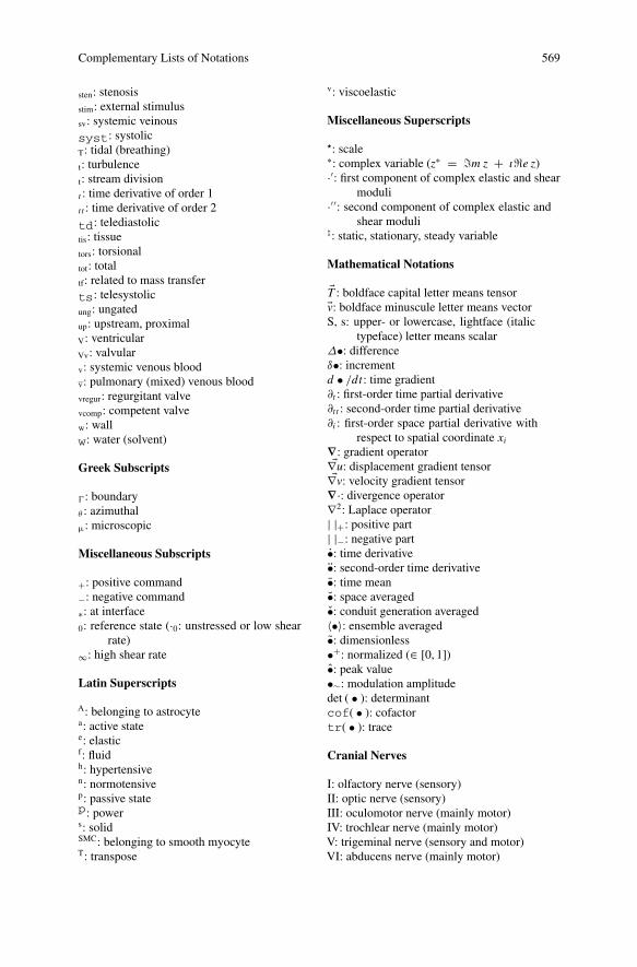
Complementary Lists of Notations 569
sten: stenosisstim: external stimulussv: systemic veinoussyst: systolicT: tidal (breathing)t: turbulencet: stream divisiont : time derivative of order 1t t : time derivative of order 2
td: telediastolictis: tissuetors: torsionaltot: totaltf: related to mass transferts: telesystolicung: ungatedup: upstream, proximalV: ventricularVv: valvularv: systemic venous bloodv: pulmonary (mixed) venous bloodvregur: regurgitant valvevcomp: competent valvew: wallw: water (solvent)
Greek Subscripts
� : boundaryθ : azimuthalμ: microscopic
Miscellaneous Subscripts
+: positive command−: negative command∗: at interface0: reference state (·0: unstressed or low shear
rate)∞: high shear rate
Latin Superscripts
A: belonging to astrocytea: active statee: elasticf: fluidh: hypertensiven: normotensivep: passive statep: powers: solidSMC: belonging to smooth myocyteT: transpose
v: viscoelastic
Miscellaneous Superscripts
�: scale∗: complex variable (z∗ = !m z + ı"e z)·′: first component of complex elastic and shear
moduli·′′: second component of complex elastic and
shear moduli%: static, stationary, steady variable
Mathematical Notations
�T : boldface capital letter means tensor�v: boldface minuscule letter means vectorS, s: upper- or lowercase, lightface (italic
typeface) letter means scalarΔ•: differenceδ•: incrementd • /dt : time gradient∂t : first-order time partial derivative∂tt : second-order time partial derivative∂i : first-order space partial derivative with
respect to spatial coordinate xi∇: gradient operator�∇u: displacement gradient tensor�∇v: velocity gradient tensor
∇·: divergence operator∇2: Laplace operator| |+: positive part| |−: negative part•: time derivative•: second-order time derivative•: time mean•: space averaged•: conduit generation averaged〈•〉: ensemble averaged•: dimensionless•+: normalized (∈ [0, 1])•: peak value•∼: modulation amplitudedet ( • ): determinantcof( • ): cofactortr( • ): trace
Cranial Nerves
I: olfactory nerve (sensory)II: optic nerve (sensory)III: oculomotor nerve (mainly motor)IV: trochlear nerve (mainly motor)V: trigeminal nerve (sensory and motor)VI: abducens nerve (mainly motor)
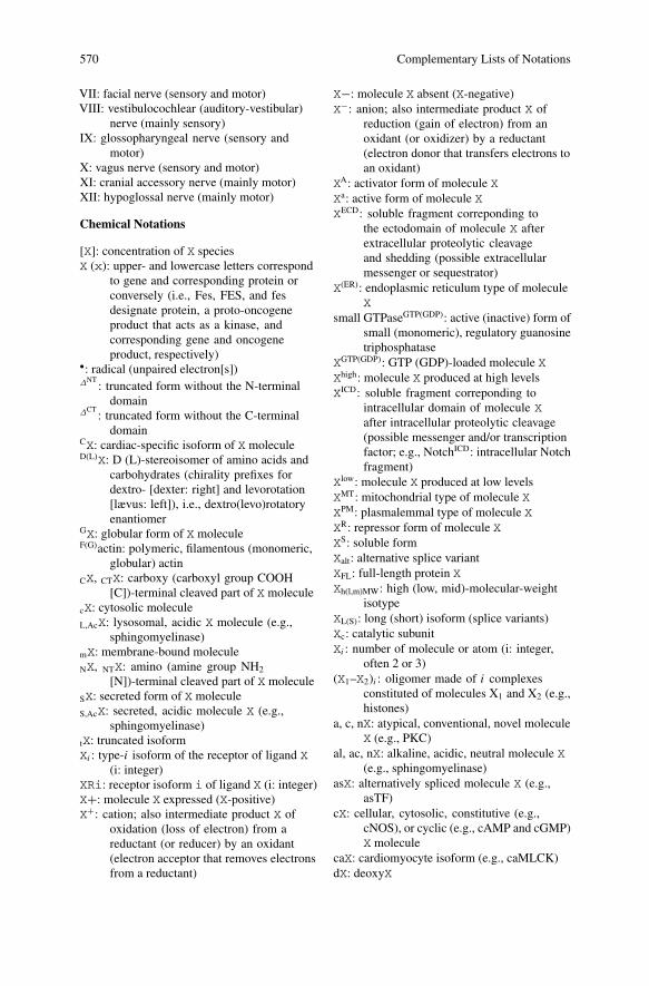
570 Complementary Lists of Notations
VII: facial nerve (sensory and motor)VIII: vestibulocochlear (auditory-vestibular)
nerve (mainly sensory)IX: glossopharyngeal nerve (sensory and
motor)X: vagus nerve (sensory and motor)XI: cranial accessory nerve (mainly motor)XII: hypoglossal nerve (mainly motor)
Chemical Notations
[X]: concentration of X speciesX (x): upper- and lowercase letters correspond
to gene and corresponding protein orconversely (i.e., Fes, FES, and fesdesignate protein, a proto-oncogeneproduct that acts as a kinase, andcorresponding gene and oncogeneproduct, respectively)
•: radical (unpaired electron[s])ΔNT
: truncated form without the N-terminaldomain
ΔCT: truncated form without the C-terminal
domainCX: cardiac-specific isoform of X moleculeD(L)X: D (L)-stereoisomer of amino acids and
carbohydrates (chirality prefixes fordextro- [dexter: right] and levorotation[lævus: left]), i.e., dextro(levo)rotatoryenantiomer
GX: globular form of X moleculeF(G)actin: polymeric, filamentous (monomeric,
globular) actinCX, CTX: carboxy (carboxyl group COOH
[C])-terminal cleaved part of X moleculecX: cytosolic moleculeL,AcX: lysosomal, acidic X molecule (e.g.,
sphingomyelinase)mX: membrane-bound moleculeNX, NTX: amino (amine group NH2
[N])-terminal cleaved part of X moleculeSX: secreted form of X moleculeS,AcX: secreted, acidic molecule X (e.g.,
sphingomyelinase)tX: truncated isoformXi : type-i isoform of the receptor of ligand X
(i: integer)XRi: receptor isoform i of ligand X (i: integer)X+: molecule X expressed (X-positive)X+: cation; also intermediate product X of
oxidation (loss of electron) from areductant (or reducer) by an oxidant(electron acceptor that removes electronsfrom a reductant)
X−: molecule X absent (X-negative)X−: anion; also intermediate product X of
reduction (gain of electron) from anoxidant (or oxidizer) by a reductant(electron donor that transfers electrons toan oxidant)
XA: activator form of molecule XXa: active form of molecule XXECD: soluble fragment correponding to
the ectodomain of molecule X afterextracellular proteolytic cleavageand shedding (possible extracellularmessenger or sequestrator)
X(ER): endoplasmic reticulum type of moleculeX
small GTPaseGTP(GDP): active (inactive) form ofsmall (monomeric), regulatory guanosinetriphosphatase
XGTP(GDP): GTP (GDP)-loaded molecule XXhigh: molecule X produced at high levelsXICD: soluble fragment correponding to
intracellular domain of molecule Xafter intracellular proteolytic cleavage(possible messenger and/or transcriptionfactor; e.g., NotchICD: intracellular Notchfragment)
Xlow: molecule X produced at low levelsXMT: mitochondrial type of molecule XXPM: plasmalemmal type of molecule XXR: repressor form of molecule XXS: soluble formXalt: alternative splice variantXFL: full-length protein XXh(l,m)MW: high (low, mid)-molecular-weight
isotypeXL(S): long (short) isoform (splice variants)Xc: catalytic subunitXi : number of molecule or atom (i: integer,
often 2 or 3)(X1–X2)i : oligomer made of i complexes
constituted of molecules X1 and X2 (e.g.,histones)
a, c, nX: atypical, conventional, novel moleculeX (e.g., PKC)
al, ac, nX: alkaline, acidic, neutral molecule X(e.g., sphingomyelinase)
asX: alternatively spliced molecule X (e.g.,asTF)
cX: cellular, cytosolic, constitutive (e.g.,cNOS), or cyclic (e.g., cAMP and cGMP)X molecule
caX: cardiomyocyte isoform (e.g., caMLCK)dX: deoxyX
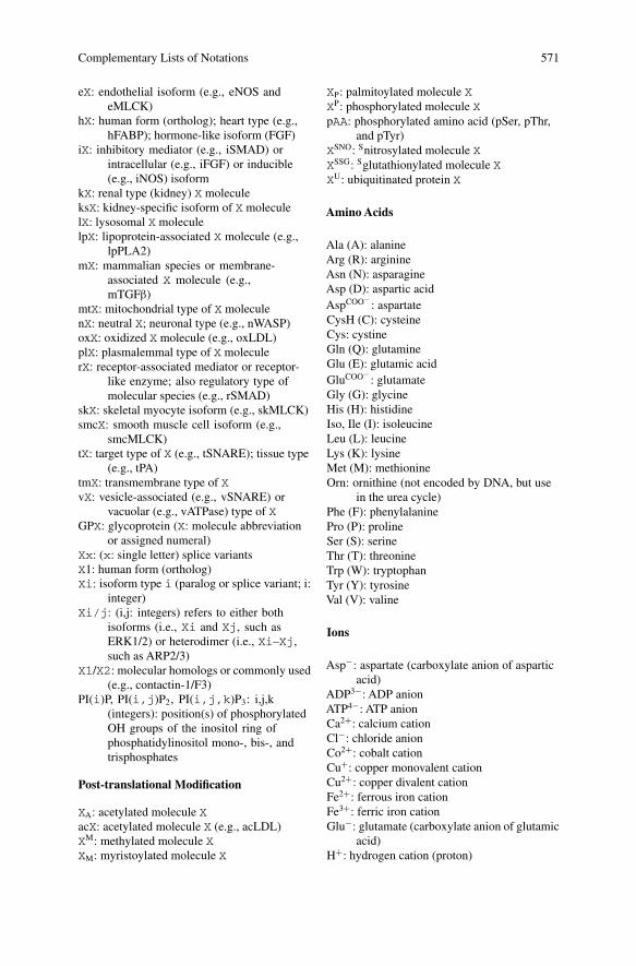
Complementary Lists of Notations 571
eX: endothelial isoform (e.g., eNOS andeMLCK)
hX: human form (ortholog); heart type (e.g.,hFABP); hormone-like isoform (FGF)
iX: inhibitory mediator (e.g., iSMAD) orintracellular (e.g., iFGF) or inducible(e.g., iNOS) isoform
kX: renal type (kidney) X moleculeksX: kidney-specific isoform of X moleculelX: lysosomal X moleculelpX: lipoprotein-associated X molecule (e.g.,
lpPLA2)mX: mammalian species or membrane-
associated X molecule (e.g.,mTGFβ)
mtX: mitochondrial type of X moleculenX: neutral X; neuronal type (e.g., nWASP)oxX: oxidized X molecule (e.g., oxLDL)plX: plasmalemmal type of X moleculerX: receptor-associated mediator or receptor-
like enzyme; also regulatory type ofmolecular species (e.g., rSMAD)
skX: skeletal myocyte isoform (e.g., skMLCK)smcX: smooth muscle cell isoform (e.g.,
smcMLCK)tX: target type of X (e.g., tSNARE); tissue type
(e.g., tPA)tmX: transmembrane type of XvX: vesicle-associated (e.g., vSNARE) or
vacuolar (e.g., vATPase) type of XGPX: glycoprotein (X: molecule abbreviation
or assigned numeral)Xx: (x: single letter) splice variantsX1: human form (ortholog)Xi: isoform type i (paralog or splice variant; i:
integer)Xi/j: (i,j: integers) refers to either both
isoforms (i.e., Xi and Xj, such asERK1/2) or heterodimer (i.e., Xi–Xj,such as ARP2/3)
X1/X2: molecular homologs or commonly used(e.g., contactin-1/F3)
PI(i)P, PI(i,j)P2, PI(i,j,k)P3: i,j,k(integers): position(s) of phosphorylatedOH groups of the inositol ring ofphosphatidylinositol mono-, bis-, andtrisphosphates
Post-translational Modification
XA: acetylated molecule XacX: acetylated molecule X (e.g., acLDL)XM: methylated molecule XXM: myristoylated molecule X
XP: palmitoylated molecule XXP: phosphorylated molecule XpAA: phosphorylated amino acid (pSer, pThr,
and pTyr)XSNO: Snitrosylated molecule XXSSG: Sglutathionylated molecule XXU: ubiquitinated protein X
Amino Acids
Ala (A): alanineArg (R): arginineAsn (N): asparagineAsp (D): aspartic acidAspCOO−
: aspartateCysH (C): cysteineCys: cystineGln (Q): glutamineGlu (E): glutamic acidGluCOO−
: glutamateGly (G): glycineHis (H): histidineIso, Ile (I): isoleucineLeu (L): leucineLys (K): lysineMet (M): methionineOrn: ornithine (not encoded by DNA, but use
in the urea cycle)Phe (F): phenylalaninePro (P): prolineSer (S): serineThr (T): threonineTrp (W): tryptophanTyr (Y): tyrosineVal (V): valine
Ions
Asp−: aspartate (carboxylate anion of asparticacid)
ADP3−: ADP anionATP4−: ATP anionCa2+: calcium cationCl−: chloride anionCo2+: cobalt cationCu+: copper monovalent cationCu2+: copper divalent cationFe2+: ferrous iron cationFe3+: ferric iron cationGlu−: glutamate (carboxylate anion of glutamic
acid)H+: hydrogen cation (proton)
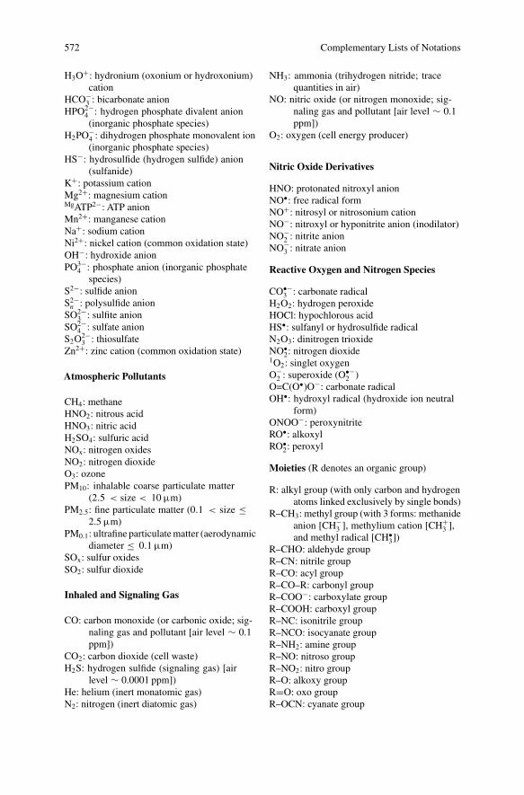
572 Complementary Lists of Notations
H3O+: hydronium (oxonium or hydroxonium)cation
HCO−3 : bicarbonate anion
HPO2−4 : hydrogen phosphate divalent anion(inorganic phosphate species)
H2PO−4 : dihydrogen phosphate monovalent ion(inorganic phosphate species)
HS−: hydrosulfide (hydrogen sulfide) anion(sulfanide)
K+: potassium cationMg2+: magnesium cationMgATP2−: ATP anionMn2+: manganese cationNa+: sodium cationNi2+: nickel cation (common oxidation state)OH−: hydroxide anionPO3−
4 : phosphate anion (inorganic phosphatespecies)
S2−: sulfide anionS2−n : polysulfide anion
SO2−3 : sulfite anion
SO2−4 : sulfate anion
S2O2−3 : thiosulfate
Zn2+: zinc cation (common oxidation state)
Atmospheric Pollutants
CH4: methaneHNO2: nitrous acidHNO3: nitric acidH2SO4: sulfuric acidNOx: nitrogen oxidesNO2: nitrogen dioxideO3: ozonePM10: inhalable coarse particulate matter
(2.5 < size < 10 μm)PM2.5: fine particulate matter (0.1 < size ≤
2.5 μm)PM0.1: ultrafine particulate matter (aerodynamic
diameter ≤ 0.1 μm)SOx: sulfur oxidesSO2: sulfur dioxide
Inhaled and Signaling Gas
CO: carbon monoxide (or carbonic oxide; sig-naling gas and pollutant [air level ∼ 0.1ppm])
CO2: carbon dioxide (cell waste)H2S: hydrogen sulfide (signaling gas) [air
level ∼ 0.0001 ppm])He: helium (inert monatomic gas)N2: nitrogen (inert diatomic gas)
NH3: ammonia (trihydrogen nitride; tracequantities in air)
NO: nitric oxide (or nitrogen monoxide; sig-naling gas and pollutant [air level ∼ 0.1ppm])
O2: oxygen (cell energy producer)
Nitric Oxide Derivatives
HNO: protonated nitroxyl anionNO•: free radical formNO+: nitrosyl or nitrosonium cationNO−: nitroxyl or hyponitrite anion (inodilator)NO−
2 : nitrite anionNO−
3 : nitrate anion
Reactive Oxygen and Nitrogen Species
CO•−3 : carbonate radical
H2O2: hydrogen peroxideHOCl: hypochlorous acidHS•: sulfanyl or hydrosulfide radicalN2O3: dinitrogen trioxideNO•
2: nitrogen dioxide1O2: singlet oxygenO−
2 : superoxide (O•−2 )
O=C(O•)O−: carbonate radicalOH•: hydroxyl radical (hydroxide ion neutral
form)ONOO−: peroxynitriteRO•: alkoxylRO•
2: peroxyl
Moieties (R denotes an organic group)
R: alkyl group (with only carbon and hydrogenatoms linked exclusively by single bonds)
R–CH3: methyl group (with 3 forms: methanideanion [CH−
3 ], methylium cation [CH+3 ],
and methyl radical [CH•3])
R–CHO: aldehyde groupR–CN: nitrile groupR–CO: acyl groupR–CO–R: carbonyl groupR–COO−: carboxylate groupR–COOH: carboxyl groupR–NC: isonitrile groupR–NCO: isocyanate groupR–NH2: amine groupR–NO: nitroso groupR–NO2: nitro groupR–O: alkoxy groupR=O: oxo groupR–OCN: cyanate group
Complementary Lists of Notations 573
R–OH: hydroxyl groupR–ONO: nitrosooxy groupR–ONO2: nitrate groupR–OO–R: peroxy groupR–OOH: hydroperoxy groupR–S–R: sulfide groupR–SH: thiol (or sulfhydryl) moietyR–SN: sulfenyl-amide moietyR–SNO: nitrosothiol (or thionitrite) moietyR–SO: sulfinylR–SO–R: sulfoxide groupR–SO2: sulfonyl groupR–SO2H: sulfinic acid (sulfinyl moiety)R–SO2N: sulfonyl-amide moietyR–SO3H: sulfonic acid (sulfonyl moiety)R–SOH: sulfenic acid (sulfenyl moiety)R–SON: sulfinyl-amide moietyR–SSH: hydropersulfide moietyR–SS–R: disulfide groupR–S(S)nS–R: polysulfide
Lung Function Testing
ERV: expiratory reserve volumefC: cardiac frequencyfR: breathing frequencyFEFf : forced expiratory flow at a fraction
(f [%]) of forced expiration (FEF25,FEF50, and FEF75)
FEV1: volume expired at the end of the firstsecond of forced expiration
FEVτ : forced expiratory volume at timeτ (fraction of a second over whichmaximally fast exhaled volume ismeasured) starting from full inspiration
FRC: functional residual capacity (lung volumeat end of rest expiration)
IC: inspiratory capacity (IRV +VT)IRV: inspiratory reserve volumeMBC: maximum breathing capacity (per mn of
effort)MVV: maximal voluntary ventilation (volume
of air breathed in a specified periodduring repetitive maximal exercise)
PEF: peak expiratory flowRR: respiratory quotient (VCO2/VO2 )RV: residual volumeTLC: total lung capacityVA: alveolar gas volume
VD: dead space volumeVL: lung volumeVT: tidal volumeV : total ventilation (air volume exhaled par mn)VA: alveolar ventilation (fR(VT − VD))VO2 : oxygen consumptionVCO2 : carbon dioxide productionVC: vital capacity (air volume quietly expelled
from full inspiration)
Time Units
s: secondmn: minuteh: hourd: daywk: weekmo: monthyr: year
SI-Based and Non-SI Units of Quantity
mmHg: millimeter of mercury (133.322 Pa[∼0.1333 kPa])
mmol, nmol, μ mol: milli-, nano-, micromoles(amount of a chemical species, one mole
containing about 6.02214078×10²³
molecules)mosm: milliosmole(osm: number of moles of a osmotically active
chemical compound)kDa: kiloDalton(Da: atomic or molecular mass unit)ppm: parts per millionl: liter
Temperature and Pressure Conditions
ATPS: ambient temperature and pressure,saturated with water at body temperature,i.e., at 37◦C, pH2O = 6.27 kPa (47mmHg)
BTPS: body temperature and ambient pressure,saturated with water
STPD: standard temperature (0◦C) and pressure(101 kPa [760 mmHg]), dry air
VBTPS = VATPS × 273 + 37
273 + T× p − pH2O
p − 47
VSTPD = VATPS × 273
273 + T× p − pH2O
760
References
Chap. 1. Pathogenesis of Cardiac Diseases—Biological Context
1. Mathiyalagan P, Keating ST, Du XJ, El-Osta A (2014) Chromatin modifications remodelcardiac gene expression. Cardiovasc Res 103:7–16
2. Demos-Davies KM, Ferguson BS, Cavasin MA, Mahaffey JH, Williams SM, Spiltoir JI,Schuetze KB, Horn TR, Chen B, Ferrara C, Scellini B, Piroddi N, Tesi C, Poggesi C, JeongMY, McKinsey TA (2014) HDAC6 contributes to pathological responses of heart and skeletalmuscle to chronic angiotensin-II signaling. Am J Physiol Heart Circ Physiol 307:H252–H258
3. Evans PC, Kwak BR (2013) Biomechanical factors in cardiovascular disease. Cardiovasc Res99:229–231
4. Frueh J, Maimari N, Homma T, Bovens SM, Pedrigi RM, Towhidi L, Krams R (2013) Systemsbiology of the functional and dysfunctional endothelium. Cardiovasc Res 99:334–341
5. Zhang C, Zeng L, Emanueli C, Xu Q (2013) Blood flow and stem cells in vascular disease.Cardiovasc Res 99:251–259
6. Oakley RH, Ren R, Cruz-Topete D, Bird GS, Myers PH, Boyle MC, Schneider MD, WillisMS, Cidlowski JA (2013) Essential role of stress hormone signaling in cardiomyocytes forthe prevention of heart disease. Proc Natl Acad Sci U S A 110:17035–17040
7. Zacchigna S, Giacca M (2014) Extra- and intracellular factors regulating cardiomyocyteproliferation in postnatal life. Cardiovasc Res 102:312–320
8. Hong TT, Smyth JW, Gao D, Chu KY, Vogan JM, Fong TS, Jensen BC, Colecraft HM, ShawRM (2010) BIN1 localizes the L-type calcium channel to cardiac T-tubules. PLoS Biol 20108:e1000312
9. Beavers DL, LandstromAP, Chiang DY,Wehrens XHT (2014) Emerging roles of junctophilin-2 in the heart and implications for cardiac diseases. Cardiovasc Res 103:198–205
10. Gorospe G, Zhu R, Millrod MA, Zambidis ET, Tung L,Vidal R (2014)Automated grouping ofaction potentials of human embryonic stem cell-derived cardiomyocytes. IEEE Trans BiomedEng 61:2389–2396
11. Nemir M, Metrich M, Plaisance I, Lepore M, Cruchet S, Berthonneche C, Sarre A, Radtke F,Pedrazzini T (2014) The Notch pathway controls fibrotic and regenerative repair in the adultheart. Eur Heart J 35:2174–2185
12. Wackerhage H, Del Re DP, Judson RN, Sudol M, Sadoshima J (2014) The Hippo signaltransduction network in skeletal and cardiac muscle. Sci Signal 7:re4
13. Zouein FA, Kurdi M, Booz GW (2013) HSPA12B and repairing the heart: beauty in simplicity.Cardiovasc Res 99:587–589
14. Li J, Zhang Y, Li C, Xie J, Liu Y, Zhu W, Zhang X, Jiang S, Liu L, Ding Z (2013) HSPA12Battenuates cardiac dysfunction and remodelling after myocardial infarction through an eNOS-dependent mechanism. Cardiovasc Res 99:674–684
© Springer International Publishing Switzerland 2015 575M. Thiriet, Diseases of the Cardiac Pump, Biomathematical and BiomechanicalModeling of the Circulatory and Ventilatory Systems 7, DOI 10.1007/978-3-319-12664-7
576 References
15. Tarone G, Brancaccio M (2014) Keep your heart in shape: molecular chaperone networks fortreating heart disease. Cardiovasc Res 102:346–361
16. Ferretti R, Sbroggiò M, Di Savino A, Fusella F, Bertero A, Michowski W, Tarone G, Brancac-cio M (2011) Morgana and melusin: two fairies chaperoning signal transduction. Cell Cycle10:3678–3683
17. Jian Z, Han H, Zhang T, Puglisi J, Izu LT, Shaw JA, Onofiok E, Erickson JR, ChenYJ, HorvathB, Shimkunas R, Xiao W, Li Y, Pan T, Chan J, Banyasz T, Tardiff JC, Chiamvimonvat N,Bers DM, Lam KS, Chen-Izu Y (2014) Mechanochemotransduction during cardiomyocytecontraction is mediated by localized nitric oxide signaling. Sci Signal 7:ra27
18. Collins HE, Zhu-Mauldin X, Marchase RB, Chatham JC (2013) STIM1/Orai1-mediatedSOCE: current perspectives and potential roles in cardiac function and pathology. Am JPhysiol Heart Circ Physiol 305:H446–H458
19. Yarotskyy V, Dirksen RT (2012) Temperature and RyR1 regulate the activation rate of store-operated Ca2+ entry current in myotubes. Biophys J 103:202–211
20. Grigoriev I, Gouveia SM, van der Vaart B, Demmers J, Smyth JT, Honnappa S, Splinter D,Steinmetz MO, Putney JW, Hoogenraad CC, Akhmanova A (2008) STIM1 is a MT-plus-end-tracking protein involved in remodeling of the ER. Curr Biol 18:177–182
21. Zeiger W, Ito D, Swetlik C, Oh-hora M, Villereal ML, Thinakaran G (2011) Stanniocalcin 2is a negative modulator of store-operated calcium entry. Mol Cell Biol 31:3710–3722
22. Prins D, Groenendyk J, Touret N, Michalak M (2011) Modulation of STIM1 and capacita-tive Ca2+ entry by the endoplasmic reticulum luminal oxidoreductase ERp57. EMBO Rep12:1182–1188
23. Srikanth S, Jung HJ, Kim KD, Souda P, Whitelegge J, Gwack Y (2010) A novel EF-handprotein, CRACR2A, is a cytosolic Ca2+ sensor that stabilizes CRAC channels in T cells. NatCell Biol 12:436–446
24. Fujii Y, Shiota M, Ohkawa Y, Baba A, Wanibuchi H, Kinashi T, Kurosaki T, Baba Y (2012)Surf4 modulates STIM1-dependent calcium entry. Biochem Biophys Res Commun 422:615–620
25. Palty R, Raveh A, Kaminsky I, Meller R, Reuveny E (2012) SARAF inactivates the storeoperated calcium entry machinery to prevent excess calcium refilling. Cell 149:425–438
26. Jha A, Ahuja M, Maléth J, Moreno CM, Yuan JP, Kim MS, Muallem S (2013) The STIM1CTID domain determines access of SARAF to SOAR to regulate Orai1 channel function. JCell Biol 202:71–79
27. Darbellay B, Arnaudeau S, Bader CR, Konig S, Bernheim L (2011) STIM1L is a new actin-binding splice variant involved in fast repetitive Ca2+ release. J Cell Biol 194:335–346
28. Ogawa A, Firth AL, Smith KA, Maliakal MV,Yuan JX (2012) PDGF enhances store-operatedCa2+ entry by upregulating STIM1/Orai1 via activation of Akt/mTOR in human pulmonaryarterial smooth muscle cells. Am J Physiol Cell Physiol 302:C405-C411
29. Xiang SY, Ouyang K, Yung BS, Miyamoto S, Smrcka AV, Chen J, Brown JH, (2013) PLCε,PKD1, and SSH1L transduce RhoA signaling to protect mitochondria from oxidative stressin the heart. Sci Signal 6:ra108
30. Puthanveetil P, Wan A, Rodrigues B (2013) FoxO1 is crucial for sustaining cardiomyocytemetabolism and cell survival. Cardiovasc Res 97:393–403
31. Liu S, Zhang J, Xiang YK (2011) FRET-based direct detection of dynamic protein kinase Aactivity on the sarcoplasmic reticulum in cardiomyocytes. Biochem Biophys Res Commun404:581–586
32. Haj Slimane Z, Bedioune I, Lechêne P, Varin A, Lefebvre F, Mateo P, Domergue-Dupont V,Dewenter M, Richter W, Conti M, El-Armouche A, Zhang J, Fischmeister R, VandecasteeleG (2014) Control of cytoplasmic and nuclear protein kinase A by phosphodiesterases andphosphatases in cardiac myocytes. Cardiovasc Res 102:97–106
33. Hirsch E, Nagai R, Thum T (2014) Heterocellular signalling and crosstalk in the heart inischaemia and heart failure. Cardiovasc Res 102:191–193
References 577
34. Carbe CJ, Cheng L, Addya S, Gold JI, Gao E, Koch WJ, Riobo NA (2014) Gi proteins mediateactivation of the canonical hedgehog pathway in the myocardium. Am J Physiol Heart CircPhysiol 307:H66–H72
35. DebA (2014) Cell–cell interaction in the heart via Wnt/β-catenin pathway after cardiac injury.Cardiovasc Res 102:214–223
36. Haghikia A, Ricke-Hoch M, Stapel B, Gorst I, Hilfiker-Kleiner D (2014) STAT3, a keyregulator of cell-to-cell communication in the heart. Cardiovasc Res 102:281–289
37. Mohamed TM, Zi M, Prehar S, Maqsood A, Abou-Leisa R, Nguyen L, Pfeifer GP, CartwrightEJ, Neyses L, Oceandy D (2014) The tumour suppressor Ras-association domain familyprotein 1A (RASSF1A) regulates TNF-α signalling in cardiomyocytes. Cardiovasc Res103:47–59 ‘
38. Roberge S, Roussel J, Andersson DC, Meli AC, Vidal B, Blandel F, Lanner JT, Le GuennecJY, Katz A, Westerblad H, Lacampagne A, Fauconnier J (2014) TNF-α-mediated caspase-8 activation induces ROS production and TRPM2 activation in adult ventricular myocytes.Cardiovasc Res 103:90–99
39. Emde A, Hornstein E (2014) miRNAs at the interface of cellular stress and disease. EMBO J33:1428–1437
40. Viereck J, Bang C, Foinquinos A, Thum T (2014) Regulatory RNAs and paracrine networksin the heart. Cardiovasc Res 102:290–301
41. Tian FJ, An LN, Wang GK, Zhu JQ, Li Q, Zhang YY, Zeng A, Zou J, Zhu RF, Han XS, ShenN, Yang HT, Zhao XX, Huang S, Qin YW, Jing Q (2014) Elevated microRNA-155 promotesfoam cell formation by targeting HBP1 in atherogenesis. Cardiovasc Res 103:100–110
42. Hartmann P, Schober A2, Weber C (2014) MicroRNA-155 and macrophages: a fatty liaison.Cardiovasc Res 103:5–6
43. Sluijter JP, Verhage V, Deddens JC, van den Akker F, Doevendans PA (2014) Microvesiclesand exosomes for intracardiac communication. Cardiovasc Res 102:302–311
44. Hollander JM, Thapa D, Shepherd DL (2014) Physiological and structural differences inspatially distinct subpopulations of cardiac mitochondria: influence of cardiac pathologies.Am J Physiol Heart Circ Physiol 307:H1–H14
45. Chen L, Gong Q, Stice JP, Knowlton AA (2009) Mitochondrial OPA1, apoptosis, and heartfailure. Cardiovasc Res 84:91–99
46. Norton M, Ng ACH, Baird S, Dumoulin A, Shutt T, Mah N, Andrade-Navarro MA, McBrideHM, Screaton RA (2014) ROMO1 is an essential redox-dependent regulator of mitochondrialdynamics. Sci Signal 7:ra10
47. Miller MW, Knaub LA, Olivera-Fragoso LF, Keller AC, Balasubramaniam V, Watson PA,Reusch JE (2013) Nitric oxide regulates vascular adaptive mitochondrial dynamics. Am JPhysiol Heart Circ Physiol 304:H1624–H1633
48. Wenceslau CF, McCarthy CG, Szasz T, Spitler K, Goulopoulou S, Webb RC, Working Groupon DAMPs in Cardiovascular Disease (2014) Mitochondrial damage-associated molecularpatterns and vascular function. Eur Heart J 35:1172–1177
49. Lopaschuk GD, Ussher JR, Folmes CD, Jaswal JS, Stanley WC (2010) Myocardial fatty acidmetabolism in health and disease. Physiol Rev 90:207–258
50. Lapuente-Brun E, Moreno-Loshuertos R, Acín-Pérez R, Latorre-Pellicer A, Colás C, BalsaE, Perales-Clemente E, Quirós PM, Calvo E, Rodríguez-Hernández MA, Navas P, CruzR, Carracedo A, López-Otín C, Pérez-Martos A, Fernández-Silva P, Fernández-Vizarra E,Enríquez JA (2013) Supercomplex assembly determines electron flux in the mitochondrialelectron transport chain. Science 340:1567–1570
51. Garcia-Dorado D, Ruiz-Meana M, Rodríguez-SinovasA (2014) Connexin 43 phosphorylationin subsarcolemmal mitochondria: a general cardioprotective signal targeted by fibroblastgrowth factor-2?. Cardiovasc Res 103:1–2
52. Srisakuldee W, Makazan Z, Nickel BE, Zhang F, Thliveris JA, Pasumarthi KB, Kardami E(2014) The FGF-2-triggered protection of cardiac subsarcolemmal mitochondria from calciumoverload is mitochondrial connexin 43-dependent. Cardiovasc Res 103:72–80
578 References
53. Foskett JK, Madesh M (2014) Regulation of the mitochondrial Ca2+ uniporter by MICU1and MICU2. Biochem Biophys Res Commun 449:377–383
54. Rizzuto R, De Stefani D, Raffaello A, Mammucari C (2012) Mitochondria as sensors andregulators of calcium signalling. Nat Rev Mol Cell Biol 13:566–578
55. Williams GS, Boyman L, Chikando AC, Khairallah RJ, Lederer WJ (2013) Mitochondrialcalcium uptake. Proc Natl Acad Sci U S A 110:10479–10486
56. Carafoli E (2014) Historical introduction. Biochem Biophys Res Commun 449:365–36657. De Stefani D, Rizzuto R (2014) Molecular control of mitochondrial calcium uptake. Biochem
Biophys Res Commun 449:373–37658. Chaudhuri D, Clapham DE (2014) Outstanding questions regarding the permeation, selectiv-
ity, and regulation of the mitochondrial calcium uniporter. Biochem Biophys Res Commun449:367–369
59. Takeuchi A, Kim B, Matsuoka S (2013) The mitochondrial Na+–Ca2+ exchanger, NCLX,regulates automaticity of HL-1 cardiomyocytes. Sci Rep 3:2766
60. Alavian KN, Beutner G, Lazrove E, Sacchetti S, Park HA, Licznerski P, Li H, Nabili P,Hockensmith K, Graham M, Porter GA, Jonas EA (2014) An uncoupling channel within thec-subunit ring of the F1FO ATP synthase is the mitochondrial permeability transition pore.Proc Natl Acad Sci U S A 111:10580–10585
61. Penna C, Mancardi D, Rastaldo R, Pagliaro P (2009) Cardioprotection: a radical view: freeradicals in pre and postconditioning. Biochim Biophys Acta 1787:781–793. (Bioenergetics)
62. Carpi A, Menabò R, Kaludercic N, Pelicci P, Di Lisa F, Giorgio M (2009) The cardiopro-tective effects elicited by p66(Shc) ablation demonstrate the crucial role of mitochondrialROS formation in ischemia/reperfusion injury. Biochim Biophys Acta 1787:774–780.(Bioenergetics)
63. Rudat C, Norden J, Taketo MM, Kispert A (2013) Epicardial function of canonical Wnt-,Hedgehog-, Fgfr1/2-, and Pdgfra-signalling. Cardiovasc Res 100:411–421
64. Zhang M, Shah AM (2014) ROS signalling between endothelial cells and cardiac cells.Cardiovasc Res 102:249–257
65. Brutsaert DL (2003) Cardiac endothelial-myocardial signaling: its role in cardiac growth,contractile performance, and rhythmicity. Physiol Rev 83:59–115
66. Hendrickx J, Doggen K, Weinberg EO, Van Tongelen P, Fransen P, De Keulenaer GW (2004)Molecular diversity of cardiac endothelial cells in vitro and in vivo. Physiol Genomics 19:198–206
67. Parodi EM, Kuhn B (2014) Signalling between microvascular endothelium and cardiomy-ocytes through neuregulin. Cardiovasc Res 102:194–204
68. Hatem SN, Sanders P (2014) Epicardial adipose tissue and atrial fibrillation. Cardiovasc Res102:205–213
69. Ghigo A, Franco I, Morello F, Hirsch E (2014) Myocyte signalling in leucocyte recruitmentto the heart. Cardiovasc Res 102:270–280
70. Van Linthout S, Miteva K, Tschöpe C (2014) Crosstalk between fibroblasts and inflammatorycells. Cardiovasc Res 102:258–269
71. Fairweather DL, Frisancho-Kiss S, Coronado MJ, Frisancho JA, Cihakova D, Rose NR (2009)Two populations of alternatively activated M2 macrophages (TLR4+ vs. Tim-3+) regulateinflammation and fibrosis in the heart during coxsackievirus-induced myocarditis. J Immun182:137.20. (Meeting Abstract Supplement)
72. Kane LP (2010) T cell Ig and mucin domain proteins and immunity. J Immun 184:2743–274973. Frantz S, Nahrendorf M (2014) Cardiac macrophages and their role in ischaemic heart disease.
Cardiovasc Res 102:240–24874. Fujiu K,Wang J, Nagai R (2014) Cardioprotective function of cardiac macrophage. Cardiovasc
Res 102:232–23975. Martinez FO, Gordon S (2014) The M1 and M2 paradigm of macrophage activation: time for
reassessment. F1000Prime Rep 6:13
References 579
76. Inoue T, Plieth D, Venkov CD, Xu C, Neilson EG (2005) Antibodies against macrophagesthat overlap in specificity with fibroblasts. Kidney International 67:2488–2493
77. Ramkisoensing AA, de Vries AA, Atsma DE, Schalij MJ, Pijnappels DA (2014) Interactionbetween myofibroblasts and stem cells in the fibrotic heart: balancing between deteriorationand regeneration. Cardiovasc Res 102:224–231
78. Chng SC, Ho L, Tian J, Reversade B (2013) ELABELA: a hormone essential for heartdevelopment signals via the apelin receptor. Dev Cell 27:672–680
79. PauliA, Norris ML,Valen E, Chew GL, Gagnon JA, Zimmerman S, MitchellA, Ma J, DubrulleJ, Reyon D, Tsai SQ, Joung JK, SaghatelianA, SchierAF (2014) Toddler: an embryonic signalthat promotes cell movement via Apelin receptors. Science 343:1248636
80. Magny EG, Pueyo JI, Pearl FM, Cespedes MA, Niven JE, Bishop SA, Couso JP (2013)Conserved regulation of cardiac calcium uptake by peptides encoded in small open readingframes. Science 341:1116–1120
81. Traaseth NJ, Ha KN, Verardi R, Shi L, Buffy JJ, Masterson LR, Veglia G (2008) Structuraland dynamic basis of phospholamban and sarcolipin inhibition of Ca2+-ATPase. Biochemistry473–13
82. Smith WS, Broadbridge R, East JM, Lee AG (2002) Sarcolipin uncouples hydrolysis of ATPfrom accumulation of Ca2+ by the Ca2+-ATPase of skeletal-muscle sarcoplasmic reticulum.Biochem J. Jan 15;361(Pt 2):277-86.
83. Hirose T, MishimaY, TomariY (2014) Elements and machinery of non-coding RNAs: Towardtheir taxonomy. EMBO Rep 15:489–507
84. Cohen SM (2014) Everything old is new again: (linc)RNAs make proteins! EMBO J 33:937–1085
85. Bazzini AA, Johnstone TG, Christiano R, Mackowiak SD, Obermayer B, Fleming ES, VejnarCE, Lee MT, Rajewsky N, Walther TC, Giraldez AJ (2014) Identification of small ORFs invertebrates using ribosome footprinting and evolutionary conservation. EMBO J 33:981–993
86. Nelson BR, Anderson DM, Olson EN (2014) Small open reading frames pack a big punch incardiac calcium regulation. Circ Res 114:18–20
87. Shukla P, Ghatta S, Dubey N, Lemley CO, Johnson ML, Modgil A, Vonnahme K, Caton JS,Reynolds LP, Sun C, O’Rourke ST (2014) Maternal nutrient restriction during pregnancyimpairs an endothelium-derived hyperpolarizing factor-like pathway in sheep fetal coronaryarteries. Am J Physiol Heart Circ Physiol 307:H134–H142
88. Lesoon LA, Mahesh VB (1992) Stimulatory and inhibitory effects of progesterone on FSHsecretion by the anterior pituitary. J Steroid Biochem Mol Biol 42:479–491
89. Giguère V (2002) To ERR in the estrogen pathway. Trends Endocrinol Metab 13:220–22590. Schreiber SN, Emter R, Hock MB, Knutti D, Cardenas J, Podvinec M, Oakeley EJ, Kralli
A (2004) The estrogen-related receptor α (ERRα) functions in PPARγ coactivator 1α (PGC-1α)-induced mitochondrial biogenesis. Proc Natl Acad Sci U S A 101:6472–6477
91. Ochnik AM, Yee D (2012) Estrogen-related receptor α: an orphan finds a family. BreastCancer Res 14:309
92. Gadkar-Sable S, Shah C, Rosario G, Sachdeva G, Puri C (2005) Progesterone receptors:various forms and functions in reproductive tissues. Front Biosci 10:2118–2130
93. Moussatche P, Lyons TJ (2012) Non-genomic progesterone signalling and its non-canonicalreceptor. Biochem Soc Trans 40:200–204
94. Hales DB (2009–2014) Hormones of pregnancy. Southern Illinois University School ofMedicine (www.siumed.edu/dbhaleslab)
95. Bergeron C (2000) Morphological changes and protein secretion induced by progesterone inthe endometrium during the luteal phase in preparation for nidation. Hum Reprod 15:119–128
96. Koistinen H, Seppälä M (2011) PAEP (progestagen-associated endometrial protein) Atlas ofGenetics and Cytogenetics in Oncology and Haematology 15:576–581 (atlasgeneticsoncol-ogy.org)
580 References
97. Rutanen EM, Koistinen R, Seppälä M, Julkunen M, Suikkari AM, Huhtala ML (1987)Progesterone-associated proteins PP12 and PP14 in the human endometrium. J SteroidBiochem 27:25–31
98. Cocchiara R, Lampiasi N, Albeggiani G, Azzolina A, Bongiovanni A, Gianaroli L, Di BlasiF, Geraci D (2006) A factor secreted by human embryo stimulates cytokine release by uterinemast cell. Mol Hum Reprod 2:781–791
99. Susini L, Besse S, Duflaut D, Lespagnol A, Beekman C, Fiucci G, Atkinson AR, Busso D,Poussin P, Marine JC, Martinou JC, Cavarelli J, Moras D, Amson R, Telerman A (2008)TCTP protects from apoptotic cell death by antagonizing bax function. Cell Death Differ15:1211–1220
100. Kashiwakura JC, Ando T, Matsumoto K, Kimura M, Kitaura J, Matho MH, Zajonc DM,Ozeki T, Ra C, MacDonald SM, Siraganian RP, Broide DH, Kawakami Y, Kawakami T(2012) Histamine-releasing factor has a proinflammatory role in mouse models of asthma andallergy. J Clin Investig 122:218–228
101. MacLeod JN, LeeAK, Liebhaber SA, Cooke NE (1992) Developmental control and alternativesplicing of the placentally expressed transcripts from the human growth hormone gene cluster.J Biol Chem 267:14219–14226
102. Lowndes K, Amano A, Yamamoto SY, Bryant-Greenwood GD (2006) The human relaxinreceptor (LGR7): expression in the fetal membranes and placenta. Placenta 27:610–618
103. Tregear GW, Bathgate RA, Layfield S, Ferraro T, Gundlach A, Ma S, Lin F, Hanson NF,Summers RJ, Rosengren J, Craik DJ, Wade JD (2005) The chemistry and biology of humanrelaxin-3. Ann N Y Acad Sci 1041:40–46
104. Liu LX,Arany Z (2014) Maternal cardiac metabolism in pregnancy. Cardiovasc Res 101:545–553
105. Pierlot CM, Lee JM, Amini R, Sacks MS, Wells SM (2014) Pregnancy-induced remodelingof collagen architecture and content in the mitral valve. Ann Biomed Eng 42:
106. Nada MA, Chace DH, Sprecher H, Roe CR (1995) Investigation of β-oxidation intermediatesin normal and MCAD-deficient human fibroblasts using tandem mass spectrometry. BiochemMol Med 54:59–66
107. Ricke-Hoch M, Bultmann I, Stapel B, Condorelli G, Rinas U, Sliwa K, Scherr M, Hilfiker-Kleiner D (2014) Opposing roles of Akt and STAT3 in the protection of the maternal heartfrom peripartum stress. Cardiovasc Res 101:587–596
108. Lok SI, Kirkels JH, Klöpping C, Doevendans PA, de Jonge N (2011) Peripartum cardiomy-opathy: The need for a national database. Neth Heart J 19:126–133
109. Chung E, Yeung F, Leinwand LA (2013) Calcineurin activity is required for cardiacremodelling in pregnancy. Cardiovasc Res 100:402–410
110. Gonzalez AM, Osorio JC, Manlhiot C, Gruber D, Homma S, Mital S (2007) Hypertro-phy signaling during peripartum cardiac remodeling. Am J Physiol Heart Circ Physiol 293:H3008–H3013
Chap. 2. Context of Cardiac Diseases
111. Faergeman O (2013) Genes and cardiovascular risk. Eur Heart J 34:949–950112. Navasiolava NM, Dignat-George F, Sabatier F, Larina IM, Demiot C, Fortrat JO, Gauquelin-
Koch G, Kozlovskaya IB, Custaud MA (2010) Enforced physical inactivity increasesendothelial microparticle levels in healthy volunteers. Am J Physiol Heart Circ Physiol299:H248–H256
113. Hoshi T, Wissuwa B, TianY, Tajima N, Xu R, Bauer M, Heinemann SH, Hou S (2013) Omega-3 fatty acids lower blood pressure by directly activating large-conductance Ca2+-dependentK+ channels. Proc Natl Acad Sci U S A 110:4816–4821
References 581
114. Hoshi T, Tian Y, Xu R, Heinemann SH, Hou S (2013) Mechanism of the modulation of BKpotassium channel complexes with different auxiliary subunit compositions by the omega-3fatty acid DHA. Proc Natl Acad Sci U S A 110:4822–4827
115. Heiss C, Keen CL, Kelm M (2010) Flavanols and cardiovascular disease prevention. EurHeart J 31:2583–2592
116. Heiss C, Kelm M (2010) Chocolate consumption, blood pressure, and cardiovascular risk.Eur Heart J 31:1554–1556
117. Corder R, Mullen W, Khan NQ, Marks SC, Wood EG, Carrier MJ, CrozierA (2006) Oenology:red wine procyanidins and vascular health. Nature 444:566
118. Hammerstone JF, Lazarus SA, Schmitz HH (2000) Procyanidin content and variation in somecommonly consumed foods. Journal of Nutrition 130:2086S–2092S
119. Buijsse B, Weikert C, Drogan D, Bergmann M, Boeing H (2010) Chocolate consumption inrelation to blood pressure and risk of cardiovascular disease in German adults. Eur Heart J31:1616–1623
120. Benavides GA, Squadrito GL, Mills RW, Patel HD, Isbell TS, Patel RP, Darley-Usmar VM,Doeller JE, Kraus DW (2007) Hydrogen sulfide mediates the vasoactivity of garlic. Proc NatlAcad Sci U S A 104:17977–17982
121. Petyaev IM, BashmakovYK (2012) Could cheese be the missing piece in the French paradoxpuzzle? Med Hypotheses 79:746–749
122. Trostchansky A, Rubbo H (2008) Nitrated fatty acids: mechanisms of formation, chemicalcharacterization, and biological properties. Free Radic Biol Med 44:1887–1896
123. Charles RL, Rudyk O, Prysyazhna O, Kamynina A, Yang J, Morisseau C, Hammock BD,Freeman BA, Eaton P (2014) Protection from hypertension in mice by the Mediterranean dietis mediated by nitro fatty acid inhibition of soluble epoxide hydrolase. Proc Natl Acad Sci US A 111:8167–8172
124. Artim DE, Bazely F, Daugherty SL, SculptoreanuA, Koronowski KB, Schopfer FJ, WoodcockSR, Freeman BA, de Groat WC (2011) Nitro-oleic acid targets transient receptor potential(TRP) channels in capsaicin sensitive afferent nerves of rat urinary bladder. Exp Neurol232:90–99
125. Cotter DG, Schugar RC, Crawford PA (2013) Ketone body metabolism and cardiovasculardisease. Am J Physiol Heart Circ Physiol 304:H1060–H1076
126. Ketteler M, Wolf M, Hahn K, Ritz E (2013) Phosphate: a novel cardiovascular risk factor.Eur Heart J 34:1099–1101
127. Heine GH, Nangaku M, Fliser D (2013) Calcium and phosphate impact cardiovascular risk.Eur Heart J 34:1112–1121
128. Gattineni J, Bates C, Twombley K, Dwarakanath V, Robinson ML, Goetz R, MohammadiM, Baum M (2009) FGF23 decreases renal NaPi-2a and NaPi-2c expression and induceshypophosphatemia in vivo predominantly via FGF receptor 1. Am J Physiol Ren Physiol297:F282–F291
129. Wierup N, Björkqvist M, Weström B, Pierzynowski S, Sundler F, Sjölund K (2007) Ghrelinand motilin are cosecreted from a prominent endocrine cell population in the small intestine.J Clin Endocrinol Metab 92:3573–3581
130. Zizzari P, Hassouna R, Grouselle D, Epelbaum J, Tolle V (2011) Physiological roles ofpreproghrelin-derived peptides in GH secretion and feeding. Peptides 32:2274–2282
131. Hassouna R, Zizzari P, Tolle VJ (2010) The ghrelin/obestatin balance in the physiologicaland pathological control of growth hormone secretion, body composition and food intake. JNeuroendocrinol 22:793–804
132. Rogler G, Rosano G (2014) The heart and the gut. Eur Heart J 35:426–430133. Yang IA, Barton SJ, Rorke S, Cakebread JA, Keith TP, Clough JB, Holgate ST, Holloway JW
(2004) Toll-like receptor 4 polymorphism and severity of atopy in asthmatics. Genes Immun5:41–45
134. Kiechl S, Lorenz E, Reindl M,Wiedermann CJ, Oberhollenzer F, Bonora E,Willeit J, SchwartzDA (2002) Toll-like receptor 4 polymorphisms and atherogenesis. N Engl J Med 347:185–192
582 References
135. Yasuno K, Bakircioglu M, Low SK, Bilgüvar K, Gaál E, Ruigrok YM, Niemelä M, Hata A,Bijlenga P, Kasuya H, Jääskeläinen JE, Krex D, Auburger G, Simon M, Krischek B, OzturkAK, Mane S, Rinkel GJ, Steinmetz H, Hernesniemi J, Schaller K, Zembutsu H, Inoue I, PalotieA, Cambien F, NakamuraY, Lifton RP, Günel M (2011) Common variant near the endothelinreceptor type A (EDNRA) gene is associated with intracranial aneurysm risk. Proc Natl AcadSci U S A 108:19707–19712
136. Sugamura K, Keaney JF (2012) Nicotine: linking smoking to abdominal aneurysms. Nat Med18:856–858
137. Yu E, Mercer J, Bennett M (2012) Mitochondria in vascular disease. Cardiovasc Res 95:173–182
138. Peluffo G, Calcerrada P, Piacenza L, Pizzano N, Radi R (2009) Superoxide-mediated inacti-vation of nitric oxide and peroxynitrite formation by tobacco smoke in vascular endothelium:studies in cultured cells and smokers. Am J Physiol Heart Circ Physiol 296:H1781–H1792
139. Vazquez-Padron RI, Mateu D, Rodriguez-Menocal L, Wei Y, Webster KA, Pham SM (2010)Novel role of Egr-1 in nicotine-related neointimal formation. Cardiovasc Res 88:296–303
140. Fukuda N (2010) Cigarette smoking induces vascular proliferative disease through theactivation of Egr-1. Cardiovasc Res 88:207–208
141. Thorin E, Thorin-Trescases N (2009) Vascular endothelial ageing, heartbeat after heartbeat.Cardiovasc Res 84(1):24-32
142. Patel KP, Li YF, Hirooka Y (2001) Role of nitric oxide in central sympathetic outflow. ExpBiol Med 226:814–824
143. Broeders MA, Doevendans PA, Bekkers BC, Bronsaer R, van Gorsel E, Heemskerk JW,Egbrink MG, van Breda E, Reneman RS, van Der Zee R (2000) Nebivolol: A third-generationβ-blocker that augments vascular nitric oxide release: endothelial β2-adrenergic receptor-mediated nitric oxide production. Circulation 102:677–684
144. Lundby A, Andersen MN, Steffensen AB, Horn H, Kelstrup CD, Francavilla C, Jensen LJ,Schmitt N, Thomsen MB, Olsen JV (2013) In vivo phosphoproteomics analysis reveals thecardiac targets of β-adrenergic receptor signaling. Sci Signal 6:rs11
145. Wirth A, Benyó Z, Lukasova M, Leutgeb B, Wettschureck N, Gorbey S, Örsy P, HorváthB, Maser-Gluth C, Greiner E, Lemmer B, Schütz G, Gutkind JS, Offermanns S (2008)G12-G13–LARG–mediated signaling in vascular smooth muscle is required for salt-inducedhypertension. Nat Med 14:64–68
146. Pandolfi A, Di Pietro N (2010) High glucose, nitric oxide, and adenosine: a vicious circle inchronic hyperglycaemia? Cardiovasc Res 86:9–11
147. Farías M, Puebla C, Westermeier F, Jo MJ, Pastor-Anglada M, Casanello P, Sobrevia L(2010) Nitric oxide reduces SLC29A1 promoter activity and adenosine transport involvingtranscription factor complex hCHOP-C/EBPα in human umbilical vein endothelial cells fromgestational diabetes. Cardiovasc Res 86:45–54
148. Xu A, Vanhoutte PM (2012) Adiponectin and adipocyte fatty acid binding protein in thepathogenesis of cardiovascular disease. Am J Physiol Heart Circ Physiol 302:H1231–H1240
149. FerranteAW (2013) Improving metabolism by throwing out all the JNK. Science 339:147–148150. Han MS, Jung DY, Morel C, Lakhani SA, Kim JK, Flavell RA, Davis RJ (2013) JNK expres-
sion by macrophages promotes obesity-induced insulin resistance and inflammation. Science339:218–222
151. Mani A, Radhakrishnan J, Wang H, Mani A, Mani MA, Nelson-Williams C, Carew KS, ManeS, Najmabadi H, Wu D, Lifton RP (2007) LRP6 mutation in a family with early coronarydisease and metabolic risk factors. Science 315:1278–1282
152. Hao HX, Cardon CM, Swiatek W, Cooksey RC, Smith TL, Wilde J, Boudina S, Abel ED,McClain DA, Rutter J (2007) PAS kinase is required for normal cellular energy balance. ProcNatl Acad Sci U S A 104:15466–15471
153. Office of Communications and Public Liaison (2014) Brain basics: understanding sleep. Na-tional Institute of Neurological Disorders and Stroke, National Institutes of Health, Bethesda,MD. www.ninds.nih.gov/disorders/brain_basics
References 583
154. Markwald RR, Melanson EL, Smith MR, Higgins J, Perreault L, Eckel RH, Wright KP (2013)Impact of insufficient sleep on total daily energy expenditure, food intake, and weight gain.Proc Natl Acad Sci U S A 110:5695–5700
155. Möller-Levet CS, Archer SN, Bucca G, Laing EE, Slak A, Kabiljo R, Lo JC, Santhi N, vonSchantz M, Smith CP, Dijk DJ (2013) Effects of insufficient sleep on circadian rhythmicityand expression amplitude of the human blood transcriptome. Proc Natl Acad Sci U S A110:E1132–E1141
156. Chouchou F, Pichot V, Pépin JL, Tamisier R, Celle S, Maudoux D, Garcin A, Lévy P,Barthélémy JC, Roche F, PROOF Study Group (2013) Sympathetic overactivity due to sleepfragmentation is associated with elevated diurnal systolic blood pressure in healthy elderlysubjects: the PROOF-SYNAPSE study. Eur Heart J 34:2122–2131
157. Argod J, Pépin JL, Smith RP, Lévy P (2000) Comparison of esophageal pressure with pulsetransit time as a measure of respiratory effort for scoring obstructive nonapneic respiratoryevents. Am J Respir Crit Care Med 162:87–93
158. Chen WF, Maguire S, Sowcik M, Luo W, Koh K, Sehgal A (2014) A neuron–glia interactioninvolving GABA transaminase contributes to sleep loss in sleepless mutants. Mol Psychiatr(dx.doi.org/10.1038/mp.2014.11)
159. Stanke-Labesque F, Pépin JL, Gautier-Veyret E, Lévy P, Bäck M (2014) Leukotrienes asa molecular link between obstructive sleep apnoea and atherosclerosis. Cardiovasc Res101:187–193
160. Kohler M, Stradling JR (2010) Mechanisms of vascular damage in obstructive sleep apnea.Nat Rev Cardiol 7:677–685
161. Nanduri J, MakarenkoV, ReddyVD,Yuan G, PawarA, Wang N, Khan SA, Zhang X, KinsmanB, Peng YJ, Kumar GK, Fox AP, Godley LA, Semenza GL, Prabhakar NR (2102) Epigeneticregulation of hypoxic sensing disrupts cardiorespiratory homeostasis. Proc Natl Acad Sci US A 109:2515–2520
162. Pakkanen TA, Kerminen VM, Korhonen CH, Hillamo RE, Aarnio P, Koskentalo T, MaenhautW (2001) Urban and rural ultrafine (PM0.1) particles in the Helsinki area. Atmos Environ35:4593–4607
163. World Health Organization (WHO), Public Health and Environment (PHE). Database 2011:outdoor air pollution in cities. www.who.int/phe/health_topics/outdoorair/databases/cities/en
164. Araujo JA, Barajas B, Kleinman M, Wang X, Bennett BJ, Gong KW, Navab M, HarkemaJ, Sioutas C, Lusis AJ, Nel AE (2008) Ambient particulate pollutants in the ultrafine rangepromote early atherosclerosis and systemic oxidative stress. Circ Res 102:589–596
165. Ying Z,Yue P, Xu X, Zhong M, Sun Q, Mikolaj M, WangA, Brook RD, Chen LC, RajagopalanS (2009) Air pollution and cardiac remodeling: a role for RhoA/Rho-kinase. Am J PhysiolHeart Circ Physiol 296:H1540–H1550
166. Mannucci PM (2013) Airborne pollution and cardiovascular disease: burden and causes of anepidemic. Eur Heart J 34:1251–1253
167. Tonne C, Wilkinson P (2013) Long-term exposure to air pollution is associated with survivalfollowing acute coronary syndrome. Eur Heart J 34:1306–1311
168. Kälsch H, Hennig F, Moebus S, Möhlenkamp S, Dragano N, Jakobs H, Memmesheimer M,Erbel R, Jöckel KH, Hoffmann B, Heinz Nixdorf Recall Study Investigative Group (Roggen-buck U, Slomiany U, Beck EM, Offner A, Münkel S, Schrader S, Peter R, Hirche H, MeinertzT, Bode C, Defeyter PJ, Güntert B, Halli T, Gutzwiller F, Heinen H, Hess O, Klein B, LöwelH, Reiser M, Schmidt G, Schwaiger M, Steinmüller C, Theorell T, Willich SN) (2014) Are airpollution and traffic noise independently associated with atherosclerosis: the Heinz NixdorfRecall Study. Eur Heart J 35:853–860
169. Cairns BJ, Baigent C (2014) Air pollution and traffic noise: do they cause atherosclerosis?Eur Heart J 35:826–828
170. Pope CA, Burnett RT, Krewski D, Jerrett M, ShiY, Calle EE, Thun MJ (2009) Cardiovascularmortality and exposure to airborne fine particulate matter and cigarette smoke: shape of theexposure–response relationship. Circulation 120:941–948
584 References
171. Rodó X, Curcoll R, Robinson M, Ballester J, Burns JC, Cayan DR, Lipkin WI, Williams BL,Couto-Rodriguez M, Nakamura Y, Uehara R, Tanimoto H, Morguí JA (2014) Troposphericwinds from northeastern China carry the etiologic agent of Kawasaki disease from its sourceto Japan. Proc Natl Acad Sci U S A 111:7952–7957
172. Bennett JW, Klich M (2003) Mycotoxins. Clin Microbiol Rev 16:497–516173. Schunkert H, Erdmann J, Samani NJ (2010) Genetics of myocardial infarction: a progress
report. Eur Heart J 31:918–925174. Shin SY, Fauman EB, PetersenAK, Krumsiek J, Santos R, Huang J,Arnold M, Erte I, Forgetta
V, Yang TP, Walter K, Menni C, Chen L, Vasquez L, Valdes AM, Hyde CL, Wang V, ZiemekD, Roberts P, Xi L, Grundberg E, Multiple Tissue Human Expression Resource (MuTHER)Consortium, Waldenberger M, Richards JB, Mohney RP, Milburn MV, John SL, TrimmerJ, Theis FJ, Overington JP, Suhre K, Brosnan MJ, Gieger C, Kastenmüller G, Spector TD,Soranzo N (2014) An atlas of genetic influences on human blood metabolites. Nat Genet46:543–550
175. Coucke PJ, Willaert A, Wessels MW, Callewaert B, Zoppi N, De Backer J, Fox JE, ManciniGMS, Kambouris M, Gardella R, Facchetti F, Willems PJ, Forsyth R, Dietz HC, Barlati S,Colombi M, Loeys B, De Paepe A (2006) Mutations in the facilitative glucose transporterGLUT10 alter angiogenesis and cause arterial tortuosity syndrome. Nat Genet 38:452–457
176. Durier S, Fassot C, Laurent S, Boutouyrie P, Couetil J-P, Fine E, Lacolley P, Dzau VJ, PrattRE (2003) Physiological genomics of human arteries: quantitative relationship between geneexpression and arterial stiffness. Circulation 108:1845–1851
177. CowleyAW (2006) The genetic dissection of essential hypertension. Nat Rev Genet 7:829–840178. Ware JS, Petretto E, Cook SA (2013) Integrative genomics in cardiovascular medicine.
Cardiovasc Res 97:623–630179. Schroeder C, Jordan J (2012) Norepinephrine transporter function and human cardiovascular
disease. Am J Physiol Heart Circ Physiol 303:H1273–H1282180. Emilsson V, Thorleifsson G, Zhang B, Leonardson AS, Zink F, Zhu J, Carlson S, Helgason
A, Walters GB, Gunnarsdottir S, Mouy M, Steinthorsdottir V, Eiriksdottir GH, BjornsdottirG, Reynisdottir I, Gudbjartsson D, Helgadottir A, Jonasdottir A, Jonasdottir A, StyrkarsdottirU, Gretarsdottir S, Magnusson KP, Stefansson H, Fossdal R, Kristjansson K, Gislason HG,Stefansson T, Leifsson BG, Thorsteinsdottir U, Lamb JR, Gulcher JR, Reitman ML, Kong A,Schadt EE, Stefansson K (2008) Genetics of gene expression and its effect on disease. Nature452:423–428
181. Rader DJ, Daugherty A (2008) Translating molecular discoveries into new therapies foratherosclerosis. Nature 451:904–913
182. Vega Genome Browser release 51 (2012) vega.sanger.ac.uk/Homo_sapiens183. Barriot R, Breckpot J, Thienpont B, Brohée S, Van Vooren S, Coessens B, Tranchevent LC,
Van Loo P, Gewillig M, Devriendt K, Moreau Y (2010) Collaboratively charting the gene-to-phenotype network of human congenital heart defects. Genome Med 2:16
184. Ching YH, Ghosh TK, Cross SJ, Packham EA, Honeyman L, Loughna S, Robinson TE,Dearlove AM, Ribas G, Bonser AJ, Thomas NR, Scotter AJ, Caves LS, Tyrrell GP, Newbury-Ecob RA, Munnich A, Bonnet D, Brook JD (2005) Mutation in myosin heavy chain 6 causesatrial septal defect. Nat Genet 37:423–428
185. Zhu Y, Gramolini AO, Walsh MA, Zhou YQ, Slorach C, Friedberg MK, Takeuchi JK, SunH, Henkelman RM, Backx PH, Redington AN, MacLennan DH, Bruneau BG (2008) Tbx5-dependent pathway regulating diastolic function in congenital heart disease. Proc Natl AcadSci U S A 105:5519–5524
186. GargV, MuthAN, Ransom JF, Schluterman MK, Barnes R, King IN, Grossfeld PD, SrivastavaD (2005) Mutations in NOTCH1 cause aortic valve disease. Nature 437:270–274
187. Rodriguez-Viciana P, Tetsu O, Tidyman WE, Estep AL, Conger BA, Santa Cruz M, Mc-Cormick F, Rauen KA (2006) Germline mutations in genes within the MAPK pathway causecardio-facio-cutaneous syndrome. Science 311:1287–1290
References 585
188. d’Udekem Y, Iyengar AJ, Cochrane AD, Grigg LE, Ramsay JM, Wheaton GR, Penny DJ,Brizard CP (2007) The Fontan procedure: contemporary techniques have improved long-termoutcomes. Circulation 116:I157–I164
189. Sheu TWH, Tsai SF, Hwang WS, Chang TM (1999) A finite element study of the blood flowin total cavopulmonary connection. Comput Fluids 28:19–39
190. Tsai SF, Sheu TWH, Chang TM (2001) Lung effect on the hemodynamics in pulmonaryartery. Int J Numer Methods Fluids 36:249–263
191. Dubini G, de Leval MR, Pietrabissa R, Montevecchi FM, Fumero R (1996) A numerical fluidmechanical study of repaired congenital heart defects. Application to the total cavopulmonaryconnection. J Biomech 29:111–121
192. Troianowski G, Taylor CA, Feinstein JA, Vignon-Clementel IE (2011) Three-dimensionalsimulations in Glenn patients: clinically based boundary conditions, hemodynamic resultsand sensitivity to input data. J Biomech Eng 133:111006
193. Baretta A, Corsini C, Yang W, Vignon-Clementel IE, Marsden AL, Feinstein JA, Hsia TY,Dubini G, Migliavacca F, Pennati G, Modeling of Congenital Hearts Alliance (MOCHA)Investigators (2011) Virtual surgeries in patients with congenital heart disease: a multi-scalemodelling test case. Philos Trans R Soc Lond Math Phys Eng Sci 369:4316–4330
194. Wu J, Chen P, Li Y, Ardell C, Der T, Shohet R, Chen M, Wright GL (2013) HIF-1α in heart:protective mechanisms. Am J Physiol Heart Circ Physiol 305:H821–H828
195. Yang F, Zhang H, Mei Y, Wu M (2014) Reciprocal regulation of HIF-1α and lincRNA-p21modulates the Warburg effect. Mol Cell 53:88–100
196. Bartels K, Grenz A, Eltzschig HK (2013) Hypoxia and inflammation are two sides of the samecoin. Proc Natl Acad Sci U S A 110:18351–18352
197. Scholz CC, Cavadas MA, Tambuwala MM, Hams E, Rodríguez J, Kriegsheim Av, CotterP, Bruning U, Fallon PG, Cheong A, Cummins EP, Taylor CT (2013) Regulation of IL-1β-induced NF-κ B by hydroxylases links key hypoxic and inflammatory signaling pathways.Proc Natl Acad Sci U S A 110:18490–18495
198. de Bernard M, Rizzuto R (2014) Toll-like receptors hit calcium. EMBO Rep 15:455–617199. Shintani Y, Drexler HC, Kioka H, Terracciano CM, Coppen SR, Imamura H, Akao M, Nakai
J, Wheeler AP, Higo S, Nakayama H, Takashima S, Yashiro K, Suzuki K (2014) Toll-likereceptor 9 protects non-immune cells from stress by modulating mitochondrial ATP synthesisthrough the inhibition of SERCA2. EMBO Rep 15:438–445
200. Lee HS, Hwang CY, Shin SY, Kwon KS, Cho KH (2014) MLK3 is part of a feedbackmechanism that regulates different cellular responses to reactive oxygen species. Sci Signal7:ra52
201. Maher TJ, RenY, Li Q, Braunlin E, Garry MG, Sorrentino BP, Martin CM (2014)ATP-bindingcassette transporter Abcg2 lineage contributes to the cardiac vasculature after oxidative stress.Am J Physiol Heart Circ Physiol 306:H1610–H1618
202. Billia F, Hauck L, Grothe D, Konecny F, Rao V, Kim RH, Mak TW (2013) Parkinson-susceptibility gene DJ-1/PARK7 protects the murine heart from oxidative damage in vivo.Proc Natl Acad Sci U S A 110:6085–6090
203. Sumandea MP, Steinberg SF (2011) Redox signaling and cardiac sarcomeres. J Biol Chem286:9921–9927
204. Sastri M, Haushalter KJ, Panneerselvam M, Chang P, Fridolfsson H, Finley JC, Ng D, SchillingJM, Miyanohara A, Day ME, Hakozaki H, Petrosyan S, Koller A, King CC, Darshi M, Blu-menthal DK, Ali SS, Roth DM, Patel HH, Taylor SS (2013) A kinase interacting protein(AKIP1) is a key regulator of cardiac stress. Proc Natl Acad Sci U S A 110:E387–E396
205. Pacher P, Beckman JS, Liaudet L (2007) Nitric oxide and peroxynitrite in health and disease.Physiol Rev 87:315–424
206. Danson EJ, Paterson DJ (2006) Reactive oxygen species and autonomic regulation of cardiacexcitability. J Cardiovasc Electrophysiol 17:S104–S112
207. Iida N (1999) Nitric oxide mediates sympathetic vasoconstriction at supraspinal, spinal, andsynaptic levels. Am J Physiol Heart Circ Physiol 276:H918–H925
586 References
Chap. 3. Adverse Cardiac Remodeling
208. Cohn JN, Ferrari R, Sharpe N (2000) Cardiac remodeling—concepts and clinical implications:a consensus paper from an international forum on cardiac remodeling. J Am Coll Cardiol35:569–582
209. Pieske B (2004) Reverse remodeling in heart failure—fact or fiction? Eur Heart J Suppl6:D66–D78
210. Richardson P, McKenna W, Bristow M, Maisch B, Mautner B, O’Connell J, Olsen E, ThieneG, Goodwin J, Gyarfas I, Martin I, Nordet P (1996) Report of the 1995 World Health Orga-nization/International Society and Federation of Cardiology Task Force on the definition andclassification of cardiomyopathies. Circulation 93:841–842
211. Bradshaw AD (2009) The role of SPARC in extracellular matrix assembly. J Cell CommunSignal 3:239–246
212. Rentz TJ, Poobalarahi F, Bornstein P, Sage EH, Bradshaw AD (2007) SPARC regulatesprocessing of procollagen I and collagen fibrillogenesis in dermal fibroblasts. J Biol Chem282:22062–22071
213. Harris BS, ZhangY, Card L, Rivera LB, Brekken RA, Bradshaw AD (2011) SPARC regulatescollagen interaction with cardiac fibroblast cell surfaces. Am J Physiol Heart Circ Physiol301:H841–H847
214. Satoh M, Nakamura M,Akatsu T, ShimodaY, Segawa I, Hiramori K (2005) Myocardial osteo-pontin expression is associated with collagen fibrillogenesis in human dilated cardiomyopathy.Eur J Heart Fail 7:755–762
215. Traianedes K, Martin TJ, Findlay DM (1996) Regulation of osteopontin expression by type Icollagen in preosteoblastic UMR201 cells. Connect Tissue Res 34:63–74
216. Kaartinen MT, Pirhonen A, Linnala-Kankkunen A, Mäenpää PH (1999) Cross-linking ofosteopontin by tissue transglutaminase increases its collagen binding properties. J Biol Chem274:1729–1735
217. Driesen RB, Nagaraju CK, Abi-Char J, Coenen T, Lijnen PJ, Fagard RH, Sipido KR, PetrovV (2014) Reversible and irreversible differentiation of cardiac fibroblasts. Cardiovasc Res101:411–422
218. Rahmutula D, Marcus GM, Wilson EE, Ding CH, Xiao Y, Paquet AC, Barbeau R, BarczakAJ, Erle DJ, Olgin JE (2013) Molecular basis of selective atrial fibrosis due to overexpressionof transforming growth factor-β1. Cardiovasc Res 99:769–779
219. Fabritz L, Kirchhof P (2013) Selective atrial profibrotic signalling in mice and man. CardiovascRes 99:592–594
220. Zhao T, Zhao W, Chen Y, Li VS, Meng W, Sun Y (2013) Platelet-derived growth factor-Dpromotes fibrogenesis of cardiac fibroblasts. Am J Physiol Heart Circ Physiol 304:H1719–H1726
221. Zhao T, Zhao W, Meng W, Liu C, ChenY, SunY (2014) Vascular endothelial growth factor-C:Its unrevealed role in fibrogenesis. Am J Physiol Heart Circ Physiol 306:H789–H796
222. Westermann D, Heymans S (2014) Fibrosis or hypertrophy: let TIMPs decide. CardiovascRes 103:196–197
223. Fan D, Takawale A, Basu R, Patel V, Lee J, Kandalam V, Wang X, Oudit GY, Kassiri Z(2014) Differential role of TIMP2 and TIMP3 in cardiac hypertrophy, fibrosis, and diastolicdysfunction. Cardiovasc Res 103:268–280
224. Sirish P, Li N, Liu JY, Lee KS, Hwang SH, Qiu H, Zhao C, Ma SM, López JE, Hammock BD,Chiamvimonvat N (2013) Unique mechanistic insights into the beneficial effects of solubleepoxide hydrolase inhibitors in the prevention of cardiac fibrosis. Proc Natl Acad Sci U S A110:5618–5623
225. van Bilsen M, van Nieuwenhoven FA, van der Vusse GJ (2009) Metabolic remodelling of thefailing heart: beneficial or detrimental? Cardiovasc Res 81:420–428
226. Ingwall JS (2009) Energy metabolism in heart failure and remodelling. Cardiovasc Res81:412–419
References 587
227. Pellieux C, Montessuit C, Papageorgiou I, Lerch R (2009) Angiotensin II downregulatesthe fatty acid oxidation pathway in adult rat cardiomyocytes via release of tumour necrosisfactor-α. Cardiovasc Res 82:341–350
228. Tsutsui H, Kinugawa S, Matsushima S (2009) Mitochondrial oxidative stress and dysfunctionin myocardial remodelling. Cardiovasc Res 81:449–456
229. Ferguson BS, Harrison BC, Jeong MY, Reid BG, Wempe MF, Wagner FF, Holson EB, McK-insey TA (2013) Signal-dependent repression of DUSP5 by class I HDACs controls nuclearERK activity and cardiomyocyte hypertrophy. Proc Natl Acad Sci U S A 110:9806–9811
230. Pandya K, Kim HS, Smithies O (2006) Fibrosis, not cell size, delineates beta-myosin heavychain reexpression during cardiac hypertrophy and normal aging in vivo. Proc Natl Acad SciU S A 103:16864–16869
231. Fu J, Yoon HG, Qin J, Wong J (2007) Regulation of P-TEFb elongation complex activity byCDK9 acetylation. Molecular and Cellular Biology 27:4641–4651
232. Catalucci D, Condorelli G (2013) HEXIM1: a new player in myocardial hypertrophy?Cardiovasc Res 99:1–3
233. Montano MM, Desjardins CL, Doughman YQ, Hsieh YH, Hu Y, Bensinger HM, WangC, Stelzer JE, Dick TE, Hoit BD, Chandler MP, Yu X, Watanabe M (2013) Induciblere-expression of HEXIM1 causes physiological cardiac hypertrophy in the adult mouse.Cardiovasc Res 99:74–82
234. Balligand JL (2013) Reducing damage through Nrf-2. Cardiovasc Res 100:1–3235. Kannan S, Muthusamy VR, Whitehead KJ, Wang L, Gomes AV, Litwin SE, Kensler TW, Abel
ED, Hoidal JR, Rajasekaran NS (2013) Nrf2 deficiency prevents reductive stress-inducedhypertrophic cardiomyopathy. Cardiovasc Res 100:63–73
236. Sano M, Minamino T, Toko H, Miyauchi H, Orimo M, QinY, Akazawa H, Tateno K, KayamaY, Harada M, Shimizu I, Asahara T, Hamada H, Tomita S, Molkentin JD, Zou Y, Komuro I(2007) p53-induced inhibition of Hif-1 causes cardiac dysfunction during pressure overload.Nature 446:444–448
237. Tijsen AJ, Pinto YM, Creemers EE (2012) Circulating microRNAs as diagnostic biomarkersfor cardiovascular diseases. Am J Physiol Heart Circ Physiol 303:H1085–H1095
238. Zampetaki A, Willeit P, Drozdov I, Kiechl S, Mayr M (2012) Profiling of circulatingmicroRNAs: from single biomarkers to re-wired networks. Cardiovasc Res 93:555–562
239. Diehl P, Fricke A, Sander L, Stamm J, Bassler N, Htun N, Ziemann M, Helbing T, El-Osta A,Jowett JB, Peter K (2012) Microparticles: major transport vehicles for distinct microRNAsin circulation. Cardiovasc Res 93:633–644
240. van Rooij E, Sutherland LB, Liu N, Williams AH, McAnally J, Gerard RD, Richardson JA,Olson EN (2006) A signature pattern of stress-responsive microRNAs that can evoke cardiachypertrophy and heart failure. Proc Natl Acad Sci U S A 103:18255–18260
241. Lin Z, Murtaza I, Wang K, Jiao J, Gao J, Li PF (2009) miR-23a functions downstream ofNFATc3 to regulate cardiac hypertrophy. Proc Natl Acad Sci U S A 106:12103–12108
242. Bernardo BC, Gao XM, Winbanks CE, Boey EJ, Tham YK, Kiriazis H, Gregorevic P, ObadS, Kauppinen S, Du XJ, Lin RC, McMullen JR (2012) Therapeutic inhibition of the miR-34family attenuates pathological cardiac remodeling and improves heart function. Proc NatlAcad Sci U S A 109:17615–17620
243. Huang GN, Thatcher JE, McAnally J, Kong Y, Qi X, Tan W, DiMaio JM, Amatruda JF,Gerard RD, Hill JA, Bassel-Duby R, Olson EN (2012) C/EBP transcription factors mediateepicardial activation during heart development and injury. Science 338:1599–1603
244. Harzheim D, Movassagh M, Foo RS, Ritter O, Tashfeen A, Conway SJ, Bootman MD,Roderick HL (2009) Increased InsP3Rs in the junctional sarcoplasmic reticulum augmentCa2+ transients and arrhythmias associated with cardiac hypertrophy. Proc Natl Acad Sci US A 106:11406–11411
245. Seo K, Rainer PP, Shalkey Hahn V, Lee DI, Jo SH, Andersen A, Liu T, Xu X, Willette RN,Lepore JJ, Marino JP Jr, Birnbaumer L, Schnackenberg CG, Kass DA (2014) CombinedTRPC3 and TRPC6 blockade by selective small-molecule or genetic deletion inhibitspathological cardiac hypertrophy. Proc Natl Acad Sci U S A 111:1551–1556
588 References
246. Minami T (2014) Calcineurin-NFAT activation and DSCR-1 auto-inhibitory loop: how ishomoeostasis regulated? J Biochem 155:217–226
247. Keune WJ, Jones DR, Divecha N (2013) PtdIns5P and Pin1 in oxidative stress signaling.Adv Biol Regul 53:179–189
248. Yang B, Rizzo V (2007) TNFα potentiates protein-tyrosine nitration through activation ofNADPH oxidase and eNOS localized in membrane rafts and caveolae of bovine aorticendothelial cells. Am J Physiol Heart Circ Physiol 292:H954–H962
249. Mehta PK, Griendling KK (2006) Angiotensin II cell signaling: physiological andpathological effects in the cardiovascular system. Am J Physiol Cell Physiol 292:C82–C97
250. Griendling KK, Sorescu D, Lassegue B, Ushio-Fukai M (2000) Modulation of protein kinaseactivity and gene expression by reactive oxygen species and their role in vascular physiologyand pathophysiology. Arterioscler Thromb Vasc Biol 20:2175–2183
251. Griendling KK, Sorescu D, Ushio-Fukai M (2000) NAD(P)H oxidase: Role in cardiovascularbiology and disease. Circ Res 86:494–501
252. Keune WJ, Jones DR, BultsmaY, Sommer L, Zhou XZ, Lu KP, Divecha N (2012) Regulationof phosphatidylinositol-5-phosphate signaling by pin1 determines sensitivity to oxidativestress. Sci Signal 5:ra86
253. Hori M, Nishida K (2009) Oxidative stress and left ventricular remodelling after myocardialinfarction. Cardiovasc Res 81:457–464
254. Camici GG, Schiavoni M, Francia P, Bachschmid M, Martin-Padura I, Hersberger M,Tanner FC, Pelicci P, Volpe M, Anversa P, Luscher TF, Cosentino F (2007) Genetic deletionof p66(Shc) adaptor protein prevents hyperglycemia-induced endothelial dysfunction andoxidative stress. Proc Natl Acad Sci U S A 104:5217–5222
255. McMahon KA, Zajicek H, Li WP, Peyton MJ, Minna JD, Hernandez VJ, Luby-Phelps K,Anderson RG (2009) SRBC/cavin-3 is a caveolin adapter protein that regulates caveolaefunction. EMBO J 28:1001–1015
256. Hernandez VJ, Weng J, Ly P, Pompey S, Dong H, Mishra L, Schwarz M, Anderson RG,Michaely P (2013) Cavin-3 dictates the balance between ERK and Akt signaling. eLIFE2:e00905
257. Ogata T, Naito D, Nakanishi N, Hayashi YK, Taniguchi T, Miyagawa K, Hamaoka T,Maruyama N, Matoba S, Ikeda K, Yamada H, Oh H, Ueyama T (2014) MURC/Cavin-4facilitates recruitment of ERK to caveolae and concentric cardiac hypertrophy induced byα1-adrenergic receptors. Proc Natl Acad Sci U S A 111:3811–3816
258. Kazakov A, Müller P, Jagoda P, Semenov A, Böhm M, Laufs U (2012) Endothelialnitric oxide synthase of the bone marrow regulates myocardial hypertrophy, fibrosis, andangiogenesis. Cardiovasc Res 93:397–405
259. Kobayashi N, Takeshima H, Fukushima H, Koguchi W, Mamada Y, Hirata H, Machida Y,Shinoda M, Suzuki N, Yokotsuka F, Tabei K, Matsuoka H (2009) Cardioprotective effectsof pitavastatin on cardiac performance and remodeling in failing rat hearts. Am J Hypertens22:176–182
260. Duncker DJ, van Deel ED (2012) Endothelial nitric oxide synthase and cardiac remodelling:Location, location, location? Cardiovasc Res 93:383–385
261. Gao S, Long CL, Wang RH, Wang H (2009) KATP activation prevents progression of cardiachypertrophy to failure induced by pressure overload via protecting endothelial function.Cardiovasc Res 83:444–456
262. Lorenz K, Schmitt JP, Schmitteckert EM, Lohse MJ (2009) A new type of ERK1/2autophosphorylation causes cardiac hypertrophy. Nat Med 15:75–83
263. Gul R, Park JH, Kim SY, Jang KY, Chae JK, Ko JK, Kim UH (2009) Inhibition ofADP-ribosylcyclase attenuates angiotensin II-induced cardiac hypertrophy. Cardiovasc Res 81:582–591
264. Ainscough JF, Drinkhill MJ, Sedo A, Turner NA, Brooke DA, Balmforth AJ, Ball SG (2009)Angiotensin II type-1 receptor activation in the adult heart causes blood pressure-independenthypertrophy and cardiac dysfunction. Cardiovasc Res 81:592–600
References 589
265. Woodcock EA, Matkovich SJ (2005) Ins(1,4,5)P3 receptors and inositol phosphates in theheart—evolutionary artefacts or active signal transducers? Pharmacol Ther 107:240–251
266. Abdellatif M, Packer SE, Michael LH, Zhang D, Charng MJ, Schneider MD (1998) ARas-dependent pathway regulates RNA polymerase II phosphorylation in cardiac myocytes:Implications for cardiac hypertrophy. Mol Cell Biol 18:6729–6736
267. Chang L, Zhang J, Tseng YH, Xie CQ, Ilany J, Brüning JC, Sun Z, Zhu X, Cui T, YoukerKA, Yang Q, Day SM, Kahn CR, Chen YE (2007) Rad GTPase deficiency leads to cardiachypertrophy. Circulation 116:2976–2983
268. Bannister RA, Colecraft HM, Beam KG (2008) Rem inhibits skeletal muscle EC couplingby reducing the number of functional L-type Ca2+ channels. Biophys J 94:2631–2638
269. Mahalakshmi RN, Nagashima K, Ng MY, Inagaki N, Hunziker W, Béguin P (2007) Nucleartransport of Kir/Gem requires specific signals and importin α5 and is regulated by calmodulinand predicted serine phosphorylations. Traffic 8:1150–1163
270. Kuwahara K, Teg Pipes GC, McAnally J, Richardson JA, Hill JA, Bassel-Duby R, Olson EN(2007) Modulation of adverse cardiac remodeling by STARS, a mediator of MEF2 signalingand SRF activity. J Clin Investig 117:1324–1334
271. Ai D, Pang W, Li N, Xu M, Jones PD, Yang J, Zhang Y, Chiamvimonvat N, Shyy JY,Hammock BD, ZhuY (2009) Soluble epoxide hydrolase plays an essential role in angiotensinII-induced cardiac hypertrophy. Proc Natl Acad Sci U S A 106:564–569
272. Letavernier E, Zafrani L, Perez J, Letavernier B, Haymann JP, Baud L (2012) The role ofcalpains in myocardial remodelling and heart failure. Cardiovasc Res 96:38–45
273. Yamaguchi O, Taneike M, Otsu K (2012) Cooperation between proteolytic systems incardiomyocyte recycling. Cardiovasc Res 96:46–52
274. Matsuda T, Zhai P, MaejimaY, Hong C, Gao S, Tian B, Goto K, Takagi H, Tamamori-AdachiM, Kitajima S, Sadoshima J (2008) Distinct roles of GSK-3α and GSK-3β phosphorylationin the heart under pressure overload. Proc Natl Acad Sci U S A 105:20900–20905
275. Muraski JA, Fischer KM, Wu W, Cottage CT, Quijada P, Mason M, Din S, Gude N,Alvarez R,Rota M, Kajstura J, Wang Z, Schaefer E, Chen X, MacDonnel S, Magnuson N, Houser SR,An-versa P, Sussman MA (2008) Pim-1 kinase antagonizes aspects of myocardial hypertrophy andcompensation to pathological pressure overload. Proc Natl Acad Sci U S A 105:13889–13894
276. Rigor DL, Bodyak N, Bae S, Choi JH, Zhang L, Ter-Ovanesyan D, He Z, McMullen JR,Shioi T, Izumo S, King GL, Kang PM (2009) Phosphoinositide 3-kinase Akt signalingpathway interacts with protein kinase Cβ2 in the regulation of physiologic developmentalhypertrophy and heart function. Am J Physiol Heart Circ Physiol 296:H566–H572
277. Lukowski R, Rybalkin SD, Loga F, Leiss V, Beavo JA, Hofmann F (2010) Cardiac hyper-trophy is not amplified by deletion of cGMP-dependent protein kinase I in cardiomyocytes.Proc Natl Acad Sci U S A 107:5646–5651 and 107:E99
278. Kass DA, Takimoto E (2010) Regulation and role of myocyte cyclic GMP-dependent proteinkinase-1. Proc Natl Acad Sci U S A 107:E98
279. Kramann N, Hasenfuβ G, Seidler T (2014) B-RAF and its novel negative regulatorreticulocalbin 1 (RCN1) modulates cardiomyocyte hypertrophy. Cardiovasc Res 102:88–96
280. Honoré B (2009) The rapidly expanding CREC protein family: members, localization,function, and role in disease. Bioessays 31:262–277
281. Hauck L, Harms C, An J, Rohne J, Gertz K, Dietz R, Endres M, von Harsdorf R (2008)Protein kinase CK2 links extracellular growth factor signaling with the control of p27(Kip1)stability in the heart. Nat Med 14:315–324
282. Fassett JT, Xu X, Hu X, Zhu G, French J, ChenY, Bache RJ (2009)Adenosine regulation of mi-crotubule dynamics in cardiac hypertrophy. Am J Physiol Heart Circ Physiol 297:H523–H532
283. Kreis TE (1987) Microtubules containing detyrosinated tubulin are less dynamic. EMBO J6:2597–2606
284. Rachmin I, Tshori S, Smith Y, Oppenheim A, Marchetto S, Kay G, Foo RS, Dagan N,Golomb E, Gilon D, Borg JP, Razin E (2014) Erbin is a negative modulator of cardiachypertrophy. Proc Natl Acad Sci U S A 111:5902–5907

590 References
285. Akazawa H, Komuro I (2014) Dickkopf-3: a stubborn protector of cardiac hypertrophy.Cardiovasc Res 102:6–8
286. Zhang Y, Liu Y, Zhu XH, Zhang XD, Jiang DS, Bian ZY, Zhang XF, Chen K, Wei X, GaoL, Zhu LH, Yang Q, Fan GC, Lau WB, Ma X, Li H (2014) Dickkopf-3 attenuates pressureoverload-induced cardiac remodelling. Cardiovasc Res 102:35–45
287. ZhouY, Bourcy K, KangYJ (2009) Copper-induced regression of cardiomyocyte hypertrophyis associated with enhanced vascular endothelial growth factor receptor-1 signalling pathway.Cardiovasc Res 84:54–63
288. Shi JJ, Wei L (2012) Regulation of JAK/STAT signalling by SOCS in the myocardium.Cardiovasc Res 96:345–347
289. González A, López B, Ravassa S, Beaumont J, Arias T, Hermida N, Zudaire A, Díez J (2009)Biochemical markers of myocardial remodelling in hypertensive heart disease. CardiovascRes 81:509–518
290. Weinberg EO, Shimpo M, Hurwitz S, Tominaga S, Rouleau JL, Lee RT (2003) Identificationof serum soluble ST2 receptor as a novel heart failure biomarker. Circulation 107:721–726
291. Cittadini A, Monti MG, Iaccarino G, Castiello MC, Baldi A, Bossone E, Longobardi S, MarraAM, PetrilloV, Saldamarco L, During MJ, Saccà L, Condorelli G (2012) SOCS1 gene transferaccelerates the transition to heart failure through the inhibition of the gp130/JAK/STATpathway. Cardiovasc Res 96:381–390
292. Villeneuve C, Caudrillier A, Ordener C, Pizzinat N, Parini A, Mialet-Perez J (2009)Dose-dependent activation of distinct hypertrophic pathways by serotonin in cardiac cells.Am J Physiol Heart Circ Physiol 297:H821–H828
293. Walther S, Pluteanu F, Renz S, Nikonova Y, Maxwell JT, Yang LZ, Schmidt K, Edwards JN,Wakula P, Groschner K, Maier LS, Spiess J, Blatter LA, Pieske B, Kockskämper J (2014) Uro-cortin 2 stimulates nitric oxide production in ventricular myocytes viaAkt- and PKA-mediatedphosphorylation of eNOS at serine 1177. Am J Physiol Heart Circ Physiol 307:H689–H700
294. Liu YH, D’Ambrosio M, Liao TD, Peng H, Rhaleb NE, Sharma U, André S, Gabius HJ,Carretero OA (2009) N-acetyl-seryl-aspartyl-lysyl-proline prevents cardiac remodeling anddysfunction induced by galectin-3, a mammalian adhesion/growth-regulatory lectin. Am JPhysiol Heart Circ Physiol 296:H404–H412
295. Chen YX, Wang XQ, Fang CF, Wang JF, Tang LJ (2008) Value of BNP and tumour markerCA125 in patients with heart failure. Acta Cardiol 63:501–506
296. Sartoretto JL, Jin BY, Bauer M, Gertler FB, Liao R, Michel T (2009) Regulation of VASPphosphorylation in cardiac myocytes: differential regulation by cyclic nucleotides andmodulation of protein expression in diabetic and hypertrophic heart. Am J Physiol HeartCirc Physiol 297:H1697–H1710
297. Pillai VB, Sundaresan NR, Kim GH, Samant S, Moreno-Vinasco L, Garcia JG, Gupta M(2013) Nampt secreted from cardiomyocytes promotes development of cardiac hypertrophyand adverse ventricular remodeling. Am J Physiol Heart Circ Physiol 304:H415–H426
298. Michael G, Xiao L, Qi XY, Dobrev D, Nattel S (2009) Remodelling of cardiac repolarization:how homeostatic responses can lead to arrhythmogenesis. Cardiovasc Res 81:491–499
299. Li H, He C, Feng J, Zhang Y, Tang Q, Bian Z, Bai X, Zhou H, Jiang H, Heximer SP, QinM, Huang H, Liu PP, Huang C (2010) Regulator of G protein signaling 5 protects againstcardiac hypertrophy and fibrosis during biomechanical stress of pressure overload. Proc NatlAcad Sci U S A 107:13818–13823
300. Heineke J, Auger-Messier M, Correll RN, Xu J, Benard MJ, Yuan W, Drexler H, PariseLV, Molkentin JD (2010) CIB1 is a regulator of pathological cardiac hypertrophy. Nat Med16:872–879
301. Kaptoge S, Seshasai SR, Gao P, Freitag DF, Butterworth AS, Borglykke A, Di AngelantonioE, Gudnason V, Rumley A, Lowe GD, Jørgensen T, Danesh J (2014) Inflammatory cytokinesand risk of coronary heart disease: new prospective study and updated meta-analysis. EurHeart J 35:578–589
302. Ridker PM (2014) Targeting inflammatory pathways for the treatment of cardiovasculardisease. Eur Heart J 35:540–543
References 591
303. Xiao L, Liu Y, Wang N (2014) New paradigms in inflammatory signaling in vascularendothelial cells. Am J Physiol Heart Circ Physiol 306:H317-H325
304. Frantz S, Bauersachs J, Ertl G (2009) Post-infarct remodelling: contribution of woundhealing and inflammation. Cardiovasc Res 81:474–481
305. Shintani Y, Kapoor A, Kaneko M, Smolenski RT, D’Acquisto F, Coppen SR, Harada-ShojiN, Lee HJ, Thiemermann C, Takeshima S, Yashiro K, Suzuki K (2013) TLR9 mediatescellular protection by modulating energy metabolism in cardiomyocytes and neurons. ProcNatl Acad Sci U S A 110:5109–5114
306. Levin JZ, Horvitz HR (1992) The Caenorhabditis elegans unc-93 gene encodes a putativetransmembrane protein that regulates muscle contraction. J Cell Biol 117:143–155
307. Seizer P, Gawaz M, MayAE (2014) CyclophilinA and EMMPRIN (CD147) in cardiovasculardiseases. Cardiovasc Res 102:17–23
308. Yuan W, Ge H, He B (2010) Pro-inflammatory activities induced by CyPA-EMMPRINinteraction in monocytes. Atherosclerosis 213:415–421
309. Sun Y (2009) Myocardial repair/remodelling following infarction: roles of local factors.Cardiovasc Res 81:482–490
310. Pchejetski D, Foussal C, Alfarano C, Lairez O, Calise D, Guilbeau-Frugier C, Schaak S,Seguelas MH, Wanecq E, Valet P, Parini A, Kunduzova O (2012) Apelin prevents cardiacfibroblast activation and collagen production through inhibition of sphingosine kinase 1. EurHeart J 33:2360–2369
311. Chen D, Fang F, Yang Y, Chen J, Xu G, Xu Y, Gao Y (2013) Brahma-related gene 1(Brg1) epigenetically regulates CAM activation during hypoxic pulmonary hypertension.Cardiovasc Res 100:363–373
312. Mackenzie Ross RV, Toshner MR, Soon E, Naeije R, Pepke-Zaba J (2013) Decreased timeconstant of the pulmonary circulation in chronic thromboembolic pulmonary hypertension.Am J Physiol Heart Circ Physiol 305:H259–H264
313. Frazziano G, Al Ghouleh I, Baust J, Shiva S, Champion HC, Pagano PJ (2014) Nox-derivedROS are acutely activated in pressure overload pulmonary hypertension: indications for aseminal role for mitochondrial Nox4. Am J Physiol Heart Circ Physiol 306:H197–H205
314. Larsen KO, Lygren B, Sjaastad I, Krobert KA, Arnkvaern K, Florholmen G, Ruud LarsenAK, Levy FO, Taskén K, Skjønsberg OH, Christensen G (2008) Diastolic dysfunction inalveolar hypoxia: a role for interleukin-18-mediated increase in protein phosphatase 2A.Cardiovasc Res 80:47–54
315. Jerkic M, Kabir MG, Davies A, Yu LX, McIntyre BA, Husain NW, Enomoto M, Sotov V,Husain M, Henkelman M, Belik J, Letarte M (2011) Pulmonary hypertension in adult Alk1heterozygous mice due to oxidative stress. Cardiovasc Res 92:375–384
316. Wenzel D, Matthey M, Bindila L, Lerner R, Lutz B, Zimmer A, Fleischmann BK (2013)Endocannabinoid anandamide mediates hypoxic pulmonary vasoconstriction. Proc NatlAcad Sci U S A 110:18710–18715
317. Okamoto Y, Wang J, Morishita J, Ueda N (2007) Biosynthetic pathways of theendocannabinoid anandamide. Chem Biodivers 4:1842–1857
Chap. 4. Cardiomyopathies
318. Bruneau BG (2008) The developmental genetics of congenital heart disease. Nature451:943–948
319. Chen Y, Lewis W, Diwan A, Cheng EH, Matkovich SJ, Dorn GW (2010) Dual autonomousmitochondrial cell death pathways are activated by Nix/BNip3L and induce cardiomyopathy.Proc Natl Acad Sci U S A 107:9035–9042
320. Day SM (2013) The ubiquitin proteasome system in human cardiomyopathies and heartfailure. Am J Physiol Heart Circ Physiol 304:H1283–H1293
592 References
321. Seidman JG, Seidman C (2001) The genetic basis for cardiomyopathy: from mutationidentification to mechanistic paradigms. Cell 104:557–567
322. Morano I (1999) Tuning the human heart molecular motors by myosin light chains. J MolMed (Berlin, Germany) 77:544–555
323. Davis JS, Hassanzadeh S, Winitsky S, Lin H, Satorius C, Vemuri R, Aletras AH, Wen H,Epstein ND (2001) The overall pattern of cardiac contraction depends on a spatial gradientof myosin regulatory light chain phosphorylation. Cell 107:631–641
324. Oakley CE, Hambly BD, Curmi PM, Brown LJ (2004) Myosin binding protein C: structuralabnormalities in familial hypertrophic cardiomyopathy. Cell Res 14:95–110
325. Mun JY, Previs MJ, Yu HY, Gulick J, Tobacman LS, Beck Previs S, Robbins J, WarshawDM, Craig R (2014) Myosin-binding protein C displaces tropomyosin to activate cardiacthin filaments and governs their speed by an independent mechanism. Proc Natl Acad Sci US A 111:2170–2175
326. Hofmann PA, Hartzell HC, Moss RL (1991) Alterations in Ca2+ sensitive tension due topartial extraction of C-protein from rat skinned cardiac myocytes and rabbit skeletal musclefibers. J Gen Physiol 97:1141–1163
327. Hofmann PA, Greaser ML, Moss RL (1991) C-protein limits shortening velocity of rabbitskeletal muscle fibres at low levels of Ca2+ activation. J Physiol 439:701–715
328. Danowski BA, Imanaka-Yoshida K, Sanger JM, Sanger JW (1992) Costameres are sites offorce transmission to the substratum in adult rat cardiomyocytes. J Cell Biol 118:1411–1420
329. Nakamori M, Takahashi MP (2011) The role of α-dystrobrevin in striated muscle. Int J MolSci 12:1660–1671
330. Srivastava D, Yu S (2006) Stretching to meet needs: integrin-linked kinase and the cardiacpump. Genes Dev 20:2327–2331
331. Chopra A, Patel A, Shieh AC, Janmey PA, Kresh JY (2012) α-Catenin localization andsarcomere self-organization on N-cadherin adhesive patterns are myocyte contractilitydriven. PLoS One 7:e47592
332. Harvey PA, Leinwand LA (2011) Cellular mechanisms of cardiomyopathy. J Cell Biol194:355–365
333. Frank D, Kuhn C, Katus HA, Frey N (2006) The sarcomeric Z-disc: a nodal point insignalling and disease. J Mol Med 84:446–468
334. Samuel JL, Schaub MC, Zaugg M, Mamas M, Dunn WB, Swynghedauw B (2008) Genomicsin cardiac metabolism. Cardiovasc Res 79:218–227
335. Hoppel CL, Tandler B, Parland W, Turkaly JS, Albers LD (1982) Hamster cardiomyopathy.A defect in oxidative phosphorylation in the cardiac interfibrillar mitochondria. J Biol Chem257:1540–1548
336. de Belder AJ, Radomski M, Why H, Richardson PJ, Bucknall CA, Salas E, Martin JF (1993)Nitric oxide synthase activities in human myocardium. Lancet 341:84–85
337. Drexler H, Kästner S, Strobel A, Studer R, Brodde OE, Hasenfuss G (1998) Expression,activity and functional significance of inducible nitric oxide synthase in the failing humanheart. J Am Coll Cardiol 32:955–963
338. Liu H, Ma Z, Lee SS (2000) Contribution of nitric oxide to the pathogenesis of cirrhoticcardiomyopathy in bile duct-ligated rats. Gastroenterology 118:937–944
339. Prendergast BD, Sagach VF, Shah AM (1997) Basal release of nitric oxide augments theFrank–Starling response in the isolated heart. Circulation 96:1320–1329
340. Paulus WJ, Vantrimpont PJ, Shah AM (1994) Acute effects of nitric oxide on left ventric-ular relaxation and diastolic distensibility in humans. Assessment by bicoronary sodiumnitroprusside infusion. Circulation 89:2070–2078
341. Heymes C, Vanderheyden M, Bronzwaer JG, Shah AM, Paulus WJ (1999) Endomyocardialnitric oxide synthase and left ventricular preload reserve in dilated cardiomyopathy.Circulation 99:3009-3016
342. Wilson Tang WH, Tong W, Shrestha K, Wang Z, Levison BS, Delfraino B, Hu B, TroughtonRW, Klein AL, Hazen SL (2008) Differential effects of arginine methylation on diastolic
References 593
dysfunction and disease progression in patients with chronic systolic heart failure. Eur HeartJ 29:2506
343. Tangney JR, Chuang JS, Janssen MS, Krishnamurthy A, Liao P, Hoshijima M, Wu X,Meininger GA, Muthuchamy M, Zemljic-Harpf A, Ross RS, Frank LR, McCulloch AD,Omens JH (2013) Novel role for vinculin in ventricular myocyte mechanics and dysfunction.Biophys J 104:1623–1633
344. Chen JF, Murchison EP, Tang R, Callis TE, Tatsuguchi M, Deng Z, Rojas M, HammondSM, Schneider MD, Selzman CH, Meissner G, Patterson C, Hannon GJ, Wang DZ (2008)Targeted deletion of Dicer in the heart leads to dilated cardiomyopathy and heart failure.Proc Natl Acad Sci U S A 105:2111–2116
345. Vennekens R (2013) A TRiP to heart failure. Cardiovasc Res 99:590–591346. Iwata Y, Ohtake H, Suzuki O, Matsuda J, Komamura K, Wakabayashi S (2013) Blockade
of sarcolemmal TRPV2 accumulation inhibits progression of dilated cardiomyopathy.Cardiovasc Res 99:760–768
347. Awad MM, Calkins H, Judge DP (2008) Mechanisms of disease: molecular genetics ofarrhythmogenic right ventricular dysplasia/cardiomyopathy. Nat Clin Pract Cardiovasc Med5:258–267
348. Hilfiker-Kleiner D, Kaminski K, Podewski E, Bonda T, Schaefer A, Sliwa K, Forster O,Quint A, Landmesser U, Doerries C, Luchtefeld M, Poli V, Schneider MD, Balligand JL,Desjardins F, Ansari A, Struman I, Nguyen NQ, Zschemisch NH, Klein G, Heusch G, SchulzR, Hilfiker A, Drexler H (2007) A cathepsin D-cleaved 16 kDa form of prolactin mediatespostpartum cardiomyopathy. Cell 128:589–600
349. Lebeche D, Davidoff AJ, Hajjar RJ (2008) Interplay between impaired calcium regulationand insulin signaling abnormalities in diabetic cardiomyopathy. Nat Clin Pract CardiovascMed 5:715–724
350. Guarini G, Ohanyan VA, Kmetz JG, DelloStritto DJ, Thoppil RJ, Thodeti CK, Meszaros JG,Damron DS, Bratz IN (2012) Disruption of TRPV1-mediated coupling of coronary bloodflow to cardiac metabolism in diabetic mice: role of nitric oxide and BK channels. Am JPhysiol Heart Circ Physiol 303:H216–H223
351. Urbina P, Singla DK (2014) BMP-7 attenuates adverse cardiac remodeling mediatedthrough M2 macrophages in prediabetic cardiomyopathy. Am J Physiol Heart Circ Physiol307:H762–H772
352. Kyrychenko S, Poláková E, Kang C, Pocsai K, Ullrich ND, Niggli E, Shirokova N (2013)Hierarchical accumulation of RyR post-translational modifications drives disease progressionin dystrophic cardiomyopathy. Cardiovasc Res 97:666–675
353. Gonzalez DR, Treuer AV, Lamirault G, Mayo V, Cao Y, Dulce RA, Hare JM (2014)NADPH oxidase-2 inhibition restores contractility and intracellular calcium handling andreduces arrhythmogenicity in dystrophic cardiomyopathy. Am J Physiol Heart Circ Physiol307:H710–H721
354. Ingwall JS (2014) The energetic cost of contraction is higher in the myocardium of patientswith hypertrophic cardiomyopathy. Cardiovasc Res 103:192–193
355. Dweck D, Sanchez-Gonzalez MA, Chang AN, Dulce RA, Badger CD, Koutnik AP,Ruiz EL, Griffin B, Liang J, Kabbaj M, Fincham FD, Hare JM, Overton JM, Pinto JR(2014) Long-term ablation of PKA-mediated cardiac troponin I phosphorylation leads toexcitation-contraction uncoupling and diastolic dysfunction in a knock-in mouse model ofhypertrophic cardiomyopathy. J Biol Chem 289:23097–23111
356. Cheng Y, Wan X, McElfresh TA, Chen X, Gresham KS, Rosenbaum DS, Chandler MP,Stelzer JE (2013) Impaired contractile function due to decreased cardiac myosin bindingprotein C content in the sarcomere. Am J Physiol Heart Circ Physiol 305:H52–H65
357. Timmer SA, Knaapen P (2013) Coronary microvascular function, myocardial metabolism,and energetics in hypertrophic cardiomyopathy: Insights from positron emission tomography.Eur Heart J Cardiovasc Imaging 14:95–10
594 References
358. Witjas-Paalberends ER, Güçlü A, Germans T, Knaapen P, Harms HJ, Vermeer AM,Christiaans I, Wilde AA, Dos Remedios C, Lammertsma AA, van Rossum AC, Stienen GJ,van Slegtenhorst M, Schinkel AF, Michels M, Ho CY, Poggesi C, van der Velden J (2014)Gene-specific increase in the energetic cost of contraction in hypertrophic cardiomyopathycaused by thick filament mutations. Cardiovasc Res 103:248–257
359. Cambronero F, Marín F, Roldán V, Hernández-Romero D, Valdés M, Lip GYH (2009)Biomarkers of pathophysiology in hypertrophic cardiomyopathy: implications for clinicalmanagement and prognosis. Eur Heart J 30:139–151
360. Sengupta PP, Sorajja D, Eleid MF, Somers VK, Ommen SR, Parish JM, Khandheria B,Tajik AJ (2008) Hypertrophic obstructive cardiomyopathy and sleep-disordered breathing:an unfavorable combination. Nat Clin Pract Cardiovasc Med 1038:14–15
361. Bayliss CR, Jacques AM, Leung MC, Ward DG, Redwood CS, Gallon CE, Copeland O,McKenna WJ, Dos Remedios C, Marston SB, MesserAE (2013) Myofibrillar Ca2+ sensitivityis uncoupled from troponin I phosphorylation in hypertrophic obstructive cardiomyopathydue to abnormal troponin T. Cardiovasc Res 97:500–508
362. Haudek SB, Trial J, Xia Y, Gupta D, Pilling D, Entman ML (2008) Fc receptor engagementmediates differentiation of cardiac fibroblast precursor cells. Proc Natl Acad Sci U S A105:10179–10184
363. Hilfiker-Kleiner D, Kaminski K, Podewski E, Bonda T, Schaefer A, Sliwa K, Forster O,Quint A, Landmesser U, Doerries C, Luchtefeld M, Poli V, Schneider MD, Balligand JL,Desjardins F, Ansari A, Struman I, Nguyen NQ, Zschemisch NH, Klein G, Heusch G, SchulzR, Hilfiker A, Drexler H (2007) A cathepsin D-cleaved 16 kDa form of prolactin mediatespostpartum cardiomyopathy. Cell 128:589–600
Chap. 5. Conduction and Rhythm Disorders
364. Poincaré H (1903) Science and method. Thomas Nelson and Sons, London (Maitland Ftranslator)
365. Salama G, Bett GCL (2014) Sex differences in the mechanisms underlying long QTsyndrome. Am J Physiol Heart Circ Physiol 307:H640–H648
366. Veeraraghavan R, Gourdie RG, Poelzing S (2014) Mechanisms of cardiac conduction: ahistory of revisions. Am J Physiol Heart Circ Physiol 306:H619–H627
367. Lopez-Izquierdo A, Pereira RO, Wende AR, Punske BB, Abel ED, Tristani-Firouzi M(2014) The absence of insulin signaling in the heart induces changes in potassium channelexpression and ventricular repolarization. Am J Physiol Heart Circ Physiol 306:H747–H754
368. Strickberger SA, Conti J, Daoud EG, Havranek E, Mehra MR, Piña IL, Young J, Council onClinical Cardiology Subcommittee on Electrocardiography and Arrhythmias and the Qualityof Care and Outcomes Research Interdisciplinary Working Group, Heart Rhythm Society(2005) Patient selection for cardiac resynchronization therapy. Circulation 111:2146–2150
369. Shea JB, Sweeney MO (2003) Cardiac resynchronization therapy. A patient’s guide.Circulation 108:e64–e66
370. Entcheva E (2014) Fiat lux in understanding cardiac pacing, resynchronization and signallingby way of optogenetics. Cardiovasc Res 102:342–343
371. Beiert T, Bruegmann T, Sasse P (2014) Optogenetic activation of Gq signalling modulatespacemaker activity of cardiomyocytes. Cardiovasc Res 102:507–516
372. Nussinovitch U, Shinnawi R, Gepstein L (2014) Modulation of cardiac tissueelectrophysiological properties with light-sensitive proteins. Cardiovasc Res 102:176–187
373. Boineau JP, Canavan TE, Schuessler RB, Cain ME, Corr PB, Coxet JL (1988) Demon-stration of a widely distributed atrial pacemaker complex in the human heart. Circulation77:1221–1237
References 595
374. Sanders P, Morton JB, Kistler PM, Spence SJ, Davidson NC, Hussin A, Vohra JK, SparksPB, Kalman JM (2004) Electrophysiological and electroanatomic characterization of theatria in sinus node disease: evidence of diffuse atrial remodeling. Circulation 109:1514–1522
375. Larson ED, St Clair JR, Sumner WA, Bannister RA, Proenza C (2013) Depressed pacemakeractivity of sinoatrial node myocytes contributes to the age-dependent decline in maximumheart rate. Proc Natl Acad Sci U S A 110:18011–18016
376. Herrmann S, Lipp P, Wiesen K, Stieber J, Nguyen H, Kaiser E, Ludwig A (2013) The cardiacsodium-calcium exchanger NCX1 is a key player in the initiation and maintenance of a stableheart rhythm. Cardiovasc Res 99:780–788
377. Sah R, Mesirca P, Van den Boogert M, Rosen J, Mably J, Mangoni ME, Clapham DE (2013)Ion channel–kinase TRPM7 is required for maintaining cardiac automaticity. Proc Natl AcadSci U S A 110:E3037–E3046
378. Billette J, Tadros R (2014) Integrated rate-dependent and dual pathway AV nodal functions:principles and assessment framework. Am J Physiol Heart Circ Physiol 306:H173–H183
379. Lab MJ (1982) Contraction-excitation feedback in myocardium. Physiological basis andclinical relevance. Circ Res 50:757–766
380. Balse E, Hatem SN (2014) A glimpse at cardiac ion channel macromolecular complexes.Cardiovasc Res 102:344–345
381. Mehta A, Sequiera GL, Ramachandra CJ, Sudibyo Y, Chung Y, Sheng J, Wong KY, TanTH, Wong P, Liew R, Shim W (2014) Re-trafficking of hERG reverses long QT syndrome2 phenotype in human iPS-derived cardiomyocytes. Cardiovasc Res 102:497–506
382. Zhang XD, Timofeyev V, Li N, Myers RE, Zhang DM, Singapuri A, Lau VC, BondCT, Adelman J, Lieu DK, Chiamvimonvat N (2014) Critical roles of a small conductanceCa2+-activated K+ channel (SK3) in the repolarization process of atrial myocytes. CardiovascRes 101:317–325
383. Mahida S, Mills RW, Tucker NR, Simonson B, Macri V, Lemoine MD, Das S, Milan DJ,Ellinor PT (2014) Overexpression of KCNN3 results in sudden cardiac death. CardiovascRes 101:326–334
384. Skibsbye L, Poulet C, Diness JG, Bentzen BH, Yuan L, Kappert U, Matschke K, WettwerE, Ravens U, Grunnet M, Christ T, Jespersen T (2014) Small-conductance calcium-activatedpotassium (SK) channels contribute to action potential repolarization in human atria.Cardiovasc Res 103:156–167
385. Nattel S, Dobrev D (2012) The multidimensional role of calcium in atrial fibrillation patho-physiology: mechanistic insights and therapeutic opportunities. Eur Heart J 33:1870–1877
386. Nattel S, Qi XY (2014) Calcium-dependent potassium channels in the heart: clarity andconfusion. Cardiovasc Res 101:185–186
387. Olsen SK, Garbi M, Zampieri N, Eliseenkova AV, Ornitz DM, Goldfarb M, Mohammadi M(2003) Fibroblast growth factor (FGF) homologous factors share structural but not functionalhomology with FGFs. J Biol Chem 278:34226–34236
388. Sampson KJ, Kass RS (2010) Location, location, regulation: a novel role for β-spectrin inthe heart. J Clin Investig 120:3434–3437
389. Hund TJ, Snyder JS, Wu X, Glynn P, Koval OM, Onal B, Leymaster ND, Unudurthi SD,Curran J, Camardo C, Wright PJ, Binkley PF, Anderson ME, Mohler PJ (2014) βIV -Spectrinregulates TREK-1 membrane targeting in the heart. Cardiovasc Res 102:166–175
390. Hund TJ, Koval OM, Li J, Wright PJ, Qian L, Snyder JS, Gudmundsson H, Kline CF, DavidsonNP, Cardona N, Rasband MN, Anderson ME, Mohler PJ (2010) A βIV -spectrin/CaMKII sig-naling complex is essential for membrane excitability in mice. J Clin Investig 120:3508–3519
391. Sacconi L, Ferrantini C, Lotti J, Coppini R, Yan P, Loew LM, Tesi C, Cerbai E, PoggesiC, Pavone FS (2012) Action potential propagation in transverse–axial tubular system isimpaired in heart failure. Proc Natl Acad Sci U S A 109:5815–5819
392. Knollmann BC, Roden DM (2008) A genetic framework for improving arrhythmia therapy.Nature 451:929–936
596 References
393. Wyman BT, Hunter WC, Prinzen FW, McVeigh ER (1999) Mapping propagation ofmechanical activation in the paced heart with MRI tagging. Am J Physiol Heart Circ Physiol276:881–891
394. Sermesant M, Rhode KS, Anjorin A, Hegde S, Sanchez-Ortiz G, Rueckert D, Lambiase P,Bucknall C, Hill D, Razavi R (2004) Simulation of the elctromechanical activity of the heartusing XMR interventional imaging. In : Barillot C, Haynor DR, Hellier P (eds) Medicalimage computing and computer-assisted intervention—MICCAI 2004. Springer, Berlin
395. Arking DE, Pfeufer A, Post W, Kao WHL, Newton-Cheh C, Ikeda M, West K, KashukC, Akyo M, Perz S, Jalilzadeh S, Illig T, Gieger C, Guo CY, Larson MG, Wichmann HE,Marbán E, O’Donnell CJ, Hirschhorn JN, Kääb S, Spooner PM, Meitinger T, ChakravartiA (2006) A common genetic variant in the NOS1 regulator NOS1AP modulates cardiacrepolarization. Nat Genet 38:644–651
396. Sato D, Xie LH, Sovari AA, Tran DX, Morita N, Xie F, Karagueuzian H, Garfinkel A, WeissJN, Qu Z (2009) Synchronization of chaotic early afterdepolarizations in the genesis ofcardiac arrhythmias. Proc Natl Acad Sci U S A 106:2983–2988
397. Roberts BN,Yang PC, Behrens SB, Moreno JD, Clancy CE (2012) Computational approachesto understand cardiac electrophysiology and arrhythmias. Am J Physiol Heart Circ Physiol303:H766–H783
398. Toischer K, Hartmann N, Wagner S, Fischer TH, Herting J, Danner BC, Sag CM, Hund TJ,Mohler PJ, Belardinelli L, Hasenfuss G, Maier LS, Sossalla S (2013) Role of late sodiumcurrent as a potential arrhythmogenic mechanism in the progression of pressure-inducedheart disease. J Mol Cell Cardiol 61:111–122
399. Yang L, Korge P, Weiss JN, Qu Z (2010) Mitochondrial oscillations and waves in cardiacmyocytes: insights from computational models. Biophys J 98:1428–1438
400. Winfree AT, Strogatz SH (1984) Organizing centres for three-dimensional chemical waves.Nature 311:611–615
401. Panfilov AV, Pertsov AM (1984) Vortex ring in a 3-dimensional active medium described byreaction-diffusion equations (Russian). Dokl Akad Nauk SSSR 274:1500–1503
402. PanfilovAV, Keener JP (1993) Generation of reentry in anisotropic myocardium. J CardiovascElectrophysiol 4:412–421
403. Panfilov AV, Kerkhof PL (2004) Quantifying ventricular fibrillation: in silico research andclinical implications. IEEE Trans Biomed Eng 51:195–196. (Erratum. 558)
404. Nash MP, Panfilov AV (2004) Electromechanical model of excitable tissue to study reentrantcardiac arrhythmias. Prog Biophys Mol Biol 85:501–522
405. Trafford AW, Clarke JD, Richards MA, Eisner DA, Dibb KM (2013) Calcium signallingmicrodomains and the t-tubular system in atrial mycoytes: potential roles in cardiac diseaseand arrhythmias. Cardiovasc Res 98:192–203
406. Le Scouarnec S, Bhasin N, Vieyres C, Hund TJ, Cunha SR, Koval O, Marionneau C, ChenB, Wu Y, Demolombe S, Song LS, Le Marec H, Probst V, Schott JJ, Anderson ME, MohlerPJ (2008) Dysfunction in ankyrin-B-dependent ion channel and transporter targeting causeshuman sinus node disease. Proc Natl Acad Sci U S A 105:15617–15622
407. Jalife J, Berenfeld O, Mansour M (2002) Mother rotors and fibrillatory conduction: amechanism of atrial fibrillation. Cardiovasc Res 54:204–216
408. Smaill BH, Zhao J, Trew ML (2013) Three-dimensional impulse propagation in myocardium:arrhythmogenic mechanisms at the tissue level. Circ Res 112:834–848
409. Mandapati R, Skanes A, Chen J, Berenfeld O, Jalife J (2000) Stable microreentrant sourcesas a mechanism of atrial fibrillation in the isolated sheep heart. Circulation 101:194–199
410. Berenfeld O (2010) Ionic and substrate mechanisms of atrial fibrillation: rotors and theexcitation frequency approach. Arch cardiol Méx 80:301–314
411. Qi B, Wei Y, Chen S, Zhou G, Li H, Xu J, Ding Y, Lu X, Zhao L, Zhang F, Chen G, Zhao J,Liu S (2014) NaV1.8 channels in ganglionated plexi modulate atrial fibrillation inducibility.Cardiovasc Res 102:480–486
412. Nattel S (2002) New ideas about atrial fibrillation 50 years on. Nature 415:219–226
References 597
413. Wittköpper K, Dobrev D, Eschenhagen T, El-Armouche A (2011) Phosphatase-1 inhibitor-1in physiological and pathological β-adrenoceptor signalling. Cardiovasc Res 91:392–401
414. Chiang DY, Li N, Wang Q, Alsina KM, Quick AP, Reynolds JO, Wang G, Skapura D, VoigtN, Dobrev D, Wehrens XH (2014) Impaired local regulation of ryanodine receptor type 2 byprotein phosphatase 1 promotes atrial fibrillation. Cardiovasc Res 103:178–187
415. Mahida S (2014) Transcription factors and atrial fibrillation. Cardiovasc Res 101:194–202416. Fredj S, Sampson KJ, Liu H, Kass RS (2006) Molecular basis of ranolazine block of LQT-3
mutant sodium channels: evidence for site of action. Br J Pharmacol 148:16–24417. Li GR, Sun HY, Zhang XH, Cheng LC, Chiu SW, Tse HF, Lau CP (2009) Omega-3
polyunsaturated fatty acids inhibit transient outward and ultra-rapid delayed rectifier K+currents and Na+ current in human atrial myocytes. Cardiovasc Res 81:286–293
418. Chaldoupi SM, Loh P, Hauer RN, de Bakker JM, van Rijen HV (2009) The role of connexin40in atrial fibrillation. Cardiovasc Res 84:15–23
419. Lip GYH (2013) Stroke and bleeding risk assessment in atrial fibrillation: when, how, andwhy? Eur Heart J 34:1041–1049
420. Cherry EM, Fenton FH, Gilmour RF (2012) Mechanisms of ventricular arrhythmias: adynamical systems-based perspective. Am J Physiol Heart Circ Physiol 302:H2451–H2463
421. Cutler MJ, Plummer BN, Wan X, Sun QA, Hess D, Liu H, Deschenes I, RosenbaumDS, Stamler JS, Laurita KR (2012) Aberrant S-nitrosylation mediates calcium-triggeredventricular arrhythmia in the intact heart. Proc Natl Acad Sci U S A 109:18186–18191
422. Jiang D, Chen W, Wang R, Zhang L, Chen SRW (2007) Loss of luminal Ca2+ activation inthe cardiac ryanodine receptor is associated with ventricular fibrillation and sudden death.Proc Natl Acad Sci U S A 104:18309–18314
423. Chopra N, Knollmann BC (2013) Triadin regulates cardiac muscle couplon structure andmicrodomain Ca2+ signalling: a path towards ventricular arrhythmias. Cardiovasc Res98:187–191
424. Florea SM, Blatter LA (2102) Regulation of cardiac alternans by β-adrenergic signalingpathways. Am J Physiol Heart Circ Physiol 303:H1047–H1056
425. Sasano T, McDonald AD, Kikuchi K, Donahue JK (2006) Molecular ablation of ventriculartachycardia after myocardial infarction. Nat Med 12:1256–1258
426. Moe GK, Rheinboldt WC, Abildskov JA (1964) A computer model of atrial fibrillation. AmHeart J 67:200–220
427. Gray RA, Pertsov AM, Jalife J (1998) Spatial and temporal organization during cardiacfibrillation. Nature 392:75–78
428. Witkowski FX, Leon LJ, Penkoske PA, Giles WR, Spano ML, Ditto WL, Winfree AT (1998)Spatiotemporal evolution of ventricular fibrillation. Nature 392:78–82
429. Bourgault Y, Éthier M, LeBlanc VG (2003) Simulation of electrophysiological waves withan unstructured finite element method. Math Model Numer Anal 37:649–661
430. Winfree AT (1989) Electrical instability in cardiac muscle: phase singularities and rotors. JTheor Biol 138:353–405
431. Panfilov AV (1998) Spiral breakup as a model of ventricular fibrillation. Chaos 8:57–64432. Samie FH, Berenfeld O, Anumonwo J, Mironov SF, Udassi S, Beaumont J, Taffet S, Pertsov
AM, Jalife J (2001) Rectification of the background potassium current: a determinant ofrotor dynamics in ventricular fibrillation. Circ Res 89:1216–1223
433. Choi BR, Liu T, Salama G (2001) The distribution of refractory periods influences thedynamics of ventricular fibrillation. Circ Res 88:e49–e58
434. Newton-Cheh C, Eijgelsheim M, Rice KM, de Bakker PI,Yin X, Estrada K, Bis JC, MarcianteK, Rivadeneira F, Noseworthy PA, Sotoodehnia N, Smith NL, Rotter JI, Kors JA, WittemanJC, Hofman A, Heckbert SR, O’Donnell CJ, Uitterlinden AG, Psaty BM, Lumley T, LarsonMG, Stricker BHC (2009) Common variants at ten loci influence QT interval duration in theQTGEN Study. Nat Genet 41:399–406
435. Pfeufer A, Sanna S, Arking DE, Müller M, Gateva V, Fuchsberger C, Ehret GB, Orrú M,Pattaro C, Köttgen A, Perz S, Usala G, Barbalic M, Li M, Pütz B, Scuteri A, Prineas RJ,
598 References
Sinner MF, Gieger C, Najjar SS, Kao WH, Mühleisen TW, Dei M, Happle C, MöhlenkampS, Crisponi L, Erbel R, Jöckel KH, Naitza S, Steinbeck G, Marroni F, Hicks AA, Lakatta E,Müller-Myhsok B, Pramstaller PP, Wichmann HE, Schlessinger D, Boerwinkle E, MeitingerT, Uda M, Coresh J, Kääb S, Abecasis GR, Chakravarti A (2009) Common variants at tenloci modulate the QT interval duration in the QTSCD Study. Nat Genet 41:407–414
436. Schwartz PJ, Ackerman MJ (2013) The long QT syndrome: a transatlantic clinical approachto diagnosis and therapy. Eur Heart J 34:3109–3116
437. Chen L, Marquardt ML, Tester DJ, Sampson KJ, Ackerman MJ, Kass RS (2007) Mutationof an A-kinase-anchoring protein causes long-QT syndrome. Proc Natl Acad Sci U S A104:20990–20995
438. Ueda K, Valdivia C, Medeiros-Domingo A, Tester DJ, Vatta M, Farrugia G, AckermanMJ, Makielski JC (2008) Syntrophin mutation associated with long QT syndrome throughactivation of the nNOS–SCN5A macromolecular complex. Proc Natl Acad Sci U S A105:9355–9360
439. Sanguinetti MC, Tristani-Firouzi M (2006) hERG potassium channels and cardiac arrhythmia.Nature 440:463–469
440. Cordeiro JM, Marieb M, Pfeiffer R, Calloe K, Burashnikov E, Antzelevitch C (2009)Accelerated inactivation of the L-type calcium current due to a mutation in CACNB2bunderlies Brugada syndrome. J Mol Cell Cardiol 46:695–703
441. Yuan L , Koivumäki JT, Liang B, Lorentzen LG, Tang C, Andersen MN, Svendsen JH,Tfelt-Hansen J, Maleckar M, Schmitt N, Olesen MS, Jespersen T (2014) Investigations ofthe NaVβ1b sodium channel subunit in human ventricle; functional characterization of theH162P Brugada syndrome mutant. Am J Physiol Heart Circ Physiol 306:H1204–H1212
442. Gollob M, Redpath C, Roberts J (2011) The short QT syndrome: proposed diagnostic criteria.J Am Coll Cardiol 57:802–812
443. Rautaharju PM, Zhou SH, Wong S, Calhoun HP, Berenson GS, Prineas R, Davignon A(1992) Sex differences in the evolution of the electrocardiographic QT interval with age. CanJ Cardiol 8:690–695
444. Deo M, Ruan Y, Pandit SV, Shah K, Berenfeld O, Blaufox A, Cerrone M, Noujaim SF,Denegri M, Jalife J, Priori SG (2013) KCNJ2 mutation in short QT syndrome 3 results inatrial fibrillation and ventricular proarrhythmia. Proc Natl Acad Sci U S A 110:4291–4296
445. Faggioni M, Knollmann BC (2012) Calsequestrin 2 and arrhythmias. Am J Physiol HeartCirc Physiol 302:H1250–H1260
446. Sidhu J, Roberts R (2003) Genetic basis and pathogenesis of familial WPW syndrome.Indian Pacing Electrophysiol J 3:197–201
447. Xie M, Zhan D, Dyck JRB, Li Y, Zhang H, Morishima M, Mann DL, Taffet GE, BaldiniA, Khoury DS, Schneider MD (2006) A pivotal role for endogenous TGF-beta-activatedkinase-1 in the LKB1/AMP-activated protein kinase energy-sensor pathway. Proc Natl AcadSci U S A 103:17378–17383
448. Henriquez CS (2014) A brief history of tissue models for cardiac electrophysiology. IEEETrans Biomed Eng 61:1457–1465
449. Meijler FL (1983) Atrial fibrillation: a new look at an old arrhythmia. J Am Coll Cardiol2:391–393
450. Despopoulos A, Silbernagl S (2009) Color atlas of physiology, 6th ed. Thieme, Stuttgart451. Duan DD (2013) Phenomics of cardiac chloride channels. Compr Physiol 3:667–692452. Jost N (2009) Transmembrane ionic currents underlying cardiac action potential in mam-
malian hearts. In: Nánási PP (ed) Advances in cardiomyocyte research. Transworld ResearchNetwork, Kerala
453. Tripathi ON (2011) Cardiac ion channels and heart rate and rhythm. In: Tripathi ON,Ravens U, Sanguinetti MC (eds) Heart rate and rhythm. Molecular basis, pharmacologicalmodulation and clinical implications. Part I: normal cardiac rhythm and pacemaker activity.Springer, Heidelberg
References 599
454. Hodgkin AL, Huxley AF (1952) A quantitative description of membrane current and itsapplication to conduction and excitation in nerve. J Physiol 117:500–544
455. Wells RB (2010) The Hodgkin–Huxley Model. Biological signal processing.www.mrc.uidaho.edu/rwells/techdocs
456. Nelson ME (2004) Electrophysiological models. In: Koslow S, Subramaniam S (eds)Databasing the brain: from data to knowledge. Wiley, New York
457. Quinn TA, Kohl P (2013) Combining wet and dry research: experience with model develop-ment for cardiac mechano-electric structure-function studies. Cardiovasc Res 97:601–611
458. Bennett PM, Maggs AM, Baines AJ, Pinder JC (2006) The transitional junction: a newfunctional subcellular domain at the intercalated disc. Mole Biol Cell 17:2091–2100
459. LeGrice IJ, Smaill BH, Chai LZ, Edgar SG, Gavin JB, Hunter PJ (1995) Laminar structureof the heart: ventricular myocyte arrangement and connective tissue architecture in the dog.Am J Physiol Heart Circ Physiol 269:H571–H582
460. Weidmann S (1955) The effect of the cardiac membrane potential on the rapid availability ofthe sodium-carrying system. J Physiol 127:213–224
461. Noble D (1960) Cardiac action and pacemaker potentials based on the Hodgkin-Huxleyequations. Nature 188:495–497
462. Brown HF, DiFrancesco D, Noble SJ (1979) How does adrenaline accelerate the heart?Nature 280:235–236
463. Kiyosue T, Arita M (1989) Late sodium current and its contribution to action potentialconfiguration in guinea pig ventricular myocytes. Circ Res 64:389–397
464. Heppner DB, Plonsey R (1970) Simulation of electrical interaction of cardiac cells. BiophysJ 10:1057–1075
465. Yang H, Borg TK, Wang Z, Ma Z, Gao BZ (2014) Role of the basement membrane inregulation of cardiac electrical properties. Ann Biomed Eng 42:1148–1157
466. Smith JM, Cohen RJ (1984) Simple finite-element model accounts for wide range of cardiacdysrhythmias. Proc Natl Acad Sci U S A 81:233–237
467. Spach MS, Miller WT, Geselowitz DB, Barr RC, Kootsey JM, Johnson EA (1981) Thediscontinuous nature of propagation in normal canine cardiac muscle. Evidence for recurrentdiscontinuities of intracellular resistance that affect the membrane currents. Circ Res 48:39–54
468. FitzHugh R (1961) Impulses and physiological states in theoretical models of nervemembrane. Biophys J 1:445–466
469. Nagumo J, Arimoto S, Yoshizawa S (1962) An active pulse transmission line simulatingnerve axons. Proc IRE 50:2061–2070
470. Aliev RR, Panfilov AV (1996) A simple two-variable model of cardiac excitation. ChaosSolitons Fractals 7:293–301
471. Mitchell CC, Schaeffer DG (2003) A two-current model for the dynamics of cardiacmembrane. Bull Math Biol 65:767–793
472. Fenton F, Karma A (1998) Vortex dynamics in three-dimensional continuous myocardiumwith fiber rotation: filament instability and fibrillation. Chaos 8:20-47
473. Djabella K, Sorine M (2006) A reduced differential model for cardiac action potentials.SIAM Conference on the Life Sciences, Raleigh, USA
474. ten Tusscher KH, Noble D, Noble PJ, Panfilov AV (2004) A model for human ventriculartissue. Am J Physiol Heart Circ Physiol 286:H1573–H1589
475. Bernus O, Wilders R, Zemlin CW, Verschelde H, Panfilov AV (2002) A computationallyefficient electrophysiological model of human ventricular cells. Am J Physiol Heart CircPhysiol 282:H2296–H2308
476. Geselowitz DB, Miller WT (1983) A bidomain model for anisotropic cardiac muscle. AnnBiomed Eng 11:191–206
477. Colli-Franzone P, Guerri L, Tentoni S (1990) Mathematical modeling of the excitationprocess in myocardial tissue: influence of fiber rotation on the wavefront propagation andpotential field. Math Biosci 101:155–235
600 References
478. Sundnes J, Lines GT, Cai X, Nielsen BF, Mardal KA, Tveito A (2006) Computing theelectrical activity in the heart. Springer, Berlin
479. Britton OJ, Bueno-Orovio A, Van Ammel K, Lu HR, Towart R, Gallacher DJ, RodriguezB (2013) Experimentally calibrated population of models predicts and explains intersubjectvariability in cardiac cellular electrophysiology. Proc Natl Acad Sci U S A 110:E2098–E2105
Chap. 6. Cardiac Valve Diseases
480. Hunt SA, Abraham WT, Chin MH, Feldman AM, Francis GS, Ganiats TG, Jessup M,Konstam MA, Mancini DM, Michl K, Oates JA, Rahko PS, Silver MA, Warner Stevenson L,Yancy CW (2005) ACC/AHA 2005 Guideline update for the diagnosis and management ofchronic heart failure in the adult: a report of the American College of Cardiology and Amer-ican Heart Association Task Force on Practice Guidelines (Writing Committee to Update the2001 Guidelines for the Evaluation and Management of Heart Failure) in collaboration withthe American College of Chest Physicians and the International Society for Heart and LungTransplantation and endorsed by the Heart Rhythm Society. Circulation 112:e154–e235.
481. Misfeld M, Sievers HH (2007) Heart valve macro- and microstructure. Philos Trans R SocLond B Biol Sci 362:1421–1436
482. Muriago M, Sheppard MN, Ho SY, Anderson RH (1997) Location of the coronary arterialorifices in the normal heart. Clin Anat 10:297–302
483. Miragoli M, Yacoub MH, El-Hamamsy I, Sanchez-Alonso JL, Moshkov A, Mongkoldhum-rongkul N, Padala M, Paramagurunathan S, Sarathchandra P, KorchevYE, Gorelik J, ChesterAH (2014) Side-specific mechanical properties of valve endothelial cells. Am J Physiol HeartCirc Physiol 307:H15–H24
484. Bäck M, Gasser TC, Michel JB, Caligiuri G (2013) Biomechanical factors in the biology ofaortic wall and aortic valve diseases. Cardiovasc Res 99:232–241
485. Ranganathan N, Lam JHC, Wigle ED, Silver MD (1970) Morphology of the human mitralvalve. II. The valve leaflets. Circulation 41:459–467
486. Ho SY (2002) Anatomy of the mitral valve. Heart 4:iv5–iv10487. McCarthy KP, Ring L, Rana BS (2010) Anatomy of the mitral valve: understanding the
mitral valve complex in mitral regurgitation. Eur J Echocardiogr 11:i3–i9488. Binder T, Goliasch G, Hübler O, Erbschwendner S, Wagner MS, Bergler-Klein J, Osis S,
Fassbach M, Vorraber I, Ksica I, Vadehra A (2014) Anatomy and function of the mitral valve.Medical University of Vienna. 123sonography.com/node/20857
489. Nonaka M, Marui A, Shimamoto T, Fukuoka M, Masuyama S, Ikeda T, Komeda M (2006)Saline injection test (SIT) may provide inaccurate evaluation of mitral-left ventricular (LV)dimension during mitral valve plasty (MVP). Circulation 114:II–565
490. Deopujari R, Sinha U, Athavale SA (2013) Anatomy of left atrioventricular valve (mitralvalve) leaflets in adult Indian cadavers. Int J Morphol 31:1276–1281
491. Jimenez JH, Liou SW, Padala M, He Z, Sacks M, Gorman RC, Gorman JH 3rd, YoganathanAP (2007) A saddle-shaped annulus reduces systolic strain on the central region of the mitralvalve anterior leaflet. J Thorac Cardiovasc Surg 134:1562–1568
492. Padala M, Hutchison RA, Croft LR, Jimenez JH, Gorman RC, Gorman JH, Sacks MS,Yoganathan AP (2009) Saddle shape of the mitral annulus reduces systolic strains on the P2segment of the posterior mitral leaflet. Ann Thorac Surg 88:1499–1504
493. Amini R, Eckert CE, Koomalsingh K, McGarvey J, Minakawa M, Gorman JH, Gorman RC,Sacks MS (2012) On the in vivo deformation of the mitral valve anterior leaflet: effects ofannular geometry and referential configuration. Ann Biomed Eng 40:1455–1467
494. Eckert CE, Zubiate B, Vergnat M, Gorman JH, Gorman RC, Sacks MS (2009) In vivodynamic deformation of the mitral valve annulus. Ann Biomed Eng 37:1757–1771
495. Padala M, Sacks MS, Liou SW, Balachandran K, He Z, Yoganathan AP (2010) Mechanicsof the mitral valve strut chordae insertion region. J Biomech Eng Trans ASME 132:081004
References 601
496. Lee CH, Amini R, Gorman RC, Gorman JH, Sacks MS (2014) An inverse modeling approachfor stress estimation in mitral valve anterior leaflet valvuloplasty for in-vivo valvularbiomaterial assessment. J Biomech 47:2055–2063
497. Baumgartner H, Hung J, Bermejo J, Chambers JB, Evangelista A, Griffin BP, Iung B, OttoCM, Pellikka PA, and Quiñones M (2009) Echocardiographic assessment of valve stenosis:EAE/ASE recommendations for clinical practice. J Am Soc Echocardiogr 22:1–23
498. Zoghbi WA, Enriquez-Sarano M, Foster E, Grayburn PA, Kraft CD, Levine RA,Nihoyannopoulos P (2003)American Society of Echocardiography: recommendations for evaluation of the severity ofnative valvular regurgitation with two-dimensional and Doppler echocardiography. A reportfrom the American Society of Echocardiography’s Nomenclature and Standards Committeeand The Task Force on Valvular Regurgitation, developed in conjunction with the AmericanCollege of Cardiology Echocardiography Committee, the Cardiac Imaging Committee,Council on Clinical Cardiology, the American Heart Association, and the European Societyof Cardiology Working Group on Echocardiography. Eur J Echocardiogr 4:237–261
499. Vanoverschelde JLJ (2012) Functional anatomy of aortic regurgitation. www.escardio.org500. le Polain de Waroux JB, Pouleur AC, Goffinet C, Vancraeynest D, Van Dyck M, Robert A,
Gerber BL, Pasquet A, El Khoury G, Vanoverschelde JL (2007) Functional anatomy of aorticregurgitation: accuracy, prediction of surgical repairability, and outcome implications oftransesophageal echocardiography. Circulation 116:I264–I269
501. Lancellotti P (2012) Grading aortic stenosis severity when the flow modifies the gradientvalvearea correlation. Cardiovasc Diagn Ther 2:6–9
502. Saito T, Muro T, Takeda H, Hyodo E, Ehara S, Nakamura Y, Hanatani A, Shimada K,Yoshiyama M (2012) Prognostic value of aortic valve area index in asymptomatic patientswith severe aortic stenosis. Am J Cardiol 110:93–97
503. Jander N, Gohlke-Bärwolf C, Bahlmann E, Gerdts E, Boman K, Chambers JB, Egstrup K,Nienaber CA, Pedersen TR, Ray S, Rossebø AB, Willenheimer R, Kienzle RP, Wachtell K,Neumann FJ, Minners J (2014) Indexing aortic valve area by body surface area increases theprevalence of severe aortic stenosis. Heart 100:28–33
504. Peltonen T, Taskinen P, Näpänkangas J, Leskinen H, Ohtonen P, Soini Y, Juvonen T, Satta J,Vuolteenaho O, Ruskoaho H (2009) Increase in tissue endothelin-1 and ETA receptor levelsin human aortic valve stenosis. Eur Heart J 30:242–249
505. Bekeredjian R, Grayburn PA (2005) Contemporary reviews in cardiovascularmedicine—valvular heart disease—aortic regurgitation. Circulation 112:125–134
506. Bruce CJ, Connolly HM (2009) Valvular heart disease: changing concepts in disease manage-ment. Right-sided valve disease deserves a little more respect. Circulation 119:2726–2734
507. Lancellotti P, Rosenhek R, Pibarot P, Iung B, Otto CM, Tornos P, Donal E, Prendergast B,Magne J, La Canna G, Piérard LA, Maurer G (2013) ESC Working Group on Valvular HeartDisease Position Paper–heart valve clinics: organization, structure, and experiences. EurHeart J 34:1597–1606
Chap. 7. Heart Failure
508. Punnoose LR, Givertz MM, Lewis EF, Pratibhu P, Stevenson LW, Desai AS (2011) Heartfailure with recovered ejection fraction: a distinct clinical entity. J Card Fail 17:527–532
509. Bui AL, Horwich TB, Fonarow GC (2011) Epidemiology and risk profile of heart failure.Nat Rev Cardiol 8:30–41
510. Januzzi JL, Filippatos G, Nieminen M, Gheorghiade M (2012) Troponin elevation in patientswith heart failure: on behalf of the third Universal Definition of Myocardial Infarction GlobalTask Force: Heart Failure Section. Eur Heart J 33:2265–2271
511. Lopez-Sendon J (2013) To BNP or not to BNP. Eur Heart J 34:2498–2500
602 References
512. Heckler EJ, Rancy PC, Kodali VK, Thorpe C (2008) Generating disulfides with theQuiescin-sulfhydryl oxidases. Biochim Biophys Acta (Mol Cell Res) 1783:567–577
513. Mebazaa A, Vanpoucke G, Thomas G, Verleysen K, Cohen-Solal A, Vanderheyden M, Bar-tunek J, Mueller C, Launay JM,Van Landuyt N, D’Hondt F,Verschuere E,Vanhaute C, TuyttenR,Vanneste L, De Cremer K,Wuyts J, Davies H, Moerman P, Logeart D, Collet C, Lortat-JacobB, Tavares M, Laroy W, Januzzi JL, Samuel JL, Kas K (2012) Unbiased plasma proteomicsfor novel diagnostic biomarkers in cardiovascular disease: identification of quiescin Q6 as acandidate biomarker of acutely decompensated heart failure. Eur Heart J 33:2317–2324
514. Meyer S, van der Meer P, van Deursen VM, Jaarsma T, van Veldhuisen DJ, van der Wal MH,Hillege HL, Voors AA (2013) Neurohormonal and clinical sex differences in heart failure.Eur Heart J 34:2538–2547
515. Pattini L, Sassi R, Cerutti S (2014) Dissecting heart failure through the multiscale approachof systems medicine. IEEE Trans Biomed Eng 61:1593–1603
516. VanBuren P, Palmer BM (2010) Cooperative activation of the cardiac myofilament. Thepivotal role of tropomyosin. Circulation 121:351–353
517. Davis JP, Tikunova SB (2008) Ca2+ exchange with troponin C and cardiac muscle dynamics.Cardiovasc Res 77:619–626
518. Sia SK, Li MX, Spyracopoulos L, Gagné SM, Liu W, Putkey JA, Sykes BD (1997) Structureof cardiac muscle troponin C unexpectedly reveals a closed regulatory domain. J Biol Chem272:18216–18221
519. Gomes AV, Guzman G, Zhao J, Potter JD (2002) Cardiac troponin T isoforms affect theCa2+ sensitivity and inhibition of force development. Insights into the role of troponin Tisoforms in the heart. J Biol Chem 277:35341–35349
520. Anderson PA, Greig A, Mark TM, Malouf NN, Oakeley AE, Ungerleider RM, Allen PD,Kay BK (1995) Molecular basis of human cardiac troponin T isoforms expressed in thedeveloping, adult, and failing heart. Circ Res 76:681–686
521. Muthuchamy M, Grupp IL, Grupp G, O’Toole BA, Kier AB, Boivin GP, Neumann J,Wieczorek DF (1995) Molecular and physiological effects of overexpressing striated muscleβ-tropomyosin in the adult murine heart. J Biol Chem 270:30593–30603
522. Rajan S, Jagatheesan G, Karam CN, Alves ML, Bodi I, Schwartz A, Bulcao CF, D’SouzaKM, Akhter SA, Boivin GP, Dube DK, Petrashevskaya N, Herr AB, Hullin R, Liggett SB,Wolska BM, Solaro RJ, Wieczorek DF (2010) Molecular and functional characterization ofa novel cardiac-specific human tropomyosin isoform. Circulation 121:410–418
523. Li XE, Holmes KC, Lehman W, Jung H, Fischer S (2010) The shape and flexibility oftropomyosin coiled coils: implications for actin filament assembly and regulation. J MolBiol 395:327–39
524. Jagatheesan G, Rajan S, Petrashevskaya N, Schwartz A, Boivin G, Vahebi S, DeTombeP, Solaro RJ, Labitzke E, Hilliard G, Wieczorek DF (2003) Functional importance of thecarboxyl-terminal region of striated muscle tropomyosin. J Biol Chem 278:23204–23211
525. Hüttelmaier S, Illenberger S, Grosheva I, Rüdiger M, Singer RH, Jockusch BM (2001)Raver1, a dual compartment protein, is a ligand for PTB/hnRNPI and microfilamentattachment proteins. J Cell Biol 155:775–786
526. Siliciano JD, Craig SW (1982) Meta-vinculin—a vinculin-related protein with solubilityproperties of a membrane protein. Nature 300:533–535
527. Thompson PM, Tolbert CE, Campbell SL (2013) Vinculin and metavinculin: Oligomerizationand interactions with F-actin. FEBS Lett 587:1220–1229
528. Mercer TR, Dinger ME, Mattick JS (2009) Long non-coding RNAs: insights into functions.Nat Rev Genet 10:155–159
529. Klattenhoff CA, Scheuermann JC, Surface LE, Bradley RK, Fields PA, Steinhauser ML,Ding H, Butty VL, Torrey L, Haas S, Abo R, Tabebordbar M, Lee RT, Burge CB, Boyer LA(2013) Braveheart, a long noncoding RNA required for cardiovascular lineage commitment.Cell 152:570–583
References 603
530. Cappola TP, Matkovich SJ, Wang W, van Booven D, Li M, Wang X, Qu L, Sweitzer NK,Fang JC, Reilly MP, Hakonarson H, Nerbonne JM, Dorn GW (2011) Loss-of-functionDNA sequence variant in the CLCNKA chloride channel implicates the cardio-renal axis ininterindividual heart failure risk variation. Proc Natl Acad Sci U S A 108:2456–2461
531. Liggett SB, Cresci S, Kelly RJ, Syed FM, Matkovich SJ, Hahn HS, Diwan A, MartiniJS, Sparks L, Parekh RR, Spertus JA, Koch WJ, Kardia SLR, Dorn GW (2008) A GRK5polymorphism that inhibits β-adrenergic receptor signaling is protective in heart failure. NatMed 14:510–517
532. Bongartz LG, Cramer MJ, Doevendans PA, Joles JA, Braam B (2005) The severe cardiorenalsyndrome: ‘Guyton revisited’. Eur Heart J 26:11–17
533. Estévez R, Boettger T, Stein V, Birkenhäger R, Otto E, Hildebrandt F, Jentsch TJ (2001)Barttin is a Cl− channel β-subunit crucial for renal Cl− reabsorption and inner ear K+secretion. Nature 414:558–561
534. Nomura N, Tajima M, Sugawara N, Morimoto T, Kondo Y, Ohno M, Uchida K, Mutig K,Bachmann S, Soleimani M, Ohta E, Ohta A, Sohara E, Okado T, Rai T, Jentsch TJ, SasakiS, Uchida S (2011) Generation and analyses of R8L barttin knockin mouse. Am J PhysiolRen Physiol 301:F297–F307
535. Wu F, Zhang J, Beard DA (2009) Experimentally observed phenomena on cardiac energeticsin heart failure emerge from simulations of cardiac metabolism. Proc Natl Acad Sci U S A106:7143–7148
536. Mudd JO, Kass DA (2008) Tackling heart failure in the twenty-first century. Nature451:919–928
537. Schägger H (2002) Respiratory chain supercomplexes of mitochondria and bacteria. BiochimBiophys Acta 1555:154–159
538. Rosca MG, Vazquez EJ, Kerner J, Parland W, Chandler MP, Stanley W, Sabbah HN, HoppelCL (2008) Cardiac mitochondria in heart failure: decrease in respirasomes and oxidativephosphorylation. Cardiovasc Res 80:30–39
539. Konstantinidis K, Kitsis RN (2012) Cardiovascular biology: Escaped DNA inflames theheart. Nature 485:179–180
540. Oka T, Hikoso S, Yamaguchi O, Taneike M, Takeda T, Tamai T, Oyabu J, Murakawa T,Nakayama H, Nishida K,Akira S,YamamotoA, Komuro I, Otsu K (2012) Mitochondrial DNAthat escapes from autophagy causes inflammation and heart failure. Nature 485:251–255
541. Guo A, Zhang C, Wei S, Chen B, Song LS (2013) Emerging mechanisms of T-tubuleremodelling in heart failure. Cardiovasc Res 98:204–215
542. Guo A, Zhang X, Iyer VR, Chen B, Zhang C, Kutschke WJ, Weiss RM, Franzini-ArmstrongC, Song LS (2014) Overexpression of junctophilin-2 does not enhance baseline functionbut attenuates heart failure development after cardiac stress. Proc Natl Acad Sci U S A111:12240–12245
543. Gorelik J, Wright PT, Lyon AR, Harding SE (2013) Spatial control of the 03b2AR systemin heart failure: the transverse tubule and beyond. Cardiovasc Res 98:216–224
544. Ibrahim M, Terracciano CM (2013) Reversibility of T-tubule remodelling in heart failure:mechanical load as a dynamic regulator of the T-tubules. Cardiovasc Res 98:225–232
545. Terentyev D, Rochira JA, Terentyeva R, Roder K, Koren G, Li W (2014) Sarcoplasmicreticulum Ca2+ release is both necessary and sufficient for SK channel activation inventricular myocytes. Am J Physiol Heart Circ Physiol 306:H738–H746
546. Zhong W, Mao S, Tobis S, Angelis E, Jordan MC, Roos KP, Fishbein MC, de AlboranIM, MacLellan WR (2006) Hypertrophic growth in cardiac myocytes is mediated by Mycthrough a cyclin D2-dependent pathway. EMBO J 25:3869–3879
547. Dorn GW (2010) Refugee receptors switch sides. Science 327:1586–1587548. LyonAR, MacLeod KT, ZhangY, Garcia E, G Kikonda Kanda, Lab MJ, KorchevYE, Harding
SE, Gorelik J (2009) Loss of T-tubules and other changes to surface topography in ventricularmyocytes from failing human and rat heart. Proc Natl Acad Sci U S A 106:6854–6859
604 References
549. Reiser PJ, Portman MA, Ning XH, Schomisch Moravec C (2001) Human cardiac myosinheavy chain isoforms in fetal and failing adult atria and ventricles. Am J Physiol Heart CircPhysiol 280:H1814–H1820
550. Mahmud H, Ruifrok WP, Westenbrink BD, Cannon MV, Vreeswijk-Baudoin I, van Gilst WH,Silljé HH, de Boer RA (2013) Suicidal erythrocyte death, eryptosis, as a novel mechanismin heart failure-associated anaemia. Cardiovasc Res 98:37–46
551. Gaillard CA, Schiffelers RM (2013) Red blood cell: barometer of cardiovascular health?Cardiovasc Res 98:3–4XS
552. Breitbart A, Auger-Messier M, Molkentin JD, Heineke1 J (2011) Myostatin from the heart:local and systemic actions in cardiac failure and muscle wasting. Am J Physiol Heart CircPhysiol 300:H1973–H1982
553. Ding Y, Li YL, Zimmerman MC, Davisson RL, Schultz HD (2009) Role of CuZn superoxidedismutase on carotid body function in heart failure rabbits. Cardiovasc Res 81:678–685
554. Ramchandra R, Hood SG, Denton DA, Woods RL, McKinley MJ, McAllen RM, May CN(2009) Basis for the preferential activation of cardiac sympathetic nerve activity in heartfailure. Proc Natl Acad Sci U S A 106:924–928
555. Solaro RJ (2008) Multiplex kinase signaling modifies cardiac function at the level ofsarcomeric proteins. J Biol Chem 283: 26829–26833
556. Sumandea CA, Garcia-Cazarin ML, Bozio CH, Sievert GA, Balke CW, Sumandea MP(2011) Cardiac troponin T, a sarcomeric AKAP, tethers protein kinase A at the myofilaments.J Biol Chem 286:530–541
557. Decker RS, Rines AK, Nakamura S, Naik TJ, Wassertsrom JA, Ardehali H (2010) Phospho-rylation of contractile proteins in response to α- and β-adrenergic stimulation in neonatalcardiomyocytes. Trans Res 155:27–34
558. Konhilas JP, Irving TC, Wolska BM, Jweied EE, Martin AF, Solaro RJ, de Tombe PP(2003) Troponin I in the murine myocardium: influence on length-dependent activation andinterfilament spacing. J Physiol 547:951–961
559. Wijnker PJ, Sequeira V, Foster DB, Li Y, Dos Remedios CG, Murphy AM, Stienen GJ,van der Velden J (2014) Length-dependent activation is modulated by cardiac troponin Ibisphosphorylation at Ser23 and Ser24 but not by Thr143 phosphorylation. Am J PhysiolHeart Circ Physiol 306:H1171–H1181
560. Zhang YH, Jin CZ, Jang JH, Wang Y (2014) Molecular mechanisms of neuronal nitric oxidesynthase in cardiac function and pathophysiology. J Physiol. jp.physoc.org
561. Kamm KE, Stull JT (2011) Signaling to myosin regulatory light chain in sarcomeres. J BiolChem 286:9941–9947
562. Huang HS, Lee EY (2008) Protein phosphatase-1 inhibitor-3 is an in vivo target of caspase-3and participates in the apoptotic response. J Biol Chem 283:18135–46
563. Cohen PTW (2002) Protein phosphatase 1—targeted in many directions. J Cell Sci115:241–256
564. Oliver CJ, Shenolikar S (1998) Physiologic importance of protein phosphatase inhibitors.Front Biosci 3:D961–D972
565. Nicolaou P, Hajjar RJ, Kranias EG (2009) Role of protein phosphatase-1 inhibitor-1 incardiac physiology and pathophysiology. J Mol Cell Cardiol 47:365–371
566. Marx SO, Reiken S, Hisamatsu Y, Jayaraman T, Burkhoff D, Rosemblit N, Marks AR (2000)PKA phosphorylation dissociates FKBP12.6 from the calcium release channel (ryanodinereceptor): defective regulation in failing hearts. Cell 101:365–376
567. Belevych AE, Radwanski PB, Carnes CA, Györke S (2013) ‘Ryanopathy’: causes andmanifestations of RyR2 dysfunction in heart failure. Cardiovasc Res 98:240–247
568. Mori J, Alrob OA, Wagg CS, Harris RA, Lopaschuk GD, Oudit GY (2013) ANG II causesinsulin resistance and induces cardiac metabolic switch and inefficiency: a critical role ofPDK4. Am J Physiol Heart Circ Physiol 304:H1103–H1113
References 605
569. Zucker IH, Schultz HD, Patel KP, Wang W, Gao L (2009) Regulation of central angiotensintype 1 receptors and sympathetic outflow in heart failure. Am J Physiol Heart Circ Physiol297:H1557–H1566
570. Pan S, Chen HH, Dickey DM, Boerrigter G, Lee C, Kleppe LS, Hall JL, Lerman A, RedfieldMM, Potter LR, Burnett JC, Simari RD (2009) Biodesign of a renal-protective peptide based onalternative splicing of B-type natriuretic peptide. Proc Natl Acad Sci U S A 106:11282–11287
571. Segers VFM, Lee RT (2008) Stem-cell therapy for cardiac disease. Nature 451:937–942572. Hilal-Dandan R, He H, Martin JL, Brunton LL, Dillmann WH (2009) Endothelin downreg-
ulates SERCA2 gene and protein expression in adult rat ventricular myocytes: Regulationby pertussis toxin-sensitive Gi protein and cAMP. Am J Physiol Heart Circ Physiol296:H728–H734
573. Dschietzig T, Richter C, Bartsch C, Laule M, Armbruster FP, Baumann G, Stangl K (2001)The pregnancy hormone relaxin is a player in human heart failure. FASEB J 15:2187–2195
574. Bronzwaer JGF, Paulus WJ (2008) Nitric oxide: the missing lusitrope in failing myocardium.Eur Heart J 29:2453–2455
575. Doehner W (2012) Diagnostic biomarkers in cardiovascular disease: the proteomicsapproach. Eur Heart J 33:2249–2251
576. Rinne A, Kapur N, Molkentin JD, Pogwizd SM, Bers DM, Banach K, Blatter LA (2010)Isoform- and tissue-specific regulation of the Ca2+-sensitive transcription factor NFAT incardiac myocytes and heart failure. Am J Physiol Heart Circ Physiol 298:H2001–H2009
577. Shimazaki M, Nakamura K, Kii I, Kashima T, Amizuka N, Li M, Saito M, Fukuda K,Nishiyama T, Kitajima S, SagaY, Fukayama M, Sata M, Kudo A (2008) Periostin is essentialfor cardiac healing after acute myocardial infarction. J Exp Med 205:295–303
578. Ruiz-Ortega M, Rodríguez-Vita J, Sanchez-Lopez E, Carvajal G, Egido J (2007) TGF-βsignaling in vascular fibrosis. Cardiovasc Research 74:196–206
579. Musa H, Tellez JO, Chandler NJ, Greener ID, Maczewski M, Mackiewicz U, Beresewicz A,Molenaar P, Boyett MR, Dobrzynski H (2009) P2 purinergic receptor mRNA in rat and humansinoatrial node and other heart regions. Naunyn Schmiedeberg’sArch Pharmacol 379:541–549
580. Hou M, Malmsjö M, Möller S, Pantev E, Bergdahl A, Zhao XH, Sun XY, Hedner T,Edvinsson L, Erlinge D. (1999) Increase in cardiac P2X1- and P2Y2-receptor mRNA levelsin congestive heart failure. Life Sci 65:1195–1206
581. Nishida M, Sato Y, Uemura A, Narita Y, Tozaki-Saitoh H, Nakaya M, Ide T, Suzuki K, InoueK, Nagao T, Kurose H (2008) P2Y6 receptor-Gα12/13 signalling in cardiomyocytes triggerspressure overload-induced cardiac fibrosis. EMBO J 27:3104–3115
582. Rikitake Y, Oyama N, Wang CY, Noma K, Satoh M, Kim HH, Liao JK (2005) Decreasedperivascular fibrosis but not cardiac hypertrophy in ROCK1+/− haploinsufficient mice.Circulation 112:2959–2965
583. Nishida M, Tanabe S, Maruyama Y, Mangmool S, Urayama K, Nagamatsu Y, TakagaharaS, Turner JH, Kozasa T, Kobayashi H, Sato Y, Kawanishi T, Inoue R, Nagao T, Kurose H(2005) Gα12/13- and reactive oxygen species-dependent activation of c-Jun NH2-terminalkinase and p38 mitogen-activated protein kinase by angiotensin receptor stimulation in ratneonatal cardiomyocytes. J Biol Chem 280:18434–18441
584. Hsu S, Nagayama T, Koitabashi N, Zhang M, Zhou L, Bedja D, Gabrielson KL, Molkentin JD,Kass DA, Takimoto E (2009) Phosphodiesterase 5 inhibition blocks pressure overload-inducedcardiac hypertrophy independent of the calcineurin pathway. Cardiovasc Res 81:301–309
585. Westenbrink BD, Ruifrok WP,VoorsAA, Tilton RG, vanVeldhuisen DJ, Schoemaker RG, vanGilst WH, de Boer RA (2010) Vascular endothelial growth factor is crucial for erythropoietin-induced improvement of cardiac function in heart failure. Cardiovasc Res 87:30–39
586. Tamariz L, Hare JM (2010) Inflammatory cytokines in heart failure: roles in aetiology andutility as biomarkers. Eur Heart J 31:768–770
587. Yan RT, Cushman M, RedheuilA, Tracy RP,Arnett DK, Rosen BD, McClelland RL, BluemkeDA, Lima JA (2010) Relationship of interleukin-6 with regional and global left-ventricularfunction in asymptomatic individuals without clinical cardiovascular disease: insights fromthe multi-ethnic study of atherosclerosis. Eur Heart J 31:875–882
606 References
588. Xuan W, Liao Y, Chen B, Huang Q, Xu D, Liu Y, Bin J, Kitakaze M (2011) Detrimentaleffect of fractalkine on myocardial ischaemia and heart failure. Cardiovasc Res 92:385–393
589. Mayr M, Zampetaki A, Kiechl S (2013) MicroRNA biomarkers for failing hearts? Eur HeartJ 34:2782–2783
590. Vogel B, Keller A, Frese KS, Leidinger P, Sedaghat-Hamedani F, Kayvanpour E, Kloos W,Backe C, Thanaraj A, Brefort T, Beier M, Hardt S, Meese E, Katus HA, Meder B (2013)Multivariate miRNA signatures as biomarkers for non-ischaemic systolic heart failure. EurHeart J 34:2812–2823
591. Matkovich SJ, Van Booven DJ, Youker KA, Torre-Amione G, Diwan A, Eschenbacher WH,Dorn LE, Watson MA, Margulies KB, Dorn GW (2009) Reciprocal regulation of myocardialmicroRNAs and messenger RNA in human cardiomyopathy and reversal of the microRNAsignature by biomechanical support. Circulation 119:1263–1271
592. Akat KM, Moore-McGriff D, Morozov P, Brown M, Gogakos T, Correa Da Rosa J, MihailovicA, Sauer M, Ji R, Ramarathnam A, Totary-Jain H, Williams Z, Tuschl T, Schulze PC (2014)Comparative RNA-sequencing analysis of myocardial and circulating small RNAs in humanheart failure and their utility as biomarkers. Proc Natl Acad Sci U S A 111:11151–11156
593. Kaye DM, Krum H (2007) Drug discovery for heart failure: a new era or the end of thepipeline? Nat Rev Drug Discov 6:127–139
594. Huang ZM, Gao E, Fonseca FV, Hayashi H, Shang X, Hoffman NE, Chuprun JK, TianX, Tilley DG, Madesh M, Lefer DJ, Stamler JS, Koch WJ (2013) Convergence of Gprotein-coupled receptor and S-nitrosylation signaling determines the outcome to cardiacischemic injury. Sci Signal 6:ra95
Chap. 8. Obstructive Coronary Artery Disease and Infarction
595. Burns CG, Burns CE (2014) A crowning achievement for deciphering coronary origins.Science 345:28–29
596. Tian X, Hu T, Zhang H, He L, Huang X, Liu Q, Yu W, He L, Yang Z, Yan Y, Yang X, ZhongTP, Pu WT, Zhou B (2014) De novo formation of a distinct coronary vascular population inneonatal heart. Science 345:90–94
597. Duncker DJ, Bache RJ (2008) Regulation of coronary blood flow during exercise. PhysiolRev 88:1009–1086
598. Labombarda F, Coutance G, Pellissier A, Mery-Alexandre C, Roule V, Maragnes P, MilliezP, Saloux E (2014) Major congenital coronary artery anomalies in a paediatric and adult pop-ulation: a prospective echocardiographic study. Eur Heart J Cardiovasc Imaging 15:761–768
599. Hudson CL, Moritz AR, Wearn JT (1932) The extracardiac anastomoses of the coronaryarteries. J Exp Med 56:919–925
600. Loukas M, Hanna M, Chen J, Tubbs RS, Anderson RH (2011) Extracardiac coronary arterialanastomoses. Clin Anat 24:137–142
601. Seiler C (2003) The human coronary collateral circulation. Heart 89:1352–1357602. Toyota E, Ogasawara Y, Hiramatsu O, Tachibana H, Kajiya F, Yamamori S, Chilian WM
(2005) Dynamics of flow velocities in endocardial and epicardial coronary arterioles. Am JPhysiol Heart Circ Physiol 288:H1598–H1603
603. Pung YF, Sam WJ, Hardwick JP, Yin L, Ohanyan V, Logan S, Di Vincenzo L, Chilian WM(2013) The role of mitochondrial bioenergetics and reactive oxygen species in coronarycollateral growth. Am J Physiol Heart Circ Physiol 305:H1275–H1280
604. Tune JD, Gorman MW, Feigl EO (2004) Matching coronary blood flow to myocardialoxygen consumption. J Appl Physiol 97:404–415
605. Perez-Aguilar S, Torres-Tirado D, Martell-Gallegos G, Velarde-Salcedo J, Barba de la RosaAP, Knabb M, Rubio R (2014) G protein-coupled receptors mediate coronary flow- andagonist-induced responses via lectin-oligosaccharide interactions. Am J Physiol Heart CircPhysiol 306:H699–H708
References 607
606. Guensch DP, Fischer K, Flewitt JA, Yu J, Lukic R, Friedrich JA, Friedrich M (2013)Breathing manoeuvre-dependent changes in myocardial oxygenation in healthy humans. EurHeart J Cardiovasc Imaging 15:409–414
607. Westerhof N, Boer C, Lamberts RR, Sipkema P (2006) Cross-talk between cardiac muscleand coronary vasculature. Physiol Rev 86:1263–1308
608. Feher A, Broskova Z, Bagi Z (2014) Age-related impairment of conducted dilation in humancoronary arterioles. Am J Physiol Heart Circ Physiol 306:H1595–H1601
609. Bhattachariya A, Dahan D, Turczynska KM, Swärd K, Hellstrand P, Albinsson S (2014)Expression of microRNAs is essential for arterial myogenic tone and pressure-inducedactivation of the PI3-kinase/Akt pathway. Cardiovasc Res 101:288–296
610. Bogaty P, Brecker SJ, White SE, Stevenson RN, el-Tamimi H, Balcon R, Maseri A (1993)Comparison of coronary angiographic findings in acute and chronic first presentation ofischemic heart disease. Circulation 87:1938–1946
611. Gensini GG (1983) A more meaningful scoring system for determining the severity ofcoronary heart disease. Am J Cardiol 51:606
612. Sullivan DR, Marwick TH, Freedman SB (1990) A new method of scoring coronaryangiograms to reflect extent of coronary atherosclerosis and improve correlation with majorrisk factors. Am Heart J 119:1262–1267
613. Rentrop KP, Cohen M, Blanke H, Phillips RA (1985) Changes in collateral channel fillingimmediately after controlled coronary artery occlusion by an angioplasty balloon in humansubjects. J Am Coll Cardiol 5:587–592
614. Neeland IJ, Patel RS, Eshtehardi P, Dhawan S, McDaniel MC, Rab ST, Vaccarino V, ZafariAM, Samady H, Quyyumi AA (2012) Coronary angiographic scoring systems: an evaluationof their equivalence and validity. Am Heart J 164:547–552
615. Ringqvist I, Fisher LD, Mock M, Davis KB, Wedel H, Chaitman BR, Passamani E, RussellRO, Alderman EL, Kouchoukas NT, Kaiser GC, Ryan TJ, Killip T, Fray D (1983) Prognosticvalue of angiographic indices of coronary artery disease from the Coronary Artery SurgeryStudy (CASS). J Clin Invest 71:1854–1866
616. Dash H, Johnson RA, Dinsmore RE, Harthorne JW (1977) Cardiomyopathic syndrome dueto coronary artery disease: I. Relation to angiographic extent of coronary disease and toremote myocardial infarction. Br Heart J 39:733–739
617. Califf RM, Phillips HR, Hindman MC, Mark DB, Lee KL, Behar VS, Johnson RA, PryorDB, Rosati RA, Wagner GS (1985) Prognostic value of a coronary artery jeopardy score. JAm Coll Cardiol 5:1055–1063
618. Mark DB, Nelson CL, Califf RM, Harrell FE, Lee KL, Jones RH, Fortin DF, Stack RS,Glower DD, Smith LR (1994) Continuing evolution of therapy for coronary artery disease.Initial results from the era of coronary angioplasty. Circulation 89:2015–2025
619. Friesinger GC, Page EE, Ross RS (1970) Prognostic significance of coronary arteriography.Trans Assoc Am Physicians 83:78–92
620. Jenkins PJ, Harper RW, Nestel PJ (1978) Severity of coronary atherosclerosis related tolipoprotein concentration. Br Med J 2:388–391
621. Hoffmann U, Brady TJ, Muller J (2003) Cardiology patient page. Use of new imagingtechniques to screen for coronary artery disease. Circulation 108:e50–3
622. Abaci A, Oguzhan A, Kahraman S, Eryol NK, Ünal S, Arinç H, Ergin A (1999) Effect ofdiabetes mellitus on formation of coronary collateral vessels. Circulation 99:2239–2242
623. Falk E, Nakano M, Bentzon JF, Finn AV, Virmani R (2013) Update on acute coronarysyndromes: the pathologists’ view. Eur Heart J 34:719–728
624. Mueller C (2014) Biomarkers and acute coronary syndromes: an update. Eur Heart J35:552–556
625. Biasucci LM, Liuzzo G, Della Bona R, Leo M, Biasillo G,Angiolillo DJ,AbbateA, RizzelloV,Niccoli G, Giubilato S, Crea F (2009) Different apparent prognostic value of hsCRP in type 2diabetic and nondiabetic patients with acute coronary syndromes. Clin Chem 55:365–368
626. Watkins H, Farrall M (2006) Genetic susceptibility to coronary artery disease: from promiseto progress. Nat Rev Genet 7:163–173
608 References
627. McPherson R, PertsemlidisA, Kavaslar N, StewartA, Roberts R, Cox DR, Hinds DA, Pennac-chio LA, Tybjaerg-HansenA, FolsomAR, Boerwinkle E, Hobbs HH, Cohen JC (2007)A com-mon allele on chromosome 9 associated with coronary heart disease. Science 316:1488–1491
628. Helgadottir A, Thorleifsson G, Manolescu A, Gretarsdottir S, Blondal T, Jonasdottir A,Jonasdottir A, Sigurdsson A, Baker A, Palsson A, Masson G, Gudbjartsson DF, MagnussonKP, Andersen K, Levey AI, Backman VM, Matthiasdottir S, Jonsdottir T, Palsson S, Einars-dottir H, Gunnarsdottir S, Gylfason A, Vaccarino V, Hooper WC, Reilly MP, Granger CB,Austin H, Rader DJ, Shah SH, Quyyumi AA, Gulcher JR, Thorgeirsson G, ThorsteinsdottirU, Kong A, Stefansson K (2007) A common variant on chromosome 9p21 affects the risk ofmyocardial nfarction. Science 316:1491–1493
629. Trégouët DA, König IR, Erdmann J, Munteanu A, Braund PS, Hall AS, Grosshennig A,Linsel-Nitschke P, Perret C, DeSuremain M, Meitinger T, Wright BJ, Preuss M, BalmforthAJ, Ball SG, Meisinger C, Germain C, Evans A, Arveiler D, Luc G, Ruidavets JB, MorrisonC, van der Harst P, Schreiber S, Neureuther K, Schäfer A, Bugert P, El Mokhtari NE,Schrezenmeir J, Stark K, Rubin D, Wichmann HE, Hengstenberg C, Ouwehand W, WellcomeTrust Case Control Consortium, Cardiogenics Consortium, Ziegler A, Tiret L, ThompsonJR, Cambien F, Schunkert H, Samani NJ (2009) Genome-wide haplotype association studyidentifies the SLC22A3-LPAL2-LPA gene cluster as a risk locus for coronary artery disease.Nat Genet 41:283–285
630. Erdmann J, Grosshennig A, Braund PS, König IR, Hengstenberg C, Hall AS, Linsel-NitschkeP, Kathiresan S,Wright B, Trégouët DA, Cambien F, Bruse P,Aherrahrou Z,WagnerAK, StarkK, Schwartz SM, Salomaa V, Elosua R, Melander O, Voight BF, O’Donnell CJ, Peltonen L,Siscovick DS, Altshuler D, Merlini PA, Peyvandi F, Bernardinelli L, Ardissino D, Schillert A,Blankenberg S, Zeller T, Wild P, Schwarz DF, Tiret L, Perret C, Schreiber S, El Mokhtari NE,Schäfer A, März W, Renner W, Bugert P, Klüter H, Schrezenmeir J, Rubin D, Ball SG, Balm-forthAJ,Wichmann HE, Meitinger T, Fischer M, Meisinger C, Baumert J, PetersA, OuwehandWH, Italian Atherosclerosis, Thrombosis, and Vascular Biology Working Group, MyocardialInfarction Genetics Consortium, Wellcome Trust Case Control Consortium, CardiogenicsConsortium, Deloukas P, Thompson JR, Ziegler A, Samani NJ, Schunkert H (2009) Newsusceptibility locus for coronary artery disease on chromosome 3q22.3. Nat Genet 41:280–282
631. Cook DN, Pisetsky DS, Schwartz DA (2004) Toll-like receptors in the pathogenesis ofhuman disease. Nat Immunol 5:975–979
632. Figueroa L, Xiong Y, Song C, Piao W, Vogel SN, Medvedev AE (2012) The Asp299Glypolymorphism alters TLR4 signaling by interfering with recruitment of MyD88 and TRIF.J Immunol 188:4506–4515
633. HelgadottirA, ManolescuA, Thorleifsson G, Gretarsdottir S, Jonsdottir H, Thorsteinsdottir U,Samani NJ, Gudmundsson G, Grant SF, Thorgeirsson G, Sveinbjornsdottir S, ValdimarssonEM, Matthiasson SE, Johannsson H, Gudmundsdottir O, Gurney ME, Sainz J, ThorhallsdottirM, Andresdottir M, Frigge ML, Topol EJ, Kong A, Gudnason V, Hakonarson H, Gulcher JR,Stefansson K (2004) The gene encoding 5-lipoxygenase activating protein confers risk ofmyocardial infarction and stroke. Nat Genet 36:233–239
634. Wang X, Ria M, Kelmenson PM, Eriksson P, Higgins DC, Samnegård A, Petros C, Rollins J,Bennet AM, Wiman B, de Faire U, Wennberg C, Olsson PG, Ishii N, Sugamura K, HamstenA, Forsman-Semb K, Lagercrantz J, Paigen B (2005) Positional identification of TNFSF4,encoding OX40 ligand, as a gene that influences atherosclerosis susceptibility. Nat Genet37:365–372
635. Gretarsdottir S, Thorleifsson G, Reynisdottir ST, Manolescu A, Jonsdottir S, JonsdottirT, Gudmundsdottir T, Bjarnadottir SM, Einarsson OB, Gudjonsdottir HM, Hawkins M,Gudmundsson G, Gudmundsdottir H, Andrason H, Gudmundsdottir AS, SigurdardottirM, Chou TT, Nahmias J, Goss S, Sveinbjörnsdottir S, Valdimarsson EM, Jakobsson F,Agnarsson U, Gudnason V, Thorgeirsson G, Fingerle J, Gurney M, Gudbjartsson D, FriggeML, Kong A, Stefansson K, Gulcher JR (2003) The gene encoding phosphodiesterase 4Dconfers risk of ischemic stroke. Nat Genet 35:131–138.
References 609
636. Shiffman D, Rowland CM, Louie JZ, Luke MM, Bare LA, Bolonick JI, Young BA, CataneseJJ, Stiggins CF, Pullinger CR, Topol EJ, Malloy MJ, Kane JP, Ellis SG, Devlin JJ (2006) Genevariants of VAMP8 and HNRPUL1 are associated with early-onset myocardial infarction.Arterioscler Thromb Vasc Biol 26:1613–1618
637. Ibelgaufts H (2010) Cytokines and Cells Online Pathfinder Encyclopaedia. www.copewithcytokines.de/cope.cgi
638. Hoffmann R, Valencia A (2004) A gene network for navigating the literature. Nat Genet36:664. (Information Hyperlinked over Proteins (www.ihop-net.org/)
639. Gudbjartsson DF, Bjornsdottir US, Halapi E, Helgadottir A, Sulem P, Jonsdottir GM,Thorleifsson G, Helgadottir H, Steinthorsdottir V, Stefansson H, Williams C, Hui J, BeilbyJ, Warrington NM, James A, Palmer LJ, Koppelman GH, Heinzmann A, Krueger M, BoezenHM, Wheatley A, Altmuller J, Shin HD, Uh ST, Cheong HS, Jonsdottir B, Gislason D, ParkCS, Rasmussen LM, Porsbjerg C, Hansen JW, Backer V, Werge T, Janson C, Jönsson UB,Ng MC, Chan J, So WY, Ma R, Shah SH, Granger CB, Quyyumi AA, Levey AI, VaccarinoV, Reilly MP, Rader DJ, Williams MJ, van Rij AM, Jones GT, Trabetti E, Malerba G,Pignatti PF, Boner A, Pescollderungg L, Girelli D, Olivieri O, Martinelli N, Ludviksson BR,Ludviksdottir D, Eyjolfsson GI, Arnar D, Thorgeirsson G, Deichmann K, Thompson PJ,Wjst M, Hall IP, Postma DS, Gislason T, Gulcher J, Kong A, Jonsdottir I, Thorsteinsdottir U,Stefansson K (2009) Sequence variants affecting eosinophil numbers associate with asthmaand myocardial infarction. Nat Genet 41:342–347
640. Bobik A, Kyaw TS, Tipping P, Toh BH (2014) M1 macrophages, key contributors tolymphoid neogenesis in atherosclerotic aorta. Cardiovasc Res 101:339–341
641. Guedj K, Khallou-Laschet J, Clement M, Morvan M, Gaston AT, Fornasa G, Dai J,Gervais-Taurel M, Eberl G, Michel JB, Caligiuri G, Nicoletti A (2014) M1 macrophages actas LTβ R-independent lymphoid tissue inducer cells during atherosclerosis-related lymphoidneogenesis. Cardiovasc Res 101:434–443
642. Kerr BA, Ma L, West XZ, Ding L, Malinin NL, Weber ME, Tischenko M, Goc A, SomanathPR, Penn MS, Podrez EA, Byzova TV (2013) Interference with Akt signaling protects againstmyocardial infarction and death by limiting the consequences of oxidative stress. Sci Signal6:ra67
643. Din S, Mason M, Völkers M, Johnson B, Cottage CT, Wang Z, Joyo AY, Quijada P, ErhardtP, Magnuson NS, Konstandin MH, Sussman MA (2013) Pim-1 preserves mitochondrialmorphology by inhibiting dynamin-related protein 1 translocation. Proc Natl Acad Sci U SA 110:5969–5974
644. Stride N, Larsen S, Hey-Mogensen M, Hansen CN, Prats C, Steinbrüchel D, Køber L, Dela F(2013) Impaired mitochondrial function in chronically ischemic human heart. Am J PhysiolHeart Circ Physiol 304:H1407–H1414
645. Majetschak M (2013) Regulation of the proteasome by ATP: implications for ischemic my-ocardial injury and donor heart preservation.Am J Physiol Heart Circ Physiol 305:H267–H278
646. Owen MK, Witzmann FA, McKenney ML, Lai X, Berwick ZC, Moberly SP, Alloosh M,Sturek M, Tune JD (2013) Perivascular adipose tissue potentiates contraction of coronaryvascular smooth muscle: Influence of obesity. Circulation 128:9–18
647. Niccoli G, Giubilato S, Di Vito L, Leo A, Cosentino N, Pitocco D, Marco V, GhirlandaG, Prati F, Crea F (2013) Severity of coronary atherosclerosis in patients with a first acutecoronary event: a diabetes paradox. Eur Heart J 34:729–741
648. Papadopoulou S, Kaski JC (2014) Microcirculation—coronary—a clinical perspective. In:Lanzer CA (ed) PanVascular medicine II, Part IV, Springer, Heidelberg
649. Crea F, Camici PG, Bairey Merz CN (2014) Coronary microvascular dysfunction: an update.Eur Heart J 35:1101–1111
650. Thygesen K, Alpert JS, Jaffe AS, Simoons ML, Chaitman BR, White HD, The Writing Groupon Behalf of the Joint ESC/ACCF/AHA/WHF Task Force for the Universal Definition ofMyocardial Infarction, Authors/Task Force Members Chairpersons: Thygesen K, Alpert JS,White HD, Biomarker Subcommittee: Jaffe AS, Katus HA, Apple FS, Lindahl B, Morrow
610 References
DA; ECG Subcommittee: Chaitman BR, Clemmensen PM, Johanson P, Hod H, ImagingSubcommittee, Underwood R, Bax JJ, Bonow RO, Pinto F, Gibbons RJ, ClassificationSubcommittee: Fox KA, Atar D, Newby LK, Galvani M, Hamm CW, Intervention Subcom-mittee: Uretsky BF, Gabriel Steg P, Wijns W, Bassand JP, Menasché P, Ravkilde J; Trials andRegistries Subcommittee: Ohman EM, Antman EM, Wallentin LC, Armstrong PW, SimoonsML, Heart Failure Subcommittee: Januzzi JL, Nieminen MS, Gheorghiade M, FilippatosG, Epidemiology Subcommittee: Luepker RV, Fortmann SP, Rosamond WD, Levy D, WoodD, Global Perspective Subcommittee: Smith SC, Hu D, Lopez-Sendon JL, Robertson RM,Weaver D, Tendera M, Bove AA, Parkhomenko AN, Vasilieva EJ, Mendis S, ESC Committeefor Practice Guidelines: Bax JJ, Baumgartner H, Ceconi C, Dean V, Deaton C, Fagard R,Funck-Brentano C, Hasdai D, Hoes A, Kirchhof P, Knuuti J, Kolh P, McDonagh T, MoulinC, Popescu BA, Reiner Z, Sechtem U, Sirnes PA, Tendera M, Torbicki A, Vahanian A,Windecker S, Document Reviewers: Morais J, Aguiar C, Almahmeed W, Arnar DO, Barili F,Bloch KD, Bolger AF, Bøtker HE, Bozkurt B, Bugiardini R, Cannon C, de Lemos J, EberliFR, Escobar E, Hlatky M, James S, Kern KB, Moliterno DJ, Mueller C, Neskovic AN, PieskeBM, Schulman SP, Storey RF, Taubert KA, Vranckx P, Wagner DR (2012) Third universaldefinition of myocardial infarction. Eur Heart J 33:2551–2567
651. López-Olañeta MM, Villalba M, Gómez-Salinero JM, Jiménez-Borreguero LJ, BreckenridgeR, Ortiz-Sánchez P, García-Pavía P, Ibáñez B, Lara-Pezzi E (2014) Induction of thecalcineurin variant CnAβ1 after myocardial infarction reduces post-infarction ventricularremodelling by promoting infarct vascularization. Cardiovasc Res 102:396–406
652. Carrick D, Berry C (2013) Prognostic importance of myocardial infarct characteristics. EurHeart J Cardiovasc Imaging 14:313–315
653. Calise J, Powell SR (2013) The ubiquitin proteasome system and myocardial ischemia. AmJ Physiol Heart Circ Physiol 304:H337–H349
654. Zelarayán LC, Noack C, Sekkali B, Kmecova J, Gehrke C, Renger A, Zafiriou MP, vander Nagel R, Dietz R, de Windt LJ, Balligand JL, Bergmann MW (2008) β-Catenindownregulation attenuates ischemic cardiac remodeling through enhanced resident precursorcell differentiation. Proc Natl Acad Sci U S A 105:19762–19767
655. Mirotsou M, Zhang Z, Deb A, Zhang L, Gnecchi M, Noiseux N, Mu H, Pachori A, DzauV (2007) Secreted frizzled related protein 2 (Sfrp2) is the key Akt-mesenchymal stemcell-released paracrine factor mediating myocardial survival and repair. Proc Natl Acad SciU S A 104:1643–1648
656. Benamer N, Vasquez C, Mahoney VM, Steinhardt MJ, Coetzee WA, Morley GE (2013)Fibroblast KATP currents modulate myocyte electrophysiology in infarcted hearts. Am JPhysiol Heart Circ Physiol 304:H1231–H1239
657. Roell W, Lewalter T, Sasse P, TalliniYN Choi BR, Breitbach M, Doran R, Becher UM, HwangSM, Bostani T, von Maltzahn J, Hofmann A, Reining S, Eiberger B, Gabris B, Pfeifer A, WelzA, Willecke K, Salama G, Schrickel JW, Kotlikoff MI, Fleischmann BK (2007) Engraftmentof connexin 43-expressing cells prevents post-infarct arrhythmia. Nature 450:819–824
658. Severs NJ, Bruce AF, Dupont E, Rothery S (2008) Remodelling of gap junctions andconnexin expression in diseased myocardium. Cardiovasc Res 80:9–19
659. Fu LW, Longhurst JC (2010) A new function for ATP: activating cardiac sympatheticafferents during myocardial ischemia. Am J Physiol Heart Circ Physiol 299:H1762–H1771
660. Morel S, Frias MA, Rosker C, James RW, Rohr S, Kwak BR (2012) The natural cardiopro-tective particle HDL modulates connexin43 gap junction channels. Cardiovasc Res 93:41–49
661. Veenstra RD (2012) Sphingosine-1-phosphate signals the way for Cx43-mediatedcardioprotection. Cardiovasc Res 93:8–9
662. Nahrendorf M, Pittet MJ, FK Swirski (2010) Monocytes: protagonists of infarct inflammationand repair. Circulation 121:2437–2445
663. Dutta P, Courties G, Wei Y, Leuschner F, Gorbatov R, Robbins CS, Iwamoto Y, ThompsonB, Carlson AL, Heidt T, Majmudar MD, Lasitschka F, Etzrodt M, Waterman P, Waring MT,Chicoine AT, van der Laan AM, Niessen HWM, Piek JJ, Rubin BB, Butany J, Stone JR,
References 611
Katus HA, Murphy SA, Morrow DA, Sabatine MS, Vinegoni C, Moskowitz MA, Pittet MJ,Libby P, Lin CP, Swirski FK, Weissleder R, Nahrendorf M (2012) Myocardial infarctionaccelerates atherosclerosis. Nature 487:325–329
664. Wang X, Ha T, Zou J, Ren D, Liu L, Zhang X, Kalbfleisch J, Gao X, Williams D, Li C (2014)MicroRNA-125b protects against myocardial ischaemia/reperfusion injury via targetingp53-mediated apoptotic signalling and TRAF6. Cardiovasc Res 102:385–395
665. Stapels M, Piper C, Yang T, Li M, Stowell C, Xiong ZG, Saugstad J, Simon RP, GeromanosS, Langridge J, Lan JQ, Zhou A (2010) Polycomb group proteins as epigenetic mediators ofneuroprotection in ischemic tolerance. Sci Signal 3:ra15
666. Qian L, Huang Y, Spencer CI, Foley A, Vedantham V, Liu L, Conway SJ, Fu JD, SrivastavaD (2012) In vivo reprogramming of murine cardiac fibroblasts into induced cardiomyocytes.Nature 485:593–598
667. Song K, Nam YJ, Luo X, Qi X, Tan W, Huang GN, Acharya A, Smith CL, Tallquist MD,Neilson EG, Hill JA, Bassel-Duby R, Olson EN (2012) Heart repair by reprogrammingnon-myocytes with cardiac transcription factors. Nature 485:599–604
668. Palpant NJ, Murry CE (2012) Regenerative medicine: reprogramming the injured heart.Nature 485:585–586
669. Fu LW, Longhurst JC (2010) Bradykinin and thromboxane A2 reciprocally interact tosynergistically stimulate cardiac spinal afferents during myocardial ischemia. Am J PhysiolHeart Circ Physiol 298:H235–H244
670. Degousee N, Simpson J, Fazel S, Scholich K,Angoulvant D,Angioni C, Schmidt H, KorotkovaM, Stefanski E, Wang XH, Lindsay TF, Ofek E, Pierre S, Butany J, Jakobsson PJ, KeatingA, Li RK, Nahrendorf M, Geisslinger G, Backx PH, Rubin BB (2012) Lack of microsomalprostaglandin E2 synthase-1 in bone marrow-derived myeloid cells impairs left ventricularfunction and increases mortality after acute myocardial infarction. Circulation 125:2904–2913
671. Kanashiro-Takeuchi RM, Tziomalos K, Takeuchi LM, Treuer AV, Lamirault G, Dulce R,Hurtado M, Song Y, Block NL, Rick F, Klukovits A, Hu Q, Varga JL, Schally AV, HareJM (2010) Cardioprotective effects of growth hormone-releasing hormone agonist aftermyocardial infarction. Proc Natl Acad Sci U S A 107:2604–2609
672. Kanashiro-Takeuchi RM, Takeuchi LM, Rick FG, Dulce R, Treuer AV, Florea V, RodriguesCO, Paulino EC, Hatzistergos KE, Selem SM, Gonzalez DR, Block NL, Schally AV, Hare JM(2012) Activation of growth hormone releasing hormone (GHRH) receptor stimulates cardiacreverse remodeling after myocardial infarction (MI). Proc Natl Acad Sci U S A 109:559–563
673. Takenaka H, Horiba M, Ishiguro H, Sumida A, Hojo M, Usui A, Akita T, Sakuma S, Ueda Y,Kodama I, Kadomatsu K (2009) Midkine prevents ventricular remodeling and improves long-term survival after myocardial infarction. Am J Physiol Heart Circ Physiol 296:H462–H469
674. Miller EJ, Li J, Leng L, McDonald C, Atsumi T, Bucala R, Young LH (2008) Macrophagemigration inhibitory factor stimulates AMP-activated protein kinase in the ischaemic heart.Nature 451:578–582
675. Kewalramani G, Puthanveetil P, Wang F, Kim MS, Deppe S, Abrahani A, Luciani DS,Johnson JD, Rodrigues B (2009) AMP-activated protein kinase confers protection againstTNF-α-induced cardiac cell death. Cardiovasc Res 84:42–53
676. Chen CH, Budas GR, Churchill EN, Disatnik MH, Hurley TD, Mochly-Rosen D (2008)Activation of aldehyde dehydrogenase-2 reduces ischemic damage to the heart. Science321:1493–1495
677. Tian F, Zhou X, Wikström J, Karlsson H, Sjöland H, Gan LM, Borén J, Akyürek LM(2009) Protein disulfide isomerase increases in myocardial endothelial cells in mice exposedto chronic hypoxia: a stimulatory role in angiogenesis. Am J Physiol Heart Circ Physiol297:H1078–H1086
678. Watson LJ, Facundo HT, Ngoh GA, Ameen M, Brainard RE, Lemma KM, Long BW, PrabhuSD, Xuan YT, Jones SP (2010) O-linked β-N-acetylglucosamine transferase is indispensablein the failing heart. Proc Natl Acad Sci U S A 107:17797–17802
612 References
679. Patanè S, Marte F (2009) Prostate-specific antigen kallikrein: from prostate cancer tocardiovascular system. Eur Heart J 30:1169–1170
680. Rossow CF, Minami E, Chase EG, Murry CE, Santana LF (2004) NFATc3-inducedreductions in voltage-gated K+ currents after myocardial infarction. Circ Res 94:1340–1350
681. Yakushev S, Band M, Tissot van Patot MC, Gassmann M, Avivi A, Bogdanova A (2012)Cross talk between S-nitrosylation and S-glutathionylation in control of the Na, K-ATPaseregulation in hypoxic heart. Am J Physiol Heart Circ Physiol 303:H1332–H1343
682. Fu LW, Phan A, Longhurst JC (2008) Myocardial ischemia-mediated excitatory reflexes: anew function for thromboxane A2? Am J Physiol Heart Circ Physiol 295:H2530–H2540
683. Huang BS, Ahmad M, White RA, Marc Y, Llorens-Cortes C, Leenen FH (2013) Inhibitionof brain angiotensin III attenuates sympathetic hyperactivity and cardiac dysfunction in ratspost-myocardial infarction. Cardiovasc Res 97:424–431
684. Guan Y, Gao L, Ma HJ, Li Q, Zhang H, Yuan F, Zhou ZN, Zhang Y (2010) Chronic inter-mittent hypobaric hypoxia decreases β-adrenoceptor activity in right ventricular papillarymuscle. Am J Physiol Heart Circ Physiol 298:H1267–H1272
685. Linkermann A, Bräsen JH, Darding M, Jin MK, Sanz AB, Heller JO, De Zen F, WeinlichR, Ortiz A, Walczak H, Weinberg JM, Green DR, Kunzendorf U, Krautwald S (2013) Twoindependent pathways of regulated necrosis mediate ischemia-reperfusion injury. Proc NatlAcad Sci U S A 110:12024–12029
686. Smith RM, Visweswaran R, Talkachova I, Wothe JK, Tolkacheva EG (2013) Uncoupling themitochondria facilitates alternans formation in the isolated rabbit heart. Am J Physiol HeartCirc Physiol 305:H9–H18
687. Nishihara M, Miura T, Miki T, Tanno M, Yano T, Naitoh K, Ohori K, Hotta H, Terashima Y,Shimamoto K (2007) Modulation of the mitochondrial permeability transition pore complexin GSK-3β-mediated myocardial protection. J Mol Cell Cardiol 43:564–570
688. Feng J, Zhu M, Schaub MC, Gehrig P, Roschitzki B, Lucchinetti E, Zaugg M (2008) Phospho-proteome analysis of isoflurane-protected heart mitochondria: phosphorylation of adenine nu-cleotide translocator-1 on Tyr194 regulates mitochondrial function. Cardiovasc Res 80:20–29
689. Hamacher-Brady A, Brady NR, Logue SE, Sayen MR, Jinno M, Kirshenbaum LA, GottliebRA, Gustafsson AB (2007) Response to myocardial ischemia/reperfusion injury involvesBnip3 and autophagy. Cell Death Differ 14:146–157
690. Zaobornyj T, Ghafourifar P (2012) Strategic localization of heart mitochondrial NOS: areview of the evidence. Am J Physiol Heart Circ Physiol 303:H1283–H1293
691. Joiner ML, Koval OM, Li J, He BJ, Allamargot C, Gao Z, Luczak ED, Hall DD, Fink BD,Chen B, Yang J, Moore SA, Scholz TD, Strack S, Mohler PJ, Sivitz WI, Song LS, AndersonME (2012) CaMKII determines mitochondrial stress responses in heart. Nature 491:269–273
692. Trendelenburg M, Theroux P, Stebbins A, Granger C, Armstrong P, Pfisterer M (2010)Influence of functional deficiency of complement mannose-binding lectin on outcome ofpatients with acute ST-elevation myocardial infarction undergoing primary percutaneouscoronary intervention. Eur Heart J 31:1181–1187
693. Chao W (2009) Toll-like receptor signaling: a critical modulator of cell survival and ischemicinjury in the heart. Am J Physiol Heart Circ Physiol 296:H1–H12
694. Zhang C, Xu X, Potter BJ, Wang W, Kuo L, Michael L, Bagby GJ, Chilian WM (2006)TNF-α contributes to endothelial dysfunction in ischemia/reperfusion injury. Arter ThrombVasc Biol 26:475–480
695. Poeckel D, Funk CD (2010) The 5-lipoxygenase/leukotriene pathway in preclinical modelsof cardiovascular disease. Cardiovasc Res 86:243–253
696. Murakami M, TaketomiY, Sato H,Yamamoto K (2011) Secreted phospholipase-A2 revisited.J Biochem 150:233–255
697. Rassaf T, Weber C, Bernhagen J (2014) Macrophage migration inhibitory factor in myocardialischaemia/reperfusion injury. Cardiovasc Res 102:321–328
698. Inserte J, Hernando V, Garcia-Dorado D (2012) Contribution of calpains to myocardialischaemia/reperfusion injury. Cardiovasc Res 96:23–31
References 613
699. Talukder MAH, Zweier JL, Periasamy M (2009) Targeting calcium transport in ischaemicheart disease. Cardiovasc Res 84:345–352
700. Yusof M, Kamada K, Kalogeris T, Gaskin FS, Korthuis RJ (2009) Hydrogen sulfidetriggers late-phase preconditioning in postischemic small intestine by an NO- and p38MAPK-dependent mechanism. Am J Physiol Heart Circ Physiol 296:H868–H876
701. Bryan NS, Calvert JW, Elrod JW, Gundewar S, Ji SY, Lefer DJ (2007) Dietary nitritesupplementation protects against myocardial ischemia-reperfusion injury. Proc Natl AcadSci U S A 104:19144–19149
702. Cohen MV, Yang XM, Liu Y, Solenkova NV, Downey JM (2010) Cardioprotective PKG-independent NO signaling at reperfusion. Am J Physiol Heart Circ Physiol 299:H2028–H2036
703. Lima B, Lam GK, Xie L, Diesen DL, Villamizar N, Nienaber J, Messina E, Bowles D,Kontos CD, Hare JM, Stamler JS, Rockman HA (2009) Endogenous S-nitrosothiols protectagainst myocardial injury. Proc Natl Acad Sci U S A 106:6297–6302
704. Prime TA, Blaikie FH, Evans C, Nadtochiy SM, James AM, Dahm CC, Vitturi DA, PatelRP, Hiley CR, Abakumova I, Requejo R, Chouchani ET, Hurd TR, Garvey JF, TaylorCT, Brookes PS, Smith RA, Murphy MP (2009) A mitochondria-targeted S-nitrosothiolmodulates respiration, nitrosates thiols, and protects against ischemia-reperfusion injury.Proc Natl Acad Sci U S A 106:10764–10769
705. Hausenloy DJ, Yellon DM (2009) Cardioprotective growth factors. Cardiovasc Res83:179–194
706. Varga ZV, Zvara A, Faragó N, Kocsis GF, Pipicz M, Gáspár R, Bencsik P, Görbe A,Csonka C, Puskás LG, Thum T, Csont T, Ferdinandy P (2014) MicroRNAs associated withischemia-reperfusion injury and cardioprotection by ischemic pre- and postconditioning:ProtectomiRs. Am J Physiol Heart Circ Physiol 307:H216–H227
707. Wang X, Ha T, Liu L, Zou J, Zhang X, Kalbfleisch J, Gao X, Williams D, Li C (2013)Increased expression of microRNA-146a decreases myocardial ischaemia/reperfusion injury.Cardiovascular Research 97:432–442
708. Wang C, Liu N, Luan R, Li Y, Wang D, Zou W, Xing Y, Tao L, Cao F, Wang H (2013) Apelinprotects sarcoplasmic reticulum function and cardiac performance in ischaemia–reperfusionby attenuating oxidation of sarcoplasmic reticulum Ca2+-ATPase and ryanodine receptor.Cardiovasc Res 100:114–124
709. Kasama S, Furuya M, Toyama T, Ichikawa S, Kurabayashi M (2008) Effect of atrial natri-uretic peptide on left ventricular remodelling in patients with acute myocardial infarction.Eur Heart J 29:1485–1494
710. Schmidt K, Tissier R, Ghaleh B, Drogies T, Felix SB, Krieg T (2010) Cardioprotectiveeffects of mineralocorticoid receptor antagonists at reperfusion. Eur Heart J 31:1655–1662
711. Kawano T, Anrather J, Zhou P, Park L, Wang G, Frys KA, Kunz A, Cho S, Orio M, IadecolaC (2006) Prostaglandin E2 EP1 receptors: downstream effectors of COX-2 neurotoxicity.Nature – Medicine 12, 225–229
712. Leary PJ, Rajasekaran S, Morrison RR, Tuomanen EI, Chin TK, Hofmann PA (2008) Acardioprotective role for platelet-activating factor through NOS-dependent S-nitrosylation.Am J Physiol Heart Circ Physiol 294:H2775–H2784
713. Doukas J, Wrasidlo W, Noronha G, Dneprovskaia E, Fine R, Weis S, Hood J, DeMaria A, SollR, Cheresh D (2006) Phosphoinositide 3-kinase gamma/delta inhibition limits infarct sizeafter myocardial ischemia/reperfusion injury. Proc Natl Acad Sci U S A 103:19866–19871
714. Miyamoto S, Murphy AN, Brown JH (2008) Akt mediates mitochondrial protection incardiomyocytes through phosphorylation of mitochondrial hexokinase-II. Cell Death Differ15:521–529
715. Kim KS, Abraham D, Williams B, Violin JD, Mao L, Rockman HA (2012) β-Arrestin-biasedAT1R stimulation promotes cell survival during acute cardiac injury. Am J of Physiol HeartCirc Physiol 303:H1001–H1010
716. Seko Y, Tobe K, Takahashi N, Kaburagi Y, Kadowaki T, Yazaki Y (1996) Hypoxia andhypoxia/reoxygenation activate Src family tyrosine kinases and p21ras in cultured rat cardiacmyocytes. Biochem Biophys Res Commun 226:530–535
614 References
717. Mizukami Y, Ono K, Du CK, Aki T, Hatano N, Okamoto Y, Ikeda Y, Ito H, Hamano K,Morimoto S (2008) Identification and physiological activity of survival factor released fromcardiomyocytes during ischaemia and reperfusion. Cardiovasc Res 79:589–599
718. Burger D, Xiang F, Hammoud L, Lu X, Feng Q (2009) Role of heme oxygenase-1 in thecardioprotective effects of erythropoietin during myocardial ischemia and reperfusion. AmJ Physiol Heart Circ Physiol 296:H84–H93
719. Ferro E, Goitre L, Retta SF, Trabalzini L (2012) The interplay between ROS and RasGTPases: physiological and pathological implications. J Signal Transduct 2012:365769
720. Yin H, Chao L, Chao J (2005) Kallikrein/kinin protects against myocardial apoptosis afterischemia/reperfusion via Akt-glycogen synthase kinase-3 and Akt-Bad.14-3-3 signalingpathways. J Biol Chem 280:8022–8030
721. Fielitz J, van Rooij E, Spencer JA, Shelton JM, Latif S, van der Nagel R, BezprozvannayaS, de Windt L, Richardson JA, Bassel-Duby R, Olson EN (2007) Loss of muscle-specificRING-finger 3 predisposes the heart to cardiac rupture after myocardial infarction. Proc NatlAcad Sci U S A 104:4377–4382
722. Hiroi T, Wajima T, Negoro T, Ishii M, Nakano Y, Kiuchi Y, Mori Y, Shimizu S(2013) Neutrophil TRPM2 channels are implicated in the exacerbation of myocardialischaemia/reperfusion injury. Cardiovasc Res 97:271–281
723. Miller BA, Wang J, Hirschler-Laszkiewicz I, Gao E, Song J, Zhang XQ, Koch WJ, MadeshM, Mallilankaraman K, Gu T, Chen SJ, Keefer K, Conrad K, Feldman AM, Cheung JY(2013) The second member of transient receptor potential-melastatin channel family protectshearts from ischemia-reperfusion injury. Am J Physiol Heart Circ Physiol 304:H1010–H1022
724. Gidday JM (2006) Cerebral preconditioning and ischaemic tolerance. Nat Rev Neurosci7:437–448
725. Cohen MV, Downey JM (2008) Oestrogen plays a permissive role in cardioprotection.Cardiovasc Res 79:353–354
726. Germack R, Griffin M, Dickenson JM (2004) Activation of protein kinase B by adenosineA1 and A3 receptors in newborn rat cardiomyocytes. J Mol Cell Cardiol 37:989–999
727. Quinlan CL, Costa ADT, Costa CL, Pierre SV, Dos Santos P, Garlid KD (2008) Conditioningthe heart induces formation of signalosomes that interact with mitochondria to openmitoKATP channels. Am J Physiol Heart Circ Physiol 295:H953–H961
728. Talukder MA, Yang F, Shimokawa H, Zweier JL (2010) eNOS is required for acute in vivoischemic preconditioning of the heart: effects of ischemic duration and sex. Am J PhysiolHeart Circ Physiol 299:H437–H445
729. Hendgen-Cotta UB, Merx MW, Shiva S, Schmitz J, Becher S, Klare JP, Steinhoff HJ,Goedecke A, Schrader J, Gladwin MT, Kelm M, Rassaf T (2008) Nitrite reductase activityof myoglobin regulates respiration and cellular viability in myocardial ischemia-reperfusioninjury. Proc Natl Acad Sci U S A 105:10256–10261
730. Sarkar K, Cai Z, Gupta R, Parajuli N, Fox-Talbot K, Darshan MS, Gonzalez FJ, SemenzaGL (2012) Hypoxia-inducible factor 1 transcriptional activity in endothelial cells is requiredfor acute phase cardioprotection induced by ischemic preconditioning. Proc Natl Acad SciU S A 109:10504–10509
731. Cai Z, Luo W, Zhan H, Semenza GL (2013) Hypoxia-inducible factor 1 is required forremote ischemic preconditioning of the heart. Proc Natl Acad Sci U S A 110:17462–17467
732. Wojtovich AP, Urciuoli WR, Chatterjee S, Fisher AB, Nehrke K, Brookes PS (2013) Kir6.2is not the mitochondrial KATP channel but is required for cardioprotection by ischemicpreconditioning. Am J Physiol Heart Circ Physiol 304:H1439–H1445
733. Ishikawa S, Kuno A, Tanno M, Miki T, Kouzu H, Itoh T, Sato T, Sunaga D, Murase H, MiuraT (2012) Role of connexin-43 in protective PI3K–Akt–GSK-3β signaling in cardiomyocytes.Am J Physiol Heart Circ Physiol 302:H2536–H2544
734. Mendoza MC, Er EE, Blenis J (2011) The Ras-ERK and PI3K-mTOR pathways: cross-talkand compensation. Trends Biochem Sci 36:320–328
References 615
735. Laclau MN, Boudina S, Thambo JB, Tariosse L, Gouverneur G, Bonoron-Adèle S, Saks VA,Garlid KD, Dos Santos P (2001) Cardioprotection by ischemic preconditioning preservesmitochondrial function and functional coupling between adenine nucleotide translocase andcreatine kinase. J Mol Cell Cardiol 33:947–956
736. Støttrup NB, Løfgren B, Birkler RD, Nielsen JM, Wang L, Caldarone CA, Kristiansen SB,Contractor H, Johannsen M, Bøtker HE, Nielsen TT (2010) Inhibition of the malate-aspartateshuttle by pre-ischaemic aminooxyacetate loading of the heart induces cardioprotection.Cardiovasc Res 88:257–266
737. Ozcan C, Palmeri M, Horvath TL, Russell KS, Russell RR (2013) Role of uncouplingprotein 3 in ischemia-reperfusion injury, arrhythmias, and preconditioning. Am J PhysiolHeart Circ Physiol 304:H1192–H1200
738. Miura T, Miki T, Yano T (2010) Role of the gap junction in ischemic preconditioning in theheart. Am J Physiol Heart Circ Physiol 298:H1115–H1125
739. Morel S, Braunersreuther V, Chanson M, Bouis D, Rochemont V, Foglia B, Pelli G,Sutter E, Pinsky DJ, Mach F, Kwak BR (2014) Endothelial Cx40 limits myocardialischaemia/reperfusion injury in mice. Cardiovasc Res 102:329–337
740. Haudek SB, Xia Y, Huebener P, Lee JM, Carlson S, Crawford JR, Pilling D, Gomer RH,Trial J, Frangogiannis NG, Entman ML (2006) Bone marrow-derived fibroblast precursorsmediate ischemic cardiomyopathy in mice. Proc Natl Acad Sci U S A 103:18284–18289
741. Zhao H (2009) Ischemic postconditioning as a novel avenue to protect against brain injuryafter stroke. J Cereb Blood Flow Metab 29:873–885
742. Vilahur G, Cubedo J, Casani L, Padro T, Sabate-Tenas M, Badimon JJ, Badimon L(2013) Reperfusion-triggered stress protein response in the myocardium is blocked bypost-conditioning. Systems biology pathway analysis highlights the key role of the canonicalaryl-hydrocarbon receptor pathway. Eur Heart J 34:2082–2093
743. Ohnuma Y, Miura T, Miki T, Tanno M, Kuno A, Tsuchida A, Shimamoto K (2002) Openingof mitochondrial KATP channel occurs downstream of PKC-ε activation in the mechanism ofpreconditioning. Am J Physiol Heart Circ Physiol 283:H440–H447
744. Tanno M, Kuno A (2013) Reversal of metabolic shift in post-infarct-remodelled hearts:possible novel therapeutic approach. Cardiovasc Res 97:195–196
745. Lou PH, Zhang L, Lucchinetti E, Heck M, Affolter A, Gandhi M, Kienesberger PC,Hersberger M, Clanachan AS, Zaugg M (2013) Infarct-remodelled hearts with limitedoxidative capacity boost fatty acid oxidation after conditioning against ischaemia/reperfusioninjury. Cardiovasc Res 97:251–261
Chap. 9. Interventional Medicine and Surgery of Cardiac Diseases
746. Thiriet M (2008) Biology and mechanics of blood flows, part II: mechanics and medicalaspects of blood flows, CRM series in mathematical physics, Springer, New York
747. Marescaux J, Leroy J, Gagner M, Rubino F, Mutter D, Vix M, Butner SE, Smith MK (2001)Transatlantic robot-assisted telesurgery. Nature 413:379–380
748. Krupa A, Gangloff J, Doignon C, de Mathelin M, Morel G, Leroy J, Soler L, Marescaux J(2003) Autonomous 3D positioning of surgical instruments in robotized laparoscopic surgeryusing visual serving. IEEE Trans Robot Autom 19:842–853
749. Coste-Manière E, Adhami L, Mourgues F, Bantiche O (2004) Optimal planning of roboticallyassisted heart surgery: transfer precision in the operating room. Int J Robot Res 23:539–548
750. Borst C (2000) Operating on a beating heart. Sci Am 283:58–63751. Cattin P, Dave H, Grunenfelder J, Szekely G, Turina M, Zuend G (2004) Trajectory of
coronary motion and its significance in robotic motion cancellation. Eur J Cardio-ThoracSurg 25:786–790
752. Hu YF, Dawkins JF, Cho HC, Marbán E, Cingolani E (2014) Biological pacemaker createdby minimally invasive somatic reprogramming in pigs with complete heart block. Sci– TranslMed 6:245ra94
616 References
753. Yamaji S, Imai S, Saito F,Yagi H, Kushiro T, Uchiyama T (2006) Does high-power computedtomography scanning equipment affect the operation of pacemakers? Circ J 70:190–197
754. Maor E, Ivorra A, Leor J, Rubinsky B (2008) Irreversible electroporation attenuatesneointimal formation after angioplasty. IEEE Trans Biomed Eng 55:2268–2274
755. Falk V, Mourgues F, Adhami L, Jacobs S, Thiele H, Nitzsche S, Mohr F, Coste-ManièreE (2005) Cardionavigation: planning, simulation and augmented reality to robotic assistedendoscopic bypass grafting. Ann Thorac Surg 79:2040–2047
756. Soler L, Delingette H, Malandain G, Montagnat J,Ayache N, Koehl C, Dourthe O, MalassagneB, Smith M, Mutter D, Marescaux J (2001) Fully automatic anatomical, pathological, andfunctional segmentation from CT scans for hepatic surgery. Comput Aided Surg 6:131–142
757. Garrigue S, Pépin JL, Defaye P, Murgatroyd F, Poezevara Y, Clémenty J, Lévy P (2007)High prevalence of sleep apnea syndrome in patients with long-term pacing. The Europeanmulticenter polysomnographic study. Circulation 115:1703–1709
758. Burgreen GW, Antaki JF, Wu ZJ, Holmes AJ (2001) Computational fluid dynamics as adevelopment tool for rotary blood pumps. Artif Org 25:336–340
759. Jarvik RK (1981) The total artificial heart. Sci Am 244:74–80760. Grandmont C, Maday Y, Métier P (2002) Existence of a solution for a unsteady elasticity
problem in large displacement and small perturbation. Comptes Rendus MathématiqueAcadémie des Sciences Paris 334:521–526
761. Métier P (2003) Modélisation, analyse mathématique et applications numériques deproblèmes d’interaction fluide-structure instationnaires [Modeling, mathematical analysis,and numerical applications of unsteady fluid-structure interaction problems]. PhD thesis,University Pierre & Marie Curie, Paris
762. Doyle MG, Tavoularis S, BourgaultY (2004) Computation of blood flow in a diaphragme-typeventricular assist device. In: Chen S, McIllwain S (eds) Proceedings of the 12th AnnualConference of the CFD Society of Canada
763. Doyle MG, Tavoularis S, Bourgault Y (2005) Closed-loop simulation of a ventricular assistdevice coupled with a circulatory system model. In: Bathe KJ (ed) Computational fluid andsolid mechanics 2005. Elsevier, Amsterdam
764. Farinas MI, Garon A (2004) Application of DOE for optimal turbomachinery design.AIAA-2004-2139 34th AIAA fluid dynamics conference and exhibit, Portland, Oregon
765. Garon A, Farinas MI (2004) Fast three-dimensional numerical hemolysis approximation.Artif Organs 28:1016–1025
766. Carpentier A (1983) Cardiac valve surgery—the “French correction”. J Thorac CardiovascSurg 86:323–337
767. Comolet R, Miraoui M (1987) Performance de valves à feuillets onguiformes [Performanceof nail-shaped membrane valves]. Journal de Biophysique et de Biomécanique 11:55–61
768. Bruss KH, Reul H, van Gilse J, Knott E (1983) Pressure drop and velocity fields at fourmechanical heart valve prostheses: Bjork–Shiley Standard, Bjork–Shiley Concave-Convex,Hall–Kaster and St. Jude Medical. Life Support Sys 1:3–22
769. WooYR, Sung HW, Williams FP, YoganathanAP (1986) In vitro fluid dynamic characteristicsof aortic bioprostheses: old versus new. Life Support Sys 4:63–85
770. van Steenhoven AA van Duppen TJ Cauwenberg JW van Renterghem RJ (1982) In vitroclosing behaviour of Bjork-Shiley, St Jude and Hancock heart valve prostheses in relation tothe in vivo recorded aortic valve closure. J Biomech 15:841–848
771. Chandran KB, Cabell GN, Khalighi B, Chen CJ (1984) Pulsatile flow past aortic valvebioprostheses in a model human aorta. J Biomech 17:609–619
772. Garitey V (1994) Étude expérimentale de l’écoulement intraventriculaire en aval de différenttypes de prothèses valvulaires [Experimental study of the intraventricular flow downstreamfrom different types of valvular prothesis]. PhD thesis, University Aix–Marseille II
773. Gutiérrez-Chico JL, Alegría-Barrero E, Teijeiro-Mestre R, Chan PH, Tsujioka H, de Silva R,Viceconte N, LindsayA, Patterson T, Foin N,Akasaka T, di Mario C (2012) Optical coherencetomography: from research to practice. Eur Heart J Cardiovasc Imaging 13:370–384
References 617
774. Feenstra PH, Taylor CA (2009) Drug transport in artery walls: a sequentialporohyperelastic-transport approach. Comput Methods Biomech Biomed Eng 12:263–76
775. Lao LL, Venkatraman SS (2008) Adjustable paclitaxel release kinetics and its efficacy toinhibit smooth muscle cells proliferation. J Controll Release 130:9–14
776. Lao LL, Venkatraman SS, Peppas NA (2009) A novel model and experimental analysis ofhydrophilic and hydrophobic agent release from biodegradable polymers. J Biomed MaterRes A 90:1054–1065
777. Zimmer S, Nickenig G (2010) Prediction and prevention by progenitors? Stent thrombosisand EPCs. Eur Heart J 31:2569–2571
778. Lev EI, Leshem-Lev D, Mager A, Vaknin-Assa H, Harel N, Zimra Y, Bental T, GreenbergG, Dvir D, Solodky A, Assali A, Battler A, Kornowski R (2010) Circulating endothelialprogenitor cell levels and function in patients who experienced late coronary stent thrombosis.Eur Heart J 31:2625
779. Ma X, Hibbert B, Dhaliwal B, Seibert T, Chen YX, Zhao X, O’Brien ER (2010) Delayedre-endothelialization with rapamycin-coated stents is rescued by the addition of a glycogensynthase kinase-3beta inhibitor. Cardiovasc Res 86:338–345
780. Guo RW, Wang H, Gao P, Li MQ, Zeng CY, Yu Y, Chen JF, Song MB, Shi YK, Huang L(2009) An essential role for stromal interaction molecule 1 in neointima formation followingarterial injury. Cardiovasc Res 81:660–668
781. Mercurius KO, Morla AO (1998) Inhibition of vascular smooth muscle cell growth byinhibition of fibronectin matrix assembly. Circ Res 82:548–556
782. Chen KH, Guo X, Ma D, Guo Y, Li Q, Yang D, Li P, Qiu X, Wen S, Xiao RP, Tang J (2004)Dysregulation of HSG triggers vascular proliferative disorders. Nat Cell Biol 6:872–883
783. Windecker S, Jüni P (2008) Safety of drug-eluting stents. Nat Clin Pract Cardiovasc Med5:316–328
784. Daemen J, Simoons ML, Wijns W, Bagust A, Bos G, Bowen JM, Braunwald E, Camenzind E,Chevalier B, DiMario C, Fajadet J, Gitt A, Guagliumi G, Hillege HL, James S, Jüni P, KastratiA, Kloth S, Kristensen SD, Krucoff M, Legrand V, Pfisterer M, Rothman M, Serruys PW,Silber S, Steg PG, Tariah I, Wallentin L, Windecker SW (2009) Report on ESC Forum on DrugEluting Stents (European Heart House, Nice, 27–28 September 2007). Eur Heart J 30:152–161
785. Van der Heiden K Gijsen FJ Narracott A Hsiao S Halliday I Gunn J Wentzel JJ Evans PC(2013) The effects of stenting on shear stress: relevance to endothelial injury and repair.Cardiovasc Res 99:269–275
786. Hsiao HM, Chiu YH (2013) Assessment of mechanical integrity for drug-eluting renal stentwith micro-sized drug reservoirs. Comput Methods Biomech Biomed Eng 16:1307–1318
787. Iqbal J, Onuma Y, Ormiston J, Abizaid A, Waksman R, Serruys P (2014) Bioresorbablescaffolds: rationale, current status, challenges, and future. Eur Heart J 35:765–776
788. Diletti R, Karanasos A, Muramatsu T, Nakatani S, van Mieghem NM, Onuma Y, Nauta ST,Ishibashi Y, Lenzen MJ, Ligthart J, Schultz C, Regar E, de Jaegere PP, Serruys PW, ZijlstraF, van Geuns RJ (2014) Everolimus-eluting bioresorbable vascular scaffolds for treatmentof patients presenting with ST-segment elevation myocardial infarction: BVS STEMI firststudy. Eur Heart J 35:777–786
789. Kocka V, Maly M, Tousek P, Budesínsky T, Lisa L, Prodanov P, Jarkovsky J, Widimsky P(2014) Bioresorbable vascular scaffolds in acute ST-segment elevation myocardial infarction:a prospective multicentre study ’Prague 19’. Eur Heart J 35:787–794
790. Barbato E, Wijn W (2014) Bioresorbable coronary scaffolds: a novel device-based solutionin search of its clinical need. Eur Heart J 35:753–757
791. Mehilli J, Byrne RA, Wieczorek A, Iijima R, Schulz S, Bruskina O, Pache J, Wessely R,Schömig A, Kastrati A for the intracoronary stenting and angiographic restenosis investi-gators – test efficacy of Rapamycin-eluting Stents with different polymer coating strategies[ISAR-TEST-3] (2008) Randomized trial of three rapamycin-eluting stents with differentcoating strategies for the reduction of coronary restenosis. Eur Heart J 29:1975–1982
618 References
792. Cribier A, Savin T, Saoudi N, Behar P, Rocha P, Mechmeche R, Berland J, Letac B (1986)Percutaneous transluminal aortic valvuloplasty using a balloon catheter. A new therapeuticoption in aortic stenosis in the elderly. Archives des Maladies du Coeur et des Vaisseaux79:1678–1686
793. Cribier A, Eltchaninoff H, Tron C, Bauer F, Agatiello C, Sebagh L, Bash A, Nusimovici D,Litzler PY, Bessou JP, Leon MB (2004) Early experience with percutaneous transcatheterimplantation of heart valve prosthesis for the treatment of end-stage inoperable patients withcalcific aortic stenosis. J Am Coll Cardiol 43:698–703
794. Boudjemline Y, Bonhoeffer P (2002) Steps towards percutaneous aortic valve replacement.Circulation 105:775–778
795. Ferrari M, Figulla HR, Schlosser M, Tenner I, Damm C, Guyenot V, Werner GS, HelligeG (2004) Transarterial aortic valve replacement with a self expanding stent in pigs. Heart90:1326–1331
796. Descoutures F, Himbert D, Lepage L, Iung B, Détaint D, Tchetche D, Brochet E, Castier Y,Depoix JP, Nataf P, Vahanian A (2008) Contemporary surgical or percutaneous managementof severe aortic stenosis in the elderly. Eur Heart J 29:1410–1417
797. Antunes MJ (2008) Percutaneous aortic valve implantation. The demise of classical aorticvalve replacement? Eur Heart J 29:1339–1341
798. Leal A, Rodríguez MI, Muñoz-Gámez JA, de Almodóvar MR, Siles E, Rivas AL, Jäättela M,Oliver FJ (2012) ROS-induced DNA damage and PARP-1 are required for optimal inductionof starvation-induced autophagy. Cell Res 22:1181–1198
799. Inoue T, Kato T, Hikichi Y, Hashimoto S, Hirase T, Morooka T, Imoto Y, Takeda Y, SendoF, Node K (2006) Stent-induced neutrophil activation is associated with an oxidative burstin the inflammatory process, leading to neointimal thickening. Thromb Haemost 95:43–48
800. Bergmann O, Bhardwaj RD, Bernard S, Zdunek S, Barnabé-Heider F, Walsh S, Zupicich J,Alkass K, Buchholz BA, Druid H, Jovinge S, Frisén J (2009) Evidence for cardiomyocyterenewal in humans. Science 324:98–102
801. Papanicolaou KN, Kikuchi R, Ngoh GA, Coughlan KA, Dominguez I, Stanley WC, WalshK (2012) Mitofusins 1 and 2 are essential for postnatal metabolic remodeling in heart. CircRes 111:1012–1026
802. Puente BN, Kimura W, Muralidhar SA, Moon J, Amatruda JF, Phelps KL, Grinsfelder D,Rothermel BA, Chen R, Garcia JA, Santos CX, Thet S, Mori E, Kinter MT, Rindler PM, Za-cchigna S, Mukherjee S, Chen DJ, Mahmoud AI, Giacca M, Rabinovitch PS, AroumougameA, Shah AM, Szweda LI, Sadek HA (2014) The oxygen-rich postnatal environment inducescardiomyocyte cell-cycle arrest through dna damage response. Cell 157:565–579
803. Terzic A, Behfar A (2014) Regenerative heart failure therapy headed for optimization. EurHeart J 35:1231–1234
804. Laflamme MA, Chen KY, Naumova AV, Muskheli V, Fugate JA, Dupras SK, Reinecke H, XuC, Hassanipour M, Police S, O’Sullivan C, Collins L, Chen Y, Minami E, Gill EA, Ueno S,Yuan C, Gold J, Murry CE (2007) Cardiomyocytes derived from human embryonic stem cellsin pro-survival factors enhance function of infarcted rat hearts. Nat Biotechnol 25:1015–1024
805. Nasseri BA, Ebell W, Dandel M, Kukucka M, Gebker R, Doltra A, Knosalla C, Choi YH,Hetzer R, Stamm C (2014) Autologous CD133+ bone marrow cells and bypass grafting forregeneration of ischaemic myocardium: the Cardio133 trial. Eur Heart J 35:1263–1274
806. ChidgeyAP Layton D Trounson A Boyd RL (2008) Tolerance strategies for stem-cell-basedtherapies. Nature 453:330–337
807. Martin-Rendon E, Brunskill SJ, Hyde CJ, Stanworth SJ, Mathur A, Watt SM (2008)Autologous bone marrow stem cells to treat acute myocardial infarction: a systematic review.Eur Heart J 29:1807–1818
808. Poitevin S, Cussac D, Leroyer AS, Albinet V, Sarlon-Bartoli G, Guillet B, Hubert L, Andrieu-Abadie N, Couderc B, Parini A, Dignat-George F, Sabatier F (2014) Sphingosine kinase 1expressed by endothelial colony-forming cells has a critical role in their revascularizationactivity. Cardiovasc Res 103:121–130
References 619
809. Guan K, Hasenfuss G (2013) Cardiac resident progenitor cells: evidence and functionalsignificance. Eur Heart J 34:2784–2787
810. Emmert MY, Emmert LS, Martens A, Ismail I, Schmidt-Richter I, Gawol A, Seifert B,Haverich A, Martin U, Gruh I (2013) Higher frequencies of BCRP+ cardiac resident cellsin ischaemic human myocardium. Eur Heart J 34:2830–2838
811. Yang L, Soonpaa MH,Adler ED, Roepke TK, Kattman SJ, Kennedy M, Henckaerts E, BonhamK, Abbott GW, Linden RM, Field LJ, Keller GM (2008) Human cardiovascular progenitorcells develop from a KDR+ embryonic-stem-cell-derived population. Nature 453:524–528
812. Tillmanns J, Rota M, Hosoda T, Misao Y, Esposito G, Gonzalez A, Vitale S, Parolin C,Yasuzawa-Amano S, Muraski J, De Angelis A, Lecapitaine N, Siggins RW, Loredo M, BearziC, Bolli R, Urbanek K, Leri A, Kajstura J, Anversa P (2008) Formation of large coronaryarteries by cardiac progenitor cells. Proc Natl Acad Sci U S A 105:1668–1673
813. Xu Y, Shi Y, Ding S (2008) A chemical approach to stem-cell biology and regenerativemedicine. Nature 453:338–344
814. García-Ayllón MS, Millán C, Serra-Basante C, Bataller R, Sáez-Valero J (2012) Readthroughacetylcholinesterase is increased in human liver cirrhosis. PLoS One 7:e44598
815. Johnson G, Moore SW (2007) Acetylcholinesterase readthrough peptide shares sequencesimilarity to the 28–53 peptide sequence of the acetylcholinesterase adhesion-mediating siteand competes for ligand binding in vitro. J Mol Neurosci 31:113–126
816. Kim JB, Zaehres H, Wu G, Gentile L, Ko K, Sebastiano V, Araúzo-Bravo MJ, Ruau D, HanDW, Zenke M, Schöler HR (2008) Pluripotent stem cells induced from adult neural stemcells by reprogramming with two factors. Nature 454:646–650
817. Re RN, Cook JL (2008) The physiological basis of intracrine stem cell regulation. Am JPhysiol Heart Circ Physiol 295:H447–H453
818. Koninckx R, Daniëls A, Windmolders S, Mees U, Macianskiene R, Mubagwa K, Steels P,Jamaer L, Dubois J, Robic B, Hendrikx M, Rummens JL, Hensen K (2013) The cardiac atrialappendage stem cell: a new and promising candidate for myocardial repair. Cardiovasc Res97:413–423
819. Yu J, Vodyanik MA, Smuga-Otto K, Antosiewicz-Bourget J, Frane JL, Tian S, Nie J,Jonsdottir GA, Ruotti V, Stewart R, Slukvin II, Thomson JA (2007) Induced pluripotent stemcell lines derived from human somatic cells. Science 318:1917–1920
820. Nam YJ, Song K, Luo X, Daniel E, Lambeth K, West K, Hill JA, DiMaio JM, Baker LA,Bassel-Duby R, Olson EN (2013) Reprogramming of human fibroblasts toward a cardiacfate. Proc Natl Acad Sci U S A 110:5588–5593
821. Davis DD, Hamilton A, Yang J, Cremar LD, Van Gough D, Potisek SL, Ong MT, Braun PV,Martínez TJ, White SR, Moore JS, Sottos NR (2009) Force-induced activation of covalentbonds in mechanoresponsive polymeric materials. Nature 459:68–72
822. Hanson JA, Chang CB, Graves SM, Li Z, Mason TG, Deming TJ (2008) Nanoscale doubleemulsions stabilized by single-component block copolypeptides. Nature 455:85–88
823. Kim KS, Lei Y, Stolz DB, Liu D (2007) Bifunctional compounds for targeted hepatic genedelivery. Gene Ther 14:704–708
824. Huang K, Voss B, Kumar D, Hamm HE, Harth E (2007) Dendritic molecular transportersprovide control of delivery to intracellular compartments. Bioconjugate Chem 18:403–409
825. Patri AK, Kukowska-Latallo JF, Baker JR (2005) Targeted drug delivery with dendrimers:comparison of the release kinetics of covalently conjugated drug and non-covalent druginclusion complex. Adv Drug Deliv Rev 57:2203–2214
826. Simberg D, Duza T, Park JH, Essler M, Pilch J, Zhang L, Derfus AM,Yang M, Hoffman RM,Bhatia S, Sailor MJ, Ruoslahti E (2007) Biomimetic amplification of nanoparticle homingto tumors. Proc Natl Acad Sci U S A 104:932–936
827. Murphy EA, Majeti BK, Barnes LA, Makale M, Weis SM, Lutu-Fuga K, Wrasidlo W,Cheresh DA (2008) Nanoparticle-mediated drug delivery to tumor vasculature suppressesmetastasis. Proc Natl Acad Sci U S A 105:9343–9348
620 References
828. Chakravarty P, Marches R, Zimmerman NS, Swafford ADE, Bajaj P, Musselman IH, PantanoP, Draper RK, Vitetta ES (2008) Thermal ablation of tumor cells with antibody-functionalizedsingle-walled carbon nanotubes. Proc Natl Acad Sci U S A 105:8697–8702
829. Dayton PA, Zhao S, Bloch SH, Schumann P, Penrose K, Matsunaga TO, Zutshi R, DoinikovA, Ferrara KW (2006) Application of ultrasound to selectively localize nanodroplets fortargeted imaging and therapy. Mol Imaging 5:160–174
830. Crowder KC, Hughes MS, Marsh JN, Barbieri AM, Fuhrhop RW, Lanza GM, Wickline SA(2005) Sonic activation of molecularly-targeted nanoparticles accelerates transmembranelipid delivery to cancer cells through contact-mediated mechanisms: implications forenhanced local drug delivery. Ultrasound Med Biol 31:1693–1700
831. Rapoport N, Gao Z, Kennedy A (2007) Multifunctional nanoparticles for combiningultrasonic tumor imaging and targeted chemotherapy. J Natl Cancer Inst 99:1095–1106
832. Shastry BS (2006) Pharmacogenetics and the concept of individualized medicine.Pharmacogn J 6:16–21
833. Jeninga EH, Bugge A, Nielsen R, Kersten S, Hamers N, Dani C, Wabitsch M, Berger R, Stun-nenberg HG, Mandrup S, Kalkhoven E (2009) Peroxisome proliferator-activated receptor γ
regulates expression of the anti-lipolytic G-protein-coupled receptor 81 (GPR81/Gpr81). JBiol Chem 284:26385–26393
834. Tunaru S, Kero J, Schaub A, Wufka C, Blaukat A, Pfeffer K, Offermanns S (2003) PUMA-Gand HM74 are receptors for nicotinic acid and mediate its anti-lipolytic effect. Nat Med9:352–355
835. Wise A, Foord SM, Fraser NJ, Barnes AA, Elshourbagy N, Eilert M, Ignar DM, MurdockPR, Steplewski K, Green A, Brown AJ, Dowell SJ, Szekeres PG, Hassall DG, Marshall FH,Wilson S, Pike NB (2003) Molecular identification of high and low affinity receptors fornicotinic acid. J Biol Chem 278:9869–9874
836. Irukayama-TomobeY, Tanaka H,Yokomizo T, Hashidate-Yoshida T,Yanagisawa M, SakuraiT (2009) Aromatic D-amino acids act as chemoattractant factors for human leukocytesthrough a G protein-coupled receptor, GPR109B. Proc Natl Acad Sci U S A 106:3930–3934
837. Hopewell JC, Parish S, Offer A, Link E, Clarke R, Lathrop M, Armitage J, Collins R,MRC/BHF Heart Protection Study Collaborative Group (2003) Impact of common geneticvariation on response to simvastatin therapy among 18705 participants in the Heart ProtectionStudy. Eur Heart J 34:982–992
838. Aalto S, Haarala C, Brück A, Sipilä H, Hämäläinen H, Rinne JO (2006) Mobile phone affectscerebral blood flow in humans. J Cereb Blood Flow Metab 26:885–890
839. Cotin S, Delingette H, Clément JM, Marescaux J, Ayache N (1997) Simulation active dechirurgie endoscopique [Active simulation of endoscopic surgery]. Rev Eur Technol Bioméd19:167–172
840. Marescaux J, Clément JM, Tassetti V, Koehl C, Cotin S, Russier Y, Mutter D, Delingette H,Ayache N (1998) Virtual reality applied to hepatic surgery simulation: the next revolution.Ann Surg 228:627–634
841. Lei M, Kleinstreuer C, Archie JP (1997) Hemodynamic simulations and computer-aideddesigns of graft-artery junctions. J Biomech Eng 119:343–348
842. Quarteroni A, Rozza G (2003) Optimal control and shape optimization in aorto-coronaricbypass anastomoses. Math Models Methods Appl Sci 13:1801–1823
843. Agoshkov V, Quarteroni A, Rozza G (2006) Shape design in aorto-coronaric bypassanastomoses using perturbation theory. SIAM J Numer Anal 44:367–384
844. Rozza G (2005) Real-time reduced-basis techniques in arterial bypass geometries (1283–1287). In: Bathe KJ (ed) Computational fluid and solid mechanics. Elsevier, Amsterdam
845. Sherwin SJ, Doorly DJ (2003) Flow Dynamics within Model Distal Arterial Bypass Grafts.In: Tura A (ed) Vascular grafts: experiment and modelling. WIT Press, Southampton
846. Giordana S, Sherwin SJ, Peiro J, Doorly DJ, Papaharilaou Y, Caro CG, Watkins N, CheshireN, Jackson M, Bicknall C, Zervas V (2005) Automated classification of peripheral distalby-pass geometries reconstructed from medical data. J Biomech 38:47–62
References 621
847. Ghista DN, Kabinejadian F (2013) Coronary artery bypass grafting hemodynamics andanastomosis design: a biomedical engineering review. Biomed Eng Online 12:129
848. Ding J, LiuY, Wang F, Bai F (2012) Impact of competitive flow on hemodynamics in coronarysurgery: numerical study of ITA-LAD model. Comput Math Methods Med 2012:356187
849. Grus T, Lindner J, Vik K, Tošovský J, Matecha J, Netrebská H, Tuma J, Adamec J (2007)Particle image velocimetry measurement in the model of vascular anastomosis. Prague MedRep 108:75–86
850. Holzapfel GA, Sommer G, Gasser CT, Regitnig P (2005) Determination of layer-specificmechanical properties of human coronary arteries with nonatherosclerotic intimal thickeningand related constitutive modeling. Am J Physiol Heart Circ Physiol 289:H2048–H2058
851. Ní Ghriallais R Bruzzia M (2014) Self-expanding stent modelling and radial force accuracy.Comput Methods Biomech Biomed Eng 17:318–333
852. Delfour MC, Garon A, Longo V (2005) Modeling and design of coated stents to optimizethe effect of the dose. SIAM J Appl Math 65:858–881
853. Lao LL, Venkatraman SS, Peppas NA (2008) Modeling of drug release from biodegradablepolymer blends. Eur J Pharm Biopharm 70:796–803
854. Blanchet G, Delfour MC, Garon A (2011) Quadratic models to fit experimental data ofpaclitaxel release kinetics from biodegradable polymers. SIAM J Appl Math 71:2269–2286
855. Garon A, Delfour MC (2014) Three-dimensional quadratic model of paclitaxel release frombiodegradable polymer films. SIAM J Appl Math 74:1354–1374
856. Creel CJ, Lovich MA, Edelman ER (2000) Arterial paclitaxel distribution and deposition.Circ Res 86:879–884
857. Fung LK, Ewend MG, Sills A, Sipos EP, Thompson R, Watts M, Colvin OM, BremH, Saltzman WM (1998) Pharmacokinetics of interstitial delivery of carmustine, 4-hydroperoxycyclophosphamide, and paclitaxel from a biodegradable polymer implant in themonkey brain. Cancer Res 58:672–684
858. Zhu X, Pack DW, Braatz RD (2014) Modelling intravascular delivery from drug-elutingstents with biodurable coating: investigation of anisotropic vascular drug diffusivity andarterial drug distribution. Comput Methods Biomech Biomed Eng 17:187–198
859. BioGRID: General Repository for Interaction Datasets; database of physical and geneticinteractions for model organisms (www.thebiogrid.org)
860. GeneCards human gene database. Crown Human Genome Center, Department of MolecularGenetics, the Weizmann Institute of Science (www.genecards.org)
861. Universal Protein Resource (UniProt) Consortium (European Bioinformatics Institute, SwissInstitute of Bioinformatics, and Protein Information Resource. www.uniprot.org)
Index
Aα-actinin, 219α-adrenergic receptor, 22, 183, 392, 399ABC transporter, 495Abl kinase, 149ACh nicotinic receptor, 115acidosis, 458actin, 145, 214action potential, 247, 257activating transcription factor (ATF), 17Activator protein-1, 368activin, 496acute coronary syndrome, 404adamlysin, 161adenine nucleotide translocase, 114, 260, 444,
464adenosine, 118, 192, 393, 399, 462, 463adenosine receptor, 463adenylate kinase, 166adherens junction, 298, 426adhesion molecule, 445adipocyte, 66, 119adiponectin, 115, 119, 436adipose tissue dysfunction, 102adrenaline, 116adrenomedullin, 78afterload, 95, 358Agatston score, 404aging, 128, 170, 251, 395air pollution, 128AKAP, 25, 256, 282Alagille syndrome, 140alarmin, 55, 66, 68, 143, 164aldehyde dehydrogenase, 437aldosterone, 207, 354, 357, 455Aliev–Panfilov model, 302aminopeptidase, 439
AMP, 463AMPK, 119, 144, 166, 207, 291, 385, 436, 446anaplerosis, 107anastomosis, 383anemia, 356aneurysm, 100angiogenesis, 61, 63, 406, 430angiotensin, 3, 23, 38, 116, 151, 166, 179, 185,
186, 188, 192, 206, 228, 354, 370, 399,430, 439, 455
angiotensin-converting enzyme, 274, 439angiotensin receptor, 274, 367, 439ankyrin, 256, 264anoctamin, 293apela, 78apelin, 207, 452apelin receptor, 78apolipoprotein, 100apoptotic body, 42, 173arachidonic acid, 446arbitrary Eulerian–Lagrangian formulation,
479arcuate nucleus, 110Argonaute, 173Arg methylase, 370arrestin, 457arrhythmia, 23, 80, 127arrythmogenic RV cardiomyopathy, 233arterial tortuosity, 134arterial tortuosity syndrome, 134asthma, 88ASxL factor, 5atheronal, 130atherosclerosis, 7, 43, 56, 100, 112, 180ATP, 143, 166, 349, 350, 372, 427ATP synthase, 260
© Springer International Publishing Switzerland 2015 623M. Thiriet, Diseases of the Cardiac Pump, Biomathematical and BiomechanicalModeling of the Circulatory and Ventilatory Systems 7, DOI 10.1007/978-3-319-12664-7
624 Index
atrial fibrillation, 241, 252, 263, 265, 301atrial flutter, 265atrial septal defect, 139atrioventricular block, 264, 289atrioventricular node, 250, 253, 255, 265, 290,
291atrogin (FBxO32) Ub ligase, 414autophagy, 442autoregulation, 388
Bβ-adrenergic receptor, 22, 25, 355, 392β2AR–cAMP axis, 353baroreceptor reflex, 115Bartter syndrome, 348basement membrane, 301basigin, 165, 204Becker muscular dystrophy, 221bestrophin, 293biglycan, 78blood–heart barrier, 58BMP, 28, 235, 453, 496Bogaty score, 401bone marrow-derived stem cell, 494bone marrow dysfunction, 356Bowditch–Treppe effect, 390bradycardia, 258bridging integrator, 9Brugada syndrome, 285bundle-branch block, 290B lymphocyte, 71
CC-reactive protein, 406, 446C/EBP factor, 118, 175CaV channel, 187, 199, 251, 259, 285, 415, 448cadherin, 220calcipressin, 177calcitriol, 108calcium, 50, 94, 108, 143, 176, 181, 186, 250,
260, 366, 441, 443calcium alternans, 277calcium handling dysfunction, 352calcium spark, 259calmodulin, 187calnexin, 17, 52calpain, 189calreticulin, 17, 52calsequestrin, 288Cam2K, 53, 437CamK, 20, 187, 190, 200, 228, 232, 237, 256,
269, 272, 275, 374, 437, 448cAMP, 25carbon monoxide, 449
cardiac afferent, 427cardiac alternans, 276cardiac dysfunction, 119, 150cardiac fibrosis, 35, 38–41, 63, 65, 69, 70, 72,
73, 75, 77, 78, 157cardiac frequency, 95, 115, 131, 390cardiac nerve, 150cardiac output, 95cardiac resynchronization therapy, 249, 476cardiac transplantation, 478cardiofaciocutaneous syndrome, 140cardiogenesis, 78cardiomyocyte, 32, 60, 61, 66, 68, 375, 430cardiomyopathy, 41, 213, 222, 338, 355, 356,
358, 376, 466cardiorenal syndrome, 348cardiosphere, 497cardiotrophin, 41, 195, 453carotid body, 151casein kinase, 192caspase, 459CASS score, 402catenin, 220, 257cathepsin, 244catulin, 219caveola, 182caveolin, 9, 182cavin, 182CBP–P300 transcriptional coactivators, 5CDK inhibitor, 192CDK kinase, 13, 167, 168, 170central nervous system, 150ceramidase, 119CFTR channel, 293CHADS2 score, 274channelopathy, 284chaos, 247chaperone, 14chemokine, 375cholesterol, 137cholinesterase, 497chorionic gonadotropin, 83circulating angiogenic cell, 185cirrhotic cardiomyopathy, 228ClCa channel, 293ClC channel, 293, 347CoA transferase (SCOT), 107cofilin, 24collagen, 160, 354collateral flow index, 384complement, 203, 456compliance, 208congenital cardiac malformation, 137
Index 625
connective tissue growth factor, 354connexin, 53, 198, 257, 268, 274, 426, 464,
466copeptin, 407copper, 194coronary artery disease, 113, 379coronary flow reserve, 398coronary microvascular dysfunction, 239, 416,
418corrected QT interval, 282cortisol, 357costamere, 217CRACR regulator, 21creatine, 349creatine kinase, 166, 350, 464CSF3, 33CTGF, 39, 160cyanotic heart defect, 139cyclin-A, 13cyclin-D, 13cyclooxygenase, 370, 456cyclophilin, 204, 441cystatin, 196cytochrome P450, 112, 502cytokine, 40cytotoxic T cell, 152
DDamköhler number, 518DAPK kinase, 363deceleration time, 327delayed afterdepolarization, 260, 272, 289depot stent, 486desmin, 145, 219desmocollin, 233desmoglein, 233desmoplakin, 233desmosome, 298, 426DHET, 162diabetes, 117, 248diabetic cardiomyopathy, 234, 377diastolic dysfunction, 140, 161, 230, 234, 239,
241, 242, 245, 335, 336, 344, 366, 441Dicer, 231Dickkopf, 30, 193diet, 102dilated cardiomyopathy, 30, 34, 158, 228, 230,
241, 243, 345, 347, 351, 352dimethylargininase, 370DM2 Ub ligase, 414docosahexaenoic acid, 103drug-eluting stent, 483Duchenne muscular dystrophy, 221
ductus arteriosus, 139, 477Duke–Jeopardy score, 402Duke CAD severity score, 402DuSP, 169dynamin-like GTPase, 412DYRK kinase, 177dysbindin, 219dyslipidemia, 118dystrobrevin, 218dystroglycan, 218dystrophic cardiomyopathy, 235dystrophin, 218, 221, 458dystrophin-associated protein complex, 218,
257
EE26 factor (ETS), 203early afterdepolarization, 259, 270early growth response factor (EGR), 115, 445eclampsia, 97ectopic calcification, 317, 324ectopic calcification, 140ectopic site, 257EDHF, 81EET, 107, 162, 188EGFR, 180eicosanoid, 162, 456eIF2α K3 kinase (PERK), 18electrical alternans, 277electron transport chain, 46, 150, 260, 370,
443, 450endocannabinoid, 210endothelial dysfunction, 81, 102, 107, 109,
114, 118, 119, 137, 181, 210, 240, 268,337, 369, 417, 418, 441, 467
endothelial progenitor cell, 185, 494endothelin, 23, 38, 114, 186, 228, 324, 354,
369, 399, 455endotheliocyte, 32, 43, 58, 60, 61, 430endothelium, 356, 369, 375eosinophil, 71ephapse, 268epicardium, 10, 57, 175erbin, 193ERK, 95, 97, 115, 118, 169, 183, 185, 188,
274, 450, 457, 468ERR factor (NR3b), 167ERR protein, 351eryptosis, 356erythropoietin, 374estrogen, 83, 462estrogen-related receptor, 83estrogen GPCR, 84estrogen receptor (NR3a), 83
626 Index
ETS-like factor (ELk/TCF), 115exosome, 42, 173, 431extrasystole, 278
FFAK kinase, 11, 220fatty acid-binding protein, 407fatty acid amide hydrolase, 211fatty acid nitroalkene, 105feedback, 31, 160, 178, 194FGF, 39, 54, 78, 108, 453, 496FGF receptor, 57, 109, 453fibroblast, 33, 61, 66, 68, 69, 77, 158, 354, 375,
426, 430fibrosis, 163, 170, 206, 242, 352FIH hydrolase, 142FitzHugh–Nagumo model, 302flavonoid, 103flavonol, 103focal adhesion, 218Friesinger score, 403Frizzled, 29FSH hormone, 82
GGABA, 124galectin, 196gap junction, 249, 299, 426, 466GATA factor, 2, 5, 269, 434, 435, 498GDF, 357, 407, 423Gem GTPase, 187gene, 135Gensini score, 402ghrelin, 110GHRH, 435GHRHR receptor, 435Gitelman syndrome, 348glucocorticoid, 8glucocorticoid receptor, 8glucose transporter, 134glutathionylation, 438glycogenolysis, 166glycolysis, 166GnRH hormone, 82GPCR kinase, 347Gregg effect, 387, 389GRK kinase, 377ground-level ozone, 130growth factor, 78growth hormone, 357, 435GSK, 442, 459GSK kinase, 163, 190G protein, 185
HHAND factor, 2, 434, 498Hb-stimulated macrophage, 411HCN channel, 251HDL, 137, 427heart valve, 307heart failure, 5, 8, 15, 18, 33, 40, 45, 48, 50,
62, 64–66, 100, 107, 112, 114, 136, 144,145, 200, 213, 248, 259, 333, 338, 469,493
Hedgehog, 28, 57helper T cell, 71hemochromatic cardiomyopathy, 237hemolysis, 479, 490hepatocyte, 120HGF, 78, 496Hh–Smo axis, 57HIF, 141, 143, 172, 350, 463HIF–VEGF axis, 163high-mobility group protein, 497Hippo (STK3/4), 13His bundle, 309histamine, 456histone acetyltransferase, 2, 168histone deacetylase, 2, 169histone methyltransferase, 2, 5His bundle, 291Hodgkin–Huxley model, 293Holt–Oram syndrome, 139homeobox (Hox), 175hormone, 78HSP, 14, 15, 164, 347, 455hydrogen peroxide, 148, 179, 181hydrogen sulfide, 105, 449hypercapnia, 127hyperglycemia, 23, 181hypertension, 100, 115, 118, 127, 135, 180,
200hypertrophic cardiomyopathy, 237, 341–343,
345hypertrophy, 3, 23, 38–41, 63, 65, 75, 161, 174,
242, 248, 259, 320, 354hypoplastic left heart syndrome, 139hypothalamic–pituitary–adrenal axis, 8hypothalamus, 110hypoxemia, 127hypoxia, 141, 207
IICAM, 115idioventricular rhythm, 278IGF, 40, 78, 357, 435, 453, 496IGF1 receptor, 13, 453
Index 627
IGF2 receptor, 453ILKAP, 219ILK kinase, 219immersed boundary method, 479immunity dysfunction, 375index of microvascular resistance, 417infective cardiomyopathy, 242inflammation, 118, 141, 181inotropy, 95, 390insulin, 118, 248, 453insulin receptor, 453insulin resistance, 84, 91, 366integrin, 218intercalated disc, 298, 426interferon, 358interleukin, 11, 40, 142, 194, 195, 203, 210,
358, 375, 463intermediate filament, 218intestinal dysfunction, 114intestine, 108, 111intimal hyperplasia, 180, 482, 491, 508IP3 receptor, 176IQGAP (RasGAP), 177ischemia–reperfusion, 31, 54, 143ischemic cardiomyopathy, 242, 352, 377ischemic postconditioning, 54, 444, 450, 458ischemic preconditioning, 54, 425, 444, 450,
458, 460ischemic tolerance, 461isocitrate dehydrogenase, 53
JJaK–STAT axis, 68, 194Jenkins score, 403JNK, 97, 120, 148, 188, 468JNK–Fos axis, 165junctophilin, 10, 214, 241, 353
KKATP channel, 185, 261, 393, 399, 426, 463,
468KCa (BK) channel, 103, 235KCa (IK) channel, 255KCa (SK) channel, 254, 353KCa channel, 394KIR channel, 185, 199, 257, 285, 348KV channel, 199, 248, 284, 394kallikrein, 437, 459kallikrein–kinin axis, 459Kawasaki disease, 133KEAP Ub ligase, 106ketoglutarate dehydrogenase, 53ketone, 107kinin receptor, 459
Kir GTPase, 187KLF, 6Klotho, 108
Llactate dehydrogenase, 457lamin, 232LCFA GPCR, 103LDL, 137, 179LDLR receptor, 137lectin–oligosaccharide complex, 387length-dependent activation, 361leptin, 110leukocyte, 68leukotriene, 125, 445LH hormone, 82LIF, 41limb-girdle muscular dystrophy, 221lincRNA, 80, 177lipoprotein, 173long-QT syndrome, 258, 279, 280, 284lusitropy, 369LV noncompaction cardiomyopathy, 242
Mmacrophage, 67, 72, 120, 152, 196, 409, 423,
429magnesium, 102, 252malate–aspartate transporter, 464MAP2K, 450MAP2K5–ERK5 axis, 6MAP3K, 179MAP3K (ASK), 146MAPK, 140, 163, 188, 192, 200, 291, 428MAPK module, 68marker, 197mastocyte, 71, 152mechanical alternans, 277mechanochemotransduction, 20mechanotransduction, 10melusin, 219membrane progesterone receptor, 85menstrual cycle, 81mesenchymal stem cell, 494metabolic syndrome, 112, 118metavinculin, 345methyltransferase, 33microparticle, 102, 173microRNA, 12, 13, 31, 34, 35, 78, 98, 163,
172, 203, 214, 346, 354, 376, 397, 432,451, 498
microtubule, 21microvascular obstruction, 419
628 Index
microvesicle, 42, 431midkine, 436migration inhibitory factor, 446mineralocorticoid receptor, 76Mitchell–Schaeffer model, 303mitochondrial Ca2+ uptake protein, 51mitochondrial Ca2+ uniporter, 51, 261mitochondrial dysfunction, 350, 413mitochondrial H+–Ca2+ exchanger, 52mitochondrial permeability transition pore,
53–55, 143, 413, 442, 443, 462mitochondrial respiratory supercomplex, 46mitochondrion, 44, 143, 164, 224, 412, 428,
441, 463mitofusin GTPase, 412mitral valve, 96MLCK, 117, 215, 362MLCP, 117MMP, 61, 77, 160monoamine oxidase, 370monocyte, 424, 429mucin, 196multipotent stem cell, 175MuRF Ub ligase, 459MyC transcription factor, 354myeloperoxidase, 406, 411myocardial infarction, 77, 98, 420, 433, 492myocardial stunning, 441myocardin, 498myocardin-related transcription factor (MRTF),
159, 187myocarditis, 31, 152myocyte enhancer factor (MEF), 2, 169, 373,
434, 498myofibroblast, 69, 71, 77, 157, 158, 160, 205,
426myofilament dysfunction, 239, 440, 448, 458myogenic differentiation factor (MyoD), 2myogenic response, 397myosin, 140, 145, 170, 214, 458myosin-binding protein-C, 146myospryn, 219myotubularin, 180
NNa+–Ca2+–K+ exchanger, 52Na+–Ca2+ exchanger, 250, 252, 260, 270, 289,
448, 460Na+–Cl− cotransporter, 348Na+–K+–2Cl− cotransporter, 348Na+–K+ ATPase, 250, 438, 460Na+–Pi cotransporter, 109NaV channel, 249, 256, 279, 285
NADPH oxidase, 20, 64, 165, 179, 237, 359,370
NAmPT, 188natriuretic peptide, 38natriuretic peptide, 117, 160, 226, 235, 337,
358, 407, 455natural killer cell, 152NEDD Ub ligase, 256nesprin, 233neuregulin, 11, 61neuropeptide, 78neutrophil, 71, 152, 424, 429, 459NFκ B, 148NFκB, 106, 114NFAT factor, 94, 174, 177, 187, 188, 190, 199,
228, 273, 371, 373NFE2-like factor (NFE2L), 106, 171NFκB, 179, 193NFκB–TGFβ axis, 203nitrative stress, 144nitric oxide, 45, 60, 63, 64, 81, 94, 96, 109,
118, 150, 163, 179, 185, 196, 228, 235,237, 275, 369, 399, 443, 447, 449, 462
nitrite, 462nitro-oleic acid, 105nitrofatty acid, 105nitrosative stress, 371nitrosylation, 275, 438, 449NKx2-5 factor, 2, 269NO–cGMP–PKG axis, 191, 362Noonan syndrome, 140noradrenaline, 135, 150, 357NOS, 20, 149, 150, 179, 183, 210, 228, 259,
275, 362, 370, 377, 428, 443NOS3–NO axis, 350Notch, 12, 28, 57, 138, 140nuclear respiratory factor, 351nuclear respiratory factor (NRF), 94nucleophosmin, 173nucleus of the solitary tract, 151
Oobesity, 100, 102, 112, 118, 128obestatin, 111Oct factor, 497oncostatin, 195opsin, 249Optic atrophy GTPase, 412Orai Ca++ channel, 20oxidation, 275oxidative phosphorylation, 46, 93, 143, 260,
413oxidative stress, 10, 14, 31, 54, 95, 144, 155,
178, 180, 237, 359, 412
Index 629
oxygen partial pressure, 9ozone, 130
PP2X channel, 372P2Y GPCR, 372P2 receptor, 372P38MAPK, 97, 188, 363, 375P53 transcription factor, 172, 180, 350P70 ribomal S6 kinase (S6K), 186pacemaker, 247, 248, 250, 264, 293, 475, 478Paired box (Pax), 497PAK kinase, 220pancreatic and duodenal homeobox PDx1, 497pannexin, 427papillary muscle dysfunction, 242parasympathetic, 150, 393parathyroid hormone, 108paraventricular nucleus, 151, 439Park, 145Park2 Ub ligase, 442Park7 peptidase zymogen, 182parvin, 219PDGF, 11, 40, 160PDGFR receptor, 57PDHK kinase, 366PDK1 kinase, 24peptidylprolyl isomerase (PIN), 180peptidylprolyl isomerase (PPI), 204, 441peri/postpartum cardiomyopathy, 98, 243periostin, 11, 371peripheral nervous system, 150peroxynitrite, 115, 150, 371, 443, 449, 491PGC factor, 167, 351pharmacodynamics, 502pharmacogenetics, 501pharmacogenomics, 501pharmacokinetics, 502pharynx, 127phosphate, 107, 108phosphodiesterase, 374phosphofructokinase, 166phospholamban, 79, 80, 209, 210, 366, 370phospholipase A, 451phosphorylation, 275PI3K, 179, 191, 428, 450, 456, 462PI3K–PKB–TOR axis, 163PI3K–PKB axis, 14, 92, 95, 97, 227, 397PI3K–PKC axis, 191PI4K, 180PI5K, 180PIM kinase, 191, 413PInK kinase, 442
Pitx factor, 139PKA, 25, 146, 148, 269, 272, 277, 359, 366,
370, 425PKB, 95, 97, 119, 179, 191, 210, 220, 411,
426, 428, 450, 459, 462, 468PKC, 118, 136, 146, 149, 191, 220, 361, 425,
428, 451, 462, 468PKD, 146, 149, 362PKG, 146, 149, 191, 362, 370, 374, 462PLA, 446plakoglobin, 233plakophilin, 233platelet, 70, 485platelet-activating factor, 456platelet dysfunction, 130PLC, 24plectin, 219PMCA pump, 250Polycomb chromatin repressor, 434postural tachycardia syndrome, 136potassium, 102PP1, 26, 210, 269, 363PP2, 26, 210, 269, 462PP3, 68, 94, 163, 177, 187, 188, 199, 200, 210,
228, 273, 371, 374, 423, 448PP3–NFAT axis, 23, 177, 200, 353PPAR factor, 93, 106, 167, 351predicted QT interval, 287preload, 95pressure half-time, 327progenitor cell, 77progesterone, 84, 95progesterone receptor (NR3c3), 84prolactin, 98, 244prolyl hydroxylase, 142prostacyclin, 400prostaglandin, 399proteasomal dysfunction, 214, 425proteasome, 414, 425protein disulfide isomerase, 21, 437proximal isovelocity surface area (PISA), 320pulmonary hypertension, 207Purkinje fiber, 250, 253, 289, 291, 299PYK kinase, 220pyruvate dehydrogenase, 53
QQT index, 287Quiescin sulfhydryl oxidase, 337
RRac GTPase, 179radiofrequency ablation, 482Rad GTPase, 187
630 Index
Ras–ERK axis, 193RASSF, 34Ras GTPase, 180, 186, 191reactive nitrogen species, 20, 64, 180, 491reactive oxygen species, 9, 20, 109, 115, 145,
150, 168, 180, 201, 211, 275, 370, 372,413, 428, 441, 463, 491
redox signaling, 63reductive stress, 171reentry, 261, 273, 278, 302regulatory enzyme dysfunction, 458regulatory T cell, 72regulator of G-protein signaling, 200, 374relaxin, 90, 369Rem GTPase, 187renal dysfunction, 337, 348renal nerve, 151renin, 135, 206, 357, 455renin–angiotensin–aldosterone axis, 31, 163,
198renin–angiotensin axis, 135, 155, 188, 348,
358, 366Rentrop score, 404reperfusion injury salvage kinase axis, 450resistance, 208resorbable scaffold (stent), 487respirasome, 225, 351restenosis, 482, 485, 491restrictive cardiomyopathy, 232, 342reticulocalbin, 192Rho–PKC axis, 397RhoA–RoCK axis, 130, 363Rho GTPase, 24, 372RIPK kinase, 436, 441RNF Ub ligase, 414robotics, 474RoCK kinase, 207, 372ROS, 63, 107, 143, 145, 146, 179, 195, 203,
209, 468ROS-induced ROS release, 260rostral ventrolateral medulla, 151ryanodine receptor, 176, 237, 250, 269, 275,
366
SS100 protein, 406S1P, 24S1P receptor, 494SARAF, 21sarcoglycan, 218sarcoidosis-associated cardiomyopathy, 244sarcolamban, 80sarcolipin, 79, 80
sarcomere, 214, 298secondary lymphoid organ, 409secreted Fz-related protein, 426septation defect, 13914-3-3 sequestrator, 459SERCA pump, 79, 80, 144, 250, 366serotonin, 195, 400serum amyloid-P, 242serum response factor (SRF), 5sFRP, 29SHC adaptor, 95, 181shear, 6short-QT syndrome, 258, 286single nucleotide polymorphism, 112sinoatrial node, 250, 264, 291sinus bradycardia, 264sinus node dysfunction, 264sinus tachycardia, 264sirtuin, 366SLC transporter, 109, 118, 135sleep, 121sleep apnea, 127, 241, 478slingshot, 24SMARC chromatin remodeler, 168smoking, 114smooth myocyte, 158smooth myocyte dysfunction, 418SOCS, 31, 163, 194soluble epoxide hydrolase, 107, 188SP1 transcription factor, 118spectrin, 256sphingosine 1-phosphate, 119, 428sphingosine kinase, 207, 494spiral wave, 278Src kinase, 149, 220, 457stanniocalcin, 21statin, 112STAT factor, 30, 95, 200, 244, 373, 374stem cell, 77, 368stenosis, 100, 481StIM Ca++ sensor, 20, 485STK37 kinase (PASK), 120STK4 kinase, 148stroke volume, 95STUB Ub ligase, 414sulfide, 105Sullivan score, 403superoxide, 115, 179, 184, 260, 445, 491superoxide dismutase, 179, 244supraoptic nucleus, 439Surf4, 21sympathetic, 114, 127, 135, 150, 155, 163, 188,
198, 277, 322, 358, 427, 429, 439
Index 631
sympathoexcitatory reflex, 439syncoilin, 219synemin, 219syntrophin, 218systolic dysfunction, 8, 230, 234, 242, 243,
245, 333, 335
TT-box factor (TBx), 2, 139, 269, 434,
498T-cell receptor, 152tachycardia, 258tachycardia-induced cardiomyopathy, 244takotsubo syndrome, 416talin, 219telethonin, 219tertiary lymphoid organ, 410tetraspanin, 43TGF, 28, 39, 134, 159, 160, 193, 205, 453thioredoxin, 179thrombin, 179, 456thrombosis, 484, 485thromboxane, 399, 439thrombus, 130, 317, 490TIMP, 77, 160, 161titin, 145, 146, 215, 219, 458TLR, 67, 68, 112, 143, 164, 190, 203, 351, 408,
444TNF, 33, 40, 114, 165, 203, 375, 436, 445TNFRSF, 375TNFSF, 11, 165, 203, 407TOR, 164toxic cardiomyopathy, 245TRAF Ub ligase, 142, 193training system, 506transforming growth factor, 354, 358transverse tubule, 257TREK1 channel, 256tricarboxylic acid cycle, 143, 260TRIM Ub ligase, 174, 414tropomyosin, 145, 215, 458troponin, 146, 210, 215, 337, 339, 370, 406,
458TRP channel, 20, 23, 34, 176, 232, 235, 250,
252, 274, 366, 459tumor-necrosis factor, 358
type-2 diabetes, 118T tubule, 9, 352
UUb conjugase, 32uncoupling protein, 52, 465urocortin, 195, 453USP deubiquitinase, 193
Vvalvate stent, 490valve prosthesis, 480valvular insufficiency, 307valvular stenosis, 307valvular cardiomyopathy, 245vascular dysfunction, 308vascular dysfunction, 150, 449vasoconstriction, 127vasodilation, 107, 150vasopressin, 151, 358VASP, 197VDAC/porin, 51VEGF, 160, 194, 374, 423, 453, 496VEGF receptor, 453ventricular assist pump, 478ventricular fibrillation, 278, 301ventricular tachycardia, 278vesicular monoamine transporter, 136VHL Ub ligase, 142vinculin, 219, 230, 345vitamin-B, 102vitamin-C, 102vitamin-D, 108vitamin-E, 102
Wwall shear stress, 387Warburg effect, 142, 384Wnt, 13, 28, 29, 57, 67, 77, 120, 163, 426, 430,
496Wnt–β Ctnn axis, 29, 57, 193Wolff–Parkinson–White syndrome, 291
Xxanthine oxidase, 370xanthine oxidoreductase, 275
Related Documents


![Springer link[1]](https://static.cupdf.com/doc/110x72/5589ccf2d8b42a472e8b45b0/springer-link1.jpg)