A new mechanism regulating the initiation of allergic airway inflammation Attila Kiss, MD, a * Martin Montes, MD, a Sarat Susarla, MD, b Elin A. Jaensson, BA, c Scott M. Drouin, PhD, d Rick A. Wetsel, PhD, d Zhengbin Yao, PhD, e Rachel Martin, PhD, e Nabeel Hamzeh, MD, a Rebecca Adelagun, BS, a Sheila Amar, MD, a Farrah Kheradmand, MD, a,c and David B. Corry, MD a,c Houston, Tex Background: The earliest immune events induced by allergens are poorly understood, yet are likely essential to understanding how allergic inflammation is established. Objective: We sought to describe the earliest signaling events activated by allergen and determine their significance to allergic inflammation. Methods: A fungal-associated allergenic proteinase (FAP) or ovalbumin was administered once intranasally to wild-type mice to determine their ability to induce allergy-associated genes and initiate allergic lung inflammation. Mice deficient in recombinase activating gene 1, C3a, the C3a anaphylatoxin receptor, and MyD88 were challenged similarly to understand the requirement of these molecules and T and B cells for allergic inflammation. Adoptive T-cell transfer experiments were further performed to determine whether signal transducer and activator of transcription 6 (STAT6) was required for cell recruitment and allergic inflammation. Results: FAP, but not ovalbumin, induced eosinophilic airway inflammation and lung IL-4 production in the absence of adaptive immune cells after the transcriptional induction of allergy-specific airway chemokines. Allergen-mediated chemokine secretion and innate allergic lung inflammation occurred in the absence of STAT6, recombinase activating gene 1, C3a, C3a anaphylatoxin receptor, Toll-like receptor 4, and MyD88 but required intact proteinase activity. Furthermore, FAP induced recruitment of T H 2 cells and eosinophils to lungs independently of STAT6, which was previously thought to be required for T H 2 cell homing. Conclusion: FAP induces allergic lung inflammation through a previously unrecognized innate immune signaling mechanism. Clinical implications: These findings reveal a new paradigm for understanding how allergic inflammation begins and suggest novel possibilities for the prevention and treatment of allergic diseases, such as asthma. (J Allergy Clin Immunol 2007;120:334-42.) Key words: T H 1/T H 2 cells, chemokines, allergy, inflammation, lung Allergic inflammation is observed in human subjects and experimental animals in response to characteristic pathogens that include helminths and fungi and after exposure to certain shed or secreted products that include pollen and a wide variety of insect, animal, and mite antigens. Although T and B cells are clearly required for allergic lung inflammation, innate signaling molecules, especially the C3a anaphylatoxin, 1,2 are also essential. 3 Of additional importance to the control of allergic inflammation is the signal transducer and activator of transcription 6 (STAT6) signaling pathway, which is critical for the development of T H 2 cells, eosinophils, and IgE-secreting B cells and is an important mediator of experimental allergic lung disease that resembles aller- gic asthma. 4,5 STAT6 is further required for the homing of T H 2 cells to the lung after allergen challenge by transcrip- tionally regulating production of allergy-specific chemo- kines, such as CCL17 and CCL11. 6 However, although pathogen-associated molecular patterns, such as endo- toxin, have been linked to allergic lung inflammation under some experimental conditions, 7 neither they nor allergenic pathogens induce STAT6 directly; indeed, no signaling pathway activated by allergenic organisms has been specifically linked to allergic inflammation and dis- ease. Consequently, the innate response to allergenic orga- nisms that might be important for the control of allergic lung disease remains unknown. Experimental respiratory allergens are distinguished by their ability to elicit allergic lung inflammation when inhaled. Ovalbumin (OVA), a commonly used experi- mental allergen, is incapable of eliciting allergic inflam- mation if administered strictly by means of inhalation, whereas pollen and fungal-derived allergens readily in- duce allergic responses when administered through the respiratory tract. 8 Previously, we showed that pollen and fungal allergens are biochemically linked through their abundance of exogenous proteinases, which are in turn re- quired for establishing allergic lung inflammation when administered through the physiologically relevant airway exposure route. 8 Additional studies have confirmed that allergic responses are induced by helminth larvae, which From the Departments of a Medicine, b Pediatrics, and c Immunology, Baylor College of Medicine; d the Institute of Molecular Medicine, University of Texas Health Science Center; and e Tanox, Inc. *Dr Kiss is currently affiliated with the Department of Respiratory Medicine, Semmelweis University, Budapest, Hungary. Supported by National Institutes of Health grants HL69585 and HL75243 (to D.B.C.) and HL64061 and HL72062 (to F.K.). Disclosure of potential conflict of interest: R. A. Wetsel has received grant support from the National Institutes of Health. Z. Yao and R. Martin are employed by Tanox. The rest of the authors have declared that they have no conflict of interest. Received for publication March 1, 2007; revised April 18, 2007; accepted for publication April 18, 2007. Available online June 4, 2007. Reprint requests: David B. Corry, MD, Baylor College of Medicine, One Baylor Plaza, BCM285, Houston, TX 77030. E-mail: [email protected]. 0091-6749/$32.00 Ó 2007 American Academy of Allergy, Asthma & Immunology doi:10.1016/j.jaci.2007.04.025 334 Mechanisms of asthma and allergic inflammation

Welcome message from author
This document is posted to help you gain knowledge. Please leave a comment to let me know what you think about it! Share it to your friends and learn new things together.
Transcript
A new mechanism regulating the initiationof allergic airway inflammation
Attila Kiss, MD,a* Martin Montes, MD,a Sarat Susarla, MD,b Elin A. Jaensson, BA,c
Scott M. Drouin, PhD,d Rick A. Wetsel, PhD,d Zhengbin Yao, PhD,e Rachel Martin, PhD,e
Nabeel Hamzeh, MD,a Rebecca Adelagun, BS,a Sheila Amar, MD,a Farrah Kheradmand,
MD,a,c and David B. Corry, MDa,c Houston, Tex
Background: The earliest immune events induced by allergens
are poorly understood, yet are likely essential to understanding
how allergic inflammation is established.
Objective: We sought to describe the earliest signaling events
activated by allergen and determine their significance to
allergic inflammation.
Methods: A fungal-associated allergenic proteinase (FAP) or
ovalbumin was administered once intranasally to wild-type
mice to determine their ability to induce allergy-associated
genes and initiate allergic lung inflammation. Mice deficient in
recombinase activating gene 1, C3a, the C3a anaphylatoxin
receptor, and MyD88 were challenged similarly to understand
the requirement of these molecules and T and B cells for
allergic inflammation. Adoptive T-cell transfer experiments
were further performed to determine whether signal
transducer and activator of transcription 6 (STAT6) was
required for cell recruitment and allergic inflammation.
Results: FAP, but not ovalbumin, induced eosinophilic airway
inflammation and lung IL-4 production in the absence of
adaptive immune cells after the transcriptional induction of
allergy-specific airway chemokines. Allergen-mediated
chemokine secretion and innate allergic lung inflammation
occurred in the absence of STAT6, recombinase activating gene
1, C3a, C3a anaphylatoxin receptor, Toll-like receptor 4, and
MyD88 but required intact proteinase activity. Furthermore,
FAP induced recruitment of TH2 cells and eosinophils to lungs
independently of STAT6, which was previously thought to be
required for TH2 cell homing.
Conclusion: FAP induces allergic lung inflammation through a
previously unrecognized innate immune signaling mechanism.
Clinical implications: These findings reveal a new paradigm for
understanding how allergic inflammation begins and suggest
From the Departments of aMedicine, bPediatrics, and cImmunology, Baylor
College of Medicine; dthe Institute of Molecular Medicine, University of
Texas Health Science Center; and eTanox, Inc.
*Dr Kiss is currently affiliated with the Department of Respiratory Medicine,
Semmelweis University, Budapest, Hungary.
Supported by National Institutes of Health grants HL69585 and HL75243
(to D.B.C.) and HL64061 and HL72062 (to F.K.).
Disclosure of potential conflict of interest: R. A. Wetsel has received grant
support from the National Institutes of Health. Z. Yao and R. Martin are
employed by Tanox. The rest of the authors have declared that they have
no conflict of interest.
Received for publication March 1, 2007; revised April 18, 2007; accepted for
publication April 18, 2007.
Available online June 4, 2007.
Reprint requests: David B. Corry, MD, Baylor College of Medicine, One
Baylor Plaza, BCM285, Houston, TX 77030. E-mail: [email protected].
0091-6749/$32.00
� 2007 American Academy of Allergy, Asthma & Immunology
doi:10.1016/j.jaci.2007.04.025
Mech
anism
sofasth
ma
and
alle
rgic
infl
am
matio
n
334
novel possibilities for the prevention and treatment of allergic
diseases, such as asthma. (J Allergy Clin Immunol
2007;120:334-42.)
Key words: TH1/TH2 cells, chemokines, allergy, inflammation, lung
Allergic inflammation is observed in human subjectsand experimental animals in response to characteristicpathogens that include helminths and fungi and afterexposure to certain shed or secreted products that includepollen and a wide variety of insect, animal, and miteantigens. Although T and B cells are clearly required forallergic lung inflammation, innate signaling molecules,especially the C3a anaphylatoxin,1,2 are also essential.3
Of additional importance to the control of allergicinflammation is the signal transducer and activator oftranscription 6 (STAT6) signaling pathway, which iscritical for the development of TH2 cells, eosinophils,and IgE-secreting B cells and is an important mediatorof experimental allergic lung disease that resembles aller-gic asthma.4,5 STAT6 is further required for the homing ofTH2 cells to the lung after allergen challenge by transcrip-tionally regulating production of allergy-specific chemo-kines, such as CCL17 and CCL11.6 However, althoughpathogen-associated molecular patterns, such as endo-toxin, have been linked to allergic lung inflammationunder some experimental conditions,7 neither they norallergenic pathogens induce STAT6 directly; indeed, nosignaling pathway activated by allergenic organisms hasbeen specifically linked to allergic inflammation and dis-ease. Consequently, the innate response to allergenic orga-nisms that might be important for the control of allergiclung disease remains unknown.
Experimental respiratory allergens are distinguishedby their ability to elicit allergic lung inflammation wheninhaled. Ovalbumin (OVA), a commonly used experi-mental allergen, is incapable of eliciting allergic inflam-mation if administered strictly by means of inhalation,whereas pollen and fungal-derived allergens readily in-duce allergic responses when administered through therespiratory tract.8 Previously, we showed that pollen andfungal allergens are biochemically linked through theirabundance of exogenous proteinases, which are in turn re-quired for establishing allergic lung inflammation whenadministered through the physiologically relevant airwayexposure route.8 Additional studies have confirmed thatallergic responses are induced by helminth larvae, which
J ALLERGY CLIN IMMUNOL
VOLUME 120, NUMBER 2
Kiss et al 335
Abbreviations usedCFSE: Carboxyfluorescein succinimidyl ester
Ct: Threshold cycle
FAP: Fungal associate allergenic proteinase
OVA: Ovalbumin
RAG-1: Recombinase activating gene 1
STAT6: Signal transducer and activator of transcription 6
TLR: Toll-like receptor
are obligate producers of exogenous proteinases.9,10
Together, these findings indicate that pathogen-specificmolecules distinct from pathogen-associated molecularpatterns, especially exogenous proteinases, might be crit-ical for initiating relevant allergic responses, but mecha-nistic insight linking pathogen-derived proteinases tospecific allergy-associated innate immune events hasbeen lacking.
We hypothesized that a fungal-associated allergenicproteinase (FAP) from Aspergillus oryzae, a potent respi-ratory allergen,8 induces allergic inflammation through aspecific innate immune signaling pathway. Our findingsconfirm that FAP induces eosinophilic inflammation in-nately, in part by coordinating the production of chemo-kines necessary for recruitment of eosinophils and otherallergic effector cells, through a novel signaling pathway.These findings clarify the pathogenesis of allergic lung in-flammation and suggest new approaches to the treatmentof allergic diseases, such as asthma.
METHODS
Mice
Inbred C57BL/6, BALB/c, C3H.OuJ, and C3H.HeJ mice and
STAT6 (BALB/c background)-null and recombinase activating gene
1 (RAG-1) –null (C57BL/6 background) mice were purchased from
the Harlan Company or Jackson Laboratories. Myd882/2 mice
(C57BL/6 background) were produced as previously described11
and generously provided by D Golenbock. Mice homozygous null
for the C3 and C3a receptor genes (C57BL/6 background) were gen-
erated as previously described.12,13 All mice were bred and housed
at either the American Association for Accreditation of Laboratory
Animal Care–accredited vivarium at Baylor College of Medicine
or the University of Texas Health Sciences Center Institute of
Molecular Medicine under pathogen-free conditions. All experimen-
tal protocols were approved by the Institutional Animal Care and Use
Committee of Baylor College of Medicine and the UT Health
Sciences Centre and followed federal guidelines.
Antigens, antibodies, and other reagents
Chicken egg OVA (grade V; Sigma, St Louis, Mo) was precip-
itated in alum (OVA/alum), as previously described.14 Lyophilized
allergenic fungal proteinase (FAP) derived from Aspergillus oryzae(Sigma) was reconstituted to 1 mg/mL with sterile PBS and stored
at 2208C. Proteinase activity of this FAP was inhibited by more
than 95% by repeated addition of phosphoramidon (500 mmol/L;
Roche Molecular Diagnostics, Pleasanton, Calif) and 1,10-phenan-
throline (10 mmol/L, Sigma) for 2 hours at room temperature,
followed by overnight dialysis against PBS. Chicken egg OVA was
reconstituted in sterile PBS at 500 mg/mL and stored at 2208C. For
intranasal challenge, 5 mL (5 mg) of FAP was added to 45 mL
(22.5 mg) of OVA immediately before intranasal administration.
Escherichia coli endotoxin (Sigma) and synthetic CpG deoxyoligo-
nucleotides (TCCATGACGTTCCTGACGT) were reconstituted
in PBS to a concentration of 100 mg/mL and stored at 2208C.
Fluorescent polystyrene beads for calibration (Flow-Check;
Beckman Coulter, Fullerton, Calif; 10-mm average diameter) were
washed 3 times by means of centrifugation and resuspended in so-
dium azide/PBS (with 0.1% sodium azide), and the concentration
was adjusted to 2 3 106 beads/mL.
Intranasal challenge
Anesthetized mice were instilled intranasally once with 50 mL of
endotoxin and CpG oligonucleotides (each 10 mg/mL), FAP prepared
with OVA, or OVA alone, as previously described.15 Mice were
killed 6 hours later for microarray analysis; 2, 8, and 12 hours later
for quantitative mRNA analysis; and 18 hours later for BAL fluid pro-
tein and cellular analyses. These time points were chosen based on
preliminary studies to show the maximal response for each variable
after a single challenge with FAP.
Quantitation of allergic inflammation
Collection of bronchoalveolar lavage fluid and mouse lungs,
analysis of bronchoalveolar lavage fluid cells, ELISPOT, and ELISA
were all performed as previously described.15
Adoptive CD41 T-cell transferand quantitation
CD41 T cells from immunized wild-type BALB/c mice were col-
lected and adoptively transferred to recipient mice, as previously de-
scribed,16 with the following modifications. Isolated CD41 T cells
were adjusted to 20 3 106/mL in PBS and labeled with carboxyfluor-
escein succinimidyl ester (CFSE; Molecular Probes, Eugene, Ore),
according to the manufacturer’s directions. CFSE-labeled cells
(12 3 106) were injected intraperitoneally into recipient mice, which
were challenged intranasally with either FAP or vehicle immediately
after cell transfer and then daily for 3 consecutive days and analyzed
18 hours after the final allergen challenge. CFSE-labeled T cells were
quantitated from whole lung cells by means of single bead-enhanced
quantitative flow cytometry, as previously described.17
Statistics
Data are presented as means 6 SEMs and are representative of
at least 2 independent experiments that used at least 4 mice in
each group, unless otherwise indicated. Significant differences are
expressed relative to OVA-challenged mice (P � .05) by using the
Kruskal-Wallis test for multiple-group comparisons.
RESULTS
Innate recruitment to the lung of allergiceffector cells by FAP
To determine whether the FAP derived from A oryzaeinduces an innate allergic inflammatory response, wequantitated total bronchoalveolar lavage fluid cells andtotal IL-4– and IFN-g–producing cells from whole lungsof RAG-12/2 mice that lack adaptive immune cells
Mech
anis
ms
ofast
hm
aand
allerg
icin
flam
mation
J ALLERGY CLIN IMMUNOL
AUGUST 2007
336 Kiss et al
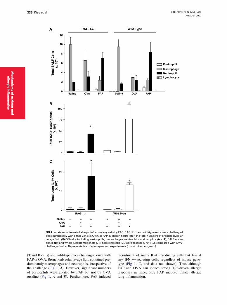
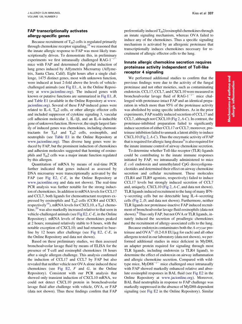
FIG 1. Innate recruitment of allergic inflammatory cells by FAP. RAG-12/2 and wild-type mice were challenged
once intranasally with either vehicle, OVA, or FAP. Eighteen hours later, the total numbers of bronchoalveolar
lavage fluid (BALF) cells, including eosinophils, macrophages, neutrophils, and lymphocytes (A); BALF eosin-
ophils (B); and whole lung homogenate IL-4–secreting cells (C), were assessed. *P < .05 compared with OVA-
challenged mice. Representative of 4 independent experiments (n 5 4 mice per group).
Mech
anism
sofasth
ma
and
alle
rgic
infl
am
matio
n
(T and B cells) and wild-type mice challenged once withFAP or OVA. Bronchoalveolar lavage fluid contained pre-dominantly macrophages and neutrophils, irrespective ofthe challenge (Fig 1, A). However, significant numbersof eosinophils were elicited by FAP but not by OVAorsaline (Fig 1, A and B). Furthermore, FAP induced
recruitment of many IL-42producing cells but few ifany IFN-g2secreting cells, regardless of mouse geno-type (Fig 1, C, and data not shown). Thus althoughFAP and OVA can induce strong TH2-driven allergicresponses in mice, only FAP induced innate allergiclung inflammation.
J ALLERGY CLIN IMMUNOL
VOLUME 120, NUMBER 2
Kiss et al 337
FAP transcriptionally activatesallergy-specific genes
Because recruitment of TH2 cells is regulated primarilythrough chemokine receptor signaling,18 we reasoned thatthe innate allergic response to FAP was most likely tran-scriptionally driven. To demonstrate this, in preliminaryexperiments we first intranasally challenged RAG-12/2
mice with FAP and determined the global induction oflung genes induced by Affymetrix Microarray (Affyme-trix, Santa Clara, Calif). Eight hours after a single chal-lenge, 1475 distinct genes, most with unknown function,were induced at least 2-fold above the levels of vehicle-challenged animals (see Fig E1, A, in the Online Reposi-tory at www.jacionline.org). The induced genes withknown or putative functions are summarized in Fig E1, B,and Table E1 (available in the Online Repository at www.jacionline.org). Several of these FAP-induced genes wererelated to IL-4, TH2 cells, or other allergic effector cellsand included suppressor of cytokine signaling 3, vascularcell adhesion molecular 1, IL-1b, and an IL-4–induciblegene of unknown function. However, the single largest fam-ily of induced genes was chemokines, including chemoat-tractants for TH1 and TH2 cells, eosinophils, andneutrophils (see Table E1 in the Online Repository atwww.jacionline.org). Thus diverse lung genes were in-duced by FAP, but the prominent induction of chemokinesand adhesion molecules suggested that homing of eosino-phils and TH2 cells was a major innate function regulatedby this allergen.
Quantitation of mRNA by means of real-time PCRfurther indicated that genes induced as assessed byDNA microarray were transcriptionally activated by theFAP (see Fig E2, C-E, in the Online Repository atwww.jacionline.org and data not shown). The real-timePCR analysis was further notable for the strong induct-ion of chemokines. In addition to mRNA levels for CCL17and CCL7, both ligands for chemoattractant receptors ex-pressed by eosinophils and TH2 cells (CCR4 and CCR3,respectively19), mRNA levels for CXCL10, a TH1 chemo-kine,20 was also markedly increased relative to that seen invehicle-challenged animals (see Fig E2, C-E, in the OnlineRepository). mRNA levels of these chemokines peakedat 2 hours; remained relatively stable to 8 hours, with thenotable exception of CXCL10; and had returned to base-line by 12 hours after challenge (see Fig E2, C-E, inthe Online Repository and data not shown).
Based on these preliminary studies, we then assessedbronchoalveolar lavage fluid by means of ELISA for thepresence of T-cell and eosinophil chemokines 18 hoursafter a single allergen challenge. This analysis confirmedthe induction of CCL17 and CCL7 by FAP but alsorevealed that neither vehicle nor OVA alone induced thesechemokines (see Fig E2, F and G, in the OnlineRepository). Consistent with our PCR analysis thatshowed only transient induction of CXCL10 mRNA, wecould not detect CXCL10 protein in bronchoalveolarlavage fluid after challenge with vehicle, OVA, or FAP(data not shown). Thus these findings confirm that FAP
preferentially induced TH2/eosinophil chemokines throughan innate signaling mechanism, whereas OVA failed toinduce any of the chemokines. Thus a specific signalingmechanism is activated by an allergenic proteinase thattranscriptionally induces chemokines necessary for re-cruitment of allergic effector cells to the lung.
Innate allergic chemokine secretion requiresproteinase activity independent of Toll-likereceptor 4 signaling
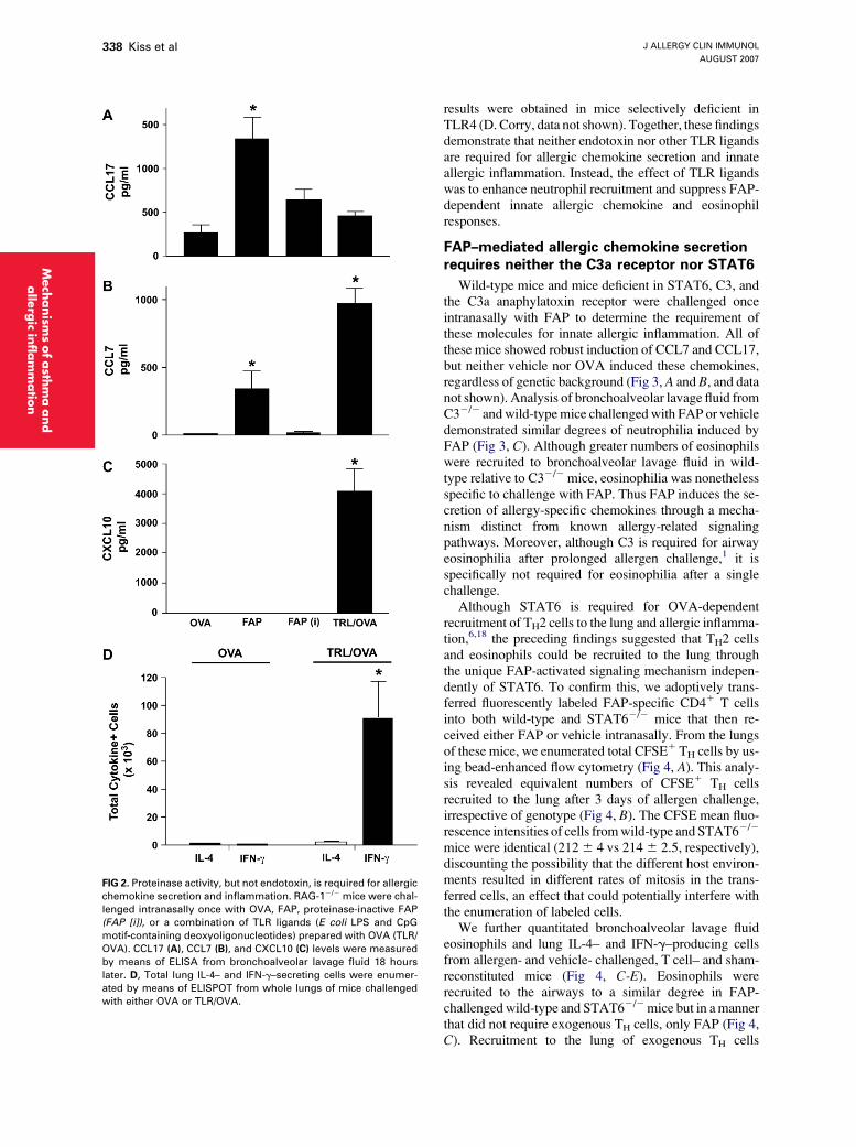
We performed additional studies to confirm that theprevious findings were due to the activity of the fungalproteinase and not other moieties, such as contaminatingendotoxin. CCL17, CCL7, and CXCL10 were measured inbronchoalveolar lavage fluid of RAG-12/2 mice chal-lenged with proteinase-intact FAP and an identical prepa-ration in which more than 95% of the proteinase activitywas abolished by using specific inhibitors. As in the priorexperiments, FAP readily induced secretion of CCL17 andCCL7, although not CXCL10 (Fig 2, A-C). In contrast, theproteinase-inhibited preparation failed to significantlyinduce secretion of either CCL17 or CCL7; moreover, pro-teinase inhibition failed to unmask a latent ability to induceCXCL10 (Fig 2, A-C). Thus the enzymatic activity of FAPthat is required for allergic lung disease8 is also required forthe innate immune control of airway chemokine secretion.
To determine whether Toll-like receptor (TLR) ligandscould be contributing to the innate immune responsesinitiated by FAP, we intranasally administered to miceE coli endotoxin and unmethylated CpG deoxyoligonu-cleotides and determined their effect on airway chemokinesecretion and cellular recruitment. These molecules(TLR4 and TLR9 agonists, respectively) failed to induceCCL17 levels but strongly induced secretion of CCL7and, uniquely, CXCL10 (Fig 2, A-C, and data not shown).TLR ligands induced recruitment to the lung of many IFN-g–secreting cells but no detectable lung IL-4–secretingcells (Fig 2, D, and data not shown). Furthermore, neitherTLR ligands nor proteinase-inactive FAP induced recruit-ment of bronchoalveolar lavage fluid eosinophils (data notshown).8 Thus only FAP, but not OVA or TLR ligands, in-nately induced the secretion of proallergic chemokinesand the recruitment of allergy-associated cells to the lung.
Because endotoxin contaminates both the A oryzae pro-teinase and OVA21 (0.2-0.8 EU/mg for each) and all otherallergens tested in our laboratory (data not shown), we per-formed additional studies in mice deficient in MyD88,an adapter protein required for signaling through mostTLR ligands, including endotoxin (a TLR4 ligand), todetermine the effect of endotoxin on airway inflammationand allergic chemokine secretion. Compared with wild-type mice, MyD882/2 mice challenged once intranasallywith FAP showed markedly enhanced relative and abso-lute eosinophil responses in BAL fluid (see Fig E2 in theOnline Repository at www.jacionline.org). Moreover,BAL fluid neutrophila in response to FAP challenge wasmarkedly suppressed in the absence of MyD88-dependentsignaling (see Fig E2 in the Online Repository). Similar
Mech
anis
ms
ofast
hm
aand
allerg
icin
flam
mation
J ALLERGY CLIN IMMUNOL
AUGUST 2007
338 Kiss et al
FIG 2. Proteinase activity, but not endotoxin, is required for allergic
chemokine secretion and inflammation. RAG-12/2 mice were chal-
lenged intranasally once with OVA, FAP, proteinase-inactive FAP
(FAP [i]), or a combination of TLR ligands (E coli LPS and CpG
motif-containing deoxyoligonucleotides) prepared with OVA (TLR/
OVA). CCL17 (A), CCL7 (B), and CXCL10 (C) levels were measured
by means of ELISA from bronchoalveolar lavage fluid 18 hours
later. D, Total lung IL-4– and IFN-g–secreting cells were enumer-
ated by means of ELISPOT from whole lungs of mice challenged
with either OVA or TLR/OVA.
Mech
anism
sofasth
ma
and
alle
rgic
infl
am
matio
n
results were obtained in mice selectively deficient inTLR4 (D. Corry, data not shown). Together, these findingsdemonstrate that neither endotoxin nor other TLR ligandsare required for allergic chemokine secretion and innateallergic inflammation. Instead, the effect of TLR ligandswas to enhance neutrophil recruitment and suppress FAP-dependent innate allergic chemokine and eosinophilresponses.
FAP–mediated allergic chemokine secretionrequires neither the C3a receptor nor STAT6
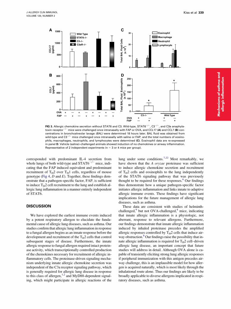
Wild-type mice and mice deficient in STAT6, C3, andthe C3a anaphylatoxin receptor were challenged onceintranasally with FAP to determine the requirement ofthese molecules for innate allergic inflammation. All ofthese mice showed robust induction of CCL7 and CCL17,but neither vehicle nor OVA induced these chemokines,regardless of genetic background (Fig 3, A and B, and datanot shown). Analysis of bronchoalveolar lavage fluid fromC32/2 and wild-type mice challenged with FAP or vehicledemonstrated similar degrees of neutrophilia induced byFAP (Fig 3, C). Although greater numbers of eosinophilswere recruited to bronchoalveolar lavage fluid in wild-type relative to C32/2 mice, eosinophilia was nonethelessspecific to challenge with FAP. Thus FAP induces the se-cretion of allergy-specific chemokines through a mecha-nism distinct from known allergy-related signalingpathways. Moreover, although C3 is required for airwayeosinophilia after prolonged allergen challenge,1 it isspecifically not required for eosinophilia after a singlechallenge.
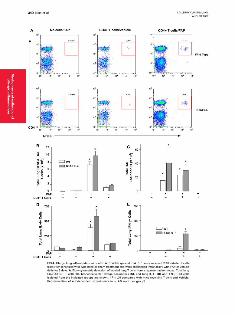
Although STAT6 is required for OVA-dependentrecruitment of TH2 cells to the lung and allergic inflamma-tion,6,18 the preceding findings suggested that TH2 cellsand eosinophils could be recruited to the lung throughthe unique FAP-activated signaling mechanism indepen-dently of STAT6. To confirm this, we adoptively trans-ferred fluorescently labeled FAP-specific CD41 T cellsinto both wild-type and STAT62/2 mice that then re-ceived either FAP or vehicle intranasally. From the lungsof these mice, we enumerated total CFSE1 TH cells by us-ing bead-enhanced flow cytometry (Fig 4, A). This analy-sis revealed equivalent numbers of CFSE1 TH cellsrecruited to the lung after 3 days of allergen challenge,irrespective of genotype (Fig 4, B). The CFSE mean fluo-rescence intensities of cells from wild-type and STAT62/2
mice were identical (212 6 4 vs 214 6 2.5, respectively),discounting the possibility that the different host environ-ments resulted in different rates of mitosis in the trans-ferred cells, an effect that could potentially interfere withthe enumeration of labeled cells.
We further quantitated bronchoalveolar lavage fluideosinophils and lung IL-4– and IFN-g–producing cellsfrom allergen- and vehicle- challenged, T cell– and sham-reconstituted mice (Fig 4, C-E). Eosinophils wererecruited to the airways to a similar degree in FAP-challenged wild-type and STAT62/2 mice but in a mannerthat did not require exogenous TH cells, only FAP (Fig 4,C). Recruitment to the lung of exogenous TH cells
J ALLERGY CLIN IMMUNOL
VOLUME 120, NUMBER 2
Kiss et al 339
FIG 3. Allergic chemokine secretion without STAT6 and C3. Wild-type, STAT62/2, C32/2, and C3a anaphyla-
toxin receptor2/2 mice were challenged once intranasally with FAP or OVA, and CCL17 (A) and CCL7 (B) con-
centrations in bronchoalveolar lavage (BAL) were determined 18 hours later. BAL fluid was obtained from
wild-type and C32/2 mice challenged once intranasally with saline or FAP, and the total numbers of eosino-
phils, macrophages, neutrophils, and lymphocytes were determined (C). Eosinophil data are re-expressed
in panel D. Vehicle (saline)–challenged animals showed induction of no chemokines or airway inflammation.
Representative of 2 independent experiments (n 5 3 or 4 mice per group).
Mech
anis
ms
ofast
hm
aand
allerg
icin
flam
mation
corresponded with predominant IL-4 secretion fromwhole lungs of both wild-type and STAT62/2 mice, indi-cating that the FAP induced equivalent and predominantrecruitment of TH2 over TH1 cells, regardless of mousegenotype (Fig 4, D and E). Together, these findings dem-onstrate that a pathogen-specific factor, FAP, is sufficientto induce TH2 cell recruitment to the lung and establish al-lergic lung inflammation in a manner entirely independentof STAT6.
DISCUSSION
We have explored the earliest immune events inducedby a potent respiratory allergen to elucidate the funda-mental cause of allergic lung diseases, such as asthma. Ourstudies confirm that allergic lung inflammation in responseto a fungal allergen begins as an innate response before thedevelopment and recruitment of the TH2 cells that controlsubsequent stages of disease. Furthermore, the innateallergic response to fungal allergen required intact protein-ase activity, which transcriptionally controlled productionof the chemokines necessary for recruitment of allergic in-flammatory cells. The proteinase-driven signaling mecha-nism underlying innate allergic chemokine secretion wasindependent of the C3a receptor signaling pathway, whichis generally required for allergic lung disease in responseto this class of allergen,1,2 and MyD88-dependent signal-ing, which might participate in allergic reactions of the
lung under some conditions.7,22 Most remarkably, wehave shown that the A oryzae proteinase was sufficientto induce allergic chemokine secretion and recruitmentof TH2 cells and eosinophils to the lung independentlyof the STAT6 signaling pathway that was previouslythought to be required for these responses.6 Our findingsthus demonstrate how a unique pathogen-specific factorinitiates allergic inflammation and links innate to adaptiveallergic immune events. These findings have significantimplications for the future management of allergic lungdiseases, such as asthma.
These data are consistent with studies of helminth-challenged,9 but not OVA-challenged,8 mice, indicatingthat innate allergic inflammation is a physiologic, notaberrant, response to relevant allergens. Furthermore,our findings demonstrate that innate allergic inflammationinduced by inhaled proteinase precedes the amplifiedallergic responses controlled by TH2 cells that induce air-way obstruction.8 Our findings raise the possibility that in-nate allergic inflammation is required for TH2 cell–drivenallergic lung disease, an important concept that futurestudies will address in detail. Although OVA alone is ca-pable of transiently eliciting strong lung allergic responsesif peripheral immunization with this antigen precedes air-way challenge, this is an implausible model for how aller-gen is acquired naturally, which is most likely through theinhalational route alone. Thus our findings are likely to bebroadly applicable to diverse allergens implicated in respi-ratory diseases, such as asthma.
J ALLERGY CLIN IMMUNOL
AUGUST 2007
340 Kiss et al
FIG 4. Allergic lung inflammation without STAT6. Wild-type and STAT62/2 mice received CFSE-labeled T cells
from FAP-sensitized wild-type mice or sham treatment and were challenged intranasally with FAP or vehicle
daily for 3 days. A, Flow cytometric detection of labeled lung T cells from a representative mouse. Total lung
CD41/CFSE1 T cells (B), bronchoalveolar lavage eosinophils (C), and lung IL-41 (D) and IFN-g1 (E) cells
isolated from the indicated groups are shown. *P < .05 compared with mice receiving T cells and vehicle.
Representative of 4 independent experiments (n 5 4-5 mice per group).
Mech
anism
sofasth
ma
and
alle
rgic
infl
am
matio
n
J ALLERGY CLIN IMMUNOL
VOLUME 120, NUMBER 2
Kiss et al 341
Our study indicates that 2 mechanisms for the recruit-ment of allergic inflammatory cells exist: a proteinase-activated, STAT6-independent pathway that initiatesallergic inflammation innately, as shown here, and asubsequent STAT6-dependent pathway that serves tosustain the TH2-driven amplification of allergic disease.6
We propose that under physiologic conditions, allergiclung inflammation is the result of both signaling pathwaysoperating in parallel, whereas only the STAT6 pathwaycontrols allergic disease induced in the absence of adju-vant (ie, with OVA alone used as the allergen). TH2differentiation, allergic lung inflammation, and airwayhyperresponsiveness all fail to develop in response toFAP in the absence of STAT6 (D. Corry and S. Susarla,manuscript in preparation), revealing that the requirementof STAT6 for allergic lung responses in wild-type mice, asestablished through OVA, cannot be escaped, regardlessof the allergen used. Thus FAP and OVA differ primarilywith respect to the innate immune pathways that they ac-tivate and that control the initial recruitment of allergiceffector cells that might be critical for subsequent allergicinflammation.
Unexpectedly, TLR ligands, such as endotoxin, arealso capable of inducing allergic airway responses inmice under some conditions,7 suggesting that endotoxincontaminating our fungal allergens could be responsiblefor the allergic inflammation that we have observed.However, proteinase inactivation of the FAP, which abro-gates its allergenic activity,8 should not affect TLR4(endotoxin) signaling. Nonetheless, proteinase-inactiveFAP failed to induce chemokines in the airway, especiallyCCL7 and CXCL10, that are strongly induced by TLRligands alone (Fig 2) or allergic lung inflammation.8
Furthermore, our studies are similar to many others thatshow that TLR signaling predominantly controls IFN-g–related responses and antagonizes allergic inflammation(Fig 2).23-25
Additionally, FAP not only induced allergic lunginflammation and chemokine secretion, these responseswere actually enhanced in the absence of a functionalsignaling pathway for endotoxin and other TLR ligands(see Fig E2, A and B, in the Online Repository at www.jacionline.org).21 Thus although endotoxin is capable ofinducing allergic lung inflammation under some condi-tions,7,26 our findings demonstrate that endotoxin contam-inating FAP is not required for innate allergic responsesinduced by a fungal proteinase.
Despite these findings, it remains widely perceived thatallergic lung inflammation is a default (ie, nonadjuvantdirected) immune response to inhaled antigen.22,23 However,the typical respiratory immune response to antigens devoidof adjuvant factors is not allergic but tolerogenic, in whichallergic lung inflammation and antigen-specific IgE andTH2 responses are suppressed.8,27,28 We have shownhere that to bypass the tolerogenic response to inhaledantigen and achieve robust TH2 responses and allergicinflammation, the respiratory tract must be specificallyinstructed. Thus rather than a default response, allergiclung inflammation caused by inhaled allergen is the result
of microbial-derived adjuvant factors, especially protein-ases, acting through an innate signaling mechanism thatspecifically induces recruitment of allergic effector cells.
Our findings indicate the existence of proteinase-activated innate immune receptors in the mouse airwaythat are crucial for the initiation of allergic inflammation.Proteinase-activated receptor 2 is an endogenously acti-vated receptor implicated in the regulation of airwaycaliber,29 but mice deficient in proteinase-activated recep-tor 2 experience airway inflammation similar to that ofwild-type mice after airway challenge with proteinase-active allergens (D. Corry, unpublished data). Further-more, although prior studies emphasized the cleavage ormanipulation by Der p 1 of molecules involved in adaptiveimmunity (IL-2Ra chain [CD25],30 low-affinity IgEreceptor [CD23],31 and IL-4 and IFN-g32), the currentstudy reveals that FAP acts within the innate immunecompartment, in which such molecules are less likely toplay critical roles. Therefore it is likely that other mole-cules sensitive to exogenous proteinases are responsiblefor initiating allergic inflammation, as shown here. Identi-fication of these and related genes will be important to fur-ther dissecting the innate control of allergic inflammationand in defining new opportunities for the prevention andtreatment of diseases, such as allergic asthma.
We thank A. Clinton White for helpful commentary and S. Akira
and D. Golenbock for MyD882/2 mice.
REFERENCES
1. Drouin SM, Corry DB, Kildsgaard J, Wetsel RA. Cutting edge: the
absence of C3 demonstrates a role for complement in Th2 effector
functions in a murine model of pulmonary allergy. J Immunol 2001;
167:4141-5.
2. Drouin SM, Corry DB, Hollman TJ, Kildsgaard J, Wetsel RA. Absence
of the complement anaphylatoxin C3a receptor suppresses Th2 effector
functions in a murine model of pulmonary allergy. J Immunol 2002;
169:5926-33.
3. Corry DB, Grunig G, Hadeiba H, Kurup VP, Warnock ML, Sheppard D,
et al. Requirements for allergen-induced airway hyperreactivity in T and
B cell-deficient mice. Mol Med 1998;4:344-55.
4. Kaplan MH, Schindler U, Smiley ST, Grusby MJ. Stat6 is required for
mediating responses to IL-4 and for development of Th2 cells. Immunity
1996;4:313-9.
5. Kuperman D, Schofield B, Wills-Karp M, Grusby MJ. Signal transducer
and activator of transcription factor 6 (Stat6)-deficient mice are protected
from antigen-induced airway hyperresponsiveness and mucus produc-
tion. J Exp Med 1998;187:939-48.
6. Mathew A, MacLean JA, DeHaan E, Tager AM, Green FH, Luster AD.
Signal transducer and activator of transcription 6 controls chemokine
production and T helper cell type 2 cell trafficking in allergic pulmonary
inflammation. J Exp Med 2001;193:1087-96.
7. Eisenbarth SC, Piggott DA, Huleatt JW, Visintin I, Herrick CA, Bot-
tomly K. Lipopolysaccharide-enhanced, toll-like receptor 4-dependent
T helper cell type 2 responses to inhaled antigen. J Exp Med 2002;196:
1645-51.
8. Kheradmand F, Kiss A, Xu J, Lee SH, Kolattukudy PE, Corry DB. A
protease-activated pathway underlying Th cell type 2 activation and
allergic lung disease. J Immunol 2002;169:5904-11.
9. Voehringer D, Shinkai K, Locksley RM. Type 2 immunity reflects
orchestrated recruitment of cells committed to IL-4 production. Immu-
nity 2004;20:267-77.
10. Lim KC, Sun E, Bahgat M, Bucks D, Guy R, Hinz RS, et al. Blockage
of skin invasion by Schistosome cercariae by serine protease inhibitors.
Am J Trop Med Hyg 1999;60:487-92.
Mech
anis
ms
ofast
hm
aand
allerg
icin
flam
mation
J ALLERGY CLIN IMMUNOL
AUGUST 2007
342 Kiss et al
11. Adachi O, Kawai T, Takeda K, Matsumoto M, Tsutsui H, Sakagami M,
et al. Targeted disruption of the MyD88 gene results in loss of IL-1- and
IL-18-mediated function. Immunity 1998;9:143-50.
12. Kildsgaard J, Hollmann TJ, Matthews KW, Bian K, Murad F, Wetsel
RA. Cutting edge: targeted disruption of the C3a receptor gene demon-
strates a novel protective anti-inflammatory role for C3a in endotoxin-
shock. J Immunol 2000;165:5406-9.
13. Circolo A, Garnier G, Fukuda W, Wang X, Hidvegi T, Szalai AJ, et al.
Genetic disruption of the murine complement C3 promoter region gener-
ates deficient mice with extrahepatic expression of C3 mRNA. Immuno-
pharmacology 1999;42:135-49.
14. Corry DB, Folkesson HG, Warnock ML, Erle DJ, Matthay MA, Wiener-
Kronish JP, et al. Interleukin 4, but not interleukin 5 or eosinophils, is
required in a murine model of acute airway hyperreactivity. J Exp Med
1996;183:109-17.
15. Xu J, Park PW, Kheradmand F, Corry DB. Endogenous attenuation
of allergic lung inflammation by syndecan-1. J Immunol 2005;174:
5758-65.
16. Lee S-H, Prince JE, Rais M, Kheradmand F, Shardonofsky F, Lu H, et al.
Differential requirement for CD18 in T helper effector homing. Nat Med
2003;9:1281-6.
17. Montes M, Jaensson EA, Orozco AF, Lewis DE, Corry DB. A general
method for bead-enhanced quantitation by flow cytometry. J Immunol
Methods 2006;317:45-55.
18. Mathew A, Medoff BD, Carafone AD, Luster AD. Cutting edge: Th2 cell
trafficking into the allergic lung is dependent on chemoattractant receptor
signaling. J Immunol 2002;169:651-5.
19. Cosmi L, Annunziato F, Maggi E, Romagnani S, Manetti R. Chemoat-
tractant receptors expressed on type 2 T cells and their role in disease.
Int Arch Allergy Immunol 2001;125:273-9.
20. Yamamoto J, Adachi Y, Onoue Y, Adachi YS, Okabe Y, Itazawa T, et al.
Differential expression of the chemokine receptors by the Th1- and Th2-
type effector populations within circulating CD41 T cells. J Leukoc Biol
2000;68:568-74.
21. Watanabe J, Miyazaki Y, Zimmerman GA, Albertine KH, McIntyre TM.
Endotoxin contamination of ovalbumin suppresses murine immunologic
responses and development of airway hyper-reactivity. J Biol Chem
2003;278:42361-8.
Mech
anism
sofasth
ma
and
alle
rgic
infl
am
matio
n
22. Piggott DA, Eisenbarth SC, Xu L, Constant SL, Huleatt JW, Herrick CA,
et al. MyD88-dependent induction of allergic Th2 responses to intranasal
antigen. J Clin Invest 2005;115:459-67.
23. Schnare M, Barton GM, Holt AC, Takeda K, Akira S, Medzhitov R.
Toll-like receptors control activation of adaptive immune responses.
Nat Immunol 2001;2:947-50.
24. Scanga CA, Aliberti J, Jankovic D, Tilloy F, Bennouna S, Denkers EY,
et al. Cutting edge: MyD88 is required for resistance to Toxoplasma
gondii infection and regulates parasite-induced IL-12 production by
dendritic cells. J Immunol 2002;168:5997-6001.
25. Velasco G, Campo M, Manrique OJ, Bellou A, He H, Arestides RSS,
et al. Toll-like receptor 4 or 2 agonists decrease allergic inflammation.
Am J Respir Cell Mol Biol 2005;32:218-24.
26. Redecke V, Hacker H, Datta SK, Fermin A, Pitha PM, Broide DH, et al.
Cutting edge: activation of Toll-like receptor 2 induces a Th2 immune
response and promotes experimental asthma. J Immunol 2004;172:
2739-43.
27. Tsitoura DC, DeKruyff RH, Lamb JR, Umetsu DT. Intranasal exposure
to protein antigen induces immunological tolerance mediated by func-
tionally disabled CD41 T cells. J Immunol 1999;163:2592-600.
28. Seymour BW, Gershwin LJ, Coffman RL. Aerosol-induced immuno-
globulin (Ig)-E unresponsiveness to ovalbumin does not require CD81
or T cell receptor (TCR)-gamma/delta1 T cells or interferon (IFN)-
gamma in a murine model of allergen sensitization. J Exp Med 1998;
187:721-31.
29. Cocks TM, Fong B, Chow JM, Anderson GP, Frauman AG, Goldie RG,
et al. A protective role for protease-activated receptors in the airways.
Nature 1999;398:156-60.
30. Schulz O, Sewell HF, Shakib F. Proteolytic cleavage of CD25, the alpha
subunit of the human T cell interleukin 2 receptor, by Der p 1, a major
mite allergen with cysteine protease activity. J Exp Med 1998;187:271-5.
31. Hewitt CR, Brown AP, Hart BJ, Pritchard DI. A major house dust mite
allergen disrupts the immunoglobulin E network by selectively cleaving
CD23: innate protection by antiproteases. J Exp Med 1995;182:1537-44.
32. Comoy EE, Pestel J, Duez C, Stewart GA, Vendeville C, Fournier C,
et al. The house dust mite allergen, Dermatophagoides pteronyssinus,
promotes type 2 responses by modulating the balance between IL-4
and IFN-gamma. J Immunol 1998;160:2456-62.
Related Documents


![Research Paper Deguelin Attenuates Allergic Airway ...Asthma is a chronic respiratory disease characterized by airway inflammation and remodeling, ... pathophysiology of asthma [4].](https://static.cupdf.com/doc/110x72/6021eed39e87047b88365ced/research-paper-deguelin-attenuates-allergic-airway-asthma-is-a-chronic-respiratory.jpg)