• WW0171MWW01771M292

The Cellular Basis of Life
• Historical contributions
• Modern Cell Theory
• Types of Microscopes
• Micrographs
•Cell membranes
• Membrane transport
• Nucleus
•Organelles

Contributors to Our Knowledge of Cells
Anton
von LeeuwenhoekAssembled the first microscope
Robert Hooke Observed and named “cells”
Robert Brown Discovered the nucleus.
Schleiden and Schwann
Determined that all plants and animals are made of cells.
Rudolf Virchow Cells come only from pre-existing cells.
Modern Cell Theory
1. All living things are made of cells.2. Cells are the basic structural unit (ie,
building blocks) of life (plant, animal, bacterial, etc.)
3. All cells come from other (pre-existing) cells.
4. The way a cell is made is determined by its function (ie, what it has to do) = principle of complementarity
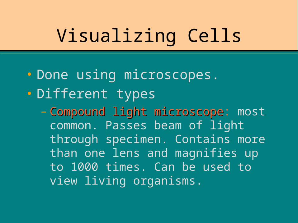
Visualizing Cells
• Done using microscopes.
• Different types– Compound light microscopeCompound light microscope: most common.
Passes beam of light through specimen. Contains more than one lens and magnifies up to 1000 times. Can be used to view living organisms.

EYEPIECE
OCULAR LENS (inside)
REVOLVING NOSEPIECE
COARSE ADJUSTMENT KNOB
FINE ADJUSTMENT KNOB
STAGE
DIAPHRAGM
LIGHT SOURCE

“Compound” lenses…
• COMPOUND MAGNIFICATION!!
• Which combination would show the largest AREA?• Which combination would you use to examine details?
Eyepiece LensObjective
LensTotal
Magnification
15x 15x
10x 10x
20x 40x
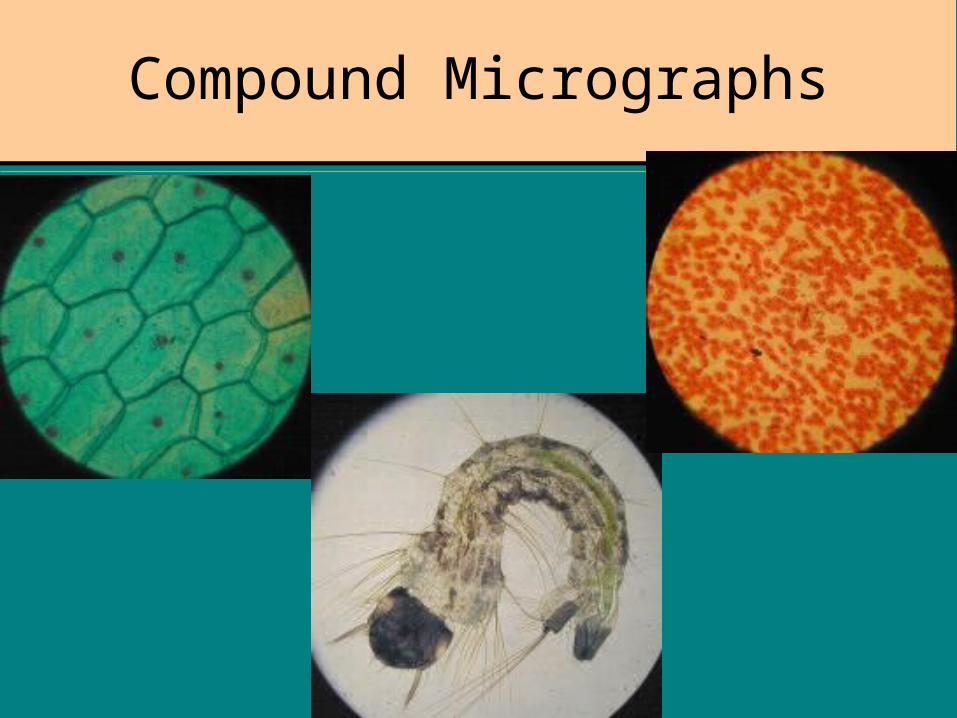
Compound Micrographs

Electron Microscopes
• TRANSMITTING = TEM• Specimen is thinly sliced.• Electrons pass through and
image forms on fluorescent screen.
• SCANNING = SEM• Specimen is coated
with metal (Os,Pb,Au)• Electrons bounce off
surface to form image.
Concentrates a beam of electrons within a vacuum; magnifies up to 1,000,000 times. Used to view much smaller organisms.

Sample Electron Micrographs
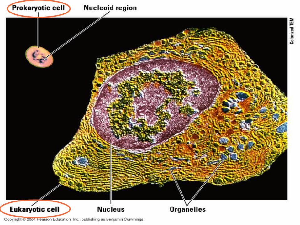
Types of Cells
• PROKARYOTE• Smaller, more
primitive• Bacteria• Fewer organelles• No nucleus; instead
has nucleoid region
• EUKARYOTE• Larger (~10 x)• Complex inner
membrane system• More organelles• Contains a true
nucleus

*Nucleus
*Endomembrane system
*Mitochondria
*Chloroplasts
Key features of Eukaryotic Cells:

What molecule is this?
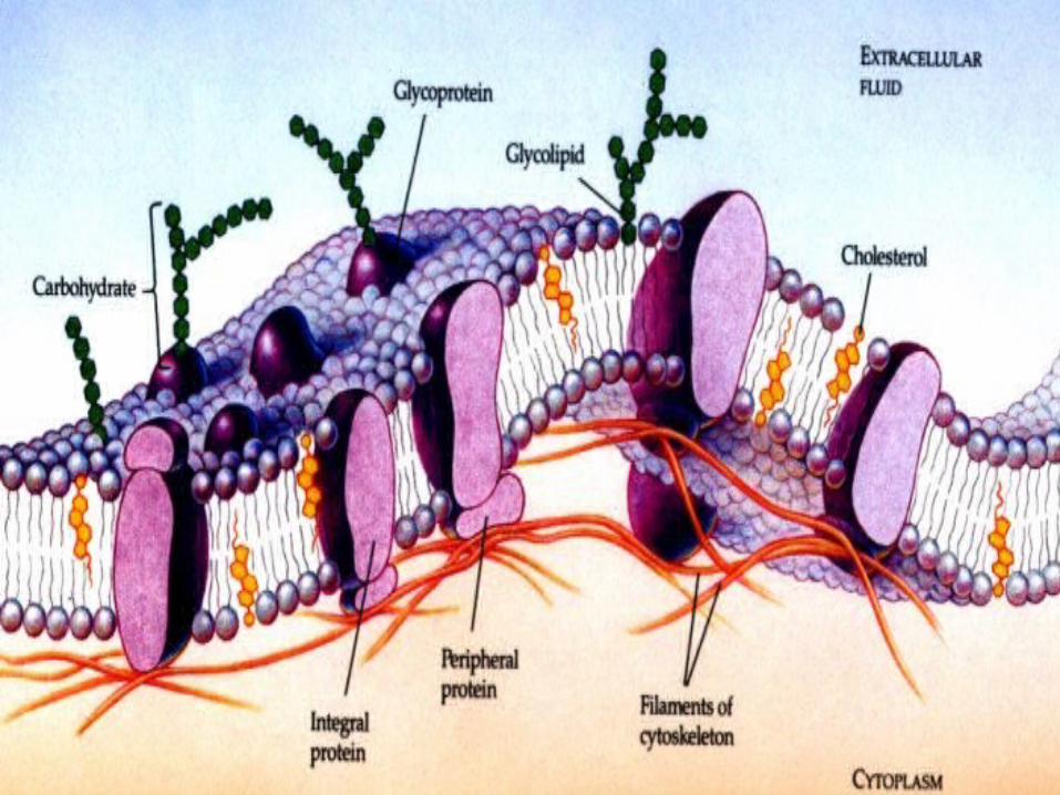
The Fluid Mosaic Model of theCell Membrane
.
AMPHIPATHIC:having both hydrophilic and hydrophobic areas


Cell Wall
• Found outside the cell membrane in PLANTS, FUNGI, and BACTERIA.
• Plants use pressure against the cell wall (called turgor pressure) to help support it.

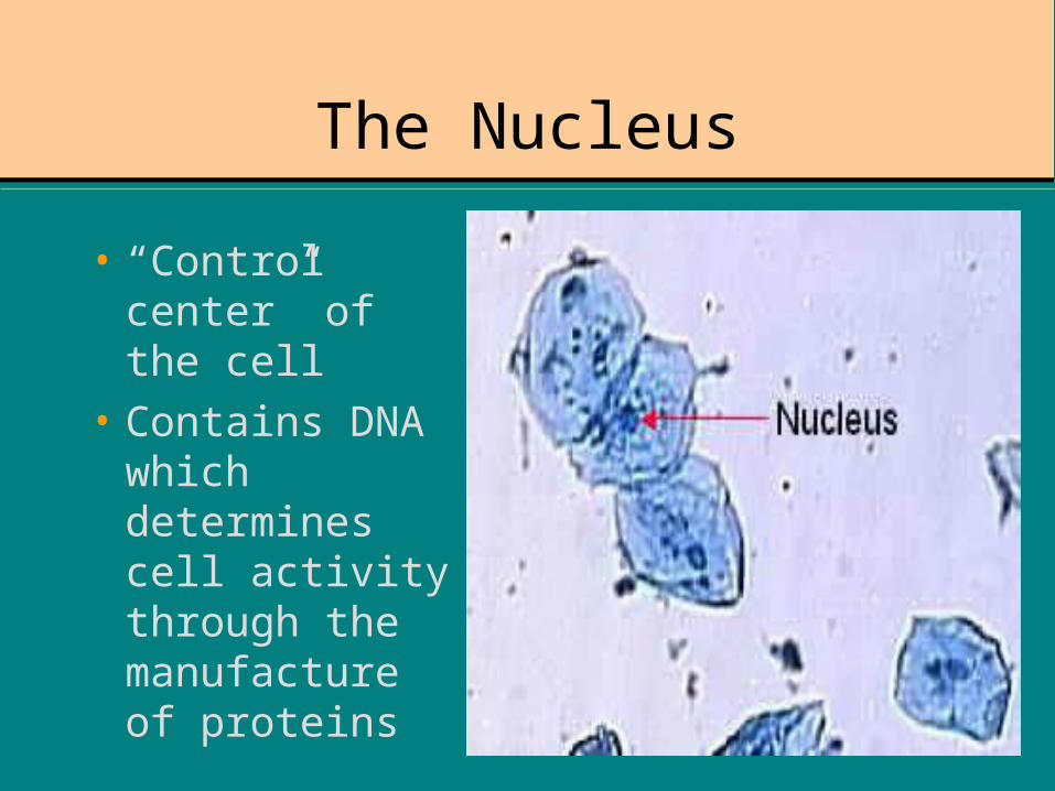
The Nucleus
• “Control center” of the cell
• Contains DNA which determines cell activity through the manufacture of proteins

The rest of the cell …
• Cytoplasm - semi-liquid material which fills the space between the membrane and the nucleus.
• Contains structural fibers called microfilaments.
• Contains “little organs” (organelles) which each have a specific job to do.
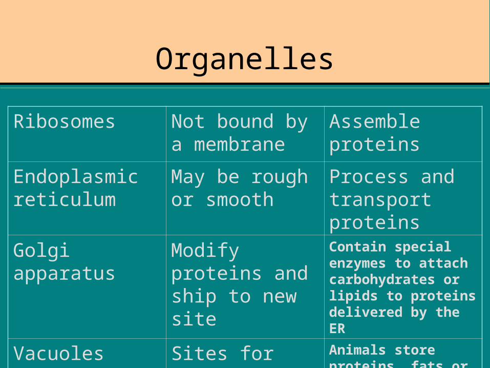
Organelles
Ribosomes Not bound by a membrane
Assemble proteins
Endoplasmic reticulum
May be rough or smooth
Process and transport proteins
Golgi apparatus Modify proteins and ship to new site
Contain special enzymes to attach carbohydrates or lipids to proteins delivered by the ER
Vacuoles Sites for storage of materials
Animals store proteins, fats or carbohydrates; plants store water or salts.

Organelles, continuedLysosomes Sacs filled with chemicals
and enzymes.Attach target, release enzymes which recycles components.
Mitochondria Powerhouse of the cell; double membrane allows maximum surface area.
Produces energy (ATP) for cellular activities using a carbohydrate source.
Nucleolus Dark area found inside the nucleus of eukaryotic cells.
Produces ribosomes.
Plastids Storage sacs. Contain starch, pigments,
Chloroplasts Contain chlorophyll Important in photosynthesis. Found only in plants, algae.

Cell Permeability

Membrane Transport Mechanisms
• PASSIVE TRANSPORT• Powered by the concentration
gradient• Must move down the gradient
from [high] to [low]• Examples: diffusion• At equilibrium, concentration is
equal on both sides.• Osmosis = diffusion of water• Facilitated diffusion = uses
carrier molecule (ex: membrane protein)
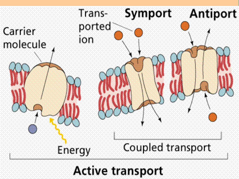
• ACTIVE TRANSPORT• Requires the use of ATP• Can move up the gradient• Examples: ion pumping• Bulk transport
– EXOcytosis moves material OUT of the cell
– ENDOcytosis moves material INTO the cell
» Water in = pinocytosis
» Food in = phagocytosis
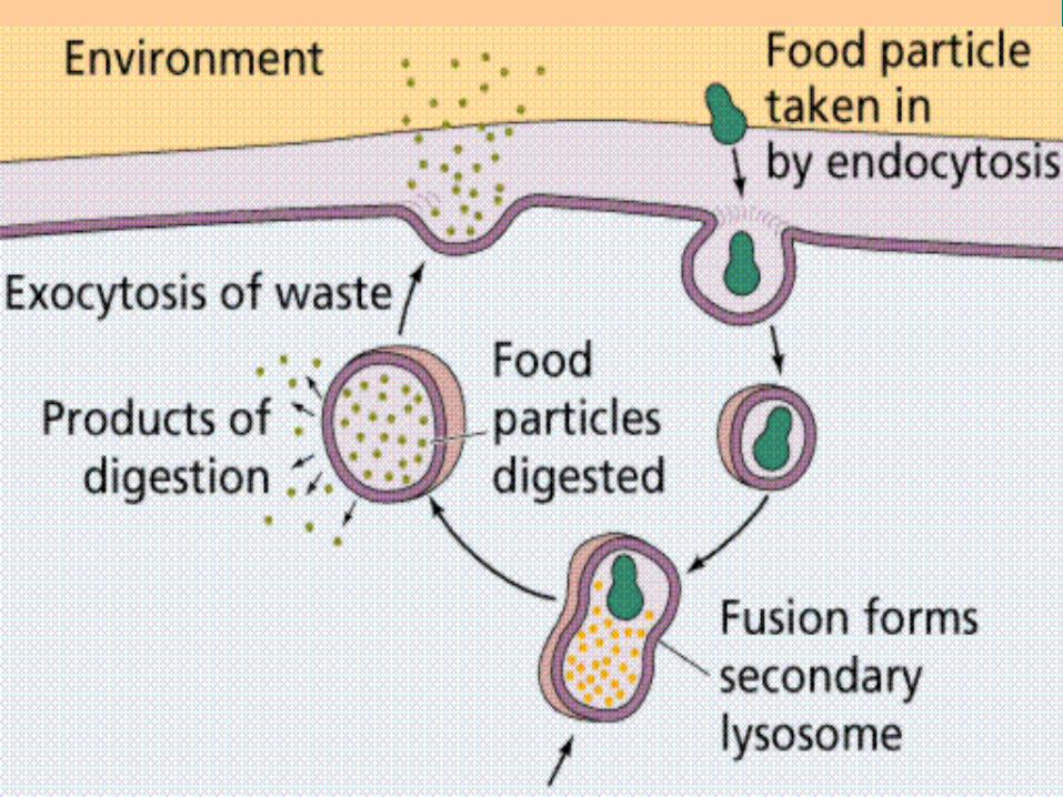
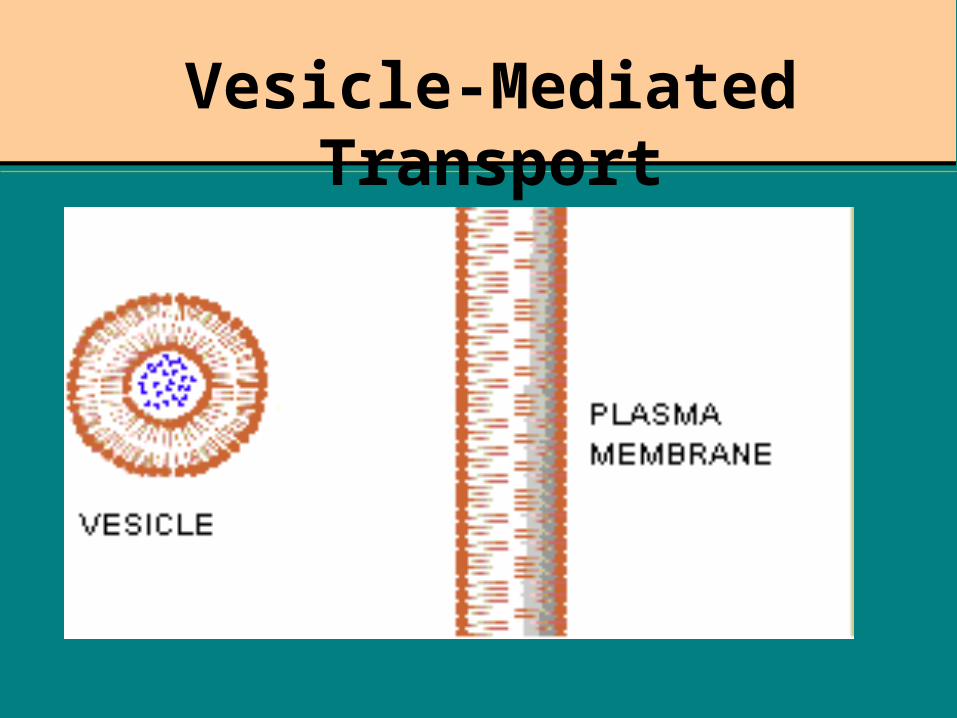
Vesicle-Mediated Transport
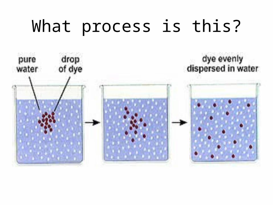
What process is this?

Tonicity
Solution is HYPOTONIC
Solution is HYPERTONIC

Complexity of Organisms
• Single-celled organisms are unicellular.• Other organisms are multicellular.• In order to maintain complexity, there must
be a hierarchy of structure.• Cells form tissues which form organs;• Organs are arranged into organ systems
which all combine to form the organism.