ORIGINAL RESEARCHpublished: 10 November 2015doi: 10.3389/fphar.2015.00260
Frontiers in Pharmacology | www.frontiersin.org 1 November 2015 | Volume 6 | Article 260
Edited by:
Sara Eyal,
Hebrew University of Jerusalem, Israel
Reviewed by:
Julio Benitez,
University of Extremadura, Spain
Constantin Ion Mircioiu,
“Carol Davila” University of Medicine &
Pharmacy, Romania
*Correspondence:
Zheng Jiao
Yiming Li
Qinghua Wang
†Present Address:
Junfeng Li,
Department of Endocrinology, Renmin
Hospital of Wuhan University, Wuhan,
China
‡These authors have contributed
equally to this work.
Specialty section:
This article was submitted to
Drug Metabolism and Transport,
a section of the journal
Frontiers in Pharmacology
Received: 03 September 2015
Accepted: 20 October 2015
Published: 10 November 2015
Citation:
Li J, Zhang Z, Liu X, Wang Y, Mao F,
Mao J, Lu X, Jiang D, Wan Y, Lv J-Y,
Cao G, Zhang J, Zhao N, Atkinson M,
Greiner DL, Prud’homme GJ, Jiao Z,
Li Y and Wang Q (2015) Study of
GABA in Healthy Volunteers:
Pharmacokinetics and
Pharmacodynamics.
Front. Pharmacol. 6:260.
doi: 10.3389/fphar.2015.00260
Study of GABA in Healthy Volunteers:Pharmacokinetics andPharmacodynamicsJunfeng Li 1 †‡, Zhaoyun Zhang 1‡, Xiaoxia Liu 1, Yi Wang 1, Fei Mao 1, Junjun Mao 2,
Xiaolan Lu 1, Dongdong Jiang 1, Yun Wan 1, Jia-Ying Lv 3, Guoying Cao 4, Jing Zhang 4,
Naiqing Zhao 3, Mark Atkinson 5, Dale L. Greiner 6, Gerald J. Prud’homme 7, Zheng Jiao 2*,
Yiming Li 1* and Qinghua Wang 1, 8, 9*
1Department of Endocrinology and Metabolism, Huashan Hospital, Fudan University, Shanghai, China, 2Department of
Pharmacy, Huashan Hospital, Fudan University, Shanghai, China, 3Department of Biostatistics, School of Public Health,
Fudan University, Shanghai, China, 4 Key Laboratory of Clinical Pharmacology of Antibiotics, Ministry of Health, Institute of
Antibiotics, Huashan Hospital, Fudan University, Shanghai, China, 5Department of Pathology, College of Medicine, University
of Florida, Gainesville, FL, USA, 6Department of Molecular Medicine, University of Massachusetts Medical School,
Worcester, MA, USA, 7Department of Laboratory Medicine and Pathobiology, Keenan Research Centre for Biomedical
Science of St. Michael’s Hospital, University of Toronto, Toronto, ON, Canada, 8Division of Endocrinology and Metabolism,
The Keenan Research Centre in the Li Ka Shing Knowledge Institute, St. Michael’s Hospital, Toronto, ON, Canada,9Department of Physiology and Medicine, University of Toronto, ON, Canada
Preclinical studies show that GABA exerts anti-diabetic effects in rodent models of
type 1 diabetes. Because little is known about its absorption and effects in humans,
we investigated the pharmacokinetics and pharmacodynamics of GABA in healthy
volunteers. Twelve subjects were subjected to an open-labeled, three-period trial
involving sequential oral administration of placebo, 2 g GABA once, and 2 g GABA three
times/day for 7 days, with a 7-day washout between each period. GABA was rapidly
absorbed (Tmax: 0.5∼ 1 h) with the half-life (t1/2) of 5 h. No accumulation was observed
after repeated oral GABA administration for 7 days. Remarkably, GABA significantly
increased circulating insulin levels in the subjects under either fasting (1.6-fold, single
dose; 2.0-fold, repeated dose; p < 0.01) or fed conditions (1.4-fold, single dose;
1.6-fold, repeated dose; p < 0.01). GABA also increased glucagon levels only under
fasting conditions (1.3-fold, single dose, p < 0.05; 1.5-fold, repeated dose, p < 0.01).
However, there were no significant differences in the insulin-to-glucagon ratio and no
significant change in glucose levels in these healthy subjects during the study period.
Importantly, GABA significantly decreased glycated albumin levels in the repeated dosing
period. Subjects with repeated dosing showed an elevated incidence of minor adverse
events in comparison to placebo or the single dosing period, most notably transient
discomforts such as dizziness and sore throat. However, there were no serious adverse
events observed throughout the study. Our data show that GABA is rapidly absorbed and
tolerated in human beings; its endocrine effects, exemplified by increasing islet hormonal
secretion, suggest potential therapeutic benefits for diabetes.
Keywords: GABA, pharmacokinetics, glucagon, insulin, glycated albumin

Li et al. GABA PK and PD in healthy volunteers
INTRODUCTION
Type 1 diabetes (T1D) is an autoimmune disease, characterizedby progressive loss of functional β-cell mass, resulting frominsulitis, which leads to insulin deficiency, elevation of bloodglucose, and various complications associated with this disease(Atkinson et al., 2014). An ideal effective therapy may requiretwo arms: β-cell regeneration and immunosuppressive effects.However, none of the current therapies effectively achieve bothgoals (Lernmark and Larsson, 2013; Atkinson et al., 2014).
Recently we and others have shown that gamma aminobutyricacid (GABA) exerts β-cell regenerative and immunoregulatoryeffects (Soltani et al., 2011; Prud’homme et al., 2013; Tian et al.,2013; Purwana et al., 2014). Specifically, GABA stimulates β-cell replication, protects β-cells against apoptosis, and attenuatesinsulitis (Soltani et al., 2011; Tian et al., 2013; Prud’homme et al.,2014; Purwana et al., 2014). These effects result in an enhancedfunctional β-cell mass and, in mice, this can reverse disease(Soltani et al., 2011). These favorable effects were first observedin mice (Tian et al., 2004, 2014), but appear valid in humans asdemonstrated in vitro as well as in xenotransplanted human islets(Tian et al., 2013; Purwana et al., 2014).
GABA, identified initially in the central nervous system, isproduced by pancreatic β-cells in large quantities (Adeghate andPonery, 2002). While acting as an inhibitory neurotransmitterin the adult brain (Owens and Kriegstein, 2002), GABA exertsexcitatory trophic effects such as neuronal cell proliferationand dendritic maturation in the developing brain (Represa andBen-Ari, 2005). GABA exerts its biological effects through theactivation of GABA receptors that are expressed in a variety ofperipheral tissues, and cells including pancreatic islet cells andimmune cells such as T and B lymphocytes (Tian et al., 2011).Within an islet, GABA suppresses glucagon secretion by the α-cells as a consequence of membrane hyperpolarization (Rorsmanet al., 1989; Braun et al., 2004) but, in contrast, it enhancesinsulin secretion by the β-cells throughmembrane depolarization(Rorsman et al., 1989; Dong et al., 2006; Bansal andWang, 2008).
In some Western countries, GABA is an amino acid healthcare product and used as an added component of various foodsor a nonprescription drug, for various indications such as sleepor anxiety disorders; and studies using GABA supplementationin healthy individuals following daily GABA intakes up to 18 gfor 4 days or 120mg for 12 months indicated that GABA waswell tolerated (Cavagnini et al., 1980; Abdou et al., 2006; Yotoet al., 2012)1. In China, GABA, which is listed in the ChinesePharmacopeia [National Drug Standards, Drug Standards No.
Abbreviations: GABA, γ-aminobutyric acid; T1D, type 1 diabetes; Cmax,
maximum concentration; PK, pharmacokinetics; PD, pharmacodynamics; FDA,
Food and Drug Administration; Tmax, time to maximum plasma concentration;
t1/2, terminal elimination half-life; CL/F, apparent clearance; V/F, apparent volume
of distribution; AUC0–4h, area under the curve from the time zero to 4 h;
AUC0–24h, area under the curve from the time zero to 24 h; AUC0–t, area
under the curve from the time zero to the last quantifiable concentration; AUCinf,
area under the curve from the time zero extrapolated to infinity; RA, ratio of
accumulation; ALT, alanine transaminase; AST, aspartate aminotransferase; GLP-1,
glucagon-like peptide-1.1http://www.fda.gov/ucm/groups/fdagov-public/@fdagov-foods-gen/documents/
document/ucm264254.pdf
WS-10001-(HD-0871)-2002], has been clinically used for otherindications such as hepatic coma rather than diabetes more thandecades.
Although GABA is administered in grams per day accordingto drug label approved by China FDA, there is little informationavailable in the public domain with respect of GABA’spharmacokinetics (PK) and pharmacodynamics (PD) in humans.In order to establish a GABA PK and PD profile in humanswhich may facilitate researchers’ efforts to exam whether GABAis effective of diabetes in humans, we conducted a phase 1 clinicalstudy which was open-labeled, three period, sequential study in12 healthy subjects. The following oral treatments were applied:placebo, single dose of 2 g GABA, or repeated dose of 2 g GABAthree times daily for 7 days, with a 7-day washout between eachperiod to evaluate the PK, PD, and safety profile.
MATERIALS AND METHODS
SubjectsA total of 12 healthy volunteers were recruited (6 male, 6female, aged 26 ± 1 years, BMI 22 ± 0.5 Kg/m2; body weight61.2 ± 2.2 Kg). One volunteer withdrew before the single doseperiod. All subjects were not on any medication 2 weeks priorto screening and had no blood donation within 3 months beforescreening. The exclusion criteria were: abnormalities of physicalexamination, laboratory tests, or electrocardiogram (ECG) inscreening, which may influence the results of the study; previousor existing history of severe heart, liver, kidney, gastrointestinal,nervous system, mental, or metabolic abnormalities, andother diseases which can affect drug absorption, circulation,metabolism, or excretion; history of alcoholism, smoking, or drugabuse within the past 1 year; participation in any clinical drugstudy within the past 30 days; any definite or suspected allergy orfamily history of allergy to GABA or any other similar drugs.
Study OverviewThis was a single-center, open-labeled, three-period (with fixedsequence of assessments: placebo, single dose, repeated dose),and self-controlled study (Supplementary Table 1). In period1 (placebo period), blood was sampled on day 1 (2 g placebotablet dosing once at 8:00 AM) to obtain the baseline values. Inperiod 2 (single dose period), blood was sampled on day 8 (2 gGABA tablet dosing once at 8:00 AM). In period 3 (repeated doseperiod), blood was sampled on day 22 [2 g GABA tablet dosingthree times per day (8:00 AM, 12:00 AM, and 6:00 PM prior tomeals) from day 15 to day 21, then 2 g GABA tablet dosing lasttime at 8:00 AM on day 22]. There were 7 days wash-out intervalsbetween two periods. Subjects were hospitalized for the entirerepeated-dose period, and had standardized food intake to avoideating foods known to contain high quantities of GABA (e.g.,potatoes, soybeans) during the study. The GABA (or placebo)tablets were from Shanghai Xinyi Pharmaceuticals CO., Ltd.
In each period, blood were sampled at pre-dose, 0.25, 0.5, 0.75,1, 1.5, 2, 3, 4, 6, 8, 10, 12, and 24 h post-dose for PK and PD study.Other laboratory tests are listed in Supplementary Table 1. Allthese blood samples were collected from forearm vein.
Frontiers in Pharmacology | www.frontiersin.org 2 November 2015 | Volume 6 | Article 260

Li et al. GABA PK and PD in healthy volunteers
This study was approved by Institutional Review Boardof Huashan Hospital, and complied with the Declaration ofHelsinki. All volunteers signed the informed consent. The studywas conducted at the Phase1 Unit, Huashan Clinical TrialBase (approved and certificated by China State Food and DrugAdministration).
This trial is registered with ClinicalTrials.gov, numberNCT01917760.
Safety AssessmentsIn each period, before 8:00 AM, subjects underwent physicalexamination, 12-lead ECG, and detection of any adverse eventsusing open questions. Vital signs (blood pressure and heartrate) and open questions for detecting adverse events wereperformed at pre-dose, 4, and 12 h post-dose. Participants couldalso report adverse events anytime during the study. At 24 h post-dose, the participants were required to undergo safety analysisincluding blood/urine routine tests, chemistry test, 12-lead ECGand monitoring of adverse events. According to U.S. FDA, anyevent which causes death, permanent damage, birth defects, orrequires hospitalization is defined as serious adverse event.
Pharmacokinetic AssessmentsBlood samples for GABA detection were collected into aprecooled Vacutainer R© EDTA-plasma tubes at pre-dose and 0.25,0.5, 0.75, 1, 1.5, 2, 3, 4, 6, 8, 10, 12, and 24 h after dosing on day 1,8, and 22. Within 3min, plasma was prepared by centrifugation(3000 g at 4◦C for 10min) and stored at−80◦C until analysis.
The determination of GABA levels was accomplished by theShanghai Center for Drug Metabolism and PharmacokineticsResearch, Shanghai Institute of Materia Medica, ChineseAcademy of Sciences (Shanghai, China).
A Shimadzu High-Performance Liquid Chromatographysystem LC-20AD (Shimadzu Corp., Japan) was used to performthe separation of GABA and internal standard (d2-GABA). Theseparation was achieved on a Phenomenex Luna HILIC column(100mm× 3.0mm, 3µm; Phenomenex Corp., USA)maintainedat 40◦C. The mobile phase consisted of water–acetonitrile (20:80,v/v) at a flow rate of 0.5mL/min. The injection volume was 5µL.
A triple quadruple mass spectrometer (ABI 4000 II, AppliedBiosystems Corp, USA) was equipped with an electrosprayionization (ESI) for analytical detection. The ESI source was set inpositive ionization mode. Multiple reactions monitoring (MRM)was used to monitor precursor to product ion transition of m/z104→69 for GABA, and m/z 106→71 for d2-GABA with scantime of 0.10 s per transition. The data acquisition and samplequantification were operated using Analyst 1.6 software (AppliedBiosystems Corp, USA).
An aliquot of the plasma sample (100µL) was transferred toan Eppendorf micro tube for processing. 25µL internal standard(500 ng/mL d2-GABA) 100µL, methanol:water (1:1, v/v) and500µL acetonitrile were added and vortex-mixed for 1min.After centrifugation for 10min at 13,000 rpm, a 5µL aliquot ofthe supernatant was injected onto the LC–MS/MS system foranalysis.
The calibration range was 5.0–1000 ng/mL in plasma. Theextraction recovery of GABA at the low, middle and high level
of quality control was 92.7, 91.5, and 98.2%, respectively. Theprecision (RSD) and accuracy (relative bias) of the method wereevaluated to be within 9.9% and from−0.9 to 4.3%.
The PK parameters for GABA were calculated aftersubstratction of baseline GABA concentration and estimatedfrom plasma samples, by means of standard non-compartmentalmethods using the WinNonlin software (Version 4.1, PharsightCorp, USA). The maximum plasma concentration (Cmax) andthe corresponding time (Tmax) were determined directly fromthe concentration-time profiles during the three periods. Theareas under the concentration-time curves from zero to 24 h(AUC0–24h) and to 4 h (AUC0–4h) were calculated using thelinear trapezoidal rule. Other PK parameters assessed includedoral clearance (CL/F), apparent volume of distribution (V/F),ratio of accumulation (RA), area under the curve from the timezero extrapolated to infinity (AUCinf).
Pharmacodynamic AssessmentsGLP-1 and GlucagonOnemilliliter blood sample for GLP-1 (active form) and glucagondetection was collected into a precooled Vacutainer R© EDTA-plasma tube that contained Sitagliptin (Sigma S8576, final bloodconcentration was 100µmol/L) and Aprotinin (Sigma A1153,final blood concentration was 250 KIU/ml) at pre-dose, 0.25, 0.5,1, 2, 4, 6, 8, 10, 12, and 24 h post-dose from subjects during thethree periods as PK analysis. Immediately, tubes were invertedto mix followed by immediate centrifugation (3000 g at 4◦C for10min) and storage at −80◦C until analysis. Plasma glucagonconcentrations were measured by ELISA (Mercodia, Catalog No.10-1271-01). Plasma GLP-1 (active form) concentrations werealso measured by ELISA (Millipore, Catalog No. EGLP-35K).
Insulin, C-peptide, and Blood Glucose0.5ml blood sample for insulin and c-peptide detection wascollected into a Vacutainer R© serum separation tube at pre-dose,0.25, 0.5, 0.75, 1, 1.5, 2, 4, 6, 8, 10, 12, and 24 h post-dose fromsubjects during the three periods as PK analysis. Concurrently,0.5ml blood sample for blood glucose detection was collectedinto a Vacutainer R© glucose tube that contained sodium fluoride.Serum insulin and c-peptide concentrations were measuredby a electrochemiluminescence immunoassay technique (RocheElecsys 2010). Plasma glucose levels were measured by a glucoseoxidase method (Abbott C8000 analyzer).
The area under the concentration-time curve of GLP-1,glucagon, insulin, c-peptide and blood glucose levels from thetime zero to 4 h (AUC0–4h) and to 24 h (AUC0–24h) werecalculated using the trapezoidal rule.
Glycated AlbuminGlycated albumin was measured by enzymatic assay on day −2,day 15, and day 23 (Asahi Kasei Corporation, Japan. Lucica™GA-L assay kit). Day −2 was the screening day. Day 15 wasthe beginning day of the repeated dose period. Day 23 was thesampling day of the repeated dose period.
Statistical MethodsDescriptive statistics of means and standard error (SE) werecalculated for continuous parameters, as well as frequencies
Frontiers in Pharmacology | www.frontiersin.org 3 November 2015 | Volume 6 | Article 260

Li et al. GABA PK and PD in healthy volunteers
and percentages for categorical parameters. AUC werelogarithmically transformed before analysis and then subjectedto ANOVA. If data were not normally distributed or did not metthe homogeneity of variances, nonparametric tests were usedfor comparisons among multiple groups. Data were processedusing GraphPad Prism version 6.0 (GraphPad Software, Inc, LaJolla, CA) and Statistical Product and Service Solutions (SPSS)version 19.0 (SPSS, Inc, Chicago, IL). All tests were two-sided,with P < 0.05 considered statistically significant.
RESULTS
Pharmacokinetic ProfileIndividual concentration-time profiles of GABA followingoral administration in the three periods were determined(Figures 1A–C). Given the obvious variations among the studysubjects in each period, data are presented as geometric mean± SE (Figure 1D). When the concentration-time profiles ofGABA were presented as area under the curve (AUC), theAUC0–4h and AUC0–24h of a single dose and repeated doseswere statistically significant compared to the placebo control(AUC0–4h: single dose vs. control = 1042.92 ± 221.95 vs.
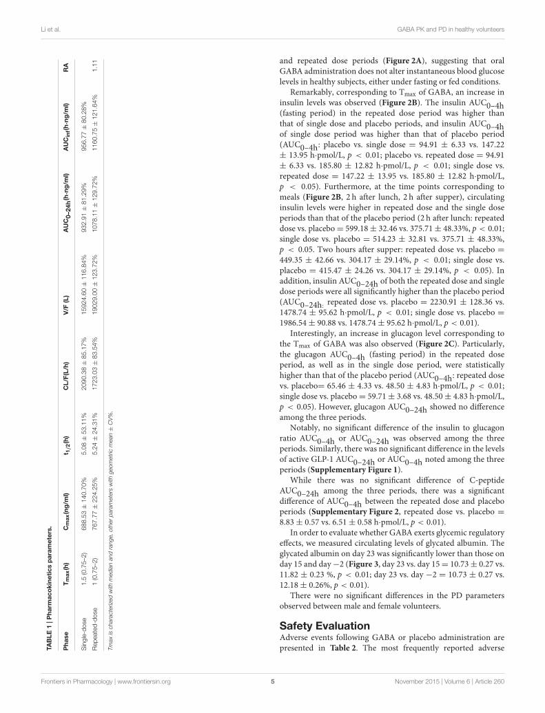
48.47 ± 2.46 h·ng/ml, p < 0.001; repeated dose vs. control= 1246.54 ± 396.46 vs. 48.47 ± 2.46 h·ng/ml, p < 0.001.AUC0–24h: single dose vs. control = 1451.68 ± 243.12 vs.272.85 ± 14.28 h·ng/ml, p < 0.001; repeated dose vs. control= 1778.69 ± 433.21 vs. 272.85 ± 14.28 h·ng/ml, p < 0.001).This indicates that GABA was rapidly absorbed, with maximumplasma concentrations achieved approximately 1–1.5 h after anoral dose, and subsequent mean elimination half-life in a rangeof 5–5.2 h. Additional derived pharmacokinetic parameters aresummarized in Table 1. As shown, oral GABA administrationboth the single dose and repeated dose reached a comparableCmax (688 vs. 767 ng/ml), suggesting that GABA reached aCmax that was not dependent on the frequency of oral dosing.The cumulative coefficient was 1.11, which suggests almost noaccumulation phenomenon during a course of oral GABA of2 g administered three times per day for 7 consecutive days.There were no significant differences of the PK profiles observedbetween male and female volunteers.
Pharmacodynamic EffectsThere were no statistical differences in blood glucose, includingthe postprandial glucose levels, amongst the placebo, single dose,
FIGURE 1 | Concentration-time profiles of GABA. Individual concentration-time curves of GABA in the three periods (A, repeated dose period; B, single dose
period; C, placebo period). Total concentration-time curves of GABA in the three periods, data are presented as geomean ± SE (D).
Frontiers in Pharmacology | www.frontiersin.org 4 November 2015 | Volume 6 | Article 260
Li et al. GABA PK and PD in healthy volunteers
TABLE1|Pharm
acokineticsparameters.
Phase
Tmax(h)
Cmax(ng/m
l)t 1
/2(h)
CL/F(L/h)
V/F
(L)
AUC0–2
4h(h
·ng/m
l)AUCinf(h·ng/m
l)RA
Single-dose
1.5
(0.75–2
)688.53±
140.70%
5.08±
53.11%
2090.38±
85.17%
15924.60±
116.84%
932.91±
81.29%
956.77±
80.28%
Repeated-dose
1(0.75–2
)767.77±
224.25%
5.24±
24.31%
1723.03±
83.54%
19029.00±
123.72%
1078.11±
129.72%
1160.75±
121.64%
1.11
Tmaxischaracterizedwithmedianandrange,otherparameterswithgeometricmean±CV%.
and repeated dose periods (Figure 2A), suggesting that oralGABA administration does not alter instantaneous blood glucoselevels in healthy subjects, either under fasting or fed conditions.
Remarkably, corresponding to Tmax of GABA, an increase ininsulin levels was observed (Figure 2B). The insulin AUC0–4h(fasting period) in the repeated dose period was higher thanthat of single dose and placebo periods, and insulin AUC0–4hof single dose period was higher than that of placebo period(AUC0–4h: placebo vs. single dose = 94.91 ± 6.33 vs. 147.22± 13.95 h·pmol/L, p < 0.01; placebo vs. repeated dose = 94.91± 6.33 vs. 185.80 ± 12.82 h·pmol/L, p < 0.01; single dose vs.repeated dose = 147.22 ± 13.95 vs. 185.80 ± 12.82 h·pmol/L,p < 0.05). Furthermore, at the time points corresponding tomeals (Figure 2B, 2 h after lunch, 2 h after supper), circulatinginsulin levels were higher in repeated dose and the single doseperiods than that of the placebo period (2 h after lunch: repeateddose vs. placebo= 599.18± 32.46 vs. 375.71± 48.33%, p < 0.01;single dose vs. placebo = 514.23 ± 32.81 vs. 375.71 ± 48.33%,p < 0.05. Two hours after supper: repeated dose vs. placebo =
449.35 ± 42.66 vs. 304.17 ± 29.14%, p < 0.01; single dose vs.placebo = 415.47 ± 24.26 vs. 304.17 ± 29.14%, p < 0.05). Inaddition, insulin AUC0–24h of both the repeated dose and singledose periods were all significantly higher than the placebo period(AUC0–24h:
repeated dose vs. placebo = 2230.91 ± 128.36 vs.1478.74 ± 95.62 h·pmol/L, p < 0.01; single dose vs. placebo =
1986.54± 90.88 vs. 1478.74± 95.62 h·pmol/L, p < 0.01).Interestingly, an increase in glucagon level corresponding to
the Tmax of GABA was also observed (Figure 2C). Particularly,the glucagon AUC0–4h (fasting period) in the repeated doseperiod, as well as in the single dose period, were statisticallyhigher than that of the placebo period (AUC0–4h: repeated dosevs. placebo= 65.46 ± 4.33 vs. 48.50 ± 4.83 h·pmol/L, p < 0.01;single dose vs. placebo= 59.71± 3.68 vs. 48.50± 4.83 h·pmol/L,p < 0.05). However, glucagon AUC0–24h showed no differenceamong the three periods.
Notably, no significant difference of the insulin to glucagonratio AUC0–4h or AUC0–24h was observed among the threeperiods. Similarly, there was no significant difference in the levelsof active GLP-1 AUC0–24h or AUC0–4h noted among the threeperiods (Supplementary Figure 1).
While there was no significant difference of C-peptideAUC0–24h among the three periods, there was a significantdifference of AUC0–4h between the repeated dose and placeboperiods (Supplementary Figure 2, repeated dose vs. placebo =
8.83± 0.57 vs. 6.51± 0.58 h·pmol/L, p < 0.01).In order to evaluate whether GABA exerts glycemic regulatory
effects, we measured circulating levels of glycated albumin. Theglycated albumin on day 23 was significantly lower than those onday 15 and day−2 (Figure 3, day 23 vs. day 15= 10.73± 0.27 vs.11.82 ± 0.23 %, p < 0.01; day 23 vs. day −2 = 10.73 ± 0.27 vs.12.18± 0.26%, p < 0.01).
There were no significant differences in the PD parametersobserved between male and female volunteers.
Safety EvaluationAdverse events following GABA or placebo administration arepresented in Table 2. The most frequently reported adverse
Frontiers in Pharmacology | www.frontiersin.org 5 November 2015 | Volume 6 | Article 260

Li et al. GABA PK and PD in healthy volunteers
FIGURE 2 | Concentration-time profiles of blood glucose, insulin and glucagon. The concentration-time curves of blood glucose, insulin, and glucagon in the
three periods (A–C), data are expressed as percent variation of baseline and presented as mean±SE. **p < 0.01 vs. placebo. *p < 0.05 vs. placebo.
FIGURE 3 | Profiles of glycated albumin. The glycated albumin levels on
day 2, 15, and 23 are shown as mean ± SE. **p < 0.01 vs. placebo;##p < 0.01 vs. single dose.
events, which appeared to be related to the dosing regimensi.e., single dosing vs. repeated dosing were sore throat, throatburning, a skin burning sensation, headache, and dizziness. Therewere higher occurrences/frequencies of adverse events in the
repeated dose period as compared to the single dose period,suggesting a dose, and duration dependency. However, therewere no clinically relevant changes in all subjects in vital signs,ECG parameters, and physical examination. Safety laboratorymeasurements such as hematology, biochemistry, and urinalysiswere all in normal ranges; except for four male volunteersthat had slightly elevated alanine transaminase (ALT) andaspartate aminotransferase (AST) levels. This elevation of liverenzymes, however, was transient, without symptoms. Withoutchanges of other biochemical indices of liver function such asbilirubin, in all cases, the ALT returned to the normal rangewithout intervention at follow-up visit (Supplementary Table 2).There were no severe adverse events observed during thestudy.
DISCUSSION
Preclinical studies by us and others reveal that GABA exerts anti-diabetic effects in several T1D mouse models due to its dualactions on β-cells and the immune system (Tian et al., 2004, 2013,2014; Soltani et al., 2011; Purwana et al., 2014). This includes
Frontiers in Pharmacology | www.frontiersin.org 6 November 2015 | Volume 6 | Article 260

Li et al. GABA PK and PD in healthy volunteers
TABLE 2 | Clinical and laboratory toxicity.
Placebo Single-dose Repeated-dose
period (D1–D2) period (D15–D16) period (D22–D30)
n (%) e n (%) e n (%) e
Dry pharynx 4 36 7
Sore throat 8 73 22
Throat
burning
4 36 12
Headache 5 45 8
Jaw pain 1 9 1
Neck pain 2 18 3
Dizziness 3 27 3 2 18 5
Poor sleep 4 36 4
Palpitation 2 18 2
Chest
stuffiness
3 27 5
Fatigue 1 9 1
Nausea 1 9 1
Skin needling
sensation
3 27 3
Skin burning
sensation
3 27 8
Limb
numbness
4 36 9
Limb pain 1 9 1
Thirsty 1 9 1
ALT 4 36 8
AST 4 36 8
n, Number of subjects; e, number of adverse events; %, proportion of subjects having the
event.
protection against β-cell apoptosis, β-cell regenerative effects,and anti-inflammatory effects. Importantly, GABA appears to actsimilarly on human and rodent cells, including xenotranplatedhuman islet cells (Tian et al., 2013; Purwana et al., 2014). Thesestudies suggest that GABA may be an effective therapeutic agentfor treating T1D. However, there is little information availableon GABA pharmacokinetics in humans, and to address thisissue we investigated the effects of GABA in healthy volunteers.In this open-labeled, three-period, self-controlled study, wedemonstrate that oral administration of GABA is effective andmodulates circulating islet hormonal levels in healthy subjects.
We found that GABA reached peak concentration in thecirculation after 1 ∼ 1.5 h (Table 1) oral administration andremained increased for several hours. We observed that afteroral GABA administration at a repeated daily dosing (2 g ×
3) for consecutive 7 days, steady-state conditions appeared tobe attained, which were accompanied by a 1/2 h shortenedTmax and a higher Cmax than that in the single dose period.As noted, previous studies (Petty et al., 1987) demonstratedthat plasma GABA levels were not affected by gender, diet,exercise, and diurnal rhythm, which suggests that GABA hasa reasonable stability for clinical pharmacological purposes.GABA is known to be metabolized by GABA transaminaseenzyme (GABA + pyruvate = succinic semialdehyde + alanine;
GABA + 2-oxoglutarate = succinic semialdehyde + glutamate;Bown and Shelp, 1997). However, these transamination productsof GABA have no known pharmacological effects. There wasnearly no accumulation noted after a three-daily dosing for aconsecutive 7 days of oral administration, and with no severeadverse event observed in the healthy volunteers during the studycourse, it implies the possibility of long-term oral GABA clinicalapplication.
GABA induced a dose-dependent increase in the fasting(8:00 AM–12:00 AM) and postprandial insulin secretion. GABAstimulated insulin secretion in humans, which is in accordwith the higher c-peptide AUC0–4h observed in the repeateddose period. Indeed, in vitro studies using isolated humanislets recently showed that GABA dose-dependently increasedinsulin secretion in a GABA receptor dependent manner(Prud’homme et al., 2013). In accord with this, GABA inducedβ-cell membrane depolarization in rodent islets (Wendt et al.,2004; Soltani et al., 2011) and human islets (Braun et al.,2010) through A-type GABA receptor (GABAAR) dependentopening of the voltage dependent Ca2+ channel (Braun et al.,2010; Purwana et al., 2014). It induced membrane depolarizingeffects in β-cells, which contrasts with its hyperpolarizing effectsin the pancreatic α-cells (Rorsman et al., 1989; Xu et al.,2006).
Glucagon is an important hormone that counterbalancesinsulin actions on blood glucose homeostasis by stimulatinghepatic glycogenolysis and gluconeogenesis (Gromada et al.,2007; Bansal and Wang, 2008). In vitro studies showed thatwhile GABA enhances insulin secretion in β-cells, it suppressesglucagon release from the α-cells (Rorsman et al., 1989; Xuet al., 2006; Braun et al., 2010; Soltani et al., 2011; Purwanaet al., 2014). However, in this study we found that GABAincreases the circulating levels of both insulin and glucagon,and that the ratio of insulin to glucagon was not altered.The reason for this discrepancy between in vitro and in vivoresults is unclear, and requires further investigation. However, wepostulate that this represents a normal physiological response tocontrol blood glucose levels. Indeed, blood glucose levels werenot significantly changed in healthy subjects upon oral GABAadministration under the fasting conditions, consistent with thenotion that elevated glucagon levels countered the action ofinsulin.
GLP-1 is an important incretin hormone in the regulationof glucose homeostasis (Campbell and Drucker, 2013). Wetherefore examined if oral GABA administration affectscirculating GLP-1 levels in the healthy subjects. Under fastingconditions, the circulating GLP-1 levels were relatively constant,and there were significant increase in postprandial activecirculating GLP-1 levels (both lunch and supper) in the subjects.Notably, oral GABA administration (either single or repeateddose) had no significant effects on the active GLP-1. It isinteresting to note that some previous in vitro studies suggestpotential reciprocal interactions between GLP-1 and GABA(Gameiro et al., 2005; Wang et al., 2007), Nevertheless, our datasuggested that at least under in vivo conditions, oral GABAadministration at the present regime did not change activeGLP-1 levels in healthy humans.
Frontiers in Pharmacology | www.frontiersin.org 7 November 2015 | Volume 6 | Article 260

Li et al. GABA PK and PD in healthy volunteers
GABA significantly decreased glycated albumin levels afteroral administration in repeated dosing (but not the single dose).Glycated albumin is an indicator of relatively recent (1–2 week)changes in blood glucose and thus has been considered as anindication of glycemic control (Inaba et al., 2007; Koga andKasayama, 2010). Our results suggest that GABA administrationmight improve glycemic control. However, blood glucose levelsmonitored at least on day 1, 8, 22 (placebo, single dose, repeateddose) showed no changes between the three groups. Therefore,there is a possibility that GABA reduced glycated albumin levelsthrough biochemical mechanisms other than blood glucose.Further studies are warranted to investigate the underlyingmechanism of action.
There were no cases of hypoglycemia resulting fromoral GABA administration in this study. Furthermore,although there were some transient adverse effects whichappeared to be dose and duration dependency, there wereno severe adverse events found during the entire course ofstudy.
To our knowledge, this is the first clinical study investigatingthe pharmacokinetics of GABA in healthy subjects. A limitationwas the lack of dose escalation to determine the optimaldose of GABA in humans. A future double-blind, randomized,placebo-controlled study is required to determine the optimaldose of GABA in humans, in either healthy or diabeticconditions.
In summary, orally administered GABA is rapidly absorbed bythe gastrointestinal tract and remains elevated in the circulationfor hours. It exerts stimulatory effects on insulin (and c-peptide)secretion, raising the possibility of its use in the treatmentof diabetes. However, in healthy subjects it has no effects oninstantaneous blood glucose levels. This may in part be attributedto GABA-induced counter regulatory mechanisms, especiallyelevated glucagon, which prevent hypoglycemia in the faceof increased insulin levels. Our study was limited to healthysubjects, and the effects of GABA in diabetic patients may bedifferent, and will be investigated in future studies. In terms ofdosage, our findings support a three-daily dosing scheme. SinceGABA targets diabetes in the key areas of islet-cell protection,regeneration, and immunotherapy, this study provides validpharmacokinetics parameters to investigate its actions in diabeticsubjects.
FUNDING
This research is supported by grants from the Juvenile DiabetesResearch Foundation (JDRF, grant number: JDRF17-2013-499).QW’s current research is supported by JDRF (2015-64-Q-R),CDA(OG-3-13-4066-QW), NSFC(81570518, 81370877).
AUTHOR CONTRIBUTIONS
ZJ, YL, and QW contributed to the conception and design of theresearch; JL, ZZ, XL, YW, FM, JM, XL, GC, JZ, ZJ, YL, and QWcontributed to the acquisition of data; JL, ZZ, JM, JY, NZ, ZJ, andQW contributed to the analysis and interpretation of data; JL, DJ,YW,MA, DG, GP, ZJ, and QW contributed to drafting the article.
All authors have revised the manuscript critically for importantintellectual content and given final approval of the version to bepublished. QW is responsible for the integrity of the work as awhole.
ACKNOWLEDGMENTS
We thank Drs.: Lv Zhang, Linling Yan, Lina Xie, Zuanli Wang,Huihui Wu, Naijia Liu, Jing Ling, Chenyan Zhao, Yintao Li, WeiWu, Mingming Yan, Qiongyue Zhang, Yehong Yang; Nurses:Jingdi Lu, Miaofang Wang, Bei Wang, Wan Zhuang, GuoyunZhang, Lanfang Yu, Quling Li, Huiyan Yao, Qing Shen, Jia Fu,Weiwei Tang, Jin Zhou (Huashan hospital, Shanghai, China) forparticipating this study.
SUPPLEMENTARY MATERIAL
The Supplementary Material for this article can be foundonline at: http://journal.frontiersin.org/article/10.3389/fphar.2015.00260
Supplementary Figure 1 | Concentration-time profiles of GLP-1 (active
form). The concentration-time curves of GLP-1 (active form) in the three periods,
data are expressed as percent variation of baseline and presented as mean ± SE.
Supplementary Figure 2 | Concentration-time profiles of c-peptide. The
concentration-time curves of c-peptide in the three periods, data are expressed as
percent variation of baseline and presented as mean ± SE.
Supplementary Table 1 | Trial Design.
Supplementary Table 2 | ALT and AST elevation of four subjects during the
repeated period.
REFERENCES
Abdou, A. M., Higashiguchi, S., Horie, K., Kim, M., Hatta, H., and Yokogoshi,
H. (2006). Relaxation and immunity enhancement effects of gamma-
aminobutyric acid (GABA) administration in humans. Biofactors 26, 201–208.
doi: 10.1002/biof.5520260305
Adeghate, E., and Ponery, A. S. (2002). GABA in the endocrine pancreas: cellular
localization and function in normal and diabetic rats. Tissue Cell 34, 1–6. doi:
10.1054/tice.2002.0217
Atkinson, M. A., Eisenbarth, G. S., and Michels, A. W. (2014). Type 1 diabetes.
Lancet 383, 69–82. doi: 10.1016/S0140-6736(13)60591-7
Bansal, P., and Wang, Q. (2008). Insulin as a physiological modulator of
glucagon secretion. Am. J. Physiol. Endocrinol. Metab. 295, E751–E761. doi:
10.1152/ajpendo.90295.2008
Bown, A. W., and Shelp, B. J. (1997). The metabolism and
functions of [gamma]-aminobutyric acid. Plant Physiol.
115, 1–5.
Braun, M., Ramracheya, R., Bengtsson, M., Clark, A., Walker, J. N., Johnson, P.
R. et al. (2010). Gamma-aminobutyric acid (GABA) is an autocrine excitatory
transmitter in human pancreatic beta-cells. Diabetes 59, 1694–1701. doi:
10.2337/db09-0797
Braun, M., Wendt, A., Birnir, B., Broman, J., Eliasson, L., Galvanovskis,
J. et al. (2004). Regulated exocytosis of GABA-containing synaptic-like
microvesicles in pancreatic beta-cells. J. Gen. Physiol. 123, 191–204. doi:
10.1085/jgp.200308966
Campbell, J. E., and Drucker, D. J. (2013). Pharmacology, physiology, and
mechanisms of incretin hormone action. Cell Metab. 17, 819–837. doi:
10.1016/j.cmet.2013.04.008
Frontiers in Pharmacology | www.frontiersin.org 8 November 2015 | Volume 6 | Article 260

Li et al. GABA PK and PD in healthy volunteers
Cavagnini, F., Invitti, C., Pinto, M., Maraschini, C., Di Landro, A., Dubini, A., et al.
(1980). Effect of acute and repeated administration of gamma aminobutyric
acid (GABA) on growth hormone and prolactin secretion in man. Acta
Endocrinol. 93, 149–154. doi: 10.1530/acta.0.0930149
Dong, H., Kumar, M., Zhang, Y., Gyulkhandanyan, A., Xiang, Y. Y., Ye, B., et al.
(2006). Gamma-aminobutyric acid up- and downregulates insulin secretion
from beta cells in concert with changes in glucose concentration. Diabetologia
49, 697–705. doi: 10.1007/s00125-005-0123-1
Gameiro, A., Reimann, F., Habib, A. M., O’Malley, D., Williams, L., Simpson, A.
K., et al. (2005). The neurotransmitters glycine and GABA stimulate glucagon-
like peptide-1 release from the GLUTag cell line. J. Physiol. 569, 761–772. doi:
10.1113/jphysiol.2005.098962
Gromada, J., Franklin, I., andWollheim, C. B. (2007). Alpha-cells of the endocrine
pancreas: 35 years of research but the enigma remains. Endocr. Rev. 28, 84–116.
doi: 10.1210/er.2006-0007
Inaba, M., Okuno, S., Kumeda, Y., Yamada, S., Imanishi, Y., Tabata, T.,
et al. (2007). Glycated albumin is a better glycemic indicator than glycated
hemoglobin values in hemodialysis patients with diabetes: effect of anemia
and erythropoietin injection. J. Am. Soc. Nephrol. 18, 896–903. doi:
10.1681/ASN.2006070772
Koga, M., and Kasayama, S. (2010). Clinical impact of glycated albumin as
another glycemic control marker. Endocr. J. 57, 751–762. doi: 10.1507/endocrj.
K10E-138
Lernmark, A., and Larsson, H. E. (2013). Immune therapy in type 1 diabetes
mellitus. Nat. Rev. Endocrinol. 9, 92–103. doi: 10.1038/nrendo.2012.237
Owens, D. F., and Kriegstein, A. R. (2002). Is there more to GABA than synaptic
inhibition? Nat. Rev. Neurosci. 3, 715–727. doi: 10.1038/nrn919
Petty, F., Kramer, G., and Feldman, M. (1987). Is plasma GABA of peripheral
origin? Biol. Psychiatry 22, 725–732. doi: 10.1016/0006-3223(87)90204-6
Prud’homme, G. J., Glinka, Y., Hasilo, C., Paraskevas, S., Li, X., and Wang,
Q. (2013). GABA protects human islet cells against the deleterious effects
of immunosuppressive drugs and exerts immunoinhibitory effects alone.
Transplantation 96, 616–623. doi: 10.1097/TP.0b013e31829c24be
Prud’homme, G. J., Glinka, Y., Udovyk, O., Hasilo, C., Paraskevas, S., and Wang,
Q. (2014). GABA protects pancreatic beta cells against apoptosis by increasing
SIRT1 expression and activity. Biochem. Biophys. Res. Commun. 452, 649–654.
doi: 10.1016/j.bbrc.2014.08.135
Purwana, I., Zheng, J., Li, X., Deurloo, M., Son, D. O., Zhang, Z., et al.
(2014). GABA promotes human beta-cell proliferation and modulates glucose
homeostasis. Diabetes 63, 4197–4205. doi: 10.2337/db14-0153
Represa, A., and Ben-Ari, Y. (2005). Trophic actions of GABA on neuronal
development. Trends Neurosci. 28, 278–283. doi: 10.1016/j.tins.2005.03.010
Rorsman, P., Berggren, P. O., Bokvist, K., Ericson, H., Mohler, H., Ostenson,
C. G., et al. (1989). Glucose-inhibition of glucagon secretion involves
activation of GABAA-receptor chloride channels. Nature 341, 233–236. doi:
10.1038/341233a0
Soltani, N., Qiu, H., Aleksic, M., Glinka, Y., Zhao, F., Liu, R., et al. (2011).
GABA exerts protective and regenerative effects on islet beta cells and
reverses diabetes. Proc. Natl. Acad. Sci. U.S.A. 108, 11692–11697. doi:
10.1073/pnas.1102715108
Tian, J., Dang, H., Chen, Z., Guan, A., Jin, Y., Atkinson, M. A., et al. (2013).
γ-Aminobutyric acid regulates both the survival and replication of human
beta-cells. Diabetes 62, 3760–3765. doi: 10.2337/db13-0931
Tian, J., Dang, H., Nguyen, A. V., Chen, Z., and Kaufman, D. L. (2014). Combined
therapy with GABA and proinsulin/alum acts synergistically to restore long-
term normoglycemia by modulating T-cell autoimmunity and promoting beta-
cell replication in newly diabetic NOD mice. Diabetes 63, 3128–3134. doi:
10.2337/db13-1385
Tian, J., Lu, Y., Zhang, H., Chau, C. H., Dang, H. N., and Kaufman, D. L. (2004).
Gamma-aminobutyric acid inhibits T cell autoimmunity and the development
of inflammatory responses in a mouse type 1 diabetes model. J. Immunol. 173,
5298–5304. doi: 10.4049/jimmunol.173.8.5298
Tian, J., Yong, J., Dang, H., and Kaufman, D. L. (2011). Oral GABA treatment
downregulates inflammatory responses in a mouse model of rheumatoid
arthritis. Autoimmunity 44, 465–470. doi: 10.3109/08916934.2011.571223
Wang, C., Mao, R., Van de Casteele, M., Pipeleers, D., and Ling, Z. (2007).
Glucagon-like peptide-1 stimulates GABA formation by pancreatic beta-cells
at the level of glutamate decarboxylase. Am. J. Physiol. Endocrinol. Metab. 292,
E1201–E1206. doi: 10.1152/ajpendo.00459.2006
Wendt, A., Birnir, B., Buschard, K., Gromada, J., Salehi, A., Sewing, S., et al. (2004).
Glucose inhibition of glucagon secretion from rat alpha-cells is mediated
by GABA released from neighboring beta-cells. Diabetes 53, 1038–1045. doi:
10.2337/diabetes.53.4.1038
Xu, E., Kumar, M., Zhang, Y., Ju, W., Obata, T., Zhang, N., et al. (2006). Intra-islet
insulin suppresses glucagon release via GABA-GABAA receptor system. Cell
Metab. 3, 47–58. doi: 10.1016/j.cmet.2005.11.015
Yoto, A., Murao, S., Motoki, M., Yokoyama, Y., Horie, N., Takeshima, K., et al.
(2012). Oral intake of gamma-aminobutyric acid affects mood and activities
of central nervous system during stressed condition induced by mental tasks.
Amino Acids 43, 1331–1337. doi: 10.1007/s00726-011-1206-6
Conflict of Interest Statement: The authors declare that the research was
conducted in the absence of any commercial or financial relationships that could
be construed as a potential conflict of interest.
Copyright © 2015 Li, Zhang, Liu, Wang, Mao, Mao, Lu, Jiang, Wan, Lv, Cao, Zhang,
Zhao, Atkinson, Greiner, Prud’homme, Jiao, Li and Wang. This is an open-access
article distributed under the terms of the Creative Commons Attribution License (CC
BY). The use, distribution or reproduction in other forums is permitted, provided the
original author(s) or licensor are credited and that the original publication in this
journal is cited, in accordance with accepted academic practice. No use, distribution
or reproduction is permitted which does not comply with these terms.
Frontiers in Pharmacology | www.frontiersin.org 9 November 2015 | Volume 6 | Article 260


![Pharmacokinetics Octreotide Hypertension; Relationship ...quently, octreotide has a muchlonger circulat-ing half-life than somatostatin in healthy volunteers [4, 5]. In normal healthy](https://static.cupdf.com/doc/110x72/60e40bd6a8bffe3dd6583b84/pharmacokinetics-octreotide-hypertension-relationship-quently-octreotide-has.jpg)