Acute stroke (Brain attack)
Based on ABC of strokeUpdated on 10th March 2003
based on RCP guidelines (2002/feb)

WHO definition
Clinical syndrome typified by rapidlydeveoloping signs of local or globaldisturbance of cerebral functions, lastingmore than 24hrs or leading to death
withno apparent causes other than of
vascularorigin

Introduction Stoke: sudden neurological deficit of
presumed vascular origin It’s a syndrome rather than a single disease Acute stroke is now a treatable condition
that deserves specialised attention A senior clinician should review all pts with
presumed stroke (class B recommendation) Drug Rx and specialised care both influence
survival and recovary

Assesing the patient Pts should be assessed at hospital
immediately after stroke Hyperacute treatments such as thrombolysis
must be administered within 3-6 hrs Stroke is a clinical diagnosis, but imaging is
required to differentiate between ischemic and primary intracerebral h’age
Following can be used to diagnose and predict prognosis Eivdence of motor, sensory or cortical
dysfunction Hemianopia

Pathophysiology For practical purposes – 2 types of stroke
(after excluding SAH) Ischaemic: 85% 1ry h’age: 15%
H’ge causes direct neuronal injury and pressure effect causes adjacent ischemia
1ry ischaemia results from atheroembolic occlusion or embolism
Usual sources of emboli are LA in pts with AF or LV in MI/LVF

Pathophysiology Vessel occlusion from atherosclerosis
Typically in internal carotid just above carotid bifurcation
If form small vessel – small vessel disease deep wihin the brain
Ischaemia causes direct neuronal injury from Lack of oxygenation Lack of nutritional support Cascade of neurochemical events that lead to
spreading damage Ischaemia may be reversible if reperfusion is obtained
quickly (3-6hrs) Chemical injuries may be interrupted by various
neuroprotective drugs (unproved in humans)

Symptoms and signs of stroke
Anterior circulation strokes Unilateral weakness Unilateral sensory loss or inattention Isolated dysarthria Dysphasia Vision
Homonymous hemianopia Monocular blindness Visual inattention

Symptoms and signs of stroke Posterior circulation strokes
Isolated homonymous hemianopea Diplopia and disconjugate eyes Nausea and vomiting Unilateral or bilateral weakness and/or sensory
loss No specific signs
Dysphagia Incontinence Loss of consciousness

Characteristics of subtypes of stroke
Lacunar PACI TACI Post
Signs Motor or sensory only
2 of the following: motor or sensory, cortical, hemianopia
All of: motor or sensorycortical, hemianopea
Hemianopia, brainstem, cerebellar
%dead at 1yr
10 20 60 20
%depend at 1 yr
25 30 35 20

Anterior cerebral stroke Ant cerebral artery gives a branch called ‘recurrent artery of
Huebner’ immediately after its origin If present it contributes to the supply of internal capsule Two ant cerebral arteries are joined together by ant
communicating artery Ant cerebral artery occlusions are rare If there is no recurrent artery
Face, arm will not be affected Entire leg area of the cortex is destroyed So it causes flaccid paralysis of the leg with cortical sensory loss
If there is recurrent artery & block occurs proximal to its origin
Ant internal capsule will also be affected UMN Facial weakness, spastic arms and useless flaccid leg The arm has good potential for recovary, because the cortical
control is there But the leg has no potential for recovary as the cortical control is
lost

Anterior cerebral stroke Perforating artery occlusion
If the recurrent artery Huebner is present and occluded weakness of face & arm will be present
Even if the dominant hemisphere is affected dysphasia will not occur, because the cortex is spared
Terminal branch occlusion Terminal vessels supply mainly the cortex
controlling the legs, so the motor & sensory function will be affected
Pt may not walk at all Less severe intellectual & sphincte
involvement than with main trunck occlusion

Anterior cerebral stroke Because of the affection of precentral gyrus
Incontinence of urine is common – when there is desire to pass they cannot control
Incontinence with flaccid leg weakness can mimick cauda equinal lesion
But the reflexes will return and plantar will be upgoing with time
Considerable memory and intellectual deficits may be there
If there is evidence of weakness of other leg also (ie, that the lesion is not strictly unilateral) a parasagital tumor should be excluded
Vascular occclusions affecting hemispheres should not produce B/L signs

Middle cerebral artery occlusion Massive infarction of the bulk of hemisphere There is considerable cerebral oedema
Coma & eventual death If non dominant hemisphere
Severe dyspraxia or Denial of existance of the left side
If dominant hemisphere is affected Global dysphasia
Motor: Destroys both pyramidal & extrapyramidal
mechanisms producing flaccid weakness of face & arm with little or no chance of recovary
The leg cortex is spared, but rarely recovers significantly

Middle cerebral artery occlusion Hemianaesthesia & hemianopia are associated
with hemiparesis Devastating type of stroke
Minimal chance of recovary High chance of death due to cerebral odema,
unresponsive to steroids or osmotic agents It is similar to total occlusion of carotid artery
in the neck With cross circulation via ant communicating artery With normal vertebro basilar artery
The anastomoses could be so efficient that pt may not have any neurological deficits

Signs to be looked for
Conscious level Neurological signs BP HR/rhythm Heart murmurs Peripheral pulses Systemic signs of infection or
neoplasm

Death rate after stroke
30 days 1 year 5 years
Ischaemic stroke
10 23 52
ICH 52 62 70
SAH 45 48 52

Conditions that can mimick stroke
Diagnosis Diagnostic features
Decompression of previous stroke Evidence of infection such as urinary or respiratory tract; metabolic dist.
Cerebral neoplasm (1ry or 2ry) Less abrupt; 1ry tumor or 2ry (lung or breast CA)
SAH Recent head injury
Epileptic seizures Possible previous fits
Traumatic brain injury H/O trauma
migraine Less abrupt onset; followed by headache; young pt
Multiple sclerosis Less abrupt onset, possible previous epi
Cerebral abscess Infection

Investigations of stroke All should have a CTwithin 48hrs to
distiguish between ischaemic and h’gic stroke
Imaging should be urgent in Depressed conscious level, fluctuating
symptoms, papilloedema, neck stiffness, fever, severe headache, previous trauma, anticoagulant treatment or bleeding diathesis (B)

MRI is superior, because it also assess blood flow and perfusion of the brain/detect wether lesion is old or new and identify carotid stenosis
Imaging will also identify stroke mimicking conditions
But a low grade glioma could still be difficult to be differentiated from cerebral infarction

Investigations of stroke All patients
CT/MRI ECG CXR FBC Clotting screen SE/creatinine Plasma glucose

Investigations of stroke
Sub groups Carotid duplex scanning ECHO Thrombophilia screen Immunology screen Syphillis serology Cerebral angiography (Rarely)

Investigations – to what extent
Depends on several factors Likely degree of recovary Presence of obvious risk factors Age of the pt (younger pts likely to
have identifiable cause such as inflammatory or clotting dissorder)
Ix better be restricted to tests that will help in the management
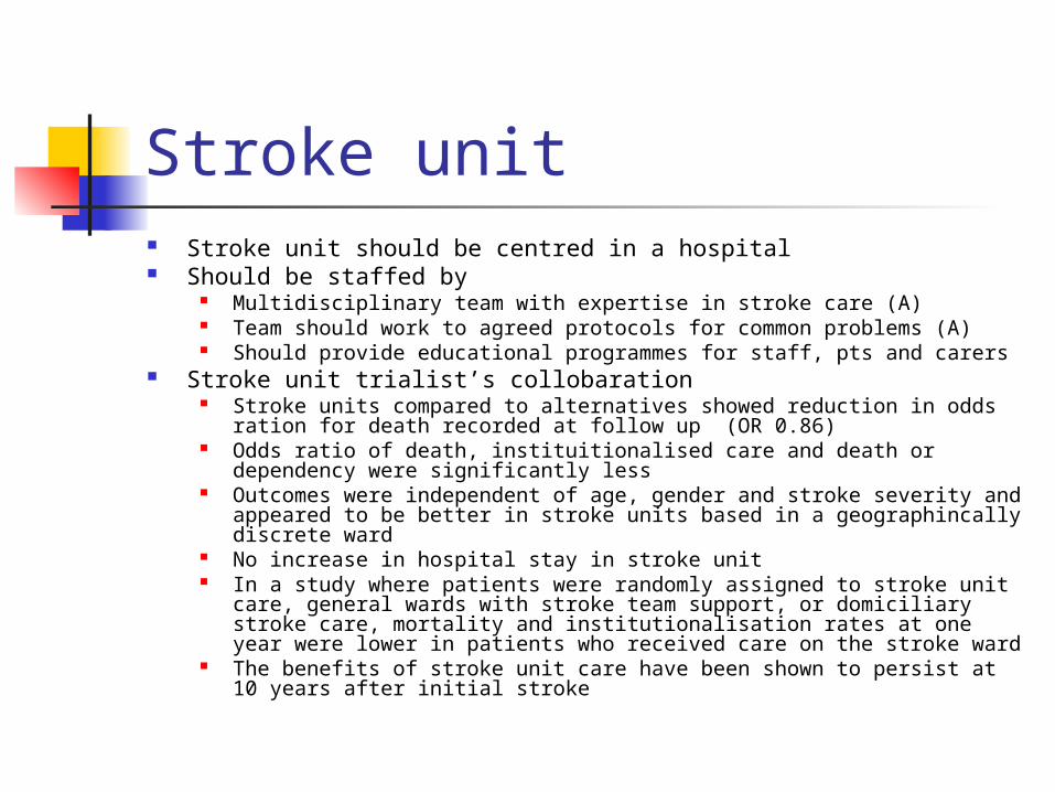
Stroke unit Stroke unit should be centred in a hospital Should be staffed by
Multidisciplinary team with expertise in stroke care (A) Team should work to agreed protocols for common problems (A) Should provide educational programmes for staff, pts and carers
Stroke unit trialist’s collobaration Stroke units compared to alternatives showed reduction in odds ration for
death recorded at follow up (OR 0.86) Odds ratio of death, instituitionalised care and death or dependency were
significantly less Outcomes were independent of age, gender and stroke severity and
appeared to be better in stroke units based in a geographincally discrete ward
No increase in hospital stay in stroke unit In a study where patients were randomly assigned to stroke unit care,
general wards with stroke team support, or domiciliary stroke care, mortality and institutionalisation rates at one year were lower in patients who received care on the stroke ward
The benefits of stroke unit care have been shown to persist at 10 years after initial stroke

Early supported discharge from hospital to a specialist rehabilitation team providing care at home is feasible for selected pts (A)
If the pt can transfer from bed to chair, can be sent home and equally effective specialised multidisciplinary care could be given at home (A)
Patients should only be managed at home if acute assessment guidelines can be adhered to and the services organised for home are flexible, and part of a specialist stroke service (A)

The guidelines do allow for the management of some patients in the community, particularly those with transient ischaemia attacks (TIAs) and strokes with good recovery
The consensus was that these patients could be managed at home provided they had access to a neurovascular clinic within two weeks (C)
More than one TIA within a short period (crescendo TIA) requires admission to hospital (C)
The guidelines are not prescriptive in defining a short period but the authors consider that recurrent TIAs within one week merit admission
The guidelines recommend that families are involved in the decision making process and have input into future plans for the patient (C)
Caring for a stroke patient can be very difficult & emotional distress is seen in 55% of caregivers at six months after stroke
Caregivers are more likely to be depressed if the patients are severely dependent or emotionally distressed themselves
The stroke team must be alert to recognising carer stress and helping carers in this difficult situation (B)
Disseminating information about the nature of stroke and on relevant local and national services improves patient and carer knowledge (A)
Introduction of stroke family support workers increases the quality of life


Emergency management
Within the 1st hour after cerebral ischaemia, part of the brain is under threat of death
The densly ischaemic area will inevitably die, but there is also tissue that could be salvaged
At this stage oxygenation, haemodynamic and metabolic factors are crucial

Management Restore vascular anatomy
Thrombolysis Angioplasty/stent Antiplatelets/anticoagulants
Stop ischaemic neurones dying Prevent complications
DVT MI Infection
Seccondary prevention

Emergency management The emergency managemet of stroke requires
Medical stabilisation Assesment of factors that may lead to complications
Swallowing hydration
It is important to keep physiological variables such as hydration,
temperature, nutrition, and oxygenation within normal range
in the acute phase of stroke (C) Thrombolysis may be considered Stroke units are associated with better outcome

Early Rx of ischemic stroke General care Specific Rx: thrombolysis, anticoagulants,
antiplatelets, neuroprotective agents Emergency aproach Stroke unit care Treatment of complications Treatment of co-morbidity Rehabilitation

Swallowing and feeding Dysphagia in ~35%
Unrecognised in mild stroke But associated with poor outcome
Aspiration Pnuemonia Poor nutrition
Presence of gag reflex is a poor guide and therefor formal assesment is essential
Fluids are more difficult to swallow than solids They should be fed through NG or percutaneous endoscopic feeding
tube Most pts will not need enteral feeding beyond a few weeks However when and how optimally to feed dysphagic pts is yet to be
determined Dysphagia Mx involves:
Initial swallow screen Diet modification Compensatory swallowing techniques
-reduces aspiration pneumonia
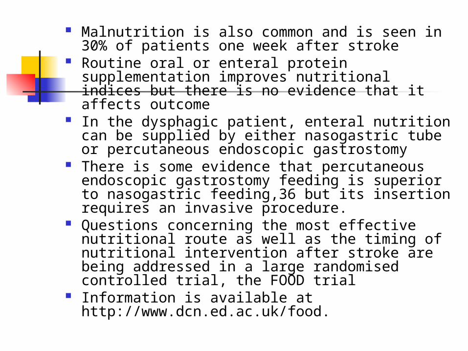
Malnutrition is also common and is seen in 30% of patients one week after stroke
Routine oral or enteral protein supplementation improves nutritional indices but there is no evidence that it affects outcome
In the dysphagic patient, enteral nutrition can be supplied by either nasogastric tube or percutaneous endoscopic gastrostomy
There is some evidence that percutaneous endoscopic gastrostomy feeding is superior to nasogastric feeding,36 but its insertion requires an invasive procedure.
Questions concerning the most effective nutritional route as well as the timing of nutritional intervention after stroke are being addressed in a large randomised controlled trial, the FOOD trial
Information is available at http://www.dcn.ed.ac.uk/food.

Communication and speech Stroke can affect communication and speech
in a variety of ways, including impaired motor speech production (dysarthria) impaired language skills (dysphasia) Impaired planning and execution of motor speech
(articulatory dyspraxia) Deficits can be subtle and every patient with a
communication difficulty needs to be assessed by a speech and language therapist
Speech therapy input is effective at improving communication, with short, intensive courses of speech therapy lasting 4–8 weeks proving most beneficial

Acute treatment of stroke Asprin: in most patients
2 large trials (160-300mg/d by PO/NG/ Rectum) started within 48hrs of stroke, reduces subsequent death and disability
NNT- 77 (reducing risk by reducing reinfarction) For 1000 pts –
12 avoid death and dependency Risk of h’age minimal (1-2/1000) Early asprin is beneficial
In a study where patients were randomly assigned to stroke unit care, general wards with stroke team support, or Patients with acute ischaemic stroke should receive aspirin
(160–300 mg) as soon as possible after stroke if a diagnosis of haemorrhage is considered unlikely (A)
But CT/MRI is essential before asprin But if CT is not availble and ischaemic stroke is highly suspected
may give asprin

IST(International Stroke Trial) and CAST (Chinese acute stroke trial) combined 40,000 pts Significant decrease in death and
dependency at 6/12 if asprin is given immediately
13 more pts alive per 1000 Rxed Increase in ICH – 2 per 1000 Reduction in recurrence - 7 per 1000

Acute treatment of stroke Heparin (conventional or LMWH)
Trials did not show any improved outcome. But useful certain groups of pts
Prophylactic: Previous venous thromboembolism Morbid obesity
Therapeutic: Carotid artery dissection Embolic, recurrent transient ischaemic
attacks

Anticoagulation has no net benefit Decreases recurrent ischaemic stroke (9
per 1000 Rxed) and pulmonary emboli (4 per 1000 Rxed)
But 9 per 1000 increase in ICH But it has definitive place in 2ry
prevention Immediate anticoagulation in AF is not
advised RCP guidelines: start anticoagulation
14d after the acute event (A) There is evidence for acute
anticoagulation in the specific stroke syndrome of cerebral venous thrombosis

Acute treatment of stroke Thrombolysis
Standard acute Rx in USA, Australia and most european countries
Type of drug and timing important NINDS trial: Alteplase (tPA) within 3 hrs
increases the chances of near complete recovary (NNT-7)
3-4x increase in ICH 20% reduction in death and dependency Rx after 6 hrs less effective (NNT-12) Complications: intra or extracranial h’age

Acute treatment of stroke Contra indications to thrombolysis:
Seizure at onset Pre Rx BP >185/110 Major infarct on CT Previous ICH Recent MI Recent or intended surgery Use of anticoagulants

Acute treatment of stroke Withhold antihypertensives for 10 days Indications for early Rx of high BP
Evidence of pre existing HBP Documented previous HT:clinic recors etc Evidence of target organ damage
Hypertensive retinopathy, LVH on ECG
Evidence of hypertensive emergancy HT encephalopathy LVF
BP is very high SBP >220-240 DBP >120

Complications of stroke Hyperglycaemia** Hypertension** Fever** Infarct extension
or bleeding Cerebral oedema Herniation coning
Aspiration Pneumonia UTI Cardiac
dysrrhythmia Recurrence DVT PE

Mood disorders are common after stroke and difficult to diagnose due to speech problems
Crying after minimal provocation is common, may be due to emotionalism and may be treated by fluexetine (A)
Pain after stroke varies in type, origin and modes of treatment Some related to stroke damage – neuropathic
or central pain (responds to tricyclics) Mechanical pain due to immobility or
exacerbation of pre-existing osteoarthritis Shoulder pain is seen in 30% Rx should begin with simple analgesia and
proper handling techniques

Venous thrombolism Is common after stroke and studies using
radiolabelled fibrinogen leg scanning suggest that deep vein thrombosis occurs in up to 50% of patients with hemiplegia
The guidelines recommend that aspirin (75–300 mg daily) should be used (A) (in non-haemorrhagic strokes)
Compression stockings should be applied to patients with weak or paralysed legs (A)
The final recommendation on the length of stocking to be used awaits results from the on-going CLOTs trial.

Spasticity Spasticity is a motor disorder characterised by a velocity
dependent increase in tonic stretch reflexes It may lead to secondary complications such as muscle and
joint contractures Management requires several coordinated interventions
including physiotherapy, drug treatment, and patient education
Physiotherapy using isokinetic strength training can improve strength and gait velocity without increasing spasticity
Drug therapy with either baclofen or tinzanadine as an adjunct to physiotherapy has been shown to reduce spasticity
In patients with disabling or symptomatically distressing symptoms, botulinum toxin is safe and effective & can be targeted to individual muscles
The guidelines indicate that spasticity should be treated if causing symptoms, though functional benefit is uncertain (B)

Mx of increased IC pressure Elevate head by 30 degrees Avoid or correct aggravating factors
Hypoxia hyperglycemia
Moderate fluid restriction Avoid hyperosmolar fluids eg: dextrose Osmotic agents: eg: manitol; as indicated Hyperventillation IV barbiturates NO STEROIDS Neuroprotective agents are not proved to be
effective

Rehabilitation Aims
Restore function Reduce the effects of stroke on pt and theirs
carers Regain independence and maximise ability in all
activities of daily living Should start early during recovery Once pt is medically stable, should be
transferred to a stroke rehabilitation unit Formal rehabilitation at a centre reduces
death, disability and hospital stay (NNT-12)

A physiotherapist with expertise in neurodisability should coordinate treatment to improve movement performance of patients with stroke (C)
The effectiveness of motor and strength rehabilitation is being underpinned by new evidence based
Progressive resistive exercise studies have also been shown to improve gait, strength, activity, and mood
There is some evidence that increased intensity of therapist input improves outcome but some patients cannot tolerate intense therapist input
The guidelines recommend that patients receive as much as they find tolerable and at least every working day (B)
It is vital that patients have the opportunity to practise rehabilitation tasks
The need for special equipment such as a wheelchair or adapted cutlery should be assessed on an individual basis as review by an occupational therapist with specialist knowledge in neurological disability can significantly reduce disability and handicap (B)
The provision of hoists or adaptation of the home environment may prevent the patient going to institutional care

Secondary prevention
Should start shortly after admission, except BP control
All pts should be offered Life style guidance Stop smoking Reduce saturated fat, alcohol and salt Asprin for life

Stroke secondary prevention

Introduction
A second stroke will not necessarily be of the same type as the initial event
Pts with previous stroke commonly suffer other vascular events like MI
Effective 2ry prevention depends on attention to all modifiable risk factors and treating the cuase of initial stroke

Four questions should be answered
Is it acute cerebrovascular disease Is it ischaemic or Haemorrhagic Cardioembolic or vascular
aetiology Anterior or posterior circulation

Is it acute CVA
Key features are Focal neurological deficit Sudden onset Absence of an alternative explanation
Exclude stroke mimicking conditions

Ischaemic or haemorrhagic History or examination cannot reliably distinguish A small bleed can produce transient symptoms Imaging is essential H’age is immediately apparent on CT, but over
few weeks it becomes indistinguishable from infarction
Small bleeds may be missed after one week MRI has a greater sensitivity for brain stem,
cerebellar and small ischaemic strokes Can also identify h’gic stroke and remains
diagnostic long after signs have disappeared

Sub cortical h’age with TIA D0 and D8

Correct imaging techniques
Symp <1h
Symp >1h onset<2w
Symp>1hronset>2w
Abrupt onset, typical CVA
Image only if anticoagulation proposed
CT MRI
Insidious onset suspicious of tumor
NA CT with contrast
CT contrast
Insidious onset suggestive of multiple sclerosis
NA MRI MRI
Cardioembolic or vascular aetiology 25% are due to embolism from heart or
major vessels Embolic stroke can affect any vascular
territory Certain features should prompt search
for embolic source TOE is justified if the results are
unequivocal or index of suspicion is high

Embolic causes of stroke found on echo MS LAH (>4cm) Dyskinetic or akinetic LV Severe global LV dysfunction Valvular vegetation LA/LV thrombi MV calcification Calcific aortic valve or stenosis
predispose to embolism but may not justify anticoagulation

Justification for echo AF HF MI within 3/12 ECG abnormalities
MI IHD BBB
Heart murmur Peripheral embolism Clinical events in >2
territories R & L hemisphere Ant & post circulation
>/= cotical events (in same territory) unless severe carotid disease

Anterior or posterior circulation Ant circulation (Carotids)
Cerebral hemispheres Post circulation (Vertebro basillar)
supplies the Brain stem Cerebellum Occipital lobe
If ant circulation stroke Carotid doppler to decide on
endarterectomy

Risk of recurrence after stroke or TIA
Stroke: 8% per year
TIA 8% risk of stroke in the first month 5% risk of stroke a year thereafter 5% risk of MI a year

Modifiable risk factors for stroke HBP Smoking DM Diet: high salt &
fat, low K & vitamins
Excess alcohol
Morbid obesity Low physical
exercise Low temperature Cholesterol
concentrations – atleast in pts with CAD

Management of risk factors
Smoking: Important correctable risk factors Risk returns to that of a non smoker
within 3-5 yrs of cessation

Management of risk factors Blood pressure
Immediate reduction may be deleterious Long term risk is inversely related to BP
achieved HT should be treated 1 or 2 weeks after the
stroke Rx reduces
Recurrence of fatal and non fatal stroke by 28% Pts at high risk of further stroke derive greatest
benefit (eg: elederly) Target BP recommended by British
Hypertension Society is <140/85

BP threshold for Rx

PROGRESS study: Pts with history of stroke or TIA were treated
with antihypertensives irrespective of baseline BP
Pts treated with Perindropril and indapamide had a reduction in BP of 12/5
And reduced stroke risk of 43% There were similar reductions in
hypertensives and non-hypertensives HOPE study
32% relative risk reduction in 1ry and 2ry stroke prevention in 9297 high risk pts with ramipril
Base line BP was 139/79 Reduction in BP was only 3.8/2.8 Efficacy of ACEI may explained by anti-
inflammatory effect and plaque stabilization

Management of risk factors Role of cholesterol – contraversial But statins reduce risk of stroke in
pts with CAD Use of statins after a athersclerotic
stroke or TIA probably reduces recurrent events and IHD
Since stroke pts are high risk pts cost Rx may be justified
Heart protection study Over 20,000 pts with high risk of vascular
disease aged 40-80 There were 1820 pts with history of non
disabling stroke or TIA All were randomised to simvastatin 40mg/d or
placebo for 5 years, independent of baseline cholesterol
Simvastatin pts showed highly significant 25% reduction in incidence rate of 1st stroke
The benefits were seen across all age ranges and base line cholesterol levels

Management of risk factors
Diabetes: Confers substantial dissadvantage for
Survival Functioning outcome on pts with acute
stroke Plasma glucose should be normalised
early BP targets for diabetics are lower

BP targets for non diabetic and diabetic stroke pts
No DM DM
Titrate to DBP </=85 </=80
Optimal BP <140/85 <130/80
Suboptimal BP >/=150/90 >/=140/85

Management of risk factors Hyperhomocystenemia:
Linked to premature vascular disease Easily lowered with vitamin
supplements Folic acid Pyridoxine
Although value of lowering homocysteine level has not been proven, younger pts with high plasma homocysteine levels may benefit

Anti platelet & anticoagulation therapy Warfarin:
Pts with AF should receive warfarin if there are no CI – INR 2-3
Pts with other sources of cardiac embolism also benefit from warfarin
Pts with mechanical prosthetic valve require INR of 2.5-4.5
If warfarin is not suitable asprin 300mg daily should be given, but it’s a less effective alternative

Main contraindications to long term warfarin treatment
GI bleeding Active peptic ulcer Frequent falls Alcohol misuse History of ICH Age by itself is not a
contraindication

Anti platelet & anticoagulation therapy Asprin
All other pts should receive antiplatelet Rx as first line
Benefits of asprin conclusively proven ASA – initial dose of 300mg & followed by 75mg/d
Dipyridamol Dipyridamole MR 200mg BD has independent and
additive effect to low dose asprin in preventing stroke, but not coronary events or overall mortality
So routine addition of dipyridamol may be cost effective
Dipyridamol alone does not prevent cardiac events

Anti platelet & anticoagulation therapy
Clopidogrel: Inhibits ADP receptors Well tolerated and slightly more effective
than asprin Not cost effective as 1st line Rx So should be used in pts with true
intolerance to asprin (allergy or intractable side effects on low dose enteric coated asprin with or without antiulcer drugs)

There is no clear evidence for superiority of one antiplatelet agent over another or for combination antiplatelet therapy in cerebrovascular disease
But if a patient on one antiplatelet agent experiences a recurrent stroke then it is better to add a second antiplatelet agent

Treatment protocol

Carotid surgery Benefit from endarterctomy upto 12 months after
the event in pts with ipsilateral severe carotid stenosis
Surgical risk Operative mortality in pts with severe disease -- 1% Risk of death or disabling stroke <4% Risk of death or any stroke <7.5%
Surgical risk less in busy units Surgeons must quote their own risk, rather than
the trial results Any pt with carotid territory symptom should be
considered a potetial candidate and doppler USS should be done, if fit for surgery
Presence or absence of carotid bruit is irrelavent

Indications for carotid endarteractomy Surgery indicated
Carotid territory symp within 6/12 and ipsilateral 70-99% stenosis
Carotid territory symp within 12/12 & ipsilateral 80-99% stenosis
Surgery not indicated: Carotid territory symp and an ipsilateral
0-69% stenosis Complete occlusion of the carotid artery

Carotid angiogram tight stenosis of internal carotid

Before & after endarterectomy
Succesful surgery is not major surgery, so pts can leave hospital within 24hrs
Neither clinical nor USS surveilance prevents late stroke, so most pts are discharged from followup after 6/52
The operation should only be carried out by a specialist with a proved low complication rate (A)
Carotid surgery

Angioplasty indications:
Fibromuscular dysplasia Radiation injury Symp stenosis after carotid endartartectomy
advantages: Less hospital stay Less cranial N injury Less wound complications Less cardiovascular morbidity
Dissadvantage: Embolic stroke at the time of surgery Recurrent stenosis

Complex cases that may require hospital referral
case Possibel treatment
Recurrent stroke or TIA despite antiplatelet treatment
High dose ASA, addition of Dipyridamol substitution or addition of clopidegrel or sub or add of warfarin
Recurrent embolic events despite adequate warfarin
Consider adding low dose asprin
Recurrent non haemodynaemic symp from severe carotid stenosis or serious I/C stenosis despite antriplalet Rx
Consider warfarin
HT or inoperable severe carotid stenosis
Consider cerebral blood flow monitoring before anti HT

Summary of management of stroke Admit to stroke unit – improves survival
& dependency Immediate CT Leg stockings (CLOTS trial) Asprin 300mg stat and 75mg thereafter Avoid heparin Thrombolysis (Randamise) Relaxed about BP Nursing, swallowing and nutrition