Endocrine-Related Cancer (2009) 16 613–622
Sonic hedgehog and pancreatic-duodenalhomeobox 1 expression distinguishbetween duodenal and pancreaticgastrinomas
Volker Fendrich1, Ricarda Ramerth1, Jens Waldmann1, Katja Maschuw1,Peter Langer1, Detlef K Bartsch1, Emily P Slater1, Annette Ramaswamy 2
and Matthias Rothmund1
Departments of 1Surgery and 2 Pathology, Philipps-University Marburg, Baldingerstraße, D-35043 Marburg, Germany
(Correspondence should be addressed to V Fendrich; Email: [email protected])
Abstract
Some 80–90% of gastrinomas are located in the gastrinoma triangle, which includes theduodenum, the pancreatic head, and the hepatoduodenal ligament. The natural history of thetumors depends on their origin. Duodenal gastrinomas are much less aggressive than pancreaticprimaries and infrequently develop liver metastases. The reason therefore is unclear. Thetranscription factor pancreatic-duodenal homeobox 1 (Pdx1) is important in differentiation anddevelopment of the pancreas and duodenum. In embryonic development, Sonic hedgehog (Shh)expression establishes a sharp molecular boundary, which allows for the proper patterning of theduodenal and pancreatic epithelium. Pancreatic polypeptide (PP) is expressed in pancreatic isletsand is known to be expressed in pancreatic endocrine tumors. This study aims to clarify theexpression pattern of Pdx1, Shh, and PP in duodenal and pancreatic gastrinomas. Tissue from 15patients with duodenal and from 11 patients with pancreatic gastrinomas that underwent surgerybetween 1987 and 2007 at our institution because of a gastrinoma were evaluated byimmunohistochemistry (IHC). Furthermore, tissue from lymph node metastases from two patientswith a so far undetected primary gastrinoma was analyzed. IHC revealed strong Pdx1 expressionin pancreatic gastrinomas, but not in duodenal gastrinomas. By contrast, there was no Shhexpression detectable in pancreatic gastrinomas, but found in all duodenal gastrinomas. Thispattern was also true for associated metastases. Shh expression combined with absence of Pdx1expression in lymph node metastases from patients with an unknown location of the primarysuggests a so far undetected duodenal gastrinoma. We show for the first time that only pancreatic,but not duodenal gastrinomas express Pdx1. Moreover, only duodenal gastrinomas express Shh,suggesting a different genetic background of these two tumors. Whereas the expression of Pdx1in pancreatic gastrinomas might suggest their endocrine origin from islets, duodenal gastrinomasdevelop from a Pdx1 negative cell cluster. The expression pattern of Pdx1, Shh, and PP inresected metastases can help to locate an otherwise undetected primary gastrinoma.
Endocrine-Related Cancer (2009) 16 613–622
Introduction
Gastrinomas, which are responsible for Zollinger–
Ellison syndrome (ZES; Zollinger & Ellison 1955),
were originally described as pancreatic neuroendocrine
tumors, but since two decades it is known that they are
Endocrine-Related Cancer (2009) 16 613–622
1351–0088/09/016–613 q 2009 Society for Endocrinology Printed in Great
most commonly located within the wall of the
duodenum. Eighty to ninety percent are located in
the so-called gastrinoma triangle, which includes the
duodenum, the pancreatic head and the hepatoduode-
nal ligament (Stabile et al. 1987). Eighty percent are
Britain
DOI: 10.1677/ERC-08-0204
Online version via http://www.endocrinology-journals.org
V Fendrich et al.: Sonic hedgehog and Pdx1 in gastrinomas
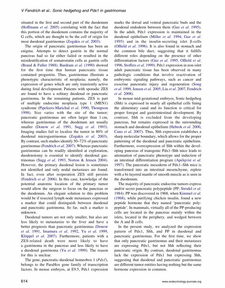
situated in the first and second part of the duodenum
(Hoffmann et al. 2005) correlating with the fact that
this portion of the duodenum contains the majority of
G cells, which are thought to be the cell of origin for
most duodenal gastrinomas (Zogakis et al. 2003).
The origin of pancreatic gastrinomas has been an
enigma. Attempts to detect gastrin in the normal
pancreas had so far either failed or resulted in the
misidentification of somatostatin cells as gastrin cells
(Brand & Fuller 1988). Bardram et al. (1990) showed
for the first time that human pancreatic tissue
contained progastrin. Thus, gastrinomas illustrate a
phenotypic characteristic of neoplasia; namely, the
expression of genes which are only transiently active
during fetal development. Patients with sporadic ZES
are found to have a solitary duodenal or pancreatic
gastrinoma. In the remaining patients, ZES is part
of multiple endocrine neoplasia type 1 (MEN1)
syndrome (Pipeleers-Marichal et al. 1990, Thompson
1998). Size varies with the site of the tumor;
pancreatic gastrinomas are often larger than 1 cm,
whereas gastrinomas of the duodenum are usually
smaller (Donow et al. 1991, Sugg et al. 1993).
Imaging studies fail to localize the tumor in 80% of
duodenal microgastrinomas (Zogakis et al. 2003).
By contrast, such studies identify 50–72% of pancreatic
gastrinomas (Fendrich et al. 2007). Whereas pancreatic
gastrinomas can be readily identified at exploration,
duodenotomy is essential to identify duodenal gas-
trinomas (Sugg et al. 1993, Norton & Jensen 2004).
However, the primary duodenal lesion is sometimes
not identified and only nodal metastases are found.
In fact, even after reoperation ZES still persists
(Fendrich et al. 2006). In this case, knowledge of the
potential anatomic location of the primary tumor
would allow the surgeon to focus on the pancreas or
the duodenum. An elegant solution to this problem
would be if resected lymph node metastases expressed
a marker that could distinguish between duodenal
and pancreatic gastrinoma. So far, such a marker is
unknown.
Duodenal tumors are not only smaller, but also are
less likely to metastasize to the liver and have a
better prognosis than pancreatic gastrinomas (Donow
et al. 1991, Imamura et al. 1992, Yu et al. 1999,
Kloppel et al. 2007). Furthermore, patients with a
ZES-related death were more likely to have
a gastrinoma in the pancreas and less likely to have
a duodenal gastrinoma (Yu et al. 1999). The reason
for this is unclear.
The gene, pancreatic-duodenal homeobox 1 (Pdx1),
belongs to the ParaHox gene family of transcription
factors. In mouse embryos, at E9.5, Pdx1 expression
614
marks the dorsal and ventral pancreatic buds and the
duodenal endoderm between them (Guz et al. 1995).
In the adult, Pdx1 expression is maintained in the
duodenal epithelium (Miller et al. 1994, Guz et al.
1995) and in the insulin-secreting islet b-cells
(Offield et al. 1996). It is also found in stomach and
the common bile duct, suggesting that it fulfills
different roles depending on the presence of other
differentiation factors (Guz et al. 1995, Offield et al.
1996, Stoffers et al. 1999). Pdx1 expression in non-islet
adult pancreatic tissue has been observed in many
pathologic conditions that involve reactivation of
embryonic signaling pathways, such as cancer and
exocrine pancreatic injury and regeneration (Song
et al. 1999, Jensen et al. 2005, Liu et al. 2007, Fendrich
et al. 2008).
In mouse mid-gestational embryos, Sonic hedgehog
(Shh) is expressed in nearly all epithelial cells lining
the alimentary canal and its function is critical for
proper foregut and gastrointestinal development. By
contrast, Shh is excluded from the developing
pancreas, but remains expressed in the surrounding
stomach and duodenal epithelium (Hebrok et al. 2000,
Cano et al. 2007). Thus, Shh expression establishes a
sharp molecular boundary, which allows for the proper
patterning of the duodenal and pancreatic epithelium.
Furthermore, overexpression of Shh within the devel-
oping pancreas of transgenic Pdx1–Shh mice leads to
attenuation of pancreatic phenotype and induction of
an intestinal differentiation program (Apelqvist et al.
1997). The pancreatic mesoderm of Pdx1–Shh mice is
transformed into an intestinal mesenchyme, replete
with a bi-layered mantle of smooth muscle as is seen in
the duodenum.
The majority of pancreatic endocrine tumors express
and/or secret pancreatic polypeptide (PP; Strodel et al.
1984). PP was discovered in 1968, when Kimmel et al.
(1968), while purifying chicken insulin, found a new
peptide hormone that they named ‘pancreatic poly-
peptide’. In mammals, virtually all of the PP-producing
cells are located in the pancreas mainly within the
islets, located in the periphery, and wedged between
the A and B cells.
In the present study, we analyzed the expression
pattern of Pdx1, Shh, and PP in duodenal and
pancreatic gastrinomas. For the first time, we show
that only pancreatic gastrinomas and their metastases
are expressing Pdx1, but not Shh reflecting their
pancreatic origin. By contrast, duodenal gastrinomas
lack the expression of Pdx1 but expressing Shh,
suggesting that duodenal and pancreatic gastrinomas
are different tumor entities having nothing but the same
hormone expression in common.
www.endocrinology-journals.org
Endocrine-Related Cancer (2009) 16 613–622
Materials and methods
Patients
Thirty-five patients underwent surgery for duodenal or
pancreatic gastrinoma and/or metastases between
1987 and April 2008 at the Department of Surgery
of the Philipps-University Marburg. Seven patients
had to be excluded from the study due to unavailable
tissue for immunohistochemical analysis. Hence,
tumor tissue from 15 patients with duodenal gastrino-
mas and from 11 patients with pancreatic gastrinomas
was analyzed. Nineteen patients had sporadic gas-
trinoma, whereas nine patients had a MEN1-associated
gastrinoma. MEN1 gene mutation analysis was
performed by Taq cycle sequencing using an
automated sequencer (ABI 310 Genetic Analyzer,
Perkin Elmer, Waltham, MA, USA) as described
previously by our group (Bartsch et al. 2005).
Furthermore, tissue from lymph node metastases
from two patients with so far undetected primary
gastrinomas was analyzed. The clinical records of all
patients with at least one operation during this time
range were analyzed with special regard to patient
demographics, clinical characteristics, pathological
findings, and long-term follow-up. Since 1997, the
majority of patients were followed annually by
biochemical testing, abdominal computed tomography,
endoscopic ultrasonography, and somatostatin-
receptor-scintigraphy at our hospital and the follow-up
resulted from the most recent examination.
Diagnosis of ZES
The diagnosis of ZES was made in the absence of
antisecretory medication: measurement of fasting
serum gastrin level, the change in serum gastrin
level after secretin stimulation, and the levels of
basal acid output. An abnormal fasting serum gastrin
level was defined as a serum gastrin concentration
O125 pg/ml. A basal acid output R15 mEq/h was
abnormal if the patient had no previous acid
reducing surgery or O5 mEq/h if the patient had
previous acid reducing surgery. An abnormal secretin
stimulation test was defined as an incremental
increase in serum gastrin level O200 pg/ml after
the i.v. administration of 2 U/kg of secretin.
Malignancy was determined on the basis of strict
criteria of infiltrative growth, lymph node or distant
metastases. Preoperative imaging routinely comprised
thin-sectioned abdominal computed tomography,
somatostatin-receptor-scintigraphy, and endoscopic
ultrasonography.
www.endocrinology-journals.org
Operative procedures
Patients underwent operative exploration to localize
and resect a primary gastrinoma and lymph node or
other metastases. The abdominal cavity was system-
atically explored for the evidence of disease. A Kocher
maneuver was performed to fully mobilize the head of
the pancreas and duodenum and the lesser sac was
opened to examine the pancreatic body and tail. The
duodenum and pancreas were carefully palpated.
Patients with no evidence for pancreatic gastrinomas
underwent longitudinal duodenotomy. If a primary
tumor was not on the medial duodenal wall, it was
elliptically excised with a margin of 2–3 mm.
Pancreatic gastrinomas were either treated by distal
pancreatic resection or pylorus-preserving pancreatico-
duodenectomy (PPPD) with regional lymph node
dissection. For MEN1–ZES either a distal pancreatic
resection to the level of the portal vein with enucleation
of any tumors in the pancreatic head, a duodenotomy
with excision of any tumors in the first to fourth portion
of the duodenum and a peripancreatic lymph node
dissection as suggested by Thompson (1998) was
routinely performed until 1997. Since then, we prefer a
PPPD with lymphadenectomy when the source of
gastrin secretion could be regionalized to the pancrea-
tic head region by preoperative selective arterial
secretin injection angiography (Imamura et al. 1987).
Pathology
Pathologic diagnosis of a primary duodenal or
pancreatic gastrinoma was made for all patients by
immunohistochemical analysis for the presence of
gastrin. The size of the tumor was measured and the
largest diameter was documented. Lymph nodes were
evaluated in a similar manner. Pathology reports were
reviewed in a retrospective fashion for the size of
the primary gastrinoma and the presence of positive
lymph nodes.
Immunohistochemical analysis of gastrinomas
For immunolabeling, formalin-fixed and paraffin
embedded archived tumor samples and corresponding
normal tissues were stained as previously described
(Esni et al. 2004). Briefly, slides from archived
gastrinomas were heated to 60 8C for 1 h, deparaffi-
nized using xylene, and hydrated by a graded series of
ethanol washes. Antigen retrieval was accomplished by
microwave heating in 10 mM sodium citrate buffer of
pH 6.0 for 10 min. For immunohistochemistry (IHC),
endogenous peroxidase activity was quenched by
10 min incubation in 3% H2O2. Non-specific binding
615
V Fendrich et al.: Sonic hedgehog and Pdx1 in gastrinomas
was blocked with 10% serum. Sections were then
probed with anti-rabbit Pdx1 (Chemicon, Temecula,
CA, USA) in a dilution with 1:100 overnight at 4 8C.
For IHC, bound antibodies were detected using the
avidin–biotin-complex (ABC) peroxidase method
(ABC Elite Kit, Vector Labs, Burlingame, CA,
USA). Final staining was developed with the Sigma
FAST DAB peroxidase substrate kit (Sigma). Pancrea-
tic islets from normal pancreatic tissue samples from
our tissue bank were used as positive controls along
with each batch of Pdx1 IHC staining.
Statistical analysis
Log-rank test was applied to identify significant
differences. P values !0.05 were considered statisti-
cally significant. Data were analyzed using SPSS
software (Version 11; SPSS, Inc., Chicago, IL, USA).
Table 1 Results of pancreatic-duodenal homeobox 1 (Pdx1), Shh
No.
Age
(years) Gender MEN1
Site of
gastrinoma
Tumor
size (mm)
1 57 M No Pancreas 33
2 28 F No Duodenum 3
3 44 F No Pancreas K4 54 M No Duodenum 10
5 49 F No Duodenum 15
6 33 F No Duodenum 8
7 61 M No Unknown K8 59 M No Pancreas 43
K
9 36 F No Unknown K10 64 F No Pancreas 40
11 73 F No Pancreas 20
12 59 M No Pancreas 10
13 70 F No Duodenum 4
14 43 M No Pancreas K
15 58 F No Pancreas K
16 52 F No Duodenum 5
K17 46 F No Duodenum 5
K
18 50 F No Duodenum 8
K19 59 F No Pancreas 22
20 48 M Yes Duodenum 10
21 49 M Yes Duodenum 3
22 29 M Yes Duodenum 8
23 46 F Yes Duodenum 3
24 48 M Yes Duodenum 6
25 47 M Yes Duodenum 3
26 45 F Yes Pancreas 25
K
27 32 F Yes Pancreas 12
28 50 M Yes Duodenum 6
MEN1, multiple endocrine neoplasia type 1; LN-metastasis, lymphdisease; AWD, alive with disease; NED, no evidence for disease; DShh, sonic hedgehog; PP, pancreatic polypeptide.
616
Results
Patients and clinical characteristics
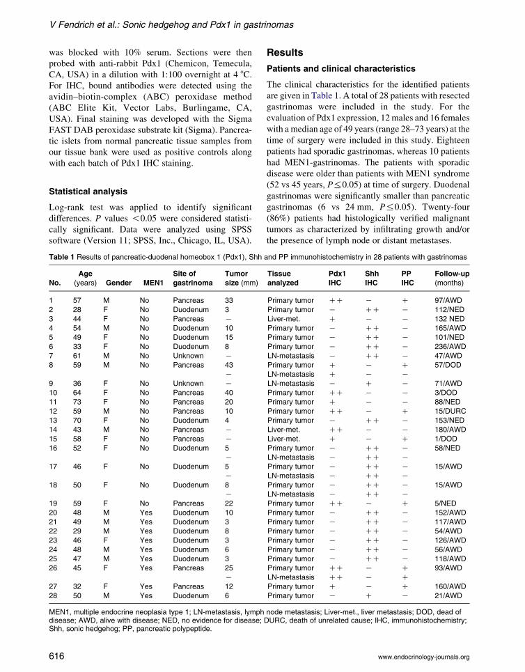
The clinical characteristics for the identified patients
are given in Table 1. A total of 28 patients with resected
gastrinomas were included in the study. For the
evaluation of Pdx1 expression, 12 males and 16 females
with a median age of 49 years (range 28–73 years) at the
time of surgery were included in this study. Eighteen
patients had sporadic gastrinomas, whereas 10 patients
had MEN1-gastrinomas. The patients with sporadic
disease were older than patients with MEN1 syndrome
(52 vs 45 years, P%0.05) at time of surgery. Duodenal
gastrinomas were significantly smaller than pancreatic
gastrinomas (6 vs 24 mm, P%0.05). Twenty-four
(86%) patients had histologically verified malignant
tumors as characterized by infiltrating growth and/or
the presence of lymph node or distant metastases.
and PP immunohistochemistry in 28 patients with gastrinomas
Tissue
analyzed
Pdx1
IHC
Shh
IHC
PP
IHC
Follow-up
(months)
Primary tumor CC K C 97/AWD
Primary tumor K CC K 112/NED
Liver-met. C K K 132 NED
Primary tumor K CC K 165/AWD
Primary tumor K CC K 101/NED
Primary tumor K CC K 236/AWD
LN-metastasis K CC K 47/AWD
Primary tumor C K C 57/DOD
LN-metastasis C K K
LN-metastasis K C K 71/AWD
Primary tumor CC K K 3/DOD
Primary tumor C K K 88/NED
Primary tumor CC K C 15/DURC
Primary tumor K CC K 153/NED
Liver-met. CC K K 180/AWD
Liver-met. C K C 1/DOD
Primary tumor K CC K 58/NED
LN-metastasis K CC KPrimary tumor K CC K 15/AWD
LN-metastasis K CC K
Primary tumor K CC K 15/AWD
LN-metastasis K CC KPrimary tumor CC K C 5/NED
Primary tumor K CC K 152/AWD
Primary tumor K CC K 117/AWD
Primary tumor K CC K 54/AWD
Primary tumor K CC K 126/AWD
Primary tumor K CC K 56/AWD
Primary tumor K CC K 118/AWD
Primary tumor CC K C 93/AWD
LN-metastasis CC K C
Primary tumor C K C 160/AWD
Primary tumor K C K 21/AWD
node metastasis; Liver-met., liver metastasis; DOD, dead ofURC, death of unrelated cause; IHC, immunohistochemistry;
www.endocrinology-journals.org
Endocrine-Related Cancer (2009) 16 613–622
Pdx1 is expressed in pancreatic gastrinomas
and their associated metastases
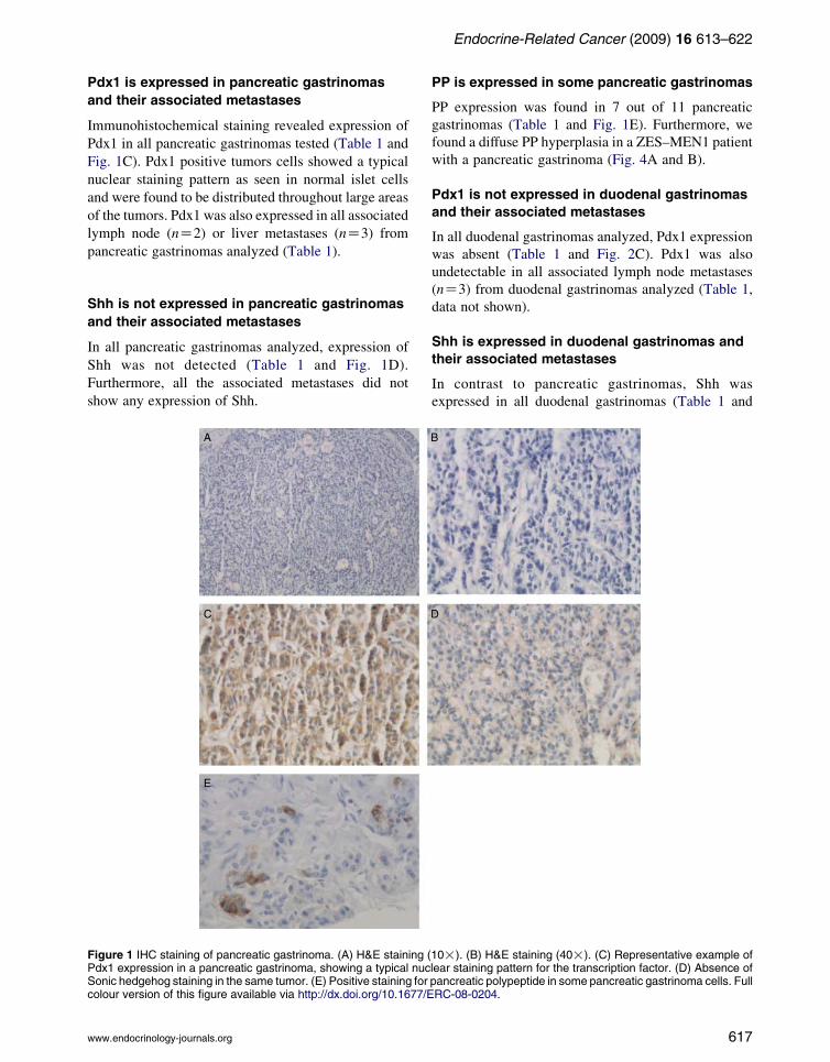
Immunohistochemical staining revealed expression of
Pdx1 in all pancreatic gastrinomas tested (Table 1 and
Fig. 1C). Pdx1 positive tumors cells showed a typical
nuclear staining pattern as seen in normal islet cells
and were found to be distributed throughout large areas
of the tumors. Pdx1 was also expressed in all associated
lymph node (nZ2) or liver metastases (nZ3) from
pancreatic gastrinomas analyzed (Table 1).
Shh is not expressed in pancreatic gastrinomas
and their associated metastases
In all pancreatic gastrinomas analyzed, expression of
Shh was not detected (Table 1 and Fig. 1D).
Furthermore, all the associated metastases did not
show any expression of Shh.
Figure 1 IHC staining of pancreatic gastrinoma. (A) H&E staining (Pdx1 expression in a pancreatic gastrinoma, showing a typical nucSonic hedgehog staining in the same tumor. (E) Positive staining forcolour version of this figure available via http://dx.doi.org/10.1677/E
www.endocrinology-journals.org
PP is expressed in some pancreatic gastrinomas
PP expression was found in 7 out of 11 pancreatic
gastrinomas (Table 1 and Fig. 1E). Furthermore, we
found a diffuse PP hyperplasia in a ZES–MEN1 patient
with a pancreatic gastrinoma (Fig. 4A and B).
Pdx1 is not expressed in duodenal gastrinomas
and their associated metastases
In all duodenal gastrinomas analyzed, Pdx1 expression
was absent (Table 1 and Fig. 2C). Pdx1 was also
undetectable in all associated lymph node metastases
(nZ3) from duodenal gastrinomas analyzed (Table 1,
data not shown).
Shh is expressed in duodenal gastrinomas and
their associated metastases
In contrast to pancreatic gastrinomas, Shh was
expressed in all duodenal gastrinomas (Table 1 and
10!). (B) H&E staining (40!). (C) Representative example oflear staining pattern for the transcription factor. (D) Absence ofpancreatic polypeptide in some pancreatic gastrinoma cells. FullRC-08-0204.
617
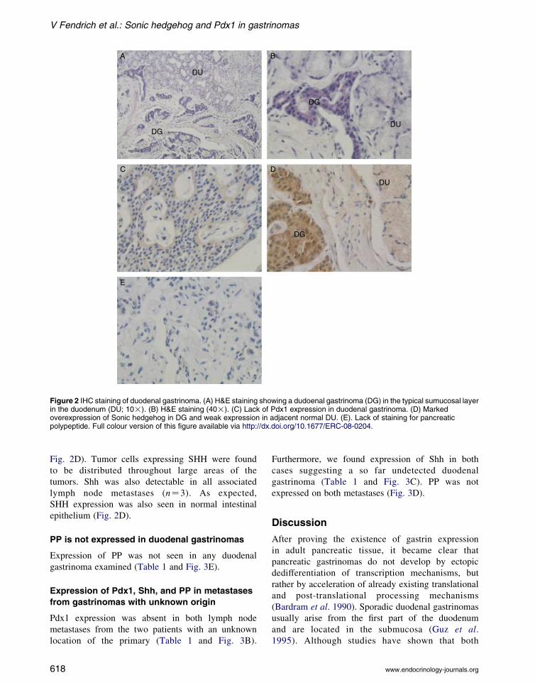
Figure 2 IHC staining of duodenal gastrinoma. (A) H&E staining showing a dudoenal gastrinoma (DG) in the typical sumucosal layerin the duodenum (DU; 10!). (B) H&E staining (40!). (C) Lack of Pdx1 expression in duodenal gastrinoma. (D) Markedoverexpression of Sonic hedgehog in DG and weak expression in adjacent normal DU. (E). Lack of staining for pancreaticpolypeptide. Full colour version of this figure available via http://dx.doi.org/10.1677/ERC-08-0204.
V Fendrich et al.: Sonic hedgehog and Pdx1 in gastrinomas
Fig. 2D). Tumor cells expressing SHH were found
to be distributed throughout large areas of the
tumors. Shh was also detectable in all associated
lymph node metastases (nZ3). As expected,
SHH expression was also seen in normal intestinal
epithelium (Fig. 2D).
PP is not expressed in duodenal gastrinomas
Expression of PP was not seen in any duodenal
gastrinoma examined (Table 1 and Fig. 3E).
Expression of Pdx1, Shh, and PP in metastases
from gastrinomas with unknown origin
Pdx1 expression was absent in both lymph node
metastases from the two patients with an unknown
location of the primary (Table 1 and Fig. 3B).
618
Furthermore, we found expression of Shh in both
cases suggesting a so far undetected duodenal
gastrinoma (Table 1 and Fig. 3C). PP was not
expressed on both metastases (Fig. 3D).
Discussion
After proving the existence of gastrin expression
in adult pancreatic tissue, it became clear that
pancreatic gastrinomas do not develop by ectopic
dedifferentiation of transcription mechanisms, but
rather by acceleration of already existing translational
and post-translational processing mechanisms
(Bardram et al. 1990). Sporadic duodenal gastrinomas
usually arise from the first part of the duodenum
and are located in the submucosa (Guz et al.
1995). Although studies have shown that both
www.endocrinology-journals.org
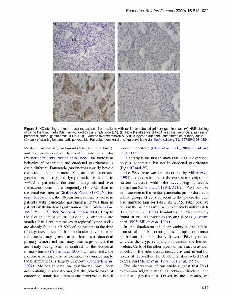
Figure 3 IHC staining of lymph node metastases from patients with so far undetected primary gastrinomas. (A) H&E stainingshowing the tumor cells (Met) surrounded by the lymph node (LN). (B) Note the absence of Pdx1 in all the tumor cells, as seen inprimary duodenal gastrinomas in Fig. 2. (C) Marked overexpression of SHH suggest a duodenal gastrinoma as primary origin.(D) Lack of staining for pancreatic polypeptide. Full colour version of this figure available via http://dx.doi.org/10.1677/ERC-08-0204.
Endocrine-Related Cancer (2009) 16 613–622
locations are equally malignant (40–70% metastases),
and the post-operative disease-free rate is similar
(Weber et al. 1995, Norton et al. 1999), the biological
behavior of pancreatic and duodenal gastrinomas is
quite different. Pancreatic gastrinomas usually have a
diameter of 2 cm or more. Metastasis of pancreatic
gastrinomas to regional lymph nodes is found in
w60% of patients at the time of diagnosis and liver
metastases occur more frequently (10–20%) than in
duodenal gastrinomas (Stabile & Passaro 1985, Norton
et al. 2006). Thus, the 10-year survival rate is worse in
patients with pancreatic gastrinomas (57%) than in
patients with duodenal gastrinomas (84%; Weber et al.
1995, Yu et al. 1999, Norton & Jensen 2004). Despite
the fact that most of the duodenal gastrinomas are
smaller than 1 cm, metastases to regional lymph nodes
are already found in 60–80% of the patients at the time
of diagnosis. It seems that periduodenal lymph node
metastases may grow faster than their duodenal
primary tumors and thus may form large tumors that
are easily recognized, in contrast to the duodenal
primary tumors (Anlauf et al. 2006). Unfortunately, the
molecular pathogenesis of gastrinomas contributing to
these differences is largely unknown (Fendrich et al.
2007). Molecular data on gastrinoma have been
accumulating in recent years, but the genetic basis of
endocrine tumor development and progression is still
www.endocrinology-journals.org
poorly understood (Chen et al. 2003, 2004, Furukawa
et al. 2005).
Our study is the first to show that Pdx1 is expressed
only in pancreatic, but not in duodenal gastrinomas
(Figs 1C and 2C).
The Pdx1 gene was first described by Miller et al.
(1994) and codes for one of the earliest transcriptional
factors detected within the developing pancreatic
epithelium (Offield et al. 1996). At E9.5, Pdx1 positive
cells are seen in the ventral pancreatic primordia and at
E11.5, groups of cells adjacent to the pancreatic duct
also immunostain for Pdx1. At E17.5, Pdx1 positive
cells in the pancreas were seen exclusively within islets
(Peshavaria et al. 1994). In adult tissue, Pdx1 is mainly
found in PP and insulin-expressing b-cells (Leonard
et al. 1993, Miller et al. 1994).
In the duodenum of older embryos and adults,
almost all cells forming the simple columnar
epithelium that line the villi were Pdx1 positive,
whereas the crypt cells did not contain the homeo-
protein. Cells of the other layers of the mucosa as well
as cells of the submucosa, muscularis and adventitial
layers of the wall of the duodenum also lacked Pdx1
expression (Miller et al. 1994, Guz et al. 1995).
The observations of our study suggest that Pdx1
expression might distinguish between duodenal and
pancreatic gastrinomas. Driven by these results, we
619
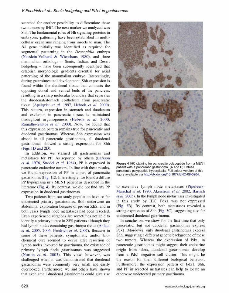
Figure 4 IHC staining for pancreatic polypeptide from a MEN1patient with a pancreatic gastrinoma. (A and B) Diffusepancreatic polypeptide hyperplasia. Full colour version of thisfigure available via http://dx.doi.org/10.1677/ERC-08-0204.
V Fendrich et al.: Sonic hedgehog and Pdx1 in gastrinomas
searched for another possibility to differentiate these
two tumors by IHC. The next marker we analyzed was
Shh. The fundamental roles of Hh signaling proteins in
embryonic patterning have been established in multi-
cellular organisms ranging from insects to man. The
Hh gene initially was identified as required for
segmental patterning in the Drosophila embryo
(Nusslein-Volhard & Wieschaus 1980), and three
mammalian orthologs – Sonic, Indian, and Desert
hedgehog – have been subsequently identified that
establish morphologic gradients essential for axial
patterning of the mammalian embryo. Interestingly,
during gastrointestinal development, Shh expression is
found within the duodenal tissue that connects the
opposing dorsal and ventral buds of the pancreas,
resulting in a sharp molecular boundary that separates
the duodenal/stomach epithelium from pancreatic
tissue (Apelqvist et al. 1997, Hebrok et al. 2000).
This pattern, expression in stomach and duodenum
and exclusion in pancreatic tissue, is maintained
throughout organogenesis (Hebrok et al. 2000,
Ramalho-Santos et al. 2000). Now, we found that
this expression pattern remains true for pancreatic and
duodenal gastrinomas. Whereas Shh expression was
absent in all pancreatic gastrinomas, all duodenal
gastrinomas showed a strong expression for Shh
(Figs 1D and 2D).
In addition, we stained all gastrinomas and
metastases for PP. As reported by others (Larsson
et al. 1976, Strodel et al. 1984), PP is expressed in
pancreatic endocrine tumors. In line with these results,
we found expression of PP in a part of pancreatic
gastrinomas (Fig. 1E). Interestingly, we found a diffuse
PP hyperplasia in a MEN1 patient as described in the
literature (Fig. 4). By contrast, we did not find any PP
expression in duodenal gastrinomas.
Two patients from our study population have so far
undetected primary gastrinomas. Both underwent an
abdominal exploration because of proven ZES, and in
both cases lymph node metastases had been resected.
Even experienced surgeons are sometimes not able to
identify a primary tumor in ZES patients although they
had lymph nodes containing gastrinoma tissue (Anlauf
et al. 2005, 2006, Fendrich et al. 2007). Because in
some of these patients, symptomatic and/or bio-
chemical cure seemed to occur after resection of
lymph nodes involved by gastrinoma, the existence of
primary lymph node gastrinomas was suggested
(Norton et al. 2003). This view, however, was
challenged when it was demonstrated that duodenal
gastrinomas were commonly very small and easily
overlooked. Furthermore, we and others have shown
that even small duodenal gastrinomas could give rise
620
to extensive lymph node metastases (Pipeleers-
Marichal et al. 1990, Akerstrom et al. 2002, Bartsch
et al. 2005). In the lymph node metastases investigated
in this study by IHC, Pdx1 was not expressed
(Fig. 3B). By contrast, both metastases revealed a
strong expression of Shh (Fig. 3C), suggesting a so far
undetected duodenal gastrinoma.
In conclusion, we show for the first time that only
pancreatic, but not duodenal gastrinomas express
Pdx1. Moreover, only duodenal gastrinomas express
Shh, suggesting a different genetic background of these
two tumors. Whereas the expression of Pdx1 in
pancreatic gastrinomas might suggest their endocrine
origin from islets, duodenal gastrinomas develop
from a Pdx1 negative cell cluster. This might be
the reason for their different biological behavior.
Furthermore, the expression pattern of Pdx1, Shh,
and PP in resected metastases can help to locate an
otherwise undetected primary gastrinoma.
www.endocrinology-journals.org
Endocrine-Related Cancer (2009) 16 613–622
Declaration of interest
The authors declare that there is no conflict of interest that
would prejudice the impartiality of this scientific work.
Funding
V F was supported by a Research Grant from the University
Medical Center Giessen and Marburg.
References
Akerstrom G, Hessman O & Skogseid B 2002 Timing and
extent of surgery in symptomatic and asymptomatic
neuroendocrine tumors of the pancreas in MEN1.
Langenbeck’s Archives of Surgery 386 558–569.
Anlauf M, Perren A, Meyer CL, Schmid S, Saremaslani P,
Kruse ML, Weihe E, Komminoth P, Heitz PU & Kloppel
G 2005 Precursor lesions in patients with multiple
endocrine neoplasia type 1-associated duodenal gastri-
nomas. Gastroenterology 128 1187–1198.
Anlauf M, Garbrecht N, Henopp T, Schmitt A, Schlenger R,
Raffel A, Krausch M, Gimm O, Eisenberger CF, Knoefel
WT et al. 2006 Sporadic versus hereditary gastrinomas of
the duodenum and pancreas: distinct clinico-pathological
and epidemiological features. World Journal of
Gastroenterology 12 5440–5446.
Apelqvist A, Ahlgren U & Edlund H 1997 Sonic hedgehog
directs specialised mesoderm differentiation in the intes-
tine and pancreas. Current Biology 7 801–804.
Bardram L, Hilsted L & Rehfeld JF 1990 Progastrin
expression in mammalian pancreas. PNAS 87 298–302.
Bartsch DK, Fendrich V, Langer P, Celik I, Kann PH &
Rothmund M 2005 Outcome of duodenopancreatic
resections in patients with multiple endocrine neoplasia
type 1. Annals of Surgery 242 757–764.
Brand SJ & Fuller PJ 1988 Differential gastrin gene
expression in rat gastrointestinal tract and pancreas during
neonatal development. Journal of Biological Chemistry
263 5341–5347.
Cano DA, Hebrok M & Zenker M 2007 Pancreatic
development and disease. Gastroenterology 132 745–762.
Chen YJ, Vortmeyer A, Zhuang Z, Huang S & Jensen RT
2003 Loss of heterozygosity of chromosome 1q in
gastrinomas: occurrence and prognostic significance.
Cancer Research 63 817–823.
Chen YJ, Vortmeyer A, Zhuang Z, Gibril F & Jensen RT
2004 X-chromosome loss of heterozygosity frequently
occurs in gastrinomas and is correlated with aggressive
tumor growth. Cancer 100 1379–1387.
Donow C, Pipeleers-Marichal M, Schroder S, Stamm B,
Heitz PU & Kloppel G 1991 Surgical pathology of
gastrinoma. Site, size, multicentricity, association with
multiple endocrine neoplasia type 1, and malignancy.
Cancer 68 1329–1334.
www.endocrinology-journals.org
Esni F, Stoffers DA, Takeuchi T & Leach SD 2004 Origin
of exocrine pancreatic cells from nestin-positive pre-
cursors in developing mouse pancreas. Mechanisms of
Development 121 15–25.
Fendrich V, Langer P, Celik I, Bartsch DK, Zielke A,
Ramaswamy A & Rothmund M 2006 An aggressive
surgical approach leads to long-term survival in patients
with pancreatic endocrine tumors. Annals of Surgery 244
845–853.
Fendrich V, Langer P, Waldmann J, Bartsch DK &
Rothmund M 2007 Management of sporadic and multiple
endocrine neoplasia type 1 gastrinomas. British Journal of
Surgery 94 1331–1341.
Fendrich V, Esni F, Garay MV, Feldmann G, Habbe N,
Jensen JN, Dor Y, Stoffers D, Jensen J, Leach SD et al.
2008 Hedgehog signalling regulates facultative
progenitor activity in regenerating exocrine pancreas.
Gastroenterology 135 621–631.
Furukawa M, Raffeld M, Mateo C, Sakamoto A, Moody TW,
Ito T, Venzon DJ, Serrano J & Jensen RT 2005
Increased expression of insulin-like growth factor I
and/or its receptor in gastrinomas is associated
with low curability, increased growth, and
development of metastases. Clinical Cancer Research
11 3233–3242.
Guz Y, Montminy MR, Stein R, Leonard J, Gamer LW,
Wright CVE & Teitelman G 1995 Expression of murine
STF-1, a putative insulin gene transcription factor, in b
cells of pancreas, duodenal epithelium and pancreatic
exocrine and endocrine progenitors during ontogeny.
Development 121 149–161.
Hebrok M, Kim SK, St Jacques B, McMahon AP &
Melton DA 2000 Regulation of pancreas development
by hedgehog signaling. Development 127 4905–4913.
Hoffmann KM, Furukawa M & Jensen RT 2005
Duodenal neuroendocrine tumors: classification,
functional syndromes, diagnosis and medical
treatment. Best Practice & Research. Clinical
Gastroenterology 19 675–697.
Imamura M, Takahashi K, Adachi H, Minematsu S,
Shimada Y, Naito M, Suzuki T, Tobe T & Azuma T
1987 Usefulness of selective arterial secretin
injection test for localization of gastrinoma in
the Zollinger–Ellison syndrome. Annals of Surgery 205
230–239.
Imamura M, Kanda M, Takahashi K, Shimada Y, Miyahara T,
Wagata T, Hashimoto M, Tobe T & Soga J 1992
Clinicopathological characteristics of duodenal micro-
gastrinomas. World Journal of Surgery 16 703–709.
Jensen JN, Cameron E, Garay MV, Starkey TW, Gianani R &
Jensen J 2005 Recapitulation of elements of embryonic
development in adult mouse pancreatic regeneration.
Gastroenterology 128 728–741.
Kimmel JR, Pollock HG & Hazelwood RL 1968 Isolation
and characterization of chicken insulin. Endocrinology 83
1323–1330.
621
V Fendrich et al.: Sonic hedgehog and Pdx1 in gastrinomas
Kloppel G, Rindi G, Anlauf M, Perren A & Komminoth P
2007 Site-specific biology and pathology of gastroenter-
opancreatic neuroendocrine tumors. Virchows Archiv 451
S9–S27.
Larsson LI, Schwartz T, Lundqvist G, Chance RE, Sundler F,
Rehfeld JF, Grimelius L, Fahrenkrug J, Schaffalitzky de
Muckadell O & Moon N 1976 Occurrence of human
pancreatic polypeptide in pancreatic endocrine tumors.
Possible implication in the watery diarrhea syndrome.
American Journal of Pathology 85 675–684.
Leonard J, Peers B, Johnson T, Ferreri K, Lee S &
Montminy MR 1993 Characterization of somatostatin
transactivating factor-1, a novel homeobox factor that
stimulates somatostatin expression in pancreatic islet
cells. Molecular Endocrinology 7 1275–1283.
Liu T, Gou SM, Wang CY, Wu HS, Xiong JX & Zhou F 2007
Pancreas duodenal homeobox-1 expression and
significance in pancreatic cancer. World Journal of
Gastroenterology 13 2615–2618.
Miller CP, McGhee RE & Habener JF 1994 IDX-1: a new
homeodomain transcription factor expressed in rat
pancreatic islets and duodenum that transactivates the
somatostatin gene. EMBO Journal 13 1145–1156.
Norton JA & Jensen RT 2004 Resolved and unresolved
controversies in the surgical management of patients
with Zollinger–Ellison syndrome. Annals of Surgery 240
757–773.
Norton JA, Fraker DL, Alexander HR, Venzon DJ,
Doppman JL, Serrano J, Goebel SU, Peghini PL,
Roy PK, Gibril F et al. 1999 Surgery to cure the
Zollinger–Ellison syndrome. New England
Journal of Medicine 341 635–644.
Norton JA, Alexander HR, Fraker DL, Venzon DJ, Gibril F &
Jensen RT 2003 Possible primary lymph node gastrinoma:
occurrence, natural history, and predictive factors: a
prospective study. Annals of Surgery 237 650–657.
Norton JA, Fraker DL, Alexander HR, Gibril F, Liewehr
DJ, Venzon DJ & Jensen RT 2006 Surgery increases
survival in patients with gastrinoma. Annals of Surgery
244 410–419.
Nusslein-Volhard C & Wieschaus E 1980 Mutations
affecting segment number and polarity in Drosophila.
Nature 287 795–801.
Offield MF, Jetton TL, Labosky PA, Ray M, Stein R,
Magnuson MA, Hogan BLM & Wright CVE 1996 PDX-1
is required for pancreatic outgrowth and differentiation of
the rostral duodenum. Development 122 983–995.
Peshavaria M, Gamer L, Henderson E, Teitelman G,
Wright CVE & Stein R 1994 XIHbox 8, an endoderm-
specific Xenopus homeodomain protein, is closely
related to a mammalian insulin gene transcription factor.
Molecular Endocrinology 8 806–816.
Pipeleers-Marichal M, Somers G, Willems G, Foulis A,
Imrie C, Bishop AE, Polak JM, Hacki WH, Stamm B,
Heitz PU et al. 1990 Gastrinomas in the duodenums of
622
patients with multiple endocrine neoplasia type 1 and the
Zollinger–Ellison syndrome. New England Journal of
Medicine 322 723–727.
Ramalho-Santos M, Melton DA & McMahon AP 2000
Hedgehog signals regulate multiple aspects of
gastrointestinal development. Development 127
2763–2772.
Song SY, Gannon M, Washington MK, Scoggins CR,
Meszoely IM, Goldenring JR, Marino CR, Sandgren
EP, Coffey RJ Jr, Wright CV et al. 1999 Expansion
of Pdx1-expressing pancreatic epithelium and islet
neogenesis in transgenic mice overexpressing trans-
forming growth factor alpha. Gastroenterology 117
1416–1426.
Stabile BE & Passaro E Jr 1985 Benign and malignant
gastrinoma. American Journal of Surgery 149 144–150.
Stabile BE, Morrow DJ & Passaro E 1987 The gastrinoma
triangle: operative implications. American Journal of
Surgery 209 550.
Stoffers DA, Heller RS, Miller CP & Habener JF 1999
Developmental expression of the homeodomain
protein IDX-1 in mice transgenic for an IDX-1
promoter/lacZ transcriptional reporter. Endocrinology
140 5374–5381.
Strodel WE, Vinik AI, Lloyd RV, Glaser B, Eckhauser FE,
Fiddian-Green RG, Turcotte JG & Thompson NW 1984
Pancreatic polypeptide-producing tumors. Silent lesions
of the pancreas? Archives of Surgery 119 508–514.
Sugg SL, Norton JA, Fraker DL, Metz DC, Pisegna JR,
Fishbeyn V, Benya RV, Shawker TH, Doppman JL &
Jensen RT 1993 A prospective study of intraoperative
methods to find and resect duodenal gastrinomas. Annals
of Surgery 218 138–144.
Thompson NW 1998 Management of pancreatic endo-
crine tumors in patients with multiple endocrine
neoplasia type 1. Surgical Clinics of North America
7 881–891.
Weber HC, Venzon DJ, Fishbein VA, Lin JT, Orbuch M,
Strader DB, Gibril F, Metz DC, Fraker DL, Norton JA
et al. 1995 Determinants of metastatic rate and
survival in patients with Zollinger–Ellison syndrome: a
prospective long-term study. Gastroenterology 108
1637–1649.
Yu F, Venzon DJ, Serrano J, Goebel SU, Doppman JL,
Gibril F & Jensen RT 1999 Prospective study of the
clinical course, prognostic factors, causes of death, and
survival in patients with long-standing Zollinger–Ellison
syndrome. Journal of Clinical Oncology 17 615–630.
Zogakis TG, Gibril F, Libutti SK, Norton JA, White DE,
Jensen RT & Alexander HR 2003 Management and
outcome of patients with sporadic gastrinoma arising in
the duodenum. Annals of Surgery 238 42–48.
Zollinger RM & Ellison EH 1955 Primary peptic ulcerations
of the jejunum associated with islet cell tumors of the
pancreas. Annals of Surgery 142 709–723.
www.endocrinology-journals.org