Sex-Specific Placental Responses
in Fetal Development
Cheryl S. Rosenfeld, DVM, PhD
Bond Life Sciences Center
University of Missouri
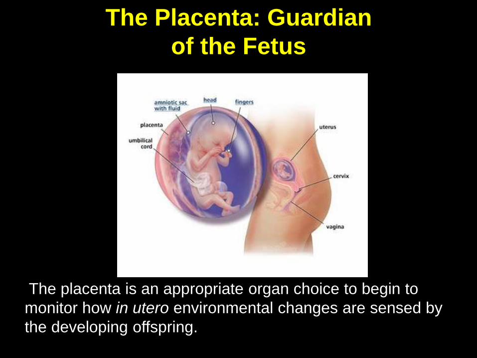
The placenta is an appropriate organ choice to begin to
monitor how in utero environmental changes are sensed by
the developing offspring.
The Placenta: Guardian
of the Fetus
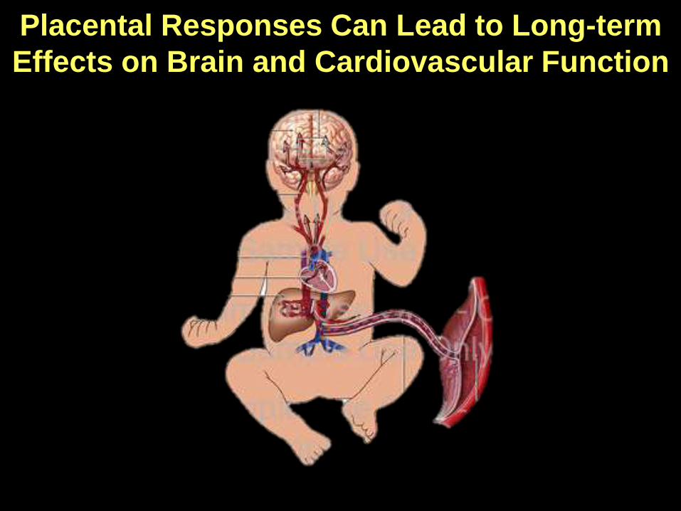
Placental Responses Can Lead to Long-term
Effects on Brain and Cardiovascular Function

Comparative Animal Placentation
Swine
Horses
Cow
Sheep
Goats
Dogs
Cats
Primates
Rodents

Pre-implantational Embryonic Development
Trophectoderm
(TE) cells gives
rise to part of the
fetal placenta
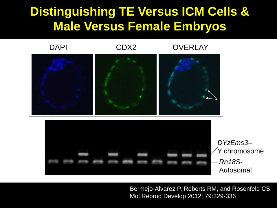
DAPI CDX2 OVERLAY
Rn18S-
Autosomal
♀ ♀ ♂ ♀ ♂ ♀ ♂ ♀ ♂ ♂ ♂
DYzEms3–
Y chromosome
Distinguishing TE Versus ICM Cells &
Male Versus Female Embryos
Bermejo-Alvarez P, Roberts RM, and Rosenfeld CS.
Mol Reprod Develop 2012; 79:329-336

Glucose
(mM)
Sex Total Cells
Mean ± SEM
TE Cells
Mean ± SEM
ICM Cells
Mean ± SEM
0.2 Male 76.3 ± 4.6a 60.8 ± 4c 15.4 ± 1.2
0.2 Female 76.3 ± 4a 61.9 ± 4.8c 14.4 ± 1.4
28 Male 61.1 ± 3.8b 45.8 ± 3.1d 15.3 ± 1.5
28 Female 54.8 ± 3.9b 38.6 ± 3.6d 16.1 ± 1.3
Major Conclusions:
No sex differences were observed in embryonic cell numbers due
to in vitro changes in glucose concentrations.
Elevated in vitro glucose concentrations that approximate
those of diabetic maternal serum decreases total cell and TE
cell numbers in male and female blastocysts,
Bermejo-Alvarez P, Roberts RM, and Rosenfeld CS.
Mol Reprod Develop 2012; 79:329-336
Effect of Glucose Concentration on
Embryo Cell Number According to Sex

We sought to examine how maternal diet
might influence the full range of placental
gene expression in male and female
conceptuses at around mid-pregnancy (12.5
days post-coitus, dpc) in the mouse.
This is when the morphological development
of the placenta is complete but the gonads are
not fully formed ( i.e. minimal steroid
production).
Overall Goal

Prior to our study, only one published study to date examined how maternal diet governs global placental gene expression (Gheorghe et al., Placenta 2009).
This study revealed that in mice a short withdrawal of protein for four days in mid-pregnancy has deleterious consequences on placental gene expression.
The study, however, did not consider the possibility that male and female conceptusesmight show different responses to the imposed diet.
Previous Studies on Effects of Maternal
Diet on Placental Gene Expression
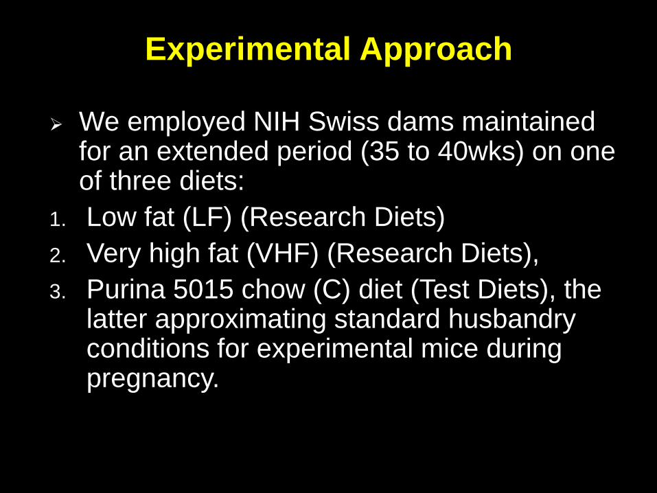
We employed NIH Swiss dams maintained for an extended period (35 to 40wks) on one of three diets:
1. Low fat (LF) (Research Diets)
2. Very high fat (VHF) (Research Diets),
3. Purina 5015 chow (C) diet (Test Diets), the latter approximating standard husbandry conditions for experimental mice during pregnancy.
Experimental Approach
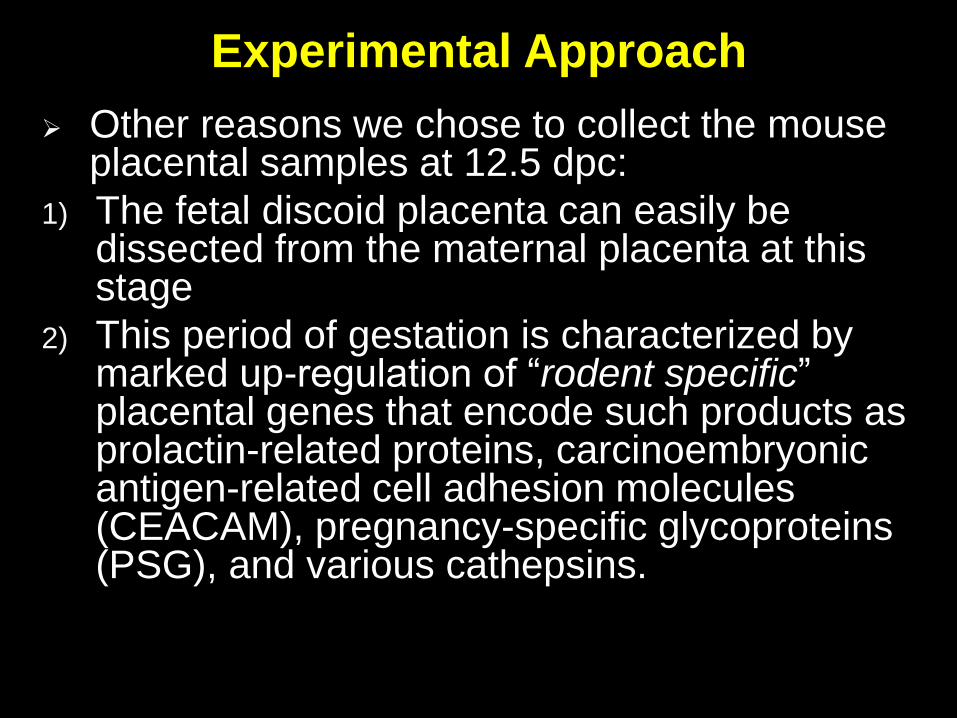
Other reasons we chose to collect the mouse placental samples at 12.5 dpc:
1) The fetal discoid placenta can easily be dissected from the maternal placenta at this stage
2) This period of gestation is characterized by marked up-regulation of “rodent specific” placental genes that encode such products as prolactin-related proteins, carcinoembryonicantigen-related cell adhesion molecules (CEACAM), pregnancy-specific glycoproteins (PSG), and various cathepsins.
Experimental Approach
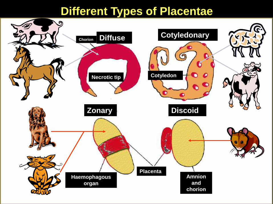
Different Types of Placentae
Diffuse Cotyledonary
Zonary Discoid
Chorion
Necrotic tip Cotyledon
PlacentaAmnion
and
chorion
Haemophagous
organ

Discoid
Placenta
XY FISH Analysis
Whyte et al., Theriogenology, 2007, Mao and
Rosenfeld, Molecul Reprod Develop 2009.
•RNA from the placenta was isolated
and reverse transcribed for
hybridization to Agilent Whole Murine
Genome 4x44K arrays and QRT-PCR.
•Female and male placentae were
pair-matched to the same mid-uterine
horn region, which was on the right
side for all but one VHF dam, where
the pair was selected from the left mid-
uterine horn.
Experimental Approach
a b
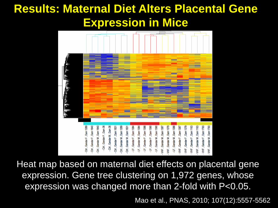
* *Heat map based on maternal diet effects on placental gene
expression. Gene tree clustering on 1,972 genes, whose
expression was changed more than 2-fold with P<0.05.
Mao et al., PNAS, 2010; 107(12):5557-5562
Results: Maternal Diet Alters Placental Gene
Expression in Mice

* *The placentae gene expression patterns of male conceptuses
clearly clusters separately from the placentae of females, when
data on the total regulated genes (with 2-fold differences) across all
dietary groups are compared (P<0.05).
Mao et al., PNAS, 2010; 107(12):5557-62
Results: The Murine Placenta Displays Strikingly
Sexually Dimorphic Differences in Placental Gene
Expression Patterns

Examples of Sexually Dimorphic Expressed
Placental Genes Confirmed by Quantitative
Real-Time PCR Analysis
•Aquaporin 9
•Chemokine (C-C motif) receptor 3
•CEA-related cell adhesion molecule 1 (mouse
placental specific gene)
•Estrogen receptor 1
•Hydroxy-delta-5-steroid dehydrogenase, 3b-and
steroid delta-isomerase 5
•Olfactory receptor 1381
•Olfactory receptor 154
•Olfactory receptor 433
•Olfactory receptor 520
•Renin1
•Renin2

Sex Steroids- Unlikely at 12.5 dpc
X- chromosome dosage- Unlikely due to X-
chromosome dosage, unless the paternal X
chromosome is incompletely silenced in the female
placentae.
Epimutations- Likely mechanisms. After our study
was published, it was demonstrated that fetal sex and
maternal diet can alter DNA methylation patterns in the
murine placenta (Gallou-Kabani et al., PLoS One. 2010;
5:e14398) and gene expression of histone demethylase
paralogues (Kdm5c and Kdm5d, Gabory et al., Plos One
2012; e47988).
How do Sexually Dimorphic
Differences Originate in the Placenta?

In the spiny mouse
(Acomys cahirinus):
• The female placenta has less
spongy zone and more
labyrinth region than males.
• There are sex-dependent and
regional differences in
placental gene expression.
O’Connell et al. Placenta 2013; 34: 119-126
Need to Examine How In Utero Environmental
Changes Affect in a Sex Dependent Manner
Specific Placental Regions and Cells
Rosenfeld Laboratory
• Dr. Jeffrey Whyte
• Dr. Jiude Mao
• Dr. Pablo Bermejo-Alvarez
• Sarah Johnson
University of Missouri
Collaborators
• Dr. R. Michael Roberts
• Dr. Luise King
External Collaborator
• Dr. Frauke Hoffmann
Department of Ecophysiology
and Aquaculture, Leibniz-
Institute of Freshwater
Ecology and Inland Fisheries,
Berlin, Germany
Funding Sources:
• NIH/NIEHS 5R21ES023150-02
• Mizzou Provost Advantage Grant
• NIEHS RC1 ES018195
• MU CVM Faculty Award
• Bond Life Sciences Center
• MU Office of Research
Acknowledgements