PERITONITIS
Nicole Spurlock DVM, DACVECCEmergency and Critical Care Medicine

www.sashvets.com
Peritoneum
• Fenestrated basement membrane
• Semi-permeable– Passive diffusion of electrolytes, urea, H20, certain
drugs/toxins– Equilibration of concentration and osmotic gradients
between peritoneal cavity and intravascular space (peritoneal dialysis)
• Large exudative and absorptive area when inflamed

www.sashvets.com
Primary Peritonitis
• Spontaneous inflammation of the peritoneum in the absence of underlying intra-abdominal pathology– <1% of cases– Hematogenous spread
• FIP– Other routes of entry
• Across lumbosacral arches from pleural cavity• From uterus via ovarian bursae• Across GIT (bacterial translocaiton)

www.sashvets.com
Primary – Small AnimalsJAVMA 2009; 234(7): 906
• 24 cases (1990-2006)
• Primary vs. secondary– Less exudative fluid– More likely Gram+ infection– Surgery may do harm to patients with primary peritonitis
• Surgery is typically not indicated for humans with primary peritonitis

www.sashvets.com
Secondary Peritonitis
• Inflammation of the peritoneum associated with pre-existing abdominal pathology
• More common
• Infectious
• Noninfectious (sterile/aseptic/chemical/traumatic)

www.sashvets.com
Secondary Peritonitis: Infectious
Gastrointestinal tract• Necrosis, rupture,
perforation• Surgical wound dehiscence• Pancreatic abscess
Urogenital tract• Pyometra/uterine rupture• Prostatic abscess• Ruptured urinary bladder
with UTI• Renal abscess• Vaginal rupture
Hepatobiliary• Necrotizing cholecystitis• Liver lobe torsion• Hepatic abscess
Other• Penetrating wounds• Surgical contamination• Evisceration• Splenic abscess• Peritoneal dialysis

www.sashvets.com
Secondary Peritonitis:Compromise of the GIT
• 50-60% cases of bacterial peritonitis
– Gastric/intestinal perforations secondary to GDV– Surgical wound dehiscence– Perforated small intestinal ulcers– Foreign body– Tumor rupture

www.sashvets.com
Secondary Peritonitis: Sterile
• Neoplasia
• Pancreatitis
• Sterile bile peritonitis
• Uroabdomen
• Sterile foreign bodies (sponge, talcum powder, suture material)

www.sashvets.com
Pathogenesis
• Bacterial/chemical irritants vascular dilatationincreased capillary permeability– Facilitates phagocytosis of bacteria and foreign material
• Local deposition of fibrin and plastering of adjacent bowel mesentery (omentum!) to inflamed areas
• Walling-off inflammation

www.sashvets.com
Potential Outcomes
• Successful walling-off, host defenses resolve infection
• Confined suppuration and abscess formation
• Diffuse peritonitis

www.sashvets.com
Diffuse Peritonitis: Pathophysiology
• Large protein-rich fluid losses (tremendous surface area) hypovolemic shock
• Exacerbated by vomiting, diarrhea
• Vasodilation relative hypovolemia
• Splanchnic edema or vasoconstricton stress ulceration/ischemic damage bacterial translocation
• Increased absorption across inflamed peritoneum septicemia, bacteremia, SIRS, sepsis

www.sashvets.com
Course Determination
• Virulence of contaminating microorganisms ± presence of certain enhancing substances (bile salts, talc, hemoglobin)
• Extent of contamination (volume, rate, duration)
• Immunocompetence of host

www.sashvets.com
History & Clinical Signs
• Lethargy/depression, • Inappetance, • Vomiting• Diarrhea• ± Acute collapse• Intact female?• Trauma?• Recent surgery?• Abdominal pain ± distention
– “Praying position”

www.sashvets.com
Physical Exam: Nebulopathy
• Evidence of perfusion impairment– Severe dehydration– Dull mentation– Pallor/injected mucous membranes– +/-Weak femoral/distal pulse quality with BP
• Symptoms of SIRS/sepsis:– Hypo/hyperthermia– Brady/tachycardia– Tachypnea– Leukocytosis or leukopenia

www.sashvets.com
Primary Peritonitis – CatsJAAHA 2009; 45: 268-76
• 13 cases
• Mortality 31%
• Common presentation: Tachypnea, bradycardia, ascites, hypoalbuminemia, anemia
• All cats surgically explored

www.sashvets.com
Laboratory Abnormalities
• Hemoconcentration/anemia
• Hypoglycemia/hyperglycemia
• Decreased sodium/potassium ratio
• Azotemia
• Elevated transaminases
• Hyperbilirubinemia
• Hypoalbuminemia/panhypoproteinemia
• Hypochloremic metabolic alkalosis with hypokalemia
• High anion gap metabolic acidosis:– Lactic– Urinary tract
• Leukocytosis with a shift to the immature forms on the differential cell count
• Absence of leukocytosis or leukopenia

www.sashvets.com
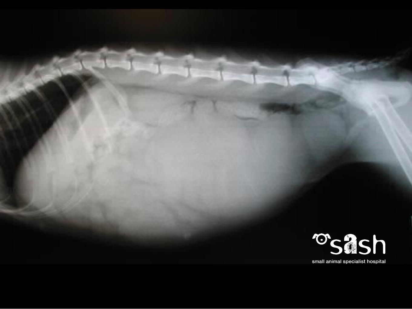
Abdominal Radiography
• Loss of intra-abdominal contrast
• Ileus with distended loops of bowel
• Isolated distended loop bowel or two distinct populations of small intestines
• Foreign body
• Mass effect
• GDV
• Free fluid (> 8 ml/kg)
• pneumoperitoneum

www.sashvets.com
When to cut: PneumoperitoneumJAVMA (223)4, Aug 15 2003JAVMA (225)2, July 15, 2004
Secondary to:– Ruptured hollow viscus– Gas-forming bacteria (abscess)– Penetrating injuries
But also secondary to:– Abdominocentesis– Recent abdominal surgery (18 d)

www.sashvets.com
Ultrasound Examination
• Free fluid• Abscesses in parenchymal organs• Pyometra• Pancreatitis• Abdominal masses• Abnormalities in organ blood flow
– Organ torsion
• Intestinal obstruction

www.sashvets.com
FASTJAVMA 225(8), Oct. 15, 2004
• Focused Abdominal Sonogram Trauma
• Adapted from human medical field to identify abdominal fluid in dogs following trauma
• 4 Views, named for landmarks in R recumbency– DH: diaphragmatico-hepatic– SR: spleno-renal– CC: cysto-colic– HR: hepato-renal
• Sensitive, not specific

www.sashvets.com
Abdominal Paracentesis
• Patient in L lateral recumbency (spleen!)
• Ventral abdomen at the level of the umbilicus clipped and prepared aseptically
• Urinary bladder evacuated
• Local infiltration with lidocaine?
• 4-quadrant centesis– 23 ga needles (4-6)– 1 ml syringe– Slides & sample collection tubes
• Closed or open-needle techniques described
• Contraindications: coagulopathy; organomegaly; distension of abdominal viscus

www.sashvets.com
Abdominal Paracentesis
Bloody fluid that clots:• r/o vessel or organ
Effective? • Canines: 5.2-6.6 ml/kg
abdominal fluid required for positive results
Alternative:• Diagnostic peritoneal lavage?

www.sashvets.com
Evaluation of Peritoneal Fluid
• Cytology/culture: Crystals? Microbes? Pigments?
• Total protein• Lactate• Glucose• Creatinine• Bilirubin• PCV
• Surgical Intervention:– Septic abdomen– Bile peritonitis– Uroabdomen (+/-)– Persistent hemoabdomen

www.sashvets.com
Normal Peritoneal Fluid
• < 1mL/kg non-clotting, clear, yellow fluid (straw-colored)
• Transudate:– < 3000 nucleated cells/L– 50% macrophages, 50% lymphocytes– < 2.5 g/dL protein (albumin)
• Functions– Decreases friction between opposing surfaces– Dissemination of localized inflammation/infection
• Drainage– Diaphragmatic lymphatics and thoracic duct to sternal lymph nodes
(80%)

Abdominal effusion
Total protein count, total nucleated cell
count
< 2.5 g/dl protein
<1500 nucleated cells/L
2.5-7.5 g/dl protein
1000-7000 nucleated cells/L
> 3 g/dl protein
> 7000 nucleated cells/L
Transudate
Modified transudate
Exudate
Algorithm to Classify Effusions

Transudate
Low serum albumin
YesNo
Proteinuria
YesNo
Diarrhea Consider PLN
No Yes
Consider hepaticinsufficiency Consider PLE
Abdominal fluid [creatinine]greater than serum
creatinine
No Yes
Consider leakagefrom intestinal
lymphatics
Uroabdomen

Modified Transudate
Fluid bile-stained, with phagocytized bile pigments
NoYes
Fluid is red, with phagocytized RBCs
NoYes
Bile peritonitis
Intracavitary hemorrhage
Fluid is milky white
Yes No
Chylous/pseudo-chylous
Small lymphocytes predominate
Yes No
Chylous/pseudo-chylous Lymphoblasts, mast cells, cells w/nuclear
criterea for malignancy

No
Neutrophils predominate
YesNo
Nonexfoliating neoplasia, FIP, chronic inflammation,
Diaphragmatic Hernia, Liver Lobe Torsion, other
Abdominal fluid [creat] > serum [creat]
Yes No
Uroperitoneum Organsims present (mycotic, rickettsial, protozoal)
Yes No
Yes No
Neoplasia
Heart failure?
Yes
Infectious pleuritis/peritonitis
FIP, tissue inflammation, nonexfoliating
neoplasia, other
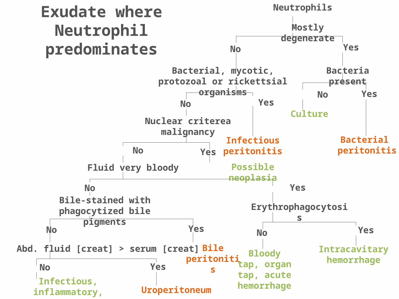
Mostly degenerate
Yes
Bacteria present
YesNo
Culture
Bacterial peritonitis
No
Bacterial, mycotic, protozoal or rickettsial organisms
Yes
Infectious peritonitis
No
Yes
Nuclear criterea malignancy
No
Possible neoplasiaFluid very bloody
YesNo
Erythrophagocytosis
YesNo
Intracavitary hemorrhageBloody tap, organ tap,
acute hemorrhage
Bile-stained with phagocytized bile pigments
Exudate where Neutrophil
predominates
Neutrophils
Yes
Bile peritonitis
No
Abd. fluid [creat] > serum [creat]
Uroperitoneum
YesNo
Infectious, inflammatory, neoplastic

Exudate where Neutrophil does not
predominate
No
Primarily mast cells
Primarily small lymphocytes
NoYes
Mast cell tumor
Chylous/pseudo- chylous
Primarily lymphoblasts
Yes NoLymphosarcoma
Primarily macrophages
Yes NoFluid is bile-stained with phagocytized bile pigment
Cells with nuclear criterea for malignancy
Yes
Yes
Bile peritonitis
NoLow-grade chronic inflammation, FIP
Yes No
Neoplasia? Refer
Exudate

www.sashvets.com
When to Cut: Septic Effusions
• Non-septic effusions: cut?
• Hepatic disease, GI neoplasia, multicentric lymphoma, splenic leiomyosarcoma, pancreatitis, right-congestive heart failure, intact pyometra
• Septic effusions: cut!
– GI – intestinal neoplasia, foreign body, postoperative enterotomy dehiscence, duodenal feeding tube leakage
Other – hepatic abscess, pancreatic abscess, mesenteric lymph node abscess, contamination from urinary bladder


www.sashvets.com
Septic Effusion: Lactate and GlucoseBonzynski et al. JAVMA 2003
• Blood-to-fluid (BFG) glucose difference > 20 mg/dl (1.1 mmol/L) – 100% sensitive and specific for dx septic peritonitis in dogs – 86% sensitive and 100% specific in cats
• Blood to fluid lactate difference < 2mmol/L – 100% sensitive and specific for dx of septic peritonitis
(dogs)
• IV administration of glucose or presence of hemoabdomen may decrease accuracy

www.sashvets.com
Evaluation of post-celiotomy peritoneal drain fluid volume, cytology, and blood-to-peritoneal fluid lactate and glucose differences in normal dogs Vet Surg 2011 40(4)
• JP drain placed after abdominal explore in 10 healthy dogs
• Peritoneal fluid analyzed q6 x 7 days
• Results after day 4:– blood-to-peritoneal glucose concentration differences were consistent with septic
effusion based on previously reported values (all dogs)
– Blood-to-peritoneal lactate concentration consistent with septic peritonitis (70% of dogs)
• Conclusion: post-operative blood to peritoneal fluid glucose & lactate may not be reliable indicators of septic peritonitis when evaluating fluid collected from closed suction drains

www.sashvets.com
When to Cut: Bile Peritonitis
• Abdominal fluid – Presence of bilirubin = 100% effective in diagnosis bile
peritonitis
– Effusion [bilirubin] > serum [bilirubin]
– Cytology: bile pigments & crystals may also be seen microscopically
– If secondary to ruptured mucocele, these changes may not be present
• Gelatinous bile may not disperse intra-abdominally

www.sashvets.com
When to Cut: Uroabdomen
• Abdominal fluid [creatinine] to peripheral blood [creatinine] ratio of >2:1
• Abdominal fluid [potassium] to peripheral blood [potassium] ratio of >1.4:1
• Remember:– Bladder palpable in majority (>50%)– Ascites can occur subsequent to severe UO– Some animals with urinary compromise will still urinate & have urine
collecting in the abdomen

www.sashvets.com
Pre-op Stabilization
• Fluid resuscitation • Blood products (peri-op)• Correction acid/base, electrolyte abnormalities• Euglycemia• Vasopressors for refractory hypotension• Inotropes for documented myocardial failure• 4-quandrant bacteriocidal systemic antibiotics• Pain management• Source control

www.sashvets.com
When to Cut: a Summary
• Septic peritonitis– Abdominal abscess– GI obstruction– Ischemic/perforated/ruptured GI
• Persistent abdominal hemorrhage– Trauma– Neoplasia
• Uroperitoneum (+/-)• Free abdominal gas (non-iatrogenic or associated with
pneumomediastinum)• Bile peritonitis

www.sashvets.com
How to know: a Summary
– Septic peritonitis:• Intracellular microbes• Blood-to-fluid glucose
concentration difference > 1.1 mmol/L
• Blood –to-fluid lactate difference < 2 mmol/L
– Bile peritonitis:• Effusion [bilirubin] > blood
[bilirubin] (usually x2)• Presence of bile pigments or
crystals
– Uroperitoneum• Fluid creatinine > 2:1 of
serum creatinine• Fluid potassium > 1.4:1
serum potassium
– Hemoperitoneum• PCV of fluid near peripheral
blood (or increasing)• Evaluate with TP: decreasing
protein levels indicate hemorrhage

www.sashvets.com.au twitter: @SASHvets
Phone - (02) 9889 0289 Fax - (02) 9889 0431
Level 1, 1 Richardson Place, North Ryde 2113, Sydney, NSW
www.sashvets.com.au twitter: @SASHvets
Phone - (02) 9889 0289 Fax - (02) 9889 0431
Level 1, 1 Richardson Place, North Ryde 2113, Sydney, NSW