SABRINA HITOMI UYEKITA
Desenvolvimento das glândulas salivares menores: relação
morfológica paralela entre a expressão das isoformas de
TGF-β e marcadores citoesqueletais da maturação glandular
Dissertação apresentada à Faculdade de Medicina da Universidade de São Paulo para obtenção do título de Mestre em Ciências Área de Concentração: Dermatologia Orientadora: Profa. Dra. Silvia Vanessa Lourenço
São Paulo
2009

Dados Internacionais de Catalogação na Publicação (CIP)
Preparada pela Biblioteca da Faculdade de Medicina da Universidade de São Paulo
©reprodução autorizada pelo autor
Uyekita, Sabrina Hitomi Desenvolvimento das glândulas salivares menores : relação morfológica paralela entre a expressão das isoformas de TGF-β e marcadores citoesqueletais da maturação glandular / Sabrina Hitomi Uyekita. -- São Paulo, 2009.
Dissertação(mestrado)--Faculdade de Medicina da Universidade de São Paulo. Departamento de Dermatologia.
Área de concentração: Dermatologia. Orientadora: Silvia Vanessa Lourenço.
Descritores: 1.Fator transformador de crescimento beta 2.Glândulas salivares/crescimento & desenvolvimento 3.Imunoistoquímica 4.Imunofluorescência
USP/FM/SBD-463/09

SABRINA HITOMI UYEKITA
Desenvolvimento das glândulas salivares menores: relação
morfológica paralela entre a expressão das isoformas de
TGF-β e marcadores citoesqueletais da maturação glandular
Dissertação apresentada à Faculdade de Medicina da Universidade de São Paulo para obtenção do título de Mestre em Ciências
Área de Concentração: Dermatologia Orientadora: Profa. Dra. Silvia Vanessa Lourenço
São Paulo
2009

DEDICATÓRIA
Dedico este trabalho aos meus pais Takeshi e Marta e aos meus irmãos
Mika, Priscila e Hitoshi pelo amor, apoio e compreensão em todos os
momentos.
Ao Jorge e Ruth por me acolherem e incentivarem sempre.
Ao Jorginho pelo amor, companheirismo e estímulo nos momentos mais
difíceis. Você ilumina minha vida.
“Acredito na eterna importância do lar
como instituição fundamental da sociedade.
Acredito nas possibilidades incomensuráveis de cada menino ou menina.
Acredito na imaginação, confiança, esperanças e ideais
que há no coração de todas as crianças.
Acredito na beleza da natureza, da arte, dos livros e da amizade.
Acredito na satisfação do dever cumprido.
Acredito nas pequenas alegrias domésticas da vida de todos os dias.”
Ozora Davis (1866-1931)

AGRADECIMENTO ESPECIAL
À minha orientadora querida Profa. Dra. Silvia Vanessa Lourenço, pela
amizade, cumplicidade, incentivo, confiança e principalmente carinho
durante todos estes anos de convívio.
"O valor das coisas não está no tempo em que elas duram, mas na
intensidade com que acontecem. Por isso existem momentos inesquecíveis,
coisas inexplicáveis e pessoas incomparáveis.”
Fernando Pessoa

AGRADECIMENTOS
À minha estimada colega de pós-graduação Dirce Mary Correia Lima pela
amizade, estímulo e presteza em todos os momentos que compartilhamos
no laboratório.
Aos meus amigos e companheiros de pesquisa Ricardo Hsieh e Fábio
Rodrigues Gonçalves de Carvalho pela amizade e solidariedade nos
momentos difíceis.
Ao colega de laboratório Leonardo Kamibeppu pela agilidade e dedicação
na confecção das lâminas para este estudo.
Ao Prof. Dr. Antonio Sesso pela gentileza em ceder o microscópio para a
aquisição das imagens para este trabalho.
"Só existem dois dias no ano que nada pode ser feito. Um se chama ontem
e o outro se chama amanhã, portanto, hoje é o dia certo para amar, acreditar,
fazer e principalmente viver.”
Dalai Lama

“O objetivo sempre nos escapa. Quanto maior o progresso, maior o
reconhecimento de nossa insignificância. A satisfação está no esforço, não
no prêmio. O esforço total é a vitória total.”
Mohandas K. Gandhi

Esta dissertação está de acordo com as seguintes normas, em vigor no
momento desta publicação:
Referências: adaptado de International Committee of Medical Journals
Editors (Vancouver)
Universidade de São Paulo. Faculdade de Medicina. Serviço de Biblioteca e
Documentação. Guia de apresentação de dissertações, teses e monografias.
Elaborado por Anneliese Carneiro da Cunha, Maria Julia de A. L. Freddi,
Maria F. Crestana, Marinalva de Souza, Suely Campos Cardoso, Valéria
Vilhena. 2a ed. São Paulo: Serviço de Biblioteca e Documentação; 2005.
Abreviaturas dos títulos dos periódicos de acordo com List of Journals
Indexed in Index Medicus.

SUMÁRIO
Lista de abreviaturas
Lista de tabelas
Lista de figuras
Resumo
Summary
1 INTRODUÇÃO 1
2 REVISÃO DA LITERATURA 4
2.1 O desenvolvimento glandular 5
2.2 Fator transformador de crescimento-beta (TGF-β) 17
2.2.1 A estrutura do TGF-β 18
3 OBJETIVOS 24
3.1 Gerais 25
3.2 Específicos 25
4 MATERIAL E MÉTODO 26
4.1 Obtenção e seleção de fetos humanos para dissecação de
estruturas glandulares 27
4.2 Processamento histológico 28
4.3 Imunoistoquímica 30
4.4 Imunofluorescência 31
5 RESULTADOS 35
5.1 Imunoistoquímica e imunofluorescência 36
6 DISCUSSÃO 45
7 CONCLUSÕES 51

8 ANEXO 53
9 REFERÊNCIAS 55
10 APÊNDICE 65

LISTA DE ABREVIATURAS E SÍMBOLOS
ºC graus Celsius
β beta
µm Micrômetros
AMH Hormônio anti-muleriano
BMP Bone morphogenetic protein
BSA Soro-albumina bovina
CA Califórnia
CK Citoqueratina
CK LMW Citoqueratina de baixo peso molecular
Co-SMAD Common-partner SMAD
DAB 3,3 Diamino-Benzidine
DNA Ácido desoxirribonucléico
EGF Fator de crescimento epitelial
FCS Soro fetal bovino
FGF Fator de crescimento fibroblástico
g Grama
kDa Quilodalton
NH2 Grupo amina
PAS Periodic Acid Schiff
PBS Phosphate Buffer Saline
pH Potencial hidrogeniônico

RNAm Ácido Ribonucléico mensageiro
R-SMAD Receptor-activared SMAD
SMA Actina de músculo liso
SMAD Small Mother Against Decapentaplegic
TGF Transforming Growth Factor
™ Trade Mark
Tris-HCl Tampão de fosfato salino
U.S.A Estados Unidos da América

LISTA DE FIGURAS
Figura 1 – Estágios de diferenciação das glândulas salivares 12
Figura 2 – Representação esquemática de uma glândula
salivar completamente desenvolvida 13
Figura 3 – Expressão das isoformas de TGF-β durante o desenvolvimento
das glândulas salivares humanas 36
Figura 4 – Marcadores da diferenciação do citoesqueleto
durante o desenvolvimento das glândulas salivares menores humanas 38

LISTA DE TABELAS
Tabela 1 - Os anticorpos primários utilizados, clone, procedência e diluição........... 31
Tabela 2 - Análise semi-quantitativa da expressão das subunidades de TGF-
beta durante as fases da morfogênese das glândulas salivares humanas e
nas glândulas salivares humanas plenamente desenvolvidas .................................. 40

Uyekita SH. Desenvolvimento das glândulas salivares menores: relação
morfológica paralela entre a expressão das isoformas de TGF-β e
marcadores citoesqueletais da maturação glandular [dissertação]. São
Paulo: Faculdade de Medicina, Universidade de São Paulo; 2009. 66p.
RESUMO
A morfogênese das glândulas salivares envolve eventos complexos e
coordenados, dependentes da interação epitélio-mesênquima e do micro-
ambiente. Fatores de crescimento coordenam vários desses processos
biológicos e o fator transformador de crescimento-beta (TGF-β) mostra-se
relevante. Utilizando imunoistoquímica e imunofluorescência, a distribuição
do TGF-β1, 2 e 3 foi mapeada e sua expressão comparada com a expressão
de marcadores de maturação em glândulas salivares humanas obtidas de
fetos que tinham entre 4ª e 24ª semanas de vida intra-uterina. O TGF-β1 foi
detectado durante a fase pseudoglandular no mesênquima. Nas outras
etapas da diferenciação glandular esse fator foi expresso no citoplasma das
células acinares até a glândula salivar adulta. O TGF-β2 foi detectado desde
o estágio de botão inicial da glândula salivar. Sua expressão foi observada
nas células ductais e sua presença aumentada ao longo da diferenciação
glandular. O TGF-β3 foi visto durante a fase pseudoglandular das glândulas
salivares, inicialmente fraco nas células ductais e foi o único detectado em
células mioepiteliais. A troca de subunidades de TGF-β durante a maturação
das glândulas salivares sugere mudanças estimuladas durante os
complexos estágios de desenvolvimento dessas glândulas. O presente

estudo reafirma essa evidência, e mostra que as subunidades do TGF-β são
fatores importantes durante a diferenciação de glândulas salivares.
Descritores: 1. Fator transformador de crescimento beta 2.Glândulas
salivares / crescimento & desenvolvimento 3.Imunoistoquímica 4.
Imunofluorescência.

Uyekita SH. Developing human minor salivary glands: morphological parallel relation between the expression of TGF-beta isoforms and cytoskeletal markers of glandular maturation [dissertation]. São Paulo: Faculdade de Medicina, Universidade de São Paulo; 2009. 66p.
SUMMARY
Morphogenesis of salivary glands involves complex coordinated events.
Synchronization between cell proliferation, polarization and differentiation,
which are dependent on epithelial–mesenchymal interactions and on the
microenvironment, is a requirement. Growth factors mediate many of these
orchestrated biological processes and transforming growth factor-beta (TGF-
β) appears to be relevant. Using immunohistochemistry and
immunofluorescence, we have mapped the distribution of TGF-β 1, 2 and 3
and compared it with the expression of maturation markers in human salivary
glands obtained from fetuses ranging from weeks 4 to 24 of gestation. TGF-β
1 first appeared during pseudoglandular stage in the surrounding
mesenchyme and, in the more differentiated stages, was expressed in the
cytoplasm of acinar cells throughout the adult gland. The TGF-β 2 was
detected since the bud initial stage of the salivary gland. Its expression was
observed in ductal cells and increased along gland differentiation. The TGF-β
3 was detected from the pseudoglandular stage of the salivary gland, being
weakly expressed on ductal cells, and it was the only factor detected on
myoepithelial cells. The data suggest that TGF-β have a role to play in
salivary gland development and differentiation.

Descriptors: 1.Transforming growth factor beta 2.Salivary glans / growth &
development 3. Immunohistochemistry 4. Immunofluorescence.

1
1 INTRODUÇÃO

2
1. INTRODUÇÃO
Durante as últimas décadas, aspectos da biologia básica vêm
assumindo importância no estudo das condições fisiológicas, patológicas e
do desenvolvimento de diversos tecidos e órgãos. Com relação às glândulas
salivares, a busca de critérios diagnósticos mais aprimorados e da
compreensão de sua histogênese tem sido objetivo de numerosos estudos.
Além disso, diversas teorias foram propostas a respeito da gênese dos
tumores que acometem as glândulas salivares, correlacionando a
diferenciação das células neoplásicas com o desenvolvimento do segmento
glandular de sua provável origem (Eversole, 1971; Regezi; Batsakis, 1977;
Batsakis, 1980; Attie; Sciubba, 1981; Batsakis et al., 1989; Dardick et al.,
1990). Entretanto, são raros os trabalhos que analisam o desenvolvimento e
a diferenciação das glândulas salivares humanas, estabelecendo uma
relação morfológica e funcional com as neoplasias (Gustafsson et al., 1988).
A imunoistoquímica, técnica que permite a identificação de antígenos
nos tecidos através da utilização de anticorpos específicos, vem sendo
utilizada exaustivamente na pesquisa, visando à busca de marcadores
protéicos peculiares a cada tecido. Estas pesquisas têm auxiliado
sobremaneira o reconhecimento de diversas doenças, representando um
avanço no diagnóstico histopatológico (Mcnicol; Richmond, 1998).
No âmbito das glândulas salivares, vários anticorpos específicos têm
se mostrado ferramentas úteis para o estudo da diversidade celular e

3
estágios de diferenciação dos processos neoplásicos, muitas vezes
definindo critérios de diagnóstico (Araújo et al., 1987; Araújo et al., 1988;
Carvalho et al., 1990; Araújo et al., 1991 a, b; Carvalho et al., 1993; Freitas;
Araújo; Araújo, 1993; Sousa, 1994; Sousa; Araújo, 1994; Crivelini; Araújo,
1995; Loyola et al., 1995; Souza et al., 1995; Araújo; Sousa, 1996; Loyola;
Araújo, 1996; Crivelini; Souza; Araújo, 1997; Figueiredo; Sousa; Araújo,
1997; Jaeger et al., 1997; Araújo et al., 1999; Araújo et al., 2000; Loducca et
al., 2000).
Nosso estudo, dessa forma, pretende avaliar morfologicamente a
participação de fatores de crescimento e comparar sua expressão com
marcadores da diferenciação durante os processos de morfogênese das
glândulas salivares humanas.

4
2 REVISÃO DA LITERATURA

5
2. REVISÃO DA LITERATURA
2.1 O desenvolvimento glandular
As glândulas salivares iniciam seu desenvolvimento a partir de botões
epiteliais que se originam do epitélio bucal na vida fetal precoce.
O primórdio da glândula parótida humana aparece entre a 4ª e 6ª
semana de vida intra-uterina nas dobras laterais do estomodeo (cavidade
oral primitiva). A glândula submandibular origina-se do soalho bucal durante
a 6ª semana e a sublingual se forma lateralmente ao primórdio da
submandibular entre a 7ª e 8ª semana de vida intra-uterina. Todas as
glândulas salivares menores formam-se mais tardiamente a partir do epitélio
de revestimento de áreas específicas da cavidade bucal (Hand, 1980;
Tonge; Luke, 1984; Dale, 1994; Ellis; Auclair, 1996).
Entende-se por processo de morfogênese das glândulas salivares as
etapas que levam ao arranjo lobulado, característico desse tecido, enquanto
que a diferenciação compreende os processos de amplificação da síntese e
armazenamento dos grânulos de secreção. A combinação desses dois
processos é denominada de citodiferenciação. Estudos sugerem que os
processos de morfogênese e de citodiferenciação das glândulas salivares
estão parcialmente ligados, mas são reguladas por processos distintos,
sendo que a expressão total de um dos dois processos é modulada ou
controlada pela matriz extracelular e por outros fatores (Cutler, 1990).

6
Segundo Tucker, em revisão de 2007, o desenvolvimento das
glândulas salivares pode ser dividido em cinco estágios.
O primeiro estágio (pré-botão) envolve a indução da proliferação do
epitélio de superfície pelo mesênquima adjacente resultando no
espessamento e formação dos botões epiteliais. O botão em crescimento é
separado do mesênquima condensado pela lâmina basal secretada pelo
próprio epitélio (Bernfield; Banderjee; Cohen, 1972; Hand, 1980; Hiatt; Sauk,
1991; Klein, 1994; Ellis; Auclair, 1996; Tucker, 2007).
O segundo estágio (botão inicial) é representado pela formação e
crescimento dos cordões epiteliais principais. Nesse estágio observam-se a
proliferação das células dos botões formando cordões celulares sólidos,
constituídos por duas camadas de células, associados à condensação e
proliferação do mesênquima adjacente. Ultra-estruturalmente, as células
indiferenciadas que compõem os cordões são caracterizadas como células
cubóides irregulares, contendo numerosos ribossomos livres, nucléolos
proeminentes, complexo de Golgi e retículo endoplasmático rugoso pouco
desenvolvido (Bernifield; Banderjee; Cohen, 1972; Hand, 1980; Cutler, 1990;
Hiatt; Sauk, 1991; Klein, 1994; Ellis; Auclair, 1996; Tucker, 2007). Adi e
Chisholm em 1994, observaram que as células epiteliais que compõem os
cordões sólidos de glândulas submandibulares humanas exibem material
PAS (Periodic Acid Schiff) positivo no seu interior, indicando já nessa fase
precoce a produção de mucina.
O terceiro estágio (pseudo-glandular) corresponde à ramificação da
porção terminal dos cordões epiteliais e à continuação da diferenciação

7
glandular. Neste, os cordões epiteliais proliferam rapidamente e ramificam-
se em bulbos terminais que são compostos por 10 a 12 células (Tucker,
2007).
O quarto estágio (canalicular) corresponde às repetidas ramificações
dos cordões epiteliais nas porções terminais formando os lóbulos compostos
por um sistema de bulbos ramificados como uma árvore (processo de
arborificação). Nesse estágio a cápsula da glândula começa a se formar a
partir do mesênquima e circunscreve o parênquima glandular (Bernfield;
Banderjee; Cohen, 1972, Hand, 1980; Cutler, 1990; Hiatt; Sauk, 1991; Klein,
1994; Ellis; Auclair, 1996). Ainda nesse estágio ocorre a canalização dos
cordões epiteliais com a formação de um orifício criando um tubo ou ducto.
Estudos experimentais indicam duas teorias a respeito do mecanismo
de canalização: (1) graus diferentes de proliferação entre camadas externas
e internas dos cordões epiteliais, (2) secreção do fluído pelas células
ductais, o que aumenta a pressão hidrostática e produz um lúmen junto ao
cordão. Tanto a ramificação das estruturas ductais quanto o crescimento dos
septos de tecido conjuntivo continuam neste estágio do desenvolvimento
(Hand, 1980; Hiatt; Sauk, 1991; Klein, 1994; Ellis; Auclair, 1996; Tucker,
2007).
A citodiferenciação dos tipos celulares específicos das glândulas
salivares parece ser iniciada após as características de arborificação
estarem estabelecidas, e corresponde ao quinto estágio (botão terminal)
(Cutler, 1990; Tucker, 2007). Esse estágio representa a diferenciação
morfológica das glândulas salivares em desenvolvimento. Durante esse

8
período a atividade mitótica ao invés de ocorrer em todo o cordão epitelial,
irá ocorrer apenas na porção terminal do bulbo, aonde as células irão se
diferenciar em células dos túbulos terminais e pró-acinares. Com a formação
dos lúmens nos bulbos terminais, ocorrem fissuras nas células
circunjacentes fazendo com que cada bulbo terminal fique dividido em várias
subunidades constituídas por duas camadas de células circundando um
lúmen e que são chamadas de túbulos terminais. Nesta fase as unidades da
porção terminal luminal estão pouco diferenciadas para serem chamadas de
ácinos e são conhecidas como sacos terminais. Acredita-se que as células
externas originem as células mioepiteliais e basais enquanto que as células
internas sofrem diferenciação para acinares e luminais (Klein, 1994; Ellis;
Auclair, 1996).
As células mioepiteliais provavelmente são originadas das células
epiteliais externas localizadas nos túbulos terminais e desenvolvem-se
concomitantemente com a citodiferenciação das células acinares. A
diferenciação das células mioepiteliais está caracterizada pela agregação de
microfilamentos e demonstração imunoistoquímica da actina, miosina e
filamentos intermediários de pré-queratina (Yaku, 1983; Klein, 1994).
A maturação das células acinares ocorre em estágios específicos
classificados de acordo com a morfologia dos grânulos de secreção e das
organelas celulares, sendo diferente entre os ácinos mucosos e serosos. O
primeiro sinal de diferenciação dos sacos terminais em ácinos está
relacionado com o aumento da dilatação das cisternas do retículo
endoplasmático rugoso pelas proteínas secretórias, seguido da observação

9
de grânulos secretórios agranulados e presença de complexo de Golgi
funcional. Progressivamente, as quantidades de retículo endoplasmático
rugoso, Golgi e grânulos secretórios vão aumentando com a maturação dos
ácinos (Yaku, 1983; Klein, 1994).
Os ductos excretores das glândulas salivares diferenciam-se a partir
dos cordões principais: as ramificações distais dos cordões principais
originam os grandes ductos estriados, enquanto que os ductos intercalares,
pequenos ductos estriados, células secretoras e mioepiteliais originam-se
das células dos túbulos terminais. A diferenciação dos ductos excretores e
estriados maiores ocorre apenas após os cordões celulares sólidos terem
sofrido canalização (Hand, 1980; Dale, 1994; Ellis; Auclair, 1996; Denny;
Ball; Redman, 1997).
Os estímulos provenientes dos mecanismos secretores e da
inervação da glândula são responsáveis pela continuação e maturação
durante a citodiferenciação, porém a glândula será totalmente formada no
período pós-natal. Este desenvolvimento pós-natal está relacionado com: (1)
a maturação do estímulo de secreção, que é responsável pela ativação dos
receptores de membrana que sinalizam caminhos de transdução de sinal
junto à célula e controlam a secreção das células; e com (2) o
estabelecimento de conexões neurais do sistema nervoso autônomo, que é
o primeiro regulador da função das glândulas salivares (Hand, 1980; Lee et
al., 1990; Klein, 1994; Ellis; Auclair, 1996).
Como resultado do processo de desenvolvimento acima descrito as
glândulas salivares tomam a forma de estrutura composta túbulo-acinar, o

10
que indica a presença de um sistema ductal ramificado e unidades
secretoras com porção tubular e acinar. Essas glândulas são exócrinas,
cujas secreções fluem para o interior da cavidade bucal.
Histologicamente, as glândulas salivares possuem três tipos de
porção secretora: mucosas, serosas e mistas, e três tipos de ductos:
intercalado, estriados (intralobulares) e excretor (interlobular). As principais
células encontradas são as células mucosas, células serosas e as
mioepteliais. Circundando e fornecendo suporte para os sistemas ductal e
secretor existe uma cápsula de tecido conjuntivo denso que se estende na
glândula formando septos dividindo o parênquima em lóbulos. O tecido
conjuntivo é importante tanto para a sustentação das glândulas como dos
nervos (autônomos), vasos sanguíneos e linfáticos (Bhaskar, 1978; Hand,
1980; Klein, 1994; Tonge; Luke, 1994; Ellis; Auclair, 1996).
As diversas estruturas das glândulas salivares completamente
desenvolvidas possuem características fenotípicas, protéicas e moleculares
peculiares, podendo ser identificadas por técnicas de detecção de proteínas
marcadoras específicas, como a imunoistoquímica. Dessa forma,
subunidades de citoqueratinas identificam os diversos segmentos epiteliais
das glândulas salivares e as células mioepiteliais podem ser detectadas pela
expressão de proteínas contráteis como, por exemplo, a actina de músculo
liso (Dardick et al., 1988; Araújo; Carvalho; Araújo, 1994).
No total, após toda a sua formação, as glândulas salivares são
compostas de: (1) glândulas maiores, que compõe todo o conjunto de
parótida, sublingual e submandibular e (2) glândulas menores, que são

11
nomeadas de acordo com a sua localização como labiais, bucais, linguais,
palatinas, gengivais e glossopalatinas (Bhaskar, 1978; Hand, 1980, Klein,
1994).
A Figura 1 exemplifica os estágios da morfogênese das glândulas
salivares humanas e a Figura 2 mostra esquematicamente a estrutura de
uma glândula salivar completamente desenvolvida.

12

13
Figura 1 – Estágios de diferenciação das glândulas salivares.
A: Fase de pré-botão composto por células de aspecto basalóide e
indiferenciado (hematoxilina/eosina).
B: Cordões epiteliais ramificados e presença de algumas estruturas
exibindo início de lúmen central (hematoxilina/eosina).
C: Cordões epiteliais ramificados e canalizados (hematoxilina/eosina);
D: Ducto excretor canalizado e exibindo estratificação epitelial
(hematoxilina/eosina).
E: Início da fase canalicular com presença de sacos terminais
rudimentares (hematoxilina/eosina).
F: Glândula salivar mucosa: fase terminal de botão com estruturas
acinares morfologicamente diferenciadas, com células de citoplasma amplo
e claro e aspecto piramidal (hematoxilina/eosina).

14

15
Semelhante a outros órgãos, a formação das glândulas salivares
envolve um mecanismo morfogenético de coordenação, incluindo mudanças
reguladas na forma celular, expressão de genes, e migração celular
direcionada levando a um completo desenvolvimento glandular com
importantes funções secretoras. Apesar dos locais e do tempo de
desenvolvimento diferir significativamente entre as diversas glândulas, o
processo envolvido no desenvolvimento é similar e sofre influências de
fatores extrínsecos e intrínsecos que regulam o processo de morfogênese,
proliferação e diferenciação celular.
Os fatores intrínsecos são definidos como características pré-
programadas da expressão genética específica para cada tipo celular.
Durante seqüência programada, os genes são ativados e desativados em
tempos apropriados levando ao crescimento e diferenciação normal das
células (Cutler, 1990; Klein, 1994; Denny; Ball; Redman, 1997).
Os fatores extrínsecos são sinais produzidos pela interação célula-
célula e célula-matriz-extracelular. Dessa forma, o desenvolvimento do
tecido glandular envolve interações entre o epitélio e o mesênquima para
formar a porção funcional da glândula (parênquima). Essas interações são
definidas como induções secundárias, nas quais a presença do mesênquima
torna-se necessária para o desenvolvimento normal do epitélio. O
mesênquima é composto por células derivadas do mesoderma e da crista
neural e tem sido chamado de ectomesênquima ou mesoectoderma.
Portanto, o ectomesênquima possui papel essencial no desenvolvimento do
tecido glandular humano visto que, sua interação com o epitélio regula tanto

16
a iniciação como o crescimento e a citodiferenciação das células do tecido
glandular (Hand, 1980; Azuma; Sato, 1994; Klein, 1994; Denny; Ball;
Redman, 1997).
A posição e forma dos órgãos são determinadas pela atividade
coordenada de um grupo de células primitivas umas com outras e com
células vizinhas. Embora as informações disponíveis sejam limitadas de
como essas atividades são coordenadas, a expressão de ligantes e
receptores específicos de superfície celular são altamente relacionadas em
uma rede complexa de sinalização, a qual regula a divisão celular, migração
e diferenciação (Thiery, 2003).
Dentre as principais citocinas envolvidas em processos de
diferenciação celular está o TGF-β.

17
2.2 Fator transformador de crescimento – beta (TGF-β)
O TGF-β (fator transformador de crescimento-beta) é uma citocina
multifuncional de 25 kDa que faz parte da “Superfamília dos TGF-β”
composta por TGF-β, activinas, inhibinas, hormônio anti-mulleriano (AMH),
proteína morfogenética óssea (BMP do inglês bone morphogenetic protein),
miostatina e outros que secretam polipeptídeos sinalizadores com diversas
funções no desenvolvimento celular e na homeostase de tecidos adultos em
todos os metazoários (Sporn; Roberts, 1990; Piek; Heldin; Ver Dijke, 1999;
Chin et al., 2004).
A família de TGF-β é composta por oito membros verdadeiros, que
compartilham entre si uma vasta seqüência homóloga de aminoácidos
englobados na “Superfamília dos TGF-β” (Millan et al., 1991). Conforme cita
Chin et al. (2004), estas proteínas atuam na regulação da proliferação,
diferenciação e apoptose de vários tipos celulares.

18
2.2.1 A estrutura do TGF-β
Clones de DNA complementar foram isolados em cinco tipos de TGF-
β (TGF β1-5), entretanto, clones de DNA purificado ou recombinante
estavam presentes somente nos tipos 1 ao 3 (Derynck, 1985; Derynck et al.,
1988; Madisen et al.,1988; Ten Dijke et al., 1988).
Além disso, somente nos tipos de 1 a 3 foram encontrados em
mamíferos. Suas estruturas moleculares são semelhantes e cada
polipeptídeo é sintetizado como uma pró-proteína monomérica que é clivada
produzindo um polipeptídeo de 112 aminoácidos que se mantém associado
com a porção molecular latente (Revisão de Lyons; Moses, 1990; Miller et al.,
1990).
Geralmente, a região desenvolvida da proteína TGF-β3 possui 80%
de semelhança com os tipos protéicos TGF-β1 e TGF-β2, além disso, a
porção NH2-terminal ou região promotora dessas três moléculas participa
em apenas 27% da seqüência homóloga (Derynck et al., 1988; ver Dijke et
al., 1988).
Encontram-se vários membros dessa superfamília em uma larga
variedade de seres vivos como insetos, anfíbios, aves e seres humanos
(Derynck et al., 1985; Padgett; Johnston; Gelbart, 1987; Weeks; Melton,
1987). Além disso, os três tipos de TGF-β apresentam padrão celular
específico em vários estágios do desenvolvimento e uma grande variedade
de tecidos expressa esta proteína (Pelton et al., 1991). O TGF-β media

19
várias interações célula-célula durante o período embrionário (Mercola;
Stiles, 1988; Whitman; Melton, 1989; Nilsen-Hamilton, 1990).
A sinalização do TGF-β via receptor serina/tirosina kinase é realizada
por meio do receptor tipo I ou tipo II, que são glicoproteínas de peso
molecular aproximado de 55 kDa e 70 kDa respectivamente. Estes formam
complexos ativos com ligantes celulares da membrana celular que são
resultados da ação de kinases inativas com receptores do tipo I. Este ligante
então fosforila e ativa membros da família SMAD (do inglês Small Mother
Against Decapentaplegic) denominados de R-Smads (do inglês receptor-
activated Smads), que incluem Smad 2 e Smad 3 no caso do TGF-β. O R-
Smad ativado forma oligômeros com um único Co-Smad (do inglês common-
partner Smads), o tipo Smad4, e rapidamente transloca-se para o núcleo
associados com outros fatores de transcrição e regulam a expressão do
gene alvo. Desse modo, os Smads transferem informações para o núcleo a
fim de controlar a expressão específica de genes que podem manifestar uma
multifuncional fisiologia do TGF-β. Os Smads são divididos em três
subclasses baseados nas funções que desempenham: R-Smad – receptor-
ativador de Smad, Co-Smad – parceiro comum de Smad e anti-Smad –
inibidor de Smad (Engel; Datta; Moses, 1998; Massagué, 1998; Wrana, 1998;
Piek; Heldin; Ver Dijke, 1999).
No geral, os TGF-βs 1, 2 e 3, apresentam ações qualitativamente
similares quando presentes em culturas de células e estudos comparativos
sugerem que essas proteínas interagem com os mesmos agentes de
superfície celular (Graycar et al., 1989), embora em muitos tipos celulares

20
exista uma determinada isoforma de TGF β que preferencialmente estimula
cada conjunto de receptor pré- estabelecido (Segarini, 1990).
Cada TGF-β possui uma diversidade biológica e muitas diferenças
são observadas in vitro e in vivo (Ohta et al., 1987; Jennings et al., 1988;
Rosa et al., 1988; Merwin et al., 1991; Piek; Heldin; Ver Dijke, 1999).
In vitro o TGF-β é mitogênico para células que derivam do tecido de
suporte como o ósseo e o cartilaginoso e é inibidor para outros vários tipos
celulares. Ele estimula a deposição de matriz extracelular, a quimiotaxia de
certas células e induz a formação o mesoderma durante o início da
embriogênese (Lehnert; Akurst, 1988; Pelton et al., 1989, 1990 a, b; Akhurst
et al., 1990; Fitzpatrick et al., 1990; Miller et al., 1990; Millan et al., 1991;
Schmid et al., 1991).
In vivo o TGF-β apresenta padrões de resposta diferentes para cada
isoforma. Estudos realizados em ratos deficientes para a molécula de TGF-β
1, apresentaram morte pós-natal precoce, com quadro de vasta inflamação
linfocitária e macrófagos em vários órgãos, ratos com falta de TGF-β 1, por
sua vez, exibiram morte in útero devido a defeitos no sistema vascular e
hematopoiético e ratos com total ausência de TGF-β 1 e nascidos de mães
deficientes para a molécula de TGF-β 1, revelaram malformações cardíacas.
Ratos knockout para TGF-β 2, mostraram um panorama de múltiplas
malformações em tecidos e órgãos que levaram a morte pré-natal. Ratos
knockout para TGF-β 3, revelaram atraso no desenvolvimento do pulmão e
morte logo após o nascimento (Piek; Heldin; Ten Dijke, 1999).

21
Ao nível celular, o TGF-β inibe a proliferação do epitélio, endotélio e
de células hematopoiéticas, regula a diferenciação do sistema imune, neural,
mesênquima e células epiteliais e modulam a suas respostas apoptóticas
(Brown; Patil; Home, 2000; Massagué, 2000). Possui ação de estimular e
inibir a replicação celular e controlar a produção de várias matrizes
extracelulares (Chin; Boyle; Parsons, 2004).
Pelton et al. (1991), estudaram em embriões de ratos a expressão de
TGF-β usando isoformas de anticorpo específicas para cada TGF-β (1-3) e
comparando a distribuição dessas proteínas com seus respectivos mRNAs.
Nesse estudo foi avaliado a expressão do TGF-β mRNA transcriptase em
um determinado tecido embrionário e a proteína TGF-β foi freqüentemente
localizada em muitos tipos celulares como o mRNA, mas em muitos casos
também o mRNA foi relacionado com a indicação de complexos padrões de
transcrição, translocação e secreção para o TGF-βs 1-3 em embriões de
camundongos. Isto também indica que o TGF-β1, β2 e β3 atuam em ambos
os mecanismos (autócrino e parácrino), durante a embriogênese dos
mamíferos. Os resultados obtidos mostram que o TGF-β é expresso num
tempo único e num padrão específico em uma ampla gama de tecidos
embrionários, sugerindo que esses fatores desempenham várias funções
durante a morfogênese e organogênese.
Pelton et al. (1991), ainda observaram a presença do TGF-β em
vários tecidos como, por exemplo: cartilagem, osso, dente, músculo, coração,
tecido vascular, pulmão, rim, intestino, fígado, olhos, ouvido, pele e tecido
nervoso, e sua manifestação pode ser individual ou em grupo (TGF-β 1, 2 e

22
3). Além disso, os três tipos de proteína de TGF-β demonstram ação célula-
específica no padrão de expressão em vários estágios do desenvolvimento e
uma larga variedade de expressão epitelial, representando as lâminas
germinativas embrionárias. Por exemplo, a localização específica do TGF-β1
foi observada nas fibras da ocular dos olhos (epitélio), o TGF-β2 foi visto no
córtex da glândula adrenal (mesoderma) e o TGF-β3 no epitélio da cóclea do
ouvido interno (endoderma).
Nas glândulas salivares, a importância de fatores de crescimento e
diferenciação ainda é obscura, principalmente durante os processos de
morfogênese glandular. Nesse âmbito, dois estudos se destacam: Jaskoll e
Melnick (1999) mostraram que diversas vias de sinalização, incluindo a do
TGF-β e seus receptores estão relacionadas com a progressão dos estágios
de desenvolvimento das glândulas submandibulares de ratos; Patel,
Rebustini e Hoffman (2006), mostraram que outros fatores de crescimento,
como por exemplo, os fatores de crescimento fibroblásticos (FGF do inglês
Fibroblastic Growth Factor) e fatores de crescimento epidérmicos (EGF do
inglês Epidermic Growth Factor) têm papel central nos estágios
pseudoglandular e de canalização das glândulas salivares de murinos.
Considerando a crescente importância em se compreender o papel
desses diversos fatores na orquestração do desenvolvimento glandular, este
trabalho se propõe a responder a seguinte questão: Qual a participação de
componentes da família do TGF-β na morfogênese das glândulas salivares
humanas.

23
A análise morfológica da expressão desses fatores em espécimes de
glândulas salivares humanas poderá responder parcialmente essa questão,
nos oferecendo importantes subsídios para a compreensão do complexo
mecanismo da morfogênese glandular.

24
3 OBJETIVOS

25
3. OBJETIVOS
3.1 GERAIS
Fundamentados em resultados preliminares do nosso grupo de
pesquisa que apontam importante participação de fatores do micro-ambiente
no desenvolvimento das glândulas salivares humanas nos propomos a
aprofundar nossos estudos, investigando a expressão das isoformas de
TGF-β em espécimes de glândulas salivares em desenvolvimento, derivados
de fetos humanos em variados estágios gestacionais.
3.2 ESPECÍFICOS
• Analisar, por meio da imunoistoquímica, a localização morfológica das
subunidades de TGF-β durante as diversas etapas da morfogênese das
glândulas salivares humanas.
• A expressão do TGF-β será comparada com marcadores da diferenciação
glandular.

26
4 MATERIAL E MÉTODO

27
4. MATERIAL E MÉTODOS
4.1 Obtenção e seleção de fetos humanos para dissecação de
estruturas glandulares
Trinta fetos humanos provenientes de abortos legais, pesando até
500g e com idades, entre a 4ª e a 24ª semana de gestação e submetidos a
exame de verificação de óbito foram utilizados em nosso estudo. Esse
material foi coletado na Divisão de Anatomia Patológica do Hospital das
Clínicas da Faculdade de Medicina da Universidade de São Paulo (em
conformidade com a autorização do Comitê de Ética desta Instituição).
Espécimes macerados e em mau estado de preservação foram excluídos da
amostra. Também foram descartados fetos que apresentavam aberrações
ou que sabidamente possuíam algum tipo de alteração genética. As idades
gestacionais dos fetos foram determinadas por meio da medida plantar
segundo Mc Bride, Baillie e Polland (1984).
Estudo anatômico da face fetal foi realizado para haver maior
precisão durante a dissecção com melhor aproveitamento das estruturas
glandulares.
As estruturas orais e glandulares foram então dissecadas com
instrumentos cirúrgicos com o intuito da máxima preservação das estruturas
e órgãos, do ponto de vista anatômico e histopatológico. Todas as estruturas

28
dissecadas foram então catalogadas de acordo com a idade fetal e
localização anatômica ou órgão e processadas histologicamente para
posterior análise em microscopia óptica para identificação das estruturas
glandulares e determinação de sua fase de desenvolvimento.
Nos fetos em fase mais precoce de desenvolvimento, cuja anatomia
facial ainda não permitia separação por estruturas, a dissecção se procedeu
“en bloc”, separando-se toda a porção inferior da face para processamento.
Em geral, foram dissecadas as seguintes estruturas:
• Mucosa jugal
• Lábios superior e inferior
• Palato
• Mandíbula
• Língua
• Soalho bucal
4.2 Processamento histológico
Todo o material dissecado foi processado histologicamente e
posteriormente incluído em blocos de parafina. Cortes histológicos de 5µm
de espessura foram realizados e estes foram corados em hematoxilina e
eosina para checagem das estruturas glandulares presentes.

29
Para facilidade da análise de nosso estudo, as estruturas glandulares
encontradas foram classificadas segundo o estágio de diferenciação das
glândulas salivares, baseados em revisão de Tucker de 2007, a saber:
• Fase de pré – botão
• Fase de botão inicial
• Fase pseudoglandular
• Fase canalicular
• Fase de botão terminal
Espécimes de glândulas salivares totalmente desenvolvidos foram
obtidos nos arquivos do Laboratório de Dermatopatologia da mesma
instituição e foram utilizados como controle. Todos os espécimes foram
fixados em formalina 10% tamponada por 24 horas e embebidos em
parafina. Lâminas coradas pela técnica histoquímica de hematoxilina e
eosina foram utilizadas para verificar a presença de glândulas salivares e
estudar a sua morfologia. Aqueles que apresentaram glândulas salivares
menores desenvolvidas foram selecionados para o presente estudo.

30
4.3 Imunoistoquímica
Cortes de 4µm dos espécimes selecionados foram desparafinizados
em dois banhos de xilol: o primeiro a 60oC por 30 minutos e o segundo a
temperatura ambiente por 20 minutos. A seguir os cortes foram re-hidratados
em cadeia descendente de etanol (100%, 95% respectivamente) e imersos
em solução de hidróxido de amônia a 10% durante 10 minutos para a
remoção de pigmentos formólicos.
Recuperação dos sítios antigênicos ocorreu com a incubação dos
cortes com pepsina a 0,5% em PBS a 37ºC pH 1,8 por 30 minutos.
Os cortes foram então lavados em água corrente e em seguida em
água destilada, e incubados em solução aquosa de peróxido de hidrogênio a
3% por 10 minutos com o intuito de bloquear a peroxidase endógena
tecidual.
Repetida a lavagem com água corrente e com água destilada, os
cortes foram imersos duas vezes em solução de PBS pH 7,4 por dois
minutos cada.
Em seguida, os cortes foram incubados com soro primário diluído em
solução de 1% BSA em PBS pH 7,4 por 12 horas. Os anticorpos primários
utilizados foram policlonais anti-coelho TGF-β 1 (clone sc -146), TGF-β 2
(clone sc – 90) e TGF-β 3 (clone sc – 82) (Santa Cruz Biotechnology – Santa
Cruz, CA, U.S.A.). A concentração utilizada foi de 1:150 para os três

31
anticorpos. A reação seguiu com a incubação com um sistema de
detecção indireta de polímeros (En Vision – Dako, Carpinteria, CA, U.S.A.).
A revelação da reação foi concluída com o cromógeno DAB (3`3
tetracloreto de diaminobenzidine) por 3 minutos. A contra-coloração foi
realizada com hematoxilina de Mayer. Seguiu-se a desidratação e
montagem das lâminas com lamínula de vidro e resina permount.
Controles negativos foram obtidos substituindo os anticorpos
primários com soro não imune.
Controles positivos internos foram considerados de acordo com a
recomendação do fabricante. Todas as reações de imunoistoquímica foram
realizadas em triplicata.
4.4 Imunofluorescência
Cortes histológicos de 3µm de espessura dos espécimes foram
submetidos à reação de imunofluorescência utilizando o seguinte protocolo:
Desparafinização com dois banhos de 30 minutos em xilol a 60 °C,
em seguida mais dois banhos de 10 minutos em xilol a temperatura
ambiente. Seguiu-se a incubação em cadeia descendente de álcoois (álcool
100% dois banhos de 2 minutos, álcool 95% dois banhos de dois minutos). A
seguir, passou-se para a remoção dos pigmentos formólicos (solução de

32
álcool 95% com hidróxido de amônia 10%) durante vinte minutos. Para a
remoção dos resíduos de xilol e hidróxido de amônia os cortes foram
submetidos à lavagem em água corrente por dez minutos e cinco minutos
em água destilada.
A recuperação antigênica foi realizada com ácido cítrico pH 6,0 em
micro-ondas. Os cortes foram novamente lavados em água corrente durante
10 minutos e em água destilada por 5 minutos para a remoção dos resíduos
das soluções tampão utilizados na etapa de recuperação antigênica.
Lavagem com tampão Tris-HCl pH 7.4 por 5 minutos foi realizada
para em seguida realizar a incubação dos cortes com os anticorpos
primários diluídos em solução de PBS-BSA. Os anticorpos primários
utilizados, clone, procedência e diluição encontram-se listados na tabela 1. A
incubação foi realizada em câmara úmida e armazenada a 4ºC por 12 horas.
A continuação da reação seguiu-se com a lavagem dos cortes em
solução de Tris-HCl pH7.4 durante 15 minutos.O trabalho realizado a partir
dessa etapa foi realizado em ambiente escuro para que com isso fosse
minimizada a perda da fluorescência do anticorpo secundário. Seguiu-se
então a incubação com o anticorpo secundário (anti-IgG marcado com o
cromógeno fluorescente fluoresceína) por 60 minutos em câmara úmida
escura a 4ºC. O anticorpo secundário foi diluído em solução de PBS-BSA.
Após a incubação os cortes foram lavados em solução de Tris-HCl pH
7,4 por 10 minutos preparando os cortes para a segunda marcação que foi
efetuada com a incubação dos cortes com um segundo anticorpo primário.
Realizamos a incubação com o segundo anticorpo primário diluído em

33
solução tampão de PBS-BSA por 60 minutos em câmara úmida escura a
4ºC.
Passado o período de incubação, os cortes foram novamente lavados
em solução de Tris-HCl pH 7,4 por 10 minutos. Procedeu-se então a
incubação com o segundo anticorpo secundário (anti IgG marcado com o
cromógeno rodamina), também diluído em tampão PBS-BSA por 60 minutos
em câmara úmida escura a 4ºC. Após o período de incubação os cortes
foram mais uma vez lavados com solução de Tris-HCl pH 7,4 e 5 lavagens
de 2 minutos com água destilada para que com isso diminuíssemos a
quantidade de precipitado decorrente de resíduos de sais e dos anticorpos
secundários.
Após as lavagens, os cortes foram montados com o agente de
montagem Vecta Shield™ (Vector Laboratories Incorporation, CA, U.S.A) e
lamínula de vidro.
O controle negativo foi realizado com a substituição do anticorpo
primário e secundário por solução tampão (PBS-BSA, pH 1,8).

34
Tabela 1 - Os anticorpos primários utilizados, clone, procedência e diluição
soro primário clone origem
recuperação
antigênica diluição
CK 14 ll 002 Neomarker ácido cítrico ph 6,0 1:50
CK low-MW 35 βh 11 DAKO ácido cítrico ph 6,0 1:50
actina músculo
liso 1A4 DAKO ácido cítrico ph 6,0 1:50
Os resultados foram analisados sob microscópio Zeiss™ equipado
com epi-iluminação e filtros de fluoresceína e rodamina e registrados com
uma câmera digital (Axiocam-MRC).
Controles positivos internos foram considerados, como por exemplo,
epitélio de revestimento e vasos sanguíneos.

35
5 RESULTADOS

36
5. RESULTADOS
5.1 Imunoistoquímica e imunofluorescência
Os espécimes estudados foram coletados de glândulas salivares
menores em vários estágios de desenvolvimento que estavam na fase de
pré-botão, botão inicial, pseudoglandular, canalicular e de botão terminal.
Glândulas salivares que estavam na fase de pré-botão / botão inicial
apresentaram resultado negativo para TGF-β 1 (Figura 3 a1). Neste estágio,
o TGF-β 2 foi detectado em algumas células (Figura 3 a2), e fraca expressão
para o TGF-β 3 foi observada ao redor de células do botão (Figura 3 a3,
seta). No estágio de pré-botão / botão inicial, poucas células foram
fracamente positivas para a citoqueratina (CK) 14 (Figura 4 a).
Durante a fase de botão inicial / pseudoglandular, ductos das
glândulas salivares em desenvolvimento puderam ser observados. TGF-β-1
foi detectado no estroma ao redor de estruturas glandulares em canalização
(Figura 3 b1). Células de estruturas rudimentares da glândula na fase
pseudoglandular foram fortemente positivas para TGF-β 2 (Figura 3 b2), e o
TGF-β 3 foi visto com fraca intensidade no pólo apical de células em contato
com o lúmen (Figura 3 b3). Durante este estágio de desenvolvimento a CK
14 e a citoqueratina de baixo peso molecular (CKWL) estavam presente nas

37
células epiteliais dos ductos. A actina músculo liso (SMA) (em vermelho) foi
encontrada somente em raras células (Figura 4 b, c, g, h).
No estágio pseudoglandular / canalicular, o TGF-β 1 foi fortemente
expresso no citoplasma de células rudimentares mucosas no final da
estrutura glandular em ramificação (Figura 3 c1). Células ductais foram
intensamente positivas para TGF-β 2 (Figura 3 c2), e o TGF-β 3 foi
fracamente expresso no pólo apical de células luminares em glândulas
salivares em desenvolvimento. Além disso, o TGF-β 3 foi positivo ao redor
de lóbulos acinares rudimentares nas células mioepiteliais (Figura 3 c3,
setas). No estágio pseudoglandular / canalicular, células ductais foram
positivas para CK 14 (Figura 4 d). Células do lúmen do sistema ductal em
formação foram positivas para CK WML. SMA (em vermelho) foi detectado
em células da porção terminal do sistema ductal e em células que estavam
ao redor de lóbulos acinares rudimentares (Figura 4 i, j).
Em uma etapa posterior, na fase de botão terminal, o TGF-β 1 foi
positivo no citoplasma em poucas células acinares em lóbulos acinares bem
desenvolvidos (Figura 3 d1). O TGF-β 2 mostrou forte expressão no
citoplasma de células do ducto em todo o sistema glandular (Figura 3 d2.1,
seta e também d2.2) e o TGF-β 3 estava presente nas células mioepiteliais
ao redor de lóbulos acinares (Figura 3 d3.1). No entanto, uma fraca
expressão deste fator foi observada em células dos ductos excretores bem
desenvolvidos (Figura 3 d3.2).
No final da citodiferenciação acinar, a CK 14 foi detectada no
citoplasma das células basais do sistema ductal. Células mioepiteliais foram

38
positivas para CK 14 (verde) e SMA (vermelho). A CK LMW foi visto em
células do lúmen do sistema ductal (Figura 4 e, f, k, l). Os espécimes de
glândulas salivares totalmente desenvolvidas estudadas foram compostos
por glândulas menores compostas por unidades secretoras mucosas, células
mioepiteliais e ductos intercalados, estriados e excretores. O TGF-β 1 foi
intensamente expresso no citoplasma de células acinares mucosas e TGF-β
2 e 3 foi detectado em células epiteliais do sistema ductal (Figura 3 a, b, c).
A análise semi-quantitativa da expressão das isoformas de TGF-β em
glândulas salivares adultas e em desenvolvimento foi apresentado e
classificado de acordo com a intensidade da marcação imunoistoquímica em
negativo (0), fraco (+), moderado (++) e forte (+++). Estes resultados estão
ilustrados na Tabela 2
A figura 3 ilustra a presença do TGF-β nas diversas fases de
desenvolvimento das glândulas salivares humanas e a figura 4 mostra o
padrão de expressão dos marcadores citoesqueletais e os períodos de sua
expressão.

39

40
Figura 3 – Expressão das isoformas de TGF-β durante o desenvolvimento
das glândulas salivares humanas.
Fase de pré-botão / botão inicial (a1) (a2) (a3) (a1) Não há nenhuma
evidência de manifestação de TGF-β1 nas células epiteliais na fase inicial da
glândula salivar em desenvolvimento (estreptavidina peroxidase ampliação
original 400x). (a2) Expressão do TGF-β2 no citoplasma de algumas células
do cordão epitelial das glândulas salivares em desenvolvimento
(estreptavidina-biotina peroxidase ampliação original 400x). (a3) Fraca
expressão de TGF-β3 ao redor das células epiteliais dos brotos da glândula
(estreptavidina-biotina peroxidase ampliação original 400x). Fase
pseudoglandular (b1) (b2) (b3) (b1) Presença de TGF-β1 no estroma em
torno do sistema de canais do ducto (estreptavidina peroxidase ampliação
original 400x). (b2) O TGF-β2 apresenta forte expressão no citoplasma de
células epiteliais do sistema de ductos da glândula salivar (estreptavidina-
biotina peroxidase ampliação original 400x). (b3) Fraca expressão do TGF-
β3 no pólo luminal das células do sistema ductal (estreptavidina peroxidase
ampliação original 400x). Fase canalicular (c1) (c2) (c3) (c1) Forte
expressão do TGF-β1 no citoplasma de células mucosas em início de
desenvolvimento na região apical do ramo em desenvolvimento da glândula
salivar (estreptavidina peroxidase ampliação original 400x). (c2) Forte
expressão do TGF-β2 no citoplasma de células epiteliais que compõem todo
o sistema ductal em ramificação das glândulas salivares, incluindo o ducto
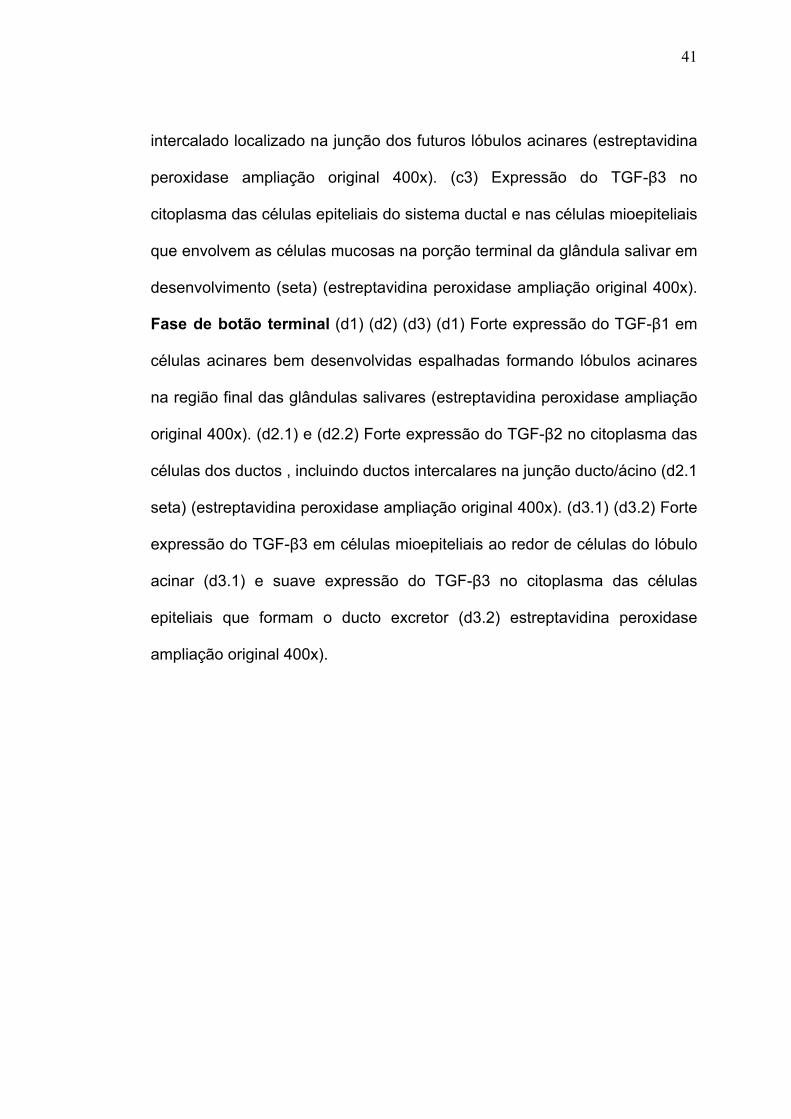
41
intercalado localizado na junção dos futuros lóbulos acinares (estreptavidina
peroxidase ampliação original 400x). (c3) Expressão do TGF-β3 no
citoplasma das células epiteliais do sistema ductal e nas células mioepiteliais
que envolvem as células mucosas na porção terminal da glândula salivar em
desenvolvimento (seta) (estreptavidina peroxidase ampliação original 400x).
Fase de botão terminal (d1) (d2) (d3) (d1) Forte expressão do TGF-β1 em
células acinares bem desenvolvidas espalhadas formando lóbulos acinares
na região final das glândulas salivares (estreptavidina peroxidase ampliação
original 400x). (d2.1) e (d2.2) Forte expressão do TGF-β2 no citoplasma das
células dos ductos , incluindo ductos intercalares na junção ducto/ácino (d2.1
seta) (estreptavidina peroxidase ampliação original 400x). (d3.1) (d3.2) Forte
expressão do TGF-β3 em células mioepiteliais ao redor de células do lóbulo
acinar (d3.1) e suave expressão do TGF-β3 no citoplasma das células
epiteliais que formam o ducto excretor (d3.2) estreptavidina peroxidase
ampliação original 400x).

42

43
Figura 4 – Marcadores da diferenciação do citoesqueleto durante o
desenvolvimento das glândulas salivares menores humanas –
Estágio de botão inicial: fraca expressão da CK 14 em algumas células das
ilhotas epiteliais das glândulas em desenvolvimento. (b) e (c) Proliferação na
fase de pseudoglandular: expressão da CK 14 no citoplasma das células
ductais. (d) Fase de botão inicial: presença de CK 14 em um maior número
de células do sistema ductal da glândula. (e) Fase canalicular:
imunoexpressão da CK 14 nas células mioepiteliais em torno do lóbulo
acinar. (f) Forte expressão da CK 14 na camada basal do epitélio e no
sistema ductal bem formado da glândula salivar. Nota-se que a parte do
ducto ao lado do epitélio de revestimento ainda é negativo para este
marcador. (g) Fase canalicular / terminal de botão: algumas estruturas
apresentam positividade para CKLMW (verde), a SMA (vermelho) é
detectada apenas nas paredes dos vasos sanguíneos. (h) e (i) Fase de
botão inicial / pseudoglandular: Forte expressão da CK LMW (verde) nas
células do sistema ductal. Expressão inicial da SMA (vermelho) em células
mioepiteliais vizinhas, principalmente na porção terminal do sistema ductal. (j)
Fase canalicular: Expressão de CK LMW (verde) no sistema ductal e SMA
(vermelho) em torno das estruturas acinares. (k) Lóbulos acinares bem
formado: Forte expressão da SMA (vermelho) em torno de estruturas
acinares. Poucas células são positivas para CK LMW (verde) normalmente
presente em estruturas do ducto intercalado. (l) Sistema ductal bem
desenvolvido fortemente positivo pra a CK LMW (verde).

44
Tabela 2 - Análise semi-quantitativa da expressão das subunidades de TGF-beta durante as fases da morfogênese das glândulas salivares humanas e nas glândulas salivares humanas plenamente desenvolvidas
TGF-beta 1
TGF-beta 2
TGF-beta 3
células epiteliais 0 ++ + ESTÁGIO DE BOTÃO INICIAL estroma 0 0 0
cordões epiteliais 0 +++ + ESTÁGIO
PSEUDOGLANDULAR estroma ++ 0 0
células ductais 0 +++ + células
mioepiteliais 0 0 ++ ESTÁGIO CANALICULAR
estroma 0 0 0
células ductais 0 +++ ++
células acinares +++ 0 0 células
mioepiteliais 0 0 ++
MORFOGÊNESE DAS
GLÂNDULAS SALIVARES HUMANAS
ESTÁGIO TERMINAL DE BOTÃO
estroma 0 0 0
células ductais 0 +++ ++ células
mioepiteliais 0 0 0
GLÁNDULA SALIVAR HUMANA
PLENAMENTE DESENVOLVIDA
células acinares +++ 0 0
0: negativo
+: positividade fraca
++: positividade moderada
+++: positividade intensa

45
6 DISCUSSÃO

46
6. DISCUSSÃO
Mecanismos moleculares envolvendo glândulas salivares em
desenvolvimento foram descritos em detalhes em ratos e em camundongos.
De acordo com esses trabalhos, sabe-se pouco sobre a expressão da TGF-
β durante o período pré-natal e pós-natal de glândulas salivares humanas.
Juntamente com outras glândulas e tecidos, é muito provável que esses
fatos sejam coordenados por inúmeras vias integrando eventos e afetando a
proliferação, a morfogênese e a interação célula-substrato.
Nossos resultados revelaram que TGF-β 1, 2 e 3 estavam presentes
obedecendo um determinado padrão nas várias fases do desenvolvimento
das glândulas salivares: pré-botão, botão inicial, pseudoglandular,
canalicular e de botão terminal. Este padrão foi dinâmico e houve variedade
de acordo com o estágio de desenvolvimento. Esse tipo de padrão também
foi visto em glândulas mamárias e outras glândulas do corpo. A transição da
forma de expressão do TGF-β no epitélio morfogeneticamente ativo foi
relatada em estruturas de roedores na cabeça e no pescoço, semelhante ao
folículo piloso, glândula salivar e germe dentário.
A superfamília do TGF-β é envolvida em muitos aspectos do
desenvolvimento e incluem: TGF-βs, BMPs, actina, inibidores e outros.
Especificamente TGF-βs, actinas e BMPs são relatados como controladores
da glândula salivar no estagio de ramificação e estudos têm investigado
esses fatores em glândula submandibular de camundongos.

47
Durante a fase inicial do desenvolvimento morfogenético das
glândulas salivares, na fase de pré-botão, o TGF-β 1 não foi positivo e o
TGF-β 2 e TGF-β 3 foram observados em raras células. Neste estágio há
grande proliferação, com pequena ou nenhuma evidência de marcação
pelos marcadores de proliferação. Considerando-se a atividade desse
estágio, pode-se prever que a expressão do TGF-β não é uma vantagem,
como tem sido previamente, pois este fator pode agir na fase terminal de
botão, estágio mais avançado do desenvolvimento glandular. A expressão
das citoqueratinas não foi detectada também neste estágio, em que a
proliferação das células é o evento principal. Neste estágio, nenhuns dos
marcadores estavam presentes em glândulas salivares em desenvolvimento,
indicando que o tecido glandular estava imaturo e realizando novas ligações
com a matriz extracelular preparando para o estágio posterior da
diferenciação, quando os fenótipos dos marcadores poderão ser detectados.
Este resultado está de acordo com o achado de Martins et al. (2002), que
não encontrou esses marcadores de diferenciação em estágios muito
avançado de desenvolvimento das glândulas salivares.
À medida que ocorreu o desenvolvimento da glândula salivar, houve o
aumento da diferenciação tecidual (botão inicial / pseudoglandular) e o TGF-
β 1 foi detectado no mesênquima ao redor do cordão epitelial de canalização
e ramificação. Esse padrão de desenvolvimento do TGF-β 1 imita o
resultado relatado por Robinson et al. (1991), em mama de ratos em
ramificação, e é também reportado em pulmão em desenvolvimento. Esta
distribuição do TGF-β 1 durante a fase de botão inicial / pseudoglandular

48
pode refletir uma reação autócrina do fator de desenvolvimento, o qual
estimula o crescimento da extremidade do parênquima do broto celular. O
TGF-β 2 e TGF-β 3 foram expressos pela célula epitelial do sistema ductal.
Estes resultados estão parcialmente de acordo com os achados publicados
de Jaskoll e Melnick (1999), que relatam a presença de TGF-β 1 e 2 em
epitélio ramificado e TGF-β 3 em epitélio e mesênquima. Nesta fase, o
sistema glandular continua apresentando substancial proliferação celular,
principalmente nas pontas do sistema de canais ductais, na qual a
subunidade TGF-β não é detectada. De qualquer forma, isso é caracterizado
pela formação do lúmen do ducto, e significando uma mudança na
expressão dos marcadores da diferenciação citoesqueletal. Neste estágio
(pseudoglandular), observou-se positividade para CK 14 e 35βH11,
indicando o progresso da citodiferenciação.
Durante o avanço das fases de botão inicial / pseudoglandular e
canalicular da glândula salivar, o fenótipo da célula é composto
principalmente por estruturas das glândulas em desenvolvimento foi bem
definido pela expressão da CK 14, CKML e SMA. O TGF-β 1 mostrou forte
expressão no citoplasma de células mucosas imaturas, nos sistemas de
terminais e ramificações. O TGF-β 2 e 3 estavam distribuídos ao longo de
todo o lúmen do sistema ductal, incluindo o ducto intercalar. O TGF-β 3 foi
observado em células mioepiteliais ao redor de células mucosas imaturas,
no extremo das células ductais. Este resultado está de acordo com os dados
de Robinson et al. (1991), o qual descreve a expressão do TGF-β 3 em
células mioepiteliais de mama de ratos. Essa expressão pode também ser

49
comparada com a expressão do TGF-β 3 em outros órgãos que apresentam
células que possuem componentes contráteis, como as células esqueléticas
do músculo cardíaco e células associadas a artérias. Células mioepiteliais da
glândula salivar compõem a camada mais externa dos lóbulos acinares e
ductos e agem tanto como células contráteis para secreção salivar e células
com grande capacidade de síntese de lâmina basal que compõe essas
estruturas. Robinson et al. (1991), descreveram a expressão do TGF-β 3 em
células-tronco mioepiteliais, especulando sobre a possível função de inibição
da diferenciação do terminal, que permitiria a formação dos botões laterais
em mamas em desenvolvimento. Eles também sugeriram que o TGF-β 3
poderia estar envolvido na elaboração dos componentes da lâmina basal.
Esses aspectos podem não ser acessíveis em nosso estudo e continua a ser
elucidado na formação de glândulas salivares humanas.
Nos últimos estágios da morfogênese da glândula salivar na fase
canalicular e de botão terminal, quando ocorre a ramificação e
citodiferenciação de células acinares, CK 14, 35βH11 e SMA foram
detectados num padrão específico. O SMA foi expresso quando os lóbulos
acinares começam a se diferenciar, enfatizando a presença de células
mioepiteliais ao redor dessas estruturas. CK 14 foi expressa por células
basais de ductos excretores e 35βH11 foi achado nas células luminares do
sistema ductal. CK 14, de acordo com outros autores, proverá uma forte
base para a conexão de células basais com a membrana basal, funcionando
como integradores do citoplasma e permitindo resistência a estresse
mecânico e manutenção da arquitetura do sistema ductal. Nessa fase, o

50
TGF-β 1 foi visto esparsamente, e as células acinares mucosas bem
desenvolvidas, e em glândulas salivares adultas, as células acinares
conservaram a expressão desse fator de crescimento. Esse padrão pode ser
indicativo de que o TGF-β 1 é sintetizado por células acinares e é importante
para a manutenção de glândulas salivares. Entretanto, a expressão de TGF-
β 1 em glândulas salivares adultas normais é discutível, e Kizu et al. (1996)
relataram a sua expressão nos ácinos e ductos, Kusafuka et al. (2000), não
acharam evidências de sua presença. O TGF-β 2 foi exclusivamente
detectado em células epiteliais do sistema ductal, e o TGF-β 3 conservou
sua expressão pelo sistema ductal e em células mioepiteliais que envolvem
os lóbulos acinares mucosos e esses padrões mantidos em estruturas
adultas.
O conjunto de resultados obtidos nesse trabalho indica, com bases
morfológicas, que o TGF-β em suas diferentes isoformas, participa de forma
importante na maturação das glândulas salivares. Os mecanismos da
atividade desses fatores são ainda obscuros e novos trabalhos devem ser
desenvolvidos, talvez utilizando-se de métodos in vitro para que se
compreenda de forma conclusiva a atividade dessa importante família de
fatores de crescimento na maturação das glândulas salivares.

51
7 CONCLUSÕES

52
7 CONCLUSÕES
Conclusão 1: A respeito da localização morfológica do TGF-β durante a
morfogênese das glândulas salivares, concluímos:
• A troca de subunidades de TGF-β ocorre concomitante a mudanças
evolutivas da morfogênese glandular durante o transcorrer do
desenvolvimento das glândulas salivares.
• Há um padrão dinâmico e variedade no momento de expressão das
subunidades de TGF-β de acordo com o estágio de desenvolvimento.
Conclusão 2: A respeito da relação do TGF-β com marcadores
citoesqueletais das glândulas salivares, concluímos:
• O TGF-β é expresso em fases mais avançadas do desenvolvimento
reafirmando sua ação na diferenciação glandular, onde se observa a
presença dos marcadores citoesqueletais da diferenciação glandular.

53
8 ANEXOS

54
ANEXO A – PARECER DO COMITÊ DE ÉTICA EM PESQUISA

55
9 REFERÊNCIAS

56
REFERÊNCIAS
Adi MM, Chisholm DM, Waterhouse JP. Sterological and immunohistochemical study of development of human fetal labial salivary glands and their S-100 protein reactivity. J Oral Pathol Med. 1994;23(1):36-40. Akhurst RJ, Lehnert SA, Gatherer D, Duffie E. The role of TGF beta in mouse development. Ann N Y Acad Sci. 1990;593:259-71. Araújo VC, Carvalho YR, Araújo NS. Actin versus vimentin in my epithelial cells of salivary gland tumors. A comparative study. Oral Surg Oral Med Oral Pathol. 1994;77:387-91 Araújo VC, Sousa SOM. Expression of different cytokeratins in salivary gland tumors. Eur J �rigin B Oral Oncol. 1996;32B(1):14-8. Araújo VC, Sousa SOM, Carvalho YR, Araújo NS. Application of immunohistochemistry to the diagnosis of salivary gland tumors. Appl Immunohistochem Mol Morph. 2000;8(3):195-202. Araújo VC, Sousa SOM, Jaeger R, Loyola A, Crivelini M, Araújo N. Characterization of the cellular component of polymorphus low-grade adenocarcinoma by immunohistochemistry and electron microscopy. Oral Oncol. 1999;35(2):164-72. Araújo VC, Souza SOM, Jaeger RG, Araújo NS. Immunohistochemical study of malignant salivary gland tumor. An analysis of 67 cases. Rev Odontol USP. 1991a;5:37-42. Araújo VC, Souza SOM, Nunes FD, Araújo NS. Immunohistochemical analysis of 89 benign salivary gland tumors. Rev Odontol USP. 1991b;5:1-6. Araújo VC, Sousa SOM, Lopes EA, Araújo NS, Sesso A. Mucus-producing adenopappilary carcinoma of minor salivary gland origin with signet ring cells and intracytoplasmic lumina. Arch Otorhinolaryngol. 1988;245(3):145-50.

57
Araújo VC, Sousa SOM, Sesso A, Sotto MN, Araújo NS. Salivary duct carcinoma: ultrastructural and histogenetic considerations. Oral Surg Oral Med Oral Pathol. 1987;63(5):592-6. Attie JN, Sciubba JJ. Tumors of major and minor salivary glands: clinical and pathologic features. Curr Probl Surg. 1981;18(2):65-155. Azuma M, Sato M. Morphogeneseis of normal human salivary gland cells in vitro. Histol Histopathol. 1994;9(4):781-90. Batsakis JG. Salivary neoplasia: an outcome of modified morphogenesis and differentiation. Oral Sur Oral Med Oral Pathol. 1980;49(3):229-32. Batsakis JG, Regezi JA, Luna MA; el-Naggar A. Histogenesis of salivary gland neoplasms: a postulate with prognostic implications. J Laryngol Otol. 1989;103(10):939-44. Bernfield MR, Banderjee SD, Cohn RH. Dependence of salivary epithelial morphology and branching morphogenesis upon acid mucopolysaccharide-protein (proteoglican) at epithelial surface. J Cell Biol. 1972;52(3):674-89. Bhaskar SN. Glândulas salivares. In: Orban BJ. Histologia e Embriologia Oral de Orban. 8ª ed. São Paulo: Artes Médicas; 1978. Brown TL, Patil S, Howe PH. Analysis of TGF-beta-inducible apoptosis. Methods Mol Biol. 2000;142:149-67. Carvalho YR, Araújo NS, Araújo VC, Sesso A. A polymorphous low-grade adenocarcinoma. An ultraestructural and immunohistochemical study. Ver Odontol UNESP. 1993;22(1):19-29.
Carvalho YR, Nogueira TO, Souza SOM, Araújo VC. Adenocarcinoma polimorfo de baixo grau de malignidade do tipo papilífero, estudo morfológico e imuno-histiquímico. Rev Odontol UNESP. 1990;19:165-71. Chin D, Boyle GM, Parsons PG, Coman WB. What is transforming grow factor-beta (TGF-β)? Br J Plast Surg. 2004;57(3):215-21.

58
Crivelini MM, Araújo VC. Estudo clínico e histológico dos carcinomas de células acinares de glândulas salivares menores. RPG Ver Pós-Grad. 1995;2(5):6-10. Crivelini MM, Souza SOM, Araújo VC. Immunohistochemical study of acinic cell carcinoma of minor salivary gland. Oral Oncol. 1997;33(3):204-25. Cutler LS. The role of extracellular matrix in the morphogenesis and differentiation of salivary glands. Adv Dent Res. 1990;4:27-33. Dale AC. Salivary gland: In Ten Cate AR. Oral histology: development, structure, and function. 4th ed. St. Louis: Mosby; 1994. p. 356. Dardick I, Naiberg J, Leung R, Ramjohn S, Christensen H, Burford-Mason A, Henderson WD, Rippstein P. Ultraestructural study of acinar and intercalated duct organization of submandibular and parotid salivary gland. Lab Invest. 1990;63(3):394-404. Dardick I, Parks WR, Little J, Brown DL. Characterization of cytoskeletal proteins in basal cells of human parotid salivary gland ducts. Virchows Arch A Pathol Anat Histopalthol. 1988;412(6):525-32. Denny PC, Ball WD, Redman RS. Salivary glands: a paradigm for diversity of gland development. Crit Rev Oral Biol Med. 1997;8(1):51-75. Derynck R, Janet JA, Chen EY, Eaton DH, Bell JR, Assoian RK, Roberts AB, Sporn MB, Goeddel DV. Human transforming growth factor ß complementary DNA sequence and expression in normal and transformed cells. Nature 1985;316(6030):701-5. Derynck R, Lindquist PB, Lee A, Wen D, Tamm J, Graycar JL, Rhee L, Mason AJ, Miller DA, Coffey RJ, et al. A new type of transforming growth factor-beta, TGF-beta 3. EMBO J. 1988;7(12):3737-43. Ellis GL, Auclair PL. Tumors of salivary glands. Bethesda: Armed Forces Institute of Pathology; 1996. (Atlas of tumor pathology, 17).

59
Engel ME, Datta PK, Moses HL. Signal transduction by transforming growth facto-beta: a cooperative paradigm with extensive negative regulation. J Cell Biochem Suppl. 1998(30-31):111-22. Eversole LR. Histogenetic classification of salivary glands. Arch Pathol. 1971;92(6):433-43. Figueiredo CRLV, Sousa SOM, Araújo VC. Estudo imunohistoquímico do carcinoma adenóide cístico de glândula salivar menor. RPG. 1997(4):231-7. Fitzpatrick DR, Denhez F, Kondaiah P, Akhurst RJ. Differential expression of TGF beta isoforms in murine palatogenesis. Development. 1990;109(3):585-95. Freitas RA, Araújo VC, Araújo NS. Argyrophilia in nucleolar �rigin�ct regions (AgNOR) in adenoid cystic carcinoma and polymorphous low-grade adenocarcinoma of salivary glands. Eur Arch Otorhinolaryngol. 1993(259):213-7. Graycar JL, Miller DA, Arrick BA, Lyons RM, Moses HL, Derynck R. Human transforming growth factor-beta 3: recombinant expression, purification, and biological activities in comparison with transforming growth factors-beta 1 and beta 2. Mol Endocrinol. 1989;3(12):1977-86. Gustafsson H, Kjörell U, Eriksson A, Virtanen I, Thornell LE. Distribution of intermediate filament proteins in developing and adult salivary glands in man. Anat Embryol. 1988;178(3):243-51. Hand AR. Salivary glands. In: Provenza DV. Oral histology inheritance and development. 2nd ed. Philadelpia Les & Febinger; 1980.
Hiatt JL, Sauk JJ. Embryology and anatomy of salivary glands In: Ellis GL, Auclair PL, Gnepp DR. Surgical pathology of salivary glands. Philadelphia: Saunders; 1991. 580 p. Jarger RG, Oliveira PT, Jaeger MMM, Araújo VC. Expression of smooth-muscle actin in cultured cells from human plasmacytoid myoepithelioma. Oral Surg Oral Med Oral Pathol Oral Radiol Endod. 1997;84(6):663-7.

60
Jaskoll T, Melnick M. Submandibular gland morphogenesis: stage-specific expression of TGF-alpha/EGF, IGF, TGF-β, TNF, and IL-6 signal transduction in normal embryonic mice and the phenotypic effects of TGF-β 2, TGF-β 3, and RGF-R null mutations. Anat Rec. 1999;256(3):252-68. Jennings JC, Mohan S, Linkhart TA, Widstrom R, Baylink DJ. Comparison of the biological actions of TGF ß beta 1 and TGF ß 2: differential activity in endothelial cells. J Cell Physiol. 1988;137(1):167-72. Kizu Y, Sakurai S, Katagiri S, Shinozaki N, Ono M, Tsubota K, Saito J. Immunohistochemical analysis of tumor growth factor-beta 1 in normal and inflamed salivary glands. J Clin Pathol. 1996;49(9):728-32. Klein RM. Development, structure and function of salivary glands. In: Avery JK. Oral development and histology. 2nd ed. New York: Thieme; 1994. Kusafuka K, Yamaguchi A, Kayano T, Takemura T. Immunohistochemical localization of members of the transforming growth factor-beta superfamily in normal human salivary glands and pleomorphic adenomas. J Oral Pathol Med. 2000;30:413-20. Lee SK, Lim CY, Chi JG, Yamada K, Kunikata M, Hashimura K, Kunikata M, Mori M. Prenatal development of human major salivary glands and immunohistochemical detection of keratins using monoclonal antibodies. Acta Histochem. 1990;89(2):213-35. Lehnert SA, Akhurst RJ. Embryonic expression pattern of TGF-β type-1 RNA suggest both paracrine and autocrine mechanisms of action. Development. 1988;104(2):263-73. Loducca SV, Raitz R, Araújo NS, Araújo VC. Polymorphous low-grade adenocarcinoma and adenoid cystic carcinoma: distinct �rigin�ctural composition revealated by collagen IV, laminin and their integrin ligands (alpha2beta1 and alpha3beta1). Histopathology. 2000;37(2):118-23. Loyola AM, Araújo VC. Carcinoma mucoepidermóide de glândulas salivares menores: estudo clínico, histopatológico. RPG. 1996;3(2):115-21.

61
Loyola AM, Araújo VC, Sousa SO, Araújo NS. Minor salivary gland tumours. A retrospective study of 164 cases in a Brazilian population. Eur J Cancer B Oral Oncol. 1995;31B(3):197-201. Lyons RM, Moses HL. Transforming growth factors and the regulation of cell proliferation. Eur J Biochem. 1990;187(3):467-73. Madisen L, Webb NR, Rose TM, Marquardt H, Ikeda T, Twardzik D, Seyedin S, Purchio AF. Transforming growth factorß2: cDNA cloning and sequence analysis. DNA. 1988;7(1):1-8. Martins MD, Araújo VC, Raitz R, Araújo NS. Expression of cytoskeletal proteins in developing human minor salivary glands. Eur J Oral Sci. 2002;110(4):316-21. Massagué J. How cells read TGF-beta signals. Nat Rev Mol Cell Biol. 2000;1(3):169-78. Massagué J. TGF beta signal transduction. Annu Rev Biochem. 1998;67:753-91. Mc Bride ML, Baillie J, Polland BJ. Growth parameters in normal fetuses. Teratology. 1984;29(2):185-91. McNicol AM, Richmond JA. Optimizing immunohistochemistry: antigen retrieval and signal amplification. Histopathology. 1998;32(2):97-103. Mercola M, Stiles CD. Growth factor superfamilies and mammalian embryogenesis. Development. 1988;102(3):451-60. Merwin JR, Newman W, Beall LD, Tucker A, Madri J. Vascular cells respond differentially to transforming growth factor beta 1 and beta 2 in vitro. Am J Pathol. 1991;138(1):37-51. Millan FA, Denhez F, Kondaiah P, Akhurst RJ. Embryonic gene expression patterns of TGF-β 1, beta 2 and beta 3 suggest different developmental functions in vivo. Development. 1991;111(1):131-43.

62
Miller DA, Pelton RW, Derynck R, Moses HL. Transforming growth factor-beta. A family of growth peptides. Ann N Y Acad Sci. 1990;593:208-17. Nilsen-Hamilton M. Transforming growth factor-beta and its actions on cellular growth and differentiation. Curr Top Dev Biol. 1990;24:95-136. Ohta M, Greenberger JS, Anklesaria P, Bassols A, Massagué J. Two forms of transforming growth factor-beta distinguished by multipotential haematopoietic progenitor cells. Nature. 1987;329(6139):539-41. Padgett RW, St Johnston RD, Gelbart WM. A transcript from a Drosophila pattern gene predicts a protein homologous to the transforming growth factor-beta family. Nature. 1987;325(6099):81-4. Patel VN, Rebustini IT, Hoffman MP. Salivary gland branching morphogenesis. Differentiation. 2006;74(7):349-64. Pelton RW, Dickinson ME, Moses HL, Hogan BL. In situ hybridization analysis of TGF beta 3 RNA expression during mouse development: comparative studies with TGF beta 1 and beta 2. Development. 1990a;110(2):609-20. Pelton RW, Hogan BL, Miller DA, Moses HL. Differential expression of genes encoding TGFs beta 1, beta 2, and beta 3 during murine palate formation. Dev Biol. 1990b;141(2):456-60. Pelton RW, Nomura S, Moses HL, Hogan BL. Expression of transforming growth factor beta 2 RNA during murine embryogenesis. Development. 1989;106(4):759-67. Pelton RW, Saxena B, Jones M, Moses HL, Gold LI. Immunohistochemical localization of TGF beta 1, TGF beta 2, and TGF beta 3 in the mouse embryo: expression patterns suggest multiple roles during embryo development. J Cell Biol. 1991;115(4):1091-105. Piek E, Heldin CH, Ten Dijke P. Specificity, diversity, and regulation in TGF-beta superfamily signaling. FASEB J. 1999;13(15):2105-24. Regezi JA, Batsakis JG. Histogenesis of salivary gland neoplasms. Otolaryngol Clin North Am. 1977;10(2):297-307.

63
Robinson SD, Silberstein GB, Roberts AB, Flanders KC, Daniel CW. Regulated expression and growth inhibits effects of transforming growth factor-beta isoforms in mouse mammary gland development. Development. 1991;113(3):867-78. Rosa F, Roberts AB, Danielpour D, Dartt LL, Sporn MB, Dawid IB. Mesoderm induction in amphibians: the role of TGF beta 2-like factors. Science. 1988;239(4841 Pt 1):783-5. Schmid P, Cox D, Bilbe G, Maier R, McMaster GK. Differential expression of TGF beta 1, beta 2, and beta 3 genes during mouse embryogenesis. Development. 1991;111(1):117-30. Segarini PR. Cell type specificity of TGF-beta binding. Ann N Y Acad Sci. 1990;593:73-90. Sousa SOM, Araújo VC. Estudo morfológico e imunohistoquímico do adenoma pleomórfico de glândula salivar menor. RPG Ver Pós-Grad. 1994;1(2):22-6. Souza SCOM, Sesso A, Araújo NS, Araújo VC. Inverted ductal papilloma of minor salivary gland �rigin: morphological aspects and cytokeratin expression. Eur Arch Otorhinolaryngol. 1995;252(6):370-3. Sporn MB, Roberts AB. TGF-beta: problems and prospects. Cell Regul. 1990;1(12):875-82. Ten Dijke P, Hansen P, Iwata KK, Pieler C, Foulkes JG. Identification of another member of the transforming growth factor type beta gene family. Proc Natl Acad Sci USA. 1988;85(13):4715-90. Thiery JP. Epithelial-mesenquimal transitions in development and pathologies. Current Opinion Cell Biol. 2003;15(6):740-6. Tonge CK, Luke DA. Dental anatomy: the salivary glands. Dent Update. 1984;11(5):264, 266, 268-70.
64
Tucker AS. Salivary gland development. Semin Cell Dev Biol. 2007;18(2):237-44. Weeks DL, Melton DA. A maternal mRNA localized to the vegetal hemisphere in Xenopus eggs codes for a growth factor related to TGF-beta. Cell. 1987;51(5):861-7. Whitman M, Melton DA. Growth factors in early embryogenesis. Annu Rev Cell Biol. 1989;5:93-117. Wrana JL. TGF-beta receptors and signaling mechanisms. Miner Electrolyte Metab. 1998;24(2-3):120-30. Yaku Y. Ultrastructural studies on development of human fetal salivary glands. Arch Histol Jpn. 1983;46(5):677-90.

65
10 APÊNDICE

Histochem Cell Biol (2007) 128:361–369
DOI 10.1007/s00418-007-0322-6ORIGINAL PAPER
Human salivary gland branching morphogenesis: morphological localization of claudins and its parallel relation with developmental stages revealed by expression of cytoskeleton and secretion markers
Silvia V. Lourenço · Cláudia M. Coutinho-Camillo · Marcilei E. C. Buim · Sabrina H. Uyekita · Fernando A. Soares
Accepted: 22 July 2007 / Published online: 9 August 2007© Springer-Verlag 2007
Abstract Development of salivary glands is a highly com-plex and dynamic process termed branching morphogenesis,where branched structures diVerentiate into mature glands.Tight junctions (TJ) are thought to play critical roles inphysiological functions of tubular organs, contributing tocell polarity and preventing lateral movement of membraneproteins. Evidence demonstrated that claudins are directlyinvolved in TJ formation and function. Using immunohisto-chemistry and immunoXuorescence we have mapped thedistribution of claudins-1, 2, 3, 4, 5, 7 and 11 and comparedit with the expression of diVerentiation markers in humansalivary glands obtained from foetuses ranging from weeks4 to 24 of gestation. Expression of all claudins, except clau-din-2 was detected in the various phases of human salivarygland development, up to fully mature salivary gland. The
expression of all claudins increased according to the pro-gression of salivary gland maturation evidenced by the clas-sical markers—cytokeratin 14, cytokeratin low molecularweight, smooth muscle actin and human secretory compo-nent. Tight junction proteins—claudins appear to be impor-tant in the Wnal shape and physiological functions of humansalivary glands and are parallel related with markers of sali-vary gland diVerentiation.
Keywords Human salivary gland · Branching morphogenesis · Claudins
Introduction
The development of salivary glands from simple precursorepithelial buds to functional glands is a highly complex anddynamic process. The mechanism by which a simple budbecomes such a complicated Wnal structure is termedbranching morphogenesis, where tissues generate branchedstructures that are subsequently diVerentiated into matureglands. BrieXy, salivary gland formation starts with the pro-liferation of a solid cord of cells from the epithelium of thestomatodeum into the underlying ectomesenchyme. Thiscord of cells extends deeply into the ectomesenchyme andbranches extensively. These cells then canalise by degrada-tion of the central cells to form the ductal system and theterminal secretory end pieces. The epithelial ingrowthsconstitute the parenchyme of a salivary gland. The ectome-senchyme diVerentiates to form the connective tissuecomponent of the gland, which supports the parenchyma(Kashimata and Gresik 1996; Cutler 1989, 1990).
Minor salivary glands begin to develop at 4–6 weeks ofembryonic life. Branches from parasympathetic and
S. V. Lourenço · F. A. SoaresDepartment of General Pathology, Dental School, University of São Paulo, São Paulo, Brazil
S. V. LourençoLaboratory of Immunopathology, Institute of Tropical Medicine, São Paulo University, São Paulo, Brazil
S. V. Lourenço · S. H. UyekitaDepartment of Dermatology, Medical School, University of São Paulo, São Paulo, Brazil
C. M. Coutinho-Camillo · M. E. C. Buim · F. A. SoaresDepartment of Pathology, Hospital A. C. Camargo, São Paulo, Brazil
S. V. Lourenço (&)Disciplina de Patologia Geral, Faculdade de Odontologia da Universidade de São Paulo, Av Prof Lineu Prestes, 2227, Cidade Universitária, CEP 05508-000 São Paulo, SP, Brazile-mail: [email protected]
123

362 Histochem Cell Biol (2007) 128:361–369
sympathetic nerves migrate to the gland and the collectingveins are formed. Salivary gland development consists of aseries of ducts ending in terminal secretory end pieces,grape-like in structure (Kashimata and Gresik 1996; Cutler1989, 1990). At the end of the maturation process, salivaryglands should be ready for salivary production, which occurin two phases: an acinar phase of production of primarysaliva and a ductal phase of electrolyte reabsorption, result-ing in hypotonic saliva. This process depends on moleculesand ions exchange, which are transported actively or pas-sively through the cell membranes of the ducts and acinarunits of the salivary glands. The process is also dependent onthe maintenance of adhesion and polarization of cells form-ing the glands (Cook et al. 1998; Guyton and Hall 2000).
Tight junctions (TJ) are thought to be the principal struc-tures that contribute to cell polarity, by acting as an intra-membrane barrier to prevent lateral movement ofmembrane proteins that form speciWc sites in the apical orbasolateral membranes, thereby playing a critical role in thephysiological functions of tubular organs (Anderson 2001;Tsukita et al. 2001). Several lines of evidence have demon-strated that claudins, proteins of a multigene family of TJ,are directly involved in the formation of TJ strands as wellas their barrier function (Tsukita et al. 2001). Claudins arealso described to confer selective barrier properties on TJs,being important in physiological functions of several tis-sues and organs (Anderson 2001; Mitic and Anderson1998; Peppi and Ghabriel 2004).
To date, little is known of the key regulators of humansalivary gland development and function. Temporal andspatial regulation of these events is likely to be important tosalivary gland development and for proper tissue functionand our group has been studying some of these aspects dem-onstrating the participation of integrins in the process (Lour-enço and Kapas 2005). However, other highly specialisedcellular mechanisms of adhesion have evolved to keep adhe-sion, to form selective barriers and to keep a polarised statebetween apical and basolateral membranes of the cells.
In the present study, we investigated the morphologicalexpression of several claudins molecules to understand theirparticipation in the lumen formation of human developingsalivary glands. Also, the expression of claudins was com-pared with the expression of maturation markers (Lourençoet al. 2007; Martins et al. 2002) in the diVerent phases ofthe human salivary glands branching morphogenesis.
Materials and methods
Tissue preparation
Fragments of the oral cavity from post-mortem of 20human foetuses (provenient from natural miscarriages) at
4–24 weeks of gestation were obtained from the MedicalSchool of the University of São Paulo and in accordancewith authorisation of the Ethical Committee of the institu-tion. The specimens were collected from diVerent oral sites,including buccal mucosa, tongue, mandible and hard palate.Five specimens of fully developed salivary gland speci-mens present in oral mucosa biopsies were retrieved fromthe archives of the Laboratory of Dermatopathology of thesame institution and were used as controls. They were his-tologically normal and presented no inXammatory or neo-plastic processes associated. All specimens were Wxed in10% buVered formalin for 24 h and embedded in paraYn.They were then histologically processed, serial-sectionedand stained with haematoxylin and eosin to check for thepresence of salivary glands and study their morphology.Those presenting developing minor salivary glands wereselected for the present immunohistochemical analysis.
Immunohistochemistry
Three �m serial sections of the specimens were re-hydratedand incubated in 3% aqueous hydrogen peroxide for 30 minto quench endogenous peroxidase activity. Incubation with1% bovine serum albumin (BSA) and 5% foetal calf serum(FCS) in Tris–HCl pH 7.4 for 60 min at room temperaturewas performed to suppress non-speciWc binding of subse-quent reagents. The sections were then submitted to antigenretrieval, followed by incubation overnight with the pri-mary antibody. Details on antigen retrieval methods as wellas primary antibodies clones, source, title are described inTable 1. The reaction followed with incubation with theindirect dextran polymer detection system (En Vision—Dako Carpinteria, CA, USA). Staining was completed byincubation with 3�3 diaminobenzidine tetrachloride (DAB)for 3 min. The specimens were then lightly counterstainedwith Mayer’s haematoxylin, dehydrated and mounted withglass coverslip and xylene based mountant.
Negative controls were achieved by substituting primaryspeciWc antibodies with non-immune serum and internalpositive controls were considered according to primaryserum manufacturer’s datasheet recommendation.
All immunohistochemical reactions were carried out intriplicate.
ImmunoXuorescence
Three �m serial sections of the formalin-Wxed, paraYnembedded developing salivary glands specimens werere-hydrated and incubated with 1% bovine serum albumin(BSA) and 5% foetal calf serum (FCS) in Tris–HCl pH 7.4for 60 min at room temperature to suppress non-speciWcbinding of subsequent reagents. The double labellingimmunoXuorescence reactions were performed in two
123

Histochem Cell Biol (2007) 128:361–369 363
steps. For that, the sections were incubated with the Wrstprimary antibody, followed by incubation with Goat Anti-Mouse IgG labelled with green Xuorescent Alexa 488(Molecular Probes, Carlsbad, CA, USA) at a concentrationof 1:100 in BSA-PBS. Incubation with the second primaryantibody was performed, followed by incubation with GoatAnti-Mouse IgG labelled with red Xuorescent Alexa 594(Molecular Probes, Carlsbad, CA, USA) at a concentrationof 1:100. Primary antibodies used, source, clone and work-ing dilutions are described in Table 1. All steps were pre-ceded by two incubations with Tris–HCl pH7.4 for 5 min atroom temperature. The sections were then air dried andmounted with Vecta Shield (Vector Laboratories, Burlin-game, CA, USA).
The results were analysed, observed and photographedunder Zeiss microscope (Zeiss Inc., Jena, Germany)equipped with epi-illumination and Wlters for Xuoresceinand rodamin with digital camera.
Negative controls were treated as above, but a solutionof 1% BSA in Tris–HCl pH 7.4 replaced the primary anti-body. Specimens incubated with normal serum were usedas additional negative controls. Internal positive controlssuch as basal layer of epithelium and blood vessels werepresent.
Results
The specimens studied were of the minor human salivaryglands in various stages of development, comprising bud,proliferation, canalisation, branching and cytodiVerentiation.
Immunohistochemistry
All claudins tested were present in the glands studied withexception of claudin-2, which was not detected in any
phase of salivary gland development or adult gland but waspositive in the controls used. These results are describedaccording to the salivary gland morphology and depicted inTable 2.
At bud/proliferation stage, when salivary glands start todevelop, claudin-1 was negative (Fig. 1a). Claudin-3 wasexpressed surrounding the cell membrane in some glandu-lar buds while others were mostly negative (Fig. 1b, barrow). Claudin-4 was expressed in the apico-lateral mem-brane of some cells of the salivary gland bud (Fig. 1c).Claudin-5 was expressed in a few cells of the solid bud ofthe very initial stage of salivary gland morphogenesis(Fig. 2a). At this stage, claudin-7 was observed surroundingthe apico-lateral membrane of cells in the centre of the solidbud and claudin-11 showed a membrane-cytoplasmicexpression in all cells of the bud (Fig. 2b, c, respectively).
At branching stage, claudin-1 was observed in scatteredcells along the branching cords of epithelial cells (Fig. 1d).Claudin-3 was expressed at the apico-lateral membrane ofepithelial cells in the centre of the branching cords andclaudin-4 followed this same pattern, however with greaterand more demarcated intensity (Fig. 1e, f, respectively).Claudins-5 and 7 were also detected in the apico-lateralcytoplasmic membrane of epithelial cells that occupy thecentre of the branching cords (Fig. 2d, e, respectively).Claudin-11 was observed in a cytoplasmic pattern ofexpression, with weak intensity, along the epithelial cells ofthe branching/canalising cords (Fig. 2f).
During canalisation/initial cytodiVerentiation, claudin-1was seen at the lateral cytoplasmic membrane of scatteredcells of well-canalised ducts. The arborised glandularsystem, including the ductal endpieces (future acinar lob-ules) was negative for this claudin (Fig. 1g). At this stageclaudins-3 and 4 showed a strong apico-lateral expressionof the cell membranes along the arborised ductal system upto the end-pieces/ rudimentary acinar lobules (Fig. 1h, i).
Table 1 Primary serum, clones, source, working title and antigen retrieval
Primary serum Clone Source Working title Antigen retrieval
Claudin-1 Policlonal Zymed (Carlsbad, CA, USA) 1:400 Citrate, PH6.0
Claudin-2 Policlonal Neomarkers (Fremont, CA, USA) 1:400 Citrate, PH6.0
Claudin-3 Policlonal Neomarkers (Fremont, CA, USA) 1:500 Citrate, PH6.0
Claudin-4 Policlonal Neomarkers (Fremont, CA, USA) 1:200 Citrate, PH6.0
Claudin-5 Policlonal Neomarkers (Fremont, CA, USA) 1:1000 Citrate, PH6.0
Claudin-7 Policlonal ZYMED (Carlsbad, CA, USA) 1:800 Citrate, PH6.0
Claudin-11 Policlonal Neomarkers (Fremont, CA, USA) 1:800 Citrate, PH6.0
CK LMW/ 35beta H11 Dako (Carpinteria, CA, USA) 1:50 Citrate, PH6.0
CK 14 LL 002 Neomarkers (Fremont, CA, USA) 1:50 Citrate, PH6.0
SMA 1A4 Dako (Carpinteria, CA, USA) 1:50 Citrate, PH6.0
HSC A0187 Dako (Carpinteria, CA, USA) 1:10 Citrate, PH6.0
Alexa 488 – Molecular Probes 1:1,000 –
Alexa 594 – Molecular Probes 1:1,000 –
123

364 Histochem Cell Biol (2007) 128:361–369
Claudins-5 and 7 were also observed following the latterpattern (Fig. 2g, h). Claudin-11 was present in the cyto-plasm of epithelial cells along the entire arborised system(Fig. 2i).
In later stages of salivary gland morphogenesis, (acinaradvanced cytodiVerentiation), claudin-1 remained withexpression limited to scattered cells in the ductal system, inthe lateral aspect of the plasma membrane. Acinar lobuleswere negative for this protein (Fig. 1j). Claudin-3 was seenat the apico-lateral portion of luminal cells of intercalatedduct and initial portion of excretory duct. In distal segmentsof excretory ducts it was observed only in scattered cells.Acinar lobules and the surrounding myoepithelial cellswere negative for this protein (Fig. 1k). Claudin-4 wasexpressed along the entire ductal system in the phase of aci-nar diVerentiation and it was also positive in myoepithelialcells surrounding immature acinar lobules (Fig. 1l1). Later,in this phase, fully developed acinar lobules were negativefor claudin-4 (Fig. 1l2). Claudin-5 was strongly positive inthe late stage of salivary gland diVerentiation. It wasobserved surrounding acinar cell membranes and in theapico-lateral aspect of luminal cells along the entire ductalsystem including intercalated duct (Fig. 2j). Claudin-7 wasalso positive at the apico-lateral aspect of the membranes ofluminal cells, but was not detected in the acinar lobules(Fig. 2k). Claudin-11 retained its cytoplasmic pattern ofexpression along the luminal cells of the entire ductal sys-tem and it was negative in acinar lobules (Fig. 2l).
In fully developed adult salivary glands, claudin-1 wasdetected at the apico-lateral aspect of ductal cells mem-branes that composed interlobular ducts and excretoryducts, but was negative on acinar cells (Fig. 3a). A similaraspect was observed for claudin-3 (Fig. 3b). Claudin-4 was
observed around the cell membrane of ductal and acinarcells (Fig. 3c). Claudin-5 was detected at the apical andbasal poles of the cell membrane of ductal cells as Wne lines(Fig. 3d). Weak expression of this claudin was seen on themembrane of acinar cells. Claudin-7 was mainly observedon the membrane of myoepithelial cells and on ductal cellsof the intercalated ducts (Fig. 3e, arrow). Finally, claudin-11 presented a weak expression, detected at the intercalatedduct cells and myoepithelial cells (Fig. 3f).
ImmunoXuorescence
During proliferation and canalisation stage of salivaryglands, solid epithelial buds and few developing salivarygland ducts were observed. In these phases, Low MolecularWeight Cytokeratin (CK LMW/35beta H11) was observedin cells of the centre of the epithelial cords (Fig. 4a1, a2-green). Solid epithelial buds were negative for this protein(Fig. 4a1, arrow). In the same phase, CK14 was present inthe ductal epithelial cells (Fig. 4b). During ramiWcation/canalisation phase, CK LMW/35beta H11) was detected inthe cytoplasm of ductal cells (green) and smooth muscleactin (SMA) was expressed in myoepithelial cells sur-rounding the initial ductal system (red) (Fig. 4c). In thisphase, strong expression of CK14 was detected in the cyto-plasm of ductal cells and weak expression of this proteinwas seen in the rudimentary end-pieces of the initial ductalsystem. In a further developmental stage (initial cytodiVer-entiation) luminal cells showed a cytoplasmic positivity forCK LMW/ 35beta H11 and myoepithelial cells surroundingproximal segments of ductal system and rudimentary acinarlobules were positive for SMA. At this stage, myoepithelialcells also demonstrated positivity for CK14 (green)
Table 2 Semi-quantitative expression on claudins in the diVerent phases of human salivary gland morphogenesis and in fully developed salivarygland
CLD claudin, ME myoepithelial, -ve negative, + positivity in scattered cells, ++ mild positivity, +++ strong and wide distributed positvity
CLD-1 CLD-2 CLD -3 CLD -4 CLD -5 CLD -7 CLD-11
Bud Epithelial cells -ve -ve -ve/+ + + + +
Branching Arborising cords -ve/+ -ve + ++ ++ ++ ++
Canalisation/initial acinar diVerentiation
Rudimentary ducts + -ve +++ +++ +++ +++ ++
Rudimentary acinar lobules (endpieces) -ve -ve +++ +++ +++ +++ ++
Acinar diVerentiation Acinar cells -ve -ve -ve -ve +++ -ve -ve
ME cells -ve -ve -ve ++ +++ -ve -ve
Intercalated ducts + -ve + +++ +++ +++ ++
Excretory ducts + -ve + +++ +++ +++ ++
Adult gland Acinar lobules -ve -ve ++ ++ ++ ++ ++
ME cells -ve -ve -ve -ve -ve -ve -ve
Intercalated ducts ++ -ve ++ ++ ++ ++ ++
Striated ducts ++ -ve ++ ++ ++ ++ ++
Excretory ducts ++ -ve ++ ++ ++ ++ ++
123

Histochem Cell Biol (2007) 128:361–369 365
Fig. 1 Expression patterns of claudins-1, 3 and 4 in the morphogene-sis of human minor salivary glands. a–c Bud/proliferation stage: solidnests of epithelial cells in the mesenchyme. In a, no evidence of clau-din-1 is observed. Claudins-3 and 4 are expressed as thin lines at theapico-lateral membranes of the epithelial cells in this early phase ofsalivary gland development (b, c, respectively). Some rudimentary sal-ivary gland nests still present a negative/weak expression of claudin-3(b arrow) (Original magniWcation £400). d–f Branching/initial cana-lisation stage: epithelial solid nests and cords elongate and branchforming an arborised structure, some with rudimentary lumens. Clau-din-1 is observed as clustered at the lateral aspect of cells along the sol-id epithelial cords (d). Claudins-3 and 4 appear as thin lines at theapico-lateral aspect of the membranes of epithelial cells that occupythe centre of the branching cords (e and f, respectively) (Original mag-niWcation £400). g–i Canalisation/initial cytodiVerentiation: the arbor-ised ductal system presents well-canalised ducts, some with epithelial
clusters at their endpieces that represent rudimentary acinar lobules.Claudin-1 is observed as clusters at the lateral aspect of scattered ductalcells in well-canalised interlobular ducts (g). Claudins-3 and 4 are de-tected as thin lines at the apico-lateral membrane of luminal cells of theentire ductal system, including the endpieces (h, i) (Original magniW-cation £400). j–l Advanced cytodiVerentiation: a well-formed andcomplex ductal system linked to developed acinar lobules is present asa Wnal stage of salivary gland morphogenesis. Claudin-1 is only seenin scatter luminal and basal cells of stratiWed interlobular excretoryducts (j). Claudin-3 is expressed as strong thin lines at the apico-lateralaspect of the membranes of luminal cells of intercalated and striatedducts, and as clusters in scattered cells of intra-lobular excretory ducts.Acinar lobules are negative for claudin-3 (K). Claudin-4 is expressedalong the entire ductal system and also surrounding immature acinarcells (L1). In mature acinar lobules claudin-4 was not detected (L2)(Original magniWcation £400)
123

366 Histochem Cell Biol (2007) 128:361–369
Fig. 2 Expression patterns of claudins-5, 7 and 11 in the morphogen-esis of human minor salivary glands. a–c Bud/proliferation stage: solidnests of epithelial cells in the mesenchyme. Claudin-5 is expressed asclusters at the membrane of scattered cells of the solid nests. It is alsodetected is endothelial cells (arrow) (a). Claudin-7 is seen as thin linesat the membranes of cells in the centre of epithelial nests (b). Claudin-11 is expressed in the cytoplasm of epithelial cells of the solid nests (c)(Original magniWcation £400). d–f Branching/initial canalisationstage: epithelial solid nests and cords elongate and branch forming anarborised structure, some with rudimentary lumens. Claudins-5 and 7are expressed as thin lines and clusters at the membranes of epithelialcells of the solid cords (d, e, respectively). Claudin-5 is also observedin endothelial cells of the intra-glandular vessels (d arrow). Claudin-11 is detected as a cytoplasmic positivity in a few epithelial cells of thebranching cords (f). (Original magniWcation £400). g–i The arborisedductal system presents well- canalised ducts, some with epithelial clus-
ters at their endpieces that represent rudimentary acinar lobules. Clau-dins-5 and 7 are detected mostly as thin lines, and sometimes asclusters at the membranes of luminal cells of well-canalised ducts, upto their endpieces (g, h). Claudin-11 is expressed in the cytoplasm ofthe well-formed arborised and canalised structure (I) (Original magni-Wcation £400). j–l Advanced cytodiVerentiation: a well-formed andcomplex ductal system linked to developed acinar lobules is present asa Wnal stage of salivary gland morphogenesis. Claudin-5 is expressedas thin lines at the membranes of luminal cells and surrounding well-developed acinar cells (j). Claudin-7 is observed as thin lines at the ap-ico-lateral membranes of luminal cells of intercalated ducts and intra-lobular ducts. Acinar lobules are negative (k). Claudin-11 is present inthe cytoplasm of epithelial cells that compose the intercalated and in-tra-lobular ducts. Acinar lobules do not express this molecule (l) (Orig-inal magniWcation £400)
123

Histochem Cell Biol (2007) 128:361–369 367
Fig. 3 Claudins expression in adult human minor salivary glands. a, bClaudins-1 and -3, respectively: Expression as Wne lines mainly delin-eating apico-lateral membrane of ductal cells (Original magniWcation£400). c Claudin-4 (Original magniWcation £400). d Claudin-5:Expression of this claudin is mainly observed as Wne lines delimitatingboth apical and basal poles of luminal cells. Weak and eventual expres-
sion of claudin-4 is also seen on acinar cells membrane. e Claudin-7:Myoepithelial cells are positive for claudin-7. Luminal cells of theintercalated ducts also express this protein (arrow) (Original magniW-cation £400). f Claudin 11: Expression of claudin-11 is weak, mainlydetected on myoepithelial cells and on cells of the intercalated ducts(Original magniWcation £400)
Fig. 4 ImmunoXuorescence: cytoskeletal and secretion markers ofsalivary gland maturation. a1, a2, b Bud/proliferation stage of salivarygland development: at this initial phase of salivary gland development,weak expression of CK LMW/ 35beta H11 (a1, a2) and CK14 (b) aredetected in the cytoplasm of a few epithelial cells of the incipient glandislets. Solid epithelial buds are negative for CK LMW/ 35beta H11 (a1arrow) (Original magniWcation £400). c, d Branching/canalizationphase: Strong expression of CK LMW/35beta H11 (green) is observedin cytoplasm of epithelial cells of the branched ductal system. Expres-sion of SMA (red) is seen in the cytoplasm of myoepithelial cells sur-rounding the terminal portion of the branching ducts (c). In this furtherstep of salivary gland development, strong expression for CK14 is ob-
served in the cytoplasm of ductal cells (d). HSC is detected as clustersin a few lumens (red) (d arrow) (Original magniWcation £400). e, fAcinar diVerentiation: At this stage, morphogenesis of salivary glandis advanced with many detectable acinar lobules. Strong expression ofSMA (red) is observed in the thin cytoplasm of elongated myoepithe-lial cells around the well-developed acinar lobules. Ductal cells arepositive for CK LMW/35beta H11 (green), (g). At this phase, immu-noexpression of CK14 is detected in myoepithelial cells surroundingthe acinar lobules (well-developed or rudimentary) and presence ofHSC is increased, being present in many luminal and acinar structures(red) (h) (Original magniWcation £400)
123

368 Histochem Cell Biol (2007) 128:361–369
(Fig. 4d), and human secretory component (HSC), a markerof salivary gland secretion was detected in the luminalaspect of some ducts and in a few acinar cells (red)(Fig. 4d, arrow). Finally, in advanced cytodiVerentiation,CK LMW/ 35beta H11 was positive in the cytoplasm ofductal cells, including cells of intercalated duct (green).Myoepithelial cells surrounding acinar lobules were SMA-positive (red) (Fig. 4e). In this phase CK14 was detected inthe cytoplasm of excretory duct cells and in myoepithelialcells (green) and the presence of HSC was now more fre-quent in the luminal aspect of the ducts and in acinar cells(red) (Fig. 4f).
Discussion
The present work showed, in the unique material of humanembryos developing salivary glands, the patterns of clau-dins expression and its evolution throughout the develop-mental phases of glandular morphogenesis. These claudinswere expressed in region-speciWc combinations along theprocess of salivary gland maturation.
Claudins were detected in all stages of salivary glanddevelopment examined, with the exception of claudin-1,which was not observed in bud stage and claudin-2, whichwas not observed in any phase of glandular morphogenesis.This fact is in accordance with the results of other workerswho reported similar events in the development of mousesalivary glands (Hashizume et al. 2004; Peppi and Ghabriel2004). Expression of claudin-2 is observed in cells thathave low transepithelial electrical resistance (TER), a mea-sure of the tightness of tight junctions (TJs) (Furuse et al.2001). This means that cells with lack of claudin-2, as seenin our results, increase their TER, which allows a selectivedecreased paracellular conductivity for sodium and potas-sium ions (Amasheh et al. 2002). This phenomenon proba-bly allows advantages in the process of cations changes andmay be consistent with the suggested roles of certain seg-ments of salivary glands such as the striated ducts in thephysiological production of hypotonic saliva (Dinudomet al. 1993).
Claudins-1, 3, 4, 5 and 7 were expressed as a thin line,mostly at the apico-lateral aspect of the membrane of ductalluminal cells, in patterns described for the localization ofTJ (Hashizume et al. 2004; Peppi and Ghabriel, 2004).
Claudin-1 was mostly detected on the membranes ofintralobular excretory ducts, diVering from the results ofother workers who detected this protein mainly in cells ofintercalated and striated ducts of rat submandibular glands(Peppi and Ghabriel 2004). This diVerence may be accountedto the type of gland as we only studied the development ofminor salivary glands. Claudin-1 is a component of epithe-lial and endothelial cells (Furuse et al. 1998). Although the
exact physiological role for claudin-1 is not clear, it ispossible that this molecule plays important roles in thefunction of water absorption by these ducts, as newbornmice with claudin-1 deWciency develop severe dehydrationleading to death (Furuse et al. 2002).
Claudins-3, 4, 5 and 7 were the tight junction proteinsexpressed in most segments of the human developing minorsalivary glands. Stronger and wider expression of theseclaudins was mostly observed at the membranes of luminalcells intercalated and striated ducts. Some claudins (clau-din-4 and 5) were also detected in the acinar lobules, eithertransiently or in a more stable type of staining. Theseresults also present some discrepancies with others pub-lished on rat developing salivary glands, and these diVer-ences may be due to specie-speciWc claudin patterns or tothe type of the gland studied (Peppi and Ghabriel 2004).
The patterns of claudins expression observed duringhuman minor salivary gland morphogenesis probablyreveal the basis for a progressive tightening of the epithe-lium, and may be similar to a proximal to distal tighteningreported in other tubular epithelial such as the intestine andnephron (Rahner et al. 2001; Kiuchi-Saishin et al. 2002).
Variations in the tightness of TJ strands are determinedby the combination ratios of claudin subtypes (Simon et al.1999; Tsukita and Furuse 2000; Furuse et al. 2001; Tsukitaet al. 2001). Other recent studies have documented selec-tive distribution of individual claudins in various tissues,suggesting that these molecules play a major role in the var-iability of barrier functions of tight junctions (Mitic et al.2000; Anderson 2001; Rahner et al. 2001). DiVerences andregion-speciWc combinations of claudins in salivary glands,since its early phases of development may contribute to thediVerences in the transepithelial electrical resistance andpermeability to molecules between the ducts and the termi-nal tubules or acini (Hand 1987). It is yet important to notethat salivary glands exhibit diVerences in permeability andquality of salivary secretion depending on the type of ner-vous stimulation (Hand 1987; Segawa 1994). Thesechanges may involve the transient regulation of adhesionmolecules, including those of the tight junctions. In thisway, claudin members’ interactions and coexpresion maybe aVected.
Claudins-4 and 5 were the only ones with notableexpression on the membranes of acinar cells. Claudin-4 wasseen when acinar cells were still immature and claudin-5was present up to the Wnal maturation of these cells. Previ-ous studies on cell lines suggested that claudin-4 formspores trough the TJ that discriminate against sodium ionsbut are indiVerent to chloride ions (van Itallie et al. 2001,2003). Claudin-5 is reported to play roles in the determina-tion and regulation of vascular permeability, controlling theparacellular movement of water and solutes across barriers(Morita et al. 1999). The combined expression of these two
123

Histochem Cell Biol (2007) 128:361–369 369
claudins in acinar cells may account for the mechanism ofhypotonic saliva production described by other authors(Dinudon et al. 1993; Peppi and Ghabriel 2004).
Unlike the other claudins, claudin-11 showed a cytoplas-mic positivity in cells that revealed ductal diVerentiation.This expression pattern is obscure and may reXect an inac-tive molecule, deserving further investigation.
Considering the major aspects of claudins expression ithas become clear that their presence is wider and strongeras development progresses and this is well illustrated withthe increasing evidence of salivary gland maturation mark-ers—cytoskeleton CKs and SMA and HSC. Other workersalso reported that expression of claudins seems to be spa-tially and temporally regulated in development, in a workthat involved salivary glands of other mammals (Hashiz-ume et al. 2004).
The present report demonstrated morphologically thepresence and distribution of claudins subunits during mor-phogenesis of human minor salivary glands. This expres-sion appears to be related with the maturation phases ofsalivary glands as demonstrated by the classical markers ofsalivary gland diVerentiation. Further studies are nowrequired to unravel obscure points on the roles and interac-tions of these proteins with other components of tight junc-tions and other adhesive proteins essential for salivarygland development, Wnal shape and physiological func-tions.
Acknowledgments FAPESP Grant 02/02676–7. Mrs. Maria Cristi-na Conceição for histological processing of all specimens
References
Amasheh S, Meiri N, Gitter AH, Schoneberg T, Mankertz J, SchulzkeJD, Fromm M (2002) Claudin-2 expression induces cation-selec-tive channels in tight junctions of epithelial cells. J Cell Sci115:4969–4976
Anderson JM (2001) Molecular structure of tight junctions and theirrole in epithelial transport. News Physiol Sci 16:126–130
Cook DI, Dinudom A, Komwatana P, Young JA (1998) Control of Na+
transport in salivary duct epithelial cells by cytosolic Cl- andNA+. Eur J Morphol 36:67–73
Cutler LS (1989) Functional diVerentiation of salivary glands. In: ForteJ (ed) Handbook of physiology, salivary, pancreatic, gastric andhepatobiliary secretion, vol 3. Am Physiological Soc Press, NewYork, pp 93–105
Cutler LS (1990) The role of extracellular matrix in the morphogenesisand diVerentiation of salivary glands. Adv Dent Res 4:27–33
Dinudom A, Young JÁ, Cook DI (1993) Na+ and Cl¡ conductances arecontrolled by cytosolic Cl¡ concentration in the intralobular ductcells of mouse mandibular glands. J Membr Biol 135:289–295
Furuse M, Fuita K, Hiiragi T, Fujimoto K, Tsukita S (1998) Claudin-1and -2: novel inegral membrane proteins localizing at tight junc-tions with no sequence similarity to occluding. J Cell Biol141:1539–1550
Furuse M, Furuse K, Sasaki H, Tsukita S (2001) Conversion of zonulaeoccludentes from tight to leaky strand type by introducing
claudin-2 into Madin-Darby canine kidney I cells. J Cell Biol153:263–272
Furuse M, Hata M, Furuse K, Yoshida Y, Haratake A, Sugitani Y,Noda T, Kubo A, Tsukita S (2002) Claudin-based tigiht junctionsare crucial for the mammalian epidermal barrier: a lesson fromclaudin-1 deWcient mice. J Cell Biol 156:1099–1111
Guyton AC, Hall JE (2000) Textbook of Medical Physiology, 10thedn. Chapter 64. WB Saunders Company, Philadelphia, pp 738–753
Hand AR (1987) Functional ultrastructure of the salivary glands. In:Sreebny LM (ed) The salivary system. CRC press, Florida, pp 44–67
Hashizume A, Ueno T, Furuse M, Tsukita S, Nakanishi Y, Hieda Y(2004) Expression patterns of claudin family of tight junctionmembrane proteins in developing mouse submandibular gland.Dev Dyn 231:425–431
Kashimata M, Gresik EW (1996) Contemporary approaches to thestudy of salivary gland morphogenesis. Eur J Morphol 34:143–147
Kiuchi-Saishin Y, Gotoh S, Furuse M, Takasuga A, Tano Y, Tsukita S(2002) DiVerential expression patterns of claudins, tight junctionmembrane proteins in mouse nephron segments. J Am Soc Neph-rol 13:875–886
Lourenço SV, Kapas S (2005) Integrin expression in developing sali-vary glands. Histochem Cell Biol 124:391–399
Lourenço SV, Lima DMC, Uyekita SH, Schultz R, de Brito T (2007)Expression of beta 1 integrin in human developing salivary glandsand its parallel relation with maturation markers: in situ hybrid-ization and immunoXuorescence study. Arch Oral Biol (in press)
Martins MD, Araújo VC, Raitz R, Araújo NS (2002) Expression ofcytoskeletal proteins in developing human minor salivary glands.Eur J Oral Sci 110:316–321
Mitic LL, Anderson JM (1998) Molecular architecture of tight junc-tions. Annu Rev Physiol 60:121–142
Mitic LL, Van Itallie CM, Anderson JM (2000) Molecular physiologyand pathophisiology of tight junctions. I. Tight junction structureand function: lessons from mutant animals and proteins. Am JPhysiol Gastrointest Liver Physion 279(2):G250–G254
Morita K, Sasaki H, Furuse M, Tsukita S (1999) Endothelial claudin:claudin-5/TMVCF constitutes tight junction strands in endothe-lial cells. J Cell Biol 147:185–194
Peppi M, Ghabriel MN (2004) Tissue-speciWc expression of the tightjunction proteins claudins and occudin in the rat salivary glands.J Anat 205:257–266
Rahner C, Mitic LL, Anderson JM (2001) Heterogeneity in expressionand subcellular localization of claudins 2, 3, 4, and 5 in the rat liv-er, pancreas, and gut. Gastroenterol 120:411–422
Segawa A (1994) Tight junctional permeability in living cells: dynam-ic changes directly visualized by confocal laser microscopy. JElectron microsc (Tokyo) 43:290–298
Simon DB, Lu Y, Choate KA, Velásquez H, Al- Sabban E, Praga M,Casari G, Bettinelli A, Colussi G, Rodríguez-Soriano J, Lifton RP(1999). Paracellin-1, a renal tight junction protein required forparacellular Mg2+ resorption. Science 285:103–106
Tsukita S, Furuse M (2000) Pores in the wall: claudins constitute tightjunction strands containing aqueous pores. J Cell Biol 149:13–16
Tsukita S, Furuse M, Itoh M (2001) Multifunctional strands in tightjunctions. Nat Rev Mol Cell Biol 2:285–293
Van Itallie CM, Fanning AS, Anderson JM (2003) Reversal of chargeselectivity in cation or anion-selective epithelial lines by expres-sion of diVerent claudins. Am J Physiol Renal Physiol 285:F1078-F1084
Van Itallie CM, Rahner C, Anderson JM (2001) Regulated expressionof claudin-4 decreases paracellular conductance through a selec-tive decrease in sodium permeability. J Clin Invest 107:1319–1327
123

a r c h i v e s o f o r a l b i o l o g y 5 2 ( 2 0 0 7 ) 1 0 6 4 – 1 0 7 1
Expression of beta-1 integrin in human developing salivaryglands and its parallel relation with maturation markers:In situ hybridisation and immunofluorescence study
avai lable at www.sc iencedi rec t .com
journa l homepage: www. int l .e lsev ierhea l th .com/ journals /arob
Silvia Vanessa Lourenco a,b,c,*, Dirce Mary C. Lima b, Sabrina Hitomi Uyekita b,Regina Schultz d, Thales de Brito b
aGeneral Pathology Department, Dental School, University of Sao Paulo, Brazilb Immunopathology Laboratory, Tropical Medicine Institute, University of Sao Paulo, BrazilcDermatology Department, Medical School, University of Sao Paulo, BrazildPathology Division, Hospital das Clınicas, Medical School, University of Sao Paulo, Brazil
a r t i c l e i n f o
Article history:
Accepted 14 May 2007
Keywords:
Salivary gland development
Human
Beta-1 integrin
a b s t r a c t
Background and objective: Salivary gland development entails co-ordinated processes invol-
ving complex molecular interactions in which integrins have a fundamental role. The
integrins are a family of heterodimeric transmembrane receptors comprising alpha and
beta subunits that mediate intercellular and extracellular signals involved in the organisa-
tion of cells in tissues and organs during development. The beta-1 integrin in particular have
been implicated in proliferation and differentiation of cells involved in the development of
epithelial tissues. To understand the role of beta-1 integrin in salivary gland development
we have studied its expression in human foetal tissues.
Design: In situ hybridisation was used to compare the expression and localisation of integrin
beta-1 with differentiation markers in developing human salivary glands obtained from
foetuses of 8–24 weeks gestation.
Results: Integrin beta-1 first appeared during bud stage in a few cells and its distribution
increased as salivary gland morphogenesis progressed. This increased pattern of beta-1
integrin expression was coincident with the appearance of the differentiation markers
CK14, CK low MW and smooth-muscle actin.
Conclusions: The developmentally regulated expression of integrin beta-1 in association
with the establishment of a mature phenotype indicated by salivary gland tissue differ-
entiation markers is suggestive of its role in salivary gland morphogenesis.
# 2007 Elsevier Ltd. All rights reserved.
1. Introduction
The position and shape of organs is determined by the co-
ordinated activity of groups of primitive cells with each other
and with surrounding cells. Cell shape changes, rearrange-
* Corresponding author at: Instituto de Medicina Tropical de Sao Paulo,Paulo, SP – Brazil. Tel.: +55 11 3061 7065; fax: +55 11 3061 7065.
E-mail address: [email protected] (S.V. Lourenco).
0003–9969/$ – see front matter # 2007 Elsevier Ltd. All rights reservedoi:10.1016/j.archoralbio.2007.05.002
ments and movements, often accompanied by regulated cell
proliferation and/or cell death, are the primary mechanisms
for generating the final shape of a functional mature organ.
Although there is limited information available on how these
cellular activities are co-ordinated, the expression of specific
Av Dr Eneas de Carvalho Aguiar, 500, 28 andar, CEP 05403-000, Sao
d.

a r c h i v e s o f o r a l b i o l o g y 5 2 ( 2 0 0 7 ) 1 0 6 4 – 1 0 7 1 1065
cell surface ligands and receptors tightly linked in a complex
web of signalling pathways that regulate cell division,
migration and differentiation is essential feature of organ
development.1
Similar to other organs the formation of the salivary gland
involves co-ordination of morphogenetic mechanisms,
including regulated cell shape changes and gene expression,
and directed cell migration leading to a fully developed gland
with important secretory functions. Morphologically, all
salivary glands develop similarly, with formation starting
with the proliferation of a solid cord of cells from the
epithelium of the stomatodeum into the underlying ectome-
senchyme. This cord of cells extends deeply into the
ectomesenchyme, branches extensively, forming canals with
secretory end pieces.2,3 The molecular mechanisms driving
the development sequence of salivary gland appear to involve
orchestration of different adhesion molecules and, in a series
of studies, we have shown that interactions between cells and
extracellular matrix are important in salivary gland biology
and disease.4–6 These interactions are partially mediated by
integrins, a family of heterodimeric transmembranic mole-
cules composed by alpha and beta subunits.7 These hetero-
dimers are divided into subfamilies on the basis of the beta
subunit and those of the beta-1 subfamily bind chiefly to
components of the extracellular matrix.7,8 In addition to their
cell attachment properties, integrins have been implicated in
the maintenance of intercellular contacts as well as being
involved in other dynamic biological mechanisms such as cell
signalling and regulation of gene expression leading to
proliferation and differentiation.7,9,10
Darribere et al.11 showed that integrins are fundamentally
important in cellular differentiation involved in embryogen-
esis beginning at the fertilisation stage and through organo-
genesis. This knowledge has been applied to studies of the
mammary and salivary gland epithelium and the key role of
beta-1 integrin plays in glandular morphogenesis and differ-
entiation verified.6,12,13 In salivary glands, however, the
expression of this adhesion molecule and its relationship to
the glandular development sequence has not been elucidated.
Given the importance of the beta-1 integrin in cell adhesion
and signalling in various tissues and organs and in develop-
mental processes, the objective of this study was to evaluate
the temporo-spatial expression of integrin beta-1 during
human salivary gland development using in situ hybridisation.
To determine the relationship of integrin beta-1 expression
with salivary gland morphogenesis the study included the
analysis of classical cytoskeletal markers of salivary gland
structures.
2. Material and methods
Specimens from the oral cavity of 26 human foetuses at 8–24
weeks of gestation were obtained from the Medical School of
the University of Sao Paulo and in accordance with authorisa-
tion of the Ethical Committee of this institution. The speci-
mens were collected from different oral sites, including the
buccal mucosa, tongue, mandible and hard palate. All speci-
mens were fixed in buffered formalin and embedded in
paraffin. Sections of the specimens were stained with
haematoxylin and eosin to check for the presence of salivary
glands and to study their morphology and determine their
developmental phase. Those presenting developing major or
minor salivary glands were selected for the present analysis.
Three specimens of adult, fully developed salivary gland were
also included in the study for comparative analysis of the
expression patterns of integrin beta-1.
3. In situ hybridisation
To assess the expression of integrin beta-1 in the specimens
selected, an oligonucleotide probe was synthesised to detect
beta-1 integrin based on the published sequence of this
molecule.13 Probe (50-30 AAA TTC CTG AGC TTA GCT GGT GTT
GTG CTA ATG TSS GGC ATC ACA GTC TTT TCC ACA AAT)
(GenBank accession number X07979) is a sense biotin-labelled
probe complementary to the sequences used for PCR. As a
negative control, a biotin-labelled scrambled probe was
employed following the same protocol (50-30 CCG TGT GGG
ACG GAA CTT ACG CGA TSS CGG CAG ACT AAT AGC AGT CGG
AAG GTC CAT). For ISH, 5 mm sections were de-paraffinised,
re-hydrated through a graded series of ethanol, and washed
with water treated with 0.1% diethylpyrocarbonate (DEPC) for
5 min. The sections were incubated with 25 mg/mL of
proteinase K (Sigma, St. Louis, MO, USA) in PBS for 10 min
at 37 8C and then permeabilised with 2% Triton in Tris–HCl
10 mM, at room temperature. These sections were rinsed with
DEPC for 10 min and incubated at 95 8C for 7 min in
hybridisation buffer (50% dextran sulphate, 20� SSC, 50�Denhart’s, 10 mg/mL of salmon sperm DNA, 10 mg/mL of
inorganic pyrophosphate in 1 M Tris buffer, pH 7.4, deionised
formamide, and DEPC). The sections were then incubated
overnight at 55 8C, with the corresponding probe, at a
concentration of 1 mM in hybridisation buffer. After incuba-
tion, the sections were washed in Tris–HCl, pH 7.4, at 42 8C for
10 min and further washed with water five times for 5 min.
The slides containing the tissue sections were then incubated
with the subsequent steps of the kit Gen point (Dako
Cytomation). The reactions were finally developed with the
chromogen 303 diaminobenzidine tetrachloride (DAB) (Sigma,
St. Louis, MO 63178, USA). The specimens were then lightly
counterstained with Carazzi’s haematoxylin, dehydrated and
mounted with a glass coverslip and a xylene-based mounting
reagent. The results were analysed under an optical micro-
scope equipped with a digital camera for image acquisition.
4. Immunofluorescence
Serial sections (3 mm) of the formalin-fixed, paraffin
embedded developing salivary glands specimens were re-
hydrated and incubated with 1% bovine serum albumin (BSA)
and 5% foetal calf serum (FCS) in Tris–HCl pH 7.4 for 60 min at
room temperature to suppress non-specific binding of sub-
sequent reagents. The double labelling immunofluorescence
reactions were performed in two steps. First, the sections were
incubated with the primary antibody, followed by incubation
with sheep anti-mouse FITC-conjugated immunoglobulin
(Novocastra) (diluted 1:100 in BSA–PBS). Incubation with the
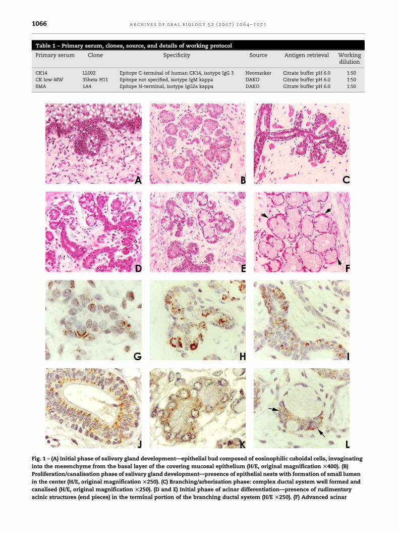
Table 1 – Primary serum, clones, source, and details of working protocol
Primary serum Clone Specificity Source Antigen retrieval Workingdilution
CK14 LL002 Epitope C-terminal of human CK14, isotype IgG 3 Neomarker Citrate buffer pH 6.0 1:50
CK low-MW 35beta H11 Epitope not specified, isotype IgM kappa DAKO Citrate buffer pH 6.0 1:50
SMA 1A4 Epitope N-terminal, isotype IgG2a kappa DAKO Citrate buffer pH 6.0 1:50
Fig. 1 – (A) Initial phase of salivary gland development—epithelial bud composed of eosinophilic cuboidal cells, invaginating
into the mesenchyme from the basal layer of the covering mucosal epithelium (H/E, original magnification T400). (B)
Proliferation/canalisation phase of salivary gland development—presence of epithelial nests with formation of small lumen
in the center (H/E, original magnification T250). (C) Branching/arborisation phase: complex ductal system well formed and
canalised (H/E, original magnification T250). (D and E) Initial phase of acinar differentiation—presence of rudimentary
acinic structures (end pieces) in the terminal portion of the branching ductal system (H/E T250). (F) Advanced acinar
a r c h i v e s o f o r a l b i o l o g y 5 2 ( 2 0 0 7 ) 1 0 6 4 – 1 0 7 11066

a r c h i v e s o f o r a l b i o l o g y 5 2 ( 2 0 0 7 ) 1 0 6 4 – 1 0 7 1 1067
second primary antibody was performed, followed by incuba-
tion with goat anti-mouse IgG labelled with red fluorescent
Alexa 594 (Molecular Probes) at a concentration of 1:100. The
details of the monoclonal primary antibodies used, their
source, and working dilutions are described in Table 1. All
steps were preceded by two incubations with Tris–HCl pH 7.4
for 5 min at room temperature. The sections were then air
dried and mounted with Vecta Shield. The results were
analysed and photographed using a Zeiss microscope
equipped with epi-illumination and a digital camera with
filters for fluorescein and rodamine. Negative controls were
treated as above, but a solution of 1% BSA in Tris–HCl pH 7.4
replaced the primary antibody. Specimens incubated with
normal serum were used as additional negative controls.
Internal positive controls such as basal layer of epithelium
were present.
5. Results
The specimens studied were of the major and minor human
salivary glands at various stages of development, comprising
bud, proliferation, canalisation, branching and cytodifferen-
tiation (Fig. 1A–F).
5.1. In situ hybridisation (ISH) of human embryonicsalivary glands (Fig. 1G–L)
Salivary glandsat thebud stage of development werescarceand
only rare cells expressed integrin beta-1, which was located
around the cell membrane at their contact with the stroma and,
sometimes, as cytoplasmic dots. During the initial steps of
proliferation and canalisation stage, a few developing salivary
gland ducts could be observed. Beta-1 integrin was present in
rare cells, showing a ring-like pattern seen in the basal pole of
some ductal cells or around the cell membrane in an
intercellular position. When canalisation was more evident,
an increased number of ductal cells expressed the beta-1
integrin in a similar pattern as described above and in a
punctate pattern around the cell membrane. At the stage of
branching and initial cytodifferentiation, beta-1 integrin was
present in a greater number of cells, usually as bipolar dots in
the cytoplasm and membrane of epithelial cells of the canalised
ducts and around the cell membrane. Expression of integrin
beta-1 was also seen at the basolateral portion of a few
rudimentary developing acinic cells. Expression of this integrin
was seen along the more mature ducts, usually showing a
bipolar distribution and at the apical pole of luminal cells.
differentiation: mucous acinar lobules well developed, compose
surrounded by thin elongated myoepithelial cells (arrow) (H/E o
expression of beta-1 integrin concentrated of the baso-lateral p
hybridisation, original magnification T400). (H and I) Canalisati
expression of beta-1 integrin on the membrane of ductal cells.
dotted pattern surrounding the entire cell membrane (in situ hyb
excretory duct: expression of beta-1 integrin in a bipolar pattern
original magnification T250). (K) Initial phase of acinar differen
membrane and in peri-nuclear position of the immature acinar
Fully developed acinar lobule: expression of beta-1 integrin con
(in situ hybridisation, original magnification T400).
Cytoplasmic signal was increasingly detected as differ-
entiation of salivary gland progressed.
At a subsequent differentiation stage (advanced cytodif-
ferentiation), beta-1 integrin was present at the basolateral
portion of some acinic structures and more abundantly in the
cells along the ductal system, more frequently as cytoplasmic
signals. The ring-like pattern of expression was still observed
in a few ductal cells. Finally, adult normal salivary glands,
used as controls for the reactions showed expression of
integrin beta-1 along all the ductal system. Intercalated ducts
showed signal for beta-1 integrin in a cell-membrane bipolar
pattern. The other segments of salivary gland duct presented
expression of integrin beta-1 both in a cytoplasmic and
membrane location. Negative controls did not show any
signal.
5.2. Immunofluorescence of human embryonic salivaryglands
At the bud stage of salivary gland development, only a few
cells were weakly positive for CK14 (Fig. 2A). During the
proliferation and canalisation stage, a few developing salivary
gland ducts were observed. In these phases, CK14 and low
molecular weight cytokeratin (CK LMW/35beta H11) were
present in the ductal epithelial cells and smooth-muscle actin
(SMA) was only found in rare cells (Fig. 2B, C, G, H).
At the stage of branching and initial cytodifferentiation,
ductal cells were positive for CK 14 (Fig. 2D). Luminal cells of
the developing ductal system were positive for CK LMW/
35beta H11. Smooth-muscle actin was detected in cells of the
terminal portion of the ductal system and in cells that
surrounded the rudimentary acinic lobules (Fig. 2 I and J). At
late acinar cytodifferentiation, CK14 was detected in the
cytoplasm of basal cells of the ductal system. Myoepithelial
cells were positive for CK14 and SMA. CK LMW/35beta H11 was
seen on luminal cells of the ductal system (Fig. 2E, F, K and L).
6. Discussion
Our results demonstrated that the expression of beta-1
integrin in different patterns along the process of differentia-
tion of human salivary glands increased as development
progressed, suggesting that it may be an indicator of salivary
gland maturation. The demonstration of integrin beta-1
mRNA using the very sensitive technique of in situ hybridisa-
tion complemented the results of our previous work in which
the beta-1 integrin protein was demonstrated the using
d by pyramidal cells with large and clear cytoplasm and
riginal T400). (G) Initial phase of proliferation/canalisation:
ortion of the membrane in a few epithelial cells (in situ
on/arborisation of the developing ductal system: increased
Note in (H), the marked expression of the molecule in a
ridisation, original magnification T250). (J) Fully developed
in the various layers of ductal cells (in situ hybridisation,
tiation: expression of beta-1 integrin around the cell
cells (in situ hybridisation, original magnification T400). (L)
centrated in the cells of the intercalary-acinar unit (arrows)

Fig. 2 – (A) Bud stage of salivary gland development: weak expression of CK14 in is detected in the cytoplasm of a few
epithelial cells of the incipient gland islets (arrows) (original magnification T400). (B and C) Proliferation/canalisation phase:
epithelial structures exhibiting strong expression of CK14 in the cytoplasm of ductal cells. Observe in (C) a well-formed and
canalised salivary gland structure (arrow). Other further developed structures start to present weak positivity surrounding
epithelial lobules, probably representing the initial maturation of myoepithelial cells (yellow arrow) (original magnification
T250 (B) and T400 (C)). (D) Branching phase: in this stage presence of CK 14 is observed in a greater number of cells of the
ductal system of the developing gland (original magnification T400). (E) Acinar differentiation: acinar lobules that present
immunoexpression of CK14 in myoepithelial cells surrounding the acinar lobules (original magnification T400). (F) Well-
developed excretory ducts: strong expression of CK14 is observed in the cytoplasm of basal keratinocytes of the lining
mucosa epithelium (red arrow) and in cytoplasm the well-formed ductal system of the salivary gland. Note that the portion
of the duct next to the covering epithelium is still negative for this marker (white arrow) (original magnification T400). (G)
Initial phase of proliferation/canalisation: a few canalising epithelial lobules of developing salivary glands present weak
cytoplasmic positivity for CK LMW/35beta H11 (green). SMA (red) is only detected in blood vessels walls in this phase. Note
the presence of more rudimentary salivary gland lobules still negative for CK LMW/35beta H11 (arrow) (original
a r c h i v e s o f o r a l b i o l o g y 5 2 ( 2 0 0 7 ) 1 0 6 4 – 1 0 7 11068

a r c h i v e s o f o r a l b i o l o g y 5 2 ( 2 0 0 7 ) 1 0 6 4 – 1 0 7 1 1069
immunohistochemistry.6 In this study, double labelling
immunofluorescence was used to identify and highlight the
presence of classical cytoskeletal markers of salivary gland
differentiation.
To date, little is known of the molecular mechanisms that
regulate the development of the human salivary gland. As
with other glands and tissues, it is very likely that develop-
ment is co-ordinated via several integrated events affecting
proliferation, morphogenesis and cell–substratum interac-
tions. These events are tightly regulated both temporally and
spatially.14 Differentiation of salivary glands begins around
week 6 of foetal development when specific oral epithelium
cells undergo organised, co-ordinated growth that is based on
epithelio–mesenchymal interactions, leading to morphogen-
esis and cytodifferentiation.15,16 Integrins appear to be key
molecules involved in these interactions and perturbations of
their expression can lead to impaired glandular form and
function. For example, Menko et al.17 have demonstrated that
the absence of the integrin dimer alpha3beta1 affects multiple
components of adhesive complexes and their associated
signalling pathways in salivary glands. These include pro-
found alterations in the signalling cascade that involves Cdc42
and RhoA, two Rho GTPases that regulate the organisation of
the actin cytoskeleton.17
The role of beta-1 integrin has been investigated in other
tissues and its importance in their developmental processes is
being established. In mammary glands, which display a
similar morphology to the salivary glands, perturbation of
beta-1 integrin results in impaired development and differ-
entiation of the secretory epithelium.18 Supporting this,
Naylor et al.13 has shown that the disturbance of mammary
gland lobular development in mice is due to the lack of beta-1
integrin which manifest as defects in epithelial proliferation
triggered by the absence of this molecule.
During the initial stages of human salivary gland mor-
phogenesis – bud stage – beta-1 integrin, was present in
scattered cells and as differentiation progressed, it was
expressed in an increasing number of cells. These results
suggest that beta-1 integrin is important in the initial phase of
salivary gland development, when interactions between the
bud epithelial cells and their newly formed extracellular
environment occur. In a parallel analysis, double staining
immunofluorescence showed none of the classical cytoske-
letal differentiation markers, indicating that the glandular
tissue is very immature at this phase. This result is in
accordance with a previous report by Martins et al.19 who
did not find any markers of differentiation in the very early
stage of salivary gland development. With further differ-
entiation, expression of beta-1 integrin increases, creating
magnification T400). (H and I) Canalisation/branching phase: str
in cytoplasm of epithelial cells of the branched ductal system. I
myoepithelial cells surrounding the terminal portion of the bra
respectively). (J) Initial phase of acinar differentiation: expressio
cytoplasm of epithelial cells of the ductal system. Smooth mus
cells surrounding the incipient acinic structures (arrows) (origin
strong expression of SMA (red) is observed in the thin cytoplasm
acinar lobules. A few cells are positive for CK LMW/35beta H11 (g
(original magnification T600). (L) Well-developed ductal system
CK LMW/35beta H11 (green) (original magnification T400).
new connections likely to be important for subsequent steps
in gland development.
In early stages of salivary gland canalisation, beta-1
integrin was expressed in a peculiar ring-like pattern in some
cells of the developing ductal network. Analogous to the
results of other authors, whose studies have implicated beta-1
integrin as a marker of the stem cell niche in mammary gland
and skin,20 it is conceivable that these cells may represent very
primitive, pluripotent cells of the salivary gland tissue.
Additionally, extracellular matrix proteins may modulate
the expression and activation of beta-1 integrins in these
cells, and local variation in the composition of basement
membrane during the gland development might play a role in
establishing and maintaining their distribution.20
At the canalisation stage when beta-1 integrin is also well
identified in cells of the ductal network, more differentiation
markers are detected in the developing salivary glands, which
stain for CK14 and CK LMW/35beta H11, indicating that
cytodifferentiation has progressed. Integrin receptors and
ligands have been shown to activate signalling pathways
involving mitogen-activated protein kinases, tyrosine kinases
or GTP-binding proteins. These are thought to affect cell
cytoskeleton and proliferative responses that are required
during organ development.21 However, this relationship has
yet to be shown at the molecular level in human salivary gland
development. On the basis of the present morphological
evidence, functional studies, possibly using tissue culture or
animal models are imperative for substantiating the sugges-
tions made from these morphological observations.
In advanced canalisation stage of salivary gland develop-
ment, beta-1 integrin was present in a greater number of cells,
with signals detected either on the membrane or in the
cytoplasm. The differential mRNA localisation may reflect a
novel type of post-transcriptional control involved in many
cellular processes, including the regulation of cellular asym-
metry required for creating and maintaining cell polarity, which
is important for cellular differentiation during development.
Thus, site-specific localisation of mRNA in the cytoplasm may
provide an important mechanism whereby specialised proteins
can be expressed at discrete sites within the cell.21,22 In
developing salivary glands, the presence of beta-1 integrin at
these locations may be important for the protein transcription
locally, which later may determine cell polarity to prepare
glandular structures for their secretory functions. Also, some
cells still retained the ring-like pattern of beta-1 integrin
expression.Other cellsof thebranchedductalsystemexpressed
beta-1 integrin at the basolateral surface of the membrane. This
varied morphological pattern of expression suggests that
integrins might play diverse roles in the process of salivary
ong expression of CK LMW/35beta H11 (green) is observed
nitial expression of SMA (red) is seen in the cytoplasm of
nching ducts (original magnification T400 and T250,
n of CK LMW/35beta H11 (green) is detected in the
cle actin (red) is present in the cytoplasm of myoepithelial
al magnification T400). (K) Well-formed acinar lobules:
elongated myoepithelial cells around the well-developed
reen), probably representing intercalated duct cells (arrow)
: epithelial cells showing cytoplasmic strong positivity for

a r c h i v e s o f o r a l b i o l o g y 5 2 ( 2 0 0 7 ) 1 0 6 4 – 1 0 7 11070
gland development, either in the differentiation and main-
tenance of the salivary gland phenotype, mediating a close
relationship between gland parenchyma and extracellular
matrix and also being associated with glandular functions, as
reported by others (see 1 for review). This pattern of distribution
is thought to be involved in the stable attachment of stationary
epithelial cells to the matrix and in the maintenance of cell–cell
interactions,23 suggesting that integrin beta-1 expression might
be a cell-membrane indicator of the evolutionary steps of
salivary gland morphogenesis. At this time, the phenotype of
cells that comprise themain structure of thedeveloping gland is
well defined by the expression of CK14, CK LMW/35beta H11 and
SMA, which provide a parallel demonstration of glandular
maturation.
In late stages of salivary gland development, indicated by
branching and cytodifferentiation of acinar cells, beta-1
integrin mimics the patterns seen in the normal adult salivary
gland structures. Thus, beta-1 integrin showed a cytoplasmic
expression in the apical pole of luminal cells, as well as baso-
lateral portion of ductal cells. This phenomenon may account
for the secretory functions of salivary glands and is similar to
what has been observed in other secretory organs such as
stomach and breast.23–25 Cytoskeletal markers of salivary
gland differentiation are present in this phase of the gland
development in specific patterns, close to those classically
described in adult glands.19,26,27 The increase in the expression
of beta-1 integrin and markers of differentiation as salivary
gland development and differentiation confirms the require-
ment for this integrin in maintaining tissue-specific functions.
For instance, being implicated in the control, of mammary
gland differentiation.13,28
The present research has shown morphological evidence
that integrins may be cell membrane indicators of human
salivary gland differentiation with a parallel relation with the
establishment of a salivary gland mature phenotype disclosed
by the classical cytoskeletal differentiation markers. The
integrins are developmentally regulated and appear to be
sensitive to changes in the gland extracellular microenviron-
ment as morphogenesis progresses. These morphological data
provide a cornerstone for future functional and molecular
studies that are at present being carried out in our laboratory.
Acknowledgements
Supported by FAPESP grants 02/02676-7 and 03/00450-4 and
British Council Academic Link SPA/881/155. Division of
Pathology of the Hospital da Clınicas, Medical School,
University of Sao Paulo, for supplying the foetal tissue used
in this work. All experiments carried out in this work comply
with the laws of the Ethical Committee of the Medical School
of the University of Sao Paulo.
r e f e r e n c e s
1. Thiery JP. Cell Adhesion in development: a complexsignalling network. Curr Opin Gen Dev 2003;13:365–71.
2. Dale AC. Salivary gland. In: Ten Cate AR, editor. Oralhistology. 4th ed. St. Louis: Mosby; 1994. p. 312–33.
3. Klein RM. Development, structure and function of salivaryglands. In: Avery JK, editor. Oral development and histology.2nd ed. New York: Thieme; 1994. p. 352–79.
4. Araujo VC, Loducca SVL, Sousa SOM, Williams DM, AraujoNS. The cribriform features of adenoid cystic carcinoma andpolymorphous low-grade adenocarcinoma: cytokeratinsand integrins expression. Ann Diagn Pathol 2001;5(6):330–4.
5. Loducca SVL, Raitz R, Araujo NS, Araujo VC. Polymorphouslow-grade adenocarcinoma and adenoid cystic carcinoma:distinct architectural composition revealed by collagen IV,laminin and their integrin ligands a2b1 and a3b1.Histopathology 2000;37:118–23.
6. Lourenco SV, Kapas S. Integrin expression in developingsalivary glands. Histochem Cell Biol 2005;124:391–9.
7. Hynes RO. Integrins: versatility, modulation, and signallingin cell adhesion. Cell 1992;69:11–25.
8. Hynes RO. Integrins: a family of cell surface receptors. Cell1987;48:549–54.
9. Ruoslahti E, Reed JC. Anchorage dependence, integrins, andapoptosis. Cell 1994;77:477–8.
10. Varner JA, Cheresh DA. Integrins and cancer. Curr Opin CellBiol 1996;8:724–30.
11. Darribere T, Skalski M, Cousin H, Gaultier A, Montmory C,Alfandari D. Integrins: regulator of embryogenesis. Biol Cell2000;92:5–25.
12. Beauvais-Joneau A, Thierry JP. Multiple roles for integrinsduring development. Biol Cell 1997;89:5–11.
13. Naylor MJ, Li N, Cheung J, Lowe ET, Lambert E, Marlow R,et al. Ablation of b1 integrin in mammary epitheliumreveals a key role for integrin in glandular morphogenesisand differentiation. J Cell Biol 2005;171:717–28.
14. Cutler LS, Functional differentiation of salivary glands.ForteJ, editor. Handbook of physiology salivary pancreatic gastric andhepatobiliary secretion, vol. 3. New York: Am Physiological SocPress; 1989. p. 93–105.
15. Cutler LS. The role of extracellular matrix in themorphogenesis and differentiation of salivary glands. AdvDental Res 1990;4:27–33.
16. Kashimata M, Gresik EW. Contemporary approaches to thestudy of salivary gland morphogenesis. Eur J Morphol1996;34:143–7.
17. Menko AS, Kreidberg JA, Ryan TT, Van Bockstaele E. Loss ofalpha3beta1 integrin function results in an altereddifferentiation program in the mouse submandibular gland.Dev Dyn 2001;220(4):337–49.
18. Faraldo MM, Deugnier MA, Lukashev M, Thierry JP,Glukhova MA. Perturbation of beta 1-integrin function altersthe development of murine mammary gland. EMBO J1998;17:2139–47.
19. Martins MD, Araujo VC, Raitz R, Araujo NS. Expression ofcytoskeletal proteins in developing human minor salivaryglands. Eur J Oral Sci 2002;110:316–32120.
20. Watt FM, Hogan LM. Out of Eden: stem cells and theirniches. Science 2000;287:1427–30.
21. Jansen RP, Kiebler M. Intracellular RNA sorting, transportand localization. Nat Strut Mol Biol 2005;12:826–9.
22. Czaplinski K, Singer RH. Pathways for mRNA localization inthe cytoplasm. Trends Biochem Sci 2006;31:687–93.
23. Coraux C, Zahm JM, Puchelle E, Gaillard D. Beta-1 integrinsare involved in migration of human fetal tracheal epithelialcells and tubular morphogenesis. Am J Pathol 2000;279:224–34.
24. Chenard M, Barque JR, Chailler P, Trembaly E, Beaulieu JF,Menard D. Expression of integrin subunits correlates withdifferentiation of epithelial cell lineages in developinghuman salivary human gastric mucosa. Anat Embryol2000;202:223–33.
25. Li N, Zhang Y, Naylor MJ, Schatzmann F, Maurer F,Wintermantel T, et al. Beta-1 integrins regulate mammary

a r c h i v e s o f o r a l b i o l o g y 5 2 ( 2 0 0 7 ) 1 0 6 4 – 1 0 7 1 1071
gland proliferation and maintain the integrity of mammaryalveoli. EMBO J 2005;24:1942–53.
26. Draeger A, Nathrath WB, Lane EB, Sundstrom BE, StigbrandTI. Cytokeratins, smooth muscle actin and vimentina inhuman normal salivary gland and pleomorphic adenomas.Immunohistochemical studies with particular reference tomyoepithelial and basal cells. APMIS 1991;99(5):405–15.
27. Zarbo RJ, Bacchi CE, Gown AM. Muscle-specific proteinexpression in normal salivary glands and pleomorphicadenomas: an immunocytochemical study withbiochemical confirmation. Mod Pathol 1991;4(5):621–6.
28. Fuchs E, Cleveland DW. A structural scaffolding ofintermediate filaments in health and disease. Science1998;279(5350):514–9.

ORIGINAL ARTICLE
Developing human minor salivary glands: morphologicalparallel relation between the expression of TGF-betaisoforms and cytoskeletal markers of glandular maturation
Silvia Vanessa Lourenço & Sabrina Hitomi Uyekita &
Dirce Mary Correia Lima & Fernando Augusto Soares
Received: 26 June 2007 /Revised: 15 October 2007 /Accepted: 14 November 2007 / Published online: 14 December 2007# Springer-Verlag 2007
Abstract Morphogenesis of salivary glands involves com-plex coordinated events. Synchronisation between cellproliferation, polarisation and differentiation, which aredependent on epithelial–mesenchymal interactions and onthe microenvironment, is a requirement. Growth factorsmediate many of these orchestrated biological processesand transforming growth factor-beta (TGF-beta) appear tobe relevant. Using immunohistochemistry and immunoflu-orescence, we have mapped the distribution of TGF-beta 1,2 and 3 and compared it with the expression of maturationmarkers in human salivary glands obtained from foetusesranging from weeks 4 to 24 of gestation. TGF-beta 1 firstappeared during canalisation stage in the surrounding
mesenchyme and, in the more differentiated stages, wasexpressed in the cytoplasm of acinar cells throughout theadult gland. TGF-beta 2 was detected since the bud stage ofthe salivary gland. Its expression was observed in ductalcells and increased along gland differentiation, TGF-beta 3was detected from the canalisation stage of the salivarygland, being weakly expressed on ductal cells, and it wasthe only factor detected on myoepithelial cells. The datasuggest that TGF-beta have a role to play in salivary glanddevelopment and differentiation.
Keywords Transforming growth factor-beta . Salivaryglands . Development
Introduction
All salivary glands develop in a similar manner. Formationstarts with the proliferation of a solid cord of cells from theepithelium of the stomatodeum into the underlying ecto-mesenchyme. This cord of cells extends deeply into theectomesenchyme and branches extensively. These cellsthen canalise by degradation of the central cells to formthe ductal system and the terminal secretory end pieces.The epithelial ingrowths constitute the parenchyme of asalivary gland. The ectomesenchyme differentiates to formthe connective tissue component of the gland, whichsupports the parenchyma.
Minor salivary glands begin to develop at 4–6 weeks ofembryonic life. Branches from parasympathetic and sym-pathetic nerves migrate to the gland and the collecting veinsare formed. Salivary gland development consists of a seriesof ducts ending in terminal secretory end pieces, grape-likein structure.
Virchows Arch (2008) 452:427–434DOI 10.1007/s00428-007-0552-y
S. V. Lourenço : F. A. SoaresDepartment of General Pathology, Dental School,University of São Paulo,São Paulo, Brazil
S. V. Lourenço :D. M. C. LimaLaboratory of Immunopathology, Institute of Tropical Medicine,São Paulo, Brazil
S. V. Lourenço : S. H. UyekitaDepartment of Dermatology, Medical School,University of São Paulo,São Paulo, Brazil
F. A. SoaresHospital do A.C. Camargo,São Paulo, Brazil
S. V. Lourenço (*)Disciplina de Patologia Geral, Faculdade de Odontologia,Universidade de São Paulo,Av Prof Lineu Prestes, 2227,CEP: 05508-000 São Paulo, Brazile-mail: [email protected]

To date, little is known of the key regulators of humansalivary gland development and function. In most systems,there is a requirement of co-ordination between cellproliferation, polarisation and differentiation. These pro-cesses are dependent on epithelial–mesenchymal interac-tions and on the microenvironment. Temporal and spatialregulation of these events is likely to be important tosalivary gland development and for proper tissue function,and our group has been investigating several of theseaspects [20].
Hormones and growth factors mediate many of thesehighly orchestrated biological processes that occur duringdevelopment, and the transforming growth factor-beta(TGF-beta) family is probably relevant for these processes.TGF-beta is a multifunctional growth factor that has severalbiological effects in vivo, including control of cell growthand differentiation, cell migration, lineage determination,motility, adhesion, apoptosis, synthesis and degradation ofextracellular matrix, and it plays an important role inregulating tissue repair and regeneration [10, 22, 26, 28].Multiple TGF-beta subunits are expressed during earlyembryonic morphogenesis in different order and levelswhich appear to have specific transcriptional regulation,fundamental in the induction and maintenance of differen-tiated cell types during craniofacial development [2].
The aim of this study was to study the expression anddistribution of TGF-beta 1, 2 and 3 in the developinghuman salivary gland and its parallel relationship withmorphology and maturation markers of these glands.
Materials and methods
Tissue preparation
Thirty post-mortem human foetuses at 4th to 24th weeks ofgestation were obtained from the Medical School of theUniversity of São Paulo (in accordance with authorisationof the Ethical Committee of this institution). Specimens oforal cavity were collected from different sites, including
buccal mucosa, tongue, mandible and hard palate. Fullydeveloped salivary gland specimens were retrieved from thearchives of the Laboratory of Dermatopathology of thesame institution and were used as controls. All specimenswere fixed in 10% buffered formalin for 24 h andembedded in paraffin. Haematoxylin and eosin slides wereused to check for the presence of salivary glands and tostudy their morphology. Those presenting developing minorsalivary glands were selected for the present analysis.
Immunohistochemistry
Three-micrometer serial sections of the specimens wereincubated in 3% aqueous hydrogen peroxide for 30 min toquench endogenous peroxidase activity and with 1%bovine serum albumin (BSA) and 5% foetal calf serum(FCS) in Tris–HCl pH 7.4 for 60 min at room temperatureto suppress non-specific binding of subsequent reagents.
Table 1 Primary serum, clones, source and details of workingprotocol
Primaryserum
Clone Source Antigenretrieval
Workingdilution
CK14 LL002 Neomarker Citrate bufferpH 6.0
1:50
CK low-MW
35βH11 DAKO Citrate bufferpH 6.0
1:50
SMA 1A4 DAKO Citrate bufferpH 6.0
1:50
Fig. 1 TGF-beta isoforms expression in developing salivary glands.a1, a2, a3 Bud stage. a1 There is no evidence of TGF-beta 1expression in epithelial cells of the very initial phase of salivary glanddevelopment (streptavidin–biotin peroxidase, original magnification×400). a2 Expression of TGF-beta 2 in the cytoplasm of a few cells ofthe solid nest of developing salivary gland (streptavidin–biotinperoxidase, original magnification ×400). a3 Weak expression ofTGF-beta 3 around epithelial cells of the budding human minorsalivary gland (arrow) (streptavidin–biotin peroxidase, original mag-nification ×400). b1, b2, b3 Canalisation stage. b1 Expression ofTGF-beta 1 in the stroma surrounding the canalising ductal system(streptavidin–biotin peroxidase, original magnification ×400). b2Strong TGF-beta 2 expression in the cytoplasm of scattered epithelialcells of the canalising ductal system (streptavidin–biotin peroxidase,original magnification ×400). b3 Weak expression of TGF-beta 3 onthe luminal pole of the ductal cells of the canalising ductal system(streptavidin–biotin peroxidase, original magnification ×400). c1, c2,c3 Branching stage. c1 Strong expression of TGF-beta 1 in thecytoplasm of the initial developing mucous cells at the end tips of thebranching salivary gland (streptavidin–biotin peroxidase, originalmagnification ×400). c2 Strong expression of TGF-beta 2 in thecytoplasm of epithelial cells that compose the entire ductal system ofthe branching salivary gland including the intercalated duct located atthe junction of the future acinar lobules (streptavidin–biotinperoxidase, original magnification ×400). c3 Expression of TGF-beta 3 in the cytoplasm of the epithelial cells of the ductal systemand in the myoepithelial cells that surround the developing mucouscells at the end tips of the branching salivary gland (arrows)(streptavidin–biotin peroxidase, original magnification ×400). d1,d2, d3 Cytodifferentiation stage. d1 Strong expression of TGF-beta1 in scattered well-developed acinar cells forming the acinic lobulesat the end tips of the developing salivary gland (streptavidin–biotinperoxidase, original magnification ×400). d2.1, d2.2 Strong expres-sion of TGF-beta 2 in the cytoplasm of the well-developed ductsincluding the intercalated duct at the ductal/acinar junction (d2.1arrow). d3.1, d3.2 Strong expression of TGF-beta 3 in myoepithelialcells surrounding the well-differentiated acinar lobules (d3.1) andmild expression of TGF-beta 3 in the cytoplasm of epithelial cellsforming an excretory duct (d3.2) (streptavidin–biotin peroxidase,original magnification ×400)
�
428 Virchows Arch (2008) 452:427–434

They were then incubated with 0.5% pepsin in phosphate-buffered saline (PBS) at 37°C pH 1.8 for 30 min for antigenretrieval, followed by incubation overnight with theprimary antibodies—rabbit polyclonal against TGF-beta 1(clone sc-146), TGF-beta 2 (clone sc-90) and TGF-beta 3(clone sc-82), all obtained from Santa Cruz Biotechnology
(Santa Cruz, CA, USA), used in a title of 1:150. Thereaction followed with incubation with the indirect dextranpolymer detection system (En Vision—Dako, Carpinteria,CA, USA). Staining was completed by incubation with 3′3diaminobenzidine tetrachloride (DAB) for 3 min. Counter-staining with Mayer’s haematoxylin, dehydrating and
Virchows Arch (2008) 452:427–434 429

mounting the slides with glass coverslip and xylene-basedmountant completed the procedure.
Negative controls were achieved by substituting prima-ry specific antibodies with non-immune serum andinternal positive controls were considered according toprimary serum manufacturer’s datasheet recommendation.
All immunohistochemical reactions were carried out intriplicate.
Immunofluorescence
The 3-μm serial sections of developing minor salivaryglands specimens were submitted to double labellingimmunofluorescence reactions, performed in two steps:sections were incubated with the first primary antibody,followed by incubation with goat anti-mouse IgG labelledwith green fluorescent Alexa 488 (Molecular Probes,Carlsbad, CA, USA) at a concentration of 1:100 in BSA–PBS. Incubation with the second primary antibody wasperformed, followed by incubation with goat anti-mouse IgGlabelled with red fluorescent Alexa 594 (Molecular Probes)at a concentration of 1:100. Primary antibodies used, source,clone and working dilutions are described in Table 1. Thesections were then air dried and mounted with Vecta Shield.
The results were analysed under Zeiss microscopeequipped with epi-illumination and filters for fluoresceinand rodamin and registered with a digital camera.
Specimens incubated with normal serum were used asnegative controls. Internal positive controls such as basallayer of epithelium and blood vessels were present.
Results
Immunohistochemistry and immunofluorescence
The specimens studied were of the minor human salivaryglands in various stages of development, which were bud,proliferation, canalisation, branching and cytodifferentia-tion. Salivary glands at bud/proliferation stages of devel-opment were negative for TGF-beta 1 (Fig. 1a1). In thesestages, a few TGF-beta 2 positive cells could be detected(Fig. 1a2), and weak expression of TGF-beta 3 wasobserved around cells of the solid bud (Fig. 1a3, arrow).At bud/proliferation stage of salivary gland development,only a few cells were weakly positive for CK14 (Fig. 2a).
During canalisation stage, developing salivary glandducts could be observed. TGF-beta 1 was detected in thestroma surrounding the canalising glandular structures(Fig. 1b1). Cells of the rudimentary canalised glandularstructures were strongly positive for TGF-beta 2 (Fig. 1b2),and TGF-beta 3 was seen with a weak intensity in the
apical pole of cells in contact with the lumen (Fig. 1b3).During this development stage, CK14 and low-molecular-weight cytokeratin (CK LMW) were present in the ductalepithelial cells. Smooth-muscle actin (SMA) (red) was onlyfound in rare cells (Fig. 2b, c, g, h).
In the stage of branching and initial cytodifferentiation,TGF-beta 1 was strongly expressed in the cytoplasm ofrudimentary mucous cells at the end of the branchingglandular structure (Fig. 1c1). Ductal cells were intenselypositive for TGF-beta 2 (Fig. 1c2), and TGF-beta 3 wasweakly expressed in the apical pole of the luminal cells ofthe developing salivary glands. TGF-beta 3 was alsopositive around the rudimentary acinic lobules in themyoepithelial cells (Fig. 1c3, arrows). In the stage ofbranching and initial cytodifferentiation, ductal cells werepositive for CK 14 (Fig. 2d). Luminal cells of thedeveloping ductal system were positive for CK LMW.SMA (red) was detected in cells of the terminal portion ofthe ductal system and in cells that surrounded therudimentary acinic lobules (Fig. 2i, j).
In a further differentiation stage (advanced cytodifferen-tiation), TGF-beta 1 was positive in the cytoplasm of a fewacinar cells of the well-established acinic lobules(Fig. 1d1). TGF-beta 2 showed a strong expression in thecytoplasm of ductal cells of the entire glandular system(Fig. 1d2.1, arrow, and d2.2) and TGF-beta 3 was presentin the myoepithelial cells surrounding the acinar lobules(Fig. 1d3.1). Yet, a weak expression of this factor wasobserved in ductal cells of well-developed excretory ducts(Fig. 1d3.2). In late acinar cytodifferentiation, CK14 wasdetected in the cytoplasm of basal cells of the ductalsystem. Myoepithelial cells were positive for CK14 (green)
Fig. 2 Markers of cytoskeletal differentiation in human developingminor salivary gland. a Bud stage of salivary gland development:weak expression of CK14 in a few cells of the epithelial islet of thedeveloping gland. b, c Proliferation/canalisation phase: expression ofCK14 in the cytoplasm of ductal cells. d Branching phase: presence ofCK 14 in a greater number of cells of the ductal system of thedeveloping gland. e Acinar differentiation: immunoexpression ofCK14 in myoepithelial cells surrounding the acinar lobules. f Strongexpression of CK14 in the basal layer of the epithelium and in thewell-formed ductal system of the salivary gland. Note that the ductalportion of the duct next to the covering epithelium is still negative forthis marker. g Initial phase of proliferation/canalisation: a fewstructures presenting positivity for CK LMW (green). SMA (red) isonly detected in blood vessels walls in this phase. h, i Canalisation/branching phase: strong expression of CK LMW (green) in the cells ofthe ductal system. Initial expression of SMA (red) in myoepithelialcells surrounding mainly the terminal portion of the ductal system. jInitial phase of acinar differentiation: expression of CK LMW (green)in the ductal system and SMA (red) surrounding the acinic structures.k Well-formed acinar lobules: strong expression of SMA (red) aroundthe acinar structures. A few cells are positive for CK LMW (green),probably representing the intercalated duct structure. l Well-developedductal system strongly positive for CK LMW (green)
�
430 Virchows Arch (2008) 452:427–434

and SMA (red). CK LMW was seen in luminal cells of theductal system (Fig. 2e, f, k, l).
The specimens of normal fully developed salivary glandsstudied comprised minor glands composed of mucoussecretory units, myoepithelial cells and intercalated, striatedand excretory ducts. TGF-beta 1 was intensely expressedon the cytoplasm of mucous acinar cells, and TGF-beta 2
and 3 were detected on epithelial cells of the ductal system(Fig. 3a, b, c).
Semi-quantitative analysis of TGF-beta isoforms expres-sion in developing and adult salivary glands was performedand classified according to the intensity of the immunohis-tochemical stain as negative (0), weak (+), mild (++) andstrong (+++). These results are illustrated in Table 2.
Virchows Arch (2008) 452:427–434 431

Discussion
The molecular mechanisms involved in salivary glanddevelopment have been described in some detail in themouse and hamster [8, 16, 24]. To date, there is littleknown of the expression of growth factors during pre-nataland post-natal development of human salivary gland. Justas other glands and tissues, it is very likely that develop-ment is coordinated via several integrated events affectingproliferation, morphogenesis and cell-substratum interac-tions. These events are tightly regulated both temporallyand spatially [4].
Our results revealed that TGF-beta 1, 2 and 3 are presentin complementary patterns in the various phases of salivarygland development, branching, morphogenesis and cytodif-ferentiation. This pattern is dynamic and changes with thestage of development, and this tendency is also reported in
the development of mammary glands and other glandularorgans [1, 7, 15, 19, 25, 27, 29]. Moreover, the overlappingpatterns of TGF-beta expression in morphogeneticallyactive epithelial structures are reported in structures ofmurine head and neck, such as whisker follicle, salivarygland and tooth bud [25]. The TGF-beta superfamily isinvolved in many aspects of development and includesTGF-betas, BMPs, activins, inhibins and others [3].Specifically, TGF-betas, activin and BMPs are reported tocontrol salivary gland branching morphogenesis in studiesthat investigated these factors in mouse submandibularglands [13, 27].
During the initial stages of salivary gland morphogene-sis, the bud stage, TGF-beta 1 was not present and TGF-beta 2 and 3 were detected in rare cells. This stagerepresents a highly proliferative phase, with little or noevidence of differentiation markers. Taking this into
Fig. 3 TGF-beta expression in human normal adult minor salivarygland. a Expression of TGF-beta 1 in the cytoplasm of numerousacinar mucous cells (streptavidin–biotin peroxidase, original magnifi-cation ×400). b Strong expression of TGF-beta 2 in the cytoplasm of
excretory ducts (streptavidin–biotin peroxidase, original magnification×400). c Expression of TGF-beta 3 in the cytoplasm of excretory ducts(streptavidin–biotin peroxidase, original magnification ×400)
Table 2 Semi-quantitativeanalysis of immunohistochem-ical expression of TGF-betaisoforms in developing andadult human minor salivaryglands
Intensity of immunohisto-chemical stain: negative (0),weak (+), mild (++) and strong(+++)
TGF-beta 1
TGF-beta 2
TGF-beta 3
Developing human minor salivary glandBud stage Epithelial cells 0 ++ +
Stroma 0 0 0Canalisation stage Ductal cells (epithelial) 0 +++ +
Stroma ++ 0 0Branching stage Ductal cells (epithelial) 0 +++ +
Rudimentary acinar cells +++ 0 0Myoepithelial cells 0 0 ++Stroma 0 0 0
Cyto-differentiationstage
Ductal cells (epithelial) 0 +++ ++Acinar cells +++ 0 0Myoepithelial cells 0 0 ++Stroma 0 0 0
Adult human minorsalivary gland
Acinar cells +++ 0 0Ductal cells (epithelial) 0 +++ ++Myoepithelial cells 0 0 0
432 Virchows Arch (2008) 452:427–434

consideration, it is predictable that TGF-beta expression isnot a feature, as it has been previously reported that thefactor inhibits proliferation and is more related to salivarygland differentiation and morphogenesis [6, 11, 14, 23].Expression of cytokeratins is also not detected in this stage,in which cell proliferation is the main event. At this stage,none of the differentiation markers were present in thedeveloping salivary glands, indicating that the glandulartissue is very immature and is establishing new connectionswith the extracellular matrix to prepare for a later stage ofdifferentiation when phenotypic markers will be thendetected. This result is in accordance with a previous reportby Martins et al., in 2002 [21], who did not find anymarkers of differentiation in the very early stage of salivarygland development.
As salivary glands develop with increased tissue differ-entiation (canalisation/branching stage), TGF-beta 1 isdetected in the mesenchyme surrounding the canalisingand branching epithelial cords. This pattern of TGF-beta 1expression imitates the one reported by Robinson et al., in1991 [29], in branching morphogenesis of murine breast,and it is also reported in lung development [12]. Thisdistribution of TGF-beta 1 during canalisation/branchingphase may reflect an autocrine function of the growthfactor, which would stimulate growth of the end-budparenchymal cells. TGF-beta 2 and 3 are expressed in theepithelial cells of the initial ductal system. These results areonly partially coincident with those published by Jaskoll andMelnick in 1999 [15], who reported the presence of TGF-1and 2 in the branching epithelia and TGF-beta 3 in anepithelial and mesenchymal distribution. In this phase, theglandular system continues to display substantial epithelialcell proliferation, mainly at the tips of the canalising ductalsystem, where TGF-beta subunits are not yet detected.However, it is also characterised by the formation of ductallumena and a significant change in the expression ofcytoskeletal differentiation markers. At this stage (canal-isation), positivity for CK14 and 35βH11 are observed,being indicative that cytodifferentiation progressed.
In advanced canalisation/branching stage and earlycytodifferentiation of the salivary gland, the phenotype ofthe cells that compose the main structure of the developinggland is being well defined by the expression of CK14, CKLMW and SMA. TGF-beta 1 showed strong expression inthe cytoplasm of immature mucous cells at the terminal endof the branched ductal system. TGF-beta 2 and 3 weredistributed along all the luminal cells of the ductal system,including the intercalated ducts. TGF-beta 3 was alsoobserved in the myoepithelial cells surrounding the imma-ture mucous cells at the tip of the ductal system. This resultagrees with the data reported by Robinson et al., 1991 [29],which described the expression of TGF-beta 3 in myoepi-thelial cells of the developing breast. This expression can
also be compared with the expression of TGF-beta 3 inother organs composed of cells that present contractilecomponents, such as skeletal and cardiac muscle and cellsassociated with blood vessels [25, 29]. Myoepithelial cellsof salivary glands form the outermost layer of aciniclobules and ducts and act as both contractile cells to aidsaliva secretion and cells with high capacity of synthesisingbasal lamina that ensheaths these structures [5]. Robinson etal., in 1991 [29], described the expression of TGF-beta 3 inmyoepithelial stem cells, speculating on a possible functionof inhibition of terminal differentiation, which would allowthe formation of lateral buds in the developing breast. Theyalso suggested that TGF-beta 3 could be involved in theelaboration of basal lamina components. These aspectscould not be assessed in our study and remain to beelucidated in the formation of human salivary glands.
In late stages of salivary gland histogenesis, branchingand cytodifferentiation of acinar cells, CK14, 35βH11 andSMA are present in this phase of gland development in aspecific pattern. SMA is expressed when acinar lobules startto differentiate, stressing the presence of myoepithelial cellsaround these structures. CK14 is expressed by the basal cellsof the excretory ducts and 35βH11 is found in the luminalcells of the ductal system. CK 14, according to other authors,will provide a strong base for the connection of basal cellswith the basement membrane, functioning as integrators ofthe cytoplasm and allowing resistance to mechanical stressand maintenance of the ductal system architecture [9]. In thisphase, TGF-beta 1 is seen in sparse, well-developedmucous acinar cells, and in adult salivary glands, the acinarcells conserved the expression of this growth factor. Thispattern may be indicative that TGF-beta 1 is synthesised byacinar cells and is important for salivary gland mainte-nance. However, expression of TGF-beta 1 in adult normalsalivary glands is disputable, and while Kizu et al. [17]reported its expression on acini and ducts, Kusafuka et al.,in 2001 [18], found no evidences of its presence. TGF-beta2 is exclusively detected along the epithelial cells of theductal system, and TGF-beta 3 conserved its expressionalong the ductal system and in the myoepithelial cells thatsurround the mucous acinar lobules and these patterns andmaintained in the adult structures.
The shifting prototype of TGF-beta subunits duringmaturation of salivary glands suggests changing stimulirequirements during the complex developmental stages ofthese glands. The present study adds strength to thisevidence, showing that TGF-beta subunits are importantfactors during salivary gland differentiation, being devel-opmentally regulated.
Acknowledgements Supported by Fundacao de Amparo a Pesquisado Estado de Sao Paulo grants 02/02676-7 and 03/00450-4 andBritish Council Academic Link SPA/881/155.
Virchows Arch (2008) 452:427–434 433

References
1. Bartram U, Speer CP (2004) The role of transforming growthfactor-beta in lung development and disease. Chest 125:754–765
2. Chai Y, Mah A, Crohin C, Groff S, Bringas P Jr, Le T, Santos V,Slavkin HC (1994) Specific transforming growth factor betasubtypes regulate embryonic mouse Meckel’s cartilage and toothdevelopment. Dev Biol 162:85–103
3. Chang H, Lau AL, Matzuk MM (2001) Studying TGF-betasuperfamily signalling by knockouts and kcnokins. Mol CellEndocrinol 180:39–46
4. Cutler LS (1989) Functional differentiation of salivary glands. In:Forte J (ed) Handbook of physiology, salivary, pancreatic, gastricand hepatobiliary secretion, vol 3. American PhysiologicalSociety Press, New York, pp 93–105
5. Cutler LS (1990) The role of extracellular matrix in themorphogenesis and differentiation of salivary glands. Adv DentRes 4:27–33
6. Derynck R (1994) TGF-beta-receptor mediated signalling. TrendsBiochem Sci 19:548–553
7. Faure E, Heisterkamp N, Groffen J, Kaartinen V (2000)Differential expression of TGF-beta isoforms during postlacta-tional mammary gland involution. Cell Tissue Res 300:89–95
8. Fernandes RP, Cotanche DA, Lennon-Hopkins K, Erkan F,Menko AS, Kukuruzinska MA (1999) Differential expression ofproliferative, cytoskeletal, and adhesive proteins during postnataldevelopment of the hamster submandibular gland. Histochem CellBiol 111(2):153–162
9. Fuchs E, Cleveland DW (1998) A structural scaffolding ofintermediate filaments in health and disease. Science 279(5350):514–519
10. Grande JP (1997) Role of transforming growth factor-beta intissue injury and repair. Proc Soc Exp Biol Med 214:27–40
11. Hardman P, Landels E, Woolf AS, Spooner BS (1994) TGF-beta 1inhibits growth and branching morphogenesis in embryonicmouse submandibular and sublingual glands in vivo. Dev GrowthDiffer 36:567–577
12. Heine UI, Muñoz EF, Flanders KC, Roberts A, Sporn MB (1990)Colocalization of TGF-beta 1 and collagen I and III, fibronectinand glycosaminoglycans during lung branching morphogenesis.Development 109:29–36
13. Heine U, Muñoz EF, Flanders KC, Ellingsworth LR, Lam HY,Thompson NL, Roberts AB, Sporn MB (1987) Role of trans-forming growth factor-beta in the development of the mouseembryo. J Cell Biol 105:2861–1876
14. Hu PP, Datto MB, Wang XF (1998) Molecular mechanisms oftransforming growth factor-beta signalling. Endocr Rev 19:349–363
15. Jaskoll T, Melnick M (1999) Submandibular gland morphogene-sis: stage-specific expression of TGF-alpha/EGF, IGF, TGF-beta,TNF, and IL-6 signal transduction in normal embryonic mice andthe phenotypic effects of TGF-beta 2, TGF-beta 3, an EGF-R nullmutations. Anat Rec 256:252–268
16. Kashimata M, Gresik EW (1996) Contemporary approaches to thestudy of salivary gland morphogenesis. Eur J Morphol 34:143–147
17. Kizu Y, Sakurai S, Katagiri S, Shinozaki N, Ono M, Tsubota K,Saito I (1996) Immunohistochemical analysis of tumour growthfactor-beta 1 in normal and inflamed salivary glands. J Clin Pathol49:728–732
18. Kusafuka K, Yamaguchi A, Kayano T, Takemura T (2001)Immunohistochemical localization of members of the transforminggrowth factor –beta superfamily in normal human salivary glandsand pleomorphic adenomas. J Oral Pathol Med 30:413–420
19. Liu J, Tseu I, Wang J, Tanswell K, Post M (1996) Transforminggrowth factor-beta 2, but not beta 1 and beta 3, is critical for earlyrat lung branching. Dev Dyn 217:343–360
20. Lourenço SV, Kapas S (2005) Integrin expression in developingsalivary glands. Histochem Cell Biol 124:391–399
21. Martins MD, Araújo VC, Raitz R, Araújo NS (2002) Expressionof cytoskeletal proteins in developing human minor salivaryglands. Eur J Oral Sci 110:316–32120
22. Massagué J (1998) TGF beta signal transduction. Annu RevBiochem 67:753–791
23. Melnick M, Cehn H, Buckley S, Warburton D, Jaskoll T (1998)Insulin-like growth factor II receptor, transforming growth factor-beta, and Cdk4 expression and the developmental epigenetics ofmouse palate morphogenesis and dysmorphogenesis. Dev Dyn211:11–25
24. Menko AS, Kreidberg JA, Ryan TT, Van Bockstaele E,Kukuruzinska MA (2001) Loss of a3ß1 integrin function resultsin an altered differentiation program in the mouse submandibulargland. Dev Dyn 220(4):337–349
25. Millan FA, Denhez F, Kondaiah P, Akhurst RJ (1991) Embryonicgene expression patterns of TGF-beta 1, beta 2 and beta 3 suggestdifferent developmental functions in vivo. Development 11:131–144
26. Moustakas A, Pardali K, Gaal A, Heldin CH (2002) Mechanismsof TGFβ signaling in regulation of cell growth and differentiation.Immunol Lett 82:85–91
27. Patel VN, Rebustini IT, Hoffman MP (2006) Salivary glandbranching morphogenesis. Differentiation 74:349–364
28. Roberts AB, Sporn MB (1993) Physiological actions and clinicalapplications of transforming growth factor beta. Growth Fact 8:1–9
29. Robinson SD, Silberstein GB, Roberts AB, Flanders KC, Daniel CW(1991) Regulated expression and growth inhibitory effects oftransforming growth factor-beta isoforms in mouse mammary glanddevelopment. Development 113:867–878
434 Virchows Arch (2008) 452:427–434