


ANNALES
de la
SOCIÉTÉ SUISSE DE ZOOLOGIEet du
MUSÉUM D'HISTOIRE NATURELLE
de Genève
tome 1 03
fascicule 3
1996
ta - .
,r^f-.
/
J
O
]§[ GENÈVE SEPTEMBRE 1996 ISSN 0035 - 418 X
OoNLUûLU
LU
>LUfi*
REVUE SUISSE DE ZOOLOGIE
TOME 103— FASCICULE 3
Publication subventionnée par l'Académie suisse des Sciences naturelles
et la Société suisse de Zoologie
VOLKER MAHNERTDirecteur du Muséum d'histoire naturelle de Genève
FRANÇOIS BAUDConservateur au Muséum d'histoire naturelle de Genève
DANIEL BURCKHARDTChargé de recherche au Muséum d'histoire naturelle de Genève
Comité de lecture
Le président de la Société Suisse de Zoologie
Le directeur du Muséum de Genève: Volker Mahnert — Systématique des
vertébrés — Muséum de GenèveLe président du comité: Ivan Löbl — Systématique des Insectes — Muséum de
Genève
Patrick Guérin— Physiologie et éthologie des arthropodes — Institut de Zoologie,
Neuchâtel
Willy Matthey— Ecologie, entomologie— Institut de Zoologie, Neuchâtel
Claude Mermod— Ethologie et écologie des vertébrés— Université de Neuchâtel
Paul Schmid-Hempel — Ecoéthologie, biologie des populations — Institut f.
Terrestrische Ökologie, ETH Zürich, Schlieren
Steve Stearns— Biologie de l'évolution— Institut f. Zoologie, Basel
Beat Tschanz— Ethologie des Vertébrés— Zoologisches Institut, Bern
Claude Vaucher— Systématique des Invertébrés— Muséum de Genève
La préférence sera donnée aux travaux concernant les domaines suivants: Biogéographie,
systématique, écologie, éthologie, morphologie, et anatomie comparée, physiologie.
Administration
MUSEUM D'HISTOIRE NATURELLE
1211 GENÈVE 6
Prix de l'abonnement:
SUISSE Fr. 225.— UNION POSTALE Fr. 230.
(en francs suisses)
Les demandes d'abonnement doivent être adressées
à la rédaction de la Revue suisse de Zoologie,
Muséum d'histoire naturelle, C.P. 6434, CH-1211 Genève 6, Suisse
ANNALES ode la
SOCIÉTÉ SUISSE DE ZOOLOGIEet du —J
MUSÉUM D'HISTOIRE NATURELLEde Genève
tome 1 03
fascicule 3 N1996
LU
ûLUC/5
Ç/5
5t/i
LU
D>LU
]§[ GENÈVE SEPTEMBRE 1996 ISSN 0035 - 418 X
REVUE SUISSE DE ZOOLOGIE
TOME 103 — FASCICULE 3
Publication subventionnée par l'Académie suisse des Sciences naturelles
et la Société suisse de Zoologie
VOLKER MAHNERTDirecteur du Muséum d'histoire naturelle de Genève
FRANÇOIS BAUDConservateur au Muséum d'histoire naturelle de Genève
DANIEL BURCKHARDTChargé de recherche au Muséum d'histoire naturelle de Genève
Comité de lecture
Le président de la Société Suisse de Zoologie
Le directeur du Muséum de Genève: Volker Mahnert — Systématique des
vertébrés— Muséum de GenèveLe président du comité: Ivan Löbl — Systématique des Insectes — Muséum de
Genève
Patrick Guérin — Physiologie et éthologie des arthropodes — Institut de Zoologie,
Neuchâtel
Willy Matthey— Ecologie, entomologie — Institut de Zoologie, Neuchâtel
Claude Mermod— Ethologie et écologie des vertébrés — Université de Neuchâtel
Paul Schmid-Hempel — Ecoéthologie, biologie des populations — Institut f.
Terrestrische Ökologie, ETH Zürich, Schlieren
Steve Stearns — Biologie de l'évolution — Institut f. Zoologie, Basel
Beat Tschanz— Ethologie des Vertébrés — Zoologisches Institut, Bern
Claude Vaucher— Systématique des Invertébrés— Muséum de Genève
La préférence sera donnée aux travaux concernant les domaines suivants: Biogéographie,
systématique, écologie, éthologie, morphologie, et anatomie comparée, physiologie.
Administration
MUSEUM D'HISTOIRE NATURELLE
1211 GENÈVE 6
Prix de l'abonnement:
SUISSE Fr. 225.— UNION POSTALE Fr. 230.
(en francs suisses)
Les demandes d'abonnement doivent être adressées
à la rédaction de la Revue suisse de Zoologie,
Muséum d'histoire naturelle, C.P. 6434, CH-1211 Genève 6, Suisse
Revue suisse de Zoologie, 103 (3): 567-579; septembre 1996
Three new species of Carniella from Thailand
(Araneae, Theridiidae)
Barbara KNOFLACHInstitute of Zoology, University of Innsbruck,
Technikerstraße 25, A-6020 Innsbruck, Austria.
Three new species of Carniella from Thailand (Araneae, Theridiidae). -
Three new species from montane forests in Thailand are tentatively
described in Carniella, hitherto known only from Europe: C. siam n. sp.
(6 9), C schwendingeri n. sp. (â) and C. orites n. sp. ( ? ). Habitat and
relationships are discussed. The following new combinations, all from
Theonoe (Theridiidae), are proposed: C. globifera (Simon, 1899), Sumatra;
C. weyersi (Brignoli, 1979), Sumatra; C. detriticola (Miller, 1970), Angola.
For comparison, the 9 epigyne/vulva of C. weyersi is illustrated.
Key-words: Araneae - Theridiidae - Taxonomy - Carniella - Theonoe -
Thailand.
INTRODUCTION
The enigmatic genus Carniella, recently described in Theridiidae by Thaler &Steinberger (1988), was known hitherto only from mid Europe by 5 males collected
in Austria, Bavaria (Dröschmeister 1994) and Belgium (Baert & Van Keer 1991),
belonging to the type species C. brignolii. The female of C. brignolii is still unknown,
and its habitat and distribution are not yet clear. However, the genus Carniella seems
to be represented by numerous species in SE-Asia. There exist clearly related species,
as has already been indicated by Wunderlich (1994). Three further Carniella species
collected by P. Schwendinger in Thailand are described in this paper.
ABBREVIATIONS
E embolus, f tegular fold, Pc paracymbium, S subtegulum, T tegulum, TAtegular apophysis. - CTh Thaler collection. MHNG Muséum d'histoire naturelle,
Genève. MHNP Muséum d'Histoire naturelle, Paris.
Manuscript accepted 05.07. 1 995.
568 BARBARA KNOFLACH
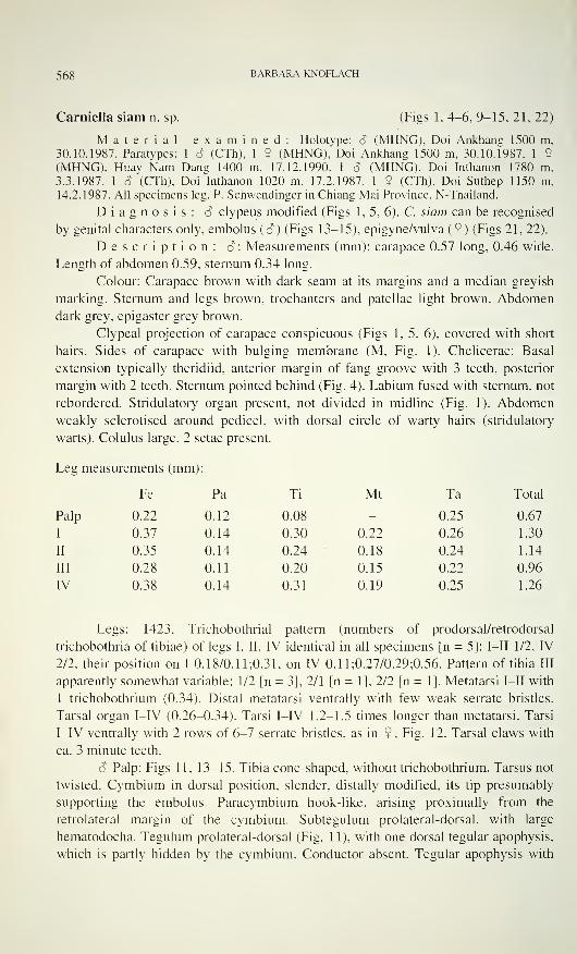
Carniella siani n. sp. (Figs 1,4-6, 9-15, 21, 22)
Material examined: Holotype: 6 (MHNG), Doi Ankhang 1500 m,
30.10.1987. Paratvpes: 1 S (CTh). 1 9 (MHNG), Doi Ankhang 1500 m, 30.10.1987. 1 9
(MHNG). Huay Nam Dang 1400 m. 17.12.1990. 1 â (MHNG). Doi Inthanon 1780 m,
3.3.1987. 1 S (CTh). Doi Inthanon 1020 m. 17.2.1987. 1 9 (CTh), Doi Suthep 1150 m,
14.2.1987. All specimens leg. P. Schweninger in Chiang Mai Province, N-Thailand.
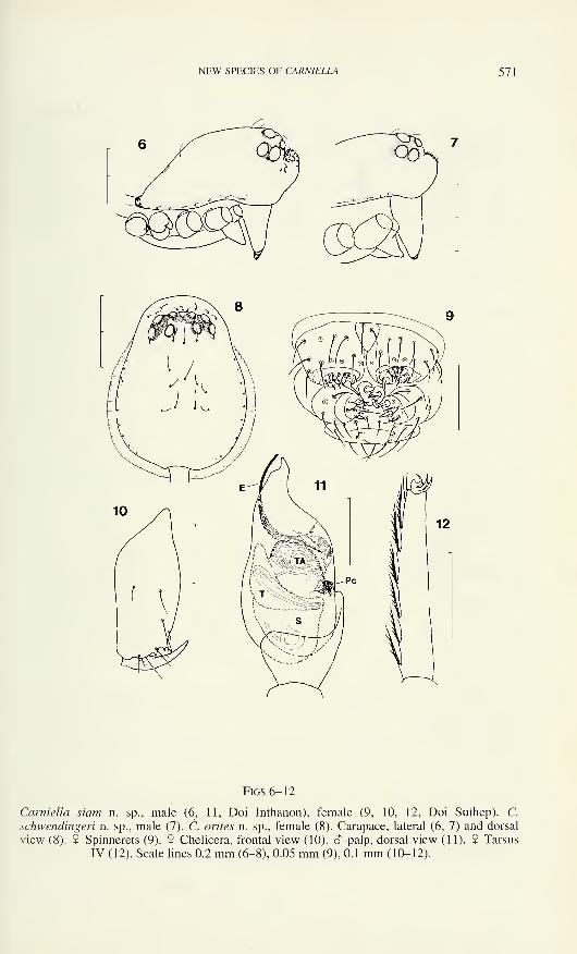
Diagnosis: â clypeus modified (Figs 1, 5, 6). C. siam can be recognised
by genital characters only, embolus (â) (Figs 13-15), epigyne/vulva (2) (Figs 21, 22).
Description: 6: Measurements (mm): carapace 0.57 long, 0.46 wide.
Length of abdomen 0.59. sternum 0.34 long.
Colour: Carapace brown with dark seam at its margins and a median greyish
marking. Sternum and legs brown, trochanters and patellae light brown. Abdomendark grey, epigaster grey brown.
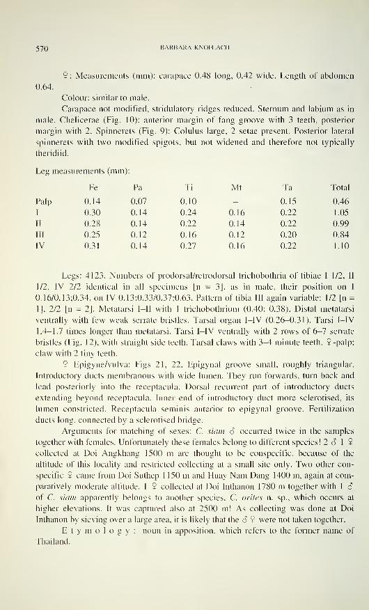
Clypeal projection of carapace conspicuous (Figs 1, 5. 6), covered with short
hairs. Sides of carapace with bulging membrane (M. Fig. 1). Chelicerae: Basal
extension typically theridiid, anterior margin of fang groove with 3 teeth, posterior
margin with 2 teeth. Sternum pointed behind (Fig. 4). Labium fused with sternum, not
rebordered. Stridulatory organ present, not divided in midline (Fig. 1). Abdomenweakly sclerotised around pedicel, with dorsal circle of warty hairs (stridulatory
warts). Colulus large, 2 setae present.
Leg measurements (mm):
Fe Pa Ti Mt Ta Total
Palp 0.22 0.12 0.08 - 0.25 0.67
I 0.37 0.14 0.30 0.22 0.26 1.30
II 0.35 0.14 0.24 0.18 0.24 1.14
III 0.28 0.11 0.20 0.15 0.22 0.96
IV 0.38 0.14 0.31 0.19 0.25 1.26
Legs: 1423. Trichobothrial pattern (numbers of prodorsal/retrodorsal
trichobothria of tibiae) of legs I. II. IV identical in all specimens [n = 5]: I—II 1/2, rV
2/2. their position on I 0.18/0.1 1:0.31. on IV 0.11:0.27/0.29:0.56. Pattern of tibia III
apparently somewhat variable: 1/2 [n = 3]. 2/1 [n = 1], 2/2 [n = 1]. Metatarsi I—II with
1 trichobothrium (0.34). Distal metatarsi ventrally with few weak serrate bristles.
Tarsal organ I-TV (0.26-0.34). Tarsi I-PV 1.2-1.5 times longer than metatarsi. Tarsi
I-IV ventrally with 2 rows of 6-7 serrate bristles, as in 9, Fig. 12. Tarsal claws with
ca. 3 minute teeth.
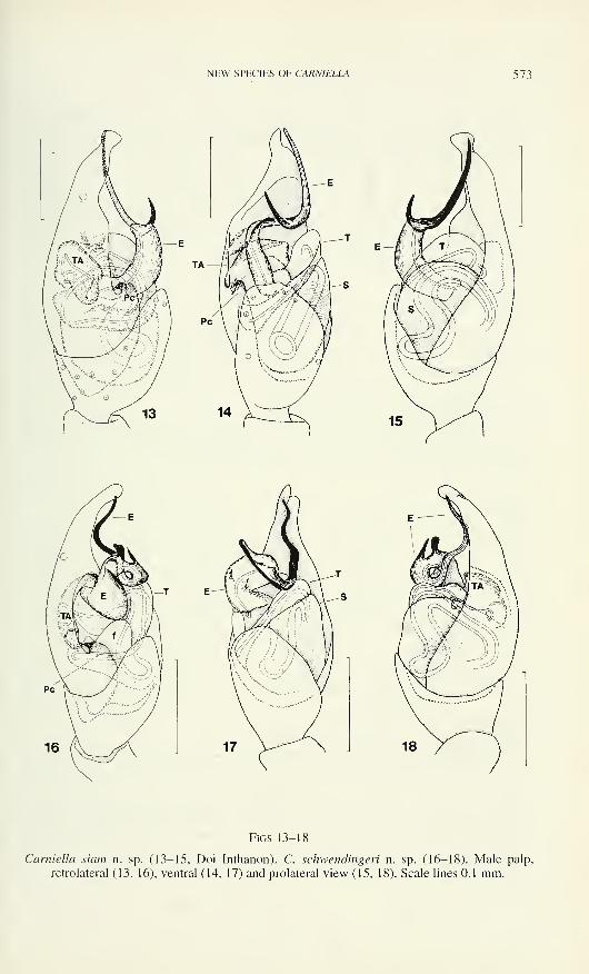
â Palp: Figs 11. 13-15. Tibia cone-shaped, without trichobothrium. Tarsus not
twisted. Cymbium in dorsal position, slender, distally modified, its tip presumably
supporting the embolus. Paracymbium hook-like, arising proximally from the
retrolateral margin of the cymbium. Subtegulum prolateral-dorsal. with large
hematodocha. Tegulum prolateral-dorsal (Fig. 11), with one dorsal tegular apophysis,
which is partly hidden by the cymbium. Conductor absent. Tegular apophysis with
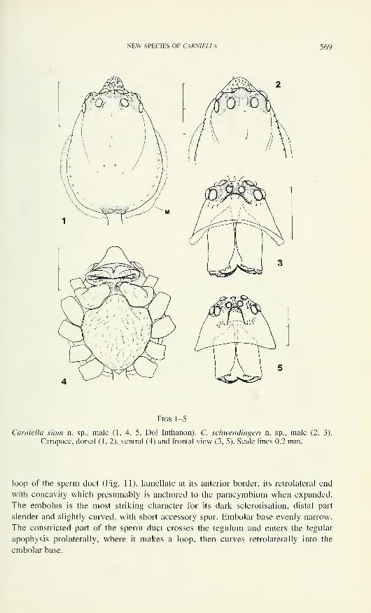
NEW SPECIES OF CARNIELLA 569
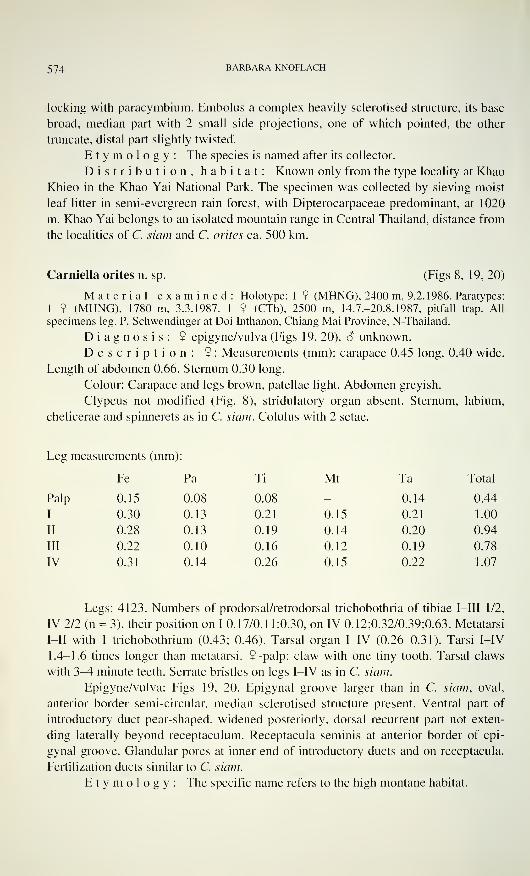
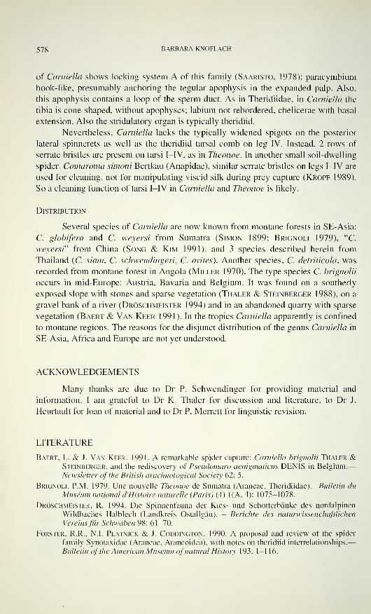
Figs 1-5
Carniella siam n. sp., male (1, 4, 5, Doi Inthanon). C. schwendingeri n. sp., male (2, 3).
Carapace, dorsal (1,2), ventral (4) and frontal view (3, 5). Scale lines 0.2 mm.
loop of the sperm duct (Fig. 11), lamellate at its anterior border, its retrolateral end
with concavity which presumably is anchored to the paracymbium when expanded.
The embolus is the most striking character for its dark sclerotisation, distal part
slender and slightly curved, with short accessory spur. Embolar base evenly narrow.
The constricted part of the sperm duct crosses the tegulum and enters the tegular
apophysis prolaterally, where it makes a loop, then curves retrolaterally into the
embolar base.
570 BARBARA KNOFLACH
9: Measurements (mm): carapace 0.48 long, 0.42 wide. Length of abdomen
0.64.
Colour: similar to male.
Carapace not modified, stridulatory ridges reduced. Sternum and labium as in
male. Chelicerae (Fig. 10): anterior margin of fang groove with 3 teeth, posterior
margin with 2. Spinnerets (Fig. 9): Colulus large, 2 setae present. Posterior lateral
spinnerets with two modified spigots, but not widened and therefore not typically
theridiid.
Leg measurements (mm):
Fe Pa Ti Mt Ta Total
Palp 0.14 0.07 0.10 - 0.15 0.46
I 0.30 0.14 0.24 0.16 0.22 1.05
II 0.28 0.14 0.22 0.14 0.22 0.99
III 0.25 0.12 0.16 0.12 0.20 0.84
IV 0.31 0.14 0.27 0.16 0.22 1.10
Legs: 4123. Numbers of prodorsal/retrodorsal trichobothria of tibiae I 1/2, II
1/2, IV 2/2 identical in all specimens [n = 3], as in male, their position on I
0.16/0.13:0.34, on IV 0.13:0.33/0.37:0.63. Pattern of tibia III again variable: 1/2 [n =
1], 2/2 [n = 2]. Metatarsi I—II with 1 trichobothrium (0.40; 0.38). Distal metatarsi
ventrally with few weak serrate bristles. Tarsal organ I-IV (0.26-0.31). Tarsi I-IV
1.4-1.7 times longer than metatarsi. Tarsi I-IV ventrally with 2 rows of 6-7 serrate
bristles (Fig. 12), with straight side teeth. Tarsal claws with 3^4 minute teeth. 9 -palp:
claw with 2 tiny teeth.
9 Epigyne/vulva: Figs 21, 22. Epigynal groove small, roughly triangular.
Introductory ducts membranous with wide lumen. They run forwards, turn back and
lead posteriorly into the receptacula. Dorsal recurrent part of introductory ducts
extending beyond receptacula. Inner end of introductory duct more sclerotised, its
lumen constricted. Receptacula seminis anterior to epigynal groove. Fertilization
ducts long, connected by a sclerotised bridge.
Arguments for matching of sexes: C. siam â occurred twice in the samples
together with females. Unfortunately these females belong to different species! 2 â 1 9
collected at Doi Angkhang 1500 m are thought to be conspecific, because of the
altitude of this locality and restricted collecting at a small site only. Two other con-
specific 9 came from Doi Suthep 1150 m and Huay Nam Dang 1400 m, again at com-
paratively moderate altitude. 1 9 collected at Doi Inthanon 1780 m together with 1 S
of C. siam apparently belongs to another species, C. orites n. sp., which occurs at
higher elevations. It was captured also at 2500 m! As collecting was done at Doi
Inthanon by sieving over a large area, it is likely that the â 9 were not taken together.
Etymology: noun in apposition, which refers to the former name of
Thailand.
NEW SPECIES OF CARN1ELLA 571
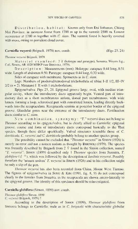
Figs 6-12
Carniella siam n. sp., male (6, 11, Doi Inthanon), female (9, 10, 12, Doi Suthep). C.
schwendingeri n. sp., male (7). C. orites n. sp., female (8). Carapace, lateral (6, 7) and dorsal
view (8). 9 Spinnerets (9). 9 Chelicera, frontal view (10). <5-palp, dorsal view (11). 9 Tarsus
IV (12). Scale lines 0.2 mm (6-8), 0.05 mm (9), 0.1 mm (10-12).
572 BARBARA KNOFLACH
Distribution, habitat: C. siam is known from 4 localities in NW-Thailand, Chiang Mai Province. Most specimens were sieved from litter of evergreen
lower montane forests at Doi Inthanon and at Doi Suthep, with Dipterocarpaceae and
oaks predominant, from about 1000 m up to 1780 m. The species is not restricted to
dense woodland. 2 â 1 9 came from sieving herb litter in a deforested small valley
with a stream at Doi Angkhang, 1 o" was taken in a pine forest (Pinus merkusii, P.
keysia) with needle litter and little undergrowth at Doi Inthanon 1020 m. The habitat
at Huay Nam Dang was a fragmented evergreen montane forest at 1400 m.
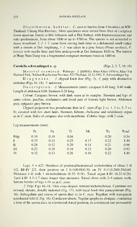
Carniella schwendingeri n. sp. (Figs 2, 3, 7, 16-18)
Material examined: Holotype: 3 (MHNG). Khao Khieo 1020 m, Khao Yai
National Park, Nakhon Ratchasima Province, NE-Thailand, 24.12.1992, P. Schwendinger leg.
Diagnosis: â clypeal knob low (Fig. 3), S palp with distinctive
embolus (Figs 16-18). 5 unknown.
Description: â Measurements (mm): carapace 0.48 long, 0.40 wide.
Length of abdomen 0.60. Sternum 0.28 long.
Colour: Carapace brown, with dark seam at its margins. Sternum and legs of
same colour, patellae, trochanters and distal part of femora light brown. Abdomengrey, epigaster grey-brown.
Clypeal projection less protuberant than in C. siam (Figs 2 vs. 1,3 vs. 5, 7 vs.
6), covered with few short hairs. Sternum, labium, chelicerae and stridulatory organ
as in C. siam. Sides of carapace also with membrane. Colulus large, with 2 setae.
Leg measurements (mm)i:
Fe Pa Ti Mt Ta Total
Palp 0.19 0.10 0.04 - 0.20 0.54
I 0.33 0.13 0.25 0.17 0.22 1.10
II 0.28 0.12 0.20 0.14 0.21 0.96
III 0.23 0.10 0.18 0.12 0.20 0.82
IV 0.32 0.13 0.26 0.16 0.22 1.10
Legs: 1 = 423. Numbers of prodorsal/retrodorsal trichobothria of tibiae I—II
1/2, III-IV 2/2, their position on I 0.14/0.08:0.30, on IV 0.11;0.28/0.30;0.60.
Metatarsi I—II with 1 trichobothrium (0.35; 0.41). Tarsal organ I-IV (0.21-0.26).
Tarsi I-IV 1.3—1.7 times longer than metatarsi. Tarsal claws with 3-4 minute teeth.
Serrate bristles of legs I-IV as in C. siam.
S Palp: Figs 16-18. Tibia cone-shaped, without trichobothrium. Cymbium not
twisted, slender, distally indented (Fig. 17), with basal hook-like paracymbium (Fig.
16). Subtegulum and course of sperm duct as in C. siam. Tegulum with transparent
retrolateral fold (f, Fig. 16). Conductor absent. Tegular apophysis elongate, containing
a loop of the sperm duct, in retrolateral-dorsal position, its retrolateral end presumably
NEW SPECIES OF CARNIELLA 573
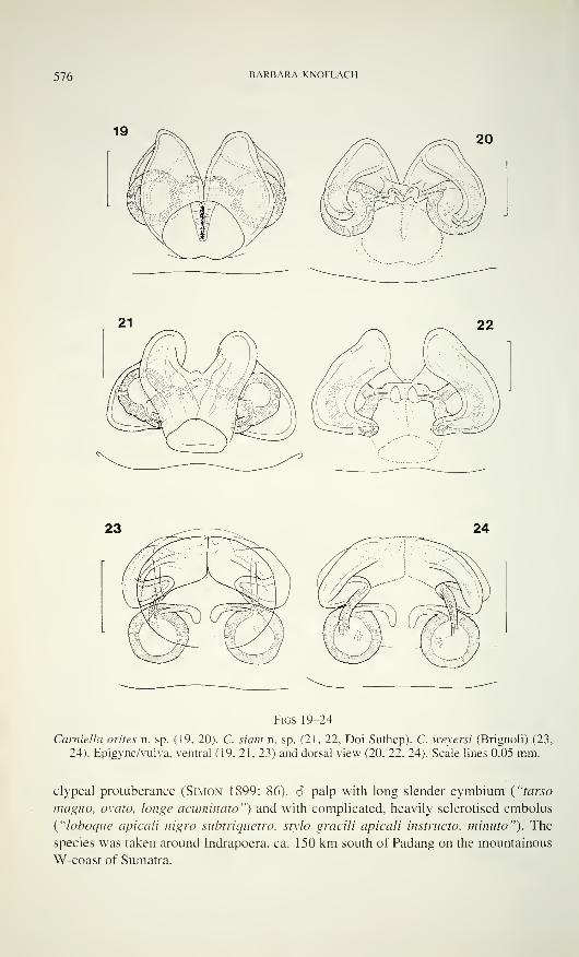
Figs 13-18
Carniella siam n. sp. (13-15, Doi Inthanon). C. schwendingeri n. sp. (16-18). Male palp,
retrolateral (13, 16), ventral (14, 17) and prolateral view (15, 18). Scale lines 0.1 mm.
574 BARBARA KNOFLACH
locking with paracymbium. Embolus a complex heavily sclerotised structure, its base
broad, median part with 2 small side projections, one of which pointed, the other
truncate, distal part slightly twisted.
Etymology: The species is named after its collector.
Distribution, habitat: Known only from the type locality at Khao
Khieo in the Khao Yai National Park. The specimen was collected by sieving moist
leaf litter in semi-evergreen rain forest, with Dipterocarpaceae predominant, at 1020
m. Khao Yai belongs to an isolated mountain range in Central Thailand, distance from
the localities of C. siam and C. orites ca. 500 km.
Carniella orites n. sp. (Figs 8, 19, 20)
Material examined: Holotype: 1 ? (MHNG). 2400 m, 9.2.1986. Paratypes:
1 9 (MHNG), 1780 m, 3.3.1987. 1 9 (CTh), 2500 m, 14.7.-20.8.1987, pitfall trap. All
specimens leg. P. Schwendinger at Doi Inthanon, Chiang Mai Province, N-Thailand.
Diagnosis: ? epigyne/vulva (Figs 19, 20). S unknown.
Description: 9 : Measurements (mm): carapace 0.45 long, 0.40 wide.
Length of abdomen 0.66. Sternum 0.30 long.
Colour: Carapace and legs brown, patellae light. Abdomen greyish.
Clypeus not modified (Fig. 8), stridulatory organ absent. Sternum, labium,
chelicerae and spinnerets as in C. siam. Colulus with 2 setae.
Leg measurements (mm):
Fe Pa Ti Mt Ta Total
Palp 0.15 0.08 0.08 0.14 0.44
I 0.30 0.13 0.21 0.15 0.21 1.00
II 0.28 0.13 0.19 0.14 0.20 0.94
III 0.22 0.10 0.16 0.12 0.19 0.78
IV 0.31 0.14 0.26 0.15 0.22 1.07
Legs: 4123. Numbers of prodorsal/retrodorsal trichobothria of tibiae I—III 1/2,
IV 2/2 (n = 3), their position on I 0.17/0.1 1:0.30, on IV 0.12;0.32/0.39;0.63. Metatarsi
I—II with 1 trichobothrium (0.43; 0.46). Tarsal organ I-IV (0.26-0.31). Tarsi I-IV
1.4-1.6 times longer than metatarsi. 9 -palp: claw with one tiny tooth. Tarsal claws
with 3-4 minute teeth. Serrate bristles on legs I-IV as in C. siam.
Epigyne/vulva: Figs 19, 20. Epigynal groove larger than in C. siam, oval,
anterior border semi-circular, median sclerotised structure present. Ventral part of
introductory duct pear-shaped, widened posteriorly, dorsal recurrent part not exten-
ding laterally beyond receptaculum. Receptacula seminis at anterior border of epi-
gynal groove. Glandular pores at inner end of introductory ducts and on receptacula.
Fertilization ducts similar to C. siam.
Etymology: The specific name refers to the high montane habitat.
NEW SPECIES OF CARNIELLA 575
Distribution, habitat: Known only from Doi Inthanon, Chiang
Mai Province, in montane forest from 1780 m up to the summit 2500 m. Lowest
occurrence at 1780 m together with C. siam. The summit forest is heavily covered
with moss, owing to prevalent cloud cover.
Carniella weyersi (Brignoli, 1979) nov. comb. (Figs 23, 24)
Theonoe weyersi Brignoli, 1 979
Material examined: 2 9 (holotype and paratype), Sumatra, Weyers leg.,
Coll. Simon, AR 1020 MHNP (see Brignoli, 1979).
Description: Measurements (mm): Holotype: carapace 0.44 long, 0.31
wide. Length of abdomen 0.50. Paratype: carapace 0.44 long, 0.33 wide.
Sides of carapace with membrane. Spinnerets as in C. siam.
Legs: Numbers of prodorsal/retrodorsal trichobothria of tibiae I—II 1/2, III—IV
2/2 (n = 2). Metatarsi I—II with 1 trichobothrium.
Epigyne/vulva: Figs 23, 24. Epigynal groove large, oval, with median trian-
gular cavity, where the introductory ducts apparently begin. Ventral part of intro-
ductory duct as a short membranous atrium, dorsal part membranous, with wide
lumen, forming a loop, sclerotised part with constricted lumen, leading directly back-
wards into the receptaculum. Receptacula seminis at posterior border of the epigynal
groove. Glandular pores near the entrance of the introductory ducts. Fertilization
ducts similar to C. siam.
New combination, synonymy: "T." weyersi does not belong to
Theonoe according to its epigyne/vulva, but is clearly allied to Carniella: epigynal
groove, course and form of introductory ducts correspond basically to the Thai
species, though these differ specifically. Vulval structures resemble those of C.
detriticola. C. weyersi and C. detriticola probably belong to another species group.
The possibility cannot be excluded that "Theonoe weyersi" in Simon (1926) is
merely an error and not a nomen nudum as thought by Brignoli (1979). The species
was formally described by Brignoli from 2 2 found in the Simon collection, named"77. weyersi". Simon (1899) described only 1 Theonoe species from Sumatra, T.
globifera (S 9), which was followed by the description of Iardinis weyersi. Possibly
therefore the 'nomen nudum' 77. weyersi in Simon (1926) and in his collection might
be only a slip of the pen.
Theonoe weyersi has also been recorded from China, Mount West Tianmu.
The figures of epigyne/vulva in Song & Kim (1991, fig. 8, 9) do not correspond
clearly to the females from Sumatra, as the receptacula are shown antero-laterally to
the epigynal groove. The identity of this specimen should be reinvestigated.
Carniella globifera (Simon, 1899) nov. comb.
Theonoe globifera Simon, 1 899C. weyersi (Brignoli, 1 979) ?
According to the description of Simon (1899), Theonoe globifera from
Sumatra belongs to Carniella: male as in C. brignolii with characteristic globular
576 BARBARA KNOFLACH
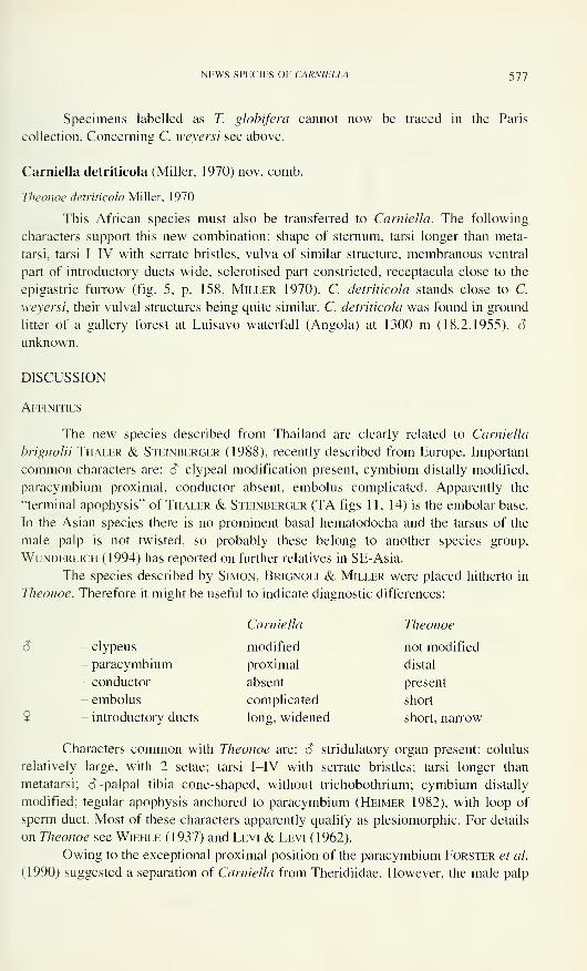
Figs 19-24
Garniella orites n. sp. (19, 20). C. siam n. sp. (21. 22, Doi Suthep). C. weyersi (Brignoli) (23,
24). Epigyne/vulva, ventral (19, 21, 23) and dorsal view (20, 22, 24). Scale lines 0.05 mm.
clypeal protuberance (Simon 1899: 86). 6 palp with long slender cymbium ("tarso
magno, ovato, longe acuminato") and with complicated, heavily sclerotised embolus
("loboque apicali nigro subtriquetro, stylo gracili apicali instructo, minuto"). The
species was taken around Indrapoera, ca. 150 km south of Padang on the mountainous
W-coast of Sumatra.
NEWS SPECIES OF CARNIELLA 577
Specimens labelled as T. globifera cannot now be traced in the Paris
collection. Concerning C. weyersi see above.
Carniella detriticola (Miller, 1970) nov. comb.
Theonoe detriticola Miller, 1 970
This African species must also be transferred to Carniella. The following
characters support this new combination: shape of sternum, tarsi longer than meta-
tarsi, tarsi I-IV with serrate bristles, vulva of similar structure, membranous ventral
part of introductory ducts wide, sclerotised part constricted, receptacula close to the
epigastric furrow (fig. 5, p. 158, Miller 1970). C. detriticola stands close to C.
weyersi, their vulval structures being quite similar. C. detriticola was found in ground
litter of a gallery forest at Luisavo waterfall (Angola) at 1300 m (18.2.1955). â
unknown.
DISCUSSION
Affinities
The new species described from Thailand are clearly related to Carniella
brignolii Thaler & Steinberger (1988), recently described from Europe. Important
common characters are: S clypeal modification present, cymbium distally modified,
paracymbium proximal, conductor absent, embolus complicated. Apparently the
"terminal apophysis" of Thaler & Steinberger (TA figs 11, 14) is the embolar base.
In the Asian species there is no prominent basal hematodocha and the tarsus of the
male palp is not twisted, so probably these belong to another species group.
Wunderlich (1994) has reported on further relatives in SE-Asia.
The species described by Simon, Brignoli & Miller were placed hitherto in
Theonoe. Therefore it might be useful to indicate diagnostic differences:
Carniella Theonoe
- clypeus modified not modified
- paracymbium proximal distal
- conductor absent present
- embolus complicated short
9 - introductory ducts long, widened short, narrow
Characters common with Theonoe are: S stridulatory organ present; colulus
relatively large, with 2 setae; tarsi I-IV with serrate bristles; tarsi longer than
metatarsi; â -palpal tibia cone-shaped, without trichobothrium; cymbium distally
modified; tegular apophysis anchored to paracymbium (Heimer 1982), with loop of
sperm duct. Most of these characters apparently qualify as plesiomorphic. For details
on Theonoe see Wiehle (1937) and Levi & Levi (1962).
Owing to the exceptional proximal position of the paracymbium Forster et al.
(1990) suggested a separation of Carniella from Theridiidae. However, the male palp
578 BARBARA KNOFLACH
of Carniella shows locking system A of this family (Saaristo, 1978): paracymbium
hook-like, presumably anchoring the tegular apophysis in the expanded palp. Also,
this apophysis contains a loop of the sperm duct. As in Theridiidae, in Carniella the
tibia is cone-shaped, without apophyses; labium not rebordered, chelicerae with basal
extension. Also the stridulatory organ is typically theridiid.
Nevertheless, Carniella lacks the typically widened spigots on the posterior
lateral spinnerets as well as the theridiid tarsal comb on leg IV. Instead, 2 rows of
serrate bristles are present on tarsi I-IV, as in Theonoe. In another small soil-dwelling
spider, Comaroma simoni Bertkau (Anapidae), similar sen-ate bristles on legs I-IV are
used for cleaning, not for manipulating viscid silk during prey capture (Kropf 1989).
So a cleaning function of tarsi I-IV in Carniella and Theonoe is likely.
Distribution
Several species of Carniella are now known from montane forests in SE-Asia:
C. globifera and C. weyersi from Sumatra (Simon 1899; Brignoli 1979), "C.
weyersï" from China (Song & Kim 1991), and 3 species described herein from
Thailand (C. siam, C. schwendingeri, C. orites). Another species, C. detriticola, was
recorded from montane forest in Angola (Miller 1970). The type species C. brignolii
occurs in mid-Europe: Austria, Bavaria and Belgium. It was found on a southerly
exposed slope with stones and sparse vegetation (Thaler & Steinberger 1988), on a
gravel bank of a river (Dröschmeister 1994) and in an abandoned quarry with sparse
vegetation (Baert & Van Keer 1991). In the tropics Carniella apparently is confined
to montane regions. The reasons for the disjunct distribution of the genus Carniella in
SE-Asia, Africa and Europe are not yet understood.
ACKNOWLEDGEMENTS
Many thanks are due to Dr P. Schwendinger for providing material and
information. I am grateful to Dr K. Thaler for discussion and literature, to Dr J.
Heurtault for loan of material and to Dr P. Merrett for linguistic revision.
LITERATURE
Baert. L. & J. Van Keer. 1991. A remarkable spider capture: Carniella brignolii Thaler &Steinberger, and the rediscovery of Pseudomaro aenigmaticus DENIS in Belgium.
—
Newsletter of the British arachnological Society 62: 5.
Brignoli, P.M. 1979. Une nouvelle Theonoe de Sumatra (Araneae, Theridiidae).- Bulletin du
Muséum national d'Histoire naturelle (Paris) (4) 1(A, 4): 1075-1078.
Dröschmeister, R. 1994. Die Spinnenfauna der Kies- und Schotterbänke des nordalpinen
Wildbaches Haiblech (Landkreis Ostallgäu). - Berichte des naturwissenchaßlichen
Vereins für Schwaben 98: 61-70.
Forster, R.R., N.I. Platnick & J. Coddington. 1990. A proposal and review of the spider
family Synotaxidae (Araneae, Araneoidea), with notes on theridiid interrelationships.
—
Bulletin of the American Museum ofnatural Histoiy 193: 1-1 16.
NEW SPECIES OF CARNIELLA 579
Heimer, S. 1982. Interne Arretierungsmechanismen an den Kopulationsorganen männlicher
Spinnen (Arachnida, Araneae). Ein Beitrag zur Phylogenie der Araneoidea.— Ento-
mologische Abhandlungen. Staatliches Museumfür Tierkunde in Dresden 45: 35-64.
Kropf, C. 1989. Web construction and prey capture of Comaroma simoni Bertkau (Araneae).—Acta zoologica fennica 190: 229-233.
Levi, W. & L.R. Levi. 1962. The genera of the spider family Theridiidae.- Bulletin of the
Museum ofcomparative Zoology 127: 1-71, Figs 1-334.
Miller, F. 1970. Spinnenarten der Unterfamilie Micryphantinae und der Familie Theridiidae
aus Angola.- Publicaçoes culturais Companhia de Diamantes de Angola 82: 75-166.
Saaristo, M.I. 197.8. Spiders (Arachnida, Araneae) from the Seychelle Islands, with notes on
taxonomy . Annales zoologici fenilici 15: 99-126.
Simon, E. 1899. Contribution à la faune de Sumatra. Arachnides recueillis par M.J.L. Weyers à
Sumatra (deuxième mémoire).- Annales de la Société entomologique de Belgique 43:
78-125.
Simon, E. 1926. Les arachnides de France 6(2): 309-532. Roret, Paris.
Song, D.X. & J.P. Kjm. 1991. On some species of spiders from Mount West Tianmu, Zhejiang,
China (Araneae).- Korean Arachnology 7: 19-27.
Thaler, K. &. K.H. Steinberger. 1988. Zwei neue Zwerg-Kugelspinnen aus Österreich
(Arachnida: Aranei, Theridiidae).- Revue suisse de Zoologie 95: 997-1004.
Wiehle, H. 1937. Spinnentiere oder Arachnoidea VIII. 26. Familie: Theridiidae oder
Haubennetzspinnen (Kugelspinnen).- Tierwelt Deutschlands 33: 119-222. Fischer,
Jena.
Wunderlich, J. 1994. Bemerkenswerte Spinnen der rezenten und fossilen Faunen Mittel-
europas und ihre biogeographischen Beziehungen zu den Tropen und Subtropen
(Arachnida: Araneae).- Arachnologische Mitteilungen 7: 53-55.
ADDENDUM
When this paper was in press, two further Carniella-species were described from Indonesia
(Wunderlich 1995): C. krakatauensis (S) from Anak Krakatau, C. sumatraensis (6 2) fromN-Sumatra. C. schwendingeri is similar to C. krakatauensis.
Wunderlich, J. 1995. Südostasiatische Arten der Gattung Carniella Thaler & Steinberger
1988, mit zwei Neubeschreibungen (Arachnida: Araneae: Theridiidae). Beiträge zur
Araneologie, 4 (1994): 553-558.

Revue suisse de Zoologie, 103 (3): 581-605; septembre 1996
Spalacosostea, an anomalous new terrestrial dryopid fromSouth East Asia (Coleoptera: Dryopidae)
Jân KODADADepartment of Zoology, Comenius University, Mlynskâ dolina B-I,
842 15 Bratislava, Slovakia.
Spalacosostea, an anomalous new terrestrial dryopid from South East
Asia (Coleoptera: Dryopidae). - A new genus, Spalacosostea with two
new species, S. loebli from Borneo and S. pselaphoides from Sumatra, is
described. Both species were sifted from vegetation debris in rain forest.
They are unusual for their small size and notable for sexual dimorphism,
affecting the metathoracic wings and sensory organs. Females are
wingless, with membranous metanotum, suboval elytra, vestigial eyes and
short maxillary palps. Males have metathoracic wings well developed,
metanotum well sclerotized and composed from several parts, large eyes,
and their maxillary palps are very long and bear conspicuous peg-like
sensilla. Taxonomically significant structures and morphological features
unique to the Spalacosostea are discussed and illustrated. Diagnostic key
to the species is given.
Key-words: Coleoptera - Dryopidae - Spalacosostea - Oriental region -
Taxonomy - Morphology - Antennal sensilla.
INTRODUCTION
The family Dryopidae, of almost world-wide distribution (Brown 1981),
presently consists of 240 species in 24 genera. They live in a variety of freshwater and
terrestrial habitats. Many adults inhabit running waters and exhibit respiratory
adaptive features, such as microplastron structures in Pomatinus Sturm, 1853 and
Elmomorphus Sharp, 1888. In the contrary, the riparian dryopids (Barr &Spangler, 1992), e.g. Dryops Olivier, 1791, Pelonomus Erichson, 1847 and Ono-
pelmus Spangler, 1980, evolved macroplastron structures (Hinton 1969). Some of
these species undertake dispersal flights and are often taken in great numbers in light
traps. The few known larvae, are terrestrial or semiaquatic (Brown 1987).
The entirely terrestrial groups, e.g. Geoparnus Besuchet, 1978, Sosteamorphus
Hinton, 1936 and Oreoparnus Deleve, 1965, have been found in forest leaf litter and
Manuscript accepted 16.09.1995.
582 JAN KODADA
flood debris. They are generally characterized by a very compact, heavily sclerotized,
more or less ovoid body without plastron structures. Most of them lack metathoracic
wings, have relatively small eyes, and their elytral striae are often strongly developed.
Members of the Neotropical Quadryops Perkins & Spangler, 1985 were found in
arboreal habitats, and those of the Indo-Malaysian Sostea Pascoe, 1860 were beaten
from the foliage of different plants in rain forests (Kodada, unpublished). The
arboreal dryopids may be roughly distinguished from the epigean ones by the more
elongate body, the presence of large eyes, the well-developed metathoracic wings and
the elytra which often have a metallic shine.
To date, nine dryopid genera have been recorded from the Oriental Realm, two
of which {Geoparnus and Sostea) are terrestrial.
Two species of an additional terrestrial dryopid genus have been found by I.
Lobi, D. H. Burckhardt, D. Agosti and A. Smetana in northern Borneo and Sumatra.
These unusually small epigean dryopids exhibit remarkable sexual dimorphism
affecting particularly the metathoracic wings and the sensory organs.
MATERIAL AND METHODS
Members of following genera of terrestrial dryopids were studied: Geoparnus
setifer Besuchet, 1978 - holotype: 6, paratypes: 1 S, 1 $ (MHNG); Geoparnus sp. -
five undescribed species of both sexes (MHNG, CKB); Guaranius carlosi Spangler,
1991 -2 66 (NMW); Sostea tuberculata (Bollow, 1940) - holotype: ? (RMS);
Oreoparnus microps Deleve, 1965 - paratype: 1 6 (MHNG); Protoparnus sp. - 1 6
(CKB); Sostea crassa Hinton, 1936 - holotype: $ (BMNH); 2 6 6, 2 $ 2 (CKB);
Sostea elmoides Pascoe, 1860 - syntypes: 2 S 6 , 2 ex. sex not examined (BMNH), 2
$ 9 (CKB); Sostea lanifera Waterhouse, 1876 - holotype 6, (BMNH), M, F (CKB);
Sostea pillila Grouvelle, 1898 - syntypes: 1 S,2 $ $ (MNHP); Sostea westwoodii
Pascoe, 1860 - syntypes: 2 6a, (BMNH), 1 9 (BMNH); Sosteamorphus verrucatus
Hinton, 1936-4 6 6,4 9 9 (TMP); undescribed genus A from South Africa (Natal
Middld., Doreen Clarck Nat. R.) - 2 6 6,2 9 9 (TMP); undescribed genus B from
Venezuela (Sierre Nevada) - 1 6 (NMW).Specimens used for morphological studies were relaxed in water, cleaned and
treated with lactic acid. Metathoracic wings were removed, spread and observed on
unmounted slides. For scanning electron microscopy specimens were dehydrated in
graded ethanol series and air-dried from absolute ethanol, mounted on stubs with
Tempfix and sputter coated with gold. Illustrations were prepared with aid of a
drawing tube, using temporary transparency mounts. The differentiation of the
sensilla is based only on the form and size of their cuticular parts. The terminology of
the metathoracic wing structures follows Kukalovä-Peck & Lawrence (1993).
Abbreviations
BMNH Natural History Museum, LondonCKB Kodada collection, Bratislava
MHNG Muséum d' histoire naturelle, Genève
SPALACOSOSTEA, A NEW DRYOPID FROM SOUTH EAST ASIA 583
MNHP Museum National d' Histoire Naturelle, Paris
NMW Naturhistorisches Museum. WienRMS Naturhistoriska Riksmuseet, Stockholm
TMP Transvaal Museum, Pretoria
D Dendritic sensilla
DF Digitiform sensilla
PI, P2, P3, P4, P5 Peg-like sensilla
SI, S2 Styloconic sensilla
TL, TM, TS, Tl. T2 Hair-like sensilla
EL Elytral length
EI Elytral index, ratio of elytral ML to combined MW of elytra
HW Width of head with eyes
LPE Medial length of pronotum and elytra
ML Medial length
MW Maximum width
01 Ocular index, ratio of HW to minimum distance between eyes
Mean value ± standard deviation
n Number of measured specimens
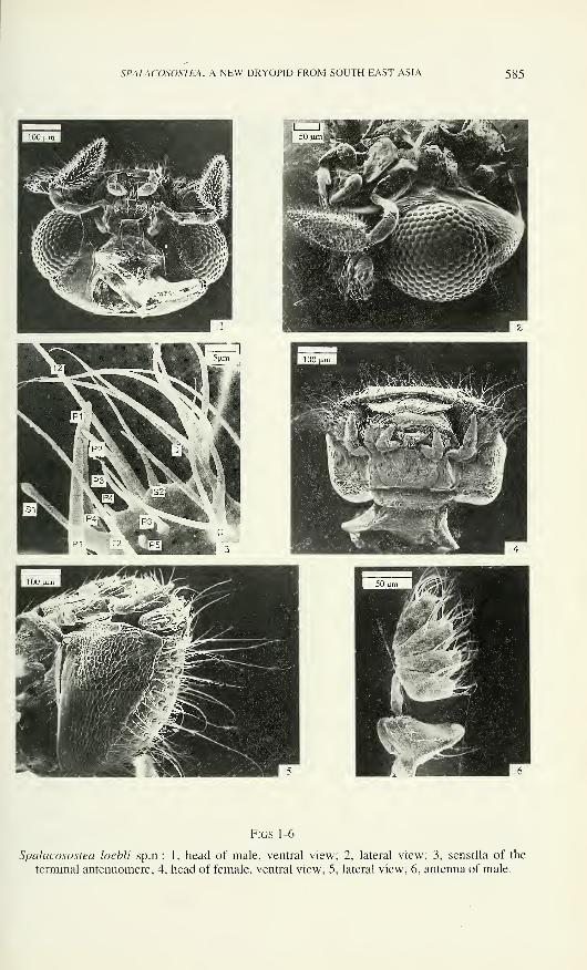
SYSTEMATIC SECTION
Spalacosostea gen.n. (Figs 1-65)
Type species: Spalacosostea loebli sp.n.
Gender: feminine.
Etymology: The generic name is a combination of names Spalax (Spalacidae:
Rodentia) and Sostea. Spalax are short-legged mammals that are extensive burrowers,
characterized by the absence of external openings for eyes, although small eyes are
present beneath the skin (referring to the vestigial eyes of females from the genus
described below). Sostea is a dryopid similar to the new genus.
Diagnosis: Spalacosostea may be distinguished from all other described
dryopids by following features in combination: ( 1 ) antennae six-segmented, pectinate
with enlarged antennomere 1; (2) eyes in female vestigial; (3) male maxillary palpus
with terminal segment unusually large, bearing conspicuous peg-like sensilla with an
enlarged, sharply tipped apex on almost entire surface; (4) metathoracic wing with
highly reduced anal veins; (5) tarsi four segmented.
The wingless female with membranous metanotum, suboval elytra, short
maxillary palps and vestigial eyes, differ conspicuously from male. The male is
characterized by the well sclerotized metanotum composed from several parts and
developed metathoracic wings, elongate elytra, large eyes and very long maxillary
palps with conspicuous sensilla. The association of both sexes is based also on
similar: (1) types and distribution of the sensilla on the antennae, labium, labrum and
legs; (2) shape of the labrum, labium and maxillae (maxillary palps excepted); (3)
type of the macro- and micropunctation; (4) vestiture; and (5) both sexes of each
species were found in the same samples.
Description â : Body form elongate (Fig. 49), slender, moderately convex
dorsally; about 2.4 times as long as wide (LPE/MW); length 1.20 - 1.60 mm (LPE).
Colour in both sexes varies from yellowish-brown to brown with yellowish antennae
and legs (obviously depending on maturity).
584 JAN KODADA
Vestiture of dorsal surface (Figs 19, 26, 35) consisting of three types of
yellowish hair-like sensilla. Type TL = very conspicuous, erect, about 200 - 300 urn
long, arising from more or less deep, indistinctly bordered sockets; longest sensilla
(TL) inserted on lateral elytral and pronotal margins. Type TM = intermediate, about
100 - 170 urn long, erect, arising from shallow socket; longest sensilla (TM) situated
on pronotum and elytra. Type TS = short, thin, about 40 - 70 um long, recumbent and
arising from shallow inconspicuous socket. Ventral surface with similar hair-like
sensilla as dorsal surface, but sensilla (TL, TM) arise mostly from large, shallow and
very distinctly bordered sockets (Figs 20, 23, 32). Some specimens covered with
encrusted material on cuticle of vertex and pronotum.
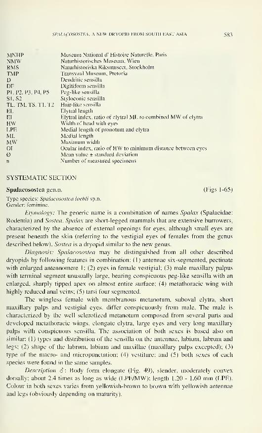
Head (Figs 1, 2, 26, 27) hypognathous, flat ventrally, arched dorsally and
laterally; occipital area distinctly shorter than longest eye diameter; moderately
retracted into prothorax. Punctation consisting of setigerous micro- and macro-
punctures. Micropunctures (sockets of sensilla TS) mainly on vertex and near
occipital ridge; distance between micropunctures about 0.5 times length of sensilla
(TS). Macropunctures represent sockets of sensilla (TL, TM), deeper and broader near
eyes than on frontoclypeus and separated by a distance of about 1 - 3 facet diameters.
Eyes (Figs 1, 2, 26) large, more or less protuberant, circular, coarsely faceted and only
with a few interfacetal sensilla (cf. type TM) on their dorsal half.
Labrum (Fig. 8) short (MW/ML = about 1.6), in posterior half strongly and
abruptly constricted to third of MW and concealed by clypeus. Lateral tormal
processes very short, bent ventro-mesally; postero-median process absent. Anterior
margin more or less emarginate, with a few closely arranged, bluntly tipped, peg-like
sensilla; lateral margin arcuate, with few hair-like sensilla. Epipharynx with two
lateral fields of moderately long, mesally directed, recumbent hair-like sensilla (about
as type TS), and postero-mesal rows of short and very closely set setae bordered
laterally by flat, broad setae. Anterior margin of clypeus arched, with a flat ridge;
frontoclypeal suture absent.
Antennae (Figs 3, 6, 7, 51) inserted into deep antennal sockets, six-segmented,
microreticulated. Antennomere 1 robust, dilated distally, dorsally with sensilla TMand one sensillum TL; latero-dorsal margin with flat ridge adjacent to clypeal ridge in
repose. Antennomere 2 as long as previous one, cylindrical and constricted in basal
third; antennomeres 3 - 6 dilated anteriorly, approximately of same length. Sensory
fields located on antero-median extension of each flagellar antennomere. Terminal
antennomere (Fig. 3) contains greatest number of different types of sensilla: (1) hair-
like sensilla type Tl = about 20 - 35 urn long and 1 urn wide at base; lateral and
dorsal face with 6-8 sensilla Tl; (2) type T2 = about 60 - 100 um long and about 2 -
3 urn wide, finely longitudinally grooved, one subapical sensillum T2, bilaterally
symmetrical on each side of the midline; (3) styloconic sensilla type SI = approxi-
mately 30 urn long and 4-5 urn wide (MW), bluntly tipped peg inserted at tip of
cylindrical projection, medio-dorsal face bears one subterminal sensillum SI; (4) type
S2 = about 10 urn long and 1 urn wide, slightly bent, sharp tipped conical peg
inserted on broad, short, basal projection, each antennomere bears only one median
sensillum S2; (5) peg-like sensilla type PI = approximately 35 - 40 pm long, 4-5 pm
SPALACOSOSTEA, A NEW DRYOPID FROM SOUTH EAST ASIA 585
Figs 1-6
Spalacosostea loebli sp.n.: 1, head of male, ventral view; 2, lateral view; 3, sensilla of the
terminal antennomere; 4, head of female, ventral view; 5, lateral view; 6, antenna of male.
586 JAN KODADA
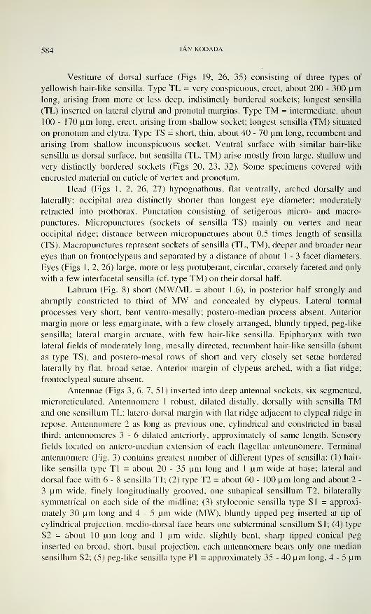
Figs 7-14
Spalacosostea loebli sp.n.: 7, terminal antennomere, male; 8, labrum, dorsal view; 9, sensilla of
the maxillary palpus, male; 10, apex of maxillary palpus, male; 1 1, maxillary palpus, male; 12
maxilla of female, ventral view; 13, dorsal view; 14, apex of galea.
SPALACOSOSTEA, A NEW DRYOPID FROM SOUTH EAST ASIA 587
wide, slightly bent, bluntly tipped, two sensilla PI inserted without a socket
posteriorly to sensilla T2; (6) type P2 = one 25 - 30 urn long, about 3 urn wide,
bluntly tipped peg without socket inserted sublaterally on dorsal face anteriorly to
sensillum PI; (7) type P3 = about 10 urn long, 1 urn wide, thin, bluntly tipped pegs,
without socket, one median sensillum P3 before sensillum S2 and two P3 laterally to
S2; 8) type P4 = about 5 urn long, 1 urn wide, or 2 sensilla P4 present on medio-
distal area before sensillum S2; (9) type P5 = only one, very short conical peg about 2
urn long and 1 urn wide, inserted on dorsal face near lateral sensillum P3; (10)
dendritic sensilla (Perkins & Spangler, 1985), type D = conspicuous branched sensilla
without socket, about 45 um long and 6 urn wide at base, antennomeres 3 to 6 bear 1
or 2 sublateral sensilla D. Antennomeres 3 to 5 similar to each other in sensillar
distribution, but lacking sensilla SI, P2, P3, P4, and number of sensilla Tl reduced
compared to antennomere 6.
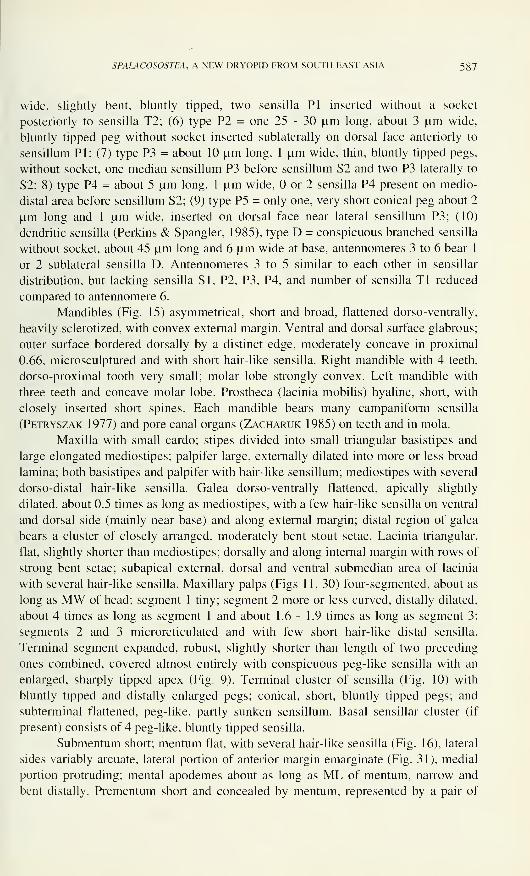
Mandibles (Fig. 15) asymmetrical, short and broad, flattened dorso-ventrally,
heavily sclerotized, with convex external margin. Ventral and dorsal surface glabrous;
outer surface bordered dorsally by a distinct edge, moderately concave in proximal
0.66, microsculptured and with short hair-like sensilla. Right mandible with 4 teeth,
dorso-proximal tooth very small; molar lobe strongly convex. Left mandible with
three teeth and concave molar lobe. Prostheca (lacinia mobilis) hyaline, short, with
closely inserted short spines. Each mandible bears many campaniform sensilla
(Petryszak 1977) and pore canal organs (Zacharuk 1985) on teeth and in mola.
Maxilla with small cardo; stipes divided into small triangular basistipes and
large elongated mediostipes; palpifer large, externally dilated into more or less broad
lamina; both basistipes and palpifer with hair-like sensillum; mediostipes with several
dorso-distal hair-like sensilla. Galea dorso-ventrally flattened, apically slightly
dilated, about 0.5 times as long as mediostipes, with a few hair-like sensilla on ventral
and dorsal side (mainly near base) and along external margin; distal region of galea
bears a cluster of closely arranged, moderately bent stout setae. Lacinia triangular,
flat, slightly shorter than mediostipes; dorsally and along internal margin with rows of
strong bent setae; subapical external, dorsal and ventral submedian area of lacinia
with several hair-like sensilla. Maxillary palps (Figs 11, 30) four-segmented, about as
long as MW of head; segment 1 tiny; segment 2 more or less curved, distally dilated,
about 4 times as long as segment 1 and about 1.6 - 1.9 times as long as segment 3;
segments 2 and 3 microreticulated and with few short hair-like distal sensilla.
Terminal segment expanded, robust, slightly shorter than length of two preceding
ones combined, covered almost entirely with conspicuous peg-like sensilla with an
enlarged, sharply tipped apex (Fig. 9). Terminal cluster of sensilla (Fig. 10) with
bluntly tipped and distally enlarged pegs; conical, short, bluntly tipped pegs; and
subterminal flattened, peg-like, partly sunken sensillum. Basal sensillar cluster (if
present) consists of 4 peg-like, bluntly tipped sensilla.
Submentum short; mentum flat, with several hair-like sensilla (Fig. 16), lateral
sides variably arcuate, lateral portion of anterior margin emarginate (Fig. 31), medial
portion protruding; mental apodemes about as long as ML of mentum, narrow and
bent distally. Prementum short and concealed by mentum, represented by a pair of
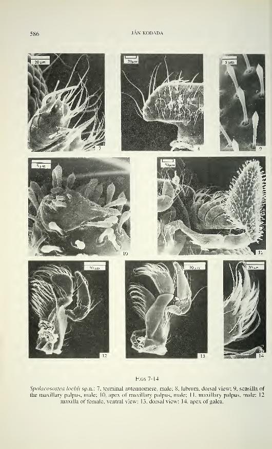
588 JAN KODADA
ii m
1 SOjjm B
pS^^^'r!^^Hp>--> '
i'|' fc>
ÉlP\ .-ssK^.-.
,; H^y-te;SjjSiy^
" S :
"'W^
! *§ "lï^-S^Ka§ßte^gljgWmËJÊ la
BUI | 20
Figs 15-20
Spalacosostea loebli sp.n.: 15, mandible, ventral view; 16, labium, male; 17 labium, female; 18,
apex of labial palpus, male; 19. pronotum of male, dorsal view; 20, ventral view.
SPALACOSOSTEA, A NEW DRYOPID FROM SOUTH EAST ASIA 589
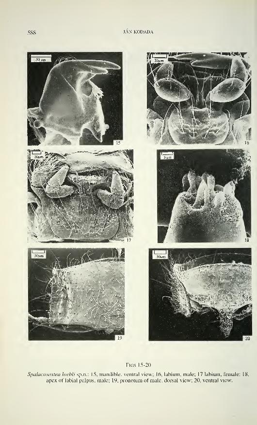
sclerites. Palpus labialis three-segmented; segment 1 short, inconspicuous, without
hair-like sensilla; segment 2 robust, about 1.8 times as long as wide (ML/MW), with
inner side almost straight, outer side strongly convex, distal half with several short
hair-like sensilla and long subapical external hair-like sensillum; segment 3 about as
long as preceding, nearly conical. Apical sensory field (Fig. 18) similar to those on
maxillary palpus, basal sensory field with two hair-like sensilla and digitiform (DF)
sensillum (Honomichl & Guse 1981). Ligula basally constricted, as long as pre-
mentum; antero-lateral portion rounded and laterally only weakly sclerotized; anterior
margin medially more or less deeply emarginated; proximal half ventrally with two
broad deep depressions on almost 0.8 of width of ligula; ventral surface bearing
several hair-like sensilla and two bluntly tipped, bent, submedian peg-like sensilla
near anterior margin; dorsal surface (hypopharynx) with short mesally directed hair-
like sensilla in two pairs of sensory fields, each pair separated by a triangular area
covered with longer, hair-like sensilla.
Dorsal tentorial arms long, thin and slightly surpassing middle of cranium;
anterior tentorial pits not visible; posterior tentorial pits conspicuous; posterior ten-
torial arms joined by a transverse bridge, bearing two short bent and flattened pro-
cesses. Gula short (MW/ML = about 4), trapezoidal, almost flat, finely micro-
sculptured; gular sutures distinct. Occipital ridge (Fig. 26) fine, separating smooth
dorsal surface from sculptured ventral parts. Occipital foramen large, in ventro-lateral
angles articulating with apices of large cervical sclerites.
Pronotum moderately convex dorsally; with conspicuous longitudinal im-
pression (Fig. 19) laterally; anterior margin arcuate, with a shallow emargination on
lateral portions; lateral margin slightly arcuate or almost straight, finely crenulate and
explanate; posterior angles almost rectangular; posterior margin trisinuate; central
area usually with numerous sensilla (TS, TM), marginal areas mainly with sensilla
TL. Internal posterior notai region bears two small condylles and a pair of ridges,
latter fitting into cavities on anterior margin of elytra. Hypomeron (Hinton 1939)
basally broadest, in apical third strongly narrowed (Fig. 20), with punctation variable.
Prosternum bordered anteriorly by a flat ridge, rounded at antero-lateral angles and
separated from hypomeron in distal fourth; about 0.5 times as long as wide in front of
procoxae; strongly deflexed and finely, sparsely punctured in anterior third; remaining
surface with large, flat bottomed punctures. Prosternai intercoxal process narrow (Fig.
20), about 2.4 times as long as wide, constricted and bluntly tipped in apical third;
procoxal cavities open posteriorly; trochantin large. Mesosternum and mesepisternum
concave anteriorly, glabrous; exposed part of mesepisternum with a transverse row of
macropunctures anteriorly; mesepimeron with macropunctures; median mesosternal
cavity deep, receives apex of prosternai process; mesocoxal cavities oval (dorsal
view); mesosternal furca with slightly divergent arms. Scutellum small and sub-
triangular (MW/ML = 1 ), glabrous, with arcuate sides. Metasternum about 3 times as
long as mesosternum (ML/ML), convex ventrally, without (Fig. 32) or with only very
short, shallow longitudinal impression in posterior third (Fig. 23); median longi-
tudinal endocarina present in posterior half; transversal suture (Crowson 1967)
absent, but its presumpted position marked by a transverse row of macropunctures;
590 JAN KODADA
metasternal intercoxal process apically emarginate, as long as wide. Macropunctures
on lateral areas of metasternum and on long, exposed triangular area of metepi-
sternum, but almost absent medially and on most of submedial-proximal areas of
metasternum. Metendosternite with a long, narrow stalk; lateral arms and anterior
tendons slender and short. Metanotum about 3 times as long as mesonotum; both
divided into prescutum, scutellum and lateral scuta (Lawrence & Britton 1991);
postnotum strongly transverse and slightly shorter than mesonotum.
Elytra elongate and parallel-sided in anterior 0.66, evenly arched towards apex
(Fig. 35); flat dorsally in cross section, deflexed laterally; apices more or less acute
(Fig. 36); anterior margin slightly elevated and finely crenulate; humeri prominent;
suturai margin slightly elevated. Each elytron with 9 more or less regular rows of
deeply impressed and densely arranged punctures (striae) between explanate lateral
and suturai margin; striae 2, 3, 7 and 8 ending on elytral declivity, striae 5 and 6
ending before, striae 1. 4, 9 reaching apex. Strial intervals slightly convex, narrow;
unpaired intervals with rows of sensilla (TL, TM), paired intervals bearing a double
row of sensilla TS. Epipleura reaching elytral apex, about as wide as apex of tibia
basally, narrowed apically (Fig. 32), oblique ventrally, more or less coarsely punctate
and set with sensilla (TL) along finely denticulate lateral margin. Ventral elytral
flange short and placed laterally slightly anterior to middle (Figs 36, 37, 38) near a
"rubbing patch" (Crowson 1981).
Metathoracic wing (Fig. 56) about 2 times as long as elytron, slightly
pigmented; apical field occupies almost 0.66 of wing; anterior margin composed of
precosta (PC), costa (C) and subcosta anterior (ScA). Subcosta posterior (ScP) and
darker pigmented radius anterior (RA) run parallel and close to anterior margin; both
together form distally a radial bar ending abruptly, short before middle of wing;
pigmented strips situated under radial bar. Radius posterior (RP) developed only in
distal portion of wing being integrated into medial loop; radial cross-vein (r4)
connecting presumed position of radial cell (incompletely bordered) with medial
hook; posterior radial branches only slightly pigmented, broadened and flattened and
not connected with RP. Media posterior (MP 1+2) broad, dark pigmented and distally
fused together with RP and cross-vein rp-mp2 to form medial hook; medial spur
hardly distinguishable. All veins in medial field broadened, flattened, slightly
pigmented and not reaching posterior wing margin; MP 3+4 short, forking into simple
MP3 and MP4 fusing with CuA 1+2; cubitus anterior (CuA) forks into CuAl+2 and
CuA 3+4: slightly pigmented spots remain from anal anterior and posterior sectors of
anal veins.
Procoxae transverse, approximately cylindrical, about 3 times as wide as long;
mesocoxae shorter, nearly conical; metacoxae transverse, with posterior excavation
for reception of femora. Pro- and mesofemur about 1.5 times as long as procoxa; both
broadest near middle; metafemur slightly longer than mesofemur, broadest basally,
with straight dorsal and ventral outlines; all femora with short and long hair-like
sensilla, longest sensilla in rows near dorsal and ventral face. Tibiae slightly longer
than femora and 1.4 - 1.6 times as long as tarsi, apically and basally narrowed; pro-
and mesotibia broadest closely posterior to middle, metatibia broadest approximately
SPALACOSOSTEA, A NEW DRYOPID FROM SOUTH EAST ASIA 591
I I ;'.^
-i
Figs 21-25
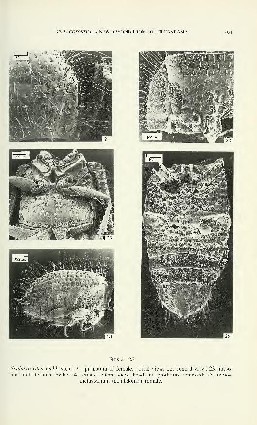
Spalacosostea loebli sp.n.: 21, pronotum of female, dorsal view; 22, ventral view; 23, meso-and metasternum, male; 24, female, lateral view, head and prothorax removed; 25, meso-,
metasternum and abdomen, female.
592 JAN KODADA
in middle; cleaning and smoothing fringe (Spangler & Perkins 1989) absent; all
tibiae with several types of hair-like sensilla in more or less regular longitudinal rows
and few peg-like sharply tipped sensilla (Figs 46, 47). Tarsal formula 4-4-4; tarsal
segments with few hair-like sensilla (Figs 41, 42, 57 - 59); claws moderately long,
narrow, microreticulate; empodium without seta.
Abdomen with five slightly convex ventrites (Fig. 32), first two connate but
separated by a distinct suture; ventrites 2, 3 and 4 about equally long; all with distinct
laterosternites (Kasap & Crowson 1975); intercoxal process about 1.25 times as long
as wide (ML/MW). Tergites 2-7 with paired submedian fields of very short, densely
arranged setae; tergite 7 bears posterior row of hair-like sensilla; pygidium with
numerous hair-like sensilla. Seven pairs of functional spiracles situated in pleural
membrane; spiracle 1 (Fig. 34) bears largest spiracular opening and filter apparatus
with short spinules; spiracles 2 - 7 with shallow atrium, circular spiracular opening,
prominent dorsal subatrial apodeme (Richter, 1969), short and bulbous ventral apo-
deme. Terminalia of all examined specimens only weakly sclerotized and hardly tra-
ceable, but similar to those in other dryopids. Aedeagus (Figs 43 - 45, 60 - 65) of
trilobate type; penis long and slender, slightly curved, tapering apically; baso-lateral pe-
nile apophyses short; ejaculatory ducts inconspicuous; ventral membranous sac without
sclerotized fibula. Parameres long, curved, tapering apically; phallobasis tubular.
Description 9 : Body form ovoid (Figs 33, 50), convex dorsally; body about 2
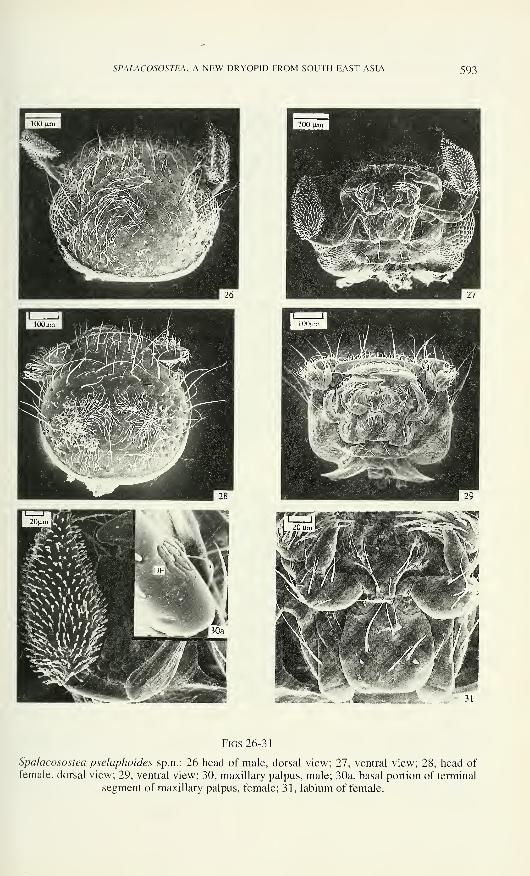
times as long as wide (LPE/MW); length (LPE) 1 .30 - 1.70 mm.Vestiture similar to those in males but sensilla longer and their sockets (mainly
of TL and TM type) slightly deeper and broader.
Eyes vestigial (Figs 4, 5) and restricted to a small triangular field lying
ventrally to cranial ridge. Labrum and mandible similar to those in male but mandible
with rather convex sharp incisor edge. Maxillary palps only 0.4 times as long as MWof head, four-segmented (Figs 12, 13); segment 1 tiny; segment 2 longer, apically
expanded; segment 3 slightly shorter than preceding; segment 2 and 3 with hair-like
sensilla; terminal segment nearly conical, usually slightly longer than combined
length of preceding segments and bearing: (1) apical sensillar cluster with several
short, peg-like bluntly tipped sensilla, few short conical bluntly tipped pegs and one
subterminal flattened peg-like and partly sunken sensillum; (2) basal sensillar cluster
with several digitiform (DF) sensilla (Fig. 30b) and a round microdepression; (3)
several hair-like sensilla (Figs 12, 13). Labium similar to that in male, but mentum
shorter and broader (Figs 17. 29) with greater number of sensilla.
Pronotum convex dorsally, with feeble lateral longitudinal impressions; lateral
margins basally more explanate and apically more arched than in male (Fig. 21);
prosternai intercoxal process about 2.0 times as long as wide, medially slightly
elevated (Fig. 22). Mesothorax as in male, but metasternum only about as long as
mesosternum. without longitudinal and transverse sutures; almost entire surface of
metasternum with deep macropunctures (Fig. 25); metepisterna weakly sclerotized
and concealed by elytra (Fig. 24). Metanotum membranous (except for anterior
margin of scuta) and not divided into distinct sclerites. Metendosternite (Fig. 48) Y-
shaped. well sclerotized and its stalk about as longs as arms, anterior tendons absent.
SPALACOSOSTEA, A NEW DRYOPID FROM SOUTH EAST ASIA 593
Figs 26-31
Spalacosostea pselaphoides sp.n.: 26 head of male, dorsal view; 27, ventral view; 28, head of
female, dorsal view; 29, ventral view; 30, maxillary palpus, male; 30a, basal portion of terminal
segment of maxillary palpus, female; 31, labium of female.
594 JAN KODADA
Elytra (Figs 24, 33) suboval, about 1.4 times as long as their combined width;
convex dorsally in cross section, strongly deflexed laterally; explanate lateral margin
invisible in dorsal view; humeri not prominent; apices acute, strongly deflexed. Strial
punctures large, dense, deeply impressed; unpaired strial intervals with rows of
sensilla (TL); paired intervals with a single row of sensilla (TS). Ventral elytral
flange, "rubbing patch" and metathoracic wings absent.
Legs (Fig. 24) in all parts shorter and broader that those in males but sensillar
distribution similar.
Abdomen (Figs 24, 25) with ventrites similar to those in male, except for
ventrite 5 bearing distinct apical shallow emargination, and tergites 2-7 lacking
fields of short setae. Terminalia similar as in other dryopids (Lawrence, 1988): ovi-
positor (slightly longer than abdomen) consisting from laterally compressed coxites
without styli; vaginal bursa without spines and sclerotized plates.
Habitat: specimens were collected by sifting vegetation debris mainly in
primary Lithocarpus - Castanopsis and Dipterocarp forests.
Spalacosostea loebli sp.nov. (Figs 1-25, 39-48)
Etymology: this species is dedicated to my friend Ivan Lobi, who collected
numerous new species of terrestrial dryopids during several expeditions to Southeast
Asia.
Material examined: Holotype S: "SABAH: Poring Hot Springs, 500 m, 7. V. 1987
#15a Burckhardt - Lobi" MHNG; Paratypes: 1 S, 4 9 9 with the same data as holotype,
MHNG;2<?c?, 139 9: same data but 6. V. 1987, MHNG; 1 S, 1 9 : same data but 11. V. 1987,
MHNG; 1 S, 6 9 9 : same data but 13. V. 1987. MHNG; 1 S, 6 9 9 : same data but 550 - 600m, 9. V. 1987, MHNG; 4 SS, 11 9 9: same data but 600 m, nr Bat Cave, 10.V. 1987, MHNG;10 SS, 31 9 9: Borneo, Sabah, Mount Kinabalu National Park, Poring Hot Springs, area
Kipungit Crk. 2, 14. - 30. VIII. 1988, A. Smetana Igt., MHNG, CKB; 1 6, 30 9 9: "SABAH:Crocker Ra. 1600m, km 51 rte Kota Kinabalu-Tambunan, 18.V. 87 Burckhardt - Lobi 30a"
MNHG, CKB; 2 S S, 4 9 9 : "SABAH: E Mt. Kinabalu 1 150m. rte Ranau-Kota Kinabalu, 24.
V. 1987 Burckhardt - Lobi 40" MNHG; 1 9: "SABAH: Crocker Ra. 1200m, km 63 rte Kota
Kinabalu-Tambunan, 19. V. 87 Burckhardt - Lobi 31a" MNHG; 5 9 9: "SABAH: Crocker Ra.
1270m. km 60 rte Kota Kinabalu-Tambunan, 17. V. 87 Burckhardt - Lobi 29a" MNHG.Description of 6 from the type locality (#15a): length (LPE) 1.20 - 1.36 mm
(0 = 1.28 ± 0.05, n= 16), maximum width 0.52 - 0.56 mm (0 = 0.54 ± 0.02, n= 16).
Cranial macropunctures very fine, their diameter distinctly shorter than facet
diameter; eyes large (Fig. 2), HW = 0.42 - 0.49 mm (0 = 0.44 ± 0.02, n= 16), 01= 1.57
- 1.88 (0 = 1.75 ± 0.07, n= 16). Each flagellar antennomere with two dendritic sensilla
(D); antennomere 6 characterised by insertion of three sensilla P3, two sensilla P4 and
one sensillum P5 (Figs 3, 6, 7). Maxillary palps with segment 3 about 1.9 times as long
as wide (Fig. 11); terminal segment bearing basal cluster of sensilla. Mentum broader
than long (MW/ML= about 1.5), microreticulated (Fig. 16). Labial palps with short
hair-like sensilla on segment 2 arranged in one more or less distinct row (Fig. 16), one
or two sensilla sometimes inserted more basally; microreticulation on segment 2
present internally.
Pronotum 0.45 - 0.54 mm (0 = 0.51 ± 0.02, n= 16) wide (MW), usually widest
at basal fifth; lateral margin almost straight (Fig. 19); diameter of macropunctures
SPALACOSOSTEA, A NEW DRYOPID FROM SOUTH EAST ASIA 595
Figs 32-38
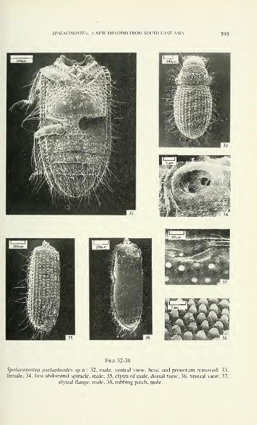
Spalacosostea pselaphoides sp.n.: 32, male, ventral view, head and pronotum removed; 33,
female; 34, first abdominal spiracle, male; 35, elytra of male, dorsal view; 36, ventral view; 37,
elytral flange, male; 38, rubbing patch, male.
596 JAN KODADA
distinctly shorter than facet diameter, macropunctures separated by a distance of
about 1-3 facet diameters; punctures becoming sparser near anterior and posterior
margins and larger laterally. Metasternum with short, indistinct median longitudinal
impression (Fig. 23), glabrous area irregular (Fig. 23).
Elytra 1.76 - 2.08 (0 = 1.85 ± 0.08, n= 16) times as long as their combined
width; strial punctures separated by a distance slightly smaller than their diameter;
strial intervals dorsally slightly wider than diameter of one strial puncture, becoming
smaller toward lateral margin.
Protibia (Fig. 46) about 1.45 times as long as protarsus; ratio (ML/MW) of
tarsal segment 4 (Figs 41, 42): of protarsus about 4.0, of mesotarsus about 4.4 and of
metatarsus about 4.7 (all measured in lateral view). Claws approximately 0.5 times as
long as length of terminal tarsal segments.
Ventrites 1 and 2 with irregularly distributed macropunctures, ventrites 3-4without or only with very few macropunctures; ventrite 5 always lacking macro-
punctures. Aedeagus (Figs 43, 44) with parameres about as long as phallobasis
(lateral view).
Description of 9 from the type locality (#15a): length (LPE) 1.26 - 1.46 mm(0 = 1.39 ± 0.04, n= 43), maximum width 0.64 - 0.70 mm (0 = 0.67 ± 0.02, n= 43).
Cranial macropunctures distinctly larger than those in males, sensilla (TS)
situated on vertex directed mesally. Antennal segment 6 without sensilla P3.
Maxillary palps (Figs 4, 12, 13) with segments 2 and 3 approximately of same length,
terminal segment 2.6 - 2.8 times as long as preceding, with 3-6 hair-like sensilla and
a cluster of digitiform sensilla (DF) approximately in basal 0.2. Mentum (Fig. 17)
about 1.5 times as wide as long (MW/ML), with microreticulation; labial palps as in
Fig. 17.
Pronotum 0.57 - 0.66 mm (0 = 0.61 ± 0.02, n= 43) wide (MW), widest at basal
third; lateral margin moderately arcuate (Fig. 21), macropunctures slightly larger than
those on cranium and separated usually by a distance equal or slightly longer than
their diameter. Glabrous area of metasternum very small (Fig. 25).
Elytra 1.36 - 1.58 (0 = 1.46 ± 0.05, n= 43) times as long as their combined
width; interstices more convex than in male.
Ratio (ML/MW) of tarsal segment 4: of protarsus about 2.5, of mesotarsus
about 2.6 and of metatarsus about 2.8; claws shorter, broader and more curved than
those in males.
Variation: Size and morphometric indexes see in Tables 1, 2. Both sexes are
larger in higher altitudes; the aedeagi from these localities are also larger but their
proportions are similar to those in males from lower altitudes (see Figs 43 - 45).
Females exhibit minor variations also in number and distribution of sensilla on
mentum, terminal segments of maxillary palps (Figs. 39, 39 a, 40) and segment 2 of
labial palps.
SPALACOSOSTEA, A NEW DRYOPID FROM SOUTH EAST ASIA 597
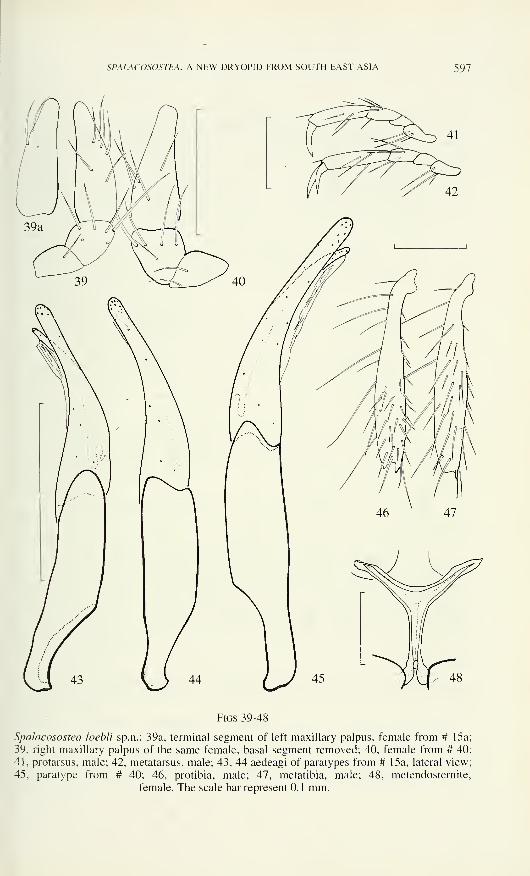
Figs 39-48
Spalacosostea loebli sp.n.: 39a, terminal segment of left maxillary palpus, female from # 15a
39, right maxillary palpus of the same female, basal segment removed; 40, female from # 40
41, protarsus, male; 42, metatarsus, male; 43, 44 aedeagi of paratypes from # 15a, lateral view
45, paratype from # 40; 46, protibia, male; 47, metatibia, male; 48, metendosternite.
female. The scale bar represent 0.1 mm.

598 JAN KODADA
Table 1
Morphometrical characteristics of males S. loebli sp.n.
locality n LPEfmm] MW of elytra EI MW of pro-
notum [mm]HW [mm] 01
30a
40
1
2
1.60
1.60
0,80
0.70, 0.72
1.56
1.66, 1,78
0.62
0.60, 0.65
0.50
0.50, 0.54
1.42
1.48, 1.42
Table 2
Morphometrical characteristics of females S. loebli sp.n.
locality n LPE [mm] MW of elytra
[mmlEI MW of pro-
notum [mm]
29a
30a
31a
40
27
1.45-1.54 0.70-0.84
0=1.48 ±0.04 0=0.76 ±0.06
1.60-1.78 0.77-0.84
0=1.67 ±0.05 0=0.81 ±0.02
1.53 0.76
1.49-1.66 0.73-0.80
0=1.59 ±0.07 0=0.77 ±0.03
1.39- 1.56
0= 1.43 ±0.13
1.37- 1.60
0= 1 .47 ± 0.06
1.42
1.45- 1.53
0= 1.50 ±0.04
0.63 - 0.70
0= 0.65 ± 0.03
0.69 - 0.76
0= 0.72 ± 0.02
0.67
0.67 - 0.70
0=0.69 ±0.01
Spalacosostea pselaphoides sp.n. (Figs 26-38, 49-65)
Etymology: from Pselaphus in reference to the unusual large maxillary palpi as
in many pselaphids.
Material examined: Holotype S : "SUMATRA: Jambi Mt. Kerinci, 1750 - 1850 m, 11.
- 12. XI. 1989, Agosti, Lobi, Burckhardt # 11" MHNG; Paratypes: 4 SS, 47 9 9 with the
same data as holotype, MHNG, CKB; 1 o\ 7 9 9: "SUMATRA: Jambi Mt. Kerinci, 1900 m,
13. XI. 1989, # 15a" MHNG, CKB; 2 9 9: "SUMATRA: Jambi W Mt. Tujuh Lake 1400 m,
14. XI. 1989, # 17" MHNG; 3 9 9: "SUMATRA: W Sum. Lubuksulasih, 30 km E Padang,
1 100 m, 8. XI. 1989, # 7" MHNG; 2 n, 9 9 9 : "SUMATRA: W Sum. #21, Palopo Nat. Res. N.
Bukittinggi. 900 m. 18 - 20. XI. 1989" MHNG; 1 S : "SUMATRA: W Sum. 5 km SEPayakumbuh, 600 m, 20-21. XI. 1989, # 24" MHNG; 3 S S, 6 9 9 : "SUMATRA: Aceh # 25a
Mt. Leuser NP, 300 - 500 m, Ketambe, 23 - 30. XI. 1989" MHNG. All Agosti, Lobi,
Burckhardt leg.
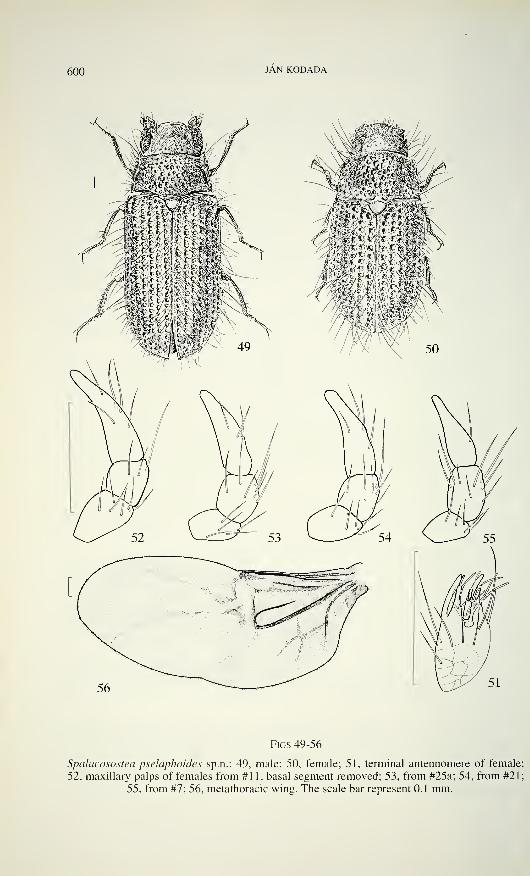
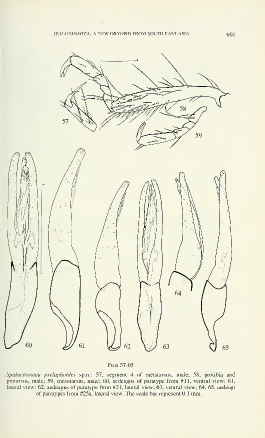
Description of S from the type locality (# 11): Habitus (Fig. 49), length (LPE)
1.54 - 1.58 mm (0= 1.56 ± 0.01, n= 5). maximum width 0.65 - 0.67 mm (0= 0.66 ±
0.01, n= 5).
Cranial macropunctures with diameter about equal to facet diameter, separated
by a distance of about 1 - 3 facet diameters (Fig. 26). Central area of vertex with more
or less distinct cluster of sensilla TS with apices directed centrally. Eyes moderately
large, HW = 0.43 - 0.47 mm (0= 0.45 ± 0.01, n= 5), 01= 1.36 - 1.41 (0= 1.38 ± 0.18,
n= 5). Each flagellar antennomere with one dendritic sensillum (D); antennomere 6
SPALACOSOSTEA, A NEW DRYOPID FROM SOUTH EAST ASIA 599
characterised by insertion of two sensilla P3 laterally to sensillum S2 and by absence
from sensilla type P4 and P5 (Fig. 51). Maxillary palps with segment 3 about 2.5
times as long as wide (Figs 27, 30); terminal segment without basal external cluster of
sensilla. Mentum about as long as wide, without microreticulation (Fig. 31), on each
posterior angle one short peg-like sensillum. Labial palps with short hair-like sensilla
on segment 2 in two more or less distinct rows (Fig. 31); microreticulation absent.
Pronotum 0.56 - 0.58 mm (0= 0.57 ± 0.01, n= 5) wide (MW), widest at base;
lateral margin almost straight; diameter of macropunctures slightly longer than facet
diameter, macropunctures separated by distance about equal their diameter; punctures
becoming smaller and sparser near anterior and posterior margins; surface with
numerous recumbent sensilla (TS) except for a small area along anterior margin.
Metasternum without median longitudinal impression, glabrous area approximately
triangular (Fig. 32).
Elytra 1.83 - 1.97 (0= 1.88 ± 0.06, n= 5) times as long as their combined
width; strial punctures separated by a distance distinctly smaller than their diameter;
strial intervals dorsally about as wide as diameter of one strial puncture.
Protibia (Fig. 58) about 1.6 times as long as protarsus; ratio (ML/MW) of
tarsal segment 4 (Figs 57 - 59): of protarsus about 3.0, of mesotarsus about 3.7 and of
metatarsus about 4.0. Claws approximately 0.5 times as long as length of terminal
tarsal segments.
Ventrues 1 and 2 with equally distributed macropunctures, ventrites 3 and 4
with macropunctures restricted onto anterior half; ventrite 5 with macropunctures near
anterior margin. Aedeagus (Figs 60, 61) with short phallobasis; parameres about 1.3
times as long as phallobasis (lateral view), bluntly tipped, moderately bent ventrally
(lateral view).
Description of 9 from the type locality (# 11): Habitus (Fig. 50), length (LPE)
1.40 - 1.67 mm (0= 1.57 ± 0.08, n= 30), maximum width 0.69 - 0.83 mm (0= 0.78 ±
0.03, n= 30).
Vertex with numerous sensilla TS with apices directed to two sublateral points
and form distinct clusters (Fig. 28). Antennomere 6 bearing, in addition to sensilla in
male, one sensillum P5. Maxillary palps with segment 2 distinctly longer than
segment 3, latter about as long as wide; terminal segment about 2.8 times as long as
preceding, bearing 5 - 7 hair-like sensilla and a cluster of digitiform sensilla (DF)
situated in basal 0.2 (Fig. 30a). Mentum about 1.4 times as wide as long (MW/ML),glabrous; labial palps as in Fig. 29.
Pronotum 0.63 - 0.71 mm (0= 0.66 ± 0.03, n= 30) wide (MW), widest at basal
third; lateral margin slightly arched; macropunctures coarse except those near anterior
and posterior margin, separated usually by a distance smaller than their diameter,
sometimes confluent.
Elytra 1.41 - 1.59 (0= 1.47 ± 0.06, n= 30) times as long as their combined
width.
Ratio (ML/MW) of tarsal segment 4: of protarsus about 2.2, of mesotarsus
about 2.4 and of metatarsus about 3.3; claws shorter, broader and more curved than
those in males.
600 JAN KODADA
Figs 49-56
Spalacosostea pseìaphoides sp.n.: 49, male; 50, female; 51, terminal antennomere of female;
52, maxillary palps of females from #11, basal segment removed; 53, from #25a; 54, from #21;
55, from #7; 56, metathoracic wing. The scale bar represent 0.1 mm.
SPALACOSOSTEA, A NEW DRYOPID FROM SOUTH EAST ASIA 601
Figs 57-65
Spalacosostea pselaphoides sp.n.: 57, segment 4 of metatarsus, male; 58, protibia andprotarsus, male; 59, mesotarsus, male; 60, aedeagus of paratype from #11, ventral view; 61,
lateral view; 62, aedeagus of paratype from #21, lateral view; 63, ventral view; 64, 65, aedeagi
of paratypes from #25a, lateral view. The scale bar represent 0.1 mm.
602 JAN KODADA
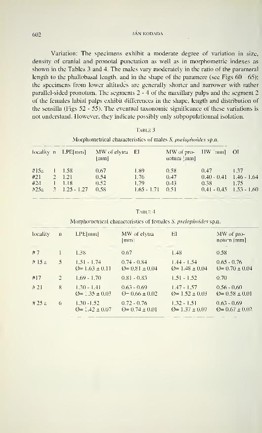
Variation: The specimens exhibit a moderate degree of variation in size,
density of cranial and pronotai punctation as well as in morphometric indexes as
shown in the Tables 3 and 4. The males vary moderately in the ratio of the parameral
length to the phallobasal length, and in the shape of the paramere (see Figs 60 - 65);
the specimens from lower altitudes are generally shorter and narrower with rather
parallel-sided pronotum. The segments 2 - 4 of the maxillary palps and the segment 2
of the females labial palps exhibit differences in the shape, length and distribution of
the sensilla (Figs 52 - 55). The eventual taxonomic significance of these variations is
not understand. However, they indicate possibly only subpopulationnal isolation.
Table 3
Morphometrical characteristics of males S. pselaphoides sp.n.
locality n LPE[mm] MW of elytra EI
[mm]MW of pro- HW [mm] 01notum [mm]
#15a 1 1.58 0.67
#21 2 1.21 0.54
#24 1 1.18 0.52
#25a 3 1.25-1.27 0.58
1.89 0.58 0.47 1.37
1.76 0.47 0.40-0.41 1.46- 1.64
1.79 0.43 0.38 1.75
1.65-1.71 0.51 0.41--0.45 1.53- 1.60
Table 4
Morphometrical characteristics of females S. pselaphoides sp.n.
locality n LPE[mm] MW of elytra
[mm]EI MW of pro-
notum [mm]
#7 1 1.38 0.67 1.48 0.58
# 15a 5 1.51 - 1.74
0= 1.63 ±0.11
0.74 - 0.84
0=0.81 ±0.04
1.44- 1.54
0= 1 .48 ± 0.04
0.65 - 0.76
0= 0.70 ± 0.04
#17 2 1.69- 1.70 0.81 -0.83 1.51 - 1.52 0.70
#21 8 1.30- 1.41
0= 1.35 ±0.030.63 - 0.69
0= 0.66 ± 0.02
1.47- 1.57
0= 1.52 ±0.030.56 - 0.60
0=0.58 ±0.01
#25 a 6 1.30-1.52
0= 1.42 ±0.070.72 - 0.76
0=0.74 ±0.01
1.32- 1.51
0= 1.37 ±0.070.63 - 0.69
0= 0.67 ± 0.02
SPALACOSOSTEA, A NEW DRYOPID FROM SOUTH EAST ASIA 603
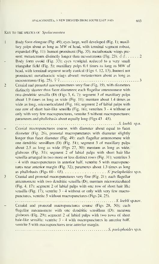
Key to the species of Spalacosostea
1 Body form elongate (Fig. 49); eyes large, well developed (Fig. 1); maxil-
lary palps about as long as MW of head, with terminal segment robust,
expanded (Fig. 11); humeri prominent (Fig. 35), metathoracic wings pre-
sent; metasternum distinctly longer than mesosternum (Fig. 23). SS 2
Body form ovoid (Fig. 33); eyes vestigial, reduced to a very small
triangular field (Fig. 5); maxillary palps 0.4 times as long as MW of
head, with terminal segment nearly conical (Figs 4, 12, 13); humeri not
prominent; metathoracic wings absent; metasternum about as long as
mesosternum (Fig. 25). 99 3
2 Cranial and pronotal macropunctures very fine (Fig. 19), with diameters
distinctly shorter than facet diameters; each flagellar antennomere with
two dendritic sensilla (D) (Figs 3, 6, 7); segment 3 of maxillary palps
about 1.9 times as long as wide (Fig. 11); mentum about 1.4 times as
wide as long, microreticulated (Fig. 16); segment 2 of labial palps with
one row of short hair-like sensilla (Fig. 16); ventrites 3-4 without or
only with very few macropunctures, ventrite 5 without macropunctures;
parameres and phallobasis about equally long (Figs 43 - 45).
S. loebli sp.n.
Cranial macropunctures coarse, with diameter about equal to facet
diameter (Fig. 26), pronotal macropunctures with diameter slightly
longer than facet diameter (Fig. 49); each flagellar antennomere with
one dendritic sensillum (D) (Fig. 51); segment 3 of maxillary palps
about 2.5 as long as wide (Figs 27, 30); mentum as long as wide,
glabrous (Fig. 31); segment 2 of labial palps with short hair-like
sensilla arranged in two more or less distinct rows (Fig. 31); ventrites 3
- 4 with macropunctures in anterior half, ventrite 5 with macropunc-
tures near anterior margin (Fig. 32); parameres about 1.3 times as long
as phallobasis (Figs 60-65) S. pselaphoides sp.n.
3 Cranial and pronotal macropunctures very fine (Fig. 21); each flagellar
antennomere with two dendritic sensilla (D); mentum microreticulated
(Fig. 4, 17); segment 2 of labial palps with one row of short hair-like
sensilla (Fig. 17); ventrite 3 - 4 without or only with very few macro-
punctures, ventrite 5 without macropunctures (Figs 24, 25).
S. loebli sp.nov.
Cranial and pronotal macropunctures coarse (Figs 28, 50); each
flagellar antennomere with one dendritic sensillum (D); mentum
glabrous (Fig. 29); segment 2 of labial palps with two rows of short
hair-like sensilla; ventrite 3 - 4 with macropunctures in anterior half,
ventrite 5 with macropunctures near anterior margin.
S. pselaphoides sp.n.
604 JAN KODADA
DISCUSSION
Spalacosostea shares the lack of the 2-nd cubito-anal cells with Quadryops and
genus B, and the four-segmented tarsi with Quadryops. Sosteamorphus, Protoparnus,
Oreoparnus and genus A lack metathoracic wings. The number of antennomeres is
reduced in genus A to nine.
Spalacosostea, Guaranius, Sosteamorphus, genus A, Quadryops, Sostea-
morphus, Oreoparnus and Protoparnus share the presence of dendritic sensilla on the
flagellar antennomeres. These sensilla are variable in branching, and they are present
also in some aquatic taxa. Their ultrastructural features and physiological functions
are unknown.
Protoparnus, Oreoparnus, and genera A and B have lateral pronotal ridges
while they are absent in Sostea, Sosteamorphus, Geoparnus and Spalacosostea.
In absence of detailed information on the morphology of Holcodryops,
Quadryops, and genus B, all but the last known only as the single type specimen
(Spangler 1987), the relationship of Spalacosostea (as other relationships within the
family) remain unknown. However, no synapomoiphy indicates close relationship of
Spalacosostea with any other sympatric terrestrial group of the dryopids {Geoparnus,
Sostea).
ACKNOWLEDGEMENTS
I am especially indebted to I. Lobi (MHNG), M. A. Jäch (NMW), S. Endrödy-
Younga (TMP), N. Berti (MNHP), E. De Boise (BMNH) and P. Lindskog (RMS) for
the material used in this study. Special thanks are due to J. Wiiest (MHNG) and J.
Kozânkovâ for SEM photographs and K. Majer for the habitus drawing of
Spalacosostea pselaphoides. M. A. Jäch, V. Kovâc, R. Leschen, I. Lobi and W. D.
Shepard critically commented the manuscript.
REFERENCES
Barr, Ch. B. & P. J. Spangler. 1992. A new genus and species of stygobiotic dryopid beetle,
Stygoparnus comalensis (Coleoptera: Dryopidae) from Comal springs, Texas. Pro-
ceedings of the Biological Society of Washington 105 (1): 40 - 54.
Brown, H. P. 1981. A distributional survey of the world genera of aquatic Dryopid beetles
(Coleoptera: Dryopidae, Elmidae and Psephenidae sens. lat.). Pan - Pacific Ento-
mologist 57 (1): 133- 148.
Brown, H. P. 1987. Biology of riffle beeetles. Annual Revue ofEntomology 32: 253 - 273.
Crowson, R. A. 1967. The Natural Classification of the Families of Coleoptera. Classey,
Middlesex, 1 87 pp.
Crowson, R. A. 1981. The Biology of the Coleoptera. Academic Press, London, New York,
802 pp.
Hinton, H. E. 1939. An Inquiry into the Natural Classification of the Dryopoidea, Based Partly
on a Study of Their Internal Anatomy (Col.). The Transactions of the Royal Ento-
mological Society ofLondon 89: 133 - 184.
SPALACOSOSTEA, A NEW DRYOPID FROM SOUTH EAST ASIA 605
Hinton, H. E. 1969. Plastron respiration in adult beetles of the suborder Myxophaga. Journal
ofZoology, London 159: 131 - 137.
Honomichl, K. & G. W. Guse. 1981. Digitiform Sensilla on the Maxillar Palp of Coleoptera
III. Fine structure in Tenebrio molitor L. and Dermestes maculatus De Geer. Acta
Zoologica 62(1): 17-25.
Kasap, H. & R. A. Crowson. 1975. A comparative anatomical study of Elateriformia and
Dasciloidea (Coleoptera). Transactions of the Royal Entomological Society of London126:441 -495.
Kukalova-Peck, J. & J. F. Lawrence. 1993. Evolution of hind wing in Coleoptera. The
Canadian Entomologist 125: 181 - 258.
Lawrence, J. F. & E. B. Bretton. 1991. Coleoptera. In: Insects of Australia. MelbourneUniversity Press, Vol.IL: 543 - 683.
Lawrence, J. F. 1988. Rhinorhipidae, a New Beetle Family from Australia, with Comments on
the Phylogeny of the Elateriformia. Invertebrate taxonomy, 1987, 2: 1 - 53.
Perkins, P. D. & P. J. Spangler. 1985. Quadryops, new genus, and three new species of
arboreal Dryopidae (Insecta: Coleoptera) from Panama and Ecuador. Proceedings ofthe biological Society of Washington 98 (2): 490 - 510.
Petryszak, A. 1977. The sense organs of the mouth parts in Libehda depressa L. and L. quadri-
maculata L. (Odonata). Acta biologica Cracoviensia, Series: Zoologia 20: 87 - 100.
Richter, P. O. 1969. Spiracles of Adult Scaraboidea (Coleoptera) and Their Phylogenetic
Significance. I. The Abdominal Spiracles. Annals of the Entomological Society ofAmerica 62 (4): 869 - 880.
Spangler, P. J. & P. D. Perkins. 1989. A Revision of the Neotropical Aquatic Beetle GenusStenhelmoides (Coleoptera: Elmidae). Smithsonian Contributions to Zoology 479: 63 pp.
Spangler, P. J. 1987. Holcodiyops moitli, an anomalous new genus and species of beetle from
Equador (Coleoptera: Dryopidae). Proceedings of the Biological Society of Washington89:616-621.
Zacharuk, R. Y. 1985. Antennae and sensillae, pp. 1 - 69. In: Comprehensive insect physio-
logy, biochemistry and pharmacology. Vol 6, Nervous system: Sensory (G. A. Kerkut& L. I. Gilbert eds). Vol. 6. Pergamon press, Oxford, 710 pp.

Revue suisse de Zoologie, 103 (3): 607-61 1; septembre 1996
A peculiar new species ofAnomotarus {Nototarus) from NewGuinea (Coleoptera, Carabidae, Lebiinae)
Martin BAEHRZoologische Staatssammlung, Münchhausenstr. 21, D-81247 München, Germany.
A peculiar new species of Anomotarus (Nototarus) from New Guinea(Coleoptera, Carabidae, Lebiinae). - Anomotarus (Nototarus) pilosus sp.
n. from Papua New Guinea is described. The species differs from all
known Australian and New Guinean Nototarus by the conspicuous, erect
pilosity of the dorsal surface, the multiplication of the anterior lateral pre-
notai setae, and the absence of any microreticulation on head, pronotum,
and elytra.
Key-words: Coleoptera - Carabidae - Lebiinae - Genus Anomotarus,
subgenus Nototarus - New species - New Guinea.
INTRODUCTION
While studying the numerous carabid material (ca. 3,000 specimens) collected
by W. Ullrich in Papua New Guinea during 1979 and 1980 and stored in the Muséumd'histoire naturelle, Genève (MHNG), I discovered a single specimen of a peculiar
new species that belongs to the subgenus Nototarus Chaudoir of the genus Anomo-
tarus Chaudoir. The new species differs from all known species of that subgenus and
also from those undescribed Australian species known to me by the peculiar erect
pilosity of the surface, the multiplication of the anterior lateral prenotai setae, and the
absence of any microreticulation on the upper surface. Due to its peculiar habitus the
species is described, although a single specimen is only available.
Nototarus is an Australian subgenus of the more widely ranging Australian-
Oriental genus Anomotarus Chaudoir combining characteristic, stout-built, flightless
lebiine Carabidae of which thus far a single species was known outside from Australia
proper, namely Nototarus papua Darlington from eastern Papua New Guinea.
Certainly the species of Nototarus are closely related to those of the Aus-
tralian-Oriental subgenus Anomotarus Chaudoir s. str., but I am not sure, whether the
current status of Nototarus as a subgenus of Anomotarus (Ball & Hilchie 1983,
Moore et al. 1987) is justified. Perhaps a revision of both subgenera will clarify the
taxonomic situation.
Manuscript accepted 28.07.1995.
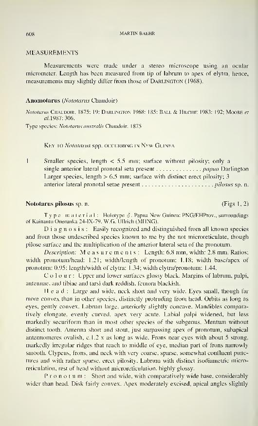
608 MARTIN BAEHR
MEASUREMENTS
Measurements were made under a stereo microscope using an ocular
micrometer. Length has been measured from tip of labrum to apex of elytra, hence,
measurements may slightly differ from those of Darlington (1968).
Anomotarus (Nototarus Chaudoir)
Nototarus Chaudoir, 1875: 19; Darlington 1968: 185; Ball & Hilchie 1983: 192; Moore et
al. 1987: 306.
Type species: Nototarus australis Chaudoir. 1 875
Key to Nototarus spp. occurring in New Guinea
1 Smaller species, length < 5.5 mm; surface without pilosity; only a
single anterior lateral pronotai seta present papua Darlington
Larger species, length > 6.5 mm; surface with distinct erect pilosity; 3
anterior lateral pronotal setae present pilosus sp. n.
Nototarus pilosus sp. n. (Figs 1, 2)
Type material: Holotvpe 6\ Papua New Guinea: PNG/EHProv., surroundings
of Kainantu Onerunka 24-IX-79, W.G. Ullrich (MHNG).
Diagnosis: Easily recognized and distinguished from all known species
and from those undescribed species known to me by the not microreticulate, though
pilose surface and the multiplication of the anterior lateral seta of the pronotum.
Description: Measurements: Length: 6.8 mm, width: 2.8 mm. Ratios:
width pronotum/head: 1.21; width/length of pronotum: 1.18; width base/apex of
pronotum: 0.95; length/width of elytra: 1.34; width elytra/pronotum: 1.44.
Colour: Upper and lower surfaces glossy black. Margins of labrum, palpi,
antennae, and tibiae and tarsi dark reddish, femora blackish.
Head: Large and wide, neck short and very wide. Eyes small, though far
more convex than in other species, distinctly protruding from head. Orbits as long as
eyes, gently convex. Labrum large, anteriorly slightly concave. Mandibles compara-
tively elongate, evenly curved, apex very acute. Labial palpi widened, but less
markedly securiform than in most other species of the subgenus. Mentum without
distinct tooth. Antenna short and stout, just surpassing apex of pronotum, subapical
antennomeres ovalish, c.1.2 x as long as wide. Frons near eyes with about 5 strong,
markedly irregular ridges that reach to middle of eye, median part of frons narrowly
smooth. Clypeus, frons, and neck with very coarse, sparse, somewhat confluent punc-
tures and with rather sparse, erect pilosity. Labrum with distinct isodiametric micro-
reticulation, rest of head without microreticulation, highly glossy.
Pronotum: Short and wide, with comparatively wide base, considerably
wider than head. Disk fairly convex. Apex moderately excised, apical angles slightly
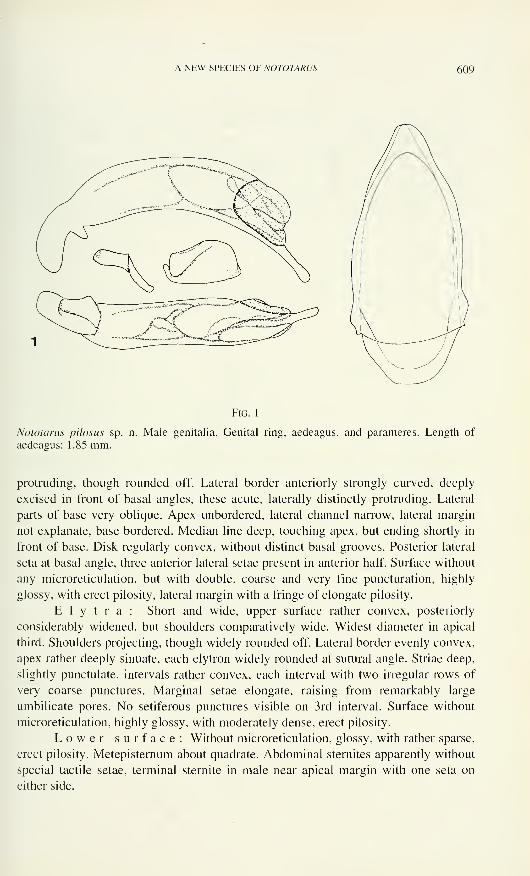
A NEW SPECIES OF NOTOTARUS 609
Fig. 1
Nototarus pilosus sp. n. Male genitalia. Genital ring, aedeagus, and parameres. Length of
aedeagus: 1.85 mm.
protruding, though rounded off. Lateral border anteriorly strongly curved, deeply
excised in front of basal angles, these acute, laterally distinctly protruding. Lateral
parts of base very oblique. Apex unbordered, lateral channel narrow, lateral margin
not explanate, base bordered. Median line deep, touching apex, but ending shortly in
front of base. Disk regularly convex, without distinct basal grooves. Posterior lateral
seta at basal angle, three anterior lateral setae present in anterior half. Surface without
any microreticulation, but with double, coarse and very fine puncturation, highly
glossy, with erect pilosity, lateral margin with a fringe of elongate pilosity.
Elytra: Short and wide, upper surface rather convex, posteriorly
considerably widened, but shoulders comparatively wide. Widest diameter in apical
third. Shoulders projecting, though widely rounded off. Lateral border evenly convex,
apex rather deeply sinuate, each elytron widely rounded at suturai angle. Striae deep,
slightly punctulate. intervals rather convex, each interval with two irregular rows of
very coarse punctures. Marginal setae elongate, raising from remarkably large
umbilicate pores. No setiferous punctures visible on 3rd interval. Surface without
microreticulation, highly glossy, with moderately dense, erect pilosity.
Lower surface: Without microreticulation, glossy, with rather sparse,
erect pilosity. Metepisternum about quadrate. Abdominal sternites apparently without
special tactile setae, terminal sternite in male near apical margin with one seta on
either side.
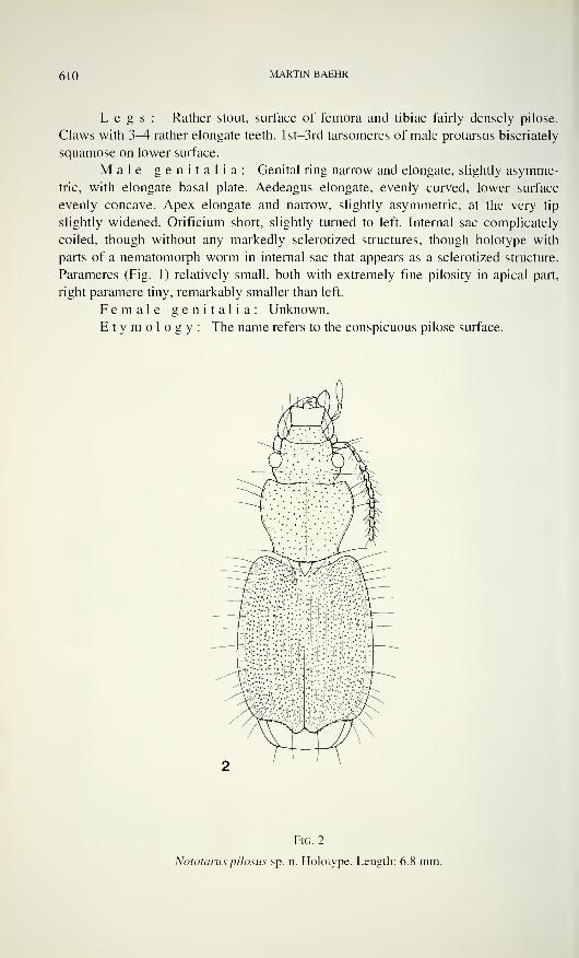
610 MARTIN BAEHR
Legs: Rather stout, surface of femora and tibiae fairly densely pilose.
Claws with 3-4 rather elongate teeth. lst-3rd tarsomeres of male protarsus biseriately
squamose on lower surface.
Male genitalia: Genital ring narrow and elongate, slightly asymme-
tric, with elongate basal plate. Aedeagus elongate, evenly curved, lower surface
evenly concave. Apex elongate and narrow, slightly asymmetric, at the very tip
slightly widened. Orificium short, slightly turned to left. Internal sac complicately
coiled, though without any markedly sclerotized structures, though holotype with
parts of a nematomorph worm in internal sac that appears as a sclerotized structure.
Parameres (Fig. 1) relatively small, both with extremely fine pilosity in apical part,
right paramere tiny, remarkably smaller than left.
Female genitalia: Unknown.
Etymology: The name refers to the conspicuous pilose surface.
Fig. 2
Nototarus pilosus sp. n. Holotype. Length: 6.8 mm.
A NEW SPECIES OF NOTOTARUS 6 1 ]
Remarks: This is a very peculiar species that differs in many respects
from the bulk of the Australian species of Nototarus as well as from N. papua
Darlington and is perhaps a highly derived species. Main apomorphic characters are
the stout built with wide and convex pronotum and elytra, massive head with small
though remarkably protruding eyes, absence of microreticulation, presence of very
coarse puncturation, presence of erect pilosity on upper surface, and multiplication of
the anterior lateral setae of the pronotum. At the present state of knowledge this
species is probably without any closer relative in Australia and New Guinea.
ACKNOWLEDGEMENTS
My thanks are due to Dr I. Lobi, Genève, who kindly submitted the specimen
for examination, alongside with a great number of New Guinean carabids.
REFERENCES
Ball, G.E. & G.J. Hilchie. 1983. Cymindide Lebiini of authors: Redefinition and reclassi-
fication of genera (Coleoptera: Carabidae). Quaestiones Entomologiae 19: 93-216.
Chaudoir, M. de. 1875. Genres aberrants du groupe des Cymindides. Bulletin de la Société
Impériale Naturelle Moscou 44(2): 1-61
.
Darlington, P.J. Jr. 1968. The Carabid beetles of New Guinea. Part III. Harpalinae continued.
Perigonini to Pseudomorphini. Bulletin of the Museum of Comparative Zoology 139:
1-253.
Moore, B.P., Weir, T.A. & J.E. Pyke. 1987. Rhysodidae and Carabidae. In: Zoological Cata-
logue of Australia, 4: 17-320. Australian Government Publishing Service, Canberra.


Revue suisse de Zoologie, 103 (3): 613-622; septembre 1996
Stigmale Plastronstrukturen, die einigen Diplopoden-Arten eine
submerse Lebensweise in kaltem und in fließendem Wasserermöglichen
Benjamin MESSNERl, Joachim ADIS2 & Klaus Peter ZULKA31 Zoologisches Institut und Museum, J.S. Bach-Str. 1 1/12,
D- 17489 Greifswald. Deutschland;
- Max-Planck-Institut für Limnologie, AG Tropenökologie, Postfach 165,
D-24302 Plön, Deutschland;
3 Institut für Zoologie, Althanstr. 14, A-1090 Wien, Österreich.
Stigmal plastron structures which enable some species of Diplopoda to
live submersed in cold and running water. - The structure of stigmata
and their microtrichia are being compared for the millipides Polydesmus
denticulatus, P. (Brachydesmus) superus (Polydesmidae), Oxidus gracilis,
Selminosoma chapmani, Mestosoma hylaeicum (Paradoxomatidae) and
Aphelidesmus sp. (Platyrhacidae). They enable plastron respiration under
water. Flood tolerance is experimentally shown for M. hylaeicum and flood
resistance for P. denticulatus and P. (B.) superus.
Key-words: Millipedes - Stigmata - Microtrichia - Plastron - Respiration -
Flood tolerance - Flood resistance - Polydesmida - Diplopoda.
EINLEITUNG
Seit den Untersuchungen von Messner & Adis (1988) weiß man, daß die
juvenilen und subadulten Stadien des brasilianischen Diplopoden Gonographis adisi
(Pyrgodesmidae) den jährlich wiederkehrenden Flutpuls des Amazonas submers
langzeitig überdauern, weil sie unter einer kutikulären Sekretschicht einen körper-
umfassenden, dünnen Luftfilm (= Plastron) zu halten vermögen, ähnlich wie es einige
Milben mit ihrem Cerotegument können (Messner et al. 1992).
Inzwischen sind weitere Diplopoden-Arten bekannt geworden, die von kaltem
Wasser überflutet, bis zu 75 Tage überleben können (Zulka 1989, 1991, 1993) sowie
in kaltem Höhlenwasser (Hoffman 1977/78) oder in schnell fließenden, flach-
gründigen Bächen langfristig oder gar permanent submers leben können (Stauder
1990).
Manuskript angenommen am 25.07.1995.
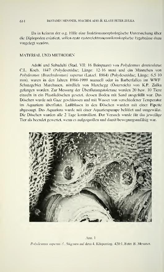
614 BENJAMIN MESSNER, JOACHIM ADIS & KLAUS PETER ZULKA
Da in keinem der o.g. Fälle eine funktionsmorphologische Untersuchung über
die Diplopoden existiert, sollen erste rasterelektronenmikroskopische Ergebnisse dazu
vorgelegt werden.
MATERIAL UND METHODEN
Adulti und Subadulti (Stad. VII: 16 Beinpaare) von Polydesmus denticulatus
C.L. Koch, 1847 (Polydesmidae; Länge: 12-16 mm) und ein Männchen von
Polydesmus (Brochydesmus) superus (Latzel, 1884) (Polydesmidae; Länge: 6,5-10
mm), waren in den Jahren 1986-1989 manuell oder in Barberfallen im WWF-Schutzgebiet Marchauen, nördlich von Marchegg (Österreich) von K.P. Zulka
gefangen worden. Zur Messung der Überflutungstoleranz wurden 20 bzw. 10 Tiere
einzeln in ein Plastikdöschen gesetzt, dessen Boden mit Sand ausgefüllt war. Das
Döschen wurde mit Gaze geschlossen und mit Wasser von verschiedener Temperatur
im Aquarium überflutet. Luftblasen in den Döschen wurden mit einer Pipette
abgesaugt. Das Aquarium wurde mit einer Aquarienpumpe belüftet und umgewälzt.
Die Döschen wurden alle 2 Tage kontrolliert. Der Versuch wurde für das jeweilige
Tier als beendet gewertet, wenn es aufgequollen und damit bewegungsunfähig war.
<AΫ^
Abb. 1
Polydesmus superus S , Stigmen auf dem 4. Körperring. 420:1, Foto: B. Messner.
STIGMALE PLASTRONSTRI 'KTUREN 615
Oxidus gracilis (Koch) (Paradoxomatidae; Länge 20-22 mm) wurde in einem
schnell fließenden Bach (Fließgeschwindigkeit ca 0,5 m/s) auf der Insel Madeira
unter Steinen gesammelt (Stauder 1990) bzw. entstammt einer Aufsammlung von
R.L. Hoffman (Martinsville/U.S.A.) aus Tifton (Georgia/U.S.A.).
Selminosoma chapmani Hoffman 1977/78 (Paradoxomatidae; max. Länge:
22.5 mm) stellte uns R. Hoffman aus der Aufsammlung von Herrn Chapman aus
Tümpeln der Höhle Selminum Tem von Papua Neu Guinea zur Verfügung.
Der Aufsammlung von J. Adis aus dem Überschwemmungsgebiet des
Amazonas-Solimöes der Jahre 1982, 1987/88 und 1993 entstammen Adulti, Subadulti
und Juvenile (Stadium VI: 15 Beinpaare) von Mestosoma hylaeicum Jeekel, 1963
(Paradoxomatidae; max. Länge: 32 mm) und Aphelidesmus sp. (Platyrhacidae; max.
Länge: 40 mm). Beide Arten sind bei Hochwasser auf Baumstämmen über demWasserspiegel, M. hylaeicum sogar im Kronenraum zu finden, nicht aber submers
(Adis 1992; Adis & Messner 1997).
Tauchversuche mit M. hylaeicum wurden von J. Adis in Aquarien bei ver-
schiedenen Wassertemperaturen im Labor durchgeführt. Zur Ermittlung der maxi-
malen Tauchzeit wurden die Tiere einzeln in aufklappbaren Metallgazesieben (Durch-
Abb. 2
Polydesmus superus â , vorderes Stigma und kolbenförmige Mikrotrichien des 5. Körperringes.
2850:1; Foto: B. Messner.
616 BENJAMIN MESSNER, JOACHIM ADIS & KLAUS PETER ZULKA
messer 5 bzw. 6 cm. Maschenweite 0,6 mm) in unbelüftetem bzw. belüftetem Wasser
(Aquarienpumpe) abgetaucht. Die Überlebensrate der Versuchstiere wurde 48 Stun-
den nach Beendigung des jeweiligen Tauchversuches unter Haltungsbedingungen im
Klimaschrank (24 °C, auf Erde) ermittelt.
Für die rasterelektronenmikroskopische Untersuchung wurden nur in Alkohol
fixierte Tiere verwendet. Die zarteren subadulten bzw. juvenilen Tiere wurden
kritisch-punktgetrocknet und die Adulti luftgetrocknet. Alle Tiere wurden mit
Nagellack auf Metallblöcke geklebt und in Argon- bzw. Stichstoffatmosphäre mit
Gold bedampft.
Die Untersuchung erfolgte mit dem Rasterelektronenmikroskop vom Typ
Tesla 3000 (CSR) bzw. Zeiss Nanolab 7 im elektronen-mikroskopischen Labora-
torium des Fachgebietes Biologie in Greifswald bzw. im Max-Planck-Institut für
Limnologie in Plön.
ERGEBNISSE
Erste Lebendbeobachtungen an den heimischen Diplopoden Polydesmus
denticulatus und P. superus von Zulka (1991, 1993) sowie an der amazonischen Art
M. hylaeicum von J. Adis (unveröff.) zeigten, daß allein die Stigmenöffnungen
Abb. 3
Oxidus gracilis, Stigmen des 4. Körperringes. 570:1; Foto: B. Messner.
STIGMALE PLASTRONSTRUKTURKN 617
hydrophob und damit in der Lage sind, eine Luftblase eine gewisse Zeit zu halten.
Andere Strukturen auf den Tergitflächen, wie kutikuläre Pusteln oder wabenartige
Vertiefungen, scheinen ohne Einfluß auf eine Plastronbildung zu sein.
Die Stigmen von Polydesmus denticulatus und P. (B.) superus befinden sich in
einem von der oberen Sternitfläche leicht überwölbten Feld an dessen Basis,
unmittelbar an den Coxen. Sie erheben sich halbkugelförmig aus der Fläche (Abb. 1).
Nach Eisenbeis & Wichard (1985) besteht die eingesenkte Artriumwand der
Stigmenöffnung aus einem kultikulären Gitter oder Netz, dem aber noch kleine,
kolbenförmige Mikrotrichien - relativ dicht stehend - aufgesetzt sind (Abb. 2).
Bei Oxidus gracilis sind die beiden Stigmen jedes Doppel-Segments unter-
schiedlich groß und von einem wulstigen Rand umgeben. Das große, vordere Stigma
ist langoval. 2/3 seiner Fläche erhebt sich deutlich über das Coxalfeld und fällt mit
einem weiteren Drittel zungenförmig nach craniad hin ab (Abb. 3). Das 2. Stigma ist
kleiner und rund. In beiden ist die Atriumwand so weit nach oben vorgewölbt, daß
eine schlitzförmige Öffnung, die in das Tracheensystem führt, übrigbleibt. Die vor-
gewölbte Atriumwand ist von dichtstehenden, borstenförmigen Mikrotrichien besetzt.
Die Stigmen des Höhlendiplopoden Selminosoma chapmani bezeichnete schon
der Erstbeschreiber (Hoffman 1977778) als auffallend große, halbkugelige Gebilde,
die von einem Ringwall umgeben werden. Das erste Stigma jedes Segments ist
Abb. 4
Selminosoma chapmani, Stigmen des 3. Körperringes. 230: 1; Foto: B. Messner.
618 BENJAMIN MESSNER, JOACHIM ADIS & KLAUS PETER ZULKA
deutlich größer als das zweite. Die mit einem dichten Mikrotrichienfeld besetzte
Atriumwand ist halbkugelförmig, weit über den Stigmenrand nach außen vorgewölbt
und läßt nur noch einen nach craniad eingesenkten Stigmenschlitz erkennen (Abb. 4
& 5). Die borstenförmigen Mikrotrichien sind apikal vielspitzig aufgezipfelt, so daß
der haarige Besatz noch dichter erscheint.
Abb. 5
Selminosoma chapmani, vorderes Stigma des 3. Körperringes. 570:1; Foto: B. Messner.
Die Ausbildung der Stigmen der beiden brasilianischen Diplopodenarten
Mestosoma hylaeicum (Abb. 6) und Aphelidesmus sp. ist mit der von Oxidus gracilis
und Selminosoma chapmani vergleichbar: die Stigmen werden in gleicher Weise von
der Mikrotrichien tragenden Atriumwand bis auf einen craniaten Schlitz ausgefüllt;
die Atriumwand wölbt sich deutlich - aber nicht so stark wie bei Selminosoma chap-
mani - über den wulstigen Stigmenrand. Die borstenförmigen Mikrotrichien von
Aphelidesmus sp. sind in der Mehrzahl ein-, selten zweizipfelig (Abb. 7) und die von
Mestosoma hylaeicum vielspitzig aufgezipfelt (Abb. 8).
DISKUSSION
Bei verschiedenen Tracheatengruppen (Diplopoden, Chilopoden und Insekten
(Thysanoptera) besteht die Artriumwand der Stigmenöffnungen aus einem
kutikulären Gitter oder filigranen Netz, dem noch kolben- oder pilzförmige Zapfen
aufgesetzt sein können (Adis & Messner 1997, Adis, Barbieri & Minelli unveröff.,
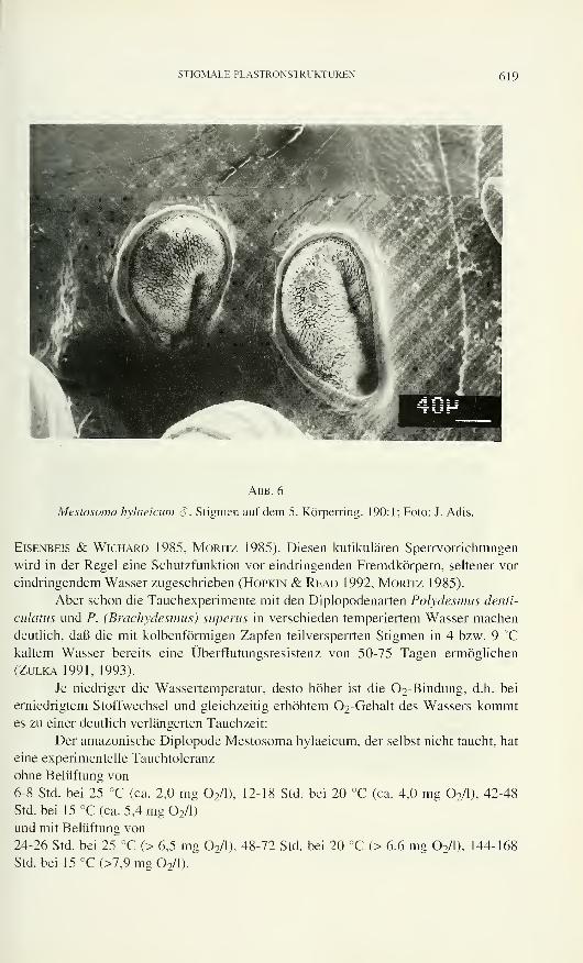
STIGMALE PLASTRONSTRUKTUREN 619
Abb. 6
Mestosoma hylaeicum 3 , Stigmen auf dem 5. Körperring. 190:1; Foto: J. Adis.
Eisenbeis & Wichard 1985, Moritz 1985). Diesen kutikulären Sperrvorrichtungen
wird in der Regel eine Schutzfunktion vor eindringenden Fremdkörpern, seltener vor
eindringendem Wasser zugeschrieben (Hopkin & Read 1992, Moritz 1985).
Aber schon die Tauchexperimente mit den Diplopodenarten Polydesmus denti-
culatus und P. (Brachydesmus) superus in verschieden temperiertem Wasser machen
deutlich, daß die mit kolbenförmigen Zapfen teilversperrten Stigmen in 4 bzw. 9 °C
kaltem Wasser bereits eine Überflutungsresistenz von 50-75 Tagen ermöglichen
(Zulka 1991, 1993).
Je niedriger die Wassertemperatur, desto höher ist die 02-Bindung, d.h. bei
erniedrigtem Stoffwechsel und gleichzeitig erhöhtem 02-Gehalt des Wassers kommtes zu einer deutlich verlängerten Tauchzeit:
Der amazonische Diplopode Mestosoma hylaeicum, der selbst nicht taucht, hat
eine experimentelle Tauchtoleranz
ohne Belüftung von
6-8 Std. bei 25 °C (ca. 2,0 mg 2/l), 12-18 Std. bei 20 °C (ca. 4,0 mg 2/l), 42-48
Std.bei 15 °C (ca. 5,4 mg 2/l)
und mit Belüftung von
24-26 Std. bei 25 °C (> 6,5 mg 2/l), 48-72 Std. bei 20 °C (> 6,6 mg 2/l), 144-168
Std.bei 15°C(>7,9mg02/l).
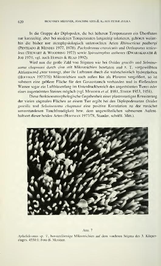
620 BENJAMIN MESSNER, JOACHIM ADIS & KLAUS PETER ZULKA
In die Gruppe der Diplopoden, die bei höheren Temperaturen ein Überfluten
nur kurzzeitig, aber bei niederen Temperaturen langzeitig tolerieren, gehören weiter-
hin die bisher nur atemphysiologisch untersuchten Arten Rhinocricus padbergi
(Penteado & Mendes 1977, 1978), Pachydesmus crassicutis und Oethoporus texico-
leus (Stewart & Woodring 1973) sowie Spirostrephus asthenes (Dwarakanath &Job 1974; vgl. auch Hopkin & Read 1992).
Wird nun die große Zahl von Stigmen wie bei Oxidus gracilis und Selmino-
soma chapmani durch eine mit Mikrotrichien besetzten und z. T. vorgewölbten
Atriumwand zwar verengt, aber ihr Luftraum durch die wahrscheinlich hydrophoben
(Hoffman 1977/78) Mikrotrichien nach außen hin als Plastron vergrößert, so ist
submers eine größere Fläche für den Gasaustausch vorhanden und in fließendem
Wasser sogar ein Luftblasenfang im Unterdruckbereich des angeströmten Tieres oder
eines angeströmten Steines möglich (vgl. Messner et al. 1981, Stride 1953, 1958).
Diese funktionsmorphologische Gegebenheit einer plastronartigen Erweiterung
der vielen stigmalen Flächen an einem Tier ergibt bei den Diplopodenarten Oxidus
gracilis und Selminosoma chapmani eine positive Korrelation zu der zunächst
unverstandenen Tauchfreudigkeit bzw. dem ungewöhnlichen submersen Aufent-
haltsort dieser beiden Arten (Hoffman 1977/78, Stauder, schriftl. Mitt.).
Abb. 7
Aphelidesmus sp. 9, borstenförmige Mikrotrichien auf dem vorderen Stigma des 3. Körper-
rinses. 4550:1; Foto B. Messner.
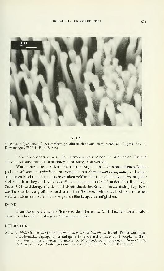
STIGMALE PLASTRONSÏRl KTUREN 621
Abb. 8
Mestosoma hylaeicum, 6 , borstenförmige Mikrotrichien auf demKörperringes. 7530:1; Foto: J. Adis.
vorderen Stigma des 4.
Lebendbeobachtungen zu den letztgenannten Arten im submersen Zustand
stehen noch aus und sollten baldmöglichst nachgeholt werden.
Warum die nahezu gleich strukturierten Stigmen bei der amazonischen Diplo-
podenart Mestosoma hylaeicum, im Vergleich mit Selminosoma chapmani, zu keinem
submersen Flucht- oder gar Tauchverhalten geführt hat, ist noch ungeklärt. Es mag aber
vielleicht daran liegen, daß die hohe Wassertemperatur (>28 °C an der Oberfläche; vgl.
Sioli 1984) und demgemäß der Löslichkeitsdruck des Sauerstoffs zu niedrig liegt bzw.
die Tiere selbst zu groß sind und somit ihre Stoffwechselrate zu hoch ist, um einen
stabilen submersen Aufenthalt energetisch überhaupt zu ermöglichen.
DANK
Frau Susanne Hamann (Plön) und den Herren E. & H. Fischer (Greifswald)
danken wir herzlich für die gute Aufnahmetechnik.
LITERATUR
Adis, J. 1992. On the survival strategy of Mestosoma hylaeicum Jeekel (Paradoxomatidae.
Polydesmida, Diplopoda), a millipede from Central Amazonian floodplains. (Pro-
ceedings 8th International Congress of Myriapodology, Innsbruck). Berichte des
Naturwisenschaftlich-Medizinischen Vereins in Innsbruck, Suppì. 10: 183-187.
622 BENJAMIN MESSNER, JOACHIM ADIS & KLAUS PETER ZULKA
Adis, J & B. Messner. 1997. Adaptations to life under water: tiger beetles and millipedes. In:
Junk, W.J. (Hrsg.): The Central-Amazon floodplain: ecology of a pulsing system.
Ecological Studies. Springer, Heidelberg: im Druck.
Dwarakanath, S. K. & S. V. Job. 1974. The effect of aqueous submersion on respiration in
the millipede Spirostreptus asthenes (Pokock). Monitore Zoologico Italiano (N.S.) 8:
11-18.
EiSENBEis, G. & W. Wichard. 1985. Atlas zur Biologie der Bodenarthropoden. Stuttgart, NewYork, 434 S .
Hoffman, R.L. 1977/78. Diplopoda from Papua Caves (Zoological results of the British
Speleological Expedition to Papua-New Guinea, 1975, 4). Interantional Journal ofSpeleology 9: 281-307.
Hopkin, S.P. & H. Read. 1992. The biology of millipedes. Oxford Univ. Press, Oxford, 233 S.
Messner, B. & J. Adis. 1988. Die Plastronstrukturen der bisher einzigen submers lebenden
Diplopodenart Gonographis adisi Hoffman 1985 (Pyrgodesmidae, Diplopoda). Zoolo-
gische Jahrbücher - Anatomie und Ontogenie der Tiere 1 17: 277-290.
Messner, B., J. Adis & E. F. Ribeiro. 1992. Eine vergleichende Untersuchung über die Plas-
tronstrukturen bei Milben (Acari). Deutsche Entomologische Zeitschrift, N.F. 39: 159-
176.
Messner, B., A. Lunk, I. Groth, H. J. Subklew & D. Taschenberger 1981. Neue Befunde
zum Atmungssystem der Grundwanze Aphelocheirus aestivalis (Heteroptera, Hydro-
corisae) I. Imagines. Zoologische Jahrbücher - Anatomie und Ontogenie der Tiere 105:
474-496.
Moritz, G. 1985. Mikromorphologische Chitinstrukturen als Plastronbildner bei terrestrisch
lebenden Thysanopteren (Insecta). Biologische Rundschau 23: 161-170.
Penteado, C.H.S. & E. G. Mendes. 1977. Respiratory metabolism and tolerance in a tropical
millipede, Rhinocricus padbergi Verhoeff, 1938. I. The structure of the tracheal pocket
and the respiratory rate at normoxic conditions and 25 °C. Revista Brasileira de
Biologia 37: 431-446.
Penteado, C.H.S & E.G. Mendes 1978. Respiratory metabolism and tolerance in a tropical
millipede, Rhinocricus padbergi Verhoeff, 1938. II. The responses to declining oxygentensions and submersion in water. Revista Brasileira de Biologia 38: 851-862.
SiOLi, H. 1984. The Amazon - Limnology and landscape ecolog of a mighty tropical river and
its basin. W. Junk, Dordrecht, 763 S.
Stauder, A. 1990. Untersuchungen des Makrozoobenthos in einem Bach auf Madeira mit zoo-
geographischen Aspekten. Diplomarbeit, Freiburg/Breisgau, 141 S.
Stewart, T.C. & J. P. Woodring 1973. Anatomical and physiological studies of water balance
in the millipedes Pachydesmus crassicutis (Polydesmida) and Orthoporus texicolens
(Spirobolida). Comparative Biochemystry and Physiology 44 A: 735-750.
Stride, G.O. 1953. The respiratory bubble of the aquatic beetle Potamodvtes tuberosus Hinton.
Nature 171: 885-886.
Stride, G.O. 1958. The application of a Bernoulli equation to problem of insect respiration.
Proceedings of the 10th International Congress ofEntomology 2: 335-336.
Zulka, K.P. 1989. Einfluß der Hochwasser auf die epigäische Arthropodenfauna im Über-
schwemmungsbereich der March (Niederösterreich). Mitteilungen der Deutschen
Gesellschaft für Allgemeine und Angewandte Entomologie 7: 74-75.
Zulka, K.P. 1991. Überflutung als ökologischer Faktor: Verteilung, Phänologie und An-passung der Diplopoden. Lithobiomorpha und Isopoda in den Flußauen der March.
Dissertation, Wien/Österreich, 65 S.
Zulka, K.P. 1993. Submersion tolerance of some diplopod species. Abstracts 9th International
Congress ofMyriapodology, Paris: 95.
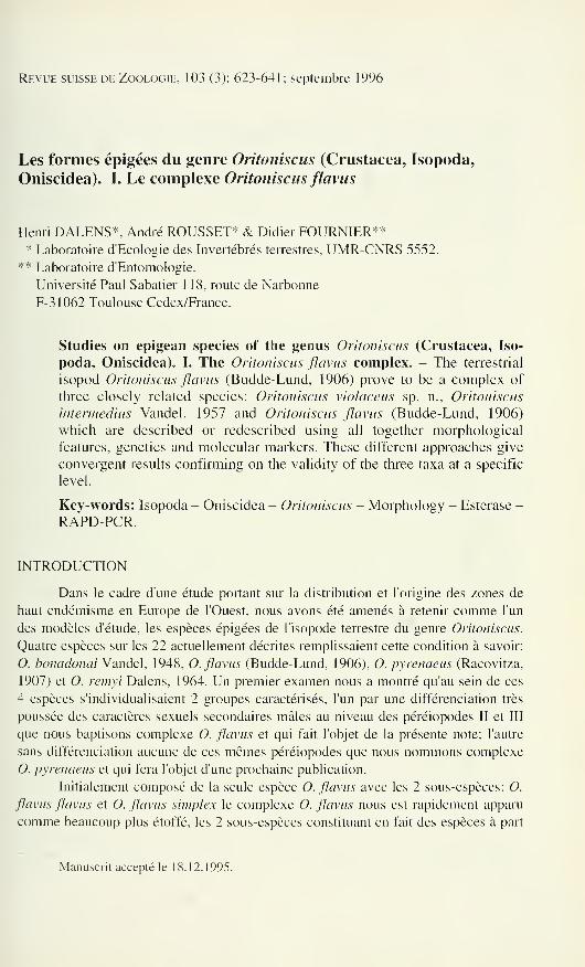
Revue suisse de Zoologie, 103 (3): 623-641; septembre 1996
Les formes épigées du genre Oritoniscus (Crustacea, Isopoda,
Oniscidea). I. Le complexe Oritoniscus flavus
Henri DALENS*. André ROUSSET* & Didier FOURNIER*** Laboratoire d'Ecologie des Invertébrés terrestres, UMR-CNRS 5552,
** Laboratoire d'Entomologie,
Université Paul Sabatier 118, route de Narbonne
F-31062 Toulouse Cedex/France.
Studies on epigean species of the genus Oritoniscus (Crustacea, Iso-
poda, Oniscidea). I. The Oritoniscus flavus complex. - The terrestrial
isopod Oritoniscus flavus (Budde-Lund, 1906) prove to be a complex of
three closely related species: Oritoniscus violaceus sp. n., Oritoniscus
intermedins Vandel, 1957 and Oritoniscus flavus (Budde-Lund, 1906)
which are described or redescribed using all together morphological
features, genetics and molecular markers. These different approaches give
convergent results confirming on the validity of the three taxa at a specific
level.
Key-words: Isopoda - Oniscidea - Oritoniscus - Morphology - Esterase -
RAPD-PCR.
INTRODUCTION
Dans le cadre d'une étude portant sur la distribution et l'origine des zones de
haut endémisme en Europe de l'Ouest, nous avons été amenés à retenir comme l'un
des modèles d'étude, les espèces épigées de l'isopode terrestre du genre Oritoniscus.
Quatre espèces sur les 22 actuellement décrites remplissaient cette condition à savoir:
O. bonadonai Vandel, 1948, O. flavus (Budde-Lund, 1906), O. pyrenaeus (Racovitza,
1907) et O. remyi Dalens, 1964. Un premier examen nous a montré qu'au sein de ces
4 espèces s'individualisaient 2 groupes caractérisés, l'un par une différenciation très
poussée des caractères sexuels secondaires mâles au niveau des péréiopodes II et III
que nous baptisons complexe O. flavus et qui fait l'objet de la présente note; l'autre
sans différenciation aucune de ces mêmes péréiopodes que nous nommons complexe
O. pyrenaeus et qui fera l'objet d'une prochaine publication.
Initialement composé de la seule espèce O. flavus avec les 2 sous-espèces: O.
flavus flavus et O. flavus simplex le complexe O. flavus nous est rapidement apparu
comme beaucoup plus étoffé, les 2 sous-espèces constituant en fait des espèces à part
Manuscrit accepté le 18.12.1995.
624 HENRI DALENS, ANDRÉ ROUSSET & DIDIER FOURNIER
entière dont l'une O. simplex n'appartient même pas au complexe étudié tandis que 2
espèces nouvelles se révélaient appartenir à cet ensemble.
La présente note est donc consacrée à la description ou à la redescription des 3
espèces du complexe O. flavus à la faveur de caractères morphologiques discrimi-
nants nouveaux. S'y ajoutent deux autres approches, l'une relative à l'analyse des
profils estérasiques des différents taxons, l'autre concernant l'étude de leur ADNgénomique au moyen de la méthode dite de RAPD-PCR, ceci dans l'optique de
vérifier si les 3 approches aboutissaient à des résultats congruents.
MATERIEL ET METHODES
Morphologie - Le matériel étudié provient essentiellement de nombreuses
récoltes effectuées par les deux premiers auteurs dans différentes régions de la chaîne
pyrénéenne. Ont également été examinées des collections du BMNH et de la Monks
Wood Experimental Station d'Huntingdom en Grande-Bretagne, ainsi que les
échantillons de la collection A. Vandel léguée au MNHN et provisoirement déposée
au Laboratoire de Zoologie-Ecologie de l'Université Paul Sabatier à Toulouse.
Polymorphisme enzymatique - L'analyse est effectuée sur des individus mâles
provenant de la même station (source Hamée, Cne d'Estadens, Hte Garonne). Afin
d'éliminer les variations d'ordre physiologique pouvant survenir au cours du cycle de
mue (Dalens & Rousset, 1988), nous avons utilisé des individus mâles adultes suivis
individuellement en élevage et fixés le troisième jour après l'ecdysis. Ils sont mis en
microtube contenant 50 pi de tampon de Trudgill (Tris-HCl 0.1 M pH 8, acide
ascorbique 0.018%, cysteine hydrochlorure 0.014%, triton X-100 0.01%). Les tubes
sont conservés à -20°C. Lors de leur utilisation les individus sont broyés in situ puis
centrifugés pendant 20 minutes à 15000g et à 4°C. Le surnageant (extrait brut) peut
être soit immédiatement chargé dans les puits du gel d'acrylamide soit conservé à
-20°C. La migration électrophorétique est individuelle, elle porte sur environ 1/4 de
l'extrait brut et est comparée à un mélange de 1/16 de chacun des 4 individus.
L'électrophorèse est conduite à 4°C, en gel de Polyacrylamide à 7% en gradient de pH(gel de tassement pH 6.8 et gel de séparation pH 8.8) en plaque 160 x 200 x 1 mm. La
migration se fait à voltage constant, d'abord 100 V pendant 30 minutes puis 300 Vpendant environ 3 h. La révélation est assurée par le colorant Fast Blue RR en
utilisant le ß-naphthyl acetate comme substrat. La caractérisation des fractions
présentant une activité enzymatique est basée sur leur mobilité électrophorétique
relative (Rf), l'indice 100 correspondant au front coloré par le bleu de bromophénol.
Polymorphisme de l'ADN - L'analyse porte également sur des individus mâles
des trois taxa O. violaceus, O. flavus et O. intermedins prélevés dans la même station
(source Hamée, Cne d'Estadens Hte Garonne). Les spécimens sont stockés à -20°C ou
en éthanol 95°. Chaque individu est broyé dans 100 pi d'une solution aqueuse à 10%
de résine chélatrice Chelex 100 (Walsh, 1991). Le broyât est incubé 30 min à 55°C
puis 15 min à 100°C et centrifugé 2 min à 15000 g. Le surnageant peut être utilisé
immédiatement ou conservé à -20°C.
COMPLEXE ORITONISCUS FLAWS 625
- Conditions d'amplification. La méthode employée est celle dite : RAPD- PCR(random amplified polymorphic DNA - polymerase chain reaction). Un pi d'extrait brut
d'ADN est amplifié dans 25 pi de tampon (75 mM tris-HCl pH 9.0 20 mM (NH4)2S04
0,01% Tween 20 1,25 mM MgCl2 ) contenant lOOmM de chaque dNTP, 40 ng d'amorce
(oligonucleotide) et 0,5 unité de Taq DNA polymerase (Eurogentec). Les échantillons
sont recouverts de 3 gouttes d'huile minérale et sont soumis à 40 cycles d'amplification;
chaque cycle comprend: 10 sec à 95°C, 30 sec à 36°C, 1 min à 72°C. Le produit
d'amplification est chargé sur un gel d'acrylamide à 7%, séparé par électrophorèse (7
V/cm pendant 4 h.), les fragments d'ADN sont révélés par coloration au bromure
d'éthidium (0,5 mg/ml) pendant 15 minutes et photographiés sur un transilluminateur
ultra-violet avec un film Polaroid (type 667 ou 665). Chaque gel contient les co-
migrations correspondantes à 8 individus de chacune des 3 espèces.
- Analyse des données - Les fragments d'ADN séparés sur les gels sont utilisés
comme marqueurs pour détecter les variations génétiques à différents niveaux. Ces
variations sont décelées parmi les biotypes, à l'intérieur de populations ou entre espèces
apparentées (Black et al., 1992). Les bandes d'ADN révélées par cette méthode se
séparent de manière indépendante, suggérant qu'elles représentent des loci individuels,
la majorité des alleles étant dominants (Williams et al, 1991). Ainsi, le phénotype
dominant, lié à un locus RAPD, se traduit sur le gel d'électrophorèse par une bande de
poids moléculaire spécifique. Une comparaison est alors possible entre les individus
pour chacun des loci: l'absence d'une bande, chez l'un et chez l'autre indique une
homozygotie alors que la présence simultanée chez les deux spécimens ne peut discri-
miner hétérozygotie ou homozygotie dominante (Welsh et al, 1991). La mesure de la
similitude entre individus s'effectue en déterminant le pourcentage de présence (ou
d'absence) simultanée d'une bande chez les deux individus selon la formule: M =
Nab/Nt où Nab est le nombre total de concordances (présence simultanée ou absence
simultanée) entre les individus A et B et Nj le nombre total de loci étudiés. La mesure
des distances génétiques est effectuée par la méthode UPGMA (unweighted pair-group
method of arithmetic averages) sur les valeurs de (1-M) en utilisant le programme infor-
matique Fortran RAPDPLOT élaboré par Kambhampati et al, 1992. Ce programme
calcule les valeurs de M à partir du tableau des données (présence - absence) indivi-
duelles relatives aux différents loci et aux différentes amorces d'oligonucléotides et
ensuite établit la matrice (1-M) qui, utilisée par les programmes NEIGHBOR et
DRAWTREE de l'ensemble PHYLIP 3.5c (distribué par J. Felsenstein, Univ. of
Washington, Seattle, WA, USA), permet une évaluation graphique des relations
phénétiques entre les taxa étudiés.
626 HENRI DALENS. ANDRÉ ROUSSET & DIDIER FOURNIER
RESULTATS
Etude morphologique
Oritoniscus flavus (Budde-Lund, 1906)
Trìchoniscus vividus, Budde-Lund 1885: 245 nec C.L. Koch 1841
Trichoniscusflavus Budde-Lund. 1906: 83
Trìchoniscus (Oritoniscus) flavus, Racovitza 1908: 332
Oritoniscus flavus, Vandel 1933: 45
Oritoniscus flavus flavus. Vandel 1960: 190, figs 83-85
Budde-Lund (1885) signale cette espèce de La Preste dans le midi de la
France, espèce qu'il rattache par erreur à Trichoniscus vividus Koch. La description
originale qu'il en donne est totalement muette sur les caractères sexuels mâles et
Figs 1-5
Oritoniscus flavus. 1: ocelle gauche; 2: Al in toto: 3: aesthetascs apicaux de Al: 4: 5e article de
la hampe et flagelle antennaire de A2: 5: aesthetascs du second pseudoarticle flagellale.
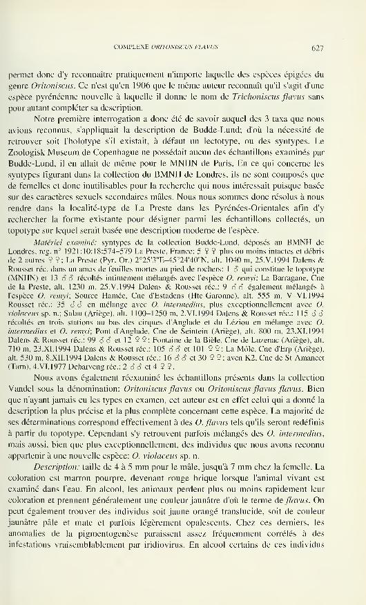
COMPLEXE ORÌTONISCUS FLAVUS 627
permet donc d'y reconnaître pratiquement n'importe laquelle des espèces épigées du
genre Oritoniscus. Ce n'est qu'en 1906 que le même auteur reconnaît qu'il s'agit d'une
espèce pyrénéenne nouvelle à laquelle il donne le nom de Trichoniscus flavus sans
pour autant compléter sa description.
Notre première interrogation a donc été de savoir auquel des 3 taxa que nous
avions reconnus, s'appliquait la description de Budde-Lund; d'où la nécessité de
retrouver soit l'holotype s'il existait, à défaut un lectotype, ou des syntypes. Le
Zoologisk Museum de Copenhague ne possédait aucun des échantillons examinés par
Budde-Lund, il en allait de même pour le MNHN de Paris. En ce qui concerne les
syntypes figurant dans la collection du BMNH de Londres, ils ne sont composés que
de femelles et donc inutilisables pour la recherche qui nous intéressait puisque basée
sur des caractères sexuels secondaires mâles. Nous nous sommes donc résolus à nous
rendre dans la localité-type de La Preste dans les Pyrénées-Orientales afin d'y
rechercher la forme existante pour désigner parmi les échantillons collectés, un
topotype sur lequel serait basée une description moderne de l'espèce.
Matériel examiné: syntypes de la collection Budde-Lund, déposés au BMNH de
Londres, reg. n° 1921:10:18:574-579 La Preste, France; 5 9 9 plus ou moins intactes et débris
de 2 autres 9 9 ; La Preste (Pyr. Or.) 2°25'3"E-45o24'40"N, ait. 1040 m, 25.V.1994 Dalens &
Rousset réc. dans un amas de feuilles mortes au pied de rochers: 1 S qui constitue le topotype
(MNHN) et 13 ô 8 récoltés intimement mélangés avec l'espèce O. renivi: La Barragane, Cnede la Preste, ait. 1230 m, 25.V. 1994 Dalens & Rousset réc: 9 S ô également mélangés à
l'espèce O. remyi; Source Hamée, Cne d'Estadens (Hte-Garonne), ait. 555 m, V-VI.1994Rousset réc: 35 S S en mélange avec O. intermedins, plus exceptionnellement avec O.
violaceus sp. n.; Salau (Ariège), ait. 1100-1250 m, 2. VI. 1994 Dalens & Rousset réc: 115 6 6récoltés en trois stations au bas des cirques d'Anglade et du Léziou en mélange avec O.
intermedins et O. remyi; Pont d'Anglade, Cne de Seintein (Ariège), ait. 800 m, 23.XI.1994
Dalens & Rousset réc: 99 S S et 12 9 9 ; Fontaine de la Bièle, Cne de Luzenac (Ariège), ait.
710 m, 23.XI.1994 Dalens & Rousset réc: 105 6 ô et 101 9 9 ; La Môle, Cne d'Erp (Ariège),
ait. 530 m, 8.XII.1994 Dalens & Rousset réc: 16 o" o" et 30 9 9 ; aven K2. Cne de St-Amancet
(Tarn), 4.VI.1977 Deharveng réc: 2 S 6 et 4 9 9
.
Nous avons également réexaminé les échantillons présents dans la collection
Vandel sous la dénomination: Oritoniscus flavus ou Oritoniscus flavus flavus. Bien
que n'ayant jamais eu les types en examen, cet auteur est en effet celui qui a donné la
description la plus précise et la plus complète concernant cette espèce. La majorité de
ses déterminations correspond effectivement à des O. flavus tels qu'ils seront redéfinis
à partir du topotype. Cependant s'y retrouvent parfois mélangés des O. intermedins,
mais aussi, bien que plus exceptionnellement, des individus que nous avons reconnu
appartenir à une nouvelle espèce: O. violaceus sp. n.
Description: taille de 4 à 5 mm pour le mâle, jusqu'à 7 mm chez la femelle. La
coloration est marron pourpre, devenant rouge brique lorsque l'animal vivant est
examiné dans l'eau. En alcool, les animaux perdent plus ou moins rapidement leur
coloration et prennent généralement une couleur jaunâtre d'où le terme de flavus. Onpeut également trouver des individus soit jaune orangé translucide, soit de couleur
jaunâtre pâle et mate et parfois légèrement opalescents. Chez ces derniers, les
anomalies de la pigmentogenèse paraissent assez fréquemment corrélés à des
infestations vraisemblablement par iridiovirus. En alcool certains de ces individus
628 HENRI DALENS. ANDRÉ ROUSSET & DIDIER FOURNIER
FlGSÓ-13
Oritoniscus flavus. 6: ischion P II. face antérieure: 7: face postérieure; 8: vue apicale: 9: vue
sternale; 10: ischion P III. face antérieure: 1 1: face postérieure; 12: vue apicale; 13: vue sternale.
COMPLEXE OR1TON1SCUS FLAVUS 629
Figs 14-17
Oritoniscus flavus. 14: péréiopode V en vue postérieure; 15: en vue antérieure; 16: pléopodes
mâle in toto; 17: extrémité de la tige de l'exopodite 1 mâle.
deviennent rapidement et totalement décolorés et paraissent alors correspondre à des
formes albines. L'oeil est constitué d'un ocelle unique de grande taille et pigmenté de
noir. Toutefois, et ceci se retrouve chez les 3 espèces dont il est question dans cette
note, lorsque l'ocelle est examiné au microscope électronique à balayage, il ne peut
être repéré que grâce au bombement de la carapace à son niveau; la structure
cuticulaire est par contre rigoureusement identique à celle du reste du vertex (Fig. 1 ),
ce qui traduit un début de régression de l'appareil oculaire, lequel a totalement disparu
chez la majorité des représentants non épigés du genre Oritoniscus. Les téguments
sont lisses et garnis de soies simples. L'antennule (Figs 2-3) est formée de 3 articles
dont le second porte une forte épine en position latéro-interne, et le distal est surmonté
de 10 aesthétascs et d'une épine externe assez courte. L'article 5 de la hampe de
l'antenne (Fig. 4) porte sur sa face antérieure une fossette longitudinale; quant au
flagelle il est formé de 5 (parfois 6) pseudo-articles dont le second porte à son tiers
inférieur 5 à 7 aesthétascs (Fig. 5). Uropode à endopodite s'insérant plus bas que
l'exopodite et plus fin que ce dernier, mais de longueur sensiblement égale. Les
péréiopodes II, III et V présentent chez le mâle des différenciations sexuelles
particulièrement nettes et caractéristiques du taxon. Au niveau du péréiopode II,
l'ischion (Figs 6-9) est ramassé et porte sur sa face postérieure une profonde
dépression d'où émerge au niveau du bord sternal une touffe d'écaillés piliformes. Sur
le péréiopode III, l'ischion (Figs 11-13) est également raccourci et porte sur la partie
distale de sa face antérieure un replat supportant une plage d'écaillés piliformes.
Quant au péréiopode V (Figs 14-15), il présente un basis fortement renflé du fait de
l'élargissement de sa face antérieure. Le basis du péréiopode VII du mâle présente
630 HENRI DALENS, ANDRÉ ROUSSET & DIDIER FOURNIER
également une très légère différenciation sexuelle qui se traduit par la présence d'une
petite touffe de soies en écailles à l'angle sterno-distal. Le premier pléopode mâle
montre un exopodite subrectangulaire portant un lobe externe d'autant plus nettement
individualisé que l'individu est de plus grande taille et un lobe interne armé
distalement d'une forte tige, verruqueuse à son extrémité, avant de se terminer par un
court flagelle cilié (Figs 16-17).
Répartition: Nos propres récoltes, ainsi que l'examen des échantillons repré-
sentés dans la collection Vandel, nous permettent d'affirmer que l'espèce est présente
de façon certaine, dans toute la moitié orientale des Pyrénées françaises, depuis la
vallée de l'Ouzom à l'ouest, jusque dans le Haut Vallespir à l'est. En Espagne nous
l'avons retrouvée dans le Val d'Aran. D'après Vandel (1960), il semble (bien que
nous n'ayons pu toujours le vérifier par nous-mêmes) que ce soit ce taxon qui ait
colonisé le Bassin Aquitain, les Charentes et le Poitou, ainsi qu'une partie du Massif
Central. Les échantillons que nous avons pu examiner, provenant de différents comtés
d'Irlande ainsi que de la région de Carmothan dans le Pays-de-Galles en Grande
Bretagne même, nous permettent de confirmer qu'il s'agit bien également de l'espèce
flavus. Ce taxon constituerait donc bien une espèce à vaste répartition et qui serait
même à l'heure actuelle en voie d'expansion, notamment dans la région septentrionale
de son aire de répartition.
Oritoniscus intermedius Vandel, 1957
Oritoniscusflavus intermedius Vandel, 1957: 92, fig. IB
Il convient ici de rappeler, que dans un travail de 1957, Vandel avait consi-
déré que l'espèce O. flavus se scindait en 3 sous-espèces dont il décrivait - très
succintement il est vrai - 2 nouvelles sous-espèces: O. flavus simplex n. ssp. et O.
flavus intermedius n. ssp. Cependant dans la Faune de France des Isopodes terrestres
parue en 1960, il ne reconnaissait plus à O. flavus que 2 sous-espèces: O. flavus flavus
et O. flavus simplex: considérant que la sous-espèce précédemment décrite sous le
nom d'intermedius, ne constituait en fait qu'une forme juvénile de la sous-espèce O.
flavus flavus. Or les échantillons que Vandel avait désignés sous le nom ^inter-
medius correspondent manifestement à un taxon bien défini.
Matériel examiné: un certain nombre d'échantillons de cette espèce figurent dans la
collection A. Vandel, seuls ou plus généralement en mélange avec O. flavus. Sous la
dénomination O. flavus: Niaux (Ariège), 2.VI. 1933, 1 o et 2 9 9 ; sous la dénomination O.
flavus flavus: Luchon (Hte-Garonne), 26.VIII.1954, 3 6 6 et 6 9 9; sous la dénomination O.
flavus intermedius: Puymorens (Ariège), 2.XI. 1955, 12 SÔ et 18 9 9; Cirque de Leziou, MtRouch/Salau (Ariège), 30. VIII. 1954, 6 6 6 et 6 9 9. Le descripteur n'ayant pas désigné
d'holotype, mais les stations d'Anglade et du Leziou sur la commune de Salau (Ariège) ayant
servi de localités types, nous avons sélectionné un échantillon mâle parmi ceux de cette station
figurant dans la collection Vandel pour servir de lectotype. La crête sternale de l'ischion II
caractéristique de l'espèce étant peu visible sur préparation microscopique et la diagnose
spécifique étant beaucoup plus facile et évidente sur l'animal in toto. nous avons jugé préférable
de conserver le lectotype intact et de baser l'illustration de notre description sur d'autres
échantillons provenant de la même localité. Giiells del Joeu, Val d'Aran en Espagne, ait. 1400
m, 20VIII.1994 Rousset réc: 4 6 6 et 8 9 9 ; Cirques d'Anglade et du Leziou, Cne de Salau
(Ariège), ait. 1100-1250 m, 2.VI. 1994 Dalens & Rousset réc: 24 o" o" en mélange avec O.
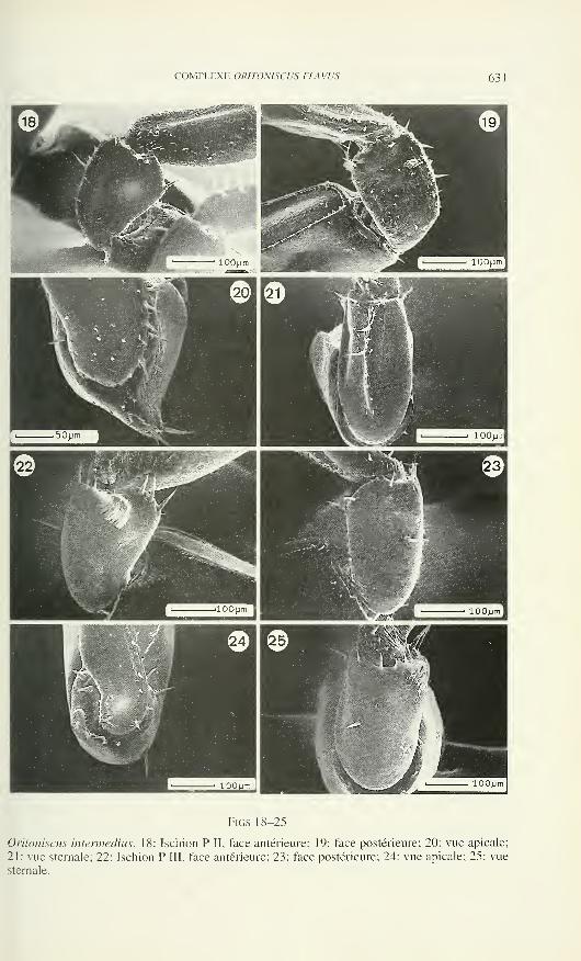
COMPLEXE ORITON1SCUS FLAVUS 63:
Figs 18-25
Oritoniscus intermedins. 18: Ischion P II, face antérieure; 19: face postérieure; 20: vue apicale;
21: vue sternale; 22: Ischion P III, face antérieure; 23: face postérieure; 24: vue apicale; 25: vue
sternale.
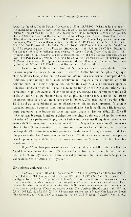
632 HENRI DALENS, ANDRÉ ROUSSET & DIDIER FOURNIER
flavus; Le Freyche, Cne de Nescus (Ariège), alt. 450 m. 28.VI.1994 Dalens & Rousset rèe: 4
6 6 mélangés à l'espèce O. renivi; Arbosec, Cne de Riverenert (Ariège), alt. 645 m, 8.XII.1994
Dalens & Rousset rèe: 15 S 8 et 41 9 9; résurgence, Cne de Taurignan-le-Vieux (Ariège), alt.
386 m, 8.XII.1994 Dalens & Rousset rèe: 2 o o en mélange avec O. remyi; Ruau, Cne Izaut de
l'Hôtel (Hte-Garonne), alt. 800 m. Vili. 1994 Rousset rèe: 7 o o en mélange avec O. violaceus;
Baillugues, Cne d'Estadens (Hte-Garonne), alt. 480 m, 26.IV. 1994 Rousset rèe: 17 o o et 41
9 9, 2.V.1994 Rousset rèe: 30 S o et 78 9 9, 16.VI.1994 Dalens & Rousset rèe: 36 Ó â et
45 9 9; source Hamée, Cne d'Estadens (Hte-Garonne), alt. 555 m, 16.VI. 1994 Dalens &Rousset rèe: 35 o o en mélange avec l'espèce O. flavus; Cap de l'Ane, Cne de Sost (Htes-
Pyrénées), alt. 757 m, 18.X. 1994 Dalens & Rousset rèe: 1 â et 4 9 9 ; Sources captées, Cne de
Sost (Htes-Pyrénées). alt. 920 m, 18.X.1994 Dalens & Rousset rèe: 14 o S en mélange avec
O. flavus et une nouvelle espèce d'Oritoniscus; Maison Bourdette, Cne de Nistos (Htes-
Pyrénées). alt. 630 m, 18.X.1994 Dalens & Rousset rèe: 52 o o et 51 9 9.
Description: taille un peu plus réduite que chez l'espèce précédente: 4 mmmaximum pour les mâles, 6 mm pour les femelles. Coloration un peu plus rouge que
chez O. flavus lorsque l'animal est examiné vivant dans une coupelle remplie d'eau.
Individus jaune-orangé translucide relativement fréquents mais toujours en petit
nombre dans une même population. Antennule avec 5 à 7 aesthétascs apicaux
flanqués d'une courte épine. Flagelle antennaire formé de 4 à 5 pseudo-articles. Les
caractères les plus évidents et discriminant l'espèce, affectent les péréiopodes mâles II
et III. Au niveau du péréiopode II, le carpos présente sur sa face sternale une brosse
de courtes soies denses qui manquent chez la femelle. C'est néanmoins l'ischion (Figs
18-21) qui est caractéristique par son élargissement dû au développement d'une crête
sternale, portant de courtes soies sur sa partie distale. Sur le péréiopode III, le carpos
porte également une brosse de soies sternales; quant à l'ischion (Figs 22-25) s'il
présente sensiblement la même architecture que chez O. flavus, la plage de soies est
ici réduite à une petite touffe, proche de l'arête sternale et est flanquée en avant et en
arrière de 3 fortes épines. L'élargissement du basis V que l'on note chez O. flavus est
absent chez O. intermedins. Par contre tout comme chez O. flavus, le basis du
péréiopode VII présente une très petite touffe de soies à l'angle sterno-distal. Les
pléopodes mâles 1 et 2 sont semblables à ceux d'O. flavus mais on ne retrouve pas le
développement hyperthélique de la pointe externe de l'exopodite 1 chez les plus
grands individus.
Répartition: Nos propres récoltes et l'examen des échantillons de la collection
Vandel. nous autorisent à dire qu'O. intermedins se trouve dans toute la partie orien-
tale de la chaîne pyrénéenne, la limite ouest paraissant être, au moins à ce jour, la
vallée de la Neste d'Aure (Htes-Pyrénées).
Oritoniscus violaceus sp. n.
Matériel examiné: Holotype (déposé au MNHN): 1 o provenant de la source Hamée,Cne d'Estadens (Hte-Garonne), ait.: 555 m, 0°51'36"E-43°1'43"N, 7.V.1994 Rousset rée,
Paratypes: 16 o 6 et 29 9 9 provenant de la même station, dans la mousse ou sous les pierres
recouvertes d'un filet d'eau courante d'un petit griffon à flanc de coteau et sur une distance d'une
dizaine de mètres en aval de ce dernier, dans le lit du ruisseau naissant, en compagnie de
gammares et de larves d'Osmyles: source Ruau, Cne d'Arbon (Hte-Garonne). ait. 784 m,
25. IV. 1993 Lek rèe: 12 c? o 23 9 9 8 immatures; sources de la Maure, Cne Izaut de l'Hôtel
(Hte-Garonne), ait. 430 m, 22.V.1993 Lek rèe: 46 S o 68 9 9 ; idem, 12.XI.1994 Rousset rée:
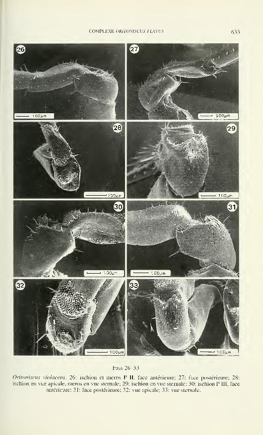
COMPLEXE OR/TONISCUS FLAWS 633
Figs 26-33
Oritoniscus violaceus. 26: ischion et meros P II, face antérieure; 27: face postérieure; 28:
ischion en vue apicale, meros en vue sternale; 29: ischion en vue sternale; 30: ischion P III, face
antérieure; 31: face postérieure; 32: vue apicale; 33: vue sternale.
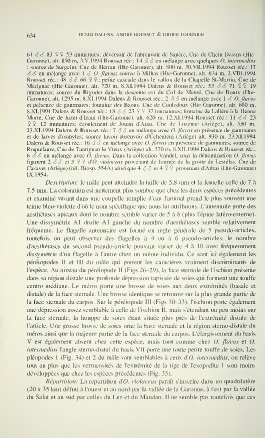
634 HENRI DALENS, ANDRÉ ROUSSET & DIDIER FOURNIER
61 6 6 83 9 9 53 immatures; déversoir de l'abreuvoir de Sauère, Cne de Chein Dessus (Hte-
Garonne), ait. 850 m, 3.V.1994 Rousset réc: 14 6 6 en mélange avec quelques O. intermedins
; source de Surgeint, Cne de Herran (Hte-Garonne), ait. 800 m, 30. VII. 1994 Rousset réc: 17
6 6 en mélange avec 1 6 O. flavus; source à Milhas (Hte-Garonne), ait. 674 m, 2. VIII. 1994
Rousset réc: 48 6 6 66 9 9; petite cascade dans le vallon de la Chapelle St-Martin, Cne de
Marignac (Hte-Garonne), ait. 720 m. 8.XI.1994 Dalens & Rousset réc: 53 6 6 71 9 9 19
immatures; source du Rigodet dans la descente est du Col de Mente, Cne de Boutx (Hte-
Garonne), ait. 1295 m, 8.XI. 1994 Dalens & Rousset réc: 2 6 6 en mélange avec 1 6 O. flavus
et présence de gammares; fontaine des Baous, Cne de Couledoux (Hte-Garonne), ait. 680 m,
8.XI.1994 Dalens & Rousset réc: 18 â 6 23 9 9 37 immatures; fontaine de Lalière à la HenneMorte, Cne de Juzet d'Izaut (Hte-Garonne), ait. 620 m, 12.XI.1994 Rousset réc: 11 6 6 23
9 9 12 immatures; écoulement de Jouan d'Arau, Cne de Luzenac (Ariège), ait. 550 m,
23.XI. 1994 Dalens & Rousset réc: 7 6 6 en mélange avec O. flavus en présence de gammareset de larves d'osmyles; source lavoir-abreuvoir d'Uchentein (Ariège) ait. 930 m, 23.XI.1994
Dalens & Rousset réc: 16 6 6 en mélange avec O. flavus en présence de gammares; source de
Roquelaure, Cne de Taurignan le Vieux (Ariège) ait. 370 m, 8.XII.1994 Dalens & Rousset réc:
6 6 6 en mélange avec O. flavus. Dans la collection Vandel, sous la dénomination O. flavus
figurent 2 6 6 et 3 9 9 d'O. violaceus provenant de l'entrée de la grotte de Lestélas, Cne de
Cazavet (Ariège) (réf. Biosp. 554A) ainsi que 4 â 6 et 4 9 9 provenant d'Arbas (Hte-Garonne)
IX. 1954.
Description: le mâle peut atteindre la taille de 5,8 mm et la femelle celle de 7 à
7,5 mm. La coloration est nettement plus sombre que chez les deux espèces précédentes
et examiné vivant dans une coupelle remplie d'eau l'animal prend le plus souvent une
teinte bleu-violette d'où le nom spécifique que nous lui attribuons. L'antennule porte des
aesthétascs apicaux dont le nombre semble varier de 5 à 8 (plus l'épine latéro-externe).
Une dissymétrie Al droite Al gauche du nombre d'aesthétascs semble relativement
fréquente. Le flagelle antennaire est formé en règle générale de 5 pseudo-articles,
toutefois on peut observer des flagelles à 4 ou à 6 pseudo-articles, le nombre
d'aesthétascs du second pseudo-article pouvant varier de 4 à 10 avec fréquemment
dissymétrie d'un flagelle à l'autre chez un même individu. Ce sont ici également les
péréiopodes II et III du mâle qui portent les caractères vraiment discriminants de
l'espèce. Au niveau du péréiopode II (Figs 26-29), la face sternale de l'ischion présente
dans sa région distale une profonde dépression tapissée de soies qui forment une touffe
centro-médiane. Le méros porte une brosse de soies aux deux extrémités (basale et
distale) de la face sternale. Une brosse identique se retrouve sur la plus grande partie de
la face sternale du carpos. Sur le péréiopode III (Figs 30-33), l'ischion porte également
une dépression assez semblable à celle de l'ischion II, mais s'étendant un peu moins sur
la face sternale, la houppe de soies étant située plus près de l'extrémité distale de
l'article. Une grosse brosse de soies orne la base sternale et la région sterno-distale du
méros ainsi que la majeure partie de la face sternale du carpos. L'élargissement du basis
V est également absent chez cette espèce, mais tout comme chez O. flavus et O.
intermedins l'angle stemo-distal du basis VII porte une toute petite touffe de soies. Les
pléopodes 1 (Fig. 34) et 2 du mâle sont semblables à ceux d'O. intermedins, on relève
tout au plus que les verrucosités de l'extrémité de la tige de l'exopodite 1 sont moins
développées que chez les espèces précédentes (Fig. 35).
Répartition: La répartition d'O. violaceus paraît s'inscrire dans un quadrilatère
(20 x 35 km) défini à l'ouest et au nord par la vallée de la Garonne, à l'est par la vallée
du Salât et au sud par celles du Lez et du Maudan. Il ne semble pas toutefois que ces
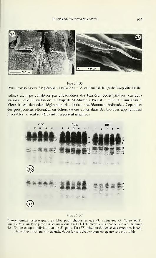
COMPLEXE OR1TON1SCUS FLAVUS 635
Figs 34-35
Oritoniscus violaceus. 34: pléopodes 1 mâle in toto; 35: extrémité de la tige de l'exopodite 1 mâle.
vallées aient pu constituer par elles-mêmes des barrières géographiques, car deux
stations, celle du vallon de la Chapelle St-Martin à l'ouest et celle de Taurignan le
Vieux à l'est débordent légèrement des limites précédemment indiquées. Cependant
des prospections effectuées en dehors de ces zones dans des biotopes apparemment
favorables, se sont révélées jusqu'à présent négatives.
viol. flav
Ulli
Figs 36-37
Zymogrammes estérasiques: en (36) pour chaque espèce O. violaceus, O. flavus et O.
intermedius l'analyse porte sur les individus 1 à 4 (1/4 du broyât dans chaque puits) et mélangede 1/16 de chaque individu dans le 5° puits. En (37) mise en évidence des fractions lentes,
même disposition mais la quantité déposée dans chaque puits est quatre fois plus faible.
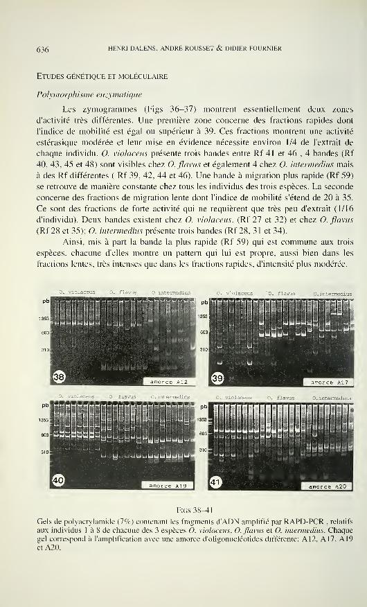
636 HENRI DALENS, ANDRÉ ROUSSET & DIDIER FOURNIER
Etudes génétique et moléculaire
Polymorphisme enzymatìque
Les zymogrammes (Figs 36-37) montrent essentiellement deux zones
d'activité très différentes. Une première zone concerne des fractions rapides dont
l'indice de mobilité est égal ou supérieur à 39. Ces fractions montrent une activité
estérasique modérée et leur mise en évidence nécessite environ 1/4 de l'extrait de
chaque individu. O. violaceus présente trois bandes entre Rf 41 et 46 , 4 bandes (Rf
40, 43, 45 et 48) sont visibles chez O. flavus et également 4 chez O. intermedius mais
à des Rf différentes ( Rf 39, 42, 44 et 46). Une bande à migration plus rapide (Rf 59)
se retrouve de manière constante chez tous les individus des trois espèces. La seconde
concerne des fractions de migration lente dont l'indice de mobilité s'étend de 20 à 35.
Ce sont des fractions de forte activité qui ne requièrent que très peu d'extrait (1/16
d'individu). Deux bandes existent chez O. violaceus, (Rf 27 et 32) et chez O. flavus
(Rf 28 et 35); O. intermedius présente trois bandes (Rf 28, 31 et 34).
Ainsi, mis à part la bande la plus rapide (Rf 59) qui est commune aux trois
espèces, chacune d'elles montre un pattern qui lui est propre, aussi bien dans les
fractions lentes, très intenses que dans les fractions rapides, d'intensité plus modérée.
O. violaceus O . intermedius O. violaceus O . intermedius
u :' 'uUJUuiuJu.: <y>
O . violaceus O . intermedius
Pb
1353!
603!
uuutJUtJjiLji ii.jùyj
HHHRNHmouuUUÌ :yyguuyu
JuuUUUUUUU
Figs38^1
Gels de Polyacrylamide (7%) contenant les fragments d'ADN amplifié par RAPD-PCR , relatifs
aux individus 1 à 8 de chacune des 3 espèces O. violaceus, O. flavus et O. intermedius. Chaquegel correspond à l'amplification avec une amorce d'oligonucléotides différente: A12, A17, A19et A20.
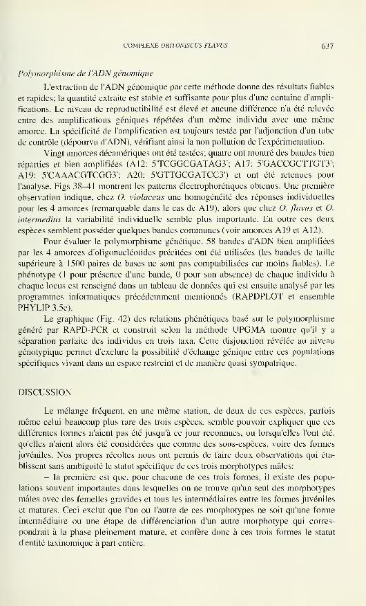
COMPLEXE ORITONISCUS FLAWS 637
Polymorphisme de l'ADN génomique
L'extraction de l'ADN génomique par cette méthode donne des résultats fiables
et rapides; la quantité extraite est stable et suffisante pour plus d'une centaine d'ampli-
fications. Le niveau de reproductibilité est élevé et aucune différence n'a été relevée
entre des amplifications géniques répétées d'un même individu avec une mêmeamorce. La spécificité de l'amplification est toujours testée par l'adjonction d'un tube
de contrôle (dépourvu d'ADN), vérifiant ainsi la non pollution de l'expérimentation.
Vingt amorces décamériques ont été testées; quatre ont montré des bandes bien
réparties et bien amplifiées (A12: 5TCGGCGATAG3'; A 17: 5'GACCGCTTGT3';
A19: 5'CAAACGTCGG3'; A20: 5'GTTGCGATCC3') et ont été retenues pour
l'analyse. Figs 38^41 montrent les patterns électrophorétiques obtenus. Une première
observation indique, chez O. violaceus une homogénéité des réponses individuelles
pour les 4 amorces (remarquable dans le cas de A 19), alors que chez O. flavus et O.
intermedins la variabilité individuelle semble plus importante. En outre ces deux
espèces semblent posséder quelques bandes communes (voir amorces A19 et A12).
Pour évaluer le polymorphisme génétique, 58 bandes d'ADN bien amplifiées
par les 4 amorces d'oligonucléotides précitées ont été utilisées (les bandes de taille
supérieure à 1500 paires de bases ne sont pas comptabilisées car moins fiables). Le
phénotype (1 pour présence d'une bande, pour son absence) de chaque individu à
chaque locus est renseigné dans un tableau de données qui est ensuite analysé par les
programmes informatiques précédemment mentionnés (RAPDPLOT et ensemble
PHYLIP 3.5c).
Le graphique (Fig. 42) des relations phénétiques basé sur le polymorphisme
généré par RAPD-PCR et construit selon la méthode UPGMA montre qu'il y a
séparation parfaite des individus en trois taxa. Cette disjonction révélée au niveau
génotypique permet d'exclure la possibilité d'échange génique entre ces populations
spécifiques vivant dans un espace restreint et de manière quasi sympatrique.
DISCUSSION
Le mélange fréquent, en une même station, de deux de ces espèces, parfois
même celui beaucoup plus rare des trois espèces, semble pouvoir expliquer que ces
différentes formes n'aient pas été jusqu'à ce jour reconnues, ou lorsqu'elles l'ont été,
qu'elles n'aient alors été considérées que comme des sous-espèces, voire des formes
juvéniles. Nos propres récoltes nous ont permis de faire deux observations qui éta-
blissent sans ambiguité le statut spécifique de ces trois morphotypes mâles:
- la première est que, pour chacune de ces trois formes, il existe des popu-
lations souvent importantes dans lesquelles on ne trouve qu'un seul des morphotypes
mâles avec des femelles gravides et tous les intermédiaires entre les formes juvéniles
et matures. Ceci exclut que l'un ou l'autre de ces morphotypes ne soit qu'une forme
intermédiaire ou une étape de différenciation d'un autre morphotype qui corres-
pondrait à la phase pleinement mature, et confère donc à ces trois formes le statut
d'entité taxinomique à part entière.
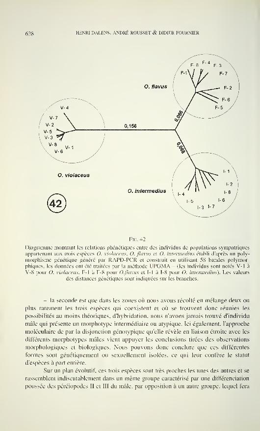
638 HENRI DALENS, ANDRÉ ROUSSET & DIDIER FOURNIER
Fio. 42
Diagramme montrant les relations phénétiques entre des individus de populations sympatriques
appartenant aux trois espèces O. violaceus, O. flavus et O. intermedins établi d'après un poly-
morphisme génétique généré par RAPD-PCR et construit en utilisant 58 bandes polymor-
phiques, les données ont été traitées par la méthode UPGMA - (les individus sont notés V-l à
V-8 pour O. violaceus, F-l à F- 8 pour O.flavus et FI à F8 pour O. intermedins). Les valeurs
des distances génétiques sont indiquées sur les branches.
- la seconde est que dans les zones où nous avons récolté en mélange deux ou
plus rarement les trois espèces qui coexistent et où se trouvent donc réunies les
possibilités au moins théoriques, d'hybridation, nous n'avons jamais trouvé d'individu
mâle qui présente un morphotype intermédiaire ou atypique. Ici également, l'approche
moléculaire de par la disjonction génotypique qu'elle révèle en liaison étroite avec les
différents morphotypes mâles vient appuyer les conclusions tirées des observations
morphologiques et biologiques. Nous pouvons donc conclure que ces différentes
formes sont génétiquement ou sexuellement isolées, ce qui leur confère le statut
d'espèces à part entière.
Sur un plan évolutif, ces trois espèces sont très proches les unes des autres et se
rassemblent indiscutablement dans un même groupe caractérisé par une différenciation
poussée des péréiopodes II et III du mâle, par opposition à un autre groupe, lequel fera
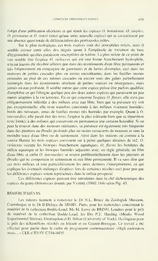
COMPLEXE ORITONISCUS FLAVUS 639
l'objet d'une publication ultérieure et qui réunit les espèces O. bonadonai, O. simplex,
0. pyrenaeus et O. remyi (ainsi qu'une autre nouvelle espèce) qui se caractérisent par
une absence quasi totale de différenciation des péréiopodes mâles.
Sur le plan écologique, ces trois espèces sont des atmophiles stricts, mais il
semble exister entre elles des degrés quant à l'amplitude de variation du taux
d'hygrométrie qu'elles paraissent susceptibles de tolérer. La plus stricte de ce point de
vue semble être l'espèce O. violaceus qui est une forme franchement hydrophile,
n'ayant jamais été récoltée ailleurs que dans des écoulements d'eau libre permanents et
très fréquemment en compagnie de gammares et de larves d'osmyles, ceci dans les
mousses de petites cascades plus ou moins encroûtantes, dans les feuilles mortes
entassées au pied de ces mêmes cascades ou encore sous des galets partiellement
immergés dans les écoulements résultant de petites sources ou résurgences, mais
jamais en eau profonde. Il semble même que cette espèce puisse être parfois qualifiée
d'amphibie ce qui l'éloigné quelque peu des deux autres espèces qui paraissent un peu
mieux adaptées à la vie terrestre. En ce qui concerne l'espèce O. flavus, elle n'est pas
obligatoirement inféodée à des milieux avec eau libre, bien que sa présence n'y soit
pas exceptionnelle; elle reste toutefois cantonnée à des milieux vraiment humides:
bords des ruisseaux, amas de feuilles mortes très humides par exemple. Quant à O.
intermedins, elle parait être des trois, l'espèce la plus tolérante bien que sa répartition
reste limitée à des milieux qui conservent en permanence une certaine humidité. Si on
peut la trouver dans des mousses suintantes, sa présence est également très fréquente
dans des pierriers ou éboulis profonds plus ou moins recouverts de mousses et sans la
moindre trace d'eau libre ou de suintement. Ainsi dans les stations où comme à la
source Hamée les trois espèces coexistent sur à peine quelques dizaines de m2, O.
violaceus occupe les biotopes franchement aquatiques, O. flavus les bordures du
milieu aquatique et les biotopes humides adjacents avec, en règle générale, un film
d'eau libre et enfin O. intermedins se trouve préférentiellement dans les pierriers et
éboulis qui ne comportent ni suintement ni eau libre permanente. Il va sans dire que
ces trois milieux et tout particulièrement les deux derniers s'interpénétrent, ce qui
explique les éventuels mélanges d'espèces lors de certaines récoltes ceci pour peu que
les différentes espèces soient représentées dans le milieu prospecté.
Ces différentes espèces peuvent être introduites dans la clef dichotomique des
espèces du genre Oritoniscus donnée par Vandel (1960: 184) selon Fig. 43.
REMERCIEMENTS
Les auteurs tiennent à remercier le Dr N.L. Bruce du Zoologisk Museum,
Copenhague et le Dr D.Defaye du MNHN, Paris, pour les recherches concernant le
matériel de la collection Budde-Lund; Ms M. Lowe du BMNH, Londres pour le prêt
de matériel de la collection Budde-Lund, les Drs P.T. Harding (Monks WoodExperimental Station), Huntingdon et D. Bilton (University of York), Heslington pour
le prêt des échantillons récoltés en Irlande et en Grande-Bretagne. Ce travail a été
effectué pour partie dans le cadre du programme communautaire «High endemismareas... » CEE n°EV5V-CT94-0435.
640 HENRI DALENS, ANDRÉ ROUSSET & DIDIER FOURNIER
lobe externe de l'exopodite 1
dépourvu de lobule détaché
- crête sternale sur
l'ischion de P II
O. intermedins
- pas de crête sternale sur
l'ischion de P II
+ plage de soies apicales
sur la face antérieure
de l'ischion de P III
O. flavus
+ dépression apicale de
la face sternale de
l'ischion de P III
avec soies médio-sternales
O. violaceus
Fig. 43
Clé dichotomique discriminant les trois espèces O. intermedins, O. flavus, O. violaceus.
REFERENCES
Black. W.C., Du Teau, N.M., Puterka, G.J., Nechols, J.R. & Pettorine J.M. 1992. Use of
the random amplified polymorphic DNA polymerase chain reaction (RAPD-PCR) to
detect DNA polymorphisms in aphids (Homoptera: Aphididae). Bulletin of Entomo-
logical Research, 82: 151-159.
Budde-Lund, G. 1885. Crustacea Isopoda Terrestria per familias et genera et species descripta.
Hauniae, 319 pp.
Budde-Lund, G. 1906. Die Landisopoden der deutschen Südpolar-Expedition 1901-1903.
Deutsche Südpolar-Expedition 1901-1903., 9, Zool. I.: 79-92.
COMPLEXE ORITONISCUS FLAVUS 641
Dalens, H. 1964. Description d'une nouvelle espèce de Trichoniscide, Oritoniscus remyi.
Bulletin de la Société d'Histoire naturelle de Toulouse 99, (1-2): 286-288.
Dalens, H. & Rousset, A. 1988. Variations de l'activité estérasique chez des Hypogastrura du
sous-groupe tullbergi (Collembola). Revue d'Ecologie et de Biologie du Sol, 25, (1):
139-147.
Kambhampati, S., Black, W.C. & Rai, K.S. 1992. Random amplified polymorphic DNA of
mosquito species and populations (Diptera: Culicidae): techniques, statistical analysis
and applications. Journal ofMedical Entomology 29: 939-945.
Racovitza, E.G. 1907. Isopodes terrestres (Première Série). Biospeologica, IV. Archives de
Zoologie expérimentale et générale, 7: 145-225.
Vandel, A. 1948. Espèces nouvelles d'Isopodes terrestres cavernicoles et endogés. Espèces
françaises nouvelles ou peu connues de Trichoniscidae - 4e note -. Notes biospéo-
logiques, 2: 7—27.
Vandel, A. 1957. Nouvelles espèces et sous-espèces appartenant au genre Oritoniscus (Iso-
podes terrestres). Bulletin de la Société d'Histoire naturelle de Toulouse, 92: 7-16.
Vandel, A. 1960. Isopodes terrestres (Première Partie). Faune de France 64, Lechevallier,
Paris: 416 pp.
Walsh, P.S., Metzger, D.A. & Higuchi, R. 1991. Chelex 100 as a medium for simple extrac-
tion of DNA for PCR-based typing for forensic material. Biotechniques, 10: 510-513.
Welsch, J., Peterson, C. & McClelland, M. 1991. Polymorphisms generated by arbitrarily
primed PCR in the Mouse application to strain identification and genetic mapping.
Nucleic Acids Research, 19: 303-306.
Williams, J.G.K., Hanafey, M.K., Rafalsky, J.A. & Tingey, S.V. 1993. Genetics analysis
using random amplified polymorphic DNA markers. Methods in Enzymology, 218:
704-740.

Revue suisse de Zoologie, 103 (3) 643-676; septembre 1996
Aleocharinae della Colombia e dell'Ecuador: Parte II
(Coleoptera, Staphylinidae)
(132° Contributo alla conoscenza delle Aleocharinae)
Roberto PACEVia Vittorio Veneto, 13. 37032 Monteforte dAlpone (Verona), Italia.
Aleocharinae from Colombia and Ecuador: Part II (Coleoptera, Sta-
phylinidae). - Three species of the tribe Falagriini and thirty two of the
tribe Athetini are described as new. The new genus Cosmogastrusa of the
tribe Athetini is described and illustrated. Its relationships with Aloconota
Thomson and Pelioptera Kraatz are discussed. Falagria munda Erichson is
transferred to Gnypeta Thomson. Atheta parcior sensu Pace, 1987, from
Guadalupa (nee Atheta parcior Bernhauer, 1927, from Argentina) is
recognised as a different species and is called Atheta (Aerofono ) semila-
cera sp. n.
Key-words: Coleoptera - Staphylinidae - Aleocharinae - Taxonomy -
Colombia - Ecuador - Guadaloupe.
INTRODUZIONE
Anche le nuove specie descritte nella presente seconda parte del lavoro sulle
Aleocharinae della Colombia e dell'Ecuador, come quelle descritte nella prima parte
pubblicata sulla "Revue suisse de Zoologie" nell'anno 1996, appartengono al materiale
raccolto dal Dr H.G. Müller di Giessen, affidatomi in esame tramite il Dr V. Puthz della
"Limnologische Flusstation" di Schlitz. Pure qui sono aggiunte delle specie
delfEcuador raccolte dal Dr L. Bartolozzi del Museo Zoologico de "La Specola"
dell'Università di Firenze e dal Prof. Dr H. Franz di Mödling. E' pure compreso un lotto
proveniente da vecchie collezioni del Museo Zoologico dell'Università Humboldt di
Berlino, avuto in esame grazie alla cortesia del Dr M. Uhlig.
Per le specie non nuove, appartenenti alle tribù qui trattate, rimando all'elenco
dato nella prima parte.
Gli holotypi delle nuove specie qui descritte sono conservati nel "Muséumd'Histoire Naturelle" di Ginevra (MG), nello "Zoologische Museum" dell'Università
Humboldt di Berlino, in collezione del Prof. Dr H. Franz (CFR) e nel MuseoZoologico de "La Specola" dell'Università di Firenze (MF).
Manoscritto accettato il 10.09.1995.
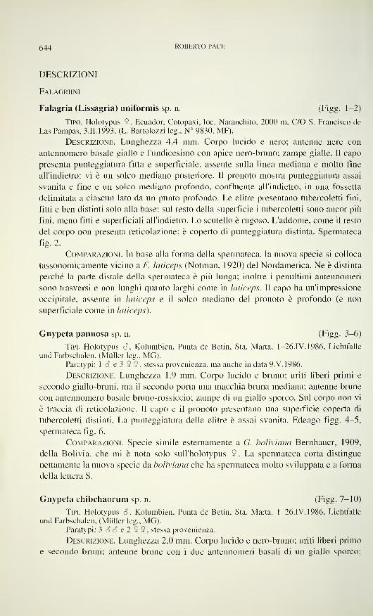
644 ROBERTO PACE
DESCRIZIONI
Falagriini
Falagria (Lissagria) uniformis sp. n. (Figg. 1-2)
Tipo. Holotypus 9, Ecuador, Cotopaxi, loc. Naranchito, 2000 m, C/O S. Francisco de
Las Pampas, 3.II.1993, (L. Bartolozzi leg., N° 9830, MF).
Descrizione. Lunghezza 4,4 mm. Corpo lucido e nero; antenne nere con
antennomero basale giallo e l'undicesimo con apice nero-bruno; zampe gialle. Il capo
presenta punteggiatura fitta e superficiale, assente sulla linea mediana e molto fine
all'indietro: vi è un solco mediano posteriore. Il pronoto mostra punteggiatura assai
svanita e fine e un solco mediano profondo, confluente all'indietro, in una fossetta
delimitata a ciascun lato da un punto profondo. Le elitre presentano tubercoletti fini,
fitti e ben distinti solo alla base: sul resto della superficie i tubercoletti sono ancor più
fini, meno fitti e superficiali all'indietro. Lo scutello è rugoso. L'addome, come il resto
del corpo non presenta reticolazione; è coperto di punteggiatura distinta. Spermateca
fig. 2.
Comparazioni. In base alla forma della spermateca, la nuova specie si colloca
tassonomicamente vicino a F. laticeps (Notman, 1920) del Nordamerica. Ne è distinta
perché la parte distale della spermateca è più lunga; inoltre i penultimi antennomeri
sono trasversi e non lunghi quanto larghi come in laticeps. Il capo ha un'impressione
occipitale, assente in laticeps e il solco mediano del pronoto è profondo (e non
superficiale come in laticeps).
Gnypeta pannosa sp. n. (Figg. 3-6)
Tipi. Holotypus 8 . Kolumbien. Punta de Betin, Sta. Marta, 1-26.IV. 1986, Lichtfalle
und Farbschalen, (Müller leg., MG).Paratypi: 1 8 e 3 9 9, stessa provenienza, ma anche in data 9.V. 1986.
Descrizione. Lunghezza 1,9 mm. Corpo lucido e bruno; uriti liberi primi e
secondo giallo-bruni, ma il secondo porta una macchia bruna mediana; antenne brune
con antennomero basale bruno-rossiccio; zampe di un giallo sporco. Sul corpo non vi
è traccia di reticolazione. Il capo e il pronoto presentano una superficie coperta di
tubercoletti distinti. La punteggiatura delle elitre è assai svanita. Edeago figg. 4-5,
spermateca fig. 6.
Comparazioni. Specie simile esternamente a G. boliviana Bernhauer, 1909,
della Bolivia, che mi è nota solo sull'holotypus 9 . La spermateca corta distingue
nettamente la nuova specie da boliviana che ha spermateca molto sviluppata e a forma
della lettera S.
Gnypeta chibchaorum sp. n. (Figg. 7-10)
Tipi. Holotypus ó\ Kolumbien. Punta de Betin. Sta. Marta, 1-26.IV. 1986, Lichtfalle
und Farbschalen, (Müller leg., MG).Paratypi: 3 8 8 e 2 9 9 , stessa provenienza.
Descrizione. Lunghezza 2,0 mm. Corpo lucido e nero-bruno; uriti liberi primo
e secondo bruni; antenne brune con i due antennomeri basali di un giallo sporco;
ALEOCHARINAE DELLA COLOMBIA E DELL ECUADOR 645
FlGG. 1-6
Habitus, spermateca ed edeago in visione laterale e ventrale. 1-2: Falagria (Lissagria) uni-
formis sp. n.; 3-6: Gnypeta pannosa sp. n.
646 ROBERTO PACE
zampe gialle. L'avancorpo è coperto di reticolazione distinta; l'addome è senza reti-
colazione. La punteggiatura o i tubercoletti dell'intero corpo sono indistinti. La fronte
è impressa. Edeago figg. 8-9, spermateca fig. 10.
Comparazioni. Specie differente da G. manda (Erichson, 1840) (comb, n.,
"olim" Falagria mundo. Er.), del Brasile, per avere gli antennomeri 4° a 6° molto
meno allungati e l'edeago meno sviluppato, con pezzi copulatori meno complessi di
quelli osservabili nell'edeago dell'holotypus di munda.
Etimologia. Dai Chibcha, popolazione colombiana stanziatasi all'interno della
Colombia al tempo della conquista spagnola.
Athetini
Cosmogastrusa gen. n. (Figg. 11-17)
Genere che si colloca tra i generi Aloconota Thomson, 1858 e Pelioptera
Kraatz, 1857. Tempie non marginate; palpi labiali di 3 articoli; ligula divisa in due
lembi triangolari, fig. 15; palpi mascellari di 4 articoli, fig. 14; mento con margine
anteriore appena arcuato all'indietro, fig 17; processo mesosternale largo all'apice che
è tronco, sicché le mesocoxe sono largamente separate tra loro; zampe anteriori
spinose al lato esterno, fig. 16; formula tarsale 4-5-5; primo tarsomero posteriore
corto; uroterghi liberi secondo a quinto impressi sulla linea mediana.
Typus generis: Cosmogastrusa curticornis sp. n.
Etimologia. Il nome del nuovo genere significa: "Colei che ha ornamenti
addominali'1
.
Comparazioni. Il nuovo genere presenta ligula simile a quella del generi
Aloconota Thomson e Pelioptera Kraatz, ma le tibie anteriori spinose, le antenne
cortissime, la pubescenza del pronoto diretta all'indietro, il primo tarsomero posteriore
corto e le impressioni mediane degli uroterghi permettono di distinguere il nuovo
genere dai due citati sopra.
Cosmogastrusa curticornis sp. n. (Figg. 1 1-17)
Tipo. Holotypus S, Kolumbien, Dept. Magdalena, Bahia de Nenguangue, Tayrona
Park, ca. 25 Km nörd-östl. Sta. Marta, 30.IX.1985, aus der Vegetation, (Müller leg., MG).
Descrizione. Lunghezza 2,8 mm. Corpo lucido e rossiccio; capo e uriti liberi
terzo, quarto e quinto bruno-rossicci; antenne brune con i tre antennomeri basali
giallo-rossicci; zampe giallo-rossicce. La reticolazione del capo e delle elitre è
estremamente svanita, quella del pronoto e dell'addome è assente. La punteggiatura
del capo è superficiale, quella del pronoto indistinta e quella delle elitre ben distinta.
La sutura delle elitre del maschio, posteriormente è sollevata a cercine. L'addome
presenta dal secondo al quinto degli uroterghi liberi una profonda impressione a fondo
concavo, reticolazione netta sul quinto urotergo libero e reticolazione vigorosa sul
sesto. Edeago figg. 12-13.&&•
Atheta (Chaetida) leda sp. n. (Figg. 18-19)
Tipo. Holotypus 9 , Kolumbien. Dept. Magdalena, nördl. Sierra Nevada de Sta. Marta,
ca. 2100 m, 24.VIII.1985, aus Moos und Streu. (Müller leg., MG).
ALEOCHARINAE DELLA COLOMBIA E DELL ECUADOR 647
Figo. 7-16
Habitus, edeago in visione laterale e ventrale, spermateca, labio con palpo labiale, maxilla conpalpo mascellare e protibia con protarsi. 7-10: Gnypeta chibchaorum sp. n.; 11-16: Cosmo-
gastrusa curticornis gen. n., sp. n.
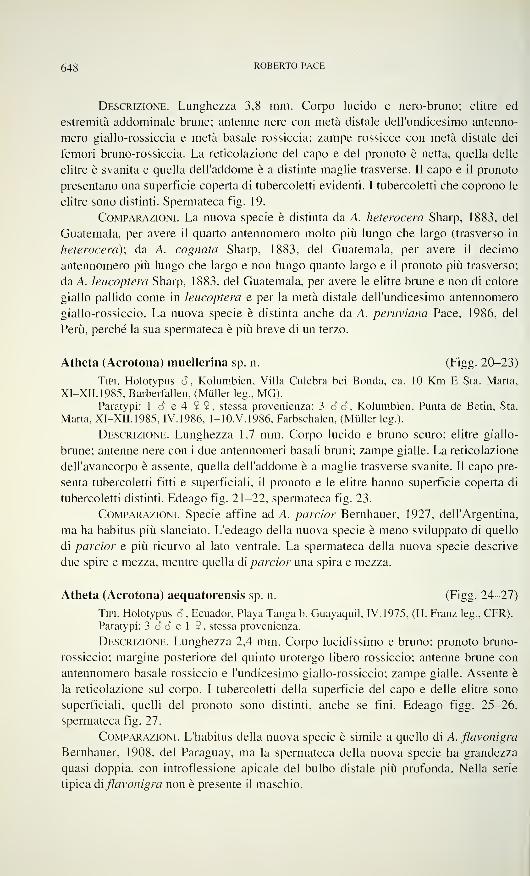
648 ROBERTO PACE
Descrizione. Lunghezza 3,8 mm. Corpo lucido e nero-bruno; elitre ed
estremità addominale brune; antenne nere con metà distale dell'undicesimo antenno-
mero giallo-rossiccia e metà basale rossiccia; zampe rossicce con metà distale dei
femori bruno-rossiccia. La reticolazione del capo e del pronoto è netta, quella delle
elitre è svanita e quella dell'addome è a distinte maglie trasverse. Il capo e il pronoto
presentano una superficie coperta di tubercoletti evidenti. I tubercoletti che coprono le
elitre sono distinti. Spermateca fig. 19.
Comparazioni. La nuova specie è distinta da A. heterocera Sharp, 1883, del
Guatemala, per avere il quarto antennomero molto più lungo che largo (trasverso in
heterocera); da A. cognata Sharp, 1883, del Guatemala, per avere il decimo
antennomero più lungo che largo e non lungo quanto largo e il pronoto più trasverso;
da A. leucoptera Sharp, 1883, del Guatemala, per avere le elitre brune e non di colore
giallo pallido come in leucoptera e per la metà distale dell'undicesimo antennomero
giallo-rossiccio. La nuova specie è distinta anche da A. peruviana Pace, 1986, del
Perù, perché la sua spermateca è più breve di un terzo.
Atheta (Acrotona) muellerina sp. n. (Figg. 20-23)
Tipi. Holotypus 6, Kolumbien, Villa Culebra bei Bonda, ca. 10 Km E Sta. Marta.
XI-XII.1985. Barberfallen. (Müller leg.. MG).Paratypi: 1 6 e 4 9 9, stessa provenienza; 3 3 3, Kolumbien. Punta de Betin, Sta.
Marta. XI-XII.1985, IV. 1986, 1-10.V.1986, Farbschalen, (Müllerleg.).
Descrizione. Lunghezza 1.7 mm. Corpo lucido e bruno scuro; elitre giallo-
brune; antenne nere con i due antennomeri basali bruni; zampe gialle. La reticolazione
dell'avancorpo è assente, quella dell'addome è a maglie trasverse svanite. Il capo pre-
senta tubercoletti fitti e superficiali, il pronoto e le elitre hanno superficie coperta di
tubercoletti distinti. Edeago fig. 21-22, spermateca fig. 23.
Comparazioni. Specie affine ad A. parcior Bernhauer, 1927, dell'Argentina,
ma ha habitus più slanciato. L'edeago della nuova specie è meno sviluppato di quello
di parcior e più ricurvo al lato ventrale. La spermateca della nuova specie descrive
due spire e mezza, mentre quella di parcior una spira e mezza.
Atheta (Acrotona) aequatorensis sp. n. (Figg. 24-27)
Tipi. Holotypus 3 , Ecuador, Playa Tanga b. Guayaquil, IV. 1975, (H. Franz leg., CFR).
Paratypi: 3 3 3 e 1 9, stessa provenienza.
Descrizione. Lunghezza 2,4 mm. Corpo lucidissimo e bruno; pronoto bruno-
rossiccio; margine posteriore del quinto urotergo libero rossiccio; antenne brune con
antennomero basale rossiccio e l'undicesimo giallo-rossiccio; zampe gialle. Assente è
la reticolazione sul corpo. I tubercoletti della superficie del capo e delle elitre sono
superficiali, quelli del pronoto sono distinti, anche se fini. Edeago figg. 25-26,
spermateca fig. 27.
Comparazioni. L'habitus della nuova specie è simile a quello di A. flavonigra
Bernhauer, 1908, del Paraguay, ma la spermateca della nuova specie ha grandezza
quasi doppia, con introflessione apicale del bulbo distale più profonda. Nella serie
tipica di flavonigra non è presente il maschio.
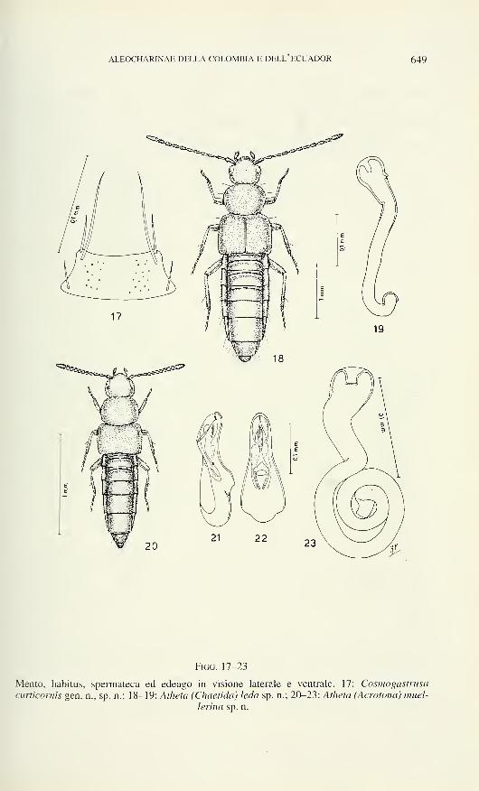
ALEOCHARINAE DELLA COLOMBIA E DELL ECUADOR 649
Figo. 17-23
Mento, habitus, spermateca ed edeago in visione laterale e ventrale. 17: Cosmogastrusa
curticornis gen. n., sp. n.; 18-19: Atheta (Chaetida) leda sp. n.; 20-23: Atheta (Acrotona) muel-
lerina sp. n.
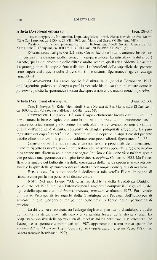
650 ROBERTO PACE
Atheta (Acrotona) omega sp. n. (Figg. 28-31)
Tipi. Holotypus 6, Kolumbien, Dept. Magdalena, nördl. Sierra Nevada de Sta. Marta,
Nähe San Lorenzo, ca. 2100 m, 21. Vili. 1985, aus Moos und Streu, (Müllerleg., MG).Paratypi: 1 $, stessa provenienza; 1 9, Kolumbien, Nördl. Sierra Nevada de Sta.
Marta, nähe El Campano, ca. 1000 m, aus Fall-Laub, 20.IV. 1986, (Müller leg.).
Descrizione. Lunghezza 2,2 mm. Corpo lucido e bruno; antenne brune con
undicesimo antennomero giallo-rossiccio; zampe rossicce. La reticolazione del capo è
assente, quella del pronoto e delle elitre è molto svanita, quella dell'addome è distinta.
La punteggiatura del capo è fitta e distinta. I tubercoletti della superficie del pronoto
sono superficiali, quelli delle elitre sono fini e distinti. Spermateca fig. 29, edeago
figg. 30-31.
Comparazioni. La nuova specie è distinta da A. parcior Bernhauer, 1927,
dell'Argentina, poiché ha edeago a profilo ventrale bisinuoso (e non arcuato come in
parcior) e poiché la spermateca mostra due spire e non una e mezza come in parcior.
Atheta (Acrotona) elvira sp. n. (Figg. 32-33)
Tipo. Holotypus 9, Kolumbien, nördl. Sierra Nevada de Sta. Marta, nähe El Campano,ca. 1000 m, 20.IV. 1986, aus Fall-Laub, (Müller leg., MG).
Descrizione. Lunghezza 1,8 mm. Corpo debolmente lucido e bruno; addome
nero, tranne la base e l'apice che sono bruni; antenne brune con antennomero basale
bruno-rossiccio; zampe giallo-brune. La reticolazione dell'avancorpo non è visibile,
quella dell'addome è distinta, composta di maglie poligonali irregolari. La pun-
teggiatura del capo è superficiale. I tubercoletti che coprono la superficie del pronoto
e delle elitre sono svaniti, quelli dell'addome sono poco salienti. Spermateca fig. 33.
Comparazioni. La nuova specie, avendo le spire prossimali della spermateca
invertite rispetto la norma, non è comparabile con nessuna specie della regione neotro-
pica tranne una discussa nella nota che segue. In Cina e Giappone vive un'altra specie
che possiede una spermateca con spire invertite: A. neglecta Cameron, 1933. Ma l'intro-
flessione apicale del bulbo distale della spermateca della nuova specie è molto più pro-
fonda e la spira della spermateca stessa è stretta e non ampia come quella di neglecta.
Etimologia. La nuova specie è dedicata a mia sorella Elvira, in segno di
riconoscenza per la sua generosità disinteressata.
Nota. Nel mio lavoro "Aleocharinae dell'Isola della Guadalupa (Antille)"
pubblicato del 1987 in "Folia Entomologica Hungarica,?
compare il disegno dell'ede-
ago e della spermateca di Atheta (Acrotona) parcior Bernhauer, 1927. Pur avendo
comparato l'edeago di tre maschi della Guadalupa con l'edeago dell'holotypus di
parcior, in quel periodo di tempo non conoscevo la forma della spermateca di
parcior.
La differenza riscontrata tra l'edeago degli esemplari della Guadalupa e quella
dell'holotypus di parcior l'attribuivo a variabilità locale della stessa specie. La
scoperta successiva della spermateca di parcior, mi ha permesso di riconoscere che
l'edeago e la spermateca pubblicati nel 1987, appartengono a una nuova specie che
nomino Atheta (Acrotona) semilacera sp. n. (Atheta parcior, sensu Pace, 1987, nec
Atheta parcior Bernhauer 1927).
ALEOCHARINAE DELLA COLOMBIA E DELL ECUADOR 651
Figo. 24-31
Habitus, edeago in visione laterale e ventrale e spermateca. 24-27: Atheta (Acrotona)
aequatorensis sp. n.; 28-31: Atheta (Acrotona) omega sp. n.
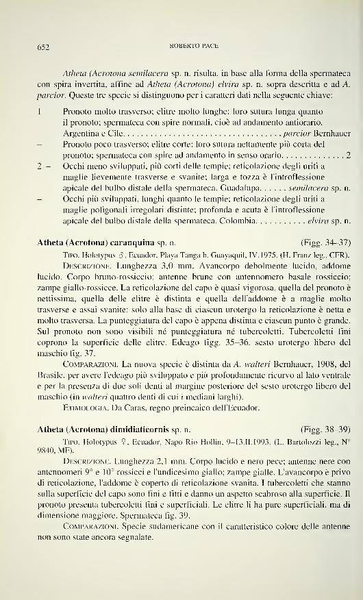
652 ROBERTO PACE
Atheta (Acrotona semilacera sp. n. risulta, in base alla forma della spermateca
con spira invertita, affine ad Atheta (Acrotona) elvira sp. n. sopra descritta e ad A.
parcior. Queste tre specie si distinguono per i caratteri dati nella seguente chiave:
1 - Pronoto molto trasverso; elitre molto lunghe: loro sutura lunga quanto
il pronoto; spermateca con spire normali, cioè ad andamento antiorario.
Argentina e Cile parcior Bernhauer
Pronoto poco trasverso; elitre corte: loro sutura nettamente più corta del
pronoto; spermateca con spire ad andamento in senso orario 2
2 - Occhi meno sviluppati, più corti delle tempie; reticolazione degli uriti a
maglie lievemente trasverse e svanite; larga e tozza è l'introflessione
apicale del bulbo distale della spermateca. Guadalupa semilacera sp. n.
Occhi più sviluppati, lunghi quanto le tempie; reticolazione degli uriti a
maglie poligonali irregolari distinte; profonda e acuta è l'introflessione
apicale del bulbo distale della spermateca. Colombia elvira sp. n.
Atheta (Acrotona) caranquina sp. n. (Figg. 34-37)
Tipo. Holotypus 8, Ecuador, Playa Tanga b. Guayaquil, IV. 1975, (H. Franz leg., CFR).
Descrizione. Lunghezza 3,0 mm. Avancorpo debolmente lucido, addome
lucido. Corpo bruno-rossiccio; antenne brune con antennomero basale rossiccio;
zampe giallo-rossicce. La reticolazione del capo è quasi vigorosa, quella del pronoto è
nettissima, quella delle elitre è distinta e quella dell'addome è a maglie molto
trasverse e assai svanite: solo alla base di ciascun urotergo la reticolazione è netta e
molto trasversa. La punteggiatura del capo è appena distinta e ciascun punto è grande.
Sul pronoto non sono visibili né punteggiatura né tubercoletti. Tubercoletti fini
coprono la superficie delle elitre. Edeago figg. 35-36, sesto urotergo libero del
maschio fig. 37.
Comparazioni. La nuova specie è distinta da A. walteri Bernhauer, 1908, del
Brasile, per avere l'edeago più sviluppato e più profondamente ricurvo al lato ventrale
e per la presenza di due soli denti al margine posteriore del sesto urotergo libero del
maschio (in walteri quattro denti di cui i mediani larghi).
Etimologia. Da Caras, regno preincaico dell'Ecuador.
Atheta (Acrotona) dimidiaticornis sp. n. (Figg. 38-39)
Tipo. Holotypus 9. Ecuador, Napo Rio Hollin, 9-13.II. 1993, (L. Bartolozzi leg., N°9840, MF).
Descrizione. Lunghezza 2,1 mm. Corpo lucido e nero pece; antenne nere con
antennomeri 9° e 10° rossicci e l'undicesimo giallo; zampe gialle. L'avancorpo è privo
di reticolazione, l'addome è coperto di reticolazione svanita. I tubercoletti che stanno
sulla superficie del capo sono fini e fitti e danno un aspetto scabroso alla superficie. Il
pronoto presenta tubercoletti fini e superficiali. Le elitre li ha pure superficiali, ma di
dimensione maggiore. Spermateca fig. 39.
Comparazioni. Specie sudamericane con il caratteristico colore delle antenne
non sono state ancora segnalate.
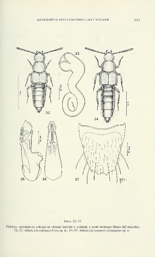
ALEOCHARINAE DELLA COLOMBIA E DELL ECUADOR 653
Figg. 32-37
Habitus, spermateca, edeago in visione laterale e ventrale e sesto urotergo libero del maschio.
32-33: Atheta (Acrotona) elvira sp. n.; 34-37: Atheta (Acrotona) caranquina sp. n.
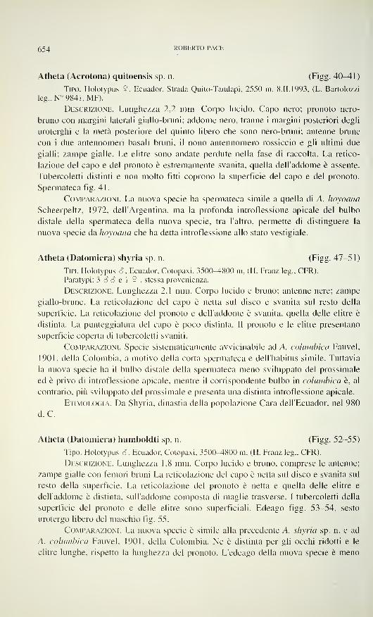
654 ROBERTO PACE
Atheta (Acrotona) quitoensis sp. n. (Figg. 4CMU)
Tipo. Holotypus 9, Ecuador, Strada Quito-Tandapi, 2550 m, 8.II.1993, (L. Bartolozzi
leg., N° 9841, MF).
Descrizione. Lunghezza 2,2 mm. Corpo lucido. Capo nero; pronoto nero-
bruno con margini laterali giallo-bruni; addome nero, tranne i margini posteriori degli
uroterghi e la metà posteriore del quinto libero che sono nero-bruni; antenne brune
con i due antennomeri basali bruni, il nono antennomero rossiccio e gli ultimi due
gialli; zampe gialle. Le elitre sono andate perdute nella fase di raccolta. La retico-
lazione del capo e del pronoto è estremamente svanita, quella dell'addome è assente.
Tubercoletti distinti e non molto fitti coprono la superficie del capo e del pronoto.
Spermateca fig. 41.
Comparazioni. La nuova specie ha spermateca simile a quella di A. hoyoana
Scheerpeltz, 1972, dell'Argentina, ma la profonda introflessione apicale del bulbo
distale della spermateca della nuova specie, tra l'altro, permette di distinguere la
nuova specie da hoyoana che ha detta introflessione allo stato vestigiale.
Atheta (Datomicra) shyria sp. n. (Figg. 47-51)
Tipi. Holotypus 6, Ecuador, Cotopaxi, 350CM800 m, (H. Franz leg., CFR).
Paratypi: 3 6 S e 1 9 , stessa provenienza.
Descrizione. Lunghezza 2,1 mm. Corpo lucido e bruno; antenne nere; zampe
giallo-brune. La reticolazione del capo è netta sul disco e svanita sul resto della
superficie. La reticolazione del pronoto e dell'addome è svanita, quella delle elitre è
distinta. La punteggiatura del capo è poco distinta. Il pronoto e le elitre presentano
superficie coperta di tubercoletti svaniti.
Comparazioni. Specie sistematicamente avvicinabile ad A. columbica Fauvel,
1901, della Colombia, a motivo della corta spermateca e dell'habitus simile. Tuttavia
la nuova specie ha il bulbo distale della spermateca meno sviluppato del prossimale
ed è privo di introflessione apicale, mentre il corrispondente bulbo in columbica è, al
contrario, più sviluppato del prossimale e presenta una distinta introflessione apicale.
Etimologia. Da Shyria, dinastia della popolazione Cara dell'Ecuador, nel 980
d. C.
Atheta (Datomicra) humboldti sp. n. (Figg. 52-55)
Tipo. Holotypus 3, Ecuador, Cotopaxi, 3500-4800 m, (H. Franz leg., CFR).
Descrizione. Lunghezza 1,8 mm. Corpo lucido e bruno, comprese le antenne;
zampe gialle con femori bruni La reticolazione del capo è netta sul disco e svanita sul
resto della superficie. La reticolazione del pronoto è netta e quella delle elitre e
dell'addome è distinta, sull'addome composta di maglie trasverse. I tubercoletti della
superficie del pronoto e delle elitre sono superficiali. Edeago figg. 53-54, sesto
urotergo libero del maschio fig. 55.
Comparazioni. La nuova specie è simile alla precedente A. shyria sp. n. e ad
A. columbica Fauvel, 1901, della Colombia. Ne è distinta per gli occhi ridotti e le
elitre lunghe, rispetto la lunghezza del pronoto. L'edeago della nuova specie è meno
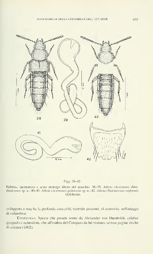
ALEOCHARINAE DELLA COLOMBIA E DELL'ECUADOR 655
Figg. 38-42
Habitus, spermateca e sesto urotergo libero del maschio. 38-39: Atheta (Acrotona) dimi-
diaticornis sp. n.; 40-41: Atheta (Acrotona) quitoensis sp. n.; 42: Atheta (Datomicra) conformis
(Erichson).
sviluppato e non ha la profonda concavità ventrale presente, al contrario, nell'edeago
di columbica.
Etimologia. Specie che prende nome da Alexander von Humboldt, celebre
geografo e naturalista, che all'ombra del Cotopaxi da lui visitato, scrisse pagine ricche
di scienza (1802).
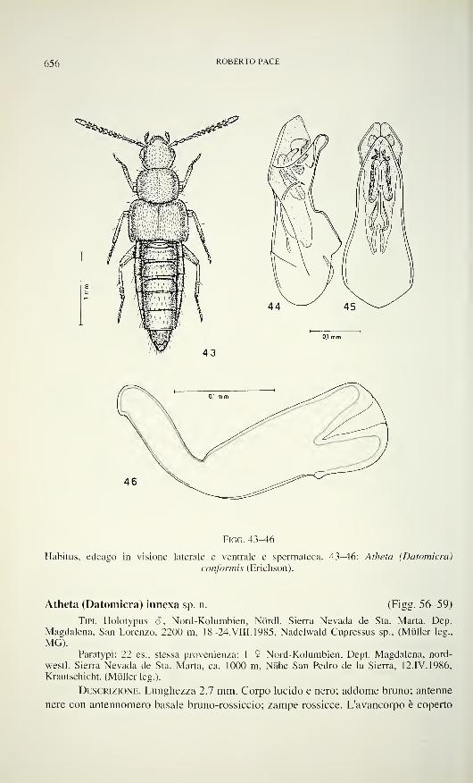
656 ROBERTO PACE
Figo. 43-46
Habitus, edeago in visione laterale e ventrale e spermateca. 43-46: Atheta (Datomicra)
conformis (Erichson).
Atheta (Datomicra) innexa sp. n. (Figg. 56-59)
Tipi. Holotypus S, Nord-Kolumbien, Nördl. Sierra Nevada de Sta. Marta, Dep.
Magdalena, San Lorenzo. 2200 m, 18-24.VIII.1985, Nadelwald Cupressus sp., (Müller leg.,
MG).Paratypi: 22 es., stessa provenienza; 1 $ Nord-Kolumbien, Dept. Magdalena, nord-
westl. Sierra Nevada de Sta. Marta, ca. 1000 m, Nähe San Pedro de la Sierra, 12.IV. 1986,
Krautschicht, (Müller leg.).
Descrizione. Lunghezza 2,7 mm. Corpo lucido e nero; addome bruno; antenne
nere con antennomero basale bruno-rossiccio; zampe rossicce. L'avancorpo è coperto
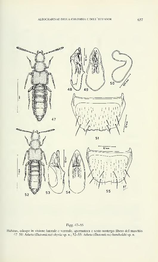
ALEOCHARINAE DELLA COLOMBIA E DELL ECUADOR 657
Figg. 47-55
Habitus, edeago in visione laterale e ventrale, spermateca e sesto urotergo libero del maschio.
47-51: Atheta (Datomicra) shyria sp. n.; 52-55: Atheta (Datomicra) humboldtì sp. n.
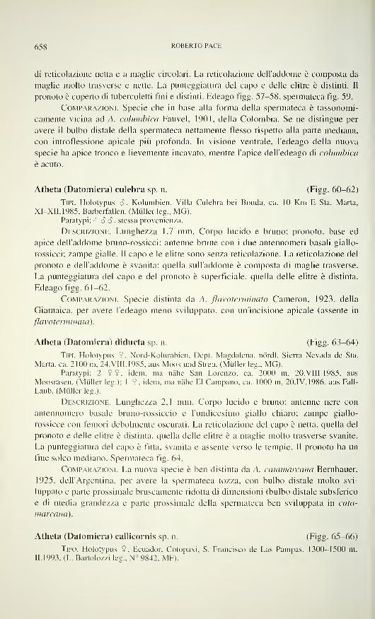
658 ROBERTO PACE
di reticolazione netta e a maglie circolari. La reticolazione dell'addome è composta da
maglie molto trasverse e nette. La punteggiatura del capo e delle elitre è distinti. Il
pronoto è coperto di tubercoletti fini e distinti. Edeago figg. 57-58, spermateca fig. 59.
Comparazioni. Specie che in base alla forma della spermateca è tassonomi-
camente vicina ad A. columbica Fauvel, 1901, della Colombia. Se ne distingue per
avere il bulbo distale della spermateca nettamente flesso rispetto alla parte mediana,
con introflessione apicale più profonda. In visione ventrale, l'edeago della nuova
specie ha apice tronco e lievemente incavato, mentre l'apice dell'edeago di columbica
è acuto.
Atheta (Datomicra) culebra sp. n. (Figg. 60-62)
Tipi. Holotypus 6, Kolumbien, Villa Culebra bei Bonda, ca. 10 Km E Sta. Marta,
XI-XII.1985, Barberfallen, (Müllerleg., MG).Paratypi: 4 S <?, stessa provenienza.
Descrizione. Lunghezza 1,7 mm. Corpo lucido e bruno; pronoto, base ed
apice dell'addome bruno-rossicci; antenne brune con i due antennomeri basali giallo-
rossicci; zampe gialle. Il capo e le elitre sono senza reticolazione. La reticolazione del
pronoto e dell'addome è svanita: quella sull'addome è composta di maglie trasverse.
La punteggiatura del capo e del pronoto è superficiale, quella delle elitre è distinta.
Edeago figg. 61-62.
Comparazioni. Specie distinta da A. flàvoterminata Cameron, 1923, della
Giamaica, per avere l'edeago meno sviluppato, con un'incisione apicale (assente in
flàvoterminata).
Atheta (Datomicra) diducta sp. n. (Figg. 63-64)
Tipi. Holotypus 5 , Nord-Kolumbien, Dept. Magdalena, nördl. Sierra Nevada de Sta.
Marta, ca. 2100 m, 24.VIII.1985, aus Moos und Streu, (Müller leg., MG).Paratypi: 2 9 9, idem, ma nähe San Lorenzo, ca. 2000 m, 20. VIII. 1985, aus
Moosrasen, (Müller leg.); 1 9. idem, ma nähe El Campano, ca. 1000 m, 20. IV. 1986, aus Fall-
Laub, (Müllerleg.).
Descrizione. Lunghezza 2,1 mm. Corpo lucido e bruno; antenne nere con
antennomero basale bruno-rossiccio e l'undicesimo giallo chiaro; zampe giallo-
rossicce con femori debolmente oscurati. La reticolazione del capo è netta, quella del
pronoto e delle elitre è distinta, quella delle elitre è a maglie molto trasverse svanite.
La punteggiatura del capo è fitta, svanita e assente verso le tempie. Il pronoto ha un
fine solco mediano. Spermateca fig. 64.
Comparazioni. La nuova specie è ben distinta da A. catamarcana Bernhauer,
1925, dell'Argentina, per avere la spermateca tozza, con bulbo distale molto svi-
luppato e parte prossimale bruscamente ridotta di dimensioni (bulbo distale subsferico
e di media grandezza e parte prossimale della spermateca ben sviluppata in cata-
marcana).
Atheta (Datomicra) callicornis sp. n. (Figg. 65-66)
Tipo. Holotypus 9, Ecuador, Cotopaxi. S. Francisco de Las Pampas, 1300-1500 m,
11.1993. (L. Bartolozzi leg., N° 9842, MF).
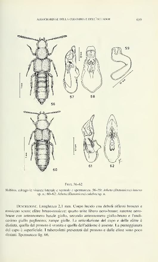
ALEOCHARINAE DELLA COLOMBIA E DELL ECUADOR 659
Figo. 56-62
Habitus, edeago in visione laterale e ventrale e spermateca. 56-59: Atheta (Datomicra) innexa
sp. n.; 60-62: Atheta (Datomicra) culebra sp. n.
Descrizione. Lunghezza 2,1 mm. Corpo lucido con deboli riflessi bronzei e
rossiccio scuro; elitre bruno-rossicce; quarto urite libero nero-bruno; antenne nero-
brune con antennomero basale giallo, secondo antennomero giallo-bruno e l'undi-
cesimo giallo paglierino; zampe gialle. La reticolazione del capo e delle elitre è
distinta, quella del pronoto è svanita e quella dell'addome è assente. La punteggiatura
del capo è superficiale. I tubercoletti presentati dal pronoto e dalle elitre sono poco
distinti. Spermateca fig. 66.
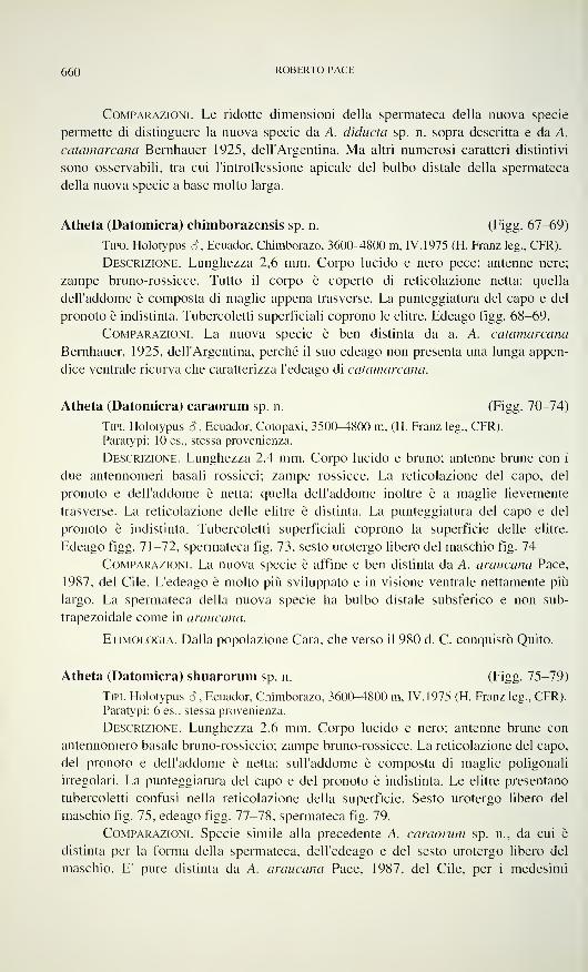
660 ROBERTO PACE
Comparazioni. Le ridotte dimensioni della spermateca della nuova specie
permette di distinguere la nuova specie da A. diducta sp. n. sopra descritta e da A.
catamarcana Bernhauer 1925, dell'Argentina. Ma altri numerosi caratteri distintivi
sono osservabili, tra cui l'introflessione apicale del bulbo distale della spermateca
della nuova specie a base molto larga.
Atheta (Datomicra) chimborazensis sp. n. (Figg. 67-69)
Tipo. Holotypus e?, Ecuador, Chimborazo, 3600-4800 m, IV. 1975 (H. Franz leg., CFR).
Descrizione. Lunghezza 2,6 mm. Corpo lucido e nero pece; antenne nere;
zampe bruno-rossicce. Tutto il corpo è coperto di reticolazione netta: quella
dell'addome è composta di maglie appena trasverse. La punteggiatura del capo e del
pronoto è indistinta. Tubercoletti superficiali coprono le elitre. Edeago figg. 68-69.
Comparazioni. La nuova specie è ben distinta da a. A. catamarcana
Bernhauer, 1925, dell'Argentina, perché il suo edeago non presenta una lunga appen-
dice ventrale ricurva che caratterizza l'edeago di catamarcana.
Atheta (Datomicra) caraorum sp. n. (Figg. 70-74)
Tipi. Holotypus e?, Ecuador, Cotopaxi, 3500-4800 m, (H. Franz leg., CFR).
Paratypi: 10 es., stessa provenienza.
Descrizione. Lunghezza 2,4 mm. Corpo lucido e bruno; antenne brune con i
due antennomeri basali rossicci; zampe rossicce. La reticolazione del capo, del
pronoto e dell'addome è netta: quella dell'addome inoltre è a maglie lievemente
trasverse. La reticolazione delle elitre è distinta. La punteggiatura del capo e del
pronoto è indistinta. Tubercoletti superficiali coprono la superficie delle elitre.
Edeago figg. 71-72, spermateca fig. 73, sesto urotergo libero del maschio fig. 74
Comparazioni. La nuova specie è affine e ben distinta da A. araucana Pace,
1987, del Cile. L'edeago è molto più sviluppato e in visione ventrale nettamente più
largo. La spermateca della nuova specie ha bulbo distale subsferico e non sub-
trapezoidale come in araucana.
Etimologia. Dalla popolazione Cara, che verso il 980 d. C. conquistò Quito.
Atheta (Datomicra) shuarorum sp. n. (Figg. 75-79)
Tipi. Holotypus ó\ Ecuador, Chimborazo, 3600-4800 m, IV. 1975 (H. Franz leg., CFR).
Paratypi: 6 es., stessa provenienza.
Descrizione. Lunghezza 2,6 mm. Corpo lucido e nero; antenne brune con
antennomero basale bruno-rossiccio; zampe bruno-rossicce. La reticolazione del capo,
del pronoto e dell'addome è netta: sull'addome è composta di maglie poligonali
irregolari. La punteggiatura del capo e del pronoto è indistinta. Le elitre presentano
tubercoletti confusi nella reticolazione della superficie. Sesto urotergo libero del
maschio fig. 75, edeago figg. 77-78, spermateca fig. 79.
Comparazioni. Specie simile alla precedente A. caraorum sp. n., da cui è
distinta per la forma della spermateca, dell'edeago e del sesto urotergo libero del
maschio. E' pure distinta da A. araucana Pace, 1987, del Cile, per i medesimi
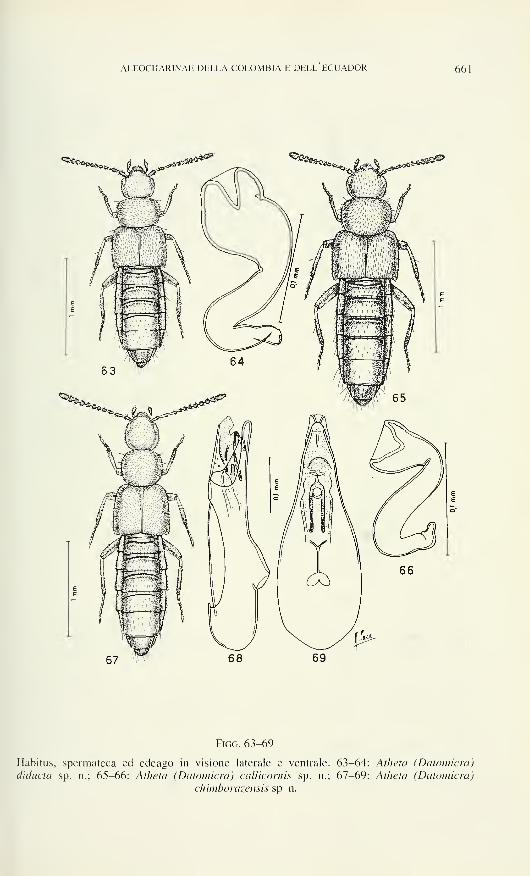
ALEOCHARINAE DELLA COLOMBIA E DELL ECUADOR 661
Figg. 63-69
Habitus, spermateca ed edeago in visione laterale e ventrale. 63-64: Atheta (Datomicra)
diducta sp. n.; 65-66: Atheta (Datomicra) callicornis sp. n.; 67-69: Atheta (Datomicra)
chimborazensis sp. n.
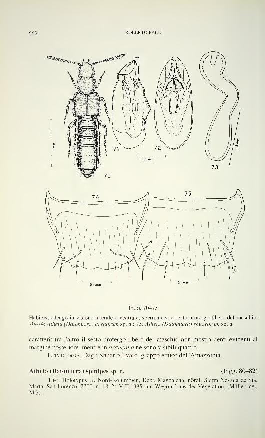
662 ROBERTO PACE
Figo. 70-75
Habitus, edeago in visione laterale e ventrale, spermateca e sesto urotergo libero del maschio.
70-74: Atheta (Datomicra) caraorum sp. n.; 75: Allieta (Datomicra) shuarorum sp. n.
caratteri: tra l'altro il sesto urotergo libero del maschio non mostra denti evidenti al
margine posteriore, mentre in araucaria ne sono visibili quattro.
Etimologia. Dagli Shuar o Jivaro, gruppo etnico dell'Amazzonia.
Atheta (Datomicra) spinipes sp. n. (Figg. 80-82)
Tipo. Holotypus S, Nord-Kolumbien, Dept. Magdalena, nördl. Sierra Nevada de Sta.
Marta, San Lorenzo, 2200 m. 18-24.VIII.1985. am Wegrand aus der Vegetation, (Müller leg.,
MG).
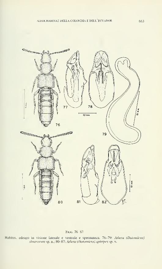
ALEOCHARINAE DELLA COLOMBIA E DELL ECUADOR 663
Figo. 76-82
Habitus, edeago in visione laterale e ventrale e spermateca. 76-79: Atheta (Datomìcra)
shuarorum sp. n.; 80-82: Atheta (Datomìcra) spinipes sp. n.
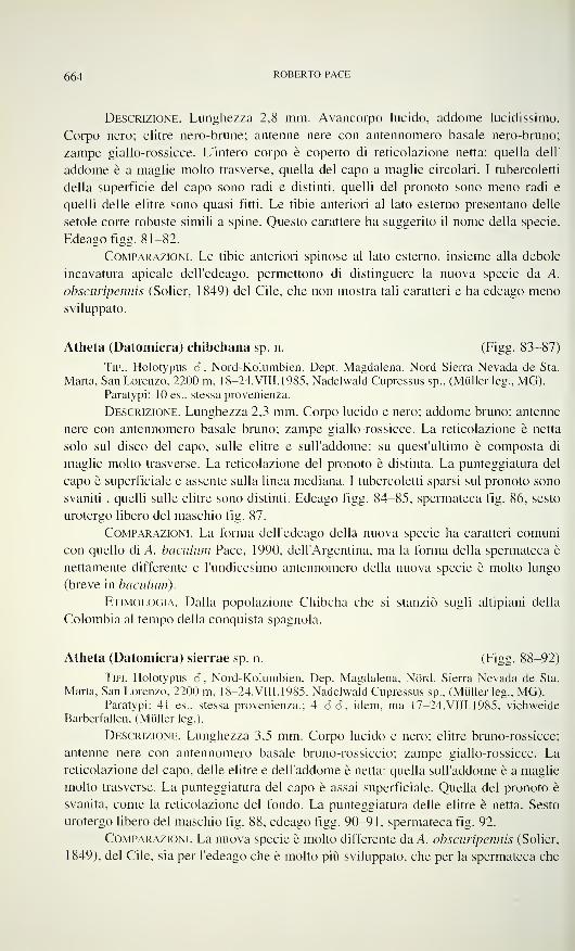
664 ROBERTO PACE
Descrizione. Lunghezza 2,8 mm. Avancorpo lucido, addome lucidissimo.
Corpo nero; elitre nero-brune; antenne nere con antennomero basale nero-bruno;
zampe giallo-rossicce. L'intero corpo è coperto di reticolazione netta: quella dell'
addome è a maglie molto trasverse, quella del capo a maglie circolari. I tubercoletti
della superficie del capo sono radi e distinti, quelli del pronoto sono meno radi e
quelli delle elitre sono quasi fitti. Le tibie anteriori al lato esterno presentano delle
setole corte robuste simili a spine. Questo carattere ha suggerito il nome della specie.
Edeagofigg. 81-82.
Comparazioni. Le tibie anteriori spinose al lato esterno, insieme alla debole
incavatura apicale dell'edeago, permettono di distinguere la nuova specie da A.
obscuripennis (Solier, 1849) del Cile, che non mostra tali caratteri e ha edeago meno
sviluppato.
Atheta (Datomicra) chibchana sp. n. (Figg. 83-87)
Tipi. Holotypus 3, Nord-Kolumbien, Dept. Magdalena, Nord Sierra Nevada de Sta.
Marta, San Lorenzo, 2200 m, 18-24.VIII.1985, Nadelwald Cupressus sp., (Müller leg., MG).Paratypi: 10 es., stessa provenienza.
Descrizione. Lunghezza 2,3 mm. Corpo lucido e nero; addome bruno; antenne
nere con antennomero basale bruno; zampe giallo-rossicce. La reticolazione è netta
solo sul disco del capo, sulle elitre e sull'addome: su quest'ultimo è composta di
maglie molto trasverse. La reticolazione del pronoto è distinta. La punteggiatura del
capo è superficiale e assente sulla linea mediana. I tubercoletti sparsi sul pronoto sono
svaniti , quelli sulle elitre sono distinti. Edeago figg. 84-85, spermateca fig. 86, sesto
urotergo libero del maschio fig. 87.
Comparazioni. La forma dell'edeago della nuova specie ha caratteri comuni
con quello di A. baculum Pace. 1990, dell'Argentina, ma la forma della spermateca è
nettamente differente e l'undicesimo antennomero della nuova specie è molto lungo
(breve in baculum).
Etimologia. Dalla popolazione Chibcha che si stanziò sugli altipiani della
Colombia al tempo della conquista spagnola.
Atheta (Datomicra) sierrae sp. n. (Figg. 88-92)
Tipi. Holotypus 3, Nord-Kolumbien, Dep. Magdalena, Nord. Sierra Nevada de Sta.
Marta, San Lorenzo, 2200 m, 18-24.VIII.1985, Nadelwald Cupressus sp., (Müller leg., MG).Paratypi: 41 es., stessa provenienza.; 4 3 3. idem, ma 17-24.VIII.1985, Viehweide
Barberfallen. (Müller leg.).
Descrizione. Lunghezza 3,5 mm. Corpo lucido e nero; elitre bruno-rossicce;
antenne nere con antennomero basale bruno-rossiccio; zampe giallo-rossicce. La
reticolazione del capo, delle elitre e dell'addome è netta: quella sull'addome è a maglie
molto trasverse. La punteggiatura del capo è assai superficiale. Quella del pronoto è
svanita, come la reticolazione del fondo. La punteggiatura delle elitre è netta. Sesto
urotergo libero del maschio fig. 88, edeago figg. 90-91, spermateca fig. 92.
Comparazioni. La nuova specie è molto differente da A. obscuripennis (Solier,
1849), del Cile, sia per l'edeago che è molto più sviluppato, che per la spermateca che
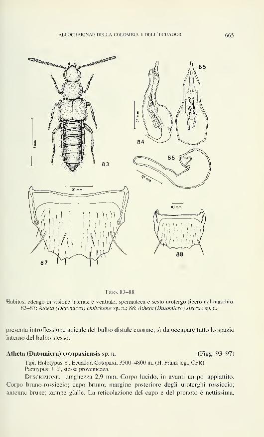
ALEOCHARINAE DELLA COLOMBIA E DELL ECUADOR 665
Figo. 83-88
Habitus, edeago in visione laterale e ventrale, spermateca e sesto urotergo libero del maschio.
83-87: Atheta (Datomicra) chibchana sp. n.; 88: Atheta (Datomicra) sierrae sp. n.
presenta introflessione apicale del bulbo distale enorme, sì da occupare tutto lo spazio
interno del bulbo stesso.
Atheta (Datomicra) cotopaxiensis sp. n. (Figg. 93-97)
Tipi. Holotypus ô , Ecuador, Cotopaxi, 3500-4800 m, (H. Franz leg., CFR).Paratypus: 1 9 , stessa provenienza.
Descrizione. Lunghezza 2,9 mm. Corpo lucido, in avanti un po' appiattito.
Corpo bruno-rossiccio; capo bruno; margine posteriore degli uroterghi rossiccio;
antenne brune; zampe gialle. La reticolazione del capo e del pronoto è nettissima,
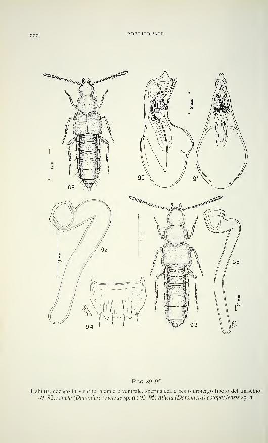
666 ROBERTO PACE
Figo. 89-95
Habitus, edeago in visione laterale e ventrale, spermateca e sesto urotergo libero del maschio.
89-92: Atheta (Datomicra) sierrae sp. n.; 93-95: Atheta (Datomicra) cotopaxiensis sp. n.
ALEOCHARINAE DELLA COLOMBIA E DELL ECUADOR 667
quella delle elitre e dell'addome è netta: su quest'ultimo è a maglie trasverse. La
punteggiatura dell'avancorpo è estremamente svanita: sul capo è assente sulla linea
mediana. Sesto urotergo libero del maschio fig. 94, spermateca fig 95, edeago figg.
96-97.
Comparazioni. L'edeago della nuova specie presenta nel sacco interno
robustissimi pezzi copulatori e la spermateca è molto sviluppata in lunghezza. Questi
caratteri sono sufficienti a distinguere la nuova specie da A. sierrae sp. n. sopra
descritta, a cui è affine, e da A. obscuripennis (Solier, 1849) del Cile che è tassono-
micamente meno vicina.
Atheta (Datomicra) nevadicola sp. n. (Figg. 98-102)
Tipi. Holotypus 6 , Nord-Kolumbien, Dep. Magdalena, Nord. Sierra Nevada de Sta.
Marta, San Lorenzo, 2200 m, 18-24.VIII.1985, Nadelwald Cupressus sp., (Müller leg., MG).Paratypi: 20 es., stessa provenienza, manche "am Wegrand aus Moos und Streu".
Descrizione. Lunghezza 3,7 mm. Corpo lucido e nero; margine posteriore
degli uroterghi bruno-rossiccio; antenne nere con antennomero basale bruno-
rossiccio; zampe giallo-rossicce. La reticolazione del capo e dell'addome è netta,
quella del pronoto e delle elitre è nettissima. La punteggiatura del capo è svanita,
quella del pronoto è fine e distinta e quella delle elitre è poco distinta. Edeago figg.
99-100, spermateca fig. 101, sesto urotergo libero del maschio fig. 102.
Comparazioni. La nuova specie mostra affinità con A. catamarcana Pace,
1990, dell'Argentina, ma l'edeago della nuova specie è più tozzo, con bulbo basale
molto largo, in visione ventrale. La spermateca della nuova specie ha parte prossimale
molto più ricurva della medesima parte della spermateca di catamarcana.
Atheta (Datomicra) magdaleniensis sp. n. (Figg. 103-106)
Tipo. Holotypus a, Nord-Kolumbien, Nord. Sierra Nevada de Sta. Marta, Dep.
Magdalena, San Lorenzo, 2200 m, 18-24.VIII.1985, Nadelwald, Cupressus sp., (Müller leg.,
MG).
Descrizione. Lunghezza 2,8 mm. Corpo lucido e nero; elitre giallo-brune;
estremità addominale bruno-rossiccia; antenne interamente nere; zampe giallo-
rossicce. La reticolazione del capo, del pronoto e dell'addome è netta: sull'addome in
più è a maglie molto tresverse. La reticolazione delle elitre è vigorosa. La pun-
teggiatura del capo è quasi indistinta. Il pronoto presenta una superficie coperta di
tubercoletti superfieli; le elitre presentano ubercoletti salienti e fitti, con una pubes-
cenza corta e fitta. Edeago figg. 104-105, sesto urotergo libero del maschio fig. 106.
Comparazioni. La forma dell'edeago e i caratteri del margine posteriore del
sesto urotergo libero del maschio, potrebbero indicare un'affinità tassonomica della
nuova specie con A. obscuripennis (Solier, 1849), del Cile. Tuttavia l'edeago della
nuova specie è maggiormente ricurvo al lato ventrale, con pezzi copulatori del sacco
interno più sviluppati. Inoltre l'incavatura mediana del sesto urotergo libero del
maschio è più ampia e il decimo antennomero è debolmente trasverso (molto
trasverso in obscuripennis).
668 ROBERTO PACE
Figo. 96-102
Edeago in visione laterale e ventrale, habitus, spermateca e sesto urotergo libero del maschio.
96-97: Atheta (Datomicra) cotopaxiensis sp. n.; 98-102: Atheta (Datomicra) nevadicola sp. n.
ALEOCHARINAE DELLA COLOMBIA E DELL ECUADOR 669
Atheta (Datomicra) hollinensis sp. n. (Figg. 107-108)
Tipo. Holotypus 9, Ecuador, Napo Rio Hollin, 1200 m, 9-1 3.II. 1993, (L. Bartolozzi
leg.. N° 9845, MF).
Descrizione. Lunghezza 2,1 mm. Corpo lucido e nero; antenne nere con
antennomero basale giallo sporco e secondo antennomero nero-bruno; zampe gialle.
La reticolazione dell'avancorpo è distinta, quella dell'addome è a maglie molto
trasverse e svanite. I tubercoletti della superficie del capo sono svaniti e assenti sulla
linea mediana; quelli del pronoto e delle elitre sono poco distinti. Spermateca fig. 108.
Comparazioni. La caratteristica forma della spermateca, a tubulo sottile e
piegata come nella lettera Z, distingue nettamente la nuova specie da A. araucana
Pace, 1987. del Cile, che ha spermateca con tubulo di calibro maggiore ed è foggiata a
forma della lettera S allungata.
Atheta (Datomicra) spirarum sp. n. (Figg. 109-1 10)
Tipo. Holotypus 2 , Nord-Kolumbien, Sierra Nevada de Sta. Marta, San Lorenzo, 2200
m. 20.IV. 1986, aus Fallaub und Moos, (Müller leg., MG).
Descrizione. Lunghezza 4,0 mm. Corpo lucidissimo e nero; elitre giallo-
brunicce: antenne brune con antennomero basale giallo-rossiccio; zampe gialle. La
reticolazione del capo e delle elitre è distinta, quella del pronoto e dell'addome è
molto svanita: quella dell'addome è composta di maglie molto trasverse. La
punteggiatura del capo è svanita: ciascun punto è grande. Tubercoletti fini e distinti
coprono il pronoto. La punteggiatura delle elitre è distinta. Spermateca fig. 110.
Comparazioni. La grande taglia corporea e la forma caratteristica della sper-
mateca, con parte prossimale avvolta in quattros spire, sono caratteri distintivi unici
nell'ambito del sottogenere.
Atheta (Datomicra) chibcha sp. n. (Figg. 111-1 15)
Tipi. Holotypus 6. Kolumbien, Villa Culebra bei Bonda, ca. 10 Km E Sta. Marta,
XI-XII.1985, Barberfallen, (Müller leg., MG).Paratypus: 1 9 . stessa provenienza.
Descrizione. Lunghezza 1,8 mm. Corpo lucido. Capo, pronoto, meta
posteriore delle elitre, uriti liberi terzo e quarto e metà basale del quinto, bruni, resto
del corpo giallo-rossiccio; antenne brune con i due antennomeri basali giallo-rossicci;
zampe giallo-rossicce. La reticolazione del capo e dell'addome è distinta: le maglie di
reticolazione dell'addome sono un po' trasverse. La reticolazione del pronoto e delle
elitre è netta. La punteggiatura del capo è svanita. I tubercoletti della superficie del
pronoto sono svaniti, quelli delle elitre sono distinti. Edeago figg. 1 12-113, sperma-
teca fig. 1 14, sesto urotergo libero del maschio fig. 115.
Comparazioni. La nuova specie è nettamente distinta da A. columbica Fauvel,
1901, della Colombia, per avere il pronoto più trasverso, l'edeago più profondamente
ricurvo al lato ventrale, con pezzi copulatori del sacco interno evanescenti (ben netti,
anche se sottili in columbica) e per la spermateca che ha un prolungamento prossi-
male (assente in columbica) e larga introflessione apicale del bulbo distale (strettis-
sima introflessione in columbica).
670 ROBKRTO PACK
Figg. 103-108
Habitus, edeago in visione laterale e ventrale, sesto urotergo libero del maschio e spermateca.
103-106: Atheta (Datomicra) magdaleniensis sp. n.; 107-108: Atheta (Datomicra) hollìnensis
sp. n.
ALEOCHARINAE DELLA COLOMBIA E DELL ECUADOR 671
Figo. 109-115
Habitus, spermateca, edeago in visione laterale e ventrale e sesto urotergo libero del maschio.
109-1 10: Aiheta (Datomicra) spirarum sp. n.; 1 1 1-1 15: Atheta (Datomicra) chibcha sp. n.
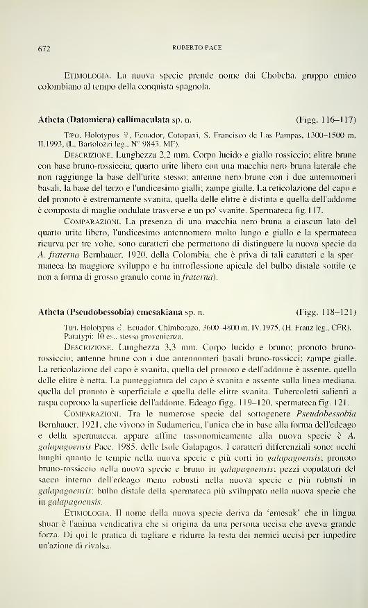
672 ROBERTO PACE
Etimologia. La nuova specie prende nome dai Chobcha, gruppo etnico
colombiano al tempo della conquista spagnola.
Atheta (Datomicra) callimaculata sp. n. (Figg. 1 16-117)
Tipo. Holotypus 2, Ecuador, Cotopaxi, S. Francisco de Las Pampas, 1300-1500 m,
11.1993, (L. Bartolozzi leg., N° 9843, MF).
Descrizione. Lunghezza 2,2 mm. Corpo lucido e giallo-rossiccio; elitre brune
con base bruno-rossiccia; quarto urite libero con una macchia nero-bruna laterale che
non raggiunge la base dell'urite stesso; antenne nero-brune con i due antennomeri
basali, la base del terzo e l'undicesimo gialli: zampe gialle. La reticolazione del capo e
del pronoto è estremamente svanita, quella delle elitre è distinta e quella dell'addome
è composta di maglie ondulate trasverse e un po' svanite. Spermateca fig. 1 17.
Comparazioni. La presenza di una macchia nero-bruna a ciascun lato del
quarto urite libero, l'undicesimo antennomero molto lungo e giallo e la spermateca
ricurva per tre volte, sono caratteri che permettono di distinguere la nuova specie da
A. fraterna Bernhauer, 1920, della Colombia, che è priva di tali caratteri e la sper-
mateca ha maggiore sviluppo e ha introflessione apicale del bulbo distale sottile (e
non a forma di grosso granulo come in fraterna).
Atheta (Pseudobessobia) emesakiana sp. n. (Figg. 118-121)
Tipi. Holotypus S, Ecuador. Chimborazo, 3600-4800 m. IV. 1975, (H. Franz leg., CFR).
Paratypi: 10 es., stessa provenienza.
Descrizione. Lunghezza 3.3 mm. Corpo lucido e bruno; pronoto bruno-
rossiccio; antenne brune con i due antennomeri basali bruno-rossicci; zampe gialle.
La reticolazione del capo è svanita, quella del pronoto e dell'addome è assente, quella
delle elitre è netta. La punteggiatura del capo è svanita e assente sulla linea mediana,
quella del pronoto è superficiale e quella delle elitre svanita. Tubercoletti salienti a
raspa coprono la superficie dell'addome. Edeago figg. 1 19-120, spermateca fig. 121.
Comparazioni. Tra le numerose specie del sottogenere Pseudobessobia
Bernhauer, 1921, che vivono in Sudamerica, l'unica che in base alla forma dell'edeago
e della spermateca, appare affine tassonomicamente alla nuova specie è A.
galapagoensis Pace. 1985, delle Isole Galapagos. I caratteri differenziali sono: occhi
lunghi quanto le tempie nella nuova specie e più corti in galapagoensis; pronoto
bruno-rossiccio nella nuova specie e bruno in galapagoensis; pezzi copulatori del
sacco interno dell'edeago meno robusti nella nuova specie e più robusti in
galapagoensis; bulbo distale della spermateca più sviluppato nella nuova specie che
in galapagoensis.
Etimologia. Il nome della nuova specie deriva da 'emesak' che in lingua
shuar è l'anima vendicativa che si origina da una persona uccisa che aveva grande
forza. Di qui le pratica di tagliare e ridurre la testa dei nemici uccisi per impedire
un'azione di rivalsa.
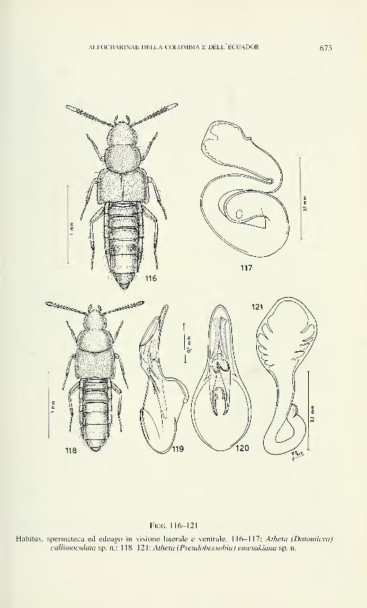
ALEOCHARINAE DELLA COLOMBIA E DELL ECUADOR 673
Figo. 116-121
Habitus, spermateca ed edeago in visione laterale e ventrale. 116-117: Atheta (Datomicra)
callimaculata sp. n.: 1 18-121: Atheta (Pseudobessobia) emesakiana sp. n.
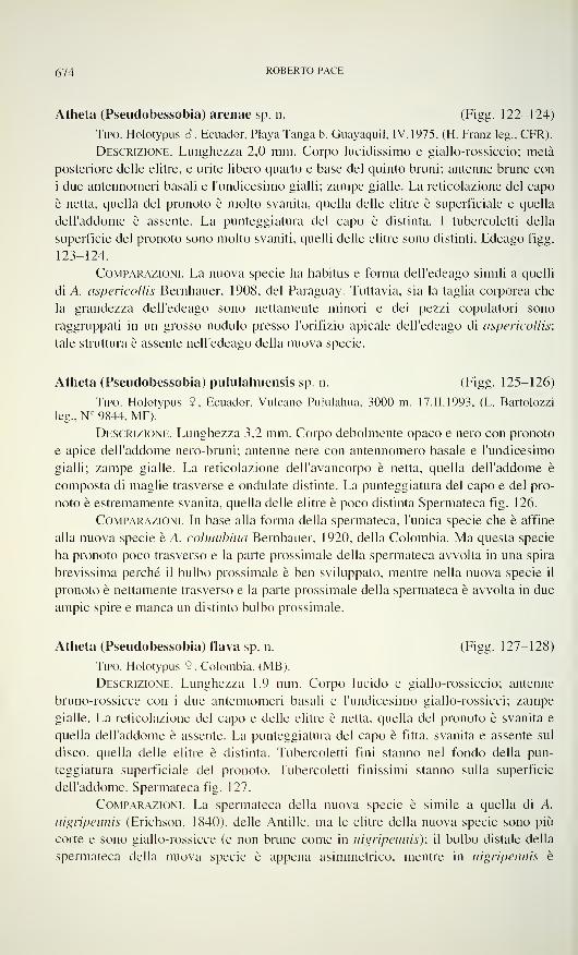
674 ROBERTO PACE
Atheta (Pseudobessobia) arenae sp. n. (Figg. 122-124)
Tipo. Holotypus <$, Ecuador, Playa Tanga b. Guayaquil, IV. 1975, (H. Franz leg., CFR).
Descrizione. Lunghezza 2,0 mm. Corpo lucidissimo e giallo-rossiccio; metà
posteriore delle elitre, e urite libero quarto e base del quinto bruni; antenne brune con
i due antennomeri basali e l'undicesimo gialli; zampe gialle. La reticolazione del capo
è netta, quella del pronoto è molto svanita, quella delle elitre è superficiale e quella
dell'addome è assente. La punteggiatura del capo è distinta. I tubercoletti della
superficie del pronoto sono molto svaniti, quelli delle elitre sono distinti. Edeago figg.
123-124.
Comparazioni. La nuova specie ha habitus e forma dell'edeago simili a quelli
di A. aspericollis Bernhauer, 1908, del Paraguay. Tuttavia, sia la taglia corporea che
la grandezza dell'edeago sono nettamente minori e dei pezzi copulatori sono
raggruppati in un grosso nodulo presso l'orifizio apicale dell'edeago di aspericollis:
tale struttura è assente nell'edeago della nuova specie.
Atheta (Pseudobessobia) pululahuensis sp. n. (Figg. 125-126)
Tipo. Holotypus 9, Ecuador. Vulcano Pululahua. 3000 m. 17.11.1993, (L. Bartolozzi
leg., N° 9844, MF).
Descrizione. Lunghezza 3,2 mm. Corpo debolmente opaco e nero con pronoto
e apice dell'addome nero-bruni; antenne nere con antennomero basale e l'undicesimo
gialli; zampe gialle. La reticolazione dell'avancorpo è netta, quella dell'addome è
composta di maglie trasverse e ondulate distinte. La punteggiatura del capo e del pro-
noto è estremamente svanita, quella delle elitre è poco distinta Spermateca fig. 126.
Comparazioni. In base alla forma della spermateca, l'unica specie che è affine
alla nuova specie è A. columbina Bernhauer, 1920, della Colombia. Ma questa specie
ha pronoto poco trasverso e la parte prossimale della spermateca avvolta in una spira
brevissima perché il bulbo prossimale è ben sviluppato, mentre nella nuova specie il
pronoto è nettamente trasverso e la parte prossimale della spermateca è avvolta in due
ampie spire e manca un distinto bulbo prossimale.
Atheta (Pseudobessobia) flava sp. n. (Figg. 127-128)
Tipo. Holotypus 9, Colombia, (MB).
Descrizione. Lunghezza 1,9 mm. Corpo lucido e giallo-rossiccio; antenne
bruno-rossicce con i due antennomeri basali e l'undicesimo giallo-rossicci; zampe
gialle. La reticolazione del capo e delle elitre è netta, quella del pronoto è svanita e
quella dell'addome è assente. La punteggiatura del capo è fitta, svanita e assente sul
disco, quella delle elitre è distinta. Tubercoletti fini stanno nel fondo della pun-
teggiatura superficiale del pronoto. Tubercoletti finissimi stanno sulla superficie
dell'addome. Spermateca fig. 127.
Comparazioni. La spermateca della nuova specie è simile a quella di A.
nigrìpennìs (Erichson. 1840), delle Antille, ma le elitre della nuova specie sono più
corte e sono giallo-rossicce (e non brune come in nigrìpennìs); il bulbo distale della
spermateca della nuova specie è appena asimmetrico, mentre in nigrìpennìs è
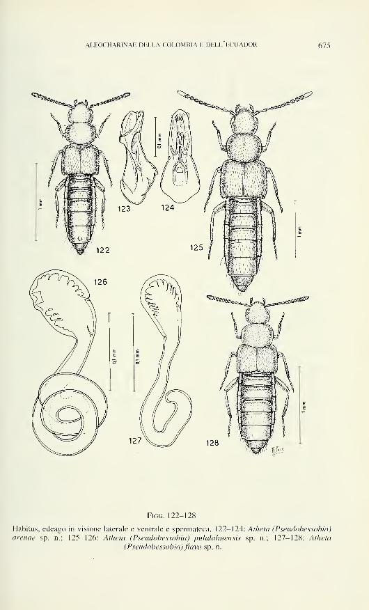
ALEOCHARINAE DELLA COLOMBIA E DELL ECUADOR 675
jjju
Figo. 122-128
Habitus, edeago in visione laterale e ventrale e spermateca. 122-124: Atheta (Pseudobessobia)
arenae sp. n.; 125-126: Atheta (Pseudobessobia) pulùlahuensis sp. n.; 127-128: Atheta
(Pseudobessobia) flava sp. n.
676 ROBERTO PACE
fortemente asimmetrico e a cuspide; la parte prossimale della spermateca della nuova
specie, è corta e largamente ricurva all'apice, mentre in nigripennis è lunga e breve-
mente ricurva all'apice.
Ringraziamenti
Per il materiale affidatomi in esame, ringrazio molto cordialmente il Dr V.
Puthz della "Limnologische Fluss-station" di Schlitz, il Dr L. Bartolozzi del Museo
Zoologico de "La Specda1' dell'Università di Firenze, Il Prof. Dr H. Franz di Mödling
e il Dr M. Uhlig dell'Università Humboldt di Berlin. Per il prestito di tipi rigrazio i
direttori e i conservatori dei seguenti Istituti: D.E.I. di Eberswalde, Museo Zoologico
dell'Università Humboldt di Berlino, Institut Royal des Sciences Naturelles de
Belgique di Bruxelles, il "Naturhistorisches Museum" di Vienna e il British Museum(Natural History) di Londra.
BIBLIOGRAFIA
Bernhauer, M. 1908. Beitrag zur Staphylinidenfauna von Südamerika. Archiv für Natur-
geschichte 74: 283-372.
Bernhauer, M. 1909. Zur Staphylinidenfauna von Südamerika. Bollettino della Società
Entomologica Italiana 60: 225-251.
Bernhauer, M. 1921. Zur Staphylinidenfauna Südamerikas, insbesondere Argentiniens. Archiv
für Naturgeschichte 86: 170-183.
Bernhauer, M. 1927. Zur Staphylinidenfauna Südamerikas, insbesondere Argentiniens. Archiv
für Naturgeschichte 91: 229-264.
Cameron, M. 1923. Descriptions of new Species of Staphylinidae from the West Indies.
Annals and Magazine ofnatural Histoty 1 1: 363^400.
Cameron, M. 1933. New Species of Staphvlinidae (Col.) from Japan. Entomological monthly
Magazine 69: 208-219.
Erichson, W.F. 1840. Genera et Species Staphylinorum Insectorum Coleopterorum Familiae 2:
Berlin, 954 pp.
Fauvel, A. 1991. Voyage de M. le Dr Ed. Bugnion au Venezuela, en Colombie et aux Antilles.
Revue d'Entomologie Caen 20: 69-91.
Kraatz, G. 1857. Genera Aleocharinorum Illustrata. Linnaea Entomologica 1 1: 1-43.
Notman, H., 1920. Staphylinidae from Florida in the collection of the American Museum of
Natural History, with descriptions of new genera and species. Bulletin of the American
Museum ofNatural History 42: 693-732.
Pace, R. 1985. Aleocharinae delle Isole Galapagos (Coleoptera, Staphylinidae). Annales de la
Société entomologique de France (N.S.) 21: 449^-156.
Pace, R. 1986. Aleocharinae del Perù (Coleptera. Staphylinidae). Redia 69: 417-467.
Pace, R. 1987. Aleocharinae del Cile (Coleoptera. Staphylinidae. Redia 70: 459-522.
Pace, R. 1990. Aleocharinae neotropiche del Museo Ungherese di Storia Naurale (Coleoptera,
Staphylinidae). Annales Historiae-Naturali Musei Nationali Hungariae 81: 53-107.
Pace. R. 1996. Aleocharinae della Colombia e dell'Ecuador: Parte I (Coleoptera, Staphy-
linidae). Revue Suisse de Zoologie, 103:395-437.
Scheerpeltz, O. 1972. Wissenschaftliche Ergebnisse der Studienreise von Gy. Topàl nach
Südwst-Argentinien (Coleoptera: Staphylinidae). Folia entomologica hungarica (N.S.)
25 (supplir 269 pp.
SoLiER. A.J. 1849. Coleoptera. In Gay: Historia fisica y politica de Chile: Insecta 4: 105-508.
Thomson, CG. 1858. Försök tili uppställnins af Sveriges Staphyliner. Öfv. Kön. Vet.-Akad.
Förh. 15: 27-10.
Revue suisse de Zoologie, 103 (3): 677-684; septembre 1996
Some Venezuelan Oligochaeta Glossoscolecidae and Octochaetidae
Gilberto RIGHI
Dept. Zoologia, Instituto de Biociências, Universidade de Sào Paulo. Caixa Postal
20520, Säo Paulo 01452-990, Brasil.
Some Venezuelan Oligochaeta Glossoscolecidae and Octochaetidae. -
Four species of earthworms are studied from Venezuela, State of Ama-zonas, Puerto Ayacucho. The Glossoscolecidae Riiinodrilus ayacu n. sp.
and R. cucho n. sp. are described. The presence of the Octochaetidae
Dichogaster modiglianii (Rosa) and D.saliens (Beddard) is registered.
Key-words: Oligochaeta - Glossoscolecidae - Octochaetidae - Venezuela -
Amazonas.
INTRODUCTION
During his ecological studies in Tropical America, Dr Maurizio G. Paoletti
(Università degli Studi di Padova, Italy) collected a small number of earthworms and
kindly put it at my disposal. I thank to Dr Paoletti for the possibility of this study.
The present collection has two main significances: one of agricultural value
and other of zoological value. The agricultural importance is the finding of the
pantropical, anthropochoric, African species Dichogaster modiglianii and D. saliens
at 20 cm depth in the soil. In other sites of the Neotropical Region (see below) these
species live from 0-10 cm depth; presenting an high populational density and a great
digging activity which result in a great number of tortuous galleries. They live mainly
in horticultural soils and they recover very well from manual or mechanical
agricultural practices (Righi, 1990). Their occurrence at 20 cm depth is another
evidence of their usefulness to the cultivable soils in the wet tropics.
The zoological value of this collection is to enlarge our knowledge about
Rhinodrilus. Righi (1985) revised the genus Riiinodrilus Perrier (1872) recognizing
37 species. Later R. lourdesae Righi (1986) and R. timote Righi (1989) were added
and R. ayacu and R. cucho are described here. 'The resulting 41 species of Rhinodrilus
are distributed in South America between the parallels of 11°N and 28°S, from
Trinidad-Tobago to North Argentina The Venezuelan species are R. paradoxus
Manuscript accepted 14.11.1995.
678 GILBERTO RIGHI
Perrier, 1872 (to Federal District and Carabobo and Aragua States), R. senex Righi,
1984 and R rimate Righi. 1989 (Aragua State). R. appura (Michaelsen, 1892). R.
sieversi (Michaelsen, 1895) and R. fuenzalidae Corderò, 1944 (Carabobo State) and
R. ayacu n. sp. and R. cucho n. sp. (Amazonas State).
MATERIAL AND METHODS
The earthworms were collected in December 1994. in Venezuela, Amazonas
State, Puerto Ayacucho (5°40'N-67°05'W) from soil cores at 20 cm depth, in the
following localities: Gallery forest; Eisenberg Farm: Mahada. The studies were made
by dissections, slides of microscopical pieces mounted in glicerin-water (1:1) and
serial microscopical sections (10 urn) stained by Mallory's triple method (Pantin
1964). The sketches were made with camera lucida. The material is deposited in the
Departmento de Zoologia. Universidade de Sào Paulo (ZU) and in the Muséumd'histoire naturelle, Geneva (MHNG).
Glossoscolecidae
Rhinodrilus ayacu n. sp. (Figs 1-9)
Material: Venezuela: Amazonas State: Puerto Ayacucho, gallery forest, 1 duellate
specimen (Holotype, ZU- 1283). 1 duellate and 2 young specimens and 1 cocoon (MHNG20326 INVE).
Description: Length 80-100 mm. Mid-body diameter 4-5 mm. Number of
segments 219-231. Unpigmented. The prostomium and a part of the segment I are
invaginated. The segments I—II have a pair of deep nephrobuccal furrows and a lot of
shallow longitudinal furrows. The other segments have smooth surface. A constricted
"caudal zone", without setae, occupies 3—4 segments beginning at CXVII-CXXI. The
setae are arranged in 4 pairs of regular lengthwise arranged series starting from II.
The setal relations are in the mid-body region (LXX-LXXX) aa : ab : be : cd : dd =
5.5 : 1.0 : 5.5 : 0.9 : 13.3 and in the posterior region (CCXX-CCXXX) = 9.1 : 1.0 :
5.3 : 1.2 : 6.0: ab = 400 um in both the regions. The space aa increases from
XX-XXII and decreases suddenly at XXIII (Fig. 1). The common setae (Figs. 2-3)
are slightly sigmoid with distal nodulus. Their apical 1/6 has 4 alternate rows of
semilunar excavations; there are 3-5 excavations per row. Occasionaly the exca-
vations are irregularly disposed. The length of the setae varies in the mid-body region:
a.b = 270-309 urn (M = 288 urn) and cd = 347-424 um (M = 384 um) and in the
posterior region: a.b = 41 1—176 um (M = 445 um) and cd = 431-476 um (M = 457
um). The ventral setae of IX. X-XII and XVIII-XXIII are modified as genital setae,
which are straight with a slight proximal bending (Fig. 4). Their distal half has 4
alternate series of excavations. The setae of IX. X-XII and XXIII have 9-10
excavations per series and the setae of XVIII-XXII have 11-13 excavations per
series. The length of the genital setae of IX-XII and XXIII varies from 604-848 urn
(M = 735 Jim); the shorter setae are in IX. X. The setae of XVIII-XXII are 874-1054
urn (M = 993 urn) long: the shorter ones are in XVIII and the longer ones in XX-XXI.
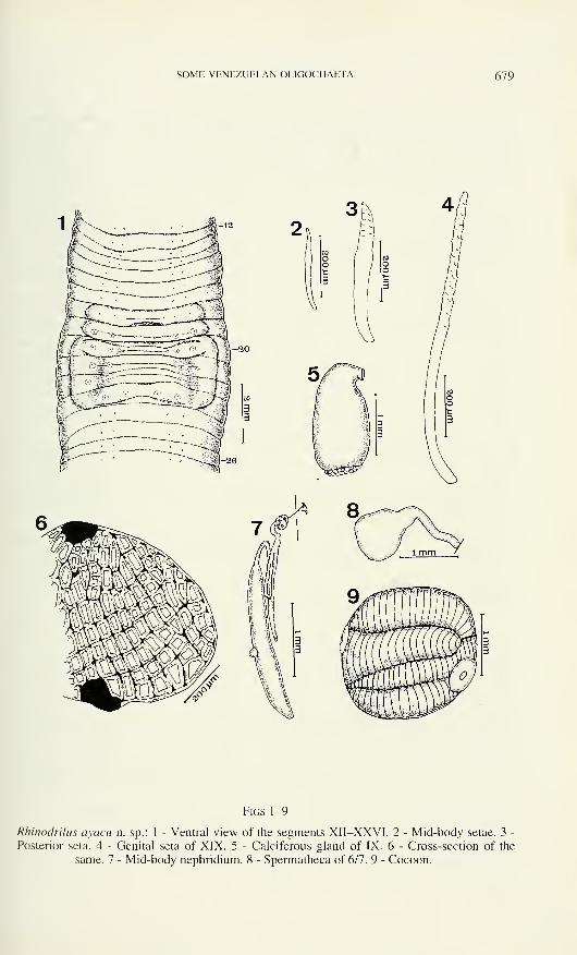
SOME VENEZUELAN OLIGOCHAETA 679
Figs 1-9
Rhinodrilus ayacu n. sp.: 1 - Ventral view of the segments XII-XXVI. 2 - Mid-body setae. 3 -
Posterior seta. 4 - Genital seta of XIX. 5 - Calciferous gland of IX. 6 - Cross-section of the
same. 7 - Mid-body nephridium. 8 - Spermatheca of 6/7. 9 - Cocoon.
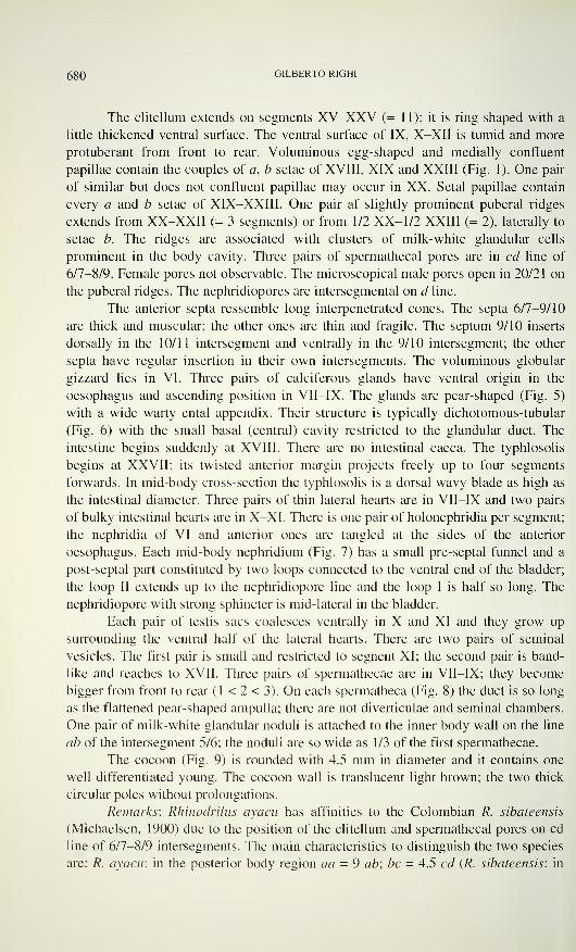
ggO GILBERTO RIGHI
The clitellum extends on segments XV-XXV (= 11); it is ring-shaped with a
little thickened ventral surface. The ventral surface of IX, X-XII is tumid and more
protuberant from front to rear. Voluminous egg-shaped and medially confluent
papillae contain the couples of a, b setae of XVIII, XIX and XXIII (Fig. 1). One pair
of similar but does not confluent papillae may occur in XX. Setal papillae contain
every a and b setae of XIX-XXIII. One pair af slightly prominent puberal ridges
extends from XX-XXII (= 3 segments) or from 1/2 XX- 1/2 XXIII (= 2), laterally to
setae b. The ridges are associated with clusters of milk-white glandular cells
prominent in the body cavity. Three pairs of spermathecal pores are in ed line of
6/7-8/9. Female pores not observable. The microscopical male pores open in 20/21 on
the puberal ridges. The nephridiopores are intersegmental on d line.
The anterior septa ressemble long interpenetrated cones. The septa 6/7-9/10
are thick and muscular; the other ones are thin and fragile. The septum 9/10 inserts
dorsally in the 10/11 intersegment and ventrally in the 9/10 intersegment; the other
septa have regular insertion in their own intersegments. The voluminous globular
gizzard lies in VI. Three pairs of calciferous glands have ventral origin in the
oesophagus and ascending position in VII-IX. The glands are pear-shaped (Fig. 5)
with a wide warty ental appendix. Their structure is typically dichotomous-tubular
(Fig. 6) with the small basal (central) cavity restricted to the glandular duct. The
intestine begins suddenly at XVIII. There are no intestinal caeca. The typhlosolis
begins at XXVII; its twisted anterior margin projects freely up to four segments
forwards. In mid-body cross-section the typhlosolis is a dorsal wavy blade as high as
the intestinal diameter. Three pairs of thin lateral hearts are in VII-IX and two pairs
of bulky intestinal hearts are in X-XI. There is one pair of holonephridia per segment;
the nephridia of VI and anterior ones are tangled at the sides of the anterior
oesophagus. Each mid-body nephridium (Fig. 7) has a small pre-septal funnel and a
post-septal part constituted by two loops connected to the ventral end of the bladder;
the loop II extends up to the nephridiopore line and the loop I is half so long. The
nephridiopore with strong sphincter is mid-lateral in the bladder.
Each pair of testis sacs coalesces ventrally in X and XI and they grow up
surrounding the ventral half of the lateral hearts. There are two pairs of seminal
vesicles. The first pair is small and restricted to segnent XI; the second pair is band-
like and reaches to XVII. Three pairs of spermathecae are in VII-IX; they become
bigger from front to rear (1 < 2 < 3). On each spermatheca (Fig. 8) the duct is so long
as the flattened pear-shaped ampulla; there are not diverticulae and seminal chambers.
One pair of milk-white glandular noduli is attached to the inner body wall on the line
ab of the intersegment 5/6; the noduli are so wide as 1/3 of the first spermathecae.
The cocoon (Fig. 9) is rounded with 4.5 mm in diameter and it contains one
well differentiated young. The cocoon wall is translucent light brown; the two thick
circular poles without prolongations.
Remarks: Rhinodrilus ayacu has affinities to the Colombian R. sibateensis
(Michaelsen. 1900) due to the position of the clitellum and spermathecal pores on cd
line of 6/7-8/9 intersegments. The main characteristics to distinguish the two species
are: R. ayacu: in the posterior body region aa = 9 ab; be = 4.5 cd (R. sibateensis: in
SOME VENEZUELAN OLIGOCHAETA 681
the same region aa = ab: be = cd); length of the posterior setae 41 1-476 um (length
up to 800 urn); genital setae ornamented with 9-13 excavations per series (5 exca-
vations per series).
The name of the new species derives from Puerto Ayacucho where it was
collected.
Rhinodrilus cucho n. sp. (Figs 10-15)
Material: Venezuela: Amazonas State: Puerto Ayacucho, gallery forest, 1 clitellate
specimen (Holotype ZU-1284A), 3 clitellate and 1 mature aclitellate specimens (ZU-1284B;
MHNG 20327 INVE).
Description: Length 56-70 mm. Mid-body diameter 3.1-3.5 mm. Number of
segments 199-226. The prostomium and a part of the I segment are invaginated.
Unpigmented. A constricted 'caudal zone' occupies three segments beginning at
segment CXX1X-CXXXIV. The setae are four pairs per segment starting from II. The
setae a and d are disposed on regular lengthwise series and the setae b and c are
irregularly disposed in the posterior 1/4-1/5 of the body. The setae b lose their regular
arrangement from CXXIII-CLIII and the setae c from CLXIV-CLXVII. The relations
among the setae in the mid-body region (LXX-LXXX) are aa : ab : be : cd': dd- 3.0
: 1.0 : 3.4 : 0.6 : 9.0 (ab = 400 um). The common setae are slightly sigmoid; their
distal 1/7 has semilunar excavations irregularly disposed or arranged in 4 alternate
series of 3-4 excavations on each one. The setae of the mid-body (Fig. 11) have a
distal nodulus and their length varies: the ventral ones from 264-283 um (M = 273
urn) and the lateral ones from 347-399 um (M = 376 urn). The posterior setae (Fig.
12) are much bigger, without nodulus, and there are no significant differences in
length among the ventral and lateral ones, which vary from 527-630 urn (M = 589
um). The ventral setae of XVIII-XX1II are modified as genital setae. They are
slightly sigmoid with the proximal bending a little bigger than the distal one (Fig. 13).
Their distal third presents 4 lengthwise series of excavations. Each two series are
disposed side by side and alternately with the other two series; sometimes the 4 series
alternate among themselves. The number of excavations per series is 6-9. The genital
setae's length varies from 540-726 um (M = 608 urn).
The clitellum extends from XVI-XXVI (= 11 segments); it is ring-shaped
openned ventrally by the male genital field in XVÏÏI-XXIII. One pair of a little thick
puberal ridges (Fig. 10) lies laterally to the setae b from 1/2 XX- 1/2 XXIII (= 3
segments) or XX-XXIII (= 4). Two pairs of puberal papillae contain the couples of
ventral setae in XVIII and XIX. The ridges and papillae are milk-white. Wide setal
papillae contain every ventral setae of XVIII-XXIII. The ventral face of X-XII is
tumid. The pair of microscopical male pores is on the puberal ridges in 20/21. The
female pores are unrecognisable. Three pairs of spermathecal pores are on line cd of
6/7-8/9. The nephridiopores are intersegmental on line cd.
The septa 6/7-9/10 are thick and muscular; the other ones are thin and fragile.
All septa have regular insertion in their corresponding intersegments. The gizzard is
rounded, voluminous and very muscular in VI. Three pairs of calciferous glands
depart ventrally from the oesophagus in VII-IX. Each calciferous gland (fig. 14) is
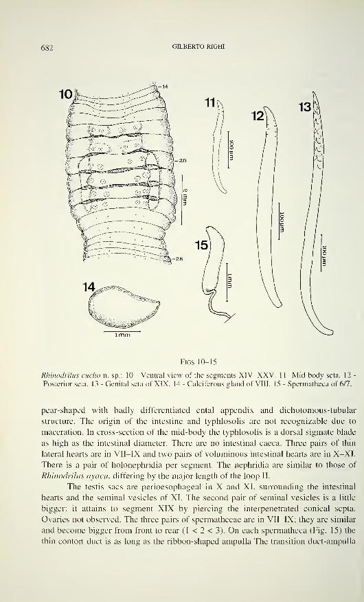
682 GILBERTO RIGHI
Figs 10-15
Rhinodrilus cucho n. sp.: 10 - Ventral view of the segments XIV-XXV. 11- Mid-body seta. 12
Posterior seta. 13 - Genital seta of XIX. 14 - Calciferous gland of VIII. 15 - Spermatheca of 6/7.
pear-shaped with badly differentiated ental appendix and dichotomous-tubular
structure. The origin of the intestine and typhlosolis are not recognizable due to
maceration. In cross-section of the mid-body the typhlosolis is a dorsal sigmate blade
as high as the intestinal diameter. There are no intestinal caeca. Three pairs of thin
lateral hearts are in VII-IX and two pairs of voluminous intestinal hearts are in X-XI.
There is a pair of holonephridia per segment. The nephridia are similar to those of
Rhinodrilus ayacu, differing by the major length of the loop II.
The testis sacs are perioesophageal in X and XI, surrounding the intestinal
hearts and the seminal vesicles of XI. The second pair of seminal vesicles is a little
bigger; it attains to segment XIX by piercing the interpenetrated conical septa.
Ovaries not observed. The three pairs of spermathecae are in VII-IX; they are similar
and become bigger from front to rear (1 < 2 < 3). On each spermatheca (Fig. 15) the
thin contort duct is as long as the ribbon-shaped ampulla The transition duct-ampulla
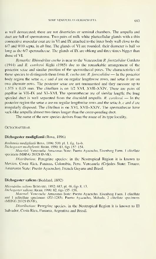
SOME VENEZUELAN OLIGOCHAETA 683
is well demarcated; there are not diverticles or seminal chambers. The ampulla and
duct are full of spermatozoa. Two pairs of milk-white pericellular glands with a thin
connective-muscular coat are in VI and IX attached to the inner body wall close to the
6/7 and 9/10 septa, in ab line. The glands of VI are rounded, their diameter is half so
long as the 6/7 spermathecae. The glands of IX are oblong and three times bigger than
those of VI.
Remarks: Rhinodrilus cucho is near to the Venezuelan R. fuenzalidae Corderò
(1944) and R. corderoi Righi (1985) due to the remarkable arrangement of the
posterior setae and similar position of the spermathecal pores. The characteristics of
these species to distinguish them from R. cucho are: R. fuenzalidae — In the posterior
body region the setae a, c and d are on regular lengthwise rows, and setae b are on
two alternate rows. The posterior setae are not ornamented and they measure up to
1.575 x 0.15 mm. The clitellum is on 1/2 XVI, XVII-XXIV. There are pairs of
papillae in VII-IX and XI-XVI. The spermathecae are of similar length; the long
spiraled duct is well separated from the discoidal ampulla. R. corderoi — In the
posterior region the setae a are on regular lengthwise rows and the setae b, c and d are
irregularly disposed. The clitellum is on XVI, XVII-XXIV. The spermathecae have
sack-like ampulla almost two times longer than the corresponding duct.
The name of the new species derives from the name of its type locality.
OCTOCHAETIDAE
Dichogaster modiglianii (Rosa, 1896)
Benhamia modiglianii Rosa, 1896: 510, pi. 1, fig. la-b.
Dichogaster modiglianii; Righi, 1990: 81, figs 152-154.
Material: Venezuela: Amazonas State: Puerto Ayacucho, Eisenberg Farm, 1 clitellate
specimen (MHNG 20328 INVE).
Distribution: Peregrine species; in the Neotropical Region it is known to
Mexico, Costa Rica, Panama, Colombia, Peru, Venezuela (Cojedes State: Tinaco;
Amazonas State: Puerto Ayacucho), French Guyana and Brasil.
Dichogaster saliens (Beddard, 1892)
Microdrilus saliens Beddard, 1892: 683, pi. 46, figs 8, 13.
Dichogaster saliens; Righi, 1990: 82, figs 155-158.
Material: Venezuela: Amazonas State: Puerto Ayacucho, Eisenberg Farm, 1 clitellate
and 1 aclitellate specimens (ZU-1285). Puerto Ayacucho, Mahada, 2 clitellate specimens(MHNG 20329 INVE).
Distribution: Peregrine species, in the Neotropical Region it is known to El
Salvador, Costa Rica, Panama, Argentina and Brasil.
6g4 GILBERTO RIGHI
REFERENCES
Beddard, F.E. 1892. On some new species of earthworms from various parts of the world.
Proceedings of the Zoological Society ofLondon, 1892: 606-706, pis. 45^-6.
Benham, W.B. 1887. Studies on earthworms. II. Quarterly Journal of Microscopical Science
(N.S.) 27: 77-108, pis. 8-9.
Corderò, E.H. 1944. Oligoquetos sudamericanos de la Familia Glossoscolecidae, IV. Sobre
algunas espécies de Venezuela Comunicaciones Zoologicas del Museo de Historia
natural de Montevideo 1(14): 1-6.
Michaelsen, W. 1892. Ferricolen der Berliner zoologischen Sammlung. II. Archiv fürNaturgeschichte 58(1): 209-261, pi. 13.
Michaelsen, W. 1895. Zur Kenntnis der Oligochaeten. Abhandlungen aus dem Gebiet der
Naturwissenschaften Verein in Hamburg 13: 1-37. 1 pl.
Michaelsen, W. 1900. Die Terricolen-Fauna Columbiens. Archiv für Naturgeschichte 66:
231-266.
Pantin, CF.A. 1964. Notes on microscopical techniques for zoologists. University Press,
Cambridge, VIII + 76 pp.
Perrier, E. 1872. Recherche pour servir à l'histoire de lombriciens terrestres. Nouvelles
Archives du Muséum d'Histoire naturelle, Paris 8(1): 5-198, pis. 1-4.
Righi, G. 1984. Nova Contribuiçào ao conhecimento dos Oligochaeta da Venezuela. Papéis
Avulsos de Zoologia, Sào Paulo 35(22): 243-256.
Righi, G. 1985. Sobre Rhinodrilus e Urobenus (Oligochaeta, Glossoscolecidae). Boletim de
Zoologia, Universidade de Sào Paulo (N.S.) 9: 231-257.
Righi, G. 1986. Alguns Oligochaeta, Glossoscolecidae, de Rondonia, Brasil. Boletim de
Zoologia, Universidade de Sào Paulo (N.S.) 10: 283-303.
Righi, G. 1989. Adiçào aos conhecimentos dos Oligochaeta da Venezuela. Revista Brasileira
de Biologia 49(4): 1065-1084.
Righi, G. 1990. Minhocas de Mato Grosso e de Rondonia. Programa Polonoroeste — CNPq,Brasilia. 157 pp.
Rosa, D. 1896. I lombrichi raccolti a Sumatra dal dott. Elio Modigliani. Annali del Museocivico di Storia naturale 'Giacomo Doria', Genova, (2)16: 502-532, 1 pl.
Revue suisse de Zoologie, 103 (3): 685-690; septembre 1996
Un nouveau Apoduvalius du bassin de la rivière Nalón,
Asturies-Espagne (Coleoptera, Trechidae)
José Maria SALGADO COSTASDpto. de Biologia Animal, Facultad de Biologia, Universidad de Leon,
E-24071 Leon, Espana.
A new Apoduvalius from basin of the river Nalón, Asturia-Spain
(Coleoptera, Trechidae). - Apoduvalius espanoli n. sp. is described and
defined specifically on the basis of the shape of the aedeagus and the
singular structures of the internal sac. Its taxonomic position is discussed in
relation to the species which are morphologically closest to it. In addition,
new data are recorded with regard to the fauna of several caves sited in the
basin of the Nalón River.
Key-words: Coleoptera - Trechidae - Apoduvalius espanoli n. sp. - Spain -
Asturia.
INTRODUCTION
De 1989 à 1995, l'auteur visita 23 cavités situées dans les massifs karstiques
des Sierra Mermeja, Sierra del Crespón, Sierra Pintacanales, Sierra Giblaniella et du
Cordai de Ponga, traversés par la rivière Nalón (Carte).
La faune troglobie découverte dans cette vaste zone calcaire, très diversifiée,
est d'un grand intérêt et il faudra s'attendre, dans le futur, à de nouvelles découvertes
biospéléologiques. Dans les travaux de Jeannel (1956, 1958), Vives (1980) et plus
récemment Salgado (1985, 1988, 1989 a-b, 1991, 1993), plusieurs espèces sont
signalées:
- Leptodirinae: Quaestus pachecoi (Bolivar, 1915), Q. recordations (Salgado,
1982), Q. amicalis amicalis (Salgado, 1984), Q. luctuosus (Salgado, 1984), Q.
nietoi (Salgado, 1988), Q. longicornis (Salgado, 1989) et Notidocharis franzi
Jeannel, 1956;
- Trechidae: Trechus fulvus Déjean, 1831, T. escalerai Abeille, 1903 et, surtout,
Apoduvalius franzi Jeannel, 1958, A. asturiensis Salgado, 1991 et A. naloni
Salgado, 1993;
Manuscrit accepté le 06.12.1995.
686 JOSE MARIA SALGADO COSTAS
- Pterostichidae: la donnée la plus significative est Pterosîichus (Jeannelinus)
drescoi (Nègre, 1957);
- Staphylinidae: Cantabrodytes vivesi Espanol, 1975, espèce très intéressante.
Dans cette liste, il faut relever la capture récente, dans la grotte de Refidieyu
"B", Munera, de deux espèces de Apoduvalius, A. naloni et A. asturiensis, seule
donnée qui montre jusqu'à présent la coexistence de deux espèces du genre Apo-
duvalius.
Carte: Coordonnées et carte de distribution de quelques Apoduvalius. 1: Grotte Les Canales
(UTM : 30TUN1 12828), A. espanoli n. sp.; 2: Grotte de Refidieyu "B" (UTM : 30TTN949909). A. asturiensis et A. naloni; 3: Grotte du Sierru (UTM : 30TTN899873), A. asturiensis;
4: Refuge de Vega Redonda (UTM : 30TUN388887), A. lecoqi; 5: Grotte de Porro Covanona(UTM : 30TUN405889), A. negrei.
La grotte de Les Canales est située dans une zone de "Calizas de Montana" du
Carbonifère et d'accès est très facile. L'entrée est très étroite et l'ouverture orientée
vers le SE, sur la rive droite de la rivière Nalon et près de son lit. La partie connue
présente environ 800 m de développement, avec de grandes salles, des galeries
étroites et vers la moitié, sur sa gauche, un puits de presque 30 m. Dans cette grotte
sinueuse, on peut observer de grandes coulées et de belles concrétions, aujourd'hui en
partie détruites. La grotte de Les Canales se trouve à 18 km de la grotte la plus proche
abritant Apoduvalius, la Grotte de Refidieyu "B", Munera et à 590 m d'altitude.
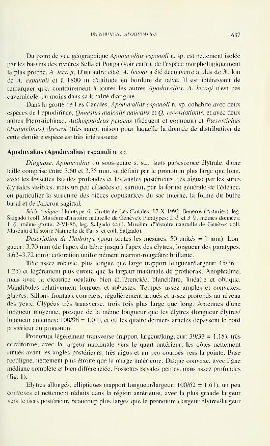
UN NOUVEAU APODUVALIUS 687
Du point de vue géographique Apoduvalius espanoli n. sp. est nettement isolée
par les bassins des rivières Sella et Ponga (voir carte), de l'espèce morphologiquement
la plus proche, A. lecoqi. D'un autre côté, A. lecoqi a été découverte à plus de 30 kmde A. espanoli et à 1800 m d'altitude en bordure de névé. Il est intéressant de
remarquer que, contrairement à toutes les autres Apoduvalius, A. lecoqi n'est pas
cavernicole, du moins dans sa localité d'origine.
Dans la grotte de Les Canales, Apoduvalius espanoli n. sp. cohabite avec deux
espèces de Leptodirinae, Quaestus amicalis amicalis et Q. recordationis, et avec deux
autres Pterostichinae, Anthisphodrus pelaeus (fréquent et commun) et Pterostichus
(Jeannelinus) drescoi (très rare), raison pour laquelle la donnée de distribution de
cette dernière espèce est très intéressante.
Apoduvalius (Apoduvalius) espanoli n. sp.
Diagnose. Apoduvalius du sous-genre s. str., sans pubescence élytrale, d'une
taille comprise entre 3,60 et 3,75 mm, se définit par le pronotum plus large que long,
avec les fossettes basales profondes et les angles postérieurs très aigus; par les stries
élytrales visibles, mais un peu effacées et, surtout, par la forme générale de l'édéage,
en particulier la structure des pièces copulatrices du sac interne, la forme du bulbe
basai et de l'aileron sagittal.
Série typique: Holotype S, Grotte de Les Canales, 17-X-1992, Beneros (Asturies), leg.
Salgado (coll. Muséum d'histoire naturelle de Genève). Paratypes: 2 6 et 3 9, mêmes données;
1 cL même grotte. 2-VI-86, leg. Salgado (coll. Muséum d'histoire naturelle de Genève; coll.
Muséum d'Histoire Naturelle de Paris, et coll. Salgado).
Description de l'holotype (pour toutes les mesures, 50 unités = 1 mm): Lon-
gueur: 3,70 mm (de l'apex du labre jusqu'à l'apex des élytres; longueur des paratypes,
3,63-3,72 mm); coloration uniformément marron-rougeâtre brillante.
Tête assez robuste, plus longue que large (rapport longueur/largeur: 45/36 =
1,25) et légèrement plus étroite que la largeur maximale du prothorax. Anophtalme,
mais avec la cicatrice oculaire bien différenciée, blanchâtre, linéaire et oblique.
Mandibules relativement longues et robustes. Tempes assez amples et convexes,
glabres. Sillons frontaux complets, régulièrement arqués et assez profonds au niveau
des yeux. Clypéus très transverse, trois fois plus large que long. Antennes d'une
longueur moyenne, presque de la même longueur que les élytres (longueur élytres/
longueur antennes: 100/96 = 1,04), et où les quatre derniers articles dépassent le bord
postérieur du pronotum.
Pronotum légèrement transverse (rapport largeur/longueur: 39/33 = 1,18), très
cordiforme, avec la largeur maximale vers le quart antérieur; les côtés nettement
sinués avant les angles postérieurs, très aigus et un peu courbés vers la pointe. Base
rectiligne, nettement plus étroite que la marge antérieure. Disque convexe, avec ligne
médiane complète et bien différenciée. Fossettes basales petites, mais assez profondes
(fig. 1).
Elytres allongés, elliptiques (rapport longueur/largeur: 100/62 = 1,61), un peu
convexes et nettement réduits dans la région antérieure, avec la plus grande largeur
vers le tiers postérieur, beaucoup plus larges que le pronotum (largeur élytres/largeur
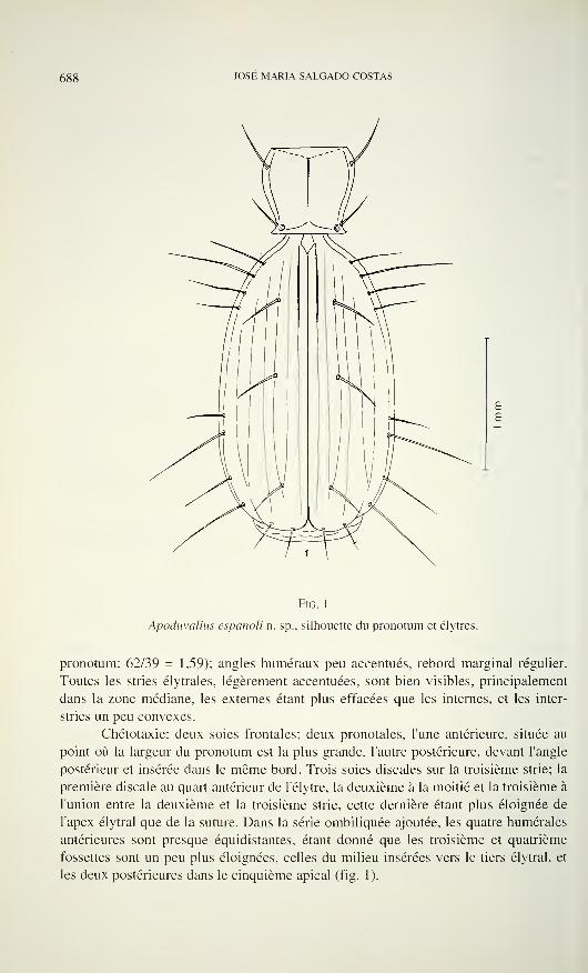
688 JOSE MARIA SALGADO COSTAS
Fig. 1
Apoduvalius espanoli n. sp.. silhouette du pronotum et élytres.
pronotum: 62/39 = 1,59); angles numéraux peu accentués, rebord marginal régulier.
Toutes les stries élytrales, légèrement accentuées, sont bien visibles, principalement
dans la zone médiane, les externes étant plus effacées que les internes, et les inter-
stries un peu convexes.
Chétotaxie: deux soies frontales; deux pronotales, l'une antérieure, située au
point où la largeur du pronotum est la plus grande, l'autre postérieure, devant l'angle
postérieur et insérée dans le même bord. Trois soies discales sur la troisième strie; la
première discale au quart antérieur de l'élytre, la deuxième à la moitié et la troisième à
l'union entre la deuxième et la troisième strie, cette dernière étant plus éloignée de
l'apex élytral que de la suture. Dans la série ombiliquée ajoutée, les quatre humérales
antérieures sont presque équidistantes, étant donné que les troisième et quatrième
fossettes sont un peu plus éloignées, celles du milieu insérées vers le tiers élytral, et
les deux postérieures dans le cinquième apical (fig. 1).
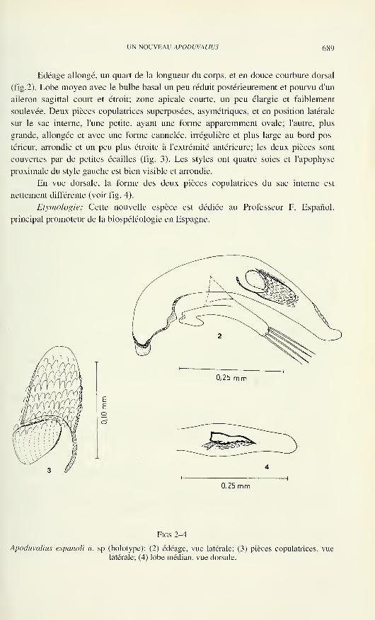
UN NOUVEAU APODUVALIUS 689
Edéage allongé, un quart de la longueur du corps, et en douce courbure dorsal
(fig.2). Lobe moyen avec le bulbe basai un peu réduit postérieurement et pourvu d'un
aileron sagittal court et étroit; zone apicale courte, un peu élargie et faiblement
soulevée. Deux pièces copulatrices superposées, asymétriques, et en position latérale
sur le sac interne, l'une petite, ayant une forme apparemment ovale; l'autre, plus
grande, allongée et avec une forme cannelée, irrégulière et plus large au bord pos-
térieur, arrondie et un peu plus étroite à l'extrémité antérieure; les deux pièces sont
couvertes par de petites écailles (fig. 3). Les styles ont quatre soies et l'apophyse
proximale du style gauche est bien visible et arrondie.
En vue dorsale, la forme des deux pièces copulatrices du sac interne est
nettement différente (voir fig. 4).
Etymologie: Cette nouvelle espèce est dédiée au Professeur F. Espanol,
principal promoteur de la biospéléologie en Espagne.
0,25 mm
0,25 mm
FlGS 2-4
Apoduvalius espanoli n. sp (holotype): (2) édéage, vue latérale; (3) pièces copulatrices, vue
latérale; (4) lobe médian, vue dorsale.
690 J0SE MARIA SALGADO COSTAS
Discussion: La nouvelle espèce, par sa répartition, est proche géographi-
quement de Apoduvalius naloni et A. asturiensis, mais s'en différencie nettement aussi
bien par la morphologie externe que par la forme des genitalia.
Par ses caractères morphologiques, Apoduvalius espanoli n. sp., se rapproche
de Apoduvalius negrei de la Grotte de Porro Covanona, à Covadonga (Jeannel,
1953); de A serrae de la Grotte de Balmori, à Lianes (Vives, 1975); de A. lecoqi, la
seule espèce nivicole qui se trouve aux Pics d'Europe, à 1.800m d'altitude, une zone
proche de Covadonga (Deuve, 1991). Elle se distingue cependant de A. negrei par
une taille un peu plus grande, les angles postérieurs du pronotum plus aigus et plus
vifs, et les stries élytrales moins visibles. Quant à l'édéage, les différences peuvent
être observées dans la zone apicale qui est plus grosse, le bulbe basai plus étroit et
l'aileron sagittal plus réduit chez A espanoli n. sp., tandis que l'endophallus renferme
deux pièces copulatrices, au contraire d'A. negrei qui n'en possède qu'une petite. Par
rapport à A. serrae, les différences de la nouvelle espèce sont les suivantes: une tête
un peu plus grosse, un pronotum un peu plus long et des angles postérieurs un peu
courbés; stries des élytres plus marquées, zone apicale de l'édéage moins pointue,
bulbe basai plus large et présence de deux pièces copulatrices dans le sac interne (une
seulement pour A. serrae). Finalement, A. espanoli diffère de A. lecoqi par une taille
un peu plus grande, une tête moins grosse, un pronotum plus court et des stries
élytrales plus effacées; par rapport à l'édéage, la zone apicale est plus courte et grosse,
le bulbe basai plus large et l'aileron sagittal plus réduit, en plus des pièces copulatrices
bien distinctes, et l'apophyse proximale du style gauche plus développée.
BIBLIOGRAPHIEDeuve, T. 1991. Un nouvel Apoduvalius, nivicole. des Picos de Europe (Col. Trechidae).
Entomologies gallica 2(4): 1 87-1 88.
Giachino, P.M. & J.M. Salgado. 1989a. Revision del gènero Notidocharis Jeannel, 1956 (Col.
Bathysciinae). Graellsia 45: 1-18.
Jeannel, R. 1953. Un genre nouveau de Trechini cavernicoles des Monts Cantabriques. Notes
Biospéologiques 8: 121-125.
Jeannel, R. 1956. Un nouveau genre de Bathysciites des Monts Cantabriques. Revue françaised'Entomologie 23( 1 ): 5-10.
Jeannel, R. 1958. Un nouvel Apoduvalius des Asturies. Notes Biospéologiques 13: 25-26.
Salgado, J.M. 1985. Nuevas o interesantes localizaciones de Carâbidos y Catópidos cavernf-
colas de la Cornisa Cantâbrica. Boletin de las Ciencias Naturales, I.D. E.A. 36: 93-108.
Salgado, J.M. 1988. Nuevo Speocharis del "Grupo occidentalis" (Col. Catopidae). MémoiresBiospéologiques 15: 61-66.
Salgado, J.M. 1989/?. Nuevos datos sobre la distribución de especies del "Grupo S.
occidentalis" (Col. Catopidae). Mémoires Biospéologiques 16: 125-130.
Salgado, J.M. 1991. Dos nuevos Apoduvalius Jeannel. 1968 (Col. Trechidae). MémoiresBiospéologiques 18: 205-208.
Salgado, J.M. 1993. Descripción de Apoduvalius (Apoduvalius) naloni n. sp. y A. (A.) purroyi
galicianus n. ssp. (Col. Trechidae). Consideraciones biogeogrâficas. Mémoires Biospé-
ologiques 20: 217-220.
Vives. E. 1975. Coleópteros cavernfcolas nuevos o interesantes de la Peninsula Ibèrica yBaléares. Spelean 22: 159-169.
Vives. E. 1980. Revision del sénero Apoduvalius Jeannel. 1953 (Col. Trechinae). Speleon, 25:
15-21.
Revue suisse de Zoologie, 103 (3): 691-695; septembre 1996
Spinolyprops pakistanicus sp. n. (Coleoptera: Tenebrionidae),
an Oriental élément in the fauna of northern Pakistan
Wolfgang SCHAWALLER*Staatliches Museum für Naturkunde, Rosenstein 1, D-70191 Stuttgart, Germany.
Spinolyprops pakistanicus sp. n. (Coleoptera: Tenebrionidae), an
Oriental element in the fauna of northern Pakistan. - Spinolyprops
pakistanicus sp. n. from Hazara and Swat in northern Pakistan is described,
having a sexually dimorphic tibia, a structure yet unknown among the
other congeners. In the fauna of northern Pakistan, generally containing
mainly Palaearctic elements, the genus Spinolyprops is a further example
with distinct Oriental distributional pattern.
Key-words: Coleoptera - Tenebrionidae - Spinolyprops - New species -
Pakistan - Zoogeography.
INTRODUCTION
The genus Spinolyprops Pic, 1917 was based on an African species and was
later recorded also from the Oriental region (Kulzer 1954, Kaszab 1965). The genus
contains five uniform species which can be separated easily by the specific shape of
the aedeagus. A further species is added here originating from Hazara and Swat in
northern Pakistan. Spinolyprops pakistanicus sp. n. is remarkable for its geographical
origin (Fig. 6) and its sexually dimorphic tibia, a feature yet unknown in this genus.
MATERIAL
HNHM Hungarian Natural History Museum Budapest
MHNG Muséum d'Histoire Naturelle Genève
SMNS Staatliches Museum für Naturkunde in Stuttgart
The Oriental species of Spinolyprops
Spinolyprops himalayicus Kaszab, 1965
Material: India, Uttar Pradesh, Mussorie, 1300 m, 10. VII. 1989 leg. A. Riedel, 1 ex.
SMNS. Nepal, Gorkha Distr., Arughat Bazar, 600 m, 26.VII.1983 leg. J. Martens & W.Schawaller, 1 ex. SMNS. Thailand, Changwat Chiang Mai, Doi Pui, 1500 m, 19.XII.1988 leg.
K. Geigenmüller & J. Trautner, 1 ex. SMNS. Thailand, Amphoe Chiang Dao, Doi Chiang Dao,
* Contribution to Tenebrionidae, no. 15. For no. 14 see: Acta Zoologica AcademiaeScientarum Hungaricae 41 : 327-334, 1995.
Manuscript accepted 14.11.1995.
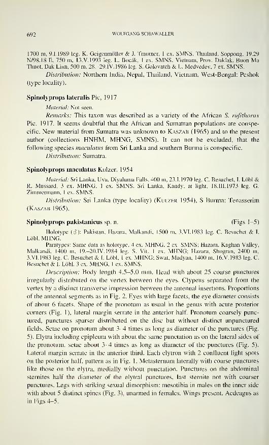
692 WOLFGANG SCHAWALLER
1700 m, 9.1.1989 leg. K. Geigenmüller & J. Trautner, 1 ex. SMNS. Thailand, Soppong, 19.29
N/98.18 E, 750 m, 13.V.1993 leg. L. Bocâk, 1 ex. SMNS. Vietnam, Prov. Daklak, Buon MaThuot, Dak Linn, 500 m, 28.-29.IV. 1986 leg. S..Golovatch & L. Medvedev, 7 ex. SMNS.
Distribution: Northern India, Nepal, Thailand, Vietnam, West-Bengal: Peshok
(type locality).
Spinolyprops lateralis Pic, 1917
Material: Not seen.
Remarks: This taxon was described as a variety of the African S. rufithorax
Pic, 1917. It seems doubtful that the African and Sumatran populations are conspe-
cific. New material from Sumatra was unknown to Kaszab (1965) and to the present
author (collections HNHM, MHNG, SMNS). It can not be excluded, that the
following species maculatus from Sri Lanka and southern Burma is conspecific.
Distribution: Sumatra.
Spinolyprops maculatus Kulzer, 1954
Material: Sri Lanka, Uva, Diyaluma Falls, 400 m, 23.1.1970 leg. C. Besuchet, I. Lobi &R. Mussard, 3 ex. MHNG, 1 ex. SMNS. Sri Lanka, Kandy, at light, 18.III.1973 leg. G.
Zimmermann, 1 ex. SMNS.
Distribution: Sri Lanka (type locality) (Kulzer 1954), S Burma: Tenasserim
(Kaszab 1965).
Spinolyprops pakistanicus sp. n. (Figs 1-5)
Holotype (<?): Pakistan, Hazara, Malkandi, 1500 m, 3. VI. 1983 leg. C. Besuchet & I.
Lobi, MHNG.Paratypes: Same data as holotype, 4 ex. MHNG, 2 ex. SMNS; Hazara, Kaghan Valley,
Malkandi, 1400 m, 19.-20.IV. 1984 leg. S. Vit, 1 ex. MHNG; Hazara, Shogran, 2400 m,
3.VI.1983 leg. C. Besuchet & I. Lobi, 1 ex. MHNG; Swat, Madyan, 1400 m. 16.V.1983 leg. C.
Besuchet & I. Lobi, 3 ex. MHNG, 1 ex. SMNS.
Description: Body length 4.5-5.0 mm. Head with about 25 coarse punctures
irregularly distributed on the vertex between the eyes. Clypeus separated from the
vertex by a distinct transverse impression between the antennal insertions. Proportions
of the antennal segments as in Fig. 2. Eyes with large facets, the eye diameter consists
of about 6 facets. Shape of the pronotum as usual in the genus with acute posterior
corners (Fig. 1), lateral margin serrate in the anterior half. Pronotum coarsely punc-
tured, punctures sparser distributed on the disc but without distinct unpunctured
fields. Setae on pronotum about 3-4 times as long as diameter of the punctures (Fig.
5). Elytra including epipleura with about the same punctation as on the lateral sides of
the pronotum, setae about 3^1 times as long as diameter of the punctures (Fig. 5).
Lateral margin serrate in the anterior third. Each elytron with 2 confluent light spots
on the posterior half, pattern as in Fig. 1. Metasternum laterally with coarse punctures
like those on the elytra, medially without punctation. Punctures on the abdominal
sternites half the diameter of the elytral punctures, last sternite not with coarser
punctures. Legs with striking sexual dimorphism: mesotibia in males on the inner side
with about 5 distinct spines (Fig. 3), unarmed in females. Wings present. Aedeagus as
in Figs 4-5.
SP1NOLYPROPS PAKISTANICUS 693
Figs 1-5
Spinolyprops pakistanicus sp. n. holotype S . - 1: Dorsal view, scale 2 mm; 2: Antenna, scale 1
mm; 3: right mesotibia, scale 0.5 mm; 4: Aedeagus, scale 0.5 mm; 5: Punctation and setation
on medial part of pronotum (left) and on disc of elytra (right).
Remarks: Spinolyprops pakistanicus sp. n. is the first known species of the
genus having sexual dimorphic legs. The other species show no differences in tibial
morphology between males and females. In addition, the aedeagus has a specific
shape, in particular the shape of the fused parameres with an acute tip (see parameres
of the other Oriental species in Fig. 6). Apart from these distinct characters, the
punctation and setation on pronotum and elytra is specific but difficult to use without
having the other species at hand. The dorsal punctation is finer and similar to that in
maculatus, but coarser in himalayicus and in particular in trautneri. The dorsal
setation is similar to that in maculatus and himalayicus, but distinctly shorter in
trautneri. The body shape and proportions and the colour pattern of the elytra are of
less taxonomic value.
Distribution: Pakistan (Hazara, Swat).
Spinolyprops trautneri Schawaller, 1 994
Material: Philippines, Leyte, Lake Danao, 500 m, 19.11.-18.10.1 991 leg. K.
Geigenmüller, W. Schawaller & J. Trautner, holotype S SMNS.
Distribution: Known only from the Philippine Island Leyte (Schawaller
1994).
694 WOLFGANG SCHAWALLER
ZOOGEOGRAPHY
The mountain ranges of the Himalayas lodge an extremely species-rich fauna,
not only of Coleoptera. A first simple reason for this 'megadiversity' is the position of
the Himalayas at the junction of 2 faunal regions, the Oriental region in the south and
the Palaearctic region in the north. Taxa penetrate from both regions into the
Himalayas thus increasing species numbers. Furthermore, a number of taxa reaches
the Himalayas from the mountains of the deep meridional upstreams of the rivers
Irawady, Salween, Mekong, Yangtse and Yalung (Himalayan subregion sensu
Martens 1979), being itself a mixture of Palaearctic and Oriental elements with
many relic endemites. Additionally, the high vertical zonation with different
climatical belts allows a coexistence of many closely related immigrants and the
diverse isolations by deep valleys and high mountain ranges promotes the evolution
of new species swarms from single ancient immigrants in particular of those with
reduced migration possibilities.
In general, the faunal diversity in the Himalayas increases from west to the
east. It is known for a long time that the very western parts in northern Pakistan and
Kashmir generally have a quite poorer (concerning species number) fauna than the
eastern Himalayas in Nepal, Sikkim, Darjeeling and the nearly unexplored
northeastern Assam. The fauna in the western Himalayas is hitherto considered to be
closely related to the faunas in the Middle Asian mountains (Pamir, Tien-Shan) and to
the Afghanistan fauna, thus containing mainly Palaearctic elements. The abundant
occurrence of the tenebrionid genera Platynoscelis (sensu lato), Prosodes or of the
silphid genus Aclypea may be mentioned here as examples for palaearctic taxa on the
genus level, lacking in the central and eastern Himalayas.
However, already Löbl (1986) recognized a number of taxa from the Cole-
optera families Pselaphidae and Scaphidiidae (subfamilies of Staphylinidae sensu
Lawrence & Newton 1995) in northern Pakistan at least in Hazara and Swat having
an Oriental origin. The family Discolomatidae, restricted to tropical habitats, also
occurs in Hazara and Swat (Schawaller 1989). The tenebrionid genus Spinolyprops
seems to be a further example for this distinct Oriental distributional pattern. Its
species are recorded (apart from eastern Africa) from southeastern continental Asia,
the Philippines, Sri Lanka and Sumatra (Fig. 6). In Nepal, himalayicus is known only
from low altitudes below 1000 m with subtropical vegetation. Thus the occurrence of
Spinolyprops pakistanicus sp. n. in Hazara and Swat but not in Chitral up to 2400 m is
remarkable. Obviously, some taxa with a generally Oriental origin can penetrate also
in the very western Himalayas through the valleys of the river Indus and its tribu-
taries. This holds true in particular for mobile taxa like Spinolyprops (with functional
hind wings).
It is not clear up to now if the occurrence of species in eastern Africa and
southeastern Asia including the Philippines point to an ancient Gondwana origin
before the separation of the continental plates or if the single African species has been
introduced secondarily from tropical Asia.
SP/NOLYPROPS PAK1STANÌCUS 695
\S\
Fig. 6
Distribution of the Oriental species of Spinolyprops and shape of the parameres (aedeagus of
lateralis unknown).
ACKNOWLEDGMENTS
For the loan of material I thank Dr Ivan Lobi (Genève) and Dr Otto Merkl
(Budapest).
REFERENCES
Kaszab, Z. 1965. Wissenschaftliche Ergebnisse der von Dr F. Schmid in Indien gesammelten
Tenebrioniden (Coleoptera). Miscelânea Zoològica Barcelona 2: 1 1 1-129.
Kulzer, H. 1954. Achter Beitrag zur Kenntnis der Tenebrioniden (Col.). Entomologische
Arbeiten aus dem Museum G. Frey 5: 20-73.
Lawrence, J.F. & Newton. A.F. 1995. Families and subfamilies of Coleoptera (with selected
genera, notes, references and data on family-group names). Biology, phylogeny, and
classification of Coleoptera, Warszawa, Muzeum i Instytut Zoologii p. 779-1092.
LObl, I. 1986. Contribution à la connaissance des Scaphidiidae (Coleoptera) du nord-ouest de
l'Inde et du Pakistan. Revue suisse de Zoologie 93: 341-367.
Martens. J. 1979. Die Fauna des Nepal-Himalaya - Entstehung und Erforschung. Natur &Museum 109: 221-244.
Schawaller, W. 1989. New Discolomidae (Coleoptera) from the Himalayas. Tropical Zoology
2: 45-58.
Schawaller, W. 1994. New Oriental Tenebrionidae (Coleoptera). Entomofauna 15: 261-280.

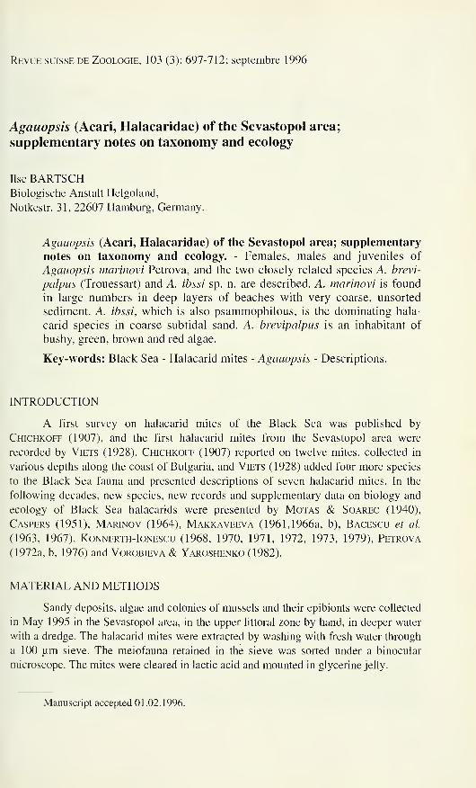
Revue suisse de Zoologie, 103 (3): 697-712; septembre 1996
Agauopsis (Acari, Halacaridae) of the Sevastopol area;
supplementary notes on taxonomy and ecology
Ilse BARTSCHBiologische Anstalt Helgoland,
Notkestr. 31, 22607 Hamburg, Germany.
Agauopsis (Acari, Halacaridae) of the Sevastopol area; supplementary
notes on taxonomy and ecology. - Females, males and juveniles of
Agauopsis inalinovi Petrova, and the two closely related species A. brevi-
palpus (Trouessart) and A. ibssi sp. n. are described. A. marinovi is found
in large numbers in deep layers of beaches with very coarse, unsorted
sediment. A. ibssi, which is also psammophilous, is the dominating hala-
carid species in coarse subtidal sand. A. brevipalpus is an inhabitant of
bushy, green, brown and red algae.
Key-words: Black Sea - Halacarid mites - Agauopsis - Descriptions.
INTRODUCTION
A first survey on halacarid mites of the Black Sea was published by
Chichkoff (1907), and the first halacarid mites from the Sevastopol area were
recorded by Viets (1928). Chichkoff (1907) reported on twelve mites, collected in
various depths along the coast of Bulgaria, and Viets (1928) added four more species
to the Black Sea fauna and presented descriptions of seven halacarid mites. In the
following decades, new species, new records and supplementary data on biology and
ecology of Black Sea halacarids were presented by Motas & Soarec (1940),
Caspers (1951), Marinov (1964), Makkaveeva (1961,1966a, b), Bacescu et al.
(1963, 1967), Konnerth-Ionescu (1968, 1970, 1971, 1972, 1973, 1979), Petrova
(1972a, b, 1976) and Vorobieva & Yaroshenko (1982).
MATERIAL AND METHODS
Sandy deposits, algae and colonies of mussels and their epibionts were collected
in May 1995 in the Sevastopol area, in the upper littoral zone by hand, in deeper water
with a dredge. The halacarid mites were extracted by washing with fresh water through
a 100 um sieve. The meiofauna retained in the sieve was sorted under a binocular
microscope. The mites were cleared in lactic acid and mounted in glycerine jelly.
Manuscript accepted 01.02.1996.
698 ILSE BARTSCH
Slides with type and voucher specimens are deposited in the Muséum d'histoire
naturelle, Genève (MHNG), Naturhistorisches Museum Basel (NMB), Zoologisches
Institut und Zoologisches Museum, Hamburg (ZIMH), and the author's collection.
Abbreviations used in the descriptions: AD, anterior dorsal plate; AE, anterior
epimeral plate; ds, dorsal setae on idiosoma, ds-1, first pair of dorsal setae; GA,
genitoanal plate; GO, genital opening; OC, ocular plate(s); P, palp, P-2, second palpal
segment; pas, parambulacral setae; PD, posterior dorsal plate; PE, posterior epimeral
plate; pgs, perigenital setae; sgs, subgenital setae; vl, ventrolateral; vm, ventromedial.
Legs numbered I to IV, leg segments 1 to 6, i.e. trochanter, basifemur, telofemur,
genu, tibia, and tarsus.
RESULTS
Three species of the genus Agauopsis were found in the Sevastopol area. The
crevice system between bushy algae was inhabited by A. brevipalpus; sublittoral
sandy deposits contained large numbers of A. ibssi, and deep layers of beaches
harboured A. marinovi.
Agauopsis brevipalpus (Trouessart, 1889) Figs 1-11
Agaue brevipalpus Trouessart, 1889a: 1180, 1181; Trouessart, 1889b: 181; Trouessart &Neumann, 1901: 253, pi. 5, figs 2, 2a-2c.
not Agaue brevipalpus, Lohmann, 1893: 22, 87; Rao & Ganapati, 1968: (117).
not Agauopsis brevipalpus brevipalpus, Newell, 1947: 186-189, fig. 316.
Agaue brevipalpus var. pontica Chichkoff, 1907: 258-259, plate 21, figs 1-7 (syn. n.).
Material Examined: One 6 , Crimea, Sevastopol, south-east coast, 2-4 cm high thickets
with Corallina sp. (red algae), 0.3-0.5 m below water level. 1 1 May 1995 (MHNG). One 9, 1
protonymph, same locality, 17 May 1995 (MHNG). One 9, same locality, 17 May 1995
(NMB). One 9, 1 S, same locality, 11 May 1995 (ZIMH A63/95). Four 9, 2 o\ 1 deuto-
nymph, 2 protonymphs, same locality, 11 and 17 May 1995, and 2 9, 2 6 , 1 deutonymph, 1
protonymph, same locality but from Enteromorpha and Cladophora spp. (green algae) and
other small algae from rock pool, 2 and 11 May 1995 (author's collection). One 9,1 S, 1
deutonymph, 1 protonymph, mole of Sevastopol harbour, Mytilus sp. and algae from concrete
tetrapods, 1-1.5 m depth, 3 May 1995; 3 9, 1 â (author's collection), Omega Bay, Cystoseira
sp. (brown algae) and other algae from pier, 0-2 m depth, 6 May 1995; 3 9, 1 â, 2 deuto-
nymphs, 2 protonymphs (author's collection), Kazachya Bay, Cladophora sp. (green algae) and
sediment, 10 m depth, 12 May 1995 (all in the author's collection).
Four 9, 1 deutonymph, Crimea, Laspi, small thickets of Corallina sp., 10-15 cm below
water level, 21 May 1995 (author's collection).
Holotype 9 , France, Atlantic coast. Le Croisic, from red algae (Muséum National
d'Histoire Naturelle, Paris, Collection E. Trouessart Nr. 18C9).
Description (Black Sea material): 9 - Length of idiosoma 470-542 pm.
Integument of plates brownish; eye spots present beneath AD and OC. Raised porose
areolae of dorsal plates with canaliculi of rosette pores piercing the integument; ostia
lacking (Fig. 4). Remainder of plate with scattered delicate canaliculi, and surface of
plate panelled due to cuticular droplets which are more or less fused, thus forming a
reticulum. AD (Fig. 1) with small frontal process; posterior margin truncate or
somewhat excavated. Plate with distinctly raised H-like costa; rosette pores reduced
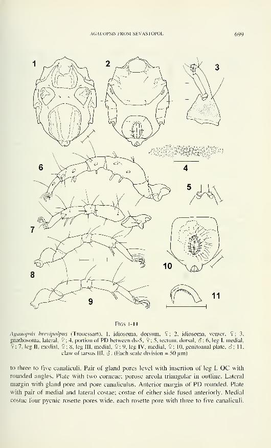
AGAUOPSIS FROM SEVASTOPOL 699
Figs 1 - 1
1
Agauopsis brevipalpus (Trouessart), 1, idiosoma, dorsum, 9; 2, idiosoma, venter, 9; 3,
gnathosoma, lateral, 9 ; 4, portion of PD between ds-5, 9 ; 5, tectum, dorsal, S ; 6, leg I, medial.
9 ; 7, leg II, medial, 9 ; 8, leg III, medial, 9 ; 9, leg IV, medial, 9 ; 10, genitoanal plate, o*; 11,
claw of tarsus III, â . (Each scale division = 50 um)
to three to five canaliculi. Pair of gland pores level with insertion of leg I. OC with
rounded angles. Plate with two corneae; porose areola triangular in outline. Lateral
margin with gland pore and pore canaliculus. Anterior margin of PD rounded. Plate
with pair of medial and lateral costae; costae of either side fused anteriorly. Medial
costae four pycnic rosette pores wide, each rosette pore with three to five canaliculi.

700 ILSE BARTSCH
Dorsal setae 10 pm long and rather stout, ds-1 inserted on AD slightly posterior level
with pair of gland pores, setae ds-2, ds-3 and ds-4 within striated integument, anterior,
between and posterior to OC respectively; ds-5 on PD within reticulate area between
medial and lateral costae and on level with insertion of leg IV.
Ventral plates porose and faintly reticulate. AE (Fig. 2) wide; opening of
epimeral pore slit-like; internal sacculus 10 pm long. Posterior margin of AE and
anterior margin of GA truncate. GO large, distance from anterior margin of genital
foramen to margin of GA 0.3 times length of foramen.
Gnathosoma slender, 1.9 longer than wide. Integument of gnathosomal base
pierced by numerous canaliculi (Fig. 3). Rostrum about as long as gnathosomal base.
Tectum (Fig. 5) triangular, extending to end of P-l. One pair of maxillary setae on
gnathosomal base, one pair near apex of rostrum. Palps slender. P-2 4.0-4.4 times
longer than high; with one dorsal seta. P-3 short, with dorsomedial bluntly ending,
pectinate spine. P-4 somewhat longer than P-3, with two basal setae.
Integument of telofemora reticulate and with canaliculi. Telofemora I - IV 2.5,
2.2, 2.2 and 2.3 times longer than high, and 1.5, 1.3, 1.1 and 1.1 times longer than
tibiae I - IV. Chaetotaxy of legs, from trochanter to tibia (setae in arabic and spines in
roman numerals): leg I, 1, 2, 5+1V, 3+II, 6+III; leg II, 1, 2, 6, 5, 7; leg III, 1, 2, 3, 3, 5;
leg IV, 0, 2, 3, 3, 5. Spines of leg I short, apically blunt and pectinate. 1-3 (Fig. 6) with
two ventral spines, basal one 26 urn, distal one 31 urn long; ventromedial spines both
25 urn long; 1-4 with 17 urn long ventral and almost 25 pm long ventromedial spine;
1-5 with one ventral and two ventromedial spines, each 25 pm long; tarsus I with 16
pm long ventromedial spine. Tibia II (Fig. 7) with three spine-like, bipectinate
bristles; these bristles about as long as height of tibia II. Tibiae III (Fig. 8) and IV
(Fig. 9) each with pair of spine-like pectinate bristles. Tip of tarsus I with pair of
doubled pas, tip of tarsus II medially with spine-like, laterally with a long and a short
seta-like pas, tarsi III and IV each with a single spine-like pas in lateral position.
Pair of claws on tarsus I smooth, distinctly shorter than those of succeeding
legs; median claw stout, bidentate. Paired claws of tarsi II - IV long, with accessory
process and pecten (Fig. 11). Numerous tines of pecten arranged along ventral margin
of the claw (distinctly seen at 400x magnification). Median claw of tarsi II - IV
reduced to a sclerite without denticles.
S - Length of idiosoma 440-469 pm. Dorsal aspect similar to that of female.
GA (Fig. 10) with truncate anterior margin. GO large. Distance from anterior margin
of GO to that of GA same as length of GO. Forty-two to sixty-seven pgs arranged in
two rings around the GO, outer ring with 30-47 perigenital setae, inner ring with 12-
18 setae. Pair of outlying setae on level with anterior setae of ring with perigenital
setae. Genital sclerites with five pairs of spur-like sgs. Spermatopositor not reaching
anterior margin of GA.
Juveniles - Dorsal plates more distinctly reticulated than in adults. Porose
areolae not as raised as in adults but due to dense arrangement of canaliculi set off
from remainder of plates. PD smaller than in adults. Deutonymphs 385-408 pm long.
Genital plate rectangular, with broadly rounded angles, not fused with anal plate.
Spines on leg I short. Genital plate with two pairs of minute subgenital setae close to
AGAUOPSIS FROM SEVASTOPOL 701
primordial genital opening and two pairs of perigenital setae. Protonymphs 290-334
urn long. Neither perigenital nor subgenital setae present on genital plate. Number of
spines of leg I summarized in Table 1
.
Table 1
Agauopsis brevipalpus, number of spines of leg I
telofemur genu tibia tarsus
Deutonymph Ivi, 2vm Ivi, lvm Ivi, 2vm lvmProtonymph Ivi, lvm Ivi, lvm Ivi, lvm lvm
Variations - Out of 20 pairs of legs examined, one female has three spines on
genu I.
Habitat: Agauopsis brevipalpus was commonly found, though never in large
numbers, in samples with algae, from the upper littoral zone down to about 10 mdepth, living amongst thickets of green, brown as well as red algae.
Remarks: With regard to the external morphological characters, the specimens
from the Sevastopol area correspond with the holotype specimen from the French
Atlantic coast.
Chichkoff (1907) described a 650-750 urn long variety, Agaue brevipalpus
var. pontica. According to Chichkoff, A. brevipalpus var. pontica is larger than A.
brevipalpus brevipalpus, and tibiae III and IV have two instead of three pectinate
spines. When the characters are reevaluated, these differences do not exist. The
somewhat depressed type of A. brevipalpus has an idiosomal length of 515 urn (530
urn according to Trouessart, 1889b), its length to tip of rostrum is 625 urn. The
length given by Chichkoff, which obviously includes the gnathosoma, is within the
range of A. brevipalpus. The number of pectinate spines is the same.
The individuals described and illustrated by Chichkoff (1907) agree perfectly
with the type specimen and the mites from the Sevastopol area, e.g., the dorsal plates
have raised costae, the areas outside these costae are coarsely reticulated; the small
triangular tectum of the gnathosoma extends to the end of P- 1 ; the dorsal setae of the
legs are rather long but the medial spines of leg I short; telofemora III and IV are
almost twice as long as high; the claws of tarsi II and III have tines of claw pecten
along their ventral flank. The ecological data, i.e. species wide-spread in the littoral
but never occurring in large numbers (Chichkoff, 1907), also corresponds with that
found to be true for A. brevipalpus in the Sevastopol area.
Distribution: Eastern North Atlantic, Mediterranean, Black Sea.
The records from the western Atlantic (Lohmann, 1893; Newell, 1947), the
Bay of Bengal (Rao & Ganapati, 1968) and from off Sydney, Australia (Lohmann,
1893) certainly are erroneus. A small collection of halacarids from the western
Atlantic, from the Bermuda Islands and the Caribbean area (in the author's collection)
includes two species of Agauopsis closely related but not conspecific with A.
brevipalpus. A slide of Newell's halacarid collection (housed in the United States
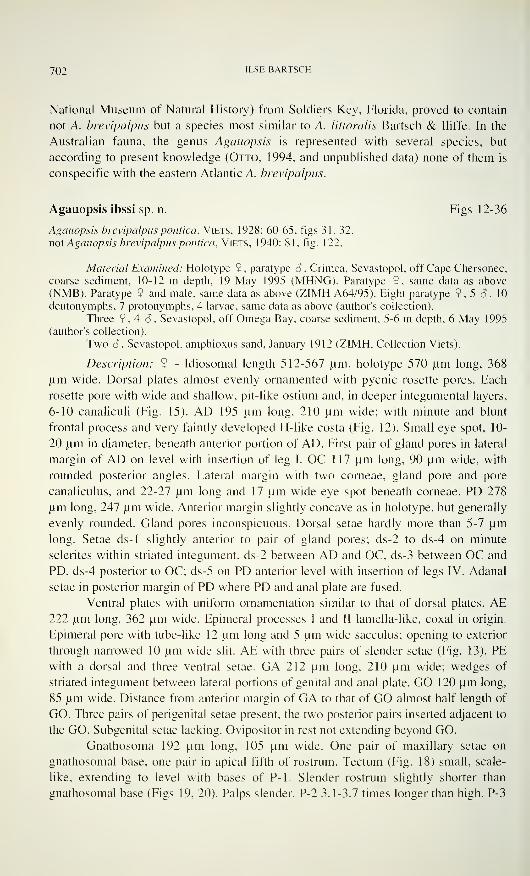
702 ILSE BARTSCH
National Museum of Natural History) from Soldiers Key, Florida, proved to contain
not A. brevipalpus but a species most similar to A. littoralis Bartsch & Iliffe. In the
Australian fauna, the genus Agauopsis is represented with several species, but
according to present knowledge (Otto, 1994, and unpublished data) none of them is
conspecific with the eastern Atlantic A. brevipalpus.
Agauopsis ibssi sp. n. Figs 12-36
Agauopsis brevipalpus politica, ViETS, 1928: 60-65, figs 31, 32.
not Agauopsis brevipalpus pontica, Viets, 1940: 81, fig. 122.
Material Examined: Holotype 9. paratype S , Crimea. Sevastopol, off Cape Chersonec,
coarse sediment, 10-12 m depth, 19 May 1995 (MHNG). Paratype 9, same data as above
(NMB). Paratype 9 and male, same data as above (ZIMH A64/95). Eight paratype 9, 5 o\ 10
deutonymphs, 7 protonymphs, 4 larvae, same data as above (author's collection).
Three 9, 4 â , Sevastopol, off Omega Bay, coarse sediment, 5-6 m depth, 6 May 1995
(author's collection).
Two 6 , Sevastopol, amphioxus sand, January 1912 (ZIMH. Collection Viets).
Description: 9 - Idiosomal length 512-567 urn, holotype 570 urn long, 368
urn wide. Dorsal plates almost evenly ornamented with pycnic rosette pores. Each
rosette pore with wide and shallow, pit-like ostium and, in deeper integumental layers,
6-10 canaliculi (Fig. 15). AD 195 urn long, 210 urn wide; with minute and blunt
frontal process and very faintly developed H-like costa (Fig. 12). Small eye spot, 10-
20 urn in diameter, beneath anterior portion of AD. First pair of gland pores in lateral
margin of AD on level with insertion of leg I. OC 1 17 um long, 90 um wide, with
rounded posterior angles. Lateral margin with two corneae, gland pore and pore
canaliculus, and 22-27 urn long and 17 um wide eye spot beneath corneae. PD 278
urn long, 247 urn wide. Anterior margin slightly concave as in holotype, but generally
evenly rounded. Gland pores inconspicuous. Dorsal setae hardly more than 5-7 urn
long. Setae ds-1 slightly anterior to pair of gland pores; ds-2 to ds-4 on minute
sclerites within striated integument, ds-2 between AD and OC, ds-3 between OC and
PD, ds-4 posterior to OC; ds-5 on PD anterior level with insertion of legs IV. Adanal
setae in posterior margin of PD where PD and anal plate are fused.
Ventral plates with uniform ornamentation similar to that of dorsal plates. AE222 urn long, 362 um wide. Epimeral processes I and II lamella-like, coxal in origin.
Epimeral pore with tube-like 12 urn long and 5 urn wide sacculus; opening to exterior
through narrowed 10 urn wide slit. AE with three pairs of slender setae (Fig. 13). PE
with a dorsal and three ventral setae. GA 212 urn long, 210 urn wide; wedges of
striated integument between lateral portions of genital and anal plate. GO 120 urn long,
85 urn wide. Distance from anterior margin of GA to that of GO almost half length of
GO. Three pairs of perigenital setae present, the two posterior pairs inserted adjacent to
the GO. Subgenital setae lacking. Ovipositor in rest not extending beyond GO.
Gnathosoma 192 um long, 105 urn wide. One pair of maxillary setae on
gnathosomal base, one pair in apical fifth of rostrum. Tectum (Fig. 18) small, scale-
like, extending to level with bases of P-l. Slender rostrum slightly shorter than
gnathosomal base (Figs 19, 20). Palps slender. P-2 3.1-3.7 times longer than high. P-3
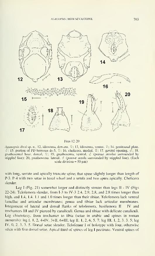
AGAUOPSIS FROM SEVASTOPOL 703
15
-iìf
17
Figs 12-20
Agauopsis ibssi sp. n., 12, idiosoma, dorsum, 9; 13, idiosoma, venter, 2; 14, genitoanal plate,
8; 15, portion of PD between ds-5, 9; 16, chelicera, medial, 2; 17, genital opening, $; 18,
gnathosomal base, dorsal, 9; 19, gnathosoma, ventral, 6 (porose areolae surrounded by
stippled line); 20, gnathosoma, lateral, 6 (porose areola surrounded by stippled line). (Each
scale division = 50 um)
with long, serrate and apically truncate spine; that spine slightly longer than length of
P-3. P-4 with two setae in basal whorl and a setula and two spurs apically. Chelicera
slender.
Leg I (Fig. 21) somewhat larger and distinctly stouter than legs II - IV (Figs
22-24). Telofemora slender, from 1-3 to IV-3 2.4, 2.9, 2.8, and 2.8 times longer than
high, and 1.4, 1.4, 1.1 and 1.0 times longer than their tibiae. Telofemora lack ventral
lamellae and articular membranes; genua and tibiae lack articular membranes.
Integument of lateral and dorsal flanks of telofemora, basifemora II - IV and
trochanters III and IV pierced by canaliculi. Genua and tibiae with delicate canaliculi.
Leg chaetotaxy, from trochanter to tibia (setae in arabic and spines in roman
numerals): leg I, 1,2, 4+IV, 3+II, 6+III; leg II, 1, 2, 6, 5, 7; leg III, 1, 2, 3, 3, 5; leg
IV, 0, 2, 3, 3, 5. Dorsal setae slender. Telofemur I of holotype with four, otherwise
often with five dorsal setae. Apical third of spines of leg I pectinate. Ventral spines of
704 ILSE BARTSCH
Figs 21-27
Agauopsis ibssi sp. n., 21, leg I, medial, 9; 22, basifemur - tarsus II, medial, 9; 23, leg III,
medial, 9; 24, leg IV, dorsomedial, 9; 25, tip of tarsus I, lateral, 9 (medial seta and claw
omitted); 26, tip of tarsus II, medial, S (medial setae and claw dashed); 27, claws of tarsus III,
â . (Each scale division = 50 urn)
telofemur 42 and 48 urn, of genu 25 urn and of tibia 42 urn long, medial spines of 1-3
36 and 40 urn, of 1-4 39 urn, of 1-5 42 and 36 urn and of 1-6 25 urn long. Tibia II with
three spine-like pectinate bristles; ventromedial bristle in its distal third widened and
bipectinate. Tibiae III and IV with pair of spine-like, pointed and delicately pectinate
bristles. Tarsus I distinctly shorter than tibia I. Medial membrane of claw fossa 2-3
urn wide; slightly larger lateral membrane (Fig. 25) with small solenidion and internal
famulus. Membranes of claw fossa of tarsi II - IV 10 urn long, and 3-5 urn high.
Solenidion of tarsus II (Fig. 26) inserted on inner flank of medial membrane of claw
fossa. Apex of tarsus I with pair of doubled pas, that of tarsus II laterally with a long
and a short seta-like pas, medially with a spur-like pas; lateral pas of tarsi III and IV
spur-like, medial pas lacking.
Paired claws of tarsus I somewhat shorter and median claw larger than claws
of succeeding tarsi. Median claw of tarsus I bidentate. Paired claws with minute
accessory process, claws on tarsi II - IV with very delicate pecten. Pecten (Fig. 27) on
AGAUOPSIS FROM SEVASTOPOL 705
outer flank of claw with 10 tines, pecten on inner flank of claw with 22 tines (tines
generally not seen by 400x magnification).
S - Idiosomal length 515-569 urn. Dorsal aspect same as that of female.
Relative to its length GA wider than that of female; GA of 533 urn long paratype 247
urn long, 247 urn wide. GO 90 urn long, 35 urn wide. Distance from anterior margin
of GO to that of GA same as length of GO (Fig. 14). With 25-34 perigenital setae
arranged around GO; of these, generally five to six pairs of setae adjacent to and 14-
22 setae in a ring around GO. Pair of outlying perigenital setae anterior to that ring
with setae. Genital sclerites (Fig. 17) with five pairs of short subgenital setae.
Spermatopositor extending somewhat beyond anterior margin of GA.
Juveniles - Dorsal and ventral plates smaller than in adults (Figs 28 - 33). PDwith distinctly demarcated costae in that the ostia of the rosette pores are smaller
within the costae than in remainder of the plate, namely 2-3 urn wide within the
costae versus 5 pm wide outside the costae.
Deutonymph 397-484 urn long. Almost quadrangular genital plate with two
pairs of pgs. Two pairs of small sgs adjacent to 10 pm long primordial genital
opening, and two pairs of internal genital acetabula beneath genital area. Telofemora
and tibia I (Fig. 34) each with a ventral and two ventromedial spines, 1-4 with long
ventromedial and short ventral spine, tarsus I with ventromedial spine. Tibia II with
three spine-like setae; tibiae III and IV with pair of spine-like setae.
Protonymph 309-337 pm long. PD with pair of medial and lateral costae.
Genital plate with single pair of genital acetabula; pgs and sgs lacking. 1-3, 1-4 and 1-5
each with a ventral and a ventromedial spine, 1-6 with ventromedial spine (Fig. 35).
Tibiae II - IV each with pair of ventral setae.
Larva 213-235 pm long. PD short, medial and lateral costae distinct due to
their more intense porosity and smaller ostia. Integument outside costae with large
ostia and almost inconspicuous canaliculi. Genital plate lacking. No spines on
telofemur I; genu I and tibia I with pair of pectinate spines, tarsus I with ventromedial
spine (Fig. 36). Tibiae II and III with pair of setae, those of III-5 tapering. Claws of
tarsus I slender.
Variations - In one of 20 pairs of adult legs examined tibia III has three instead
of two ventral bristles.
In one of the protonymphs the OC are fused with the PD.
Etymology: The species is named after the Institute of Biology of Southern
Seas, IB SS, which was founded in 1871, 125 years ago.
Habitat: Agauopsis ibssi is an inhabitant of sublittoral sandy deposits. In the
Sevastopol area the species was found in large numbers in coarse, unsorted
amphioxus sand.
Remarks: Agauopsis ibssi differs from A. brevipalpus in that the PD is almost
uniformly porose whereas the PD of A. brevipalpus has two pairs of costae; the dorsal
setae are somewhat smaller and the ds-1 insert slightly anterior to the gland pores, the
palps are 3.1-3.7 times longer than high, the tectum extends to level with bases of P-l,
the telofemora III and IV are more slender than in A. brevipalpus, the tines of claw
pecten are not recognizable at 400x magnification and the claws of tarsus I and the
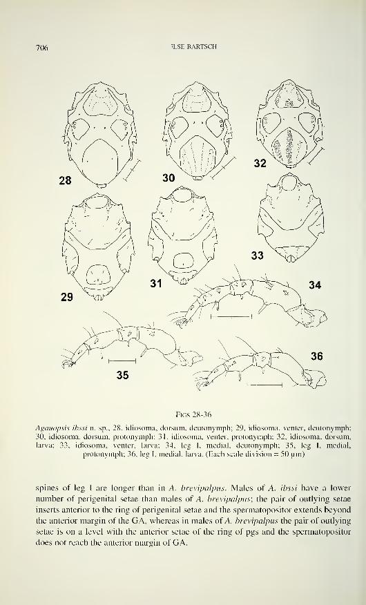
706 ILSE BARTSCH
Figs 28-36
Agaiiopsis ibssi n. sp., 28, idiosoma, dorsum, deutonymph; 29, idiosoma, venter, deutonymph;
30, idiosoma, dorsum, protonymph; 31, idiosoma, venter, protonymph; 32, idiosoma, dorsum,
larva; 33, idiosoma, venter, larva; 34, leg I. medial, deutonymph; 35, leg I, medial,
protonymph; 36, leg I, medial, larva. (Each scale division = 50 um)
spines of leg I are longer than in A. brevipalpus. Males of A. ibssi have a lower
number of perigenital setae than males of A. brevipalpus; the pair of outlying setae
inserts anterior to the ring of perigenital setae and the spermatopositor extends beyond
the anterior margin of the GA, whereas in males of A. brevipalpus the pair of outlying
setae is on a level with the anterior setae of the ring of pgs and the spermatopositor
does not reach the anterior margin of GA.
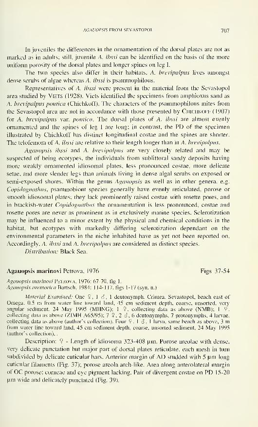
AGAU0PS1S FROM SEVASTOPOL 7()7
In juveniles the differences in the ornamentation of the dorsal plates are not as
marked as in adults; still, juvenile A. ibssi can be identified on the basis of the more
uniform porosity of the dorsal plates and longer spines on leg I.
The two species also differ in their habitats, A. brevipalpus lives amongst
dense scrubs of algae whereas A. ibssi is psammophilous.
Representatives of A. ibssi were present in the material from the Sevastopol
area studied by Viets (1928). Viets identified the specimens from amphioxus sand as
A. brevipalpus politica (Chichkoff). The characters of the psammophilous mites from
the Sevastopol area are not in accordance with those presented by Chichkoff (1907)
for A. brevipalpus var. pontica. The dorsal plates of A. ibssi are almost evenly
ornamented and the spines of leg I are long; in contrast, the PD of the specimen
illustrated by Chichkoff has distinct longitudinal costae and the spines are shorter.
The telofemora of A. ibssi are relative to their length longer than in A. brevipalpus.
Agauopsis ibssi and A. brevipalpus are very closely related and may be
suspected of being ecotypes, the individuals from sublittoral sandy deposits having
more weakly ornamented idiosomal plates, less pronounced costae, more delicate
setae, and more slender legs than animals living in dense algal scrubs on exposed or
semi-exposed shores. Within the genus Agauopsis as well as in other genera, e.g.
Copidognathus, psammobiont species generally have evenly reticulated, porose or
smooth idiosomal plates; they lack prominently raised costae with rosette pores, and
in brackish-water Copidognathus the ornamentation is less pronounced, costae and
rosette pores are never as prominent as in exclusively marine species. Sclerotization
may be influenced to a minor extent by the physical and chemical conditions in the
habitat, but ecotypes with markedly differing sclerotization dependant on the
environmental parameters in the niche inhabited have as yet not been reported on.
Accordingly, A. ibssi and A. brevipalpus are considered as distinct species.
Distribution: Black Sea.
Agauopsis marinovi Petrova, 1976 Figs 37-54
Agauopsis marinovi Petrova, 1976: 67-70, fig 1.
Agauopsis aremorica Bartsch, 1984: 1 14-117, figs 1-17 (syn. n.)
Material Examined: One 9 , 1 S , 1 deutonymph, Crimea, Sevastopol, beach east of
Omega, 0.5 m from water line toward land, 45 cm sediment depth, coarse, unsorted, very
angular sediment, 24 May 1995 (MHNG); 1 9, collecting data as above (NMB); 1 9,collecting data as above (ZIMH A65/95); 7 9,2 6,6 deutonymphs, 7 protonymphs, 4 larvae,
collecting data as above (author's collection). Four 9, 1 6 , 1 larva, same beach as above, 3 mfrom water line toward land, 45 cm sediment depth, coarse, unsorted sediment, 24 May 1995
(author's collection), .
Description: 9 - Length of idiosoma 323-408 urn. Porose areolae with dense,
very delicate punctation but major part of dorsal plates reticulate, each mesh in turn
subdivided by delicate cuticular bars. Anterior margin of AD studded with 5 urn long
cuticular filaments (Fig. 37); porose areola arch-like. Area along anterolateral margin
of OC porose; corneae and eye pigment lacking. Pair of divergent costae on PD 15-20
urn wide and delicately punctated (Fig. 39).
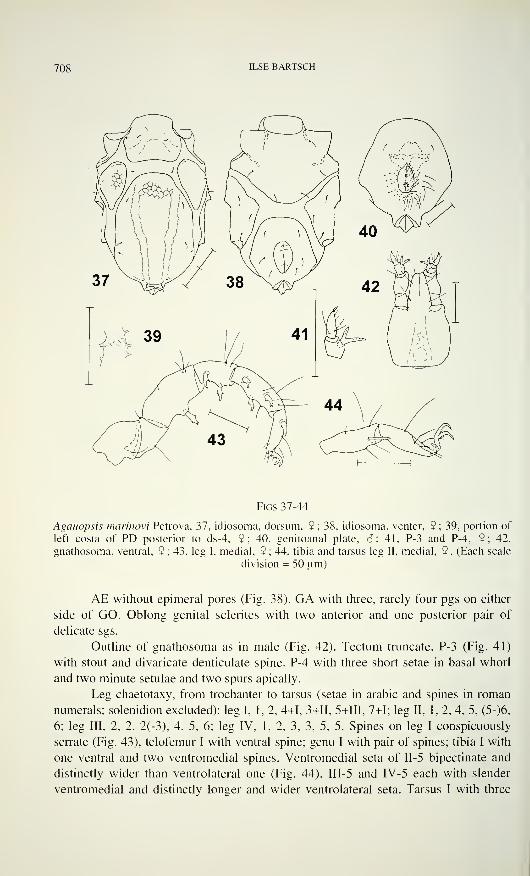
708 ILSE BARTSCH
Figs 37-44
Agaitopsis marinovi Petrova, 37, idiosoma, dorsum, 9; 38, idiosoma, venter, 9; 39, portion of
left costa of PD posterior to ds-4, 9; 40, genitoanal plate, <?; 41, P-3 and P-4, 9; 42,
gnathosoma, ventral, 9 ; 43, leg I, medial, 9 ; 44, tibia and tarsus leg II, medial, 9 . (Each scale
division = 50 pm)
AE without epimeral pores (Fig. 38). GA with three, rarely four pgs on either
side of GO. Oblong genital sclerites with two anterior and one posterior pair of
delicate sgs.
Outline of gnathosoma as in male (Fig. 42). Tectum truncate. P-3 (Fig. 41)
with stout and divaricate denticulate spine. P-4 with three short setae in basal whorl
and two minute setulae and two spurs apically.
Leg chaetotaxy, from trochanter to tarsus (setae in arabic and spines in roman
numerals; solenidion excluded): leg I, 1, 2, 4+1, 3+II, 5+III, 7+1; leg II, 1, 2, 4, 5, (5-)6,
6; leg III, 2, 2, 2(-3), 4, 5, 6; leg IV, 1, 2, 3, 3, 5, 5. Spines on leg I conspicuously
sen-ate (Fig. 43), telofemur I with ventral spine; genu I with pair of spines; tibia I with
one ventral and two ventromedial spines. Ventromedial seta of II-5 bipectinate and
distinctly wider than ventrolateral one (Fig. 44). III-5 and IV-5 each with slender
ventromedial and distinctly longer and wider ventrolateral seta. Tarsus I with three
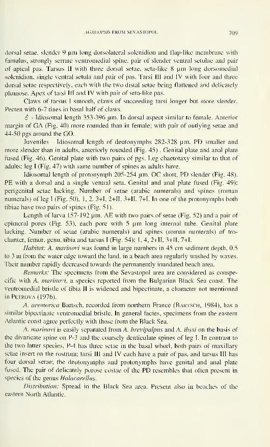
AGAUOPSIS FROM SEVASTOPOL 709
dorsal setae, slender 9 pm long dorsolateral solenidion and flap-like membrane with
famulus, strongly serrate ventromedial spine, pair of slender ventral setulae and pair
of apical pas. Tarsus II with three dorsal setae, seta-like 8 pm long dorsomedial
solenidion, single ventral setula and pair of pas. Tarsi III and IV with four and three
dorsal setae respectively, each with the two distal setae being flattened and delicately
plumose. Apex of tarsi III and IV with pair of seta-like pas.
Claws of tarsus I smooth, claws of succeeding tarsi longer but more slender.
Pecten with 6-7 tines in basal half of claws.
S - Idiosomal length 353-396 pm. In dorsal aspect similar to female. Anterior
margin of GA (Fig. 40) more rounded than in female; with pair of outlying setae and
44-50 pgs around the GO.
Juveniles - Idiosomal length of deutonymphs 282-328 pm. PD smaller and
more slender than in adults, anteriorly rounded (Fig. 45) . Genital plate and anal plate
fused (Fig. 46). Genital plate with two pairs of pgs. Leg chaetotaxy similar to that of
adults; leg I (Fig. 47) with same number of spines as adults have.
Idiosomal length of protonymph 205-254 pm. OC short, PD slender (Fig. 48).
PE with a dorsal and a single ventral seta. Genital and anal plate fused (Fig. 49);
perigenital setae lacking. Number of setae (arabic numerals) and spines (roman
numerals) of leg I (Fig. 50), 1, 2, 3+1, 2+II, 3+II, 7+1. In one of the protonymphs both
tibiae have two pairs of spines (Fig. 51).
Length of larva 157-192 pm. AE with two pairs of setae (Fig. 52) and a pair of
epimeral pores (Fig. 53), each pore with 5 pm long internal tube. Genital plate
lacking. Number of setae (arabic numerals) and spines (roman numerals) of tro-
chanter, femur, genu, tibia and tarsus I (Fig. 54): 1, 4, 2+II, 3+II, 7+1.
Habitat: A. marinavi was found in large numbers in 45 cm sediment depth, 0.5
to 3 m from the water edge toward the land, in a beach area regularly washed by waves.
Their number rapidly decreased towards the permanently inundated beach area.
Remarks: The specimens from the Sevastopol area are considered as conspe-
cific with A. marinovi, a species reported from the Bulgarian Black Sea coast. The
ventromedial bristle of tibia II is widened and bipectinate, a character not mentioned
in Petrova (1976).
A. aremorica Bartsch, recorded from northern France (Bartsch, 1984), has a
similar bipectinate ventromedial bristle. In general facies, specimens from the eastern
Atlantic coast agree perfectly with those from the Black Sea.
A. marinovi is easily separated from A. brevipalpus and A. ibssi on the basis of
the divaricate spine on P-3 and the coarsely denticulate spines of leg I. In contrast to
the two latter species, P-4 has three setae in the basal whorl, both pairs of maxillary
setae insert on the rostrum; tarsi III and IV each have a pair of pas, and tarsus III has
four dorsal setae; the deutonymphs and protonymphs have genital and anal plate
fused. The pair of delicately porose costae of the PD resembles that often present in
species of the genus Halacarellus.
Distribution: Spread in the Black Sea area. Present also in beaches of the
eastern North Atlantic.
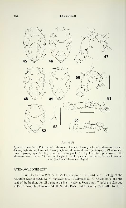
710 ILSE BARTSCH
Figs 45-54
Agauopsis marinovi Petrova, 45, idiosoma, dorsum, deutonymph; 46, idiosoma, venter,
deutonymph; 47, leg I, medial, deutonymph; 48, idiosoma, dorsum, protonymph; 49, idiosoma,
venter, protonymph; 50, leg I. medial, protonymph; 51, leg I. ventral, protonymph; 52,
idiosoma, venter, larva; 53, portion of right AE with epimeral pore, larva; 54, leg I, ventral,
larva. (Each scale division = 50 urn)
ACKNOWLEDGEMENT
I am indebted to Prof. V. E. Zaika, director of the Institute of Biology of the
Southern Seas (IBSS), Dr N. Shalovenkov, T. Nikolaenko, E. Kolesnikova and the
staff of the Institute for all the help during my stay at Sevastopol. Thanks are also due
to Dr H. Dastych, Hamburg, M. H. Naudo, Paris, and R. Smiley, Beltsville, for loan
AGAUOPSIS FROM SEVASTOPOL 7 \ \
of halacarid mites stored in the museums. The study of the halacarid fauna of
Sevastopol area, Crimea, was supported by the Deutsche Forschungsgemeinschaft
which is gratefully acknowledged.
REFERENCES
Bartsch, I. 1984. Ergänzungen zur Halacariden-Fauna der Bretagne-Küste und Beschreibung
einer neuen Art (Halacaridae, Acari). Cahiers de Biologie marine 25: 1 13-122.
Bacescu, M., Dumitrescu, E., Gomoiv, M.T. & A. Petran. 1967. Éléments pour la caractéri-
sation de la zone sédimentaire médio-littorale de la Mer Noire. Travaux du Muséumd'Histoire Naturelle Grigore Antipa 7: 1-14.
Bacescu, M., Dumitrescu, E., Marcus, A., Paladian, G. & R. Mayer. 1963. Données
quantitatives sur la faune pétricole de la Mer Noire à Agigea (Secteur Roumain), dans
les conditions spéciales de Tannée 1961. Travaux du Muséum d'Histoire Naturelle
Grigore Antipa 4: 131-155.
Caspers, H. 1951. Quantitative Untersuchungen über die Bodentierwelt des Schwarzen Meeres
im bulgarischen Küstenbereich. Archivfür Hydrobiologie 45: 1-192.
Chichkoff, G. 1907. Halacaridae des côtes Bulgares. Archives de Zoologie expérimentale et
générale 7: 247-268.
Konnerth-Ionescu, A. 1970. Nouvelles données sur les halacarides de la zone psamicole du
littoral roumain de la Mer Noire. Travaux du Muséum d'Histoire Naturelle Grigore
Antipa 10: 19-23.
Konnerth-Ionescu, A. 1971. Les Halacaridae (Acari) du substrat pétricole (littoral roumain de
la Mer Noire). Travaux du Muséum d'Histoire Naturelle Grigore Antipa 1 1 : 91-97.
Konnerth-Ionescu, A. 1979. Halacarus (Halacarellus) procerus Viets (Acari, Halacaridae) unhalacaride nouveau pour le littoral roumain de la Mer Noire. Travaux du Muséumd'Histoire Naturelle Grigore Antipa 20: 121-123.
Makkaveeva, E.B. 1961. Melkie cervi, rakoobraznie i morskie klesci biocenoza cistoziri.
Trudy sevastopolskoi biologiceskoi Standi 14: 147-162.
Makkaveeva, E.B. 1966b. On ecology and quantitative distribution of arachnids of the
northeastern part of the Black Sea. Gidrobiologiceskii Zhurnal, Kiev 2 (5): 30-33. In
Russian, with English Summary.
Marinov, T. 1964. On the microzoobenthos fauna of the Black Sea (Kinorhyncha andHalacaridae). Izvestija na naucnoizsledovatelskija Instituta po Ribovudstvo i Ribolov,
Varna 4: 61-71. In Bulgarian, with English Summary.
Motas, C. & J. Soarec, 1940: Sur quelques halacarides de la Mer Noire. Annales scientifiques
de l'Université de Jassy 26: 139-175.
Newell, I.M. 1947. A systematic and ecological study of the Halacaridae of eastern North
America. Bulletin of the Bingham Océanographie Collection 10: 1-232.
Otto, J.C. 1994. New species of Halacaridae from Australia (Acarina: Prostigmata). Acaro-
logia 35: 31-48.
Petrova, A. 1972. Sur la présence d'Halacarellus subterraneus Schulz, 1933 et Halacarellus
phreaticus n. sp. (Halacaridae, Acari) en Bulgarie. Acarologia, 13: 367-373.
Petrova, A. 1976. Une nouvelle espèce dAgauopsis (Prostigmata, Halacaridae) du littoral de
la Mer Noire. Chidrobiologiya, Sofiya 4: 67-70.
Rao, G.C. & P.N. Ganapati. The interstitial fauna inhabiting the beach sands of Waltair coast.
Proceedings of the National Institute ofSciences ofIndia B, 34: 82-125.
Trouessart, E. 1889a. Sur les acariens marins des côtes de France. Compte rendu hebdo-
madaire des séances de l'Académie des sciences 108: 1 178-1 181.
712 ILSE BARTSCH
Trouessart, E. 1889b. D'acariens marins (Halacaridae) des côtes de France. Diagnoses
d'espèces et genres nouveaux. Naturaliste, Paris, sér. 2, 11: 181.
Trouessart, E. & G. Neumann. 1901. Note sur les acariens marins (Halacaridae) récoltés par
M. Henri Gadeau de Kerville dans la région d'Omonville-la-Rouge (Manche) et dans la
fosse de la Hague. Bulletin de la Société des amis des sciences naturelles, Rouen, sér 4,
14: 247-266, pi. IV, V.
Viets, K. 1928. Wassermilben aus dem Schwarzen Meer, dem Kaspischen Meer und dem Aral-
See. Abhandlungen. Naturwissenschaftlichen Verein zu Bremen 27: 47-80.
Viets, K. 1940. Meeresmilben aus der Adria (Halacaridae und Hydrachnellae, Acari). Archiv
für Naturgeschichte N.F. 9: 1-135.
Vorobieva, L.V. & N.A. Yaroshenko. 1982. The quantitative composition of Halacaridae of
the Odessa Bay and Black Sea limans. Gidrobiologiceskii Zhurnal, Kiev 18 (3): 40-43.
In Russian, English Summary.
Revue suisse de Zoologie, 103 (3) 713-736; septembre 1996
Stiliderus and Stilicoderus: New data and new species
(Coleoptera, Staphylinidae, Paederinae)*
G.M. de ROUGEMONT27 Walcot Square, London SEI 1 4UB, England, UK.
Stiliderus and Stilicoderus: New data and new species (Coleoptera,
Staphylinidae, Paederinae). - A systematic catalogue of the species of
Stiliderus and Stilicoderus is presented. Fifteen new species are described.
Males of two known species are described for the first time. Eleven species
are recorded for the first time from China; nine species are described from
Sulawesi, all of them endemic. Species are recorded for the first time from
other countries.
Key-words: Coleoptera - Staphylinidae - Paederinae - Stiliderus -
Stilicoderus - China - Sulawesi.
INTRODUCTION
In the nine years that have elapsed since the publication of a series of articles,
including a revision in two parts of this group, other species have been described, and
a large volume of new material has passed through my hands. Material studied in this
paper comes in part from my own occasional prospections in Asia, but the largest
contribution comes from recent collections made by I. Lobi and others for the Geneva
Natural History Museum. Further material was provided by the Natural History
museums in London and Vienna. The study of large collections from Australia in the
Australian National Insect Collection, Canberra, and the Field Museum of Natural
History, Chicago, is to be published separately, as a review of the Australian fauna
(Rougemont, 1996).
In the present paper, apart from the descriptions of new species or of pre-
viously undescribed male sex characters, I have only included such new data as
significantly adds to our knowledge of species distribution, such as first records for
countries or major islands.
Although in my earlier papers I followed Blackwelder's synonymy of Stilico-
derus with Stiliderus, I have gone on thinking of these as distinct genera, and reverted
to labelling specimens as such according to the character state of the fourth tarsomeres
(In the paper on the Australian fauna all species are described as Stilicoderus).
* 29th contribution to the knowledge of Staphylinidae
Manuscript accepted 04.12.1995.
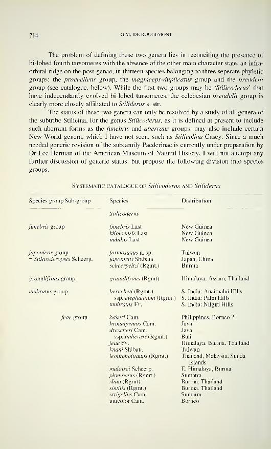
714 G.M. DE ROUGEMONT
The problem of defining these two genera lies in reconciling the presence of
bi-lobed fourth tarsomeres with the absence of the other main character state, an infra-
orbital ridge on the post-genae, in thirteen species belonging to three seperate phyletic
groups: the praecellens group, the magniceps-duplicatus group and the brendelli
group (see catalogue, below). While the first two groups may be'
Stilicoderus' that
have independantly evolved bi-lobed tarsomeres, the celebesian brendelli group is
clearly more closely affiliated to Stiliderus s. str.
The status of these two genera can only be resolved by a study of all genera of
the subtribe Stilicina, for the genus Stilicoderus, as it is defined at present to include
such aberrant forms as the funebris and aberrans groups, may also include certain
New World genera, which I have not seen, such as Stilicolina Casey. Since a much
needed generic revision of the subfamily Paederinae is currently under preparation by
Dr Lee Herman of the American Museum of Natural History, I will not attempt any
further discussion of generic status, but propose the following division into species
groups.
Systematic catalogue of Stilicoderus and Stiliderus
Species group Sub-group Species Distribution
Stilicoderus
funebris group funebris Last
kiloloensis Last
nubilus Last
New Guinea
New GuineaNew Guinea
japomcus group
= Stilicoderopsis Scheerp.
formosanus n. sp.
japonicus Shibata
scheerpehzi (Rgmt.)
Taiwan
Japan, China
Burma
granulifrons group
umbratus group
granulifrons (Rgmt)
besuchen (Rgmt.)
ssp. elephantium (Rgmt.)
umbratus Fv.
Himalaya, Assam, Thailand
S. India: Anaimalai Hills
S. India: Paini Hills
S. India: Nilgiri Hills
feae group bakeri Cam.bruneipennis Cam.drescheri Cam.
ssp. baliensis (Rgmt.)
feae Fv.
kuani Shibata
leontopolitanus (Rgmt.)
malaisei Scheerp.
plumbatus (Rgmt.)
shan (Rgmt)
similis (Rgmt.)
strigelIus Cam.unicolor Cam.
Philippines, Borneo ?
Java
Java
Bali
Himalaya, Burma, Thailand
TaiwanThailand, Malaysia, Sunda
Islands
E. Himalaya, BurmaSumatra
Burma, Thailand
Burma, Thailand
Sumatra
Borneo
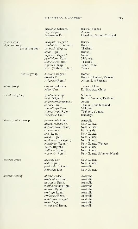
ST1LIDERUS AND STILICODERUS 715
feae-discalis-
signatus group
signants group
discalis group
minor group
variolosus group
hieroglyphicus group
aerosus group
aberrans group
birmanus Scheerp.
clayi (Rgmt.)
fenestratus Fv.
incognitas (Rgmt.)
kambaitiensis Scheerp.
lomholdti (Rgmt.
)
maai (Rgmt.)
nepalensi (Rgmt.)
pendleburyi Cam.siamensis (Rgmt.)
signatus Sharp
n. sp. (Shibata, in litt.)
bacchusi (Rgmt.)
discalis Fv.
strigosus (Rgmt.)
exiguitas Shibata
minor Cam.
gondaicus n. sp.
helferi (Rgmt.)
nagamontium (Rgmt.)
parvus Cam.rotundiceps Cam.trapezeiceps (Rgmt.)
variolosus Coiff.
ferromontis Rgmt.
hieroglyphicus Fv.
hornabrooki (Rgmt.)
kaiensis n. sp.
leai (Rgmt.)
loksai (Rgmt.)
madangensis (Rgmt.)
maritimus (Rgmt.)
sharpi (Rgmt.)
wallacei (Rgmt.)
wauensis (Rgmt.)
aerosus Last
lasti (Rgmt.)
peninsularis Rgmt.
solitarius Last
aberrans Steel
arnhemicus Rgmt.
inusitatus Rgmt.
matthewsianus Rgmt.
newtoni Rgmt.orbiceps Rgmt.
pruinosus Rgmt.
quadraticeps Rgmt.taylori Rgmt.
woodwardi Rgmt.
Burma, YunnanAssamHimalaya, Burma, Thailand
BurmaBurmaThailand
BorneoNepal
Malaysia
Thailand
Japan, China
Taiwan
Borneo
Burma, Thailand, Vietnam
Assam S. to Sumatra
Taiwan, China
E. Himalaya, China
S. India
Burma, Yunnan, Thailand
AssamThailand, Sunda Islands
Malaysia
Thailand, YunnanHimalaya
Australia
New Guinea
New Guinea
Kai Islands
New Guinea
New Guinea
New Guinea
New Guinea, WaigeoNew Guinea
New GuineaNew Guinea, Solomon Islands
New GuineaNew Guinea
Australia
New Guinea
Australia
Australia
Australia
Australia
Australia
Australia
Australia
Australia
Australia
Australia
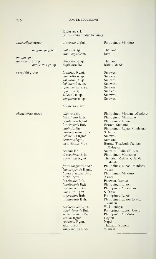
716 G.M. DI-: ROUGI-MONT
Stiìiderus s. 1.
(infra-orbital reidge lacking)
praecellens group praecellens Bnh. Philippines: Mindoro
magniceps group cottemi n. sp. Thailand
magniceps Cam. Java
magniceps-
duplicatus group depressus n. sp. Thailand
duplicatus group duplicatus Ito Riuku Islands
brendelli group brendelli Rgmt. Sulawes i
conicollis n. sp. Sulawes i
kakihitam n. sp. Sulawes i
kakimerah n. sp. Sulawes t
opacipennis n. sp. Sulawes i
opacus n. sp. Sulawes i
schoedli n. sp. Sulawes i
yangbesar n. sp. Sulawesi
Stiìiderus s. str.
cicatricosus group ancora Bnh. Philippines: Masbate. Mindorobakerianus Bnh. Philippines: Mindanaobernhaueri Rgmt. Philippines: Luzonbrevipennis Bnh. Borneo, Sumatra
capitalis Bnh. Philippines: Leyte, Mindanaocardamomensis n. sp. S. India
celebensis Rgmt. Sulawesi
cernatus Rgmt. Sumatra
cicatricosus Mots. Burma, Thailand, Yunnan,
Malaysia
crassus Kr Sulawesi, India, SE Asia
densissimus Bnh. Philippines: Mindanaoexpectatus Rgmt. Thailand, Malaysia, Sunda
Islands
flavomarginatus Bnh. Philippines: Luzon, Mindorokamarupensis Rgmt. Assamlatericarinatus Bnh. Philippines: Mindoro/oé'/p// Rgmt. Assamlongicollis Bnh. Palawan, Borneo
longipennis Bnh. Philippines: Luzonmicropterus Bnh. Philippines: Mindanaomussardi Rgmt. S. India
nigerrimus Bnh. Philippines: Luzonnitidipennis Bnh. Philippines: Luzon, Leyte,
Samanoccidentalis Rgmt. W. Himalayapulchripennis Bnh. Philippines: Luzon, Leyte
semicoeruleus Rgmt. Philippines: Mindorosimoni Rgmt. Ceylon
smetanai Rgmt. Nepal
y/À.'or n. sp. Thailand, Yunnanyunnanensis n. sp. Yunnan
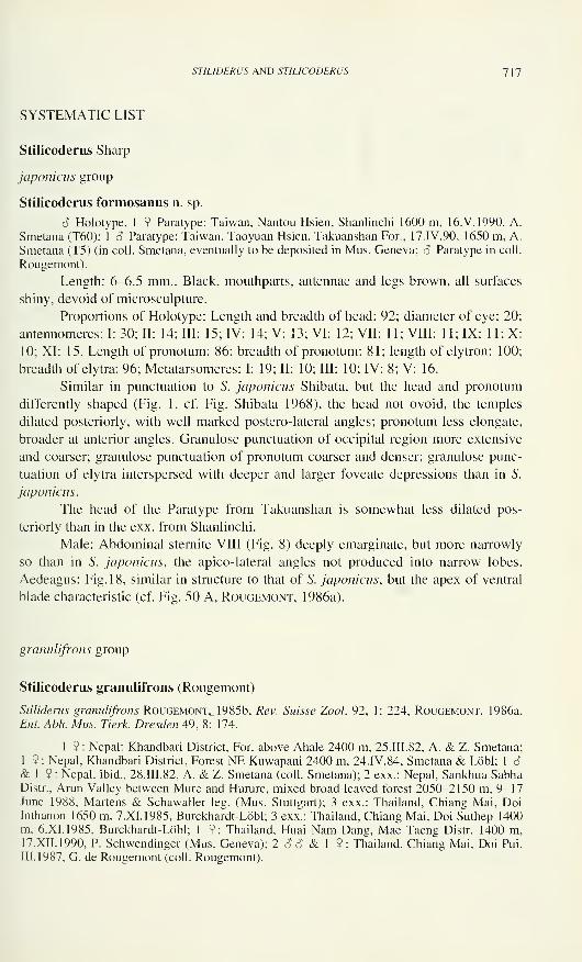
STILIDERUS AND STIL/CODERUS 7 \ 7
SYSTEMATIC LIST
Stilicoderus Sharp
japonicus group
Stilicoderus formosanus n. sp.
6 Holotype, 1 9 Paratype: Taiwan, Nantou Hsien, Shanlinchi 1600 m, 16.V.1990, A.
Smetana (T60); 1 6 Paratype: Taiwan, Taoyuan Hsien, Takuanshan For., 17. IV. 90, 1650 m. A.
Smetana (T5) (in coll. Smetana, eventually to be deposited in Mus. Geneva; 3 Paratype in coll.
Rougemont).
Length: 6-6.5 mm.. Black, mouthparts, antennae and legs brown, all surfaces
shiny, devoid of microsculpture.
Proportions of Holotype: Length and breadth of head: 92; diameter of eye: 20
antennomeres: I: 30; II: 14; III: 15; IV: 14; V: 13; VI: 12; VII: 11; VIII: 11; IX: 11; X10; XI: 15. Length of pronotum: 86; breadth of pronotum: 81; length of elytron: 100
breadth of elytra: 96; Metatarsomeres: I: 19; II: 10; III: 10; IV: 8; V: 16.
Similar in punctuation to S. japonicus Shibata, but the head and pronotum
differently shaped (Fig. 1, cf. Fig. Shibata 1968), the head not ovoid, the temples
dilated posteriorly, with well marked postero-lateral angles; pronotum less elongate,
broader at anterior angles. Granulose punctuation of occipital region more extensive
and coarser; granulose punctuation of pronotum coarser and denser; granulose punc-
tuation of elytra interspersed with deeper and larger foveate depressions than in 5".
japonicus.
The head of the Paratype from Takuanshan is somewhat less dilated pos-
teriorly than in the exx. from Shanlinchi.
Male: Abdominal sternite VIII (Fig. 8) deeply emarginate, but more narrowly
so than in S. japonicus, the apico-lateral angles not produced into narrow lobes.
Aedeagus: Fig. 18, similar in structure to that of S. japonicus, but the apex of ventral
blade characteristic (cf. Fig. 50 A, Rougemont, 1986a).
granulifrons group
Stilicoderus granulifrons (Rougemont)
Stiliderus granulifrons Rougemont, 1985b, Rev. Suisse Zool. 92, 1: 224; Rougemont, 1986a,
Ent. Abh. Mus. Tierk. Dresden 49, 8: 174.
1 9 : Nepal: Khandbari District, For. above Aliale 2400 m, 25. III. 82, A. & Z. Smetana;
1 9: Nepal, Khandbari District, Forest NE Kuwapani 2400 m, 24.IV.84, Smetana & Lobi; 1 6& 1 9 : Nepal, ibid., 28.III.82, A. & Z. Smetana (coll. Smetana); 2 exx.: Nepal, Sankhua SabhaDistr., Arun Valley between Mure and Hurure, mixed broad-leaved forest 2050-2150 m, 9-17June 1988, Martens & Schawaller leg. (Mus. Stuttgart); 3 exx.: Thailand, Chiang Mai, DoiInthanon 1650 m, 7.XI.1985, Burckhardt-Löbl; 3 exx.: Thailand, Chiang Mai, Doi Suthep 1400m, 6.XI.1985, Burckhardt-Löbl; 1 9: Thailand, Huai Nam Dang, Mae Taeng Distr. 1400 m,17.XII.1990, P. Schwendinger (Mus. Geneva); 2 OS & 1 9: Thailand, Chiang Mai, Doi Pui,
III. 1987, G. de Rougemont (coll. Rougemont).
718 G.M. DE ROUGEMONT
New to Thailand and to Nepal. This, species was described from the single
female Type from the Karen Hills of Burma. The male was later described from the
Khasi Hills of Meghalaya (NE India). Males among the new material from all three
states, Thailand, Meghalaya and Nepal, show characteristic differences in the
aedeagus which are however too slight to warrant the descriptions of subspecies.
While the Nepalese exx. have distincly smaller, especially narrower, elytra than those
from other areas, the forms are otherwise indistinguishable by external characters.
The forms from both Nepal and Thailand differ from the Meghalayan population by
the more slender ventral blade and parameroid lobes. The Thai race is distinguished
from the Nepalese one by the shape of the parameroid lobes which are longer, and
particularly the right lobe, apically explanate (Figs. 19, 20).
The aedeagus of the nominate form is almost certainly identical with that of
the Thai race.
feae-signatus-discalis group
Stilicoderus feae Fauvel
Stilicoderus feae Fauvel. 1895. Rev. d'Ent. 14: 224.
Stiliderusfeae, Rougemont 1986a, Ent. Abh. Mus. Tierk. Dresden 49, 8: 177.
4 6 ô & 1 2: China. Yunnan. Xishuangbanna. Mengdian, 22.1.1993, G. de Rouge-mont;- 1 9 : China, Yunnan, Ruili, 3. II. 1993, G. de Rougemont (coll. Rougemont).
Widely distributed from the Himalaya to Thailand. New to China.
Stilicoderus similis Rougemont
Stiliderus similis Rougemont, 1986a, Eut. Abh. Mus. Tierk. Dresden, 49, 8: 177.
1 S: Thailand. Chiang Mai. Doi Pui. III. 1987. G. de Rougemont (coll. Rougemont). 2
ÒSA 9 : Doi Suthep, Chiang Mai, Thailand, I.V. 1990, T. Ito.
This species was described from the single Type from the Shan States of
Burma. It is new to Thailand.
More new material attributable to S. leontopolitanus Rougemont, described in
Rougemont (1986a: 179), shows that this taxon is both variable and widespread: I
have seen exx. ranging from southern Thailand (Chantaburi Province, Khao Sabap
N.P., 23.XI. 85, Burckhardt-Löbl), the Cameron Highlands, Malaysia (tentative
determination based on females), Singapore, Sumatra and Bali (5 exx: Batukau.
18. VI. 1984, Rougemont). The morphological variability suggests that these two taxa
may be conspecific, the apparent differences now seem to be limited to the narrower
impunctate band of the pronotum and the longer ventral blade of the aedeagus in S.
similis. I will not formally propose the synonymy until more material becomes
available from continental SE Asia.
Stilicoderus plumbatus (Rougemont)
Stiliderus plumbatus Rougemont, 1986a. Ent. Abh. Mus. Tierk. Dresden 49, 8: 174.
STILIDERUS AND STILICODERUS 7 J 9
2 <?c? & 2 9 9: Sumatra, Jambi, Mt. Kerinci 1750-1850 m, 1 1-12.XI.1989, Agosti,
Lobi, Burckhardt (Mus. Geneva).
Hitherto only known by the three female Types.
Male: Abdominal sternite VIII with a deep, finely margined emargination (Fig.
9). Aedeagus: Fig. 21.
Stilicoderus birmanus Scheerpeltz
Stilicoderus birmanus Scheerpeltz, 1965, A rk. Zool. 17: 181.
Stiliderus birmanus, Rougemont 1986a, Ent. Abh. Mus. Tierk. Dresden 49, 8: 166.
1 9 : China, Yunnan, Kunming, 9.X.1985, G. de Rougemont.
This species was only known by the single Type from Kambaiti, Burma: Newto China.
Stilicoderus maai (Rougemont)
Stiliderus maai Rougemont, 1986a, Eut. Abh. Mus. Tierk. Dresden 49, 8: 168.
Colleagues Burckhardt and Lobi collected 58 exx. of this Bornean endemic,
previously only known by two females, from various localities on Mount Kinabalu,
and from the Crocker Range: 1600 m, km 51 rte Kinabalu-Tambunan, 18.V. 1987, and
I took a further 3 exx. from Mount Kinabalu in March 1990. The male sex characters
may now be described:
Abdominal sternite VII broadly and very shallowly excised, with a double row
of fine setae; sternite VIII broadly and deeply emarginate, the apico-lateral angles
each with a single seta (Fig. 10); aedeagus (Fig. 22) with a pair of broad lamellate
parameroid lobes from between which protrude paired, ploughshare-shaped inner
struts. Urite IX broad, the tergite emarginate, apico-laterally with numerous stout
setae of unequal length.
This material shows that the differences given between the Type and the
specimen from Pangi in the original description are within the range of variability of a
single species.
Stilicoderus fenestratus Fauvel
Stilicoderusfenestratus Fauvel, \%95,Rev. d'Ent. 14: 225.
Stiliderus fenestratus; Rougemont, 1986a, Ent. Abh. Mus. Tierk. Dresden 49, 8: 169.
4 exx.: China, Yunnan, Xishuangbanna, Mengdian, 22.1.1993, G. de Rougemont.
New to China.
Stilicoderus strigosus (Rougemont)
Stiliderus strigosus Rougemont, 1985b, Rev. Suisse Zool. 92, 1: 219; Rougemont 1986a, Ent.
Abh. Mus. Tierk. Dresden 49, 8: 163; Rougemont 1986, Ann. Hist. Nat. Mus. Nat. Hung. 78: 80.
2 exx: Thailand, NE Bankok, Khao Yai Nat. Park, E Heo Suwat waterfalls 800-900 m,1.XII. 1985, Burckhardt-Löbl; 30 exx.: Thailand, Phetchaburi, Kaeng Krachan Nat. Pk. 450 m,19.XI.1985, Burckhardt-Löbl (Mus. Geneva); 3 exx.: Thailand, 300 m, Thung Yai Wildlife
720 G -M - DE ROUGEMONT
Sanctuary, 15°28'N 98°48'E, Tak Province, Omphang District, Song Bae stream,
18-27.IV. 1988, evergreen rain forest, M.J.D. Brendell (BMNH); 14 exx.: Thailand, ChiangMai, Doi Pui, III. 1987, G. de Rougemont; 28 exx.: China, Yunnan, Xishuangbanna. Mengdian,
22.1.1993, G. de Rougemont (coll. Rougemont).
The new material from China and from Khao Yai and Thung Yai are typical S.
strigosus, but the series from Phetchaburi Province represent a new form
characterised by the shape of the very stout ventral blade of the aedeagus (Fig. 23, cf.
Figs 2B and 2C, Rougemont 1985b). It may be that both this form and the one
localized in Meghalaya (Rougemont 1985b) both merit subspecific status, but more
material is needed from possible interfaces to determine this.
S. bacchusi Rougemont, of which we also have new material (19 exx.: Sabah,
Poring Hot Springs, 500 m, 13.V.1987, Burckhardt-Löbl, and 1 6 & 1 9 : Ibid.,
9. III. 1990, G. de Rougemont), is the Bornean representative of S. strigosus, and in
consistence with the eventual naming of the two forms mentioned above should
perhaps also be regarded as a subspecies of S. strigosus.
minor group
Stilicoderus minor Cameron
Stilicoderus minor Cameron, 1931, Faun. Brit. Ind., Col. Staph. 2: 253.
Stiliderus minor, Rougemont 1985b, Rev. Suisse Zool. 92. 1: 219; Rougemont 1986 a, Ent.
Abh. Mus. Tlerk. Dresden 49, 8: 162.
5 S S & 1 9 : Nepal, Raswa Distr. 1.5 km NE Bhargu 2000 m, 12.IV.85, A. Smetana; 1
6 & 1 9: Nepal, Khandbari Distr., For. NE Kuwapani 2500 m, 28.III.1982, A. & Z. Smetana,
2 S S & 2 9 9 : Nepal, Khandbari District, Bakan W of Tashigaon 3200 m, 4.IV.1982, A. & Z.
Smetana; 2 SS & 2 9 9 : Nepal, Khandbari District, Induwa Khola Valley 2000 m,
14.IV.1984, Smetana & Lobi (coll. Smetana); 1 9: China, Yunnan, Kunming, 9.X.1985, G. de
Rougemont; 2 ??: Ibid., X.1986, G. de Rougemont; 1 S & 1 9: China, Gansu, Maijishan,
VIII. 1986, G. de Rougemont; l S & 1 9: China, Shaanxi, Nanwutai. 17.IX.1995, G. de
Rougemont (coll. Rougemont).
Known from Darjeeling and Bhutan. New to Nepal and China.
Stilicoderus exiguitas Shibata
Stilicoderus exiguitas Shibata, 1974, Bull. Jap. Ent. Acad. 8, 1: 11.
Stiliderus exiguitas, Rougemont 1986a, Ent. Abh. Mus. Tierk. Dresden 49, 8: 162.
17 exx.: China, Guizhou. Huaxi near Guiyang, X.1986, G. de Rougemont.
This is the sister-species of S. minor Cam., and assumed until now to be
endemic to Taiwan. Although both species are now known to occur on the continent,
they may still be allopatric, S. exiguitas occupying a more easterly range than S. minor.
Both species are very similar, but S. exiguitas is readily identifiable, given
comparison material, by its broader, more quadrate head, with more prominent pos-
terior angles and consequently more rectilinear base. In addition the sides of the
pronotum are more convex, and the apex of the ventral blade is less strongly recurved
than in S. minor. Differences in proportions of the fore-bodies are as follows:
Length/breadth of head: S. minor: 76:78; S. exiguitas: 81:86; length/breadth of pro-
notum: S. minor: 78:74; S. exiguitas: 81:80.
STILIDERUS AND ST1LIC0DERUS 721
variolosus group
Stilicoderus parvus Cameron
Stilicoderus parx'us Cameron, 1936, Tijdschr. Ent. 79: 46.
Stilicoderus parvus, Rougemont 1986a, Ent. Abh. Mus. Tierk. Dresden 49, 8: 162.
1 6 & 1 9: Thailand, Chiang Rai. Nam tok Ban Du, III.1987, G. de Rougemont; 10
exx.: Sabah, Poring Hot Springs, 500 m, 7.V. 1987, Burkhardt-Löbl; 3 exx.: Java, Cibodas 50
km E of Bogor, 1400 m, 3-6.XI.1989, Agosti, Lobi, Burckhardt; 3 exx.: Sumatra, Aceh, Mt.
Leuser NP, 300-500 m, Ketambe, 23-30.XI.1989, Lobi, Agosti, Burckhardt (Mus. Geneva); 2
9 9: Sumatra, 29.11.1982, Sibolangit, Indonesia leg. Schillhammer (Mus. Vienna).
Hitherto only known by the singler Type from Java.
Stilicoderus trapezeiceps (Rougemont)
Stiliderus trapezeiceps Rougemont, 1986a, Ent. Abh. Mus. Tierk. Dresden 49, 8: 161.
1 6: China, Yunnan, Xishuangbanna, Mengdian, 22.1.1983, G. de Rougemont; 1 ?:
China, Yunnan, Ruili, 3. II. 1983, G. de Rougemont.
This species was previously only known from northern Thailand, where I
obtained a further series from the type locality in March 1987.
Stilicoderus helferi (Rougemont)
Stiliderus helferi Rougemont, 1985b, Rev. Suisse Zool. 92: 218; Rougemont, 1986a, Ent. Abh.
Mus. Tierk. Dresden 49, 8: 159.
1 a: Thailand, Khlong Nathan Wildlife sanctuary, Kapoe District, 30 m, P. Schwen-
dinger; 3 6 6 & 1 9 : China, Yunnan, Ruili, 3. II. 1993, G. de Rougemont.
This species is otherwise only known by the single Type from Burma (see
description of 5. gondaicus n. sp., below).
Stilicoderus gondaicus n. sp.
Stiliderus helferi Rougemont, 1985b, pro parte, Rev. Suisse Zool. 92: 218.
6 Holotype, 30 6 6 & 17 9 9 Paratypes: India, Kerala, Cardamom Hills between
Pambanar and Peermade 950 m, 9.XI.1972, Besuchet, Lobi, Mussard (Mus. Geneva); 6 exx.: S.
India, Kerala, Cardamom Hills, 10 km, SW Kumily 77°07'E 09°31'N, Vallakadavu 1000 m,24.XII.1993 leg. Boukal & Kejval; 15 exx.: S. India, Tamil Nadu, Nilgiri Hills, 15 km SEKotagiri (3), Kunchanappanai 900 m, 70°56'E 11°22'N leg. Boukal & Kejval (Mus. Vienna).
(Type series in Mus. Geneva, 5 Paratypes in coll. Rougemont).
The Type series of this new species was determined as S. helferi Rgmt. in
Rougemont (1985b). In that article I expressed some doubt about the provenance of
the Type of S. helferi (Burma). The rediscovery of S. helferi in Thailand and Yunnan
and re-examination of the material shows that these are distinct, allopatric species: S.
gondaicus n. sp. in South India, and S. helferi Rgmt. in continental SE Asia.
The two species are so similar that a full description of S. gondaicus would be
superfluous. S. gondaicus n. sp. differs from S. helferi in the finer and denser punc-
tuation of the fore-body, especially noticeable on the head, where the diameter of
punctures scarcely exceeds that of eye-facets, whereas they are very much larger than
eye-facets in S. helferi.
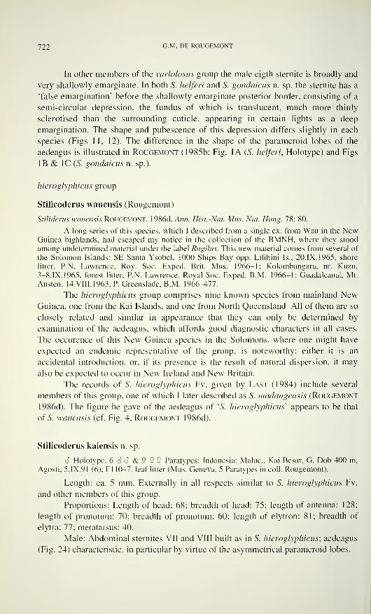
722 G -M - DE ROUGEMONT
In other members of the varlolosus group the male eigth sternite is broadly and
very shallowly emarginate. In both S. helferi and S. gondaicus n. sp. the sternite has a
'false emargination' before the shallowly emarginate posterior border, consisting of a
semi-circular depression, the fundus of which is translucent, much more thinly
sclerotised than the surrounding cuticle, appearing in certain lights as a deep
emargination. The shape and pubescence of this depression differs slightly in each
species (Figs 11, 12). The difference in the shape of the parameroid lobes of the
aedeagus is illustrated in Rougemont (1985b: Fig. 1A (S. helferi, Holotype) and Figs
IB & 1C (S. gondaicus n. sp.).
hieroglyphicus group
Stilicoderus wauensis (Rougemont)
Stiliderus wauensis Rougemont, I986d, Ann. Hist. -Nat. Mus. Nat. Hung. 78: 80.
A long series of this species, which I described from a single ex. from Wau in the NewGuinea highlands, had escaped my notice in the collection of the BMNH, where they stood
among undetermined material under the label Rugilus. This new material comes from several of
the Solomon Islands: SE Santa Ysobel, 1000 Ships Bay opp. Lilihini Is., 20.IX. 1965, shore
litter, P.N. Lawrence. Roy. Soc. Exped. Brit. Mus. 1966-1: Kolombangara, nr. Kuzu,
3-8.IX.1965, forest litter, P.N. Lawrence, Royal Soc. Exped. B.M. 1966-1; Guadalcanal, Mt.
Austen, 14.VIII.1963, P. Greenslade, B.M. 1966-477.
The hieroglyphicus group comprises nine known species from mainland NewGuinea, one from the Kai Islands, and one from North Queensland. All of them are so
closely related and similar in appearance that they can only be determined by
examination of the aedeagus. which affords good diagnostic characters in all cases.
The occurence of this New Guinea species in the Solomons, where one might have
expected an endemic representative of the group, is noteworthy: either it is an
accidental introduction, or, if its presence is the result of natural dispersion, it may
also be expected to occur in New Ireland and New Britain.
The records of S. hieroglyphicus Fv. given by Last (1984) include several
members of this group, one of which I later described as S. madangensis (Rougemont
1986d). The figure he gave of the aedeagus of 'S. hieroglyphicus'' appears to be that
of S. wauensis (cf. Fig. 4, Rougemont 1986d).
Stilicoderus kaiensis n. sp.
6 Holotype, 6 6 6 & 9 2 ? Paratypes: Indonesia: Maluc, Kai Besar, G. Dob 400 m,
Agosti, 5. IX.91 (6), Fl 1047, leaf litter (Mus. Geneva, 5 Paratypes in coll. Rougemont).
Length: ca. 5 mm. Externally in all respects similar to S. hieroglyphicus Fv.
and other members of this group.
Proportions: Length of head: 68; breadth of head: 75; length of antenna: 128;
length of pronotum: 70; breadth of pronotum: 60; length of elytron: 81; breadth of
elytra: 77; metatarsus: 40.
Male: Abdominal sternites VII and VIII built as in S. hieroglyphicus; aedeagus
(Fig. 24) characteristic, in particular by virtue of the asymmetrical parameroid lobes.
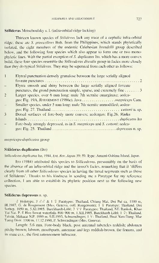
STILIDERUS AND STILICODERUS 723
Stiliderus Motschoulsky s. 1. (infra-orbital ridge lacking)
Thirteen known species of Stiliderus lack any trace of a cephalic infra-orbital
ridge; these are S. praecellens Bnh. from the Philippines, which stands phyletically
isolated, the eight members of the endemic Celebesian brendelli group described
below, and the following four species which also appear to form one or two mono-
phyletic lines. With the partial exception of S. duplicatus Ito, which has a more convex
build, these four species resemble the Stilicoderus discalis group in facies more closely
than they do typical Stiliderus. They may be seperated from each other as follows:
1 Elytral punctuation densely granulose between the large serially aligned
foveate punctures 2
Elytra smooth and shiny between the large serially aligned foveate
punctures, the grond punctuation simple, sparse, and extremely fine 3
2 Larger species, over 6 mm long; male 7th sternite emarginate; aedea-
gus: Fig. 19A, Rougemont (1986e). Java magniceps Cam.
Smaller species, under 5 mm long; male 7th sternite unmodified; aedea-
gus: Fig. 27. Thailand cottemi n. sp.
3 Dorsal surfaces of fore-body more convex; aedeagus: Fig.26. Riuku
Islands duplicatus Ito
Fore-body strongly depressed, as in S. magniceps and S. cottoni; aedea-
gus: Fig. 25. Thailand depressus n. sp.
magniceps-duplicatus group
Stiliderus duplicatus (Ito)
Stilicoderus duplicatus Ito, 1984, Ent. Rev. Japan 39: 59. Type: Amami-Oshima Island, Japan.
Ito (1984) attributed this species to Stilicoderus, presumably on the basis of
the absence of an infra-orbital ridge and the insect's facies, remarking that it 'differs
clearly from all other Stilicoderus species in having the tarsal segments such as those
of Stiliderus'. Thanks to his kindness in sending me a Paratype for my reference
collection, I am able to establish its phyletic position next to the following newspecies.
Stiliderus depressus n. sp.
6 Holotype, 3 S 6 & 1 9 Paratypes: Thailand, Chiang Mai, Doi Pui ca. 1100 m,III. 1987, G. de Rougemont (Mus. Geneva, coll. Rougemont); 1 9 Paratype: Thailand, DoiSuthep, 1050 m, 5.XI.1985, Burckhardt-Löbl; 3 9 9 Paratypes: Thailand, NE Bankok, KhaoYai Nat. P. E Heo Suwat waterfalls 800-900 m, 1.XII.1985, Burckhardt-Löbl; 1 9: Thailand,
Taksin, Maharat N.P. 1000 m, 9.II.1993, Schwendinger; 1 9: Thailand, Huai Nam Dang, MaeTaeng Distr. 1 100 m. 17.12.1990, P. Schwendinger (Mus. Geneva).
Length: 5.6 mm. Fore-body black, post antennal tubercles reddish; abdomen
pitchy-brown; labrum, mouthparts, antennae and legs reddish-brown, the femora, and
in some exx., the first antennomere infuscate.
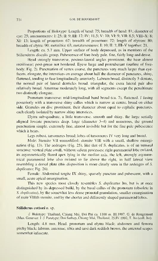
724 G -M - DE ROUGEMONT
Proportions of Holotype: Length of head: 75; breadth of head: 81; diameter of
eye: 25; antennomeres: I: 25; II: 9; III: 12; IV: 1 1.5; V: 10; VI: 9; VII: 8.5; VIII-X: 8;
XI: 13; length of pronotum: 67; breadth of pronotum: 72; length of elytron: 80;
breadth of elytra: 90; metatibia: 63; metatarsomeres: I: 10; II: 7; III-V together: 21.
Length: ca. 5.7 mm. Upper surface of body depressed, as in members of the
Stilicoderus discalis group. Pubescence of fore-body pale, fine, fairly long and dense.
Head strongly transverse, postero-lateral angles prominent, the base almost
rectilinear; post-genae not bordered. Eyese large and protruberant (outline of fore-
body: Fig. 2). Punctuation of vertex coarse, the punctures very much larger than eye-
facets, elongate, the interstices on average about half the diameter of punctures, shiny,
flattened, tending to fuse longitudinally anteriorly. Labrum broad, distinctly 5-dentate,
the normal pair of lateral denticles broad, triangular, the extra lateral pair also
relatively broad. Antennae moderately long, with all segments except the penultimate
two distinctly elongate.
Pronotum transverse; mid-longitudinal band broad (ca. 7), flattened, f fusing
posteriorly with a transverse shiny callus which is narrow at centre, broad on either
side. Granules on disc prominent, their diameter about equal to cephalic punctures,
each clearly isolated by narrow shiny interstices.
Elytra sub-quadrate, a little transverse, smooth and shiny, the large serially
aligned foveate punctures deep, large (diameter 3-4) and numerous, the ground
punctuation simple, extremely fine, almost invisible but for the fine pale pubescence
which it bears.
Legs robust, tarsomeres broad; lobes of tarsomeres IV very long and broad.
Male: Sterilite VII unmodified; sternite VIII with a small, shallow emargi-
nation (Fig. 13). The aedeagus (Fig. 25), like that of S. duplicatus, is of an unusual
structure: ventral plate small, without salient processes; right parameroid lobe twisted,
its asymmetrically flared apex lying in the median axis, the left, strongly asymme-
trical parameroid lobe also twisted to lie above the right, in half lateral view
resembling a dorsal plate (this disposition is more clearly seen in the aedeagus of S.
duplicatus: Fig. 26).
Female: Abdominal tergite IX shiny, sparsely punctate and pubescent, with a
small, acute apical emargination.
This new species most closely resembles S. duplicatus Ito, but is at once
distinguished by its depressed build, by the basal callus of the pronotum (obsolete in
S. duplicatus), by the somewhat less dense pronotal granulation, smaller emargination
of male Vlllth sternite, and by the shorter and differently shaped parameroid lobes.
Stiliderus cottoni n. sp.
6 Holotype: Thailand, Chiang Mai, Doi Pui ca. 1100 m, III.1987, G. de Rougemont(Mus. Geneva); 1 â Paratype: Doi Suthep, Chiang Mai, Thailand, 25.IV. 1992, T. Ito (coll. Ito).
Length: 4.8 mm. Head, pronotum and elytra black; abdomen and femora
pitchy black; labrum, antennae, tibia and tarsi dark reddish brown, the antennal scapes
somewhat infuscate.
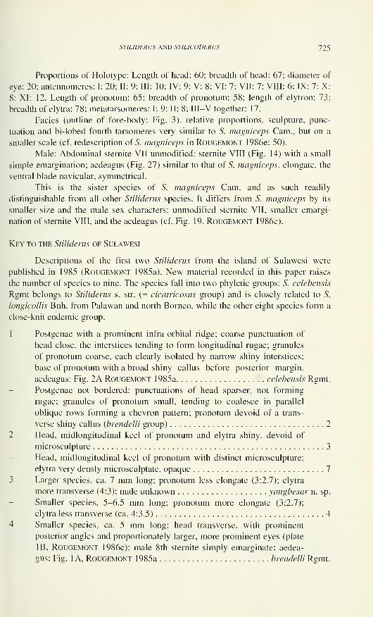
STILIDERUS AND STILICODERUS 725
Proportions of Holotype: Length of head: 60; breadth of head: 67; diameter of
eye: 20; antennomeres: I: 20; II: 9; III: 10; IV: 9; V: 8; VI: 7; VII: 7; VIII: 6; IX: 7; X:
8; XI: 12. Length of pronotum: 65; breadth of pronotum: 58; length of elytron: 73;
breadth of elytra: 78; metatarsomeres: I: 9; II: 8; III—V together: 17.
Facies (outline of fore-body: Fig. 3), relative proportions, sculpture, punc-
tuation and bi-lobed fourth tarsomeres very similar to S. magniceps Cam., but on a
smaller scale (cf. redescription of S. magniceps in Rougemont 1986e: 50).
Male: Abdominal sternite VII unmodified; sternite VIII (Fig. 14) with a small
simple emargination; aedeagus (Fig. 27) similar to that of S. magniceps, elongate, the
ventral blade navicular, symmetrical.
This is the sister species of S. magniceps Cam. and as such readily
distinguishable from all other Stiliderus species. It differs from S. magniceps by its
smaller size and the male sex characters: unmodified sternite VII, smaller emargi-
nation of sternite VIII, and the aedeagus (cf. Fig. 19, Rougemont 1986e).
Key to the Stiliderus of Sulawesi
Descriptions of the first two Stiliderus from the island of Sulawesi were
published in 1985 (Rougemont 1985a). New material recorded in this paper raises
the number of species to nine. The species fall into two phyletic groups: S. celebensis
Rgmt belongs to Stiliderus s. str. (= cicatricosus group) and is closely related to S.
longicollis Bnh. from Palawan and north Borneo, while the other eight species form a
close-knit endemic group.
1 Postgenae with a prominent infra-orbital ridge; coarse punctuation of
head close, the interstices tending to form longitudinal rugae; granules
of pronotum coarse, each clearly isolated by narrow shiny interstices;
base of pronotum with a broad shiny callus before posterior margin.
aedeagus: Fig. 2A Rougemont 1985a celebensis Rgmt.
Postgenae not bordered; punctuations of head sparser, not forming
rugae; granules of pronotum small, tending to coalesce in parallel
oblique rows forming a chevron pattern; pronotum devoid of a trans-
verse shiny callus (brendelli group) 2
2 Head, midlongitudinal keel of pronotum and elytra shiny, devoid of
microsculpture 3
Head, midlongitudinal keel of pronotum with distinct microsculpture;
elytra very densly microsculptate, opaque 7
3 Larger species, ca. 7 mm long; pronotum less elongate (3:2.7); elytra
more transverse (4:3); male unknown yangbesar n. sp.
Smaller species, 5-6.5 mm long; pronotum more elongate (3:2.7);
elytra less transverse (ca. 4:3.5) 4
4 Smaller species, ca. 5 mm long; head transverse, with prominent
posterior angles and proportionately larger, more prominent eyes (plate
IB, Rougemont 1986e); male 8th sternite simply emarginate; aedea-
gus: Fig. 1 A, Rougemont 1985a brendelli Rgmt.
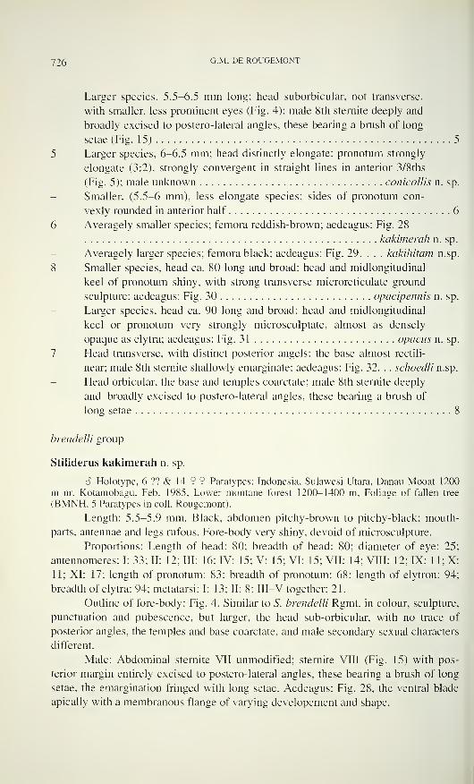
726 G -M - DE ROUGEMONT
Larger species, 5.5-6.5 mm long; head suborbicular. not transverse,
with smaller, less prominent eyes (Fig. 4); male 8th sternite deeply and
broadly excised to postero-lateral angles, these bearing a brush of long
setae (Fig. 15) 5
5 Larger species, 6-6.5 mm; head distinctly elongate; pronotum strongly
elongate (3:2), strongly convergent in straight lines in anterior 3/8ths
(Fig. 5); male unknown conicollis n. sp.
Smaller. (5.5-6 mm), less elongate species; sides of pronotum con-
vexly rounded in anterior half 6
6 Averagely smaller species; femora reddish-brown; aedeagus: Fig. 28
kakimerah n. sp.
Averagely larger species; femora black; aedeagus: Fig. 29. . . . kakihitam n.sp.
8 Smaller species, head ca. 80 long and broad; head and midlongitudinal
keel of pronotum shiny, with strong transverse microreticulate ground
sculpture; aedeagus: Fig. 30 opacipennis n. sp.
Larger species, head ca. 90 long and broad; head and midlongitudinal
keel or pronotum very strongly microsculptate. almost as densely
opaque as elytra; aedeagus: Fig. 31 opacus n. sp.
7 Head transverse, with distinct posterior angels; the base almost rectili-
near; male 8th sternite shallowly emarginate; aedeagus: Fig. 32. . . schoedli n.sp.
Head orbicular, the base and temples coarctate: male 8th sternite deeply
and broadly excised to postero-lateral angles, these bearing a brush of
long setae 8
brendelli group
Stiliderus kakimerah n. sp.
6 Holotype, 6 ?? & 14 9 9 Paratypes: Indonesia, Sulawesi Utara, Danau Mooat 1200
m nr. Kotamobagu. Feb. 1985, Lower montane forest 1200-1400 m. Foliage of fallen tree
(BMNH. 5 Paratypes in coll. Rougemont).
Length: 5.5-5.9 mm. Black, abdomen pitchy-brown to pitchy-black; mouth-
parts, antennae and legs rufous. Fore-body very shiny, devoid of microsculpture.
Proportions: Length of head: 80; breadth of head: 80; diameter of eye: 25;
antennomeres: I: 33: II: 12; III: 16: IV: 15: V: 15: VI: 15; VII: 14; VIII: 12; IX: 1 1; X:
1 1: XI: 17; length of pronotum: 83; breadth of pronotum: 68; length of elytron: 94;
breadth of elytra: 94: metatarsi: I: 13; II: 8; III—V together: 21.
Outline of fore-body: Fig. 4. Similar to 5. brendelli Rgmt. in colour, sculpture,
punctuation and pubescence, but larger, the head sub-orbicular, with no trace of
posterior angles, the temples and base coarctate, and male secondary sexual characters
different.
Male: Abdominal sternite VII unmodified; sternite VIII (Fig. 15) with pos-
terior margin entirely excised to postero-lateral angles, these bearing a brush of long
setae, the emargination fringed with long setae. Aedeagus: Fig. 28, the ventral blade
apically with a membranous flange of varying developement and shape.
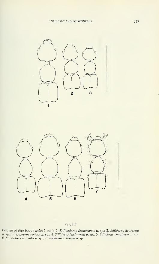
STILIDERUS AND STIIJCODERUS 727
Figs 1-7
Outline of fore-body (scale: 3 mm): 1. Stilicoderus formosanus n. sp.; 2. Stiliderus depressus
n. sp.; 3. Stiliderus cottoni n. sp.; 4. Stiliderus kakimerah n. sp.; 5. Stiliderus yangbesar n. sp.;
6. Stiliderus conicollis n. sp.; 7. Stiliderus schoedli n. sp.
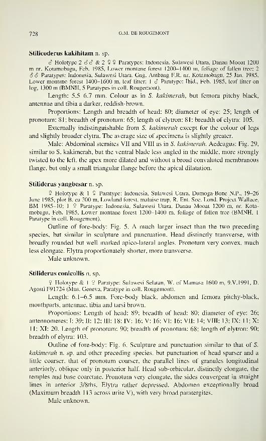
728 G -M - DE ROUGEMONT
Stilicoderus kakihitam n. sp.
â Holotype 2 â S & 2 9 9 Paratypes: Indonesia, Sulawesi Utara, Danau Mooat 1200
m nr. Kotamobagu, Feb. 1985, Lower montane forest 1200-1400 m, foliage of fallen tree; 2
6 6 Paratypes: Indonesia, Sulawesi Utara, Gng. Ambang F.R. nr. Kotamobagu, 25 Jan. 1985,
Lower montane forest 1400-1600 m, leaf litter; 1 6 Paratype: Ibid., Feb. 1985, leaf litter on
log, 1300 m (BMNH, 5 Paratypes in coll. Rougemont).
Length: 5.5-6.7 mm. Colour as in S. kakimerah, but femora pitchy black,
antennae and tibia a darker, reddish-brown.
Proportions: Length and breadth of head: 80; diameter of eye: 25; length of
pronotum: 81; breadth of pronotum: 65; length of elytron: 81; breadth of elytra: 105.
Externally indistinguishable from S. kakimerah except for the colour of legs
and slightly broader elytra. The average size of specimens is slightly greater.
Male: Abdominal sternites VII and VIII as in S. kakimerah. Aedeagus: Fig. 29,
similar to S. kakimerah, but the ventral blade less angled in the middle, more strongly
twisted to the left, the apex more dilated and without a broad convaluted membranous
flange, but only a small triangular flange before the apical dilatation.
Stiliderus yangbesar n. sp.
9 Holotype & 1 9 Paratype: Indonesia, Sulawesi Utara, Dumoga-Bone N.P., 19-26
June 1985, plot B. ca 300 m. Lowland forest, malaise trap, R. Ent. Soc. Lond. Project Wallace,
BM 1985—10; 1 9 Paratype: Indonesia, Sulawesi Utara, Danau Mooat 1200 m, nr. Kota-
mobagu, Feb. 1985, Lower montane forest 1200-1400 m, foliage of fallen tree (BMNH, 1
Paratype in coll. Rougemont).
Outline of fore-body: Fig. 5. A much larger insect than the two preceding
species, but similar in sculpture and punctuation. Head distinctly transverse, with
broadly rounded but well marked apico-lateral angles. Pronotum very convex, much
less elongate. Elytra proportionately shorter, more transverse.
Male unknown.
Stiliderus conicollis n. sp.
9 Holotype & 1 9 Paratype: Sulawesi Selatan, W. of Mamasa 1600 m, 9.V.1991, D.
Agosti F91724 (Mus. Geneva, Paratype in coll. Rougemont).
Length: 6.1-6.5 mm. Fore-body black, abdomen and femora pitchy-black,
mouthparts, antennae, tibia and tarsi brown.
Proportions: Length of head: 89; breadth of head: 80; diameter of eye: 26
antennomeres: I: 39; II: 12; III: 18; IV: 16; V: 16; VI: 16; VII: 14; VIII: 13; IX: 11; X11; XI: 20. Length of pronotum: 90; breadth of pronotum: 68; length of elytron: 90
breadth of elytra: 103.
Outline of fore-body: Fig. 6. Sculpture and punctuation similar to that of S.
kakimerah n. sp. and other preceding species, but punctuation of head sparser and a
little coarser, that of pronotum coarser, the parallel lines of granules longitudinal
anteriorly, oblique only in posterior half. Head sub-orbicular, distinctly elongate, the
temples and base coarctate. Pronotum very elongate, the sides convergent in straight
lines in anterior 3/8ths. Elytra rather depressed. Abdomen exceptionally broad
(Maximum breadth 113 across urite V), with very broad paratergites.
Male unknown.
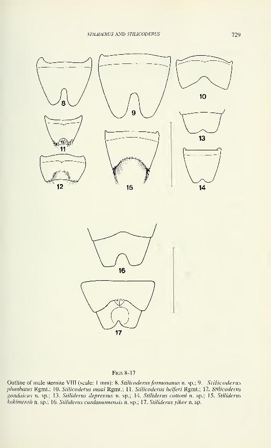
STIUDERUS AND STILICODERUS 729
Figs 8-17
Outline of male sternite VIII (scale: 1 mm): 8. Stilicoderusformosanus n. sp.; 9. Stilicoderus
plumbatus Rgmt.; 10. Stilicoderus maai Rgmt.; 1 1. Stilicoderus helferi Rgmt.; 12. Stilicoderus
gondaicus n. sp.; 13. Stiliderus depressus n. sp.; 14. Stiliderus cottoni n. sp.; 15. Stiliderus
kakimerah n. sp.; 16. Stiliderus cardamomensis n. sp.; 17. Stiliderus yikom. sp.
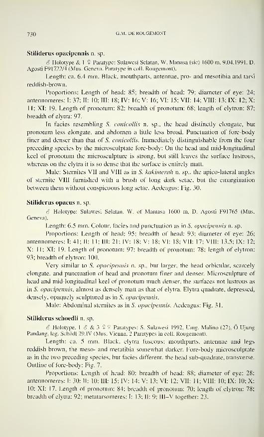
730 G -M - DE ROUGEMONT
Stiliderus opacipennis n. sp.
Ô Holotype & 1 9 Paratype; Sulawesi Selatan, W. Manasa (sic) 1600 m, 9.04.1991, D.
Agosti F9 1722/4 (Mus. Geneva, Paratype in coll. Rougemont).
Length: ca. 6.4 mm. Black, mouthparts, antennae, pro- and mesotibia and tarsi
reddish-brown.
Proportions: Length of head: 85; breadth of head: 79; diameter of eye: 24
antennomeres: I: 37; II: 10; III: 18; IV: 16; V: 16; VI: 15; VII: 14; VIII: 13; IX: 12; X11; XI: 19. Length of pronotum: 82; breadth of pronotum: 68; length of elytron: 87
breadth of elytra: 97.
In facies resembling S. conicollis n. sp., the head distinctly elongate, but
pronotum less elongate, and abdomen a little less broad. Punctuation of fore-body
finer and denser than that of S. conicollis. Immediately distinguishable from the four
preceding species by the microsculptate fore-body: On the head and mid-longitudinal
keel of pronotum the microsculpture is strong, but still leaves the surface lustrous,
whereas on the elytra it is so dense that the surface is entirely matt.
Male: Sternites VII and VIII as in S. kakimerah n. sp., the apico-lateral angles
of sternite VIII furnished with a brush of long dark setae, but the emargination
between them without conspicuous long setae. Aedeagus: Fig. 30.
Stiliderus opacus n. sp.
6 Holotype: Sulawesi Selatan, W. of Mamasa 1600 m. D. Agosti F91765 (Mus.
Geneva).
Length: 6.5 mm. Colour, facies and punctuation as in S. opacipennis n. sp.
Proportions: Length of head: 95; breadth of head: 93; diameter of eye: 26
antennomeres: I: 41; II: 11; III: 21; IV: 18; V: 18; VI: 18; VII: 17; VIII: 13.5; IX: 12
X: 11; XI: 19. Length of pronotum: 97; breadth of pronotum: 78; length of elytron
93: breadth of elytron: 100.
Very similar to S. opacipennis n. sp., but larger, the head orbicular, scarcely
elongate, and punctuation of head and pronotum finer and denser. Microsculpture of
head and mid-longitudinal keel of pronotum much denser, the surfaces not lustrous as
in S. opacipennis, almost as densely matt as that of elytra. Elytra quadrate, depressed,
densely, opaquely sculptured as in S. opacipennis.
Male: Abdominal sternites as in S. opacipennis. Aedeagus: Fig. 31.
Stiliderus schoedli n. sp.
S Holotype, 1 S & 3 9 9 Paratypes: S. Sulawesi 1992, Umg. Malino (27), Ö UjungPandang, leg. Schödl 29.IV (Mus. Vienna, 2 Paratypes in coll. Rougemont).
Length: ca. 5 mm. Black, elytra fuscous; mouthparts, antennae and legs
reddish-brown, the meso- and metatibia somewhat darker. Fore-body microsculptate
as in the two preceding species, but facies different, the head sub-quadrate, transverse.
Outline of fore-body: Fig. 7.
Proportions: Length of head: 80; breadth of head: 88; diameter of eye: 28
antennomeres: I: 30: II: 10; III: 15; IV: 14; V: 13; VI: 12; VII: 11; VIII: 10; IX: 10; X10; XI: 17. Length of pronotum: 84; breadth of pronotum: 70; length of elytron: 78
breadth of elytra: 92; metatarsomeres: I: 13; II: 9; III-V together: 23.
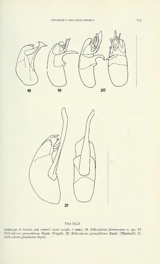
STIUDERUS ANI) STILICODERUS 731
Figs 18-21
Aedeagus in lateral and ventral views (scale: 1 mm): 18. Stilicoderus formosamis n. sp.; 19.
Stilicoderus granulifrons Rgmt. (Nepal); 20. Stilicoderus granulifrons Rgmt. (Thailand); 21.
Stilicoderus plumbatus Rgmt.
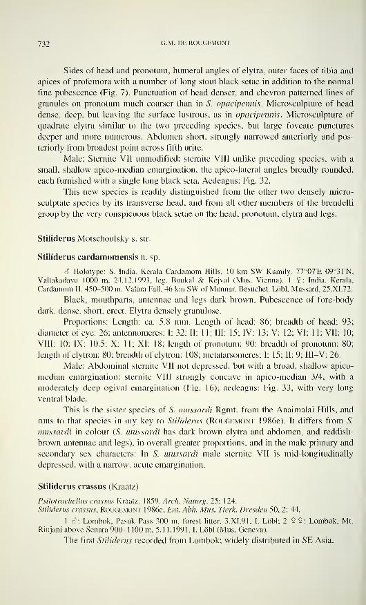
732 G -M - DE ROUGEMONT
Sides of head and pronotum, humeral angles of elytra, outer faces of tibia and
apices of profemora with a number of long stout black setae in addition to the normal
fine pubescence (Fig. 7). Punctuation of head denser, and chevron patterned lines of
granules on pronotum much coarser than in S. opacipennis. Microsculpture of head
dense, deep, but leaving the surface lustrous, as in opacipennis. Microsculpture of
quadrate elytra similar to the two preceding species, but large foveate punctures
deeper and more numerous. Abdomen short, strongly narrowed anteriorly and pos-
teriorly from broadest point across fifth urite.
Male: Sternite VII unmodified; sternite VIII unlike preceding species, with a
small, shallow apico-median emargination, the apico-lateral angles broadly rounded,
each furnished with a single long black seta. Aedeagus: Fig. 32.
This new species is readily distinguished from the other two densely micro-
sculptate species by its transverse head, and from all other members of the brendelli
group by the very conspicuous black setae on the head, pronotum, elytra and legs.
Stiliderus Motschoulsky s. str.
Stiliderus cardamomensis n. sp.
S Holotype: S. India, Kerala Cardamom Hills, 10 km SW Kumily, 77°07'E 09°31'N,
Vallakadavu 1000 m, 24.12.1993, leg. Boukal & Kejval (Mus. Vienna). 1 9: India, Kerala,
Cardamom H. 450-500 m, Valara Fall. 46 km SW of Munnar, Besuchet, Lobi, Mussard, 25.XI.72.
Black, mouthparts, antennae and legs dark brown. Pubescence of fore-body
dark, dense, short, erect. Elytra densely granulose.
Proportions: Length: ca. 5.8 mm. Length of head: 86; breadth of head: 93
diameter of eye: 26; antennomeres: I: 32; II: 11; III: 15; IV: 13; V: 12; VI: 11; VII: 10
VIII: 10; IX: 10.5; X: 11; XI: 18; length of pronotum: 90; breadth of pronotum: 80
length of elytron: 80; breadth of elytron: 108; metatarsomeres: I: 15; II: 9; III—V: 26.
Male: Abdominal sternite VII not depressed, but with a broad, shallow apico-
median emargination; sternite VIII strongly concave in apico-median 3/4, with a
moderately deep ogival emargination (Fig. 16); aedeagus: Fig. 33, with very long
ventral blade.
This is the sister species of S. mussardi Rgmt. from the Anaimalai Hills, and
runs to that species in my key to Stiliderus (Rougemont 1986e). It differs from S.
mussardi in colour (S. mussardi has dark brown elytra and abdomen, and reddish-
brown antennae and legs), in overall greater proportions, and in the male primary and
secondary sex characters: In S. mussardi male sternite VII is mid-longitudinally
depressed, with a narrow, acute emargination.
Stiliderus crassus (Kraatz)
Psilotrachellus crassus Kraatz, 1859, Arch. Naturg. 25: 124.
Stiliderus crassus, Rougemont 1986e, Ent. Abh. Mus. Tierk. Dresden 50, 2: 44.
1 S: Lombok, Pasuk Pass 300 m, forest litter. 3.XI.91, I. Lobi; 2 9 9: Lombok, Mt.
Rinjani above Senara 900-1 100 m. 5.1 1.1991, 1. Löbl (Mus. Geneva).
The first Stiliderus recorded from Lombok; widely distributed in SE Asia.
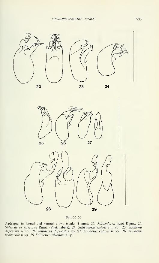
ST!LIDERUS AND STILICODERUS 733
Figs 22-29
Aedeagus in lateral and ventral views (scale: 1 mm): 22. Stilicoderus maai Rgmt.; 23.
Stilicoderus strigosus Rgmt. (Phetchaburi); 24. Stilicoderus kaiensis n. sp.; 25. Stiliderus
depressus n. sp.; 26. Stiliderus duplicatus Ito; 27. Stiliderus cottomi n. sp.; 28. Stiliderus
kakimerah n. sp.; 29. Stiliderus kakihitam n. sp.
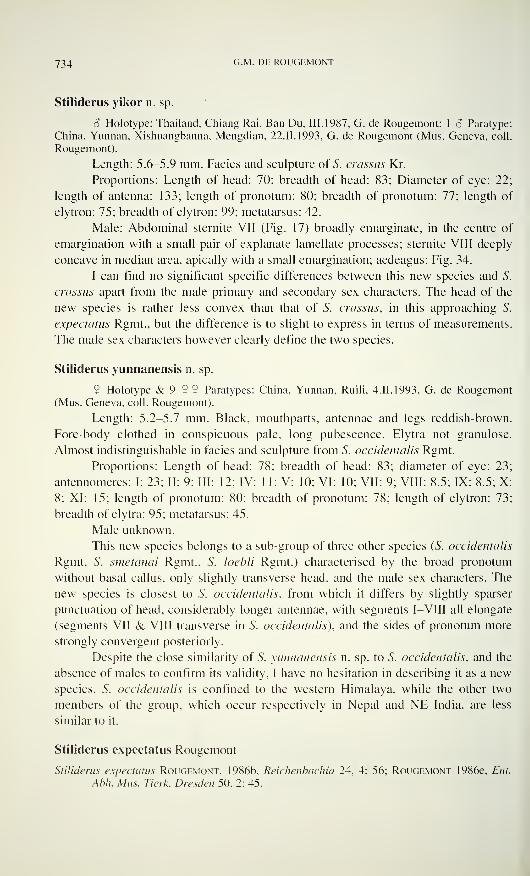
734 G -M - DE ROUGEMONT
Stiliderus yikor n. sp.
S Holotype: Thailand, Chiang Rai, Ban Du, III. 1987, G. de Rougemont; 1 â Paratype:
China, Yunnan, Xishuangbanna, Mengdian, 22.11.1993, G. de Rougemont (Mus. Geneva, coll.
Rougernont).
Length: 5.6-5.9 mm. Facies and sculpture of 5". crassus Kr.
Proportions: Length of head: 70; breadth of head: 83; Diameter of eye: 22;
length of antenna: 133; length of pronotum: 80; breadth of pronotum: 77; length of
elytron: 75; breadth of elytron: 99; metatarsus: 42.
Male: Abdominal sternite VII (Fig. 17) broadly emarginate, in the centre of
emargination with a small pair of explanate lamellate processes; sternite VIII deeply
concave in median area, apically with a small emargination; aedeagus: Fig. 34.
I can find no significant specific differences between this new species and S.
crassus apart from the male primary and secondary sex characters. The head of the
new species is rather less convex than that of 5. crassus, in this approaching S.
expectants Rgmt., but the difference is to slight to express in terms of measurements.
The male sex characters however clearly define the two species.
Stiliderus yunnanensis n. sp.
? Holotype &9 ÎÎ Paratypes: China, Yunnan, Rudi, 4.II.1993, G. de Rougemont(Mus. Geneva, coll. Rougemont).
Length: 5.2-5.7 mm. Black, mouthparts, antennae and legs reddish-brown.
Fore-body clothed in conspicuous pale, long pubescence. Elytra not granulose.
Almost indistinguishable in facies and sculpture from S. occidentalis Rgmt.
Proportions: Length of head: 78; breadth of head: 83; diameter of eye: 23
antennomeres: I: 23; II: 9; III: 12; IV: 11; V: 10; VI: 10; VII: 9; VIII: 8.5; IX: 8.5; X8; XI: 15; length of pronotum: 80; breadth of pronotum: 78; length of elytron: 73
breadth of elytra: 95; metatarsus: 45.
Male unknown.
This new species belongs to a sub-group of three other species (S. occidentalis
Rgmt, 5. smetanai Rgmt., S. loebli Rgmt.) characterised by the broad pronotum
without basal callus, only slightly transverse head, and the male sex characters. The
new species is closest to S. occidentalis, from which it differs by slightly sparser
punctuation of head, considerably longer antennae, with segments I—VIII all elongate
(segments VII & VIII transverse in S. occidentalis), and the sides of pronotum more
strongly convergent posteriorly.
Despite the close similarity of S. yunnanensis n. sp. to S. occidentalis, and the
absence of males to confirm its validity, I have no hesitation in describing it as a new
species. S. occidentalis is confined to the western Himalaya, while the other two
members of the group, which occur respectively in Nepal and NE India, are less
similar to it.
Stiliderus expectatus Rougemont
Stiliderus expectatus Rougemont, 1986b. Reichenbachia 24, 4: 56; Rougemont 1986e, Ent.
Abh. Mus. Tierk. Dresden 50, 2: 45.
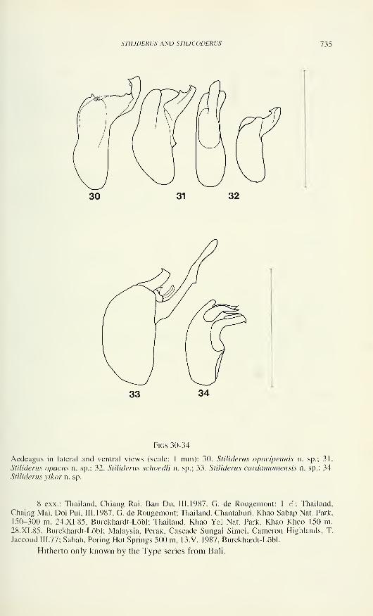
STILIDERUS AND STILICODERUS 735
Figs 30-34
Aedeagus in lateral and ventral views (scale: 1 mm): 30. Stiliderus opacipennis n. sp.; 31.
Stiliderus opacus n. sp.; 32. Stiliderus schoedli n. sp.; 33. Stiliderus cardamomensis n. sp.; 34.
Stiliderus yikor n. sp.
8 exx.: Thailand, Chiang Rai, Ban Du, III. 1987, G. de Rougemont; 1 S: Thailand.
Chaing Mai, Doi Pui, III. 1987, G. de Rougemont; Thailand, Chantaburi, Khao Sabap Nat. Park,
150-300 m, 24.XI.85, Burckhardt-Löbl; Thailand, Khao Yai Nat. Park, Khao Kheo 150 m.
28.XI. 85, Burckhardt-Löbl; Malaysia, Perak, Cascade Sungai Simei, Cameron Highlands, T.
Jaccoud 111.77; Sabah, Poring Hot Springs 500 m, 13.V. 1987, Burckhardt-Löbl.
Hitherto only known by the Type series from Bali.

736 G -M - DE ROUGEMONT
Stiliderus cicatricosus Motschoulsky
Stiliderus cicatricosus Motschoulsky, 1858, Bull. Mose. 31,2: 639; Rougemont 1986e, Ent.
Abh. Mus. Tierk. Dresden 50. 2: 43.
Sumatra, Aceh no. 25a, Mt. Leuser NP, 300-500 m, Ketamba, 23-30.XI.1989, Lobi,
Agosti, Burckhardt; 1 9: China, Yunnan. Xishuangbanna, Mengdian, 22.1.1993, G. de
Rougemont; 1 3: China, Yunnan, Ruili, 4.II.1993, G. de Rougemont.
New to Sumatra and China.
ACKNOWLEDGMENTS
My thanks to those friends and colleagues who made most of this material
available for study: M. Brendell and P. Hammond (Natural History Museum,
London), T. Ito (Kyoto), I. Lobi (Geneva Museum), W. Schawaller (Stuttgart
Museum), H. Schillhammer (Vienna Museum), A. Smetana (Ottawa).
REFERENCES
Ito, T. 1984. A new species of the genus Stilicodreus from Japan. Entomological Review Japan
39, 1:59-61.
Last, H. 1984. Recorded and new species of Coleoptera (Staphylinidae, Paederinae in Papua
New Guinea. Folia Entomologica Hungarica XLV, 2: 109-125.
Rougemont, G. de. 1985a. Two new species of Stiliderus from Celebes (Coleoptera, Staphy-
linidae). 15th contribution to the knowledge of Staphylinidae. Entomological Monthly
Magazine 121: 119-122.
Rougemont, G de. 1985b. Les Stiliderus de la collection du Musée de Genève (Coleoptera,
Staphylinidae). 16ème contribution à la connaissance des Staphylinides. Revue Suisse
de Zoologie 92, 1:217-228.
Rougemont, G. de. 1986a. Revision of the genus Stiliderus Motschulsky, 1858. Part I (=
Stilicoderus Sharp 1889). (Coleoptera, Staphylinidae). 14th contribution to the knowl-
edge of Staphylinidae. Entomologische Abhandlungen des Museums für Tierkunde,
Dresden 49, 8: 139-187.
Rougemont, G de. 1986b. New data on the genus Stiliderus Motsch. (Coleoptera, Staphy-
linidae, Paederinae). 18th contribution to the knowledge of Staphylinidae. Reichen-
bachia 24, 4: 53-58.
Rougemont, G de. 1986d. New Stiliderus Motschulsky from the Indo-Australian Region
(Coleoptera, Staphylinidae). 22nd contribution to the knowledge of Staphylinidae.
Annales Historiae-Naturalis Musei Naturalis Hungariae 78: 79-82.
Rougemont, G de. 1986e. Revision of the genus Stiliderus Motschulsky 1858. Part II: Thespecies with bi-lobed IVth tarsomeres (Coleoptera, Staphylinidae, Paederinae). 23rd
contribution to the knowledge of Staphylinidae. Entomologische Abhandlungen des
Museums für Tierkunde, Dresden 50, 2: 33-58.
Rougemont, G de. 1996. Review of the Australian species of the subtribe Stilicina (Cole-
optera, Staphylinidae, Paederinae). 30th contribution to the knowledge of Staphy-
linidae. Ehtron 000:000.
Revue suisse de Zoologie, 103 (3): 737-748; septembre 1996
Indo-australische Anthicidae (Coleoptera) imNaturhistorischen Museum in Genf*
Gerhard UHMANNTannenhofstraße 10, D-92690 Pressath, Deutschland.
Indo-Australian Anthicidae (Coleoptera) from the Natural History
Museum in Geneva. - Following Tomoderini are described and illustrated:
Derarimus calamei sp. n., Derarimus fortepunctatus sp. n., Derarimus
luteipes sp. n., Derarimus ovipennis sp. n., Derarimus pahangensis sp. n.,
Derarimus selangorensis sp. n., Tomoderus bicolor sp. n. and Tomoderus
globicollis sp. n..
Key-words: Coleoptera - Anthicidae - Taxonomy - Malaysia - Thailand -
Indonesia - Australia.
EINLEITUNG
In einer weiteren Bestimmungssendung, die mir Herr Dr Ivan Lobi vomNaturhistorischen Museum in Genf zukommen ließ, fanden sich acht bisher
unbeschriebene Arten aus den Gattungen Derarimus und Tomoderus, die nachfolgend
beschrieben werden. Für die neuen Derarimus-Arten wird ein Schlüssel beigefügt,
nach dem sie in die Bestimmungstabelle (Uhmann 1994) aufgenommen werden
können.
Herrn Dr Ivan Löbl danke ich sehr für die Ausleihe der Käfer und für die
Überlassung einiger Tiere für meine Sammlung.
Alle Holotypen befinden sich im Naturhistorischen Museum in Genf. Alle
Maße sind in mm angegeben.
ARTENLISTE
Mecynotarsus nanus Laferté, 1 848
Verbreitung: Indien, Thailand, Philippinen.
Thailand, Ban Kiriwong, Khao Luang Nat. Park, 170 m, 10.2.1991, 2 Ex.. leg. P.
Schwendinger.
* 53. Beitrag zur Kenntnis der Anthicidae.
Manuscript angenommen um 04.01.1996.
738 GERHARD UHMANN
Derarimus calamei sp. n. (Abb. 1^)
W. -Malaysia, Pahang, Genting Highlands, 1700 m, 2.4.1993, 2 Ex. (Holotypus,
Paratypus), leg. I. Lobi und F. Calarne, # 26.
Länge 4,9, größte Breite 2,1. Kopf 0,9 lang, 0,9 breit. Halsschild 1,3 lang, 0,8
breit. Flügeldecken 2,8 lang, 2,1 gemeinsam breit.
Färbung: Kopf und Halsschild rotbraun, Flügeldecken dunkelbraun mit
leichtem, kupferigem Metallglanz. Beine und Taster braun. Fühler dunkelbraun, die
zwei ersten und die zwei letzten Glieder wesentlich heller.
Kopf: Glänzend. Fein, ziemlich flach und verstreut punktiert. Behaarung
braun, kräftig, halb abstehend, größtenteils quer liegend. Außerdem mit wenigen
längeren Borsten besetzt. Fühler kräftig behaart und beborstet.
Halsschild: Glänzend. Vorn sehr fein und sehr verstreut punktiert. In der
Einschnürung mit einem Längkiel in der Mitte, daneben runzelig, seitlich glatt. Hinter
der Einschnürung etwas kräftiger und dichter aber flach punktiert. Behaarung braun,
ziemlich fein, kurz, halb abstehend, größtenteils quer liegend, gebogen. Dazwischen
stehen einige nicht sehr lange Borsten steil ab. Hals grob gekörnt. Vorn in der Mitte
mit einer flachen Grube (nur bei den Männchen).
Flügeldecken: Glänzend. Punkte mittelgroß und flach. Nach hinten zu werden
die Punkte nur wenig kleiner. Zwischenräume etwa 2-6mal so groß wie die Punkte.
Behaarung hellbraun, kräftig, nicht sehr lang, gebogen, etwas abstehend, nach hinten
gerichtet. Außerdem stehen einige nicht sehr lange, gebogene Borsten steil ab. In der
Hinterhälfte, neben der Naht etwas niedergedrückt.
Beine nicht auffällig behaart.
Beziehungen: Dem Derarimus javanus Uhmann aus Java ähnlich, aber größer,
D. calamei etwa 4,9 mm, D. javanus etwa 3,2 mm. Die Punktur in der Halsschild-
Einschnürung ist bei D. calamei viel feiner.
Derivatio nominis: Diese Art benenne ich nach einem der beiden Entdecker,
Herrn François Calarne aus Genf.
Derarimus fortepunctatus sp. n. (Abb. 5 und 6)
W-Malaysia, Pahang, Cameron Highlands, 1720 m, Gunung Jasar, # 19 b, 25.3.1993, 1
Ex. (Holotypus), 25.3.1993, leg. I. Löbl und F. Calarne.
Länge 3,7, größte Breite 1,5. Kopf 0,7 lang, über die Augen gemessen 0,7
breit. Halsschild 0,9 lang, 0,7 breit. Flügeldecken 2,1 lang, 1,5 gemeinsam breit.
Färbung: Kopf dunkelbraun. Halsschild heller braun. Flügeldecken schwarz.
Fühler braun mit hellerer Spitze. Taster und Beine braun.
Kopf: Glänzend. Vorn sehr fein, hinten kräftiger punktiert. Zwischenräume
überall viel größer als die Punkte. Behaarung hellbraun, kräftig, ziemlich lang, etwas
gebogen, abstehend, in verschiedene Richtungen weisend. Dazwischen stehen einige
lange, gerade Borsten senkrecht ab. Fühler kräftig behaart und beborstet.
Halsschild: Etwas glänzend. Sehr kräftig punktiert. In der Mitte sind die
Punkte verrunzelt. Im Bereich der Einschnürung oben mit einem Längskiel, davor mit
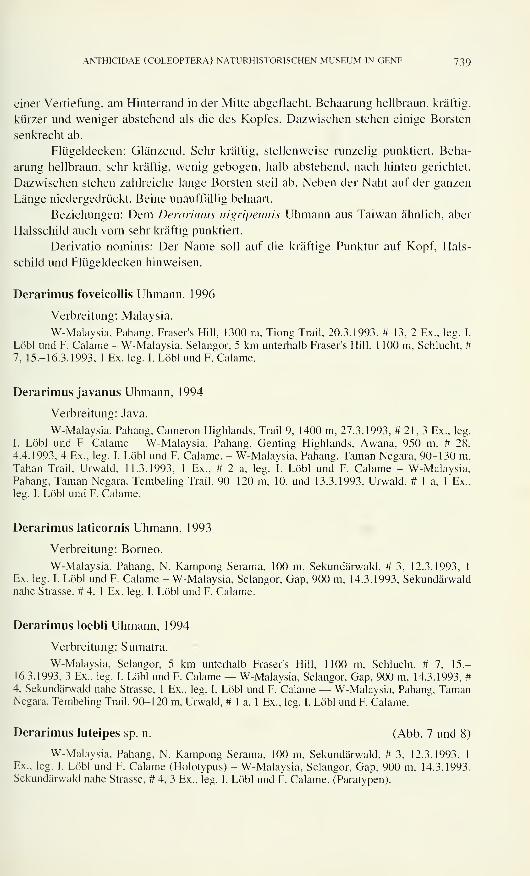
ANTHICIDAE (COLEOPTERA) NATURHISTORISCHEN MUSEUM IN GENF 739
einer Vertiefung, am Hinterrand in der Mitte abgeflacht. Behaarung hellbraun, kräftig,
kürzer und weniger abstehend als die des Kopfes. Dazwischen stehen einige Borsten
senkrecht ab.
Flügeldecken: Glänzend. Sehr kräftig, stellenweise runzelig punktiert. Beha-
arung hellbraun, sehr kräftig, wenig gebogen, halb abstehend, nach hinten gerichtet.
Dazwischen stehen zahlreiche lange Borsten steil ab. Neben der Naht auf der ganzen
Länge niedergedrückt. Beine unauffällig behaart.
Beziehungen: Dem Derarimus nigripennis Uhmann aus Taiwan ähnlich, aber
Halsschild auch vorn sehr kräftig punktiert.
Derivatio nominis: Der Name soll auf die kräftige Punktur auf Kopf, Hals-
schild und Flügeldecken hinweisen.
Derarimus foveicollis Uhmann, 1996
Verbreitung: Malaysia.
W-Malaysia, Pahang, Fraser's Hill. 1300 m. Tiong Trail, 20.3.1993, # 13, 2 Ex., leg. I.
Löbl und F. Calarne - W-Malaysia, Selangor, 5 km unterhalb Fraser's Hill, 1 100 m, Schlucht, #7. 15.-16.3.1993. 1 Ex. leg. I. Löbl und F. Calarne.
Derarimus javanus Uhmann, 1994
Verbreitung: Java.
W-Malaysia, Pahang, Cameron Highlands, Trail 9, 1400 m, 27.3.1993, #21,3 Ex., leg.
I. Löbl und F. Calarne - W-Malaysia, Pahang, Genting Highlands, Awana, 950 m, # 28,
4.4.1993, 4 Ex., leg. I. Löbl und F. Calarne. - W-Malaysia, Pahang, Laman Negara, 90-130 m.
Tahan Trail, Urwald, 11.3.1993, 1 Ex., # 2 a, leg. I. Löbl und F. Calarne - W-Malaysia,Pahang, Taman Negara, Tembeling Trail, 90-120 m, 10. und 13.3.1993. Urwald, # 1 a, 1 Ex..
leg. I. Löbl und F. Calarne.
Derarimus laticornis Uhmann, 1993
Verbreitung: Borneo.
W-Malaysia, Pahang, N. Kampong Serama, 100 m, Sekundärwald, # 3, 12.3.1993, 1
Ex. leg. I. Löbl und F. Calarne - W-Malaysia, Selangor, Gap, 900 m, 14.3.1993, Sekundärwaldnahe Strasse. # 4. 1 Ex. leg. I. Löbl und F. Calarne.
Derarimus Ioebli Uhmann, 1994
Verbreitung: Sumatra.
W-Malaysia, Selangor, 5 km unterhalb Fraser's Hill, 1100 m, Schlucht, # 7, 15-16.3.1993, 3 Ex., leg. I. Löbl und F. Calarne — W-Malaysia, Selangor, Gap, 900 m, 14.3.1993, #
4, Sekundärwald nahe Strasse, 1 Ex., leg. I. Löbl und F. Calarne — W-Malaysia, Pahang. TamanNegara, Tembeling Trail, 90-120 m. Urwald, # 1 a. 1 Ex., leg. I. Löbl und F. Calarne.
Derarimus luteipes sp. n. (Abb. 7 und 8)
W-Malaysia, Pahang, N. Kampong Serama, 100 m, Sekundärwald, # 3, 12.3.1993, 1
Ex., leg. I. Löbl und F. Calarne (Holotypus) - W-Malaysia, Selangor, Gap, 900 in, 14.3.1993,
Sekundärwald nahe Strasse, # 4, 3 Ex., leg. I. Löbl und F. Calarne, (Paratypen).
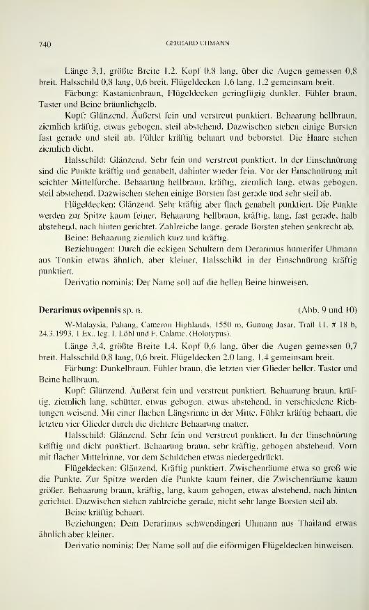
740 GERHARD UHMANN
Länge 3,1, größte Breite 1,2. Kopf 0,8 lang, über die Augen gemessen 0,8
breit. Halsschild 0,8 lang, 0,6 breit. Flügeldecken 1,6 lang, 1,2 gemeinsam breit.
Färbung: Kastanienbraun, Flügeldecken geringfügig dunkler. Fühler braun,
Taster und Beine bräunlichgelb.
Kopf: Glänzend. Äußerst fein und verstreut punktiert. Behaarung hellbraun,
ziemlich kräftig, etwas gebogen, steil abstehend. Dazwischen stehen einige Borsten
fast gerade und steil ab. Fühler kräftig behaart und beborstet. Die Haare stehen
ziemlich dicht.
Halsschild: Glänzend. Sehr fein und verstreut punktiert. In der Einschnürung
sind die Punkte kräftig und genabelt, dahinter wieder fein. Vor der Einschnürung mit
seichter Mittelfurche. Behaarung hellbraun, kräftig, ziemlich lang, etwas gebogen,
steil abstehend. Dazwischen stehen einige Borsten fast gerade und sehr steil ab.
Flügeldecken: Glänzend. Sehr kräftig aber flach genabelt punktiert. Die Punkte
werden zur Spitze kaum feiner. Behaarung hellbraun, kräftig, lang, fast gerade, halb
abstehend, nach hinten gerichtet. Zahlreiche lange, gerade Borsten stehen senkrecht ab.
Beine: Behaarung ziemlich kurz und kräftig.
Beziehungen: Durch die eckigen Schultern dem Derarimus humerifer Uhmannaus Tonkin etwas ähnlich, aber kleiner, Halsschild in der Einschnürung kräftig
punktiert.
Derivatio nominis: Der Name soll auf die hellen Beine hinweisen.
Derarimus ovipennis sp. n. (Abb. 9 und 10)
W-Malaysia, Pahang, Cameron Highlands, 1550 m, Gunung Jasar, Trail 11, # 18 b,
24.3.1993, 1 Ex., leg. I. Löbl und F. Calarne, (Holotypus).
Länge 3,4, größte Breite 1,4. Kopf 0,6 lang, über die Augen gemessen 0,7
breit. Halsschild 0,8 lang, 0,6 breit. Flügeldecken 2,0 lang, 1,4 gemeinsam breit.
Färbung: Dunkelbraun. Fühler braun, die letzten vier Glieder heller. Taster und
Beine hellbraun.
Kopf: Glänzend. Äußerst fein und verstreut punktiert. Behaarung braun, kräf-
tig, ziemlich lang, schütter, etwas gebogen, etwas abstehend, in verschiedene Rich-
tungen weisend. Mit einer flachen Längsrinne in der Mitte. Fühler kräftig behaart, die
letzten vier Glieder durch die dichtere Behaarung matter.
Halsschild: Glänzend. Sehr fein und verstreut punktiert. In der Einschnürung
kräftig und dicht punktiert. Behaarung braun, sehr kräftig, gebogen abstehend. Vorn
mit flacher Mittelrinne, vor dem Schildchen etwas niedergedrückt.
Flügeldecken: Glänzend. Kräftig punktiert. Zwischenräume etwa so groß wie
die Punkte. Zur Spitze werden die Punkte kaum feiner, die Zwischenräume kaum
größer. Behaarung braun, kräftig, lang, kaum gebogen, etwas abstehend, nach hinten
gerichtet. Dazwischen stehen zahlreiche gerade, nicht sehr lange Borsten steil ab.
Beine kräftig behaart.
Beziehungen: Dem Derarimus schwendingeri Uhmann aus Thailand etwas
ähnlich aber kleiner.
Derivatio nominis: Der Name soll auf die eiförmigen Flügeldecken hinweisen.
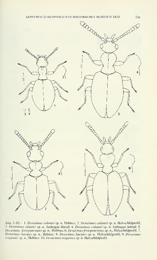
ANTHICIDAE (COLEOPTERA) NATURHISTORISCHEN MUSEUM IN GENF 741
Abb. 1-10. - 1. Derarimus calamei sp. n. Habitus; 2. Derarimus calamei sp. n. Halsschildprofil;
3. Derarimus calamei sp. n. Aedeagus dorsal; 4. Derarimus calamei sp. n. Aedeagus lateral; 5.
Derarimus fortepunctatus sp. a., Habitus; 6. Derarimus fortepunctatus sp. n., Halsschildprofil; 7.
Derarimus luteipes sp. n., Habitus; 8. Derarimus luteipes sp. n., Halsschildprofil; 9. Derarimusovipennis sp. n., Habitus; 10. Derarimus ovipennis sp. n. Halsschildprofil.
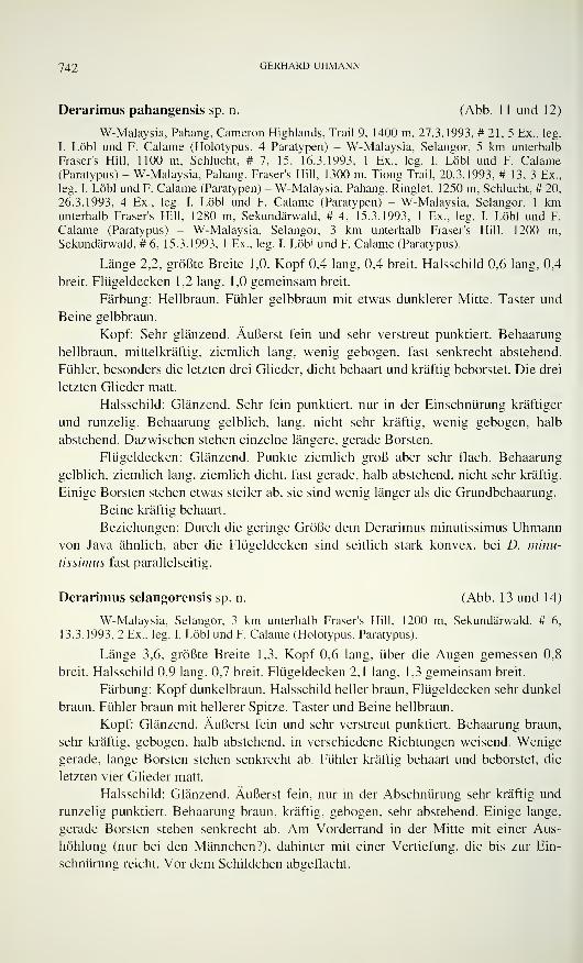
742 GERHARD UHMANN
Derarimus pahangensis sp. n. (Abb. 11 und 12)
W-Malaysia, Pahang, Cameron Highlands, Trail 9, 1400 m, 27.3.1993, #21,5 Ex., leg.
I. Lobi und F. Calarne (Holotypus, 4 Paratypen) - W-Malaysia, Selangor, 5 km unterhalb
Fraser's Hill, 1100 m, Schlucht, # 7, 15.-16.3.1993, 1 Ex., leg. I. Löbl und F. Calarne
(Paratypus) - W-Malaysia, Pahang, Fraser's Hill, 1300 m, Tiong Trail, 20.3.1993, # 13, 3 Ex.,
leg. I. Löbl und F. Calarne (Paratypen) - W-Malaysia, Pahang, Ringlet, 1250 m, Schlucht, # 20,
26.3.1993, 4 Ex., leg. I. Löbl und F. Calarne (Paratypen) - W-Malaysia, Selangor, 1 kmunterhalb Fraser's Hill. 1280 m, Sekundärwald. # 4, 15.3.1993, 1 Ex., leg. I. Löbl und F.
Calarne (Paratypus) - W-Malaysia, Selangor, 3 km unterhalb Fraser's Hill, 1200 m,
Sekundärwald, # 6, 15.3.1993, 1 Ex., leg. I. Löbl und F. Calarne (Paratypus).
Länge 2,2, größte Breite 1,0. Kopf 0,4 lang, 0,4 breit. Halsschild 0,6 lang, 0,4
breit. Flügeldecken 1,2 lang, 1,0 gemeinsam breit.
Färbung: Hellbraun. Fühler gelbbraun mit etwas dunklerer Mitte. Taster und
Beine gelbbraun.
Kopf: Sehr glänzend. Äußerst fein und sehr verstreut punktiert. Behaarung
hellbraun, mittelkräftig, ziemlich lang, wenig gebogen, fast senkrecht abstehend.
Fühler, besonders die letzten drei Glieder, dicht behaart und kräftig beborstet. Die drei
letzten Glieder matt.
Halsschild: Glänzend. Sehr fein punktiert, nur in der Einschnürung kräftiger
und runzelig. Behaarung gelblich, lang, nicht sehr kräftig, wenig gebogen, halb
abstehend. Dazwischen stehen einzelne längere, gerade Borsten.
Flügeldecken: Glänzend. Punkte ziemlich groß aber sehr flach. Behaarung
gelblich, ziemlich lang, ziemlich dicht, fast gerade, halb abstehend, nicht sehr kräftig.
Einige Borsten stehen etwas steiler ab, sie sind wenig länger als die Grundbehaarung.
Beine kräftig behaart.
Beziehungen: Durch die geringe Größe dem Derarimus minutissimus Uhmannvon Java ähnlich, aber die Flügeldecken sind seitlich stark konvex, bei D. minu-
tissimus fast parallelseitig.
Derarimus selangorensis sp. n. (Abb. 13 und 14)
W-Malaysia, Selangor, 3 km unterhalb Fraser's Hill, 1200 m, Sekundärwald, # 6,
13.3.1993, 2 Ex., leg. I. Löbl und F. Calarne (Holotypus, Paratypus).
Länge 3,6, größte Breite 1,3. Kopf 0,6 lang, über die Augen gemessen 0,8
breit. Halsschild 0,9 lang, 0,7 breit. Flügeldecken 2,1 lang, 1,3 gemeinsam breit.
Färbung: Kopf dunkelbraun, Halsschild heller braun, Flügeldecken sehr dunkel
braun. Fühler braun mit hellerer Spitze. Taster und Beine hellbraun.
Kopf: Glänzend. Äußerst fein und sehr verstreut punktiert. Behaarung braun,
sehr kräftig, gebogen, halb abstehend, in verschiedene Richtungen weisend. Wenige
gerade, lange Borsten stehen senkrecht ab. Fühler kräftig behaart und beborstet, die
letzten vier Glieder matt.
Halsschild: Glänzend. Äußerst fein, nur in der Abschnürung sehr kräftig und
runzelig punktiert. Behaarung braun, kräftig, gebogen, sehr abstehend. Einige lange,
gerade Borsten stehen senkrecht ab. Am Vorderrand in der Mitte mit einer Aus-
höhlung (nur bei den Männchen?), dahinter mit einer Vertiefung, die bis zur Ein-
schnürung reicht. Vor dem Schildchen abgeflacht.
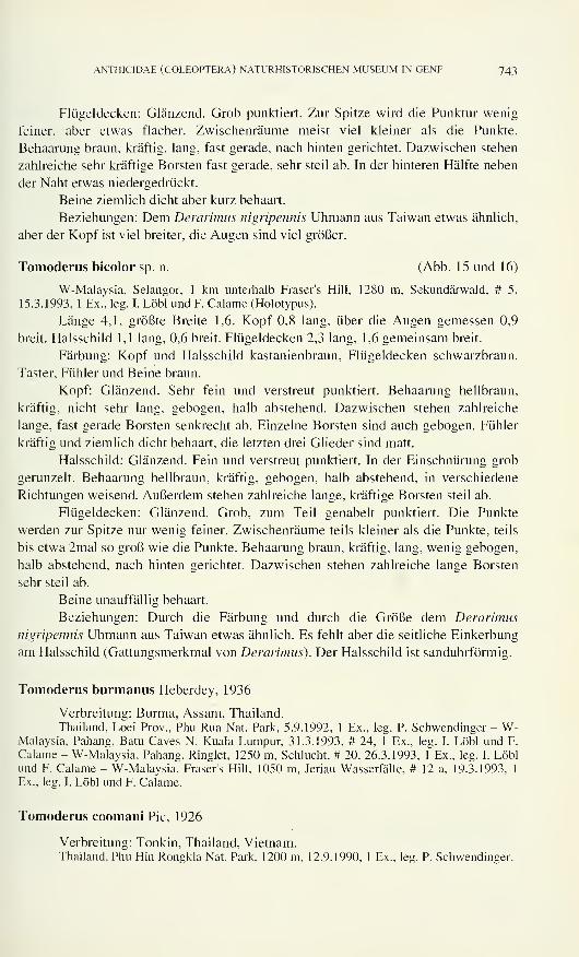
ANTHICIDAE (COLEOPTERA) NATURHISTORISCHEN MUSEUM IN GENF 743
Flügeldecken: Glänzend. Grob punktiert. Zur Spitze wird die Punktur wenig
feiner, aber etwas flacher. Zwischenräume meist viel kleiner als die Punkte.
Behaarung braun, kräftig, lang, fast gerade, nach hinten gerichtet. Dazwischen stehen
zahlreiche sehr kräftige Borsten fast gerade, sehr steil ab. In der hinteren Hälfte neben
der Naht etwas niedergedrückt.
Beine ziemlich dicht aber kurz behaart.
Beziehungen: Dem Derarimus nigripennis Uhmann aus Taiwan etwas ähnlich,
aber der Kopf ist viel breiter, die Augen sind viel größer.
Tomoderus bicolor sp. n. (Abb. 15 und 16)
W-Malaysia, Selangor, 1 km unterhalb Fraser's Hill, 1280 m, Sekundärwald, # 5.
15.3.1993, 1 Ex., leg. I. Löbl und F. Calarne (Holotypus).
Länge 4,1, größte Breite 1,6. Kopf 0,8 lang, über die Augen gemessen 0,9
breit. Halsschild 1,1 lang, 0,6 breit. Flügeldecken 2,3 lang, 1,6 gemeinsam breit.
Färbung: Kopf und Halsschild kastanienbraun, Flügeldecken schwarzbraun.
Taster, Fühler und Beine braun.
Kopf: Glänzend. Sehr fein und verstreut punktiert. Behaarung hellbraun,
kräftig, nicht sehr lang, gebogen, halb abstehend. Dazwischen stehen zahlreiche
lange, fast gerade Borsten senkrecht ab. Einzelne Borsten sind auch gebogen. Fühler
kräftig und ziemlich dicht behaart, die letzten drei Glieder sind matt.
Halsschild: Glänzend. Fein und verstreut punktiert. In der Einschnürung grob
gerunzelt. Behaarung hellbraun, kräftig, gebogen, halb abstehend, in verschiedene
Richtungen weisend. Außerdem stehen zahlreiche lange, kräftige Borsten steil ab.
Flügeldecken: Glänzend. Grob, zum Teil genabelt punktiert. Die Punkte
werden zur Spitze nur wenig feiner. Zwischenräume teils kleiner als die Punkte, teils
bis etwa 2mal so groß wie die Punkte. Behaarung braun, kräftig, lang, wenig gebogen,
halb abstehend, nach hinten gerichtet. Dazwischen stehen zahlreiche lange Borsten
sehr steil ab.
Beine unauffällig behaart.
Beziehungen: Durch die Färbung und durch die Größe dem Derarimus
nigripennis Uhmann aus Taiwan etwas ähnlich. Es fehlt aber die seitliche Einkerbung
am Halsschild (Gattungsmerkmal von Derarimus). Der Halsschild ist sanduhrförmig.
Tomoderus burmanus Heberdey, 1936
Verbreitung: Burma, Assam, Thailand.
Thailand, Loei Prov., Phu Rua Nat. Park, 5.9.1992, 1 Ex., leg. P. Schwendinger - W-Malaysia, Pahang, Batu Caves N. Kuala Lumpur, 31.3.1993. # 24, 1 Ex.. leg. I. Löbl und F.
Calarne - W-Malaysia, Pahang, Ringlet, 1250 m, Schlucht, # 20, 26.3.1993, 1 Ex., leg. I. Löblund F. Calarne - W-Malaysia, Fraser's Hill, 1050 m, Jeriau Wasserfälle, # 12 a, 19.3.1993. 1
Ex., leg. I. Löbl und F. Calarne.
Tomoderus coomani Pic, 1926
Verbreitung: Tonkin, Thailand, Vietnam.Thailand, Phu Hin Rongkla Nat. Park. 1200 m, 12.9.1990, 1 Ex., leg. P. Schwendinger.
744 GERHARD UHMANN
Tomoderus globicollis sp. n. (Abb. 17 und 18)
Thailand, Phu Hin Rongkla Nat. Park, 1200 m, 12.9.1990, 1 Ex., leg. P. Schwendinger
(Holotypus).
Länge 2,3, größte Breite 1,0. Kopf 0,5 lang, über die Augen gemessen 0,4
breit. Halsschild 0,6 lang, 0,4 breit. Flügeldecken 1,2 lang, 1,0 gemeinsam breit.
Färbung: Braun. Taster, Fühler und Beine hellbraun.
Kopf: Glänzend. Äußerst fein und verstreut punktiert. Behaarung gelbbraun,
mittelmäßig in Stärke und Länge, gebogen, fast ganz abstehend. Dazwischen stehen
zahlreich nicht sehr lange Borsten senkrecht ab.
Halsschild: Glänzend. Sehr fein und verstreut punktiert, nur in der
Einschnürung kräftig gerunzelt. Behaarung gelbbraun, mittelmäßig, gebogen, fast
ganz abstehend. Dazwischen stehen zahlreiche lange, nicht sehr kräftige Borsten
gerade und senkrecht ab.
Flügeldecken: Glänzend. Sehr stark punktiert. Zwischenräume kleiner als die
Punkte. Zur Spitze werden die Punkte etwas feiner, die Zwischentäume etwa 1 bis
3mal so groß wie die Punkte. Behaarung gelbbraun, ziemlich lang, nicht sehr kräftig,
etwas gebogen, halb abstehend. Dazwischen stehen zahlreiche lange, nicht sehr
kräftige Borsten steil ab.
Beine nicht sehr auffällig behaart.
Beziehungen: Dem Tomoderus promiscuus Krekich von den Philippinen und
dem T. sulcifer Pic aus Java und Sumatra etwas ähnlich, aber die Fühler sind kräf-
tiger, der Kopf ist breiter, die Flügeldecken sind bauchiger. Von T. binodulus
Uhmann von Borneo unterscheidet er sich durch kräftigere Fühler, geringere
Körpergröße und durch vorn breiteren Halsschild.
Tomoderus plicicollis Pic, 1901
Verbreitung: Neuguinea, Australien.
Austr., Queensland, Cape Tribulation National Park, Pilgrim Sands, 26.7.-3.8.1986, 3
Ex., leg. S. Kiener.
Tomoderus promiscuus Krekich, 1929
Verbreitung: Philippinen, Borneo, Sulawesi.
Indonesia, Sulawesi, Sei. W of Manasa, 1600 m, 9.4.1991, leg. D. Agosti, 4 Ex.
Abb. 11-18
11. Derarimus pahangensis sp. n., Habitus; 12. Derarimus pahangensis sp. n., Halsschildprofil;
13. Derarimus selangorensis sp. n., Habitus; 14. Derarimus selangorensis sp. n., Halsschild-
profil; 15. Tomoderus bicolor sp. n., Habitus; 16. Tomoderus bicolor sp. n., Halsschildprofil;
17. Tomoderus globicollis sp. n., Habitus; 18. Tomoderus globicollis sp. n., Halsschildprofil.
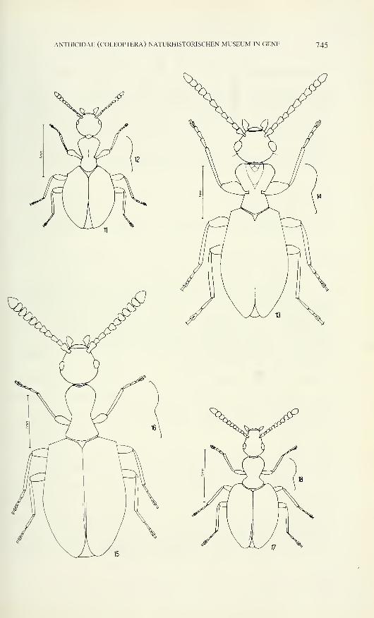
ANTHICIDAE (COLEOPTERA) NATURHISTORISCHEN MUSEUM IN GENF 745
746 GERHARD UHMANN
Tomoderus schuhi Uhmann, 1996
Verbreitung: Malaysia.
W-Malaysia, Pahang, Cameron Highlands, Trail 9, 1400 m, 27.3.1993, # 21, 2 Ex., leg.
I. Lobi und F. Calarne - W-Malaysia, Pahang, Ringlet, 1250 m, Schlucht, # 20, 26.3.1993, 1
Ex., leg. I. Löbl und F. Calarne - W-Malaysia, Pahang, Fraser's Hill, 1300 m, Tiong Trail, # 13,
20.3.1993, 4 Ex., leg. I. Löbl und F. Calarne - W-Malaysia. Pahang, Taman Negara, 90-120 m,
Tembeling Trail, Urwald. # 1 a. 10 und 13.3.1993. 1 Ex.. leg. I. Löbl und F. Calarne - W-Malaysia, Pahang, Fraser's Hill, 1050 m. Jerian Wasserfälle, # 12 a, 19.3.1993, 1 Ex., leg. I.
Löbl und F. Calarne - W-Malaysia, Selangor, Gap, 900 m, 14.3.1993, Sekundärwald nahe
Strasse. 1 Ex.. leg. I. Löbl und F. Calarne - W-Malaysia, Selangor, 5 km unterhalb Fraser's Hill,
1 100 m. Schlucht, # 7, 13.-16.3.1993, 2 Ex., leg. I. Löbl und Prahme.
Tomoderus subcostulatus Pic, 1901
Verbreitung: Sumatra.
W-Malaysia, Pahang, Genting Highlands. Awana, 1 150 m, 3.4.1993, # 27 a, 1 Ex., leg.
I. Löbl und F. Calarne.
Tomoderus unifasciatus Pic, 1907
Verbreitung: Indien. Thailand, Burma.
Thailand. Taksin Maharat N.P., 1000 m. 9.2.1993, 1 Ex., leg. P. Schwendinger -
Thailand, Chiang Mai Prov., Doi Suthep, 1200 m, 9.11.1991, 3 Ex., leg. P. Schwendinger -
Thailand, Prachuab Kiri Khan Prov.. Huay Yang N. Park. 670 m, 29.11.1991, 4 Ex., leg. P.
Schwendinger- Thailand, Lamphun Province, 1000 m, Doi Khuntan N. Park, 25.9.1991, 3 Ex.,
leg. P. Schwendinger - Thailand, Loel Prov.. Phu Rua Nat. Park. 1080-1250 m. 3.9.1992, 1
Ex., leg. P. Schwendinger - Thailand. Ban Maeo Microwave, Mae Hong Son Dist., 1250 m,
15.12.1990, 1 Ex.. leg. P. Schwendinger - Thailand, Lamphun Prov.. Doi Khuntan N.P.. 1 100
m, 8.10.1992. 1 Ex., leg. P. Schwendinger.
Formicomus armatus Boheman. 1858
Verbreitung: Indien, Thailand, Vietnam, Japan, Java, Sumatra, Malaysia, Sri
Lanka. Philippinen. China.
Thailand. Mts. N. Umphang, Mae Sot Umphang, 1250 m. 10.2.1993. 2 Ex., leg. P.
Schwendinger.
Formicomus conjugatus Krekich, 1926
Verbreitung: Annam, Thailand.
Thailand, Mts. N. Umphang, Mae Sot Umphang. 1250 m. 10.2.1993, 1 Ex.Jeg. P.
Schwendinser.
Formicomus longispinus Pic, 1912
Verbreitung: Taiwan, Yunnan, Thailand.
Thailand. Chiang Mai Prov.. Mae Hia, 350 m, 18.1.1992, 3 Ex., leg. P. Schwendinger.
ANTHICIDAE (COLEOPTERA) NATURHISTORISCHEN MUSEUM IN GENF 747
Anthicomorphus siamensis Krekich, 1 926
Verbreitung: Thailand, Burma, Vietnam.
Thailand, Sur at Thani P., Khao Sok N. Park, 70 m, 6.12.1991, 2 Ex., leg. P.
Schweninger.
Pseudoleptaleus sculptus Krekich, 1926
Verbreitung: Indien, Thailand.
Thailand, Huay ya mae Khi, Umphang dist., 670 m, 10.2.1993, 2 Ex., leg. P.
Schwendinger.
Omonadus formicarius (Goeze, 1777)
Verbreitung: Kosmopolit.
Thailand, Huay Yang Nat. Park, Huay Yang Waterfall, Thap Sake Dist., 50 m,
12.2.1991, 1 Ex., leg. P. Schwendinger.
Clavicomus cordatus (Krekich, 1931)
Verbreitung: Indien, Nepal, Thailand.
Thailand, Tak Prov. Doi Musoe, 950 m, 18.9.1990, 1 Ex., leg. P. Schwendinger.
Sapintus breviceps (Laferté, 1
Verbreitung: Burma, Thailand, Pakistan.
Thailand, Ban Kiriwong, Khao Luang, Nat. Park, 170 m, 10.2.1991, 4 Ex., leg. P.
Schwendinger.
Sapintus immaturus (Laferté, 1848)
Verbreitung: Indien, Thailand, Bangla Desh.
Thailand, Phrae Province, 560 m, Mae Khaem Waterfall, 21.9.1991, 1 Ex., leg. P.
Schwendinger.
Sapintus javanus (Marseul, 1882)
Verbreitung: Malaysia, Indonesien, Thailand, Vietnam, Sri Lanka, Indien.
Thailand, Ban Maeo Microwave, Mae Hong Son Dist., 1250 m, 15.12.1990, 2 Ex., leg.
P. Schwendinger.
Sapintus siamensis (Pic, 1914)
Verbreitung: Thailand, Vietnam.
Thailand, Banglang Nat. Park, Than To Waterfall, Than To Dist., 150 m. 1.2.1991. 2
Ex., leg. P. Schwendinger — Thailand, Ban Kiriwong, Khao Luang Nat. Park, 170 m,10.2.1991, 1 Ex., leg. P. Schwendinger.
748 GERHARD UHMANN
Ergänzung des bestimmuns-schlüssels für die derarimus-ARTEN
1 Etwa 2 mm lang la
la Flügeldecken seitlich schwach konvex minutissimus UhmannFlügeldecken seitlich stark konvex pahangensis sp. n.
8 Kopf breiter als lang, hinter den Augen sofort stark verengt 8a
8a Größer (ca 3.8), Halsschild in der Einschnürung ziemlich fein gerunzelt
humerifer UhmannKleiner (ca 3), Halsschild in der Einschnürung kräftig punktiert . luteipes sp. n.
15
Flügeldecken nicht kräftig punktiert, mit großen Zwischenräumen 15a
15a Etwa 3,4 mm, Malaysia ovipennis sp. n.
Etwa 4 mm, Thailand schwendingerì Uhmann26 Kopf und Halsschild braun, Flügeldecken schwarzbraun 26a
Anders 27
26a Kopf so lang wie breit, Schultern weniger markant 26b
Kopf breiter als lang, Schultern sehr markant selangorensis sp. n.
26b Halsschild vorn fein punktiert nigripennis UhmannHalsschild bis vorn kräftig punktiert fortepunctatus sp. n.
30 Hinterkopf konkav 30a
30a Etwa 3,2 mm javanus UhmannEtwa 4,9 mm calamei sp. n.
LITERATUR
Uhmann, G. 1994. Südostasiatische Anthicidae aus dem Naturhistorischen Museum in Genf, 4- Revue suisse de Zoologie, 101(3): 655-676.
Revue suisse de Zoologie, 103 (3): 749-756; septembre 1996
Further data on the Nicolettidae (Zygentoma),
with description of a new species from Mauritius
Luis F. MENDESCentro de Zoologia, Instituto de Investigaçào Cientifica Tropical,
R. da Junqueira, 14, 1300 Lisboa, Portugal.
Further data on the Nicolettidae (Zygentoma), with description of a
new species from Mauritius. - Lepidospora (L.) mascareniensis sp. n. is
described from Mauritius. New data are reported on Coletinia mendesi
Wygodzinsky in Portugal, and C. muggii (Grassi) is recorded for the first
time from France.
Key-words: Nicolettidae - Mauritius - Europe - New species - New data.
INTRODUCTION
The present paper deals with the study of one new species of Lepidospora s. s.
(Nicoletiidae) collected in Mauritius Island and with a few samples of another genus
of the same family, Coletinia, from Portugal and France. L. (L.) mascareniensis sp. n.
is the first thysanuran recorded from Mauritius.
The material is deposited in the following collections: Centro de Zoologia of
the Instituto de Investigaçào Cientifica Tropical, Lisboa, Portugal (CZ); Muséumd'histoire naturelle, Geneva, Switzerland (MHNG) and Muséum National d'Histoire
Naturelle, Paris, France (MNHNP).
Lepidospora (L.) mascareniensis sp. n.
Materia] examined: He Maurice — Trois Cavernes, near Cascavelle, 6-8/XI/1994, 1 9
holotype, leg. P. Strinati (MHNG).
Body length: 5.8 mm; thorax length: 1.9 mm; thorax width: 1.4 mm; antennae
(dammaged): 3.5 mm.Body elongated, more or less parallel-sided. Hypodermal pigment absent, the
general colour whitish. Macrochaetae light brown, the stronger ones apically biphid.
Scales typical, with a moderate number of thin rays.
Head as in Fig. 1, with some strong elongated macrochaetae, the scales res-
tricted to the posterior half of the cephalic capsule. Antennae typical, not completely
preserved.
Manuscript accepted 04.0 1 . 1 996.
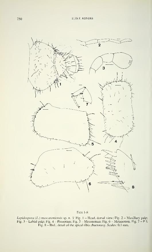
750 LUIS F. MENDES
Figs 1-8
Lepidospora (L.) mascareniensis sp. n. $ Fig. 1 - Head, dorsal view; Fig. 2 - Maxillary palp;
Fig. 3 - Labial palp; Fig. 4 - Pronotum; Fig. 5 - Mesonotum; Fig. 6 - Metanotum; Fig. 7 - P I;
Fig. 8 - Ibid., detail of the apical tibia chaetotaxy. Scales: 0.1 mm.
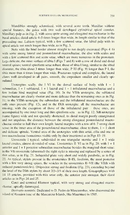
FURTHER DATA ON NICOLETIIDAE 75 \
Mandibles strongly sclerotized, with several acute teeth. Maxillae without
special features, the galea with two well developed cylindrical apical conules.
Maxillary palp as in Fig. 2, with some quite strong and elongated macrochaetae in the
basal articles; distal article 4-5 times longer than wide, its length similar to that of the
preceeding one. Labium typical, with a few scattered setae, the labial palp with its
apical article not much longer than wide, as in Fig. 3.
Nota with the hind border almost straight to not deeply excavated (Figs 4-6)
with some strong lateral and posterolateral macrochaetae, the disc with scales and
with a few scattered thin and acute setae, which are more numerous in the pronotum.
Legs delicate, the outer surface of tibia I (Figs 7 and 8) with a row of distal and distal
ventral spines; ventral spiniform setae robust, those of tibia I long, similar to the tibial
diameter, the tibia about 3 times longer than wide; P III (Fig. 9) longer than P I, the
tibia more than 4 times longer than wide. Praetarsus typical and complete, the lateral
claws well developed in all pairs, smooth, the empodium smaller and clearly spi-
nulated.
Urotergites scally, the I-VI in the dorsal surface of body with 1 + 1
submedian, 1 + 1 sublateral, 1 + 1 lateral and 1 + 1 infralateral macrochaetae and a
few isolate hind marginal setae (Fig. 10). In the Vllth urotergite, the sublateral
macrochaetae are clearly shorter and more delicate than the remaining ones as in Fig.
1 1 ; in the VHIth urotergite the submedian and the infralateral macrochaetae are the
only ones present (Fig. 12), and in the IXth urotergite, all the macrochaetae are
missing, with the exception of those of the infralateral pair - these ones, are
accompanied by one inner long and thin spiniform seta - as in Fig. 12; Xth urotergite
(same figure) wide and not specially shortened, its distal margin poorly emarginated
and not angulous, the distance between the strong elongated posterolateral macro-
chaetae similar to half their own length; lateral margins with a row of 6-7 strong short
setae; in the inner area of the posterolateral macrochaetae, close to them, 1 + 1 short
and delicate spinule. Ventral area of the urotergites with thin setae, cilia and one or
two macrochaetae (sometimes visible only by their insertions) as in Figs 13-19.
Urosternite I typical, subdivided in one triangular sternite and 1 + 1 wide
lateral coxites, almost devoided of setae. Urosternites II-VI as in Fig. 20, with 1 + 1
anterior and 1 + 1 posterior submedian macrochaetae besides the marginal short setae;
in the Vth urosternite (abnormal) the right stylet is missing and replaced by a series of
thin setae, as in Fig. 21. Subgenital plate very short and widely parabolic, as in Fig.
22. As typical, stylets present in the urosternites II-IX, fusiform, the most posterior
with a few very strong spines, the vesicles in the urosternites II—VII (the Vllth with
reduced pseudovesicles). Ovipositor strong and fusiform (Fig. 23), extending beyond
the level of the IXth stylets by about 2/3-3/4 of their own length. Gonapophyses with
14—15 articles, provided with thin setae only, the anterior pair stronger, their distal
articles as in Figs 24 and 25.
Cerci and terminal filament typical, with very strong and elongated macro-
chaetae, apically dammaged.
Derivano nominis: Dedicated to D. Pedro de Mascarenhas, who discovered the
island of Reunion (one of the Mascarene Islands, like Mauritius) in 1505.
752 LUIS F. MENDES
FIGS9-18
Lepidospora (L.) mascareniensis sp. n. 2 Fig. 9 - P III; Fig. 10 - Vth urotergite; Fig. 1 1 - Vllth
urotergite; Fig. 12 - IXth and Xth urotergites; Fig. 13 - Infralateral chaetotaxy of Illrd urotergite;
Fig. 14 - Ibid. of urotergite IV; Fig. 15 - Ibid. of urotergite V; Fig. 16 - Ibid. of urotergite VI;
Fig. 17 - Ibid. of urotergite VII; Fig. 18 - Ibid. of urotergite VIII. Scales: 0.1 mm.
FURTHER DATA ON NICOLETIIDAE 753
Figs 19-25
Lepidospora (L.) mascareniensis sp. n. 9 Fig. 19 - Infralateral chaetotaxy of IXth urotergite;
Fig. 20 - Illrd urosternite; Fig. 21 - Vth anomalous urosternite (the right stylet is lacking); Fig.
22 - Posterior border of Vllth urosternite and subgenital plate; Fig. 23 - Ventral posterior area
of abdomen; Fig. 24 - VHIth gonapophyses, distal articles; Fig. 25 - IXth gonapophyses, distal
articles. Scale: 0.1 mm.
754 LUIS F - MENDES
Discussion: Lepidospora (L.) mascareniensis sp. n., though known by the holo-
type female only, is well characterized among the remaining Afrotropical and Malagasy
known species. L. afra Silvestri, from Kenya and Tanzania (Silvestri 1908 b and 1918).
L. meridionalis Silvestri, from Natal and Zululand (Silvestri 1913 a) and L. vilhenai
Silvestri, from Angola (Silvestri 1949), present a much longer ovipositor, which
extends beyond the level of the IXth stylets by 1 .5-2 times their own length (2/3-3/4 of
this length in the new species): further, L meridionalis and L. vilhenai present, both, a
much thiner ovipositor composed by a greater number of articles (20-26 versus about 15
in the new species), and L. afra (with a similar number of articles) distinct Xth urotergite
and a quite different subgenital plate. L. insularum Wygodzinsky, from the atlantic coast
of the Cape Province (Wygodzinsky 1955), presents a more deeply incised Xth
urotergite, a distinct subgenital plate and a different cephalic chaetotaxy. L. braueri
Escherich, from the Seychelles (Escherich 1905), which female sex was described by
Carpenter (1916), presents a shorter ovipositor and not clearly annulated gonapophysis
(not completely developed female though long as 1 1 mm?) shows a much more deeply
incised Xth urotergite, a more rounded and enlarged subgenital plate and also (if
Carpenter's figure is correct) an unique empodial claw shape.
Relatively to the Plaearctic and Oriental described Lepidospora s. s., the new
taxon seems to approach particularly L. ceylonica Silvestri, from Sri Lanka and India
(Silvestri 1911) and L. wygodzinskyi Mendes, from the Kos and Rhodes Islands
(Mendes 1992 and, as L. escherichia Wygodzinsky 1980), both with a similar
ovipositor: however, the oriental taxon is clearly bigger (8 mm body length), with a
distinct shape of the subgenital plate, a different chaetotaxy and a deeper excavation
in the Xth urotergite. and the mediterranean species has shorter ovipositor, with a
lower number of articles, and presents also a different chaetotaxy and a deeper Xth
urotergal notch. L. kurda Mendes, from the iraquian Kurdistan (Mendes 1985) and L.
notabilis Silvestri, from India (Silvestri 1913 b), both known by the males only, are
completely isolated under the geographical point of view. All the remaining species
show much shorter ovipositor, exceeding the level of the IXth stylets by no more than
half their own length, as well as a much deeper Xth urotergite incision: L. buxtoni
Silvestri, from the eastern Iraq (Silvestri 1923), L. silvestrii Wygodzinsky, from
Israel (see Wygodzinsky 1942) and L. escherichi Silvestri, from the Corfu island
(Silvestri 1908 a. recently redescribed upon almost topotypical material off the
greek islands of Leucade and Ipiros by Mendes 1985).
L. grassi Escherich, from Sicily and (?) continental Italy (?) (Wygodzinsky
1980) and L. gracilis Escherich, from Sumatra (Escherich 1905) are much more
difficult to compare with the remaining Lepidospora s. s., on account of their short
and undetailed descriptions; however, both belong to the 'long-type' ovipositor and L.
grassi has, further, an atypical, enlarged, lepismatoid body shape.
Coletinia mendesi Wygodzinsky, 1980
Material examined: Portugal - Lapa de Colaride (Cacém), 20/111/1993, 1 S 1 9, leg. F.
Regalo & R. Mergulho (CZ); ibid., ll/IX/1993, 1 o\ ibid., (CZ). Lisboa, archaeological
excavations of Sé Velha de Lisboa, nos claustros, no date, 1 young 9 , leg. R. Mergulho (CZ).
FURTHER DATA ON NICOLETIIDAE 755
Coletinia mendesi was described upon material collected in the Algarve
Province, southern Portugal (Wygodzinsky 1980) and further recorded from southern
Spain, in the Cordoba Province, Andaluzia (Roca et al. 1985). The newly referred
material, clearly enlarges northward (about 300 Km) the known distribution of the
species, and the specimens from the Lapa de Colaride represent the first reference to
C. mendesi as a troglobiont; the male collected in the 20th March, with 8.1 mm of
body length, is much bigger than the type-specimen (6.7 mm only), though all the
major features agree to what is mentioned in the original description; the typical
elongation of the second tibia ventral macrochaetae, noticed as being longer than
those of the remaining pairs of legs, is conspicuous even in the young female
collected during the archaeological excavations in the old area of Lisboa.
Coletinia maggii (Grassi, 1887)
Material examined: France - Paris, Catacombes of the Museum, 26/XII/1947, 4 3 â 1 9
,
no collector (MNHNP).
Coletinia maggii was firstly collected in Italy (Sicily) and subsequently
(Wygodzinsky 1980) redescribed based on material from Italy mainland and from
Yugoslavia, Dalmatia (now, Croatia); it was also mentioned for Malta and Italy
(Mendes 1981) and for Austria and, again, for Italy (Mendes 1992). It is the very first
time that the species is recorded to occur in France. The studied specimens, deposited
in the entomological collection of the Paris Museum, were determined by Balazuc as
Nicoletia sp.
ACKNOWLEDGEMENTS
I am deeply grateful to Dr P. Strinati, who collected, and to Dr V. Aellen, wholoaned the single specimen of the new Lepidospora described below. We want to
thank also Mrs F. Regala and R. Mergulho (both from the "Associaçào de Estudos
Subterrâneos e Defesa do Ambiente" in Torres Vedras, Portugal) for the donated
Coletinia collected in and around Lisboa, as well as to Miss Kelsner-Pillault, from the
Paris Museum, for the sympathy and assistance during our survey of the Zygentoma
collection several years ago.
REFERENCES
Bach de Roca, C, L.F. Mendes & M. Gaju-Ricart. 1985. Sur une nouvelle espèce et unenouvelle citation de Nicoletiinae de Sierra Morena (Cordoue, Espagne) (Nicoletiidae,
Zygentoma). Bolletino della Società entomologica italiana, Genova, 117(8/10):
132-140.
Carpenter, G.H. 1916. The Apterygota of the Seychelles. Proceedings of the royal Irish
Academy 33(1): 1-70, pi. I-X.
Escherich, K. 1905. Das System der Lepismatiden. Zoologica, Stuttgart, 43(18): 1-164.
Mendes, L.F. 1981. Nova nota sobre os tisanuros (Apterygota, Microcoryphia e Zygentoma) da
Europa e da bacia mediterrânica. Boletim da Sociedade portuguesa de Entomologia
1(18): 1-8.
756 LUIS F - MENDES
Mendes, L.F. 1985. Sur quelques thysanoures (Microcoryphia et Zygentoma) de l'Asie sud-
occidentale. Notes et descriptions. Nouvelle Revue d'Entomologie (N.S.) 2(3): 303-317.
Mendes, L.F. 1992. Some new data on the Nicoletiidae (Insecta: Zygentoma) from Europe and
Asia Minor. Revue suisse de Zoologie 99(4): 821-834.
Silvestri, F. 1908 a. Materiali per lo studio dei Tisanuri. X. Su alcuni tisanuri di Corfu.
Bolletino dei Laboratorio de Zoologia generale e agraria di Portici 2: 381-393.
Silvestri, F. 1908 b. Apterygogenea. 2. Thysanura. In: Sjostedts Kilmandjaro-Meru Exped.,
Uppsala 18(2): 11-14.
Silvestri, F. 1911. Materiali per lo studio dei Tisanuri. XIV. Nuova specie di Lepidospora di
Ceylan. Bolletino dei Laboratorio de Zoologia generale e agraria di Portici 5: 95-96.
Silvestri, F. 1913 a. Tisanuri raccolti dal Dr I. Trägardh nel Natal e nel Zululand. Arkiv vor
Zoologie 8(1 ): 1-15.
Silvestri, F. 1913 b. On some thysanura in the Indian Museum. Records of the Indian
Museum, Calcutta, 9: 51-62.
Silvestri, F. 1918. Insects Apterygogéniens. 1. Thysanura. Voyage Ch. Alluaud et R. Jeannel
Afr. Orient. (1911-1912). Result, scient.: 1-28.
Silvestri, F. 1923. Thysanura, Termitidae and Embiidae collected in Mesopotamia and N.W.Persia by W. Edgar Evans, B. Sc, late Capt. R.A.M.C, and Dr P.A. Buxton. Tran-
sactions of the entomological Society ofLondon 105: 258-262.
Silvestri, F. 1949. Insecta Thysanura hucusque in Lunda reperta. Publicaçoes culturais da
Companhia dos Diamantes de Angola 6: 61-84.
Wygodzinsky, P. 1942. Second contribution towards the knowledge of Diplura and Thysanura
from Palestine. Revista brasileira de Biologia 2(1): 29-46.
Wygodzinsky, P. 1955. Thysanura. In: Hanstrom, B., P. Brinck & C. Rudebeck (eds.), South
african animal life, Uppsala 2: 83-190.
Wygodzinsky, P. 1980. A survey of the Nicoletiinae of Europe (Nicoletiidae, Thysanura,
Insecta). American Museum Novitates 2695: 1-24.
Revue suisse de Zoologie, 103 (3): 757-766; septembre 1996
Une nouvelle espèce du genre Ptychadena (Anura, Ranidae)
du Kenya
Jean-Luc PERRETMuséum d'histoire naturelle. Case postale 6434, CH-121 1 Genève 6, Suisse.
A new species of the genus Ptychadena (Anura, Ranidae) from Kenya.- Discovered during the Geneva Museum zoological missions (1974-
1977) in Kenya, a new species of grass frog: Ptychadena mahnerti n. sp. is
described. A highland savanna dweller, it is readily distinguished by its
particular femoral pattern as well as by its voice. The other Kenyan
Ptychadena species are compared and discussed.
Key-words: Amphibia - Ranidae - Ptychadena - Taxonomy - Biology -
Kenya.
INTRODUCTION
Deux missions zoologiques du Muséum de Genève au Kenya (1974 et 1977)
ont été accomplies par Volker Mahnert et l'auteur. L'important matériel herpétolo-
gique récolté n'a pas encore été totalement identifié et reste en cours d'étude.
Lors du premier voyage (1974), un ranidé du genre Ptychadena m'avait tout de
suite suggéré la découverte d'une forme probablement nouvelle, mais un seul
exemplaire capturé était insuffisant pour la définir. Au second voyage (1977), retrou-
vée dans diverses localités et assez bien observée dans son biotope, elle pouvait être
reconnue. Un caractère flagrant qui la distingue des autres Ptychadena réside en son
patron fémoral qui exhibe deux taches séparées sur chaque cuisse; chez les autres
espèces du genre, ce caractère spécifique présente des patrons fémoraux différents:
rayés, marbrés ou multi-tachetés.
Au Muséum de Nairobi, une confrontation de nos spécimens révéla que
quelques exemplaires non identifiés ou mêlés à une autre espèce se trouvaient en
collection. Ils provenaient des marais de Limuru où nous avons trouvé cette grenouille
abondante. Enfin, le Dr Alex Duff-MacKay (Conservateur à l'époque) nous dit qu'il
avait dans ses archives sonores, la voix enregistrée d'une Ptychadena species indéter-
minée de Limuru ... restée en question!
Elle est enfin décrite aujourd'hui.
Manuscrit accepté le 31.01.1996.
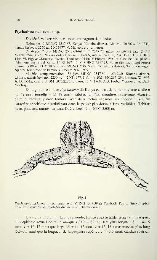
758 JEAN-LUC PERRET
Ptychadena mahnerti n. sp.
Dédiée à Volker Mahneit, mon compagnon de mission.
Holotype: S MHNG 2547.67. Kenya. Kiambu district. Limuru. (01°07'S 36°38'E),
marais herbeux. 2250 m, 2 XI 1977. V. Mahnen et J.-L. Perret.
Paratypes: 2 SS MHNG 2547.68-69, 1 9 2547.70. même localité et date. 2 SSMHNG 2547.71-72, Nakuru district. Njoro. 20 km S. marais. 2400 m, 7 XI 1977. 1 S MHNG1543.39, Elgeyo-Marakwet district, Tambach, 25 km E Eldoret. 1900 m. flanc de haut plateau
s'abaissant sur le val Kerio. 17 XI 1974. 1 9 MHNG 2547.73, Embu district, Irangi Forest
Station, 2000 m, 1 1 X 1977. 6 juv. MHNG 2547.74-79. Nyandarua district, South Kinangop,
Njabini. forêt, zone de bambous. 2550 m. 9 XI 1977.
Matériel complémentaire: 152 juv. MHNG 2547.80 - 2549.30, Kiambu district,
Limuru. marais herbeux. 2250 m. 1-2 XI 1977. 1 S. 1 9 BM 1970.293-294, Limuru, XI 1967,
A. Duff-MacKay. 1 S BM 1975.2286. Limuru. 11 V 1968. A.D. Forbes-Watson et A. Duff-
MacKay.
Diagnose: une Ptychadena du Kenya central, de taille moyenne (mâle =
35-42 mm, femelle = 45^49 mm); habitus ranoïde; membres postérieurs élancés;
palmure réduite: patron fémoral avec deux taches séparées sur chaque cuisse, un
caractère spécifique discriminant dans le genre; plis dorsaux fins, variables. Habitat:
hauts plateaux, marais herbeux, lisière forestière. 2000-2500 m.
Fig. 1
Ptychadena mahnerti n. sp., paratype S MHNG 1543.39 de Tambach. Patron fémoral spéci-
fique avec deux taches marbrées distinctes sur chaque cuisse.
Description: habitus ranoïde, élancé chez le mâle, femelle plus trapue;
dimorphisme sexuel de taille marqué {S 19 = 83 %); tête plus longue (S - 14-15
mm. 9 = 16-17 mm) que large (cT = 11-13 mm, 9 = 13-15 mm); museau plus long
(5.5-7.5 mm) que la longueur de la paupière supérieure (4-5.5 mm); canthus rostralis
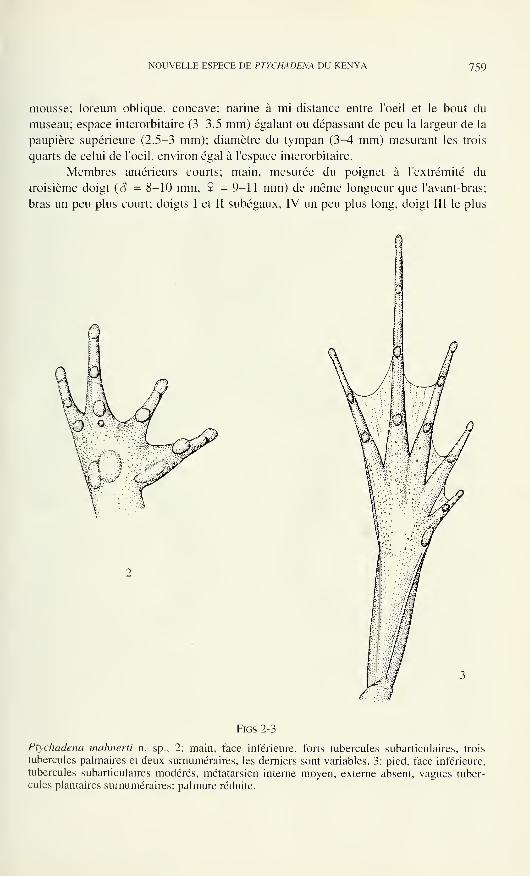
NOUVELLE ESPECE DE PTYCHADENA DU KENYA 759
mousse; loreum oblique, concave; narine à mi-distance entre l'oeil et le bout du
museau; espace interorbitaire (3-3.5 mm) égalant ou dépassant de peu la largeur de la
paupière supérieure (2.5-3 mm); diamètre du tympan (3-4 mm) mesurant les trois
quarts de celui de l'oeil, environ égal à l'espace interorbitaire.
Membres antérieurs courts; main, mesurée du poignet à l'extrémité du
troisième doigt (â = 8-10 mm, 9 = 9-11 mm) de même longueur que l'avant-bras;
bras un peu plus court; doigts I et II subégaux, IV un peu plus long, doigt III le plus
Figs 2-3
Ptychadena mahnerti n. sp., 2: main, face inférieure, forts tubercules subarticulaires, trois
tubercules palmaires et deux surnuméraires, les derniers sont variables. 3: pied, face inférieure,
tubercules subarticulaires modérés, métatarsien interne moyen, externe absent, vagues tuber-
cules plantaires surnuméraires; palmure réduite.
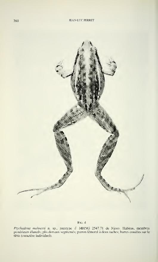
760 JEAN-LUC PERRET
/
Fig. 4
Ptychadena mahnerti n. sp., paratype S MHNG 2547.71 de Njoro. Habitus, membrespostérieurs élancés; plis dorsaux segmentés; patron fémoral à deux taches; barres coudées sur le
tibia (caractère individuel).
NOUVELLE ESPECE DE PTYCHADENA DU KENYA 76 ]
long (longueur main); extrémité des doigts arrondie, bien formée; tubercules subarti-
culaires forts, celui du pouce le plus saillant; paume de la main chagrinée avec trois
tubercules palmaires: un thénaire elliptique, un médian rond, contigu à un externe
allongé épousant sa forme; des tubercules surnuméraires variables, parfois présents
au-dessous des doigts II, III, IV.
Membres postérieurs élancés, plus longs chez le mâle où l'articulation tibio-
tarsienne dépasse largement le bout du museau tandis qu'elle n'atteint pas ou dépasse
de peu seulement l'extrémité du museau de la femelle; tibia plus long que la moitié du
corps (T/MA = 60%), plus long que la cuisse mais faiblement plus court que le pied
(TYP = 95%); tubercules subarticulaires moyens, arrondis; palmure pédieuse réduite, à
demi développée; formule générale (phalange libre de palmure à chaque orteil, côté
interne et côté externe): I ext. 1.75-2; II int. 2.25-2.50, ext. 1.75-2; III int. 3, ext. 2;
IV int. 3.25-3.50. ext. 3; V int. 1.75-2.
Tubercule métatarsien interne modéré (1.8-2 mm), relié au talon par un pli
tarsien (8-9 mm); tubercule métatarsien externe absent ou présent mais alors très petit
et souvent mal défini; sole granuleuse avec des verrues variables, certaines alignées
sous le quatrième métatarsien, alias tubercules plantaires surnuméraires.
Tégument dorsal garni de fins plis glanduleux variables qui différent dans le
détail chez chaque individu: externes plus épais parfois segmentés, de l'épaule à l'aine,
se terminant par des boutons; latérodorsaux à mi-corps plus ou moins allongés;
postpalpébraux fragmentés, discontinus sur le bas du dos; médians naissant en arrière
de l'oeil se terminant au milieu du dos; sacrés naissant entre les plis médians et
convergeant vers l'anus, parfois plus courts et isolés au bas du dos; chez la femelle
adulte, les plis peuvent être atténués voire effacés sur le dos; entre les plis la peau est
lisse chez la femelle, chez le mâle elle est finement verruqueuse sur le bas du dos, les
cuisses et les tibias où de faibles plis cruraux obliques sont décelables.
Flancs chagrinés réticulés. Tégument ventral lisse chez les deux sexes.
Coloration (en vie): dos généralement brun clair ou marron, rarement ocre
rouge; de petites taches noires étroites alignées sur les plis sauf sur les externes
uniformément clairs; une large raie médiane beige clair du museau à l'anus, présente
ou absente; flancs plus ou moins maculés de noir vers l'avant; lèvre supérieure
finement pigmentée, lèvre inférieure tachetée; bande canthale noire étroite du museau
à l'oeil, recouvrant la narine; une large tache temporale noire contrastée, noyant le
tympan, courbée en arrière, sa pointe terminale rencontrant l'extrémité du bourrelet
labial postoculaire; derrière le bras un trait foncé du coude à l'articulation brachiale;
devant le haut du bras une tache noire allongée se prolongeant sous la commissure
labiale; avant-bras avec deux barres sombres et une tache noire sur le poignet; cuisse,
face supérieure avec cinq barres brunes parfois dédoublées, face postérieure ornée de
deux taches distinctes caractéristiques, marbrées de noir et jaune sur fond saumon,
l'une plus courte en haut vers l'anus, l'autre plus longue en bas s'étendant du milieu de
la cuisse au genou; un tel patron fémoral est unique dans le genre Ptychadena; tibia,
face supérieure avec quatre barres foncées parfois obliques et coudées, une ligne
claire longitudinale du talon au genou, présente ou absente, côté interne avec une
rangée de taches noires massives espacées, côté externe avec des taches sombres
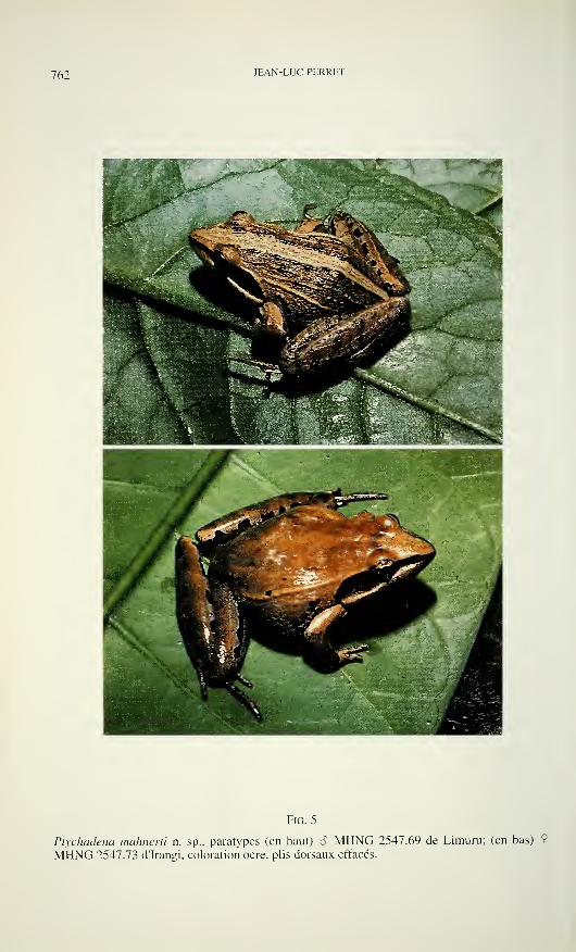
76: JEAN-LUC PERRET
Fig. 5
Ptychadena mahnerti n. sp., paratypes (en haut) S MHNG 2547.69 de Limuru; (en bas) 9
MHNG 2547.73 d'Irangi, coloration ocre, plis dorsaux effacés.
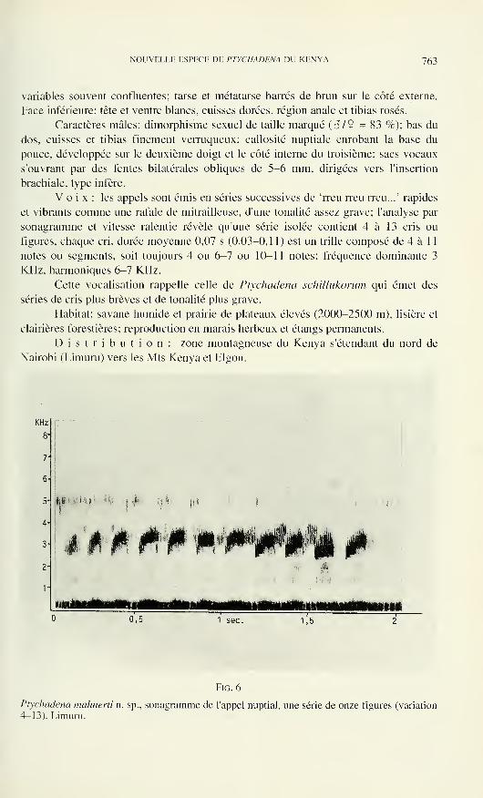
NOUVELLE ESPECE DE PTYCHADENA DU KENYA 763
variables souvent confluentes; tarse et métatarse barrés de brun sur le côté externe.
Face inférieure: tête et ventre blancs, cuisses dorées, région anale et tibias rosés.
Caractères mâles: dimorphisme sexuel de taille marqué (â/9 = 83 %); bas du
dos, cuisses et tibias finement verruqueux; callosité nuptiale enrobant la base du
pouce, développée sur le deuxième doigt et le côté interne du troisième; sacs vocaux
s'ouvrant par des fentes bilatérales obliques de 5-6 mm, dirigées vers l'insertion
brachiale, type infère.
Voix: les appels sont émis en séries successives de 'rreu rreu rreu...7
rapides
et vibrants comme une rafale de mitrailleuse, d'une tonalité assez grave; l'analyse par
sonagramme et vitesse ralentie révèle qu'une série isolée contient 4 à 13 cris ou
figures, chaque cri, durée moyenne 0,07 s (0,03-0,11) est un trille composé de 4 à 11
notes ou segments, soit toujours 4 ou 6-7 ou 10-11 notes; fréquence dominante 3
KHz, harmoniques 6-7 KHz.
Cette vocalisation rappelle celle de Ptychadena schillukorum qui émet des
séries de cris plus brèves et de tonalité plus grave.
Habitat: savane humide et prairie de plateaux élevés (2000-2500 m), lisière et
clairières forestières; reproduction en marais herbeux et étangs permanents.
Distribution: zone montagneuse du Kenya s'étendant du nord de
Nairobi (Limuru) vers les Mts Kenya et Elgon.
KHz
8'
71
6
5
4
2
m i•
i
Fie. 6
Ptychadena mahnerti n. sp., sonagramme de l'appel nuptial, une série de onze figures (variation
4-13). Limuru.
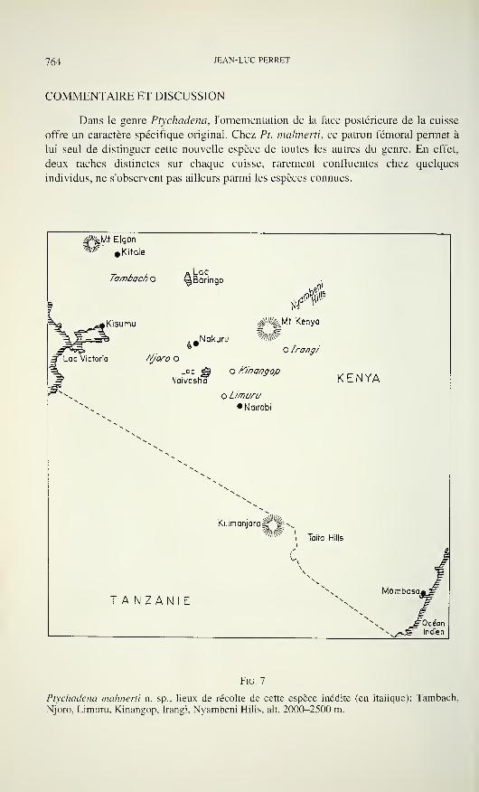
764 JEAN-LUC PERRET
COMMENTAIRE ET DISCUSSION
Dans le genre Ptychadena, l'ornementation de la face postérieure de la cuisse
offre un caractère spécifique original. Chez Pt. mahnerti, ce patron fémoral permet à
lui seul de distinguer cette nouvelle espèce de toutes les autres du genre. En effet,
deux taches distinctes sur chaque cuisse, rarement confluentes chez quelques
individus, ne s'observent pas ailleurs parmi les espèces connues.
#%Mt Elqon
%^'»Kitale
Tambacf) o
Kisumu
Lac^Baringo
.Nakuru
c Victoria Njoro O
Lac ^ o /C/'nangop
Naivasha
o Limuru• Nairobi
ç#%Mt Kenya
O Irangi
Kilimanjaro^^
KENYA
Taita Hills
TANZANIEMombasa
Océanndien
FlG. 7
Ptychadena mahnerti n. sp., lieux de récolte de cette espèce inédite (en itaiique): Tambach,
Njoro, Limuru, Kinangop, Irangi, Nyambeni Hills, ait. 2000-2500 m.
NOUVELLE ESPECE DE PTYCHADENA DU KENYA 765
Tableau I
Ptychadena mahnerti n. sp. Données morphométriques en mm: MA = longueur museau-anus; T= longueur tibia; P = longueur pied
Paramètres Sexe S N = 4 Sexe 9 N = 2
MA 35-42 M = 39.25 45-49 M = 47.00
T 22-26 M = 24.25 24-31 M = 27.50
T/MA% 60.9-62.8 M = 61.8-1 53.3-63.2 M = 58.25
P 23-27 M = 25.25 26-32 M = 29.00
P/MA% 63.4-65.7 M = 64.37 57.7-65.3 M = 61.50
T/P% 95.6-96.2 M = 96.00 92.3-96.8 M = 94.55
Commentaires: dimorphisme sexuel de taille marqué (â/9 = 83 %); tibia (T/MA = 60 %) et
pied (P/MA = 63 %), plus longs que la moitié du corps; pied (TYP = 95 %), plus long que le
tibia.
Les Ptychadena recensées au Kenya par les auteurs (Loveridge 1957; Duff-
MacKay 1979; Inger 1968; Frost 1985; Poynton & Broadley 1985) diffèrent
encore diversement: Pt. oxyrhynchus, de taille bien plus grande et fortement palmée;
Pt. anchietae, Pt. mascareniensis, Pt. mossambica, Pt. porosissima, toutes également
à palmure plus développée et à plis dorsaux réguliers plus marqués; Pt. schillukorum,
forme plus trapue à membres postérieurs plus courts et palmure plus forte; enfin, Pt
stenocephala (confondue avec Pt. chysogaster, Duff-MacKay 1979), récoltée à
Rapogi, Province Nyanza bordant le lac Victoria, est une espèce très svelte à palmure
extrêmement réduite, bien différente de Pt. mahnerti.
Ecologie et distribution: très largement répandues en savane, du niveau de la
mer jusqu'à l'étage montagnard, Pt. anchietae et Pt. mascareniensis sont sympatrides
de Pt. mahnerti à la limite altitudinaire dans certaines localités (Tambach); Pi.
mossambica et Pt. schillukorum, savanicoles planitiaires, distribuées de la côte sud
vers le nord plus aride et la Somalie, sont nettement allopatrides; Pt. oxyrhynchus est
connue de la côte sud, des Shimba Hills ainsi qu'à l'ouest en forêt de Kakamega
(Duff-MacKay 1979); nous avons récolté Pt. porosissima à Endebess au pied du
mont Elgon, à 2000 m, cette forme de hauts plateaux africains est probablement aussi
sympatride de Pt. mahnerti au Kenya.
Par sa palmure pédieuse laissant libres deux phalanges aux orteils III et V ainsi
que trois phalanges ou plus à l'orteil IV, Pt. mahnerti entre dans le groupe des formes
de Ptychadena à palmure réduite (Perret 1994).
Par sa voix propre, elle se distingue de toutes les autres espèces connues du
genre.
REMERCIEMENTS
Raymond Laurent et Robert Drewes ont confirmé mes vues sur cette espèce
inédite. Malcolm Largen m'a aimablement communiqué les numéros des spécimens
en collection au British Museum. Les dessins ont été interprétés par Gilles Roth, les
photograhies réalisées par Claude Ratton au Muséum de Genève.
766 JEAN-LUC PERRET
BIBLIOGRAPHIE
Duff-MacKay, A. 1979. The conservation status of the amphibia of Kenya. A report to the
scientific and technical committee. National Museum of Kenya, Nairobi, 64 pp.
Frost, D.R. 1985. Amphibian species of the world, a taxonomic and geographical reference.
Allen Press, Lawrence, Kansas, USA, 732 pp.
Inger, R.F. 1968. Amphibia. Exploration du Parc National de la Garamba, Mission H. DeSaeger, Kinshasa 52: 1-190.
Loveridge, A. 1957. Check list of the reptiles and amphibians of East Africa (Uganda; Kenya;
Tanganyika; Zanzibar). Bulletin of the Museum of Comparative Zoology, Harvard 117:
151-362, XXXVI.
Perret, J.L. 1994. Description de Ptychadena largeni n. sp. (Anura, Ranidae) d'Ethiopie.
Bulletin de la Société neuchâteloise des Sciences naturelles 1 17: 61-11
.
Poynton, J.C. & D.G. Broadley. 1985. Amphibia Zambesiaca 2. Ranidae. Annals of the Natal
Museum, Pietermarizburg 27: 115-181.
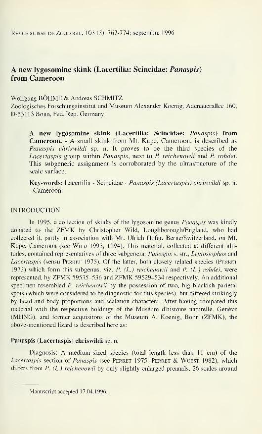
Revue suisse de Zoologie, 103 (3): 767-774; septembre 1996
A new lygosomine skink (Lacertilia: Scincidae: Panaspis)
from Cameroon
Wolfgang BÖHME & Andreas SCHMITZZoologisches Forschungsinstitut und Museum Alexander Koenig, Adenauerallee 160,
D-531 13 Bonn, Fed. Rep. Germany.
A new lygosomine skink (Lacertilia: Scincidae: Panaspis) fromCameroon. - A small skink from Mt. Kupe, Cameroon, is described as
Panaspis chriswildi sp. n. It proves to be the third species of the
Lacertaspis group within Panaspis, next to P. reichenowii and P. rohdei.
This subgeneric assignment is corroborated by the ultrastructure of the
scale surface.
Key-words: Lacertilia - Scincidae - Panaspis (Lacertaspis) chriswildi sp. n.
- Cameroon.
INTRODUCTION
In 1995, a collection of skinks of the lygosomine genus Panaspis was kindly
donated to the ZFMK by Christopher Wild, Loughborough/England, who had
collected it, partly in association with Mr. Ulrich Hofer, Berne/Switzerland, on Mt.
Kupe, Cameroon (see Wild 1993, 1994). This material, collected at different alti-
tudes, contained representatives of three subgenera: Panaspis s. str., Leptosiaphos and
Lacertaspis (sensu Perret 1975). Of the latter, both closely related species (Perret
1973) which form this subgenus, viz. P. (L.) reichenowii and P. (L.) rohdei, were
represented, by ZFMK 59535-536 and ZFMK 59529-534 respectively. An additional
specimen resembled P. reichenowii by the possession of two, big blackish parietal
spots (which were considered to be diagnostic for this species), but differed strikingly
by head and body proportions and scalation characters. After having compared this
material with the respective holdings of the Muséum d'histoire naturelle, Genève
(MHNG), and former acquisitons of the Museum A. Koenig, Bonn (ZFMK), the
above-mentioned lizard is described here as:
Panaspis (Lacertaspis) chriswildi sp. n.
Diagnosis: A medium-sized species (total length less than 11 cm) of the
Lacertaspis section of Panaspis (see Perret 1975, Perret & Wüest 1982), which
differs from P. (L.) reichenowii by only slightly enlarged preanals, 26 scales around
Manuscript accepted 17.04.1996.

768 WOLFGANG BÖHME & ANDREAS SCHMITZ
Eo
w
FlG. 1
Dorsal (left) and ventral (right) view of the holotype of Panaspis (Lacertaspis) chriswildi sp. n.
midbody, a less pointed snout, larger parietal spots and a triangular first loreal; it
differs from P. (L.) rohdei by the smaller size, the presence of parietal spots and
shorter limbs. It is distinguished from both species by 18-19 lamellae under the fourth
toe and 2 pairs of enlarged nuchal scales.
Holotype: ZFMK 59537, ? (Fig. 1 et 2), Cameroon: Mt. Kupe, 1550 m a.s.l., C. Wild,
18.V.1994.
Head-body length 45 mm, tail length 58 mm + (regenerated). Habitus slender,
lacertiform, tail round in cross section, not laterally compressed. Limbs well de-
veloped, pentadactyle, without web rudiments at the basis of the toes. Head stout with
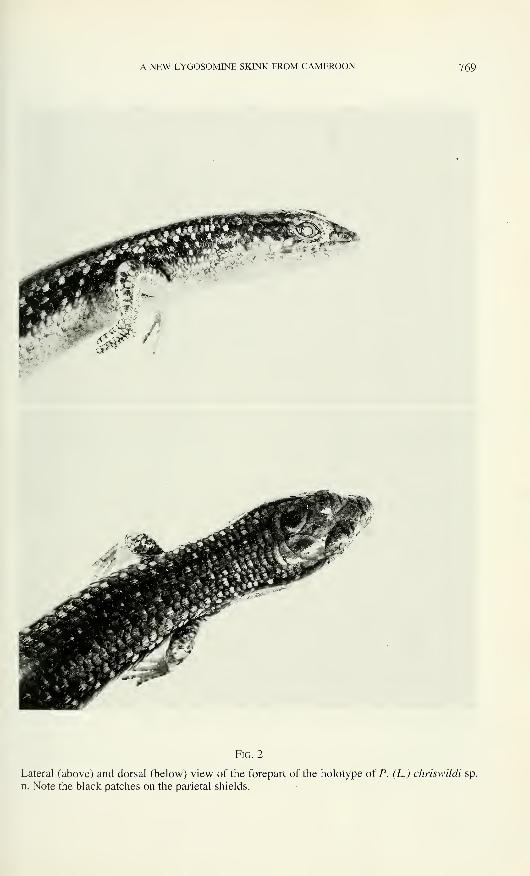
A NEW LYGOSOMINE SKINK FROM CAMEROON 769
Fig. 2
Lateral (above) and dorsal (below) view of the forepart of the holotype of P. (L.) chriswildi sp.
n. Note the black patches on the parietal shields.
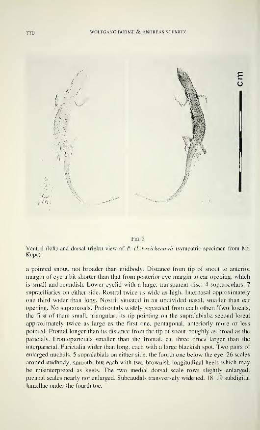
770 WOLFGANG BÖHME & ANDREAS SCHMITZ
«V 1
v.-
â
Fig. 3
Ventral (left) and dorsal (right) view of P. (L.) reichenowii (sympatric specimen from Mt.
Kupe).
a pointed snout, not broader than midbody. Distance from tip of snout to anterior
margin of eye a bit shorter than that from posterior eye margin to ear opening, which
is small and roundish. Lower eyelid with a large, transparent disc. 4 supraoculars, 7
supraciliaries on either side. Rostral twice as wide as high. Internasal approximately
one third wider than long. Nostril situated in an undivided nasal, smaller than ear
opening. No supranasals. Prefrontals widely separated from each other. Two loreals,
the first of them small, triangular, its tip pointing on the supralabials; second loreal
approximately twice as large as the first one, pentagonal, anteriorly more or less
pointed. Frontal longer than its distance from the tip of snout, roughly as broad as the
parietals. Frontoparietals smaller than the frontal, ca. three times larger than the
interparietal. Parietalia wider than long, each with a large blackish spot. Two pairs of
enlarged nuchals. 5 supralabials on either side, the fourth one below the eye. 26 scales
around midbody, smooth, but each with two brownish longitudinal heels which may
be misinterpreted as keels. The two medial dorsal scale rows slightly enlarged,
preanal scales nearly not enlarged. Subcaudals transversely widened. 18-19 subdigital
lamellae under the fourth toe.
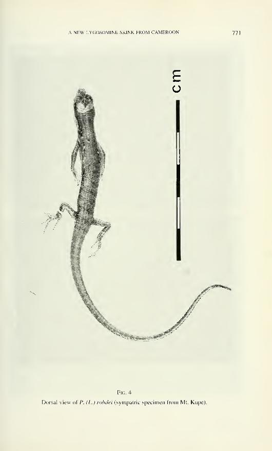
A NEW LYGOSOMINE SKINK FROM CAMEROON 771
Ü
Fig. 4
Dorsal view of P. (L.) rohdei (sympatric specimen from Mt. Kupe).
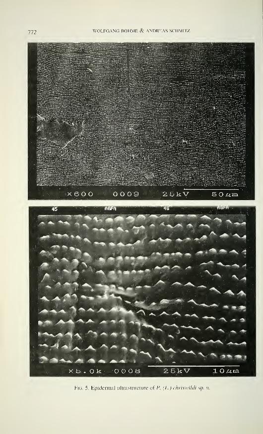
772 WOLFGANG BÖHME & ANDREAS SCHMITZ
s*»»» OTurarovs
Fig. 5. Epidermal ultrastructure of P. (L.) chriswildi sp. n.

A NEW LYGOSOMINE SKINK FROM CAMEROON 773
Fig. 6
Holotype of P. (L.) chriswildi sp. n. in life.
Upper parts brownish, with scarcely distributed light spots. A broad lateral
band, running from behind the eye to the tail tip, also spotted with light dots, and
becoming narrower from midbody hindwards. Between lower eyelid and tympanum a
big, light fleck (Fig. 2). 3 dark transverse bars on the sutures of the supraocular scales.
Upper side of limbs spotted with light dots. Fingers and toes with a marked brown-
white annulation. Lower parts whitish, with dark spots mainly in the inguinal and
cloacal region. The colouration of the living specimen is documented in fig. 6.
COMPARISONSAlthough only one specimen of P. chriswildi sp. n. is available, it differs so
clearly from its relatives that its specific distinctness is beyond any doubt. As stated in
the diagnosis, its greatest affinities are with P. reichenowii (Fig. 3) and P. rohdei (Fig.
4), i.e. the two species forming the subgenus Lacertaspis Perret, 1975. According to
Perret (op. cit.: 11) Lacertaspis is among else characterized by a lacertiform habitus, a
long tail, smooth, proximal caudilateral scales, robust, pentadactyl limbs overlapping
when adpressed, and a great, visible, superficial tympanum. These characters are most
likely connected with the habitat and habit, i.e. they could well be parallel adaptations
rather than synapomorphies documenting a monophyletic group. The remaining —scalation — characters used as diagnostic occur also in the other subgenera (see Perret
1975: table, p. 13).
774 WOLFGANG BÖHME & ANDREAS SCHMITZ
In a later study, however, Perret & Wüest (1982) were able to detect an
interesting new feature, viz. the ultrastructure of the scale epidermis. It turned out that
the two species assigned to Lacertaspis showed a common pattern which
distinguished them from all other Panaspis species studied. Although a polarity
decision was neither made by Perret & Wüest (op. cit.) nor can it be done here, the
common microdermatoglyph pattern of P. (L.) reichenowii and P. (L.) rohdei supports
their joint delimitation from the subgenera Panaspis and Leptosiaphos better than the
scalation and proportion differences.
P. chriswildi shows exactly the same pattern of epidermal ultrastructure (Fig.
5) as P. reichenowii and P. rohdei (see fig. 7 in Perret & Wüest, op. cit.: 277). Weassign it therefore as the third species to Lacertaspis. The diagnosis of this subgenus
has. however, to be modified accordingly: Habitus sepsino'id to lacertiform;
tympanum large, superficial to small, recessed: subarboricolous forms of lowland
forest to terrestrial forms of (sub-)montane forest. This means that Panaspis
(Lacertaspis) chriswildi bridges the morphological gap between Lacertaspis and
Leptosiaphos as far as adaptive, habitat-related characters are concerned. Within
Lacertaspis, P. chriswildi seems to be closer to P. reichenowii than to P. rohdei. The
unique parietal flecks, not occurring in any other Panaspis, links it with the former, as
does the ventral spotting. We regard therefore P. reichenowii to be the sister species
of P. chriswildi. More material to be collected has to further clarify the systematic and
ecological position of this remarkable new skink.
Etymology: The new species is dedicated to the collector of the holotype,
Chris Wild, in order to acknowledge his continuous support and cooperation.
ACKNOWLEDGEMENTSIn addition to the collector of the relevant material, Christopher Wild,
Loughborough, who provided also the colour photograph of the holotype, we are
indebted to Dr Wolfgang Speidel and to Jörn Köhler (both ZFMK Bonn) for their
valuable help with the SEM and the habitus photos. Ursula Bott (ZFMK) helped
preparing the manuscript.
REFERENCESPerret. J.L. 1973. Contribution à l'étude des 'Panaspis' (Reptilia. Scincidae) d'Afrique occi-
dentale avec la description de deux espèces nouvelles. Revue suisse de Zoologie 80:
595-630.
Perret. J.L. 1975. La différenciation dans le genre Panaspis Cope (Reptilia. Scincidae).
Bulletin de la Société neuchâteloise des Sciences naturelles 98: 5-16.
Perret. J.L. & J. Wüest. 1982. La structure fine des écailles de quelques Lygosominae afri-
cains (Lacertilia. Scincidae). révélée par le microscope électronique à balayage. Revue
suisse de Zoologie 89: 269-280.
Wild, C. 1993. Notes on the rediscovery and congeneric associations of the Pfeffer's Cha-
meleon Chamaeleo pfefferi (Tornier. 1900) (Sauria: Chamaeleonidae) with a brief des-
cription of the hitherto unknown female of the species. British Herpetological Society
Bulletin 45: 25-32.
Wild, C. 1994. Ecology of the Western Pygmy Chameleon Rhampholeon spectrum Buchholz
1874 (Sauria: Chamaeleonidae). British Herpetological Society Bulletin 49: 29-35.
REVUE SUISSE DE ZOOLOGIE
Tome 103 — Fascicule 3
Pages
Knoflach, Barbara. Three new species of Carniella from Thailand
(Araneae, Theridiidae) 567-579
Kodada, Jan. Spalacosostea, an anomalous new terrestrial dryopid from
South East Asia (Coleoptera: Dryopidae) 581-605
Baehr, Martin. A peculiar new species of Anomotarus (Nototarus) from
New Guinea (Coleoptera, Carabidae, Lebiinae) 607-61
1
Messner, Benjamin, Joachim Adis & Klaus Peter Zulka. Stigmale Pla-
stronstrukturen, die einigen Diplopoden-Arten eine submerse Lebens-
weise in kaltem und in fließendem Wasser ermöglichen 613-622
Dalens, Henri, André Rousset & Didier Fournier. Les formes épigées du
genre Oritoniscus (Crustacea, Isopoda, Oniscidea). I. Le complexeOritoniscus flavus 623-641
Pace, Roberto. Aleocharinae della Colombia e dell' Ecuador: Parte II
(Coleoptera, Staphylinidae) 643-676
Righi, Gilberto. Some Venezuelan Oligochaeta Glossoscolecidae andOctochaetidae 677-684
Salgado Costas, José Maria. Un nouveau Apoduvalius du bassin de la
rivière Nalón, Asturies-Espagne (Coleoptera, Trechidae) 685-690
Schawaller, Wolfgang. Spinolyprops pakistanicus sp. n. (Coleoptera:
Tenebrionidae), an Oriental element in the fauna of northern Pakistan 691-695
Bartsch, Ilse. Agauhopsis (Acari, Halacaridae) of the Sevastopol arcea;
supplementary notes on taxonomy and ecology 697-712
Rougemont, G.M. de. Stilideriis and Stilicoderus: New data and new spe-
cies (Coleoptera, Staphylinidae, Paederinae) 713-736
Uhmann, Gerhard. Indo-australische Anthicidae (Coleoptera) im Natur-
historischen Museum in Genf 737-748
Mendes, Luis F. Further data on the Nicolettidae (Zygentoma), with des-
cription of a new species from Mauritius 749-756
Perret, Jean-Luc. Une nouvelle espèce du genre Ptychadena (Anura,
Ranidae) du Kenya 757-766
Böhme, Wolfgang & Andreas Schmitz. A new lygosomine skink (Lacer-
tilia: Scincidae: Panaspis) from Cameroon 161-71'4
REVUE SUISSE DE ZOOLOGIE
Volume 103 — Number 3
Knoflach, Barbara. Three new species of Carniella from Thailand
(Araneae, Theridiidae)
Kodada, Jan. Spalacosostea, an anomalous new terrestrial dryopid fromSouth East Asia (Coleoptera: Dryopidae)
Baehr, Martin. A peculiar new species of Anomotarus (Notoiarus) fromNew Guinea (Coleoptera, Carabidae, Lebiinae)
Messner, Benjamin, Joachim Adis & Klaus Peter Zulka. Stigmal plastron
structures which enable some species of Diplopoda to live submersedin cold and running water
Dalens, Henri, André Rousset & Didier Fournier. Studies on epigean spe-
cies of the genus Oritoniscus (Crustacea, Isopoda, Oniscidea). I. TheOritoniscus flavus complex
Pace, Roberto. Aleocharinae from Colombia and Ecuador: Part II (Coleop-
tera, Staphylinidae)
Righi, Gilberto. Some Venezuelan Oligochaeta Glossoscolecidae and
Octochaetidae
Salgado Costas, José Maria. A new Apoduvalius from basin of the river
Nalón, Asturia-Spain (Coleoptera, Trechidae)
Schawaller, Wolfgang. Spinolyprops pakistanicus sp. n. (Coleoptera:
Tenebrionidae), an Oriental element in the fauna of northern Pakistan.
Bartsch, Ilse. Agauopsis (Acari, Halacaridae) of the Sevastopol area; sup-
plementary notes on taxonomy and ecology
Rougemont, G.M. de. StUiderus and Stilicoderus: New data and new spe-
cies (Coleoptera, Staphylinidae, Paederinae)
Uhmann, Gerhard. Indo-Australian Anthicidae (Coleoptera) from the
Natural History Museum in Geneva
Mendes, Luis F. Further data on the Nicolettidae (Zygentoma), with des-
cription of a new species from Mauritius
Perret, Jean-Luc. A new species of the genus Ptychadena (Anura,
Ranidae) from Kenya
Böhme, Wolfgang & Andreas Schmitz. A new lygosomine skink (Lacer-
tilia: Scincidae: Panaspis) from Cameroon
Indexed in Current Contents
PUBLICATIONS DU MUSEUM D'HISTOIRE NATURELLE DE GENEVE
CATALOGUE DES INVERTÉBRÉS DE LA SUISSEFase. 1
.
SARCODINÉS par E. Penard Fr. 1 2.—2. PHYLLOPODES par Th. Stingelin 12.—3. ARAIGNÉES par R. de Lessert 42.—4. ISOPODES par J. Carl 8.—5. PSEUDOSCORPIONS par R. de Lessert 5.50
6. INFUSOIRES par E. André 18.—7. OLIGOCHÈTES par E. Piguet et K. Bretscher 18.—8. COPÉPODES par M. Thiébaud 18.—9. OPILIONS par R. de Lessert IL-
IO. SCORPIONS par R. de Lessert 3.50
IL ROTATEURS par E.-F. Weber et G. Montet 38.—12. DÉCAPODES par J. Carl IL—13. ACANTHOCÉPHALES par E. André IL—14. GASTÉROTRICHES par G. Montet 18.—15. AMPHIPODES par J. Carl 12.—16. HIRUDINÉES. BRANCHIOBDELLES
et POLYCHÈTES par E. André 17.50
17. CESTODES par O. Fuhrmann 30.—
REVUE DE PALÉOBIOLOGIE Echange
LE RHINOLOPHE (Bulletin du centre detude des chauves-souris) par fascicule Fr. 10.
—
CATALOGUE ILLUSTRÉ DE LA COLLECTION LAMARCKAPPARTENANT AU MUSÉUM D'HISTOIRE NATURELLE DE GENÈVElre partie - Fossiles - 1 vol. 4° avec 1 17 planches Fr. 300.
—
COLLEMBOLENFAUNA EUROPASvon H. Gisin
312 Seiten, 554 Abbildungen Fr. 30.—
THE EUROPEAN PROTURA THEIR TAXONOMY, ECOLOGY ANDDISTRIBUTION WITH KEYS FOR DETERMINATIONby J. Nosek346 pages. 1 1 1 figures in text. 1973 Fr. 30.
—
CLASSIFICATION OF THE DIPLOPODApar Richard L. Hoffman237 pages. 1979 Fr. 30.—
LES OISEAUX NICHEURS DU CANTON DE GENÈVEpar P. Géroudet, C. Guex et M. Maire351 pages, nombreuses cartes et figures Fr. 45.
—
CATALOGUE COMMENTÉ DES TYPES D'ECHINODERMES ACTUELSCONSERVÉS DANS LES COLLECTIONS NATIONALES SUISSES.SUIVI D'UNE NOTICE SUR LA CONTRIBUTION DE LOUIS AGASSIZÀ LA CONNAISSANCE DES ECHINODERMES ACTUELSpar Michel Jangoux67 pages. 1 1 planches Fr. 15.
—
.ADULAS DE GASTÉROPODES LITTORAUX DE LA MANCHE(COTENTIN-BAIE DE SEINE. FRANCE)par Y. Finet, J. Wüest et K. Mareda62 pages, nombreuses figures, 1991 Fr. 10.
—
JASTROPODS OF THE CHANNEL AND ATLANTIC OCEAN;SHELLS AND RADULASby Y. Finet, J. Wüest and K. Mareda, 1 992 Fr. 30.
—
. SCHMIDT SPONGE CATALOGUEpar R. Desqueyroux-Faundez & S.M. Stone, 1992 Fr. 40.
—
.TLAS DE RÉPARTITION DES AMPHIBIENSET REPTILES DU CANTON DE GENÈVEpar A. Keller, V. Aellen et V. Mahnert, 1993 Fr. 15.
—
THE MARINE MOLLUSKS OF THE GALAPAGOS ISLANDS:A DOCUMENTED FAUNAL LISTpar Yves Finet, 1995 Fr. 30.
—
NOTICE SUR LES COLLECTIONS MALACOLOGIQUESDU MUSEUM D'HISTOIRE NATURELLE DE GENEVEpar Jean-Claude Cailliez, 1995 "
Fr. 22.
—


Revue suisse de Zoologie: Instructions to Authors
The Revue suisse de Zoologie publishes papers by members of the Swiss Zoological Society and scientific
results based on the collections of the Muséum d'histoire naturelle, Geneva. Submission of a manuscript implies
that it has been approved by all named authors, that it reports their unpublished work and that it is not being
considered for publication elsewhere. A financial contribution may be asked from the authors for the impression of
colour plates and large manuscripts. All papers are refereed by expert(s).
In order to facilitate publication and avoid delays authors should follow the Instructions to Authors and refer to
a current number of R.S.Z. for acceptable style and format. Manuscripts not conforming with these directives are
liable to be returned to the authors. Papers may be written in French, German, Italian and English. Authors should
aim to communicate ideas and information clearly and concisely in language suitable for the moderate specialist.
Authors not writing in their native language should pay particular attention to the linguistical quality of the text.
Manuscripts must be typed, on one side only and double-spaced, on A4 (210 x 297 mm) or equivalent paper
and all pages should be numbered. All margins must be at least 25 mm wide. Authors must submit one original
and two copies, including tables and figures, in final fully corrected form, and are expected to retain another copy.
Papers should conform to the following general layout:
Title page. A concise but informative full title plus as running title of not more than 40 letters and spaces,
name(s) in full and surname(s) of author(s), and full address(es).
Abstract. This must be on a separate page. The abstract is in English, composed of the title and a short text of
up to 200 words. It should summarise the contents and conclusions of the paper. The abstract is followed by less
than 10 key-words, separated by hyphens, which are suitable for indexing.
Introduction. A short introduction to the background and the reasons for the work.
Materials and methods. Sufficient experimental details must be given to enable other workers to repeat the
work. The full binominal name should be given for all organisms. The Zoological Code must be strictly followed.
Cite the authors of species on their first mention. Use SI units and the appropriate symbols.
Results. These should be concise and should not include me.fhods or discussion. Text, tables and figures should
not duplicate the same information. New taxa must be distinguished from related taxa. The abbreviations gen. n., sp.
n., syn. n. and comb. n. should be used to distinguish all new taxa, synonymies or combinations. Primary types
should be deposited in a museum or similar institution. In taxonomic papers the species heading should be followed
by synonyms, material examined and distribution, description and comments. All material examined should be listed
in similar, compact and easily intelligible format; the information should be in the same language as the text.
Sex symbols should be used rather than "male" and "female".
Discussion. This should not be excessive and should not repeat results nor contain new information, but
should emphasize the significance and relevance of the results reported.
References. The Harvard System must be used for the citation of references in the text, e.g. White & Green(1995) or (White & Green 1995). For references with three and more authors the form Brown et al. should be
used. Authors' names should not be written in capitals. The list of references must include all publications in the
text but only these. References must be listed in alphabetical order of authors, and both the title and name of the
journal must be given in full in the following style:
Penard, E, 1888. Recherches sur le Ceratium macroceros . These . Genève . 43 pp.Penard, E. 1889. Etudes sur quelques Héliozoaires d'eau douce. Archives de Biologie , 9:1-61.
Mertens, R. & H. Wermuth. 1960. Die Amphibien und Reptilien Europas, Kramer , Frankfurt am Main , XI + 264 pp.Handley, CO. Jr. 1966. Checklist of the mammals of Panama, pp. 753-795. Im Ectoparasites of Panama (R.L.
Wenzel & V.J. Tipton, eds). Field Museum of Natural History . Chicago . XII + 861 pp.References should not be interspaced and, in the case of several papers by the same author, the name has to berepeated for each reference.
The text should be in roman (standard) type face throughout, including headings, with genus and species
names underlined with pencil; bold, small capitals, large capitals italics and other type faces should not be used.
Footnotes and cross-references by page should be avoided.
Also, we encourage authors to submit the text on floppy disk (3,5" or 5 1/4", Macintosh or IBM compatible,
with "Microsoft Word" or similar programmes). The authors should refrain from using special codes for text
formating.
Tables. These should be self-explanatory, with the title at the top organised to fit 122 x 180 mm. Each table
should be typed, double spaced, on a separate page and numbered consecutively and its position indicated in the text.
Figures. These may be line drawings or half tones and all should be numbered consecutively, and their position
indicated in the text. Figures should be arranged in plates which can be reduced to 122 x 160 mm. Drawings andlettering should be prepared to withstand reduction. Magnification should be indicated with scale lines. Authorsshould refrain from mixing the drawings and half tones. Original drawings will not be returned automatically. TheRevue suisse de Zoologie declines responsability for lost or damaged slides or other documents.
Legends to figures. These should be typed in numerical order on a separate sheet.
Proofs. Page proofs only are supplied, and authors may be charged for alterations (other than printer's errors) if
they are numerous.
Offprints. The authors receive 25 offprints free of charge; more copies may be ordered at current prices whenproofs are returned.
Correspondence. All correspondence should be addressed to
Revue suisse de Zoologie
Muséum d'histoire naturelle
CP 6434CH-1211 Genève 6
Switzerland.
Phone: (022) 418 63 33 - Fax (022) 418 63 01
e-mail: volker.mahnert @ mhn.ville-ge.ch
LU j
ANNALES
de la
SOCIÉTÉ SUISSE DE ZOOLOGIEet du
MUSÉUM D'HISTOIRE NATURELLEde la Ville de Genève
tome 103
fascicule 4
1996
oooNLU
û
>LU
M GENÈVE DÉCEMBRE 1996 ISSN 0035 -418 X HBH
REVUE SUISSE DE ZOOLOGIE
TOME 103 — FASCICULE 4
Publication subventionnée par l'Académie suisse des Sciences naturelles
et la Société suisse de Zoologie
VOLKER MAHNERTDirecteur du Muséum d'histoire naturelle de Genève
FRANÇOIS BAUDConservateur au Muséum d'histoire naturelle de Genève
DANIEL BURCKHARDTChargé de recherche au Muséum d'histoire naturelle de Genève
Comité de lecture
Le président de la Société Suisse de Zoologie
Le directeur du Muséum de Genève: Volker Mahnert — Systématique des
vertébrés— Muséum de GenèveLe président du comité: Ivan Löbl — Systématique des Insectes — Muséum de
Genève
Patrick Guérin — Physiologie et éthologie des arthropodes — Institut de Zoologie,
Neuchâtel
Willy Matthey— Ecologie, entomologie— Institut de Zoologie, Neuchâtel
Claude Mermod— Ethologie et écologie des vertébrés— Université de Neuchâtel
Paul Schmid-Hempel — Ecoéthologie, biologie des populations — Institut f.
Terrestrische Ökologie, ETH Zürich, Schlieren
Steve Stearns — Biologie de l'évolution— Institut f. Zoologie, Basel
Beat Tschanz— Ethologie des Vertébrés — Zoologisches Institut, Bern
Claude Vaucher— Systématique des Invertébrés — Muséum de Genève
La préférence sera donnée aux travaux concernant les domaines suivants: Biogéographie,
systématique, écologie, éthologie, morphologie, et anatomie comparée, physiologie.
Administration
MUSEUM D'HISTOIRE NATURELLE
1211 GENÈVE 6
Prix de l'abonnement:
SUISSE Fr. 225.— UNION POSTALE Fr. 230.
(en francs suisses)
Home page RSZ: http://www/musinfo/mhng/publications/revues.htm
Les demandes d'abonnement doivent être adressées
à la rédaction de la Revue suisse de Zoologie,
Muséum d'histoire naturelle, C.P. 6434, CH-1211 Genève 6, Suisse
UJ
ANNALES ode la
SOCIÉTÉ SUISSE DE ZOOLOGIEet du aa^ij
MUSÉUM D'HISTOIRE NATURELLEde la Ville de Genève O
otome 103
fascicule 4 N1996
LU
ÛLUUT)
un
DunLU
D>LU
M GENEVE DÉCEMBRE 1996 ISSN 0035 -418 X Où
REVUE SUISSE DE ZOOLOGIE
TOME 103 — FASCICULE 4
Publication subventionnée par l'Académie suisse des Sciences naturelles
et la Société suisse de Zoologie
VOLKER MAHNERTDirecteur du Muséum d'histoire naturelle de Genève
FRANÇOIS BAUDConservateur au Muséum d'histoire naturelle de Genève
DANIEL BURCKHARDTChargé de recherche au Muséum d'histoire naturelle de Genève
Com ité de lecture
Le président de la Société Suisse de Zoologie
Le directeur du Muséum de Genève: Volker Mahnert — Systématique des
vertébrés — Muséum de GenèveLe président du comité: Ivan LÖBL — Systématique des Insectes — Muséum de
Genève
Patrick Guérin — Physiologie et éthologie des arthropodes — Institut de Zoologie,
Neuchâtel
Willy Matthey— Ecologie, entomologie — Institut de Zoologie, Neuchâtel
Claude Mermod— Ethologie et écologie des vertébrés — Université de Neuchâtel
Paul Schmid-Hempel — Ecoéthologie, biologie des populations — Institut f.
Terrestrische Ökologie, ETH Zürich, Schlieren
Steve Stearns — Biologie de l'évolution — Institut f. Zoologie, Basel
Beat Tschanz— Ethologie des Vertébrés— Zoologisches Institut, Bern
Claude Vaucher— Systématique des Invertébrés — Muséum de Genève
La préférence sera donnée aux travaux concernant les domaines suivants: Biogéographie,
systématique, écologie, éthologie, morphologie, et anatomie comparée, physiologie.
Administration
MUSEUM D'HISTOIRE NATURELLE
1211 GENÈVE 6
Prix de l'abonnement:
SUISSE Fr. 225.— UNION POSTALE Fr. 230.-
(en francs suisses)
Home page RSZ: http://www/musinfo/mhng/publications/revues.htm
Les demandes d'abonnement doivent être adressées
à la rédaction de la Revue suisse de Zoologie,
Muséum d'histoire naturelle, C.P. 6434. CH-121 1 Genève 6, Suisse
Revue suisse de Zoologie, 103 (4): 777-794; décembre 1996
ZOOLOGIA ET BOTANICA 96
Fribourg, 21-23 February 1996
(Annual Conference of the Swiss Zoological Society)
Determinants of community structure in plants and animals
ABSTRACTS
R. Arditi (Institut de zoologie et d'écologie animale, Université-Dorigny, 1015
Lausanne et Écologie des populations et communautés, Bâtiment 362, Université
Paris-Sud XI, F-91405 Orsay Cedex, France): Stability and complexity - what do
density - dependent food webs bring?
The link between complexity and stability is one of the oldest theoretical questions of
community ecology. Based on the observation that complex ecosystems (e.g., tropical forests)
incurred apparently small fluctuations, the accepted viewpoint for 50 years was that complexity
generated stability. In the early 70' s. May's theoretical work reversed the paradigm: mathe-
matical models of randomly assembled communities showed that the more complex they were,
the less probable that they would be stable. This work was later refined and more thoroughly
developed by Pimm in the context of food webs. An important restriction of May's models was
that they were linear, assuming that communities were always close to equilibrium. Although
Pimm investigated non-equilibrium situations, he assumed that trophic interactions were
density-independent, i.e., that the amount of resources consumed by an individual consumers
did not depend on the number of conspecifics. Presently, there is little doubt that density-
dependence is frequent in nature as well as in laboratory conditions. Model food webs with
strong density-dependence are not easy to develop. One type of such model will be presented.
First results show that these webs possess unique properties that could not be foreseen from
density-independent models. These properties make them more realistic and suggest a possible
reconciliation between theoretical predictions and empirical observations.
C. Arnold, A. Buttler & B. Werffeli (University of Neuchâtel, Switzerland):
Ecological determinism of bryophyte community patterns in alluvial steppes.
Alluvial steppes are brushwood communities characterised by xero-thermophilous
species. Despite these steppes represent functional units with increasing disconnection to the
alluvial system, they reveal the highest species pool (184 herbaceous species) among all the
communities recorded in the Swiss alluvial zones of national importance, after the WhiteAlder flood plain forest (about 250 species). Unfortunately, as a transitional stage, where the
bank cannot be reached any longer by the river, Picea abies becomes an invasive species andPinus silvestris and Betula pendula may grow tall, turning the general ecological conditions
towards a more forested environment, with a related loss of species diversity.
In this paper we aim at understanding, by means of spatial multivariate statistics, the
ecological determinism of the Bryophyte community patterns. Three plots representing a
successional gradient towards a tree dominated environment are investigated. It is shown howthe vegetation structure changes in relation to the shift from exogenous to endogenousecological conditions and how the spatial component can be used in detecting some structural
features.
778 ZOOLOGIA ET BOTANICA 96
T.C.M. Bakker, D. Mazzi & S. Zala (Zoologisches Institut, Universität
Bern): Enhanced transmission of the parasite Pomphorhynchus laevis through be-
havioural and colour changes in its intermediate host, Gammarus pulex.
Parasites with a complex life cycle involving several host species often induce changes
in their intermediate hosts. The parasite-induced changes make the intermediate host a moreprofitable prey to the next host of the parasite thereby increasing the chances of transmission of
the parasite.
The acanthocephalan parasite Pomphorhynchus laevis is transmitted by the amphipodGammarus pulex to its next host, fish. The parasite is clearly visible through the transparent
cuticle of G. pulex as a conspicuous orange spot. Additionally, with the onset of infectivity of
P. laevis to its next host, G. pulex shows a changed photoreaction; it becomes photophilic andpositively phototactic. Hungry stickleback fish, Gasterosteus aculeatus, preferred to eat
infected G. pulex over uninfected ones. By means of experimental manipulation of the colour
effects of the parasite in its intermediate host, we showed for the first time that both
behavioural and colour changes in the intermediate host promote parasite transmission in such
acanthocephalan systems.
A. Baur, L. Fröberg & B. Baur (Institut für Natur-, Landschafts- und
Umweltschutz, Universität Basel; Dept. of Systematic Botany, University of Lund):
Herbivory in a calcicolous lichen community: effects of selective snail grazing.
The land snails Chondrina clienta and Balea pen>ersa coexist on old stone walls where
they feed on calcicolous lichens. Previous studies showed that both intra- and interspecific
competition most probably for food occur in these species. A total of 32 calcicolous lichen
species, one alga and one bryophyte were recorded on a limestone wall in the grassland Great
Alvar on the Baltic island of Oland, Sweden. Fourteen (41%) of these 34 species and free-
living cyanobacteria showed herbivore damage, most probably due to grazing by the land snails
Chondrina clienta and Balea perversa that inhabited the limestone wall. Three laboratory
experiments were conducted to examine the food preferences of individuals of C. clienta and B.
perversa collected at this site and to evaluate any association between their preference and the
net food quality of the lichens to the snails. C. clienta and B. perversa exhibited food
preferences, which differed significantly between species. Within species, variation in food
choice was similar among individuals. This indicates that snail populations may be composedof polyphagous individuals with similar food preferences. Different lichen species were of
different net food quality to the snails as indicated by growth rate differences. In both snail
species the most preferred lichen species of the choice experiment caused the largest weight
increase in juveniles, viz. Caloplaca flavovirescens in C. clienta and Aspicilia calcarea in B.
perversa. This suggests that the snail species studied differ in their abilities to deal with
secondary compounds and physical characteristics of certain lichens or that they can utilize the
energy and nutrients of these lichens to a different extent. It is suggested that differential food
preferences might reduce the intensity of interspecific competition for resources (lichens)
between C. clienta and B. derversa.
G. Bernasconi & L. Keller (Universität Bern, Ethologische Station; IZEA,
Université de Lausanne): Reproductive conflicts in cooperative associations of Fire
Ant queens (Solenopsis invida).
In ants unrelated queens frequently associate to initiate a colony cooperatively. Thejoint reproductive effort of the cofoundresses increases growth and survival of the incipient
colony. Yet, such associations are unstable. Soon after emergence of the first workers, queen-
queen and queen-worker fights lead to the death or expulsion of all but one cofoundress.
Because no sexual offspring are produced in incipient colonies the surviving queen mono-
ZOOLOGIA ET BOTANICA 96 779
polises the entire future reproductive success of the colony. Two factors, the queens' relative
fighting ability and their relative contribution to worker production (assuming that workers can
recognize and selectively favour their mother) have been proposed to influence the survival
prospects of individual queens within associations. The effect of these two factors were tested
in the fire ant Solenopsis invicta. Initial size differences, a potential measure of the queens'
fighting ability, affected the outcome of the conflicts, so that the initially heavier queen was
more likely to survive. Differential weight loss by initially equal nest mates was also related to
survival, with the queen losing more weight being more likely to die. The manipulation of the
queens' relative contribution to the worker brood had no significant effect on the queens'
survival probability, suggesting that workers are unable to favour their mother.
L. Bersier & G. Sugihara (Scripps Institution of Oceanography, UC San
Diego): Scale invariance versus scale dependence in food web properties.
There is a current controversy as to the scaling behaviour of food web properties.
Earlier studies based on large collections of food webs found that some food web properties
(e.g., the link density, the fractions of top, intermediate and basal species) were scale invariant:
they remained constant across webs of different sizes. These scaling properties fostered the
formulation of interesting hypotheses on how food webs are assembled and how they operate.
However, together with criticisms of the data used to demonstrate these properties, recently
compiled food webs have cast doubt on the validity of the scale invariant laws. Moreover, the
scale invariance found in some data sets has recently been challenged. The properties mayindeed vary with web size. The scale dependent hypothesis appears to be the new paradigm in
food web theory. Here, we review the evidence in favour of both hypotheses, and show that the
scale dependent hypothesis is weakly supported empirically. More specifically, for one large
collection of food webs, we analyze systematically a suite of competing models describing the
relationship between food web properties and web size. We present quantitative evidence that
the most robust model is a piecewise linear regression, indicating that food web properties are
bounded to different scaling regions. The properties of small webs exhibit scale dependence,
while they are scale invariant for large webs. Finally, we discuss the generality of the link
density property, and point out its extreme sensitivity to sampling effort.
W. Brown (Institut de Zoologie et d'Écologie Animale, Université de Lau-
sanne): Female choice for an indicator of male competitiveness in the Black-horned
Tree Cricket.
Although evidence of mate choice is now well founded, distinguishing among alter-
native evolutionary models of female choice remains problematic. An approach that has beensurprisingly under-utilized is the study of functional design or teleonomy. Models of sexual
selection make fundamentally different predictions about the way in which preferences should
be structured. I studied the design of female choice for song in Black-horned Tree Crickets,
predicting from indicator models that choices should be ( 1 ) based on comparisons, (2) directed
at elements of sex-limited signals that indicate fecundity benefits and/or heritable fitness, and
(3) these elements should lack susceptibility to cheating. These predictions were contrasted
with the predictions from runaway models that choices should (1) be fixed within females and(2) show consistent variability among females. Results showed that comparisons were neces-
sary for choices, females preferred low-frequency songs, and low frequency was a reliable indi-
cator of both fecundity and putative heritable benefits of mating, supporting all 3 predictions ofindicator models. By contrast, female preferences were neither fixed within nor variable amongfemales, falsifying the necessary conditions for runaway selection. Combining these results
with genetic studies of mate choice will determine the model of sexual selection that is
operating on cricket song.
780 ZOOLOGIA ET BOTANICA 96
P. Charlier, P. Fürst, G. Blandenier & S. Pozzi (Plant Ecology and
Biogeography Laboratory, University of Geneva; Laboratory of Animal Ecology,
University of Neuchâtel; Natural History Museum of Geneva): Arachnological and
botanical transect from a forest's rim to a wet meadow.
We studied in 1992 and 1993 the characteristics of spiders' communities of small
natural areas with ecological conditions as wide as an Oak forest's rim (Quercus robur, Q.
petrea), a wet meadow containing mainly Molinia arundinacea and a sword-flag {Gladiolus
paluster) and a tilling field in full regeneration. All these plant communities are gathered on a
35 meters long line. What are the variations, speaking of specifications and quantities of
spiders' communities in the wet meadow:- over a year from March to October 1992; - with
three different kinds of mowing management? The trapping's method of Barbers has been used.
I. Dajoz, J. Shykoff, Y. Loublier, M.H. Pham-Delegue & I. Till-Bottraud
(Laboratoire d'Écologie, Université de Paris 6; ESV, Université de Paris 11; Institut
für experimentelle Ökologie ETH Zürich; Neurobiologie Comparée des Invertébrés,
INRA; Biologie des Populations d'Altitude, Université de Grenoble): Pollinator
mediated selection on pollen morphology in the genus Viola.
Many plants produce several pollen morphs differing in aperture number. Species of
Viola exhibit this heteromorphism, with pollen with 3-6 apertures. The more apertures a pollen
grain has, the more quickly it germinates, but the shorter its life-span is. Theoretical models
find an ESS for several pollen morphs. Models predict a higher proportion of many-aperturate
pollen grains on plants that receive more pollinator visits. Lastly, variation in ploidy level can
lead to variation in pollen aperture number. Our aim in this paper is to address the following
questions:
What is the distribution of pollen heteromorphism in Violai Is pollen heteromorphism
in Viola solely due to variations in plant ploidy level? Or, can selection act on proportions of
different morphs? What is the nature of this selection?
For the 2 subgenera of Viola: pansies (18 spp.) are heteromorphic, with no correlation
between chromosome number and mean aperture number. In violets (12 spp.) those with 2n =
20 are monomorphic, those with 2n = 40 are heteromorphic.
In pansies there is a negative correlation between elevation of collection site and meanaperture number. This agrees with our predictions of the model: pollination limitation at high
elevation should select for low aperture number.
Within species, there is a positive correlation between flower or spur size, and meanaperture number. Also, pollinators prefer large flowers.
These data suggest that some selection pressures act on pollen grain morphology, and
that these selective pressures are partly due to differences in pollinator activity.
M. Fischer & D. Matthies (Institut für Umweltwissenschaften, Universität
Zürich): Conservation biology of rare grassland plants in the Swiss Jura mountains.
In a comparison of vegetation records from 26 grasslands in the Swiss Jura mountains
from 1950 and 1985 short-lived species with low local abundance and high habitat specificity
showed the highest rates of local extinction. The biennial Gentianella germanica belongs to
this group. Populations of G. germanica had a low mean annual population growth rate (0.84)
with large spatial (among seven sites) and temporal variation (among the years 1993-95). In a
survey of 23 populations number of seeds per plant was reduced in small compared with large
populations. The population growth rate estimated from the number of flowering plants 1993,
94 and 95 showed the same pattern. This could be due to poorer habitat quality, disruption of
plant-pollinator mutualism or genetic erosion in small populations. However, habitat properties
did not explain the pattern and hand pollination did not increase seed set as compared with
open pollination, whereas genetic variation as measured with RAPD-PCR was reduced in
ZOOLOGIA ET BOTANICA 96 78 1
smaller populations. Management of the species should avoid disturbance during the
time of flowering or fruit set and allow disturbance that leads to the creation of
vegetation gaps, because it improves seedling establishment. Conservation biology
should integrate from the gene to the community, use descriptive and experimental
methods and study spatial scales comprising several to many populations.
D. Fraefel, M. Haffner & V. Ziswiler (Zoologisches Museum der Universität
Zürich): Morphological and micro-anatomical investigations of the integument of the
European red squirrel Sciurus vulgaris L., 1758 (Mammalia, Rodentia).
The Red squirrel Sciurus vulgaris shows adaptations in its body for the arboreal way of
life. Morphological and micro-anatomical investigations of the integument of different parts of
the body were earned out to find such adaptations and relate them to their functions. The Redsquirrel has vibrissae on the head, on the forelimbs, on the breast and on the abdomen. While
the number of Vibrissae mystaciales and the Vibrissae anconeales is constant, the number of
the other vibrissae shows an intraspecific variability. For the first time an accumulation of
apocrine glands was found on the chin of the Red squirrel. These glands occur on both sides of
the chin near the corner of the mouth and are present in both sexes. The secretions may be
spread on the head with the forefeet during face-wiping. Eccrine glands occur in all foot pads.
Instead of the tarsal pads, sparsely haired areas occur on the heels. There, the hairs are equipped
with enlarged sebaceous glands. The secretions of these glands may be used for scent-marking
the frequently visited feeding places. The forefeet are adapted to grasp twigs and handle food.
Both carpal pads contain muscles and cartilage. They provide, together with the reduced first
digit, an abutment against the grasping digits two to five.
J. Frantzen (Institut de biologie végétale, Université de Fribourg): Avoidance
and resistance in the weed pathosystem Senecio vulgaris L.-Puccinia lagenophorae
Cooke.
The rust fungus P. lagenophorae might be used to control the annual weed S. vulgaris.
A system management approach of biological weed control, which is followed to study the
suitability of P. lagenophorae as biological control agent, is presented. The system
management approach both depends on, and also provides, fundamental knowledge of
mechanisms at the individual and population level. One study of the ongoing research wasdirected to the disease defence mechanisms of avoidance and resistance in the weedpathosystem S. vulgaris-P. lagenophorae.
In an integrated approach, three plant lines of S. vulgaris were tested for avoidance of,
and resistance to infection by P. lagenophorae. Differences in susceptibility to P. lagenophorae
infection between plant stages were determined independent of plant line. Plant lines differed
also in growth rate, and so, the fraction of plants in the most susceptible plant stage at the time
of inoculation differed between plant lines. The combination of the effects of plant stage oninfection and plant line on growth rate resulted in differences in avoidance of P. lagenophorae
infection between plant lines. Differences in resistance to P. lagenophorae infection betweenplant lines were determined in one of the two experiments. The results are discussed with
respect to the impact of pathogens on plant populations.
J. Georgakopoulos, J. Berlie, V. Derivaz & B. Reverdin (Laboratoire
d'Éthologie, Université de Genève, rte des Acacias, 1227 Carouge): Structuring space
through path integration.
Recent theories of navigation postulate that path integration may be linked with a stable
representation of space. Thus, the subject can constantly update its position on this "map" bykeeping track of its own movements through path integration. Likewise, path integration maybe used during exploration to place relevant sites or cues in space. The first experiment studies
782 ZOOLOGIA ET BOTANICA 96
the interaction between path integration and visual cues in golden hamsters (6 subjects). Thesubjects are trained to find food in one of four identical cylinders which are distinguishable
only by their location. No other visual cues are present. In test trials, the subject is led in
darkness along a new path to a given release point. The light is then switched on and the animal
goes to the food source on its own. The subjects chose the correct cylinder in 86% of test trials.
This successful orientation can be explained in two ways: 1) By keeping track of its movementsthrough familiar space, the subject can anticipate a given visual panorama. When the light
comes on, it can then compare what it sees with this anticipated view in order to identify the
correct goal. The goal can then be reached using the visual cues. 2) The hamster may orient
towards a point in absolute space independently of visual cues, by storing a vector linked to the
position of the goal. It can then at any moment locate the goal by combining the short-term
vector derived from path integration (which gives the animals present location) with the
memorised vector associated with the goal. The second possibility was verified by repeating the
first experiment in total darkness. In a high percentage of trials (33%) the subjects refused the
task. However, when the animals attempted to reach the goal they succeeded in 99% of these
trials (64% of the total). In a final experiment no external cues mark the goal. All hoarding trips
are carried out in total darkness, eliminating visual information. The animal is trained to find
food in a constant location. In test trials it is led along a new path to a given release point fromwhich it must go to the goal. In this situation, where nothing indicates that the goal has been
reached, the animals usually do not stop at the correct location. However, in 56% of trials, the
subject reaches this location by a fairly straight path (up to 1.5 times the shortest possible path).
These l-..«t two experiments suggest that hamsters are indeed able to use path integration in
conjunci.^n with their knowledge of a familiar environment: nevertheless, path integration
appears to require additional confirmation before the subject acts on this information.
L. Gigord (Laboratoire d'Évolution et Systématique, Université Paris XI-
CNRS. Laboratoire de Biologie et Physiologie Végétale, Université de La Réunion;
Conservatoire National Botanique de Mascarin, Ile de La Réunion): Inbreeding
depression in two natural and fragmented populations of an endemic and self-
incompatibility tree of La Réunion Island (Mascarenes Archipelago, Indian Ocean):
'Dombeya acutangula Cav. (Sterculiaceae).
La Réunion Island resembles other volcanic islands (Hawaii. Galapagos...) with excep-
tional flora of approximatively one thousand of endemic and indigenous species. Habitat
fragmentation has reached a critical level within forest formations at low altitude with ever
increasing anthropisation following the arrival of man three centuries ago. The lack of bio-
diversity and rapidly decreasing habitat have become a major concern for the various island
authorities.
This study is to examine the consequences of fragmentation of natural habitat on a
species endemic of the archipelago: Dombeya acutangula Cav. Two very different island
populations have been examined: one very fragmented (close to a highly anthropomorphised
region and organised in groups of variable sizes (3 to 10 individuals) and another not frag-
mented in a natural reserve. Cross breeding experiments between individuals from the two
populations was carried out to find the level of decrease in consanguinity in descendants
according to the geographical distance between the individuals. The following cross-breeds
were realised: self-fertilization without human intervention, self-fertilization with humanintervention, within-population crosses between neighbouring individuals, within-population
crosses between distant individuals and between-population crosses. Addition crosses breeding
between individuals from these Réunion populations and other individuals from an neigh-
bouring island (Mauritius) were also carried out. Fitness components of descendants from these
cross-breeds were measured (number of seeds per flower, weight of seeds, germination rate,
growth, etc.). The most striking result of the study shows that there is no significant difference
ZOOLOGIA ET BOTANICA 96 783
between the descendants as a whole, except for those emerging from cross breeding between
individuals originating from the same deme in the fragmented population. In these individuals,
this cross breeding leads to the production of significantly weaker and smaller seeds than those
produced by the non fragmented population.
M. Gosteli (Natural History Museum, Bernastrasse 15, CH-3005 Bern):
Ecological relationships between snail communities and vegetation.
An inventory of the snail fauna was taken at 33 sample sites situated in four different
habitat types near Merishausen in the northern Swiss Jura. To gain information about micro-
climate and soil quality in a given habitat plant communities and their ecological indicator
values known from literature were scored at the sites. Snail faunas and plant communities are
significantly correlated, indicating that ecological requirements of snails and plants occurring at
the same site are similar. Both depend on microclimatic conditions as humidity, light, tem-
perature and on soil factors as permeability, pH and nutrient content. However, the relatively
low similarity coefficient of 0.5 between communities of snails and plants indicates that there
may be other ecological factors (a biotic or biotic) influencing snails and plants in a different
manner.
M. Haffner (Zoologisches Museum, Universität Zürich): Adaptations to
digging an climbing in the sole of the feet of some small mammals.
The feet of nine species were studied by means of gross morphology and microscopical
anatomy. Adaptations to digging and climbing were found in the integument and the claws. Thepalms of Talpa europaea are smooth. They have neither pads nor glands, for any elevation or
moist surface would hinder the soil from rolling. Although Arvicola terrestris cuts soil with its
incisors, the reduced number of foot pads, their flatness and the glandless sole indicate that the
feet are used as shovel-like tools. Bats climb by hooking their long, curved claws into crevices
and cracks. Foot pads are absent, but regions with a thickened epidermis reflect increased
mechanical strain. Rhinolophus ferrumequinum, which is specialised in hanging on twigs while
hunting insects, has epidermal thickenings at the soles of the middle phalanges of the toes.
When Myotis myotis walks or climbs on horizontal surfaces the bearing part of the wing is the
scutelliform-shaped integumentary thickening on the wrist, while in the hind feet the thickened
soles of the first toes are mainly loaded. In Micromys minutus nap-shaped integumentary
thickenings with eccrine glands at the base of the toes and around the carpal pads are
adaptations to climbing by grasping stacks. Muscardinus avellanarius can climb by pressing
the soles of its feet against the bark which is reflected by big softpads with a huge amount of
eccrine glands.
B. Hagele & M. Rowell-Rahier (Zoologisches Institut der Universität Basel:
Institut de Zoologie, Université de Neuchâtel): The choice and performance of two
generalist and two specialist insect herbivores towards cacalol and seneciphyllin, a
sesquiterpene and a pyrrholizidine alcaloid found in Adenostyles alpina (Asteraceae,
Senecioneae).
Potential deterrent or attractive effects of two chemicals (cacalol, seneciphyllin and the
combination of both) were tested in a first set of three experiments in standard leaf disk choice
experiments. In a second set of experiments the same compounds were force fed to all species
by painting them on leaf disks which were fed to the larvae over a period of ten to 15 days. Theeffects on the performance of the four species could be assessed by comparing the growth of
the groups fed the leaf disks treated with the chemicals to those fed with disks treated with the
solvent only. Larvae of the specialist leaf beetles Oreina cacaliae and O. speciosissima
(Coleoptera, Chrysomelidae) showed neither an attractive nor a repellent reaction towards the
784 ZOOLOGIA ET BOTANICA 96
two chemicals when tested individually. The combination of both chemicals however wasrepellent to both species. Larval growth of the two leaf beetle species was not influenced by the
experimental treatments. Larvae of the generalist Cylindrotoma distinctissima (Diptera,
Tipulidae) were repelled by the cacalol, seneciphyllin and the combination treatment. Larval
growth was reduced by all treatments except the alkaloid high concentration treatment.
Caterpillars of the generalist Callimorpha dominula (Lepidoptera, Arctiidae) were repelled by
cacalol and the combination treatment and not affected by the seneciphyllin treatment. After
experience with seneciphyllin however it became attractive to the caterpillars. The high dose
combination treatment was also effective in reducing growth of the caterpillars. Growth in all
other treatments was not different from the control group.
L. Heer (Zoologisches Institut, Universität Bern): Intense sperm competition
in the polygynandrous Alpine Accentor {Prunella collaris).
Alpine Accentors are breeding in groups consisting of 3-5 males and 2-3 females. All
males of one unit compete for the access to fertile females within their breeding group. Of 7
females, focus animal samplings were performed to work out the role of females in controlling
copulations. Alpine Accentors have a high copulation frequency and males do guard their
mates closely to enhance their certainty of paternity. Females show fertility-advertisement by
displaying themselves on elevated rocks and uttering long series of calls. Further, females
actively seek out males what normally results in intense flight pursuits. The females are mainly
responsible for soliciting copulations. Paternity is multiple in Alpine Accentors with up to three
males siring young within one clutch. Results of DNA fingerprints indicate that frequent
copulations and intense mate guarding do not ensure one male's full paternity in such a pro-
miscuous system.
B. Hellriegel (Zoologisches Museum, Universität Zürich-Irchel, Winterthurer-
str. 190, 8057 Zürich): Sperm competition in Diptera: adding the female perspective.
Sperm competition has traditionally been modelled exclusively from the male per-
spective: females have been taken as passive during the entire process. However, many female
insects have a complex reproductive system which suggests a function in postcopulatory female
choice to control or at least influence the paternity of their offspring. One of the traits
constituting this complexity is the presence of multiple sperm stores; many female flies have
two or three spermathecae.
The main purpose of this modelling approach is to determine the fertilisation success of
different males under the assumption of postcopulatory female choice. The model mimics the
essential features and possible functions of a reproductive tract with two (or more) separate
sperm stores, which in principle enables the female to selectively store and use the ejaculates of
different males. By comparing results of the mathematical model with experimental evidence,
mainly from Scatophaga stercoraria, I address two questions: 1 ) Is there an advantage of
having more than one spermatheca? 2) What are possible consequences of an uneven
distribution of inseminations from different males between two (or more) separate sper-
mathecae?
R. Hoess & A. Scholl (Zoologisches Institut, Universität Bern): The Glomeris-
taxa hexasticha Brandt and intermedia Latzel (Diplopoda: Glomeridae): species or
subspecies? - Allozyme data.
Diplopod species are usually distinguished by means of the gonopods of the males.
Glomehs do not possess gonopods. and the telopods that males use to grasp the females' vulvae
are similar in all species. Thus the color pattern of the tergites, often very variable, remains the
only reliable feature for distinction. The taxa hexasticha and intermedia almost have the same
color pattern and therefore are distinguished with reliability only by means of the form of the
hind margin of the last tergite. Most myriapodologists presently consider intermedia as a
ZOOLOGIA ET BOTANICA 96 785
subspecies of G. hexasticha because of its vicariant distribution, even though in the beginning
of this century Verhoeff raised it to species rank.
Based on a biochemical analysis using enzyme electrophoresis, we found very low
genetic similarity between hexasticha and intermedia. Compared with three other clearly
distinct Glomeris species, G. intermedia proved to be more closely related to G. conspersa and
G. transalpina than to G. hexasticha. Both G. hexasticha and G. marginata are only distantly
related to this trio. Consequently, G. intermedia must be treated as a bona-fide species.
R. Holderegger (Institute of Systematic Botany, University of Zürich,
Switzerland): Anemone nemorosa: seed bank, germination and the effect of litter
removal.
Anemone nemorosa L. is a dominant spring geophyte in the herb layer of different
woodland communities in Central Europe. From the literature, seedlings are considered to be
rare, germination rate to be low in (nature and in culture), recruitment to be irregular, and
vegetative reproduction by clonal growth to be the main local dispersal agent. Litter removal
experiments in a beech forest near Zürich, Switzerland, showed substantial germination of A.
nemorosa in plots where litter has been removed as well as in control plots with litter. Litter
removal strongly enhanced germination. A mean of 47.8 seedlings per square meter was found
in plots where the litter was removed. A significant correlation between seedling number and
ramet density within the plots implied dominant short-distance seed dispersal. Surprisingly, no
diaspores of A. nemorosa could be detected in the seed bank. According to these results, sexual
reproduction in the self-incompatible A. nemorosa may be as important as vegetative repro-
duction for the maintenance or increase of its local populations and in the determination of
intrapopulational genetic structure. The genetic variation within populations of A. nemorosawill be investigated with isozymes electrophoresis.
C. Huber (Zoologische Aussenstation der Universität Zürich, Seestrasse 185,
8802 Kilchberg): Interspecific sperm competition in viviparous snails.
Until now, sperm competition was investigated exclusively within species. Especially
when females mate with more than one male there is likely to be competition among the sperm
for success at fertilizing the available eggs. This situation arises between species too, if two
species hybridise in nature and females mate with males of both species. I investigated the
possibility of interspecific sperm competition between the freshwater snail species Viviparus
ater and V. contectus, which hybridise in Lake Garda, Italy. In previous crosses, virgin V. ater
females mated to V. contectus males produced half as many offspring as intraspecific V. ater
crosses, while the reverse combination was almost sterile. In my experiment I allowed 24 virgin
V. ater females to mate once each with a V. ater and a V. contectus male, isolated the females
and counted their offspring during 14 months. Hybrid offspring can be visually identified by
the hair on their shells (V. ater offspring have none). Seventeen females produced 193 V. ater
and 74 hybrid offspring. Three enzyme systems fixed for different alleles in the two species
confirmed the visual identification of both V. ater and hybrids in 99% of all examined indi-
viduals. In the first two month, equal numbers of hybrid and V. ater offspring were produced.
Then the number of both types of offspring declined, the decline being faster with hybrid than
V. ater offspring. My interpretation of these results is that sperm competition did not occur,
possibly because two matings did not provide enough sperm to ensure normal offspring pro-
duction throughout the experimental period, and that V. contectus sperm can be equally
successful in fertilising V. ater eggs as conspecific sperm.
M.J. Hutchings (School of Biological Sciences, University of Sussex, Falmer,
Brighton, Sussex, BN1 9QG, UK): The role of heterogeneity in structuring clonal
plant populations and plant communities.
786 ZOOLOGIA ET BOTANICA 96
The availability of essential resources is heterogeneous in space and time in most
habitats. Despite this, measurements of the spatial and temporal scales at which resource
provision is heterogeneous, and of levels of contrast of resource provision, are scarce.
Information on the effects of habitat heterogeneity on the performance of plants has also been
lacking until recently. Some of the information now available will be reviewed using studies of
the clonal species Glechoma hederacea. It is now well known that G hederacea, and manyother plant species, are able to respond to heterogeneous resource distribution either through
morphological or physiological plasticity, and that these responses can enhance resource
acquisition and growth. Some of the effects of heterogeneous resource provision on
performance will be illustrated, and predictions will be made about possible consequences for
the composition of plant communities.
B. Imhoof & P. Schmid-Hempel (Experimentelle Ökologie, ETH Zürich): Anendoparasite in a social insect: influence of the trypanosome Crithidia bombi on
colony development of the bumble bee Bombus ierrestris in the field.
Crithidia bombi is an endoparasite of bumblebees and is vertically transmitted within
their colonies. Because it is horizontally transmitted between colonies and because it occurs in
multiple infections inside one host it could afford to be very virulent, according to current
standard theory. However, laboratory experiments comparing infected and uninfected bumble-
bee colonies revealed only mild effects, e.g. a delay in the production of males and youngqueens, but no reduction in number. In addition. Crithidia bombi did not affect the lifespan of
bumblebee workers.
In the spring of 1995 I placed 16 colonies of Bombus terrestris infected in the lab and
16 uninfected colonies in three field sites. I monitored Crithidia infections in the faeces of the
workers. All colonies became rapidly infected. Time to infection decreased over the season.
There was no difference in the number of males and young queens produced in lab-infected or
field-infected colonies, nor in the time span to their production after the placement in the field.
The number of sexuals produced differed between locations. Crithidia bombi seems to be a
parasite with very efficient transmission, but with very low virulence. In winter it is limited to
vertical transmission by young queens. Therefore, the damage it can do to a colony is limited. It
may even evolve towards mutualism to assure a sufficiently high production of young queens
in infected nests.
P.M. Kareiva (Dept. of Zoology, University of Washington. 106 Kinkaid
Hall, Seattle, WA 91895, U.S.A.: Detecting the consequences of global change: too
many effects, too many causes and too few data.
As ecologists attack "big problems" such as landscape change or climate change, it is
hard to know where to start. I will discuss a variety of approaches ranging from simulation
models, to manipulative experiments, to long-term observations, to simplistic analytical theory.
Each approach offers insights into the risks of habitat fragmentation and climate change, yet
each also has severe limitations. I argue for using all approaches, and for being more ambitious
in our efforts to address these pressing environmental problems using basic ecological theory.
The organisms discussed will include Spotted Owls, pond-dwelling amphibians, endangered
plants and animals in the United States, plant-insect-predator associations, and of course,
"virtual organisms" in a computer.
L. Kohli, O. Daniel, F. Schonholzer & J. Zeyer (Soil Biology, Institute of
Terrestrial Ecology, Swiss Federal Institute of Technology): Effect of the plant litter
quality on the feeding rate of Lumbricus terrestris.
The effect of plant litter quality on a terrestrial food chain was investigated in a fallow
(ecological compensation area) and in a plantation of the Japanese Pampas grass (Miscanthus
ZOOLOGIA ET BOTANICA 96 7g7
sinensis). Twelve different plant species were selected for the study and the food chain
considered included plant litter --> bacteria/fungi —> Lumbricus terrestris. The content of
polysaccharides (cellulose and non-cellulose) in the plant litter was quantified chemically and the
growth of bacteria/fungi in the litter was followed by using specific staining techniques and
automated image analysis. The feeding rates of L. terrestris were studied under field conditions
and in microcosms.
Many plant species used in ecological compensation areas were suitable food resources
for earthworms. The feeding rate depended on the plant species and was about 70% of the
withdrawal rate. Field experiments with selected plants revealed a similar pattern of feeding and
withdrawal. Feeding was positively correlated with bacteria in plants and negatively with cellu-
lose content.
A. Krismann (Zoologisches Museum der Universität Zürich, Winterthurerstr.
190, 8057 Zürich): The suitability of moths to evaluate adjacent areas in different
biotopes.
The use of moths by conservationists as bio-indicators is controversial because they are
believed to fly long distances and so not to be habitat specific. I examined if the numbers of
moth species and individuals differed between areas with different management regimes. I
caught moths in 48 lighttraps in experimental exclosures and adjacent areas in a meadow and a
pasture near the top of the "Schwäbische Alb" (southern Germany). The numbers of species
and individuals differed significantly inside and outside the exclosures, especially for the
Microlepidoptera. I also caught moths with light traps in a nearby forest, where the numbers of
species and individuals was highest. The numbers were lowest in the meadow. Catches were
also strongly positively correlated with maximum daytime temperature. This work suggests
moths can be suitable bio-indicators to distinguish habitats. In a parallel study I comparedarthropods caught in pitfalls and yellow bowls (Gelbschalen) in the same three habitats. Theywere equally useful in differentiating the habitats, in particular by identifying Hymenoptera.
S. B.M. Kraak & B. Mundwiler (Abteilung Verhaltensökologie, Universität
Bern): Field data ion the duration of egg collecting phase of the nesting cycle of the
Three-spined Stickleback.
In 1953 van Iersel published a thorough causal study on the factors that induce the
transition from courtship behaviour to parental behaviour in the male Three-spined Stickleback,
Gasterosteus aculeatus. He found that at a certain moment in the nesting cycle, a malestickleback will stop courting additional females and collecting additional clutches of eggs, andwill devote his time entirely to the care of the present brood until independence of the offspring.
The relevant factors that were found to induce this transition are the number and the age of
clutches in the male's nest. Recently, theoretical models have focused on the functional aspects of
this transition. Since 1953, stickleback researchers have often cited van Iersel, when stating that
male sticklebacks stop collecting eggs after 3 to 4 days. However, one must bear in mind that vanIersel's study was conducted in the laboratory under only a few, strictly controlled, conditions.
We present for the first time data on the actual duration of egg collection by male sticklebacks in
the field, under natural conditions. Not only did we find that the egg collection phase is often
much longer than 3 to 4 days (up to 10 days), but also we found a large variation among males,
and a decrease in the duration of the egg collection phase in the course of the breeding season.
The variation in the duration of the egg collection phase will be discussed in the light of
theoretical considerations.
C. Lang & O. Reymond (Conservatior la faune, Saint-Sulpice): Oligo-
chaetes, organic sedimentation and trophic statt ow to assess the biological reco-
very of sediments in lakes?
Oligochaete communities were used to assess j biological recovery of sediments after
the abatement of eutrophication in Lake Genev., Switzerland). In 1995, mean relative
788 ZOOLOGIA ET BOTANICA 96
abundance of oligotrophic worm species (OS) was around 30% in areas of low organic
sedimentation (LOS) whereas, in areas of high organic sedimentation (HOS), it was below15%. The abundance of OS observed in LOS areas was close to the value predicted from total
phosphorus (TP) concentrations in the water. Accordingly recovery from eutrophication
proceeded at the same place in the water (TP) and in the sediment (OS) of LOS areas, whereas
it was delayed in the sediment of HOS areas. Lake Geneva was mesotrophic according to the
oligochaete communities of LOS areas, but it was meso-eutrophic according to those of HOSareas.
C. Lavigne, X. Reboud & J. Gasquez (Institut für Umweltwissenschaften,
Universität Zürich, Irchel). (Lab. de Malherbologie, INRA. BV 1540. 21034 Dijon
cedex, France): Risk assessment of the release of herbicide resistant crops: impor-
tance of the cost associated with the resistance and its measurement in two crop
species.
Applications for the commercial release of herbicide resistant crops, the majority of
which transgenic, are becoming more and more frequent. The ecological concerns raised by
their large scale use calls for risk assessment studies. By modelling the dispersal of such
resistance genes with different genetic bases outside the field grown with the resistant crop, weshow that the cost associated with the resistance (defined as the decrease in fitness of the
resistant line compared to the susceptible one when no herbicide is applied) is the mainparameter explaining the expected frequency of resistant plants outside the field.
Such a cost was tentatively measured in two crops. Foxtail Millet (Setaria italica) and
White Chicory (Cichorium intybus) for which nearly isogenic susceptible and resistant lines
were available. Resistance to atrazine was found to decrease the growth rate of the resistant line
of Foxtail Millet whereas the sulfonylurea resistant line of Chicory did not differ significantly
from the susceptible one. This suggests that this latter resistance gene would not be selected
against if it escaped to populations of wild Chicories. The ecological consequences of our
results will be discussed in relation to the biology of the species.
E. Mitchell, A. Buttler, J. Gobat, J. van Leeuwen, P. van der Knaap, B.
Ammann & B. Warner (Botany Institute. University of Neuchâtel, Switzerland;
Geobotany Institute, University of Bern, Switzerland; University of Waterloo,
Ontario, Canada): Forest clearance in the early 19th century, an indirect cause for the
present abundance of pine (Pinus uncinata ssp. rotundata) in the Jura bogs?
The important pine cover of most bogs in the Jura mountains is sometimes believed to
be the result of a natural succession and therefore interpreted as a climax vegetation. However,
pollen records show that the present abundance of pine is relatively recent and therefore
appears to be artificial. Some authors suggested that these trees have been planted in the bogs
or that they spread as a result of drainage, but the question is still debated. A multidisciplinary
study was undertaken in the Praz-Rodet bog in the Vallée de Joux (Swiss Jura) using testaceans
(Protozoa; Rhizopoda). plant macrofossil and pollen analyses. The results suggest that there is a
long history of pine presence in the bog. but that until the early 19th century it was much less
abundant than today. Pine spread over the bog after the surrounding forest was cleared to create
new pasture. It is suggested that before that time, the bog was protected from summer hydric
stress by the surrounding forest. Forest clearance suddenly exposed the bog much more to
wind, which increased summer évapotranspiration, lowered the water table, and finally allowed
pine to spread over the bog. Implications of this theory on bog conservation in the Jura are
discussed.
ZOOLOGIA ET BOTANICA 96 7gO.
A. Monsutti & N. Perrin (Institut de Zoologie et d'Écologie animale, BB,
Université de Lausanne): Life-history responses to size-dependent prédation in Physa
acuta (Gastropoda).
Life-history theory predicts that an organism's resource allocation patterns should
depend on extrinsic mortality factors. Adaptive response to changes in mortality regime mayarise through either genetic evolution or phenotypic plasticity.
To investigate these topics, we subjected experimental populations of the freshwater
snail Physa acuta to different mortality patterns. One predator (Macrostomum sp.) feeds only
on small juveniles (up to 2 mm). Theory in this case predicts a shift in prey life-history towards
larger offspring. The other predator (Dugesia polychroa) feeds on adult snails as well, but only
up to 6-7 mm. In this case, theory predicts shifts towards both larger offspring and larger
adults. These precise shifts were observed in the experimental populations within 3 months of
coexistence with predators.
Macrostomum presence induced a shift towards larger eggs, but did not affect Physa
adult size. By contrast Dugesia induced shifts towards both larger adults and larger eggs. Since
some of these responses to prédation might be indirect, we performed analyses of partial effects
as well as density-manipulation experiments. Predator presence remained the most significant
factor responsible for the observed shifts. We conclude that Physa acuta can adjust on a plastic
basis important life-history traits to the presence of different types of predators.
L. Passera, E. Roncin, B. Kaufmann & L. Keller (Universités de Lausanne
et Berne): Increased soldier production in ant colonies exposed to intraspecific
competition.
The most familiar examples of the advantages arising from division of labour and caste
differentiation come from social insects. It has been suggested that the proportion of workers of
various physical castes has evolved to enhance the fitness of colony members with the pre-
diction that caste ratios should vary with environmental factors such as prédation, competition
and food availability. We experimentally tested this prediction in the ant Pheidole palliatila bycomparing soldier production between colonies in which workers perceived the presence of
foreign-colony workers and control colonies with no contact between foreign colonies. As pre-
dicted by caste ratio theory P. palliatila colonies increased soldier production in response to the
presence of foreign workers. This is the first experimental evidence of a social insect altering
physical caste ratios in relation to environmental factors.
J. Perritaz, A. Grub & H. Müller-Schärer (Institut de biologie végétale/
écologie, Université de Fribourg; Eidg. Forschungsanstalt Liebefeld, Bern): Conser-
vation of endemic wheed flora in Winter Wheat: comparison of weed- and wheat-
developement between acid and alcaline soils.
The studied strip management involves the conservation of a strip of 6 m to 12 m in
width along the border of crop fields, where no herbicide and fertiliser treatments are applied. It
constitutes a type of ecological compensation recommended in Integrated Production system in
Swiss agriculture, aiming at the enhancement of species biodiversity by conserving the endemicflora of companion plants ("weeds") in crops. Fourteen fields of winter wheat of varying soil
pH where selected in the canton of Fribourg (CH) and in each field, a control (commonagricultural practice) and a strip treatment (with no herbicide and fertiliser) were applied in a
plot of 6 m x 12 m. Number of plant species and number of individuals were greatly increasedby 400% and 300%, respectively, in the strip plots as compared with the control plots, but weedbiomass was only increased by 200% resulting in a 50% reduction of crop biomass. Number ofplant species emerging from soil samples were positively correlated with the observed numberof weed species in the strip plots. Here, the number of individuals, but not of species, was
790 ZOOLOGIA ET BOTANICA 96
significantly higher in acid soils (pH < 6.0) as compared with alkaline soils (pH > 6.0). Thuswith regard to augmenting species biodiversity, both soils types are equally suitable, but weedpressure (biomass) is expected to be lower on alkaline soils.
LR. Sanders, M. van der Heijden, R. Streitwolf-Engel, T. Boiler & A.
Wiemken (Botanisches Institut, Universität Basel): Arbuscular mycorrhizal fungi as
driving forces of plant population and community structure.
Arbuscular mycorrhizal fungi (AMF) grow on the roots of 90% of vascular plants, with
which they form mutualistic symbioses. Although AMF have already been shown to improve
plant growth and to be strong determinants of floristic diversity, little attention has been given
to understanding the ecological effects of different AMF species (or types). Studies comprising
part of the Swiss Biodiversity Programme have focused on the effects of different species of
native AMF on life histories, and reproductive strategies in plants and, consequently, as
determinants in the structuring of plant communises. Our results from several studies combinedsuggest that: 1. Colonisation by different AMF isolates can alter the reproductive strategy of
some plant species, in some cases affecting whether a plant is likely to reproduce clonally or
sexually. 2. That plant species differ in how they will respond to different AMF isolates and
that for some plants AMF diversity could be more important in determining the population
structure than in other species. 3. That AMF diversity should be a strong determinant of plant
community diversity in terms of the species composition and community structure and in the
diversity of populations making up the community.
A. Sierro (Swiss Ornithological Institute, Sempach): Foraging activity and diet
of Caprimulgus europaeus in Valais, South Switzerland: implication for conservation
and landscape management.
Foraging by Nightjars away from their nesting areas has already been observed in the
south of England. The birds preferred deciduous or mixed woodland habitats, while they
avoided conifer plantations and arable or improved grassland.
In central Valais, similar observations have now been made in a pine wood in the imme-diate vicinity of vineyards. In 1994, 3 male Nightjars were fitted with glue-on radiotransmitters
during Spring and early Summer. Each bird was followed from two fixed antennas during 5 to 13
nights; their locations were recorded every 5 minutes. In order to understand the significance of
these regular flights away from the forest, three light traps were installed to catch flying insects at
different locations from early June to mid August. The three main habitats, open woodland,
hedgerows, and intensively cultivated vineyards were chosen. The discovery of a nest allowed the
taking of 16 food samples brought by the adults to one nestling. The results showed a clear
preference for open woodland (61.3% of locations) and steppes or hedgerowed cultures (31.4%).
Nightjars clearly avoided intensively cultivated vineyards, even if some sightings were made over
this habitat (5.6%). 1.7% of sightings were recorded in artificial environments. The home range
of the unmated male was much larger than those of the two mated males. The farthest records of
displacement were more than 2 km. The bulk of the diet consisted of Lepidoptera (83%),
Neuroptera (14%), Diptera (2%). The most visited habitats were related to the richest food
availability. However, the hedgerowed vineyards were less visited, while they are often richer
than the woodlands. The trapping methods of insects are probably responsible for this bias. Thenightly visits to the hedgerowed vineyards highlight the necessity of revitalisation of the most
intensively cultivated areas and a global protection concept for Nightjar conservation.
N.C. Stenseth (Department of Biology, University of Oslo, P.O. Box 1050
Blindem, N0316 Oslo, Norway): Regional determinants of vole population cycles in
Northern biomes.
The paper focuses on the population dynamic patterns of northern vertebrate popu-
lations as deduced from time series data on abundances. The deduced temporal patterns are
ZOOLOGIA ET BOTANICA 96 79 \
interpreted on the basis of general ecological models. A general discussion is first presented
where it is argued that the population dynamics of vertebrate populations are either only
regulated from above or both from above and below; they are never likely to be regulated only
from below unless the region is too arid for a predator level to exist. This is argued to be
unlikely for the boreal system. Three vertebrate systems in the boreal zone are discussed:
1. The small rodent population cycles in Fennoscandia: North of 60 degree, cycles are
found in all studied microtine populations. As going north from 60 degree, the length of the cycle
as well as the amplitude of the cycle increases. In statistical terms, this gradient is appropriately
described by a clinal gradient in the first order autoregressive parameter (of a second order log-
linear autoregressive model). The cyclic nature of the microtine rodents are argued due to
specialist predators such as mustelids; the clinal structure in Fennoscandia is argued due to a
gradient in the abundance of generalist predators. That is, a predator-prey model is assumedappropriate and the microtine rodents in Fennoscandia are argued to be regulated from above.
2. The grey-sided vole in the northernmost part of Hokkaido: Data from 90 populations
are discussed (this being a subset of a much larger set); these populations are all found in the
boreal zone. A gradient from the western coast to the mountains in the interior part of the island
is found - along the coast only seasonal fluctuations are found whereas multiannual cycles (of
the kind seen in Fennoscandia) is found. This population dynamic gradient is due to a clinal
gradient in the second order autoregressive parameter; this is furthermore argued due to
changing snow cover (with more and longer snow cover in the mountainous interior part of the
northernmost part of Hokkaido). Again, a predator-prey model (and regulation from above) is
argued to be appropriate; however, even though the geographic pattern in the population
dynamic characteristics are superficially similar to those seen in Fennoscandia, the causes of
the observed biogeographic patterns in Fennoscandia and in Hokkaido are argued due to
different processes (generalist predators and snow cover, respectively).
3. The snowshoe hare and lynx dynamics (as seen in the Canadian boreal zone): It is
shown that the dynamics of the snowshoe hare is appropriately seen as a three dimensional
process (where the hare dynamics is controlled both from below and above), whereas the lynx
is appropriately seen as a two dimensional process (where the lynx dynamics is controlled only
from below - the top predators presumably being too scarce to be of any major influence).
Ecological models developed on the basis of the statistical analyses are found consistent with
available data (including experimental data).
The paper is concluded by a discussion of the importance of environmental stochasti-
city in northern ecosystems (such as the boreal forests). This is done in the framework of a
discussion of the relative importance of non-linearity and environmental stochasticity: If the
process is strongly non-linear, environmental stochasticity is not necessary in order to sustain
the population cycles seen in northern ecosystems. However, if the process is not strongly non-linear, then the deterministic part of the system (primarily determined by biotic interactions
within the system) may be seen to determine the periodicity of the periodic fluctuations
whereas the environmental stochasticity (e.g., perturbations due to factors such as weather) maybe seen as the factor sustaining the cyclic nature of the population dynamics.
As a side issue, the issue of chaos is in this connection touched upon: it is suggested that
- consistent with the results of testing for non-linearity - there is no clear indication of chaos in
the northern vertebrate populations discussed above. This is partly shown by calculating con-fidence intervals for point estimates of the dominant Lyapunov exponents for the analysed timeseries. The same conclusion is also reached by another avenue studying the cyclic process as
such.
As a post script, some reflections are advanced in the form a general discussion of theneed for combining the study of patterns (as deduced form long term time series data) with the
study of processes (as, e.g., deduced from experimental studies). Indeed, it is argued that
experimental and theoretical ecology must be combined with natural history type ofapproaches. It is finally argued that this merging most efficiently is done by combiningstatistical modelling and mathematical modelling. Examples of this is given throughout thepaper.
792 ZOOLOGIA ET BOTANICA 96
H.B. Stutz (Zoologisches Museum, Universität Zürich): Population estimates
of Myotis myotis in Switzerland.
Over a period of six years (1990-1995) the maximal number of adult individuals
(mainly females) was estimated for 60 maternity roosts of Myotis myotis/ blythii in the eastern
parts of Switzerland. From May to mid-July, bats were counted at daytime while roosting or at
dusk when emerging from their roosts. To estimate the whole population of Myotis myotis, the
following unknown factors, besides the comparability of counting methods, have to be taken
into account: the number of individuals of the sibling species Myotis blythii in approximately
10% of the maternity roosts, adult males outside the maternity roosts, unknown maternity
roosts, exchange of individuals with colonies outside the study area. Estimating these factors by
means of hypotheses derived from literature and taking into account the mean values of the
years '93, '94, '95, a total number of approximately 21'500 adult individuals of Myotis myotis
can be calculated. In comparison to similar population estimates of Myotis myotis in some parts
of Germany, the Swiss population density is only half this size. But altitude distribution of
Myotis myotis in relation to the specific topographic situation in the eastern parts of Switzerland
could highly influence our estimate.
M. Tschan, A. Aebischer & D.R. Meyer (Institute of Zoology, University of
Fribourg): Is food availability related to territory quality or breeding success in the
Savi's Warbler?
The Savi's warbler (Locustella luscinioides; Sylviidae) winters in West Africa. Thebreeding site under investigation is 17 ha and consists of continuous, inundated reed and sedge
stands; it is situated at the Southern shore of Lake Neuchâtel. A discriminant analysis based on
eleven parameters of vegetation structure was performed in order to find the canonical function
that provides the best segregation between territories, and pseudo-territories randomly
generated on paper. The score on the axis of the discriminant function correlated negatively
with the male/female arrival date and positively with male/female breeding success. (Aebischer
et ai. 1996). The food availability in the territories (insect larvae and freshly metamorphosed
adults of water insects) was measured by collecting all invertebrates on small plots. It does not
correlate with territory score nor with the arrival dates of males.
We erected observer platforms to survey the foraging excursions of selected birds
feeding young. We noted that 78% of all excursions (always on the ground) lead outside their
proper territory, into nonoccupied land. Video monitoring of nests and analysing neck ring
probes revealed that for the first brood 55% of the fed biomass consists of freshly hatched
dragonflies. later in the season the warblers become more generalist in behaviour. The biomass
fed per hour per young decreases slightly with the season, the feeding frequency increases.
T. Turlings (Institute of Plant Sciences, Applied Entomology, ETH Zürich):
Do herbivore-damaged plants protect themselves by attracting parasitic wasps?
Plants that are under attack by herbivores initiate the release of highly specific volatiles.
The emissions of these plant volatiles result in an odor that is used by natural enemies of the
herbivores to locate the damaged plants. Recent studies have shown that parasitic wasps makeeffective use of these odors. It is tempting to suggest that the plants emit the odors to lure in the
parasitoids and predators for their protection. If indeed the plants emit an odorous signal to
broadcast the presence of herbivorous insects to natural enemies of the herbivores, such a signal
would be particularly effective if: 1) The signal is easily detected by parasitoids and clearly
distinguishable from background odors. 2) The signal is specific enough that it would enable
the parasitoids to recognize plant damage that is done by suitable hosts. 3) The emission of the
signal occurs shortly after the herbivore starts feeding and is emitted at the time that the natural
enemies forage. Evidence that support or contradict these three criteria for an useful signal will
be presented. The controversy concerning the function of herbivore-induced emissions of plant
volatiles will be further discussed.
ZOOLOGIA ET BOTANICA 96 793
C. Wedekind & M. Milinski (Abt. Verhaltensökologie, Zoologisches Institut,
Universität Bern. 3032 Hinterkappelen): Do sticklebacks avoid consuming copepods,
the first intermediate host of Schistocephalus solidus? - An experimental analysis of
behavioural resistance.
Many parasites that use intermediate hosts are transmitted to the next host through
prédation. If the next host's fitness is strongly reduced by the parasite, it is under selection
either to recognize and avoid infected intermediate hosts or to exclude that prey species from its
diet when alternative prey are available. We investigated the predator-prey interaction between
laboratory bred Three-spined Sticklebacks (Gasterosteus aculeatus), the second intermediate
host of the cestode Schistocephalus solidus, from two parasitised and one unparasitised popu-
lation, and different prey types: infected and uninfected copepods and size-matched Daphnia as
alternative prey. Copepods with infective procercoids were more active, had a lower swimmingability and were easier to catch than uninfected controls. The sticklebacks preferred movingcopepods. Therefore parasitised copepods were preferentially attacked and consumed. There
was no effect of the sticklebacks' parent population being parasitised or not. The sticklebacks
switched from Daphnia to (uninfected) copepods in the course of a hunting sequence; this
switch occurred earlier in smaller fish. With this strategy the fish maximised their feeding rate:
Daphnia were easier to catch than copepods but increasingly difficult to swallow when the
stomach was filling up especially for smaller fish. However, there was no indication that stick-
lebacks from infected populations either consumed Daphnia rather than copepods or switched
later in the hunting sequence or at a smaller size to consuming copepods than fish from an
uninfected population. Thus, sticklebacks did not avoid parasitised prey although S. solidus
usually has a high prevalence and causes a strong fitness reduction in its stickleback host.
M. Weggier (Zoologisches Institut, Universität Zürich): Why do some males
look like females in passerine birds?
Male passerine birds of several species adopt for their first breeding season a dull-
coloured, female-like plumage. Delayed plumage maturation is viewed as an adaptation to
enhance breeding success and/or survival of sexually mature but still inexperienced males.
Female-coloration in males could either function as a dishonest signal (female mimicry) to gain
access to breeding opportunities or copulations deceptively. Alternatively it could work as an
honest signal to avoid aggression in territorial combats (aggression avoidance) or it could
simply be a moult constraint without measurable fitness consequences. I tested these hypo-theses using the Black Redstart Phoenicurus ochruros as my study organism. A bimodal morphdistribution in first year males with males either indistinguishable by sight from females or
males that look like adult males provides the unique possibility to separate effects of age fromeffects of coloration in this species. Preliminary results analysing primary and secondary repro-
ductive success of an alpine population studied for two consecutive years do not favour the
female mimicry hypothesis.
A. Widmer & P. Schmid-Hempel, A. Estoup & A. Scholl (ETH Zürich,
Institut für experimentelle Ökologie; Laboratoire de Génétique des Poissons, INRA,France; Zoologisches Institut, Universität Bern): Genetic differentiation and gene
flow in the pollinators Bombiis pasquorum and B. terrestris (Hymenoptera).
Levels of genetic differentiation among populations are the result of both historical andpresent day processes. Historically, cycles of glacial and interglacial periods have repeatedly
disrupted formerly continuous distributions. Populations survived cold periods in isolated
réfugia from where recolonisation occurred. These processes have had major impact on the
amount and distribution of genetic variation within and among populations. Whether these
historical traces are still evident in present-day populations mainly depends on the level of geneflow and drift. We studied the genetic population structure of two pollinators, the bumblebees
794 ZOOLOGIA ET BOTANICA 96
Bombus pascuorum and Bambus terrestris, using microsatellite markers. We found that the
levels of genetic differentiation among European populations strongly differ in the two species.
We discuss these results with respect to historical and present-day scenarios.
A. Wust-Saucy (IZEA, University of Lausanne, 1015 Lausanne): Phylogeo-
graphy and genetic differentiation of the ecological forms of the water vole, Arvicola
terrestris, in Europe.
Arvicola terrestris is a highly polymorphic species with a wide geographic distribution
covering most Eurasia. The many (more than 35) subspecies described by taxonomists can be
grouped according to their ecology into semi-aquatic and fossorial forms. The significance of
these two forms, which are differentiated by characters such as body size, colour, weight,
population dynamics, home range size, mating behaviour and habitat, is still controversial. Thesequencing of the cytochrome b gene of the mitochondrial DNA shows a genetic divergence
between the two ecological forms of Arvicola terrestris. Furthermore the phylogenetic analyses
describe the fossorial populations as monophyletic whereas the aquatic ones as polyphyletic.
Eventually the study of the European phylogeography of this species allows us to propose somehistorical hypotheses to explain its present distribution.
Revue suisse de Zoologie, 103 (4): 795-800; décembre 1996
Synusial structure of heathlands at the subalpine/alpine ecocline
in Valais (Switzerland) '2
André SCHLÜSSEL* & Jean-Paul THEURILLAT***Conservatoire Botanique Genève, Case postale 60, CH-1292 Chambésy.
**Fondation J.-M. Aubert, CH-1938 Champex & Conservatoire Botanique Genève,
Case postale 60, CH-1292 Chambésy.
Synusial structure of heathlands at the subalpine/alpine ecocline in
Valais (Switzerland). - In order to assess the impact of predicted climate
changes in the next future, the floristic and synusial structure of heathlands
were studied along two altitudinal transects at the subalpine and alpine
levels in the Alps of the Valais. The first, south-east oriented transect is
dominated with thermophilous, low heaths of Arctostaphylos uva-ursi (L.)
Sprengel. The second, east oriented transect is dominated with mesophi-
lous, taller heaths with Rhododendron ferrugineum L., Vaccinium myrtülus
L., V. uliginosum subsp. microphyllum (Lange) Tolm. Both ecosystems
show a thermal inertia but they will not react in the same way to climate
change because the thermophilous heaths are mainly driven by temperature
and the mesophilous heaths by snow cover.
Key-words: Alps - Climate change - Heath - Plant community - Subalpine -
Alpine - Synusiae.
INTRODUCTION
In the near future, global climatological models predict an increase of mean
global temperatures of 1.5-4.5 K with a doubling of the CO2 concentration in the
atmosphere (Houghton & al, 1990, 1992). In order to assess the impact of this
predicted climate change on the vegetation at the subalpine/alpine ecocline, the
floristic and synusial structure of heathlands were studied along two altitudinal
transects on siliceous rocks in the Alps of the Valais.
The first transect, where thermophilous, low heaths of Arctostaphylos uva-ursi
(L.) Sprengel and Calluna vulgaris (L.) Hull are predominant, is located in the Val
dArpette (Orsières) on a steep, rocky, south to south-east oriented slope, ranging from
1720 to 2814 m asl. The steep slopes of this site prevent any accumulation of large
1 The present study is part of the project "Reaction of the vegetation to climate changein the subalpine/alpine ecocline" of the Priority Programme Environment of the Swiss National
Science Foundation (Grant 5001-35341).
2 Main lecture presented at Zoologia and Botanica 96.
796 ANDRÉ SCHLÜSSEL & JEAN-PAUL THEURILLAT
quantities of snow, and the southern aspect induces a rapid snowmelt in early spring,
particularly when spring snow precipitation has been low. According to our obser-
vations, the difference in the date of snowmelt from plots at the bottom of the transect
and those at the limit of the upper heath communities (around 2500 m asl.) does not
exceed one week.
The second transect, dominated with mesophilous, taller heaths with Rhodo-
dendron ferrugineum L., Vaccinium myrtillus L., V. uliginosum subsp. microphyllum
(Lange) Tolm., and Empetrum nigrum subsp. hermaphroditum (Hagerup) Böcher, is
located in the Aletsch region (Naters) on a more gentle, east oriented slope, ranging
from 1900 to 2855 m asl. Contrary to the Val d'Arpette, there is an important snow
cover in Belalp. Therefore, there is a difference of about three to four weeks for the
date of snowmelt in the spring between the bottom of the transect and the top of the
heaths at 2400-2500 m asl.
METHOD
According to the Braun-Blanquet method (Braun-Blanquet, 1932, Westhoff
& Van Der Maarel, 1980), 77 phytosociological relevés (vegetation samples) (44 at
the Val dArpette and 33 at Belalp) were earned out. Within the same plots, 539
synusial relevés were conducted (310 at the Val dArpette and 229 at Belalp). The
synusiae were defined according to four main parameters: biological types, strati-
fication, horizontal distribution, and seasonal replacement of the species. In this respect,
synusiae group together plants having more or less the same stategy in their develop-
ment, and in their ecological requirements. They represent ecological and organisa-
tional compartments of the community (Barkman, 1980; Walter, 1984, 1985).
The Braun-Blanquet relevés of heaths were classified into 17 plant commu-
nities, and the synusial relevés into 84 types of synusiae. Classification was per-
formed with the help of the package for multivariate analyses MULVA 5 (Wildi,
1991, 1994).
RESULTS
Figure 1 shows the distribution of the different types of synusiae according to
elevation at the Val dArpette. The synusiae appear to behave as two different
systems, a lower subalpine and an upper subalpine system respectively. These two
systems intergrade into each other gradually, but there is a clear discontinuity between
2000 and 2100 m where the lower subalpine synusiae disappear and most of the upper
subalpine synusiae appear. This break appears to be related to a change of the slope at
the tree line, the slope being less steep above.
Figure 2 shows the results for the Belalp site. Here, the synusial structure
presents a great altitudinal uniformity amongst four synusiae, including the two most
important ones. However, there is a regular, stepwise change in the composition of
other synusiae with elevation.
SYNUSIAL STRUCTURE OF HEATHLANDS AT THE SUBALPINE/ALPINE ECOCLINE IN VALAIS 797
DISCUSSION
In considerations of alpine climate, temperature and snow cover are the two
main factors to be considered in high mountains at mid-latitudes, where temperature
decreases regularly with elevation (0.55 K/100 m) (e.g. Ozenda, 1985). Snow cover
is important in protecting plants against frost (e.g. Larcher, 1994), and both tem-
perature and snow cover determine the length of the vegetation growth period (e.g.
Ellenberg. 1986, 1988).
The early disappearance of snow in the spring on the southern slopes in the
Val d'Arpette determines a relatively long vegetation growth period but, as a
consequence, vegetation is poorly protected against late frost. Therefore, most
dominant species of the prevalent synusiae are thermophilous but frost resistant
species, such as Arctostaphylos uva-ursi (L.) Sprengel, Juniperus communis subsp.
alpina (Suter) Celak, Calluna vulgaris (L.) Hull and Festuca scabriculmis subsp.
luedii Mgf. - Dbg. Moreover, the structure of this vegetation forms a real "slipping
plane" which helps snow slipping and snow creeping. Under climatic conditions
which manifest only an increase in temperature, the lower subalpine synusiae will
probably slowly invade higher elevations and push upwards, or even eliminate some of
the upper subalpine synusiae, especially in the case of the highest predicted increase in
temperature. This may happen with some inertia due to the delayed reaction of the plant
species (e.g. persistance, dispersal), extreme climatic events, and the influence of the
relief, in particular the slope between 2000 and 2100 m asl (Theurillat & al, in print).
These predicted changes may not be very dramatic because generally speaking, the
heaths are floristically relatively homogeneous, in particular in the dominant synusiae,
despite their important altitudinal range. Indeed, often the same species shifts from one
synusiae to another, from the bottom to the top of the transect. For example, Arcto-
staphylos uva-ursi is dominant in the lower subalpine Arctostaphylos/Vaccinium
myrtillus synusia, and it is also dominant in the upper subalpine Arctostaphylos/
Vaccinium uliginosum synusia, in which Vaccinium myrtillus is still present. Thus, in
the Val d'Arpette, temperature appears to be the main factor determining the compo-
sition and the structure of heaths, whereas snow appears to be of secondary importance
in this respect, and a reduction in the snow cover may probably not greatly affect the
heath formation, because of its frost resistance.
In contrast, the heath structure at Belalp seems to be mainly determined by the
quantity of snow cover, with mesophilous, late frost sensitive species, such as
Rhododendron ferrugineum L., Empetrum nigrum subsp. hermaphroditum (Hagerup)
Böcher, and Vaccinium myrtillus L., but also a gradual stepwise replacement of some
synusiae by others. The even distribution of the dominant synusiae independent of
elevation shows that an increase in temperature alone will not modify these synusiae.
However, with a longer vegetation growth period resulting from temperature increase,
these synusiae will be able to slowly invade the alpine meadows. However, if there is
a concominent decrease in winter precipitation, in particular of snow, the reduced
snow cover which will result may no longer provide adequate protection to the
sensitive, structurally predominant species. These species could thus be severely
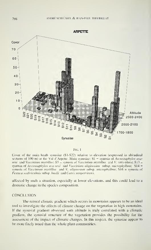
798 ANDRÉ SCHLÜSSEL & JEAN-PAUL THEURILLAT
ARPETTE
" co T^w w - £ -
wco™
£! - 'w m - E 0573 W - $ «
Synusiae
Altitude
>T 2300-2400
2000-2100
1700-1800
Fic. 1
Cover of the main heath synusiae (S1-S22) relative to elevation (expressed in altitudinal
sections of 100 m) at the Val d'Arpette. Main synusiae: SI = synusia of Ardostaphylos uva-
arsi and Vaccinium myrtillus; S5 = synusia of Vaccinium myrtillus and V. vitis-idaea; S 13 =
synusia of Arctostaphylos uva-ursi and Vaccinium uliginosum subsp. microphyllum; S 14 =
synusia of Vaccinium myrtillus and V. uliginosum subsp. microphyllum; S 16 = synusia of
Festuca scabriculmis subsp. luedii and Carex sempervirens.
affected by such a situation, especially at lower elevations, and this could lead to a
dramatic change in the species composition.
CONCLUSION
The natural climatic gradient which occurs in mountains appears to be an ideal
tool to investigate the effects of climate change on the vegetation in high mountains.
If the synusial gradient observed with altitude is truly correlated with a climatic
gradient, the synusial structure of the vegetation provides the possibility for the
assessment of the impact of climatic changes. In this respect, the synusiae appear to
be more finely tuned than the whole plant communities.
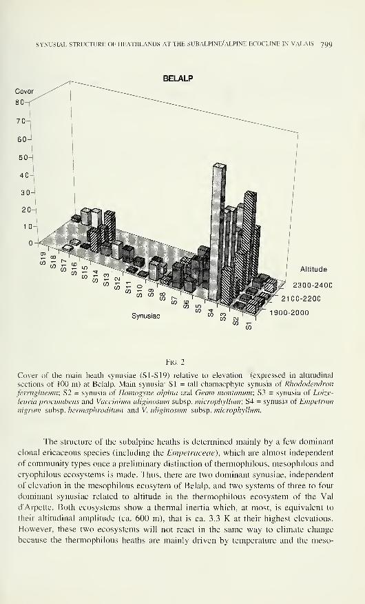
SYNUSIAL STRUCTURE OF HEATHLANDS AT THE SUBALPINE/ALPINE ECOCLINE IN VALAIS 799
BELALP
Synusiae
Altitude
2300-2400
2100-2200
Ì900-2000
Fig. 2
Cover of the main heath synusiae (SI -SI 9) relative to elevation (expressed in altitudinal
sections of 100 m) at Belalp. Main synusia: SI = tall chamaephyte synusia of Rhododendron
ferrugineum; S2 = synusia of Homogyne alpina and Geum montanum; S3 = synusia of Loize-
leuria procumbens and Vaccinium uliginosum subsp. microphyllum; S4 = synusia of Empetrumnigrum subsp. hermaphroditum and V. uliginosum subsp. microphyllum.
The structure of the subalpine heaths is determined mainly by a few dominant
clonal ericaceous species (including the Empetraceae), which are almost independent
of community types once a preliminary distinction of thermophilous, mesophilous and
cryophilous ecosystems is made. Thus, there are two dominant synusiae, independent
of elevation in the mesophilous ecosytem of Belalp, and two systems of three to four
dominant synusiae related to altitude in the thermophilous ecosystem of the Val
d'Arpette. Both ecosystems show a thermal inertia which, at most, is equivalent to
their altitudinal amplitude (ca. 600 m), that is ca. 3.3 K at their highest elevations.
However, these two ecosystems will not react in the same way to climate change
because the thermophilous heaths are mainly driven by temperature and the meso-
gOO ANDRÉ SCHLÜSSEL & JEAN-PAUL THEURILLAT
philous heaths by snow cover. Therefore, a climate change involving a reduction of
snow cover may act more on mesophilous heaths, because of their sensitivity to frost,
than on thermophilous ones.
ACKNOWLEDGEMENTS
We are very grateful to Loraine Kohorn, Duke University, Durham, NC, for
the linguistic revision, and to Prof. R. Spichiger for the facilities provided at the
Conservatoire et Jardin botaniques of the city of Geneva.
REFERENCES
Barkman, J. J. (1980). Synusial approaches to classification. In: R. H. Whittaker (ed.)
Classification ofplant communities, ed. 2. Junk, Den Haag: 1 1 1-165.
Braun-Blanquet, J. (1932). Plant sociology. McGraw Hill, London, 439 pp.
ELLENBERG, H. (1986). Vegetation Mitteleuropas mit den Alpen in ökologischer Sicht, ed. 3.
Ulmer, Stuttgart, 989 pp.
Ellenberg, H. (1988). Vegetation ecology of Central Europe, ed. 4. Cambridge University
Press, Cambridge, 732 pp.
Houghton, J. T., G. J. Jenkins, & J. J. Ephraums (eds.) 1990. Climate change - the IPCCscientific assessment. Cambridge University Press, Cambridge, 365 pp.
Houghton, J. T., B. A. Callander & S. K. Varney (eds.) Climate change 1992. The
supplementary report to the IPCC scientific assessment. Cambridge University Press,
Cambridge, 200 pp.
Larcher, W. (1994). Ökophysiologie der Pflanzen. Ulmer, Stuttgart, 394 pp.
Ozenda, P. (1985). La végétation de la chaîne alpine dans l'espace montagnard européen.
Masson, Paris, 344 pp. + 1 carte.
Theurillat, J.-P., F. Felber, P. Geissler, J.-M. Gobât, M. Fierz, A. Fischlin, P. Kupfer, A.
Schlüssel, C. Velluti, G. -F. Zhao & J. Williams (in print). Sensitivity of plant and
soil ecosystems of the Alps to climate change. In: Cebon, P., U. Dahinden, H. Davies,
D. Imboden & C. Jäger (eds.) A view from the Alps: Regional perspectives on climate
change. MIT Press. Boston.
Walter, H. (1984). Vegetation und Klimazonen. Ulmer, Stuttgart, 382 pp. + 1 carte.
Walter, H . (1985). Vegetation of the earth and ecological systems ofgeobiosphere, ed. 3.
Springer, Heidelberg, 318 pp.
Westhoff, V. & E. Van Der Maarel (1980). The Braun-Blanquet approach. In: R. H.
Whittaker (ed.) Classification ofplant communities, ed. 2. Junk, Den Haag: 287-399.
Wildi, O. (1991 ). Mulva-4, a processing environment for vegetation analysis. In: Feoli, E. & L.
Orloci (eds.) Computer assisted vegetation analysis. Handbook Veg. Sci. 11, Klüver,
Dordrecht: 407-428.
Wildi, O. (1994). Datenanalyse mit Mulva-5. Arbeitskopie, WSL, Birmensdorf, 73 pp.
Revue suisse de Zoologie, 103 (4): 801-833; décembre 1996
Variation in species richness of plants and diverse
groups of invertebrates in three calcareous grasslands
of the Swiss Jura mountains
Bruno BAUR 1
, Jasmin JOSHU, Bernhard SCHMID?, Ambros HÄNGGU,Daniel BORCARD^, Josef STARY5, Ariane PEDROLI-CHRISTEN6,
G. Heinrich THOMMENL Henryk LUKA?, Hans-Peter RUSTERHOLZ*,Peter OGGIER', Stephan LEDERGERBER ' & Andreas ERHARDT«1 Institut für Natur-, Landschafts- und Umweltschutz der Universität Basel (NLU),
St. Johanns-Vorstadt 10, CH-4056 Basel, Switzerland,
- Institut für Umweltwissenschaften, Universität Zürich, Winterthurerstrasse 190,
CH-8057 Zürich, Switzerland,
3 Naturhistorisches Museum Basel, Augustinergasse 2, CH-4001 Basel, Switzerland,
4 Institut de Zoologie, Université de Neuchâtel, Rue Emile-Argand 1 1
,
CH-2007 Neuchâtel, Switzerland,
5 Institute of Soil Biology, Academy of Sciences of the Czech Republic,
Na sâdkâch 7, CZ-37005 Ceské Budëjovice, Czech Republic,
6 Centre suisse de cartographie de la faune, Musée d'histoire naturelle, Terreaux 14,
CH-2000 Neuchâtel, Switzerland,
7 Forschungsinstitut für biologischen Landbau, Bernhardsberg,
CH-4104 Oberwil, Switzerland,
8 Botanisches Institut der Universität Basel, Schönbeinstrasse 6,
CH-4056 Basel, Switzerland.
Variation in species richness of plants and diverse groups of inver-
tebrates in three calcareous grasslands of the Swiss Jura mountains. -
Species richness and abundance of vascular plants and several groups of
invertebrates (spiders, oribatid mites, diplopods, grasshoppers and bush
crickets, ground beetles, butterflies and terrestrial gastropods) were re-
corded in three calcareous grasslands (Nenzlingen, Movelier and Vieques)
in the northwestern Swiss Jura mountains. Species richness varied both
between taxonomical groups and between sites (species richness ranges:
96-116 vascular plants, 60-66 spiders, 18 oribatid mites at each site, 1-7
diplopods, 10-16 grasshoppers and bush crickets, 19-21 ground beetles, 32-
46 butterflies and 15-21 terrestrial gastropods). Species overlap (number of
species that occurred at all sites) was relatively large in terrestrial gas-
tropods (59.1%), butterflies (56.5%), vascular plants (53.8%) and grass-
hoppers (47.1%), but relatively low in oribatid mites (32.3%), spiders
Manuscript accepted 16.01.1996.
802 BRUNO BAUR ET AL.
(25.0%), ground beetles (18.4%) and diplopods (12.5%). Diversity
expressed by the Shannon-Wiener index (H') was compared for five groups
of invertebrates. Diversity was largest in spiders and ground beetles and
lowest in terrestrial gastropods. Different taxonomical groups had their
maximum diversity at different sites: each grassland had the highest
diversity in at least one group. The three sites also varied in the abundance
of different invertebrate groups. Most groups had the highest densities in
Nenzlingen and the lowest densities in Vieques. All three sites contained a
high proportion of species listed in the Red Data Book of Switzerland with
values averaging 49.5% in grasshoppers and bush crickets, 28.9% in
butterflies, 18.9% in vascular plants. 11.2% in terrestrial gastropods, and
6.7% in ground beetles. One spider species (Oxyptila pullata) and two mite
species {Epilohmannia cylindrica minima and Pergalumna myrmophila)
were recorded for the first time in Switzerland.
Key-words: Acari - Araneae - Biodiversity - Calcareous grassland -
Coleoptera - Diplopoda - Gastropoda - Lepidoptera - Orthoptera - Vascular
plants.
INTRODUCTION
Many human activities, such as intensive agriculture, urbanisation, defores-
tation, and the building of roads and railways reduce natural habitats to remnants of
different sizes. In addition to the overall decrease in the area available for the orga-
nisms, the fragmentation of habitats leads to a division of existing populations into
isolated subpopulations of small size and to changes in habitat characteristics in the
fragments (Soulé 1986, Saunders et al. 1991). It is generally assumed that habitat
fragmentation contributes significantly to the local extinction of animal and plant
species (Saunders et al. 1991 ).
In the northwestern Jura mountains, unfertilized calcareous grasslands were
large and continuous at the beginning of this century, but since the 1950's changes in
agricultural practices such as the use of modern machinery, chemical fertilizers, her-
bicides and pesticides, and new breeds of plants have reduced the size of these areas
and split them into small and isolated fragments. For example, between 1950 and
1985 the total area of unfertilized calcareous grassland was reduced by 78% in the
Passwang region in northwestern Switzerland (Zoller et al. 1986). This overall
reduction of the unfertilized grassland area and the isolation of the remnants may have
led to a dramatic loss of species within a short period.
In 1993, a multidisciplinary coordinated project was started to experimentally
analyse fundamental aspects of biodiversity using dry grasslands on calcareous soils
in the northwestern Swiss Jura mountains as an exemplary model (Leser 1994, Baur
& Erhardt 1995). Most of the field work is done at three sites (Nenzlingen, Movelier
and Vieques). The present paper examines species richness and abundance of vascular
plants and various groups of invertebrates (spiders, oribatid mites, diplopods, grass-
hoppers and bush crickets, ground beetles, butterflies and terrestrial gastropods) in
SPECIES RICHNESS IN CALCAREOUS GRASSLANDS 803
these grasslands. In particular, we address the following questions: ( 1 ) Do the three
grasslands differ in plant and invertebrate species richness? (2) Are there any
associations between the species richnesses of different taxonomical groups? (3) Dodifferent taxonomical groups differ in species overlap at the three sites? (4) Do the
three sites differ in the overall density of particular taxonomical groups? and (5) Dothe three sites differ in number and proportion of rare and/or threatened species?
STUDY SITES AND GENERAL METHODS
The three field sites are nutrient-poor, dry calcareous grasslands (belonging to
the Teucrio-Mesobrometum type; Ellenberg 1988) situated near Nenzlingen (10 kmS of Basel), Movelier (5 km N of Delémont) and Vieques (5 km E of Delémont). The
three sites are situated within 20 km of each other.
There are local differences between the three sites. The study site in Nenz-
lingen (1.5 ha in size) is situated on a SW-facing slope with an inclination of 19-22°
at an altitude of 500 m a.s.l. A deciduous forest borders the investigation area to the
NE. Annual precipitation averages 991 mm in Grellingen (3 km E of the study site).
Soil properties and soil profiles of the investigation area are presented in Ogermann
et al (1994). Until 1993, the site was grazed by cattle from May to September with a
high stocking rate. The lower part of the slope was moderately fertilized by cattle
dung.
The investigation area in Movelier (1.3 ha) is situated on a SSE-facing slope
(inclination 20-22°) at an altitude of 780 m. Half of the site is surrounded by deci-
duous forest. The humus layer is relatively thick, contains some clay and is moister
than in Nenzlingen and Vieques. Until 1993, the site was grazed by cattle and a
moderate amount of artificial fertilizer was used.
The investigation area in Vieques (2.0 ha) is situated on a SE-facing slope
(inclination 15-27°) at an altitude of 570 m. The humus layer is thin and there are
several patches of exposed bedrock (this type of habitat is lacking at the other sites).
There is mixed forest at the SW-border of the area. Until 1993, the site was exposed
to a low grazing pressure by cattle.
In spring 1993, we initiated a field experiment to investigate possible effects of
habitat fragmentation on animal and plant populations at these sites. We experi-
mentally fragmented selected areas of grassland into plots of different sizes to exa-
mine whether species diversities of plants and various groups of invertebrates change
after fragmentation. The fragmentation was created by mowing the vegetation around
the experimental plots (see Baur & Erhardt (1995) for a figure of the experimental
set-up). An experimental unit (= block) contains one large (4.5 x 4.5 m), one medium(1.5 x 1.5 m) and two small fragments (0.5 x 0.5 m), all of them separated by a 5-mwide strip of mown vegetation, as well as the corresponding control plots. The
experiment consists of 12 blocks distributed over the three sites (five blocks in
Nenzlingen, three blocks in Movelier and four blocks in Vieques). The fragmentation
is maintained by regularly mowing the isolation area. This type of fragmentation is
804 BRUNO BAUR ET AL.
reversible, but reduces dispersal of pollen and seeds in many plant species as well as
dispersal of several invertebrate species.
In this paper, we present data on species richness and abundance of vascular
plants and various groups of invertebrates at the beginning of the long-term experi-
ment. We have tried to minimize any additional disturbances of the fragmented eco-
system by using non-destructive methods whenever possible. Data were collected
either within the experimental blocks (plants, grasshoppers and terrestrial gastropods),
at the border of each experimental block (oribatid mites), and/or in the areas adjacent
to or surrounding the experimental blocks (spiders, diplopods, grasshoppers, ground
beetles, butterflies and terrestrial gastropods). Species determination of grasshoppers,
butterflies and gastropods occurred in situ. Pitfall trapping was used for other taxo-
nomical groups in which species identification is difficult in the field (spiders, diplo-
pods, ground beetles). Details on the collecting methods are given in the corres-
ponding sections dealing with the different groups of organisms.
DATA ANALYSIS
Diversity has two components, the species richness measured by the total
number of species in an area, and the equitability of species abundances. The number
of species was used as a measure of the relative richness of the grasslands, but it
cannot be thought of as the absolute richness of a site, since numerous rare species
may not have been recorded in this study. The diversities of different taxonomic
groups were compared using the Shannon-Wiener index (H', base e) and the equi-
tability or eveness (E) of distribution of individuals among the species (Lloyd &Ghelardi 1964, Krebs 1988).
Species-abundance distributions were plotted to compare the proportion of rare
species (species with one individual) among the different taxonomical groups. The
similarity of the different species assemblages were compared using the coefficient of
S0rensen (based on presence/absence data) and the Renkonen index (based on
abundance data; Krebs 1988, Mühlenberg 1989).
VASCULAR PLANTSJasmin Joshi and Bernhard Schmid
Methods
Plant species richness was recorded in all fragments and control plots during
the growing seasons of 1993 and 1994. Data on the presence/absence of plant species
were obtained in Nenzlingen in May, June and August 1993 and in May and August
1994, in Movelier in June and August 1993 and at the beginning of June and in
August 1994, and in Vieques at the beginning of July and in August 1993 and in Mayand August 1994. In August 1994, the recording in Nenzlingen took place after
mowing of the experimental plots. Therefore, the presence of some grass species mayhave been overlooked.
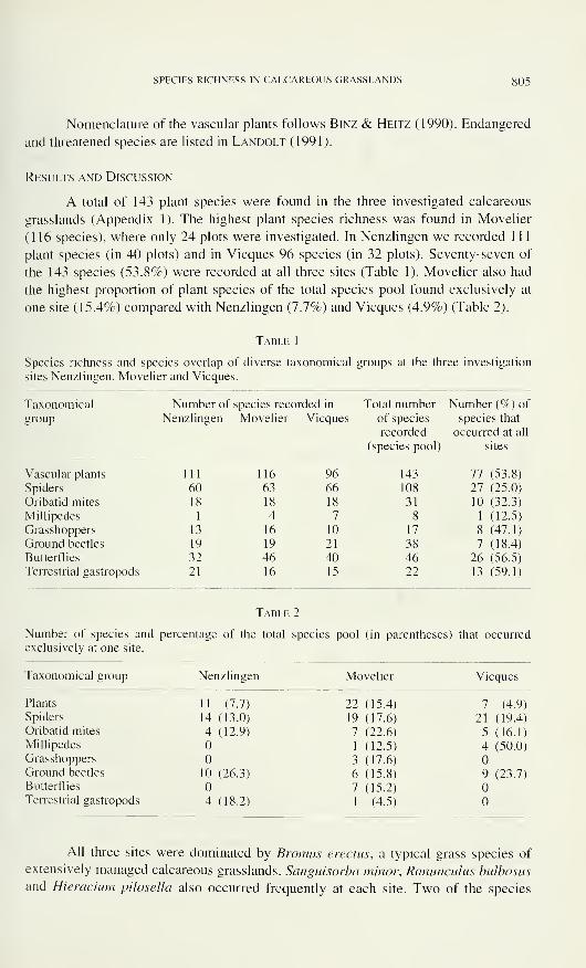
SPECIES RICHNESS IN CALCAREOUS GRASSLANDS 805
Nomenclature of the vascular plants follows Binz & Heitz (1990). Endangered
and threatened species are listed in Landolt (1991).
Results and Discussion
A total of 143 plant species were found in the three investigated calcareous
grasslands (Appendix 1). The highest plant species richness was found in Movelier
(116 species), where only 24 plots were investigated. In Nenzlingen we recorded 1 1
1
plant species (in 40 plots) and in Vieques 96 species (in 32 plots). Seventy-seven of
the 143 species (53.8%) were recorded at all three sites (Table 1). Movelier also had
the highest proportion of plant species of the total species pool found exclusively at
one site (15.4%) compared with Nenzlingen (7.7%) and Vieques (4.9%) (Table 2).
Table 1
Species richness and species overlap of diverse taxonomical groups at the three investigation
sites Nenzlingen, Movelier and Vieques.
Taxonomical Number of species reco:rded in Total number Number (%) of
group Nenzlingen Movelier Vieques of species
recorded
(species pool)
species that
occurred at all
sites
Vascular plants 111 116 96 143 77 (53.8)
Spiders 60 63 66 108 27 (25.0)
Oribatid mites 18 18 18 31 10 (32.3)
Millipedes 1 4 7 8 1 (12.5)
Grasshoppers 13 16 10 17 8 (47.1)
Ground beetles 19 19 21 38 7 (18.4)
Butterflies 32 46 40 46 26 (56.5)
Terrestrial gastropods 21 16 15 22 13 (59.1)
Table 2
Number of species and percentage of the total species pool (in parentheses) that occurred
exclusively at one site.
Taxonomical group Nenzlingen Movelier Vieques
Plants 11 (7.7)
Spiders 14 (13.0)
Oribatid mites 4 (12.9)
Millipedes
Grasshoppers
Ground beetles 10 (26.3)
Butterflies
Terrestrial gastropods 4 (18.2)
22 (15.4) 7 (4.9)
19 (17.6) 21 (19.4)
7 (22.6) 5 (16.1)
1 (12.5) 4 (50.0)
3 (17.6)
6 (15.8) 9 (23.7)
7 (15.2)
1 (4.5)
All three sites were dominated by Bromus erectus, a typical grass species of
extensively managed calcareous grasslands. Sanguisorba minor, Ranunculus bulbosus
and Hieracium pilosella also occurred frequently at each site. Two of the species
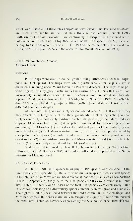
806 BRUNO BAUR ET AL.
which were found at all three sites {Trifolium ochroleucon and Veronica prostrata)
are listed as vulnerable in the Red Data Book of Switzerland (Landolt 1991).
Furthermore, Gentiana cruciata, found exclusively in Vieques, is also considered as
vulnerable in Switzerland. Altogether, seven of the 143 species (4.9%) recorded
belong to the endangered species, 19 (13.3%) to the vulnerable species and one
(0.7%) to the rare plant species in the northern Jura mountains (Landolt 1991 ).
SPIDERS (Arachnida, Araneae)
Ambros Hänggi
Methods
Pitfall traps were used to collect ground-living arthropods (Araneae, Diplo-
poda and Coleoptera). The traps were white plastic jars, 7 cm deep x 7 cm in
diameter, containing about 50 ml formalin (4%) with detergent. The traps were pro-
tected against rain by grey plastic roofs (measuring 18 x 18 cm) that were fixed
horizontally about 10 cm above ground. Specimens were removed and formalin
replaced at intervals of two weeks from 5 May to 12 November 1994. At each site,
nine traps were placed in groups of three (within-group distance 1 m) in three
different grassland subtypes.
At each site, the grassland subtypes considered were 50 - 100 m apart; they
may reflect the heterogeneity of the three grasslands. In Nenzlingen the grassland
subtypes were (1) a moderately fertilized patch of the pasture, (2) an unfertilized area
(typical Mesobrometum), and (3) a patch dominated by bracken (Pteridium
aquilinum); in Movelier (1) a moderately fertilized patch of the pasture, (2) an
unfertilized area (typical Mesobrometum), and (3) a part of the slope structured by
cow paths; in Vieques (1) an unfertilized area of the pasture with exposed bedrock
(karst rocks), (2) an unfertilized area (typical Mesobrometum), and (3) a patch of the
pasture (5 x 10 m) partly covered with bramble {Rubiis spp.).
Spiders were determined by Theo Blick, Hummeltal (Germany). Nomenclature
follows Maurer & Hänggi (1990). All spiders collected are deposited in the Natur-
historisches Museum Basel.
Results and Discussion
A total of 2701 adult spiders belonging to 108 species were collected at the
three study sites (Appendix 2). The sites were similar in species richness (60 species
in Nenzlingen, 63 in Movelier and 66 in Vieques), but differed in species composition
(Table 1, Appendix 2). Only 27 of the 108 species (25.0%) were found at all three
sites (Table 1). Twenty-one (19.4%) of the total 108 species were exclusively found
in Vieques, indicating an extraordinary spider community in this grassland (Table 2).
The highest similarity was found between the spider communities in Nenzlingen and
Movelier, whereas the spider community in Vieques was quite different from those of
the other sites (Table 3). Diversity expressed by the Shannon-Wiener index (H') was
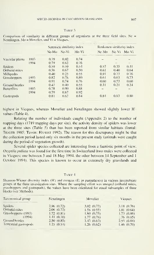
SPECIES RICHNESS IN CALCAREOUS GRASSLANDS 807
Table 3
Comparison of similarity in different groups of organisms at the three field sites. NeNenzlingen, Mo = Movelier, and Vi = Vieques.
S0renser i similarity index Renkoneri similarity index
Ne-Mo Ne-Vi Mo-Vi Ne-Mo Ne-Vi Mo-Vi
Vascular plants 1993 0.79 0.82 0.74 _ _ _
1994 0.79 0.82 0.76 - - -
Spiders 0.59 0.59 0.54 0.57 0.35 0.41
Oribatid mites 0.56 0.67 0.50 0.61 0.40 0.64
Millipedes 0.40 0.25 0.55 0.91 0.73 0.76
Grasshoppers 1993 0.82 0.78 0.80 0.61 0.63 0.73
1994 0.91 0.74 0.76 0.60 0.53 0.60
Ground beetles 0.47 0.40 0.55 0.31 0.21 0.24
Butterflies 1993 0.78 0.90 0.88 - - -
1994 0.79 0.87 0.92 - - -
Gastropods 0.81 0.83 0.84 0.84 0.83 0.80
highest in Vieques, whereas Movelier and Nenzlingen showed slightly lower H-values (Table 4).
Relating the number of individuals caught (Appendix 2) to the number of
trapping days (1719 trapping days per site), the activity density of spiders was lower
at the three sites (Table 5) than has been reported from similar habitats (Inntal:
Thaler 1985; Tessin: Hänggi 1992). The reason for this discrepancy might be that
the collection period lasted only six months in the present study (animals were caught
during the period of vegetation growth).
Several spider species collected are interesting from a faunistic point of view.
Oxyptila pallata was found for the first time in Switzerland (two males were collected
in Vieques; one between 5 and 18 May 1994, the other between 14 September and 1
October 1994). This species is known to occur in extremely dry grasslands and
Table 4
Shannon-Wiener diversity index (H') and eveness (E; in parentheses) in various invertebrate
groups at the three investigation sites. Where the sampling effort was unequal (oribatid mites,
grasshoppers and gastropods), the values have been calculated for equal subsamples of three
blocks (see Methods).
Taxonomical group
Spiders
Oribatid mites
Grasshoppers (1993)
(1994)
Ground beetles
Terrestrial gastropods
Nenzlingen Movelier
2.96 (0.72)
2.06 (0.72)
1.72 (0.81)
1.41 (0.70)
2.50 (0.85)
1.25 (0.51)
3.02 (0.73)
1.58 (0.55)
1.80 (0.75)
1.77 (0.78)
2.43 (0.83)
1.26 (0.62)
Vieques
3.18 (0.76)
1.81 (0.64)
1.73 (0.86)
1.76 (0.85)
2.55 (0.84)
1.46 (0.70)
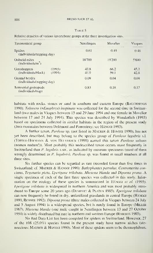
808 BRUNO BAUR ETAL.
Table 5
Relative densities of various invertebrate groups at the three investigation sites.
Taxonomical group Nenzlingen Movelier Vieques
Spiders
(individuals/trapping day)
0.61 0.49 0.48
Oribatid mites
(individuals/m2 )
18700 19'200 5'600
Grasshoppers (1993)
(individuals/block) (1994)
43.8
47.9
66.2
59.1
45.2
42.8
Ground beetles
(individuals/trapping day)
0.09 0.04 0.08
Terrestrial gastropods
(individuals/trap)
0.83 0.18 0.13
habitats with rocks, stones or sand in southern and eastern Europe (Bauchhenss
1990). Talavera (.-Euophrys) inopinata was collected for the second time in Switzer-
land (two males in Vieques between 15 and 29 June 1994 and one female in Movelier
between 17 and 24 July 1994). This species was described by Wunderlich (1993)
based on specimens collected in similar habitats in the region of the present study
(Jura mountains between Delémont and Porrentruy; see Hänggi 1993).
A further taxon, Pardosa sp. (not listed in Maurer & Hänggi 1990), has not
yet been described, but may belong to the species group of Pardosa lugubris s.l.
(Töpfer-Hofmann & von Helversen (1990) named this taxon Pardosa saltans
(nomen nudum!)). Most probably this undescribed taxon occurs more frequently in
Switzerland than P. lugubris s.str., as indicated by museum specimens (most of them
wrongly determined as P. lugubris). Pardosa sp. was found in small numbers at all
three sites.
Six further species can be regarded as rare (recorded fewer than five times in
Switzerland; cf. Maurer & Hänggi 1990): Bathyphantes parvulus, Centromerita con-
cinna, Tegenaria pietà, Eperigone trilobata, Mioxena blanda and Dipoena prona. Asingle specimen of each of the first three species was collected in this study. Infor-
mation on the ecology of these species is summarized in Hänggi et al. (1995).
Eperigone trilobata is widespread in northern America and was most probably intro-
duced to Europe some 20 years ago (Dumpert & Platen 1985). Eperigone trilobata
can now frequently be found in dry, unfertilized grasslands in central Europe (Hänggi
1990, Renner 1992). Dipoena prona (three males collected in Vieques between 24 July
and 5 August 1994) is a widespread species, but is rarely found in Europe (Miller
1967). Mioxena blanda (one male caught in Nenzlingen between 13 and 27 October
1994) is widely distributed but rare in northern and western Europe (Roberts 1985).
No Red Data List has been compiled for spiders in Switzerland. However, 27
of the 108 (25.0%) species found in the present study have narrow niches (ste-
noecious; Maurer & Hänggi 1990). Most of these spiders seem to be thermophilous,
SPECIES RICHNESS IN CALCAREOUS GRASSLANDS 809
preferring dry, open habitats. These types of habitat have dramatically declined in
Switzerland. Consequently, these 27 species can be regarded as potentially endan-
gered mainly due to habitat destruction. Vieques harbours a particularly threatened
spider fauna since 11 of the 21 species recorded exclusively at this site belong to the
group of potentially endangered species.
ORIBATID MITES (Acari, Oribatei)
Daniel Borcard & Josef Stary
Methods
Cylindrical soil samples, 5 cm in diameter and 8 cm in depth, were taken at the
margins of the control areas of each experimental block, using the soil augers
described by Bieri et al. (1978). The samples were divided into two parts (0-4 cm and
4-8 cm). The soil fauna was extracted by means of a MacFadyen-derived extractor
(Borcard 1986). Each site was sampled three times, on 10 March, 19 July and 14
November 1994. Four samples were taken in each block on each occasion. In all, 144
samples were taken.
Results and Discussion
A total of 4079 adult oribatid mites belonging to 3 1 different species were
captured at the three sites (Appendix 3). At each site, 18 mite species were found.
However, species number is often influenced by sampling effort. To account for the
lower number of blocks sampled in Movelier (n = 3), we computed the expected
number of species at the two other sites, assuming that only three blocks had been
sampled. The average of the total number of species of each possible combination of
three blocks was 17.5 in Nenzlingen and 17.0 in Vieques.
Eight of the 31 mite species (25.8%) were found at all three sites (Table 1,
Appendix 3). In contrast, four species (12.9%) were exclusively found in Nenzlingen,
seven species (22.6%) exclusively in Movelier and five species (16.1%) exclusively
in Vieques (Table 2).
The differences in species composition between seasons were small, accoun-
ting for 2.7% of the total variation as indicated by a canonical correspondence ana-
lysis (CCA; ter Braak 1986). Consequently, data from the three sampling occasions
were pooled for each site for further analysis. Moreover, unless noted otherwise, all
mites collected (depth 0-8 cm) were considered, although the majority of the mites
were found in the upper soil layer (between and 4 cm depth; Nenzlingen 84.2%,
Movelier 71 .8% and Vieques 65.6%).
The among-site differences in species composition were significant as indi-
cated by a CCA including all 144 samples (Monte Carlo test, 999 permutations, p =
0.001). These differences accounted for 11.4% of the total variation. In this analysis,
Vieques represented the most heterogeneous sampling site. Overall the species
richness was low when compared with other biotopes (such as forests), but normal for
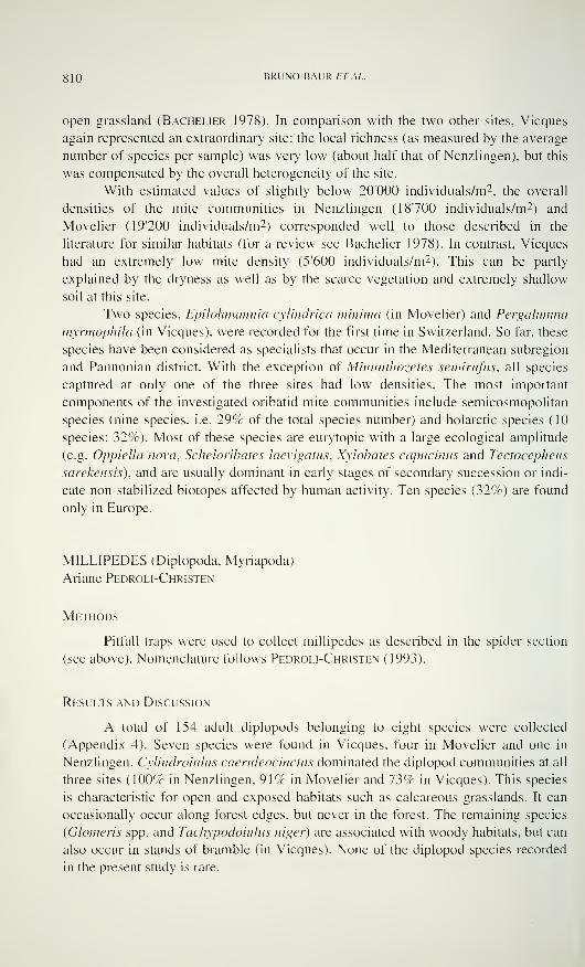
810 BRUNO BAUR ET AL.
open grassland (Bachelier 1978). In comparison with the two other sites, Vieques
again represented an extraordinary site: the local richness (as measured by the average
number of species per sample) was very low (about half that of Nenzlingen), but this
was compensated by the overall heterogeneity of the site.
With estimated values of slightly below 20'000 individuals/m2. the overall
densities of the mite communities in Nenzlingen (18700 individuals/m2 ) and
Movelier (19'200 individuals/m-) corresponded well to those described in the
literature for similar habitats (for a review see Bachelier 1978). In contrast, Vieques
had an extremely low mite density (5'600 individuals/m2 ). This can be partly
explained by the dryness as well as by the scarce vegetation and extremely shallow
soil at this site.
Two species, Epilohmannia cylindrica minima (in Movelier) and Pergalumna
myrmophila (in Vieques), were recorded for the first time in Switzerland. So far, these
species have been considered as specialists that occur in the Mediterranean subregion
and Pannonian district. With the exception of Minuntiiozetes semirufus, all species
captured at only one of the three sites had low densities. The most important
components of the investigated oribatid mite communities include semicosmopolitan
species (nine species, i.e. 29% of the total species number) and holarctic species (10
species; 32%). Most of these species are eurytopic with a large ecological amplitude
(e.g. OppieUa nova, Scheloribates laevigatas, Xylobates capucinus and Tectocepheus
sarekensis), and are usually dominant in early stages of secondary succession or indi-
cate non-stabilized biotopes affected by human activity. Ten species (32%) are found
only in Europe.
MILLIPEDES (Diplopoda, Myriapoda)
Ariane Pedroli-Christen
Methods
Pitfall traps were used to collect millipedes as described in the spider section
(see above). Nomenclature follows Pedroli-Christen (1993).
Results and Discussion
A total of 154 adult diplopods belonging to eight species were collected
(Appendix 4). Seven species were found in Vieques, four in Movelier and one in
Nenzlingen. Cylindroiulus caeruleocinctus dominated the diplopod communities at all
three sites (100% in Nenzlingen, 91% in Movelier and 73% in Vieques). This species
is characteristic for open and exposed habitats such as calcareous grasslands. It can
occasionally occur along forest edges, but never in the forest. The remaining species
(Glomeris spp. and Tachypodoiulus niger) are associated with woody habitats, but can
also occur in stands of bramble (in Vieques). None of the diplopod species recorded
in the present study is rare.
SPECIES RICHNESS IN CALCAREOUS GRASSLANDS 8] ]
GRASSHOPPERS. BUSH CRICKETS AND TRUE CRICKETS(Orthoptera: Saltatoria)
G. Heinrich Thommen
Methods
The relative abundances of the different Saltatorian species were recorded in
the experimental blocks (isolated fragments and the corresponding control plots)
using a direct census method. The entire vegetation of the experimental plots was
carefully searched for Saltatoria. Plants were slightly moved with a bamboo rod for an
easier detection of the insects. In addition, in each block an area of 4.5 x 4.5 m of
mown vegetation (isolation area of the experiment) was searched for Saltatoria. The
number of individuals observed was recorded for each species (no animals were
caught). Monitoring was repeated three times at all sites between July and early
September both in 1993 and 1994.
A transect count technique was used to record the composition and relative
abundance of Saltatorian species in the grassland surrounding the experimental
blocks. These areas (1-2 ha in size) were slowly walked through in a zigzag line. All
adult individuals seen within a strip of 1-1.2 m width were counted. The adjacent
areas were surveyed once at each of the three sites during summer 1 994.
Only Saltatoria species that typically inhabit unwooded open land were
censused. True arboreal species and inhabitants of the forest edge were not included
in this study. Nomenclature follows Bellmann (1993).
Results and Discussion
Thirteen species of Saltatoria were recorded in Nenzlingen (11 of them in the
experimental blocks), 16 species in Movelier (14 in the experimental blocks), and 10
species in Vieques (9 in the experimental blocks; Appendix 5). A total of 17 species
were recorded from all three sites combined, which represents approximately half of
the Saltatorian species known in dry grasslands of the Swiss Jura mountains. Eight of
the 17 (47.1%) species occurred at all three sites (Table 1 ).
The higher species richness in the areas adjacent to the experimental blocks
could be explained by the larger size and higher degree of heterogeneity of these
areas. The surrounding areas are more variable in cover and height of vegetation, in
soil moisture content, inclination of slope and in the structure of the surface (e.g.
single rocks or exposed limestone bedrock) than the experimental blocks. Patches
with relatively high soil moisture may explain the presence of Metrioptera roeselii in
Nenzlingen as well as Metrioptera brachyptera and Chrysochraon dispar in Move-lier; all three species are mesohygrophilic, preferring moist or tall grass habitats.
These three species are lacking in Vieques, the driest of the three sites.
Chorthippus parallelus, usually associated with fertilized meadows, was one
of the most frequent species (19-32% of all adult individuals) at all three investigation
sites. Stenobothrus lineatus, a mesoxerophilic grasshopper species, was frequent (23-
31%) in the experimental blocks at all three sites, but less frequent (10-17%) in the
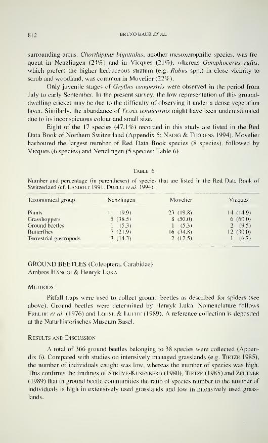
8 1
2
BRUNO BAUR ET AL.
surrounding areas. Chorthippus biguttulus, another mesoxerophilic species, was fre-
quent in Nenzlingen (24%) and in Vieques (21%), whereas Gomphocerus nifiis,
which prefers the higher herbaceous stratum (e.g. Rubus spp.) in close vicinity to
scrub and woodland, was common in Movelier (22%).
Only juvenile stages of Gryllus campestris were observed in the period from
July to early September. In the present survey, the low representation of this ground-
dwelling cricket may be due to the difficulty of observing it under a dense vegetation
layer. Similarly, the abundance of Tetrix tenuicornis might have been underestimated
due to its inconspicuous colour and small size.
Eight of the 17 species (47.1%) recorded in this study are listed in the Red
Data Book of Northern Switzerland (Appendix 5; Nadig & Thorens 1994). Movelier
harboured the largest number of Red Data Book species (8 species), followed by
Vieques (6 species) and Nenzlingen (5 species; Table 6).
Table 6
Number and percentage (in parentheses) of species that are listed in the Red Data Book of
Switzerland (cf. Landolt 1991, Duelli et al. 1994).
Taxonomical group Nenzlingen Movelier Vieques
Plants 11 (9.9) 23 (19.8) 14 (14.9)
Grasshoppers 5 (38.5) 8 (50.0) 6 (60.0)
Ground beetles 1 (5.3) 1 (5.3) 2 (9.5)
Butterflies 7 (21.9) 16 (34.8) 12 (30.0)
Terrestrial gastropods 3 (14.3) 2 (12.5) 1 (6.7)
GROUND BEETLES (Coleoptera, Carabidae)
Ambros Hänggi & Henryk Luka
Methods
Pitfall traps were used to collect ground beetles as described for spiders (see
above). Ground beetles were determined by Henryk Luka. Nomenclature follows
Freude et al. (1976) and Lohse & Lucht (1989). A reference collection is deposited
at the Naturhistorisches Museum Basel.
Results and Discussion
A total of 366 ground beetles belonging to 38 species were collected (Appen-
dix 6). Compared with studies on intensively managed grasslands (e.g. Tietze 1985),
the number of individuals caught was low, whereas the number of species was high.
This confirms the findings of Strüve-Kusenberg (1980), Tietze (1985) and Zeltner
(1989) that in ground beetle communities the ratio of species number to the number of
individuals is high in extensively used grasslands and low in intensively used grass-
lands.
SPECIES RICHNESS IN CALCAREOUS GRASSLANDS 813
The three sites had similar numbers of species (19 species in Nenzlingen, 19 in
Movelier and 21 in Vieques). However, the three sites differed considerably in spe-
cies composition. Seven of the 38 carabid species (18.4%) were found at all three
sites (Table 1 ). On the other hand, a high proportion of species occurred exclusively
at one site: 47.4% in Nenzlingen, 31.6% in Movelier, and 42.9% in Vieques (Table
2). This resulted in a low similarity of the species assemblages between the sites
(Table 3). Diversity expressed by the Shannon-Wiener index H' was higest in
Vieques, followed by Nenzlingen and Movelier (Table 4).
Four of the 38 (10.5%) carabid species found are listed in the Red Data Book
of Switzerland (Marggi 1994). Anisodactylus nemorivagus was exclusively found in
Nenzlingen. Little is known concerning the life history of this species (Lindroth
1945, Marggi 1992). We collected individuals of A. nemorivagus in the moderately
fertilized patch and the typical Mesobrometum part of the grassland in Nenzlingen.
Only one individual of Carabus convexus was found in the typical Mesobrometum
part of Movelier. This species does not tolerate intensively cultivated grasslands and
is considered as threatened (Marggi 1992). In general, C. convexus is not rare in the
Swiss Jura mountains (Marggi 1992). Two further species listed in the Red Data
Book were found exclusively in Vieques. All 10 individuals of Carabus auratus were
caught in the patch partly covered with bramble. Carabus auratus is also restricted to
extensively cultivated open areas. The four individuals of Panagaeus bipustulatus
were caught in the patch partly covered with bramble and in the area with exposed
bedrock in Vieques. This species seems to be strongly xerophilous (Marggi 1992).
A high number of polyphagous-phytophagous species (belonging to the genera
Amara, Anisodactylus, Harpalus and Parophonus), which also climb onto the
vegetation, was collected in Nenzlingen (cf. Wachmann et al. 1995). In Movelier,
there was a surprisingly high proportion of large species of Carabus (32% of all
species, 37% of all individuals). Among them was Carabus monilis, which does not
tolerate intensive management of grasslands (Marggi 1992, Pfiffner & Luka 1994).
indeed, we collected no specimens of this species in the fertilized patch of the grass-
land. The carabid community of Vieques was characterized by thermophilous species
such as Panagaeus bipustulatus and Callistus lunatus. The latter is known to prefer
temperatures reaching 40-50 °C (Becker 1975, Thiele 1977).
BUTTERFLIES (Lepidoptera)
Hans-Peter Rusterholz & Andreas Erhardt
Methods
Estimates of species richness were obtained from observations on the activity
of foraging butterflies. Three 10 x 10 m plots were established in undisturbed grass-
land vegetation adjacent to the blocks of the fragmentation experiment at each site.
Butterfly activity was recorded for periods of 45 min. between 10.00 a.m. and 4.30
p.m. during sunny weather conditions. The number of individuals of each species was
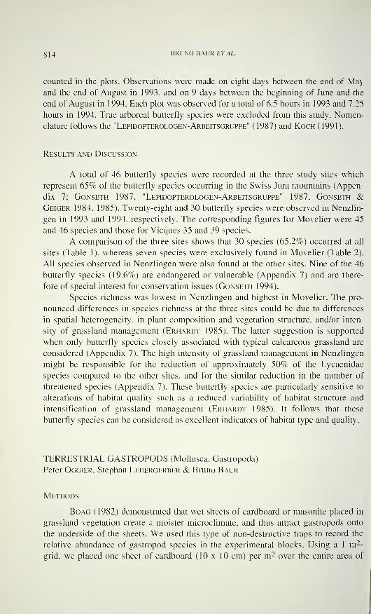
814 BRUNO BAUR£7\4L.
counted in the plots. Observations were made on eight days between the end of Ma)
and the end of August in 1993, and on 9 days between the beginning of June and the
end of August in 1994. Each plot was observed for a total of 6.5 hours in 1993 and 7.25
hours in 1994. True arboreal butterfly species were excluded from this study. Nomen-
clature follows the "Lepidopterologen-Arbeitsgruppe" (1987) and Koch (1991).
Results and Discussion
A total of 46 butterfly species were recorded at the three study sites which
represent 65% of the butterfly species occurring in the Swiss Jura mountains (Appen-
dix 7; Gonseth 1987. "Lepidopterologen-Arbeitsgruppe" 1987, Gonseth &Geiger 1984, 1985). Twenty-eight and 30 butterfly species were observed in Nenzlin-
gen in 1993 and 1994, respectively. The corresponding figures for Movelier were 45
and 46 species and those for Vieques 35 and 39 species.
A comparison of the three sites shows that 30 species (65.2%) occurred at all
sites (Table 1 ). whereas seven species were exclusively found in Movelier (Table 2).
All species observed in Nenzlingen were also found at the other sites. Nine of the 46
butterfly species (19.6%) are endangered or vulnerable (Appendix 7) and are there-
fore of special interest for conservation issues (Gonseth 1994).
Species richness was lowest in Nenzlingen and highest in Movelier. The pro-
nounced differences in species richness at the three sites could be due to differences
in spatial heterogeneity, in plant composition and vegetation structure, and/or inten-
sity of grassland management (Erhardt 1985). The latter suggestion is supported
when only butterfly species closely associated with typical calcareous grassland are
considered (Appendix 7). The high intensity of grassland management in Nenzlingen
might be responsible for the reduction of approximately 50% of the Lycaenidae
species compared to the other sites, and for the similar reduction in the number of
threatened species (Appendix 7). These butterfly species are particularly sensitive to
alterations of habitat quality such as a reduced variability of habitat structure and
intensification of grassland management (Erhardt 1985). It follows that these
butterfly species can be considered as excellent indicators of habitat type and quality.
TERRESTRIAL GASTROPODS (Mollusca, Gastropoda)
Peter Oggier. Stephan Ledergerber & Bruno Baur
Methods
Boag (1982) demonstrated that wet sheets of cardboard or masonite placed in
grassland vegetation create a moister microclimate, and thus attract gastropods onto
the underside of the sheets. We used this type of non-destructive traps to record the
relative abundance of gastropod species in the experimental blocks. Using a 1 m2-
grid. we placed one sheet of cardboard (10 x 10 cm) per m2 over the entire area of
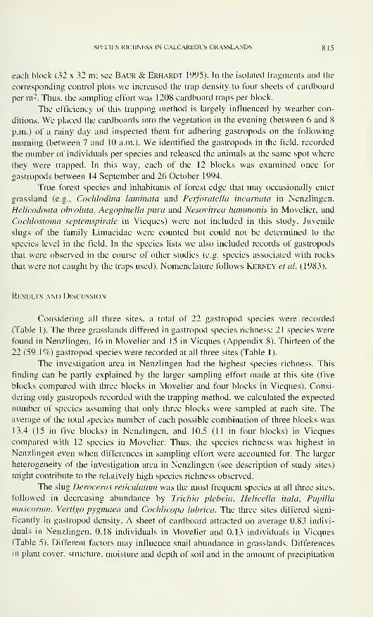
SPECIES RICHNESS IN CALCAREOUS GRASSLANDS 815
each block (32 x 32 m; see Baur & Erhardt 1995). In the isolated fragments and the
corresponding control plots we increased the trap density to four sheets of cardboard
per m2. Thus, the sampling effort was 1208 cardboard traps per block.
The efficiency of this trapping method is largely influenced by weather con-
ditions. We placed the cardboards into the vegetation in the evening (between 6 and 8
p.m.) of a rainy day and inspected them for adhering gastropods on the following
morning (between 7 and 10 a.m.). We identified the gastropods in the field, recorded
the number of individuals per species and released the animals at the same spot where
they were trapped. In this way, each of the 12 blocks was examined once for
gastropods between 14 September and 26 October 1994.
True forest species and inhabitants of forest edge that may occasionally enter
grassland (e.g., Cochlodina laminata and Perforate!la incarnata in Nenzlingen,
Helicodonta obvoluta, Aegopinella pura and Nesovitrea hammonis in Movelier, and
Cochlostoma septemspirale in Vieques) were not included in this study. Juvenile
slugs of the family Limacidae were counted but could not be determined to the
species level in the field. In the species lists we also included records of gastropods
that were observed in the course of other studies (e.g. species associated with rocks
that were not caught by the traps used). Nomenclature follows Kerney et al. (1983).
Results and Discussion
Considering all three sites, a total of 22 gastropod species were recorded
(Table 1). The three grasslands differed in gastropod species richness: 21 species were
found in Nenzlingen, 16 in Movelier and 15 in Vieques (Appendix 8). Thirteen of the
22 (59.1%) gastropod species were recorded at all three sites (Table 1).
The investigation area in Nenzlingen had the highest species richness. This
finding can be partly explained by the larger sampling effort made at this site (five
blocks compared with three blocks in Movelier and four blocks in Vieques). Consi-
dering only gastropods recorded with the trapping method, we calculated the expected
number of species assuming that only three blocks were sampled at each site. The
average of the total species number of each possible combination of three blocks was
13.4 (15 in five blocks) in Nenzlingen, and 10.5 (11 in four blocks) in Vieques
compared with 12 species in Movelier. Thus, the species richness was highest in
Nenzlingen even when differences in sampling effort were accounted for. The larger
heterogeneity of the investigation area in Nenzlingen (see description of study sites)
might contribute to the relatively high species richness observed.
The slug Deroceras reticulatum was the most frequent species at all three sites,
followed in decreasing abundance by Trichia plebeia, Helicella itala, Pupilla
muscorum, Vertigo pygmaea and Cochlicopa lubrica. The three sites differed signi-
ficantly in gastropod density. A sheet of cardboard attracted on average 0.83 indivi-
duals in Nenzlingen, 0.18 individuals in Movelier and 0.13 individuals in Vieques
(Table 5). Different factors may influence snail abundance in grasslands. Differences
in plant cover, structure, moisture and depth of soil and in the amount of precipitation
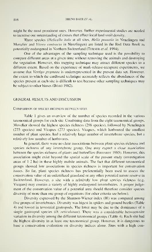
g 1
6
BRUNO BAUR ET AL.
might be the most prominent ones. However, further experimental studies are needed
to increase our unterstanding of causes that affect local land snail density.
Three species (Helicella itala at all sites, Helix pomatia in Nenzlingen and
Movelier and Vitrea contracta in Nenzlingen) are listed in the Red Data Book as
potentially endangered in Northern Switzerland (Turner et al. 1994).
One of the advantages of the sampling technique used is the possibility to
compare different areas at a given time without removing the animals and destroying
the vegetation. However, this trapping technique may attract different species to a
different extent. Based on the experience of mark-release-recapture experiments, we
assume that Vertigo pygmaea is underrepresented in the present data set. However,
the extent to which the cardboard technique accurately reflects the abundances of the
species present at each site is difficult to test because other sampling techniques may
be subject to other biases (Boag 1982).
GENERAL RESULTS AND DISCUSSION
Comparison of species richness between sites
Table 1 gives an overview of the number of species recorded in the various
taxonomical groups for each site. Combining data from the eight taxonomical groups,
Movelier showed the highest species richness (298 species), followed by Nenzlingen
(275 species) and Vieques (273 species). Vieques, which harboured the smallest
number of plant species, had a relatively large number of invertebrate species, but a
relatively low number of herbivores.
In general, there were no clear associations between plant species richness and
species richness of any invertebrate group. One may expect a close association
between the species richness of plants and butterflies (Erhardt 1985). However, this
association might exist beyond the spatial scale of the present study (investigation
areas of 1-2 ha) in these highly mobile animals. The fact that different taxonomical
groups showed low associations in species richness is important for conservation
issues. So far, plant species richness has preferentially been used to assess the
conservation value of an unfertilized grassland or any other potential nature reserve in
Switzerland. However, a site with a relatively low plant species richness (e.g.
Vieques) may contain a variety of highly endangered invertebrates. A proper judge-
ment of the conservation value of a potential area should therefore consider species
diversity of more than one group of organisms (for other criteria see below).
Diversity expressed by the Shannon-Wiener index (H) was compared among
five groups of invertebrates. Diversity was higest in spiders and ground beetles (Table
4) and lowest in terrestrial gastropods. The latter may be due to the dominance of a
single gastropod species (D. reticulatum). There was a considerable between-site
variation in diversity among the different taxonomical groups (Table 4). Each site had
the highest diversity in at least one taxonomical group. However, it is dangerous to
base a conservation evaluation on diversity indices alone. Sites with a high con-
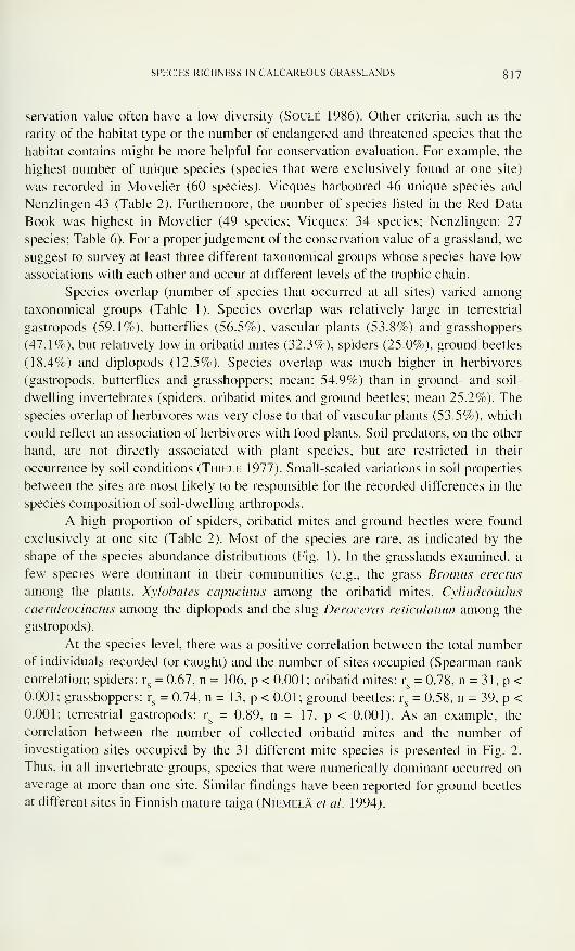
SPECIES RICHNESS IN CALCAREOUS GRASSLANDS 817
servation value often have a low diversity (Soulé 1986). Other criteria, such as the
rarity of the habitat type or the number of endangered and threatened species that the
habitat contains might be more helpful for conservation evaluation. For example, the
highest number of unique species (species that were exclusively found at one site)
was recorded in Movelier (60 species). Vieques harboured 46 unique species and
Nenzlingen 43 (Table 2). Furthermore, the number of species listed in the Red Data
Book was highest in Movelier (49 species; Vieques: 34 species; Nenzlingen: 27
species; Table 6). For a proper judgement of the conservation value of a grassland, we
suggest to survey at least three different taxonomical groups whose species have low
associations with each other and occur at different levels of the trophic chain.
Species overlap (number of species that occurred at all sites) varied among
taxonomical groups (Table 1). Species overlap was relatively large in terrestrial
gastropods (59.1%), butterflies (56.5%), vascular plants (53.8%) and grasshoppers
(47.1%), but relatively low in oribatid mites (32.3%), spiders (25.0%), ground beetles
(18.4%) and diplopods (12.5%). Species overlap was much higher in herbivores
(gastropods, butterflies and grasshoppers; mean: 54.9%) than in ground- and soil-
dwelling invertebrates (spiders, oribatid mites and ground beetles; mean 25.2%). The
species overlap of herbivores was very close to that of vascular plants (53.5%), which
could reflect an association of herbivores with food plants. Soil predators, on the other
hand, are not directly associated with plant species, but are restricted in their
occurrence by soil conditions (Thiele 1977). Small-scaled variations in soil properties
between the sites are most likely to be responsible for the recorded differences in the
species composition of soil-dwelling arthropods.
A high proportion of spiders, oribatid mites and ground beetles were found
exclusively at one site (Table 2). Most of the species are rare, as indicated by the
shape of the species abundance distributions (Fig. 1 ). In the grasslands examined, a
few species were dominant in their communities (e.g., the grass Bromus erectus
among the plants, Xylobates capuchins among the oribatid mites, Cylindroiulus
caeruleocinctus among the diplopods and the slug Deroceras reticulatum among the
gastropods).
At the species level, there was a positive correlation between the total number
of individuals recorded (or caught) and the number of sites occupied (Spearman rank
correlation; spiders: rs= 0.67, n = 106, p < 0.001; oribatid mites: r
s= 0.78, n - 31. p <
0.001; grasshoppers: rs= 0.74, n = 13, p < 0.01; ground beetles: r
s= 0.58, n = 39. p <
0.001; terrestrial gastropods: rs= 0.89, n = 17, p < 0.001). As an example, the
correlation between the number of collected oribatid mites and the number of
investigation sites occupied by the 31 different mite species is presented in Fig. 2.
Thus, in all invertebrate groups, species that were numerically dominant occurred on
average at more than one site. Similar findings have been reported for ground beetles
at different sites in Finnish mature taiga (Niemelä et al. 1994).
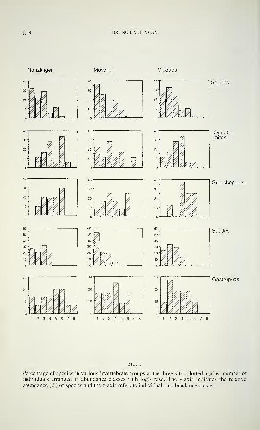
818 BRUNO BAURET AL.
Nenzlingen Movelier Vieques
Spiders
30-
20"
10-
V7)
11 vm
Oribatid
mites
50-
40-
30-
20-
10-:
1nFn- :„'
4U -
771
30-
20-
10-
1 1 1
Grasshoppers
50-
40-
30- Vsf'
20-
10- \ ^I
Beetles
DD1 2 3 4 5 6 7
^
iU Ji1 2 3 4 5 6 7
>z
Gastropods
1 2 3 4 5 6 7
Fig. 1
Percentage of species in various invertebrate groups at the three sites plotted against number of
individuals arranged in abundance classes with log3 base. The y-axis indicates the relative
abundance (%) of species and the x-axis refers to individuals in abundance classes.
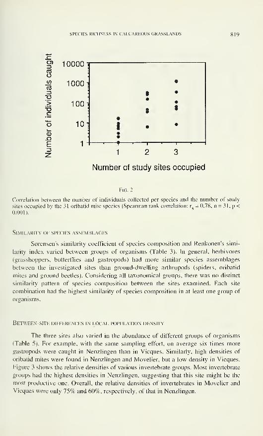
SPECIES RICHNESS IN CALCAREOUS GRASSLANDS 819
3 10000cdo
ni1000
vu3"D*>
100T>.E
o 10i_<D-QE 1
31
Number of study sites occupied
Fig. 2
Correlation between the number of individuals collected per species and the number of study
sites occupied by the 31 oribatid mite species (Spearman rank correlation: r„ = 0.78, n = 31, p <
0.001).
Similarity of species assemblages
S0rensen's similarity coefficient of species composition and Renkonen's simi-
larity index varied between groups of organisms (Table 3). In general, herbivores
(grasshoppers, butterflies and gastropods) had more similar species assemblages
between the investigated sites than ground-dwelling arthropods (spiders, oribatid
mites and ground beetles). Considering all taxonomical groups, there was no distinct
similarity pattern of species composition between the sites examined. Each site
combination had the highest similarity of species composition in at least one group of
organisms.
Between-site differences in local population density
The three sites also varied in the abundance of different groups of organisms
(Table 5). For example, with the same sampling effort, on average six times more
gastropods were caught in Nenzlingen than in Vieques. Similarly, high densities of
oribatid mites were found in Nenzlingen and Movelier, but a low density in Vieques.
Figure 3 shows the relative densities of various invertebrate groups. Most invertebrate
groups had the highest densities in Nenzlingen, suggesting that this site might be the
most productive one. Overall, the relative densities of invertebrates in Movelier and
Vieques were only 75% and 60%, respectively, of that in Nenzlingen.
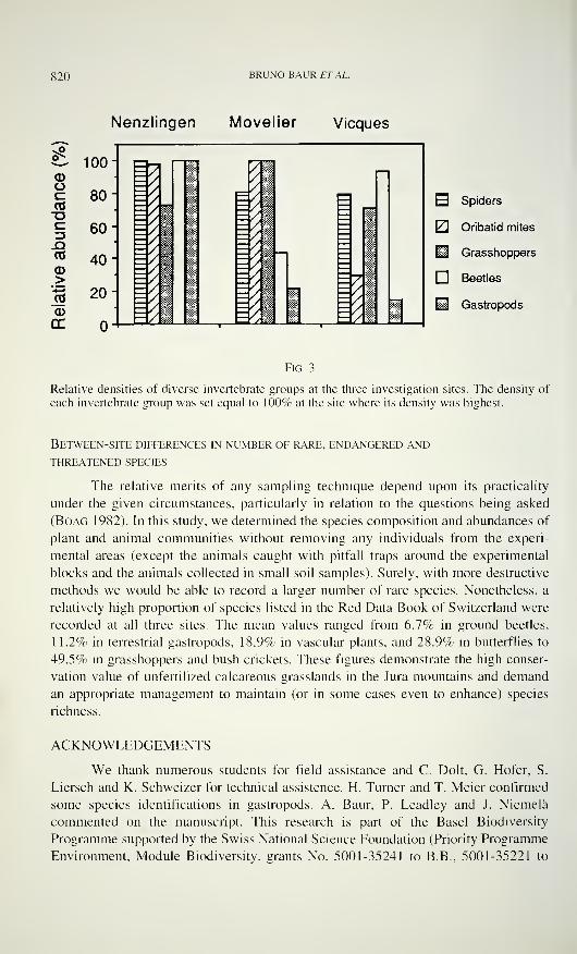
820 BRUNO BAUR ET AL.
Nenzlingen Movelier Vieques
^ 100- acdoeCOoc13JDCO
CD>
CD
ce
H Spiders
Oribatid mites
IH Grasshoppers
Beetles
H Gastropods
Fig. 3
Relative densities of diverse invertebrate groups at the three investigation sites. The density of
each invertebrate group was set equal to 100% at the site where its density was highest.
Between-site differences in number of rare, endangered and
threatened species
The relative merits of any sampling technique depend upon its practicality
under the given circumstances, particularly in relation to the questions being asked
(Boag 1982). In this study, we determined the species composition and abundances of
plant and animal communities without removing any individuals from the experi-
mental areas (except the animals caught with pitfall traps around the experimental
blocks and the animals collected in small soil samples). Surely, with more destructive
methods we would be able to record a larger number of rare species. Nonetheless, a
relatively high proportion of species listed in the Red Data Book of Switzerland were
recorded at all three sites. The mean values ranged from 6.7% in ground beetles,
11.2% in terrestrial gastropods, 18.9% in vascular plants, and 28.9% in butterflies to
49.5% in grasshoppers and bush crickets. These figures demonstrate the high conser-
vation value of unfertilized calcareous grasslands in the Jura mountains and demand
an appropriate management to maintain (or in some cases even to enhance) species
richness.
ACKNOWLEDGEMENTS
We thank numerous students for field assistance and C. Dolt. G. Hofer, S.
Liersch and K. Schweizer for technical assistence. H. Turner and T. Meier confirmed
some species identifications in gastropods. A. Baur, P. Leadley and J. Niemelä
commented on the manuscript. This research is part of the Basel Biodiversity
Programme supported by the Swiss National Science Foundation (Priority Programme
Environment, Module Biodiversity, grants No. 5001-35241 to B.B., 5001-35221 to
SPECIES RICHNESS IN CALCAREOUS GRASSLANDS 821
A.E. and 5001-35229 to B.S.). Additional financial support was received from the
Amt für Orts- und Regionalplanung, Liestal (Kanton Baselland), the Emilia
Guggenheim-Schnurr Stiftung der Naturforschenden Gesellschaft Basel-Stadt and the
Freiwillige Akademische Gesellschaft der Universität Basel.
REFERENCES
Bachelier, G. 1978. La faune des sols, son écologie et son action. ORSTOM, Paris, 395 pp.
Bauchhenss, E. 1990. Mitteleuropäische Xerotherm-Standorte und ihre epigäische Spinnen-
fauna - eine autökologische Betrachtung. Abhandhingen der naturwissenschaftlichen
Vereinigung Hamburg, (NF) 31/32: 153-162.
Baur, B. & A. Erhardt. 1995. Habitat fragmentation and habitat alterations: principle threats
to most animal and plant species. Gaia, 4: 221-226.
Becker, J. 1975. Art und Ursachen der Habitatbindung von Bodenarthropoden (Carabidae-
Coleoptera, Diplopoda, Isopoda) xerothermer Standorte in der Eifel. Beiträge der
Landespflege Rheinland-Pfalz, Beiheft A: 89-140.
Bellmann, H. 1993. Heuschrecken beobachten - bestimmen (2. Auflage). Naturbuch-Verlag,
Augsburg, 349 pp.
Bieri, M., V. Delucchi & C. Lienhard. 1978. Beschreibung von zwei Sonden zur standardi-
sierten Entnahme von Bodenproben für Untersuchungen an Mikroarthropoden. Mit-
teilungen der Schweizerischen entomologischen Gesellschaft, 5 1 : 327-330.
Binz, A. & C. Heitz. 1990. Schul- und Exkursionsflora für die Schweiz. Schwabe & Co., Basel,
659 pp.
Boag, D. A. 1982. Overcoming sampling bias in studies of terrestrial gastropods. Canadian
Journal ofZoology, 60: 1289-1292.
Borcard, D. 1986. Une sonde et un extracteur destinés à la récolte dAcariens (Acari) dans les
sphaignes (Sphagnum spp.). Mitteilungen der Schweizerischen entomologischen Gesell-
schaft, 59: 283-288.
Duelli, P. et al. 1994. Rote Listen der gefährdeten Tierarten in der Schweiz. Bundesamt fürUmwelt, Wald und Landschaft. Bern, 97 pp.
Dumpert, K. & R. Platen. 1985. Zur Biologie eines Buchenwaldbodens. 4. Die Spinnenfauna.
Carolinea. 42: 75-106.
Ellenberg, H. 1988. Vegetation Ecology of Central Europe. Cambridge University Press,
Cambridge, 731 pp.
Erhardt, A. 1985. Day-active Lepidoptera, sensitive indicators of cultivated and abandonedgrassland. Journal ofApplied Ecology, 22: 841-861.
Freude, H., K. W. Harde & G. A. Lohse. 1976. Die Käfer Mitteleuropas. Band 2. Adephaga1. Goecke & Evers, Krefeld, 302 pp.
Gonseth, Y. 1987. Verbreitungsatlas der Tagfalter der Schweiz (Lepidoptera, Rhopalocera).
Documenta Faunistica Helvetiae, 6, CSCF, Neuchâtel, 242 pp.
Gonseth, Y. 1994. Rote Liste der gefährdeten Tagfalter der Schweiz, pp. 48-51. In: Rote
Listen der gefährdeten Tierarten in der Schweiz (P. Duelli et al.). Bundesamt fürUmwelt, Wedel und Landschaft, Bern, 97 pp.
Gonseth, Y. & W. Geiger. 1984. Bibliographie sur les Rhopalocères de Suisse: 1900 - 1982 et
principales publications antérieures à 1900 (1ère partie). Bulletin de la Société
Romande d'Entomologie 2, N. 1 : 37-80.
Gonseth, Y. & W. Geiger. 1985. Bibliographie sur les Rhopalocères de Suisse: 1900 - 1982 et
principales publications antérieures à 1900 (2e partie). Bulletin de la Société Romanded'Entomologie 3, N.l: 27-72.
822 BRUNO BAUR ETAL.
Hänggi, A. 1990. Beiträge zur Kenntnis der Spinnenfauna des Kt. Tessin III - Für die Schweiz
neue und bemerkenswerte Spinnen (Arachnida: Araneae). Mitteilungen der Schwei-
zerischen entomologischen Gesellschaft, 63: 153-167.
Hänggi, A. 1992. Spinnenfänge in Magerwiesen und Brachen aus dem Tessin - Unkommen-tierte Artenlisten. Arachnologische Mitteilungen, 4: 59-78.
Hänggi, A. 1993. Nachträge zum "Katalog der schweizerischen Spinnen" - 1. Neunachweisevon 1990 bis 1993. Arachnologische Mitteilungen, 6: 2-11.
Hänggi, A., E. Stöckli & W. Nentwig. 1995. Habitats of Central European Spiders. Mis-
cellanea Faunistica Helvetiae, 4. CSCF Neuchâtel, 459 pp.
Kerney, M. P., R. A. D. Cameron & J. H. Jungbluth. 1983. Die Landschnecken Nord- undMitteleuropas. Parey, Hamburg, 384 pp.
KOCH, M. 1991. Wir bestimmen Schmetterlinge. Verlag J. Neumann, Neudamm, Leipzig, 792 pp.
Krebs, C. J. 1988. Ecological Methodology. Harper & Row, New York, 654 pp.
Landolt, E. 1991. Rote Liste. Gefährdung der Farn- und Blütenpflanzen in der Schweiz.
Bundesamtfür Umwelt, Wald und Landschaft, Bern, 1 85 pp.
Lepidopterologen-Arbeitsgruppe 1987. Tagfalter und ihre Lebensräume. Schweizerischer
Bund für Naturschutz, Basel, 516 pp.
Leser, H. 1994. Das Biodi versitätsprojekt auf der Nenzlinger Weide. Interdisziplinäre Arbeit
aus geoökologischer Sicht. Regio Basiliensis, 35: 81-90.
Lindroth, C. H. 1945. Die Fennoskandischen Carabidae. I. Spezieller Teil. Elanders, Göte-
borg, 709 pp.
Lloyd, M. & R. J. Ghelardi. 1964. A table for calculating the 'equitability' component of
species diversity. Journal ofAnimal Ecology, 33: 217-225.
Lohse, G. A. & W. H. Lucht. 1989. Die Käfer Mitteleuropas. 1. Supplementband mit Kata-
logteil. Goecke & Evers, Krefeld, 346 pp.
Marggi, W. A. 1992. Faunistik der Sandlaufkäfer und Laufkäfer der Schweiz (Cincindelidae
and Carabidae), Coleoptera. Teil 1/Text. Documenta Faunistica Helvetiae, 13, CSCF,Neuchâtel, All pp.
Marggi, W. A. 1994. Rote Liste der gefährdeten Laufkäfer und Sandlaufkäfer der Schweiz, pp.
55-59. In: Rote Listen der gefährdeten Tierarten in der Schweiz (P. Duelli et al.).
Bundesamtfür Umwelt, Wald und Landschaft, Bern, 97 pp.
Maurer, R. & A. Hänggi. 1990. Katalog der schweizerischen Spinnen. Documenta Faunistica
Helvetiae 12, CSCF, Neuchâtel, 412 pp.
Miller, F. 1967. Studie über die Kopulationsorgane der Spinnengattung Zelotes, Micaria,
Robertas und Dipoena nebst Beschreibung einiger neuen oder unvollkommen bekann-
ten Spinnenarten. Acta scientiarum naturalium Academiae scientiarum bohemoslo-
vacaeBmo, 1:251-298.
Mühlenberg, M. 1989. Freilandökologie - 2. Auflage. UTB 595. Quelle & Mever, Heidelberg,
431 pp.
Nadig A. & P. Thorens. 1994. Rote Liste der gefährdeten Heuschrecken der Schweiz, pp. 66-
68. In: Rote Listen der gefährdeten Tierarten in der Schweiz (P. Duelli et al.). Bun-
desamtfür Umwelt, Wald und Landschaft, Bern, 97 pp.
Niemelä, J., H. Tukia & E. Halme. 1994. Patterns of carabid diversity in Finnish mature taiga.
Annales Zoologici Fenìlici, 31: 123-129.
Ogermann, P., B. Spycher, D. Schaub & R. Sollberger. 1994. Die Landschaftstruktur im
Raum Nenzlingen - geoökologisch gesehen. Regio Basiliensis, 35: 91-100.
Pedroli-Christen, A. 1993. Faunistique des mille-pattes de Suisse (Diplopoda). Documenta
Faunistica Helvetiae 14, CSCF, Neuchâtel, 248 pp.
Pfiffner, L. & H. Luka. 1994. Erfolgskontrolle des ökologischen Ausgleichs anhand von epi-
gäischen Nutzarthropoden: Schwerpunkt Laufkäfer. Nachrichten der Deutschen Gesell-
schaft für allgemeine und angewandte Entomologie, 8: 38-39.
SPECIES RICHNESS IN CALCAREOUS GRASSLANDS 823
Renner, F. 1992. Liste der Spinnen Baden-Würtembergs (Araneae). Teil 1 : Bibliographie und
Liste der Linyphiidae, Nesticidae, Theridiidae, Anapidae und Mysmenidae. Arachno-
logische Mitteilungen, 3: 14-53.
Roberts, M. J. 1985. The Spiders of Great Britain and Ireland, Vol. 1. Brill, Leiden, 229 pp.
Saunders, D. A., R. J. Hobbs & C. R. Margules. 1991. Biological consequences of ecosystem
fragmentation: a review. Conservation Biology, 5: 18-32.
Soûlé, M. E. (ed.) 1986. Conservation Biology: The Science of Scarcity and Diversity. Sinauer
Associates, Sunderland, 584 pp.
Strüve-Kusenberg, R. 1980. Untersuchungen über die Laufkäfer (Coleoptera: Carabidae)
verschieden alter Brachflächen: Besiedlung und Sukzession. Drosera, 80: 25-40.
ter Braak, C. J. F. 1986. Canonical correspondence analysis: a new eigenvector technique for
multivariate direct gradient analysis. Ecology, 67: 1 167-1 179.
Thaler, K. 1985. Über die epigäische Spinnenfauna von Xerothermstandorten des Tiroler Inn-
tales (Österreich) (Arachnida: Aranei). Veröffentlichung des Museums Ferdinandeum
(Innsbruck), 65: 81-102.
Thiele, H.-U. 1977. Carabid Beetles in Their Environments. Springer Verlag, Berlin, 369 pp.
Tietze, F. 1985. Veränderungen der Arten- und Dominanzstruktur in Laufkäfertaxozönosen
(Coleoptera - Carabidae) bewirtschafteter Grasökosysteme durch Intensivierungsfak-
toren. Zoologisches Jahrbuch Systematik, 1 12: 367-382.
Töpfer-Hofmann, G. & O. von Helversen. 1990. Four species of the Pardosa lugubris-group
in Central Europe (Araneae, Lycosidae) - A preliminary report. Bulletin de la Société
Européenne dArachnologie, 1: 349-352.
Turner, H., M. Wüthrich & J. Rüetschi. 1994. Rote Liste der gefährdeten Weichtiere der
Schweiz, pp. 75-79. In: Rote Listen der gefährdeten Tierarten in der Schweiz (P. Duelli
et al.). Bundesamt für Umwelt, Wald und Landschaft, Bern, 97 pp.
Wachmann, E., R. Platen & D. Barndt. 1995. Laufkäfer. Beobachtung, Lebensweise. Natur-
buch Verlag, Augsburg, 235 pp.
Wunderlich, J. 1993. Beschreibung der Springspinne Talavera inopinata n. sp. aus Mittel-
europa (Arachnida: Araneae: Salticidae). Entomologische Zeitschrift, 103: 109-112.
Zeltner, U. 1989. Einfluss unterschiedlicher Pflegeintensitäten von Grünland auf die Arthro-
poden-Fauna im urbanen Bereich. Faunistisch-ökologische Mitteilungen, Supplement 8:
1-68.
Zoller, H., C. Wagner & V. Frey. 1986. Nutzungsbedingte Veränderungen in Mesobromion-Halbtrockenrasen in der Region Basel - Vergleich 1950-1980. Abhandlungen des West-
fälischen Museums für Naturkunde, 48: 93-107.
Appendix 1
Percentage of plots in which plant species were present in 1993 and 1994. A total of 40 plots
were examined in Nenzlingen, 24 plots in Movelier and 32 plots in Vieques .- indicates the
absence of a particular species and asterisks refer to species listed in the Red Data Book of
Switzerland (Landolt 1991); 1: Endangered species in northern Jura; 2: Vulnerable species in
northern Jura; 3: Rare species in northern Jura .
Nenzlingen Movelier Vieques
Species 1993 1994 1993 1994 1993 1994
Acer campestre L.
Achillea millefolium L. s.l.
Agrimonia eupatorio L.
Agrostis tenuis Sibth.
3 - - 21 38 16
40 30 33 33 41 25
45 38 50 42 50 53
55 50 54 63 72 78
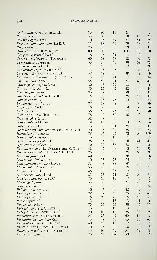
824 BRUNO BAUR ETAL.
Anthoxanthum odoratimi L. s.l.
Bellis perennis L.
Betonica officinalis L.
Brachypodium pinnatum (L.) B.P.
Briza media L.
Bromus erectus Hudson s.str.
Campanula rotundifolia L.
Carex caryophyllea La TourretteCarexfiacca SchreberCentaurea jacea L. s.l.
Centaurium erythraea Rafn * -
Cerastiumfontanum Baumg. s.l.
Chamaespartium sagittale (L.) P. Gibbs
Cirsium acaule Scop.
Crataegus monogyna Jacq..
Cynosurus cristatus L.
Dactylis glomerata L.
Danthonia decumbens (L.) DC.Daucus carota L.
Euphorbia cyparissias L.
Fagus sylvatica L.
Festuca ovina L. s.l.
Festuca pratensis Hudson s.l.
Festuca rubra L. s.l.
Galium album MillerGalium verum L.
Helianthemum nummularium (L.) Miller s.l.
Hieracium pilosella L.
Hippocrepis comosa L.
Hypericum perforatum L.
Hypochoeris radicata L.
Knautia arvensis (L.) Coulter emend. DubyKoeleria pyramidata (Lam.) P.B. s.l * 2
Lathyrus pratensis L.
Leontodon hispidus L. s.l.
Leucanthemum vulgare Lam. s.l.
Unum catharticum L. * -
Lolium perenne L.
Lotus corniculatus L. s.l.
Luzula campestris (L.) DC.Medicago lupulina L.
Ononis repens L.
Phleum pratense L. s.l.
Plantago lanceolata L.
Plantago media L.
Po« compressa L.
Poa pratensis L. s.l.
Polygala amarella CrantzPolygala comosa Schkuhr * 2
Potentilla erecta (L.) RAuschelPotentilla neumanniana RCHB.
Potentilla sterilis (L.) GarckePrimula veris L. emend. Hudson s.l.
Prunella grandiflora (L.) SchollerPrunella vulgaris L.
83 90 13 21 - 3
53 50 8 8 13 22
50 48 67 71 81 78
35 18 42 79 53 75
73 33 58 79 75 81
00 100 100 100 97 100
8 5 21 13 9 13
60 58 58 88 69 78
33 55 46 88 69 75
45 40 83 63 81 78
3 3 8 - 25 31
58 58 29 29 3 9
13 13 21 17 81 94
55 50 71 71 47 41
20 18 29 33 16 16
83 25 42 42 44 44
83 48 50 58 38 47
45 3 46 29 56 44
13 18 75 83 75 72
78 63 8 - 66 59
5 - 8 4 - 6
58 58 75 63 59 50
78 8 50 38 3 -
38 8 4 - - 6
8 13 8 13 16 6
13 8 21 25 41 47
20 23 25 29 25 22
78 75 96 92 94 100
13 5 13 17 31 13
25 35 38 38 41 47
50 38 58 63 44 38
48 45 8 8 56 53
25 13 67 79 63 78
60 50 33 38 9 6
40 25 75 75 6 3
53 43 88 79 19 13
33 20 75 79 75 72
45 8 25 17 16 3
83 73 71 83 94 91
75 65 17 17 3 9
55 30 58 21 28 9
13 8 63 67 72 72
10 5 33 42 9 3
78 58 67 75 59 63
78 80 79 75 69 63
3 - 13 13 41 6
75 55 25 38 25 25
3 5 13 13 9 -
20 10 13 25 38 25
35 23 67 63 16 13
5 8 63 63 63 63
60 68 13 17 22 6
40 28 42 50 6 9
13 10 42 54 69 78
73 65 54 50 28 16
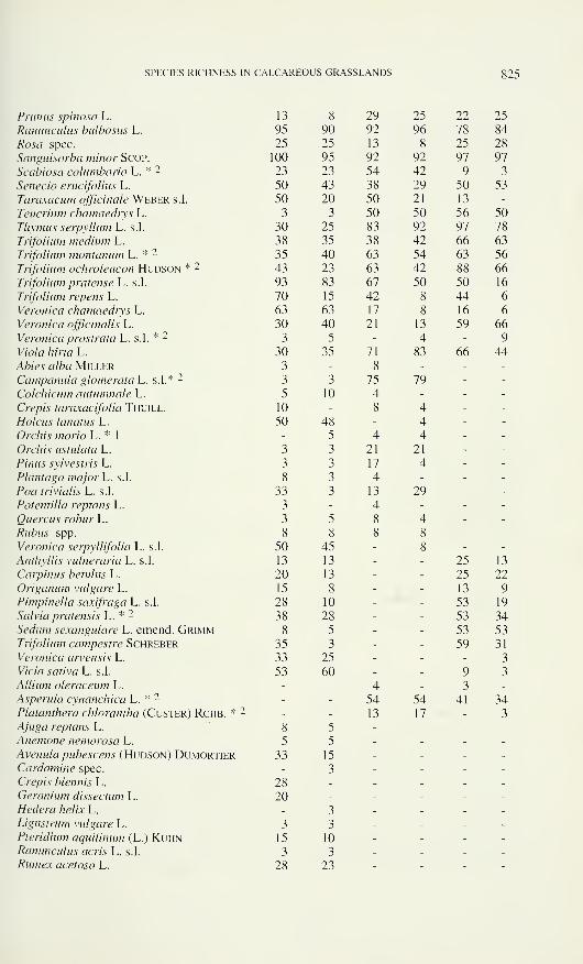
SPECIES RICHNESS IN CALCAREOUS GRASSLANDS 825
Prunus spinosa L. 13 8 29 25 22 25
Ranunculus bulbosus L. 95 90 92 96 78 84
Rosa spec. 25 25 13 8 25 28
Sanguisorba minor Scop. 100 95 92 92 97 97
Scabiosa columbaria L. * 2 23 23 54 42 9 3
Senecio erucifolius L. 50 43 38 29 50 53
Taraxacum officinale Weber s.l. 50 20 50 21 13 -
Teucrium chamaedrys L. 3 3 50 50 56 50
Thymus serpyllum L. s.l. 30 25 83 92 97 78
Trifolium medium L. 38 35 38 42 66 63
Trifolium montanum L. * 2 35 40 63 54 63 56
Trifolium ochroleucon Hudson * 2 43 23 63 42 88 66
Trifolium pratense L. s.l. 93 83 67 50 50 16
Trifolium repens L. 70 15 42 8 44 6
Veronica chamaediys L. 63 63 17 8 16 6
Veronica officinalis L. 30 40 21 13 59 66
Veronica prostrata L. s.l. * 2 3 5 - 4 - 9
V/o/a /î/rto L. 30 35 71 83 66 44
Ae/<?s a/to Miller 3 - 8 - - -
Campanula glomerata L. s.l.* 2 3 3 75 79 - -
Colchicum autumnale L. 5 10 4 - - -
Crépis taraxacifolia Thuill. 10 - 8 4 - -
Hoieus lanatus L. 50 48 - 4 - -
Orchis morio L. * 1- 5 4 4 - -
Orchis ustulata L. 3 3 21 21 - -
Pinus sylvestris L. 3 3 17 4 - -
Plantago major L. s.l. 8 3 4 - - -
Poa trivialis L. s.l. 33 3 13 29 - -
Potentilla reptans L. 3 - 4 - - -
Quercus robur L. 3 5 8 4 - -
Rubus spp. 8 8 8 8 - -
Veronica serpyllifolia L. s.l. 50 45 - 8 - -
Anthyllis vulneraria L. s.l. 13 13 - - 25 13
Carpinus betulus L. 20 13 - - 25 22
Origanum vulgare L. 15 8 - - 13 9
Pimpinella saxifraga L. s.l. 28 10 - - 53 19
Salvia pratensis L. * 2 38 28 - - 53 34Sedum sexangulare L. emend. Grimm 8 5 - - 53 53
Trifolium campestre Schreber 35 3 - - 59 31
Veronica arvensis L. 33 25 - - - 3
Vicia sativa L. s.l. 53 60 - - 9 3
Allium oleraceum L. - - 4 - 3 -
Asperula cynanchica L. * 2 - - 54 54 41 34Platanthera chlorantha (Custer) Rchb. * 2 - - 13 17 - 3
Ajuga reptans L. 8 5 - - - -
Anemone nemorosa L. 5 5 - - - -
Avenula pubescens (Hudson) Dumortier 33 15 - - - -
Cardamine spec. - 3 - - - -
Crépis biennis L. 28 - - - - -
Geranium dissectum L. 20 - - - - -
Hedera helix L. - 3 - - - -
Ligustrum vulgare L. 3 3 - - - -
Pteridium aquilinum (L.) Kuhn 15 10 - - - -
Ranunculus acris L. s.l. 3 3 - - - -
Rumex acetosa L. 28 23 - - - -
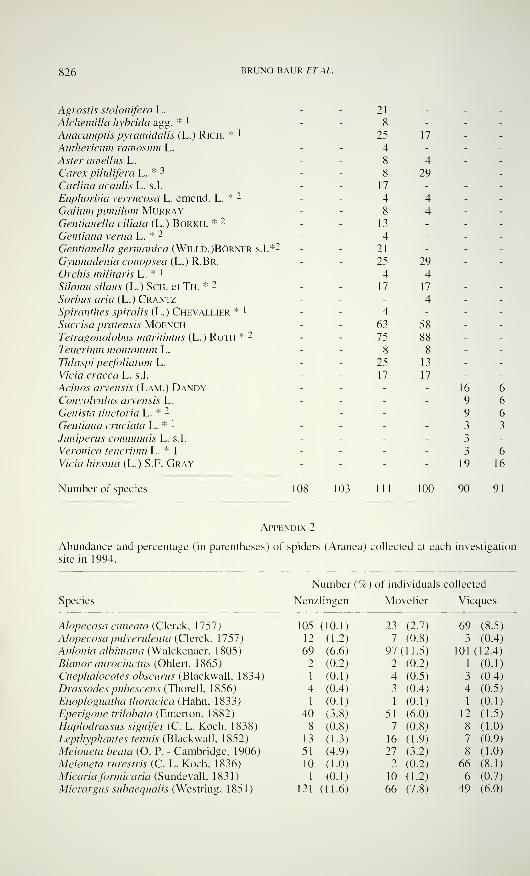
826 BRUNO BAUR ET AL.
Agrostis stolonifera L.
Alchemilla hybrida agg. * '
Anacamptis pyramidalis (L.) Rich. * '
Anthericum ramosum L.
Aster amellus L.
Carex pilulifera L. * 3
Carlina acaulis L. s.l.
Euphorbia verrucosa L. emend. L. * 2
Galium pumilum MurrayGentianella aliata (L.) Borkh. * 2
Gentiana verna L. * 2
Gentianella germanica (Willd.)Börner s.l.*2
Gymnadenia conopsea (L.) R.Br.
Orchis militaris L. * !
Silaum silaus (L.) Sch. et Th. * 2
Sorbus aria (L.) CrantzSpiranthes spiralis (L.) Chevallier * '
Succisa pratensis MOENCHTetragonolobus maritimus (L.) Roth * -
Teucrium montanum L.
Thlaspi peifoliatum L.
Vicia cracca L. s.l.
Acinos ai~vensis (Lam.) DandyConvolvulus arvensis L.
Genista tinctoria L. * 2
Gentiana cruciata L. * 1
Juniperus communis L. s.l.
Veronica teucrium L. * 1
Vicia hirsuta (L.) S.F. Gray
21
25 17 - -
4 - - -
8 4 - -
8 29 - -
17 - - -
4 4 - -
8 4 - -
13 - - -
4 - - -
21 _ _ _
25 29 - -
4 4 - -
17 17 - -
- 4 - -
4 - - -
63 58 - -
75 88 - -
8 8 - -
25 13 - -
17 17 - -
- - 16 6- - 9 6- - 9 6- _ - 3 3
- - 3 -
- - 3 6- - 19 16
Number of species 103 111 100 90 91
Appendix 2
Abundance and percentage (in parentheses) of spiders (Aranea) collected at each investigation
site in 1994.
Species
Number (%) of individuals collected
Nenzlingen Movelier Vieques
Alopecosa cuneata (Clerck, 1757)
Alopecosa pulverulenta (Clerck, 1757)
Antonia albimana (Walckenaer, 1805)
Bianor aurocinctus (Ohlert, 1 865 )
Cnephalocotes obscurus (Blackwall, 1834)
Drassodes pubescens (Thorell, 1856)
Enoplognatha thoracica (Hahn, 1833)
Eperigone trilobata (Emerton, 1882)
Haplodrassus signifer (C. L. Koch, 1838)
Lepthyphantes tenuis (Blackwall, 1852)
Meioneta beata (O. P. - Cambridge. 1906)
Meioneta rurestris (C. L. Koch, 1836)
Micaria formicaria (Sundevall, 1831)
Micrargus subaequalis (Westring, 1851)
05 (10.1) 23 (2.7) 69 (8.5)
12 (1.2) 7 (0.8) 3 (0.4)
69 (6.6) 97(11.5) 101 (12.4)
2 (0.2) 2 (0.2) 1 (0.1)
1 (0.1) 4 (0.5) 3 (0.4)
4 (0.4) 3 (0.4) 4 (0.5)
1 (0.1) 1 (0.1) 1 (0.1)
40 (3.8) 51 (6.0) 12 (1.5)
8 (0.8) 7 (0.8) 8 (1.0)
13 (1.3) 16 (1.9) 7 (0.9)
51 (4.9) 27 (3.2) 8 (1.0)
10 (1.0) 2 (0.2) 66 (8.1)
1 (0.1) 10 (1.2) 6 (0.7)
21 (11.6) 66 (7.8) 49 (6.0)
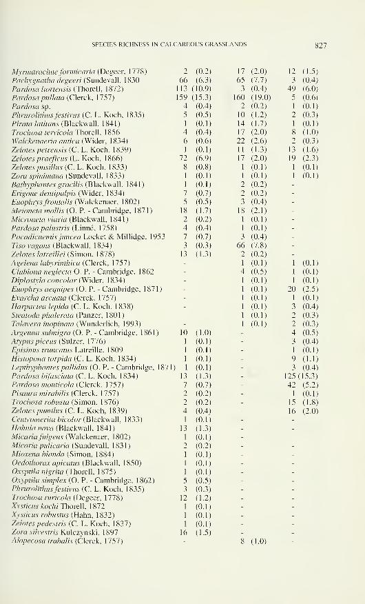
SPECIES RICHNESS IN CALCAREOUS GRASSLANDS 827
Myrmarachneformicaria (Degeer, 1778)
Pachygnatha degeeri (Sundevall, 1830
Pardosa hortensis (Thorell, 1872)
Pardosa pullata (Clerck, 1757)
Pardosa sp.
Phrurolithus festivus (C. L. Koch, 1835)
Pirata latitans (Blackwall, 1841 )
Trochosa terricola Thorell, 1856
Walckenaeria antica (Wider. 1834)
Zelotes petrensis (C. L. Koch, 1839)
Zelotes praeficus (L. Koch, 1866)
Zelotes pusillus (C. L. Koch. 1833)
Zora spinimana (Sundevall, 1833)
Bathyphantes gracilis (Blackwall, 1841 )
Erigone dentipalpis (Wider, 1834)
Euophrys frontalis (Walckenaer, 1802)
Meioneta mollis (O. P. - Cambridge, 1871)
Microneta viaria (Blackwall, 1841)
Pardosa palustris (Linné, 1758)
Pocadicnemis juncea Locket & Millidge, 1953
Tiso vagans (Blackwall, 1834)
Zelotes latreillei (Simon, 1878)
Agelena labyrinthica (Clerck, 1757)
Clubiona neglecta O. P. - Cambridge, 1862
Diplostyla concolor (Wider, 1834)
Euophrys aequipes (O. P. - Cambridge, 1871 )
Evarcha arcuata (Clerck, 1757)
Harpactea lepida (C. L. Koch, 1838)
Steatoda phalerata (Panzer. 1801)
Talavera inopinata (Wunderlich, 1993)
Argenna subnigra (O. P. - Cambridge, 1861 )
Atypus piceus (Sulzer, 1776)
Episinus truncatus Latreille, 1809
Histopona torpida (C. L. Koch, 1834)
Lepthyphantes pallidas (O. P. - Cambridge, 1871
Pardosa bifasciata (C. L. Koch, 1834)
Pardosa monticola (Clerck, 1757)
Pisaura mirabilis (Clerck, 1757)
Trochosa robusta (Simon, 1876)
Zelotes pumilus (C. L. Koch, 1839)
Centromerita bicolor (Blackwall, 1833)
Hahnia nava (Blackwall. 1841)
Micaria fuigens (Walckenaer, 1802)
Micaria pulicaria (Sundevall, 1831)
Mioxena blanda (Simon, 1884)
Oedothorax apicatus (Blackwall, 1 850)
Oxyptila nigrita (Thorell, 1875)
Oxyptila simplex (O. P. - Cambridge, 1 862)Phrurolithus festivus (C. L. Koch, 1835)
Trochosa ruricola (Degeer, 1778)
Xysticus kochi Thorell, 1 872Xysticus robustus (Hahn, 1832)
Zelotes pedestris (C. L. Koch. 1837)
Zora silvestris Kulczynski, 1 897Alopecosa trabalis (Clerck, 1757)
2 (0.2) 17 2.0) 12 (1.5)
66 (6.3) 65 7.7) 3 (0.4)
113 ( 10.9) 3 (0.4) 49 (6.0)
159 ( 15.3) 160 (19.0) 5 (0.6)
4 (0.4) 2 (0.2) 1 (0.1)
5 (0.5) 10 (1.2) 2 (0.3)
1 (0.1) 14 1.7) 1 (0.1)
4 (0.4) 17 2.0) 8 (1.0)
6 (0.6) 22 2.6) 2 (0.3)
1 (0.1) 11 1.3) 13 (1.6)
72 (6.9) 17 2.0) 19 (2.3)
8 (0.8) 1 (0.1) 1 (0.1)
1 (0.1) 1 (0.1) 1 (0.1)
1 (0.1) 2 (0.2) -
7 (0.7) 2 0.2) -
5 (0.5) 3 0.4) -
18 (1.7) 18 (2.1) -
9 (0.2) 1 (0.1) -
4 (0.4) 1 (0.1) -
7 (0.7) 3 (0.4) -
3 (0.3) 66 7.8) -
13 (1.3) 2 0.2) -
-1 (0.1) 1 (0.1)
- 4 0.5) 1 (0.1)
-1 (0.1) 1 (0.1)
-1 0.1) 20 (2.5)
-1 (0.1) 1 (0.1)
- 1 (0.1) 3 (0.4)
- 1 0.1) 2 (0.3)
- 1 (0.1) 2 (0.3)
10 (1.0) - 4 (0.5)
1 (0.1) - 3 (0.4)
1 (0.1) -1 (0.1)
1 (0.1) - 9 (1.1)
1 (0.1) - 3 (0.4)
13 (1.3) - 125(15.3)
7 (0.7) - 42 (5.2)
2 (0.2) - 1 (0.1)
2 (0.2) - 15 (1.8)
4 (0.4) - 16 (2.0)
1 (0.1) - -
13 (1.3) - -
1 (0.1) - -
2 (0.2) - -
1 (0.1) -
1 (0.1) - -
1 (0.1) - -
5 (0.5) - -
3 (0.3) - -
12 (1.2) - -
1 (0.1) - -
1 (0.1) - -
1 (0.1) - -
16 (1.5) - -
8 (1.0)
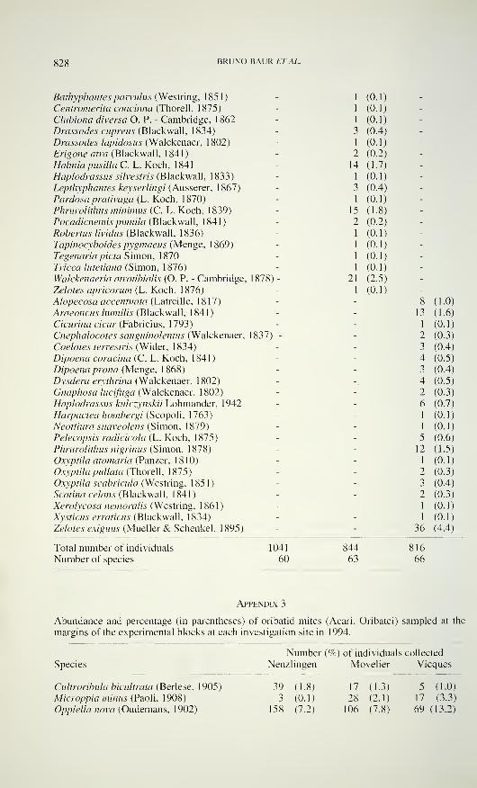
828 BRUNO BAUR ET AL.
Bathyphantes parvulus (Westring, 1 85 1 )
Centromerita concinna (Thorell, 1875)
Clubìona diversa O. P. - Cambridge, 1 862
Drassodes cupreus (Blackwall, 1834)
Drassodes lapidosus (Walckenaer, 1802)
Erigone atra (Blackwall, 1841 )
Halinia pusilla C. L. Koch, 1841
Haplodrassus silvestris (Blackwall, 1833)
Lepthyphantes keyserlingi (Äusserer, 1867)
Pardosa prativaga (L. Koch, 1 870)
Phrurolithus minimus (C. L. Koch, 1839)
Pocadicnemis puntila (Blackwall, 1841)
Robertus lividus (Blackwall, 1836)
Tapinocyboides pygmaeus (Menge, 1869)
Tegenaria pietà Simon, 1870
Tricca lutetiana (Simon, 1876)
Walckenaeria atrotibialis (O. P. - Cambridge, 1878) -
Zelotes apricorum (L. Koch, 1876)
Alopecosa accentuata (Latreille, 1817)
Araeoncus humilis (Blackwall, 1841)
Cicurina cicur (Fabricius, 1793)
Cnephalocotes sanguinolentus (Walckenaer, 1837) -
Coelotes terrestris (Wider, 1 834)
Dipoena coracina (C. L. Koch, 1841)
Dipoena prona (Menge, 1 868)
Dysdera erythrina (Walckenaer, 1802)
Gnaphosa lucifuga (Walckenaer, 1802)
Haplodrassus kulczynskii Lohmander, 1 942
Harpactea hombergi (Scopoli, 1763)
Neottiura suaveolens (Simon, 1879)
Pelecopsis radicicola (L. Koch, 1875)
Phrurolithus nigrinus (Simon, 1878)
Oxyptila atomaria (Panzer, 1810)
Oxyptila pullata (Thorell, 1875)
Oxyptila scabricula (Westring, 1 85 1 )
Scotina Celans (Blackwall, 1841 )
Xerolycosa nemoralis (Westring, 1 86 1 )
Xysticus erraticus (Blackwall, 1834)
Zelotes exiguus (Mueller & Schenkel, 1895)
1 (0.1 -
1 (0.1 -
1 (0.1 -
3 (0.4 -
1 (0.1 -
2 (0.2 -
14 (1.7 -
1 (0.1 -
3 (0.4 -
1 (0.1 -
15 (1.8 -
2 (0.2 -
1 (0.1 -
1 (0.1 -
1 (0.1 -
1 (0.1 -
21 (2.5 -
1 (0.1 -
- 8
- 13
_ 2- 3
- 4- 3
.- 4- 2- 6
1
-1
5
- 12
_
i
2- 3
- 2
1
-1
36
(1.0
(1.6
(0.1
(0.3
(0.4
(0.5
(0.4
(0.5
(0.3
(0.7
(0.1
(0.1
(0.6
(1.5
(0.1
(0.3
(0.4
(0.3
(0.1
(0.1
(4.4
Total number of individuals
Number of species
1041
60
844
63
816
66
Appendix 3
Abundance and percentage (in parentheses) of oribatid mites (Acari, Oribatei) sampled at the
margins of the experimental blocks at each investigation site in 1994.
Species
Number (%) of individuals collected
Nenzlingen Movelier Vieques
Cultroribula bicultrata (Berlese. 1905)
Microppia minus (Paoli, 1908)
Oppiella nova (Oudemans, 1902)
39 (1.8) 17 (1.3) 5 (1.0)
3 (0.1) 28 (2.1) 17 (3.3)
158 (7.2) 106 (7.8) 69 (13.2)
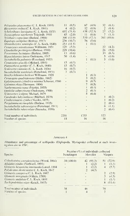
SPECIES RICHNESS IN CALCAREOUS GRASSLANDS 829
Peloptulus phaeonotus (C. L. Koch, 1844)
Rhysotritia ardua (C. L. Koch, 1841)
Scheloribates laevigatas (C. L. Koch, 1835)
Tectocepheus sarekensis Trägärdh, 1910
Xylobates capucinus (Berlese, 1908)
Eupelops curtipilus (Berlese, 1917)
Mimmtiwzetes semirufus (C. L. Koch, 1840)
Ceratozetes mimitissimus Willmann, 195
1
Ctenobelba pectinigera (Berlese, 1910)
Fosseremus laciniatus (Berlese, 1905)
Hypochthonius luteus Oudemans, 1917
Suctobelbella palustris (Forsslund, 1953)
Ceratozetes gracilis (Michael, 1884)
Platynothrus peltifer (C. L. Koch, 1840)
Scutovertex minutus (C. L. Koch, 1836)
Suctobelbella sarekensis (Forsslund, 1941)
Brachychthonius berlesei Willmann. 1928
Ceratoppia quadridentata (Haller, 1882)
Epilohmannia cylindrica minima Schuster, 1960
Galumna alata (Hermann, 1804)
Nanhermannia nana (Nicolet, 1855)
Oppiella subpectinata (Oudemans, 1900)
Scutovertex sculptus Michael, 1879
Carabodes labyrinthicus (Michael, 1879)
Cymberemaeus cymba (Nicolet. 1855)
Pergalumna myrmophila (Berlese, 1915)
Suctobelbella subcornigera (Forsslund, 1941 )
Suctobelbella tuberculata (Strenzke, 1950)
1 1 (0.5) 67 (4.9) 32 (6.1)
4 (0.2) 12 (0.9) 2 (0.4)
692 (31.4) 438 (32.3) 27 (5.2)
45 (2.0) 11 (0.8) 7 (1.3)
306 (13.9) 570 (42.1) 261 (49.9)
236 (10.7) 76 (5.6) -
253 (11.5) 1 (0.1) -
129 (5.9) - 22 (4.2)
229 (10.4) - 26 (5.0)
13 (0.6) - 33 (6.3)
30 (1.4) - 5 (1.0)
-1 (0.1) 5 (1.0)
15 (0.7) - -
15 (0.7) - -
16 (0.7) - -
7 (0.3) - -
-1 (0.1) -
- 3 (0.2) -
- 9 (0.7) -
- 7 (0.5) -
-1 (0.1) -
- 2 (0.1) -
- 5 (0.4) -
- -1 (0.2)
- -1 (0.2)
- - 2 (0.4)- - 6 (1.1)
- - 2 (0.4)
Total number of individuals
Number of species
2201
18
1355
18
523
18
Appendix 4
Abundance and percentage of millipedes (Diplopoda, Myriapoda) collected at each inves-
tigation site in 1994.
Species
Number (%) of individuals collected
Nenzlingen Movelier Vieques
Cylindroiulus caeruleocinctus (Wood, 1864)
Allajulus nitidus (Verhoeff, 1 89 1 )
Glomeris hexasticha intermedia Latzel, 1 884Polydesmus denticulatus C. L. Koch, 1847
Glomeris conspersa C. L. Koch, 1847
Glomeris marginata (Villers, 1789)
Glomeris undulata C. L. Koch, 1844Tachypodoiulus niger (Leach, 1815)
38(100.0) 42 (91.3) 51 (72.9)-
1 (2.2) 1 (1.4)
-1 (2.2) 4 (5.7)
- 2 (4.3) -
- - 2 (2.9)
- - 3 (4.3)
- -1 (1.4)
- - 8 (11.4)
38 46 70
1 4 7
Total number of individuals
Number of species
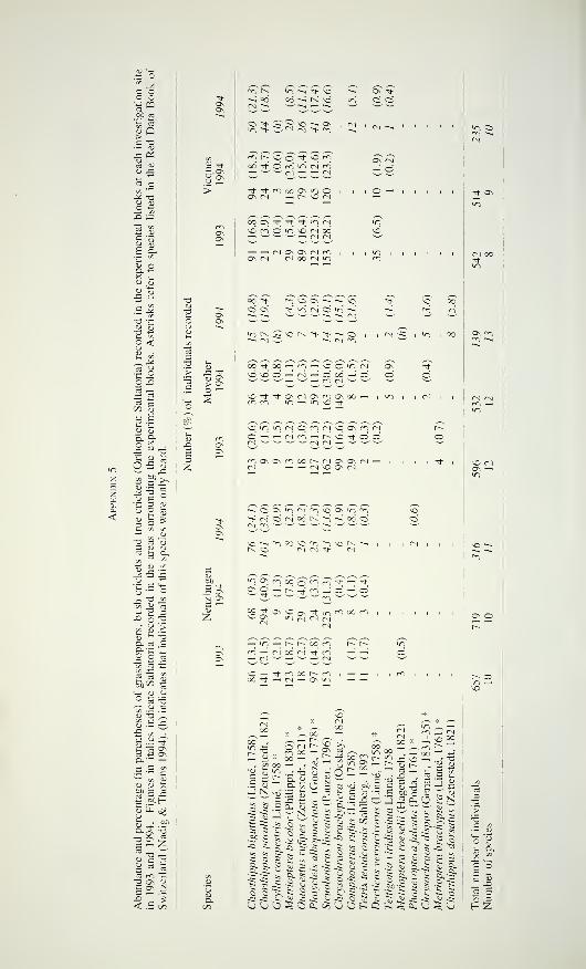
.2 °
C Oo o3«.SP 3<u Q
.e PÜ
03 e ü sco
'*" >x -a« S
E &
° <
~R "o — "^ so
oo ~-ì K vd
- "^ <N M- rr,
co t~~ so o "* so m
co OS M1 ^ ^ Vi N
00 "Sh <"n O OS ~-i ~-i
Cs Os \h >0 M O ^
'« K > >
Os \t-
O C5
Os CN—' Ö
> CTs
CO 't co — ci « \o o m PI
ni .3Ih u-
o-s s
CO M -Ss
4) •£= 1=
u e u'G 3 u
E 2
»CVUTiNOWNCOnN
-sQOsViiMfiiVoOsVifr)<j (si O ci oó (< fi ~i co ci
5 72
2 rS I
OJ "O
O e
a
,5 £I?IS
^-'co <U
u y o
OOn|e — Ä53 —
I
"
£ " Jetì co *G-a os <u
C OS ä3 — -<
"ÎOs^oog^^Tt-'*2- § 3 CS ^ li -; 2c2
^S; "^ CN ^ CN
— io — r- r- oo m
co ..
£^•— Sì
4j.
o CSco oo0O -H
a"«
.5 S
coIOf- Cu cu
D. 60
13
J n viU-
OO
i2 S e~~ ~?~
t-4
S S &
.5.5: us ; io
Ë2B _ cu
:_-' OO ,
—
i
5 '-> — io °° —-^ oo CO (-. io --
yQ
y — "MU * XI .
S31« Me e m ,3
> ,£ X) ,—' 1
^ yO oo
— N S S
_ft 6o 5 QO S? s Sä
ft. ? -Si ft
•2 S o -ftïodS
JS 6, - ^ " w S^ÌT
j I) co
C Sc-k -ê k*-Si te -S «u J~ ft 'S
riicivoridissi roeseh
a
falca
n
dispbrachy
dorsal
O tui- O.
Decticus
ver
Tettigonia
vi
MetriopteraPhaneroptet
Chrysochrac
MetriopteraChorthippus
CU co
•s «-E oa bi
c «_: x3 E
H Z
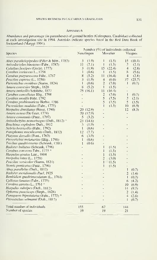
SPECIES RICHNESS IN CALCAREOUS GRASSLANDS 831
Appendix 6
Abundance and percentage (in parentheses) of ground beetles (Coleoptera, Carabidae) collected
at each investigation site in 1994. Asterisks indicate species listed in the Red Data Book of
Switzerland (Marggi 1994).
Species
Number (%) of individuals collected
Nenzlingen Movelier Vieques
Abax parallelepipedus (Piller & Mitt., 1783)
Anisodactylus binotatus (Fabr., 1787)
Calathus fuscipes (Goeze, 1777)
Carabus coriaceus L., 1758
Carabus purpurascens Fabr., 1787
Poecilus cupreus (L., 1758)
Pterostichus ovoideus (Sturm, 1824)
Amara convexior Steph., 1828
Amara lunicollis Schiödte, 1837
Carabus cancellatus Illig., 1798
Carabus monil is Fabr. 1792
Carabus problematìcus Herbst, 1786
Pterostichus madidus (Fabr.. 1775)
Harpalus dimidiatus (Rossi. 1790)
Amara aenea (De Geer, 1 774)
Amara communis (Panz., 1797)
Anisodactylus nemorivagus (Duft., 1812) *
Brachinus explodens Duft., 1812
Nebria brevicollis (Fabr., 1792)
Parophonus maculicornis (Duft., 1812)
Platynus dorsalis (Pont., 1763)
Pterostichus melanarius (Illig., 1798)
Trechus quadristriatus (Schrank, 1781)
Badister bullatus (Schrank, 1798)
Carabus convexus Fabr., 1775 *
Harpalus atratus Latr., 1804
Harpalus latus (L., 1758)
Poecilus versicolor (Sturm, 1 824)
Stomis pumicatus (Panz., 1796)
Abax parallelus (Duft., 1812)
Badister meridionalis Puel, 1925
Bembidion quadrimaculatum (L., 1761)
Callistus lunatus (Fabr., 1775)
Carabus auratus L., 1761 *
Harpalus rubripes (Duft., 1812)
Ophonus puncticeps (Steph., 1828)
Panagaeus bipustulatus (Fabr., 1775) *
Pterostichus selmanni (Duft., 1812)
3 (1.9) 1 (1-5) 15 (10.4)
11 (7.1) 1 (1.5) 3 (2.1)
3 (1.9) 15 (22.4) 4 (2.8)
1 (0.6) 1 (1.5) 8 (5.6)
8 (5.2) 11 (16.4) 4 (2.8)
3 (1.9) 4 (6.0) 37 (25.7)
1 (0.6) 2 (3.0) 1 (0.7)
8 (5.2) 1 (1.5) -
25 (16.1) 11 (16.4) -
- 2 (3.0) 1 (0.7)
- 5 (7.5) 3 (2.1)
- 5 (7.5) 5 (3.5)-
1 (1.5) 10 (6.9)
20 (12.9) - 12 (8.3)
20 (12.9) - -
5 (3.2) - -
23 (14.8) - -
3 (1.9) - -
1 (0.6) - -
12 (7.7) - -
6 (3.9) - -
1 (0.6) - -
1 (0.6) - -
-1 (1.5) -
-1 (1.5) -
-1 (1.5) -
- 2 (3.0) -
-1 (1.5) -
-1 (1.5) -
- -1 (0.7)
- - 2 (1.4)- -
1 (0.7)- - 6 (4.2)- - 10 (6.9)- - 14 (9.7)- - 2 (1.4)- - 4 (2.8)- -
1 (0.7)
Total number of individuals
Number of species
155
19
67
19
144
21
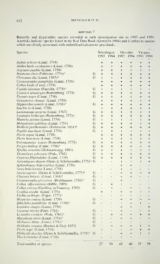
832 BRUNO BAUR ETAL.
Appendix 7
Butterfly and Zygaenidae species recorded at each investigation site in 1993 and 1994.
Asterisks indicate species listed in the Red Data Book (Gonseth 1994) and G refers to species
which are closely associated with unfertilized calcareous grasslands.
Species Nenzlingen Movelier Vieques
1993 1994 1993 1994 1993 1994
Aglais urticae (Linné, 1758)
Anthocharìs cardamines (Linné. 1758)
Argynnispaphia (Linné. 1758)
Brintesia circe (Fabricius. 1775)*
Clossiana dia (Linné, 1767)*
Coenonympha pamphiliis (Linné, 1758)
Colias hyale (Linné, 1758)
Cupido minimus (Fuesslin, 1775)*
Cyaniris semiargus (Rottemburg. 1775)
Erynnis tages (Linné, 1758)
Gonepteryx rhamni (Linné, 1758)
Hipparchia semele (Linné, 1758)*
Inachis io (Linné, 1758)
Lasiommata megera (Linné, 1767)
Lysandra bellargus (Rottemburg. 1775)
Maniolajurtina (Linné, 1758)
Melanargia galathea (Linné. 1758)
Mellicta parthenoides (Keferstein, 1851)*
Papilio machaon (Linné. 1758)
Pieris rapae (Linné, 1758)
Pieris brassicae (Linné, 1758)
Polyommatus icarus (Rottemburg, 1775)
Pyrgus malvae (Linné, 1758)*
Spialia sertorius (Hofmannsegg. 1804)
Thymelicus sylvestris (Poda, 1761)
Zygaena filipendulae (Linné. 1758)
Agriodiaetus damon (Denis & Schiffermüller, \115YApìianthopus hyperanthus (Linné, 1758)
Araschnia levana (Linné, 1758)
Arida agesus (Denis & Schiffermüller. 1775)*
Chazara briseis (Linné, 1764)*
Coenonympha glycerion (Borkhausen. 1788)*
Colias alfacariensis (Ribbe, 1905)
Colias crocea (Geoffroy in Fourcroy, 1785)
Cynthia cardili (Linné. 1758)
Erebia aethiops (Esper, Mil)*Hesperia comma (Linné, 1758)
Iphiclides podalirius (Linné, 1758)*
Leptidea sinapis (Linné. 1758)
Lycaena tityrus (Poda. 1761 )
Lysandra coridon (Poda, 1761)*
Maculinea arion (Linné, 1758)*
Melitaea cinxia (Linné, 1758)*
Ochlodes venatus (Bremer & Grey 1853)
Pieris napi (Linné, 1758)
Plebicula doiyias (Denis & Schiffermüller, 1775)*
Thecla betulae (Linné, 1758)
+ + + + + ++ + + + + ++ + + + + +
G + + + + + +G + + + + + +
+ + + + + ++ + + + + +
G + + + + + +G + + + + + +G + + + + + +
+ + + + + +G + + + + + +
+ + + + + +G + + + + + +G + + + + + +G + + + + + +G + + + + + +G + + + + + +G + + + + + +
+ + + + + +
+ + + + + +G + + + + + +
G + + + + + +
G + + + + + +
G + + + + + +
G + + + + + +
G - - + + + +- - + + - -
- - + + - -
G - - + + + +G - - + + - -
G - - + - - -
G - + + + - +
G - + - + - +
+ - + + + +- - + + - -
G - - + + + +G - - + + + -
- - + + - +
G - - + + - +
G - + + + + +
G - - + + - +
G - - - + - -
G - + + + + ++ - + + + +
G - - + + + +- - + + - -
Total number of species 27 30 45 46 35 39
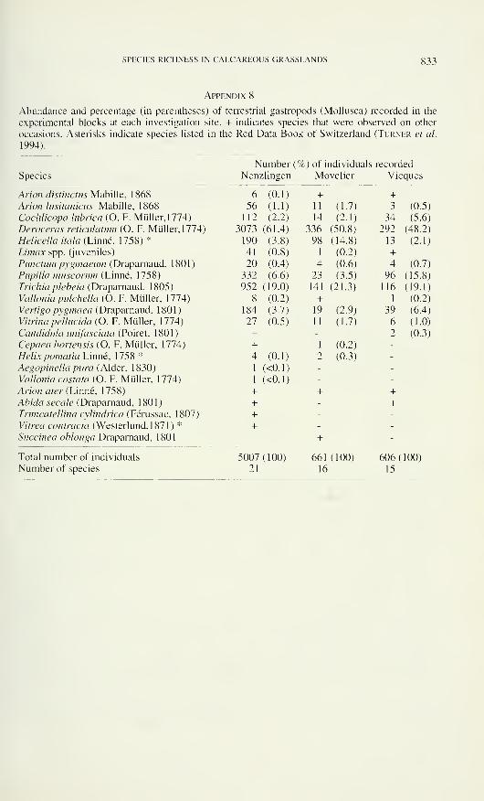
SPECIES RICHNESS IN CALCAREOUS GRASSLANDS g33
Appendix 8
Abundance and percentage (in parentheses) of terrestrial gastropods (Mollusca) recorded in the
experimental blocks at each investigation site. + indicates species that were observed on other
occasions. Asterisks indicate species listed in the Red Data Book of Switzerland (Turner et al.
1994).
Number (%) of individuals recorded
Species Nenzlingen Movelier Vieques
Arion distinctus Mabille, 1868 6 (0.1) + +Arion lusitanicus Mabille, 1868 56 (1.1) 11 (1.7) 3 (0.5)
Cochlicopa lubrica (O. F.Müller, 1774) 112 (2.2) 14 (2.1) 34 (5.6)
Dewceras reticulatmn (O. F. Müller, 1774) 3073 (61.4) 336 (50.8) 292 (48.2)
Helicella itala (Linné, 1758)* 190 (3.8) 98 (14.8) 13 (2.1)
Umax spp. (juveniles) 41 (0.8) 1 (0.2) +Punctum pvgmaeum (Drapammd, 1801) 20 (0.4) 4 (0-6) 4 (0.7)
Pupilla muscorum (Linné, 1758) 332 (6.6) 23 (3.5) 96 (15.8)
Trichia plebeia (Draparnaud, 1805) 952(19.0) 141(21.3) 116 (19.1)
Valloniapulchella(0. F. Müller, 1774) 8 (0.2) + 1 (0.2)
Vertigo pygmaea (Draparnaud, 1801) 184 (3.7) 19 (2.9) 39 (6.4)
Vitrina pellucida (O. F.Müller, 1774) 27 (0.5) 11 (1.7) 6 (1.0)
Candidulaimifasciata(Poiret,\$Oì) + - 2 (0.3)
CepaeahortensisiO. F. Müller, 1774) + 1 (0.2)
Helix pomatia Linné, 1758 * 4 (0.1) 2 (0.3)
Aegopinella pura (Alder, 1830) 1 (<0. 1)
Vallonia costata (O. F. Müller. 1774) 1 (<0.1 )
Arion ater (Linné, 1758) + + +Abida secale (Draparnaud, 1 80 1 ) + - +Truncatellina cilindrica (Férussac, 1807) + -
Vitrea contracta (Westerlund,l 871) * + -
Succinea oblonga Draparnaud, 1801 +
Total number of individuals 5007(100) 661(100) 606(100)Number of species 21 16 15

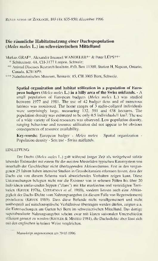
Revue suisse de Zoologie, 103 (4): 835-850; décembre 1996
Die räumliche Habitatnutzung einer Dachspopulation
{Mêles mêles L.) im schweizerischen Mittelland
Markus GRAF*. Alexander Imanuel WANDELER** & Peter LÜPS**** Schützenstr. 4A, CH-3177 Laupen, Schweiz;
** Animal Diseases Research Institute, P.O. Box 1 1300, Station H, Nepean, Ontario,
Canada, K2H 8P9;
*** Naturhistorisches Museum, Bernastr. 15, CH-3005 Bern, Schweiz.
Spatial organization and habitat utilization in a population of Euro-
pean badgers (Mêles mêles L.) in a hilly area of the Swiss midlands. - Asmall population of European badgers {Meles mêles L.) was studied
between 1977 and 1981. The use of 42 badger dens and of numerous
latrines was monitored. The home ranges of 3 radio-collared individuals
were surprisingly large, measuring 132, 591 and 438 hectares. The
population density was estimated to be only 0.5 individuals/1 km2. The use
of a wide variety of food resources was observed. Low population density,
ranging behaviour and resource utilization did not appear to be obvious
consequences of resource availability.
Key-words: European badger - Mêles meles - Spatial organization -
Population density - Sett use - Swiss midlands.
EINLEITUNG
Der Dachs {Mêles meles L.) galt während langer Zeit als weitgehend solitär
lebender Erdmarder mit einem für die meisten Musteliden typischen Raumsystem von
innerhalb der Geschlechter nicht überlappenden Aktionsräumen. Erst in den vergan-
genen 25 Jahren haben intensive Studien in Grossbritannien erkennen lassen, dass der
Dachs ein von diesem Schema stark abweichendes Verhalten zeigen kann. Diese
Untersuchungen belegten nicht nur die Existenz von in seltenen Fällen bis über 20
Individuen umfassenden Sippen ("clans") mit klar markierten und verteidigten Terri-
torien (Kruuk 1978a, Cheeseman et al. 1988), sondern liessen auch eine Abhän-
gigkeit der Dachs-Dichte vom Nahrungsangebot (in diesem Falle von Regenwürmern)
postulieren (Kruuk 1989). Dass diese Befunde nicht verallgemeinert und nicht
unüberprüft auf mitteleuropäische Verhältnisse übertragen werden dürfen, zeigten u.a.
die Untersuchungen am Gurten bei Bern im schweizerischen Mittelland. Das dortige
superabundante Nahrungsangebot scheint zwar mit klaren saisonalen Unterschieden
effizient genutzt zu werden (Kistler & Misteli 1984), die Dachsdichte aber lässt sich
mit den englischen in keiner Weise vergleichen.
Manuskript angenommen am 29.03.1996.
836 MARKUS GRAF, ALEXANDER IMANUEL WANDELER & PETER LÜPS
Es war das Ziel der vorliegenden Untersuchung, Einblick in das Raum-Zeit-
System der Dachse am Gurten zu gewinnen, unter besonderer Berücksichtigung der
Nutzung der Ressourcen Nahrung und Baue. Auf diesem Wege sollten die Unter-
schiede zu den Verhältnissen in Grossbritannien definiert und Erklärungen für deren
Bestehen herausgearbeitet werden.
DAS UNTERSUCHUNGSGEBIET
Dieses grenzt unmittelbar an den südlichen Siedlungsrand der Stadt Bern und
liegt in der Hügelzone. Das Gelände steigt von 508 m ü.M. am Aare-Ufer bis zur
höchsten Erhebung, dem Gurten, auf 937 m ü.M. Die Fläche des Untersuchungs-
gebietes beträgt etwa 10 km2 (Abb. 1 ). Im ganzen Perimeter befinden sich nur weiche
:*#i
;* •
•- v:>vt:;>:>:'/ >>:M:: 508 "•
-".'#• • «858 ''••":*•%•''
::.:': :
-
•# •• • .*
,*• ••• m1? •• '••:::
:-V:; .
: • • m: • 4v• • «'••
'•••••••••••# #
»»#
937
+600/194
Fig. 1
Lage und Grösse des Untersuchungsgebietes sowie Lage und Verteilung der 42 Dachsbaue.
Punktlinie: Untersuchungsperimeter; heller Raster: Wald; dunkler Raster: Siedlung; grosse
Punkte: Baue. Situation of the study area and distribution of setts (dots). Dark shading: settle-
ments; light shading: forest.
RÄUMLICHE HABITATNUTZUNG EINER DACHSPOPULATION g37
geologische Formationen, vorwiegend Molasse, Moräne und Schotter. Die Beschaf-
fenheit des Bodens bietet somit den Dachsen günstige Voraussetzungen für das
Graben ausgedehnter Höhlensysteme.
Die höheren Lagen waren während der Untersuchungszeit im Winter länger als
einen Monat mit Schnee bedeckt. Die Hälfte des Untersuchungsgebietes ist bewaldet,
hauptsächlich Laubmischwald mit Fichten {Picea abies) und Buchen {Fagus silva-
tica), das übrige Gebiet wird als Wiese, Weide- und Ackerland (v. a. Weizen, Mais
und Kartoffel) genutzt (Hegg 1973, Kistler & Misteli 1984).
METHODEN
Baue, Latrinen
Baue und Latrinen der Dachse wurden seit 1977 durch regelmässiges Ab-
schreiten des Geländes gesucht und auf ihre Benützung hin mit über die Eingänge
gelegten gekreuzten Zweigen und an Hand von Trittsiegeln in Sand, Lehm und
Schnee vor den Eingangsröhren kontrolliert. Kotgruben auf landwirtschaftlich
intensiv genutzen Flächen wurden wegen der eingeschränkten Begehbarkeit und als
Folge ihrer Kurzlebigkeit wohl vielfach nicht erfasst.
Direkte und indirekte Beobachtung der Dachse
Von 1977-80 wurden die nachtaktiven Dachse mit Hilfe eines Infrarot-Nacht-
sichtgerätes hauptsächlich am Bau beobachtet. Spuren der Nahrungssuche und
Trittsiegel ergaben weitere wertvolle Hinweise über die Raumnutzung der Dachse.
1980 und 1981 konnten am Gurten zwei 9 kg schwere Dachsfähen und ein 13
kg schwerer Rüde mit Schlingen gefangen, mit Radiohalsbändern des Typs 294178 B,
148 MHz, entwickelt von D. Burchard (vgl. auch Taylor & Lloyd 1978, Amlaner& Macdonald 1979, Cederlund et al. 1979, Graf 1988), ausgerüstet und ihre
Aktivität erfasst werden. Fähe Nr 1 liess sich während 7 Monaten, Fähe Nr 2 während
8,5 Monaten und der Rüde lediglich während 1,5 Monaten beobachten. Angaben zu
Fangdatum, Anzahl Sendetage, Peilungen und effektiver Beobachtungsdauer sind
Tab. 1 zu entnehmen. Durch Direktbeobachtung, ergänzt mit systematisch durch-
geführter Überwachung der Baue, konnte zudem die Populationsgrösse von 1977-81
erfasst werden.
Berechnung der Aktionsraumgrösse und der Benutzungsverteilung
Die Bestimmung der Aktionsraumgrösse erfolgte nach der Gitterzellen-
methode (Riedwyl & Schüpbach 1983) sowie der leicht abgeänderten "Minimumarea method" (Voigt & Tinline 1980). Bei der Gitterzellenmethode haben wir zur
Darstellung absoluter Häufigkeiten auf das Untersuchungsgebiet ein quadratisches
Gitternetz, bestehend aus Einzelflächen von 1 ha Grösse, gelegt. Die absolute
Häufigkeit wird in die Maschenweite des Netzes abgebildet. Die Häufigkeiten lassen
sich durch Auszählen oder Messen der Maschenweite vergleichen. Diese Methode
838 MARKUS GRAF, ALEXANDER IMANUEL WANDELER & PETER LÜPS
wurde gewählt, weil die radiotelemetrischen Daten über die Raumnutzung wegen der
geringen Individuenzahl statistisch nicht ausgewertet werden können und auch nicht
als unabhängige Stichproben betrachtet werden dürfen.
Unter Aktionsraum wird nachfolgend die Summe aller Gitterzellen verstanden,
in denen ein radiomarkierter Dachs in den Jahren 1980-81 mindestens einmal
beobachtet wurde. Mit dieser Darstellung lassen sich die Aktionsraumgrössen durch
Addition der begangenen Flächen, die Schwerpunkte bezüglich Aufenthaltshäufigkeit
und die Aufenthaltsdauer pro Flächeneinheit eines Tieres, aber auch die Werte der
unterschiedlich lang und nicht zur selben Zeit beobachteten Dachse untereinander
vergleichen.
Bei der abgeänderten "Minimum area method" wurde die flächenmässig
kleinstmögliche Abgrenzung durch die Verbindung äusserster Beobachtungspunkte
vorgenommen und zugleich eine nach ökologischen Kriterien einschränkende Grenz-
linie gewählt, indem von markierten Dachsen auch nur teilweise begangene Vegeta-
tionseinheiten als ganze Flächeneinheit dem Aktionsraum beigefügt wurden. Aller-
dings wurden bei Wäldern, die vielfach grosse Ausdehnungen aufweisen, nur die
wirklich durch Dachse begangenen Flächen als Aktionsraum miteinbezogen. Hin-
gegen wurden durch Dachse nicht nutzbare Flächen, wie z.B. dicht überbautes Gebiet,
nicht in die Flächenberechnungen miteinbezogen. In den vorliegenden Ergebnissen
werden dadurch im Vergleich zu den Flächenberechnungen nach der eigentlichen
Minimum area Methode wesentlich geringere Flächen nachgewiesen.
Pro Nacht genutzte Fläche
Für die Dachse Nr 1 und Nr 2 wurden die zurückgelegten Wege und die für die
Nahrungssuche genutzten Flächen so genau als möglich auf Karten (Massstab
FIO'OOO) eingezeichnet und ausgemessen. Somit konnte die insbesondere für die
Nahrungssuche benötigte Fläche geschätzt und mit der gesamten Aktionsraumgrösse
sowie dem verfügbaren Nahrungsangebot pro Fläche in Beziehung gebracht werden
(vgl. auch Kistler & Misteli 1984).
RESULTATE
Die Aktionsraumorganisation
Lage und Verteilung der Baue
Im 10 km2 grossen Untersuchungsgebiet wurden insgesamt 42 Baue gefunden,
verteilt über die ganze Fläche (Abb. 1). Ein Bau befindet sich in einer schmalen
Hecke, alle übrigen im Wald oder am Waldrand. Von diesen liegen drei Baue
ausserhalb der Aktionsräume der radiomarkierten Dachse.
Die Distanz zweier benachbarter Baue beträgt minimal 10 bis maximal 850 m,
durchschnittlich 215 m.
RÄUMLICHE HABITATNUTZUNG EINER DACHSPOPULATION 839
Charakterisierung der Baue
Baugrösse:
Von den 42 Bauen weisen 31 nur ein bis drei, 10 mehr als sechs und ein Bau
28 Eingangsröhren auf.
Exposition:
Die Baue sind mehrheitlich nach Nordosten und Südwesten ausgerichtet. Diese
Exposition wird weitgehend durch den Verlauf des Gurtens und des Ulmizberges von
NW nach SE bestimmt.
Hangneigung:
Für 30 Baue weist der Hang Steigungen zwischen 16 und 35% auf, für 4 mehr
als 16% und für 8 mehr als 35%. Sehr flache und sehr steile Hänge werden gemieden.
Sichtdeckung, Dickichte in Baunähe:
Bei 29 Bauen fehlte während der Untersuchungszeit eine Deckung bietende
Kraut- oder Strauchschicht. Bei 23 Bauen befanden sich in unmittelbarer Nähe
Dickichte, welche den Dachsen als bevorzugte Latrinenstandorte dienen konnten.
Distanz zu Waldrand:
Die Entfernungen der sich im Wald befindlichen Baue zum nächsten Waldrand
variieren zwischen 1 und 205, durchschnittlich 65 m. Für 33 Baue betragen die
Strecken weniger als 100 m, für 24 sogar weniger als 50 m.
Tabelle 1
Angaben zur Beobachtungsdauer der drei radiomarkierten Dachse. Observation times for the
three radio-collared badgers.
Dachse Fangdatum Letzte Sendetage
Lokalisation
Anzahl
Peilungen
Tag Nacht
Anzahl
Peilungen
ganze Nacht
Effektive
Beobachtungs-
dauer
1: ? 11.09.1980 20.03.1981 191 104 56
2: 9 22.04.1981 12.12.1981 235 140 67
3: â 19.06.1981 31.07.1981 43 13 5
125h 30'
418h 45*
56h 15*
Lage, Verteilung und Grösse der durch Dachse benützten Baue
Die 42 erfassten Baue wurden während der Jahre 1977-81 regelmässig be-
züglich Benützungshäufigkeit durch Dachse bzw. Füchse kontrolliert. 1980-81
konnte zudem dank der markierten Dachse ihr jeweiliger Übertagungsbau lokalisiert
werden. Die Kontrolle der Baue ergab:
- Vier der 42 Baue wurden nie nachweislich von einem Dachs befahren.
- Insgesamt 35 der 38 befahrenen Baue befinden sich innerhalb der Aktions-
räume mindestens eines der drei markierten Dachse.
Für Tier Nr 1 sind es 24, für Nr 2: 34 und für Nr 3: 28 Baue. Von den 35
Bauen haben die radiomarkierten Dachse jedoch nur 15 verschiedene Baue nach-
weislich benützt (Dachs Nr 1: 7, Nr 2: 12 und Nr 3: 7 Baue; Tab. 2).
840 MARKUS GRAF, ALEXANDER IMANUEL WANDELER & PETER LÜPS
Tabelle 2
Anzahl Übertagungen in 15 verschiedenen Bauen durch die radiomarkierten Dachse 1980/81.
Fähe Nr 1: 104 Übertagungen, Fähe Nr 2: 139 Übertagungen, Rüde Nr 3: 21 Übertagungen.
Days spent in 15 setts by radio-collared badgers in 1980/81. Female no 1: 104 days, female no
2: 139 days, male no 3: 21 days.
Datum Dachse Bau-Nr Total
1980/81 Nr 1 2 3 4 5 6 7
Sept.
Okt.
3
9
4
4 3
1
3
Nov. 15 7
Dez. 13
Jan. 12
Febr. 11
März 2 13
Apr.
Mai2
2
1
1
5
10 2
1
1
1
2
Juni 2
3
3 4
1
1
2
3
1
1 1
Juli 2
3
2
3
3
1
5
9
5 1
1
Aug. 2 5 5 5 1 3
Sept.
Okt.
2
2 3
7
9
2
3
1
Nov. 2 9 5 3 4
Dez. 2 11
10 11 12 13 14 15
1 9
21
22
13
12
11
16
9
18
19
5
17
16
19
10
15
21
11
Total 92 71 29 28 12 11 4 5 2 1 2 264
- Grosse Baue und solche, die sich weniger als 50 m innerhalb des Wald-
randes befinden, wurden von den radiomarkierten Dachsen bevorzugt aufgesucht.
- Nur in drei von 24 möglichen Fällen (zwei Dachse gleichzeitig mit Sendern
ausgerüstet) verbrachten zwei markierte Dachse miteinander den Tag in demselben Bau.
- Die Baue wurden nicht dauernd bewohnt. Nur für die Winterruhe (Baue Nr
1, 2, 4, 9) oder für die Welpenaufzucht (Bau Nr 2) wurde derselbe Bau während mehr
als 6 aufeinanderfol senden Tagen benützt!
Lage und Verteilung der Latrinen
Obwohl sicher nur ein Teil der benützten Latrinen erfasst werden konnte
(vergi. "Methode"), liessen sich deren 62, bestehend aus einer oder mehreren Kot-
gruben (bis 30 Kotgruben pro Latrine) verteilt über das ganze Untersuchungsgebiet,
finden (Abb. 2). In unmittelbarer Nähe von benützten Bauen treten sie gehäuft auf.
Die meisten der 62 Latrinen befinden sich im Sichtschutz von Fichtenaufforstungen
(32%), in Dickichten (21%) oder in unmittelbarer Baunähe (Distanz < 20 m ohne
Sichtdeckung; 21%). Die restlichen befinden sich im Wald oder am Waldrand in
ungedeckter Umgebung (26%). Kistler & Misteli (1984) haben durch systema-
tisches Suchen weitere Latrinen, auch ausserhalb des Waldes, gefunden.
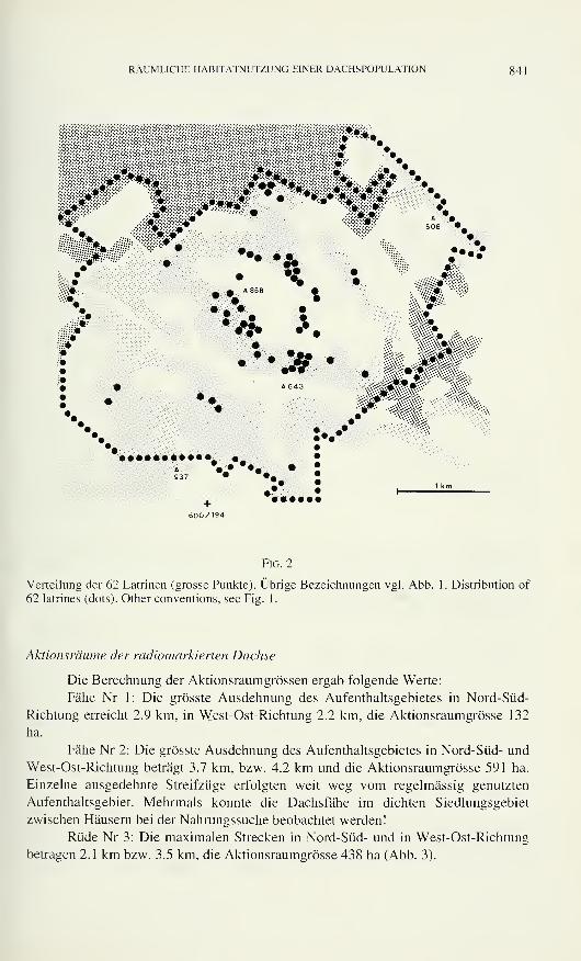
RÄUMLICHE HABITATNUTZUNG EINER DACHSPOPULATION 841
il$*I
m
mmmmmmm
/>*<
IP*»
•
:
:::---::x*:'
V «JL
•*
^ .* *.
••:»
508 :•
.... *%:::-.
•m«*
»••••••••• •„
+600/194
Fig. 2
Verteilung der 62 Latrinen (grosse Punkte). Übrige Bezeichnungen vgl. Abb. 1. Distribution of
62 latrines (dots). Other conventions, see Fig. 1.
Aktionsräume der radiomarkierten Dachse
Die Berechnung der Aktionsraumgrössen ergab folgende Werte:
Fähe Nr 1: Die grösste Ausdehnung des Aufenthaltsgebietes in Nord-Süd-
Richtung erreicht 2.9 km, in West-Ost-Richtung 2.2 km, die Aktionsraumgrösse 132
ha.
Fähe Nr 2: Die grösste Ausdehnung des Aufenthaltsgebietes in Nord-Süd- und
West-Ost-Richtung beträgt 3.7 km, bzw. 4.2 km und die Aktionsraumgrösse 591 ha.
Einzelne ausgedehnte Streifzüge erfolgten weit weg vom regelmässig genutzten
Aufenthaltsgebiet. Mehrmals konnte die Dachsfähe im dichten Siedlungsgebiet
zwischen Häusern bei der Nahrungssuche beobachtet werden!
Rüde Nr 3: Die maximalen Strecken in Nord-Süd- und in West-Ost-Richtung
betragen 2.1 km bzw. 3.5 km, die Aktionsraumgrösse 438 ha (Abb. 3).
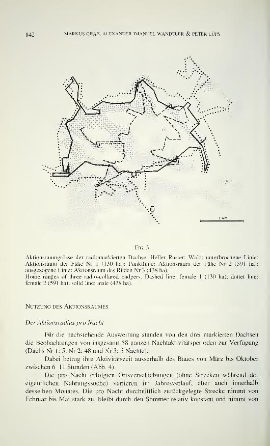
842 MARKUS GRAF, ALEXANDER IMANUEL WANDELER & PETER LÜPS
I J
Fig. 3
Aktionsraumgrösse der radiomarkierten Dachse. Heller Raster: Wald; unterbrochene Linie:
Aktionsraum der Fähe Nr 1 (130 ha): Punktlinie: Aktionsraum der Fähe Nr 2 (591 ha);
ausgezogene Linie: Aktionsraum des Rüden Nr 3 (438 ha).
Home ranges of three radio-collared badgers. Dashed line: female 1 (130 ha); dottet line:
female 2 (591 ha); solid line: male (438 ha).
Nutzung des Aktionsraumes
Der Aktionsradius pro Nacht
Für die nachstehende Auswertung standen von den drei markierten Dachsen
die Beobachtungen von insgesamt 58 ganzen Nachtaktivitätsperioden zur Verfügung
(Dachs Nr 1 : 5, Nr 2: 48 und Nr 3: 5 Nächte).
Dabei betrug ihre Aktivitätszeit ausserhalb des Baues von März bis Oktober
zwischen 6-1 1 Stunden (Abb. 4).
Die pro Nacht erfolgten Ortsverschiebungen (ohne Strecken während der
eigentlichen Nahrungssuche) variieren im Jahresverlauf, aber auch innerhalb
desselben Monates. Die pro Nacht durchnittlich zurückgelegte Strecke nimmt von
Februar bis Mai stark zu, bleibt durch den Sommer relativ konstant und nimmt von
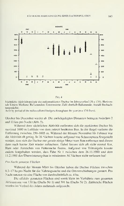
RÄUMLICHE HABITATNUTZUNG EINER DACHSPOPULATION 843
1980 1981
N D J F M A M J J A S Ni
MIN.
700 -
600 -i
a9
7
1500 -
400 -
2
1 1 ! iI t t
—
300 -
200 -
100 -
2
- - 24 " - 1 5
l
N D J F1
M A M1 1
J J A1
S N D
Fig. 4
Nächtliche Aktivitätsperiode der radiomarkierten Dachse im Jahresverlauf (N = 134). Horizon-
tale Linien: Mediane; Balkenenden: Extremwerte; Zahl oberhalb Balkenende: Anzahl Beobach-
tungsnächte.
Activity period of the radio-collared badgers throughout the year (n = 134 fixes).
Oktober bis Dezember wieder ab. Die zurückgelegten Distanzen betragen zwischen 2
und 1 1 km pro Nacht (Abb. 5).
Während ihrer nächtlichen Aktivität entfernten sich die markierten Dachse bis
maximal 1600 m Luftlinie von dem zuletzt benützten Bau. In der Regel variierte die
Entfernung zwischen 350-1005 m. Während der Monate November bis Februar war
die Aktivität oft gering. In 26 Nächten konnte aufgrund von Schneespuren festgestellt
werden, dass sich die Dachse nur gerade einige Meter vom Bau entfernten und diesen
dann nach kurzer Zeit wieder aufsuchten. Dabei Hessen sich oft nicht einmal Kot,
Harn oder Anzeichen von Futtersuche finden. Aufgrund von Trittsiegeln konnte
zudem festgehalten werden, dass Fähe Nr 1 zwischen dem 26.11.1980 und dem1 1.2.1981 den Überwinterungsbau in mindestens 56 Nächten nicht verlassen hat!
Pro Nacht genutzte Flächen
Während der Monate März bis Oktober haben die Dachse Flächen zwischen
0.3-17 ha pro Nacht für die Nahrungssuche und die Ortsverschiebungen genutzt. Pro
Nacht nutzten sie eine Fläche von durchschnittlich ca. 4 ha.
Die effektiv genutzten Flächen sind somit klein im Verhältnis zum gesamten
Aktionsraum von 130 ha (Dachs Nr 1) und 591 ha (Dachs Nr 2). Zahlreiche Flächen
wurden im Verlauf des Jahres mehrmals aufgesucht.
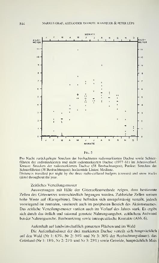
844 MARKUS GRAF, ALEXANDER IMANUEL WANDELER & PETER LÜPS
KILO-METER
MONATEJFMAMJ JASI I I I I
' ' KILO-METER
Fig. 5
Pro Nacht zurückgelegte Strecken der beobachteten radiomarkierten Dachse sowie Schnee-
fährten der radiomarkierten und nicht radiomarkierten Dachse (1977-81) im Jahresverlauf.
Kreuze: Strecken der radiomarkierten Dachse (58 Beobachtungen); Punkte: Strecken der
Schneefährten (38 Beobachtungen); horizontale Linien: Mediane.
Distances travelled per night by the three radio-collared badgers (crosses) and snow tracks
(dots) throughout the year.
Zeitliches Verteilungsmuster
Auswertungen mit Hilfe der Gitterzellenmethode zeigen, dass bestimmte
Zellen des Gitternetzes unterschiedlich begangen wurden. Zahlreiche Zellen weisen
hohe Werte auf (Kerngebiete). Diese befinden sich unregelmässig verteilt, jedoch
vorwiegend im zentralen, vereinzelt auch im peripheren Bereich des Aktionsraumes.
Das zeitliche Verteilungsmuster variiert auch im Verlauf des Jahres stark. Es ergibt
sich durch das örtlich und saisonal genutzte Nahrungsangebot, zeitlichem Aufwand
bei der Nahrungssuche, Baubenutzung sowie intraspezifische Kontakte (Abb. 6).
Aufenthalt auf landwirtschaftlich genutzten Flächen und im Wald
Die Aufenthaltsdauer der drei markierten Dachse verteilt sich hauptsächlich
auf den Wald (Nr 1: 63%, Nr 2: 54% und Nr 3: 30% der Beobachtungsdauer), das
Grünland (Nr 1: 18%, Nr 2: 21% und Nr 3: 25%) sowie Getreide, hauptsächlich Mais
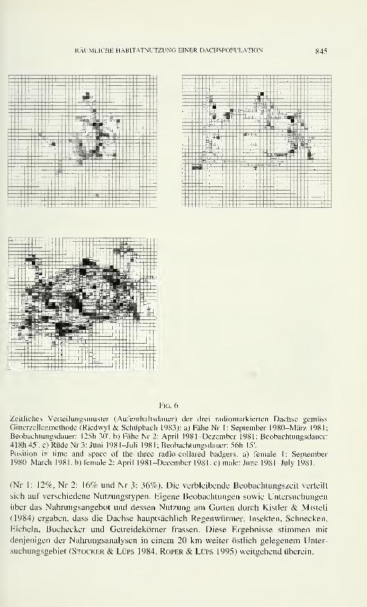
RÄUMLICHE HABITATNUTZUNG EINER DACHSPOPULATION 845
J-Bfr
- !!B i
r HM''~ i
" H"ü -H- "™i:i W.
: ' |ij«|
an1 mm fnfc( •..:»
SH :
^i4 ± .
. S r
--_-t- _i !» .- .. •."=; ;
;-;;;
' ' 1 -j, . . jj-
. T. 3. -rar." v '^ i'4 1
> 3 utì ~3E *>+ 4 ' J ' -'iim'tr "1
—
P1 i±*2,^ l±±J S--:--1 ±ü-'- "
"
: :
i
Sg=C Ï3 "
_:is| I'-:- •
Fig. 6
Zeitliches Verteilungsmuster (Aufenthaltsdauer) der drei radiomarkierten Dachse gemässGitterzellenmethode (Riedwyl & Schüpbach 1983): a) Fähe Nr 1: September 1980-März 1981;
Beobachtungsdauer: 125h 30'. b) Fähe Nr 2: April 1981-Dezember 1981; Beobachtungsdauer:
418h 45'. c) Rüde Nr 3: Juni 1981—Juli 1981; Beobachtungsdauer: 56h 15'.
Position in time and space of the three radio-collared badgers, a) female 1: September1980-March 1981. b) female 2: April 1981-December 1981. c) male: June 1981-July 1981.
(Nr 1: 12%, Nr 2: 16% und Nr 3: 36%). Die verbleibende Beobachtungszeit verteilt
sich auf verschiedene Nutzungstypen. Eigene Beobachtungen sowie Untersuchungen
über das Nahrungsangebot und dessen Nutzung am Gurten durch Kistler & Misteli
(1984) ergaben, dass die Dachse hauptsächlich Regenwürmer, Insekten, Schnecken,
Eicheln, Buchecker und Getreidekörner frassen. Diese Ergebnisse stimmen mit
denjenigen der Nahrungsanalysen in einem 20 km weiter östlich gelegenem Unter-
suchungsgebiet (Stocker & Lüps 1984, Roper & Lüps 1995) weitgehend überein.
846 MARKUS GRAF. ALEXANDER IMANUEL WANDELER & PETER LÜPS
Populationsgrösse
Folgende Indizien lassen den Schluss zu, dass die Grösse der Dachspopulation
innerhalb des intensiv überwachten Untersuchungsgebietes von 1977-81 drei bis
maximal fünf adulte Tiere umfasste:
- Die Mehrzahl der Baue wurde während der Beobachtungsperiode nur selten
befahren.
- Anstrengungen, weitere Dachse für die Markierung zu fangen, waren erfolg-
los.
- Unmarkierte Dachse wurden selten beobachtet.
- Nur siebenmal wurde ein unmarkiertes adultes Individuum zusammen mit
einem radiomarkierten Dachs begegnet, während zwei markierte Dachse
sechsmal zusammen beobachtet wurden.
- Während der Winterruheperioden wurde jeweils nur ein Bau benutzt.
- Während der Beobachtungsperiode konnte nur ein Geheck nachgewiesen
werden (1977. 3 Welpen).
Somit betrug die Populationsdichte am Gurten während der ganzen Unter-
suchungszeit maximal ein Dachs pro 200 ha!
DISKUSSION
Die Aktionsraumgrösse. das Muster der räumlichen und zeitlichen Habitat-
nutzung, die Grösse und die Dichte der Dachspopulation am Gurten unterscheiden
sich wesentlich von denjenigen anderer untersuchter europäischer Dachspopulationen.
Da der Intensität der Datenerhebung über einzelne Individuen die Extensität (Aus-
weitung des Untersuchungsgebietes, Erhöhung der Individuenzahl) zum Opfer fiel,
dürfen in der Konsequenz keine allzu weitreichenden Verallgemeinerungen aus den
Daten von nur drei verfolgten Individuen gezogen werden. Folgende Bemerkungen
scheinen uns jedoch zulässig:
Die Nutzung der Baue
Im Untersuchungsgebiet befinden sich zahlreiche grosse Baue, die eine gün-
stige Lage bezüglich Nahrungsressourcen und Schutz vor menschlichen Störungen
aufweisen. Alle grossen und häufig benützten Baue befinden sich im zentralen Be-
reich der Aktionsräume.
Grosse Baue wurden von den Dachsen gewählt für längere Winterruhepe-
rioden und für die nur einmal festgestellte Jungenaufzucht. Die Dachse wechseln den
Bau häufig ohne erkennbaren Grund. Viele grosse Baue werden nur selten benützt.
Dies ist wohl auf die geringe Dachsdichte zurückzuführen. Kleine Baue werden durch
Dachse regelmässig unterhalten, jedoch selten zur Übertagung aufgesucht. Sie dienen
allenfalls als Zufluchtsorte bei überraschenden Störungen (Butler & Roper 1995).
RÄUMLICHE HABITATNUTZUNG EINER DACHSPOPULATION 847
Aufgrund der ständigen Baukontrollen ist anzunehmen, dass die intra- und
interspezifische (Füchse) Konkurrenz wahrscheinlich gering war. In Grossbritannien
werden unter ungestörten Verhältnissen in der Regel alle Hauptbaue, auch nahe
beieinanderliegende, ununterbrochen benützt (C. Cheeseman pers. Mitt., Kruuk &Parish 1977, Neal 1977, Roper 1992).
Aktionsraumgrösse
Die Ergebnisse zeigen, dass die Aktionsräume mit 130, 438 und 591 ha uner-
wartet gross sind. Zudem überlappen sie sich vollständig. Ähnliche Resultate hat
Mouches (1981) mit Aktionsraumgrössen von 280-400 ha in Westfrankreich erhalten.
Mit 22-147 ha haben Cheeseman et al. (1981), Kruuk & Parish (1977) und Kruuk
(1978a) sowie Harris (1982) in Grossbritannien wesentlich kleinere Aktionsräume
erhoben.
POPULATIONSGRÖSSE
Im 10 km2 grossen Untersuchungsgebiet hielten sich während der Beobach-
tungszeit (1977-81) nur maximal 5 adulte Dachse auf. Mit 0.5 Dachsen pro 1 km2 ist
die Dichte im Vergleich zu der anderer Gebiete sehr niedrig: Cheeseman et al. (1981
und 1985) erwähnt 5-20, Kruuk & Parish (1982) 1-6, Mouches (1981) 1,5 Dachse
pro 1 km2. Dabei ist allerdings zu beachten, dass nur langfristige umfangreiche Unter-
suchungen zuverlässig Auskunft über die für ein Gebiet charakteristische Dichte geben
können, da Populationsschwankungen in einem gewissen Umfang zu erwarten sind. Die
niedrige Dichte der untersuchten Population kann mit der vorliegenden Untersuchung
nicht erklärt werden. Wetterfaktoren als bestandesregulierende Einflüsse in Gebirgs-
regionen, wie sie Eiberle & Matter (1985) postulieren, treffen für das Gurtengebiet
(500-900 m ü. M.) kaum zu. Das Nahrungsangebot kann nicht als limitierender Faktor
für die geringe Dichte zutreffen: Kistler & Misteli (1984) haben auch für den
Sommer, wenn die als Hauptnahrung genutzten Regenwürmer ein Populationsminimum
aufweisen und häufig schlecht erreichbar sind, gezeigt, dass Nahrungsressourcen
superabundant vorhanden sind. Die Untersuchung lässt auch keine Aussagen zu, ob die
geringe Dichte auf die Tollwut, welche ab 1979 in der Region Köniz und Gurten
aufgetreten ist, und auf die Tollwutbekämpfungsmassnahmen zurückzuführen ist.
HABITATNUTZUNG
Es zeigen sich auch Unterschiede bezüglich Strategien der Habitatnutzung im
Vergleich zu den Resultaten von Kruuk (1978a, b) und Cheeseman et al. (1981). Die
Dachse am Gurten nutzen wesentlich grössere Flächen und legen pro Nacht durch-
schnittlich längere Strecken zurück, obwohl nutzbare Nahrungsressourcen viel näher
lägen. Kistler & Misteli (1984) gelangten aufgrund ihrer Analysen zum Schluss, dass
bezüglich Nahrungsangebot wahrscheinlich ein Aktionsraum von ca. 78-113 ha den
maximal fünf Dachsen am Gurten genügen würde, falls sie sich allein von Regen-
würmern ernähren würden. Dasselbe gilt auch für andere wichtige Nahrungsressourcen
wie Mais. So würde im Herbst ein einzelnes Maisfeld den Dachsen genügend Nahrung
bieten. Trotzdem suchten Dachse während derselben Nacht mehrere weit ausein-
g48 MARKUS GRAF, ALEXANDER IMANUEL WANDELER & PETER LÜPS
anderliegende Maisfelder zur Nahrungssuche auf. Allerdings ist die Nutzungs-
intensität nicht dieselbe wie in Grossbritannien, wo die Dachse als "Regenwurm-
spezialisten" (Kruuk 1989 und Kruuk & Parish 1981) einige wenige kleine, aber
ergiebige Futterplätze ("patches"; Kruuk 1978b), intensiv nach Regenwürmern ab-
suchen. Die Dachse am Gurten sind nicht an solche "Regenwurmpatches" gebunden,
sondern sind eher als Nahrungsgeneralisten (Roper 1994) zu betrachten, die ein
vielfältiges Nahrungsangebot lokal, jahreszeitlich und individuell mit unterschied-
licher Intensität nutzen (Kistler & Misteli 1984).
Territorialität
Kruuk (1978a) sowie Kruuk & Parish (1982) und Cheeseman et al. (1981)
wiesen nach, dass Dachssippen, bestehend aus 2-12 Dachsen, kleinstmögliche, durch
Latrinen und Duftstoffe deutlich von einander abgegrenzte Territorien verteidigen
(20-310 ha). Die Grösse der Sippe Hess sich zudem mit der Biomasse der Regen-
würmer innerhalb eines Territoriums korrelieren. Harris (1982) hingegen hat in
vorstädtischen Verhältnissen gezeigt, dass Dachse ein breites Nahrungsspektrum
nutzen und das territoriale Verhalten nicht so ausgeprägt ist. Dachse unterschiedlicher
Sippen suchen ihre Nahrung teilweise in sich überlappenden Gebieten (vgl.
Cresswell & Harris 1988, Cheeseman et al. 1988).
Die grosse Zahl der Baue mit gelegentlichen Anzeichen von Dachsaktivität
hatte uns ursprünglich eine weit höhere Populationsdichte vermuten lassen. Aufgrund
der räumlichen Verteilung grosser Baue erschien uns auch eine Aufteilung des
Untersuchungsgebietes in mehrere "clan"-Territorien durchaus denkbar.
Trotz der Grösse unseres Untersuchungsgebietes von 10 km- liessen sich nicht
mehrere Sippen und Territorien abgrenzen. Unsere telemetrische Studie lieferte keine
Hinweise für verteidigte Territorien:
- Die drei mit Sendern ausgerüsteten Dachse bewegten sich in vollständig über-
lappenden Aktionsräumen. Sie benutzten auch dieselben Baue, allerdings selten
gleichzeitig.
Nur in ganz wenigen Fällen wurden mindestens zwei Dachse bei gemeinsamer
Nahrungssuche oder zusammen am Bau beobachtet.
Aggressivität zwischen sich begegnenden Dachsen wurde nie festgestellt.
- Die Aktionsräume weisen Grenzlängen auf, die sich vermutlich nicht vertei-
digen lassen.
Latrinen wurden hauptsächlich in der näheren Umgebung häufig benutzter Baue
("Hinterland-Latrinen", vgl. Roper et al. 1993), selten an der Peripherie der
Aktionsräume gefunden ("Grenz-Latrinen"). Letztere dürften allerdings wegen
landwirtschaftlicher Nutzung nur für kurze Zeit Bestand haben und deshalb
nicht gefunden worden sein. Dennoch kann die Situation im Sinne Ropers da-
hingehend interpretiert werden, dass die in Baunähe genutzten Latrinen in
starkem Mass zur gegenseitigen Erkennung und zur Kennzeichnung der Wurf-
baue dienen.
Unsere Beobachtungen erwecken den Eindruck, dass die untersuchten Dachse
einen riesigen "clan"-Aktionsraum nutzen. In diesem Aktionsraum gibt es keine
RÄUMLICHE HABITATNUTZUNG EINER DACHSPOPULATION g49
markierten Grenzen. Vermutlich wird er auch nicht verteidigt. Wir wissen allerdings
nicht, ob und in welcher Form Interaktionen mit Nachbar-Dachsen stattfinden. Eben-
sowenig kennen wir die populationsbiologischen Parameter der Dachse ausserhalb
unseres Untersuchungsgebietes.
Unsere Resultate haben ergeben, dass die Populationsdichte niedrig ist, dass sich
die Tiere in grossen Aktionsräumen bewegen, als Nahrungsgeneralisten ein breites
Nahrungsspektrum in weit entfernten Gebieten nutzen und ein Überangebot an Bauen
besteht. Diese Beobachtungen unterscheiden sich in wesentlichen Punkten von An-
gaben über Populationsdichten und Raum-Zeit-Systeme anderer Dachspopulationen. Im
Falle der Dachse am Gurten stellen weder das Angebot an Bauen oder an den zu deren
Anlage günstigen Strukturen einen limitierenden Faktor dar (vgl. dazu Doncaster &Woodroffe 1993, Woodroffe & Macdonald 1992), noch das vorhandene
Nahrungsangebot. Der Umstand der weitgehend fehlenden sozialen Organisation und
das damit in Beziehung stehende räumliche und zeitliche Verhaltensmuster der
einzelnen Tiere (vgl. Harris 1982, Cheeseman et al. 1988, Cresswell et al. 1992,
Roper & Lüps 1993) lassen keine direkten Zusammenhänge zu den oben geannnten
Parametern erkennen. Sie dürften am ehesten eine Folge der geringen Populationsdichte
sein, für die im Falle des Gurtens eine plausible Erklärung allerdings fehlt. Sowohl
bezüglich Nahrung wie auch punkto Sozialverhalten und räumlicher Organisation
erweist sich der Dachs, dies geht aus den zahlreichen Untersuchungen aus ver-
schiedenen Teilen Europas und aus deren Vergleich deutlich hervor, als viel variablere
Art als dies lange Zeit angenommen worden ist.
LITERATUR
Amlaner, C.J. & D.W. Macdonald. 1979. A Handbook on biotelemetry and radio tracking.
Pergamon Press, Oxford. 804 pp.
Butler, J.M. & T.J. Roper. 1995. Escape tactics and alarm responses in badgers Mêles mêles:
a field experiment. Ethology 99: 313-322.
Cederlund, G., T. Dreyfert & P.A. Lemnell. 1979. Radiotracking techniques and the
reliability of systems used for larger birds and smalls mammals. Naturvardsverket,
Rapport. 102 pp.
Cheeseman, C.L., GW. Jones, J. Gallagher & P.J. Mallinson. 1981. The population
structure, density and prevalence of tuberculosis in Bagders from four areas in South-
West England. Journal ofApplied Ecology 18: 795-804.
Cheeseman, C.L., T.W.A. Little, P.J. Mallinson, R.J.C. Page, J.W. Wilesmith & D.G.Pritchard. 1985. Population ecology and prevalence of tuberculosis in Badgers in area
of Staffordshire. Mammal Review 15(3): 125-135.
Cheeseman, C.L., W.J. Cresswell, S. Harris & P.J. Mallinson. 1988. Comparison of
dispersal and other movements in two Badger (Mêles meles) populations. MammalReview 18(1): 51-59.
Cresswell, W.J. & S. Harris. 1988. Foraging behaviour and home-range utilization in a
suburban Badger {Meles meles) population. Mammal Review 18(1): 37-49.
Cresswell, W.J., S. Harris, C.L. Cheeseman & P.J. Mallinson. 1992. To breed or not to
breed: an analysis of the social and density-dependent constraints on the fecundity of
female badgers (Meles meles). Philosophical Transactions of the Royal Society ofLondon, Series B, 338: 393-407.
Doncaster, C.P. & R. Woodroffe. 1993. Den site can determine shape and size of badger
territories: implications for group-living. Oikos 66: 88-93.
850 MARKUS GRAF, ALEXANDER IMANUEL WANDELER & PETER LÜPS
Eiberle, K. & J.F. Matter. 1985. Bestandesregulierende Einflüsse von Wetterfaktoren beimDachs. Berichte der Botanisch-Zoologischen Gesellschaft Liechtenstein-Sargans-Wer-
denberg 14: 59-69.
Graf, M. 1988. Die räumliche und zeitliche Habitatnutzung einer Dachspopulation am Gurten
bei Bern. Dissertation Zoologisches Institut der Universität Bern. 161 pp.
Harris, S. 1982. Activity Patterns and Habitat Utilization of Badgers in suburban Bristol: Aradio Tracking Study. Symposium of the Zoological Society ofLondon 49: 301-323.
Hegg, O. 1973. Die Pflanzenwelt der Region Bern. In: Aerni, K. (ed.), "Bern — von der Natur-
landschaft zur Stadtregion". Geographische Gesellschaft Bern. 248 pp.
Kistler, P. & F. Misteli. 1984. Ökologische Untersuchungen über das Nahrungsangebot unddessen Nutzung durch den Europäischen Dachs im Gebiet des Gurten bei Bern.
Lizentiatsarbeit Zoologisches Institut der Universität Bern. 161 pp.
Kruuk, H. 1978a. Spatial organization and territorial behaviour of the European badger Meles
meles. Journal ofZoology, London 184: 1-19.
Kruuk, H. 1978b. Foraging and Spatial Organisation of the European Badger, Meles meles L.
Behavioral Ecology and Sociobiology 4: 75-89.
Kruuk, H. 1989. The social badger. Oxford University Press, Oxford. 155 pp.
Kruuk, H. & T. Parish. 1977. Behaviour of Badgers. Institute of Terrestrial Ecology,
Cambridge. 16 pp.
Kruuk, H. & T. Parish. 1981. Feeding specialization of the European badger Meles meles in
Scotland. Journal ofAnimal Ecology 50: 773-788.
Kruuk, H. & T. Parish. 1982. Factors affecting population density, group size and territory of
the European badger, Meles meles. Journal ofZoology, London 196: 31-39.
LUps, P., T.J. Roper & G. Stocker. 1991. Magen-Analysen bei Dachsen Meles meles aus der
Umgebung Berns ( 1987—89). Naturhistorisches Museum Bern. Kleine Mitteilungen 1-10.
Mouches, A. 1981. Eco-éthologie du blaireau européen Meles meles L:. Stratégies d'utilisation de
l'habitat et des ressources alimentaires. Thèse de 3ème Cycle. Université de Rennes. 139 p.
Neal, E.N. 1977. Badgers. Blandford Press Ltd., Poole, Dorset. 321 pp.
Riedwyl, H. & M. SchUpbach. 1983. Siebdiagramme. Institut für Mathematische Statistik undVersicherungslehre der Universität Bern. Technischer Bericht "Ho. 12. 16 pp.
Roper, T.J. 1992. Badger Meles meles setts - architecture, internal environment and function.
Mammal Review 22(1): 43-53.
Roper, T.J. 1994. The European badger Meles meles: food specialist or generalist? Journal of
Zoology, London 234: 437-452.
Roper, T.J. & P. LUps. 1993. Disruption of territorial behaviour in bagders Meles meles.
Zeitschrift für Säugetierkunde 58: 252-255.
Roper, T.J., L. Conradt. J. Butler, S.E. Christian, J. Ostler & T.K. Schmid. 1993. Territorial
marking with faeces in badgers Meles meles: a comparison of boundary and hinterland
latrine use. Behaviour 111: 289-307.
Roper, T.J. & P. LUPS. 1995. Diet of badgers {Meles meles) in central Switzerland: an analysis of
stomach contents. Zeitschriftfür Säugetierkunde 60: 9-19.
Stocker, G. & P. LUps. 1984. Qualitative und quantitative Angaben zur Nahrungswahl des
Dachses Mêles mêles im Schweizerischen Mittelland. Revue suisse de Zoologie 91:
1007-1015.
Taylor, K.D. & H.G. Lloyd. 1978. The design, construction and use of a radio-tracking system
for some British mammals. Mammal Review 8: 1 17-141.
Voigt, D.R. & R.R. Tinline. 1980. Strategies for Analyzing Radio Tracking Data. A Handbook
on biotelemetn- and radio tracking by C.J. Amlaner and D.W. Macdonald. Pergamon
Press, Oxford: 387-404.
Woodroffe, R. & D.W. Macdonald. 1992. Badger clans: demographic groups in an antisocial
species. Journal ofZoology, London 227: 696-698.
Revue suisse de Zoologie, 103 (4): 851-858; décembre 1996
Le zoobenthos comme indicateur des perturbations
d'origine humaine dans deux lacs de montagne
Claude LANG et Olivier REYMONDConservation de la faune, Marquisat 1,
CH-1025 St-Sulpice, Suisse.
Zoobenthos as indicator of man-made perturbations in two mountain
lakes. - In Lake Lioson (Switzerland), chironomid larvae were relatively
abundant compared with oligochaetes (mostly Tubifex tubifex). In addition,
taxa indicative of oligotrophic conditions were present in the profundal (10
- 20 m deep). In contrast, these taxa were absent from Lake Chavonnes and
chironomids were scarce. Oligochaetes, which were very abundant at a
depth of 12 m, were almost absent deeper than 20 m. Composition and
distribution of zoobenthos according to depth were typical for an oligo-
trophic mountain lake in Lioson, for a eutrophic lake in Chavonnes.
Oxygen profiles according to depth and phosphorus concentrations in the
water confirmed this interpretation.
Key-words: Biomonitoring - Chironomidae - Eutrophication - Mountain
lake - Oligochaeta.
INTRODUCTION
Les lacs de montagne des Alpes sont en général oligotrophies s'ils ne sont pas
influencés par l'homme (Chacornac 1986). Du fait de la rareté du phosphore, la
production primaire reste faible et l'oxygène est abondant à toutes profondeurs, sauf
en hiver, ce qui les différencie des lacs de plaine oligotrophes (Wetzel 1975). En
effet, la présence d'une couche de glace et de neige, pendant parfois plus de sept mois,
empêche les apports atmosphériques. De ce fait, les couches d'eau profondes s'appau-
vrissent en oxygène vers la fin de l'hiver (moins de 1 mg / 1) ce qui entraîne la remise
en suspension du phosphore contenu dans le sédiment. Ces phénomènes sont ana-
logues à ceux qui s'observent pendant l'été dans l'hypolimnion des lacs de plaine
eutrophes, mais la cause en est différente (Capblancq & Laville 1983).
Lorsque le phosphore augmente dans les lacs de plaine, la production primaire
s'accroît, avec elle la sédimentation organique, d'où une baisse des concentrations en
oxygène dans les couches d'eau profonde pendant la phase de stagnation estivale
Manuscrit accepté le 19.04.1996.
852 CLAUDE LANG & OLIVIER REYMOND
(Wetzel 1975). La zone profonde des lacs de montagne est donc particulièrement
sensible aux effets d'un excès de phosphore d'origine humaine puisqu'elle est déjà
exposée à un manque d'oxygène d'origine naturelle. Si les conditions du milieu
changent dans cette zone, le zoobenthos, c'est-à-dire les invertébrés qui colonisent le
sédiment, va se modifier d'une façon interprétable, d'où son utilisation comme indi-
cateur (Lang 1986).
Dans la présente étude, nous avons utilisé le zoobenthos pour suivre l'évolution
à long terme (1978 - 1995) de deux lacs de montagne situés dans les Préalpes vau-
doises: le lac Lioson (altitude 1848 m) et le lac des Chavonnes (1690 m). Sur un plan
plus général, les résultats obtenus permettent d'évaluer l'utilité du zoobenthos commeindicateur des perturbations d'origine humaine dans un milieu particulier. En effet, les
deux lacs étudiés sont couverts de glace et de neige de la mi-novembre à la mi-juin ce
qui expose le benthos à des conditions extrêmes (Capblancq & Laville 1983).
STATIONS ET METHODES
Les lacs étudiés sont relativement profonds (28 m au maximum) par rapport à
leur taille modeste (Lioson 6 ha, Chavonnes 4 ha). La pente du fond, forte entre et
22 m de profondeur ne s'atténue qu'à partir de 24 m. Entre et 8 m s'étend un éboulis
et les sédiments meubles ne deviennent prépondérants qu'à partir de 10 m, ce qui
détermine la profondeur supérieure des stations de prélèvements.
Les prélèvements effectués entre 1978 et 1995 sont présentés dans le tableau 1.
A chaque profondeur 5 à 12 carottes de sédiment, couvrant 16 cm" chacune, sont
prélevées en plongée. Seul le macrobenthos des sédiments meubles de la zone pro-
fonde (10 à 20 m) a été étudié.
En laboratoire, le sédiment est tamisé et le refus du tamis (maille 0.2 mm) est
fixé au formol 5%. Les oligochètes et les larves de chironomides, qui constituent
l'essentiel de la macrofaune, sont ensuite comptés carotte par carotte puis, pour
chaque profondeur, l'abondance moyenne (nombre moyen d'individus / 16 cm2) est
calculée (Tab. 1). Pour chaque profondeur, un sous-échantillon d'oligochètes et de
chironomides. monté dans un milieu approprié (Reymond 1994), est identifié jusqu'au
niveau du genre ou de l'espèce (Tab. 2). Contrairement aux oligochètes, les chirono-
mides n'ont pas été identifiés chaque année.
Le rapport nombre de chironomides sur nombre de chironomides plus nombre
d'oligochètes est utilisé pour décrire l'évolution des lacs (Wiederholm 1980). Ce
rapport est d'abord calculé carotte par carotte puis, à partir de ces valeurs indivi-
duelles, une valeur moyenne est calculée pour chacun des ensembles comparés (Tab.
3). Une augmentation de la valeur de ce rapport indique une amélioration de l'état du
milieu: les chironomides sont en effet moins résistants que les oligochètes au manque
d'oxygène. De plus, les taxons de chironomides et d'oligochètes identifiés (Tab. 2)
peuvent être classés en deux catégories (Saether 1979, Lang 1990): les espèces
caractéristiques des lacs oligotrophes, sensibles au manque d'oxygène, et les espèces
plus résistantes.
ZOOBENTHOS COMME INDICATEUR §53
Tableau 1
Abondance moyenne (nombre moyen d'individus / 16 cm2) des oligochètes et des larves de
chironomides en fonction de la profondeur dans le lac Lioson (L) et le lac des Chavonnes (C). n
= nbre total de carottes de 16 cm 2 prélevées. Dans les colonnes 5 à 12, les espaces blancs
correspondent à des profondeurs qui n'ont pas été échantillonnées cette année là.
Oligochètes Chironomides
Mois 1 Lac n
Profondeur (m) Profondeur (m)
Année 10 12 15 20 10 12 15 20
1978 9 L 18 5.2 13.0 11.7 4.7 0.5 0.2
8 C 18 12.0 1.7 1.8 3.5 0.3
1979 7 L 30 2.2 8.3 5.2 4.5 1.0 0.2
7 C 29 25.5 9.1 1.1 0.6
1980 8 L 10 5.0 10.6 0.2 1.2
8 C 10 12.0 1.2
1981 8 L 10 10.0 8.0 0.2
8 C 10 9.8 0.2 1.0
1984 10 L 10 8.0 11.0 2.2 1.0
10 C 10 17.6 3.4
1985 8 L 10 3.2 15.6 8.6 0.2
9 C 10 10.0 0.2 1.0
1986 9
8
LC
12
10 42.1
7.0 4.0
0.6
11.5 4.0
1988
9
LC 6 28.0 0.5
1989
9
LC 6 32.3 0.2
1990
9
LC 12 44.8 0.9
1991 10 L 18 10.5 10.5 15.2 1.7 3.0 1.7
9 C 12 31.7 44.8 1.0 0.5
1992 - L9 C 18 18.3 17.0 4.2 1.7
1993 8 L 24 0.2 0.7 3.8 15.7 10.2 13.0 8.7 0.5
8 C 24 6.5 23.0 7.8 0.3 1.0 0.2 0.3
1995 8 L 24 1.0 3.3 5.0 10.2 4.5 5.7 8.2 2.0
8 C 24 14.7 8.2 16.8 0.2 4.8 2.5 2.3
Total L 166 3.1 6.4 9.0 9.9 5.7 5.4 3.6 0.6
C 199 22.3 29.2 10.3 0.3 1.8 1.6 0.9 0.1
Test de t (Prob.) 0.000 0.000 0.515 0.000 0.000 0.000 0.000 0.000
1 Mois où les prélèvements de zoobenthos ont été effectués: - pas de prélèvements effectués
cette année là.
RESULTATS
A 10 et 12 m de profondeur (Tab. 1, ligne Total), les oligochètes sont beau-
coup plus abondants dans le lac des Chavonnes que dans le lac Lioson; à 15 m de
profondeur, les abondances sont égales; à 20 m de profondeur, elles sont plus élevées
dans le lac Lioson. De ce fait, l'abondance des oligochètes augmente avec la pro-
fondeur dans le lac Lioson tandis que, au contraire, elle diminue dans le lac des
Chavonnes. Dans les deux lacs, c'est Tubifex tubifex qui constitue l'essentiel des
communautés d'oligochètes (Tab. 2).
854 CLAUDE LANG & OLIVIER REYMOND
Tableau 2
Composition du zoobenthos en fonction de la profondeur dans le lac Lioson (L) et dans le lac
des Chavonnes (C). Chironomides: taxons 1 - 8. oligochètes: taxons 9 et 10. Taxons présents en
1993 ou en 1995 (+), présent en 1993 et en 1995 (*).
Taxons Lac
Profondeur (m)
Code 10 12 15 20
1 Procladius L * * * *
C + * +2 Brillici L
C+
3 Heterotrissocladius ! LC
* * *
4 Chironomus L + * + +C + + +
5 Dicrotendipes LC + * * +
6 Paracladopelma ' LC
+ * + +
7 Tanyîarsus LC
* * * *
8 Paracladius LC
+
9 Tubifex tubifex L * * * *
C * * * *
10 Stylodrilus hering ian us ' LC
* *
Nbre de taxons L 9 7 6 5
C 4 4 4 2
taxons caractéristiques des lacs oligotrophes (Saether 1979, Lang 1990).
Tableau 3
Evolution à long terme (1978/79 comparée à 1993/95) du zoobenthos dans le lac Lioson (L) et
le lac des Chavonnes (C). Nombre moyen d'individus par 16 cm 2. Signification statistique des
résultats de l'analyse de variance (* effet significatif, *** très significatif, NS non significatif).
A x P: interaction année par profondeur. Profondeurs utilisées: 10, 15, 20 m.
Périodes comparées Signification
Taxons Lac 78/79 1 93/952 Année Profondeur AxP
Oligochètes (nombre)
Chironomides (nombre)
Oligochètes + Chironomides (nombre)
Chironomides/Oligochètes
+ Chironomides (%)
7.00
9.34
1.85
0.85
8.85
10.19
L 23.1
C 8.2
5.97
7.66
5.67
1.33
11.64
9.00
57.2
13.6
NSNS
NS
NS
NS
***
***
NS
NS
NSNS***
NS
1 Nombre de carottes: 48 (L) et 47 (C), 2 36 et 36.
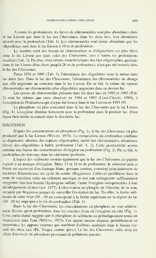
ZOOBENTHOS COMME INDICATEUR 355
A toutes les profondeurs, les larves de chironomides sont plus abondantes dans
le lac Lioson que dans le lac des Chavonnes; dans les deux lacs, leur abondance
décroît avec la profondeur (Tab. 1). Les chironomides sont moins abondants que les
oligochètes, sauf dans le lac Lioson à 10 m de profondeur.
Le nombre total des taxons de chironomides et d'oligochètes est plus élevé
dans le lac Lioson que dans celui des Chavonnes, ceci à toutes les profondeurs
étudiées (Tab. 2). De plus, trois taxons caractéristiques des lacs oligotrophes, présents
dans le lac Lioson (l'un d'eux jusqu'à 20 m de profondeur), n'ont pas été trouvés dans
le lac des Chavonnes.
Entre 1978 et 1995 (Tab. 3), l'abondance des oligochètes reste la même dans
les deux lacs. Dans le lac des Chavonnes, l'abondance des chironomides ne change
pas, elle augmente au contraire dans le lac Lioson. De ce fait, la valeur du rapport
chironomides sur chironomides plus oligochètes augmente dans ce dernier lac.
Les taxons de chironomides présents dans les deux lacs en 1993 et 1995 (Tab.
2) sont les mêmes que ceux observés en 1984 et 1985 (Lods-Crozet 1989), à
l'exception de Prodiamesa qui n'a pas été trouvé dans le lac Lioson en 1993 /95.
Le phosphore est plus concentré dans le lac des Chavonnes que le lac Lioson
(Fig. 1). L'oxygène diminue fortement avec la profondeur dans le premier lac, d'une
façon bien moins accentuée dans le deuxième lac.
DISCUSSION
D'après les concentrations en phosphore (Fig. 1), le lac des Chavonnes est plus
productif que le lac Lioson (Wetzel 1975). La composition du zoobenthos confirme
ce diagnostic: absence des espèces oligotrophes, rareté des chironomides, abondance
élevée des oligochètes à faible profondeur (Tab. 1, 2). Cette productivité accrue
entraîne une baisse des concentrations d'oxygène en profondeur (Fig. 1). De ce fait, le
zoobenthos devient rare dans les sédiments profonds.
L'aspect des sédiments montre également que le lac des Chavonnes est parfois
exposé à un manque d'oxygène. Entre 15 et 18 m de profondeur, le sédiment noir et
fluide est recouvert d'un feutrage blanc, presque continu, constitué principalement de
bactéries filamenteuses du cycle du soufre {Beggiatoa). Celles-ci prolifèrent dans la
zone de transition entre un sédiment anoxique et une eau surnageante suffisamment
oxygénée: l'un leur fournit l'hydrogène sulfuré, l'autre l'oxygène indispensables à leur
développement (Jorgensen 1977). L'observation en plongée de l'étendue de la zone
occupée par Beggiatoa permet de surveiller l'évolution du lac. En effet, la limite infé-
rieure de cette zone ( 1 8 - 20 m) correspond à la limite supérieure de la région du lac
(20 - 28 m) impropre à la vie du zoobenthos (Tab. 1).
Dans le lac des Chavonnes, les concentrations en phosphore ne sont relative-
ment élevées qu'en profondeur, dans les couches d'eau où l'oxygène est rare (Fig. 1).
Cette particularité suggère que le phosphore du sédiment est périodiquement remis en
suspension dans l'eau (Wetzel 1975). Cet apport interne dépasse probablement en
importance les apports externes qui semblent d'ailleurs maîtrisés dans le bassin ver-
sant des deux lacs (Ph. Vioget, comm. pers.). Le lac des Chavonnes subit ainsi les
effets d'un excès de phosphore provenant de pollutions passées.
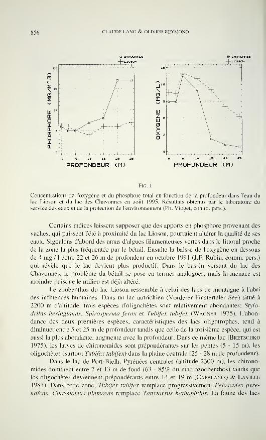
856 CLAUDE LANG & OLIVIER REYMOND
e CHAUONNES
-|—LIOSON
<
z\CD
E
!' '
1 I 1 1
1
1 1 I I 1 1 1
' ' !' '
0-
|
^v.~~-~f
À-- -+-' -4:
i , , , , i 1 , , , , 1
^fB CHAUONNES
LIOSON
PROFONDEUR (M) PROFONDEUR (M)
Fig. 1
Concentrations de l'oxygène et du phosphore total en fonction de la profondeur dans l'eau du
lac Lioson et du lac des Chavonnes en août 1995. Résultats obtenus par le laboratoire du
service des eaux et de la protection de l'environnement (Ph. Vioget, comm. pers.).
Certains indices laissent supposer que des apports en phosphore provenant des
vaches, qui paissent l'été à proximité du lac Lioson, pourraient altérer la qualité de ses
eaux. Signalons d'abord des amas d'algues filamenteuses vertes dans le littoral proche
de la zone la plus fréquentée par le bétail. Ensuite la baisse de l'oxygène en-dessous
de 4 mg / 1 entre 22 et 26 m de profondeur en octobre 1991 (J.F. Rubin, comm. pers.)
qui révèle que le lac devient plus productif. Dans le bassin versant du lac des
Chavonnes, le problème du bétail se pose en termes analogues, mais la menace est
moindre puisque le milieu est déjà altéré.
Le zoobenthos du lac Lioson ressemble à celui des lacs de montagne à l'abri
des influences humaines. Dans un lac autrichien (Vorderer Finstertaler See) situé à
2200 m d'altitude, trois espèces d'oligochètes sont relativement abondantes: Srylo-
drilus heringianus, Spìrosperma ferox et Tubifex tubifex (Wagner 1975). L'abon-
dance des deux premières espèces, caractéristiques des lacs oligotrophies, tend à
diminuer entre 5 et 25 m de profondeur tandis que celle de la troisième espèce, qui est
aussi la plus abondante, augmente avec la profondeur. Dans ce même lac (Bretschko
1975), les larves de chironomides sont prépondérantes sur les pentes (5-15 m), les
oligochètes (surtout Tubifex tubifex) dans la plaine centrale (25 - 28 m de profondeur).
Dans le lac de Port-Bielh, Pyrénées centrales (altitude 2300 m), les chirono-
mides dominent entre 7 et 13 m de fond (63 - 85% du macrozoobenthos) tandis que
les oligochètes deviennent prépondérants entre 14 et 19 m (Capblancq & Laville
1983). Dans cette zone, Tubifex tubifex remplace progressivement Peloscolex pyre-
naïcus, Chironomus plumosus remplace Tanytarsus bathophilus. La faune des lacs
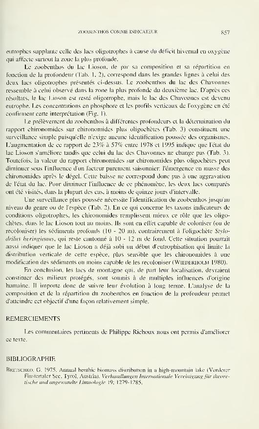
ZOOBENTHOS COMME INDICATEUR 857
eutrophes supplante celle des lacs oligotrophes à cause du déficit hivernal en oxygène
qui affecte surtout la zone la plus profonde.
Le zoobenthos du lac Lioson, de par sa composition et sa répartition en
fonction de la profondeur (Tab. 1, 2), correspond dans les grandes lignes à celui des
deux lacs oligotrophes présentés ci-dessus. Le zoobenthos du lac des Chavonnes
ressemble à celui observé dans la zone la plus profonde du deuxième lac. D'après ces
résultats, le lac Lioson est resté oligotrophy mais le lac des Chavonnes est devenu
eutrophe. Les concentrations en phosphore et les profils verticaux de l'oxygène en été
confirment cette interprétation (Fig. 1).
Le prélèvement du zoobenthos à différentes profondeurs et la détermination du
rapport chironomides sur chironomides plus oligochètes (Tab. 3) constituent une
surveillance simple puisqu'elle n'exige aucune identification poussée des organismes.
L'augmentation de ce rapport de 23% à 57% entre 1978 et 1995 indique que l'état du
lac Lioson s'améliore tandis que celui du lac des Chavonnes ne change pas (Tab. 3).
Toutefois, la valeur du rapport chironomides sur chironomides plus oligochètes peut
diminuer sous l'influence d'un facteur purement saisonnier: l'émergence en masse des
chironomides après le dégel. Cette baisse ne correspond donc pas à une aggravation
de l'état du lac. Pour diminuer l'influence de ce phénomène, les deux lacs comparés
ont été visités, dans la plupart des cas, à moins de quinze jours d'intervalle.
Une surveillance plus poussée nécessite l'identification du zoobenthos jusqu'au
niveau du genre ou de l'espèce (Tab. 2). En ce qui concerne les taxons indicateurs de
conditions oligotrophes. les chironomides remplissent mieux ce rôle que les oligo-
chètes, dans le lac Lioson tout au moins. Ils sont en effet capable de coloniser (ou de
recoloniser) les sédiments profonds (10-20 m), contrairement à l'oligochète Stylo-
drilus heringianus, qui reste cantonné à 10 - 12 m de fond. Cette situation pourrait
aussi indiquer que le lac Lioson a déjà subi un début d'eutrophisation qui limite la
distribution verticale de cette espèce, plus sensible que les chironomides à une
modification des sédiments ou moins capable de les recoloniser (Wiederholm 1980).
En conclusion, les lacs de montagne qui, de part leur localisation, devraient
constituer des milieux protégés, sont soumis à de multiples influences d'origine
humaine. Il importe donc de suivre leur évolution à long terme. L'analyse de la
composition et de la répartition du zoobenthos en fonction de la profondeur permet
d'atteindre cet objectif d'une façon relativement simple.
REMERCIEMENTS
Les commentaires pertinents de Philippe Richoux nous ont permis d'améliorer
ce texte.
BIBLIOGRAPHIE
Bretschko, G. 1975. Annual benthic biomass distribution in a high-mountain lake (Vorderer
Finstertaler See, Tyrol, Austria). Verhandlungen Internationale Vereinigung für theore-
tische und angewandte Limnologie 19: 1279-1285.
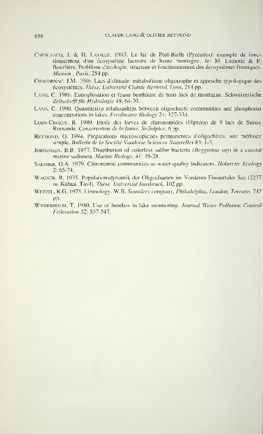
858 CLAUDE LANG & OLIVIER REYMOND
Capblancq, J. & H. Laville. 1983. Le lac de Port-Bielh (Pyrénées): exemple de fonc-
tionnement d'un écosystème lacustre de haute montagne. In: M. Lamotte & F.
Bourlière. Problème d'écologie: structure et fonctionnement des écosystèmes limniques.
Masson , Paris, 254 pp.
Chacornac, J.M. 1986. Lacs d'altitude: métabolisme oligotrophe et approche typologique des
écosystèmes. Thèse, Université Claude Bernard, Lyon. 214 pp.
Lang, C. 1986. Eutrophisation et faune benthique de trois lacs de montagne. Schweizerische
Zeitschriftfür Hydrologie 48: 64-70.
Lang, C. 1990. Quantitative relationships between oligochaete communities and phosphorus
concentrations in lakes. Freshwater Biology 24: 327-334.
Lods-Crozet, B. 1989. Etude des larves de chironomides (Diptera) de 9 lacs de Suisse
Romande. Conservation de lafaune, St-Sulpice, 6 pp.
Reymond, O. 1994. Préparations microscopiques permanentes d'oligochètes: une méthode
simple. Bulletin de la Société Vaudoise Sciences Naturelles 83: 1-3.
J0RGENSEN, B.B. 1977. Distribution of colorless sulfur bacteria (Beggiatoa ssp) in a coastal
marine sediment. Marine Biology, 41: 19-28.
Saether. O.A. 1979. Chironomid communities as water quality indicators. Holarctic Ecology
2: 65-74.
Wagner, B. 1975. Populationsdynamik der Oligochaeten im Vorderen Finstertaler See (2237
m. Kühtai, Tirol). Thèse, Université Innsbruck, 102 pp.
Wetzel, R.G. 1975. Limnology. W.B. Saunders company, Philadelphia, London, Toronto, 743
pp.
Wiederholm, T. 1980. Use of benthos in lake monitoring. Journal Water Pollution Control
Federation 52: 537-547.
Revue suisse de Zoologie, 103 (4): 859-891; décembre 1996
Über die Verbreitung der Mauthner Axone bei Fischen undAmphibien und ihren Zusammenhang mit der Schreckreaktion
der Ostariophysi und Anura *
Manuela GÖHNER & Wolfgang PFEIFFER
Zoologisches Institut, Universität Tübingen
Auf der Morgenstelle 28, D-72076 Tübingen, Germany.
The distribution of the Mauthner axons in fish and amphibians and its
relation to the fright reaction in Ostariophysi and Anura. - Mauthner
neurons were found in 165 of the 200 families of fish studied. They are
absent in most Elasmobranchii, all Anguilliformes, several marine bottom-
dwelling fishes, and many others. Mauthner axons during phylogeny have
been lost independently in more than a dozen groups of fish. While
Loricariidae possess the Mauthner axons and the Mauthner-initiated startle
response, both are absent in Aspredinidae, although these are also bottom-
dwelling, night-active catfish. No Mauthner axons could be found in
Gymnotus carapo and Campus acus. This indicates that Mauthner neurons
are absent in species in which the caudal motoric system is reduced or
lacking. The Mauthner-initiated startle response has been investigated in
Ancistrus dolichopterus (Loricariidae). There is no difference between a
startle response during the day under light conditions and a startle response
during the night in complete darkness. The Mauthner-initiated startle
response differs fundamentally from a normal turning movement by its
enormous speed: in the Mauthner-initiated startle response, the angular
velocity is 5300 degrees/s and the displacement speed 20 body lengths/s; in
the normal turning movement 700 degrees/s and 3 body lengths/s. All
ostariophysean species having epidermal alarm substance cells possess
Mauthner axons as well. This is in accordance with the fact that the fright
reaction elicited by the alarm substance in Ostariophysi always begins with
a Mauthner-initiated startle response. In amphibians the situation is com-pletely contrary. The tadpoles of Bufonidae are the only amphibians pos-
sessing a fright reaction elicited by the alarm substance from conspecifics,
and they are the only ones lacking Mauthner axons.
Key-words: fish - amphibians - Mauthner neuron - Mauthner-startle
response - alarm substance cells - fright reaction - Ostariophysi - Ancistrus
dolichopterus (Loricariidae).
*In memoriam Prof. Dr. Ernst Hadorn.
Manuskript angenommen am 13.05.1996.
ggO MANUELA GÖHNER & WOLFGANG PFEIFFER
EINLEITUNG
1859 entdeckte Ludwig Mauthner zwei Riesenaxone im Rückenmark vomHecht (Esox lucius L.). Sie tragen heute seinen Namen und sind Besitz der meisten
Fische und Amphibien bzw. deren Kaulquappen. Die beiden Mauthner Zellen
befinden sich nahe der Eintrittsstelle des Nervus vestibularis (Mayser 1881). Sie
liegen als paarige große Nervenzellen lateral vom Fasciculus longitudinalis medialis
im Boden des 4. Ventrikels. Ihr Soma hat bis zu 100 um Durchmesser. Jede Mauthner
Zelle besitzt neben einer Anzahl kleinerer Dendriten einen großen Ventral- und
Lateraldendriten. Der laterale Dendrit zieht fast ohne Verzweigung caudolateral zum
VIII. Hirnnerven (Deiters' scher Kern). Der ventrale Dendrit verläuft in cranio-
ventraler Richtung und endet innerhalb des Nucleus motorius tegmenti (Otsuka
1962). Das myelinisierte Axon übertrifft an Größe alle anderen Rückenmark-Axone
der Fische. Kennzeichnend ist eine Neuropile („axoncap"), die den unmyelinisierten
Axonanfang und den Axonhügel umgibt (Zottoli 1978b). Ranvier'sche Schnürringe
fehlen. In unregelmäßigen Abständen gibt es Schmidt-Lantermann'sche Inzisuren, die
schraubenförmig von außen nach innen verlaufen (Celio 1976). Das Axon tritt dorsal
in der Mitte aus dem Soma und zieht medial zur Mittellinie der Medulla. Hier kommt
es zur Überkreuzung der korrespondierenden Mauthner Axone (Diamond 1971). Sie
verlaufen dann caudad im Fasciculus longitudinalis medialis ventral des Zentralkanals
(Schwartz 1975). Die Verbindung zur Rumpf- und Schwanzmuskulatur geschieht
über Kollateralen, die an die Dendriten der ipsilateralen motorischen Vordersäulen-
zellen gehen (Kirsche 1967). Die am Soma des Dendriten und am Axonhügel an-
kommenden afferenten Endigungen stammen aus: Fasern des VIII. Gehirnnerven,
Nucleus vestibularis, Tractus cerebellotegmentalis, Nucleus principalis trigemini,
Nucleus des Nervus lateralis posterior, Mesencephalon und Tectum opticum
(Bartelmez 1915, Retzlaff 1957).
Diamond (1971) beschreibt die Mauthner Reaktion, ausgelöst durch eine
Erregung aus Vestibularapparat oder Seitenlinienorgan über den Nervus statoacus-
ticus, als eine kraftvolle Kontraktion der Rumpf- und Schwanzmuskulatur auf der
kontralateralen Seite der aktivierten Zelle. Als eindeutige physiologische Identifi-
zierung der Mauthner Zelle wird ein durch direkte Reizung des Axons hervorgeru-
fenes negatives Potential angesehen. Es läßt sich in unmittelbarer Nähe der erregten
Zelle nachweisen. Elektrische Synapsen zwischen dem ipsilateralen Nervus statoacus-
ticus und der Mauthner Zelle, sowie eine Leitungsgeschwindigkeit von 70-100 m/s
(die höchste im Rückenmark der Teleostei) garantieren eine kurze Latenzzeit (Faber
& Korn 1978). Beide Augen, der Unterkiefer und beide Kiemendeckel werden
während der Aktivierung einer Mauthner Zelle bewegt (Schwartz 1975). Bei syn-
chroner Erregung beider Mauthner Zellen kommt es zu keiner Reaktion wegen zweier
kollateraler Hemmechanismen, einem chemisch postsynaptischen und einem elek-
trischen durch Hyperpolarisation (Faber & Korn 1975).
Die biologische Bedeutung der Mauthner Reaktion liegt in der Flucht vor
Räubern. z.B. einem herabstoßenden Vogel. Durch die schnelle Körperkrümmung
springt der Fisch blitzartig zur Seite und wird vom Angreifer verfehlt. Die durch den
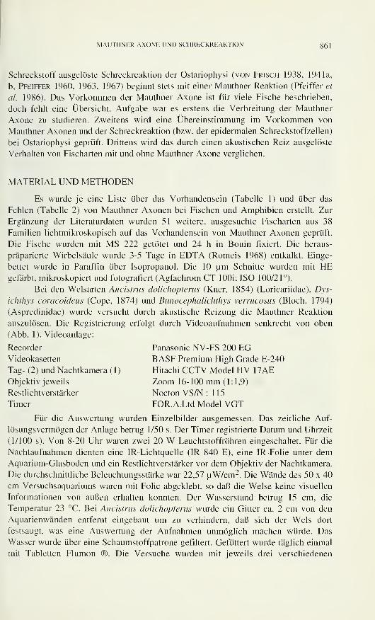
MAUTHNER AXONE UND SCHRECKREAKTION 861
Schreckstoff ausgelöste Schreckreaktion der Ostariophysi (von Frisch 1938, 1941a,
b. Pfeiffer 1960, 1963, 1967) beginnt stets mit einer Mauthner Reaktion (Pfeiffer et
al. 1986). Das Vorkommen der Mauthner Axone ist für viele Fische beschrieben,
doch fehlt eine Übersicht. Aufgabe war es erstens die Verbreitung der Mauthner
Axone zu studieren. Zweitens wird eine Übereinstimmung im Vorkommen von
Mauthner Axonen und der Schreckreaktion (bzw. der epidermalen Schreckstoffzellen)
bei Ostariophysi geprüft. Drittens wird das durch einen akustischen Reiz ausgelöste
Verhalten von Fischarten mit und ohne Mauthner Axone verglichen.
MATERIAL UND METHODEN
Es wurde je eine Liste über das Vorhandensein (Tabelle 1) und über das
Fehlen (Tabelle 2) von Mauthner Axonen bei Fischen und Amphibien erstellt. Zur
Ergänzung der Literaturdaten wurden 51 weitere, ausgesuchte Fischarten aus 38
Familien lichtmikroskopisch auf das Vorhandensein von Mauthner Axonen geprüft.
Die Fische wurden mit MS 222 getötet und 24 h in Bouin fixiert. Die heraus-
präparierte Wirbelsäule wurde 3-5 Tage in EDTA (Romeis 1968) entkalkt. Einge-
bettet wurde in Paraffin über Isopropanol. Die 10 um Schnitte wurden mit HEgefärbt, mikroskopiert und fotografiert (Agfachrom CT lOOi; ISO 100/21°).
Bei den Welsarten Ancistrus dolichopterus (Kner, 1854) (Loricariidae), Dys-
ichthys coracoideus (Cope, 1874) und Bunocephaiichthys verrucosus (Bloch, 1794)
(Aspredinidae) wurde versucht durch akustische Reizung die Mauthner Reaktion
auszulösen. Die Registrierung erfolgt durch Videoaufnahmen senkrecht von oben
(Abb. 1 ). Videoanlage:
Recorder Panasonic NV-FS 200 EGVideokasetten BASF Premium High Grade E-240
Tag- (2 ) und Nachtkamera ( 1 ) Hitachi CCTV Model HV 1
7
AEObjektiv jeweils Zoom 16-100 mm (1:1,9)
Restlichtverstärker Nocton VS/N : 1 1
5
Timer FOR.A.Ltd Model VGT
Für die Auswertung wurden Einzelbilder ausgemessen. Das zeitliche Auf-
lösungsvermögen der Anlage betrug 1/50 s. Der Timer registrierte Datum und Uhrzeit
(1/100 s). Von 8-20 Uhr waren zwei 20 W Leuchtstoffröhren eingeschaltet. Für die
Nachtaufnahmen dienten eine IR-Lichtquelle (IR 840 E), eine IR-Folie unter demAquarium-Glasboden und ein Restlichtverstärker vor dem Objektiv der Nachtkamera.
Die durchschnittliche Beleuchtungsstärke war 22,57 uW/cm2. Die Wände des 50 x 40
cm Versuchsaquariums waren mit Folie abgeklebt, so daß die Welse keine visuellen
Informationen von außen erhalten konnten. Der Wasserstand betrug 15 cm, die
Temperatur 23 °C. Bei Ancistrus dolichopterus wurde ein Gitter ca. 2 cm von den
Aquarienwänden entfernt eingebaut um zu verhindern, daß sich der Wels dort
festsaugt, was eine Auswertung der Aufnahmen unmöglich machen würde. Das
Wasser wurde über eine Schaumstoffpatrone gefiltert. Gefüttert wurde täglich einmal
mit Tabletten Flumon ®. Die Versuche wurden mit jeweils drei verschiedenen
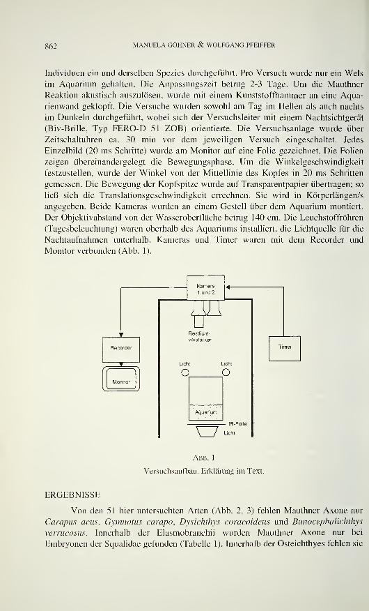
862 MANUELA GÖHNER & WOLFGANG PFEIFFER
Individuen ein und derselben Spezies durchgeführt. Pro Versuch wurde nur ein Wels
im Aquarium gehalten. Die Anpassungszeit betrug 2-3 Tage. Um die Mauthner
Reaktion akustisch auszulösen, wurde mit einem Kunststoffhammer an eine Aqua-
rienwand geklopft. Die Versuche wurden sowohl am Tag im Hellen als auch nachts
im Dunkeln durchgeführt, wobei sich der Versuchsleiter mit einem Nachtsichtgerät
(Biv-Brille, Typ FERO-D 51 ZOB) orientierte. Die Versuchsanlage wurde über
Zeitschaltuhren ca. 30 min vor dem jeweiligen Versuch eingeschaltet. Jedes
Einzelbild (20 ms Schritte) wurde am Monitor auf eine Folie gezeichnet. Die Folien
zeigen übereinandergelegt die Bewegungsphase. Um die Winkelgeschwindigkeit
festzustellen, wurde der Winkel von der Mittellinie des Kopfes in 20 ms Schritten
gemessen. Die Bewegung der Kopfspitze wurde auf Transparentpapier übertragen; so
ließ sich die Translationsgeschwindigkeit errechnen. Sie wird in Körperlängen/s
angegeben. Beide Kameras wurden an einem Gestell über dem Aquarium montiert.
Der Objektivabstand von der Wasseroberfläche betrug 140 cm. Die Leuchstoffröhren
(Tagesbeleuchtung) waren oberhalb des Aquariums installiert, die Lichtquelle für die
Nachtaufnahmen unterhalb. Kameras und Timer waren mit dem Recorder und
Monitor verbunden (Abb. 1).
Kamera
1 und 2
JIM1 r
uResti icht-
Recorder
Verstärker
Licht Licht
Timer
{{ Ì
Monitor
c <3
Aquarium
- IR-Folie
LichtVJAbb. 1
Versuchsaufbau. Erklärung im Text.
ERGEBNISSE
Von den 51 hier untersuchten Arten (Abb. 2, 3) fehlen Mauthner Axone nur
Carapus acus, Gymnotus campo, Dysichthys coracoideus und Bunocephalichthys
verrucosus. Innerhalb der Elasmobranchii wurden Mauthner Axone nur bei
Embryonen der Squalidae gefunden (Tabelle 1). Innerhalb der Osteichthyes fehlen sie
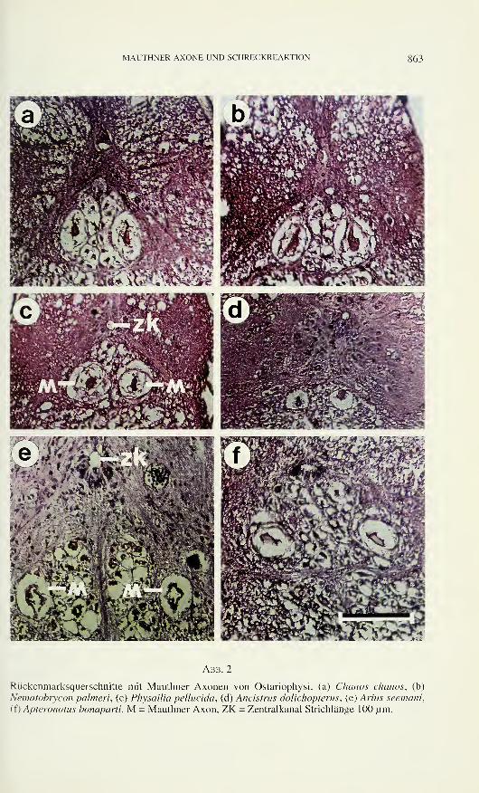
MAUTHNER AXONE UND SCHRECKREAKTION 863
Abb. 2
Rückenmarksquerschnitte mit Mauthner Axonen von Ostariophysi: (a) Chanos chanos, (b)
Nematobrycon palmeri, (c) Physailia pellucida, (d) Ancistrus dolichoptems, (e) Arius seemani,
ff) Apteronotus bonaparti. M = Mauthner Axon, ZK = Zentralkanal Strichlänge 100 um.
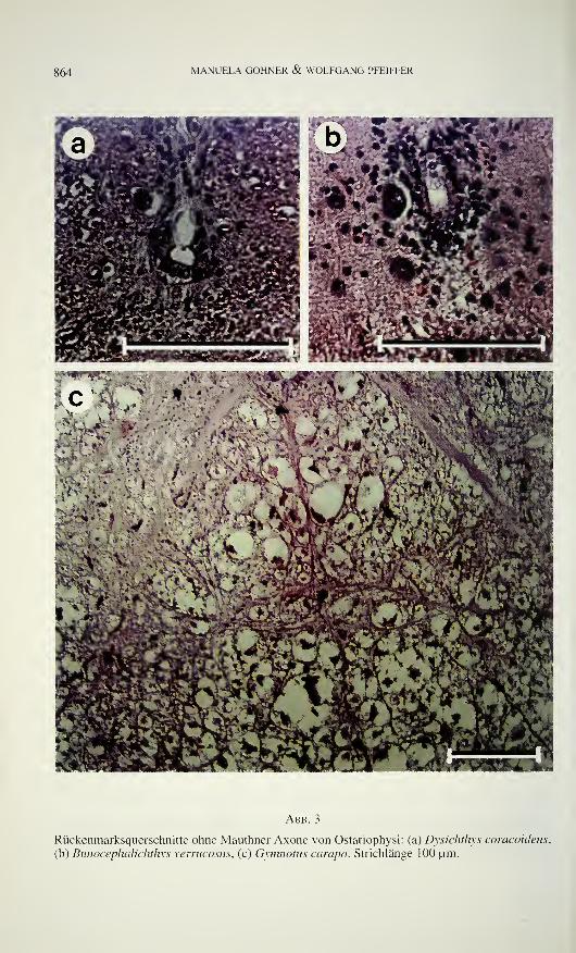
864 MANUELA GÖHNER & WOLFGANG PFEIFFER
Abb. 3
Rückenmarksquerschnitte ohne Mauthner Axone von Ostariophysi: (a) Dysichthys coracoideus,
(b) Bunocephalichthys verrucosus, (c) Gymnotus carapo. Strichlänge 100 um.

MAUTHNER AXONE UND SCHRECKREAKTION 865
Tabelle 1.
Fische - und Amphibien mit Mauthnerzellen (Spezies ohne Literaturangabe wurden von uns
untersucht)
OrdnungFamilie Art
Petromyzoniformes
Petromyzonidae Petromyzones
Petromyzon fluviatilis
Petromyzon marinus
Squaliformes
Squalidae
(nur Embryonen)
Chimaeriformes
Chimaeridae
Petromyzon planeri
dogfish sharks
Scymnorhinus lìcha
Squalus acanthias
ChimaeraChimaera (juvenil)
Hydrolagus
Coelacanthiformes
Coelacanthidae Latimeria chalumnaeCeratodiformes
Ceratodidae Neoceratodus
Lepidosireniformes
Lepidosirenidae Lepidosiren
Protopteridae Protopterus annectens
Acipenseriformes
Acipenseridae
Polyodontidae
Polypteriformes
Polypteridae
Acipenser ruthenus
Acipenser
paddlefishes, Löffelstöre
Literatur
ZOTTOLI (1978b)
Stefanelli (1932, 1933), Whiting (1957)
Currie & Carlsen (1987a,b), Rovainen(1974),
Stefanelli (1932, 1933)
Stefanelli (1932, 1933, 1951)
ZoTTOLi(1978b)
Bone (1977), Bone & Marshall (1983)
Bone (1977), Bone & Marshall (1983)
ZoTTOLi(1978b)
Bone & Marshall (1983)
Bone & Marshall (1983)
ZoTTOLi(1978b)
ZoTTOLi(1978b)
ZoTTOLi(1978b)
Beccari (1907), Wilson (1959), Zottoli
(1978b)
Beccari (1907), Otsuka ( 1964a)
Zottoli (1978b)
Zottoli (1978b)
Calamoìchihys calabarìcus
Polypterus'
Zottoli ( 197 8b)
Polypterus annectens Beccari ( 1 907)
Lepisosteiformes
Lepisosteidae Lepisosteus Zottoli (1978b)
Amiiformes
Amiidae Amia calva Zottoli (1978b)
Osteoglossiformes
Notopteridae featherbacks, Messerfische Zottoli (1978b)
Mormyridae
Notacanthiformes
Halosauridae
Notacanthidae
Clupeiformes
Clupeidae
mormyrids, Nilhechte
Gnathonemus petersìi
Aldrovandia
Halosauropsis
Halosaurus
Polyacanthonotus
Clupea
Clupea harengus
Clupea pollasi
Etrumeus micropus
Zottoli (1 978b)
Bone & Marshall (1983)
Bone & Marshall (1983)
Bone & Marshall (1983)
Bone & Marshall (1983)
Bone & Marshall (1983)
Otsuka (1964a)
Uchihashi et al. (1960)
Uchihashi et al. (I960)
866 MANUELA GÖHNER & WOLFGANG PFEIFFER
Engraulidae
Gonorynchiformes
Chanidae
Kneriidae
Phractolaemidae
Cypriniformes
Cyprinidae
Catostomidae
Cobitidae
Gyrinocheilidae
Homalopteridae
Harengula zunasi
herrings, Heringe
Konosirus punctatus
Sardinops melanosticta
anchovis, Sardellen
Engraulis japonica
Chanos chanos
Kneria maydelli
Phractolaemus ansorgei
Acheilognathus lanceolata
Abramis bramaBarbus fluviatilis
Brachydanio albolineatus
Brachydanio rerio
Carassius auratus
carps, minnows, Weißfische
Cyprinus carpio
Danio malabaricus
Gobio fluviatilis
Labeo rohita
Leuciscus cephalus
Luciosoma spilopleura
Notemigonus crysoleucas
Phoxinus phoxinus
Rutilus rutilus
Tinca tinca
Tribolodon hakonensis
Catostomus commersoni
suckers, Sauger
Acanthophthalmus kuhlii
Cobitis fossilis
loaches, Schmerlen
Misgurnus anguillicaudatus
Gyrinocheilus aymonieri
Homalopterus
Sinohomaloptera
Uchihashi et al. (1960)
Zottoli (1978b)
Uchihashi et al. ( 1 960)
Uchihashi et al. (1960)
Zottoli (1978b)
Uchihashi et al. (1960)
Uchihashi et al. (1960)
Mayser(1881)Mayser(1881)Kimmel(1972)Alfei et cd. (1991, 1992), Baffoni & Serra
(1952), Eaton étal. (1977a,b, 1984),
Eaton & Farley (1973, 1974), Eaton &Kimmel (1980), Eaton & Nissanov (1985),
KiMMELefa/. (1974, 1980, 1981, 1982),
Prugh et al. (1982)
Bodian (1937), Celio (1976), Diamond(1971), Eaton et al. (1977a), EdstrOm(1964), Faber & Korn (1975), Furami et al.
( 1965), Furshpan & Furukawa (1962),
Furukawa et al. (1963), Kohno (1970),
Otsuka (1962, 1964a), Robertson et al.
(1963), Triller & Korn (1982).
Uchihashi et al. (1960), Yasargil &Diamond (1968), Zottoli (1977)
Zottoli (1978b)
Beccari (1907), Otsuka (1962, 1964a),
Schwartz (1975), Uchihashi et al. (1960)
Belsare(1975)Mayser(1881)Belsare (1975)
Mayser(1881)
Zottoli (1978b)
Otsuka (1964a)
Otsuka (1964a)
Baumann & Yasargil (1981), Celio (1976),
Otsuka (1964a), Triller & Korn (1980,
1982), Yasargil étal. (1982, 1986),
Yasargil & Sandri (1987)
Uchihashi et al. (1960)
Zottoli (1978b)
Zottoli (1978b)
Mayser(1881)Zottoli (1978b)
Otsuka (1962, 1964a)
MAUTHNER AXONE UND SCHRECKREAKTION 867
Characiformes
Characidae
Lebiasinidae
Gasteropelecidae
Anoptichthys jordani
Astyanax bimaculatus
Astyanax mexicanus
characins. Salmler
Hydrocynus maculatus
HyphessobryconßammeusMicralestes interruptus
Nematobrycon palmeti
Pastella maxillaris
Stevardia riisei
Nannostomus beckfordi
Poecilobrycon ocellatus
Carnegiella strigata
Gasteropelecus sternicla
Otsuka (1964a)
Otsuka( 1964a)
Zottoli (1978b)
Eaton et al. (1977a)
Otsuka (1964a)
Schwartz (1975)
Hemiodontidae
Anostomidae
Citharinidae
Ctenoluciidae
Crenuchidae
Siluriformes
Siluridae
Schilbeidae
Ictaluridae
Bagridae
Clariidae
Malapteruridae
MochokidaeAriidae
Plotosidae
Callichthyidae
Loricariidae
Gymnotiformes
GymnotidaeApteronotidae
Gasteropelecus
hatchetfishes, Beilbauchfische Zottoli (1978b)
Hemiodus semitaeniatus
Anostomus anostomus
Leporinus affinis
Phago maculatus
Boulengerella lucia
Crenuchus spilurus
Eaton et al. (1977a)
Auerbach & Benett (1969), Eaton et al.
(1977a)
UvsEetal. (1985)
eurasian catfishes, WelseKryptopterus bicirrhis
Parasilurus asotus
Pangasius sutchi
Physailia pellucida
freshwater catfishes,
Katzenwelse
Ictalurus melas
Ictalurus nebulosus
Chrysichthys
bagrid catfish, Stachelwels
Mystus gulio
Ciarias batrachus
Heteropneustes fossilis
labyrinthic catfish, Raubwels
electric catfishes, Zitterwelse
Synodontis nigriventris
Arius seemaniPlotosus lineatus
callichthyid armoured
catfishes, Panzerwelse
Corydoras arcuatus
Dianema urostriata
Ancistrus dolichopterus
Gymnotus carapo
Apteronotus albifrons
Apteronotus bonaparti
Zottoli (1978b)
Otsuka (1962, 1964a), Uchihashi et al.
(1960)
Zottoli (1978b)
Bodian(1937)Eaton et al. ( 1977a), Otsuka (1964b),
Zottoli (1978a)
Otsuka (1964a)
Zottoli (1978b)
Belsare(1975)
Belsare (1975)
Zottoli (1978b)
Zottoli (1978b)
Zottoli (1978b)
Trujillo-Cenoz & Bertolotto (1989)
Eaton et al (1977a)
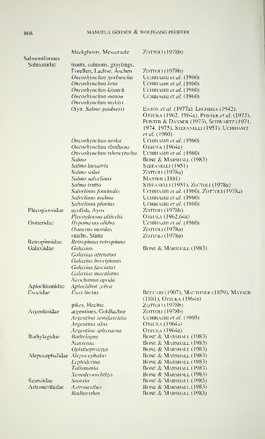
MANUELA GÖHNER & WOLFGANG PFEIFFER
Salmoniformes
Salmonidae
Plecoglossidae
Osmeridae
Retropinnidae
Galaxiidae
Aplochitonidae
Esocidae
blackghosts, Messeraale
trouts, salmons, graylings,
Forellen, Lachse, ÄschenOhcorhynchus gorbuscha
Oncorhynchus keta
Oncorhynchus kisutch
Oncorhynchus masouOncorhynchus mykiss
(Syn. Salmo gaidneri)
Oncorhynchus nerka
Oncorhynchus rhodurus
Oncorhynchus tshawytscha
SalmoSalmo lacustris
Salmo salar
Salmo salvelinus
Salmo trutta
Salvelinus fontinalis
Salvelinus malmaSalvelinus pluvius
ayofish, AyosPlecoglossus altivelis
Hypomesus olidus
Osmerus mordaxsmelts. Stinte
Retropinna retropinna
Galaxias
Galaxias attenatus
Galaxias brevipinnis
Galaxias fasciatus
Galaxias maculatus
Neochanna apodaAplochiton zebra
Esox lucius
pikes, Hechte
Argentinidae argentines, Goldlachse
Argentina semifasciata
Argentina silus
Argentina sphyraena
Bathylagidae Bathylagus
Nansenia
Opisthoproctus
Alepocephalidae Alepocephalus
Leptoderma
Talismania
Xenodermichthys
Searsiidae Searsia
Astronesthidae Astronesthes
Radinesthes
Zottoli (1978b)
Zottoli (1978b)
Uchihashi et al. (1960)
UCHIHASHI et al. (I960)
Uchihashi et al. (1960)
Uchihashi et al. (1960)
Eaton et al. (1977a), Leghissa (1942),
Otsuka (1962, 1964a), Pfister et al. (1973),
Pfister & Danner (1973), Schwartz (1971,
1974, 1975), Stefanelli (1951), Uchihashi
et al. (1960)
Uchihashi et al. (1960)
Otsuka (1964a)
Uchihashi et al. (1960)
Bone & Marshall (1983)
Stefanelli (1951)
Zottoli (1978a)
Mayser(1881)Stefanelli (1951), Zottoli (1978a)
Uchihashi et al. (1960), Zottoli (1978a)
Uchihashi et al. (1960)
Uchihashi et al. (1960)
Zottoli (1978b)
Otsuka (1962,64a)
Uchihashi et al. (1960)
Zottoli (1978a)
Zottoli (1978a)
Bone & Marshall ( 1983)
Beccari (1907), Mauthner (1859), Mayser(1881), Otsuka (1964a)
Zottoli (1978b)
Zottoli (1978b)
Uchihashi et al. (1960)
Otsuka (1964a)
Otsuka (1964a)
Bone & Marshall (1983)
Bone & Marshall (1983)
Bone & Marshall ( 1 983 )
Bone & Marshall (1983)
Bone & Marshall (1983)
Bone & Marshall (1983)
Bone & Marshall (1983)
Bone & Marshall (1983)
Bone & Marshall (1983)
Bone & Marshall (1983)
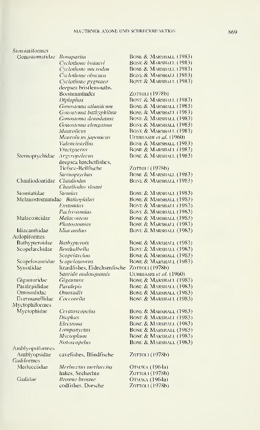
MAUTHNKR AXONE UNI) SCI IKI X'KKHAkHON 869
Stomiatiformes
Gonostomatidae
Sternoptychidae
Bonapartia
Cyclothone braueri
Cyclothone microdon
Cyclothone obscurci
Cyclothone pygmaeadeepsea bristlemouths,
Borstenmünder
Diplophos
Gonostoma atlanticum
Gonostoma bathyphilum
Gonostoma denudation
Gonostoma elongation
Maurolicus
Maurolicus japonicus
Valencienellus
Vinciguerra
Argyropelecus
deepsea hatchetfishes,
Tiefsee-Beilfische
Sternoptychus
Chauliodus
Chauliodus sloani
Stomias
Chauliodontidae
Stomiatidae
Melanostomiatidae Bathophilus
EustomiasPachystomias
Malacosteus
Photostomias
Idiacanthus
Malacosteidae
Idiacanthidae
Aulopiformes
Bathypteroidae
Scopelarchidae
Scopelosauridae
Synodidae
Giganturidae
Paralepididae
OmosudidaeEvermanellidae
Myctophiformes
Myctophidae
AmblyopsiformesAmblyopsidae
Gadiformes
Merlucciidae
Gadidae
Bone & Marshall (1983)
Bone & Marshall (1983)
Bone & Marshall (1983)
Bone & Marshall (1983)
Bone & Marshall (1983)
Zottoli (1978b)
Bone & Marshall (1983)
Bone & Marshall (1983)
Bone & Marshall (1983)
Bone & Marshall (1983)
Bone & Marshall (1983)
Bone & Marshall (1983)
Uchihashi et al. (1960)
Bone & Marshall (1983)
Bone & Marshall (1983)
Bone & Marshall (1983)
Zottoli (1978b)
Bone & Marshall (1983)
Bone & Marshall (1983)
Bathypterois
Benthalbella
Scopelarchus
Scopelosaurus
lizardfishes. Eidechsenfische
Saurida undosquamis
Gigantura
Paralepis
OmosudisCoccorella
Ceratoscopelus
Diaphus
Electrona
Lampanyctus
MyctophumNotoscopelus
BoneBoneBoneBoneBoneBoneBone
& Marshall& Marshall& Marshall& Marshall& Marshall& Marshall& Marshall
(1983)
(1983)
(1983)
(1983)
(1983)
(1983)
(1983)
Bone & Marshall (1983)
Bone & Marshall (1983)
Bone & Marshall (1983)
Bone & Marshall (1983)
Zottoli (1978b)
Uchihashi et al. (1960)
Bone & Marshall (1983)
Bone & Marshall (1983)
Bone & Marshall (1983)
Bone & Marshall (1983)
Bone & Marshall (1983)
Bone & Marshall (1983)
Bone & Marshall (1983)
Bone & Marshall (1983)
Bone & Marshall (1983)
Bone & Marshall (1983)
cavefishes, Blindfische Zottoli (1978b)
Merluccius merluccius
hakes, Seehechte
Brosme brosmecodfishes, Dorsche
Otsuka (1964a)
Zottoli (1978b)
Otsuka (1964a)
Zottoli (1978b)
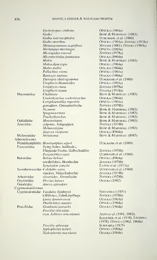
870 MANUELA GÖHNER & WOLFGANG PFEIFFER
Macrouridae
Ophidiidae
Zoarcidae
Enchelyopus cimbrius
GadusGadus macrocephalus
Gadus morrhuaMelanogrammus aeglefinus
Merlangius merlangus
Microgadus tomcodMicromesistius poutassou
MolvaMolva dipterygia
Molva molva
Pollachius virens
Raniceps raninus
Theragra chalcogrammaUrophycis blennioides
Urophycis chuss
Urophycis tenuis
Chalinura
Coelorhynchus coelorhynchus
Coryphaenoides rupestris
grenadiers, Grenadierfische
NezumiaSpagemacrurus
Trachyrhynchus
Monomitopuseelpouts, AalquappenMelanostigma
Zoarces viviparus
MelanonusMelanonidae
Atheriniformes
Hemirhamphidae Hemiramphus sajori
Exocoetidae flying fishes, halfbeaks,
Fliegende Fische, Halbschnäbler
Prognichthys agooBelonidae Belone belone
needlefishes, Hornhechte
Xenotodon cancila
Scomberesocidae Cololabis saira
sauriers, Makrelenhechte
Atherinidae silversides, Ährenfische
Oryziatidae Oryzias latipes
Goodeidae Ameca splendens
Cyprinodontiformes
Cyprinodontidae Fundulus (Embryo)
killifishes, Zahnkärpflinge
Limia dominicensis
Phallichthys amates
Poeciliidae Gambusia patruelis
Poecilia reticulata
(syn. Lebistes reticulatus)
Poecilia sphenops
Xiphophorus helleri
Xiphophorus maculatus
Otsuka (1964a)
Bone & Marshall (1983)
Uchuhashi et al. (1960)
Otsuka (1964a), Zottoli (1978a)
Mayser (1881), Otsuka (1964a)
Otsuka (1964a)
Zottoli (1978a)
Otsuka (1964a)
Bone & Marshall (1983)
Otsuka (1964a)
Otsuka (1964a)
Otsuka (1964a)
Otsuka (1964a)
Uchihashi et al (1960)
Otsuka (1964a)
Zottoli (1978a)
Zottoli (1978a)
Bone & Marshall (1983)
Otsuka (1964a)
Otsuka (1964a)
Zottoli (1978b)
Bone & Marshall (1983)
Bone & Marshall (1983)
Bone & Marshall (1983)
Bone & Marshall (1983)
Zottoli (1978b)
Bone & Marshall (1983)
Otsuka (1964a)
Bone & Marshall (1983)
Uchihashi et al. (1960)
Zottoli (1978b)
Uchihashi et al. (1960)
Otsuka (1964a)
Zottoli (1978b)
Eaton et al. (1977a)
Uchihashi et al. (1960)
Zottoli (1978b)
Zottoli (1978b)
Otsuka (1962)
Stefanelli (1951)
Zottoli (1978b)
Otsuka (1964a)
Otsuka (1964a)
Otsuka (1964a)
Alfei et al. (1991, 1992),
Jakoubek et al. (1970), Leghissa
(1978), Otsuka (1962, 1964a)
Schwartz (1975)
Otsuka (1964a)
Otsuka (1964a)
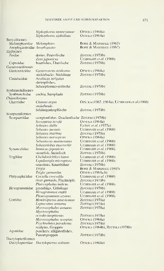
MAUTHNER AXONE UND SCHRECKREAKTION 871
Beryciformes
Melamphaeidae
Anoplogasteridae
Zeiformes
Zeidae
Caproidae
Gasterosteiformes
Gasterosteidae
Centriscidae
Synbranchiformes
Synbranchidae
Channiformes
Channidae
Scorpaeniformes
Scorpaenidae
Synanceiidae
Trialidae
Platycephalidae
Hexagrammidae
Cottidae
Agonidae
Xiphophorus montezumaeXiphophorus xiphidium
MelamphaesAnoplogaster
dories, Petersfische
Zeus japonicus
boarfishes, Eberfische
Gasterosteus aculeatus
sticklebacks, Stichlinge
Aeoliscus strigatus
shrimpfishes,
Schnepfenmesserfische
cuchia, Sumpfaale
Channel argus
snakeheads,
Schlangenkopffische
scorpionfishes. Drachenfische
Scorpaena scrofa
Sebastes dallii
Sebastes biennis
Sebastes marinus
Sebastes norvegicus
Sebastiscus marmoratus
Sebastolobus macrochir
Inimicus japonicus
stonefish, Steinfisch
Chelidonichthys kumuLepidotrigla microptera
searobins, Knurrhähne
Trigla
Trigla gurnardus
Cociella crocodila
river gurnards, Flachköpfe
Platycephalus indicus
greenlings, Grünlinge
Hexagrammos otakii
Pleurogrammus azonus
Hemitripterus americanus
Leptocottus armatus
Myoxocephalus aenaeus
Myoxocephalus
oetodeeimspinosus
Myoxocephalus scorpius
Psychrolutes paradoxus
sculpins, Groppenpoachers, alligatorfishes,
Panzergroppen
Otsuka (1964a)
Otsuka (1964a)
Bone & Marshall (1983;
Bone & Marshall (1983;
ZottolK 1978b)
Uchihashi et al. (1960)
Zottoli (1978b)
Otsuka (1964a)
Zottoli (1978b)
Dactylopteriformes
Dactylopteridae Dactylopterus volitans
Zottoli (1978b)
Zottoli (1978b)
Otsuka (1962, 1964a), Uchihashi etal.{\960)
Zottoli (1978b)
Zottoli (1978b)
Otsuka (1964a)
Eaton et al. (1977a)
Uchihashi et al. ( 1 960)
Zottoli (1978a)
Otsuka (1964a)
Uchihashi et al. (1960)
Uchihashi et al. (1960)
Uchihashi et al. (1960)
Zottoli (1978b)
Uchihashi et al. (1960)
Uchihashi et al. (1960)
Zottoli (1978b)
Bone & Marshall (1983)
Otsuka (1964a,b)
Uchihashi et al. (1960)
Zottoli (1978b)
Uchihashi et al. (1960)
Zottoli (1978b)
Uchihashi et al. (1960)
Uchihashi et al. (1960)
Zottoli (1978a)
Zottoli (1978a)
Zottoli (1978a)
Zottoli (1978a)
Otsuka (1964a)
Zottoli (1978a)
Otsuka (1964b), Zottoli (1978b)
Zottoli (1978b)
Otsuka (1964a)
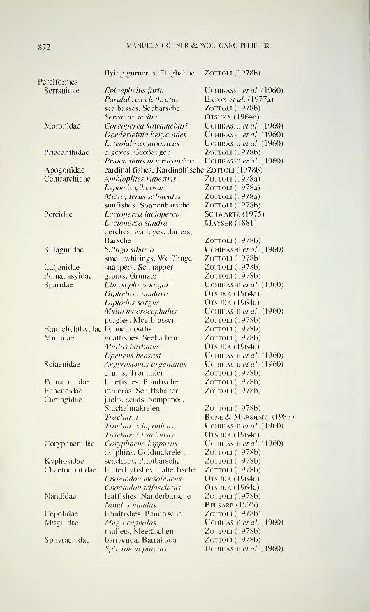
872 MANUELA GÖHNER & WOLFGANG PFEIFFER
Pereiforme s
Serranidae
Moronidae
Priacanthidae
Apogonidae
Centrarchidae
Percidae
Sillaginidae
Lutjanidae
Pomadasyidae
Sparidae
Emmelichthyidae
Mullidae
Sciaenidae
PomatomidaeEcheneidae
Carangidae
Coryphaenidae
Kyphosidae
Chaetodontidae
Nandidae
Cepolidae
Mugilidae
Sphyraenidae
flying gurnards, Flughähne Zottoli (1978b)
Epinephelus fario
Paralabrax clathratus
sea basses, Seebarsche
Serranus scriba
Coreoperca kawamebariDoederleinia beiycoides
Lateoiabrax japonicus
bigeyes, GroßaugenPriacanthus macracanthus
cardinal fishes. Kardinalfische
Ambloplites rupestris
Lepomis gibbosus
Micwpterus salmoides
sunfishes, Sonnenbarsche
Lucioperca lucioperca
Lucioperca sancirà
perches, walleyes, darters,
Barsche
Sillago sihama
smelt whitings, Weißlinge
snappers. Schnapper
grunts, Grunzer
Chrysophrys major
Diplodus annularis
Diplodus sargus
Mylio macrocephalus
porgies, Meerbrassen
bonnetmouths
goatfishes, Seebarben
Miillus barbatus
Upeneus bensasi
A rgyrosomus argen tatus
drums, Trommlerbluefishes. Blaufische
rémoras, Schiffshalter
jacks, scads, pompanos,
Stachelmakrelen
Trachurus
Trachurus japonicus
Trachurus trachurus
Cotyphaena hippurus
dolphins, Goldmakrelen
seachubs, Pilotbarsche
butterflyfishes, Falterfische
Chaetodon mesoleucus
Chaetodon trifasciatus
leaffishes, Nanderbarsche
Nandus nandus
bandfishes. Bandfische
Mugil cephalus
mullets. Meeräschen
barracuda, Barrakuda
Sphyraena pinguis
Uchihashi et al. (1960)
Eaton et al. (1977a)
Zottoli (1978b)
Otsuka (1964a)
Uchihashi et al. (1960)
Uchihashi et al. (1960)
Uchihashi et al. ( 1960)
Zottoli (1978b)
Uchihashi et al. (1960)
Zottoli (1978b)
Zottoli (1978a)
Zottoli (1978a)
Zottoli (1978a)
Zottoli (1978b)
Schwartz (1975)
Maysf.r(1881)
Zottoli (1978b)
Uchihashi et al. (1960)
Zottoli (1978b)
Zottoli (1978b)
Zottoli (1978b)
Uchihashi et al. ( 1 960)
Otsuka (1964a)
Otsuka (1964a)
Uchihashi et al. ( 1 960)
Zottoli (1978b)
Zottoli (1978b)
Zottoli (1978b)
Otsuka (1964a)
Uchihashi et al. (1960)
Uchihashi et al. (1960)
Zottoli (1978b)
Zottoli (1978b)
Zottoli (1978b)
Zottoli (1978b)
Bone & Marshall (1983)
Uchihashi et al. (1960)
Otsuka (1964a)
Uchihashi et al. (1960)
Zottoli (1978b)
Zottoli (1978b)
Zottoli (1978b)
Otsuka (1964a)
Otsuka (1964a)
Zottoli (1978b)
Belsare(1975)Zottoli (1978b)
Uchihashi et al. (1960)
Zottoli (1978b)
Zottoli (1978b)
Uchihashi et al. (1960)
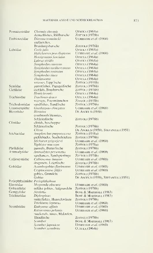
MAUTHNER AXONE UND SCHRECKREAKTION 873
Pomacentridae
Embiotocidae
Labridae
Scaridae
Cichlidae
Trachinidae
Trichodontidae
Uranoscopidae
Blenniidae
Clinidae
Stichaeidae
Pholididae
Ammodytidae
Callionymidae
Gobiidae
Periophthalmidae
Eleotridae
Gobioididae
GempylidaeTrichiuridae
Scombridae
Chrom is ehramis
damselfishes, Riffbarsche
Ditrema temmineki
surfperches,
Brandungsbarsche
Coris julis
Halichoeres poeeiìopterus
Hemigymnus fasciatus
Lahrus viridis
Symphodus cinereus
Symphodus mediterraneus
Symphodus rostratus
Symphodus tinca
Thalassoma
wrasses, Lippfische
parrotfishes, Papageifische
cichlids, Buntbarsche
HemichromisTrachinus draco
weevers, Petermännchen
sandfishes. Sandfische
Gnathagnus elongatus
Blennius
combtooth blennies.
Schleimfische
clinids, beschuppte
Schleimfische
Clinus
Anoplarchus purpureseenspicklebacks, Stachelrücken
Stichaeus grigorjewi
Xiphister mucosusgunnels, Butterfische
Ammodytes personatus
sandlances, Sandspierlinge
Callionymus lunatus
dragonets, Leierfische
Acanthogobius flavimanus
Cryptocen trus fiUfer
gobies, Grundein
Gobius
PeriophthaImusMogurnda obscurci
eellike gobies, Aalgrundeln
Neolotus
Diplospinus
snakefishes, HaarschwänzeTrichiurus lepturus
Euthynnus affinis
Katsuwonus pelamis
mackerels, tunas, Makrelen,
Thunfische
ScomberScomberjaponicusScomber scombrus
Otsuka (1964a)
Zottoei (1978b)
Uchihashi et al. ( 1 960)
ZoTTOLi (1978b)
Otsuka (1964a)
Uchihashi et al. (1960)
Otsuka (1964a)
Otsuka (1964a)
Otsuka (1964a)
Otsuka (1964a)
Otsuka (1964a)
Otsuka (1964a)
Otsuka (1964a)
Zottoli (1978b)
Zottoli (1978b)
Zottoli (1978b)
Otsuka (1964a)
Otsuka (1964a)
Zottoli (1978b)
Zottoli (1978b)
Uchihashi et al. ( 1960)
De Angelis (1950)
Zottoli (1978b)
Zottoli (1978b)
De Angelis (1950), Stefanelli (1951)
Zottoli (1978a)
Zottoli (1978b)
Uchihashi et al. (1960)
Zottoli (1978a)
Zottoli (1978b)
Uchihashi et al. ( 1 969)
Zottoli (1978b)
Uchihashi et al. (1960)
Zottoli (1978b)
Uchihashi et al. ( 1 960)
Uchihashi et al. ( 1 960)
Zottoli (1978b)
De Angelis ( 1 950), Stefanelli (1951)
Uchihashi et al. (1960)
Zottoli (1978b)
Bone & Marshall (1983)
Bone & Marshall (1983)
Zottoli (1978b)
Uchihashi et al. (1960)
Uchihashi et al. (1960)
Uchihashi et al. ( 1 960)
Zottoli (1978b)
Bone & Marshall (1983)
Uchihashi et al. ( 1 960)
Otsuka (1964a)

874 MANUELA GÖHNER & WOLFGANG PFEIFFER
Scomberomoriis niphonius
Thunnus thynnus
Stromateidae butterfishes, Medusenfische
Anabantidae climbing perces,
Kletterfische
Belontiidae Betta splendens
siamese fighting fish,
Kampffisch
Mastacembelidae mastacembelid eels,
Stachelaale
Mastacembelus loennbergi
knifejaws, MesserkieferOplegnathidae
Pleuronectiformes
Scophthalmidae
Bothidae
Pleuronectidae
Soleidae
Tetraodontiformes
Triacanthodidae
Balistidae
Lepidorhombus whiffiagonis
Psetta maximalefteye flounders,
Linksaugenflundern
Paralichthys californiens
Paralichthys dentatus
Paralichthys olivaceus
Tanakius kita.ha.rai
Citharichthys sordidus
Glyptocephalus cynoglossus
Glyptocephalus stellen
Hippoglossoides dubius
Hippoglossoides elassodon
Hippoglossoides platessoides
Hippoglossus stenolepis
Limanda ferrugineaLimanda herzensteini
Pia tiehthys flesus
Platichthys stellatus
Pleuronectes platessa
Pseudopleuronectes
americanus
righteye flounder, Scholle
Scophthalmus aquosus
Solea
soles, Seezungen
Tetraodontidae
Navodon modestus
Stephanolepis cirrhifer
triggerfishes, Drückerfische
Colomesus psittacus
Tetraodon fluviatilis
Tetraodon steindachneri
AmphibiaUrodela
Cryptrobranchidae Cryptobranchus japonicum
giant salamanders,
Riesensalamander
Ambystomatidae Ambystoma
Ambvstoma maculatum
Uchihashi et al. (1960)
UCHIHASHI et al. (1960)
ZoTTOLi (1978b)
Zottoli (1978b)
Uchihashi et al. (1960)
Zottoli (1978b)
Zottoli (1978b)
Eaton et al. (1977a)
Zottoli (1978b)
Otsuka (1964a,b)
OTSUKA(1964a,b)
Zottoli (1978b)
Eaton et al. (1977a)
Zottoli (1978a)
Uchihashi et al. ( 1960)
Uchihashi et al. (1960)
Zottoli (1978a)
Zottoli (1978a)
Uchihashi et al. (1960)
Uchihashi et al. (1960)
Zottoli (1978a)
Otsuka (1964a), Zottoli (1978a)
Uchihashi et al. (1960)
Zottoli (1978a)
Uchihashi et al. ( 1960)
Otsuka (1964a)
Zottoli (1978a)
Otsuka (1964a), Zottoli ( 1978a)
Zottoli (1978a, 1981)
Zottoli (1978b)
Zottoli (1978a)
De Angelis (1950), Stefanelli (1951)
Zottoli (1978b)
Uchihashi et al. (1960)
Uchihashi et al. (1960)
Zottoli (1978b)
Kingsbury (1895), Stefanelli (1951),
TUERKHEIM(1903)
Zottoli (1978b)
Detwiler (1927), Herrick (1914),
Holtfreter ( 1 93 1 )
Piatt (1969)
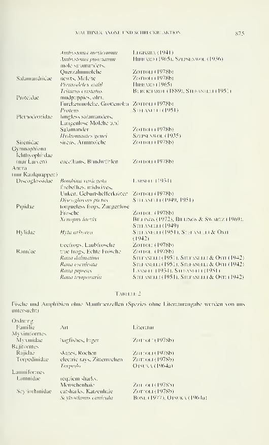
MAITHM.R AXON!- I'M) SCHRKCKRKAKTION 875
Salamandridae
Proteidae
Plethodontidae
Sirenidae
GymnophionaIchthyophiidae
(nur Larven)
Anura
(nur Kaulquappen:
Discoiilossidae
Pipidae
Hvlidae
Ranidae
Anibystonia me.xit mutin
Ainhystomci pitnctatnm
mole salamanders.
Querzahnmolchenewts. Molehe
Pleiirodeles waltl
Triinms criMains
mudpuppies. olm.
Furchenmolche. Grottenolm Zottoli ( 1478b)
Legissha ( 1441 )
hlbbari) ( 1965). szepsenwol ( 1936)
Zoi ioli ( 1978b)
Zottoli (1978b)
HlBBARI) ( 1965)
Burckhardt ( 1889). Stefanelli ( 1951 )
Proteus
lungless salamanders.
Lungenlose Molehe und
Salamander
Hydromantes genet
sirens. Armmolche
eaeeilians. Blindwühlen
Bombino variegata
firebellies. midwives.
Unken. Geburtshelferkröten
Discoglosso* pleins
tongueless frogs. Zungenlose
Frösche
Xenopns laevis
Hxla arborea
treefrogs. Laubfrösche
true frogs. Echte Frösche
Rana dalinatina
Rana esculento
Rana pipiens
Rana teinporaria
Su. i am. li. i ( 1951 )
Zottoli ( 1978b)
S/.L.P.SLN\\oi. ( 1935)
Zoi ioli ( 1978b)
Zottoli ( 1978b)
Lärm. Li. ( 1934)
Zottoli (1978b)
Stefanelli (1949. 1951)
Zottoli ( 1978b)
Billings (1972). Billings & S
Stei-anelli (1949)
Stefanelli ( 195 1 ). Stefanell
(1942)
Zottoli (1978b)
Zottoli (1978b)
Stefanelli ( 1951 ). Stefanelli
Stefanelli ( 1951 ). Stefanelli
Larsell ( 1934). Stefanelli ( I
Stefanelli ( 1951 ). Stefanelli
WART/ (
i & Osti
969)
& Osti (
& Osti (
95 I )
& Osti (
942)
942)
942)
Tabelle 2
Fische und Amphibien ohne Mauthner/ellen (Spezies ohne Literaturangabe wurden von uns
untersucht)
OrdnungFamilie
Myxiniformes
Myxinidae
Raji formes
Rajidae
Torpedinidae
Lanini formes
Lamnidae
Art
hagfishes. Irrger
skates. Rochenelectric rays. Zitterrochen
Torpedo
requiem sharks.
Menschenhaie
Scyliorhinidae catsharks. Katzenhaie
Scvliorhinns can iettici
Literatur
Zottoli ( 1978b)
Zottoli ( 1978b)
Zo itoli ( 1978b)
Orsi k.\ ( 1964a)
Zottoli (1978b)
Zottoli (1978b)
Bone ( 1977). Otsuka (1964a)
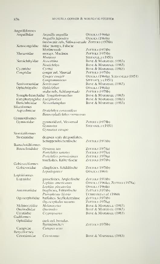
876 MANUELA GÖHNER & WOLFGANG PFEIFFER
Angui] 1 iformes
Ansuillidae
Xenoconszridae
Anguilla anguilla Otsuka ( 1 964a )
Anguilla japonica Otsuka (1964a)
freshwater eels. Süßwasseraale Zottoli (1978b)
false morays, Falsche
Muränenaale
morays. MuränenMuraenaAvocettina
Nemichthys
Cyemaconger eel. Meeraal
Conger conger
CongromuraenaSerrivomer
Ophichthus
snake eels, Schlangenaale
Synaphobranchidae Synaphobranchus
Eurypharyngidae Eurypharynx
Derichthyidae Nessorhamphns
Silunformes
Aspredinidae
Muraenidae
Nemichthyidae
CyemidaeConsridae
Serrivomeridae
Ophichthyidae
Zottoli (1978b)
Zottoli (1978b)
Stefanelli ( 1 95 1
)
Bone & Marshall (1983)
Bone & Marshall ( 1983)
Bone & Marshall (1983)
Zottoli (1978b)
Otsuka (1964a), Stefanelli (1951)
Stefanelli (1951)
Bone & Marshall (1983)
Otsuka (1964a)
Zottoli (1978b)
Bone & Marshall (1983)
Bone & Marshall (1983)
Bone & Marshall (1983)
Gymnotiformes
Gymnotidae
Stomiatiformes
Stomiatidae
Batrachoidiformes
Batrachoididae
Gobiesociformes
Gobiesocidae
Lophiiformes
Lophiidae
Antennariidae
Ogcocephalidae
Melanocetidae
Oneirodiidae
Ceratiidae
Gadiformes
Ophidiidae
Carapidae
Ben ciformes
Cetomimidae
Dysichthys coracoidens
Bunocephalichthys verrucosus
gymnotid eel, Messeraal
GymnotusGymnotus campo
deepsea scaly dragonfishes,
Schuppendrachenfische
Opsanus tau
Porichthys notatus
Porichthys porosissimus
toadfishes, Krötenfische
clingfishes. Schildfische
Lepadogaster
goosefishes, Anglerfische
Lophius americanus
Lophius piscatorius
frogfishes. Fühlerfische
Pterophryne histrio
batfishes, Seefledermäuse
Ogcocephalus nasutus
Melanocetus
Oneirodes
Cryptopsaras
cusk-eel, brotulas,
Bartmännchen
Carapus acus
Cetostomus
Zotolli (1978b)
Stefanelli (1951)
Zottoli (1978b)
Zottoli (1978a)
Zottoli (1978a)
Zottoli (1978a)
Zottoli (1978b)
Zottoli (1978b)
Otsuka (1964)
Zottoli (1978b)
Otsuka (1964a), Zottoli (1978a)
Otsuka (1964a)
Zottoli (1978b)
Uchihashi et al. (1960)
Zottoli (1978b)
Zottoli (1978a)
Bone & Marshall (1983)
Bone & Marshall (1983)
Bone & Marshall (1983)
Zottoli (1978b)
Bone & Marshall (1983)
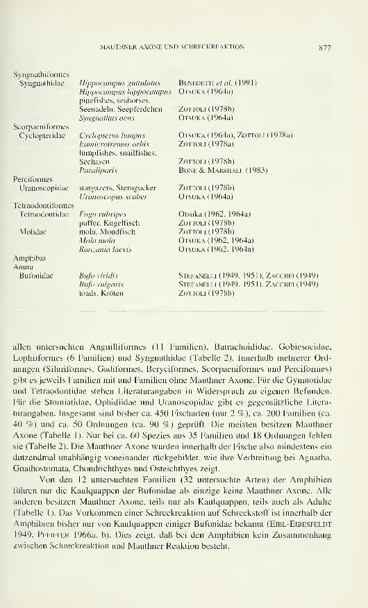
MAUTHNER AXONE UND SCHRECKREAKTION 877
Syngnathiformes
Syngnathidae Hippocampus guttulatus Benedetti et al. ( 1 99 1 )
Hippocampus hippocampus Otsuka (1964a)
pinefishes, seahorses,
Seenadeln, Seepferdchen Zottoli (1978b)
Syngnathus acus Otsuka (1964a)
Scorpaeniformes
Cyclopteridae Cyclopterus lumpus Otsuka (1964a), Zottoli (1978a)
Eamicrotremus orbis Zottoli (1978a)
lumpfishes, snailfishes,
Seehasen Zottoli (1978b)
Paraliparis Bone & Marshall (1983)
Perciformes
Uranoscopidae stargazers, Sterngucker Zottoli (1978b)
Uranoscopus scaber Otsuka (1964a)
Tetraodontiformes
Tetraodontidae Fugu rubripes Otsuka (1962, 1964a)
puffer, Kugelfisch Zottoli (1978b)
Molidae mola, Mondfisch Zottoli (1978b)
Mola mola Otsuka (1962, 1964a)
Ranzania laevis Otsuka (1962, 1964a)
Amphibia
AnuraBufonidae Bufo viridis Stefanelli ( 1 949, 1 95 1 ), Zacchei ( 1 949)
Bufo vulgaris Stefanelli (1949, 1951), Zacchei (1949)
toads, Kröten Zottoli (1978b)
allen untersuchten Anguilliformes (11 Familien), Batrachoididae, Gobiesocidae,
Lophiiformes (6 Familien) und Syngnathidae (Tabelle 2). Innerhalb mehrerer Ord-
nungen (Siluriformes, Gadiformes, Beryciformes, Scorpaeniformes und Perciformes)
gibt es jeweils Familien mit und Familien ohne Mauthner Axone. Für die Gymnotidae
und Tetraodontidae stehen Literaturangaben in Widerspruch zu eigenen Befunden.
Für die Stomiatidae, Ophidiidae und Uranoscopidae gibt es gegensätzliche Litera-
turangaben. Insgesamt sind bisher ca. 450 Fischarten (nur 2 %), ca. 200 Familien (ca.
40 %) und ca. 50 Ordnungen (ca. 90 %) geprüft. Die meisten besitzen Mauthner
Axone (Tabelle 1). Nur bei ca. 60 Spezies aus 35 Familien und 18 Ordnungen fehlen
sie (Tabelle 2). Die Mauthner Axone wurden innerhalb der Fische also mindestens ein
dutzendmal unabhängig voneinander rückgebildet, wie ihre Verbreitung bei Agnatha,
Gnathostomata, Chondrichthyes und Osteichthyes zeigt.
Von den 12 untersuchten Familien (32 untersuchte Arten) der Amphibien
führen nur die Kaulquappen der Bufonidae als einzige keine Mauthner Axone. Alle
anderen besitzen Mauthner Axone, teils nur als Kaulquappen, teils auch als Adulte
(Tabelle 1). Das Vorkommen einer Schreckreaktion auf Schreckstoff ist innerhalb der
Amphibien bisher nur von Kaulquappen einiger Bufonidae bekannt (Eibl-Eibesfeldt
1949, Pfeiffer 1966a, b). Dies zeigt, daß bei den Amphibien kein Zusammenhangzwischen Schreckreaktion und Mauthner Reaktion besteht.
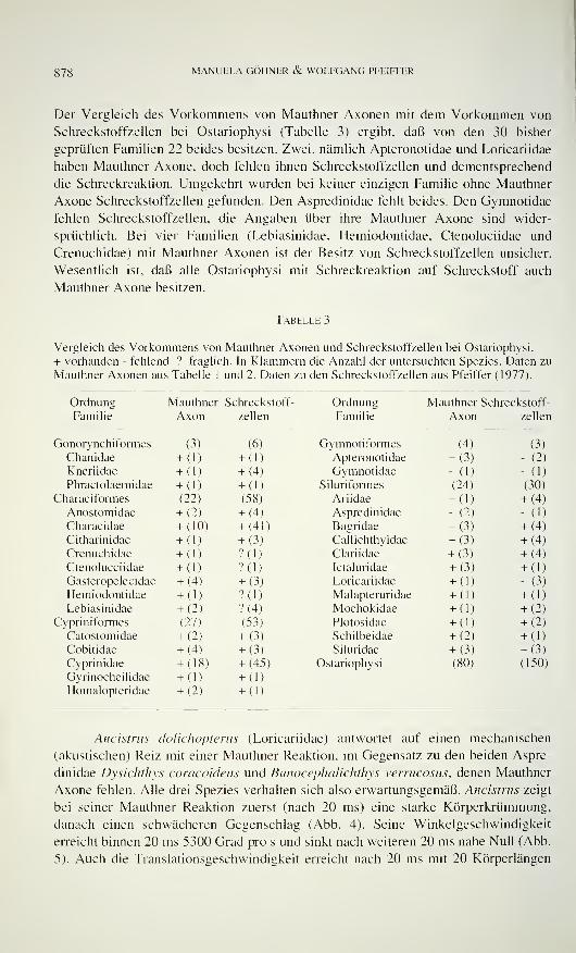
878 MANUELA GÖHNER & WOLFGANG PFEIFFER
Der Vergleich des Vorkommens von Mauthner Axonen mit dem Vorkommen von
Schreckstoffzellen bei Ostariophysi (Tabelle 3) ergibt, daß von den 30 bisher
geprüften Familien 22 beides besitzen. Zwei, nämlich Apteronotidae und Loricariidae
haben Mauthner Axone, doch fehlen ihnen Schreckstoffzellen und dementsprechend
die Schreckreaktion. Umgekehrt wurden bei keiner einzigen Familie ohne Mauthner
Axone Schreckstoffzellen gefunden. Den Aspredinidae fehlt beides. Den Gymnotidae
fehlen Schreckstoffzellen, die Angaben über ihre Mauthner Axone sind wider-
sprüchlich. Bei vier Familien (Lebiasinidae, Hemiodontidae, Ctenoluciidae und
Crenuchidae) mit Mauthner Axonen ist der Besitz von Schreckstoffzellen unsicher.
Wesentlich ist, daß alle Ostariophysi mit Schreckreaktion auf Schreckstoff auch
Mauthner Axone besitzen.
Tabelle 3
Vergleich des Vorkommens von Mauthner Axonen und Schreckstoffzellen bei Ostariophysi.
+ vorhanden - fehlend ? fraglich. In Klammern die Anzahl der untersuchten Spezies. Daten zu
Mauthner Axonen aus Tabelle 1 und 2. Daten zu den Schreckstoffzellen aus Pfeiffer (1977).
OrdnungFamilie
Mauthner Schreckstoff-
Axon zellen
OrdnungFamilie
Mauthner Schreckstoff-
Axon zellen
Gonorynchiformes (3) (6)
Chanidae + (1) + (DKneriidae + (1) + (4)
Phractolaemidae + (1) + (DCharacifonnes (22) (58)
Anostomidae + (2) + (4)
Characidae + (10) + (41)
Citharinidae + (1) + (3)
Crenuchidae + (1) ?(1)
Ctenolucciidae + (1) ?(1)Gasteropelecidae + (4) + (3)
Hemiodontidae + (1) ?(1)
Lebiasinidae + (2) ?(4)Cypriniformes (27) (53)
Catostomidae + (2) + (3)
Cobitidae + (4) + (3)
Cyprinidae + (18) + (45)
Gyrinocheilidae + (1) + (1)
Homalopteridae + (2) + (1)
Gvmnotiformes (4; (3)
Apteronotidae + (3 ) - (2)
Gymnotidae - (1 ) - (1)
Siluriformes (24 (30)
Ariidae + (1 + (4)
Aspredinidae - (2 ) - (1)
Bagridae + (3 + (4)
Callichthyidae + (3 + (4)
Clariidae + (3 + (4)
Ictaluridae + (3 ) +(DLoricariidae + (1 ) - (3)
Malapteruridae + (1 ) +(DMochokidae + (1 ) +(2)Plotosidae + (1 ) + (2)
Schilbeidae + (2 ) +(DSiluridae + (3 ) +(3)
Ostariophysi (80 (150)
Ancistnts doliciwpterus (Loricariidae) antwortet auf einen mechanischen
(akustischen) Reiz mit einer Mauthner Reaktion, im Gegensatz zu den beiden Aspre-
dinidae Dysichthys coracoideus und Bunocephalichthys verrucosus, denen Mauthner
Axone fehlen. Alle drei Spezies verhalten sich also erwartungsgemäß. Ancistrus zeigt
bei seiner Mauthner Reaktion zuerst (nach 20 ms) eine starke Körperkrümmung,
danach einen schwächeren Gegenschlag (Abb. 4). Seine Winkelgeschwindigkeit
erreicht binnen 20 ms 5300 Grad pro s und sinkt nach weiteren 20 ms nahe Null (Abb.
5). Auch die Translationsgeschwindigkeit erreicht nach 20 ms mit 20 Körperlängen
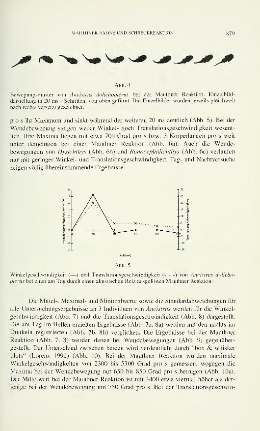
MAUTHNER AXONE UND SCHRECKREAKTION 879
Abb. 4
Bewegungsmuster von Ancistrus dolichopterus bei der Mauthner Reaktion. Einzelbild-
darstellung in 20 ms - Schritten, von oben gefilmt. Die Einzelbilder wurden jeweils gleichweit
nach rechts versetzt gezeichnet.
pro s ihr Maximum und sinkt während der weiteren 20 ms deutlich (Abb. 5). Bei der
Wendebewegung steigen weder Winkel- noch Translationsgeschwindigkeit wesent-
lich. Ihre Maxima liegen mit etwa 700 Grad pro s bzw. 3 Körperlängen pro s weit
unter denjenigen bei einer Mauthner Reaktion (Abb. 6a). Auch die Wende-
bewegungen von Dysichthys (Abb. 6b) und Bunocephalichthys (Abb. 6c) verlaufen
nur mit geringer Winkel- und Translationsgeschwindigkeit. Tag- und Nachtversuche
zeigen völlig übereinstimmende Ergebnisse.
Winkelgeschwindigkeit (— ) und Translationsgeschwindigkeit ( ) von Ancistrus dolicho-
pterus bei einer am Tag durch einen akustischen Reiz ausgelösten Mauthner Reaktion.
Die Mittel-, Maximal- und Minimalwerte sowie die Standardabweichungen für
alle Untersuchungsergebnisse an 3 Individuen von Ancistrus werden für die Winkel-
geschwindigkeit (Abb. 7) und die Translationsgeschwindigkeit (Abb. 8) dargestellt.
Die am Tag im Hellen erzielten Ergebnisse (Abb. 7a, 8a) werden mit den nachts im
Dunkeln registrierten (Abb. 7b, 8b) verglichen. Die Ergebnisse bei der Mauthner
Reaktion (Abb. 7. 8) werden denen bei Wendebewegungen (Abb. 9) gegenüber-
gestellt. Der Unterschied zwischen beiden wird verdeutlicht durch ''box & whisker
plots" (Lorenz 1992) (Abb. 10). Bei der Mauthner Reaktion wurden maximale
Winkelgeschwindigkeiten von 2300 bis 5300 Grad pro s gemessen, wogegen die
Maxima bei der Wendebewegung nur 650 bis 850 Grad pro s betrugen (Abb. 10a).
Der Mittelwert bei der Mauthner Reaktion ist mit 3400 etwa viermal höher als der-
jenige bei der Wendebewegung mit 750 Grad pro s. Bei der Translationsgeschwin-
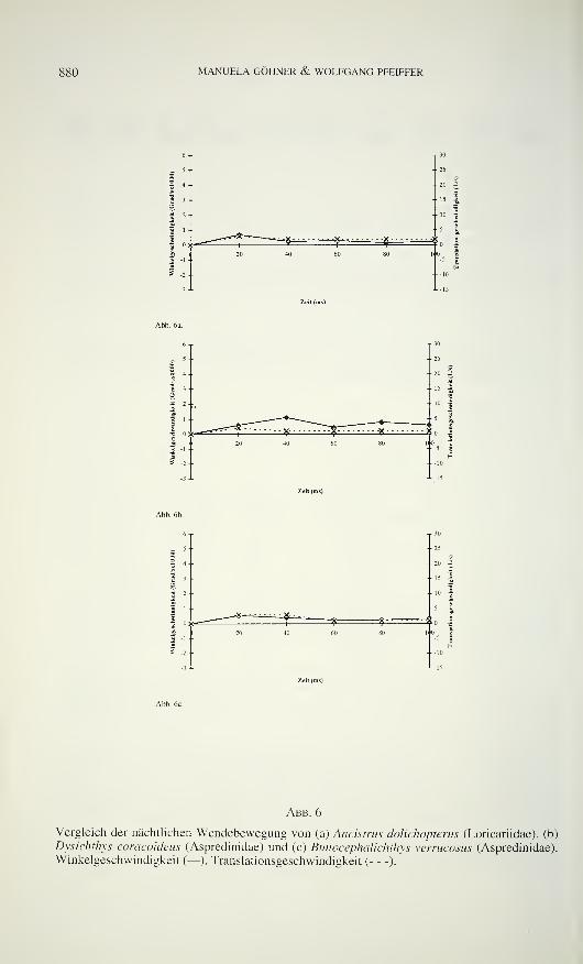
880 MANUELA GÖHNER & WOLFGANG PFEIFFER
30
-23
20 3
' 5 -E
10 I
Abb. 6
Vergleich der nächtlichen Wendebewegung von (a) Ancistrus dolichopterus (Loricariidae), (b)
Dysichthys coracoideus (Aspredinidae) und (c) Bunocephalichthys verrucosus (Aspredinidae).
Winkelgeschwindigkeit (— ), Translationsgeschwindigkeit ( ).
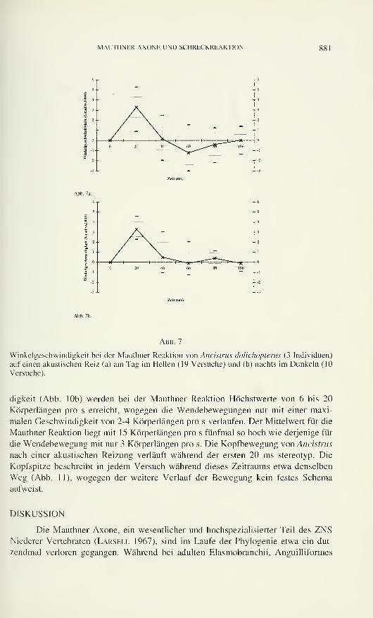
MAITHNHR AXONK l'ND SCIIRHCKRHAKTION 881
Abb. 7
Winkelgeschwindigkeit bei der Mauthner Reaktion von Ancistrus dolichoptems (3 Individuen)
auf einen akustischen Reiz (a) am Tag im Hellen (19 Versuche) und (b) nachts im Dunkeln (10
Versuche).
digkeit (Abb. 10b) werden bei der Mauthner Reaktion Höchstwerte von 6 bis 20
Körperlängen pro s erreicht, wogegen die Wendebewegungen nur mit einer maxi-
malen Geschwindigkeit von 2-4 Körperlängen pro s verlaufen. Der Mittelwert für die
Mauthner Reaktion liegt mit 15 Körperlängen pro s fünfmal so hoch wie derjenige für
die Wendebewegung mit nur 3 Körperlängen pro s. Die Kopfbewegung von Ancistrus
nach einer akustischen Reizung verläuft während der ersten 20 ms stereotyp. Die
Kopfspitze beschreibt in jedem Versuch während dieses Zeitraums etwa denselben
Weg (Abb. 1 1 ), wogegen der weitere Verlauf der Bewegung kein festes Schema
aufweist.
DISKUSSION
Die Mauthner Axone, ein wesentlicher und hochspezialisierter Teil des ZNSNiederer Vertebraten (Larsell 1967), sind im Laufe der Phylogenie etwa ein dut-
zendmal verloren gegangen. Während bei adulten Elasmobranchii, Anguilliformes
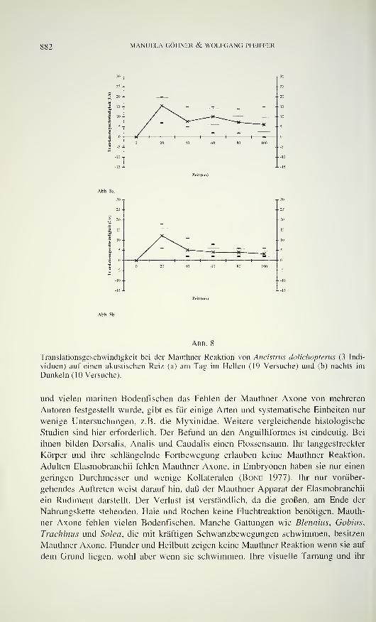
882 MANUELA GÖHNER & WOLFGANG PFEIFFER
Abb. 8
Translationsgeschwindigkeit bei der Mauthner Reaktion von Ancistrus dolichopterus (3 Indi-
viduen) auf einen akustischen Reiz (a) am Tag im Hellen (19 Versuche) und (b) nachts im
Dunkeln ( 10 Versuche).
und vielen marinen Bodenfischen das Fehlen der Mauthner Axone von mehreren
Autoren festgestellt wurde, gibt es für einige Arten und systematische Einheiten nur
wenige Untersuchungen, z.B. die Myxinidae. Weitere vergleichende histologische
Studien sind hier erforderlich. Der Befund an den Anguilliformes ist eindeutig. Bei
ihnen bilden Dorsalis, Analis und Caudalis einen Flossensaum. Ihr langgestreckter
Körper und ihre schlängelnde Fortbewegung erlauben keine Mauthner Reaktion.
Adulten Elasmobranchii fehlen Mauthner Axone, in Embryonen haben sie nur einen
geringen Durchmesser und wenige Kollateralen (Bone 1977). Ihr nur vorüber-
gehendes Auftreten weist darauf hin, daß der Mauthner Apparat der Elasmobranchii
ein Rudiment darstellt. Der Verlust ist verständlich, da die großen, am Ende der
Nahrungskette stehenden, Haie und Rochen keine Fluchtreaktion benötigen. Mauth-
ner Axone fehlen vielen Bodenfischen. Manche Gattungen wie Blennius, Gobius,
Trachinus und Solea, die mit kräftigen Schwanzbewegungen schwimmen, besitzen
Mauthner Axone. Flunder und Heilbutt zeigen keine Mauthner Reaktion wenn sie auf
dem Grund liegen, wohl aber wenn sie schwimmen. Ihre visuelle Tarnung und ihr
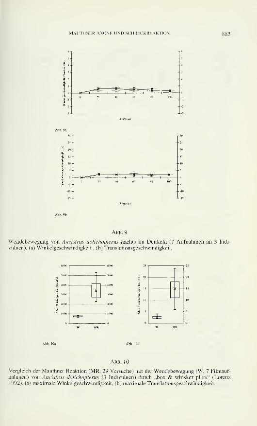
MAUTHNER AXONE UND SCHRECKREAKTION 883
Abb. 9
Wendebewegung von Ancistrus dolichopterus nachts im Dunkeln (7 Aufnahmen an 3 Indi-
viduen), (a) Winkelgeschwindigkeit, (b) Translationsgeschwindigkeit.
1
] c
4000
3000
i
U
1
|
=x=
3 :
CiW MR
Abb. 10
Vergleich der Mauthner Reaktion (MR. 29 Versuche) mit der Wendebewegung (W. 7 Filmauf-
nahmen) von Ancistrus dolichopterus (3 Individuen) durch „box & whisker plots" (Lorenz
1992). (a) maximale Winkelgeschwindigkeit, (b) maximale Translationsgeschwindigkeit.
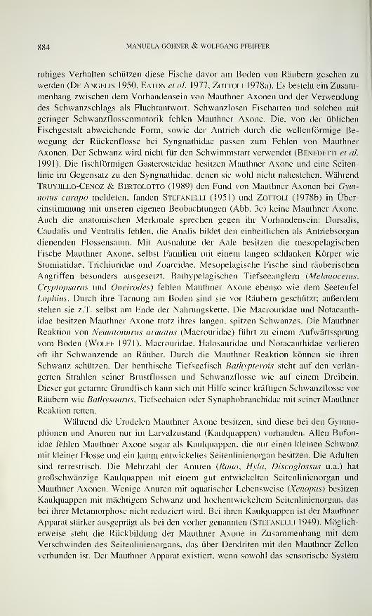
8g4 MANUELA GÖHNER & WOLFGANG PFEIFFER
ruhiges Verhalten schützen diese Fische davor am Boden von Räubern gesehen zu
werden (De Angelis 1950, Eaton et al. 1977, Zottoli 1978a). Es besteht ein Zusam-
menhang zwischen dem Vorhandensein von Mauthner Axonen und der Verwendung
des Schwanzschlags als Fluchtantwort. Schwanzlosen Fischarten und solchen mit
geringer Schwanzflossenmotorik fehlen Mauthner Axone. Die, von der üblichen
Fischgestalt abweichende Form, sowie der Antrieb durch die wellenförmige Be-
wegung der Rückenflosse bei Syngnathidae passen zum Fehlen von Mauthner
Axonen. Der Schwanz wird nicht für den Schwimmstart verwendet (Benedetti et cd.
1991). Die fischförmigen Gasterosteidae besitzen Mauthner Axone und eine Seiten-
linie im Gegensatz zu den Syngnathidae, denen sie wohl nicht nahestehen. Während
Truyjillo-Cenoz & Bertolotto (1989) den Fund von Mauthner Axonen bei Gym-
notus campo meldeten, fanden Stefanelli (1951) und Zottoli (1978b) in Über-
einstimmung mit unseren eigenen Beobachtungen (Abb. 3c) keine Mauthner Axone.
Auch die anatomischen Merkmale sprechen gegen ihr Vorhandensein: Dorsalis,
Caudalis und Ventralis fehlen, die Analis bildet den einheitlichen als Antriebsorgan
dienenden Flossensaum. Mit Ausnahme der Aale besitzen die mesopelagischen
Fische Mauthner Axone, selbst Familien mit einem langen schlanken Körper wie
Stomiatidae. Trichiuridae und Zoarcidae. Mesopelagische Fische sind räuberischen
Angriffen besonders ausgesetzt. Bathypelagischen Tiefseeanglern {Melanocetus,
Cryptopsaras und Oneirodes) fehlen Mauthner Axone ebenso wie dem Seeteufel
Lophius. Durch ihre Tarnung am Boden sind sie vor Räubern geschützt; außerdem
stehen sie z.T. selbst am Ende der Nahrungskette. Die Macrouridae und Notacanth-
idae besitzen Mauthner Axone trotz ihres langen, spitzen Schwanzes. Die Mauthner
Reaktion von Nematonurus armatus (Macrouridae) führt zu einem Aufwärtssprung
vom Boden (Wolff 1971). Macrouridae, Halosauridae und Notacanthidae verlieren
oft ihr Schwanzende an Räuber. Durch die Mauthner Reaktion können sie ihren
Schwanz schützen. Der benthische Tiefseefisch Bathypterois steht auf den verlän-
gerten Strahlen seiner Brustflossen und Schwanzflosse wie auf einem Dreibein.
Dieser gut getarnte Grundfisch kann sich mit Hilfe seiner kräftigen Schwanzflosse vor
Räubern wie Bathysaurus, Tiefseehaien oder Synaphobranchidae mit seiner Mauthner
Reaktion retten.
Während die Urodelen Mauthner Axone besitzen, sind diese bei den Gymno-
phionen und Anuren nur im Larvalzustand (Kaulquappen) vorhanden. Allen Bufon-
idae fehlen Mauthner Axone sogar als Kaulquappen, die nur einen kleinen Schwanz
mit kleiner Flosse und ein kaum entwickeltes Seitenlinienorgan besitzen. Die Adulten
sind terrestrisch. Die Mehrzahl der Anuren (Rana, Hyla, Discoglossus u.a.) hat
großschwänzige Kaulquappen mit einem gut entwickelten Seitenlinienorgan und
Mauthner Axonen. Wenige Anuren mit aquatischer Lebensweise (Xenopus) besitzen
Kaulquappen mit mächtigem Schwanz und hochentwickeltem Seitenlinienorgan, das
bei ihrer Metamorphose nicht reduziert wird. Bei ihren Kaulquappen ist der Mauthner
Apparat stärker ausgeprägt als bei den vorher genannten (Stefanelli 1949). Möglich-
erweise steht die Rückbildung der Mauthner Axone in Zusammenhang mit demVerschwinden des Seitenlinienorgans, das über Dendriten mit den Mauthner Zellen
verbunden ist. Der Mauthner Apparat existiert, wenn sowohl das sensorische System
MAUTHNER AXONE UND SCHRECKREAKTION 885
des Seitenlinienorgans als auch das motorische System des Schwanzes vorhanden ist.
Die Kaulquappen der Bufonidae haben als einzige Amphibien die Mauthner Axone
und damit die Mauthner Reaktion im Laufe der Phylogenie verloren; sie haben als
einzige Amphibien als wichtigen Schutz gegen den Verlust durch räuberische Über-
fälle eine Schreckreaktion auf den Schreckstoff aus der Haut von Artgenossen
entwickelt (Eibl-Eibesfeldt 1949, Pfeiffer 1966a, b).
Innerhalb der Fische ist eine ähnliche Schreckreaktion auf einen Schreckstoff
aus der Haut von Artgenossen bekannt (von Frisch 1938, 1941a, b). Die Schreck-
reaktion ist Besitz der meisten Ostariophysi sensu loto (d.h. Anotophysi oder Gono-
rynchiformes plus Otophysi. nämlich Siluriformes, Characiformes und Cyprini-
formes). Der Schreckstoff entstammt besonderen epidermalen Schreckstoffzellen
(Pfeiffer 1960, 1963, 1967). Die histologischen Ergebnisse über das Vorkommen
von Mauthner Axonen bei Ostariophysi (Tabelle 1, 2) werden erstmals mit denjenigen
über das Vorhandensein von Schreckstoffzellen (Pfeiffer 1977) verglichen (Tabelle
3). Nur die Aspredinidae und Gymnotidae haben beides rückgebildet, sowohl die
Mauthner Axone als auch die Schreckstoffzellen. Die Rückbildung muß aufgrund der
Verbreitung angenommen werden. Beide Familien sind nachtaktive Einzelgänger.
Während den Gymnotidae die Schwanzflosse fehlt, besitzen die Aspredinidae einen
dünnen, langen Schwanzstiel mit reduzierter Muskulatur. Auch die Loricariidae und
Apteronotidae haben Schreckstoffzellen und Schreckreaktion rückgebildet, unter
Beibehaltung ihrer Mauthner Axone. Auch sie sind nachtaktive Einzelgänger; dies
macht den Verlust ihrer Schreckreaktion verständlich. Die Apteronotidae besitzen
eine Schwanzflosse, die Loricariidae eine Schwanzmotorik, die ihnen bemerkens-
werte Mauthner Reaktionen erlaubt. Wie Verhaltensexperimente zeigten, besitzt
Ancistrus dolichopterus (Loricariidae) eine Mauthner Reaktion, im Gegensatz zu
Dysichthys coracoideus (Aspredinidae) und Bunocephalichthys verrucosus (Aspre-
dinidae). Die Ergebnisse passen zu den histologischen Befunden. Die graphische
Auswertung der Mauthner Reaktion von Ancistrus dolichopterus zeigt eine plötzliche
Erhöhung der Winkel- und Translationsgeschwindigkeit. Der Versuchsfisch wurde
akustisch gereizt wenn er ruhig am Boden lag. Während der schnellen Körperkrüm-
mung erreichte er die größte Winkel- und Translationsgeschwindigkeit (bis zu 5300
Grad/s und 20 Längen/s). Die erste Phase der Mauthner Reaktion ist beendet, wenn
sich die Winkelgeschwindigkeit Null Grad nähert. Zu diesem Zeitpunkt beginnt der
Gegenschlag. Er ist durch die relativ hoch bleibende Translationgeschwindigkeit
gekennzeichnet. Durch die schnelle Körperkrümmung bringt der Fisch seinen ver-
letzbaren Kopf aus der Gefahrenzone und leitet den kräftigen Schwanzschlag ein.
Zwischen den am Tag im Hellen erzielten Mauthner Reaktionen und den nachts im
Dunkeln registrierten besteht kein Unterschied. Eaton et al. (1977a) haben eine
Latenzzeit der Reaktion von 5-10 ms festgestellt. Sie arbeiteten mit einer besser
auflösenden Videoanlage (1 Bild in 5 ms). Aufgrund der geringen zeitlichen
Auflösung (1 Bild in 20 ms) konnte unsere Videoanlage nicht immer die Extremwerte
erfassen. Bei der Auswertung von mehreren Bewegungen zeigt sich, daß die Reaktion
innerhalb der ersten 20 ms mit einer stereotypen Bewegung von Kopf und Schwanz
nach einer Seite abläuft. Der Gegenschlag des Schwanzes ist nicht stereotyp, d.h. der
g86 MANUELA GÖHNER & WOLFGANG PFEIFFER
Winkel der neuen Schwimmrichtung nicht voraussagbar. Daß es sich bei den dar-
gestellten Auswertungen um Mauthner Reaktionen handelt, zeigt der Vergleich mit
einer Wendebewegung von Ancistrus dolichopterus. Hier treten nur geringe Winkel-
und Translationsgeschwindigkeiten auf. Die beiden Aspredinidae Dysichthys cora-
coideus und Bunocephalichthys verrucosus antworten auf akustische Reizung nicht
mit einer Mauthner Reaktion, entsprechend den histologischen Ergebnissen. Es
konnte nur ihre Wendebewegung registriert werden, die sich nicht von derjenigen des
Ancistrus unterscheidet. Zwischen einer ausgeprägten Schwanzmotorik und demBesitz von Mauthner Neuronen besteht ein Zusammenhang. Die Aspredinidae
schwimmen mit „Düsenantrieb" indem sie Wasser unter Druck aus den Kiemen-
spalten ausstoßen. Ihre Schwanzstielmuskulatur ist reduziert. Nur bei einer gut ent-
wickelten Schwanzmuskulatur kann die Mauthner Reaktion von Fischen genutzt
werden. Wenn die körperlichen Voraussetzungen nicht gegeben sind, geht der
Mauthner Apparat verloren, wie im Laufe der Phylogenie bei den Ostariophysi zwei-
mal, innerhalb der Teleostei öfter als ein dutzendmal geschehen.
LITERATUR
Alfei, L., L. Medolago-Albani, G.M.A. Zezze & A. Stefanelli. 1991. The cytoskeletal
composition of fish Mauthner axon during postlarval growth. Acta embiyologiae et
morphologiae experimentalis 12:67-68.
Alfei, L., L. Medolago-Albani, G.M.A. Zezze & A. Stefanelli. 1992. Morphogenetic ana-
lysis of Mauthner axon cytoskeletal components in adult and subadult fish. Journal ofSubmicroscopic Cytology and Pathology 24: 1 15-122.
Auerbach, A.A. & M.V.L. Benett. 1969. Chemically mediated transmission at a giant fiber
synapse in the central nervous system of a vertebrate. Journal of General Physiology
53:183-210.
Baffoni, G.M. & L. Serra. 1952. La citomorfosi del neurone mauthneriano di un teleosteo
durante lo sviluppo. Rivista di biologia 44:469-491.
Baumann, J.R. & G.M. Yasargil. 1981. Effect of acute increase in body temperature on the
excitatory and inhibitory synaptic transmission in fish spinal cord. Advances in
Physiological Sciences 32:73-75.
Bartelmez, C. 1915. Mauthner 's cell and the nucleus motorius tegmenti. Journal of Compara-tive Neurology 25:87-128.
Beccare N. 1907. Richerche sulle cellule e fibre del Mauthner e sulle loro conessioni in pesci
ed anfibii. Archivio italiano di anatomia e di embriologia 6:660-708.
Belsare, D.K. 1975. Comparative histomorphology of the Mauthner cell in some freshwater
teleosts. Zeitschrift für mikroskopisch-anatomische Forschung 89:804-814.
Benedetti, I., D. Sassi & A. Stefanelli. 1991. Mauthner neurons in syngnathid bony fishes.
Acta embiyologiae et morphologiae experimentalis 12:75-76.
Billings, S.M. 1972. Development of the Mauthner cell in Xenopus laevis: a light and electron
microscopic study of the pericaryon. Zeitschrift für Anatomie und Entwicklungsge-
schichte 136:168-191.
Billings, S.M. & FJ. Swartz. 1969. DNA content of Mauthner cell nuclei in Xenopus laevis:
a spectrophotometric study. Zeitschrift für Anatomie und Entwicklungsgeschichte
129:14-23.
Bodian, D. 1937. The structure of the vertebrate synapse. A study of the axon endings on
Mauthner 's cell and neighboring centers in the goldfish. Journal of Comparative Neu-
rology 68:117-159.
MAUTHNFR AXONE UND SCHRECKREAKTION
Bone, Q. 1977. Mauthner neurons in elasmobranchs. Journal of the Marine Biological
Association of the United Kingdom 57:253-259.
Bone, Q. & N.B. Marshall. 1983. The Mauthner system of deep sea teleosts. Journal of the
Marine Biological Association of the United Kingdom 63:881-896.
Burckhardt, K.R. 1889. Histologische Untersuchungen am Rückenmark der Tritonen. Archiv
für mikroskopische Anatomie und Entwicklungsmechanik 34:131-156.
Celio, R.M. 1976. Die Schmidt-Lantermann 'sehen Einkerbungen der Myelinscheide des
Mauthner-Axons: Orte longitudinalen Wachstums? Brain Research 108:221-235.
Currie, S.N. & R.C. Carlsen. 1987a. Functional significance and neural basis of larval
lamprey startle behavior. Journal of Experimental Biology 133: 121- 1 35.
Currie, S.N. & R.C. Carlsen. 1987b. Modulated vibration-sensitivity of lamprey Mauthner
neurons. Journal ofExperimental Biology 129:41-5 1
.
Day, J.W., W.D. Huse & M.V.L. Benett. 1985. Time course of miniature postsynaptic
potentials at the Mauthner fiber giant synapse of the hatchetfish. Brain Research
325:115-128.
De Angelis, C. 1950. Le cellule di Mauthner in alcuni Teleostei di fondo. Bolletino di pesca,
piscicoltura e idrobiologia 26:256-263.
Detwiler, S.R. 1927. Experimental studies on Mauthner 's cells in Amblystoma. Journal ofExperimental Zoology 48: 15-30.
Diamond, J. 1971. The Mauthner cell. In: Fish physiology Vol. 5 (W.S. Hoar & D.J. Randall,
eds). Academic press. New York 265-346.
Eaton, R.C. & R.A. Bombardieri. 1978. Behavioral functions of the Mauthner neuron. In:
Neurobiology of the Mauthner cell (D.S. Faber & H. Korn, eds.). Raven Press, NewYork 221-244.
Eaton, R.C, R.A. Bombardieri & D.L. Meyer. 1977a. The Mauthner-initiated startle res-
ponse in teleost fish. Journal of Experimental Biology 66:65-8 1
.
Eaton, R.C. & R.D. Farley. 1973. Development of the Mauthner neurons in embryos and
larvae of the zebrafish, Brachydanio rerio. Copeia 4:673-682.
Eaton, R.C. & R.D. Farley. 1974. Mauthner neuron field potential in newly larvae of the
zebrafish. Journal ofNeurophysiology 38:502-512.
Eaton, R.C, R.D. Farley, C.B. Kimmel & E. Schabtach. 1977b. Functional development in
the Mauthner cell system of embryos and larvae of the zebrafish. Journal of Neuro-
biology 2:151-172.
Eaton, R.C. & C.B. Kimmel. 1980. Directional sensitivity of the Mauthner cell system to
vibrational stimulation in zebrafish larvae. Journal of Comparative Physiology 140:
337-342.
Eaton, R.C. & J. Nissanov. 1985. A review of Mauthner-initiated escape behavior and its
possible role in hatching in the immature zebrafish, Brachydanio rerio. EnvironmentalBiology of Fishes 12:265-279.
Eaton, R.C, J. Nissanov & CM. Wieland. 1984. Differential activation of Mauthner and
non-Mauthner startle circuits in the zebrafish: implications for functional substitution.
Journal of Comparative Physiology 155:813-820.
Eibl-Eibesfeldt, I. 1949. Über das Vorkommen von Schreckstoffen bei Erdkröten-Kaul-
quappen. Experientia 5:236.
Edström, A. 1964. The ribonucleic acid in the Mauthner neuron of the goldfish. Journal ofNeurochemistry 1 1 :309-3 14.
Faber, S.D. & H. Korn. 1975. Inputs from the posterior lateral line nerves upon the goldfish
Mauthner cell. Evidence that the inhibitory components are mediated by interneurons of
the recurrent collateral network. Brain Research 96:49-356.
Faber, D.S. & H. Korn. 1978. Electrophysiology of the Mauthner cell: Basic properties,
synaptic mechanisms and associated networks. //;: Neurobiology of the Mauthner cell
(D.S. Faber. & H. Korn, eds.). Raven Press, New York 47-13 1 .
"
MANUELA GÖHNER & WOLFGANG PFEIFFER
Frisch, K. von. 1938. Zur Psychologie des Fisch-Schwarmes. Naturwissenschaften 26:601-
606.
Frisch, K. von. 1941a. Über einen Schreckstoff der Fischhaut und seine biologische Be-deutung. Zeitschrift für vergleichende Physiology 29:46-145.
Frisch, K. von. 1941b. Die Bedeutung des Geruchsinnes im Leben der Fische. Naturwissen-
schaften 29:321-333.
Fukami, Y., T. Furukawa & Y. Asada. 1965. Excitability changes of the Mauthner cell during
collateral inhibition. Journal of General Physiology 48:581-600.
Furshpan, E.J. & T. Furukawa. 1962. Intracellular and extracellular responses of the several
regions of the Mauthner cell of the goldfish. Journal ofNeurophysiology 25:732-77 1
.
Furukawa, T. 1966. Synaptic interaction of the Mauthner cell of goldfish. Progress in Brain
Research 21(a):44-70.
Furukawa, T., Y. Furami & Y. Asada. 1963. A third type of inhibition in the Mauthner cell of
goldfish. Journal ofNeurophysiology 26:7'59-77'4.
Furukawa, T. & E.J. Furshpan. 1963. Two inhibitory mechanisms in the Mauthner neurons of
goldfish. Journal ofNeurophysiology 26:140-176.
Herrick, C.J. 1914. The medulla oblongata of larval Ambhstoma. Journal of ComparativeNeurology 24:43-427.
Hibbard, E. 1965. Orientation and directed growth of Mauthner cell axons from duplicated
vestibular nerve roots. Experimental Neurology 13:289-301.
Holtfreter, J. 1931. Potenzprüfungen am Amphibienkeim mit Hilfe der Isolationsmethode.
Zoologischer Anzeiger, Supplement 5:158-166.
Huse, W.D., J.W. Day & M. V.L. Benett. 1985. Postsynaptic current at the Mauthner fiber
giant synapse of the hatchetfish. Brain Research 325:129-141.
Jakoubek, B., B. Semiginovsky, A. Bass & D. Scott. 1970. The transport of proteins in
Mauthner-axon in fish as studied by autoradiography and interference microscopy.
Journal ofNeurobiology 3:263-274.
Kimmel, C. B. 1972. Mauthner-axons in living fish larvae. Developmental Biology 27:272-275.
Kimmel, C.B., R.C. Eaton & S.L. Powell. 1980. Decreased fast start performance of zebrafish
larvae lacking Mauthner neurons. Journal of Comparative Physiology 140:343-350.
Kimmel, C.B., J. Patterson & R.Q. Kimmel. 1974. The development and behavioral charac-
teristics of the startle response in the zebrafish. Developmental Psychobiology 7:47-60.
Kimmel, C.B., S.L. Powell & R.J. Kimmel. 1982. Specific reduction of development of the
Mauthner neuron lateral dentrit after otic capsule ablation in Brachydanio rerio.
Developmental Biology 9 1 :468-473.
Kimmel, C.B., S.K. Session & R.J. Kimmel. 1981. Morphogenesis and synaptogenesis of the
zebrafish Mauthner neuron. Journal of Comparative Neurology 198:101-120.
Kingsbury, B.F. 1895. On the brain of Necturus maculatus. Journal of Comparative Neurology5:139-205.
Kirsche, W. 1967. Zur funktionellen Morphologie und Architektonik besonderer synaptischer
Endapparate im Gehirn von Knochenfischen. Journalfür Hirnforschung 9:3-61.
Kohno, K. 1970. Symmetrical axo-axonic synapses in the axon cap of the goldfish Mauthner
cell. Brain Research 23:255-258.
Larsell, O. 1934. The differentiation of peripheral and central acoustic apparatus in the frog.
Journal of Comparative Neurology 60:473-527.
Larsell. O. 1967. Elasmobranchii. In: The comparative anatomy and histology of the cere-
bellum from Myxinoids through birds (J. Jansen, ed). University of Minnesota, Minne-
sota Press 37-6 1
.
Leghissa, S. 1941. Sviluppo dell'apparato del'Mauthner in larve di Amblystoma mexicanum(Axolotl). Archivio zoologico italiano 29:213-251.
MAUTHNER AXONE UND SCHRECKREAKTION
Leghissa, S. 1942. Sul determinismo del differenziamento delle cellule nervose e muscolari
negli embrioni di Teleostei (Salmo fario ed irideus) e dei Ciclostomi (Petromyzon
pìaneri). Archivio italiano di anatomia e di embriologia 47:564-61 1
.
Leghissa, S. 1978. La fine struttura della fibra del Mauthner in Poecilia reticulata. Atti
dellAcademia delle scienze dell'Istituto di Bologna, Rendiconti 4:25-32.
Lorenz. R. J. 1992. Grundbegriffe der Biometrie. 3. Aufl. Gustav Fischer, Stuttgart 241 pp.
Mauthner, L. 1859. Untersuchungen über den Bau des Rückenmarks der Fische. Sitzungs-
berichte der kaiserlichen Akademie der Wissenschaft, Wien 34:31-36.
Mayser, P. 1881. Vergleichende anatomische Studien über das Gehirn der Knochenfische mit
besonderer Berücksichtigung der Cypriniden. Zeitschrift für wissenschaftliche Zoologie
36:259-364.
Otsuka, N. 1962. Histologisch-entwicklungsgeschichtliche Untersuchungen an Mauthnerschen
Zellen von Fischen. Zeitschrift für Zellforschung und mikroskopische Anatomie 58: 33-
50.
Otsuka. N. 1964a. Weitere vergleichend-anatomische Untersuchungen an Mauthnerschen Zel-
len von Fischen. Zeitschriftfür Zellforschung und mikroskopische Anatomie 62:61-71
.
Otsuka. N. 1964b. Vergleichende Untersuchungen an den Mauthnerschen Zellen der Fische.
Anatomischer Anzeiger , Supplement 113:352-357.
Pfeiffer, W. 1960. Über die Schreckreaktion bei Fischen und die Herkunft des Schreckstoffes.
Zeitschriftfür vergleichende Physiologie 43:578-614.
Pfeiffer, W. 1963. Vergleichende Untersuchungen über die Schreckreaktion und den Schreck-
stoff der Ostariophysen. Zeitschriftfür vergleichende Physiologie 47: 1 1 1-147.
Pfeiffer, W. 1966a. Die Verbreitung der Schreckreaktion bei Kaulquappen und die Herkunft
des Schreckstoffes. Zeitschriftfür vergleichende Physiologie 52:79-98.
Pfeiffer, W. 1966b. Die Schreckreaktion der Fische und Kaulquappen. Naturwissenschaften
22:565-570.
Pfeiffer, W. 1967. Schreckreaktion und Schreckstoffzellen bei Ostariophysi und Gonorhynchi-
formes. Zeitschriftfür vergleichende Physiologie 56:380-396.
Pfeiffer, W. 1977. The distribution of fright reaction and alarm substances cells in fishes.
Copeia 4:653-665.
Pfeiffer, W., M. Denoix, R. Wehr, D. Gnass, I. Zachert & M. Breisch. 1986. Videotech-
nische Verhaltensanalyse der Schreckreaktion von Ostariophysen (Pisces) und die
Bedeutung des Mauthner-Reflexes. Zoologische Jahrbücher; Allgemeine Zoologie undPhysiologie der Tiere 90: 1 15-165.
Pfister, P. & H. Danner. 1973. Über die PAS-Substanz an den Mauthnerschen Neuronen vonSalmo irideus (Gibbons 1855). Zeitschrift für mikroskopisch-anatomische Forschung87:669-677.
Pfister, C, J. Ritter, H. Wenk, A. Schwartz & U. Meyer. 1973. EnzymhistochemischeUntersuchungen an den Mauthnerschen Zellem von Salmo irideus (Gibbons 1855) in
der Biomorphose. ). Zeitschriftfür mikroskopisch-anatomische Forschung 87:128-144.
Piatt, J. 1969. The influence of the 7th and the 8th cranial nerve roots upon the differentiation
of Mauthner 's cell in Amblystoma. Developmental Biology 19:608-616.
Prugh, J.I. P., C.B. Kimmel & M.K. Metcalf. 1982. Noninvasive recording of the Mauthnerneuron action potential in zebrafish. Journal of Experimental Biology 101:83-92.
Retzlaff, E. 1957. A mechanism for excitation and inhibition of the Mauthner cells in teleosts.
A histological and neurophysiological study. Journal of Comparative Neurology107:209-225.
Retzlaff, E. & J. Fontaine. 1960. A differential staining reaction demonstrateing reciprocal
activity in Mauthner 's cells. Experientia 16:359-361.
Robertson, J.D., T.S. Bodenheimer & D.E. Stage. 1963. The ultrastructure of Mauthner cell
synapses and nodes in goldfish brains. Journal of Cell Biology 19:159-199.
890 MANUELA GÖHNER & WOLFGANG PFEIFFER
Romeis, B. 1968. Mikroskopische Technik. 16. Aufl. R. Oldenbourg-Verlag, München XI +757 pp.
Rovainen, CM. 1974. Regeneration of Müller and Mauthner axons after spinal transsection in
larval lampreys. Journal of Comparative Neurology 168:545-554.
Schwartz, A. 1971. Mehrfachbildung von Mauthnerschen Zellen bei Salmo (trutta) irideus
(Gibbons 1855). Zeitschriftfür mikroskopisch-anatomische Forschung 84:333-339.
Schwartz, A. 1974. Metrische Untersuchungen zur Biomorphose der Mauthnerschen Zellen
von Salmo irideus W. Gibb. Zeitschrift für mikroskopisch-anatomische Forschung88:257-272.
Schwartz, A. 1975. Beitrag zur vergleichenden funktionsbezogenen Morphologie der Mauth-nerschen Zellen von Salmo gairdneri RICH, und Cyprinus carpio L. (Teleostei) unter
Berücksichtigung der synaptischen Strukturen. Dissertation Universität Berlin.
Stefanelli, A. 1932. Costanza di numero e di forma di alcuni peculiari elementi del sistema
tegmentale dei Petromyzonti. Bolletino della Società italiana di biologia sperimentale
7:f. 2.
Stefanelli, A. 1933. Numero, grandezza e forma di alcuni elementi nervosi dei Petromyzonti.
Zeitschrift für Zellforschung und mikroskopische Anatomie 1 8: 146-165.
Stefanelli, A. 1949. Richerche morfo-e fisio-ecologicho sull'apparato di Mauthner degli
Anfibi. Rivista di biologia 4
1
:249-470.
Stefanelli, A. 1951. The Mauthnerian apparatus in the ichthyopsida, its nature and function
and correlated problems of neurohistogenesis. Quarterly Review ofBiology 26:17-34.
Stefanelli, A. & A. Osti. 1942. L'apparato di Mauthner degli Anfibi anuri. Bolletino di
zoologia 13:135-144.
Szepsenwol, J. 1935. L'existence de la cellule de Mauthner chez Geotriton fuscus. Compterendu de la Société de biologie. Paris 1 19:1350-1352.
Szepsenwol, J. 1936. Existence de deux cellules volumineuses situées en avant des neurons
géants de Mauthner dans le rhombencéphale des larves d'Amblystoma punctatum.
Compte rendu de la Société de biologie. Paris 120:366-367.
Triller, A. & H. Korn. 1980. Glio-axonic junctional like complexes at the Mauthner cell's
axon cap of teleosts: a possible morphological basis for field effect inhibitions.
Neuroscience Letters 18:275-281.
Triller, A. & H. Korn. 1982. Transmission at a central inhibitory synapse 3. Ultrastructure of
physiologically identified and stained terminals. Journal of Neurophysiology 48:708-
736.
Tuerckheim, W. 1903. Über das Rückenmark des Ctyptobranchus japonicus. Dissertation
Universität Leipzig.
Trujillo-Cenoz O. & C. Bertolotto. 1989. Mauthner cells in the medulla of the weakly
electric fish Gymnotus carapo. Experientia 46:441-443.
Uchihashi. K., H. Shimamura & A. Honda. 1960. A study on the Mauthner cell in teleosts in
relation to the swimming behavior. Annual report of the japan sea regional fisheries
research laboratory 6:203-216.
Whiting, H.P. 1957. Mauthner neurons in young larval lampreys (Lampetra spp.). Quarterly
Journal ofMicroscopical Science 98:163-178.
Wilson, D.M. 1959. Function of giant Mauthner 's neurons in the lungfish. Science 129:841-
842.
Wolff, T. 1971. Archimede dive 7 to 4160 metres at Madeira: observations and collecting
results. Videnskabelige Meddelelser fra Dansk naturhistorik Forening i Kj0benhavn134:127-147.
Yasargil, G.M., K. Akert & C. Sandri. 1986. Further morphological (freeze-fracture)
evidence for an impulse generation function of Mauthner axon collatérales in the tench
(Tinca tinca L.) spinal cord. Neuroscience Letters 71:43-47.
MAUTHNER AXONE UND SCI IRhCKRI.AKTION
Yasargil, G.M. & J. Diamond. 1968. Startle response in teleost fish: an elementary circuit for
neural discrimination. Nature 220:241-243.
Yasargil, G.M., G.G. Nikolaus, H.R. Luescher, K. Akert & C. Sandri. 1982. The structural
correlate of saltatory conduction along the Mauthner axon in the tench {Tinca tinca L.):
identification of modal equivalents at the axon collaterals. Journal of Comparative
Neurology 212:417-424.
Yasargil. G.M. & C. Sandri. 1987. Morphology of the Mauthner axon inhibitory system in
tench (Tinca tinca L.) spinal cord. Neuroscience Letters 80:63-68.
Zacchel M. 1949. 3 centri tegmentali dei Bufonidi. Accademia Nazionale dei Lincei (Roma)7:150-153.
ZoTTOLi. S.J. 1977. Correlation of the startle reflex and Mauthner cell auditory responses in
unrestrained goldfish. Journal of Experimental Biology 66:243-254.
Zottoli, S.J. 1978a. Comparison of Mauthner cell size in teleosts. Journal of Comparative
Neurology 178:741-758.
Zottoli, S.J. 1978b. Comparative morphology of the Mauthner cell in fish and amphibians. In:
Faber, D. S. & H. Korn (eds.): Neurobiology of the Mauthner cell. Raven Press, NewYork 13-46.
Zottoli, S.J. (1981): Electrophysiological and morphological characterisation of the winter
flounder Mauthner cell. Journal of Comparative Physiology 143:541-553.

Revue suisse de Zoologie, 103 (4): 893-904; décembre 1996
Die Asteiidae (Diptera) der Schweiz
Bernhard MERZInstitut für Pflanzenwissenschaften, Entomologische Sammlung ETH, ETH Zentrum,
CH-8092 Zürich, Schweiz.
The Asteiidae (Diptera) of Switzerland. - A study of all specimens of
Asteiidae ever collected in Switzerland and stored in Swiss collections
revealed that 7 species occur in this country; six of them are reported here
for the first time. For all specimens exact localities and dates are given. Anillustrated key is provided for the identification of the 8 Central European
species.
Key words: Diptera - Asteiidae - Key - Switzerland
EINLEITUNG
Es ist nicht erstaunlich, dass die Asteiiden zu den unbekanntesten Dipteren-
familien in Mitteleuropa gehören: Die Fliegen sind sehr klein und unscheinbar, sie
leben ziemlich versteckt und fallen trotz der gelegentlich auftretenden Schwarm-
bildung kaum in Erscheinung. Nur sehr wenige Publikationen sind bisher über ihre
Biologie erschienen. Freidberg (1984) beschreibt minutiös das Paarungs- und Terri-
torialverhalten von Asteia elegantula und erwähnt kurz einige Verhaltensweisen der
Imagines von Asteia arhoena. Von der letzteren Art berichten Perry & Stubbs
(1978), dass sich die Larven im Saftstrom von Bäumen entwickeln. Die Larven der
anderen schweizerischen Gattung, Leiomyza, ernähren sich von Pilzen (Papp 1972;
Chandler 1978). Erstaunliches weiss man von der 3. Gattung, die in Mitteleuropa
vorkommt, Astiosoma: Deren Imagines findet man auf Asche frisch abgebrannter
Wälder (Chandler 1978).
Weltweit sind bis heute etwa 130 Arten bekannt (Freidberg 1984), wovonetwa 30 Arten in 4 Gattungen in der Palaearktis vorkommen (Papp 1984). Die meisten
Arten sind entweder zentralasiatisch oder mediterran verbreitet, während nur 8 Arten
aus 3 Gattungen bis nach Mitteleuropa vordringen. Die ungenügende Bearbeitung der
Familie zeigt sich unter anderem darin, dass die meisten der 17 Arten Israels noch
unbeschrieben sind (Freidberg 1988).
Zur Zeit läuft in der Schweiz ein Katalog-Projekt zur Erfassung der Dipteren-
fauna (Merz et ed., in Vorb.). Dabei wurden sämtliche in den Sammlungen der
Schweiz aufbewahrten Asteiidae untersucht. Bis heute ist für die Schweiz einzig
Manuskript angenommen am 02.05.1996.
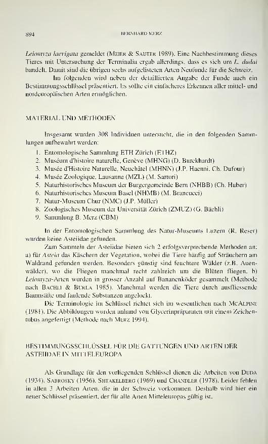
894 BERNHARD MERZ
Leiomyza laevigata gemeldet (Meier & Sauter 1989). Eine Nachbestimmung dieses
Tieres mit Untersuchung der Terminalia ergab allerdings, dass es sich um L. dudai
handelt. Damit sind die übrigen sechs aufgelisteten Arten Neufunde für die Schweiz.
Im folgenden wird neben der detaillierten Angabe der Funde auch ein
Bestimmungsschlüssel präsentiert. Es sollte ein einfacheres Erkennen aller mittel- und
nordeuropäischen Arten ermöglichen.
MATERIAL UND METHODEN
Insgesamt wurden 308 Individuen untersucht, die in den folgenden Samm-lungen aufbewahrt werden:
1. Entomologische Sammlung ETH Zürich (ETHZ)
2. Muséum d'histoire naturelle, Genève (MHNG) (D. Burckhardt)
3. Musée d'Histoire Naturelle, Neuchâtel (MHNN) (J.P. Haenni, Ch. Dufour)
4. Musée Zoologique, Lausanne (MZL) (M. Sartori)
5. Naturhistorisches Museum der Burgergemeinde Bern (NHBB) (Ch. Huber)
6. Naturhistorisches Museum Basel (NHMB) (M. Brancucci)
7. Natur-Museum Chur (NMC) (J.P. Müller)
8. Zoologisches Museum der Universität Zürich (ZMUZ) (G. Bächli)
9. Sammlung B. Merz (CBM)
In der Entomologischen Sammlung des Natur-Museums Luzern (R. Reser)
wurden keine Asteiidae gefunden.
Zum Sammeln der Asteiidae bieten sich 2 erfolgsverprechende Methoden an:
a) für Asteia das Käschern der Vegetation, wobei die Tiere häufig auf Sträuchern amWaldrand gefunden werden. Besonders günstig sind feuchtere Wälder (z.B. Auen-
wälder), wo die Fliegen manchmal recht zahlreich um die Blüten fliegen, b)
Leiomyza-Arten wurden in grosser Anzahl auf Bananenköder gesammelt (Methode
nach Bächli & Burla 1985). Manchmal werden die Tiere durch ausfliessende
Baumsäfte und faulende Substanzen angelockt.
Die Terminologie im Schlüssel richtet sich im wesentlichen nach McAlpine
(1981). Die Abbildungen wurden anhand von Glycerinpräparaten mit einem Zeichen-
tubus angefertigt (Methode nach Merz 1994).
BESTIMMUNGSSCHLUSSEL FUR DIE GATTUNGEN UND ARTEN DERASTEIIDAE IN MITTELEUROPA
Als Grundlage für den vorliegenden Schlüssel dienen die Arbeiten von Duda
(1934), Sabrosky (1956), Shtakelberg (1969) und Chandler (1978). Leider fehlen
in allen 3 Arbeiten Arten, die in der Schweiz vorkommen. Deshalb wird hier ein
neuer Schlüssel präsentiert, der für alle Arten Mitteleuropas gültig ist.
DIE ASTEIIDAE DER SCHWEIZ 895
Ader v\ lang, verläuft sehr lange parallel der Costa und mündet viel
näher der i"2+3 als der sc in die Costa (Abb. 2); ein Paar kräftige,
proklinate Orbitalborsten vorhanden (Abb. 4); Arista fast nackt, deren
Haare höchstens wenig dicker als die Dicke der Aristabasis; Tiere mit
schwarzem Thorax (Leiomyza) 5
Ader r\ kurz, mündet viel näher der sc als der r2+3 in die Costa (Abb.
1); Orbitalborste entweder reklinat (Abb. 3) oder sich von übriger Be-
haarung der Stirn nicht unterscheidend; Arista fast kahl oder gefiedert
(Abb. 3); zumindest Pleuren des Thorax vorherrschend gelb 2
Querader dm-cu fehlt und Alula wenig entwickelt (Abb. 1); Arista mit
langen Fiederhaaren und Orbitalborste kräftig, reklinat (Abb. 3) (Asteia) .... 3
Querader dm-cu vorhanden, Alula stärker entwickelt; Arista fast kahl,
keine differenzierte, kräftige Orbitalborste vorhanden Asüosoma rufifrons Duda
[Nur aus Grossbritannien und dem ehemaligen Jugoslawien bekannt; ausführliche
Beschreibung und Illustrationen bei Chandler, 1978]
Gesicht über dem Mundrand mit einem Paar glänzender, schwarzer
Flecken (Abb. 5); Stirn gelb; Scutum glänzend dunkelbraun bis
schwarz gefärbt, mit 1 Paar hellerer Längsstreifen vor der Suturalquer-
naht; Pleuren kontrastierend gelb glänzend; Scutellum auf der Dorsal-
fläche schwarz, entlang der Seite gelb; Halteren völlig gelb: Surstyli
nur schwach asymmetrisch (Abb. 7-9) Asteia concinna
Gesicht über dem Mundrand mit einem silbrigglänzenden, schwarz
berandeten Querband (Abb. 6); Scutellum einheitlich gelb; Halteren
nicht völlig gelb; Surstyli kräftig asymmetrisch (Abb. 10-16) 4
Scutum schwarz, Pleuren gelb gefärbt; Stirn schwarz, nur am Vor-
derrand über der Lunula mit hellem Band; Halterenkopf gleichmässig
verdunkelt; rechter Surstylus grösser als der linke, rinnenförmig (Abb.
10-13) Asteia amoena
Thorax rötlich gefärbt, auf dem Scutum mit zwei Paar dunklerer Längs-
streifen auf der Rückenfläche und einem Paar entlang der Notopleu-
ralnaht; Stirn orange-braun, etwas längsgestreift erscheinend; Halteren-
kopf gelb mit grossem, schwarzem Fleck; rechter Surstylus viel grösser
als der linke, schaufeiförmig (Abb. 14-16) Asteia elegantula
Halterenkopf gelb gefärbt, fß völlig gelb gefärbt 6
Halterenkopf schwärzlich gefärbt. f3 völlig gelb oder mit dunklem Ring
an der Basis 7
Dorsozentralborsten um weniger als ihre Länge vom Scutellumrand
entfernt inseriert (Abb. 17); Cerci mit sehr langen Haaren, welche die
Spitze der Surstyli erreichen (Abb. 19, 20); Aedeagus massig skiero-
tisiert, Spitze membranös, leicht verbreitert (Abb. 21, 22)
Leiomyza curvinervis
Dorsozentralborsten viel weiter als ihre eigene Länge vom Scutell-
umrand entfernt, etwa auf der Höhe des Notopleuralhinterrandes in-
seriert (Abb. 18); Cerci kürzer behaart, erreichen nicht die Spitze der
896 BERNHARD MERZ
Surstyli (Abb. 23, 24); Aedeagus sehr schwach skierotisiert, Spitze
membranös, nicht verbreitert (Abb. 25) Leiomyza scatophagina
7 Dorsozentralborsten um weniger als ihre Länge vom Scutellumrand
entfernt inseriert (Abb. 17); f3 subapikal verdunkelt; Innerer Surstylus
gleichmässig gegen die Spitze verjüngt (Abb. 29, 30); Aedeagus stark
skierotisiert, mit skierotisierter, becherförmiger Spitze (Abb. 3 1 )
Leiomyza laevigata
Dorsozentralborsten etwa auf der Höhe der Flügelbasis inseriert, vomScutellumhinterrand viel weiter entfernt als ihre maximale Länge
(Abb. 18); fß völlig gelb; Innerer Surstylus an der Spitze mit kleinem
Haken (Abb. 26, 27); Aedeagus stark skierotisiert, Spitze unregel-
mässig becherförmig (Abb. 28) Leiomyza dudai
ARTENLISTE
Die Nomenklatur im folgenden Katalog richtet sich nach Papp (1984). Die
Lokalitäten werden alphabetisch nach Kanton und Ortschaft aufgeführt. Dabei werden
die Kantone wie folgt abgekürzt: AG = Aargau; BE = Bern; BL = Baselland; BS =
Baselstadt; FR = Fribourg; GE = Genève; GR = Graubünden; JU = Jura; LU =
Luzern; NE = Neuchâtel; SG = St. Gallen; SH = Schaffhausen; TI = Ticino; VD =
Vaud; VS = Valais; ZH = Zürich. In Klammern stehen alle Funddaten einer Fund-
stelle. Die allgemeine Verbreitung wurde Papp (1984) und lokalen Faunenlisten ent-
nommen. Die Länder sind von Norden nach Süden, und von Westen nach Osten
geordnet.
Asteia amoena Meigen, 1830 (Abb. 1, 3, 6, 10-13)
AG: 1 9 , Würenlingen (26.VII.1990) (Bächli) (ZMUZ). BE: 29, Bid (27-31.VII.1973)
(Bächli) (ZMUZ); le?, Lamboing, Près de Macolin Derrière (16.V.1993) (Merz) (ETHZ). BS:
le?, Basel (1.III.1961) (Keiser) (NHMB). FR: 19, Bonaudon (16.IX.1956) (Aubert) (MZL).GE: le?, Chancy, La Laire (20.VII.1995) (Burckhardt) (MHNG). GR: lc?, Rothenbrunnen
(19.VIII.1993) {Merz) (CBM). JU: 16, Courrendlin, vers vielle Eglise, Mini-Malaise-Falle
(13.-27.X.1988) (Gonseth) (MHNN). SH: 19, Rüdlingen (2/3 .VII. 1994) (Merz & Eggen-
berger) (ETHZ). TI: le?, 1 Î, Arcegno (8-1
1
.Vili. 1970) (Bächli) (ZMUZ); 66 6, 39, Biasca-
Loderio (15.III.1994, 31.III.1994, 9.IV.1995, 8.VI.1992, 16-20VI.1995) (Merz) (CBM, ETHZ,ZMUZ); 46, 49, Bolle di Magadino (17-20.VI.1995, Fangzelt, 19.VI.1995) (Bächli) (ZMUZ);\6. Brusino (3.IV.1988) (Merz) (ETHZ); 19, Gordola (17-20VI.1995, Fangzelt) (Bächli)
(ZMUZ); 3e?, 29, Maggia (10-12.IX.1991) (Bächli) (ZMUZ). VD: là, 19, Aigle (4-
7.VIII.1970) (Bächli) (ZMUZ); 16, 69 9, Aigle (8. VI. 1955, 2.VII.1955. 20.VI.1956,
30V.1957, 2.VI.1957) (Passello) (MZL); le?, Hauteville (28.VI.1955) (Passello) (MZL); le?,
Jorat (30.IX.1948) (Schmid) (MZL); 36, St. Sulpice (VII.1945) (Schmid) (MZL); le?, St.
Triphon (9V.1955) (Passello) (MZL); le?, Vidy-Lausanne (24.IX.1954) (Schmid) (MZL). VS:
1 9, Hohtenn-Bahnhof (3.IX.1991) (Merz) (ETHZ); le?. Muraz (24.V.1959) (Passello) (MZL);le?, Vionnaz (2.VI.1957) (Passello) (MZL). ZH: 5e?, 79, Dietikon (10-14.IX.1985,
5VIII.1989, 11-14.IX.1989. 19-23VII.1990, 13VI.1991, 22.VIII.1991, 27-31. VIII. 1991, 15-
19VII.1993, 14-18VII.1995) (Bächli) (ZMUZ); 19. Dietikon-Reppisch (22-25.VIII.1982)
(Bächli) (ZMUZ); 19. Dübendorf (17.IX.1993) (Gautschi) (ETHZ); le?, Flaach/Thurauen
(1.IX.1993) (Merz) (CBM); 19. Zürich (10VIII.1991) (Merz) (ETHZ); le?, Zürich (14-
DI h ASTHIIDAH DKK SCHWKIZ 897
Abb. 1-6
Flügel und Köpfe von Asteiidae. 1-2, Flügel. 1, Asteia amoena Meigen (<?, ZH, Zürich-
Örlikon, 29.V.1991); 2, Leiomyza curvinervis (Zetterstedt) (S , VS, Visperterminen, 18.VIL1995). 3-6, Köpfe. 3, Asteia amoena Meigen, Profil (S , ZH, Zürich-Örlikon, 29.V.1991); 4,
Leiomyza curvinervis (Zetterstedt), Profil (S, VS, Visperterminen. 18.VII.1995); 5, Asteia
concinna Meigen, Frontal (S , TI, Biasca-Loderio, 28.VI. 1992); 6, Asteia amoena Meigen.
Frontal (6, ZH, Zürich-Örlikon, 29.V.1991). Massstab: 1 mm.
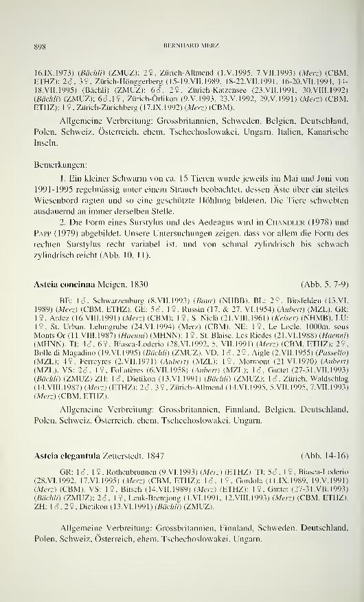
898 BERNHARD MERZ
16.IX.1973) (Badili) (ZMUZ); 29, Zürich-Allmend (I.V. 1995. 7.VII.1993) (Merz) (CBM.ETHZ); 2d, 3$, Zürich-Hönggerberg ( 15- 19. VII. 1989, 18-22.VII.1991, 16-20.VII.1994, 14-
18.VII.1995) (Bächli) (ZMUZ); 6c?, 29, Zürich-Katzensee (23.VII.1991, 30.VIII.1992)
(Bächli) (ZMUZ); 6c?, 19, Zürich-Örlikon (9.V.1993, 23.V.1992, 29.V.1991) (Merz.) (CBM,ETHZ); 19, Zürich-Zürichberg (17.IX.1992) (Merz) (CBM).
Allgemeine Verbreitung: Grossbritannien, Schweden, Belgien, Deutschland,
Polen, Schweiz, Österreich, ehem. Tschechoslowakei, Ungarn, Italien, Kanarische
Inseln.
Bemerkungen:
1. Ein kleiner Schwärm von ca. 15 Tieren wurde jeweils im Mai und Juni von
1991-1995 regelmässig unter einem Strauch beobachtet, dessen Äste über ein steiles
Wiesenbord ragten und so eine geschützte Höhlung bildeten. Die Tiere schwebten
ausdauernd an immer derselben Stelle.
2. Die Form eines Surstylus und des Aedeagus wird in Chandler (1978) und
Papp (1979) abgebildet. Unsere Untersuchungen zeigen, dass vor allem die Form des
rechten Surstylus recht variabel ist, und von schmal zylindrisch bis schwach
zylindrisch reicht (Abb. 10, 11).
Asteia concinna Meigen. 1830 (Abb. 5, 7-9)
BE: \â. Schwarzenburg (8.VII.1993) (Baiir) (NHBB). BL: 29. Birsfelden (13.VI.
1989) (Merz) (CBM, ETHZ). GE: 5c?, 19. Russin (17. & 27. VI. 1954) (Auberi) (MZL). GR:19. Ardez (16. VIII. 1991) (Merz) (CBM); 1 9, S. Nicla (21. VIII. 1961 ) (Reiser) (NHMB). LU:
19. St. Urban, Lehmgrube (24.VI.1994) (Merz) (CBM). NE: 19, Le Lode. 1000m, sous
Monts Or ( 1 1. Vili. 1987) (Haenni) (MHNN); 1 9, St. Blaise, Les Riedes (21.VI. 1988) (Haenni)
(MHNN). TI: 4c?, 69. Biasca-Loderio (28.VI.1992, 5. VII. 1991) (Merz) (CBM, ETHZ); 29,Bolle di Magadino (19.VI.1995) (Bächli) (ZMUZ). VD: lc?, 29, Aigle (2.VII.1955) (Passello)
(MZL); 49, Ferreyres (2.VII.1971) (Aubert) (MZL); 19, Mormont (21.VI.1970) (Aubert)
(MZL). VS: 2c?. 19, Follatères (6.VII.1958) (Aubert) (MZL); lc?, Guttet (27-31.VII. 1993)
(Bächli) (ZMUZ) ZH: lc?, Dietikon (13.VI.1991) (Bächli) (ZMUZ); lc?, Zürich. Waldschlag
(14.VIII.1987) (Merz) (ETHZ); 2d, 3 9, Zürich-Allmend (14. VI. 1995, 5.VII.1995, 7.VII.1993)
(Merz) (CBM, ETHZ).
Allgemeine Verbreitung: Grossbritannien, Finnland, Belgien, Deutschland,
Polen. Schweiz, Österreich, ehem. Tschechoslowakei, Ungarn.
Asteia elegantula Zetterstedt, 1847 (Abb. 14-16)
GR: Id, 1 9 , Rothenbrunnen (9. VI. 1993) (Merz) (ETHZ). TI: 53. 1 9 , Biasca-Loderio
(28.VI.1992, 17.VI.1995) (Merz) (CBM, ETHZ); lc?, 19, Gordola (11.IX.1989, 19.V.1991)
(Merz) (CBM). VS: 19, Bitsch (14.VII.1989) (Merz) (ETHZ); 19. Guttet (27-31.VII.1993)
(Bächli) (ZMUZ); 2c?, 19. Leuk-Brentjong (1.VI.1991, 12.VIII.1993) (Merz) (CBM, ETHZ).ZH: lc?, 29, Dietikon (13.VI.1991) (Bächli) (ZMUZ).
Allgemeine Verbreitung: Grossbritannien, Finnland, Schweden, Deutschland,
Polen, Schweiz, Österreich, ehem. Tschechoslowakei, Ungarn.
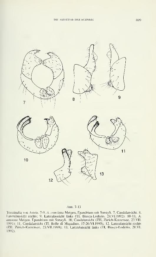
DIE ASTEIIDAE DER SCHWEIZ
l&Z
10
Abb. 7-13
Terminalia von Asteia. 7-9, A. concinna Meigen, Epandrium mit Surstyli. 7, Caudalansicht; 8,
Lateralansicht rechts; 9, Lateralansicht links (TI, Biasca-Loderio, 28. VI. 1992). 10-13. A.
amoena Meigen, Epandrium mit Surstyli. 10, Caudalansicht (ZH, Zürich-Katzensee, 23.VII.
1991); 11, Caudalansicht (TI, Bolle di Magadino, 17-20.VI.1995); 12, Lateralansicht rechts
(ZH, Zürich-Katzensee, 23.VII.1991); 13, Lateralansicht links (TI, Biasca-Loderio, 28.VI.
1992).
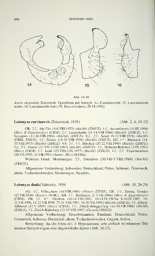
900 BERNHARD MERZ
14 ^-vvo
Abb. 14-16
Asteia elegantula Zetterstedt, Epandrium mit Surstyli. 14, Caudalansicht; 15, Lateralansicht
rechts; 16, Lateralansicht links (TI, Biasca-Loderio, 28. VI. 1992).
Leiomyza curvinervis (Zetterstedt. 1838) (Abb. 2, 4, 19-22)
GR: 26, Alp Flix (4-8.VIII.1975) (Bächli) (ZMUZ); le?, Ausserferrera (11. IX. 1994)
(Merz & Eggenberger) (CBM); 26, Lenzerheide (11-14.VIII.1988) {Bächli) (ZMUZ); 16,
Savognin (17-20.VIII.1988) (Bächli) (ZMUZ); 96, 39, Scuoi (9-12.VIII.1978) (Badili)
(CBM, ZMUZ); le?, Zernez (15-18.VIII.1978) (Bächli) (ZMUZ). SG: 19, Rheineck (14-
17.VIII.1973) (Bächli) (ZMUZ). VS: 26, 19, Bürchen (20-22.VIII.1993) (Bächli) (ZMUZ);26, 59, Guttet (31.VII-2.VIII. 1993) (Bächli) (ZMUZ); 19, Hohtenn-Bahnhof (3.IX.1991)
(Merz) (CBM); 19, Leuk (23.VIII-2.IX.1977) (Bächli) (ZMUZ); le?, 29, Visperterminen
(18.VII.1995, 24.VH.1991) (Sauter, Merz) (ETHZ).
Weiterer Fund: Montenegro: 29, Durmitor (30.VII-5. Vili. 1988) (Bächli)
(ZMUZ).
Allgemeine Verbreitung: Schweden, Deutschland, Polen, Schweiz, Österreich,
ehem. Tschechoslowakei, Montenegro. Nearktis.
Leiomyza dudai Sabrosky. 1956 (Abb. 18, 26-28)
AG: lc?, Villnachern ( 10.VIII.1986) (Meier) (ETHZ). GR: 19, Zernez, Gondas(17.VII.1930) (Keiser) (NMC). SH: 19, Rüdlingen (2-3.VII.1994) (Merz & Eggenberger)
(CBM). ZH: 2c?, 69, Dietikon (10-14.VIII.1982, 10-14.IX. 1985m 6-10.IX.1987, 19-
23.VII.1990, 18-22.VII.1991. 27-31.VIII.1991, 16-20.VII.1992) (Bächli) (ZMUZ); 1 9, Zürich-
Allmend (17.V.1995) (Merz) (CBM); 19, Zürich-Hönggerberg (14-19.VII.1988) (Bächli)
(ZMUZ); 1 9. Zürich-Käferberg (12- 15. VIII. 1987) (Gosteli) (ZMUZ).
Allgemeine Verbreitung: Grossbritannien, Finnland, Deutschland, Polen,
Frankreich, Schweiz, Österreich, ehem. Tschechoslowakei, Ungarn, Italien.
Bemerkung: An der Form des 6 Hypandriums sehr einfach zu erkennen: Die
inneren Surstyli tragen eine abgewinkelte Spitze (Abb. 26-27).
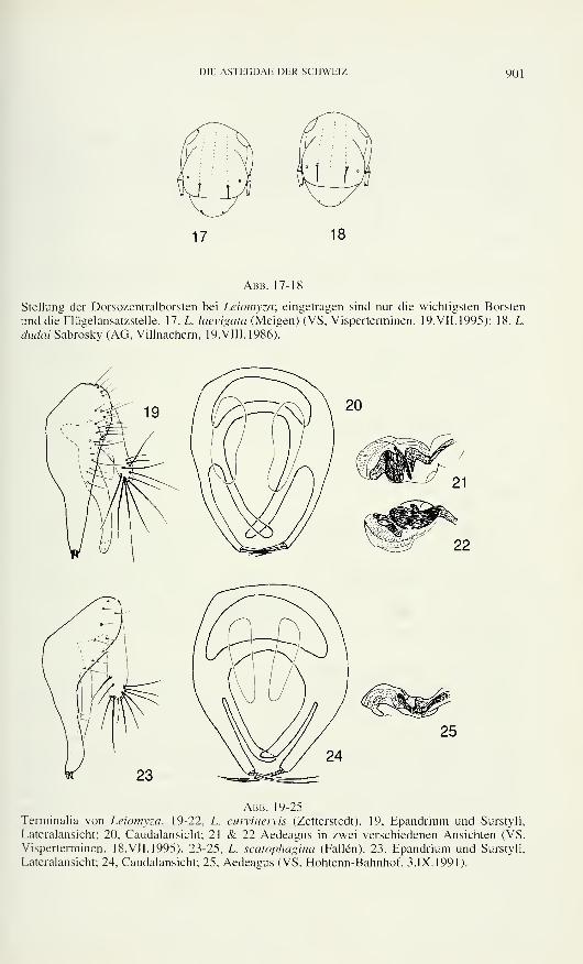
DIE ASTEIIDAE DER SCHWEIZ 901
17 18
Abb. 17-18
Stellung der Dorsozentralborsten bei Leiomyza; eingetragen sind nur die wichtigsten Borsten
und die Flügelansatzstelle. 17. L. laevigata (Meigen) (VS, Visperterminen, 19. VII. 1995); 18, L.
dudai Sabrosky (AG, Villnachern, 19. VIII. 1986)"
Abb. 19-25
Terminalia von Leiomyza. 19-22, L. curvinervis (Zetterstedt). 19, Epandrium und Surstyli,
Lateralansicht; 20, Caudalansicht; 21 & 22 Aedeagus in zwei verschiedenen Ansichten (VS,
Visperterminen, 18.VII.1995). 23-25, L. scatophagina (Fallen). 23, Epandrium und Surstyli,
Lateralansicht; 24, Caudalansicht; 25, Aedeagus (VS, Hohtenn-Bahnhof, 3.IX.1991).
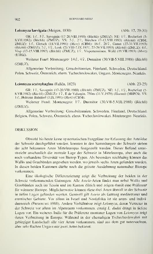
902 BERNHARD MERZ
Leiomyza laevigata (Meigen, 1830) (Abb. 17, 29-31)
GR: 16, 59, Savognin (17-20.VIII. 1988) (Bächli) (ZMUZ). NE: 19, Rochefort (5-
8. VII. 1982) (Bächli) (ZMUZ). VS: 3c?, 29. Bürchen (7-13.VIII.1993) (Bächli) (CBM,ZMUZ); là, Gletsch (18.VII.1991) (Merz) (CBM); 16c?, 289, Guttet (27-31. VII. 1993)
(Bächli) (ZMUZ); 2c?, 19, Leuk (23.VIII-2.IX. 1977, 27-29.VII.1993) (Bächli) (ZMUZ); 4c?,
Visp (13-15.VIII.1993) (Bächli) (ZMUZ); 19, Visperterminen, Wald (19.VII.1993) (Merz)
(ETHZ).
Weiterer Fund: Montenegro: 14c? , 49, Durmitor (30.VII-5.VIII. 1988) (Bächli)
(ZMUZ).
Allgemeine Verbreitung: Grossbritannien, Finnland, Schweden, Deutschland,
Polen, Schweiz, Österreich, ehem. Tschechoslowakei, Ungarn, Montenegro. Nearktis.
Leiomyza scatophagina (Fallen, 1823) (Abb. 23-25)
GR: 19, Savognin (17-20.VIII.1988) (Bächli) (ZMUZ). NE: lc?, 19, Rochefort (5-
8.VII.1982) (Bächli) (ZMUZ); 1 9. E de Valangin, 750m (3 I.V. 1979) (Haenni) (MHNN). VS:
le?, Hohtenn-Bahnhof (3.IX. 1991) (Merz) (CBM).
Weiterer Fund: Montenegro: 19. Durmitor (30.VII-5.VIII. 1988) (Bächli)
(ZMUZ).
Allgemeine Verbreitung: Grossbritannien, Schweden, Finnland, Deutschland,
Belgien, Polen, Schweiz, Österreich, ehem. Tschechoslowakei, Montenegro. Nearktis.
DISKUSSION
Obwohl bis heute keine systematischen Fangpläne zur Erfassung der Asteiidae
der Schweiz durchgeführt wurden, konnten in den Sammlungen der Schweiz sieben
der acht bekannten Arten Mitteleuropas festgestellt werden. Dieser Befund unter-
streicht anschaulich die zentrale Lage der Schweiz in Mitteleuropa, aber auch die
noch vorhandene Diversität von Biotop-Typen. Als besonders reichhaltig können das
Wallis und Graubünden angesehen werden, wo jeweils sechs Arten gefunden wurden.
In diesen beiden Kantonen dürfte noch die grösste Ausdehnung naturnaher Biotope
vorkommen.
Eine ökologische Differenzierung zeigt die Verbreitung der beiden in der
Schweiz vorkommenden Gattungen: Alle Asteia-Arten findet man nebst Wallis und
Graubünden auch im Tessin und im Kanton Zürich und zeigen damit eine Präferenz
für wärmere Biotope. Möglicherweise können diese drei Arten überall in der Schweiz
in tiefen Lagen gefunden werden. Generell gilt Asteia als Gattung mediterraner und
eremischer Gebiete. Vor allem in Israel und Nordafrika ist sie arten- und indivi-
duenreich (Freidberg 1988). Andere Verhältnisse zeigt Leiomyza, deren Vertreter in
der Schweiz vor allem im Alpenraum vorkommen, einzig L. dudai dringt in tiefere
Lagen vor. Ein weiteres Indiz für die Präferenz montaner Lagen von Leiomyza zeigt
deren Verbreitung in Europa: Während in der ehemaligen Tschechoslowakei mit
gebirgiger Landschaft alle vier Arten vorkommen, sind aus dem gut untersuchten,
aber sehr flachen Ungarn nur zwei Arten bekannt.
DIE ASTEIIDAE DER SCHWEIZ 903
Abb. 26-31. Terminalia von Leiomyza. 26-28, L. ciuciai Sabrosky. 26, Epandrium und Surstyli,
Lateralansicht; 27, Caudalansicht;"28, Aedeagus (AG, Villnachern, 19.VIII.1986). 29-31, L.
laevigata (Meigen). 29, Epandrium und Surstyli, Lateralansicht; 30, Caudalansicht; 31.
Aedeagus (VS, Gletsch, 18.VII.1991).
Interessanterweise kommen an gewissen Fundstellen, wie z.B. Biasca-Loderio,
Dietikon oder Savognin mehrere Arten offenbar sympatrisch miteinander vor. In
Biasca wurden zahlreiche Tiere von allen 3 Asteia-Arten auf blühenden Salix und
anderen Sträuchern gekäschert. Diese Tendenz zur Aggregation ist auch aus Israel
bekannt, wo verschiedene Asteia und Phlebosolera manchmal unter Tamahx-Sträu-
chern Schwärme bilden (Freidberg 1984, eigene Beobachtungen). Der Grund für
dieses Verhalten ist unbekannt, könnte mit ähnlichem Paarungsverhalten oder ähn-
licher Larvallebensweise zusammenhängen.
904 BERNHARD MERZ
VERDANKUNGEN
Ganz herzlich danke ich allen oben aufgeführten Konservatoren für die
Erlaubnis, die jeweiligen Insektensammlungen nach Asteiiden durchzukämmen. Ein
weiterer grosser Dank geht an M. Eggenberger für die Durchsicht des Manuskriptes
und an K.Tschudi-Rein für die Kontrolle der englischen Zusammenfassung.
LITERATUR
BäCHLI, G. & Burla. H., 1985. Diptera, Drosophilidae. Inserta Helvetica. Fauna 7: 1-116.
Chandler, P., 1978. A revision of the British Asteiidae (Diptera) including two additions to the
British List. Proceedings of the British Natural History Society 11: 23-34.
Duda, O., 1934. 58b. Astiidae. In: Lindner, E. (Ed.). Die Fliegen der Palaearktischen Region,
Vol. VI (1): 1-15.
Freidberg, A., 1984. The Mating Behavior of Asteia elegantula with Biological Notes onSome Other Asteiidae (Diptera). Entomologica Generalis 9 (4): 217-224.
Freidberg, A., 1988. 10. Zoogeography of the Diptera of Israel. In: Yom-Tov, Y. &Tchernov, E. (Eds). The Zoogeography of Israel: 277-308. Dr. W. Junk Publishers,
Dordrecht.
McAlpine, LP., 1981. Morphology and terminology - adults. In: McAlpine LP. et al. (Eds).
Manual of Nearctic Diptera - 1. Monograph of Biosvstematics Research Institute,
Ottawa 27: 9-63.
Meier, C. & Sauter, W., 1989. Zur Kenntnis der Insektenfauna eines Auenwaldreservates an
der Aare bei Villnachern AG. Mitteilungen der Aargauer Naturforschenden Gesell-
schaft 32: 217 -258.
Merz, B., 1994. Diptera. Tephritidae. Insecta Helvetica, Fauna 10: 1-198.
Papp. L., 1972. Systematical and ecological investigations on fly pests of fungi in Hungary, II.
Sphaeroceridae and Asteiidae (Diptera). Annales Historico Naturalis Musei Nationalis
Hugaricae 64: 3 1 5-3 17.
Papp, L., 1979. Seven New Species of the Palaearctic Lauxaniidae and Asteiidae. Reichen-
bachia 17 (12): 87 -97.
Papp, L., 1984. Family Asteiidae. In: Soos, A. & Papp, L. (Eds). Catalogue of Palaearctic
Diptera, Vol. 10: 63- 66.
Perry. I. & Stubbs. A.E., 1978. Dead Wood and Sap Runs. In: Stubhs, A. & Chandler, P.
(Eds). A Dipterist's Handbook. The Amateur Entomologist 15: 65-73.
Sabrosky, C.W., 1956. Additions to the Knowledge of Old World Asteiidae (Diptera). Revue
française d' Entomologie 23 (4): 216-243.
Shtakelberg. A.A.. 1969. 87. Asteiidae. In: Bei-Bienko, G.Ya. (Ed). OpredeliteV NasekomykhEvropeiskoi Chasti SSSR., Vol. V (2): 334-335. Nauka Publishers, Leningrad (Auf Rus-
sisch, Englische Übersetzung 1989: Keys to the Insects of the European Part of the
USSR, Vol. V (2): 555-556, Amerind Publishing Co. Pvt. Ltd., New Dehli).
Revue suisse de Zoologie, 103 (4): 905-913; décembre 1996
Molostrongylus acanthocolpos gen. n., sp. n., (Nematoda,
Trichostrongylina, Molineoidea) parasite de Molossops temmincki
(Chiroptera, Molossidae) au Paraguay
Marie-Claude DURETTE-DESSET* & Claude VAUCHER*** Muséum national d'Histoire naturelle, Laboratoire de Biologie parasitaire,
Protozoologie, Helminthologie, associé au CNRS, 61, rue de Buffon,
75231 Paris cedex 05, France;
** Muséum d'histoire naturelle, case postale 6434, 1211 Genève 6, Suisse.
Molostrongylus acanthocolpos gen. n. sp. n. (Nematoda, Trichostron-
gylina, Molineoidea) parasite of Molossops temmincki (Chiroptera,
Molossidae) from Paraguay. - A new genus and species are described
from the intestine of the bat Molossops temmincki in Paraguay. It belongs
to a group of genera which possesses a cephalic hood. The new taxon
appears closely related to Anoplostrongylus, but may be distinguished
mainly by the characters of the caudal bursa in male and by the presence of
spines in the ovejector region in female.
Key-words: Nematoda - Molineoidea - Anoplostrongylinae - Molostron-
gylus gen. n. - Molossidae - Paraguay.
INTRODUCTION
Parmi les Anoplostrongylinae parasites de Chiroptères, trois genres possèdent
une tête dépourvue d'épines et de crochets mais pourvue d'un capuchon céphalique
(Durette-Desset & Chabaud, 1975): Torrestrongylus Perez-Vigueras, 1935 et Tri-
choleiperia Travassos, 1935 tous deux néotropicaux et Anoplostrongylus Boulenger,
1926 présent à la fois dans l'Ancien Monde chez Tupaia et dans le Nouveau Mondechez les Chauve-Souris.
Un abondant matériel de Nematodes Trichostrongles a été récolté chez les
Chauve-souris du Paraguay au cours des différentes missions du Muséum d'histoire
naturelle de Genève à partir de 1979. Cette note concerne la description d'une nou-
velle espèce appartenant à un genre nouveau, parasite de l'intestin de Molossidae qui,
par ses caractères céphaliques, appartient à ce groupe.
Manuscrit accepté le 10.05.1996.
906 MARIE-CLAUDE DURETTE-DESSET & CLAUDE VAUCHER
MATERIEL ET METHODES
Les Nematodes ont été fixés au formol à 4% et conservés dans de l'alcool à
70°. Ils sont déposés dans les Collections du Muséum d'histoire naturelle de Genève
(MHNG) et dans celles du Muséum national d'histoire naturelle de Paris (MNHN).
Molostrongylus n.gen.
Molineoidea, Molineidae, Anoplostrongylinae. Nematodes de petite taille ne
présentant aucun enroulement. Tête avec capuchon céphalique en deux parties.
Synlophe avec axe d'orientation ventro-dorsal confondu avec Taxe sagittal. Présence
d'ailes latérales fortement développées. Bourse caudale de type 2-3, allongée
transversalement avec côtes 2 et 3 de longueur équivalente à celle des 4 et des 6.
Côtes 5 plus longues que les côtes adjacentes. Côte dorsale longue, divisée à son
extrémité distale. Côtes 8 plus courtes que la côte dorsale. Spicules non ailés en forme
de cône allongé, à pointe unique et mousse. Gubernaculum présent. Chez la femelle,
présence d'épines cuticulaires dans la région de l'ovéjecteur; queue plus haute que
large avec trois grosses pointes et un filament médian.
Parasite de Chiroptères néotropicaux.
Espèce-type: Molostrongylus acanthocoipos n.sp.
Molostrongylus acanthocoipos n.sp.
Matériel type: S holotype. MHNG 21111 INVE, 9 allotype 21112 INVE, 8 ô\ 6 2, 1
immature 2, paratypes 21113 INVE, chez Molossops temmincki Estancia General Diaz, dépt.
Alto Paraguay, Paraguay, 18.1 1.1987.
Autre matériel: 7 S MHNG 21 120 INVE, 7 o\ 2 2 MHNG 21121 INVE, 4 o\ 4 2, 1
morceau postérieur 2 , 2 morceaux antérieurs MNHN 934 MD, comme holotype; 2 morceauxantérieurs 2 MHNG 21 1 14 INVE, pont sur le Rio Aguaray, dépt. Misiones, 16.10.1982; 2 o\ 1
2, 1 morceau antérieur 2 MHNG 211 15 INVE, Panchito Lopez, dépt. Neembucu, 24.10.1982;
7 S MHNG 21 1 16 INVE, 9 6, 8 2 MNHN 188 KP, Santa Maria, dépt. Misiones, 27.10.1982;
1 o\ 2 2 MNHN 932 MD, arroyo Tagatija-mi, env. de l'estancia Santa Maria, dépt. Con-cepción, 14.10.1983; 1 o\ 2 2, 2 morceaux postérieurs 2 MHNG 21117 INVE, arroyo
Tagatija-guazu, env. de J'estancia Santa Maria, dépt. Concepción, 15.10.1983; 1 S , 2 2 MNHN933 MD, Monte Lindo, route Transchaco km 212, 23.08.1984; 3 6 MHNG 21118 INVE,Belén, dépt. Concepción, 11.10.1985; 1 6 , 1 morceau antérieur MHNG 21119 INVE, estancia
Apendice, route Transchaco km 293, dépt. Presidente Hayes, 8.1 1.1985.
Petits Nematodes ne présentant aucun enroulement. Pore excréteur situé au
niveau de la deuxième moitié de l'oesophage, très en arrière du bord antérieur des
glandes excrétrices. Glandes excrétrices très développées (fig. 3 E). Sinus excréteur
d'abord dirigé vers l'avant. Deirides de forme triangulaire et en position latéro-dorsale,
plus ou moins proches du pore excréteur mais toujours situées plus antérieurement
que celui-ci. Oesophage présentant une légère constriction dans sa partie médiane
(fig. 3 D).
Tête: Présence d'une vésicule céphalique composée de deux parties: une partie
antérieure en forme de capuchon et une partie postérieure, généralement plus longue.
Présence d'une petite dent oesophagienne dorsale (fig. 3 A, B). En vue apicale,
MOLOSTRONGYLUS ACANTHOCOLPOS 907
Fig. 1
Molostrongylus acanthocolpos n. gen. n. sp. Mâle (long de 3200 um). Coupes transversales du
corps A - au niveau des deirides. B - à 420 um en arrière de la tête. C - au milieu du corps ( à
1750 um de la tête). D - à 1200 um en avant de la bourse caudale. E - à 700 um en avant de la
bourse caudale. Toutes les coupes sont orientées comme la figure A. Abréviation: de. = deiride,
d. = dos, g. = gauche, v. = ventre, dr. = droite. Echelle: 30 um .
bouche arrondie entourée de deux grosses amphides. de 4 papilles labiales externes (2
dorsales et 2 ventrales) et de quatre papilles céphaliques. Dorsalement, entre la
bouche et les papilles céphaliques existe une mince plaque en arc de cercle qui suit le
contour de la bouche. Cette plaque est pouvue de trois épaississements, un à chaque
extrémité et un médian (fig. 3 A, C).
Synlophe: (étudié en coupe transversale du corps chez 3 mâles et 3 femelles
paratypes ainsi que 2 mâles et 2 femelles du matériel complémentaire). Dans les deux
sexes, le corps est parcouru par des crêtes cuticulaires continues dont le nombre aug-
mente d'avant en arrière mais à des niveaux différents pour chacun des deux sexes.
En arrière de la vésicule céphalique naissent deux ailes latérales qui atteignent
leur maximum de largeur (25 um) au niveau de la jonction oesophago-intestinale puis
908 MARIE-CLAUDE DURETTE-DESSET & CLAUDE VAUCHER
Fig. 2. Molostrongylus acanthocolpos n. gen. n. sp. Femelle (longue de 5200 um). Coupes
transversales du corps. A - au niveau du pore excréteur. B - à 460 um en arrière de la tête. C - à
1450 um en arrière de la tête. D - à 2000 um en arrière de la tête., montrant la naissance de 2
crêtes dorsales. E - au milieu du corps (à 2350 um en arrière de la tête), montrant la naissance
d'une crête ventrale. F - juste en avant de l'anneau copulateur. G - H: autre femelle, synlophe en
avant et en arrière de la vulve, montrant l'anneau copulateur. Toutes les coupes sont orientées
comme la figure A. Abréviation: ép. = épine, d. = dos. g. = gauche, v. = ventre, dr. = droite. Les
flèches indiquent la naissance de nouvelles crêtes cuticulaires. Echelles: A - F: 30 um ; G, H, 50
um.

MOLOSTRONGYLUS ACANTHOCOLPOS 909
Fig. 3. Molostrongylus acanthocolpos n. gen. n. sp. A - femelle, tête en vue apicale. B - autre
femelle, tête en vue latérale gauche. C - mâle, tête en vue dorsale. D - mâle, extrémité
antérieure, vue latérale droite. E - id, vue ventrale, montrant la position latero-dorsale des
deirides et le fort développement des glandes excrétrices F - mâle, bourse caudale, vue ventrale.
G - autre mâle, détail des côtes 8 et de la côte dorsale, vue dorsale. H - mâle, gubernaculum.
vue latérale droite. I - mâle, cône génital, vue ventrale. J - autre mâle, bourse caudale, vue
latérale droite. Echelles: A: 50 um ; B, H, I: 30 um ; C: 20 um ; D, E, G: 40 um ; F, J: 50 um.
9 1MARIE-CLAUDE DURETTE-DESSET & CLAUDE VAUCHER
la largeur des ailes décroît progressivement d'avant en arrière mais elles restent
toujours plus développées que les autres crêtes sauf en avant de la vulve chez la
femelle. Les ailes disparaissent à environ 200 um en avant de la bourse caudale chez
le mâle et au niveau de la vulve chez la femelle.
Dans les deux sexes et chez la majorité des individus, en plus des deux ailes
latérales, 6 crêtes naissent entre la vésicule céphalique et la jonction oesophago-
intestinale: 2 crêtes dorsales en avant de l'anneau nerveux (fig. 2 A); 2 crêtes
ventrales en avant du pore excréteur chez le mâle (fig. 1 A) et en arrière de celui-ci
chez la femelle (fig. 2 B) puis naissent une crête ventrale (fig. 1 B) et une crête
dorsale. Chez quelques individus, ces deux dernières crêtes naissent plus postérieu-
rement en arrière de la jonction oesophago-intestinale. On atteint le chiffre de 3
dorsales et de 3 ventrales aussi bien chez le mâle (fig. 1 C) que chez la femelle (fig. 2
C). Chez le mâle, ce chiffre reste constant jusqu'à la moitié du corps environ (fig. 2
C) puis apparaissent 2 crêtes dorsales supplémentaires (fig. 1 D) et enfin à environ
400 urn plus postérieurement. 2 crêtes ventrales ce qui correspond à un total de 5
dorsales et 5 ventrales (fig. 1 E). A environ 300 um en avant de la bourse caudale les
crêtes disparaissent progressivement sur les faces dorsale et ventrale. Il n'y a plus de
crêtes à environ 80 um en avant de la bourse caudale. Chez la femelle, 2 crêtes
dorsales supplémentaires apparaissent à environ 400 um avant le milieu du corps (fig.
2 D) puis une crête ventrale 100 urn plus postérieurement, ce qui donne 5 dorsales et
4 ventrales au milieu du corps (fig. 2 E). Ce chiffre reste constant jusqu'au niveau pré-
vulvaire où, selon les spécimens, 1 à 2 crêtes ventrales supplémentaires apparaissent.
A ce niveau, les ailes latérales très réduites sont légèrement décalées dorsalement (fig.
2 F). Au-delà de la vulve, les crêtes disparaissent progressivement.
Les ailes et les crêtes sont fortement orientées du ventre vers le dos dans le
tiers antérieur du coips (fig. 1 B, 2 B). Cette orientation fait progressivement place à
une orientation où les crêtes sont perpendiculaires à la paroi du corps.
En plus du synlophe. il existe dans la région vulvaire des séries d'épines
disposées longitudinalement et dont la pointe est dirigée vers l'arrière (fig. 4 C-G).
L'étendue de la spinulation correspond plus ou moins à la longueur de l'ovéjecteur (de
130 à 200 um selon les spécimens). Le nombre et la taille des épines est variable d'un
spécimen à l'autre. D'environ 8 à 12 um de haut pour la majorité d'entre elles, elles
peuvent atteindre 18 um pour les plus grandes, généralement situées près de la vulve.
D'une façon générale, les ventrales sont plus grandes que les autres. Les épines sont
enfouies dans l'anneau de copulation, lorsqu'il est présent (fig. 2 G, H).
Mâle holotype: long de 3500 um et large de 55 um dans sa partie
moyenne.Vésicule céphalique haute de 38 um (18 um pour le capuchon) sur 30 umde large à la jonction des deux parties. Anneau nerveux, pore excréteur et deirides
situés respectivement à 150 um. 290 um et 250 um de l'apex. Oesophage long de 510
um. soit 15% de la longueur totale du corps (fig. 3 D. E).
Bourse caudale plus large que haute, de type 2-3 (fig. 3 F). Côtes 2 et 3 de
longueur équivalente aux côtes 4 et 6. Côtes 5 plus longue que les côtes adjacentes.
Papilles des côtes 5 plus proches de celles des 6 que des 4. Côtes 8 naissant presqu'à
la racine de la dorsale, dessinant un arc de cercle et légèrement plus courtes que cette
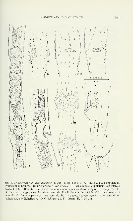
MOLOSTRONGYLUS ACANTHOCOLPOS 91
Fig. 4. Molostrongylus acanthocolpos n. gen. n. sp. Femelle. A - avec anneau copulateli!",
ovéjecteur et branche utérine antérieure, vue dorsale. B - sans anneau copulateur, vue latérale
droite. C à G: différents exemples de l'ornementation épineuse dans la région de l'ovéjecteur. C.
D: femelle paratype, vues dorsale et ventrale. E - F: femelle du lot 934 MD. vues dorsale et
ventrale. G: femelle paratype, vue ventrale. H, I - queue, successivement vues ventrale et
latérale gauche. Echelles: A - D. G: 150 um ; E. F: 100 um; H. I : 50 um.
9 1
2
MARIE-CLAUDE DURETTE-DESSET & CLAUDE VAUCHER
dernière (fig. 3 J). Dorsale relativement longue, atteignant le bord de la bourse caudale.
Les côtes 9 se détachent de la côte dorsale après la division de cette dernière dans sa
partie distale. Côtes 10 plus longues que les côtes 9, pointues à leur extrémité (fig. 3 G).
Chez quelques mâles du matériel complémentaire, nous avons observé une membrane
tendue entre les deux lobes latéraux, à l'intérieur de la bourse caudale (fig. 3 J).
Spicules en forme de cône allongé, à pointe unique et non ailés, longs de 90
um. Gubernaculum en forme de lame en vue latérale. Vu de profil, il mesure 48 umde haut sur 2 um de large dans sa partie moyenne. Le quart distal, sinueux, se termine
par une pointe effilée. Cône génital de forme triangulaire portant, sur sa lèvre
ventrale, une papille zéro bien développée et, sur sa lèvre dorsale, deux papillles 7
filiformes (fig. 3 I).
Femelle allotype: longue de 4700 um, large de 80 um dans sa partie moyenne
et de 100 um au niveau de la vulve. Vésicule céphalique haute de 42 um (20 um pour
le capuchon) sur 33 um de large à la jonction des deux parties. Anneau nerveux, pore
excréteur et deirides situés respectivement à 160 um, 400 um et 320 um de l'apex.
Oesophage long de 550 pm soit 12 % de la longueur totale du corps.
Didelphie. La vulve s'ouvre à 1300 pm de la pointe caudale, soit au début du
tiers postérieur du corps. Présence d'un anneau copulateur au niveau de la vulve
interrompu sur la face dorsale (fig. 4 C. D). Cet anneau est présent chez toutes les
femelles paratypes à l'exception de l'immature. Dans le matériel complémentaire, une
femelle sur 2 présente cet anneau. Vagina vera dirigé vers l'avant, long de 30 pm,
divisant le vestibule en deux parties de longueur équivalente. Vestibule: 65 pm;
sphincters: 22 x 32 pm; trompe antérieure: 70 pm, postérieure: 80 pm; branches
utérines très courtes: 500 pm pour l'antérieure, 420 pm pour la postérieure soit un peu
moins de 20% de la longueur totale du corps. La branche utérine antérieure contient 5
oeufs et la branche postérieure 3. Les oeufs, au stade morula sont hauts de 80 pm sur 50
pm de large (fig. 4 A, B).
Queue épaisse, courte, presque aussi large que haute (40 x 50 pm). Elle porte
une épine caudale médiane longue de 31 pm. une grosse pointe dorsale, deux grosses
pointes latero-ventrales et deux tubercules latéraux. Les phasmides, en forme de
boutons, sont visibles juste en avant des pointes latéro-ventrales (fig. 4 I, H).
DISCUSSION
La présence d'une tête non armée de crochets ou d'épines mais munie d'un
capuchon céphalique est interprétée comme une synapomorphie groupant les parasites
du Molossops avec le groupe constitué par Anoplostrongylus {Anoplostrongylus)
Boulenger,1926, parasite de Molossidae, Torresîrongylus Perez-Vigueras, 1935 para-
site de Phyllostomidae et Tricholeipeiria Travassos,1935, parasite de Phyllostomidae,
de Noctilionidae, de Molossidae mais surtout de Natalidae. En plus de leurs caractères
céphaliques, ces trois genres ont également en commun avec nos spécimens, un
synlophe formé d'au moins deux crêtes latérales et possédant un axe d'orientation
ventro-dorsal au moins dans la partie antérieure du corps, et une queue femelle avec
un filament médian et 3 grosses pointes, une dorsale et 2 latéro-ventrales.
MOLOSTRONGYLUS ACANTHOCOLPOS 9 1
3
La forme du capuchon céphalique qui est double et non simple, le type de
bourse caudale, la forme et la longueur des spicules, certains caractères du synlophe
éloignent les parasites du Molossops des genres Torrestrongylus et Tricholeipeiria.
De plus, chez ce dernier genre, le gubernaculum est absent et l'ovéjecteur de la
femelle asymétrique.
Le genre le plus proche est Anoplostrongylus avec en particulier chez le mâle
des côtes 2 et 3 de longueur identique, des côtes 8 naissant à la racine de la côte 9 et
plus courtes que la côte dorsale, des côtes 9 plus courtes que les côtes 10, des spicules
courts et à pointe unique et chez la femelle, un vestibule dont les branches sont de
même longueur. Cependant le parasite de Molossops diffère d'Anoplostrongylus par
les caractères suivants: chez le mâle, les côtes 2 et 3 sont de longueur équivalente à
celles des 4 et 6 alors qu'elles sont plus développées chez Anoplostrongylus, la côte
dorsale est longue et divisée dans son tiers distal alors qu'elle est courte et divisée très
profondément chez Anoplostrongylus, les côtes 8 sont plus courtes que la côte
dorsale, les spicules se terminent par une pointe mousse et non en hameçon; la
femelle est pourvue d'épines vulvaires dans la région de l'ovéjecteur, absentes chez
Anoplostrongylus.
REMERCIEMENTS
Nous remercions le Ministère de l'Agriculture et des Elevages du Paraguay,
pour les autorisations relatives au travail de terrain, nos collègues de mission, parti-
culièrement Carlo Dlouhy (Asuncion) et le Dr. F. J. Baud (Genève) pour l'identifica-
tion des hôtes.
BIBLIOGRAPHIE
Boulenger, C. L. 1926. Report on a collection of parasitic nematodes, mainly from Egypt. Part
IV. Trichostrongylidae and Strongylidae. Parasitology 18: 86-100.
Durette-Desset, M.-C. & A.G. Chabaud. 1975. Nematodes Trichostrongyloidea parasites de
Microchiroptères. Annales de Parasitologie Humaine et comparée 50: 303-337.
Perez-Vigueras, I. 1935. Torrestrongylus torrei n. gen. n. sp. parasite de Chiroptera. Memo-rias de la Sociedad Cubana de Historia Natural "Felipe Poey" 9 : 57-58, pl.VII.
TraVassos, L. 1935. Contribuiçao ao conhecimento dos Trichostrongylidae. Anais da Aca-
demia Brasileira de Ciencias. 7: 355-364.

Revue suisse de Zoologie, 103 (4): 915-918; décembre 1996
On some Ancistria spp. from the Natural History Museum, Vienna
(Coleoptera, Passandridae)
Daniel BURCKHARDTMuséum d'histoire naturelle, Case postale 6434, CH-121 1 Genève 6, Switzerland.
On some Ancistria spp. from the Natural History Museum, Vienna
(Coleoptera, Passandridae). - Information is provided on a collection of
Ancistria spp. deposited in the Natural History Museum, Vienna. A. schuhi
sp. n. is described and illustrated based on material from Java. Characters
are listed to separate it from its closest relative, A. grouvellei. A. indicus
and A. tenera, known up to now only from their type series, are recorded
from additional localities in India: Kerala and Java respectively. A spe-
cimen of A. retusa from Brazil confirms the presence of the species in the
New World.
Key-words: Coleoptera - Passandridae - Ancistria - taxonomy.
INTRODUCTION
Ancistria is with 33 described species the largest genus of the cucujoid family
Passandridae. It is restricted to the Old World with the exception of a single specimen
of A. retusa (Fabricius) which may originate from Brazil. Ancistria is most diverse in
the Indo-Australian tropics. The taxonomy of the group was revised by Burckhardt
& Slipinski (1995) who diagnosed the species and provided a key for their iden-
tification.
The present paper deals with a collection of Ancistria deposited in the Natural
History Museum, Vienna (NHMW) which contains five known and one new species.
Morphological terminology follows Burckhardt & Slipinski (1995). Duplicates are
kept in the Natural History Museum, Geneva (MHNG).
Ancistria apicalis Reitter
Material examined. China: 1 specimen, Hunan, Huitong, 1992, ex larva (C. Holzschuh)
(NHMW).
Previously reported from Japan, China and Vietnam (Burckhardt &Slipinski, 1995).
Manuscript accepted 07.06.1996.
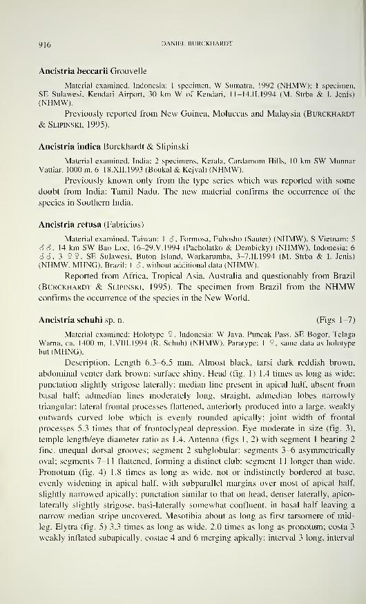
916 DANIEL BURCKHARDT
Ancistria beccarii Grouvelle
Material examined. Indonesia: 1 specimen, W Sumatra, 1992 (NHMW); 1 specimen,
SE Sulawesi, Kendari Airport, 30 km W of Kendari, 11-14.11.1994 (M. Strba & I. Jenis)
(NHMW).
Previously reported from New Guinea, Moluccas and Malaysia (Burckhardt
& Slipinski, 1995).
Ancistria indica Burckhardt & Slipinski
Material examined. India: 2 specimens, Kerala, Cardamom Hills, 10 km SW MunnarVattiar, 1000 m, 6-18.XII.1993 (Boukal & Kejval) (NHMW).
Previously known only from the type series which was reported with some
doubt from India: Tamil Nadu. The new material confirms the occurrence of the
species in Southern India.
Ancistria retusa (Fabricius)
Material examined. Taiwan: 1 â , Formosa, Fuhosho (Sauter) (NHMW). S Vietnam: 5
So, 14 km SW Bao Loc, 16-29.V.1994 (Pacholatko & Dembicky) (NHMW). Indonesia: 6
S S, 3 9 2, SE Sulawesi, Buton Island. Warkarumba, 3-7.II.1994 (M. Strba & I. Jenis)
(NHMW, MHNG). Brazil: 1 3, without additional data (NHMW).
Reported from Africa. Tropical Asia. Australia and questionably from Brazil
(Burckhardt & Slipinski, 1995). The specimen from Brazil from the NHMWconfirms the occurrence of the species in the New World.
Ancistria schuhi sp. n. (Figs 1-7)
Material examined: Holotype 9 . Indonesia: W Java, Puncak Pass, SE Bogor, Telaga
Warna, ca. 1400 m, 1. Vili. 1994 (R. Schuh) (NHMW). Paratype: 1 9, same data as holotype
but (MHNG).
Description. Length 6.3-6.5 mm. Almost black, tarsi dark reddish brown,
abdominal venter dark brown; surface shiny. Head (fig. 1) 1.4 times as long as wide;
punctation slightly strigose laterally; median line present in apical half, absent from
basal half; admedian lines moderately long, straight, admedian lobes narrowly
triangular; lateral frontal processes flattened, anteriorly produced into a large, weakly
outwards curved lobe which is evenly rounded apically; joint width of frontal
processes 5.3 times that of frontoclypeal depression. Eye moderate in size (fig. 3),
temple length/eye diameter ratio as 1.4. Antenna (figs 1, 2) with segment 1 bearing 2
fine, unequal dorsal grooves; segment 2 subglobular; segments 3-6 asymmetrically
oval; segments 7-1 1 flattened, forming a distinct club; segment 11 longer than wide.
Pronotum (fig. 4) 1.8 times as long as wide, not or indistinctly bordered at base,
evenly widening in apical half, with subparallel margins over most of apical half,
slightly narrowed apically; punctation similar to that on head, denser laterally, apico-
laterally slightly strigose, basi-laterally somewhat confluent, in basal half leaving a
narrow median stripe uncovered. Mesotibia about as long as first tarsomere of mid-
leg. Elytra (fig. 5) 3.3 times as long as wide, 2.0 times as long as pronotum; costa 3
weakly inflated subapically, costae 4 and 6 merging apically; interval 3 long, interval
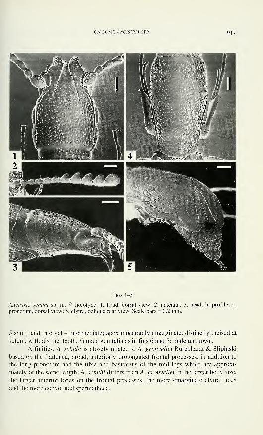
ON SOME ANC1STR1A SPP. 917
Figs 1-5
Ancistria schuhi sp. n., 9 holotype. 1, head, dorsal view; 2. antenna; 3, head, in profile; 4,
pronotum. dorsal view; 5, elytra, oblique rear view. Scale bars = 0.2 mm.
5 short, and interval 4 intermediate; apex moderately emarginate, distinctly incised at
suture, with distinct tooth. Female genitalia as in figs 6 and 7; male unknown.
Affinities. A. schuhi is closely related to A. grouvellei Burckhardt & Slipinski
based on the flattened, broad, anteriorly prolongated frontal processes, in addition to
the long pronotum and the tibia and basitarsus of the mid-legs which are approxi-
mately of the same length. A. schuhi differs from A. grouvellei in the larger body size,
the larger anterior lobes on the frontal processes, the more emarginate elytral apex
and the more convoluted spermatheca.
918 DANIEL BURCKHARDT
Figs 6-7
Ancistrìa schuhi sp. n.. 9 paratype. 6, spermatheca; 7, ostium bursae. Scale bars = 0.1 mm.
Ancistria tenera Günther
Material examined. Indonesia: 2 3 S , W Java, Puncak Pass, SE Bogor, Telaga Warna,
ca. 1400 m, 1 .Vili. 1994 (R. Schuh) (NHMW, MHNG).
Previously known only from the S holotype from W Java (Heller & Günther,
1936).
ACKNOWLEDGMENTS
I thank R. Schuh and H. Schillhammer (NHMW) for the loan of the material,
J. Wuest (MHNG) for the preparation of the SEM-pictures, G. Roth (MHNG) for
inking the drawings and A. Reversy (Geneva) for technical assistance.
REFERENCES
Burckhardt, D. & S.A. Slipinski. 1995. A review of the Passandridae of the world (Cole-
optera, Cucujoidea). IV. Genus Ancistria. - Revue suisse de Zoologie 102: 995-1044.
Heller. K.M. & K. Günther. 1936. Über einige von H. Overbeck 1933/34 auf Java gesam-
melten Insekten. Tijdschrift voor Entomologie 79: 64-76.
Revue suisse de Zoologie, 103 (4): 919-938; décembre 1996
A review of the Japanese Tychobythinus and Bythoxenites
(Coleoptera, Staphylinidae, Pselaphinae)
Ivan LÖBL 1 & Serguei A. KURBATOV 2
1 Muséum d'histoire naturelle, Case postale 6434, CH-121 1 Genève 6, Switzerland.
- Severodvinskaya 9-84, Moscow 129224, Russia.
A review of the Japanese Tychobythinus and Bythoxenites (Coleoptera,
Staphylinidae, Pselaphinae). - Tychobythinus is represented in Japan by 3
species, with T. japonicus, described as new. Bythoxenites holds 10 species
and is widely distributed in Japan. The group is defined by the presence of
a single pair of foveae at the base of the 4th abdominal ventrite and of an
internal aedeagal ridge. The type species of Bythoxenites, B. japonicus, is
known from a cave, but most species inhabit moist forest floor litter. Eight
new species of Bythoxenites are described: B. longicornis, B. torticornis, B.
diversicornis, B. pubiceps, B. brevicornis, B. brevipilis, B. breviceps and B.
frontalis. Keys to Japanese genera of Bythinini and to Japanese species of
Tychobythinus and Bythoxenites are provided.
Key-words: Coleoptera - Staphylinidae - Pselaphinae - Bythinini -
taxonomy - Japan.
INTRODUCTION
The last and most complete account of the Japanese Bythinini is by Jeannel
(1958). He treated two genera, the monobasic Bythoxenites Jeannel and the widely
distributed Bryaxis Kugelann with 14 species in Japan. Since, Nomura (1995)
described an additional Japanese species of Bryaxis, B. kintaro. Thus, the knowledge
of the Japanese Bythinini appears inadequate, compared to that of the Taiwanese
(LObl & Kurbatov, 1995; 1996) or Far East Russian Bythinini (Kurbatov, 1994).
A large amount of additional material of Bythinini has since been collected in
Japan. It consists mainly of numerous species of Bryaxis, however, a number of
specimens belonging to Tychobythinus Ganglbauer and Bythoxenites have been found
also and are treated in the present paper.
MATERIAL AND METHODS
The material examined is deposited in the Muséum d'histoire naturelle, Geneva
(MHNG), except for several paratypes of Bythoxenites brevicornis, B. diversicornis
and B. frontalis which are also in the private collection of the junior author (CSKM).
Manuscript accepted 02.07.1996.
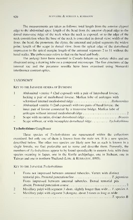
920 IVAN LÖBL & SERGUEI A. K.URBATOV
The measurements are taken as follows: total length from the anterior clypeal
edge to the abdominal apex; length of the head from the anterior clypeal edge to the
dorsal transverse ridge of the neck when the neck is exposed, or to the edge of the
neck constriction when the base of the neck is concealed in dorsal view; width of the
frons, the head, the pronotum, the elytra, the antennal and palpal segments the widest
point; length of the scape in dorsal view, from the apical edge of the dorsobasal
impression to the apical margin; length of the antennal segments 2 to 1 1 without the
basal stalks. The pubescence refers to that on the head and body.
The aedeagi have been mounted in Canada balsam on acetate slides and are
illustrated using a drawing tube on a compound microscope. The fine structures of the
internal sac and the paramere sensilla have been examined using Nomarski
interference contrast optics.
TAXONOMY
Key to the Japanese genera of Bythinini
1 Abdominal ventrite 4 (2nd exposed) with a pair of laterobasal foveae,
lacking a pair of mediobasal foveae. Median lobe of aedeagus with
sclerotised internal mediodorsal ridge Bythoxenites
Abdominal ventrite 4 (2nd exposed) with two pairs of basal foveae, the
inner pair of foveae connected by a transverse bridge. Median lobe of
aedeagus without internal mediodorsal ridge 2
2 Scape with an entire, distinct dorsobasal ridge Bryaxis
Scape without, or with incomplete dorsobasal ridge Tychobythinus
Tychobythinus Ganglbauer
Three species of Tychobythinus are represented within the collections
examined, but only one of them is known from the male sex. It is a new species,
described below. The other two species are likely new but as each is known in a
single female, we find preferable not to name and describe them. Presently, the
members of Tychobythinus appear to by found sporadically in eastern Asia, with three
species occuring in Japan, one in the Kurile archipelago, one in Sichuan, one in
Taiwan and one in northern Thailand (Löbl & Kurbatov, 1995).
Key to the Japanese Tychobythinus
1 Frons not impressed between antennal tubercles. Vertex with distinct
tentorial pits. Pronotai punctation fine T. japonicus
Frons impressed between antennal tubercles. Dorsal tentorial pits
absent. Pronotal punctation coarse 2
2 Maxillary palpi with segment 3 short, slightly longer than wide . . T. species AMaxillary palpi with segment 3 elongate, about 3 times as long as wide
T. species B
JAPANESE TYCHOBYTHINUS AND BYTHOXENITES 921
Tychobythinus japonicus sp. n.
Holotype 6: Japan, Gifu pref., 9 km E Gero, 470 m, 31. VII. 1980, leg. I. Löbl (nr 24);
sifted leaf litter and wood on a steep slope in a ravine, near a stream (MHNG).
Length 1.20 mm. Body and appendages uniformly light ochreous. Pubescence
semi-erect, long, that on abdomen recumbent.
Head 0.25 mm long, 0.24 mm wide. Punctation coarse and very dense, not
confluent, puncture intervals consisting mostly of very narrow ridges, larger on
posterior portion of vertex. Most punctures about as large as facets. Frons with four
conspicuously large impressed punctures arranged in a rectangle, each bearing a
conspicuous, widened seta. Pubescence orientated anteriorly on anterior portion of
frons, obliquely mesally on lateral portions of vertex, apically on median portion of
vertex. Frons narrow, hardly narrowed posterior to antennal tubercles, widened
irregularly toward eyes, with anterior edge distinctly angulate, lateral edges slightly
angulate anterior to eyes. Frontal impression absent. Frontoclypeus almost vertical,
not projecting below, with clypeal margin convex. Vertex convexly raised. Dorsal
tentorial pits situated each in a shallow impression in level of anterior eye margin,
small, about as large as surrounding punctures. Interval between tentorial pits almost
10 times as large as diameter of one tentorial pit. Vertex and occiput without median
carina. Eyes small, flat, with 6 facets. Maxillary palpi with 2nd segment 0.15 mmlong, gradually thickened apically, at apex 0.03 mm wide, tuberculate ventrally; 3rd
segment 0.04 mm long, slightly longer than wide, as wide as 2nd segment, bearing a
few ventral tubercles; 4th segment stout, 0.18 mm long, 0.06 mm wide.
Antennae fairly short. Scape straight, 0.07 mm long, 0.05 mm wide, cylin-
drical, not narrowed subbasally in dorsal view; dorsobasal ridge absent, except near
lateral edges. Pedicel asymmetrical, about as wide as scape, slightly wider than long.
Segments 3 to 10 symmetrical. Segment 3 almost as long as wide. Segments 4 to 8
evenly large, shorter than, and as wide as segment 3, about as long as half of length of
pedicel. Segment 9 as long as 8 but wider, almost as wide as pedicel and about 2
times as wide as long. Segment 10 longer and wider than 9, almost 2 times as wide as
long. Segment 1 1 slightly asymmetrical, much longer than scape, about 1 .4 times as
long as wide, slightly longer than segments 7 to 10 combined.
Pronotum 0.33 mm long, 0.25 mm wide. Punctation very fine and sparse,
discal punctures not clearly delimited, mostly much smaller than intervals between
them, punctation between antebasal sulcus and base fairly coarse and dense, punctures
much larger than intervals between them. Discal pubescence longer than that on head,
orientated mesally on lateral areas, and orientated apically on median portion;
pubescence near base short, orientated anteriorly. Antebasal sulcus deep, fairly wide,
with sharp posterior edge. Lateral fovea situated in a large depression.
Elytra 0.44 mm long, combined 0.53 mm wide. Punctation fine and fairly
dense, punctures well delimited, intervals between punctures 3 to 4 times larger than
their diameters. Humeral area rounded, slightly raised. Marginal carina visible in
dorsal view, touching subhumeral fovea from below.
Metathoracic wings not examined.
922 IVAN LÖBL & SERGUEI A. KURBATOV
FlGS 1-5
Aedeagi in Tychobythinus and Bythoxenites; 1. T. japonìcus; 2. B. longicornis; 3. B.
brevìcornis; 4. B. torticornis; 5. B. diversicornis. Scale bars = 0.1 mm.
JAPANESE TYCHOBYTHINUS AND BYTHOXENITES 923
Metasternum and abdominal sternites evenly very finely punctate; punctation
on median portion of metasternum denser than that on lateral portions of
metasternum.
Protibiae straight. Mesotibiae straight, flattened apically. Metatibiae gradually
stouter toward apical third, curved and flattened in apical fourth.
Male sexual characters: Frons, vertex, and appendages apparently lacking
sexual characters. Head swollen ventrally, anterior to level of eyes, to form a
transverse ridge separated from mouthparts by a narrows and shallow impression.
Transverse ridge punctate and pubescent ventrally, with posterior surface strongly
inflexed and glabrous. Aedeagus (Fig. 1) 0.25 mm long. Parameres abruptly narrowed
apically, each with one wide and two slender, hair-like sensilla. Internal sac bearing a
pair of apical, symmetrical, curved sclerites and an asymmetrically sinuous median
sclerite bearing a ventral apophyse, accompanied by three minute teeth-like sclerites.
Comments. This species shares with T. aino Kurbatov the abruptly narrowed
apical portion of the parameres. It differs however drastically by the shape of the
sclerites of the internal sac of the aedeagus. Externally, it resembles T. siamensis from
which it may be readily distinguished by the tuberculate 2nd and 3rd segment of the
maxillary palpi.
Tychobythinus sp. A
Material: 1 ?. Japan. Honshu. Gifu pref., 8 km SE Gero, 500 m, 31.VII. 1980, leg. I.
Lobi (MHNG), in sifted deep layer of rotten wood and leafs on a slope, near a stream.
Comments. This species may be distinguished from other East and Southeast
Asian species of Tychobythinus, except species B, by the head coarsely and densely
punctate, and lack of dorsal tentorial pits. In general appearence it resembles T.
siamensis Lobi & Kurbatov, but may be easily distinguished by the much lighter body
and the conspicuously tuberculate 2nd and 3rd segments of the maxillary palps.
Tychobythinus siamensis possesses maxillary palpi with segment 3 much shorter than
that in species B.
Tychobythinus sp. B
Material: 1 9 , Japan, Shikoku, Ehime pref., Ishizuchi Nat. Park, Omogo, 12.VIII.1980,
leg. C. Besuchet (MHNG).
Comments. This species may be distinguished readily from other Asian species
of Tychobythinus by the elongate 3rd segment of the maxillary palpi, the scape
narrowed subbasally, the pronotai punctures variably large, with edges distinctly
raised, and lacking dorsal tentorial pits.
Bythoxenites Jeannel
Yoshida & Nomura (1952) described Machaerites (Bythoxenus) japonica
from a limestone cave in Japan, Okutama province, Honsyu. It has been found «allied
to M. (Bythoxenus) Revelieri Reitter» , an European species endemic to Corsica,
placed by Jeannel (1950) in his Amaurobythus which is synonym of Tychobythinus
924 IVAN LÖBL & SERGUEI A. KURBATOV
(Besuchet, 1974). According to the description and illustration, M. japonica is
characterised by the combination of following features: 1) nead rugosely punctate; 2)
antennae with scape long, about as long as one fourth body length; 3) maxillary palpi
with segment 2 tuberculate except at base; 4) segment 4 of the maxillary palpi large,
tapering; 5) elytra conspicuously long and gradually widened apically. Jeannel (1958)
based his new genus Bythoxenites on the description of Machaerites (Bythoxenus)
japonica. According to him, this genus differs from the Slovenian Machaerites Miller
(and, implicitly from Bythoxenus Motschulsky) by «numerous characters», particu-
larly the rugosely punctate head.
Nine species exhibit the characters of Bythoxenites within the examined collec-
tions. In addition, they share an unique aedeagal feature, a sclerified dorsomedian
ridge situated below the compression plate of the median lobe. This ridge is not
attached to the internal sac, as seen in one specimen which has the internal sac com-
pletely extruded. Thus, Bythoxenites appears to be one of the few Bythinini genera
defined by a robust autapomorphy. Unlike in Biyaxis and Tychobythinus, the 4th (2nd
exposed) abdominal sternite in Bythoxenites lacks internal sclerotised structures, the
pair of the mediobasal foveae and the basal grooves.
We have not been able to see the type material of Bythoxenites japonicus
(Yoshida & Nomura), and cannot supplement its description which is lacking in some
critical details. The species of Bythoxenites we have examined vary in numerous
characters, in particular in the size of the antennal and palpal segments. They are
notably smaller than B. japonicus (which is according to the description 2.2 mmlong), and none of them have the maxillary palpi as figured in that species. Thus, we
do not hesitate to consider them as new.
Yoshida & Nomura (1952) believed Bythoxenites japonicus to be micro-
phthalmous, and compared it to European anophthalmous taxa known to inhabit caves
and soil. However, the eyes in male Bythoxenites are not reduced. The males have
distinct humeral angles which indicate the presence of functional metathoracic wings.
In large-eyed males of B. diversicornis, B. frontalis, and B. longiceps, the metathoracic
wings are well developed while in females of B. brevicornis, B. diversicornis and B.
frontalis, they are absent. The presence of the metathoracic wings has not been exa-
mined in the remaining species which are represented by one or two specimens only.
Like in many other Bythinini, the species of Bythoxenites exhibit secondary
sexual characters on the gular area, the tibiae, and have larger eyes and longer elytra
with more distinct humeral angles in males than in females. More unusual are the
sexually modified frons and vertex, the tuberculate profemora in males of some
species, and the protrochanters which are more or less distinctly angulate in males.
The antennae are usually similar in both sexes, but the 4th antennal segment is
enlarged in male of B. torticornis and B. diversicornis.
Bythoxenites is similar to Tychobythinus and unlike Bryaxis, in having the
dorsobasal scapai ridge more or less reduced, and the elytral bases not raised. Its
relationships are unknown as no attempt has yet been made to analyse the
relationships within the Bythinini. Also, the key to the Palaearctic Bythinini genera
(Besuchet, 1974) is inapplicable to Bythoxenites.
JAPANESE TYCHOBYTHINUS AND BYTHOXENITES 925
Key to the species of Bythoxenites
1 Length 2.20 mm B. japonicus
Length 1.55-2.05 mm 2
Elytral pubescence conspicuously short and recumbent, similar to that
on abdomen B. brevipilis
Elytral pubescence not conspicuously short, semi-erect or erect, usually
longer than that on abdomen 3
3 Punctation on pronotai centre conspicuously coarser than that on lateral
prenotai area B. brevicornis
Pronotum with centre as finely punctate, or centre slightly coarser
punctate than lateral portions 4
4 Frontoclypeus bearing sparse setae orientated anteriorly or gradually
curved ventrally 5
Frontoclypeus with dense pubescence orientated ventrally 9
5 Anntenna with segment 9 elongate 6
Antennae with segments 9 as long as wide, or wider than long 7
6 Vertex raised, with short median carina and distinct tentorial pits
B. longicornis
Vertex flattened, with long median carina and obsolete tentorial pits species AAntennal segment 4 unmodified and symmetrical in male B. frontalis
Antennal segment 4 modified and asymmetrical in male 8
8 Male with outer apical angle of antennal segment 4 not prominent.
Aedeagus with apical laminae of internal sac ending by a row of evenly
narrow denticles B. torticornis
Male with outer apical angle of 4 antennal segment prominent. Aedea-
gus with apical laminae of internal sac ending by denticles becoming
larger toward mid-line B. diversicornis
9 Antennal segments 9 and 10 each longer than wide B. longiceps
Antennal segments 9 and 10 each shorter than wide B. pubiceps
Bythoxenites longicornis sp. n.
Holotype 8: Japan, Snikoku, Ehime pref.. Ishizuchi Mt. Nat. Park, Tsuchigoya, 1400
m. 1 1-1 8. VIII. 1980, leg. S. & J. Peck, malaise trap-trough Fagus-Abies forest (MHNG).
Length 2.0 mm. Body reddish-brown, appendices lighter. Pubescence fairly
long, erect on heao and pronotum, semi-erect on elytra, almost recumbent on
abdomen.
Head 0.40 mm long, with eyes 0.40 mm wide, frons 0.25 mm wide anteriorly.
Frontal impression wide and deep, distinctly, irregularly punctate anteriorly,
impunctate posteriorly. Antennal tubercles raised, smooth, each narrower than frontal
impression. Anterior edge of frons sharply delimited, angular. Frontoclypeus vertical
and rounded below, with long setae orientated anteriorly. Frons distinctly narrowed
posterior to antennal tubercles, then gradually widened toward eyes. Dorsal edges of
genae straight and sharp. Vertex slightly convex. Dorsal tentorial pits inconspicuous.
926 IVAN LÖBL & SERGUEI A. KURBATOV
situated slightly posterior of level of anterior eye margin, much closer to dorsal eye
margin than to vertexal mid-line. Vertexal sulci inconspicuous, very shallow and
narrow, traceable from tentorial pits to frontal impression. Anterior portion of vertex
abruptly delimited by a smooth area obliquely inflexed toward frontal impression.
Vertex with high mesal carina starting at level of tentorial pits and ending on inflexed,
smooth area. Vertexal punctation coarse and very dense, punctures deep, mostly
larger than facets, separated by narrow ridges. Eyes large, convex, prominent, with
numerous facets, slightly longer than tempora (lateral view). Tempora rounded. Neck
concealed in dorsal view.
Postgenae oblique and strongly inflexed between neck and gular groove,
bearing long erect setae curved anteriorly. Gular groove transverse, deep, with sharp
anterior and obtuse posterior edges. Posterior edge prominent in middle and bearing
two fairly long, horizontal, divergent tufts of setae; anterior edge with two very short,
vertical laminae. Area between gular groove and mouthparts impressed, with a low
median ridge.
Maxillary palpi with 2nd segment tuberculate; 3rd segment tuberculate api-
cally; 4th segment large, 0.39 mm long, 4 times longer than wide, with concave outer
edge.
Antennae long. Scape subcylindrical, in dorsal view straight, about 3 times as
long as wide, widest near apex, from widest point gradually narrowed basally and
strongly narrowed apically. Scape slightly curved. Pedicel subcylindrical, slightly
narrowed basally, in length slightly exceeding width of scape, 1.6-1.7 times as long
as wide. Dorsobasal scapai ridge indistinct in middle. Segments 3 to 8 evenly wide,
about as wide as two thirds of pedicel. Segment 3 as long as pedicel, 2.5 times as long
as wide, narrowed basally. Segments 4 and 5 each shorter than pedicel, evenly long
and wide, each about 2 times as long as wide. Segments 6 and 8 shorter than segment
5, evenly long and wide, each about 1.5 times as long as wide. Segment 7 longer than
6 (or 8), slightly shorter than 3, about 2 times as long as wide. Segment 9 and 10
evenly long, each slightly longer than pedicel, 9 about 1.7 times as long as wide, 10
thicker, 1.3 times as long as wide. Segment 11 somewhat longer than 9 and 10
combined, distinctly shorter than scape, 2.5 times as long as wide.
Pronotum 0.46 mm long, 0.50 mm wide. Antebasal sulcus sinuate, not well
delimited, widened in middle. Punctation coarse and very dense, irregular, partly
confluent between antebasal sulcus and basal edge; intervals between punctures formed
by narrow ridges. Punctation fairly dense and very fine on most of area between
antebasal sulcus and anterior pronotal edge, with punctures several times smaller than
intervals; punctation distinctly denser near anterior edge than middle of disc.
Elytra 0.84 mm long, combined 0.79 mm wide, gradually widened apically,
almost regularly vaulted dorsally. Basal foveae deep, well delimited, their diameters
slightly smaller than intervals between them. Humeral hump low, elongate. Discal
punctation very fine and sparse.
Profemora lacking tubercles. Protibiae straight, gradually stouter toward apical
fourth, with a deep subapical notch and sharp denticle. Mesotibiae becoming mo-
derately, gradually stouter toward apical third, rather abruptly curved just before
JAPANESE TYCHOBYTHINUS AND BYTHOXEN1TES 927
apical third, almost evenly thick in apical third. Metatibiae almost evenly thick,
straight in basal half, slightly curved in apical half, with an apical denticle.
Aedeagus (Fig. 2) 0.505 mm long. Parameres widened and rounded apically,
with outer edges angulate subapically; each bearing 3 hair-like sensilla. Internal sac
with a pair of symmetrical basal sclerites, a mediobasal vesicle, median denticle and
large apical laminae.
Comments. This species may be readily distinguished by the notably elongate
antennae, in combination with the raised vertex.
Bythoxenites brevicornis sp. n.
Holotype 6: Japan. Ehime pref., Mt. Ishizuchi Nat. Park, steep ravin near Skyline
Road, 1000 m, 14.VIII.1980, leaf litter and rotten wood, leg. I. Lobi (MHNG).Paratypes: 3 9, Japan, Ehime pref., Mt. Ishizuchi National Park, Omogo Valley, 700
m, 18-25.VIII.1980, S. & J. Peck, moss, fungi & log litter, warm temp, forest (MHNG,CSKM); 1 9 . same data but Tsuchigoya 1400 m, 16.VIII., log & moss litter (MHNG).
Similar to B. longicomis from which it differs as follows:
Length 1.80-1.85 mm. Head, pronotum and elytra dark reddish-brown,
abdomen, antennae, palpi, femora and tibiae lighter, tarsi yellow. Pronotal pubescence
semi-erect.
Head 0.37-0.40 mm long, with eyes 0.35-0.37 mm wide; frons 0.21-0.23 mmwide; anterior portion of frontal impression coarsely punctate; mesal vertexal ridge
lower and longer, hardly extending on anterior inflexed area. Vertexal sulci more
distinct. Eyes in 9 with 11 or 12 facets. Tempora oblique. Segment 4 of maxillary
palpi 0.35 mm long, 3 times as long as wide, with straight outer margin. Prominent
middle portion of posterior edge of gular groove bearing long horizontal setae
divergent anteriorly.
Antennae shorter than those in B. longicomis. Scape slightly more than 2 times
as long as wide, widest near base, slightly narrowed toward apex; straight in dorsal
view, flattened dorsoventrally, with almost flat ventral side. Dorsobasal scapai ridge
complete and distinct. Pedicel subcylindrical, 1.4 times as long as wide, as long as
scape wide. Segments 3 to 8 evenly wide, as wide as 4/5 of width of pedicel. Segment
3 slightly shorter than pedicel, 1.5 times as long as wide, gradually widened apically.
Segments 4 to 6 gradually shorter, 4 and 5 distinctly longer than wide, 6 to 8 evenly
long, each about as long as wide. Segments 9 and 10 each as long as segment 3; 9
longer than wide, 10 as long as wide. Segments 1 1 about as long as segments 8 to 10
combined, 2.2 times longer than wide, hardly shorter and wider than scape.
Pronotum 0.38-0.43 mm long, 0.42-0.46 mm wide; discal punctation strongly
irregular; punctures on central area coarse, about as large as those on vertex, regularly
round, well delimited, partly larger than intervals between them; discal punctation
becoming denser and slightly finer toward anterior edge, much finer toward lateral
edges.
Elytra 0.71-0.74 mm long, combined 0.73-0.76 mm wide; diameters of basal
foveae as large as intervals between them.
Profemora tuberculate. Mesotibiae straight.
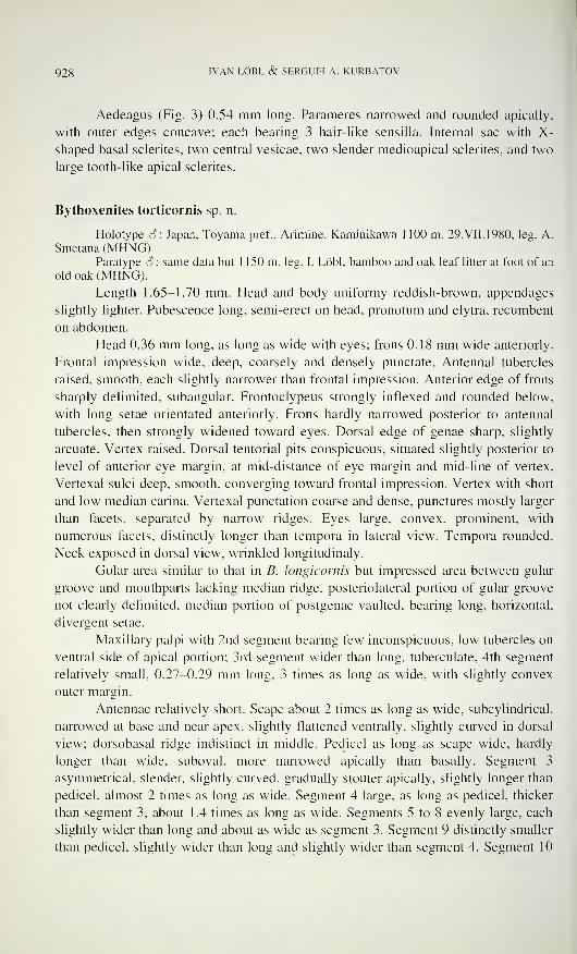
928 IVAN LÖBL & SERGUEI A. KURBATOV
Aedeagus (Fig. 3) 0.54 mm long. Parameres narrowed and rounded apically,
with outer edges concave; each bearing 3 hair-like sensilla. Internal sac with X-
shaped basal sclerites, two central vesicae, two slender medioapical sclerites, and two
large tooth-like apical sclerites.
Bythoxenites torticornis sp. n.
Holotype 3: Japan, Toyama pref.. Arimine, Kaminikawa 1 100 m, 29.VII.1980, leg. A.
Smetana (MHNG).Paratype S : same data but 1 150 m, leg. I. Lobi, bamboo and oak leaf litter at foot of an
old oak (MHNG).
Length 1.65-1.70 mm. Head and body uniformy reddish-brown, appendages
slightly lighter. Pubescence long, semi-erect on head, pronotum and elytra, recumbent
on abdomen.
Head 0.36 mm long, as long as wide with eyes; frons 0.18 mm wide anteriorly.
Frontal impression wide, deep, coarsely and densely punctate. Antennal tubercles
raised, smooth, each slightly narrower than frontal impression. Anterior edge of frons
sharply delimited, subangular. Frontoclypeus strongly inflexed and rounded below,
with long setae orientated anteriorly. Frons hardly narrowed posterior to antennal
tubercles, then strongly widened toward eyes. Dorsal edge of genae sharp, slightly
arcuate. Vertex raised. Dorsal tentorial pits conspicuous, situated slightly posterior to
level of anterior eye margin, at mid-distance of eye margin and mid-line of vertex.
Vertexal sulci deep, smooth, converging toward frontal impression. Vertex with short
and low median carina. Vertexal punctation coarse and dense, punctures mostly larger
than facets, separated by narrow ridges. Eyes large, convex, prominent, with
numerous facets, distinctly longer than tempora in lateral view. Tempora rounded.
Neck exposed in dorsal view, wrinkled longitudinaly.
Gular area similar to that in B. longicornis but impressed area between gular
groove and mouthparts lacking median ridge, posteriolateral portion of gular groove
not clearly delimited, median portion of postgenae vaulted, bearing long, horizontal,
divergent setae.
Maxillary palpi with 2nd segment bearing few inconspicuous, low tubercles on
ventral side of apical portion; 3rd segment wider than long, tuberculate; 4th segment
relatively small, 0.27-0.29 mm long, 3 times as long as wide, with slightly convex
outer margin.
Antennae relatively short. Scape about 2 times as long as wide, subcylindrical,
narrowed at base and near apex, slightly flattened ventrally. slightly curved in dorsal
view; dorsobasal ridge indistinct in middle. Pedicel as long as scape wide, hardly
longer than wide, suboval, more narrowed apically than basally. Segment 3
asymmetrical, slender, slightly curved, gradually stouter apically, slightly longer than
pedicel, almost 2 times as long as wide. Segment 4 large, as long as pedicel, thicker
than segment 3, about 1.4 times as long as wide. Segments 5 to 8 evenly large, each
slightly wider than long and about as wide as segment 3. Segment 9 distinctly smaller
than pedicel, slightly wider than long and slightly wider than segment 4. Segment 10
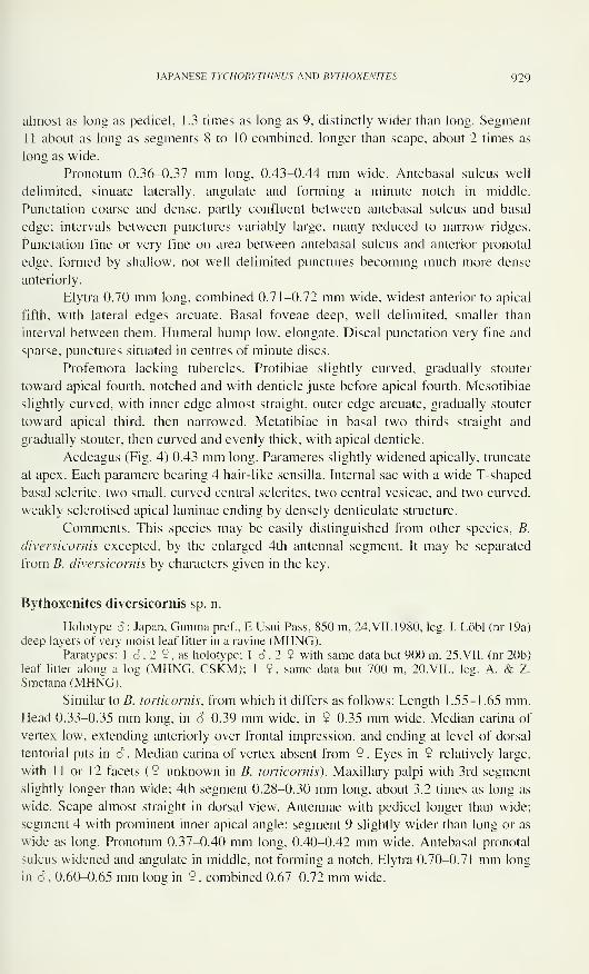
JAPANESE TYCHOBYTHINUS AND BYTH0XEN1TES 929
almost as long as pedicel, 1.3 times as long as 9, distinctly wider than long. Segment
11 about as long as segments 8 to 10 combined, longer than scape, about 2 times as
long as wide.
Pronotum 0.36-0,37 mm long, 0.43-0.44 mm wide. Antebasal sulcus well
delimited, sinuate laterally, angulate and forming a minute notch in middle.
Punctation coarse and dense, partly confluent between antebasal sulcus and basal
edge; intervals between punctures variably large, many reduced to narrow ridges.
Punctation fine or very fine on area between antebasal sulcus and anterior pronotal
edge, formed by shallow, not well delimited punctures becoming much more dense
anteriorly.
Elytra 0.70 mm long, combined 0.71-0.72 mm wide, widest anterior to apical
fifth, with lateral edges arcuate. Basal foveae deep, well delimited, smaller than
interval between them. Humeral hump low, elongate. Discal punctation very fine and
sparse, punctures situated in centres of minute discs.
Profemora lacking tubercles. Protibiae slightly curved, gradually stouter
toward apical fourth, notched and with denticle juste before apical fourth. Mesotibiae
slightly curved, with inner edge almost straight, outer edge arcuate, gradually stouter
toward apical third, then narrowed. Metatibiae in basal two thirds straight and
gradually stouter, then curved and evenly thick, with apical denticle.
Aedeagus (Fig. 4) 0.43 mm long. Parameres slightly widened apically, truncate
at apex. Each paramere bearing 4 hair-like sensilla. Internal sac with a wide T-shaped
basal sclerite, two small, curved central sclerites, two central vesicae, and two curved,
weakly sclerotised apical laminae ending by densely denticulate structure.
Comments. This species may be easily distinguished from other species, B.
diversicornis excepted, by the enlarged 4th antennal segment. It may be separated
from B. diversicornis by characters given in the key.
Bythoxenites diversicornis sp. n.
Holotype 6 : Japan, Gunma pref., E Usui Pass, 850 m, 24.VII.1980, leg. I. Lobi (nr 19a)
deep layers of very moist leaf litter in a ravine (MHNG).Paratypes: 1 6,2 9, as holotype; 1 6, 2 9 with same data but 900 m, 25. VII. (nr 20b)
leaf litter along a log (MHNG. CSKM); 1 $, same data but 700 m, 20.VII., leg. A. & Z.
Smetana (MHNG).
Similar to B. torticornis, from which it differs as follows: Length 1.55-1.65 mm.Head 0.33-0.35 mm long, in 6 0.39 mm wide, in 9 0.35 mm wide. Median carina of
vertex low, extending anteriorly over frontal impression, and ending at level of dorsal
tentorial pits in 6 . Median carina of vertex absent from 9. Eyes in 9 relatively large,
with 11 or 12 facets (9 unknown in B. torticornis). Maxillary palpi with 3rd segment
slightly longer than wide; 4th segment 0.28-0.30 mm long, about 3.2 times as long as
wide. Scape almost straight in dorsal view. Antennae with pedicel longer than wide;
segment 4 with prominent inner apical angle; segment 9 slightly wider than long or as
wide as long. Pronotum 0.37-0.40 mm long, 0.40-0.42 mm wide. Antebasal pronotal
sulcus widened and angulate in middle, not forming a notch. Elytra 0.70-0.71 mm long
in (5 , 0.60-0.65 mm long in 9 , combined 0.67-0.72 mm wide.
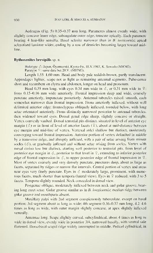
930 IVAN LÖBL & SERGUEI A. KURBATOV
Aedeagus (Fig. 5) 0.35-0.37 mm long. Parameres almost evenly wide, with
slightly concave inner edge, subangulate outer edge, truncate apically. Each paramere
bearing 4 hair-like sensilla. Basal sclerite narrower than in B. torticornis; apical
sclerotised laminae wider, ending by a row of denticles becoming larger toward mid-
line.
Bythoxenites brevipilis sp. n.
Holotype 3: Japan, Ôyamazuki, Kyoto-Fu, 18.V.1967, K. Sawada (MHNG).Paratype ? : same data but 20.V. (MHNG).
Length 1.55-1.60 mm. Head and body pale reddish-brown, partly translucent.
Appendages lighter, scape not as light as remaining antennal segments. Pubescence
short and recumbent on elytra and abdomen, longer on head and pronotum.
Head 0.35 mm long, with eyes 0.34 mm wide in 3, or 0.31 mm wide in 9;
frons 0.15-0.16 mm wide anteriorly. Frontal impression deep and wide, coarsely
punctate anteriorly, impunctate posteriorly. Antennal tubercles raised, smooth, each
somewhat narrower than frontal impression. Frons anteriorly inflexed, without well
delimited anterior edge; frontoclypeus obliquely inflexed, rounded below, with long
setae orientated anteriorly. Frons distinctly narrowed posterior to antennal tubercles,
then widened toward eyes. Dorsal genal edge sharp, slightly concave or straight.
Vertex convexly vaulted. Dorsal tentorial pits distinct, situated in level of anterior eye
margin (3) or in front of level of anterior facets ( 9 ), about at mid-distance between
eye margin and mid-line of vertex. Vertexal sulci shallow but distinct, moderately
converging toward frontal impression. Anterior portion of vertex delimited in middle
by a transverse ridge, and abruptly inflexed, with a pair of setae arising from minute
socles ( 3 ), or gradually inflexed and without setae arising from socles. Vertex with
mesal carina low but distinct, starting well posterior to tentorial pits, from level of
posterior eye margin in 3 , posterior to that level in 9 , extending to inferior posterior
edge of frontal impression in 3 , to upper posterior edge of frontal impression in 9.
Most of vertex coarsely and very densely punctate, punctures deep, about as large as
facets, separated by ridges or narrow flat intervals. Central portion of vertex and areas
near eyes very finely punctate. Eyes in 3 moderately large, prominent, with nume-
rous facets, much shorter than tempora (lateral view). Eyes in 9 reduced, with 3 to 5
facets. Tempora slightly rounded. Neck concealed in dorsal view.
Postgenae oblique, moderately inflexed between neck and gular groove, bear-
ing long erect setae. Gular groove similar as in B. longicornis; median ridge between
gular groove and mouthparts absent.
Maxillary palpi with 2nd segment conspicuously tuberculate, except on basal
portion; 3rd segment about as long as wide; 4th segment 0.36-0.37 mm long, A.2-A.6
times as long as wide, with outer margin slightly concave, at apex slightly inflexed
ventrally.
Antennae long. Scape slightly curved, subcylindrical, about 4 times as long as
wide in dorsal view, evenly wide in posterior 3/4, narrowed basally, with ventral side
flattened. Dorsobasal scapai ridge widely interrupted in middle. Pedicel cylindrical, in
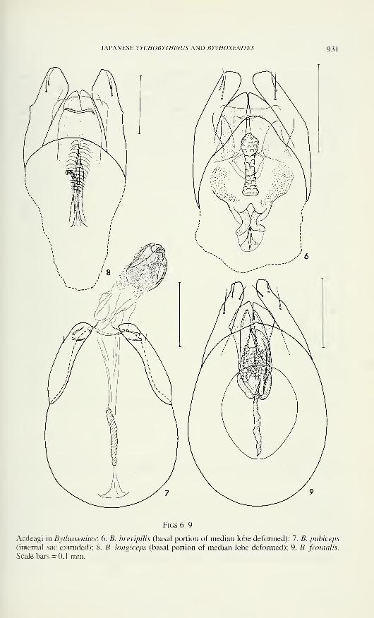
JAPANESE TYCHOHYTHINUS AND BYTHOXENITES 931
Figs 6-9
Aedeagi in Bytlioxenites; 6. B. brevipilis (basal portion of median lobe deformed): 7. B. pubiceps
(internal sac extruded); 8. B. longiceps (basal portion of median lobe deformed); 9. B. frontalis.
Scale bars = 0.1 mm.
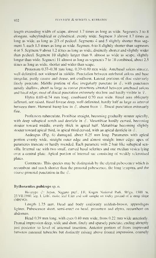
932 IVAN LÖBL & SERGUEI A. KURBATOV
length exceeding width of scape, almost 1.7 times as long as wide. Segments 3 to 8
elongate, subcylindrical or cylindrical, evenly wide. Segment 3 almost 1 .7 times as
long as wide, as long as 2/3 of pedicel. Segments 4 and 5 slightly shorter than seg-
ment 3, each 1.5 times as long as wide. Segment 6 to 8 slightly shorter than segments
4 or 5. Segment 9 about 1 .2 times as long as wide, distinctly shorter and slightly wider
than pedicel. Segment 10 slightly larger than 9, almost as wide as scape, slightly
longer than wide. Segment 1 1 almost as long as segments 7 to 10 combined, about 2.5
times as long as wide, shorter and wider than scape.
Pronotum 0.37-0.38 mm long. 0.39-0.40 mm wide. Antebasal sulcus sinuate,
well delimited, not widened in middle. Punctation between antebasal sulcus and base
irregular, partly coarse and dense, not confluent. Lateral portions of disc extremely
finely punctate. Middle portion of disc irregularly punctate in cT, with punctures
mostly shallow, about as large as coarse punctures situated between antebasal sulcus
and basal edge: most of discal punctation extremely shallow and hardly visible in 9
.
Elytra 0.69-0.74 mm long, combined 0.70 mm wide. Basal area obliquely
inflexed, not raised. Basal foveae deep, well delimited, hardly half as large as interval
between them. Humeral hump low in â . absent from 9 . Discal punctation extremely
fine.
Profemora tuberculate. Protibiae straight, becoming gradually stouter apically,
with deep subapical notch and denticle in ô. Mesotibiae hardly curved, becoming
stouter toward middle, evenly thick in apical half. Metatibiae becoming slightly
stouter toward apical third, in apical third curved, with an apical denticle in 6
.
Aedeagus (Fig. 6) damaged, about 0.25 mm long. Parameres with apical
portion evenly wide, straight outer edge and almost straight inner edge; apex of
parameres truncate or hardly rounded. Each paramere with 2 hair-like subapical sen-
silla. Internal sac with two small, curved basal sclerites and one median vesica lying
over a central plate. Apical portion of internal sac consisting of weakly sclerotised
plates.
Comments. This species may be distinguish by the elytral pubescence which is
recumbent and much shorter than the pronotal pubescence, the long tempora, and the
coarse pronotal punctation in the <S
.
Bythoxenites pubiceps sp. n.
Holotype 6: Japan, Nagano pref., J.E. Kogen National Park. Shiga, 1500 m.
23.VII. 1980. leg. I. Lobi, moist leaf litter and soil sample on rocky ground of a steep slope
(MHNG).
Length 1.75 mm. Head and body uniformly reddish-brown, appendages
lighter. Pubescence short, semi-erect on head, pronotum and elytra, recumbent on
abdomen.
Head 0.39 mm long, with eyes 0.40 mm wide; frons 0.22 mm wide anteriorly.
Frontal impression deep, wide and short, finely and sparsely punctate, ending abruptly
just posterior to level of antennal insertion. Anterior portion of frons impressed
between antennal tubercles but distinctly raising above frontal impression, coarsely
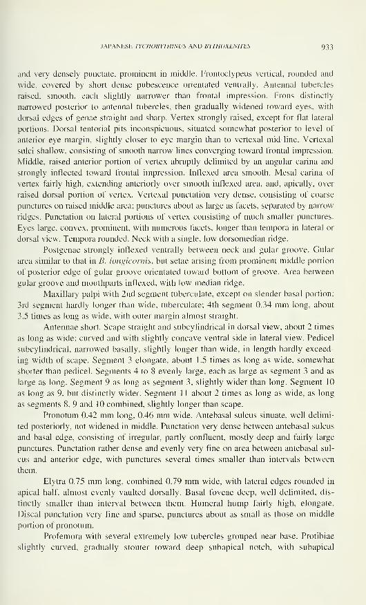
JAPANESE TYCHOBYTHINUS AND BYTHOXENITES 933
and very densely punctate, prominent in middle. Frontoclypeus vertical, rounded and
wide, covered by short dense pubescence orientated ventrally. Antennal tubercles
raised, smooth, each slightly narrower than frontal impression. Frons distinctly
narrowed posterior to antennal tubercles, then gradually widened toward eyes, with
dorsal edges of genae straight and sharp. Vertex strongly raised, except for flat lateral
portions. Dorsal tentorial pits inconspicuous, situated somewhat posterior to level of
anterior eye margin, slightly closer to eye margin than to vertexal mid-line. Vertexal
sulci shallow, consisting of smooth narrow lines converging toward frontal impression.
Middle, raised anterior portion of vertex abruptly delimited by an angular carina and
strongly inflected toward frontal impression. Inflexed area smooth. Mesal carina of
vertex fairly high, extending anteriorly over smooth inflexed area, and, apically, over
raised dorsal portion of vertex. Vertexal punctation very dense, consisting of coarse
punctures on raised middle area; punctures about as large as facets, separated by narrow
ridges. Punctation on lateral portions of vertex consisting of much smaller punctures.
Eyes large, convex, prominent, with numerous facets, longer than tempora in lateral or
dorsal view. Tempora rounded. Neck with a single, low dorsomedian ridge.
Postgenae strongly inflexed ventrally between neck and gular groove. Gular
area similar to that in B. longicornis, but setae arising from prominent middle portion
of posterior edge of gular groove orientated toward bottom of groove. Area between
gular groove and mouthparts inflexed, with low median ridge.
Maxillary palpi with 2nd segment tuberculate, except on slender basal portion;
3rd segment hardly longer than wide, tuberculate; 4th segment 0.34 mm long, about
3.5 times as long as wide, with outer margin almost straight.
Antennae short. Scape straight and subcylindrical in dorsal view, about 2 times
as long as wide; curved and with slightly concave ventral side in lateral view. Pedicel
subcylindrical, narrowed basally, slightly longer than wide, in length hardly exceed-
ing width of scape. Segment 3 elongate, about 1.5 times as long as wide, somewhat
shorter than pedicel. Segments 4 to 8 evenly large, each as large as segment 3 and as
large as long. Segment 9 as long as segment 3, slightly wider than long. Segment 10
as long as 9, but distinctly wider. Segment 1 1 about 2 times as long as wide, as long
as segments 8, 9 and 10 combined, slightly longer than scape.
Pronotum 0.42 mm long, 0.46 mm wide. Antebasal sulcus sinuate, well delimi-
ted posteriorly, not widened in middle. Punctation very dense between antebasal sulcus
and basal edge, consisting of irregular, partly confluent, mostly deep and fairly large
punctures. Punctation rather dense and evenly very fine on area between antebasal sul-
cus and anterior edge, with punctures several times smaller than intervals between
them.
Elytra 0.75 mm long, combined 0.79 mm wide, with lateral edges rounded in
apical half, almost evenly vaulted dorsally. Basal foveae deep, well delimited, dis-
tinctly smaller than interval between them. Humeral hump fairly high, elongate.
Discal punctation very fine and sparse, punctures about as small as those on middle
portion of pronotum.
Profemora with several extremely low tubercles grouped near base. Protibiae
slightly curved, gradually stouter toward deep subapical notch, with subapical
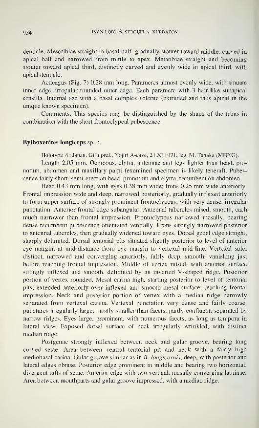
934 IVAN LÖBL & SERGUEI A. KURBATOV
denticle. Mesotibiae straight in basal half, gradually stouter toward middle, curved in
apical half and narrowed from mittle to apex. Metatibiae straight and becoming
stouter toward apical third, distinctly curved and evenly wide in apical third, with
apical denticle.
Aedeagus (Fig. 7) 0.28 mm long. Parameres almost evenly wide, with sinuate
inner edge, irregular rounded outer edge. Each paramere with 3 hair-like subapical
sensilla. Internal sac with a basal complex sclerite (extruded and thus apical in the
unique known specimen).
Comments. This species may be distinguished by the shape of the frons in
combination with the short frontoclypeal pubescence.
Bythoxenites longiceps sp. n.
Holotype 6: Japan, Gifu pref., Nojiri A-cave, 21.XI.1971, leg. M. Tanaka (MHNG).
Length 2.05 mm. Ochreous, elytra, antennae and legs lighter than head, pro-
notum, abdomen and maxillary palpi (examined specimen is likely teneral). Pubes-
cence fairly short, semi-erect on head, pronotum and elytra, recumbent on abdomen.
Head 0.43 mm long, with eyes 0.38 mm wide; frons 0.25 mm wide anteriorly.
Frontal impression wide and deep, narrowed posteriorly, gradually inflexed anteriorly
to form upper surface of strongly prominent frontoclypeus; with very dense, irregular
punctation. Anterior frontal edge subangular. Antennal tubercles raised, smooth, each
much narrower than frontal impression. Frontoclypeus narrowed mesally, bearing
dense recumbent pubescence orientated ventrally. Frons strongly narrowed posterior
to antennal tubercles, then gradually widened toward eyes. Dorsal genal edge straight,
sharply delimited. Dorsal tentorial pits situated slightly posterior to level of anterior
eye margin, at mid-distance from eye margin to vertexal mid-line. Vertexal sulci
distinct, narrowed and converging anteriorly, fairly deep, smooth, vanishing just
before reaching frontal impression. Middle of vertex raised, with anterior surface
strongly inflexed and smooth, delimited by an inverted V-shaped ridge. Posterior
portion of vertex rounded. Mesal carina high, starting posterior to level of tentorial
pits, extended anteriorly over inflexed and smooth mesal surface, reaching frontal
impression. Neck and posterior portion of vertex with a median ridge narrowly
separated from vertexal carina. Vertexal punctation very dense and fairly coarse,
punctures irregularly large, mostly smaller than facets, partly confluent, separated by
narrow ridges. Eyes large, prominent, with numerous facets, as long as tempora in
lateral view. Exposed dorsal surface of neck irregularly wrinkled, with distinct
median ridge.
Postgenae strongly inflexed between neck and gular groove, bearing long
curved setae. Area between ventral tentorial pit and neck with a fairly high
mediobasal carina. Gular groove similar as in B. longicornis, deep, with posterior and
lateral edges obtuse. Posterior edge prominent in middle and bearing two horizontal,
divergent tufts of setae. Anterior edge with two vertical, mesally converging laminae.
Area between mouthparts and gular groove impressed, with a median ridge.
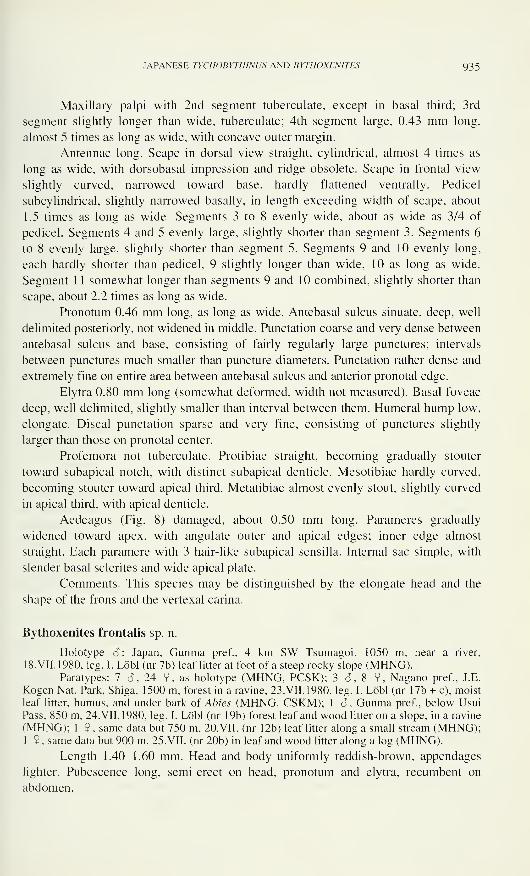
JAPANESE TYCHOBYTHINUS AND BYTHOXENITES 935
Maxillary palpi with 2nd segment tuberculate, except in basal third; 3rd
segment slightly longer than wide, tuberculate; 4th segment large, 0.43 mm long,
almost 5 times as long as wide, with concave outer margin.
Antennae long. Scape in dorsal view straight, cylindrical, almost 4 times as
long as wide, with dorsobasal impression and ridge obsolete. Scape in frontal view
slightly curved, narrowed toward base, hardly flattened ventrally. Pedicel
subcylindrical, slightly narrowed basally, in length exceeding width of scape, about
1.5 times as long as wide. Segments 3 to 8 evenly wide, about as wide as 3/4 of
pedicel. Segments 4 and 5 evenly large, slightly shorter than segment 3. Segments 6
to 8 evenly large, slightly shorter than segment 5. Segments 9 and 10 evenly long,
each hardly shorter than pedicel, 9 slightly longer than wide, 10 as long as wide.
Segment 1 1 somewhat longer than segments 9 and 10 combined, slightly shorter than
scape, about 2.2 times as long as wide.
Pronotum 0.46 mm long, as long as wide. Antebasal sulcus sinuate, deep, well
delimited posteriorly, not widened in middle. Punctation coarse and very dense between
antebasal sulcus and base, consisting of fairly regularly large punctures; intervals
between punctures much smaller than puncture diameters. Punctation rather dense and
extremely fine on entire area between antebasal sulcus and anterior pronotal edge.
Elytra 0.80 mm long (somewhat deformed, width not measured). Basal foveae
deep, well delimited, slightly smaller than interval between them. Humeral hump low,
elongate. Discal punctation sparse and very fine, consisting of punctures slightly
larger than those on pronotal center.
Profemora not tuberculate. Protibiae straight, becoming gradually stouter
toward subapical notch, with distinct subapical denticle. Mesotibiae hardly curved,
becoming stouter toward apical third. Metatibiae almost evenly stout, slightly curved
in apical third, with apical denticle.
Aedeagus (Fig. 8) damaged, about 0.50 mm long. Parameres gradually
widened toward apex, with angulate outer and apical edges; inner edge almost
straight. Each paramere with 3 hair-like subapical sensilla. Internal sac simple, with
slender basal sclerites and wide apical plate.
Comments. This species may be distinguished by the elongate head and the
shape of the frons and the vertexal carina.
Bythoxenites frontalis sp. n.
Holotype S: Japan, Gunma pref., 4 km SW Tsumagoi, 1050 m, near a river,
18.VII.1980, leg. I. Lobi (nr 7b) leaf litter at foot of a steep rocky slope (MHNG).Paratypes: 7 S, 24 9 , as holotype (MHNG, PCSK); 3 S, 8 9, Nagano pref., J.E.
Kogen Nat. Park. Shiga. 1500 m, forest in a ravine, 23. VII. 1980, leg. I. Lobi (nr 17b + c), moist
leaf litter, humus, and under bark of Abies (MHNG, CSKM); 1 â , Gunma pref., below Usui
Pass, 850 m, 24. VII. 1980, leg. I. Lobi (nr 19b) forest leaf and wood litter on a slope, in a ravine
(MHNG); 1 9 , same data but 750 m, 20.VII. (nr 12b) leaf litter along a small stream (MHNG);1 9 , same data but 900 m, 25. VII. (nr 20b) in leaf and wood litter along a log (MHNG).
Length 1.40-1.60 mm. Head and body uniformly reddish-brown, appendages
lighter. Pubescence long, semi-erect on head, pronotum and elytra, recumbent on
abdomen.
936 IVAN LÖBL & SERGUEI A. KURBATOV
Head 0.34-0.36 mm long, with eyes 0.33-0.36 mm wide in â, and 0.31-0.32
mm wide in 9. Frons anteriorly 0.19-0.20 mm wide. Frontal impression wide and
punctate, in â shallow and very short, not extending posterior to level of antennal
tubercles, in 9 deep, much longer than in 6 , similar to that in other species of the
group. Frons in 6 delimited from above by minute transverse ridge; surface inflexed
toward frontal impression smooth, but with two long setae orientated anteriorly.
Antennal tubercles raised, smooth, each distinctly narrower than frontal impression.
Anterior edge of frons sharply delimited, prominent and angulate. Frontoclypeus
almost vertical, rounded below, with long setae orientated anteriorly. Frons narrowed
posterior to antennal tubercles, then widened gradually toward eyes. Dorsal genal
edge straight and sharp. Vertex relatively flat. Dorsal tentorial pits situated in a small
impression, at level of anterior eyes margin in S , slightly anterior to anterior eye
margin in 9 . Vertexal sulci inconspicuous, very narrow and shallow, traceable from
tentorial pits to frontal impression. Mesal carina evenly low from frontal impression
to level of tentorial pits in ó*, shortened in 9 . Area on each side of mesal carina in
same level as, or slightly below level of, vertex, moderately raised above lateral
edges, irregularly and finely punctate in S , more coarsely punctate in 9 . Punctation
on vertex and along lateral edges of frons coarse and very dense, many punctures
larger than facets, separated by narrow ridges, sometimes confluent. Eyes in 6 large,
prominent, with numerous facets, longer than tempora in lateral view. Eyes in 9
small but prominent, consisting of 12 to 14 facets, shorter than tempora in lateral
view. Tempora rounded. Neck dorsally with a median ridge and irregular fine
punctation, or finely rugose.
Postgenae oblique and strongly inflexed, bearing long setae curved anteriorly.
Gular groove in 6 transverse, deep, with sharp anterior, obtuse posterior edges.
Posterior edge prominent in middle and bearing two flat, obliquely divergent
horizontal tufts of short setae; anterior edge of gular groove with two minute vertical
laminae. Area between gular groove and mouthparts impressed, with a low median
ridge.
Maxillary palpi with 2nd segment bearing low apical tubercles; 3rd segment
tuberculate; 4th segment 0.27-0.30 mm long, about 3 times as long as wide, with
slightly convex outer margin.
Antennae fairly short. Scape subcylindrical, about 2 times as long as wide,
somewhat narrowed apically, flattened ventrally, with straight posterior edge and
slightly convexly rounded anterior edge in dorsal view. Dorsobasal scapai ridge
indistinct, except laterally. Pedicel elongate-oval, as long as scape wide, 1.4 times as
long as wide. Segments 3 to 8 evenly wide. Segment 3 elongate, shorter than pedicel,
as wide as 4/5 of pedicel. Segment 4 as long as wide. Segments 5 to 8 each slightly
shorter than 4. Segments 9 and 10 slightly shorter than 3, both distinctly wider than
long, segment 10 larger than 9. Segment 1 1 about 2 times as long as wide, as long as
segments 8 to 10 combined.
Pronotum 0.37-0.39 mm long, 0.40-0.42 mm wide. Antebasal sulcus sinuate,
well delimited, widened in middle. Punctation irregular, coarse and very dense, partly
JAPANESE TYCHOBYTHINUS AND BYTHOXENITES 937
confluent between antebasal sulcus and basal edge; intervals between punctures
formed mostly by narrow ridges. Punctation sparse and very fine on area between
antebasal sulcus and anterior pronotal edge, some specimens with large but extremely
shallow punctures on centre of pronotal disc.
Elytra 0.65-0.68 mm long in 3, 0.60-0.64 mm long in 9, combined 0.64-
0.72 mm wide. Basal foveae deep, well delimited, smaller than interval between them.
Humeral hump low but distinct in 3 , obsolete in 9 .
Profemora lacking tubercles. Protibiae in 3 slightly curved, gradually stouter
toward apical third, with shallow subapical notch and small denticle. Mesotibiae in 3
straight and becoming gradually stouter toward apical third, in apical third evenly
thick and curved. Mesotibia in 9 becoming stouter toward middle and curved in
middle portion. Metatibiae in both sexes becoming slightly stouter toward middle,
slightly curved in apical half.
Aedeagus (Fig. 9) 0.32-0.35 mm long. Parameres slightly narrowed apically,
with almost straight inner edge, outer edge concave near apex, each paramere bearing
3 hair-like sensilla. Internal sac with two small, basal, elongate denticles, one long
curved central sclerite, two weakly sclerotised apical laminae, and a central
membranous vesicle.
Comments. This species may be readily identified by the very short frontal
impression in â .
Bythoxenites species A
Material: 1 9, Japan. Tochigi pref., Nikko Nat. Park, Chuzenji, 1350 m, 14.VII.1980,
leg. I. Lobi (no 1 ); bamboo leaf litter with rotten wood at a forest edge (MHNG).
This specimen may be distinguished by the following characters: Length 1.75
mm; pubescence long and semi-erect (except on abdomen); anterior edge of frons
prominent; frontoclypeus narrowed mesally to form a keel, bearing long erect setae;
vertex flattened, with mesal carina very low, extending from edge of frontal
impression to transverse ridge of neck; eyes prominent, with 1 1 or 12 facets; antennae
with pedicel and 3rd segment evenly long, segments 4 to 6 gradually shorter, 6 to 8
evenly long; maxillary palpi with 4th segment about 4 times as long as wide;
pronotum very finely punctate between antebasal sulcus and anterior pronotal edge.
The specimen obviously represents a distinct species. In absence of the
knowledge of the male sexual characters, we cannot define it adequately, and prefer
not to name it.
ACKNOWLEDGEMENTS
Mr S. Hisamatsu, Matsuyama, provided useful information on collecting sites
in Shikoku. His hospitality made the stay of the senior author to Japan very pleasant.
Drs Jarmila Kukalova-Peck, Stewart B. Peck, Kohei Sawada, Ales Smetana, and
Masahiro Tanaka provided interesting additional material. The comments of Donald
S. Chandler led to improvement of the paper and are greatfully acknowledged.
938 IVAN LÖBL & SERGUEI A. KURBATOV
REFERENCES
Besuchet, C. 1974. Les Psélaphides cavernicoles de l'Espagne (Coleoptera, Pselaphidae).
Miscellànea Zoològica 3: 41-69.
Jeannel, R. 1950. Coléoptères Psélaphides. Faune de France, 53: i-iii, 1 —42 1
.
Jeannel, R. 1958. Révision des Psélaphides du Japon. Mémoires du Muséum national d'his-
toire naturelle, Série A, Zoologie 18: 1-138.
Kurbatov, S.A. 1994. Les Bryaxis de l'Extrême-Est de la Russie (Coleoptera, Pselaphidae).
Russian Entomological Journal 3: 39—47.
Löbl, ï. & S.A. Kurbatov. 1995. New Tychobythinus (Coleoptera, Staphylinidae, Pselaphinae)
from East and Southeast Asia. Mitteilungen der Schweizerischen entomologischen
Gesellschaft 68: 297-304.
Löbl, I. & S.A. Kurbatov. 1996. The Bryaxis of Taiwan (Coleoptera: Staphylinidae: Psela-
phinae). Bulletin ofNational Museum ofNatural Science (in press).
Nomura, S. 1995. Taxonomic notes on Bryaxis koltzei (Reitter) and its allied new species from
Japan (Coleoptera, Pselaphidae). Esakia 35: 129-134.
Yoshida, A. & S. Nomura. 1952. A list of Arthropoda in the limestone caves in Kantô-Moun-
tainland, with the description of a new genus and three species. Chûhô 6: 1-8, 2 pis.
Revue suisse de Zoologie, 103 (4): 939-949; décembre 1996
Nouveaux Cholevinae d'Asie (Coleoptera Leiodidae)
Michel PERREAULaboratoire de Magnétisme des Surfaces, Université Paris 7
2, place Jussieu, F-75251 Paris cedex 05, France.
New Cholevinae from Asie (Coleoptera Leiodidae). - Following newtaxa of Leiodidae Cholevinae from China and Far East Russia are
described: Pandemia sinica n. sp., Nemadus sìchuanus n. sp., Anemadiola
kurbatovi n. sp., Anemadus wolongianus n. sp., a new subgenus oï Nargus:
Eunargus, and Sciaphyes kurbatovi. Sciaphyes is redescribed and the
presence of a likely new species is discussed. This genus is notable by the
5-segmented protarsi in the female. The characters used to define the
Leptoderini and the possible paraphy of this taxon are discussed.
Key-words: Coleoptera - Leiodidae - Cholevinae - China - Far East Russia
- Taxonomy.
INTRODUCTION
Cet article s'inscrit dans une série d'études sur la faune des Cholevinae d'ex-
trême-orient. De nombreuses lignées endémiques de cette région ont déjà été mises en
évidence par les travaux de Szymczakowski, Nakane, Hayashi, Miyama, Nishikawa et
Perreau, et ont été résumées dans un article précédent (Perreau, 1996). Le présent
travail expose de nouvelles découvertes effectuées par S. Kurbatov lors de plusieurs
expéditions en Chine et en Russie d'extrême-orient, et continue de montrer que cette
région a été le lieu de nombreuses spéciations, et l'origine d'un grand nombre de
lignées.
La localisation des specimens examinés est indiquée comme suit: Muséumd'histoire naturelle de Genève: MHNG; et collection M. Perreau: CMP.
TAXONOMIE
Pandania sinica n. sp.
Holotype 6: Chine, S. Yunnan, réserve naturelle de Mangyang, 500 m dans du bois
pourri. 1 1 .X. 1994, Kurbatov leg. (MHNG).
Description: Longueur: 2,9 mm. Espèce ailée, de coloration brune uniforme,
un peu plus foncée sur la tête. Tout le corps recouvert d'une fine pubescence dorée.
Tête non striolée transversalement, finement ponctuée et microréticulée entre
les points. Suture clypeo-frontale absente. Yeux bien développés. Antennes corn-
Manuscrit accepté le 01 .06. 1 996.
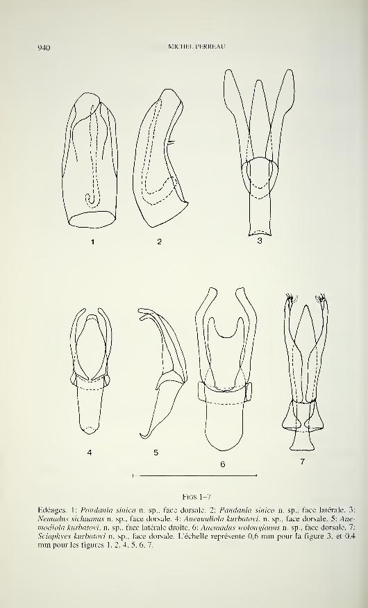
940 MICHEL PERREAU
Figs 1-7
Edéages. 1: Pandemia sinica n. sp., face dorsale. 2: Pandania sinica n. sp., face latérale. 3:
Nemadus sichuamts n. sp., face dorsale. 4: Anemadiola kurbatovi, n. sp., face dorsale. 5: Ane-
madiola kurbatovi, n. sp., face latérale droite. 6: Anemadus wolongianus n. sp., face dorsale. 7:
Sciaphyes kurbatovi n. sp., face dorsale. L'échelle représente 0,6 mm pour la figure 3, et 0,4
mm pour les figures 1. 2, 4, 5, 6, 7.
NOUVEAUX CHOLEVINAE D ASIE 94]
pactes, les articles non rétrécis en avant ni en arrière, au niveau de leur insertion. La
formule antennaire est la suivante (longueur des antennomères en micromètres): 165,
95, 50, 55, 65, 45. 105, 50, 95, 295 (précision: 5 micromètres). Le dernier anten-
nomère est donc particulièrement long.
Pronotum 1.57 fois plus large que long, la plus grande largeur près de la base.
Surface non striolée transversalement, assez finement et densément ponctuée, et
microréticulée entre les points. Cette sculpture est similaire à celle de la tête.
Elytres 1,3 fois plus longs que larges. Surface finement striolée transver-
salement, et microréticulée entre les strioles. Structure sternale conforme au plan de
base des Ptomaphaginina.
Tibias antérieurs bordés d'une rangée de petites épines égales sur le bord
apical antérieur, et sur le bord externe, comme chez tous les Ptomaphaginina. Tarses
antérieurs et intermédiaires non dilatés. Tous les tarses comprimés, étroits, et épais.
Segment IX avec un spiculum gastrale ne dépassant pas en avant le bord
antérieur des latérotergites.
Edéage ovale, conforme au plan de base des Ptomaphaginus, épais et arrondi à
l'extrémité, avec deux soies perpendiculaires au plan du lobe médian situées au milieu
de sa longueur, et latéralement. Stylet interne long et grêle, (figures 1 et 2).
Femelle inconnue.
Nous plaçons cette espèce dans le genre Pandemia Szymczakowski, 1964, dont
elle présente le caractère distinctif principal: la ponctuation non alignée en strioles
transversales sur le pronotum. Une seule espèce était connue jusqu'à présent dans ce
genre: Pandania oxytropis Szymczakowski, 1964, récoltée à Sumatra, et dont le mâle
est toujours inconnu. P. sinica est bien différente car nettement moins trapue. D'autres
caractères importants séparent ces deux espèces: la ponctuation du pronotum est plus
serrée et plus grosse chez P. sinica, la carène mésosternale est basse de même que
chez tous les Ptomaphaginus, et non pas haute et anguleuse comme chez P. oxytropis.
La conformation des antennes de P. sinica, compactes et avec l'antennomère 1 1 parti-
culièrement long évoque tout à fait celles des Philomessor, Attaephilus, et de certains
Catopomorphus, et diffère notamment de celles de P. oxytropis. Ce parallélisme dans
deux lignées bien différentes de Choievinae peut surprendre. Un mode de vie sem-
blable peut être une explication, et ceci suggère pour P. sinica une propension à la
myrmécophilie. Rien n'est connu sur la biologie de P. sinica excepté qu'elle a été
récoltée dans du bois en décomposition. Cet habitat est assez surprenant pour un
Ptomaphagini dont les espèces sont en général récoltées dans la litière des forêts.
Toutes les différences morphologiques séparant les deux espèces rendent très
hypothétiques les relations de cette nouvelle espèce avec P. oxytropis. Il faudra attendre
de connaître le mâle de P. oxytropis et la femelle de P. sinica pour confirmer si ces
deux espèces sont bien congénériques ou s'il faut isoler P. sinica dans un genre à part.
Nemadus sichuanus n. sp.
Holotype 6: Chine, Sichuan, mont Emei, 1400 m, débris végétaux, 22.IX. 1994, leg. S.
Kurbatov (MHNG).
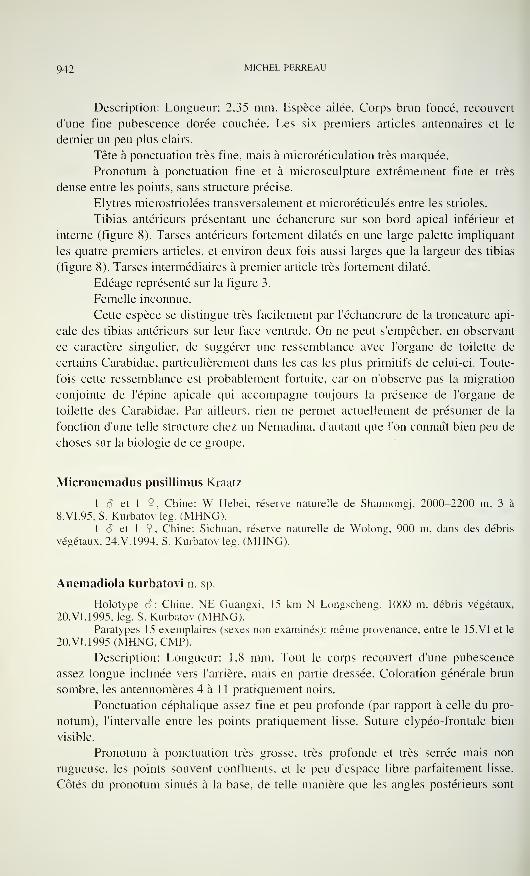
942 MICHEL PERREAU
Description: Longueur: 2,35 mm. Espèce ailée. Corps brun foncé, recouvert
d'une fine pubescence dorée couchée. Les six premiers articles antennaires et le
dernier un peu plus clairs.
Tête à ponctuation très fine, mais à microréticulation très marquée.
Pronotum à ponctuation fine et à microsculpture extrêmement fine et très
dense entre les points, sans structure précise.
Elytres microstriolées transversalement et microréticulés entre les strioles.
Tibias antérieurs présentant une échancrure sur son bord apical inférieur et
interne (figure 8). Tarses antérieurs fortement dilatés en une large palette impliquant
les quatre premiers articles, et environ deux fois aussi larges que la largeur des tibias
(figure 8). Tarses intermédiaires à premier article très fortement dilaté.
Edéage représenté sur la figure 3.
Femelle inconnue.
Cette espèce se distingue très facilement par l'échancrure de la troncature api-
cale des tibias antérieurs sur leur face ventrale. On ne peut s'empêcher, en observant
ce caractère singulier, de suggérer une ressemblance avec l'organe de toilette de
certains Carabidae, particulièrement dans les cas les plus primitifs de celui-ci. Toute-
fois cette ressemblance est probablement fortuite, car on n'observe pas la migration
conjointe de l'épine apicale qui accompagne toujours la présence de l'organe de
toilette des Carabidae. Par ailleurs, rien ne permet actuellement de présumer de la
fonction d'une telle structure chez un Nemadina, d'autant que l'on connaît bien peu de
choses sur la biologie de ce groupe.
Micronemadus pusillimus Kraatz
1 â et 1 9 , Chine: W Hebei, réserve naturelle de Shannongj, 2000-2200 m, 3 à
8.VI.95, S. Kurbatov leg. (MHNG).1 S et 1 9, Chine: Sichuan, réserve naturelle de Wolong, 900 m. dans des débris
végétaux, 24.V.1994, S. Kurbatov leg. (MHNG).
Anemadiola kurbatovi n. sp.
Holotype 6: Chine. NE Guangxi, 15 km N Longscheng, 1000 m, débris végétaux,
20.VI.1995, leg. S. Kurbatov (MHNG)."Paratypes 15 exemplaires (sexes non examinés): même provenance, entre le 15.VI et le
20.VI.1995 (MHNG, CMP).
Description: Longueur: 1,8 mm. Tout le corps recouvert d'une pubescence
assez longue inclinée vers l'arrière, mais en partie dressée. Coloration générale brun
sombre, les antennomères 4 à 1 1 pratiquement noirs.
Ponctuation céphalique assez fine et peu profonde (par rapport à celle du pro-
notum), l'intervalle entre les points pratiquement lisse. Suture clypéo-frontale bien
visible.
Pronotum à ponctuation très grosse, très profonde et très serrée mais non
rugueuse, les points souvent confluents, et le peu d'espace libre parfaitement lisse.
Côtés du pronotum sinués à la base, de telle manière que les angles postérieurs sont
NOUVEAUX CHOLEVINAE D ASIE 943
droits. Deux fovéoles sont présentes latéralement, au niveau du tiers basai, et du quart
latéral. La fovéole la plus interne est située un peu plus en arrière que l'autre. Base
nettement rebordée.
Elytres striolées transversalement, avec de très gros points enfoncés dans les
stries longitudinales, et une microréticulation entre les strioles.
Tarses antérieurs légèrement dilatés, mais moins larges que l'apex du tibia.
Deux premiers articles des tarses intermédiaires dilatés.
Edéage régulièrement rétréci de la base à l'apex, recourbé vers le bas commetoutes les autres espèces du genre, mais la courbure est plus régulière sur toute la
longueur de l'édéage, alors que chez les autres espèces, elle est localisée sur la
deuxième moitié de la longueur (figures 4 et 5). Lame basale du tegmen réduite à une
bandelette étroite. Sac interne muni de deux rangées de phanères dans la première
moitié de sa longueur, puis régulièrement tapissé de petites écailles dans sa deuxième
moitié.
Femelle semblable au mâle à l'exception de la dilatation des tarses. Le ventrite
VIII et le spiculum ventrale sont représentés sur la figure 18.
A. kurbatovi est la première espèce dAnemadiola continentale. Trois espèces
étaient jusqu'à présent connues de Taiwan: A. itotateoi Hayashi, 1990 et A. smetanai
Perreau, 1996, et du lapon A. inordinata Szymczakowski, 1963. L'édéage d'A. kur-
batovi dont la courbure apicale est moins prononcée et dont l'amincissement est
régulier est le plus primitif parmi les quatre espèces, suggérant pour le genre Ane-
madiola une origine continentale. Toutefois, la morphologie externe rapproche cette
espèce de A. itotateoi. Toutes deux présentent en effet des fovéoles basales sur le
pronotum, et une ponctuation très forte sur le pronotum et les élytres.
Le tableau de détermination des espèces dAnemadiola, donné dans un précédent
article (Perreau, 1996), peut être modifié comme suit pour y intégrer A. kurbatovi:
1 Pronotum sans fovéole basolatérale. Côtés du pronotum non sinués
devant les angles postérieurs qui sont obtus. Elytres sans ponctuation
autre que les petits points alignés en strioles tranversales 2
Pronotum avec des fovéoles basolatérales. Sinuosité des côtés du pro-
notum marquée, les angles postérieurs droits ou légèrement aigus.
Elytres avec des points alignés longitudinalement à la place des stries 3
2 Forme large et trapue. Les angles postérieurs du pronotum arrondis. La
surface du pronotum régulièrement convexe inordinata Szymczakowski
Forme plus allongée. Les angles postérieurs du pronotum obtus mais
marqués. La surface du pronotum aplatie près des angles postérieurs
smetanai Perreau
3 Pronotum avec deux fovéoles, situées symétriquement au quart de la
largeur du pronotum, et au tiers de sa longueur. Surface pronotale à
ponctuation fine et espacée, la surface lisse entre les points . . itotateoi Hayashi
Pronotum avec quatre fovéoles: deux de chaque côtés, situées symétri-
quement au quart de la largeur pronotale, et au tiers de sa longueur, la
fovéole la plus interne un peu plus en arrière que l'externe. Surface du
pronotum très irrégulière, la sculpture formée de gros nodules saillants
très proches les uns des autres kurbatovi n. sp.
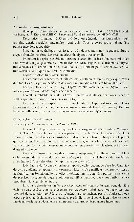
944 MICHEL PERREAU
Anemadus wolongianus n. sp.
Holotype 8: Chine, Sichuan, réserve naturelle de Wolong, 900 m, 23.V.1994, débris
végétaux, leg. S. Kurbatov (MHNG). Paratypes 2 9, même provenance (MHNG, CMP).
Description: Longueur: 2,35 mm. Coloration générale brun-jaune clair, seuls
les cinq derniers articles antennaires rembrunis. Tout le corps couvert d'une fine
pubescence dorée, couchée.
Ponctuation céphalique très forte et très dense, mais non rugueuse. Suture
clypéo-frontale très forte. Le bord antérieur du clypeus très arrondi.
Pronotum à angles postérieurs largement arrondis, la base finement rebordée
sauf près des angles postérieurs. Ponctuation très forte, rugueuse, confluente en lignes
transversales en certains endroits, mais pas suffisamment pour former des strioles
transversales telles que chez certains Nemadini.
Elytres striolées transversalement.
Tarses antérieurs légèrement dilatés, mais nettement moins larges que l'apex
du tibia. Les deux premiers articles des tarses intermédiaires très faiblement dilatés.
Edéage à lobe médian très large, l'apex profondément échancré (figure 6). Sac
interne primitif, avec deux rangées de phanères.
Femelle semblable au mâle, à l'exception de la dilatation des tarses. Ventrite
VIII et spiculum ventrale représentés sur la figure 17.
L'édéage de cette espèce est très caractéristique, l'apex est très large et très
largement échancré, et permet une reconnaissance aisée de l'espèce (figure 6). De plus
la petite taille n'autorise aucune confusion avec des espèces déjà connues.
Nargus (Eunargus) n. subgen.
Espèce-type: Nargus taiwanensis Perreau, 1996
Le caractère le plus important qui isole ce sous-genre des deux autres: Nargus s.
str. et Demochrus est la conformation particulière de l'édéage. Les zones dorsale et
ventrale du lobe médian sont constituées de deux longues lames bifurquées à l'apex et
qui se croisent à leur extrémité, la première se déplaçant vers la gauche, et la seconde
vers la droite. Le sac interne est muni de courtes dents isolées, de phanères, et à la base,
d'une très forte dent.
Par comparaison avec les deux autres sous-genres, la taille est comparable à
celle des grandes espèces du sous-genre Nargus s. str., mais l'absence de rangées de
soies égales à l'apex des tibias, le rapproche des Demochrus.
L'évolution de l'organe copulateur rappelle celle rencontrée chez les Catopina
du genre endémique japonais Apterocatops Nakane. Une meilleure compréhension de
la signification fonctionnelle de telles modifications structurales permettra peut-être
de préciser l'origine de cette évolution parallèle dans les deux sous-tribus, et se
produisant dans la même région.
Lors de la description du Nargus (Eunargus) taiwanensis Perreau, cette dernière
était le seule espèce connue présentant ces caractères originaux, nous n'avions pas
proposé de séparation générique ou subgénérique ne sachant pas s'il s'agissait d'une
espèce présentant isolément des caractères particuliers, ou si l'on était en présence d'une
lignée nouvellement découverte et comportant d'autres espèces encore inconnues.
NOUVEAUX CHOLEVINAE D ASIE 945
11
12 15
FIGS8-188: Nemadus siçhuanus n. sp.. tibia et tarse antérieurs droits face ventrale. Figures 9 à 14. Sciaphyes
kurbatovi n. sp. 9: tibia et tarse antérieurs droits face dorsale (femelle). 10: apex du paramère droit
face ventrale interne. 11: segment IX (mâle). 12: ventrite VIII et spiculum ventrale (femelle). 13:
ventrites des 4 premiers urites abdominaux, face ventrale (femelle). 14: bord inférieur de la carène
mesosternale (mâle). Figures 15-16. Sciaphyes sp., femelle. 15: spermathèque. 16: Ventrite des
trois premiers urites abdominaux face latérale. Figure 17: Anemadus wolongianus n. sp., ventrite
VIII et spiculum ventrale (femelle). Figure 18: Anemadiola kurbatovi n. sp., ventrite VIII et
spiculum ventrale (femelle). L'échelle représente 0,14 mm pour la figure 10, 0,6 mm pour les
figures 8, 13, 14, 16 et 17 et 0,4 mm pour les figures 9, 1 1, 12, 15 et 18.
946 MICHEL PERREAU
Or plusieurs exemplaires d'une autre espèce nouvelle, présentant des caractères
analogues, figurent dans les collections du Muséum d'histoire naturelle de Genève, et
proviennent de la province de Sichuan en Chine. Par ailleurs, mon collègue et ami
Jürgen Frank de Stuttgart, possède également plusieurs exemplaires de cette mêmeespèce, provenant également de Chine. Il ne fait plus de doute maintenant que l'on est
ici en présence d'une nouvelle lignée, dont d'autres espèces peuplent probablement
l'Asie d'extrême-orient. C'est la raison pour laquelle nous décrivons un sous-genre
nouveau. Jürgen Frank m'ayant auparavant fait part de son intention de décrire
l'espèce nouvelle, je lui laisse la priorité.
Sciodrepoides watsoni Spence
4 S et 1 9, W. Siberia: 250 km à l'est de Kuraan, environs de Kazanskoye, 17.VI à
20.VI.1993, leg. S. Kurbatov (MHNG).
Scyaphyes Jeannel, 1910
Espèce-type: Bathyscia sibirica Reitter, 1887
Nous donnons ici une description complémentaire de ce genre et de la des-
cription d'une espèce nouvelle:
Forme bathyscioïde allongée, parallèle. Espèces aptères, à coloration générale
brun clair, dépigmentée. Tout le corps recouvert d'une pubescence longue et éparse.
Tête microréticulée, mais non microstriolée. Carène occipitale régressée, seu-
lement présente sous forme de deux courtes carènes situées en arrière des yeux. Ces
derniers, très petits, sont réduits à quelques ommatidies et totalement dépigmentés.
Suture clypeofrontale présente. Palpes maxillaires à second article large et épais, le
troisième court et conique. Les deux premiers antennomères de même longueur.
Pronotum microréticulé transversalement, mais non microstriolé, large à la
base et rétréci vers l'avant, nullement rebordé, ni le long du bord antérieur, ni du bord
postérieur.
Elytres microstriolés transversalement. Cavités mésocoxales séparées par une
carène basse. Cavités métacoxales également distantes, séparées par un processus
étroit mais distinct. Métasternum présentant deux sillons latéraux symétriques,
débutant en arrière des cavités mésocoxales et dirigés vers l'arrière.
Arceau ventral correspondant à la fusion des trois premiers urites abdominaux
muni d'une fine carène longitudinale médiane sur toute sa longueur (figures 13 et 16).
Troncature apicale des tibias intermédiaires et postérieurs portant deux fortes
épines externes et une rangée de petites épines à peu près égales. Tarses antérieurs
pentamères dans les deux sexes. Onychium présentant deux soies non articulées entre
les deux ongles.
Spermathèque nullement sclérifiée, membraneuse, en forme de poire allongée
(figure 15). Ventrite VIII femelle muni d'un spiculum ventrale (figure 12).
Edeage complet, avec des paramères bien développés et présentant au moins
une soie apicale interne très forte recourbée vers l'arrière et non effilée à l'apex
NOUVEAUX CHOLEVINAE D ASIE 947
(figure 7). L'apex des paramères présente bien d'autres soies dont le nombre semble
dépendre de l'espèce. Le sac interne est simplement tapissé de dents hyalines indiffé-
renciées. L'urite IX est réduit à un cadre grossièrement circulaire entourant l'édéage
(figure 11).
Discussion: Lors de sa description, Jeannel plaça Sciaphyes au sein des Lepto-
dirini du groupe des Euryscapes. L'examen détaillé montre plusieurs caractères
inhabituels pour ce groupe. La pentamerie des protarses femelle est le plus surprenant.
Non moins étonnante est la présence de sillons métasternaux latéraux semblables à
ceux des Ptomaphagini et de certains Nemadini. Ce genre présente donc des carac-
tères intermédiaires entre les Ptomaphagini, les Nemadini, et les Leptodirini. Plus
précisément, Sciaphyes partage avec les Leptodirini la séparation des cavités méso-
coxales et métacoxales, la forme du spiculum ventrale, la conformation de l'édéage, et
les caractères régressifs habituels de l'habitat souterrain: réduction oculaire, réduction
de la carène frontale, dépigmentation. Il partage avec les Ptomaphagini la séparation
des cavités mésocoxales par une nette carène mésosternale, la présence des sillons
métasternaux latéraux, et la présence d'une rangée d'épines subégales sur le bord de
la troncature apicale des tibias intermédiaires et postérieurs. Il partage avec les
Nemadini la spermathèque primitive non sclérifiée, et avec certaines espèces seule-
ment de Nemadini les carènes métasternaux latéraux.
Nous continuons ici à considérer Sciaphyes comme un Leptodirini. en raison
principalement de la séparation des hanches postérieures. Cette spécificité constitue
donc seul caractère qui permet de définir strictement les Leptodirini. Tous les autres
caractères invoqués par le passé comportent des exceptions, en particulier la sper-
mathèque sclérifiée aux deux extrémités (Cerruti, 1958, Perreau, 1989) et la réduc-
tion du nombre des protarsomères de 5 à 4 chez la femelle. Ce dernier caractère ne
garantissait d'ailleurs pas le caractère monophylétique du groupe puisque des réduc-
tions plus ou moins prononcées du nombre des tarsomères se rencontrent dans d'autres
sous-familles de Leiodidae. Mais la séparation des hanches postérieures ne peut pas non
plus être considérée à priori comme un caractère apomorphe. En effet, au sein des
Leiodidae, la tendance pour les cavités coxales est de migrer depuis la périphérie du
corps vers le centre. L'insertion centrale des appendices, combinée à l'allongement de
ceux-ci est en effet le meilleur compromis entre l'agilité (la mobilité) et la stabilité.
Dans ce cadre, des cavités mésocoxales et/ou métacoxales distantes doivent être
considérées comme plésiomorphes par rapport à des cavités coxales confluentes.
La conséquence est que suivant cette interprétation, les Leptodirini ainsi
définis doivent être considérés comme un groupe non monophylétique. A l'inverse
l'ensemble des Anemadini (Nemadina, Eocatopina, Paracatopina et Anemadina) plus
les Cholevini (Catopina et Cholevina) dont les cavités coxales intermédiaires et
postérieures sont confluentes doit alors être considéré comme monophylétique.
Sciaphyes kurbatovi n. sp.
Holotype â: S. Primorje, réserve de Kedrovaya pad. 1. VIII. 1987, leg. S. Kurbatov
(MHNGj. Paratype 1 2, S. Primorje, E Ussurijsk environ de Kamenushka, 7. VII. 1987, les. S.
Kurbatov (MHNG).
948 MICHEL PERREAU
Description: En plus des caractères génériques, on peut signaler les points
suivants:
Longueur: 1,38 mm. Microréticulation du pronotum et de la tête forte et
uniforme, nettement orientée transversalement malgré l'absence de strides. Celle de
la tête reste néanmoins moins forte que celle du pronotum.
Strioles élytrales au nombre d'une quarantaine sur la longueur d'un élytre qui
mesure 1 mm.Carène mésosternale basse mais comportant un angle obtus (figure 14). Carène
abdominale représentée sur la figure 13. Segment IX représenté sur la figure 11.
Tarses antérieurs bien dilatés, un peu plus larges que la largeur des tibias.
Tarses intermédiaires non dilatés.
Edéage allongé, à paramères larges dans la moitié basale, plus étroits dans la
moitié apicale, le plateau apical muni de deux soies fines dorsales, deux soies fines
ventrales au centre, deux fortes soies ventrales internes et d'une soie très épaisse
ventrale externe non amincie à l'extrémité, courbée vers l'arrière (figures 7 et 10).
Femelle semblable au mâle exceptée la dilatation des tarses antérieurs. Tarses
antérieurs de 5 articles (figure 9). Ventrite VIII et spiculum ventrale représentés sur la
figure 12.
Cette espèce se distingue facilement du Sciaphyes sibiricus par l'édéage régu-
lièrement rétréci puis arrondi à l'apex, et non pas effilé en pointe.
Sciaphyes sp.
Une autre espèce probablement nouvelle n'est connue que par une 9: S.
Primorje 25 km NW Terney, sapin pourri, 3. VII. 1992, leg. S. Kurbatov (MHNG).
Nous attendons d'être en possession d'un mâle pour procéder à sa description
formelle.
Un peu plus petite que l'espèce précédente (1,28 mm), elle s'en distingue par
la tête pratiquement lisse, avec seulement les quelques points très fins correspondant à
l'insertion des soies, une microréticulation faible, et une ponctuation très délicate sur
le pronotum dont la surface discale est presque lisse.
Les strioles élytrales sont au nombre de moins d'une trentaine sur la longueur
d'un élytre qui mesure 0,715 mm. On peut remarquer que le nombre de strioles par
unité de longueur est le même que chez l'espèce précédente, alors que les longueurs
des élytres sont sensiblement différentes.
Une investigation pourrait être intéressante à réaliser dans d'autres genres de
Cholevinae, pour déterminer si le nombre de strioles est un caractère spécifique donné
ou s'il dépend de la taille. L'observation ci-dessus soutient la seconde hypothèse.
La carène abdominale est représentée en vue latérale sur la figure 16, et la
spermathèque sur la figure 15.
NOUVEAUX CHOLEVINAE D ASIE 949
REFERENCES
Cerruti, M. 1958. Studi sui "Bathysciinae" (Coleopt.-Catopidae). Atti del Vili Congresso
Nazionale di Speleologia - Como, 1956. Memoria IV volume 2: 125-126.
GiACHiNO, P.M. & Etonti, M. 1995. Il genere Remyella Jeannel, 1931 (Coleoptera Cholevidae
Leptodirinae). Atti del Museo civico di Storia naturale Trieste, 46: 77-98.
Hayashi, Y. 1990. Notes on Catopidae from Taiwan (II). Entomological Review of Japan, 45:
29-35.
Perreau. M. 1989. De la phylogénie des Cholevidae et des familles apparentées. Archives des
Sciences Genève, 39 (3): 579-590.
Perreau, M. 1996. Contribution à la connaissance des Cholevidae du Japon et de Taiwan(Coleoptera). Revue Suisse de Zoologie, 103( 1 ): 283-297.
Szymczakowski. W. 1963. Contribution à la connaissance des Anemadinae (Coleoptera Cato-
pidae). Polskie Pismo Entomologiczne, 33: 101-117.
Szymczakowski, W. 1964. Analyse systématique et zoogéographique des Catopidae (Cole-
optera) de la région orientale. Acta zoologica cracoviensia, 9(2): 55-283.

REVUE SUISSE DE ZOOLOGIE
Tome 103 — Fascicule 4
Pages
Zoologia et Botanica 96, Fribourg, 21-23 February 1996 (Annual
Conference of the Swiss Zoological Society) 777-794
Schlüssel, André & Jean-Paul Theurillat. Synusial structure of heath-
lands at the subalpine/alpine ecocline in Valais (Switzerland) 795-800
Baur, Bruno, Jasmin Joshi, Bernhard Schmid, Ambros Hänggi, Daniel
Borcard, Josef Stary, Ariane Pedroli-Christen, G. Heinrich
Thommen, Henryk Luka, Hans-Peter Rusterholz, Peter Oggier,Stephan Ledergerber & Andreas Erhardt. Variation in species
richness of plants and diverse groups of invertebrates in three cal-
careous grasslands of the Swiss Jura mountains 801-833
Graf, Markus, Alexander Imanuel Wandeler & Peter Lüps. Die räum-liche Habitatnutzung einer Dachspopulation (Mêles mêles L.) imschweizerischen Mittelland 835-850
Lang, Claude & Olivier Reymond. Le zoobenthos comme indicateur des
perturbations d'origine humaine dans deux lacs de montagne 851-858
GOhner. Manuela & Wolfgang Pfeiffer. Über die Verbreitung der
Mauthner Axone bei Fischen und Amphibien und ihren Zusammen-hang mit der Schreckreaktion der Ostariophysi und Anura 859-89
1
Merz, Bernhard. Die Asteiidae (Diptera) der Schweiz 893-904
Durette-Desset, Marie-Claude & Claude Vaucher. Molostrongylus
acanthocolpos gen. n., sp. n., (Nematoda, Trichostrongylina, Moli-
neoidea) parasite de Molossops temmincki (Chiroptera, Molossidae)
au Paraguay 905-913
Burckhardt, Daniel. On some Ancistria spp. from the Natural History
Museum, Vienna (Coleoptera, Passandridae) 915-918
Löbl, Yvan & Serguei A. Kurbatov. A review of the Japanese Tycho-
bythinus and Byihoxenites (Coleoptera, Staphylinidae, Pselaphinae). 919-938
Perreau, Michel. Nouveaux Cholevinae d'Asie (Coleoptera Leiodidae). 939-949
REVUE SUISSE DE ZOOLOGIE
Volume 103 — Number 4
Pages
Zoologia et Botanica 96, Fribourg, 21-23 February 1996 (Annual
Conference of the Swiss Zoological Society)
Schlüssel, André & Jean-Paul Theurillat. Synusial structure of heath-
lands at the subalpine/alpine ecocline in Valais (Switzerland). . .
795
Baur, Bruno, Jasmin Joshi, Bernhard Schmid, Ambros Hänggi, Daniel
Borcard, Josef Stary. Ariane Pedroli-Christen, G. Heinrich
Thommen, Henryk Luka, Hans-Peter Rusterholz, Peter Oggier,Stephan Ledergerber & Andreas Erhardt. Variation in species
richness of plants and diverse groups of invertebrates in three calcare-
ous grasslands of the Swiss Jura mountains 801
Graf, Markus, Alexander Imanuel Wandeler & Peter Lüps. Spatial orga-
nization and habitat utilization in a population of European badgers
(Meles meles L.) in a hilly area of the swiss midlands 835
Lang, Claude & Olivier Reymond. Zoobenthos as indicator of man-madeperturbations in two mountain lakes 851
GOhner, Manuela & Wolfgang Pfeiffer. The distribution of the Mauthneraxons in fish and amphibians and its relation to the fright reaction in
Ostariophysi and Anura 859
Merz, Bernhard. The Asteiidae (Diptera) of Switzerland 893
Durette-Desset, Marie-Claude & Claude Vaucher. Molostrongylus
acanthocolpos gen. n., sp. n., (Nematoda. Trichostrongylina, Moli-
neoidea) parasite de Molossops temmincki (Chiroptera. Molossidae)
from Paraguay 905
Burckhardt, Daniel. On some Ancistria spp. from the Natural History
Museum, Vienna (Coleoptera, Passandridae) 915
Löbl, Yvan & Serguei A. Kurbatov. A review of the Japanese Tycho-
bythinus and Bythoxenites (Coleoptera, Staphylinidae, Pselaphinae). 919
Perreau, Michel. New Cholevinae from Asie (Coleoptera Leiodidae). . .939
Indexed in Current Contents
PUBLICATIONS DU MUSEUM D'HISTOIRE NATURELLE DE GENEVE
CATALOGUE DES INVERTÉBRÉS DE LA SUISSEFase. 1
.
SARCODINÉS par E. Penard Fr. ] 2.—2. PHYLLOPODES par Th. Stingelin 12.—3. ARAIGNÉES par R. de Lessert 42.—4. ISOPODES par J. Carl 8.—5. PSEUDOSCORPIONS par R. de Lessert 5.50
6. INFUSOIRES par E. André 1 8.—7. OLIGOCHÈTES par E. Piguet et K. Bretscher 1 8.—8. COPÉPODES par M. Thiébaud 18.—9. OPILIONS par R. de Lessert IL-
IO. SCORPIONS par R. de Lessert 3.50
1 1
.
ROTATEURS par E.-F. Weber et G. Montet 38.—12. DÉCAPODES par J. Carl 1 1.—13. ACANTHOCÉPHALES par E. André IL—14. GASTÉROTRICHES par G. Montet 18.—15. AMPH1PODES par J. Carl 12.—16. HIRUDINÉES. BRANCHIOBDELLES
et POLYCHÈTES par E. André 1 7.50
17. CESTODES par O. Fuhrmann 30.—
REVUE DE PALÉOBIOLOGIE Echange
LE RHINOLOPHE (Bulletin du centre detude des chauves-souris) par fascicule Fr. 10.
—
CATALOGUE ILLUSTRÉ DE LA COLLECTION LAMARCKAPPARTENANT AU MUSÉUM D'HISTOIRE NATURELLE DE GENÈVElre partie - Fossiles - 1 vol. 4° avec 1 17 planches Fr. 300.
—
COLLEMBOLENFAUNA EUROPASvon H. Gisin
312 Seiten. 554 Abbildungen Fr. 30.—
THE EUROPEAN PROTURA THEIR TAXONOMY. ECOLOGY ANDDISTRIBUTION WITH KEYS FOR DETERMINATIONby J. Nosek346 pages, 1 1 1 figures in text, 1973 Fr. 30.
—
CLASSIFICATION OF THE DIPLOPODApar Richard L. Hoffman237 pages. 1979 Fr. 30,—
LES OISEAUX NICHEURS DU CANTON DE GENÈVEpar P. Géroudet, C. Guex et M. Maire35 1 pages, nombreuses cartes et figures Fr. 45.
—
CATALOGUE COMMENTÉ DES TYPES D'ECHINODERMES ACTUELSCONSERVÉS DANS LES COLLECTIONS NATIONALES SUISSES,SUIVI D'UNE NOTICE SUR LA CONTRIBUTION DE LOUIS AGASSIZÀ LA CONNAISSANCE DES ECHINODERMES ACTUELSpar Michel Jangoux67 pages. 1 1 planches Fr. 1 5.
—
RADULAS DE GASTÉROPODES LITTORAUX DE LA MANCHE(COTENTIN-BAIE DE SEINE. FRANCE)par Y. Finet, J. Wüest et K. Mareda62 pages, nombreuses figures, 1991 Fr. 10.
—
GASTROPODS OF THE CHANNEL AND ATLANTIC OCEAN;SHELLS AND RADULASby Y. Finet. J. Wüest and K. Mareda, 1 992 Fr. 30.—
O. SCHMIDT SPONGE CATALOGUEpar R. Desqueyroux-Faundez & S. M. Stone, 1992 Fr. 40.
—
ATLAS DE RÉPARTITION DES AMPHIBIENSET REPTILES DU CANTON DE GENÈVEpar A. Keller, V. Aellen et V. Mahnert, 1993 Fr. 15.
—
THE MARINE MOLLUSKS OF THE GALAPAGOS ISLANDS:A DOCUMENTED FAUNAL LISTpar Yves Finet, 1995 Fr. 30.—
NOTICE SUR LES COLLECTIONS MALACOLOGIQUESDU MUSEUM D -
HISTOIRE NATURELLE DE GENEVEpar Jean-Claude Cailliez, 1995 Fr. 22.
—

Revue suisse de Zoologie: Instructions to Authors
The Revue suisse de Zoologie publishes papers by members of the Swiss Zoological Society and scientific
results based on the collections of the Muséum d'histoire naturelle, Geneva. Submission of a manuscript implies
that it has been approved by all named authors, that it reports their unpublished work and that it is not being
considered for publication elsewhere. A financial contribution may be asked from the authors for the impression of
colour plates and large manuscripts. All papers are refereed by expert(s).
In order to facilitate publication and avoid delays authors should follow the Instructions to Authors and refer to
a current number of R.S.Z. for acceptable style and format. Manuscripts not conforming with these directives are
liable to be returned to the authors. Papers may be written in French, German, Italian and English. Authors should
aim to communicate ideas and information clearly and concisely in language suitable for the moderate specialist.
Authors not writing in their native language should pay particular attention to the linguistical quality of the text.
Manuscripts must be typed, on one side only and double-spaced, on A4 (210 x 297 mm) or equivalent paper
and all pages should be numbered. All margins must be at least 25 mm wide. Authors must submit one original
and two copies, including tables and figures, in final fully corrected form, and are expected to retain another copy.
Papers should conform to the following general layout:
Title page. A concise but informative full title plus as running title of not more than 40 letters and spaces,
name(s) in full and surname(s) of author(s), and full address(es).
Abstract. This must be on a separate page. The abstract is in English, composed of the title and a short text of
up to 200 words. It should summarise the contents and conclusions of the paper. The abstract is followed by less
than 10 key-words, separated by hyphens, which are suitable for indexing.
Introduction. A short introduction to the background and the reasons for the work.
Materials and methods. Sufficient experimental details must be given to enable other workers to repeat the
work. The full binominal name should be given for all organisms. The Zoological Code must be strictly followed.
Cite the authors of species on their first mention. Use SI units and the appropriate symbols.
Results. These should be concise and should not include methods or discussion. Text, tables and figures should
not duplicate the same information. New taxa must be distinguished from related taxa. The abbreviations gen. n., sp.
n., syn. n. and comb. n. should be used to distinguish all new taxa, synonymies or combinations. Primary types
should be deposited in a museum or similar institution. In taxonomic papers the species heading should be followed
by synonyms, material examined and distribution, description and comments. All material examined should be listed
in similar, compact and easily intelligible format; the information should be in the same language as the text.
Sex symbols should be used rather than "male" and "female".
Discussion. This should not be excessive and should not repeat results nor contain new information, but
should emphasize the significance and relevance of the results reported.
References. The Harvard System must be used for the citation of references in the text, e.g. White & Green
(1995) or (White & Green 1995). For references with three and more authors the form Brown et al. should be
used. Authors' names should not be written in capitals. The list of references must include all publications in the
text but only these. References must be listed in alphabetical order of authors, and both the title and name of the
journal must be given in full in the following style:
Penard, E. 1888. Recherches sur le Ceratium macroceros . These . Genève . 43 pp.
Penard, E. 1889. Etudes sur quelques Héliozoaires d'eau douce. Archives de Biologie , 9:1-61.
Mertens, R. & H. Wermuth. 1960. Die Amphibien und Reptilien Europas, Kramer . Frankfurt am Main , XI + 264 pp.
Handley, CO. Jr. 1966. Checklist of the mammals of Panama, pp. 753-795. In: Ectoparasites of Panama (R.L.
Wenzel & V.J. Tipton, eds). Field Museum of Natural History , Chicago , XII + 861 pp.
References should not be interspaced and, in the case of several papers by the same author, the name has to be
repeated for each reference.
The text should be in roman (standard) type face throughout, including headings, with genus and species
names underlined with pencil: bold, small capitals, large capitals italics and other type faces should not be used.
Footnotes and cross-references by page should be avoided.
Also, we encourage authors to submit the text on floppy disk (3,5" or 5 1/4", Macintosh or IBM compatible,
with "Microsoft Word" or similar programmes). The authors should refrain from using special codes for text
formating.
Tables. These should be self-explanatory, with the title at the top organised to fit 122 x 180 mm. Each table
should be typed, double spaced, on a separate page and numbered consecutively and its position indicated in the text.
Figures. These may be line drawings or half tones and all should be numbered consecutively, and their position
indicated in the text. Figures should be arranged in plates which can be reduced to 122 x 160 mm. Drawings and
lettering should be prepared to withstand reduction. Magnification should be indicated with scale lines. Authors
should refrain from mixing the drawings and half tones. Original drawings will not be returned automatically. TheRevue suisse de Zoologie declines responsability for lost or damaged slides or other documents.
Legends to figures. These should be typed in numerical order on a separate sheet.
Proofs. Page proofs only are supplied, and authors may be charged for alterations (other than printer's errors) if
they are numerous.
Offprints. The authors receive 25 offprints free of charge; more copies may be ordered at current prices whenproofs are returned.
Correspondence. All correspondence should be addressed to
Revue suisse de Zoologie
Muséum d'histoire naturelle
CP 6434CH-1211 Genève 6
Switzerland.
Phone: (022) 418 63 33 - Fax (022) 418 63 01
e-mail: volker.mahnert @ mhn.ville-ge.ch
Home page RSZ: http://www/musinfo/mhng/publications/revues.htm


^
HECKMAN IXIBINDERY INC. |S|
JULY 97