Restoring brain function after stroke Nick S Ward
1
Restoring brain function after stroke — bridging the gap between animals and humans
Nick S Ward1,2,3
1Sobell Department of Motor Neuroscience, UCL Institute of Neurology, 33 Queen Square, London
WC1N 3BG.
2The National Hospital for Neurology and Neurosurgery, Queen Square, London WC1N 3BG.
3UCLPartners Centre for Neurorehabilitation, UCL Institute of Neurology, Queen Square, London
WC1N 3BG
Biography
Nick Ward is an academic neurologist at UCL Institute of Neurology and the National Hospital for
Neurology and Neurosurgery, Queen Square, London, UK, where he works in the stroke and
neurorehabilitation service. His research uses structural and functional brain imaging to study how
reorganisation of brain networks supports recovery of upper limb movement after stroke. His goal is
to understand the mechanisms of recovery so that we might predict both optimal treatments of upper
limb impairment and long-term outcomes after stroke.
Restoring brain function after stroke Nick S Ward
2
Abstract
Stroke is the leading cause of complex adult disability in the world. Recovery from stroke is often
incomplete, which leaves many people dependent on others for their care. The improvement of long-
term outcomes should, therefore, be a clinical and research priority. As a result of the advances in
our understanding of the biological mechanisms involved in recovery and repair after stroke,
therapeutic opportunities to promote recovery through manipulation of post-stroke plasticity have
never been greater. This work has almost exclusively been carried out in preclinical animal models
of stroke with little translation into human studies. The challenge ahead is to develop a mechanistic
understanding of recovery from stroke in humans. Advances in neuroimaging techniques now enable
us to reconcile behavioural accounts of recovery with molecular and cellular ones. Consequently,
clinical trials can be designed in a stratified manner that takes into account when an intervention
should be delivered and who is most liable to benefit. This approach is expected to lead to a
substantial change in how restorative therapeutic strategies are delivered in patients after stroke.
Key points
Stroke is the leading cause of complex adult disability in the world, but currently we do not provide
enough of the right physical or behavioural interventions to drive recovery
Clear lesion-induced changes occur in brain structure and function early after stroke, which result
in an environment with unique heightened plasticity that can support restoration of function,
termed spontaneous biological recovery
Intense, high-dose behavioural training aimed at the reduction of impairment and the restoration
of function should be (but currently is not) delivered in this critical time window
The basis of spontaneous biological recovery in humans is unclear, which yields uncertainty over
how and when to augment or prolong this process with novel therapies — further characterization
is required to enable realistic phase III trials

Restoring brain function after stroke Nick S Ward
3
Human neuroimaging techniques combined with modelling approaches can provide the
appropriate biomarkers with which to map out a mechanistic approach to understand who and
when to treat
The use of structural imaging to quantify damage in a range of brain regions can help predict
long-term outcomes and provide the basis for stratification in restorative trials
Restoring brain function after stroke Nick S Ward
4
Almost 17 million people worldwide experience a first-time stroke each year1, which is equivalent to
one new stroke every 2 seconds. Stroke mortality is declining2 but in the UK over 1 million people
live with the consequences of stroke, of whom over one-third are dependent on others for their care.
The epidemiological shift of stroke disease burden towards long-term conditions means that these
numbers will continue to rise3 Often, the decline in functional abilities that takes place in many
patients 4 goes unrecognized, and so, unsurprisingly, the overall economic burden of stroke is high
(estimated at over UK£9 billion a year in the UK). The fact that stroke is both a chronic and a
progressive condition should influence research priorities in this area, but funding for research into
stroke, and stroke recovery in particular, lags far behind cancer, coronary heart disease and
dementia5. Improvement of recovery and long-term outcomes is an urgent clinical and scientific goal,
but success is slow to materialize.
How are the most dramatic clinical improvements expected to be achieved? Care in the hyperacute
and acute period after stroke has improved dramatically over the past two decades, but our attention
must now turn to treatments that actively promote recovery. One reason for optimism is that work in
animal models points to a time-limited period of heightened plasticity after focal brain injury.
However, achieving the best possible outcomes in patients after stroke requires two key challenges
to be addressed. The first is how to take advantage of this critical period through the optimal timing,
intensity, amount and even type of behavioural training that makes up neurorehabilitation. This
question has been discussed elsewhere but, in brief, studies support the use of intense training that
focuses on reducing impairment in the first few weeks and months post-stroke to take advantage of
biological repair mechanisms 6. The second challenge, and the focus of this Review, is how to
augment the biological mechanisms of post-stroke plasticity to enhance or prolong the effects of
behavioural training in patients after a stroke. The translational nature of this question is important,
because although work in preclinical animal models has been pivotal in highlighting the biological
basis of recovery, as yet virtually no benefit has been observed for humans. I will discuss the reasons
why this lack of benefit might be and the prospects for developing a mechanistic understanding of
post-stroke plasticity in humans. In particular, exciting prospects exist for the development of human
biomarkers that provide an appropriate intermediate level of mechanistic description with which to
Restoring brain function after stroke Nick S Ward
5
bridge the current explanatory gap between what we know about recovery from pre-clinical studies
and human studies.
Recovery after stroke is proportional
A starting point for determining the biological basis of recovery in patients after a stroke is to ask
why some patients fail to recover. Stroke is one of the most common causes of physical disability
worldwide and ~80% of stroke survivors experience impairment of movement on one side of the
body7. Hand and arm impairment in particular is often persistent, disabling8 and a major contributor
to reduced quality of life. In one study, only 38% of patients who presented with an initially paralysed
upper limb regained some dexterity by 6 months9, and by 4 years two-thirds of patients perceived
that loss of arm function was still a major problem10. These studies and many others clearly
demonstrate that recovery is variable and difficult to predict. Factors associated with poor outcomes
include right hemisphere damage, somatosensory deficit, visual inattention, homonymous
hemianopia and urinary incontinence9,11. However, the dominant factor for predicting long-term
upper limb outcome is initial severity of motor impairment. Additional factors that have independent
predictive power over and above their association with this initial severity have not been identified.
The ability of initial severity to predict upper limb recovery was first quantified as the proportional
recovery rule [G] 12. When applied to real clinical data, two key findings exist (FIG. 1) that provide
challenges but also opportunities for the field. The first is that initial upper limb impairment predicts
later upper limb outcome extremely accurately in patients presenting with mild to moderate
impairment. This result is disconcerting to those involved in post-stroke neurorehabilitation because
it implies that any variability in the dose of rehabilitation delivered in the first 3 months exerts no
substantial effect on a patient's level of motor impairment. The second key result is that proportional
recovery fails in about half of patients presenting with severe impairment. In other words, in patients
presenting with the same high level of initial severity, about half recover proportionately and half fail
to make any substantial recovery (FIG. 1). Importantly, this finding tells us that the causes of initial
impairment are probably independent from the biological factors that are important for the
subsequent recovery process. This interpretation provides an opportunity, because factors important
Restoring brain function after stroke Nick S Ward
6
for recovery might represent targets for novel therapeutics that aim to optimize the biological factors
that maximize the effects of behavioural training.
The proportional recovery rule has been confirmed in the motor domain several times12–15 and
suggests two clear clinical questions. Firstly, how can we help patients with stroke to regain more
than 70% of lost function and secondly, how can we turn poor recoverers into proportional
recoverers? The answers to these questions will dramatically change our approaches to promoting
recovery after stroke. Evidence for proportional recovery has also been shown for non-motor
domains such as language16 and neglect17, and so this striking clinical phenomenon provides a novel
and important model for investigating both potentially modifiable biological factors that are necessary
for maximising recovery of function after stroke in humans, as well as currently non-modifiable
factors that will help to make accurate predictions of long-term outcome.
Spontaneous biological recovery
Why do some patients experience poor recovery after stroke and yet others who are clinically
indistinguishable have good recovery? The differences in these two groups manifest in the first few
days and weeks after stroke. During this time there might (or might not) be a rapid generalized
improvement in impairment that is in contrast to the modest gains that are made in the chronic
phase18. Decades of work in animal models clearly show that a window of opportunity exists after
focal brain damage within which behavioural training will have a much greater effect than outside
the window. This early post-stroke phase has been described as a period of spontaneous
biological recovery [G]. Early evidence of this critical period for recovery-related training was
provided by Biernaskie and colleagues19 who found that rats that commenced motor training of the
affected forelimb starting at 30 days post-stroke exhibited little improvement when compared with
those whose treatment commenced earlier at 5–14 days post-stroke The causal role of the lesion
itself in initiating spontaneous biological recovery was illustrated further by Zeiler and colleagues20
who showed that intensive reach training of a mouse commenced 7 days after stroke was not able
to promote full recovery. However, when the same animal was given a second stroke and training
was commenced 2 days later (presumably within the critical period), then recovery was substantially
Restoring brain function after stroke Nick S Ward
7
enhanced, and resulted in performance levels that approached those seen before either stroke.
Clearly, focal brain damage sets in motion a series of biological events that, when combined with
appropriate type and intensity of behavioural training6, can support dramatic recovery.
Structural plasticity after stroke
A substantial amount of work has been undertaken in animal models to define the molecular and
cellular processes that underlie the formation of new local and large-scale brain circuits that support
recovery from stroke. These studies are well described elsewhere21–25. Briefly, the basic elements of
neural repair that can be seen in animal models of stroke include axonal sprouting, dendritic
branching, synaptogenesis, neurogenesis and gliogenesis, and all can be enhanced in the early
post-stroke period. Regeneration seems to occur in brain regions connected to the damaged area,
including peri-infarct, ipsilesional and contralesional brain and spinal cord networks. Not all sprouting
is clinically beneficial, and only axonal sprouting that links functionally related brain areas is
consistently associated with improved post-stroke outcomes26. Definitive evidence of these
restorative processes in humans is scarce, but markers suggestive of neurogenesis27, gliogenesis28
and axonal sprouting27 have been found in human post-stroke perilesional brain tissue.
Consequently, the occurrence of similar biological responses to brain injury in both animals and
humans seems probable.
The precise temporal and spatial ordering of these post-stroke biological events is governed by
alterations in gene expression. Researchers have often remarked that the biological environment of
the post-stroke brain resemble that of the developing brain, and that 'recovery recapitulates
ontogeny'23. However, a clear distinction between regenerative and developmental transcriptomes
has been shown, which indicates a unique regenerative molecular program at work29. Furthermore,
expression of the regenerative transcriptome is strongly influenced by age at stroke onset, with
earlier induction of growth-inhibiting molecules and later expression of growth-promoting molecules
exhibited by older animals than by younger animals30.
Restoring brain function after stroke Nick S Ward
8
Preclinical work has attempted to both promote neuronal regeneration and, most commonly, to block
extracellular inhibitory signals that counteract regeneration, with some successes (BOX 1)24,31.
Changes to the structure of brain networks will not independently restore function, and all of these
studies stress the need for appropriate levels of behavioural training, something that is often omitted
from preclinical studies in animal models. The potential to form new functionally relevant circuitry
that can be shaped by behavioural training provides a compelling mechanistic framework for
functional recovery after stroke. However, the timing of administration of growth-promoting
compounds, both in relation to the initial stroke damage and to the behavioural training itself, will
clearly have a major effect on the therapeutic capacity. Whether training is delivered at the same
time as growth-promoting molecules or sequentially could influence the type of sprouting that occurs
and, consequently, whether behaviour is helped or hindered32. In addition, the effect that post-stroke
behaviour can have on regenerative processes themselves is important to understand. For example,
early compensatory use of the contralesional forelimb impairs recovery of the affected limb33,
possibly through aberrant synaptogenesis in the perilesional cortex34. Any behaviour, if overtrained,
will take advantage of the increased post-stroke potential for experience-dependent plasticity, and
so abnormal or compensatory patterns of behaviour can become learned. Once again, this finding
highlights the need for an appropriate form of behavioural training that can take advantage of any
spontaneous or therapeutically enhanced potential for plasticity.
As well as asking ‘when’ treatment should be administered, ‘where’ is probably an equally important
question. Most of the compounds discussed have been administered via intravenous or intrathecal
routes, but accurate spatial and temporal delivery might both be necessary to achieve the desired
outcomes. Advances made in the last few years in tissue engineering35,36 and optogenetics37 provide
potential methods for precisely delivering regenerative molecules to functionally relevant brain
regions.
Functional plasticity after stroke
Identification of the trigger for post-stroke regenerative processes could provide further therapeutic
opportunities. In addition to the structural changes described above, focal brain damage results in
Restoring brain function after stroke Nick S Ward
9
alterations in neuronal excitability38. Immediately after stroke, signalling by the excitatory
neurotransmitter glutamate is excitotoxic and contributes to cell death, whereas signalling by the
inhibitory neurotransmitter GABA can counteract this toxicity through cell hyperpolarization39. This
period lasts about 3 days post-stroke in the mouse40 and for an uncertain time in humans, after which
the beneficial and detrimental effects of GABA and glutamate signalling seem to reverse.
Specifically, changes to the cortical excitatory–inhibitory balance have long been known to influence
the potential for experience-dependent plasticity in cortex and can reopen critical periods of plasticity
in the adult brain41. Reduced inhibitory tone can lead to facilitation of downstream changes in
neuronal structure42 and one possibility is that the altered levels of neuronal activity that result from
a change in excitability regulate neurogenesis and the activity of growth factors (such as brain
derived neurotrophic factor; BDNF) through epigenetic mechanisms43. Reduced cortical inhibitory
mechanisms can lead to expanded and less specific receptive fields44,45, enhanced long-term
potentiation46 and remapping of sensorimotor functions to surviving cortex47 in both hemispheres48,
all of which is potentially useful when functional reorganisation of residual post-stroke brain
structures is important for recovery of normal function. An altered balance between inhibitory
GABAergic and excitatory glutamatergic signalling in surviving stroke regions and networks could,
therefore, be a key event that sets other restorative mechanisms in motion.
In 2009, Murphy and Corbett21 proposed that after the acute stroke period, attenuation of neuronal
activity in brain regions connected to the damaged region might be reversed by a homeostatic
increase in neuronal excitability, a process that can last at least several weeks21. Levels of neuronal
excitability are determined by the balance in activity between GABA and glutamate, both of which
are known to be altered after stroke38. For example, enhanced glutamate signalling through AMPA
receptors, the major excitatory signalling system in the adult brain, is associated with improved
recovery in stroke models49. This effect is probably due to downstream induction of BDNF49, which
once again links altered neuronal excitability with downstream changes in axonal structure50. Much
work on GABAergic signalling after stroke has focussed on the reduction in phasic (that is, synaptic)
inhibition in the first few weeks after injury51 to increase the likelihood of long-term potentiation46.
Specifically, GABAA receptors are dowregulated48,52, and the density of a number of inhibitory
Restoring brain function after stroke Nick S Ward
10
interneurons is reduced after focal brain damage44,53. Both increased glutamatergic signalling and
reduced phasic GABAergic signalling would be consistent with the idea of a homeostatic restitution
of neuronal activity21. However, two studies have suggested that increased perilesional tonic
inhibitory signalling via extrasynaptic GABAA receptors might be the dominant response to
stroke40,54. When this tonic inhibition was reversed (using an α5 subunit that contained an
extrasynaptic GABAA-receptor inverse agonist) motor outcomes improved in both mouse40 and rat51
models of stroke. Although the increase in extracellular GABA in response to cerebral ischaemia is
transient, the increase in tonic inhibitory signalling can persist for more than 1 month38 making this
therapeutic window attractive compared with the window available for reperfusion strategies.
The interactions between excitatory pyramidal cells and numerous inhibitory interneurons in the
cortex is clearly complex and becomes more complex after stroke55. In addition, prolonged ischaemia
affects different cell types unequally56 and causes alterations in the distribution of receptor
subtypes57. The numbers of inhibitory interneurons (some of which inhibit other inhibitory
interneurons) and pyramidal cells, as well as the ratios of receptor subtypes in the surviving cortex
are not only unclear, but can differ between individuals. Nevertheless, the weight of evidence from
animal studies to date suggests that spontaneous biological recovery is either augmented by a
homeostatic restitution of cortical activity secondary to reduced phasic GABAergic inhibitory
signalling, or blocked by excessive tonic GABAergic inhibitory signalling. Beyond the hyperacute
period (up to 3 days post-stroke), what follows at a cellular level suggests that alterations in cortical
inhibitory and excitatory mechanisms are important to determine the potential for plasticity and
downstream structural changes that support recovery. Consequently, components of these inhibitory
and excitatory mechanisms represent exciting and novel therapeutic targets for enhancing
behavioural training after stroke.
As with mechanisms of structural plasticity, the mechanisms responsible for the alterations in cortical
excitatory–inhibitory balance that underlie changes in post-stroke functional plasticity are amenable
to pharmacological and non-pharmcological manipulation. The most popular non-pharmacological
approach is the use of non-invasive brain stimulation which appears to be able to enhance the effects
of behavioural training to a small degree58,59. In a mouse model, direct current stimulation to the brain
Restoring brain function after stroke Nick S Ward
11
appeared to augment synaptic plasticity through BDNF dependent mechanisms60. However, in
human studies it is not clear how much or how accurately electrical current is delivered to target
brain regions and consequently results are inconsistent and potential mechanisms poorly
understood61,62.
As described, tonic inhibition can be reversed by antagonists or inverse agonists of the α5-subunit-
containing extrasynaptic GABAA receptor, and compounds for use in humans are currently available
and are under investigation in phase I studies. Zolpidem is an interesting pharmacological agent that
binds with high affinity to α1-containing GABAA receptors through which it mediates sedative and
hypnotic effects. However, zolpidem can also influence tonic inhibition through α5-containing GABAA
receptors in a dose-dependent manner, such that low levels of the drug augment tonic inhibition and
high levels reduce it63. Zolpidem can improve recovery in a mouse model of stroke64, and has been
reported to mediate interesting effects such as the temporary reversal of deficits in language,
cognitive and motor function in single patient cases with stroke 65,66. However, given the uncertainty
over how zolpidem works, the mechanism of recovery in these individuals remains unclear.
The idea that pharmacological approaches can help promote recovery of function after stroke has
been well described67. Modulation of a number of neurotransmitter systems has shown positive
effects in animal models of stroke, usually correlating with their effect on long-term potentiation67. A
key message from this early work is that close temporal coupling of the drug and the behavioural
training is required for maximum therapeutic effect, which suggests that the therapeutic mechanisms
are short lived and reversible, rather than being due to chronic effects. This point has not always
translated into study design, but should be considered when interpreting the results of a
pharmacotherapy study.
The current interest in selective serotonin reuptake inhibitors (SSRIs) comes from the fluoxetine for
motor recovery after acute ischemic stroke (FLAME) study in which 20 mg fluoxetine daily, started
5–10 days after ischaemic stroke and continued for 3 months, enhanced upper-limb motor
recovery68. Many smaller studies of SSRIs have similar findings, but heterogeneity between studies
is high69. Although SSRIs can influence structural plasticity, compelling evidence supports a
plasticity-modifying effect mediated through the GABAergic system. Chronic doses of fluoxetine can
Restoring brain function after stroke Nick S Ward
12
reinstate critical-period plasticity in adult rats through a reduction of extracellular levels of GABA and
an increase in BDNF expression70. Furthermore, in a mouse model of stroke, Ng and colleagues71
showed that fluoxetine treatment was able to prolong (but not reinstate) the critical period of post-
stroke plasticity through the reduction of inhibitory interneuron expression in intact cortex71.
Serotonin can have inhibitory (via 5HT1A receptors) or facilitatory (via 5HT2A receptors) effects on
pyramidal cells, but most fast-spiking inhibitory interneurons are inhibited by serotonin through 5HT1A
receptors72. However, in the hippocampus, fluoxetine reduces fast-spiking inhibitory interneuron
activity, which reduces gamma oscillations, independently of its action on monoamines73. In the
cortex, chronic fluoxetine administration induces a reduction in layer II–III inhibitory interneuron
activity which facilitates experience-driven structural dendritic remodelling74. A separate study in
human primary motor cortex slices demonstrated that fluoxetine-induced reduction of inhibitory tone
comes about through suppression of layer II–III monosynaptic excitatory connections from pyramidal
cells to inhibitory interneurons, which leaves the monosynaptic output of GABAergic cells
unaffected75. This layer-specific effect of fluoxetine is interesting in the context of work that
demonstrates that early post-stroke ‘enriched rehabilitation’ is more effective than environmental
enrichment or reach training alone as a result of the enhancement of use-dependent plasticity in
peri-infarct layer II–III cortex76. One idea is that fluoxetine (and other pharmacotherapies) might
influence training effects by replicating the biological effects of enriched environments.
Translation: animals to humans and back
How can this work be translated from animal studies into patients with stroke? Opportunities
undoubtedly exist for understanding the biology that underlies regeneration and recovery after stroke
further by addressing some of the shortcomings in preclinical models, such as development of
biological connectome-style mapping of large-scale axonal, dendritic and synaptic changes,
increased use of subcortical white-matter models of stroke and use of older. However, unidirectional
translation from preclinical work has not led to dramatic improvements in human stroke recovery.
Understanding the biological basis of recovery in humans by navigating the translational pipeline in
a bidirectional and iterative79 is consequently an urgent priority, because opportunities to augment
Restoring brain function after stroke Nick S Ward
13
or prolong spontaneous biological recovery would radically alter our understanding of how and when
to best promote recovery after stroke. Establishing the nature and duration of a post-stroke critical
period in humans is a crucial first step. The questions of whether hyperexcitability or hypoexcitability
dominate in the post-stroke period, how long these changes last and whether all patients have the
same response all remain to be determined (FIG. 2). Put simply, is the aim to prolong the critical
period provided by spontaneous biological recovery or to reinstate it in the chronic phase of stroke,
or both? We currently have an explanatory gap between preclinical and human accounts of post-
stroke recovery mechanisms, which is a barrier to translational work in the recovery. Clinical trials of
plasticity-modifying interventions in patients after stroke are currently being implemented without
biological targets, which makes treatment of the appropriate patients at the best time almost
impossible. Rational therapies require mechanistic approaches, without which large-scale phase-III
randomized-control trials of plasticity-modifying interventions are unlikely to succeed80.
Animal studies of structural plasticity enhancement suggest that successful outcomes come about
through new local and large-scale connectivity. In patients after a stroke, diffusion tensor imaging
can be used to examine large white matter tracts 81,82 but cannot be used to examine axonal terminal
fields where a number of important post-stroke changes take place. However, new anatomical
connections should bring with them changes in post-stroke functional brain architecture. Functional
brain imaging can detect differences in task-related activation patterns that alter in relation to time
since stroke83,84 and degree of impairment85–87. In addition, connectivity patterns after stroke can be
assessed either at rest88 or during an activity89 and these patterns might reflect the combination of
new local and large-scale connectivity that is seen in animal models90. As yet however, human
neuroimaging has not been used to convincingly demonstrate the efficacy of therapies that aim to
promote structural plasticity.
Alterations in cortical excitation and inhibition can influence outcome after stroke in animal models
and consequently represent exciting and novel therapeutic targets. Studies in humans using
transcranial magnetic stimulation91, magnetic resonance spectroscopy92 and PET 93 support the idea
that GABAergic mechanisms are involved in stroke recovery without resolving the questions posed
by work in pre-clinical models, including, as mentioned previously, the time scale of changes in
Restoring brain function after stroke Nick S Ward
14
cortical excitability, whether hyperexcitability or hypoexcitability predominates (or whether they occur
sequentially), and whether all patients have same response. Without answering these questions,
designing an effective clinical trial to test any therapeutic intervention that claims to interact with
these biological processes is difficult. For example, knowing when an α5-subunit-containing
extrasynaptic GABAA-receptor agonist, fluoxetine, or even noninvasive brain stimulation should be
used and who are the patients most liable to respond requires an appropriate biomarker [G] with
which to reconcile animal and human accounts of post-stroke recovery94. To be truly useful, a
biomarker will link observed behaviour to unseen biological phenomena in order to make meaningful
mechanistic inferences about that behaviour95. In the example of patients with severe upper limb
impairment very early after stroke, we have discussed how the observed behaviour (initial
impairment) dissociates from the subsequent recovery pathway. Here, we would hope to be able to
identify underlying biological phenomena that predict recovery, in a way that observed behaviour
cannot, to ask whether failure of recovery is due to failure of the mechanisms underlying
spontaneous biological recovery.
A number of tools have been used in humans in an attempt to identify the appropriate biomarker,
but most have considerable limitations. For example, transcranial magnetic stimulation is dependent
on the presence of evoked potentials in affected muscles, and blood-oxygen-level dependent
functional MRI relies on intact neurovascular coupling, limitations that effectively rule out the use of
these tools in a large proportion of the patients that we need to study. Magnetic resonance
spectroscopy can detect GABA, but it is likely that the majority of the signal is from intracellular,
rather than synaptic or extrasynaptic, GABA. PET can assess GABAAergic activity 96,97 using
flumazenil but this likely reflects cerebral hypoperfusion and neuronal density and integrity 98, rather
than cortical excitability per se. Consequently, interest in the use of neuronal oscillations [G] as
biomarkers of the potential for activity-dependent plasticity after stroke is growing 94,99,100. Neuronal
oscillations can be measured noninvasively with magnetoencephalography (MEG) or
electroencephalography, which detect the magnetic or electrical fields generated by neuronal activity
of the brain101. Specifically, MEG measures the summation of postsynaptic fields from pyramidal
cells102 with excitatory glutamatergic projections, which are reciprocally connected to interneurons
Restoring brain function after stroke Nick S Ward
15
with inhibitory GABAergic projections. MEG signals are, therefore, dependent on the interaction
between inhibition and excitation within cortical microcircuits103. For example, resting beta band (15–
30Hz) power is enhanced by GABAergic signalling103,104. Furthermore, typical movement-related
beta desynchronisation is enhanced only by tonic inhibition105,106. These neuronal oscillations show
high intraindividual reliability107 and could serve as appropriate longitudinal biomarkers of net
inhibitory and excitatory mechanisms in human cortex after stroke and enable the differentiation
between the contribution of phasic and tonic inhibition to the measured signal, thereby providing a
window into the mechanisms of activity-dependent plasticity that are important for recovery.
The utility of neuronal oscillations as biomarkers of plasticity mechanisms after stroke is further
supported by a number of findings. Firstly, in patients after stroke, poor outcomes are associated
with a persistent increase in low-frequency oscillations108, similar to those caused by
benzodiazepines (a GABAA-agonist that causes phasic inhibition) and tiagabine (a GABA reuptake
inhibitor that induces in tonic inhibition)104–106, which suggests that inhibitory mechanisms
predominate in the perilesional cortex, and impair recovery. Secondly, low beta-rebound in response
to tactile finger stimulation (which indicates increased early post-stroke sensorimotor excitability)109
and increased sensory map size110 predict good recovery in patients with stroke, as in animal
models21. Lastly, in a single patient with stroke, zolpidem reversed the increases in perilesional theta
(4–10Hz) and beta oscillations and led to clinical improvement66. Zolpidem is pharmacologically
interesting in that it has effects on both phasic and tonic GABAergic signalling that can change with
dose. The key aspect in this result is that, however zolpidem was acting, the change in neuronal
oscillations matched the clinical improvement, which highlights the potential of neuronal oscillations
as biomarkers of cortical excitatory–inhibitory balance.
A fundamental understanding of post-stroke recovery has been argued to require the development
of computational models of the salient neural processes, including plasticity and learning systems of
the brain111. This would allow models of underlying biological phenomena to be linked to appropriate
behavioural processes. A particular advantage of MEG for this computational neurorehabilitation
[G] approach is that the high temporal resolution of the spectral data lends itself to the use of
biophysical models. Consequently, mechanistic inferences about post-stroke changes in oscillations
Restoring brain function after stroke Nick S Ward
16
can be made at both intracortical (mesoscopic) and network (macroscopic) levels (FIG. 3). The
model features are neurobiologically motivated112,113 so results offer a mechanistically meaningful
interpretation at different scales of brain architecture. At the macroscopic level, stroke disrupts
functional connections in the peri-infarct region and remotely connected regions, and so investigation
of brain-wide network dynamics is important during post-stroke recovery114. Modelling of MEG data
enables inferences at the cortical network level115 and the assessment of both inhibitory and
(separately) excitatory effective coupling between cortical motor regions at the same frequency (that
is, linear coupling; for example, beta to beta) and different frequencies (that is, non-linear coupling;
for example beta to gamma). This assessment is useful as nonlinear coupling is important for
functional integration across the brain and could reflect altered structural connectivity across
networks that support recovery. Interestingly, inferences can also be made at the cortical
microcircuit [G] level112. This novel mathematical modelling approach has been validated using
local field potentials in animal models where independent pharmacological and microdialysis assays
corroborated the modelling results113. For example, a novel biophysical model of human primary
motor cortex 116 has been developed to reproduce key neurophysiological characteristics of mouse
primary motor cortex 117. Here, model parameters represent either the strength of connections
between pyramidal cells and inhibitory interneurons, or the overall excitability in each population of
cells118. Ultimately the combination of both scales within a single generative model framework will
be possible, to construct a comprehensive model of post-stroke functional architecture. These
models can also be applied to local field potential data113, providing a way to directly compare, and
so validate, recovery mechanisms in future studies in animal models and humans to develop a
mechanistic understanding of recovery in humans.
Rehabilitation
The rationale for understanding how to optimize the post-stroke brain environment is to maximize
the effect of behavioural training — which can take the form of physical, cognitive or speech therapy.
The presence of a critical period of plasticity advocates for the delivery of high dose and high intensity
behavioural training during this window of opportunity to maximize recovery of function by minimising
Restoring brain function after stroke Nick S Ward
17
impairment18. For the upper limb, trials of intensive training that commence before the first 3 months
after stroke still provide only modest amounts of therapy and the effect sizes range from minimal to
modest119–121. One small study started 2-4 weeks after stroke did find that an extra 90 hours of upper
limb training (3 hours per day for 6 weeks) increased the upper-limb Fugl–Meyer score (a reasonable
assessment of motor impairment) by a clinically meaningful extra 12 points compared with those
receiving an extra 30 hours122. Trials in patients with chronic stroke (in whom over 6 months had
elapsed since stroke) have generally delivered up to 30 hours of additional therapy, usually at an
hour per day, but have not had dramatic effects on impairment123–125. However, one study delivered
300 hours of various upper-limb therapies over 12 weeks to chronic stroke and achieved
comparatively large reductions in impairment of 11 points on the Fugl–Meyer scale126. Similar
changes have been reported in a single-centre service delivering 90 hours of high-dose upper-limb
therapy over 3 weeks127. In aphasia, the number of hours of therapy also clearly has an effect, with
positive studies delivering a mean of 98.4 hours treatment, and negative studies a mean of 43.6
hours128. Whether equivalent doses of therapy have an increased effect on impairment if delivered
in the early compared with the late post-stroke phase is not yet clear.
Much has been written about what form of behavioural training should be used, how it should be
scheduled and what method of delivery is optimal6. However, as illustrated by the proportional
recovery rule, these deliberations are not currently effecting outcomes at the level of impairment —
at least, not in the motor domain129. The currently used dose and intensity of rehabilitation is probably
too low130,131, and an increase in both dose and intensity using an appropriate training approach after
stroke could lead to the large effect sizes that patients and clinicians want to see. Parallels can be
drawn with data from animal studies that demonstrate a threshold of reaching activity, below which
little effect on post-stroke outcomes is observed 132. The amount of therapy (particularly the amount
of time on task) has been shown to have a positive influence on outcomes133, but these findings are
not currently influencing clinical practice.
A key question is whether the lack of a dramatic effect is due to biological factors — in which case,
have we already reached the limit of achievable improvements? Alternatively, are we simply not
providing enough treatment (at least, not of the correct type or at the right time) or not using the most
Restoring brain function after stroke Nick S Ward
18
advantageous combinations of treatment? The use of aspirational approaches to investigate what is
possible rather than what is pragmatic is vitally important. Current studies tend only to investigate
interventions that could be delivered in current health care systems. Only knowledge of the true limits
of recovery after stroke, in both the early and chronic phase, will enable the design an appropriate
clinical service to achieve maximal recovery in an efficient and cost-effective way. Currently, the
resources to deliver intensive early rehabilitation are scarce, and are virtually nonexistent for patients
with chronic stroke. In the 1990s, the same was true of acute stroke services, but clinical trials of
thrombolysis demonstrated improvements in outcome for stroke patients so compelling134 that the
way acute stroke care was delivered had to be radically altered to accommodate this new knowledge.
In effect, stroke recovery programs need a ‘thrombolysis moment’, which will only come about
through aspirational rather than pragmatic approaches.
Future predictions
The ability to accurately predict long-term clinical outcomes in patients after stroke is important for a
number of reasons. Firstly, outcome prediction is useful to plan treatments and to set goals in a
rehabilitation program. Secondly, these predictions will enable clinical trials of restorative treatments
to stratify patients in control and treatment groups based on expected outcome, without extremely
large numbers of subjects will be required135. Thirdly, predictions of long-term outcomes in response
to current treatment approaches could become the new benchmark with which to judge novel
treatment approaches. In other words, the goal of any new intervention might be to deliver an
outcome better than currently predicted, either at an individual or group level.
Currently, the best predictor of long-term outcome — certainly in the motor domain — is initial
severity. The limitations of initial severity as an outcome predictor are reflected in the proportional
recovery rule, which fails in about half of patients with stroke who present with initially severe
impairment14. Resolution of the reasons behind the failure to recover in these patients (compared
with other patients who have equally severe initial impairment) will not only improve predictive
models of long-term outcome, but will reveal the factors important for the recovery process itself. As
discussed in previous sections, measures to investigate the mechanisms of post-stroke plasticity in
Restoring brain function after stroke Nick S Ward
19
patients after a stroke might be usefully incorporated into a predictive model for long-term outcome.
Small scale approaches have shown how functional imaging data can readily be incorporated into
these models136,137.
Any attempt to predict long-term outcome must take into account damage to key brain regions. For
example, optimal recovery of movement after stroke requires preservation of anatomical structures
that convey sensory signals to the brain, and those that convey motor commands out of the brain,
to enable behavioural interventions to drive remapping of sensorimotor functions in surviving brain
areas and networks21. Indeed, in humans, more extensive corticospinal tract (CST) damage causes
greater upper limb impairment138; although CST damage correlates with initial upper limb
impairment, it can account for some proportion of upper limb outcome over and above that predicted
by initial severity15,139. Most of this work has been carried out in patients with subcortical strokes and
so the effect of damage to widespread cortical areas, especially those required for cognitive functions
important for learning such as memory and sustained attention, has not been assessed.
Quantification of damage within CST was shown to be poor at accounting for impairment in patients
with infarcts involving both subcortical and cortical areas (FIG 4A&B)140. In fact, a combination of
cortical motor areas and CST is the most accurate way to account for upper limb motor impairment
in a wide range of patients with stroke who have infarcts that involve subcortical and/or cortical
regions (FIG 4C&D)141. In the language domain, the Predicting Language Outcome and Recovery
After Stroke (PLORAS) system142 demonstrates that using similar machine-learning approaches, the
individual trajectory of language recovery can be predicted from structural brain scans.
Whether adding information about residual functional architecture will provide independently useful
predictive information remains to be seen. In the motor domain, most findings point to lower resting
connectivity between primary motor cortices in patients with more motor impairment 143 and greater
corticospinal tract damage 144. During movement of the affected hand the influence of contralesional
to ipsilesional M1 is more inhibitory, but once again, only in more impaired patients 89. In one study
that examined a number of demographic, genetic and brain imaging characteristics of chronic stroke
patients undergoing 3 weeks of upper limb robotic training, lower CST damage, absence of cortical
Restoring brain function after stroke Nick S Ward
20
damage and greater connectivity between primary motor cortices were factors indicating higher
chance of clinical improvement145.
The incorporation of information about brain structure and function together with readily available
clinical information should provide the optimal approach to develop new models that predict long-
term outcome after stroke. The size of databases containing this information now needs to increase
to maximize the precision with which predictions can be made, because predictive accuracy is liable
to be important in determining patient and clinician uptake in utilising this information.
Conclusions
Great advances have been made in understanding the biological basis of restoration of neurological
function after stroke. However, translation into human studies has been slow. Two key elements
promote optimal restoration of function after stroke: effective behavioural training that targets
impairment as well as function, and treatments that can augment and/or prolong plasticity in the
post-stroke critical period of plasticity. Current implementation of new treatments to promote
recovery (such as drugs and noninvasive brain stimulation) in phase III trials lacks a clear
mechanistic rationale and is, therefore, premature80. To achieve progress, mechanistic studies to
understand post-stroke mechanisms of plasticity must move into humans with stroke and future
investigation in the translational pipeline must become bidirectional and iterative79,95. Effective
behavioural therapies and appropriate biomarkers of post-stroke plasticity mechanisms are both
desperately needed to help understand who and when to treat, and the methodologies to achieve
these aims are now readily available. This information must lead to a step-change in how restorative
treatments for stroke are delivered. Clinical trial design must take account of the biological
mechanisms underlying stroke and should stratify different patient subpopulations, rather than using
a ‘one size fits all’ approach. Attempts to treat impairment in chronic stroke have been disappointing
and have not produced the dramatic effect sizes required to transform the field79. Targeting the
mechanisms that underlie early spontaneous biological recovery in humans represents the most-
promising path to dramatically improve patients’ outcomes18 and should be prioritized. However, the

Restoring brain function after stroke Nick S Ward
21
limits of what is possible in chronic stroke have not yet been explored, especially if the delivery of
high doses of behavioural therapy in reopened critical periods of plasticity becomes possible.
The author declares no competing interests.
Restoring brain function after stroke Nick S Ward
22
References
1. Feigin, V. L. et al. Global and regional burden of stroke during 1990-2010: findings from the
Global Burden of Disease Study 2010. Lancet. 383, 245–254 (2014).
2. Lackland, D. T. et al. Factors influencing the decline in stroke mortality: a statement from the
American Heart Association/American Stroke Association. Stroke 45, 315–353 (2014).
3. Crichton, S. L., Bray, B. D., McKevitt, C., Rudd, A. G. & Wolfe, C. D. A. Patient outcomes up to
15 years after stroke: survival, disability, quality of life, cognition and mental health. J. Neurol.
Neurosurg. Psychiatry 87, 1091–1098 (2016).
4. Wade, D. T. & Hewer, R. L. Functional abilities after stroke: measurement, natural history and
prognosis. J. Neurol. Neurosurg. Psychiatry 50, 177–182 (1987).
5. Luengo-Fernandez, R., Leal, J. & Gray, A. UK research spend in 2008 and 2012: comparing
stroke, cancer, coronary heart disease and dementia. BMJ Open 5, e006648 (2015).
6. Krakauer, J. W., Carmichael, S. T., Corbett, D. & Wittenberg, G. F. Getting neurorehabilitation
right: what can be learned from animal models? Neurorehabil. Neural Repair 26, 923–931
(2012).
7. Langhorne, P., Coupar, F. & Pollock, A. Motor recovery after stroke: a systematic review.
Lancet Neurol. 8, 741–754 (2009).
8. Lai, S.-M., Studenski, S., Duncan, P. W. & Perera, S. Persisting consequences of stroke
measured by the Stroke Impact Scale. Stroke 33, 1840–1844 (2002).
9. Kwakkel, G., Kollen, B. J., van der Grond, J. & Prevo, A. J. H. Probability of regaining dexterity
in the flaccid upper limb: impact of severity of paresis and time since onset in acute stroke.
Stroke 34, 2181–2186 (2003).
10. Broeks, J. G., Lankhorst, G. J., Rumping, K. & Prevo, A. J. The long-term outcome of arm
function after stroke: results of a follow-up study. Disabil. Rehabil. 21, 357–364 (1999).
11. Coupar, F., Pollock, A., Rowe, P., Weir, C. & Langhorne, P. Predictors of upper limb recovery
after stroke: a systematic review and meta-analysis. Clin. Rehabil. 26, 291–313 (2012).
12. Prabhakaran, S. et al. Inter-individual variability in the capacity for motor recovery after
ischemic stroke. Neurorehabil. Neural Repair 22, 64–71 (2008).
Restoring brain function after stroke Nick S Ward
23
13. Zarahn, E. et al. Prediction of motor recovery using initial impairment and fMRI 48 h poststroke.
Cereb. Cortex 21, 2712–2721 (2011).
14. Winters, C., van Wegen, E. E. H., Daffertshofer, A. & Kwakkel, G. Generalizability of the
Proportional Recovery Model for the Upper Extremity After an Ischemic Stroke. Neurorehabil.
Neural Repair 29, 614-22 (2014).
15. Byblow, W. D., Stinear, C. M., Barber, P. A., Petoe, M. A. & Ackerley, S. J. Proportional
recovery after stroke depends on corticomotor integrity. Ann. Neurol. 78, 848-59 (2015).
16. Lazar, R. M. et al. Improvement in aphasia scores after stroke is well predicted by initial
severity. Stroke 41, 1485–1488 (2010).
17. Nijboer, T. C. W., Kollen, B. J. & Kwakkel, G. Time course of visuospatial neglect early after
stroke: a longitudinal cohort study. Cortex 49, 2021–2027 (2013).
18. Zeiler, S. R. & Krakauer, J. W. The interaction between training and plasticity in the poststroke
brain. Curr. Opin. Neurol. 26, 609–616 (2013).
19. Biernaskie, J., Chernenko, G. & Corbett, D. Efficacy of rehabilitative experience declines with
time after focal ischemic brain injury. J. Neurosci. 24, 1245–1254 (2004).
20. Zeiler, S. R. et al. Paradoxical Motor Recovery From a First Stroke After Induction of a Second
Stroke: Reopening a Postischemic Sensitive Period. Neurorehabil. Neural Repair 30, 794-800
(2015).
21. Murphy, T. H. & Corbett, D. Plasticity during stroke recovery: from synapse to behaviour. Nat.
Rev. Neurosci. 10, 861–872 (2009).
22. Carmichael, S. T. Emergent properties of neural repair: elemental biology to therapeutic
concepts. Ann. Neurol. 79, 895–906 (2016).
23. Cramer, S. C. & Chopp, M. Recovery recapitulates ontogeny. Trends Neurosci. 23, 265–271
(2000).
24. Wahl, A.-S. & Schwab, M. E. Finding an optimal rehabilitation paradigm after stroke: enhancing
fiber growth and training of the brain at the right moment. Front. Hum. Neurosci. 8, 381 (2014).
25. Wieloch, T. & Nikolich, K. Mechanisms of neural plasticity following brain injury. Curr. Opin.
Neurobiol. 16, 258–264 (2006).
Restoring brain function after stroke Nick S Ward
24
26. Carmichael, S. T., Kathirvelu, B., Schweppe, C. A. & Nie, E. H. Molecular, cellular and
functional events in axonal sprouting after stroke. Exp. Neurol. 287, 384-394 (2016).
27. Jin, K. et al. Evidence for stroke-induced neurogenesis in the human brain. Proc. Natl. Acad.
Sci. U. S. A. 103, 13198–13202 (2006).
28. Sanin, V., Heeß, C., Kretzschmar, H. A. & Schüller, U. Recruitment of neural precursor cells
from circumventricular organs of patients with cerebral ischaemia. Neuropathol. Appl.
Neurobiol. 39, 510–518 (2013).
29. Li, S. et al. An age-related sprouting transcriptome provides molecular control of axonal
sprouting after stroke. Nat. Neurosci. 13, 1496–1504 (2010).
30. Li, S. & Carmichael, S. T. Growth-associated gene and protein expression in the region of
axonal sprouting in the aged brain after stroke. Neurobiol. Dis. 23, 362–373 (2006).
31. Benowitz, L. I. & Carmichael, S. T. Promoting axonal rewiring to improve outcome after stroke.
Neurobiol. Dis. 37, 259 (2010).
32. Wahl, A. S. et al. Neuronal repair. Asynchronous therapy restores motor control by rewiring of
the rat corticospinal tract after stroke. Science 344, 1250–1255 (2014).
33. Allred, R. P., Maldonado, M. A., Hsu And, J. E. & Jones, T. A. Training the ‘less-affected’
forelimb after unilateral cortical infarcts interferes with functional recovery of the impaired
forelimb in rats. Restor. Neurol. Neurosci. 23, 297–302 (2005).
34. Kim, S. Y. et al. Experience with the ‘good’ limb induces aberrant synaptic plasticity in the
perilesion cortex after stroke. J. Neurosci. 35, 8604–8610 (2015).
35. Nih, L. R., Carmichael, S. T. & Segura, T. Hydrogels for brain repair after stroke: an emerging
treatment option. Curr. Opin. Biotechnol. 40, 155–163 (2016).
36. Memanishvili, T. et al. Generation of cortical neurons from human induced-pluripotent stem
cells by biodegradable polymeric microspheres loaded with priming factors. Biomed. Mater. 11,
025011 (2016).
37. Pendharkar, A. V. et al. Optogenetic modulation in stroke recovery. Neurosurg. Focus 40, E6
(2016).
38. Carmichael, S. T. Brain excitability in stroke: the yin and yang of stroke progression. Arch.
Neurol. 69, 161–167 (2012).
Restoring brain function after stroke Nick S Ward
25
39. Lai, T. W., Zhang, S. & Wang, Y. T. Excitotoxicity and stroke: identifying novel targets for
neuroprotection. Prog. Neurobiol. 115, 157–188 (2014).
40. Clarkson, A. N., Huang, B. S., Macisaac, S. E., Mody, I. & Carmichael, S. T. Reducing
excessive GABA-mediated tonic inhibition promotes functional recovery after stroke. Nature
468, 305–309 (2010).
41. Bavelier, D., Levi, D. M., Li, R. W., Dan, Y. & Hensch, T. K. Removing brakes on adult brain
plasticity: from molecular to behavioral interventions. J. Neurosci. 30, 14964–14971 (2010).
42. Chen, J. L. et al. Structural basis for the role of inhibition in facilitating adult brain plasticity. Nat.
Neurosci. 14, 587–594 (2011).
43. Felling, R. J. & Song, H. Epigenetic mechanisms of neuroplasticity and the implications for
stroke recovery. Exp. Neurol. 268, 37–45 (2015).
44. Alia, C. et al. Reducing GABAA-mediated inhibition improves forelimb motor function after focal
cortical stroke in mice. Sci. Rep. 6, 37823 (2016).
45. Winship, I. R. & Murphy, T. H. In vivo calcium imaging reveals functional rewiring of single
somatosensory neurons after stroke. J. Neurosci. 28, 6592–6606 (2008).
46. Hagemann, G., Redecker, C., Neumann-Haefelin, T., Freund, H. J. & Witte, O. W. Increased
long-term potentiation in the surround of experimentally induced focal cortical infarction. Ann.
Neurol. 44, 255–258 (1998).
47. Takatsuru, Y. et al. Neuronal circuit remodeling in the contralateral cortical hemisphere during
functional recovery from cerebral infarction. J. Neurosci. 29, 10081–10086 (2009).
48. Que, M. et al. Changes in GABA(A) and GABA(B) receptor binding following cortical
photothrombosis: a quantitative receptor autoradiographic study. Neuroscience 93, 1233–1240
(1999).
49. Clarkson, A. N. et al. AMPA receptor-induced local brain-derived neurotrophic factor signaling
mediates motor recovery after stroke. J. Neurosci. 31, 3766–3775 (2011).
50. Schäbitz, W.-R. et al. Intravenous brain-derived neurotrophic factor enhances poststroke
sensorimotor recovery and stimulates neurogenesis. Stroke 38, 2165–2172 (2007).
Restoring brain function after stroke Nick S Ward
26
51. Neumann-Haefelin, T., Hagemann, G. & Witte, O. W. Cellular correlates of neuronal
hyperexcitability in the vicinity of photochemically induced cortical infarcts in rats in vitro.
Neurosci. Lett. 193, 101–104 (1995).
52. Schiene, K. et al. Neuronal hyperexcitability and reduction of GABAA-receptor expression in
the surround of cerebral photothrombosis. J. Cereb. Blood Flow Metab. 16, 906–914 (1996).
53. Zeiler, S. R. et al. Medial premotor cortex shows a reduction in inhibitory markers and mediates
recovery in a mouse model of focal stroke. Stroke 44, 483–489 (2013).
54. Lake, E. M. R. et al. The effects of delayed reduction of tonic inhibition on ischemic lesion and
sensorimotor function. J. Cereb. Blood Flow Metab. (2015). doi:10.1038/jcbfm.2015.86
55. Clarkson, A. N. Perisynaptic GABA Receptors The Overzealous Protector. Adv. Pharmacol.
Sci. 2012, 708428 (2012).
56. Sakuma, M., Hyakawa, N., Kato, H. & Araki, T. Time dependent changes of striatal
interneurons after focal cerebral ischemia in rats. J. Neural Transm. 115, 413–422 (2008).
57. Kharlamov, E. A., Downey, K. L., Jukkola, P. I., Grayson, D. R. & Kelly, K. M. Expression of
GABA A receptor alpha1 subunit mRNA and protein in rat neocortex following photothrombotic
infarction. Brain Res. 1210, 29–38 (2008).
58. Hsu, W.-Y., Cheng, C.-H., Liao, K.-K., Lee, I.-H. & Lin, Y.-Y. Effects of repetitive transcranial
magnetic stimulation on motor functions in patients with stroke: a meta-analysis. Stroke. 43,
1849–1857 (2012).
59. Kang, N., Summers, J. J. & Cauraugh, J. H. Transcranial direct current stimulation facilitates
motor learning post-stroke: a systematic review and meta-analysis. J. Neurol. Neurosurg.
Psychiatry (2015). doi:10.1136/jnnp-2015-311242
60. Fritsch, B. et al. Direct current stimulation promotes BDNF-dependent synaptic plasticity:
potential implications for motor learning. Neuron 66, 198–204 (2010).
61. Bonaiuto, J. J. & Bestmann, S. Understanding the nonlinear physiological and behavioral
effects of tDCS through computational neurostimulation. Prog. Brain Res. 222, 75–103 (2015).
62. de Berker, A. O., Bikson, M. & Bestmann, S. Predicting the behavioral impact of transcranial
direct current stimulation: issues and limitations. Front. Hum. Neurosci. 7, 613 (2013).
Restoring brain function after stroke Nick S Ward
27
63. Prokic, E. J. et al. Cortical oscillatory dynamics and benzodiazepine-site modulation of tonic
inhibition in fast spiking interneurons. Neuropharmacology 95, 192–205 (2015).
64. Hiu, T. et al. Enhanced phasic GABA inhibition during the repair phase of stroke: a novel
therapeutic target. Brain awv360 (2015). doi:10.1093/brain/awv360
65. Cohen, L., Chaaban, B. & Habert, M.-O. Transient improvement of aphasia with zolpidem. N.
Engl. J. Med. 350, 949–950 (2004).
66. Hall, S. D. et al. GABA(A) alpha-1 subunit mediated desynchronization of elevated low
frequency oscillations alleviates specific dysfunction in stroke--a case report. Clin.
Neurophysiol. 121, 549–555 (2010).
67. Phillips, J. P., Devier, D. J. & Feeney, D. M. Rehabilitation pharmacology: bridging laboratory
work to clinical application. J. Head Trauma Rehabil. 18, 342–356 (2003).
68. Chollet, F. et al. Fluoxetine for motor recovery after acute ischaemic stroke (FLAME): a
randomised placebo-controlled trial. Lancet Neurol. 10, 123–130 (2011).
69. Mead, G. E. et al. Selective serotonin reuptake inhibitors (SSRIs) for stroke recovery.
Cochrane Database Syst. Rev. 11, CD009286 (2012).
70. Maya Vetencourt, J. F. et al. The antidepressant fluoxetine restores plasticity in the adult visual
cortex. Science 320, 385–388 (2008).
71. Ng, K. L. et al. Fluoxetine Maintains a State of Heightened Responsiveness to Motor Training
Early After Stroke in a Mouse Model. Stroke 46, 2951–2960 (2015).
72. Puig, M. V., Watakabe, A., Ushimaru, M., Yamamori, T. & Kawaguchi, Y. Serotonin modulates
fast-spiking interneuron and synchronous activity in the rat prefrontal cortex through 5-HT1A
and 5-HT2A receptors. J. Neurosci. 30, 2211–2222 (2010).
73. Méndez, P., Pazienti, A., Szabó, G. & Bacci, A. Direct alteration of a specific inhibitory circuit of
the hippocampus by antidepressants. J. Neurosci. 32, 16616–16628 (2012).
74. Chen, J. L. et al. Structural basis for the role of inhibition in facilitating adult brain plasticity. Nat.
Neurosci. 14, 587–594 (2011).
75. Komlósi, G. et al. Fluoxetine (prozac) and serotonin act on excitatory synaptic transmission to
suppress single layer 2/3 pyramidal neuron-triggered cell assemblies in the human prefrontal
cortex. J. Neurosci. 32, 16369–16378 (2012).
Restoring brain function after stroke Nick S Ward
28
76. Clarke, J., Langdon, K. D. & Corbett, D. Early poststroke experience differentially alters
periinfarct layer II and III cortex. J. Cereb. Blood Flow Metab. 34, 630–637 (2014).
77. Luria, A. Restoration of function after brain injury. (Pergammon Press, 1963).
78. Luria, A., Naydin, V., Tsvetkova, L. & Vinarskaya, E. in Handbook of Clinical Neurology 3, 368–
433 (North Holland Publishing Company, 1963).
79. Cumberland Consensus Working Group et al. The future of restorative neurosciences in
stroke: driving the translational research pipeline from basic science to rehabilitation of people
after stroke. Neurorehabil. Neural Repair 23, 97–107 (2009).
80. Ward, N. S. Getting lost in translation. Curr. Opin. Neurol. 21, 625–627 (2008).
81. Schulz, R. et al. Assessing the integrity of corticospinal pathways from primary and secondary
cortical motor areas after stroke. Stroke. 43, 2248–2251 (2012).
82. Schulz, R. et al. White matter integrity of premotor-motor connections is associated with motor
output in chronic stroke patients. NeuroImage Clin. 7, 82–86 (2015).
83. Ward, N. S., Brown, M. M., Thompson, A. J. & Frackowiak, R. S. J. Longitudinal changes in
cerebral response to proprioceptive input in individual patients after stroke: an FMRI study.
Neurorehabil. Neural Repair 20, 398–405 (2006).
84. Ward, N. S., Brown, M. M., Thompson, A. J. & Frackowiak, R. S. J. Neural correlates of motor
recovery after stroke: a longitudinal fMRI study. Brain 126, 2476–2496 (2003).
85. Ward, N. S., Brown, M. M., Thompson, A. J. & Frackowiak, R. S. J. Neural correlates of
outcome after stroke: a cross-sectional fMRI study. Brain 126, 1430–1448 (2003).
86. Ward, N. S. et al. Motor system activation after subcortical stroke depends on corticospinal
system integrity. Brain 129, 809–819 (2006).
87. Ward, N. S., Brown, M. M., Thompson, A. J. & Frackowiak, R. S. J. The influence of time after
stroke on brain activations during a motor task. Ann. Neurol. 55, 829–834 (2004).
88. Wang, L. et al. Dynamic functional reorganization of the motor execution network after stroke.
Brain 133, 1224–1238 (2010).
89. Grefkes, C. et al. Cortical connectivity after subcortical stroke assessed with functional
magnetic resonance imaging. Ann. Neurol. 63, 236–246 (2008).
Restoring brain function after stroke Nick S Ward
29
90. Dijkhuizen, R. M., Zaharchuk, G. & Otte, W. M. Assessment and modulation of resting-state
neural networks after stroke. Curr. Opin. Neurol. 27, 637–643 (2014).
91. Swayne, O. B. C., Rothwell, J. C., Ward, N. S. & Greenwood, R. J. Stages of motor output
reorganization after hemispheric stroke suggested by longitudinal studies of cortical
physiology. Cereb. Cortex 18, 1909–1922 (2008).
92. Blicher, J. U. et al. GABA Levels Are Decreased After Stroke and GABA Changes During
Rehabilitation Correlate With Motor Improvement. Neurorehabil. Neural Repair 29, 278–286
(2015).
93. Kim, Y. K., Yang, E. J., Cho, K., Lim, J. Y. & Paik, N.-J. Functional Recovery After Ischemic
Stroke Is Associated With Reduced GABAergic Inhibition in the Cerebral Cortex: A GABA PET
Study. Neurorehabil. Neural Repair 28, 576–583 (2014).
94. Ward, N. S. Using oscillations to understand recovery after stroke. Brain 138, 2811–2813
(2015).
95. Bernhardt, J. et al. Moving rehabilitation research forward: Developing consensus statements
for rehabilitation and recovery research. Int. J. Stroke 11, 454–458 (2016).
96. Heiss, W.-D. et al. Permanent Cortical Damage Detected by Flumazenil Positron Emission
Tomography in Acute. Stroke 29, 454–461 (1998).
97. Kim, Y. K., Yang, E. J., Cho, K., Lim, J. Y. & Paik, N.-J. Functional Recovery After Ischemic
Stroke Is Associated With Reduced GABAergic Inhibition in the Cerebral Cortex: A GABA PET
Study. Neurorehabil. Neural Repair (2014). doi:10.1177/1545968313520411
98. Baron, J.-C., Yamauchi, H., Fujioka, M. & Endres, M. Selective neuronal loss in ischemic
stroke and cerebrovascular disease. J. Cereb. Blood Flow Metab. 34, 2–18 (2014).
99. Rabiller, G., He, J.-W., Nishijima, Y., Wong, A. & Liu, J. Perturbation of Brain Oscillations after
Ischemic Stroke: A Potential Biomarker for Post-Stroke Function and Therapy. Int. J. Mol. Sci.
16, 25605–25640 (2015).
100. Paggiaro, A. et al. Magnetoencephalography in Stroke Recovery and Rehabilitation. Front.
Neurol. 7, 35 (2016).
101. Proudfoot, M., Woolrich, M. W., Nobre, A. C. & Turner, M. R. Magnetoencephalography.
Pract. Neurol. 14, 336–343 (2014).
Restoring brain function after stroke Nick S Ward
30
102. Murakami, S. & Okada, Y. Contributions of principal neocortical neurons to
magnetoencephalography and electroencephalography signals. J. Physiol. 575, 925–936
(2006).
103. Yamawaki, N., Stanford, I. M., Hall, S. D. & Woodhall, G. L. Pharmacologically induced and
stimulus evoked rhythmic neuronal oscillatory activity in the primary motor cortex in vitro.
Neuroscience 151, 386–395 (2008).
104. Nutt, D. et al. Differences between magnetoencephalographic (MEG) spectral profiles of
drugs acting on GABA at synaptic and extrasynaptic sites: a study in healthy volunteers.
Neuropharmacology 88, 155–163 (2015).
105. Hall, S. D. et al. The role of GABAergic modulation in motor function related neuronal network
activity. NeuroImage 56, 1506–1510 (2011).
106. Muthukumaraswamy, S. D. et al. The effects of elevated endogenous GABA levels on
movement-related network oscillations. NeuroImage 66, 36–41 (2013).
107. Espenhahn, S., de Berker, A. O., van Wijk, B. C. M., Rossiter, H. E. & Ward, N. S. Movement-
related beta oscillations show high intra-individual reliability. NeuroImage (2016).
doi:10.1016/j.neuroimage.2016.12.025
108. Laaksonen, K. et al. Alterations in spontaneous brain oscillations during stroke recovery. PloS
One 8, e61146 (2013).
109. Laaksonen, K. et al. Effect of afferent input on motor cortex excitability during stroke recovery.
Clin. Neurophysiol. 123, 2429–2436 (2012).
110. Roiha, K. et al. Reorganization of the primary somatosensory cortex during stroke recovery.
Clin. Neurophysiol. 122, 339–345 (2011).
111. Reinkensmeyer, D. J. et al. Computational neurorehabilitation: modeling plasticity and
learning to predict recovery. J. Neuroengineering Rehabil. 13, 42 (2016).
112. Moran, R. J. et al. Bayesian estimation of synaptic physiology from the spectral responses of
neural masses. NeuroImage 42, 272–284 (2008).
113. Moran, R. J. et al. Dynamic causal models and physiological inference: a validation study
using isoflurane anaesthesia in rodents. PloS One 6, e22790 (2011).
Restoring brain function after stroke Nick S Ward
31
114. Ward, N. S. Does neuroimaging help to deliver better recovery of movement after stroke?
Curr. Opin. Neurol. 28, 323–329 (2015).
115. Chen, C.-C. et al. Nonlinear coupling in the human motor system. J. Neurosci. 30, 8393–8399
(2010).
116. Bhatt, M. B. et al. Computational modelling of movement-related beta-oscillatory dynamics in
human motor cortex. NeuroImage 133, 224–232 (2016).
117. Weiler, N., Wood, L., Yu, J., Solla, S. A. & Shepherd, G. M. G. Top-down laminar organization
of the excitatory network in motor cortex. Nat. Neurosci. 11, 360–366 (2008).
118. Muthukumaraswamy, S. D. et al. Broadband cortical desynchronization underlies the human
psychedelic state. J. Neurosci. 33, 15171–15183 (2013).
119. Winstein, C. J. et al. Effect of a Task-Oriented Rehabilitation Program on Upper Extremity
Recovery Following Motor Stroke: The ICARE Randomized Clinical Trial. JAMA 315, 571–581
(2016).
120. Kwakkel, G. et al. Effects of Unilateral Upper Limb Training in Two Distinct Prognostic Groups
Early After Stroke: The EXPLICIT-Stroke Randomized Clinical Trial. Neurorehabil. Neural
Repair (2016). doi:10.1177/1545968315624784
121. Harris, J. E., Eng, J. J., Miller, W. C. & Dawson, A. S. A self-administered Graded Repetitive
Arm Supplementary Program (GRASP) improves arm function during inpatient stroke
rehabilitation: a multi-site randomized controlled trial. Stroke 40, 2123–2128 (2009).
122. Han, C., Wang, Q., Meng, P. & Qi, M. Effects of intensity of arm training on hemiplegic upper
extremity motor recovery in stroke patients: a randomized controlled trial. Clin. Rehabil. 27,
75–81 (2013).
123. Lo, A. C. et al. Robot-assisted therapy for long-term upper-limb impairment after stroke. N.
Engl. J. Med. 362, 1772–1783 (2010).
124. Lang, C. E. et al. Dose-response of task-specific upper limb training in people at least 6
months post stroke: A Phase II, single-blind, randomized, controlled trial. Ann. Neurol. (2016).
doi:10.1002/ana.24734
125. Klamroth-Marganska, V. et al. Three-dimensional, task-specific robot therapy of the arm after
stroke: a multicentre, parallel-group randomised trial. Lancet Neurol. 13, 159–166 (2014).
Restoring brain function after stroke Nick S Ward
32
126. McCabe, J., Monkiewicz, M., Holcomb, J., Pundik, S. & Daly, J. J. Comparison of robotics,
functional electrical stimulation, and motor learning methods for treatment of persistent upper
extremity dysfunction after stroke: a randomized controlled trial. Arch. Phys. Med. Rehabil. 96,
981–990 (2015).
127. Ward NS, Suhaimei U, Strawson A, O’Neill C, Briggs J, Brander F, Kelly K. The Queen
Square intensive upper limb rehabilitation programme. Int. J. Stroke 11(4S): S14 (2016).
128. Bhogal, S. K., Teasell, R. & Speechley, M. Intensity of aphasia therapy, impact on recovery.
Stroke34, 987–993 (2003).
129. Krakauer, J. W. & Marshall, R. S. The proportional recovery rule for stroke revisited. Ann.
Neurol. (2015). doi:10.1002/ana.24537
130. Lang, C. E. et al. Observation of amounts of movement practice provided during stroke
rehabilitation. Arch. Phys. Med. Rehabil. 90, 1692–1698 (2009).
131. Bernhardt, J., Dewey, H., Thrift, A. & Donnan, G. Inactive and alone: physical activity within
the first 14 days of acute stroke unit care. Stroke 35, 1005–1009 (2004).
132. MacLellan, C. L. et al. A critical threshold of rehabilitation involving brain-derived neurotrophic
factor is required for poststroke recovery. Neurorehabil. Neural Repair 25, 740–748 (2011).
133. Lohse, K. R., Lang, C. E. & Boyd, L. A. Is more better? Using metadata to explore dose-
response relationships in stroke rehabilitation. Stroke 45, 2053–2058 (2014).
134. Tissue plasminogen activator for acute ischemic stroke. The National Institute of Neurological
Disorders and Stroke rt-PA Stroke Study Group. N. Engl. J. Med. 333, 1581–1587 (1995).
135. Winters, C., Heymans, M. W., van Wegen, E. E. H. & Kwakkel, G. How to design clinical
rehabilitation trials for the upper paretic limb early post stroke? Trials 17, 468 (2016).
136. Saur, D. et al. Early functional magnetic resonance imaging activations predict language
outcome after stroke. Brain 133, 1252–1264 (2010).
137. Rehme, A. K. et al. Identifying Neuroimaging Markers of Motor Disability in Acute Stroke by
Machine Learning Techniques. Cereb. Cortex N. Y. N 1991 (2014). doi:10.1093/cercor/bhu100
138. Stinear, C. M. & Ward, N. S. How useful is imaging in predicting outcomes in stroke
rehabilitation? Int. J. Stroke. 8, 33–37 (2013).
Restoring brain function after stroke Nick S Ward
33
139. Bigourdan, A. et al. Early Fiber Number Ratio Is a Surrogate of Corticospinal Tract Integrity
and Predicts Motor Recovery After Stroke. Stroke 47, 1053–1059 (2016).
140. Park, C.-H., Kou, N. & Ward, N. S. The contribution of lesion location to upper limb deficit
after stroke. J. Neurol. Neurosurg. Psychiatry (2016). doi:10.1136/jnnp-2015-312738
141. Rondina, J. M., Filippone, M., Girolami, M. & Ward, N. S. Decoding post-stroke motor function
from structural brain imaging. NeuroImage Clin. 12, 372–380 (2016).
142. Seghier, M. L. et al. The PLORAS Database: A data repository for Predicting Language
Outcome and Recovery After Stroke. NeuroImage (2015).
doi:10.1016/j.neuroimage.2015.03.083
143. Carter, A. R. et al. Upstream dysfunction of somatomotor functional connectivity after
corticospinal damage in stroke. Neurorehabil. Neural Repair 26, 7–19 (2012).
144. Carter, A. R. et al. Resting interhemispheric functional magnetic resonance imaging
connectivity predicts performance after stroke. Ann. Neurol. 67, 365–375 (2010).
145. Burke Quinlan, E. et al. Neural function, injury, and stroke subtype predict treatment gains
after stroke. Ann. Neurol. 77, 132–145 (2015).
146. Lindau, N. T. et al. Rewiring of the corticospinal tract in the adult rat after unilateral stroke and
anti-Nogo-A therapy. Brain. 137, 739–756 (2014).
147. Meininger, V. et al. Safety, pharmacokinetic, and functional effects of the nogo-a monoclonal
antibody in amyotrophic lateral sclerosis: a randomized, first-in-human clinical trial. PloS One
9, e97803 (2014).
148. Cramer, S. C. et al. Safety, pharmacokinetics, and pharmacodynamics of escalating repeat
doses of GSK249320 in patients with stroke. Stroke 44, 1337–1342 (2013).
149. Pizzorusso, T. et al. Reactivation of ocular dominance plasticity in the adult visual cortex.
Science 298, 1248–1251 (2002).
150. Gherardini, L., Gennaro, M. & Pizzorusso, T. Perilesional treatment with chondroitinase ABC
and motor training promote functional recovery after stroke in rats. Cereb. Cortex 25, 202–212
(2015).
151. Anderson, M. A. et al. Astrocyte scar formation aids central nervous system axon
regeneration. Nature 532, 195–200 (2016).
Restoring brain function after stroke Nick S Ward
34
152. Overman, J. J. et al. A role for ephrin-A5 in axonal sprouting, recovery, and activity-
dependent plasticity after stroke. Proc. Natl. Acad. Sci. U. S. A. 109, E2230-2239 (2012).
153. Zai, L. et al. Inosine alters gene expression and axonal projections in neurons contralateral to
a cortical infarct and improves skilled use of the impaired limb. J. Neurosci. 29, 8187–8197
(2009).
154. Dachir, S. et al. Inosine improves functional recovery after experimental traumatic brain injury.
Brain Res. 1555, 78–88 (2014).
155. Zai, L. et al. Inosine augments the effects of a Nogo receptor blocker and of environmental
enrichment to restore skilled forelimb use after stroke. J. Neurosci. 31, 5977–5988 (2011).
156. Li, S. et al. GDF10 is a signal for axonal sprouting and functional recovery after stroke. Nat.
Neurosci. 18, 1737–1745 (2015).
157. Kalladka, D. & Muir, K. W. Where are we in clinical applications of stem cells in ischaemic
stroke? Adv. Clin. Neurosci. Rehabil. 16, (2016).
158. Azad, T. D., Veeravagu, A. & Steinberg, G. K. Neurorestoration after stroke. Neurosurg.
Focus 40, E2 (2016).
159. Tornero, D. et al. Human induced pluripotent stem cell-derived cortical neurons integrate in
stroke-injured cortex and improve functional recovery. Brain 136, 3561–3577 (2013).
160. Steinberg, G. K. et al. Clinical Outcomes of Transplanted Modified Bone Marrow-Derived
Mesenchymal Stem Cells in Stroke: A Phase 1/2a Study. Stroke 47, 1817–1824 (2016).
161. Kalladka, D. et al. Human neural stem cells in patients with chronic ischaemic stroke
(PISCES): a phase 1, first-in-man study. Lancet 388, 787–796 (2016).
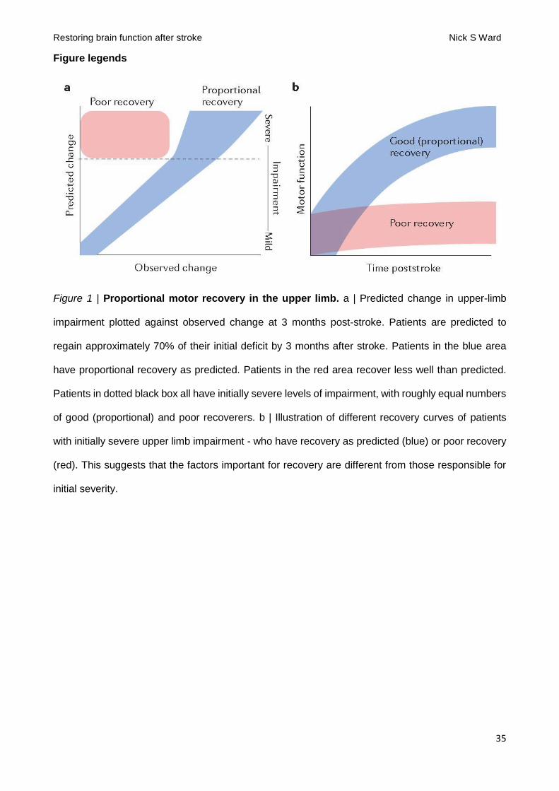
Restoring brain function after stroke Nick S Ward
35
Figure legends
Figure 1 | Proportional motor recovery in the upper limb. a | Predicted change in upper-limb
impairment plotted against observed change at 3 months post-stroke. Patients are predicted to
regain approximately 70% of their initial deficit by 3 months after stroke. Patients in the blue area
have proportional recovery as predicted. Patients in the red area recover less well than predicted.
Patients in dotted black box all have initially severe levels of impairment, with roughly equal numbers
of good (proportional) and poor recoverers. b | Illustration of different recovery curves of patients
with initially severe upper limb impairment - who have recovery as predicted (blue) or poor recovery
(red). This suggests that the factors important for recovery are different from those responsible for
initial severity.
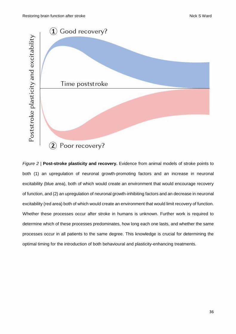
Restoring brain function after stroke Nick S Ward
36
Figure 2 | Post-stroke plasticity and recovery. Evidence from animal models of stroke points to
both (1) an upregulation of neuronal growth-promoting factors and an increase in neuronal
excitability (blue area), both of which would create an environment that would encourage recovery
of function, and (2) an upregulation of neuronal growth-inhibiting factors and an decrease in neuronal
excitability (red area) both of which would create an environment that would limit recovery of function.
Whether these processes occur after stroke in humans is unknown. Further work is required to
determine which of these processes predominates, how long each one lasts, and whether the same
processes occur in all patients to the same degree. This knowledge is crucial for determining the
optimal timing for the introduction of both behavioural and plasticity-enhancing treatments.
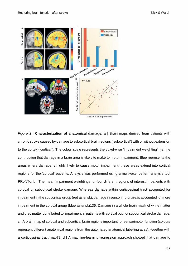
Restoring brain function after stroke Nick S Ward
37
Figure 3 | Characterization of anatomical damage. a | Brain maps derived from patients with
chronic stroke caused by damage to subcortical brain regions (‘subcortical’) with or without extension
to the cortex (‘cortical’). The colour scale represents the voxel-wise ‘impairment weighting’, i.e. the
contribution that damage in a brain area is likely to make to motor impairment. Blue represents the
areas where damage is highly likely to cause motor impairment: these areas extend into cortical
regions for the ‘cortical’ patients. Analysis was performed using a multivoxel pattern analysis tool
PRoNTo. b | The mean impairment weightings for four different regions of interest in patients with
cortical or subcortical stroke damage. Whereas damage within corticospinal tract accounted for
impairment in the subcortical group (red asterisk), damage in sensorimotor areas accounted for more
impairment in the cortical group (blue asterisk)136. Damage in a whole brain mask of white matter
and grey matter contributed to impairment in patients with cortical but not subcortical stroke damage.
c | A brain map of cortical and subcortical brain regions important for sensorimotor function (colours
represent different anatomical regions from the automated anatomical labelling atlas), together with
a corticospinal tract map78. d | A machine-learning regression approach showed that damage to
Restoring brain function after stroke Nick S Ward
38
voxels contained in the map in part c accounted for 68% of motor impairment in 50 patients with
chronic stroke, as illustrated in the graph of predicted motor impairment plotted against real motor
impairment in these patients. The same analysis using only the corticospinal tract region of interest
accounted for only 42% of the motor impairment, suggesting that knowledge of damage to a range
of motor related brain structures, not just corticospinal tract, is important for predicting outcome137.
Parts a and b modified with permission from BMJ Publishing group ltd. © Park, C.-H., Kou, N. &
Ward, N. S. J. Neurol. Neurosurg. Psychiatry (2016). Parts c and d modified with permission from
Elsevier © Rondina, J. et al. NeuroImage Clin. 12, 372–380 (2016).
Restoring brain function after stroke Nick S Ward
39
Glossary
Proportional recovery rule: The amount of function regained after stroke is a proportion of the
initial deficit. For example, by 3 months patients will regain ~70% of the upper limb motor function
that had been lost on day 3 post-stroke.
Spontaneous biological recovery: Recovery occurring in the first few weeks and months after
stroke, attributable to enhanced post-stroke plasticity mechanisms. Recovery is rapid, occurs at the
level of impairment and generalizes beyond the tasks that are used in post-stroke training, compared
with improvements seen in the chronic phase of stroke.
Biomarkers: Indicators of disease state that can be used clinically as a measure reflecting
underlying molecular or cellular processes that might be difficult to measure directly in humans, and
can be used to predict recovery or treatment response95.
Neuronal oscillations: Rhythmic fluctuations in activity generated either spontaneously or in
response to stimuli by neural tissue in the CNS. Entrained oscillations in multiple neurons and neural
networks are thought to form a critical interface between cellular activity and large-scale functions in
the CNS.
Computational neurorehabilitation: a newly emerging field aimed at mathematical modelling of
plasticity and learning to understand and improve recovery of individuals with neurologic impairment.
Cortical microcircuit: The pattern of connections between specific excitatory and inhibitory neurons
in the cortex.
Hemispatial neglect: Reduced awareness of stimuli on one side of space, even though sensory
loss might be absent.
Restoring brain function after stroke Nick S Ward
40
Box 1 | Factors that block inhibition of neuronal regeneration
Myelin-associated proteins
Myelin-associated proteins such as Reticulon4 (also known as NogoA), myelin-associated
glycoprotein (MAG), and myelin-associated oligodendrocyte basic protein have been shown to block
neuronal regeneration. An anti-NogoA antibody has been used in preclinical models both of stroke
and of spinal cord injury, and leads to improved recovery profiles. Sprouting is often seen across the
midline, either at the level of brain stem or spinal cord. Lindau and colleagues146 found that rats
treated with anti-NogoA antibody recovered motor control after sensorimotor cortex ablation because
intact corticospinal tract had extensively sprouted across the midline into the denervated spinal
hemicord, which led to a somatotopic anatomical and functional side switch in the projection of adult
corticospinal neurons. The safety of anti-NogoA antibodies has been tested in patients with spinal
cord injury24 and amyotrophic lateral sclerosis147 and anti-MAG has been tested in patients with
stroke 148.
Extracellular matrix proteins
Chondroitin sulphate proteoglycans mediate the inhibitory properties of perineuronal nets and are
known to block axon growth. Cortical infarcts lead to reduced density of PNNs in peri-infarct cortex,
maximal at 30 days post-lesion44. The enzyme chondroitinase ABC can reinstate critical period
plasticity via the inactivation of chondroitin sulphate proteoglycans and therefore PNNs149. In a rat
model of stroke, chondroitinase ABC helped restore motor function after both acute and delayed
administration150. However, extracellular matrix proteins are not always inhibitory, for example, the
prevention of astrocytic scar formation can reduce stimulated axon regrowth. e.g. preventing
astrocytic scar formation can reduce stimulated axon regrowth151.
Growth cone inhibitors
Neuronal regeneration can also be inhibited by molecules that inhibit the axonal growth cone, such
as semaphorins and ephrins. Ephrin-A5 is induced in astrocytes in peri-infarct cortex, which leads to
inhibition of axonal sprouting. When ephrin-A5 signalling is blocked, then motor training is more liable
to promote recovery152. In this case, sprouting leads to a new pattern of reparative axonal projections
in motor-related cortices of the ipsilesional hemisphere.
Restoring brain function after stroke Nick S Ward
41
Box 2 | Factors that promote neuronal regeneration
Inosine
The naturally occurring purine nucleoside inosine has been reported to enhance axon growth and
improve outcomes in a preclinical model of stroke. Inosine promotes axonal collateral sprouting into
areas that have lost their normal innervation, such as the corticospinal tract after stroke153 or
hippocampus after experimental traumatic brain injury154. Furthermore, inosine can augment the
effects of anti-NogoA antibody (BOX 1) to restore skilled forelimb use after stroke155.
Growth and differentiating factor 10 (GDF10)
The gene encoding GDF10 is highly upregulated in the axonal regenerative transcriptome induced
in peri-infarct neurons and promotes functionally useful axonal sprouting156.
Stem cells
Increasingly, interest has been shown in the use of stem cell therapy to promote recovery after
stroke157. The two main lines of stem cell therapies are endogenous (promoting the production of
existing neural stem cells) or exogenous (transplanted from another source)158. Over the past few
years, research has explored how to reprogram adult human somatic cells to induced pluripotent
stem cells thereby producing patient-specific cells for autologous transplantation159. Rather than
restoring lost tissue, stem cells could act as stimulants for trophic factors and modulators of
immunological and inflammatory changes after stroke. Trials of exogenous cells in humans have
proved safe and claims have been made for improved clinical outcomes in patients with chronic
stroke160,161.
Box 3 | Pharmacotherapy for stroke recovery: an historical perspective
The idea of pharmacotherapy for stroke recovery is not recent. Early work in this field was performed
in 1963 by Alexander Luria and colleagues in soldiers with head injuries77. In addition to cell death,
Luria et al. proposed that symptoms could be induced by functional inhibition of intact neurons and
that “removal of the diaschisis, restoration of synaptic conduction or to use another term,
‘deblocking’” might be helpful78. The investigators proposed that this task could be achieved by the
combination of two approaches. First, the administration of a pharmacological agent (generally
Restoring brain function after stroke Nick S Ward
42
anticholinesterases) “capable of removing inhibition, modifying mediator metabolism and restoring
disturbed synaptic conduction”78, and second by methods of training which promote ‘de-blocking’,
the essence of which is “that by means of various methods the level of excitability in certain functional
systems is raised and the corresponding functions are ‘de-inhibited’”78. The general concepts have
a familiar ring compared to current concepts, but although the underlying mechanisms might now be
more apparent than in the past, the clinical outcomes have not advanced a great deal.