Department of Neurology Helsinki University Central Hospital University of Helsinki BRAIN PLASTICITY AND STROKE RECOVERY Kristina Laaksonen (née Roiha) Brain Research Unit O.V. Lounasmaa Laboratory Aalto University ACADEMIC DISSERTATION To be publicly discussed with the permission of the Faculty of Medicine of the University of Helsinki, in the lecture hall 4, Helsinki University Central Hospital, Meilahti, Haartmaninkatu 4, on 2 nd of November 2012, at 12 noon. Helsinki 2012

Welcome message from author
This document is posted to help you gain knowledge. Please leave a comment to let me know what you think about it! Share it to your friends and learn new things together.
Transcript

Department of Neurology Helsinki University Central Hospital
University of Helsinki
BRAIN PLASTICITY AND STROKE RECOVERY
Kristina Laaksonen
(née Roiha)
Brain Research Unit O.V. Lounasmaa Laboratory
Aalto University
ACADEMIC DISSERTATION
To be publicly discussed with the permission of the Faculty of Medicine of the University of Helsinki, in the lecture hall 4, Helsinki University Central Hospital,
Meilahti, Haartmaninkatu 4, on 2nd of November 2012, at 12 noon.
Helsinki 2012

SUPERVISORS
Docent Nina Forss, M.D., Ph.D. Department of Neurology Helsinki University Central Hospital Helsinki, Finland Brain Research Unit O.V. Lounasmaa Laboratory Aalto University Espoo, Finland Docent Erika Kirveskari, M.D., Ph.D. Department of Clinical Neurophysiology HUS Medical Imaging Center Helsinki University Central Hospital Helsinki, Finland Brain Research Unit O.V. Lounasmaa Laboratory Aalto University Espoo, Finland REVIEWERS
Professor Risto O. Roine, M.D., Ph.D. Department of Neurology Turku University Hospital Turku, Finland Docent Juha Huttunen, M.D., Ph.D. BioMag Laboratory HUS Medical Imaging Center Helsinki University Central Hospital Helsinki, Finland OPPONENT
Professor Franҫois Mauguiѐre, M.D., Ph.D. Department of Functional Neurology and Epilepsy Neurological Hospital, Claude Bernard Lyon 1 University Lyon, France ISBN 978-952-10-8284-9 (nid.) ISBN 978-952-10-8285-6 (PDF) http://ethesis.helsinki.fi Unigrafia Oy Helsinki 2012

Table of Contents
LIST OF ORIGINAL PUBLICATIONS ................................................................................ I
ABBREVIATIONS ................................................................................................................. II
ABSTRACT .............................................................................................................................. 1
1 INTRODUCTION ............................................................................................................... 3
2 REVIEW OF THE LITERATURE ................................................................................... 5
2.1 Anatomy and physiology of the somatosensory system ............................................ 5
2.1.1 Somatosensory pathways, touch .............................................................................. 5
2.1.2 Primary somatosensory cortex (SI) ......................................................................... 6
2.1.3 Secondary somatosensory cortex (SII) .................................................................... 8
2.1.4 Other somatosensory cortices .................................................................................. 9
2.1.5 Cortical connections of somatosensory areas ........................................................ 10
2.2 Motor function and sensorimotor integration ......................................................... 10
2.2.1 Cortical connections between somatosensory and motor cortices ........................ 11
2.2.2 Sensorimotor integration ....................................................................................... 12
2.3 Spontaneous brain oscillations .................................................................................. 13
2.3.1 Posterior alpha rhythm .......................................................................................... 13
2.3.2 Rolandic mu rhythm .............................................................................................. 14
2.3.3 Other cortical rhythms ........................................................................................... 16
2.3.4 Pathological low-frequency oscillations ............................................................... 16
2.4 Stroke ........................................................................................................................... 17
2.4.1 Epidemiology ........................................................................................................ 17
2.4.2 Risk factors ............................................................................................................ 18
2.4.3 Treatment of stroke ............................................................................................... 18
2.5 Plasticity and functional reorganization after stroke .............................................. 19
2.5.1 Neuroplasticity ...................................................................................................... 19
2.5.2 Reorganization of representational maps after stroke ........................................... 20
2.5.3 Changes in excitation/inhibition balance after stroke ........................................... 21
2.6 FUNCTIONAL BRAIN IMAGING IN STROKE .................................................. 21
2.6.1 Magnetoencephalography (MEG) ......................................................................... 21
2.6.2 Other functional imaging methods ........................................................................ 24
2.7 SOMATOSENSORY EVOKED RESPONSES ....................................................... 25
2.7.1 Somatosensory evoked fields (SEFs) .................................................................... 26
3 AIMS OF THE STUDY .................................................................................................... 29

4 MATERIALS AND METHODS ...................................................................................... 30
4.1 Subjects ........................................................................................................................ 30
4.1.1 Stroke patients ....................................................................................................... 30
4.1.2 Control subjects ..................................................................................................... 30
4.2 CLINICAL TESTING ............................................................................................... 31
4.3 STIMULATION ......................................................................................................... 31
4.4 MAGNETOENCEPHALOGRAPHIC RECORDINGS ......................................... 31
4.5 DATA ANALYSIS...................................................................................................... 32
4.5.1 Preprocessing of the data ....................................................................................... 32
4.5.2 Dipole modeling .................................................................................................... 33
4.5.3 Temporal-spectral-evolution method (TSE) ......................................................... 34
4.5.4 Analysis of spontaneous brain activity .................................................................. 34
4.5.5 Statistical analysis ................................................................................................. 35
5 EXPERIMENTS ................................................................................................................ 36
5.1 SI REORGANIZATION AFTER STROKE (STUDY I) ....................................... 36
5.1.1 Results ................................................................................................................... 36
5.1.2 Discussion ............................................................................................................. 37
5.2 SII ACTIVATION AFTER STROKE...................................................................... 38
5.2.1 Results ................................................................................................................... 38
5.2.2 Discussion ............................................................................................................. 41
5.3 MOTOR CORTEX EXCITABILITY AFTER STROKE ..................................... 42
5.3.1 Results ................................................................................................................... 42
5.3.2 Discussion ............................................................................................................. 45
5.4 SPONTANEOUS BRAIN OSCILLATIONS AFTER STROKE .......................... 46
5.4.1 Results ................................................................................................................... 46
5.4.2 Discussion ............................................................................................................. 48
6 GENERAL DISCUSSION ................................................................................................ 51
6.1 Temporal evolution of plastic changes after stroke................................................. 51
6.2 Motor cortex excitability after stroke ....................................................................... 52
6.3 Cortical excitability and reorganization of the cerebral cortex ............................. 53
6.4 Sensorimotor integration ........................................................................................... 54
6.5 Future perspectives in monitoring recovery after stroke ....................................... 55
7 SUMMARY & CONCLUSIONS ..................................................................................... 57
ACKNOWLEDGEMENTS ................................................................................................... 58
REFERENCES ....................................................................................................................... 60

LIST OF ORIGINAL PUBLICATIONS
This thesis is based on the following publications, which will be referred to in the text by their Roman numerals. I Roiha K, Kirveskari E, Kaste M, Mustanoja S, Mäkelä JP, Salonen O, Tatlisumak T,
Forss N. Reorganization of the primary somatosensory cortex during stroke recovery. Clin Neurophysiol. 2011; 122(2):339-45
II Forss N, Mustanoja S, Roiha K, Kirveskari E, Mäkelä JP, Salonen O, Tatlisumak T,
Kaste M. Activation in parietal operculum parallels motor recovery in stroke. Hum
Brain Mapp. 2012; 33(3):534-41 III Laaksonen K, Kirveskari E, Mäkelä JP, Kaste M, Mustanoja S, Nummenmaa L,
Tatlisumak T, Forss N. Effect of afferent input on motor cortex excitability during stroke recovery. Clin Neurophysiol. 2012; http://dx.doi.org/10.1016/j.clinph.2012.05.017
IV Laaksonen K, Helle L, Parkkonen L, Kirveskari E, Mäkelä JP, Mustanoja S,
Tatlisumak T, Kaste, M., Forss N. Alterations in spontaneous brain oscillations during stroke recovery. Submitted.
The original publications have been reproduced with the permission of the copyright holders. Contributions of the author
All the publications included in this Thesis are results of teamwork. I performed the MEG
recordings of the healthy control subjects and participated in the MEG recordings of the
patients. In publications I, III, and IV, I performed the data analysis, interpreted the results
and was the principal author of the manuscripts. In publication II, I participated in the data
analysis, participated in the interpretation of the results, and in the preparation of the
manuscript. In studies III and IV, I participated in the study design.

ABBREVIATIONS
AH Affected hemisphere
AP Action potential
ARAT Action Research Arm Test
BEM Boundary element model
BI Barthel Index
BOLD Blood oxygenation level dependent
CBF Cerebral blood flow
DALY Disability adjusted life years
ECD Equivalent current dipole
EEG Electroencephalography
FFT Fast Fourier Transformation
fMRI Functional magnetic resonance imaging
g Goodness of fit
ICF Intracortical facilitation
ICI Intracortical inhibition
ISI Interstimulus interval
LAI Long-latency afferent inhibition
MEG Magnetoencephalography
MI Primary motor cortex
MRI Magnetic resonance image
mRS modified Rankin Scale
NIHSS National Institutes of Stroke Scale
Peg Nine-hole peg board test
PET Positron emission tomography
PM Premotor cortex
PPC Posterior parietal cortex
PSP Postsynaptic potential
PV Parietal ventral area
SAI Short-latency afferent inhibition
SEF Somatosensory evoked field
SEP Somatosensory evoked potential
SI Primary somatosensory cortex

SII Secondary somatosensory cortex
SMA Supplementary motor area
SQUID Superconducting quantum interference device
SSS Signal space separation method
TBI Traumatic brain injury
TMS Transcranial magnetic stimulation
tSSS Temporally extended signal space separation method
UH Unaffected hemisphere
VPL Ventral posterior lateral nucleus of the thalamus
1
ABSTRACT
Recovery from stroke is based on the capability of the brain to reorganize its structure and
function after lesion. An acute stroke triggers a cascade of time-dependent metabolic and
physiological reactions, which enable changes in the organization and function of widespread
cortical regions. A wide range of studies, using various functional imaging methods, have
thrown light on the reorganizational changes after stroke. However, less is known about the
temporal evolution of these changes and their correlation to clinical recovery.
In this thesis, different aspects of neurophysiological changes related to sensorimotor
recovery were studied in 18 patients with first-ever stroke in the middle cerebral artery
territory, affecting upper limb motor function. Follow-up recordings of somatosensory evoked
fields (SEF) and spontaneous rhythmic brain activity were performed with whole-head MEG
within 1 week (T0), 1 month (T1), and 3 months (T2) after stroke with concomitant evaluation
of clinical outcome. MEG suits stroke studies especially well, as it is independent from
hemodynamic alterations, and the signals are practically unaffected by morbid tissue.
The results indicated that the hand representation in the primary somatosensory cortex (SI) in
the affected hemisphere (AH) was transiently enlarged at T1 and returned to normal size
concomitantly with clinical improvement of hand function (Study I). Study II showed that the
activation in the contralateral secondary somatosensory cortex (cSII) was decreased in the AH
at T0 and increased during follow-up. The strength of cSII activation paralleled the recovery
of hand function during the 3 months follow-up, suggesting that cSII may be an important
region in mediating the somatosensory input to the motor cortex. The results in Study III
indicated that afferent-input-modulated motor cortex excitability was increased in the AH in
the acute phase after stroke and decreased during follow-up in association with recovery of
hand function. Study IV showed that the ~10-Hz oscillations were enhanced in the AH at T1
and T2. Moreover, pathological perilesional low-frequency oscillations were detected in 7/16
patients at T0, and the low-frequency oscillations persisted for at least 3 months in 4 patients.
These 4 patients had a worse clinical outcome at T2 than the rest of the patients.
The results indicate that even small lesions can cause widespread neurophysiological changes
in the cortical network. Certain brain regions, such as SII, seem to be specifically important
for the recovery of hand function. The results underline the importance of parallel recovery of
the somatosensory and motor systems for fluent hand function. The most evident

2
neurophysiological changes were observed within 1 month after stroke in parallel with
steepest improvement of clinical recovery, suggesting that the first 4 weeks are critical for
functional recovery.

3
1 INTRODUCTION
Stroke is one of the leading causes of permanent disability in western countries. In recent
years, therapeutic interventions such as thrombolysis have been developed to treat acute
stroke. However, due to the short time window (within 4.5 hours from onset of symptoms) of
this treatment, it still reaches the minority of stroke patients, and even then only half of the
occluded vessels are re-canalized (Rha and Saver, 2007). Hence, for most patients intensive
rehabilitation is the only way to minimize impairment and to regain lost function.
Rehabilitation is based on the capability of the central nervous system to reorganize and to
adjust to environmental needs. Studies in animals have shown reorganization of the cerebral
cortex both after peripheral deafferentation (Merzenich et al., 1984, Pons et al., 1991) and
after central lesions (Frost et al., 2003, Nudo and Milliken, 1996, Xerri et al., 1998). In
animals, cortical reorganization has been linked to changes in cortical inhibition (Jacobs and
Donoghue, 1991).
Consistently, different aspects of cortical reorganization have also been observed in patients
after stroke. Enlargement of cortical motor or somatosensory representation areas (Calautti et
al., 2001, Rossini et al., 1998a, Rossini et al., 2001, Ward et al., 2003a, Ward et al., 2003b)
and alterations in the cortical excitability (Butefisch et al., 2003, Liepert et al., 2000b,
Manganotti et al., 2002, Ward and Cohen, 2004) have been detected in patients after stroke.
However, the functional significance of these findings is not thoroughly understood.
Although plastic changes allow functional recovery, plasticity is not necessarily a solely
positive phenomenon. For example, focal dystonia in musicians has been linked to an over
activation of the primary sensorimotor cortex (Pujol et al., 2000), and prolonged pain in
patients with complex regional pain syndrome (CRPS; Juottonen et al., 2002, Maihofner et
al., 2003) and in patients suffering from phantom limb pain (Flor et al., 1995) has been linked
to maladaptive plasticity.
The aim of this thesis was to study recovery of the somatosensory and motor cortices after
acute stroke, and to correlate the observed neurophysiological changes with clinical recovery.
To achieve this, we performed follow-up measurement of somatosensory evoked fields and
spontaneous brain activity in 18 patients with first-ever stroke in the middle cerebral artery
4
territory. The motivation of this thesis was to better understand the mechanisms and temporal
behavior of plastic changes after stroke, and to find objective parameters to monitor recovery
after stroke.

Review of the literature
5
2 REVIEW OF THE LITERATURE
2.1 Anatomy and physiology of the somatosensory system
2.1.1 Somatosensory pathways, touch
Somatosensory sensation comprises four major modalities: touch, proprioception,
nociception, and temperature sense. These submodalities are mediated through two major
pathways (dorsal column-medial lemniscus system and anterolateral system) to the brain
(Kandel and Jessel, 1991). Discriminative touch is required to recognize the size, shape,
weight, and texture of objects.
Touch is mediated via four types of mechanoreceptors which lie in the skin and underlying
tissue. The rapidly adapting receptors (Meissner’s corpuscles in the superficial skin and
Pacinian corpuscles in the deeper tissue) detect changes in texture, whereas slowly adapting
receptors (Merkel`s cells in the superficial skin and Ruffini’s corpuscles in the deeper tissue)
respond to sustained touch and pressure (Kandel and Jessel, 1991).
The information from these four receptor types is conveyed by axons of nerve cells in the
dorsal root ganglia to the spinal cord. The majority of the central axons of the dorsal root
ganglia neurons ascend in the ipsilateral dorsal column, which relays both tactile and
proprioceptive information in a topographic arrangement, to the junction of the spinal cord
and the medulla, where they synapse with second-order neurons in two dorsal column nuclei
(nuclei cuneate and gracilis; Figure 1). The axons of the second-order neurons cross the
midline in the medulla oblongata and ascend in the lemniscus medialis to the thalamus, where
they synapse in the ventral posterior lateral nucleus (VPL) and to a lesser extent in the
posterior nuclei with third-order neurons. Some tactile information is also relayed in the
anterolateral system together with information about pain and temperature. Thus patients with
dorsal column lesions retain some crude tactile sensibility (Kandel and Jessel, 1991).
The axons of third-order neurons relaying information from the cutaneous mechanoreceptors
mainly terminate in Broadmann area 3b in the primary somatosensory cortex (SI), lying in the
posterior wall of the central sulcus in the parietal lobe. From there, neurons project to
Broadmann areas 1 and 2 in the primary somatosensory cortex, to the posterior parietal cortex

6
(PPC), and to the secondary somatosensory cortex (SII). Thalamic neurons also project
directly to Broadmann areas 1 and 2 as well as to the PPC and SII.
The topographic arrangement of receptors in the skin is preserved throughout the whole
somatosensory pathway, and the somatosensory cortex consists of several somatotopically
organized maps of the body surface.
Fig.1. Diagram of the ascending somatosensory pathways. The dorsal column-medial lemniscus system relays tactile sensations and arm proprioception (modified from Martin and Jessel, 1991).
2.1.2 Primary somatosensory cortex (SI)
SI is located in the parietal lobe, in the posterior bank of the central sulcus and in the
postcentral gyrus (Figure 2). It consists of Broadmann areas 3a, 3b, 1, and 2. Most thalamic
fibers terminate in areas 3a and 3b. Areas 3b and 1 receive information from cutaneous
mechanoreceptors, whereas areas 3a and 2 receive proprioceptive information from muscles
and joints (Kandel and Jessel, 1991). All of these four areas are interconnected extensively.
The information flows mainly in the anteroposterior direction from areas 3a and 3b to areas 1
and 2; at each stage of somatosensory processing, the size of the receptive field becomes
larger and the feature-detecting properties become more complex (Hyvarinen and Poranen,
1978). Area 3b receives mainly information about simple stimulus-related properties, such as

Review of the literature
7
intensity and site of stimulation, whereas areas 1 and 2 input are concerned with properties
such as direction of movement on the skin and the three-dimensional perception of objects.
Fig.2 a) The anatomical locations of the three major divisions of the somatosensory cortices from a lateral perspective of the cortical surface. b) SI is subdivided into four cytoarchitectonic areas (Broadmann’s areas 3a, 3b,1, and 2; modified from Gardner and Kandel, 2000).
The somatosensory projection from the body is somatotopically organized in SI. This means
that each body part has its own representational area. Actually, each area in SI (Broadmann
3a, 3b, 2, and 1) has its own, completely independent body map, with the foot area lying most
medially and the face area most laterally (Kaas et al., 1979). Each body part is represented
according to its innervation density (Penfield and Jasper, 1954). Areas of the body that are
densely innervated and important for tactile discrimination, such as the fingertips and lips,
have a disproportionally large representation compared with areas with less extensive
innervation, such as the trunk. This means that the receptive fields of cortical neurons
innervating the fingertips are much smaller than the ones innervating the trunk. Although the
general medial-to-lateral somatotopical organization is similar in all individuals, the sizes of
representation area of different body parts are not fixed, but they vary between individuals
and change by use (Clark et al., 1988, Jenkins et al., 1990). For instance, in monkeys who
were trained to touch a rotating disk with their fingertips, the fingertip representations in

8
cutaneous area 3b were expanded after several weeks of touching the disk (Jenkins et al.,
1990). In accordance, the representations of the left hand digits of string players have been
shown to be larger than those in non-musicians (Elbert et al., 1995).
It is suggested that afferent connections to neurons in the somatosensory cortex are formed on
the basis of correlated firing. In monkeys, increased correlation of afferent input, obtained by
connecting surgically two adjacent fingers, fused the representation areas of these two fingers
(Clark et al., 1988). In line with this study, in two patients who were studied before and after
surgical separation of webbed fingers, the postsurgical hand representation was considerably
larger than the presurgical hand representation, correlating with the new functional status of
the separated fingers (Mogilner et al., 1993).
2.1.3 Secondary somatosensory cortex (SII)
SII, located in the parietal operculum along the superior bank of the lateral sulcus, was first
described by Adrian in electrophysiological studies in cats (Adrian, 1941). Since then, it has
been described in many other animals including primates (Woolsey, 1946). In humans, the SII
region was first described by Penfield and Jasper (1954) by means of electrical stimulations of
the lateral sulcus during neurosurgery. The first noninvasive observations of activation in the
SII region were described in magnetoencephalographic recordings (Hari et al., 1984). The
definition of the boundaries and connections of SII has been challenging; the smaller size and
the location of SII render it much more difficult to study than SI (Burton, 1986). Moreover, a
variety of different adjacent regions to SII with responsiveness to somatosensory stimuli have
been found in different species, but the boundaries of these regions have been difficult to
determine (Burton, 1986). Microelectrode recordings in monkeys (Krubitzer et al., 1995) and
fMRI in humans (Disbrow et al., 2000) have revealed at least two somatotopically organized
areas in the parietal operculum: the SII cortex, and rostral to it, the parietal ventral area (PV),
which have mirror symmetric maps of the body surface and share common boundaries at the
representations of the face, hands and feet. The activation patterns within SII and PV have
been shown to be highly variable across subjects (Disbrow et al., 2000), which has further
hampered the exact determination of the boundaries of SII.
SII shows somatotopical organization, with cranial parts of the body located anterolaterally
and caudal parts posteriomedially. In general, the receptive fields of neurons in SII are larger

Review of the literature
9
and more overlapping than in SI (Burton, 1986, Mazzola et al., 2006). It appears that the
spatial differentiation of the body map in SII is sufficiently developed to provide a resolution
capable of identifying the body part that has been touched, but the spatial discrimination is not
as good as in SI (Burton, 1986).
In contrast to SI, SII is bilaterally activated to unilateral stimulation and neurons in SII have
been shown to have bilateral receptive fields (Whitsel et al., 1969, Robinson and Burton,
1980, Mazzola et al., 2006). In accordance with the relatively large, overlapping, and bilateral
receptive fields of SII, the functional role of SII in primates has been suggested to be critical
for coordinating sensorimotor tasks involving multiple body parts, such as the digits of the
hand or the two hands (Simoes and Hari, 1999, Disbrow et al., 2000).
In rhesus monkeys, ablation of the SII region led to impairment of discrimination of the shape
and texture of objects (Murray and Mishkin, 1984). Accordingly, in humans, lesions of SII
have been suggested to be associated with tactile agnosia (Caselli, 1993). However, this view
was challenged by a subsequent study showing consistently abnormal somatosensory evoked
potentials (SEPs) in SI in patients with tactile agnosia (Mauguiere and Isnard, 1995). In
agreement with the latter findings, impaired SII activation was always associated with
abnormal SI responses in the damaged hemisphere of chronic stroke patients (Forss et al.,
1999).
2.1.4 Other somatosensory cortices
The posterior parietal cortex (PPC) is located posterior to area 2 in SI. In humans, the PPC
stretches over Broadmann areas 5 and 7. However, the borders of PPC are not strictly
delineated. In addition to dense connections with ipsi- and contralateral SI and SII, PPC is
connected with the visual, auditory, and motor cortices. Thus, PPC is not a pure
somatosensory association area; rather, it combines somatosensory information from personal
body parts with extrapersonal spatial information and serves higher-level cognitive functions
related to movement (Andersen and Buneo, 2002, Hyvarinen, 1982). Thus, lesions of PPC
cause complex defects such as disturbances in spatial perception, visuomotor integration, and
selective attention. Probably the most well-known consequence of a lesion in the right PPC is
neglect syndrome, a deficit in the visuospatial perception of the left side of the body as well as
the environment on the left side.
10
Parts of the mesial cortex are also activated during somatosensory processing (Caselli, 1993,
Forss et al., 1996, Penfield and Jasper, 1954). This area is known as the supplementary
sensory area, and it probably stretches over the mesial area 5 and anterior portion of mesial
area 7 (Caselli, 1993). Extensive lesions of this area caused disruption of somesthetic
processing and apraxia (Caselli, 1993). Activation in the mesial cortex in response to
somatosensory stimuli has been shown to be attention dependent (Forss et al., 1996).
2.1.5 Cortical connections of somatosensory areas
Studies in monkeys have shown dense, topographically specific, reciprocal connections from
all four areas in SI (3a, 3b, 1, and 2) to SII (Jones et al., 1978). Input from the different areas
appear to converge within SII in the representation of a given body part (Friedman et al.,
1980). SI also has efferent projections to areas 5 and 7 in the ipsilateral PPC. In addition to
intrahemispheric connections, SI has transcallosal connections to homotopical areas in the SI
of the opposite hemisphere. These connections are sparse between areas 3b and relatively
dense between areas 2 (Killackey et al., 1983). Moreover, transcallosal connections between
hand and foot representations within each field are much less dense than those between face
and trunk representations; in area 3b they are practically non-existent (Killackey et al., 1983).
SI also has transcallosal connections to somatotopically-related areas in the contralateral SII
(Burton, 1986). However, the functional significance of these connections is not well known.
In stroke patients, an SII response ipsilateral to the stimulated impaired hand was found in all
patients regardless of the responsiveness of the contralateral SI and/or SII, suggesting that
ipsilateral SII may be activated mainly directly through thalamocortical connections (Forss et
al., 1999).
Area SII has shown to have connections to the insular cortex and to area 7 in the PPC
(Burton, 1986). Moreover, SII is connected in a topographical fashion to contralateral SII via
transcallosal connections (Burton, 1986).
2.2 Motor function and sensorimotor integration
Voluntary movements require a complex interaction of cortical motor areas and an integration
of sensory input with motor programs. The motor cortices, divided into the primary motor

Review of the literature
11
cortex (MI) and the premotor areas, are located anterior to the central sulcus, occupying
approximately the posterior third of the frontal lobes. MI is located in the precentral gyrus and
in the anterior wall of the central sulcus (Broadmann area 4). The somatototopical
organization of MI resembles the organization of SI: the foot area is located most medially
and the face area most laterally. Body parts such as the face, hands, and fingers that are used
in motor tasks requiring precision and fine control have disproportionally large
representations.
The premotor areas, comprising Broadmann’s area 6 anterior to MI consist of two major
areas: medially, the supplementary motor area (SMA) and laterally, the premotor cortex (PM).
The premotor areas project to MI and to subcortical structures (striatum and thalamic nuclei)
as well as directly to the spinal cord. Stimulation of the premotor areas often evoke complex
movements involving multiple joints and bilateral body parts (Krakauer and Ghez, 2000).
2.2.1 Cortical connections between somatosensory and motor cortices
Discriminative touch and proprioception are essential for the execution of fine, skilled
movements. Although some direct thalamocortical afferent connections to MI exist (Asanuma
et al., 1979), the modulatory afferent input to the motor cortex is mediated mainly via cortico-
cortical connections from SI and SII (Chen et al., 1999, Disbrow et al., 2000, Hinkley et al.,
2007). Studies in monkeys have shown direct connections from areas 1 and 2 in SI to area 4
in ipsilateral MI, whereas direct connections between the main cutaneous area 3b and MI have
shown to be sparse or even non-existent (Jones et al., 1978). In contrast, area SII has been
shown to have strong anatomical connections to area 4 in ipsilateral MI and to SMA (Jones
and Wise, 1977).
Fig.4 Ipsilateral connections between somatosensory cortices and the primary motor cortex.
12
2.2.2 Sensorimotor integration
Fluent motor performance requires an integration of afferent somatosensory input with motor
programs to adjust the strength, speed, and range of movements. For example, in monkeys, a
combined removal of the dorsal column and SI led to permanent severe deficits of hand
dexterity (Asanuma and Arissian, 1984). Accordingly, a patient with severe peripheral
sensory neuropathy and intact motor circuits was relatively unable to use his hands in daily
life, as he could not automatically correct or maintain movements without visual feedback
(Rothwell et al., 1982).
In addition to anatomical connections between SII and MI (Jones and Wise, 1977), functional
imaging studies have shown a close interaction between SII activation and motor functions.
Navigated transcranial magnetic stimulation (nTMS) of the SII region has been shown to
facilitate motor performance in healthy subjects (Raij et al., 2008). Deficient activation of SII
has been observed in patients with impaired hand dexterity due to Unverricht-Lundborg type
epilepsy or focal dystonia (Butterworth et al., 2003, Forss et al., 2001). Taken together, SII
seems to play an essential role in sensorimotor integration, especially in tasks involving
multiple, functionally-related body parts (Disbrow et al., 2000, Hinkley et al., 2007).
It has been proposed that afferent somatosensory input mediates its effect on motor functions
by modulating the excitability of motor cortex neurons before and during movement
(Asanuma and Arissian, 1984, Favorov et al., 1988). Accordingly, reduced afferent input due
to transient ischemic block of cutaneous afferents or transient immobilization of a limb has
been shown to cause motor cortex disinhibition (Brasil-Neto et al., 1992, Todd et al., 2006).
In line with these findings, in a TMS study, decreased inhibition of the ipsilesional motor
cortex was observed in stroke patients with defective somatosensory input due to lesions in SI
or VPL (Liepert et al., 2004).
Taken together, the integration of afferent somatosensory input from multiple body parts,
such as the two hands or the fingers of a hand, with motor functions, may be mainly mediated
via SII. The integration of afferent somatosensory input with motor programs may function by
changing the excitability of motor cortex neurons. Thus, defective sensorimotor functioning
may result from insufficient somatosensory feedback due to somatosensory system

Review of the literature
13
dysfunction or from defective sensorimotor integration due to altered afferent modulation of
motor cortex neuron activity.
2.3 Spontaneous brain oscillations
Neurons of the cerebral cortex exhibit intrinsic oscillations (Llinas, 1988). The synchronous
oscillations of neuronal populations form the basis of cerebral cortical rhythms. Various
cortical brain regions in the healthy human brain exhibit their own intrinsic, frequency-
specific rhythms with modality-specific reactivity. The best known cortical rhythms of the
human brain are the alpha rhythm, detected over the posterior parts of the brain, and the mu-
rhythm, detected over the rolandic regions. These rhythms and their modulation are well
detectable with electroencephalographic (EEG) and MEG recordings (Salmelin and Hari,
1994a, Steriade et al., 1990). The thalamus has been suggested to play an essential role in
driving cortical rhythmic activity (Hughes and Crunelli, 2005, Steriade and Llinas, 1988), and
thalamic lesions have been shown to attenuate cortical rhythmic activity (Makela et al., 1998).
Over the last few years, cortical rhythms have attracted new widespread interest. For decades,
cortical rhythms were interpreted to reflect an idling state of the neurons (Pfurtscheller et al.,
1996), but the differences in spatial and temporal occurrence, as well as in modality-specific
reactivity of these rhythms, suggest that these rhythms have higher functional significance
(Salmelin et al., 1995). However, the exact functional role of cortical rhythms is still under
debate. Cortical rhythms have been suggested to have an important role in cognitive
processing (Llinas and Ribary, 1993, Jensen et al., 2002, Haegens et al., 2010, Palva et al.,
2005) and in perceptual binding of distributed neural activity (Fries, 2005, von der Malsburg,
1995). Moreover, changes in the amplitude or frequency of brain rhythms may reveal
pathological phenomena of the brain (Lewine et al., 1999, Pfurtscheller et al., 1981, Tecchio
et al., 2007, Van Huffelen et al., 1984).
2.3.1 Posterior alpha rhythm
The posterior alpha rhythm, first described by Hans Berger in 1929 (for a review see
Niedermeyer, 1999), is the best known cortical rhythm. It occurs during wakefulness in the
frequency range of 8–13 Hz over the posterior region of the brain. This rhythm is blocked by
eye opening and re-appears with eye closure. Alpha rhythms with the same peak frequency

14
have been recorded from both the visual thalamus (lateral geniculate and pulvinar nuclei) and
from the visual cortex (Lopes Da Silva and Storm Van Leeuwen, 1977). Although
simultaneously-recorded alpha rhythms from the thalamus and from the cortex have been
shown to be partly coherent, the coherence between alpha rhythms recorded between closely
spaced electrodes in the cortex has been shown to be much stronger than thalamocortical
coherence (Lopes Da Silva and Storm Van Leeuwen, 1977). The genesis of the alpha rhythm
is still not thoroughly understood. It has been assumed that there are several generator areas of
alpha rhythms in the cerebral cortex and that the rhythm spreads from these areas in different
directions (Lopes Da Silva and Storm Van Leeuwen, 1977, Steriade et al., 1990). However, so
far there has been no evidence of a synchronizing mechanism for the alpha rhythm at the
cortical level (Steriade et al., 1990), whereas the thalamic reticular nucleus has been
suggested to play an essential role in the synchronization of thalamic oscillations (Steriade
and Deschenes, 1984). Thus, it is assumed that there are both thalamocortical and cortico-
cortical systems which interact in the generation of these rhythms (Steriade et al., 1990).
Alpha oscillations have been suggested to play an important functional role in cognitive
processing (Jensen et al., 2002, Palva et al., 2005) and in orienting attention (Foxe et al.,
1998, Handel et al., 2011). Occipital alpha is supposed to reflect inhibition of task-irrelevant
areas, thus directing the sensory inflow to task-relevant areas (Jensen and Mazaheri, 2010).
2.3.2 Rolandic mu rhythm
The features of the cortical rhythm detected over the rolandic regions were first described in
detail by Gastaut et al. in 1952 (for a review see Niedermeyer, 1999). The rhythm consists of
a slower alphoid (~10 Hz) and a faster beta (~20 Hz) component. Relatively independent mu
rhythm generating systems exist in both hemispheres (Storm van Leeuwen et al., 1976). The
alphoid component of the rolandic mu rhythm has been suggested to be generated mainly in
the postcentral gyrus in the primary somatosensory cortex (Salmelin et al., 1995, Salmelin
and Hari, 1994b), whereas the beta component has been shown to have its main generator
areas in the primary motor cortex (Pfurtscheller et al., 1996, Salmelin and Hari, 1994b). The
beta rhythm has been shown to be coherent with the simultaneously recorded EMG signal
from an isometrically contracted limb muscle (Conway et al., 1995, Salenius et al., 1997a),
which further supports the association of the beta rhythm with motor functions.

Review of the literature
15
The reactivity of the rolandic mu rhythm indicates that it is closely related to sensorimotor
functions. The mu rhythm is suppressed by movement execution, observation or even motor
imagery (Hari et al., 1998, Neuper and Pfurtscheller, 1996, Salenius et al., 1997b, Salmelin
and Hari, 1994b). The rhythm is suppressed already 1-2 s before movement and subsequently
increased (rebound) 0.5-2.5 s after movement termination (Pfurtscheller, 1992, Salmelin and
Hari, 1994b). In addition to motor activation, afferent somatosensory input, such as peripheral
tactile or electric stimulation, also elicits an initial suppression followed by a rebound of the
mu rhythm (Salenius et al., 1997b, Salmelin and Hari, 1994b). The reactivity of the rhythm is
bilateral to unilateral movement or somatosensory stimulation, but the reactivity in the
contralateral hemisphere to the site of the movement/somatosensory stimulation is more
pronounced (Salenius et al., 1997b, Salmelin and Hari, 1994b). Both alphoid and beta
components of the mu rhythm display movement-related reactivity, but the reactivity,
especially the rebound, is faster and stronger for the beta component than for the alphoid
component (Pfurtscheller, 1992, Salenius et al., 1997b, Salmelin and Hari, 1994b).
It has been suggested that there are at least two distinct beta rhythms with different
frequencies and different functional roles (Hall et al., 2011, Jurkiewicz et al., 2006,
Pfurtscheller et al., 1997, Szurhaj et al., 2003). These different beta components have been
reported to behave differently in their reactivity to movement, with the lower beta (~15 Hz)
component contributing more to the movement-related rebound and the higher beta (~20 Hz)
component displaying quite a similar pattern of reactivity than the alphoid component
(Pfurtscheller et al., 1997). In line with these findings, the suppression and rebound of the
beta rhythm have been suggested to have different generator areas: the rebound has its main
sources in MI in the precentral gyrus (Jurkiewicz et al., 2006, Salmelin et al., 1995), whereas
the sources of suppression have been more variable (Feige et al., 1996, Jurkiewicz et al.,
2006).
The rebound of the beta rhythm is dampened by motor cortex activation due to movement
execution, observation or motor imagery (Hari et al., 1998, Salenius et al., 1997a, Schnitzler
et al., 1997), and it has been suggested to reflect deactivation, removal of excitation
(Pfurtscheller, 1992, Salmelin et al., 1995), or active inhibition of the motor cortex (Chen and
Hallett, 1999, Franzkowiak et al., 2010). Accordingly, decreased motor cortex excitability has
been detected with TMS from 200 ms to 1000 ms after digit or median nerve stimulation, a
time course comparable to the beta rebound (Abbruzzese et al., 2001, Chen et al., 1999). A

16
combined MEG and magnetic resonance spectroscopy study showed a linear relation between
the beta rebound strength and the inhibitory neurotransmitter γ-Aminobutyric acid (GABA;
Gaetz et al., 2011), further strengthening the inhibitory role of the beta rebound. Consistently,
the beta rebound has been shown to be attenuated in disorders with suspected motor cortex
hyperexcitability or disinhibition such as Unverricht-Lundborg type epilepsy or complex
regional pain syndrome (Juottonen et al., 2002, Silen et al., 2000, Visani et al., 2006,
Kirveskari et al., 2010).
2.3.3 Other cortical rhythms
In addition to the well known occipital alpha and rolandic mu rhythms, a less well known tau
rhythm in the the 8–10 Hz range has been observed in the temporal-lobe (Tiihonen et al.,
1991). This rhythm is not dampened by opening the eyes, but it is transiently suppressed by
auditory stimuli (Lehtela et al., 1997). The sources of this rhythm cluster to the supratemporal
cortex, close to the generator sites of auditory evoked fields (Lehtela et al., 1997). In addition,
a sigma rhythm in the 7–9 Hz range has been observed in the parietal operculum, most likely
in the SII (Narici et al., 2001). The sources of this rhythm were observed clearly lateral to the
sources of the sensorimotor mu rhythm and superior to the sources of the tau rhythm. The
sigma rhythm has been shown to react bilaterally to median nerve stimulation with an initial
suppression and a subsequent rebound of the rhythm (Della Penna et al., 2004).
2.3.4 Pathological low-frequency oscillations
Injured neuronal tissues generate abnormal cortical low-frequency oscillations in the
frequency range below 4 Hz. These oscillations were first classified as “delta-waves” in 1936
by Grey Walter, who localized cerebral tumors due to pathologic low-frequency oscillations
(for a review see Amzica and Lopes da Silva, 2011). However, nowadays the delta term is
also related to physiological cortical activities during sleep and anesthesia, and it is defined as
oscillations in a frequency band between 0–4 Hz (IFSECN, 1974), thus the delta term does
not reveal the mechanism underlying these oscillations.
Studies in animals have suggested that partial cortical deafferentation may play a pivotal role
in the generation of pathological low-frequency oscillations. These low-frequency oscillations

Review of the literature
17
have been suggested to have a role in guiding axonal sprouting after brain lesions (Carmichael
and Chesselet, 2002), thus promoting recovery.
In humans, pathological low-frequency oscillations have also been detected after traumatic
brain injury (TBI) and stroke (Butz et al., 2004, Huang et al., 2009, Lewine et al., 1999,
Vieth, 1990). In a study combining MEG and diffusor tensor imaging (DTI), pathological
low-frequency oscillations were found in co-occurrence with axonal injury in patients with
TBI (Huang et al., 2009). A combined MEG and proton magnetic resonance spectroscopic
imaging study suggested an association between pathological low-frequency oscillations and
abnormal metabolic activity in preserved but dysfunctioning cortical neurons adjacent to an
ischemic lesion (Kamada et al., 1997). In TBI patients, low-frequency activity has been linked
to certain cognitive symptoms (Huang et al., 2012), whereas no correlations with clinical
parameters and low-frequency oscillations have been found in stroke patients (Butz et al.,
2004).
2.4 Stroke
According to the World Health Organization, stroke is defined as “ rapidly developing clinical
signs of focal (at times global) disturbance of cerebral function, lasting more than 24 h or
leading to death with no apparent cause other than that of vascular origin” (Hatano, 1976).
The definition does not distinguish between the causes of stroke, but includes intracerebral
and subarachnoid hemorrhage, and ischemic cerebral infarction. Around 75 % of all strokes
are ischemic (Thrift et al., 2001). The sudden interruption of the blood supply to the brain
results in neurological deficits such as sensorimotor impairment, inability to produce or to
understand speech, or defects in the visual field.
2.4.1 Epidemiology
Stroke causes approximately 10 % of all deaths worldwide and is the second most common
cause of death after ischemic heart disease (WHO, 2008, Lopez et al., 2006). Globally, the
incidence of stroke was estimated at approximately 9 million in the year 2004 (WHO, 2008),
and in Finland there were approximately 10500 incident hospital-treated stroke patients each
year from 1999 to 2007 (Meretoja et al., 2011). In high-income countries, stroke is the

18
second leading cause of disability, as measured by disability adjusted life years (DALY), after
ischemic heart disease (Lopez et al., 2006). Only around one third of patients recover fully
from stroke (WHO, 2002), the remaining surviving patients suffer from permanent disability.
Stroke causes a significant burden on the society in developed countries. As a diagnostic
entity, stroke is ranked 6th place, consuming around 3 % of total health care costs (Evers et al.,
2004). It has been suggested that stroke mortality is decreasing more rapidly than stroke
incidence, which will place increased demands on the health-care system (Donnan et al.,
2008).
2.4.2 Risk factors
The most important risk factor for stroke is advanced age. Other non-modifiable risk factors
are male sex, black race, and family history of stroke. The most important modifiable risk
factor is hypertension. Other modifiable risk factors are dyslipidemia, smoking, diabetes,
obesity, physical inactivity, and atrial fibrillation (Goldstein et al., 2011) .
2.4.3 Treatment of stroke
Stroke is an emergency situation in which rapid re-supplement of the cerebral blood flow can
minimize damage of brain tissue and thus prevent severe neurological deficits. In the last few
years, considerable progress in the treatment of acute stroke has been made (Donnan et al.,
2008). Thrombolysis therapy with the recombinant tissue plasminogen activator alteplase,
when used within 4.5 hours, enhances the chance of favorable outcome. However, the benefit
of the treatment decreases the longer the treatment is delayed from stroke onset (Lees et al.,
2010). Efforts have been made to develop additional interventions to treat acute stroke, among
others the enhancement of thrombolysis with low-frequency ultrasound or mechanical
thrombectomy with special devices (Donnan et al., 2008).
Despite the progress in acute treatment, stroke is still the most common cause of permanent
disability among elderly people (Donnan et al., 2008). Thus, for most patients, intensive
rehabilitation is the most efficient way to regain lost function. Clinical experience has shown
that early systematic treatment by an interdisciplinary team improves the prospects of
successful rehabilitation. However, the effectiveness of rehabilitation varies among patients

Review of the literature
19
and it declines with time. No clear evidence of the benefit of rehabilitation continued after one
year post stroke exists (Aziz et al., 2008). In the last few years, efforts have been made to
better understand the mechanisms underlying recovery of function, with the target to develop
new effective therapeutic strategies (Ward and Cohen, 2004).
2.5 Plasticity and functional reorganization after stroke
2.5.1 Neuroplasticity
According to the Oxford English dictionary, plasticity refers to the quality of being easily
shaped or moulded. The term plasticity was first introduced to neuroscience in 1890 by
William James in reference to the tendency of human behavior to be modifiable (for a review
see Pascual-Leone et al., 2005). Plasticity is not an occasional occurrence in the central
nervous system; rather, it is ongoing, allowing the central nervous system to reorganize and to
adjust to environmental needs throughout an individual’s life (Pascual-Leone et al., 2005).
At the functional and structural level, plasticity comprises, e.g., the reorganization of
representational maps in the cerebral cortex. Such reorganization occurs, e.g., after changes in
afferent input, motor learning or after loss of function due to lesions in the central nervous
system (Merzenich et al., 1984, Nudo and Milliken, 1996, Nudo et al., 1996a). On one hand,
plasticity is a mechanism for development and learning. For instance, the cortical
representation of the reading finger in Braille readers has been shown to be enlarged as a
result of intensive training (Pascual-Leone and Torres, 1993). On the other hand, plasticity
can also be maladaptive and a cause of pathology. Thus, in amputees, the reorganization of
the cerebral representation area of the amputated limb has shown to be associated with
phantom limb pain (Flor et al., 1995).
At the cellular level, neuroplasticity comprises, e.g., the unmasking of previously existing
silent connections (Jacobs and Donoghue, 1991) which lead to a rapid modulation of cortical
representational maps that can occur within minutes (Braun et al., 2001). Changes over longer
periods of time involve additional mechanisms such as axonal and dendritic sprouting, and
formation of new and strengthening of pre-existing synapses (Carmichael et al., 2001,
Stroemer et al., 1995). All these plastic changes are driven by both behavioral changes and
mediated by local molecular changes.

20
2.5.2 Reorganization of representational maps after stroke
A study in monkeys showed that after photothrombotically induced small lesions to distal
forelimb representation areas in MI, the remaining forelimb representation areas adjacent to
the stroke also shrank without training, and the monkeys did not regain lost function (Nudo
and Milliken, 1996). In contrast, in monkeys who received training, these areas were spared
or they even enlarged concomitantly with recovery of function (Nudo et al., 1996b). In
humans, in addition to enlarged motor- and somatosensory representation areas within the
primary sensorimotor cortices (Rossini et al., 2001, Rossini et al., 1998b, Weiller et al., 1993,
Ward et al., 2003b), more large-scale changes have also been observed after stroke. Motor
tasks have been shown to activate secondary motor areas such as the SMA, PM, and even
contralesional primary and secondary motor areas after stroke (Weiller et al., 1993, Ward et
al., 2003b). Both enlarged representation areas in the primary sensorimotor cortices (Rossini
et al., 2001, Rossini et al., 1998b) and recruitment of secondary association areas (Rossini et
al., 2001, Rossini et al., 1998b, Ward et al., 2003b) have been associated with poor clinical
outcome.
Longitudinal studies have shown that, in patients with good recovery, the neuronal activation
pattern may initially be enlarged and include non-primary motor regions, but re-focuses
towards more normal contralateral activation patterns in parallel with recovery, while in
patients with residual impairment the recruitment of secondary motor areas remains (Calautti
et al., 2001, Ward et al., 2003a). A study with patients who received constraint-induced
movement therapy demonstrated that during therapy the cortical representation of the affected
hand in MI in the affected hemisphere (AH) enlarged in parallel with recovery of hand
function. In follow-up examinations up to 6 months after treatment, the motor performance of
the affected hand had remained good, although the cortical representation in the AH had
returned to normal (Liepert et al., 2000a). Studies in monkeys have suggested that
reorganization of cortical representations is learning-dependent and not simply use-dependent.
Enlargement of cortical maps was observed in monkeys in parallel with new motor skill
acquisition, whereas corresponding changes were not observed in monkeys who simply
repeated a task that they performed optimally from the initial exposure of the task (Nudo et
al., 1996a, Plautz et al., 2000). Taken together, reorganization of cortical representation maps
occurs after stroke, and this reorganization may be related to re-learning of motor skills.

Review of the literature
21
2.5.3 Changes in excitation/inhibition balance after stroke
Several studies in humans and animals have indicated hyperexcitability both in the affected
and unaffected hemispheres after stroke (Buchkremer-Ratzmann and Witte, 1997, Butefisch
et al., 2003, Domann et al., 1993, Liepert et al., 2000b, Manganotti et al., 2002). Changes in
cortical excitability have been linked to unmasking of silent connections and thus to
reorganization of cortical representations (Jacobs and Donoghue, 1991). On the other hand, a
normalization of cortical excitability has been associated with good recovery of stroke
patients (Calautti et al., 2001, Swayne et al., 2008). In line with these findings,
hyperexcitability of the motor cortex has been linked to impaired motor performance in
several other neurological disorders such as Unverricht-Lundborg Type Epilepsy or focal
dystonia (Abbruzzese et al., 2001, Silen et al., 2000).
Motor cortex activity depends on the balance between the influences of several different
excitatory and inhibitory systems. These influences range from effects of local corticocortical
inhibitory circuits to effects of interhemispheric and afferent connections. TMS allows a
segregation of several different types of excitatory and inhibitory circuits. However, the
different excitatory and inhibitory influences are complex even in healthy subjects, and the
interaction of these is not well known (Chen, 2004). Most TMS studies on stroke patients
have applied intracortical inhibition (ICI) and intracortical facilitation (ICF) paradigms
(Liepert et al., 2005, Liepert et al., 2000b, Manganotti et al., 2002). The effect of afferent
input on motor cortex excitability after stroke is less studied. A previous TMS study evaluated
changes in both ICI and afferent inhibition after stroke (Di Lazzaro et al., 2012). They found
that changes in afferent inhibition correlated well with long-term recovery, but no correlations
with recovery and ICI were found. This study further corroborates earlier findings that
cortical excitability modulated by afferent input is driven by different circuits than those
mediating ICI or ICF (Sailer et al., 2002).
2.6 FUNCTIONAL BRAIN IMAGING IN STROKE
2.6.1 Magnetoencephalography (MEG)
MEG is a totally noninvasive method which measures, from outside the skull, the magnetic
fields produced by neuronal currents. MEG has an excellent temporal resolution on the
22
millisecond scale and the locations of underlying neuronal activity can be estimated from the
measured signals under suitable conditions with a spatial accuracy of a few millimeters. MEG
is especially suitable for stroke studies, as it is independent from hemodynamic alterations
and as the presence of morbid tissue does not significantly affect the distribution of the
neuronal signals (Huang et al., 1990).
The first MEG signals were measured in 1968 by David Cohen using induction coils as the
detector (for a review see Hari and Kaukoranta, 1985). The subsequent development of
SQUID (superconducting quantum interference device) sensors by James Zimmermann led to
the rapid development of MEG instrumentation (for a review see Hämäläinen et al., 1993).
Present-day MEG devices are designed with a helmet-shaped sensor array that covers the
whole head and allows the recording of neuronal activation over the whole brain. Thus it
enables investigations of simultaneous activation of multiple cortical sites forming a neuronal
network.
2.6.1.1 Neural basis of MEG signals
When neurons are activated they produce time-varying electrical currents. We can distinguish
between two main types of currents, the fast action potential (AP) and the more protracted
postsynaptic potential (PSP). An AP lasts only for ~1 millisecond and it produces two
oppositely-directed dipoles. The quadrupolar field produced by these dipoles diminishes
rapidly with distance. In contrast, a PSP forms one single current dipole whose magnetic field
decays much more slowly as a function of distance than that of a quadrupole. Moreover, a
PSP lasts tens of milliseconds, allowing the summations of several simultaneous PSPs
(Hämäläinen et al., 1993). MEG measures mainly the magnetic fields produced by PSPs in
the apical dendrites of pyramidal cells in the cerebral cortex. The apical dendrites lie
perpendicular to the cortex and in parallel with each other, which allows the summation of
magnetic fields of tens of thousands of neurons, hence producing a signal strong enough to be
measured from outside the skull.

Review of the literature
23
2.6.1.2 Instrumentation
The magnetic field generated by neuronal currents is typically around 50–500 fT outside the
head, which is 109 times weaker than the earth’s steady magnetic field (Hämäläinen et al.,
1993). Therefore, MEG measurements are, in general, performed in a magnetically shielded
room to avoid contamination of the cerebral signals with artifacts caused by external magnetic
noise.
The magnetic fields are measured with SQUID sensors which are embedded in liquid helium
(-269 º C) to maintain superconductivity. The magnetic fields are coupled to the SQUIDS
with pickup coils, which convert the magnetic signals into electric currents. The present
device (Elekta Neuromag®, Helsinki, Finland), used both in the Brain Research Unit, Aalto
University and in the BioMag Laboratory, HUCH, consists of 102 triple sensor elements, each
comprising two orthogonal planar gradiometers and one magnetometer. The design of the
pickup coils is important for the sensitivity of the SQUID to different source currents and
artifacts. A gradiometer is figure-eight shaped and it consists of two coils which are wound in
opposite directions. With this design, signals originating from the background noise produce
practically homogeneous fields in the coils and are thus canceled out. In contrast, signals
coming from nearby sources in the brain produce a net change in the output of the coils.
Because the field gradient of a dipolar source is steepest just above the source, planar
gradiometers give strongest signals just above the cortical sources. A magnetometer consists
of only one single pick-up loop; it is sensitive to magnetic signals from the brain but also
much more sensitive to environmental noise than a gradiometer (Hämäläinen et al., 1993,
Hari, 2011).
2.6.1.3 Source analysis
MEG measures non-invasively magnetic fields produced by neuronal currents. In principle,
several current distributions can produce identical magnetic field patterns outside the head.
Thus, there is no unique solution for the reconstruction of the sources underlying the
measured signals. This is called the inverse problem. However, with accurate prior knowledge
of the anatomy and physiology of the brain, the MEG signals can be constrained to
meaningful solutions (Hämäläinen et al., 1993) .

24
In MEG analysis, the head is typically modeled as a spherical homogeneous conductor. This
model approximates the head geometry around the sensorimotor cortex, the area of main
interest in our studies, reasonably well (Hämäläinen et al., 1993). In a spherical conductor
model, radially orientated currents do not produce measurable magnetic fields outside the
conductor, because the intracellular currents and the simultaneously produced opposing
volume currents cancel each other out. Thus, MEG measures mainly activity from neurons in
the fissures of the cortex, which produce currents tangential to the head surface (Hämäläinen
et al., 1993). Luckily, the main cortical areas of the sensorimotor system are located within
the fissures and are thus easily detected with MEG.
2.6.1.4 Comparison of MEG and EEG
MEG and EEG are closely related to each other, as the primary currents causing the signals
are the same. The main advantage of MEG over EEG is that the skull and other tissues
surrounding the brain practically do not affect the magnetic fields, whereas they substantially
distort the electric potentials measured by EEG. Thus, the spatial resolution of MEG is much
better than that of EEG. In contrast to EEG, MEG is reference-free, which makes the
interpretation of source locations of magnetic signals more straightforward (Hari, 2011). The
advantage of EEG is the much cheaper and more flexible instrumentation, which enables
telemetric, long-term, and bed-side recordings.
MEG is mainly sensitive to tangential currents, whereas EEG also detects radial currents.
Moreover, EEG is more sensitive to very deep sources. Simultaneous MEG and EEG
recordings may be advantageous because the acquired information can complement each
other (Hari, 2011).
2.6.2 Other functional imaging methods
Over the past few years, there has been growing interest in the study of functional
reorganization of the cerebral cortex after stroke. Considerable efforts have been made to
better understand the underlying mechanisms promoting or prohibiting stroke recovery. In
addition to MEG and EEG, functional magnetic resonance imaging (fMRI), positron emission
tomography (PET), and transcranial magnetic stimulation (TMS) have been widely used for
functional brain imaging after stroke.

Review of the literature
25
PET and fMRI rely on the assumption that changes in neuronal activity are closely coupled to
changes in cerebral blood flow (CBF) due to an increase in metabolism. PET is a nuclear
imaging technique, in which the distribution of a radioactive tracer is measured to make
assessments of CBF, oxygen consumption, and glucose metabolism in the brain tissue
(Eliassen et al., 2008). Most fMRI studies use blood oxygenation level dependent (BOLD)
imaging techniques. The BOLD signal is based on the different magnetic properties of
oxygenated and deoxygenated hemoglobin. In regions with increased CBF, the concentration
of oxygenated and deoxygenated hemoglobin changes, which can be detected as changes in
the BOLD signal (Ward, 2007).
TMS is a tool which allows the noninvasive stimulation of the cerebral cortex using a rapidly
changing magnetic field. Among other things, it can be used to investigate or manipulate the
physiology of the motor system. The response of motor cortices to stimulation is determined
by measuring the size, latency, and required stimulus intensity of motor-evoked responses in a
target muscle. These measures have been widely used in stroke recovery studies to probe the
motor system physiology and to detect changes in intracortical and interhemispheric
excitation/inhibition balance.
All these imaging techniques have certain limitations. Given the complexity of the
mechanisms underlying recovery from stroke, the best understanding of the functionality of
the cerebral cortex after stroke could potentially be achieved by combining these methods
(Eliassen et al., 2008).
2.7 SOMATOSENSORY EVOKED RESPONSES
Somatosensory evoked potentials (SEPs) and somatosensory evoked fields (SEFs) can be
used to investigate the physiology and functional organization of the somatosensory system.
In clinical use, alterations in strength, latency, generator areas and morphology of
somatosensory evoked responses can reveal pathological phenomena.
Single cortical responses to external stimuli are difficult to distinguish from background
noise. Thus, SEPs and SEFs are typically studied by averaging responses time-locked to the
stimulus to improve the signal-to-noise ratio. Electric stimuli to peripheral nerves have been

26
widely used to study the somatosensory system since they are easy to apply and produce clear
and strong responses. However, electric stimulation activates a large variety of fibers
innervating both muscle and skin (Burke et al., 1981). In contrast, tactile stimulation, used in
our studies, is a more natural stimulus and it selectively activates rapidly adapting cutaneous
mechanoreceptors and elicits clear responses in cytoarchitectonic area 3b, the main cutaneous
area of SI (Forss et al., 1994b).
SEPs can be recorded directly from the cortex during surgery or with implanted intracranial
electrodes, or noninvasively from the scalp. Scalp SEPs are widely used since they are easy to
measure. However, the skull and other extracerebral tissues differ in their electric
conductivities, thus they smear the electric potentials and weaken the spatial resolution of
EEG.
The earliest cortical SEP responses to electric median nerve stimulation are observed as a
surface negative deflection (N20) over the contralateral parietal cortex at about 20 ms after
the stimulus, followed by a surface positive deflection (P30) at about 30 ms. A waveform
with similar latencies but opposite polarity (P20, N30) is recorded from the frontal scalp, and
an intermediate waveform (P25,N35) is recorded near the central sulcus (Allison et al., 1991).
Several scalp and intracecebral SEP studies in healthy subjects as well as lesion studies in
humans and monkeys have suggested that these potentials are generated by one tangential
source in area 3b and one radial source in area 1 in contralateral SI (Allison et al., 1991).
However, some studies have suggested that the radial source may be generated in the
precentral cortex (Desmedt and Cheron, 1981, Mauguiere et al., 1983).
2.7.1 Somatosensory evoked fields (SEFs)
SEFs were first described by Brenner et al. who studied the somatotopical organization of
generator areas of the magnetic signals to thumb and little finger stimulation in SI (Brenner et
al., 1978). Since then, many others have reported similar findings for SEFs (Hari et al., 1984,
Okada et al., 1984). Compared with SEPs, SEFs have the advantage of a much better spatial
resolution. By measuring SEFs in response to somatosensory stimuli applied to different parts
of the body, it is possible to reproduce quantitatively the entire somatosensory homunculus in
the primary somatosensory cortex (Nakamura et al., 1998). One of the most interesting
applications in the study of somatotopically-organized SEFs is the reorganization of

Review of the literature
27
representation areas in SI, as discussed in sections 2.1.2 and 2.4. In addition to functional
mapping of cortical representation areas, SEFs can be used to study the functional
organization of the whole cortical somatosensory network (Forss et al., 1994a, Hari et al.,
1983). Thus, alterations at different levels of the cortical sensory processing stream can be
investigated totally noninvasively.
The earliest response to electric median nerve stimulation (N20m) peaks at about 20 ms, and
the corresponding equivalent current dipole (ECD) points anteriorly in the contralateral SI.
The next deflection (P35m) peaks at 30–35 ms, and the corresponding ECD is also located in
the contralateral SI but has approximately opposite polarity (Tiihonen et al., 1989). The
earliest response to tactile somatosensory stimulation of the digits using balloon diaphragms
driven by compressed air is elicited over the contralateral SI at 50‒60 ms; the corresponding
ECD is oriented posteriorly, corresponding to the P35m response (Mertens and Lutkenhoner,
2000). The differences in latencies can be explained by differences in the rise time of the
stimuli, the different stimulation sites, and the transduction from mechanical stimulation to a
neural response. The longer rise time of the tactile stimulus results in a temporally-smeared
input to the somatosensory cortex, which produces an insufficient early synchronization of the
SI neural population (Mertens and Lutkenhoner, 2000). This insufficient early
synchronization together with the smaller number of stimulated afferent fibers probably
explains the lack of a correlate of the N20m response after tactile stimulation.
Later responses to somatosensory stimulation are detected at around 100 ms in bilateral
parietal opercula at locations corresponding to the SII region (Hari et al., 1983, Hari et al.,
1984). In general, the SII response contralateral to the stimulated hand peaks slightly earlier
and more strongly than the SII response ipsilateral to the stimulated hand (Hari et al., 1983,
Hari et al., 1984). In addition, activation has been observed at 70‒110 ms in the contralateral
PPC (Forss et al., 1994a) and in the mesial cortex (Forss et al., 1996). Latencies in the later
responses do not significantly differ between tactile and electrical stimulation (Forss et al.,
1994b).
Averaged SEF amplitudes depend on the interstimulus interval (ISI; Forss et al., 1994a, Hari
et al., 1993, Tiihonen et al., 1989, Wikstrom et al., 1996). Long-latency responses, generated
outside of SI, may be conveyed through polysynaptic pathways and typically have a longer
recovery cycle than short-latency responses (Forss et al., 1994a, Hari et al., 1990). Such long-
28
latency responses are best detected at ISIs greater than 1 s and require ISIs around 3 s to be
optimally recorded. An ISI of 1 s, however, is sufficient to record optimal short-latency
responses (Wikstrom et al., 1996, Hari et al., 1983, Huttunen et al., 1992, Mertens and
Lutkenhoner, 2000).
The amplitudes of SI responses, generated mainly in area 3b, to median nerve stimulation,
have been shown to increase almost linearly with increasing stimulus intensity up to 3 times
sensory perception threshold (Jousmaki and Forss, 1998, Lin et al., 2003), emphasizing the
crucial role of SI in encoding the somatosensory stimulus intensity. The PPC and SII
responses have been shown to saturate at a stimulus intensity 2 times sensory perception
threshold (Lin et al., 2003) corresponding to stimulation intensity slightly above motor
threshold (Jousmaki and Forss, 1998). These later responses have been shown to be strongly
modulated by selective attention (Mima et al., 1998, Mauguiere et al., 1997, Hamada et al.,
2003). The modulation by attention together with the strong convergence of afferent
somatosensory input and connections to other association cortices suggest that SII and PPC
are involved in higher-order processing of somatosensory signals (Jousmaki and Forss, 1998,
Lin et al., 2003).

Aims of the study
29
3 AIMS OF THE STUDY
The aim of this thesis was to study noninvasively alterations in the sensorimotor network in
the acute phase after stroke and during recovery, and to correlate these changes with recovery
of hand function. The specific aims were the following:
1. To correlate the reorganization of the SI hand representation area with recovery of hand
function to find out if representational changes are associated with functional recovery after
stroke (Study I).
2. To evaluate how changes in the activation of the somatosensory cortical network are
associated with motor recovery after stroke (Study II).
3. To study how afferent somatosensory input modulates motor cortex excitability after stroke
and how it is associated with recovery of hand function (Study III).
4. To investigate how alterations in spontaneous brain activity and presence of pathological
low-frequency oscillations correlate with functional recovery after stroke (Study IV).
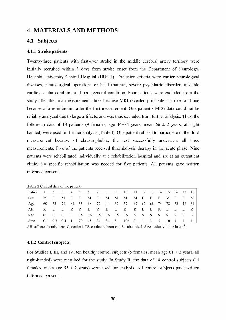
30
4 MATERIALS AND METHODS
4.1 Subjects
4.1.1 Stroke patients
Twenty-three patients with first-ever stroke in the middle cerebral artery territory were
initially recruited within 3 days from stroke onset from the Department of Neurology,
Helsinki University Central Hospital (HUCH). Exclusion criteria were earlier neurological
diseases, neurosurgical operations or head traumas, severe psychiatric disorder, unstable
cardiovascular condition and poor general condition. Four patients were excluded from the
study after the first measurement, three because MRI revealed prior silent strokes and one
because of a re-infarction after the first measurement. One patient’s MEG data could not be
reliably analyzed due to large artifacts, and was thus excluded from further analysis. Thus, the
follow-up data of 18 patients (9 females; age 44‒84 years, mean 66 ± 2 years; all right
handed) were used for further analysis (Table I). One patient refused to participate in the third
measurement because of claustrophobia; the rest successfully underwent all three
measurements. Five of the patients received thrombolysis therapy in the acute phase. Nine
patients were rehabilitated individually at a rehabilitation hospital and six at an outpatient
clinic. No specific rehabilitation was needed for five patients. All patients gave written
informed consent.
Table 1 Clinical data of the patients
Patient 1 2 3 4 5 6 7 8 9 10 11 12 13 14 15 16 17 18 Sex M F M F F M F M M M M F F F M F F M Age 60 72 74 84 55 68 72 44 62 57 67 67 68 74 78 72 48 61 AH R L L R R L R L L R R L L R L L L R Site C C C C CS CS CS CS CS CS S S S S S S S S Size 0.1 0.3 0.4 1 70 48 24 34 5 106 7 1 3 5 10 3 1 4
AH, affected hemisphere. C, cortical. CS, cortico-subcortical. S, subcortical. Size, lesion volume in cm3.
4.1.2 Control subjects
For Studies I, III, and IV, ten healthy control subjects (5 females, mean age 61 ± 2 years, all
right-handed) were recruited for the study. In Study II, the data of 18 control subjects (11
females, mean age 55 ± 2 years) were used for analysis. All control subjects gave written
informed consent.

Materials and methods
31
4.2 CLINICAL TESTING
The patients underwent clinical examination within 1 week (T0), at 1 month (T1), and at 3
months (T2) after stroke. A neurologist from the research team performed National Institutes
of Stroke Scale (NIHSS) scoring to evaluate stroke-related deficits, and Barthel Index (BI)
and modified Rankin scale (mRs) scoring to evaluate the patients’ ability to cope in daily life.
Tactile sensitivity (light and sharp touch) was categorized into two groups: normal or
decreased (as compared with the healthy hand). To evaluate hand motor function and fine
motor skills, a physio- or ergotherapist performed the Action Research Arm Test (ARAT;
Koh et al., 2006) and the 9-hole peg board test (Peg; Heller et al., 1987). In ARAT, gripping,
pinching, arm lifting, and picking objects is tested in four subtests. The performance is
evaluated from 0 to 57 points, with 57 being the best score. In Peg, nine pegs have to be
removed and replaced one at a time into nine holes as quickly as possible. The time needed to
finish the task is measured. In our studies, 120 s was set as the maximum time, and this value
was given if the task could not be performed faster.
4.3 STIMULATION
Balloon diaphragms driven by compressed air (Mertens and Lutkenhoner, 2000) were used to
deliver tactile stimuli (duration 141 ms, peak 50 ms) to the fingertips. The stimulus intensity
was kept constant for all patients and control subjects to allow exact comparisons of the
results during recovery. All subjects were able to detect the stimulus as light touch. The
patients wore ear plugs to avoid perception of any stimulus-related sound. The thumb and
little finger of both hands were stimulated alternately with an ISI of 1005 ms to define the
extent of the hand representation area in the SI cortex (Study I). To evaluate changes in the
activation of the somatosensory network (Study II) and the effect of afferent input on motor
cortex excitability (Study III), both index fingers were stimulated alternately with an ISI of
3005 ms.
4.4 MAGNETOENCEPHALOGRAPHIC RECORDINGS
The MEG recordings were performed with a 306-channel helmet-shaped neuromagnetometer
(Elekta Neuromag®, Helsinki, Finland) and carried out in the BioMag laboratory, HUCH, in
a magnetically-shielded room. Before the measurements, four indicator coils were placed on
the scalp. Magnetic signals produced by tiny currents fed into the coils were detected in the

32
beginning of each measurement to align the exact head position with respect to the sensor
array. During the recordings, the subjects were either in a sitting or supine position with the
head supported against the helmet-shaped sensor array. Raw-data and evoked responses (~120
responses to thumb and little finger stimulation of both hands in the first session and ~60
responses to stimulation of both index fingers in the second session) were recorded. In a third
session, spontaneous brain activity with eyes open and eyes closed was recorded for three
minutes each. Eye movements were monitored with a vertical electro-oculogram, and MEG
epochs that coincided with eye movements were automatically rejected from the data. The
patients were instructed to relax, to keep their head and fingers in constant position, and not to
pay any attention to the stimuli. A nurse inside the magnetically shielded room observed the
patients for their general condition and for any possible movements. The signals were band-
pass filtered between 0.03‒308 Hz and digitized at 941 Hz.
4.5 DATA ANALYSIS
4.5.1 Preprocessing of the data
To suppress artificial signals caused by interfering sources, the signal space separation
method (SSS; Taulu et al., 2004) or its temporal extension (tSSS; Taulu and Simola, 2006)
implemented in the MaxFilterTM software was applied. The SSS method efficiently suppresses
external magnetic artifacts from the measured data. The method relies on Maxwell equations
and divides the signals into components arising from inside the sensor array and from the
environment surrounding the sensor array (Taulu et al., 2004). SSS is a purely spatial method
and the interference rejection factor grows with increasing distance from the interference
source. The SSS method is not able to efficiently extract artifacts caused by sources lying
very close to the sensor array (e.g. dental work or other magnetic components in the body).
tSSS, however, also suppresses artifacts caused by nearby sources, in addition to distant
interference, by utilizing the temporal behavior of the signals (Taulu and Simola, 2006).
Similar temporal patterns in both internal and external SSS subspaces suggest that a nearby
artifact source has generated the field, and those signals are removed from the data. Earlier
studies have successfully applied both SSS and tSSS methods to clinical MEG data
(Nevalainen et al., 2008, Park et al., 2009, Tanaka et al., 2009). In Studies I and II, either the
SSS or tSSS method was applied, depending on the data quality, to preprocess the data of the

Materials and methods
33
patients and the control subjects. In Studies III and IV, all data were preprocessed with the
tSSS method.
In Study IV, a signal space projection (Uusitalo and Ilmoniemi, 1997) was applied to remove
the cardiac artifacts from the data. The signal space characteristic of cardiac artifacts was
estimated by averaging the MEG signals with respect to the magnetocardiographic signal,
applying principal component analysis to the average, and selecting the two components
associated with the highest singular values. These components were projected out from the
tSSS-processed continuous data.
4.5.2 Dipole modeling
To identify the sources of SEFs, equivalent current dipoles (ECDs) best describing the local
source currents were calculated with a least-squares method (Studies I and II). Deflections
exceeding the noise level in averaged signals were visually detected to divide the data into
time periods of interest. In each time period, the ECD best describing the most dominant
source was chosen using a subset of channels (10‒18) over the response area. The
calculations resulted in single dipoles, located three-dimensionally (x, y, z) in a spherical
conductor model. The goodness of fit (g) was calculated to determine in percentage how
much of the measured field variance the dipole accounted for. Only ECDs with a g > 85 % at
the selected time period were accepted. Single dipole fitting sufficiently explained the
activation in SI (Study I), and the source strengths of the responses were measured at the peak
amplitude of the single ECDs. In Study II, the number of ECDs found varied from 0 to 6.
After identifying single dipoles, the analysis period was extended to the entire time period of
the signals, and all channels were taken into account in computing a time-varying multidipole
model. The validity of the multidipole model was evaluated by comparing the measured
signals with responses predicted by the model. If signals of any brain region were left
inadequately explained by the model, the data were re-evaluated to find a more accurate
estimation for the generator areas. In Study II, the source strengths of the responses were
determined from the peak amplitudes of the ECDs in the multidipole model.
The Euclidean distance between the sources of the earliest responses to thumb and little finger
stimulation were calculated to determine the size of the hand representation area in the SI
cortex (Study I).
34
4.5.3 Temporal-spectral-evolution method (TSE)
The temporal-spectral-evolution (TSE) method (Salmelin and Hari, 1994b) gives a reliable
measure of the amount of event-related rhythmic activity with respect to time. The TSE
method was applied to evaluate stimulus-related changes in rhythmic beta activity (Study III).
Before TSE calculations, the dominant beta peaks were defined from amplitude spectra
calculated from spontaneous brain activity (eyes open), and the frequency range of strongest
modulation of beta activity was determined from time-frequency representations (TFRs;
Tallon-Baudry et al., 1997) in each subject. Thereafter, the averaged SEFs were subtracted
from the raw data, which were then band-pass filtered through an individually-chosen
frequency range. The frequency ranges were 10 Hz in width and between 12 and 26 Hz. Next,
the filtered signals were rectified and time-locked averaged to the stimulus. The analysis
period was 3.5 s with a pre-stimulus baseline of 300 ms. Event-related modulation
(suppression and rebound) of rhythmic beta activity was quantified using 2–4 MEG channels
(1–2 channels over each hemisphere) showing the strongest modulation. The absolute
suppression/rebound values, determined from the peak amplitude of deflection, were
converted into relative values by calculating the increase/decrease of the rhythm with respect
to the pre-stimulus baseline.
4.5.4 Analysis of spontaneous brain activity
4.5.4.1 Spectral analysis
To determine the amount of rhythmic brain activity in different frequency ranges, amplitude
spectra were calculated (separately for eyes open and eyes closed) by applying Fast Fourier
Transformations (FFTs) with a sliding window and 50 % overlap (Studies III and IV).
Windowing minimizes the spectral leakage. To evaluate the amplitudes of rhythmic brain
activity in the 5‒90 Hz range, a flat-top window of 2048 samples giving a frequency
resolution of ~0.5 Hz was used, and spectral peaks were quantified from the channels
showing strongest deflections. A flat-top window was chosen as it gives accurate amplitude
estimation, given that amplitude strengths were of particular interest. To search for potential
pathological low-frequency oscillations in the <4 Hz range (Study I), a Hanning window of
8192 samples yielding a frequency resolution of ~0.1 Hz was applied. A Hanning window

Materials and methods
35
gives a better frequency separation than the flat-top window, which is especially important in
identifying low-frequency oscillations.
4.5.4.2 Minimum current estimate in the frequency domain (fdMCE)
The sources of spontaneous oscillatory activity were localized using L1-norm based
frequency domain minimum current estimation (fdMCE; Jensen and Vanni, 2002). As in
spectral estimation, in fdMCE, the data are windowed into subsections of time and Fourier
transforms are calculated for each window. To identify sources of ~10-Hz oscillations, an
individual frequency range around the ~10-Hz peak, detected in the spectra, was defined for
each subject and Hanning-windowed, half-overlapping FFTs with a 0.46 Hz frequency
resolution were computed across the recording. To identify sources of pathological low-
frequency activity, the same procedure was applied with a 0.23 Hz frequency resolution for a
<4 Hz range. Thereafter, source localizations were estimated using L1 minimum norm
estimation for the transformed data. A boundary element model (BEM) of a standard brain
was used to restrict the search volume to the brain and a spherical conductor model was used
for forward computations. The source locations were projected to the BEM surface and the
strength of oscillatory activity was defined over each hemisphere over a standard (~10-Hz
oscillations) and an individually-selected (low-frequency oscillations) region of interest
(ROI).
4.5.5 Statistical analysis
Repeated measures ANOVAs with within subjects factors time (T0, T1, T2) and hemisphere
(affected, unaffected) were used to analyze the differences in the MEG parameters and in the
hand function tests. When a significant main effect or an interaction between time and
hemisphere was detected, pair-wise comparisons with paired t-tests were performed between
different time points or between hemispheres. Bonferroni correction was used to control for
multiple comparisons. Parameters between patients and control subjects were compared with
independent samples t-tests. Correlations were evaluated with Spearman’s correlation
coefficient.
36
5 EXPERIMENTS
5.1 SI REORGANIZATION AFTER STROKE (STUDY I)
Studies in humans and animals have shown that movement or somatosensory stimulation
related activation areas in the cerebral cortex are enlarged after stroke (Nudo et al., 1996b,
Rossini et al., 2001, Rossini et al., 1998b, Ward et al., 2003a, Ward et al., 2003b). In this
study, we investigated the temporal evolution of plastic reorganization in the SI cortex in 15
first-ever acute stroke patients (3 patients were excluded because of technical problems in the
MEG measurements, preventing reliable analysis of the data) and correlated the changes with
recovery of hand function. Follow-up measurements were performed within 1 week (T0), 1
month (T1), and 3 months (T2) after stroke and the MEG findings were compared with
findings of the healthy control subjects.
5.1.1 Results
The patients recovered well: the affected hand function in the 9-hole peg board test (Peg) and
in the ARAT was significantly improved at T1 and T2 as compared with T0 (57 ± 10 s and 45
± 9 s vs. 79 ± 10 s, p < 0.05 for Peg; and 49 ± 4 and 50 ± 5 vs. 37 ± 5, p < 0.005 for ARAT).
At T2, the affected hand function did not significantly differ from the healthy hand function
(45 ± 9 s vs. 26 ± 2, p = 0.08 for Peg; and 50 ± 5 vs. 57 ± 0, p = 0.15 for ARAT). At T1, the
size of the hand representation area in the SI cortex in the affected hemisphere (AH) was
enlarged as compared with T0 or T2 (12.6 ± 0.8 mm vs. 9.6 ± 0.8 mm and 10.2 ± 0.8 mm,
respectively, p < 0.05; Figure 5.1.1). Moreover, the SI hand representation size in the AH at
T1 was significantly larger than in the unaffected hemisphere (UH) and in the control subjects
(12.6 ± 0.8 mm vs. 10.8 ± 0.8 mm and 9.5 ± 0.6 mm, respectively, p < 0.005).

Experiments
37
Fig. 5.1.1 Mean (+SEM) size of the hand representation in the SI cortex in all three measurements of the patients (n = 15 for T0 and T1, and n = 14 for T2) and in the control subjects (n = 10; right and left hand pooled; *p < 0.05, **p < 0.005).
At whole group level, no correlation between the enlargement of the SI hand representation
and hand function was found. However, in patients with subcortical infarction (n = 8) the
increase in the SI hand representation during the first month after stroke correlated strongly
with impairment of hand function (rs = 0.8, p < 0.01; Figure 5.1.2).
Fig. 5.1.2 Correlation between the increase in the size of the SI hand representation during the first month after stroke (Δ hand representation = size of the hand representation at T1 – size of the representation at T0) and the results of the Peg test at T1 in subcortical stroke patients. The Peg test is evaluated in seconds needed for the task; the shorter the time, the better the hand function. Regression line is shown in black.
5.1.2 Discussion
In the patients, the SI hand representation in the AH was transiently enlarged 1 month after
stroke and returned to normal size at 3 months, concomitantly with recovery of hand

38
dexterity. Earlier MEG studies have indicated a tendency towards a larger hand representation
in the AH than in the UH at 9 weeks to 6 months after stroke, in association with incomplete
recovery of hand function (Rossini et al., 2001, Rossini et al., 1998b). In the present study, an
initial enlargement of the hand representation was also observed in patients who recovered
well later on. The size of the hand representation returned to normal size in parallel with
recovery of hand function.
Studies in animals have indicated that cortical plasticity is learning-dependent and not simply
use-dependent. In monkeys who were trained for a new task, cortical map changes in motor
representations were observed in parallel with improvement of performance (Nudo et al.,
1996a). In contrast, no such changes were observed in monkeys who just repeated a simple
task that they performed optimally from the beginning (Plautz et al., 2000). In line with these
findings, enlarged cortical representation of the affected hand in MI was observed in chronic
stroke patients during constraint-induced movement therapy in parallel with clinical
improvement of hand function. After this treatment, the cortical representation of the hand
returned to normal, whereas the motor performance remained good (Liepert et al., 2000a).
We suggest that the transient enlargement of the SI hand representation in our stroke patients
may reflect the re-learning of motor skills, whereas the normalization of the representation
size may reflect the maintenance of re-learned function.
5.2 SII ACTIVATION AFTER STROKE
Fluent motor function requires continuous inflow of somatosensory input to the motor system.
In this study, we recorded SEFs to tactile index finger stimulation within 1 week (T0), 1
month (T1), and 3 months (T2) after stroke with concomitant evaluation of clinical recovery
(NIHSS, BI, mRS, ARAT, Peg) to study the effect of altered activation in the somatosensory
cortical network on motor recovery after stroke. Eighteen first-ever stroke patients and 18
healthy control subjects were enrolled in the study.
5.2.1 Results
The patients recovered well, and all clinical parameters improved significantly from T0 to T1
and T2 (p < 0.005; Table 5.2.1; Figure 5.2.1).

Experiments
39
Table 5.2.1 Clinical scores of the patients (Peg, ARAT, mean ± SEM; NIHSS, BI, mRs, median ± SEM)
T0, within 1-7 days; T1, 1month; T2, 3 months from stroke onset. ah, affected hand. uh, unaffected hand. Peg, 9-hole peg board test, time (s). ARAT, Action Research Arm Test (0–57). NIHSS, National Institutes of Health Stroke Scale (0–42). BI, Barthel Index (0–100). mRS, modified Rankin Scale (0-6).
Fig.5.2.1 Mean (+ SEM) Peg times and ARAT scores of the patients (n = 18 for T0 and T1, and n = 17 for T2) at T0, T1, and T2. ah, affected hand. hh, healthy hand. (*p < 0.05, **p < 0.005).
In the control subjects, tactile stimulation elicited responses in contralateral SI at 58 ± 1 ms
and in bilateral SII regions at 109 ± 6 ms and 118 ± 4 ms (contra- and ipsilateral SII,
respectively). At T0, SI responses were found in 15 patients and contralateral SII (cSII)
responses in 9 patients. Latencies of the responses were comparable with latencies of the
control subjects. At T1 and T2, SI responses were detected in all patients and cSII responses in
17 patients. When an SII response was detected, it was always preceded by an SI response. In
the patients, the SI responses were significantly stronger in the UH than in the AH at T0 and
T1. No time effect was observed for the SI responses. In contrast, the cSII response in the AH
Peg (ah) Peg (uh) ARAT(ah) ARAT(uh) NIHSS BI mRS
T0 84 9 36 4 35 5 56 0 4 1 60 7 3 0
T1 59 10 28 1 46 5 57 0 2 0 90 4 2 0
T2 51 9 26 1 48 4 57 0 1 0 100 3 2 0

40
was significantly weaker at T0 than at T1 or T2 (14 ± 4 nAm vs.25 ± 5 nAm and 26 ± 4 nAm,
respectively, p < 0.05; Figure 5.2.2). Moreover, the cSII response in the AH at T0 tended to be
weaker than in the UH (14 ± 4 nAm vs. 25 ± 6 nAm, p = 0.06) and was significantly weaker
than in the control subjects (14 ± 4 nAm vs. 29 ± 2 nAm, p < 0.01). No significant differences
between the hemispheres were observed in the ipsilateral SII responses.
Fig 5.2.2 Mean (+SEM) amplitudes (nAm) of SI and cSII responses in the patients (n = 18 for T0 and T1, and n = 17 for T2) in the affected hemisphere (AH) and unaffected hemisphere (UH) to contralateral tactile finger stimulation. The mean (+SEM) amplitudes of the control subjects (n = 18; right and left hand pooled) are shown with dashed and grey horizontal lines (**p < 0.005).
The amplitudes of the SII responses correlated with results of the Peg test at all time points (rs
= -0.6, p < 0.01 for T0; rs = -0.5, p < 0.05 for T1, and rs = -0.6, p < 0.05 for T2; Figure 5.2.3). In
contrast, SI responses did not correlate with results of the Peg test at any time point.
Fig. 5.2.3 Correlation between the strength of SII activation and Peg time (s) of the affected hand at 1 week (T0), 1month (T1), and 3 months (T2) from stroke. Regression line is in black.

Experiments
41
5.2.2 Discussion
In the patients, the strength of SII activation was decreased in the AH compared with the UH
and the control subjects in the acute phase and increased during follow-up. The strength of SI
activation was also decreased in the AH compared with UH, but no significant increase of SI
activation was observed during follow-up. The strength of SII activation, but not SI
activation, in the AH correlated with results of hand function tests both in the acute phase and
during recovery, indicating that the SII region is important in hand sensorimotor functions.
Earlier studies on somatosensory activation after stroke have shown varying correlations
between the strength of SI activation and clinical outcome (Oliviero et al., 2004, Rossini et
al., 2001, Wikstrom et al., 2000). In healthy subjects, the SI amplitude has been shown to
reflect tactile sensitivity and stimulus properties like intensity (Hari et al., 1993, Huttunen et
al., 1987, Tiihonen et al., 1989). In line with these findings, a follow-up study in stroke
patients showed improved 2-point discrimination in patients with increase of SI responses
during follow-up (Wikstrom et al., 2000). Alterations in activation of the SII region,
suggested to be involved in higher-order somatosensory processing (Jousmaki and Forss,
1998, Lin et al., 2003, Simoes and Hari, 1999, Disbrow et al., 2000), have so far not been
studied with whole-head MEG.
Somatosensory input is crucial for fluent motor functions. Continuous inflow of
somatosensory input to the motor system is needed to adjust the speed, range, and strength of
movements. Despite some direct afferent thalamic connections to MI (Asanuma et al., 1979),
somatosensory input to the motor cortex is suggested to be mediated mainly via cortico-
cortical connections (Chen et al., 1999, Disbrow et al., 2000, Hinkley et al., 2007).
Anatomical studies have shown strong connections from SII to MI (Jones and Wise, 1977,
Mori et al., 1989), whereas connections from the main cutaneous area 3b in SI to MI have
been shown to be sparse (Jones et al., 1978). A close interaction between SII activation and
motor functions have also been shown in functional imaging studies in humans. Navigated
TMS of the SII region facilitates motor performance in healthy subjects (Raij et al., 2008). In
addition, clinical studies have shown an association between SII activation and motor
functions. For instance, Unverricht-Lundborg type progressive myoclonus epilepsy patients
with absent SII activation had more severe motor symptoms than the patients in whom SII
activation was observed (Forss et al., 2001).

42
We suggest that the deficient SII activation in our acute stroke patients reflects insufficient
flow of somatosensory input to the motor cortex, which results in impaired sensorimotor
integration. During follow-up, activation in SII paralleled motor recovery of hand function,
supporting the view that the SII region is an important node in sensorimotor integration.
5.3 MOTOR CORTEX EXCITABILITY AFTER STROKE
Motor cortex excitability has been shown to be altered after stroke (Butefisch et al., 2003,
Liepert et al., 2000b, Manganotti et al., 2002). In addition to cortical excitatory and inhibitory
circuits, afferent input has also been suggested to modulate motor cortex excitability
(Asanuma and Arissian, 1984, Favorov et al., 1988). The effect of afferent input on motor
cortex excitability can be studied by monitoring the reactivity of the motor cortex beta rhythm
to somatosensory stimulation, which elicits an initial suppression followed by a subsequent
rebound of the rhythm. The rebound of the rhythm is suggested to reflect decreased motor
cortex excitability (Chen et al., 1999, Franzkowiak et al., 2010, Gaetz et al., 2011, Salmelin et
al., 1995). In this study, we monitored the reactivity of the beta rhythm to tactile index finger
stimulation in 18 patients after acute stroke (T0) and during recovery (T1 and T2), as well as in
10 healthy control subjects. The rebound of the beta rhythm was correlated with hand function
as evaluated with the Peg test. Moreover, the rebound was correlated with the strength of the
SEFs (adapted from Study II) to determine how alterations in motor cortex rhythms are
modulated by changes in afferent input.
5.3.1 Results
In the control subjects, the beta rhythm started to decrease at 120 ± 15 ms after stimulus and
reached its peak at 250 ± 15 ms in both hemispheres. The subsequent rebound started at 550 ±
35 ms after stimulus and peaked at 900 ± 85 ms. Comparable latencies of suppression and
rebound were found in both hemispheres in the patients.
In the patients, the beta rebound was weaker at T0 than at T1 or T2 in both the AH and UH (p
< 0.01 for the AH, p < 0.05 for the UH; Table 5.3.1.; Figures 5.3.1 and 5.3.2). Moreover, the
beta rebound in the AH at T0 was significantly weaker than that in the control subjects (p <

Experiments
43
0.05). Within patients, the beta rebound in the AH was significantly weaker than in the UH at
all time points (p < 0.005 for T0; p < 0.001 for T1; p < 0.05 for T2).
Table 5.3.1 SEF amplitudes (mean ± SEM) to index finger tactile stimulation of the affected hand (adapted from Study II), and beta rebound (increase of the rhythm with respect to the reference baseline; mean ± SEM) in patients and in control subjects
T0, 1-7 days; T1, 1 month; T2, 3 months after stroke. Ctrl.; control subjects (left and right hands pooled). SI, primary somatosensory cortex. SII, secondary somatosensory cortex. AH, affected hemisphere. UH, unaffected hemisphere.
Fig. 5.3.1 Average strength of the beta rhythm (15–25 Hz) over the sensorimotor region in the affected (AH) and unaffected (UH) hemispheres to contralateral tactile index finger stimulation within 1 week (T0), 1month (T1), and 3 months (T2) after stroke and in the control subjects (right and left hemispheres pooled). 0 indicates the onset of the tactile stimulus.
SI ampl, AH (nAm) SII ampl, AH(nAm) Rebound, AH (%) Rebound, UH (%)
T0 17 3 14 4 22 7 43 7
T1 22 3 25 5 43 11 68 9
T2 23 3 26 4 37 9 57 7
Ctrl. 25 3 31 3 61 11 61 11

44
Fig.5.3.2 Mean (+SEM) strength of the beta rebound over the sensorimotor region in the affected and unaffected hemispheres to contralateral tactile index finger stimulation at 1 week (T0), 1month (T1), and 3 months (T2) from stroke and in the control subjects (right and left hemispheres pooled; *p< 0.05, **p < 0.005). No association between absent/diminished rebound and decreased tactile sensitivity was
observed. The strength of the beta rebound in the AH correlated with Peg results of the
affected hand in all three measurements (nonlinear regression rs = -0.8, p < 0.001 for T0; rs =
-0.5, p < 0.05 for T1; and rs = -0.6, p < 0.05 for T2; Figure 5.3.3).
Fig. 5.3.3 Association between the strength of beta rebound in the affected hemisphere and the Peg time (s) of the affected hand at 1 week (T0), 1month (T1), and 3 months (T2) from stroke. Nonlinear (x2) regression curve is shown in black. At T0, the strength of SII activation correlated with the strength of the beta rebound (rs = 0.5,
p < 0.05), whereas no correlation between SI activation and beta rebound was found.

Experiments
45
5.3.2 Discussion
In the patients, the beta rebound was decreased in the AH at the acute phase after stroke,
suggesting increased motor cortex excitability after acute stroke. The beta rebound increased
during follow-up, suggesting decreasing motor cortex excitability during stroke recovery.
These findings are in line with a recent TMS study suggesting reduced short-latency afferent
inhibition in the AH after acute stroke (Di Lazzaro et al., 2012). The strength of beta rebound
correlated with hand function test results at the acute phase and during recovery, suggesting
that increased excitability is associated with poor control of hand function, and that a return
towards normal excitability is associated with good recovery of hand function. These findings
are in line with earlier studies, showing increased motor cortex excitability in association with
impaired fine motor skills in patients suffering from Unverricht-Lundborg Type Myoclonus
epilepsy or complex regional pain syndrome (Canafoglia et al., 2004, Juottonen et al., 2002,
Schwenkreis et al., 2003, Silen et al., 2000). Accordingly, several studies in stroke patients,
applying different measures of motor cortex excitability, have shown that motor cortex
excitability is increased in the acute phase, but returns to normal if the patients recover well
(Calautti et al., 2001, Swayne et al., 2008, Ward et al., 2003a).
As discussed in Study II, anatomical studies in animals (Jones and Wise, 1977, Mori et al.,
1989) and functional studies in humans (Disbrow et al., 2000, Hinkley et al., 2007) have
suggested that the SII region may be important in integrating somatosensory information with
motor functions. In our study, the strength of the beta rebound correlated with the strength of
SII but not with SI activation in the acute phase after stroke. This finding supports the earlier
suggestions that the modulatory afferent input to motor cortex excitability may be mediated
via SII.
In principle, all observed changes of motor cortex excitability could be solely due to recovery
of somatosensory afferents. However, in the light of our results, this possibility is not likely.
SI amplitudes increase almost linearly with increasing stimulus intensity (Jousmaki and Forss,
1998, Torquati et al., 2002). In our patients, the stimulus intensity was kept constant across all
measurements; hence enhanced afferent input due to recovery of somatosensory afferents
would elicit increased SI amplitudes. However, no significant changes in SI amplitudes in our
stroke patients were observed (Study II). Neither was there a clear relationship between
absent/diminished rebound and decreased tactile sensitivity. We suggest that the changes in
the beta rebound result from recovery of both the somatosensory and the motor systems. The

46
parallel recovery of the sensory and motor systems allows a fluent sensorimotor integration,
which is a prerequisite for normal hand dexterity.
5.4 SPONTANEOUS BRAIN OSCILLATIONS AFTER STROKE
Alterations in the frequency and amplitude of spontaneous rhythmic brain activity have been
associated with pathological phenomena in the brain (Lewine et al., 1999, Pfurtscheller et al.,
1981, Van Huffelen et al., 1984) and have even been suggested to predict recovery from
stroke (Tecchio et al., 2007). In this study, we recorded spontaneous brain activity in 16 first-
ever stroke patients within 1 week (T0), 1 month (T1), and 3 months (T2) after stroke and in
ten healthy control subjects, to find out how alterations in spontaneous brain oscillations are
associated with functional recovery after stroke.
5.4.1 Results
The main sources of the ~10-Hz oscillations in the eyes-open condition were detected in the
temporo-parietal region, clearly distinct from occipital alpha sources (Fig. 5.4.1), but slightly
lateral to the typical location of rolandic ~10-Hz oscillations.
Fig 5.4.1 Averages of sources of ~10-Hz oscillations (eyes open) in the patients with lefthemispheric stroke (arbitrary scale). The sources are clearly stronger in the affected hemisphere.
In the patients, the source strength of ~10-Hz oscillations (eyes open) in the AH was
significantly stronger than in the UH at T1 and at T2 (2.6 ± 0.3 nAm vs. 1.6 ± nAm, p < 0.05
and 2.5 ± 0.3 nAm vs. 1.7 ± 0.2 nAm, p < 0.05, respectively; Figure 5.4.2). The strength of
~10-Hz oscillations in the AH at T1 and T2 had a tendency to be stronger than in the control
subjects (2.6 ± 0.3 nAm vs. 1.9 ± 0.2 nAm, p = 0.08 and 2.5 ± 0.3 nAm vs. 1.9 ±0.2 nAm, p =
0.10, respectively). In contrast, no differences in the strength of the ~10-Hz oscillations for

Experiments
47
the eyes-closed conditions or the rolandic beta rhythm were detected between the hemispheres
or between patients and control subjects. The amplitude of the ~10-Hz sources seemed to
increase in bursts, showing strong variation in source strength from window to window used
in FFT estimation. As fdMCE calculates the mean strength of all the FFT-windows, the
absolute values for the ~10-Hz sources are rather small.
Fig. 5.4.2 Mean (+SEM) source strength of ~10-Hz oscillations (eyes open) over the affected hemisphere (AH) and unaffected hemisphere (UH) of the patients and in the control subjects (right and left hemispheres pooled, *p < 0.05). T0, within 1 week; T1, 1month, and T2, 3 months after stroke.
Pathological low-frequency oscillations at <4 Hz with strongest amplitudes on average at 1 ±
0.1 Hz were detected in 7/16 patients at T0, in 6 patients at T1, and in 4 patients at T2. In
patients with cortical involvement of the stroke, the sources of low-frequency oscillations
were detected in the surrounding of the cortical lesion (Figure 5.4.3). The patients with low-
frequency activity had a significantly larger lesion than the rest of the patients (35 ± 15 vs. 2 ±
1 cm3, p < 0.05). The 4 patients showing persistence of low-frequency activity at T2 had a
worse clinical outcome than the rest of the patients (88 ± 21 vs. 33 ± 3, p < 0.001 for Peg; 3 ±
1 vs.1 ± 0, p < 0.05 for NIHSS).

48
Fig. 5.4.3 A) Source location of pathological low-frequency activity within 1 week (T0), 1month (T1), and 3 months (T2) after stroke (arbitrary scale). B) MRIs from the same patient; the arrow points to the cortical lesion; the ellipsoids illustrate the generator area of pathological low-frequency oscillations.
5.4.2 Discussion
In the patients, the strength of ~10-Hz oscillations was increased for the eyes-open condition
in the AH at T1 and T2 as compared with the UH. In contrast, no differences between the
hemispheres were observed for the eyes-closed condition. The source locations were distinct
from those of occipital alpha sources, but slightly lateral to the typical location of rolandic
~10-Hz sources. In addition to occipital and rolandic ~10-Hz oscillations, sources of rhythmic
activity in 7–10 Hz range have been detected in the temporal-lobe (tau rhythm; Tiihonen et
al., 1991) and in the parietal operculum (sigma rhythm; Narici et al., 2001). The source
location of the prominent ~10-Hz oscillations detected in our patients could agree with the
location of the sigma rhythm, found in the SII region. However, a contribution of rolandic
~10-Hz oscillations to these sources cannot be excluded. Although the exact generator areas
for the ~10-Hz oscillations cannot be determined, the differences between the eyes-open and
eyes-closed conditions indicate that the occipital alpha rhythm does not contribute to this
rhythm. Rather we suggest that the enhanced ~10-Hz oscillations detected in the AH of our
stroke patients reflects a somatosensory rhythm, with possible contribution of both the
rolandic and the SII regions.

Experiments
49
The sigma rhythm has not been extensively studied, and to our knowledge no earlier reports
of alterations of this rhythm in stroke patients exist. Earlier observations of alterations in the
strength of rolandic ~10-Hz oscillations in stroke patients have been controversial, as both
increases and decreases of the oscillatory amplitudes have been observed (Pfurtscheller et al.,
1981, Tecchio et al., 2006, Tecchio et al., 2005, Van Huffelen et al., 1984). One previous
study found that in patients with mild or moderate deficits, rolandic ~10-Hz oscillations
increased in the acute phase after stroke. In patients with severe neurological deficits, this
increment was observed months later in association with slow clinical recovery (Pfurtscheller
et al., 1981). In the present study, no clear association between the strength of ~10-Hz
oscillations and clinical outcome was observed. However, the steepest improvement in
clinical function was observed from T0 to T1, and the ipsilesional increase in ~10-Hz
oscillation amplitudes was observed at T1.
In healthy subjects, both rolandic and occipital ~10-Hz oscillations have been associated with
cognitive processing (Llinas and Ribary, 1993, Jensen et al., 2002, Haegens et al., 2010, Palva
et al., 2005), and particularly in directing the flow of information to optimize performance
(Haegens et al., 2012, Jensen and Mazaheri, 2010). In light of these findings, it may be that
the enhancement of ~10-Hz activity in the AH observed in our stroke patients could possibly
be engaged in allocating resources for recovery mechanisms. However, this remains
hypothetical, and future studies are needed to understand the functional significance of
enhanced ~10-Hz oscillations in the AH after stroke.
Perilesional low-frequency oscillations have been detected in earlier stroke studies (Butz et
al., 2004, Fernandez-Bouzas et al., 2000, Vieth, 1990), but the functional significance has
remained unclear. In the present study, low-frequency oscillations could be detected in 7
patients in the acute phase, and in 4 patients, low-frequency activity still persisted 3 months
after stroke. In a rat study, pathological low-frequency oscillations after thermal ischemic
lesions were strongly correlated with axonal sprouting, suggesting that low-frequency
oscillations may have a role in plastic reorganization after brain lesion (Carmichael and
Chesselet, 2002). On the other hand, a study combining MEG and proton magnetic resonance
spectroscopic imaging, suggested that pathological low-frequency oscillations are associated
with abnormal metabolic activity in malfunctioning perilesional neurons (Kamada et al.,
1997). In the present study, the patients with low-frequency activity had larger lesions and the

50
patients with persistent low-frequency activity had a worse clinical outcome at T2 than the rest
of the patients. However, as the number of patients with persistent low-frequency activity was
small, and the follow-up period was only 3 months, no definitive conclusions about the
clinical significance of low-frequency oscillations can be drawn.
An earlier MEG study suggested that alterations in spontaneous brain oscillations may be
used to predict the outcome from stroke; particularly delta (2–3.5 Hz) power in the UH and
gamma (33.5–44 Hz) power in the AH were found to predict long-term outcome after stroke
(Tecchio et al., 2007). We did not find clear UH delta or AH gamma peaks in our patients.
Pathological low-frequency oscillations were detected in less than half of our patients and the
presence of low-frequency oscillations significantly correlated with lesion size, thus this
parameter is not likely to provide any additional information in predicting outcome after
stroke. In contrast, ~10-Hz activity is a very robust signal, which is detected practically in
every subject; and enhancement of AH ~10-Hz activity was detected at some time point in
13/16 of our well-recovering patients. Thus enhanced ~10-Hz activity may have functional
significance in recovery from stroke. Future studies should aim at investigating if enhanced
~10-Hz activity could be used to predict the outcome from stroke.

General discussion
51
6 GENERAL DISCUSSION
In this thesis, different levels of adaptive alterations in the sensorimotor system after acute
stroke were studied in a longitudinal design. The aim was to elucidate the temporal evolution
of plastic changes after acute stroke and their correlation to clinical recovery in order to better
understand the recovery mechanisms of the injured human brain.
6.1 Temporal evolution of plastic changes after stroke
Although the brain alters its structure and function throughout life, injury to the central
nervous system seems to be a special trigger that elicits plastic mechanisms (Nudo, 2006).
Focal brain injury results in a variety of time-dependent metabolic and pathophysiological
reactions which enable widespread cortical regions to change structure and function.
Although there is a wide range of studies investigating plastic changes such as changes in
cortical somatosensory and motor representations (Calautti et al., 2001, Rossini et al., 1998a,
Rossini et al., 2001, Ward et al., 2003a, Ward et al., 2003b) or excitability (Butefisch et al.,
2003, Liepert et al., 2000b, Manganotti et al., 2002, Ward and Cohen, 2004) after stroke,
longitudinal data recording the evolution of these changes are rare. A study in monkeys
indicated that, if motor rehabilitation was delayed over 1 month after stroke, the spared hand
representation areas within the peri-infarct area of the motor cortex were not maintained,
although the monkeys achieved recovery of hand function after delayed training (Barbay et
al., 2006). In contrast, if monkeys obtained early post-infarct rehabilitation the hand
representation areas in the peri-infarct zone were maintained or even enlarged (Nudo et al.,
1996b). These results suggest that, during early rehabilitative training, the cerebral cortex may
be more capable of reorganizing, whereas in chronic stages other compensatory mechanisms
may emerge.
In line with the animal studies, in our Studies I, II, and III, all significant changes in brain
activation were observed within the first month after stroke: the size of the hand
representation in the SI cortex in the AH (Study I), the strength of cSII activation in response
to affected hand stimulation (Study II), and the strength of the beta rebound in the AH (Study
III) all increased significantly from T0 to T1. Also the increase in ~10-Hz oscillations was
observed at T1. The steepest improvement in clinical function was observed in parallel with
the detected functional changes.

52
We did not control our patients for any specific rehabilitative treatment. All patients were
treated in the Department of Neurology, Helsinki University Central Hospital in the acute
phase, usually for 1 to 2 weeks, and afterwards the patients received specific treatment
according to their individual needs. Thus, we cannot distinguish between the effects of
rehabilitative training and spontaneous recovery on neurophysiological changes and clinical
improvement. Clinical experience has shown that further recovery still occurs after 1 month
from stroke, but changes are more subtle. In line with these findings, clinical improvement
was still observed from T1 to T2 in our studies, although no significant changes in the
measured neurophysiological parameters were observed in that time period. It may be that
within the first month after stroke, the cerebral cortex is particularly responsive to plastic
changes, and after that time window, other compensatory mechanisms take over to yield
functional recovery. This finding would encourage clinicians to force the initiation of
effective rehabilitative training as early as possible. However, it is also possible that we did
not observe further significant changes in neurophysiological parameters after 1 month
because most of our patients had already recovered well. Future follow-up studies with more
severe stroke patients should aim at investigating whether there is a time window for plastic
changes after stroke, and whether it would be beneficial to extend this time window to
improve clinical outcome.
6.2 Motor cortex excitability after stroke
Animal studies have indicated increased excitability after central ischemic lesions in cortical
areas both in the AH and UH. These changes have been observed both after
photothrombotically-induced lesions (Buchkremer-Ratzmann and Witte, 1997, Domann et al.,
1993) and after middle cerebral artery occlusion (Reinecke et al., 1999), suggesting that
changes in excitability are independent of the lesion mechanism.
In humans, most of the earlier studies on motor cortex excitability after stroke have been
performed with TMS and intracortical inhibition (ICI) and intracortical facilitation (ICF)
paradigms (Liepert et al., 2005, Liepert et al., 2000b, Manganotti et al., 2002, Swayne et al.,
2008). There are only a few studies using TMS after conditioning somatosensory stimulation
to detect changes in motor cortex excitability after stroke (Di Lazzaro et al., 2012, Oliviero et
al., 2005). Changes in motor cortex excitability due to alterations in afferent input are most

General discussion
53
likely mediated by different circuits than those mediating ICI or ICF (Sailer et al., 2002). A
recent TMS study evaluated both short-latency ICI (SICI) and short-latency afferent inhibition
(SAI) in stroke patients and found a correlation between SAI and clinical recovery, but not
between SICI and recovery. TMS recordings after somatosensory stimulation have shown, in
addition to SAI, long-latency inhibition at approximately 200 ms, called as “long-latency
afferent inhibition” (LAI; Chen et al., 1999, Sailer et al., 2002). LAI occurs bilaterally after
unilateral somatosensory stimulation and has been suggested to be predominantly mediated
via polysynaptic cortico-cortical projections (Abbruzzese et al., 2001, Chen et al., 1999, Sailer
et al., 2003). Both paradigms, LAI in TMS and beta rebound in MEG, use peripheral afferent
input to study changes in cortical excitability. In both methods, unilateral afferent stimulation
induces bilateral changes in cortical excitability at comparable long latencies. It is not known
if LAI and beta rebound share any common mechanisms, but such comparison would be an
interesting target for future investigations.
6.3 Cortical excitability and reorganization of the cerebral cortex
Earlier studies have suggested that altered cortical excitability is a prerequisite for cortical
reorganization after stroke. Jacobs and Donoghue (1991) suggested that expansions of motor
cortical representations are dependent on adjustments in local inhibition, and proposed that
inhibitory circuits are critically placed to unmask latent intracortical connections and to
readjust cortical motor presentations. However, representational changes are not restricted to
the motor cortex. Direct intracortical measurements in rats demonstrated an enlargement of
representational areas of the vibrissae in the somatosensory cortex in the neighbourhood of an
ischemic cortical lesion (Schiene et al., 1999). This enlargement was linked to decreased
intracortical inihibition (ICI), due to a reduction of GABAA receptors, which may lead to an
unmasking of pre-existing silent connections (Schiene et al., 1996). In line with these
findings, we found a transient expansion of the hand representation area in the SI cortex one
month after stroke (Study I).
In our patients, the strength of the beta rebound (measured in Study III) and the enlargement
of the SI hand representation (Study I) did not correlate. However, it has to be noted that the
beta rebound reflects the afferent modulation of motor cortex excitability. Changes in
afferent-input-modulated inhibition can occur independent of changes in local inhibitory
circuits, as was shown in an earlier TMS study combining afferent inhibition and ICI (Di

54
Lazzaro et al., 2012). In fact, it is not well known how different excitatory and inhibitory
systems interact together (Chen, 2004). An earlier TMS study investigating ICI after short-
latency (1‒10 ms) median nerve stimulation conditioning suggested that short-latency
afferent inhibition may reduce ICI (Ridding and Rothwell, 1999). However, the interaction
between long-latency afferent inhibition, which probably would resemble the beta rebound we
studied, and ICI is not well known. Clarifying the interaction of afferent and intracortical
inhibitory circuits would further help to understand the complex interplay between plastic
reorganization and different inhibitory and excitatory mechanisms.
.
6.4 Sensorimotor integration
Prior studies in monkeys and in humans have indicated that the SII cortex participates in
sensorimotor integration, especially in tasks involving multiple functionally-related body
parts like both hands or the digits of one hand (Disbrow et al., 2000, Forss et al., 2001,
Hinkley et al., 2007, Jones et al., 1978, Raij et al., 2008). However, the exact functional role
of SII in sensorimotor integration remains uncertain. In our Studies II and III, we suggest that
modulatory afferent input reaches the motor cortex via SII. Furthermore, we suggest that the
somatosensory input is integrated with motor functions by modulating the excitability of the
motor cortex.
Both, changes in motor cortex excitability and deficient SII activation, have been reported in
several disorders with motor impairment, such as Unverricht-Lundborg Type Epilepsy, focal
dystonia, and Parkinson disease (Abbruzzese et al., 2001, Boecker et al., 1999, Butterworth et
al., 2003, Forss et al., 2001, Sailer et al., 2003, Silen et al., 2000). However, the association
between motor cortex excitability and SII activation has not yet been directly studied. To our
knowledge, we are the first to suggest that the SII region may mediate motor functions by
modulating the motor cortex excitability.
SII activation and strength of beta rebound correlated at T0, but not at T1, or T2. The strength
of SII activation in the AH was normalized already at T1, whereas the beta rebound in the AH
was still weaker than in the UH still at T1 and T2. Changes in the strength of the beta rebound
are not likely to result solely from the integrity of the somatosensory inflow to the motor
cortex. In addition, the functional integrity of the motor cortex including local cortical
excitability influences is likely to affect the strength of the beta rebound. A faster recovery of
General discussion
55
the afferent somatosensory system than the motor system may explain the discrepancy in the
correlation between SII activation and beta rebound in the acute phase but not during follow-
up.
6.5 Future perspectives in monitoring recovery after stroke
Monitoring stroke recovery is complex, since the definition of successful outcome varies and
many physical and psychosocial aspects may affect neurological functions (Duncan et al.,
2000). Still, a precise definition of recovery is lacking, and so far, evaluation of the efficacy
of rehabilitation has mainly been based on a variety of clinical parameters. Although it is
known that early rehabilitation is a powerful tool to modulate recovery after stroke, early
extensive training may sometimes be maladaptive. A forced overuse of the affected forelimb
in rats after brain injury led to exaggeration of the neuronal injury (Kozlowski et al., 1996).
Hence, it is important to find parameters for objective monitoring of recovery from stroke and
to define optimal parameters for successful rehabilitation. Searching for such parameters was
one motivation of this thesis.
In light of our findings, one suitable parameter for monitoring recovery from stroke appears to
be the enlargement of the hand representation in the SI cortex. Animal studies have shown
that increased motor repetition alone is not sufficient to drive changes in cortical
representation maps, rather, it demands acquisition of new skills (Nudo et al., 1996a, Plautz et
al., 2000, Remple et al., 2001). In line with these findings, in a study with stroke patients
receiving constraint-induced movement therapy, a significant increase in the cortical
representation of the affected hand was observed in parallel with significant improvement of
motor functions. In follow-up measurements up to 6 months after treatment, the motor
performance had remained good, but the cortical representation had decreased and normalized
(Liepert et al., 2000a). Hence, the transient enlargement of cortical representations, also
observed in our study, may reflect the acquisition of new skills, and may thus provide an
objective parameter to monitor the sufficiency of rehabilitative training.
Our results clearly extend the earlier knowledge of sensorimotor recovery after stroke.
However, many issues still remain unresolved. Earlier studies have indicated that post-stroke
recovery mechanisms in cortical versus subcortical strokes may differ (Ameli et al., 2009,
Liepert et al., 2005). The correlation between the enlargement of SI hand representation and
hand motor function in patients with subcortical stroke but not in patients with cortical lesions

56
(Study I) was in accordance with this idea. However, as the number of patients with pure
cortical strokes was small, no definitive conclusions about the differences in the recovery
mechanisms between different stroke sites can be drawn from our data. Future studies should
aim at comparing neurophysiological changes after cortical and subcortical strokes.
All the changes in neurophysiological parameters studied in this thesis were observed within
the first month after stroke, indicating that the brain’s maximum responsiveness to plastic
changes may be restricted to a relatively short time window. However, all our patients
recovered relatively well, thus it may also be that no further changes were observed because
of the relatively good clinical outcome 1 month after stroke. Another possibility is that the
steepest changes in neurophysiological parameters occur within 1 month and that the
evolution of changes decelerates from then on. Hence, 1 month and 3 months would be too
close in time to observe any significant changes. Future studies should aim at including more
severe stroke patients in follow-up measurements, and follow-up measurements should be
performed at least over 6 months or even a year to obtain better understanding of the brain’s
capability for plastic changes beyond the subacute phase.
We observed changes in various cortical neurophysiological parameters that were associated
with clinical recovery after stroke. It may be meaningful to study the effect of therapeutic
interventions such as repetitive TMS or pharmacological treatment on these parameters to
evaluate whether the time window for these alterations can be extended, and whether it has a
positive influence on recovery. Such knowledge may further help to define the optimal
parameters for successful rehabilitation and to allow better targeting of therapeutic
interventions.
.

Summary & Conclusions
57
7 SUMMARY & CONCLUSIONS
In this thesis, plastic changes related to hand sensorimotor functions were studied in 18 acute
stroke patients in a longitudinal design. The main results shed light on alterations in the
central sensorimotor mechanisms important for fluent hand function. The observed transient
enlargement and subsequent normalization of the SI hand representation in our stroke patients
(Study I) strengthens the earlier observations of differences in learning-dependent and use-
dependent plasticity. This finding underlines the importance of sufficient challenge during
rehabilitation in order to provoke plastic reorganization in the brain.
The observation that patients with persisting perilesional low-frequency oscillations have a
worse clinical outcome than the other patients (Study IV) may indicate that this phenomenon
is an indicator of more severe brain lesions. On the other hand it may also be a signal of
ongoing plastic reorganization and thus longer follow-up times would be needed to elucidate
the clinical significance of perilesional low-frequency oscillations.
Our observations in Studies II and III add important knowledge to the role of afferent input in
motor functions. The observed changes in cSII but not in SI activation in parallel with
recovery of hand dexterity strongly indicate that cSII is an important node in mediating the
modulatory somatosensory input to the motor cortex. This finding is further supported by the
correlation of the beta rebound, suggested to reflect the effect of afferent input on motor
cortex excitability, with cSII activation (Study III). Furthermore, the increase in beta rebound
in association with hand motor recovery underlines the importance of parallel recovery of the
sensory and motor systems to allow fluent sensorimotor integration, which is a prerequisite
for normal hand function.
MEG provides a suitable tool to study cortical neurophysiological alterations after stroke. We
observed a variety of alterations which seem to be significantly related to clinical recovery. In
the future, studies with more severe stroke patients and longer follow-up times as well as
interventional studies may lead to an improvement of individually designed and well-targeted
rehabilitation to maximize the recovery potential after stroke.

58
ACKNOWLEDGEMENTS
The work of this thesis was carried out in the Brain Research Unit (BRU) of the O.V. Lounasmaa Laboratory (formerly Low Temperature Laboratory), Aalto University, and in the Department of Neurology, Helsinki University Central Hospital, during the years 2008–2012. This work was financially supported by the SalWe Research Program for Mind and Body
(Tekes - the Finnish Funding Agency for Technology and Innovation grant 1104/10), the
Academy of Finland (National Centers of Excellence Program 2006-2011), the Helsinki
University Central Hospital Research Fund, and the Finnish Medical Foundation.
I am grateful to Professor Mikko Paalanen, former head of the laboratory, for providing such a unique research environment. I would also like to thank Professor Timo Erkinjuntti and Professor Markus Färkkilä, Department of Neurology, for their positive attitude towards my research. I warmly thank Academician of Science, Professor Riitta Hari, head of the BRU. Riitta has, with her enthusiastic but down-to-earth attitude towards everything she does, and her warm personality, generated an excellent and extremely motivating research atmosphere in the BRU. I am really fortunate and proud to have had the opportunity to work in this laboratory.
My deepest gratitude belongs to Docent Nina Forss, who has been the best possible supervisor one can ever imagine. With her expertise, effectiveness and positive attitude both in clinical and in scientific work, she is a true role model for me. Thank you, Nina, for all the efforts you put into this work and for all the patience and encouragement you showed for me. You often pulled me out when I was struggling with details — without you this project would possibly have never ended. I want to express my warmest gratitude to Docent Erika Kirveskari, my other supervisor, who patiently taught me the secrets of analyzing neuromagnetic signals and introduced me to the world of clinical neurophysiology. Erika’s expertise and clinical experience in neurophysiology were a priceless contribution to this work. Thank you, Erika, for the encouraging and warm-hearted supervision — I really enjoyed working with you. I am grateful to Docent Juha Huttunen and Professor Risto Roine, the official reviewers of this thesis, for their careful evaluation and constructive criticism of this work, and for showing great flexibility and understanding regarding the timetables. I want to thank my co-authors Liisa Helle, Professor Markku Kaste, Dr Satu Mustanoja, Docent Jyrki Mäkelä, Professor Lauri Nummenmaa, Professor Lauri Parkkonen, Docent Oili Salonen, and Docent Turgut Tatlisumak for fruitful collaboration and for all their efforts. Special thanks go to Lauri Parkkonen and Liisa Helle, for their endless patience while introducing me to the secrets of more complex analysis tools, as well as to Lauri Nummenmaa, for never getting tired of answering my questions concerning statistical analysis. I am grateful to Suvi Heikkilä, Mia Illman, and Jari Kainulainen, who assisted in the data collection and pre-processing, for all their help and flexibility. Particularly I want to thank

Acknowledgements
59
Mia for all the help, support, good company and friendship during the last years. I would also like to thank all the former and present secretaries of the laboratory as well as the former and present secretaries of the Department of Neurology for helping me with all the practical issues and making it so easy for me to just concentrate on my thesis. I am grateful to Docent Veikko Jousmäki and his support team for all their help with technical issues. I owe my gratefulness to all the patients, who despite their acute severe illness, were willing to participate in this study, and to all the control subjects, who gave their time for the sake of science — it is self-evident that this work would not have been possible without them. I would also like to thank the physio- and ergotherapists of the Department of Neurology for important help in the clinical testing of the patients. Without naming them all, I want to thank all my former and present colleagues at the Brain Research Unit, for their support and friendship. It has been a privilege to work in a multidisciplinary group with such an inspiring atmosphere. I have learned so much from all of you. Particularly I want to thank Dr. Annika Hultѐn, Dr. Jan Kujala, Dr. Mia Liljeström, Dr. Hanna Renvall, and Dr. Johanna Vartiainen for their time and patience for all my sometimes stupid questions. Specifically I want to thank Satu Lamminmäki and Hanna Renvall for all the emotional support and friendship, especially during the last six months of finishing this thesis. Special thanks go to Dr. Cathy Nangini for revising my English. I would also like to thank Dr. Samu Taulu, Elekta Neuromag, for all the help in data pre-processing. I thank all my colleagues at the Department of Neurology, for a nice atmosphere and good company during my times of clinical work. It has always been a great pleasure to interrupt the scientific work with periods of clinical work. Particularly I want to thank Katja Airaksinen for sharing my anxiety concerning the analysis of neuromagnetic signals. Outside of science, I would like to thank all my friends for the fun and relaxing times spent during the last years. All those moments truly gave me strength to continue with my work. Especially I want to thank Elviira for all the friendship and “therapeutic runs”, which definitely helped me to clear my thoughts when I was struggling. I am ever grateful to my parents, Tiina and Jukka Roiha, for their support and encouragement throughout my life. At times I might have been a bit hasty and stubborn in my decisions, but you still supported me and never stopped believing in me — for that, among everything else, I really want to thank you. I want to thank my siblings Hanna, Karo and Antti for all the unforgettable moments we have shared together and for being always there for me. I would also like to thank my parents-in-law, Vuokko and Lasse Laaksonen, and my sister-in-law, Katja, for all their support and understanding during the last years. Finally, I want to thank my beloved husband Tarmo, for making the last four years, despite all the hard work, by far the best of my life. Thank you, Tarmo, for your everlasting love, support and patience. Your support has been indispensable, especially during the last six months of finishing this thesis. With you by my side I feel stronger, no matter what I do or where I go. Helsinki, September 2012
Kristina Laaksonen

60
REFERENCES
Abbruzzese, G., Marchese, R., Buccolieri, A., Gasparetto, B. & Trompetto, C. 2001. Abnormalities of sensorimotor integration in focal dystonia: a transcranial magnetic stimulation study. Brain, 124, 537-45.
Adrian, E. D. 1941. Afferent discharges to the cerebral cortex from peripheral sense organs. J Physiol, 100, 159-91.
Allison, T., Mccarthy, G., Wood, C. C. & Jones, S. J. 1991. Potentials evoked in human and monkey cerebral cortex by stimulation of the median nerve. A review of scalp and intracranial recordings. Brain, 114 ( Pt 6), 2465-503.
Ameli, M., Grefkes, C., Kemper, F., Riegg, F. P., Rehme, A. K., Karbe, H., Fink, G. R. & Nowak, D. A. 2009. Differential effects of high-frequency repetitive transcranial magnetic stimulation over ipsilesional primary motor cortex in cortical and subcortical middle cerebral artery stroke. Ann Neurol, 66, 298-309.
Amzica, F. & Lopes Da Silva, F. H. 2011. Cellular Substrates of Brain Rhythms. In: SCHOMER, D. L. & LOPES DA SILVA, F. H. (eds.) Niedermeyer's
Electroencephalography: Basic Principles, Clinical Applications and Related Fields. 6th ed. Philadelphia: Lippincott Williams & Wilkins.
Andersen, R. A. & Buneo, C. A. 2002. Intentional maps in posterior parietal cortex. Annu
Rev Neurosci, 25, 189-220. Asanuma, H. & Arissian, K. 1984. Experiments on functional role of peripheral input to
motor cortex during voluntary movements in the monkey. J Neurophysiol, 52, 212-27. Asanuma, H., Larsen, K. D. & Zarzecki, P. 1979. Peripheral input pathways projecting to
the motor cortex in the cat. Brain Res, 172, 197-208. Aziz, N. A., Leonardi-Bee, J., Phillips, M., Gladman, J. R., Legg, L. & Walker, M. F.
2008. Therapy-based rehabilitation services for patients living at home more than one year after stroke. Cochrane Database Syst Rev, CD005952.
Barbay, S., Plautz, E. J., Friel, K. M., Frost, S. B., Dancause, N., Stowe, A. M. & Nudo, R. J. 2006. Behavioral and neurophysiological effects of delayed training following a small ischemic infarct in primary motor cortex of squirrel monkeys. Exp Brain Res, 169, 106-16.
Boecker, H., Ceballos-Baumann, A., Bartenstein, P., Weindl, A., Siebner, H. R., Fassbender, T., Munz, F., Schwaiger, M. & Conrad, B. 1999. Sensory processing in Parkinson's and Huntington's disease: investigations with 3D H(2)(15)O-PET. Brain, 122 ( Pt 9), 1651-65.
Brasil-Neto, J. P., Cohen, L. G., Pascual-Leone, A., Jabir, F. K., Wall, R. T. & Hallett, M. 1992. Rapid reversible modulation of human motor outputs after transient deafferentation of the forearm: a study with transcranial magnetic stimulation. Neurology, 42, 1302-6.
Braun, C., Heinz, U., Schweizer, R., Wiech, K., Birbaumer, N. & Topka, H. 2001. Dynamic organization of the somatosensory cortex induced by motor activity. Brain, 124, 2259-67.
Brenner, D., Lipton, J., Kaufman, L. & Williamson, S. J. 1978. Somatically evoked magnetic fields of the human brain. Science, 199, 81-3.
Buchkremer-Ratzmann, I. & Witte, O. W. 1997. Extended brain disinhibition following small photothrombotic lesions in rat frontal cortex. Neuroreport, 8, 519-22.
Burke, D., Skuse, N. F. & Lethlean, A. K. 1981. Cutaneous and muscle afferent components of the cerebral potential evoked by electrical stimulation of human peripheral nerves. Electroencephalogr Clin Neurophysiol, 51, 579-88.
Burton, H. 1986. Second somatosensory corex and related areas. In EG Jones, A Peters (Eds). Cerebral Cortex. Plenum, 5, 31-98.

References
61
Butefisch, C. M., Netz, J., Wessling, M., Seitz, R. J. & Homberg, V. 2003. Remote changes in cortical excitability after stroke. Brain, 126, 470-81.
Butterworth, S., Francis, S., Kelly, E., Mcglone, F., Bowtell, R. & Sawle, G. V. 2003. Abnormal cortical sensory activation in dystonia: an fMRI study. Mov Disord, 18, 673-82.
Butz, M., Gross, J., Timmermann, L., Moll, M., Freund, H. J., Witte, O. W. & Schnitzler, A. 2004. Perilesional pathological oscillatory activity in the magnetoencephalogram of patients with cortical brain lesions. Neurosci Lett, 355, 93-6.
Calautti, C., Leroy, F., Guincestre, J. Y., Marie, R. M. & Baron, J. C. 2001. Sequential activation brain mapping after subcortical stroke: changes in hemispheric balance and recovery. Neuroreport, 12, 3883-6.
Canafoglia, L., Ciano, C., Panzica, F., Scaioli, V., Zucca, C., Agazzi, P., Visani, E., Avanzini, G. & Franceschetti, S. 2004. Sensorimotor cortex excitability in Unverricht-Lundborg disease and Lafora body disease. Neurology, 63, 2309-15.
Carmichael, S. T. & Chesselet, M. F. 2002. Synchronous neuronal activity is a signal for axonal sprouting after cortical lesions in the adult. J Neurosci, 22, 6062-70.
Carmichael, S. T., Wei, L., Rovainen, C. M. & Woolsey, T. A. 2001. New patterns of intracortical projections after focal cortical stroke. Neurobiol Dis, 8, 910-22.
Caselli, R. J. 1993. Ventrolateral and dorsomedial somatosensory association cortex damage produces distinct somesthetic syndromes in humans. Neurology, 43, 762-71.
Chen, R. 2004. Interactions between inhibitory and excitatory circuits in the human motor cortex. Exp Brain Res, 154, 1-10.
Chen, R., Corwell, B. & Hallett, M. 1999. Modulation of motor cortex excitability by median nerve and digit stimulation. Exp Brain Res, 129, 77-86.
Chen, R. & Hallett, M. 1999. The time course of changes in motor cortex excitability associated with voluntary movement. Can J Neurol Sci, 26, 163-9.
Clark, S. A., Allard, T., Jenkins, W. M. & Merzenich, M. M. 1988. Receptive fields in the body-surface map in adult cortex defined by temporally correlated inputs. Nature, 332, 444-5.
Conway, B. A., Halliday, D. M., Farmer, S. F., Shahani, U., Maas, P., Weir, A. I. & Rosenberg, J. R. 1995. Synchronization between motor cortex and spinal motoneuronal pool during the performance of a maintained motor task in man. J Physiol, 489 ( Pt 3), 917-24.
Della Penna, S., Torquati, K., Pizzella, V., Babiloni, C., Franciotti, R., Rossini, P. M. & Romani, G. L. 2004. Temporal dynamics of alpha and beta rhythms in human SI and SII after galvanic median nerve stimulation. A MEG study. Neuroimage, 22, 1438-46.
Desmedt, J. E. & Cheron, G. 1981. Non-cephalic reference recording of early somatosensory potentials to finger stimulation in adult or aging normal man: differentiation of widespread N18 and contralateral N20 from the prerolandic P22 and N30 components. Electroencephalogr Clin Neurophysiol, 52, 553-70.
Di Lazzaro, V., Profice, P., Pilato, F., Capone, F., Ranieri, F., Florio, L., Colosimo, C., Pravata, E., Pasqualetti, P. & Dileone, M. 2012. The level of cortical afferent inhibition in acute stroke correlates with long-term functional recovery in humans. Stroke, 43, 250-2.
Disbrow, E., Roberts, T. & Krubitzer, L. 2000. Somatotopic organization of cortical fields in the lateral sulcus of Homo sapiens: evidence for SII and PV. J Comp Neurol, 418, 1-21.
Domann, R., Hagemann, G., Kraemer, M., Freund, H. J. & Witte, O. W. 1993. Electrophysiological changes in the surrounding brain tissue of photochemically induced cortical infarcts in the rat. Neurosci Lett, 155, 69-72.
Donnan, G. A., Fisher, M., Macleod, M. & Davis, S. M. 2008. Stroke. Lancet, 371, 1612-23.
Duncan, P. W., Lai, S. M. & Keighley, J. 2000. Defining post-stroke recovery: implications for design and interpretation of drug trials. Neuropharmacology, 39, 835-41.

62
Elbert, T., Pantev, C., Wienbruch, C., Rockstroh, B. & Taub, E. 1995. Increased cortical representation of the fingers of the left hand in string players. Science, 270, 305-7.
Eliassen, J. C., Boespflug, E. L., Lamy, M., Allendorfer, J., Chu, W. J. & Szaflarski, J. P. 2008. Brain-mapping techniques for evaluating poststroke recovery and rehabilitation: a review. Top Stroke Rehabil, 15, 427-50.
Evers, S. M., Struijs, J. N., Ament, A. J., Van Genugten, M. L., Jager, J. H. & Van Den Bos, G. A. 2004. International comparison of stroke cost studies. Stroke, 35, 1209-15.
Favorov, O., Sakamoto, T. & Asanuma, H. 1988. Functional role of corticoperipheral loop circuits during voluntary movements in the monkey: a preferential bias theory. J Neurosci, 8, 3266-77.
Feige, B., Kristeva-Feige, R., Rossi, S., Pizzella, V. & Rossini, P. M. 1996. Neuromagnetic study of movement-related changes in rhythmic brain activity. Brain Res, 734, 252-60.
Fernandez-Bouzas, A., Harmony, T., Fernandez, T., Silva-Pereyra, J., Valdes, P., Bosch, J., Aubert, E., Casian, G., Otero Ojeda, G., Ricardo, J., Hernandez-Ballesteros, A. & Santiago, E. 2000. Sources of abnormal EEG activity in brain infarctions. Clin Electroencephalogr, 31, 165-9.
Flor, H., Elbert, T., Knecht, S., Wienbruch, C., Pantev, C., Birbaumer, N., Larbig, W. & Taub, E. 1995. Phantom-limb pain as a perceptual correlate of cortical reorganization following arm amputation. Nature, 375, 482-4.
Forss, N., Hari, R., Salmelin, R., Ahonen, A., Hamalainen, M., Kajola, M., Knuutila, J. & Simola, J. 1994a. Activation of the human posterior parietal cortex by median nerve stimulation. Exp Brain Res, 99, 309-15.
Forss, N., Hietanen, M., Salonen, O. & Hari, R. 1999. Modified activation of somatosensory cortical network in patients with right-hemisphere stroke. Brain, 122 ( Pt 10), 1889-99.
Forss, N., Merlet, I., Vanni, S., Hamalainen, M., Mauguiere, F. & Hari, R. 1996. Activation of human mesial cortex during somatosensory target detection task. Brain Res, 734, 229-35.
Forss, N., Salmelin, R. & Hari, R. 1994b. Comparison of somatosensory evoked fields to airpuff and electric stimuli. Electroencephalogr Clin Neurophysiol, 92, 510-7.
Forss, N., Silen, T. & Karjalainen, T. 2001. Lack of activation of human secondary somatosensory cortex in Unverricht-Lundborg type of progressive myoclonus epilepsy. Ann
Neurol, 49, 90-7. Foxe, J. J., Simpson, G. V. & Ahlfors, S. P. 1998. Parieto-occipital approximately 10 Hz
activity reflects anticipatory state of visual attention mechanisms. Neuroreport, 9, 3929-33. Franzkowiak, S., Pollok, B., Biermann-Ruben, K., Sudmeyer, M., Paszek, J., Jonas, M.,
Thomalla, G., Baumer, T., Orth, M., Munchau, A. & Schnitzler, A. 2010. Altered pattern of motor cortical activation-inhibition during voluntary movements in Tourette syndrome. Mov
Disord, 25, 1960-6. Friedman, D. P., Jones, E. G. & Burton, H. 1980. Representation pattern in the second
somatic sensory area of the monkey cerebral cortex. J Comp Neurol, 192, 21-41. Fries, P. 2005. A mechanism for cognitive dynamics: neuronal communication through
neuronal coherence. Trends Cogn Sci, 9, 474-80. Frost, S. B., Barbay, S., Friel, K. M., Plautz, E. J. & Nudo, R. J. 2003. Reorganization of
remote cortical regions after ischemic brain injury: a potential substrate for stroke recovery. J
Neurophysiol, 89, 3205-14. Gaetz, W., Edgar, J. C., Wang, D. J. & Roberts, T. P. 2011. Relating MEG measured
motor cortical oscillations to resting gamma-aminobutyric acid (GABA) concentration. Neuroimage, 55, 616-21.

References
63
Gardner, E. P. & Kandel, E. R. 2000. Touch. In: KANDEL, E. R., SCHWARTZ, J. H. & JESSEL, T. M. (eds.) Principles of Neural Science. 4th ed.: McGraw-Hill.
Goldstein, L. B., Bushnell, C. D., Adams, R. J., Appel, L. J., Braun, L. T., Chaturvedi, S., Creager, M. A., Culebras, A., Eckel, R. H., Hart, R. G., Hinchey, J. A., Howard, V. J., Jauch, E. C., Levine, S. R., Meschia, J. F., Moore, W. S., Nixon, J. V. & Pearson, T. A. 2011. Guidelines for the primary prevention of stroke: a guideline for healthcare professionals from the American Heart Association/American Stroke Association. Stroke, 42, 517-84.
Haegens, S., Luther, L. & Jensen, O. 2012. Somatosensory anticipatory alpha activity increases to suppress distracting input. J Cogn Neurosci, 24, 677-85.
Haegens, S., Osipova, D., Oostenveld, R. & Jensen, O. 2010. Somatosensory working memory performance in humans depends on both engagement and disengagement of regions in a distributed network. Hum Brain Mapp, 31, 26-35.
Hall, S. D., Stanford, I. M., Yamawaki, N., Mcallister, C. J., Ronnqvist, K. C., Woodhall, G. L. & Furlong, P. L. 2011. The role of GABAergic modulation in motor function related neuronal network activity. Neuroimage, 56, 1506-10.
Hamada, Y., Okita, H. & Suzuki, R. 2003. Effect of interstimulus interval on attentional modulation of cortical activities in human somatosensory areas. Clin Neurophysiol, 114, 548-55.
Handel, B. F., Haarmeier, T. & Jensen, O. 2011. Alpha oscillations correlate with the successful inhibition of unattended stimuli. J Cogn Neurosci, 23, 2494-502.
Hari, R. 2011. Magnetoencephalography: Methods and Applications. In: SCHOMER, D. L. & LOPES DA SILVA, F. H. (eds.) Niedermeyer's Electroencephalography : Basic
Principles, Clinical Applications, and Related Fields. 6th ed. Philadelphia: Lippincott Williams & Wilkins.
Hari, R., Forss, N., Avikainen, S., Kirveskari, E., Salenius, S. & Rizzolatti, G. 1998. Activation of human primary motor cortex during action observation: a neuromagnetic study. Proc Natl Acad Sci U S A, 95, 15061-5.
Hari, R., Hamalainen, H., Hamalainen, M., Kekoni, J., Sams, M. & Tiihonen, J. 1990. Separate finger representations at the human second somatosensory cortex. Neuroscience, 37, 245-9.
Hari, R., Hamalainen, M., Kaukoranta, E., Reinikainen, K. & Teszner, D. 1983. Neuromagnetic responses from the second somatosensory cortex in man. Acta Neurol Scand, 68, 207-12.
Hari, R., Karhu, J., Hamalainen, M., Knuutila, J., Salonen, O., Sams, M. & Vilkman, V. 1993. Functional organization of the human first and second somatosensory cortices: a neuromagnetic study. Eur J Neurosci, 5, 724-34.
Hari, R. & Kaukoranta, E. 1985. Neuromagnetic studies of somatosensory system: principles and examples. Prog Neurobiol, 24, 233-56.
Hari, R., Reinikainen, K., Kaukoranta, E., Hamalainen, M., Ilmoniemi, R., Penttinen, A., Salminen, J. & Teszner, D. 1984. Somatosensory evoked cerebral magnetic fields from SI and SII in man. Electroencephalogr Clin Neurophysiol, 57, 254-63.
Hatano, S. 1976. Experience from a multicentre stroke register: a preliminary report. Bull
World Health Organ, 54, 541-53. Heller, A., Wade, D. T., Wood, V. A., Sunderland, A., Hewer, R. L. & Ward, E. 1987.
Arm function after stroke: measurement and recovery over the first three months. J Neurol
Neurosurg Psychiatry, 50, 714-9. Hinkley, L. B., Krubitzer, L. A., Nagarajan, S. S. & Disbrow, E. A. 2007. Sensorimotor
integration in S2, PV, and parietal rostroventral areas of the human sylvian fissure. J
Neurophysiol, 97, 1288-97.

64
Huang, J. C., Nicholson, C. & Okada, Y. C. 1990. Distortion of magnetic evoked fields and surface potentials by conductivity differences at boundaries in brain tissue. Biophys J, 57, 1155-66.
Huang, M. X., Nichols, S., Robb, A., Angeles, A., Drake, A., Holland, M., Asmussen, S., D'andrea, J., Chun, W., Levy, M., Cui, L., Song, T., Baker, D. G., Hammer, P., Mclay, R., Theilmann, R. J., Coimbra, R., Diwakar, M., Boyd, C., Neff, J., Liu, T. T., Webb-Murphy, J., Farinpour, R., Cheung, C., Harrington, D. L., Heister, D. & Lee, R. R. 2012. An automatic MEG low-frequency source imaging approach for detecting injuries in mild and moderate TBI patients with blast and non-blast causes. Neuroimage, 61, 1067-1082.
Huang, M. X., Theilmann, R. J., Robb, A., Angeles, A., Nichols, S., Drake, A., D'andrea, J., Levy, M., Holland, M., Song, T., Ge, S., Hwang, E., Yoo, K., Cui, L., Baker, D. G., Trauner, D., Coimbra, R. & Lee, R. R. 2009. Integrated imaging approach with MEG and DTI to detect mild traumatic brain injury in military and civilian patients. J Neurotrauma, 26, 1213-26.
Hughes, S. W. & Crunelli, V. 2005. Thalamic mechanisms of EEG alpha rhythms and their pathological implications. Neuroscientist, 11, 357-72.
Huttunen, J., Ahlfors, S. & Hari, R. 1992. Interaction of afferent impulses in the human primary sensorimotor cortex. Electroencephalogr Clin Neurophysiol, 82, 176-81.
Huttunen, J., Hari, R. & Leinonen, L. 1987. Cerebral magnetic responses to stimulation of ulnar and median nerves. Electroencephalogr Clin Neurophysiol, 66, 391-400.
Hyvarinen, J. 1982. Posterior parietal lobe of the primate brain. Physiol Rev, 62, 1060-129.
Hyvarinen, J. & Poranen, A. 1978. Receptive field integration and submodality convergence in the hand area of the post-central gyrus of the alert monkey. J Physiol, 283, 539-56.
Hämäläinen, M., Hari, R., Ilmoniemi, J., Knuutila, J. & Lounasmaa, O. V. 1993. Magnetoencephalography -- theory, instrumentation, and applications to noninvasive studies of the working human brain. Rev Mod Phys, 65, 413-497.
Ifsecn 1974. A glossary of terms most commonly used by clinical electroencephalographers. Electroencephalogr Clin Neurophysiol, 37, 538-548.
Jacobs, K. M. & Donoghue, J. P. 1991. Reshaping the cortical motor map by unmasking latent intracortical connections. Science, 251, 944-7.
Jenkins, W. M., Merzenich, M. M., Ochs, M. T., Allard, T. & Guic-Robles, E. 1990. Functional reorganization of primary somatosensory cortex in adult owl monkeys after behaviorally controlled tactile stimulation. J Neurophysiol, 63, 82-104.
Jensen, O., Gelfand, J., Kounios, J. & Lisman, J. E. 2002. Oscillations in the alpha band (9-12 Hz) increase with memory load during retention in a short-term memory task. Cereb
Cortex, 12, 877-82. Jensen, O. & Mazaheri, A. 2010. Shaping functional architecture by oscillatory alpha
activity: gating by inhibition. Front Hum Neurosci, 4, 186. Jensen, O. & Vanni, S. 2002. A new method to identify multiple sources of oscillatory
activity from magnetoencephalographic data. Neuroimage, 15, 568-74. Jones, E. G., Coulter, J. D. & Hendry, S. H. 1978. Intracortical connectivity of
architectonic fields in the somatic sensory, motor and parietal cortex of monkeys. J Comp
Neurol, 181, 291-347. Jones, E. G. & Wise, S. P. 1977. Size, laminar and columnar distribution of efferent cells
in the sensory-motor cortex of monkeys. J Comp Neurol, 175, 391-438. Jousmaki, V. & Forss, N. 1998. Effects of stimulus intensity on signals from human
somatosensory cortices. Neuroreport, 9, 3427-31.

References
65
Juottonen, K., Gockel, M., Silen, T., Hurri, H., Hari, R. & Forss, N. 2002. Altered central sensorimotor processing in patients with complex regional pain syndrome. Pain, 98, 315-23.
Jurkiewicz, M. T., Gaetz, W. C., Bostan, A. C. & Cheyne, D. 2006. Post-movement beta rebound is generated in motor cortex: evidence from neuromagnetic recordings. Neuroimage, 32, 1281-9.
Kaas, J. H., Nelson, R. J., Sur, M., Lin, C. S. & Merzenich, M. M. 1979. Multiple representations of the body within the primary somatosensory cortex of primates. Science, 204, 521-3.
Kamada, K., Saguer, M., Moller, M., Wicklow, K., Katenhauser, M., Kober, H. & Vieth, J. 1997. Functional and metabolic analysis of cerebral ischemia using magnetoencephalography and proton magnetic resonance spectroscopy. Ann Neurol, 42, 554-63.
Kandel, E. R. & Jessel, T. M. 1991. Touch. In: KANDEL, E. R., SCHWARTZ, J. H. & JESSEL, T. M. (eds.) Principles of Neural Science. Appleton & Lange.
Killackey, H. P., Gould, H. J., 3rd, Cusick, C. G., Pons, T. P. & Kaas, J. H. 1983. The relation of corpus callosum connections to architectonic fields and body surface maps in sensorimotor cortex of new and old world monkeys. J Comp Neurol, 219, 384-419.
Kirveskari, E., Vartiainen, N. V., Gockel, M. & Forss, N. 2010. Motor cortex dysfunction in complex regional pain syndrome. Clin Neurophysiol, 121, 1085-91.
Koh, C. L., Hsueh, I. P., Wang, W. C., Sheu, C. F., Yu, T. Y., Wang, C. H. & Hsieh, C. L. 2006. Validation of the action research arm test using item response theory in patients after stroke. J Rehabil Med, 38, 375-80.
Kozlowski, D. A., James, D. C. & Schallert, T. 1996. Use-dependent exaggeration of neuronal injury after unilateral sensorimotor cortex lesions. J Neurosci, 16, 4776-86.
Krakauer, J. & Ghez, C. 2000. Voluntary Movement. In: KANDEL, E. R., SCHWARTZ, J. H. & JESSEL, T. M. (eds.) Principles of Neural Science. McGraw-Hill.
Krubitzer, L., Clarey, J., Tweedale, R., Elston, G. & Calford, M. 1995. A redefinition of somatosensory areas in the lateral sulcus of macaque monkeys. J Neurosci, 15, 3821-39.
Lees, K. R., Bluhmki, E., Von Kummer, R., Brott, T. G., Toni, D., Grotta, J. C., Albers, G. W., Kaste, M., Marler, J. R., Hamilton, S. A., Tilley, B. C., Davis, S. M., Donnan, G. A., Hacke, W., Allen, K., Mau, J., Meier, D., Del Zoppo, G., De Silva, D. A., Butcher, K. S., Parsons, M. W., Barber, P. A., Levi, C., Bladin, C. & Byrnes, G. 2010. Time to treatment with intravenous alteplase and outcome in stroke: an updated pooled analysis of ECASS, ATLANTIS, NINDS, and EPITHET trials. Lancet, 375, 1695-703.
Lehtela, L., Salmelin, R. & Hari, R. 1997. Evidence for reactive magnetic 10-Hz rhythm in the human auditory cortex. Neurosci Lett, 222, 111-4.
Lewine, J. D., Davis, J. T., Sloan, J. H., Kodituwakku, P. W. & Orrison, W. W., Jr. 1999. Neuromagnetic assessment of pathophysiologic brain activity induced by minor head trauma. AJNR Am J Neuroradiol, 20, 857-66.
Liepert, J., Bauder, H., Wolfgang, H. R., Miltner, W. H., Taub, E. & Weiller, C. 2000a. Treatment-induced cortical reorganization after stroke in humans. Stroke, 31, 1210-6.
Liepert, J., Hamzei, F. & Weiller, C. 2004. Lesion-induced and training-induced brain reorganization. Restor Neurol Neurosci, 22, 269-77.
Liepert, J., Restemeyer, C., Kucinski, T., Zittel, S. & Weiller, C. 2005. Motor strokes: the lesion location determines motor excitability changes. Stroke, 36, 2648-53.
Liepert, J., Storch, P., Fritsch, A. & Weiller, C. 2000b. Motor cortex disinhibition in acute stroke. Clin Neurophysiol, 111, 671-6.
Lin, Y. Y., Shih, Y. H., Chen, J. T., Hsieh, J. C., Yeh, T. C., Liao, K. K., Kao, C. D., Lin, K. P., Wu, Z. A. & Ho, L. T. 2003. Differential effects of stimulus intensity on peripheral and neuromagnetic cortical responses to median nerve stimulation. Neuroimage, 20, 909-17.

66
Llinas, R. & Ribary, U. 1993. Coherent 40-Hz oscillation characterizes dream state in humans. Proc Natl Acad Sci U S A, 90, 2078-81.
Llinas, R. R. 1988. The intrinsic electrophysiological properties of mammalian neurons: insights into central nervous system function. Science, 242, 1654-64.
Lopes Da Silva, F. H. & Storm Van Leeuwen, W. 1977. The cortical source of the alpha rhythm. Neurosci Lett, 6, 237-41.
Lopez, A. D., Mathers, C. D., Ezzati, M., Jamison, D. T. & Murray, C. J. 2006. Global and regional burden of disease and risk factors, 2001: systematic analysis of population health data. Lancet, 367, 1747-57.
Maihofner, C., Handwerker, H. O., Neundorfer, B. & Birklein, F. 2003. Patterns of cortical reorganization in complex regional pain syndrome. Neurology, 61, 1707-15.
Makela, J. P., Salmelin, R., Kotila, M., Salonen, O., Laaksonen, R., Hokkanen, L. & Hari, R. 1998. Modification of neuromagnetic cortical signals by thalamic infarctions. Electroencephalogr Clin Neurophysiol, 106, 433-43.
Manganotti, P., Patuzzo, S., Cortese, F., Palermo, A., Smania, N. & Fiaschi, A. 2002. Motor disinhibition in affected and unaffected hemisphere in the early period of recovery after stroke. Clin Neurophysiol, 113, 936-43.
Martin, J. H. & Jessel, T. M. 1991. Anatomy of the Somatic Sensory System. In: KANDEL, E. R., SCHWARTZ, J. H. & JESSELL, T. M. (eds.) Principles of Neural Science. East Norwalk, Connecticut: Appleton & Lange.
Mauguiere, F., Desmedt, J. E. & Courjon, J. 1983. Astereognosis and dissociated loss of frontal or parietal components of somatosensory evoked potentials in hemispheric lesions. Detailed correlations with clinical signs and computerized tomographic scanning. Brain, 106 (Pt 2), 271-311.
Mauguiere, F. & Isnard, J. 1995. [Tactile agnosia and dysfunction of the primary somatosensory area. Data of the study by somatosensory evoked potentials in patients with deficits of tactile object recognition]. Rev Neurol (Paris), 151, 518-27.
Mauguiere, F., Merlet, I., Forss, N., Vanni, S., Jousmaki, V., Adeleine, P. & Hari, R. 1997. Activation of a distributed somatosensory cortical network in the human brain: a dipole modelling study of magnetic fields evoked by median nerve stimulation. Part II: Effects of stimulus rate, attention and stimulus detection. Electroencephalogr Clin Neurophysiol, 104, 290-5.
Mazzola, L., Isnard, J. & Mauguiere, F. 2006. Somatosensory and pain responses to stimulation of the second somatosensory area (SII) in humans. A comparison with SI and insular responses. Cereb Cortex, 16, 960-8.
Meretoja, A., Kaste, M., Roine, R. O., Juntunen, M., Linna, M., Hillbom, M., Marttila, R., Erila, T., Rissanen, A., Sivenius, J. & Hakkinen, U. 2011. Trends in treatment and outcome of stroke patients in Finland from 1999 to 2007. PERFECT Stroke, a nationwide register study. Ann Med, 43 Suppl 1, S22-30.
Mertens, M. & Lutkenhoner, B. 2000. Efficient neuromagnetic determination of landmarks in the somatosensory cortex. Clin Neurophysiol, 111, 1478-87.
Merzenich, M. M., Nelson, R. J., Stryker, M. P., Cynader, M. S., Schoppmann, A. & Zook, J. M. 1984. Somatosensory cortical map changes following digit amputation in adult monkeys. J Comp Neurol, 224, 591-605.
Mima, T., Nagamine, T., Nakamura, K. & Shibasaki, H. 1998. Attention modulates both primary and second somatosensory cortical activities in humans: a magnetoencephalographic study. J Neurophysiol, 80, 2215-21.
Mogilner, A., Grossman, J. A., Ribary, U., Joliot, M., Volkmann, J., Rapaport, D., Beasley, R. W. & Llinas, R. R. 1993. Somatosensory cortical plasticity in adult humans revealed by magnetoencephalography. Proc Natl Acad Sci U S A, 90, 3593-7.

References
67
Mori, A., Waters, R. S. & Asanuma, H. 1989. Physiological properties and patterns of projection in the cortico-cortical connections from the second somatosensory cortex to the motor cortex, area 4 gamma, in the cat. Brain Res, 504, 206-10.
Murray, E. A. & Mishkin, M. 1984. Relative contributions of SII and area 5 to tactile discrimination in monkeys. Behav Brain Res, 11, 67-83.
Nakamura, A., Yamada, T., Goto, A., Kato, T., Ito, K., Abe, Y., Kachi, T. & Kakigi, R. 1998. Somatosensory homunculus as drawn by MEG. Neuroimage, 7, 377-86.
Narici, L., Forss, N., Jousmaki, V., Peresson, M. & Hari, R. 2001. Evidence for a 7- to 9-Hz "sigma" rhythm in the human SII cortex. Neuroimage, 13, 662-8.
Neuper, C. & Pfurtscheller, G. 1996. Post-movement synchronization of beta rhythms in the EEG over the cortical foot area in man. Neurosci Lett, 216, 17-20.
Nevalainen, P., Pihko, E., Metsaranta, M., Andersson, S., Autti, T. & Lauronen, L. 2008. Does very premature birth affect the functioning of the somatosensory cortex?--A magnetoencephalography study. Int J Psychophysiol, 68, 85-93.
Niedermeyer, E. 1999. The Normal EEG of the Waking Adult, Williams & Wilkins. Nudo, R. J. 2006. Plasticity. NeuroRx, 3, 420-7. Nudo, R. J. & Milliken, G. W. 1996. Reorganization of movement representations in
primary motor cortex following focal ischemic infarcts in adult squirrel monkeys. J
Neurophysiol, 75, 2144-9. Nudo, R. J., Milliken, G. W., Jenkins, W. M. & Merzenich, M. M. 1996a. Use-dependent
alterations of movement representations in primary motor cortex of adult squirrel monkeys. J
Neurosci, 16, 785-807. Nudo, R. J., Wise, B. M., Sifuentes, F. & Milliken, G. W. 1996b. Neural substrates for the
effects of rehabilitative training on motor recovery after ischemic infarct. Science, 272, 1791-4.
Okada, Y. C., Tanenbaum, R., Williamson, S. J. & Kaufman, L. 1984. Somatotopic organization of the human somatosensory cortex revealed by neuromagnetic measurements. Exp Brain Res, 56, 197-205.
Oliviero, A., Leon, A. M., Holler, I., Vila, J. F., Siebner, H. R., Della Marca, G., Di Lazzaro, V. & Alvarez, J. T. 2005. Reduced sensorimotor inhibition in the ipsilesional motor cortex in a patient with chronic stroke of the paramedian thalamus. Clin Neurophysiol, 116, 2592-8.
Oliviero, A., Tecchio, F., Zappasodi, F., Pasqualetti, P., Salustri, C., Lupoi, D., Ercolani, M., Romani, G. L. & Rossini, P. M. 2004. Brain sensorimotor hand area functionality in acute stroke: insights from magnetoencephalography. Neuroimage, 23, 542-50.
Palva, J. M., Palva, S. & Kaila, K. 2005. Phase synchrony among neuronal oscillations in the human cortex. J Neurosci, 25, 3962-72.
Park, H., Kim, J. S., Paek, S. H., Jeon, B. S., Lee, J. Y. & Chung, C. K. 2009. Cortico-muscular coherence increases with tremor improvement after deep brain stimulation in Parkinson's disease. Neuroreport, 20, 1444-9.
Pascual-Leone, A., Amedi, A., Fregni, F. & Merabet, L. B. 2005. The plastic human brain cortex. Annu Rev Neurosci, 28, 377-401.
Pascual-Leone, A. & Torres, F. 1993. Plasticity of the sensorimotor cortex representation of the reading finger in Braille readers. Brain, 116 ( Pt 1), 39-52.
Penfield, W. & Jasper, H. 1954. Epilepsy and the Functional Anatomy of the Human
Brain, Boston, Little, Brown and Company. Pfurtscheller, G. 1992. Event-related synchronization (ERS): an electrophysiological
correlate of cortical areas at rest. Electroencephalogr Clin Neurophysiol, 83, 62-9.
68
Pfurtscheller, G., Sager, W. & Wege, W. 1981. Correlations between CT scan and sensorimotor EEG rhythms in patients with cerebrovascular disorders. Electroencephalogr
Clin Neurophysiol, 52, 473-85. Pfurtscheller, G., Stancak, A., Jr. & Edlinger, G. 1997. On the existence of different types
of central beta rhythms below 30 Hz. Electroencephalogr Clin Neurophysiol, 102, 316-25. Pfurtscheller, G., Stancak, A., Jr. & Neuper, C. 1996. Post-movement beta
synchronization. A correlate of an idling motor area? Electroencephalogr Clin Neurophysiol, 98, 281-93.
Plautz, E. J., Milliken, G. W. & Nudo, R. J. 2000. Effects of repetitive motor training on movement representations in adult squirrel monkeys: role of use versus learning. Neurobiol
Learn Mem, 74, 27-55. Pons, T. P., Garraghty, P. E., Ommaya, A. K., Kaas, J. H., Taub, E. & Mishkin, M. 1991.
Massive cortical reorganization after sensory deafferentation in adult macaques. Science, 252, 1857-60.
Pujol, J., Roset-Llobet, J., Rosines-Cubells, D., Deus, J., Narberhaus, B., Valls-Sole, J., Capdevila, A. & Pascual-Leone, A. 2000. Brain cortical activation during guitar-induced hand dystonia studied by functional MRI. Neuroimage, 12, 257-67.
Raij, T., Karhu, J., Kicic, D., Lioumis, P., Julkunen, P., Lin, F. H., Ahveninen, J., Ilmoniemi, R. J., Makela, J. P., Hamalainen, M., Rosen, B. R. & Belliveau, J. W. 2008. Parallel input makes the brain run faster. Neuroimage, 40, 1792-7.
Reinecke, S., Lutzenburg, M., Hagemann, G., Bruehl, C., Neumann-Haefelin, T. & Witte, O. W. 1999. Electrophysiological transcortical diaschisis after middle cerebral artery occlusion (MCAO) in rats. Neurosci Lett, 261, 85-8.
Remple, M. S., Bruneau, R. M., Vandenberg, P. M., Goertzen, C. & Kleim, J. A. 2001. Sensitivity of cortical movement representations to motor experience: evidence that skill learning but not strength training induces cortical reorganization. Behav Brain Res, 123, 133-41.
Rha, J. H. & Saver, J. L. 2007. The impact of recanalization on ischemic stroke outcome: a meta-analysis. Stroke, 38, 967-73.
Ridding, M. C. & Rothwell, J. C. 1999. Afferent input and cortical organisation: a study with magnetic stimulation. Exp Brain Res, 126, 536-44.
Robinson, C. J. & Burton, H. 1980. Somatotopographic organization in the second somatosensory area of M. fascicularis. J Comp Neurol, 192, 43-67.
Rossini, P. M., Caltagirone, C., Castriota-Scanderbeg, A., Cicinelli, P., Del Gratta, C., Demartin, M., Pizzella, V., Traversa, R. & Romani, G. L. 1998a. Hand motor cortical area reorganization in stroke: a study with fMRI, MEG and TCS maps. Neuroreport, 9, 2141-6.
Rossini, P. M., Tecchio, F., Pizzella, V., Lupoi, D., Cassetta, E. & Pasqualetti, P. 2001. Interhemispheric differences of sensory hand areas after monohemispheric stroke: MEG/MRI integrative study. Neuroimage, 14, 474-85.
Rossini, P. M., Tecchio, F., Pizzella, V., Lupoi, D., Cassetta, E., Pasqualetti, P., Romani, G. L. & Orlacchio, A. 1998b. On the reorganization of sensory hand areas after mono-hemispheric lesion: a functional (MEG)/anatomical (MRI) integrative study. Brain Res, 782, 153-66.
Rothwell, J. C., Traub, M. M., Day, B. L., Obeso, J. A., Thomas, P. K. & Marsden, C. D. 1982. Manual motor performance in a deafferented man. Brain, 105 (Pt 3), 515-42.
Sailer, A., Molnar, G. F., Cunic, D. I. & Chen, R. 2002. Effects of peripheral sensory input on cortical inhibition in humans. J Physiol, 544, 617-29.
Sailer, A., Molnar, G. F., Paradiso, G., Gunraj, C. A., Lang, A. E. & Chen, R. 2003. Short and long latency afferent inhibition in Parkinson's disease. Brain, 126, 1883-94.

References
69
Salenius, S., Portin, K., Kajola, M., Salmelin, R. & Hari, R. 1997a. Cortical control of human motoneuron firing during isometric contraction. J Neurophysiol, 77, 3401-5.
Salenius, S., Schnitzler, A., Salmelin, R., Jousmaki, V. & Hari, R. 1997b. Modulation of human cortical rolandic rhythms during natural sensorimotor tasks. Neuroimage, 5, 221-8.
Salmelin, R., Hamalainen, M., Kajola, M. & Hari, R. 1995. Functional segregation of movement-related rhythmic activity in the human brain. Neuroimage, 2, 237-43.
Salmelin, R. & Hari, R. 1994a. Characterization of spontaneous MEG rhythms in healthy adults. Electroencephalogr Clin Neurophysiol, 91, 237-48.
Salmelin, R. & Hari, R. 1994b. Spatiotemporal characteristics of sensorimotor neuromagnetic rhythms related to thumb movement. Neuroscience, 60, 537-50.
Schiene, K., Bruehl, C., Zilles, K., Qu, M., Hagemann, G., Kraemer, M. & Witte, O. W. 1996. Neuronal hyperexcitability and reduction of GABAA-receptor expression in the surround of cerebral photothrombosis. J Cereb Blood Flow Metab, 16, 906-14.
Schiene, K., Staiger, J. F., Bruehl, C. & Witte, O. W. 1999. Enlargement of cortical vibrissa representation in the surround of an ischemic cortical lesion. J Neurol Sci, 162, 6-13.
Schnitzler, A., Salenius, S., Salmelin, R., Jousmaki, V. & Hari, R. 1997. Involvement of primary motor cortex in motor imagery: a neuromagnetic study. Neuroimage, 6, 201-8.
Schwenkreis, P., Janssen, F., Rommel, O., Pleger, B., Volker, B., Hosbach, I., Dertwinkel, R., Maier, C. & Tegenthoff, M. 2003. Bilateral motor cortex disinhibition in complex regional pain syndrome (CRPS) type I of the hand. Neurology, 61, 515-9.
Silen, T., Forss, N., Jensen, O. & Hari, R. 2000. Abnormal reactivity of the approximately 20-Hz motor cortex rhythm in Unverricht Lundborg type progressive myoclonus epilepsy. Neuroimage, 12, 707-12.
Simoes, C. & Hari, R. 1999. Relationship between responses to contra- and ipsilateral stimuli in the human second somatosensory cortex SII. Neuroimage, 10, 408-16.
Steriade, M. & Deschenes, M. 1984. The thalamus as a neuronal oscillator. Brain Res, 320, 1-63.
Steriade, M., Gloor, P., Llinas, R. R., Lopes De Silva, F. H. & Mesulam, M. M. 1990. Report of IFCN Committee on Basic Mechanisms. Basic mechanisms of cerebral rhythmic activities. Electroencephalogr Clin Neurophysiol, 76, 481-508.
Steriade, M. & Llinas, R. R. 1988. The functional states of the thalamus and the associated neuronal interplay. Physiol Rev, 68, 649-742.
Storm Van Leeuwen, W., Arntz, A., Spoelstra, P. & Wieneke, G. H. 1976. The use of computer analysis for diagnosis in routine electroencephalography. Rev Electroencephalogr
Neurophysiol Clin, 6, 318-27. Stroemer, R. P., Kent, T. A. & Hulsebosch, C. E. 1995. Neocortical neural sprouting,
synaptogenesis, and behavioral recovery after neocortical infarction in rats. Stroke, 26, 2135-44.
Swayne, O. B., Rothwell, J. C., Ward, N. S. & Greenwood, R. J. 2008. Stages of motor output reorganization after hemispheric stroke suggested by longitudinal studies of cortical physiology. Cereb Cortex, 18, 1909-22.
Szurhaj, W., Derambure, P., Labyt, E., Cassim, F., Bourriez, J. L., Isnard, J., Guieu, J. D. & Mauguiere, F. 2003. Basic mechanisms of central rhythms reactivity to preparation and execution of a voluntary movement: a stereoelectroencephalographic study. Clin
Neurophysiol, 114, 107-19. Tallon-Baudry, C., Bertrand, O., Wienbruch, C., Ross, B. & Pantev, C. 1997. Combined
EEG and MEG recordings of visual 40 Hz responses to illusory triangles in human. Neuroreport, 8, 1103-7.

70
Tanaka, N., Thiele, E. A., Madsen, J. R., Bourgeois, B. F. & Stufflebeam, S. M. 2009. Magnetoencephalographic analysis in patients with vagus nerve stimulator. Pediatr Neurol, 41, 383-7.
Taulu, S., Kajola, M. & Simola, J. 2004. Suppression of interference and artifacts by the Signal Space Separation Method. Brain Topogr, 16, 269-75.
Taulu, S. & Simola, J. 2006. Spatiotemporal signal space separation method for rejecting nearby interference in MEG measurements. Phys Med Biol, 51, 1759-68.
Tecchio, F., Pasqualetti, P., Zappasodi, F., Tombini, M., Lupoi, D., Vernieri, F. & Rossini, P. M. 2007. Outcome prediction in acute monohemispheric stroke via magnetoencephalography. J Neurol, 254, 296-305.
Tecchio, F., Zappasodi, F., Pasqualetti, P., Tombini, M., Caulo, M., Ercolani, M. & Rossini, P. M. 2006. Long-term effects of stroke on neuronal rest activity in rolandic cortical areas. J Neurosci Res, 83, 1077-87.
Tecchio, F., Zappasodi, F., Pasqualetti, P., Tombini, M., Salustri, C., Oliviero, A., Pizzella, V., Vernieri, F. & Rossini, P. M. 2005. Rhythmic brain activity at rest from rolandic areas in acute mono-hemispheric stroke: a magnetoencephalographic study. Neuroimage, 28, 72-83.
Thrift, A. G., Dewey, H. M., Macdonell, R. A., Mcneil, J. J. & Donnan, G. A. 2001. Incidence of the major stroke subtypes: initial findings from the North East Melbourne stroke incidence study (NEMESIS). Stroke, 32, 1732-8.
Tiihonen, J., Hari, R. & Hamalainen, M. 1989. Early deflections of cerebral magnetic responses to median nerve stimulation. Electroencephalogr Clin Neurophysiol, 74, 290-6.
Tiihonen, J., Hari, R., Kajola, M., Karhu, J., Ahlfors, S. & Tissari, S. 1991. Magnetoencephalographic 10-Hz rhythm from the human auditory cortex. Neurosci Lett, 129, 303-5.
Todd, G., Butler, J. E., Gandevia, S. C. & Taylor, J. L. 2006. Decreased input to the motor cortex increases motor cortical excitability. Clin Neurophysiol, 117, 2496-503.
Torquati, K., Pizzella, V., Della Penna, S., Franciotti, R., Babiloni, C., Rossini, P. M. & Romani, G. L. 2002. Comparison between SI and SII responses as a function of stimulus intensity. Neuroreport, 13, 813-9.
Uusitalo, M. A. & Ilmoniemi, R. J. 1997. Signal-space projection method for separating MEG or EEG into components. Med Biol Eng Comput, 35, 135-40.
Van Huffelen, A. C., Poortvliet, D. C. & Van Der Wulp, C. J. 1984. Quantitative electroencephalography in cerebral ischemia. Detection of abnormalities in "normal" EEGs. Prog Brain Res, 62, 3-28.
Ward, N. S. 2007. Future perspectives in functional neuroimaging in stroke recovery. Eura Medicophys, 43, 285-94.
Ward, N. S., Brown, M. M., Thompson, A. J. & Frackowiak, R. S. 2003a. Neural correlates of motor recovery after stroke: a longitudinal fMRI study. Brain, 126, 2476-96.
Ward, N. S., Brown, M. M., Thompson, A. J. & Frackowiak, R. S. 2003b. Neural correlates of outcome after stroke: a cross-sectional fMRI study. Brain, 126, 1430-48.
Ward, N. S. & Cohen, L. G. 2004. Mechanisms underlying recovery of motor function after stroke. Arch Neurol, 61, 1844-8.
Weiller, C., Ramsay, S. C., Wise, R. J., Friston, K. J. & Frackowiak, R. S. 1993. Individual patterns of functional reorganization in the human cerebral cortex after capsular infarction. Ann Neurol, 33, 181-9.
Whitsel, B. L., Petrucelli, L. M. & Werner, G. 1969. Symmetry and connectivity in the map of the body surface in somatosensory area II of primates. J Neurophysiol, 32, 170-83.
Who 2002. World health report 2002 - Reducing Risks, Promoting Healthy Life. Geneva, Switzerland.
References
71
Who 2008. The global burden of disease:2004 update. Geneva, Switzerland. Vieth, J. B. 1990. Magnetoencephalography in the study of stroke (cerebrovascular
accident). Adv Neurol, 54, 261-9. Wikstrom, H., Huttunen, J., Korvenoja, A., Virtanen, J., Salonen, O., Aronen, H. &
Ilmoniemi, R. J. 1996. Effects of interstimulus interval on somatosensory evoked magnetic fields (SEFs): a hypothesis concerning SEF generation at the primary sensorimotor cortex. Electroencephalogr Clin Neurophysiol, 100, 479-87.
Wikstrom, H., Roine, R. O., Aronen, H. J., Salonen, O., Sinkkonen, J., Ilmoniemi, R. J. & Huttunen, J. 2000. Specific changes in somatosensory evoked magnetic fields during recovery from sensorimotor stroke. Ann Neurol, 47, 353-60.
Visani, E., Agazzi, P., Canafoglia, L., Panzica, F., Ciano, C., Scaioli, V., Avanzini, G. & Franceschetti, S. 2006. Movement-related desynchronization-synchronization (ERD/ERS) in patients with Unverricht-Lundborg disease. Neuroimage, 33, 161-8.
Von Der Malsburg, C. 1995. Binding in models of perception and brain function. Curr
Opin Neurobiol, 5, 520-6. Woolsey, C. N. 1946. Comparative studies on dual somatic afferent areas in cerebral
cortex of rabbit, cat, dog, pig, sheep and monkey. Fed Proc, 5, 116. Xerri, C., Merzenich, M. M., Peterson, B. E. & Jenkins, W. 1998. Plasticity of primary
somatosensory cortex paralleling sensorimotor skill recovery from stroke in adult monkeys. J
Neurophysiol, 79, 2119-48.
Related Documents


