University of Southern Denmark
Protein kinase CK2 regulates redox homeostasis through NF-κB and Bcl-xL incardiomyoblasts
Schaefer, Susanne; Guerra, Barbara
Published in:Molecular and Cellular Biochemistry
DOI:10.1007/s11010-017-3085-y
Publication date:2017
Document version:Final published version
Citation for pulished version (APA):Schaefer, S., & Guerra, B. (2017). Protein kinase CK2 regulates redox homeostasis through NF-κB and Bcl-xL incardiomyoblasts. Molecular and Cellular Biochemistry, 436(1-2), 137–150. https://doi.org/10.1007/s11010-017-3085-y
Go to publication entry in University of Southern Denmark's Research Portal
Terms of useThis work is brought to you by the University of Southern Denmark.Unless otherwise specified it has been shared according to the terms for self-archiving.If no other license is stated, these terms apply:
• You may download this work for personal use only. • You may not further distribute the material or use it for any profit-making activity or commercial gain • You may freely distribute the URL identifying this open access versionIf you believe that this document breaches copyright please contact us providing details and we will investigate your claim.Please direct all enquiries to [email protected]
Download date: 09. Sep. 2022

Protein kinase CK2 regulates redox homeostasis through NF-jBand Bcl-xL in cardiomyoblasts
Susanne Schaefer1 • Barbara Guerra1
Received: 14 March 2017 / Accepted: 30 May 2017
� Springer Science+Business Media New York 2017
Abstract Oxygen consumption is particularly elevated in
cardiac cells as they are equipped with a large number of
mitochondria and high levels of respiratory chain compo-
nents. Consequently, production of reactive oxygen species
(ROS) is tightly controlled as an imbalance in redox
reactions can lead to irreversible cellular damage. siRNA-
mediated down-regulation of protein kinase CK2 has been
implicated in the accumulation of ROS in cells. The pre-
sent study was undertaken in order to investigate the role of
CK2 in redox homeostasis in cardiomyoblasts. We found
that inhibition or silencing of CK2 causes elevated levels of
ROS, notably superoxide radical, and this is accompanied
by suppression of NF-jB transcriptional activity and
mitochondrial dysfunction. We show that CK2 regulates
the expression of manganese superoxide dismutase, the
enzyme catalyzing the dismutation of superoxide, in cancer
cells but not in cardiomyoblasts. Furthermore, we report
evidence that impaired expression of CK2 results in
destabilization of the Bcl-2 mammalian homolog Bcl-xL,
which is known to stabilize the mitochondrial membrane
potential, through a mechanism involving disruption of the
chaperone function of heat shock protein 90. Analysis of
differential mRNA expression related to oxidative stress
revealed that CK2 silencing caused a statistically signifi-
cant deregulation of four genes associated with the oxida-
tive damage, i.e., Fmo2, Ptgs1, Dhcr24, and Ptgs2.
Overall, the results reported here are consistent with the
notion that CK2 plays a role in conferring protection
against oxidative stress by positively regulating pro-sur-
vival signaling molecules and the protein folding machin-
ery in cardiomyoblasts.
Keywords CK2 � ROS � NF-jB/RelA � Bcl-xL � HSP-90 �Cardiomyoblasts
Introduction
Reactive oxygen species (ROS) comprise a group of highly
reactive molecules that are derived from molecular oxygen
and formed as a by-product of aerobic metabolism. Mito-
chondria are considered the major source of ROS, being the
site for ATP generation through oxidative phosphorylation
[1]. The primary ROS generated in these organelles is
superoxide anion (O2�-) that is the precursor of other
biologically relevant ROS such as hydroxyl radical (OH�)and hydrogen peroxide (H2O2, reviewed in [2]). Superox-
ide is produced both enzymatically by NADPH oxidase,
cytochrome P450-dependent oxygenase, xanthine oxidase,
and nitric oxide synthase, and non-enzymatically by direct
transfer of an electron to O2 [3]. Scavenger enzymes (e.g.,
thioredoxin peroxidases, glutathione peroxidases, catalase,
and superoxide dismutase) control the production of ROS
by keeping their concentration in the picomolar range that
is necessary for preserving cellular homeostasis. When
ROS are excessively produced or antioxidants are not
Electronic supplementary material The online version of thisarticle (doi:10.1007/s11010-017-3085-y) contains supplementarymaterial, which is available to authorized users.
& Barbara Guerra
1 Department of Biochemistry and Molecular Biology,
University of Southern Denmark, Campusvej 55,
5230 Odense M, Denmark
123
Mol Cell Biochem
DOI 10.1007/s11010-017-3085-y

adequately expressed, cells undergo life-threatening
oxidative stress, which results in protein oxidation, lipid
peroxidation, and/or DNA damage (reviewed in [4]).
Compelling evidence supports the notion that ROS also
play an important role in cellular signaling and commu-
nication by acting as secondary messengers [5]. In this
respect, protein kinases and phosphatases are affected by
direct or indirect redox modifications. Oxidative stress has
been shown to inhibit phosphotyrosine phosphatases (i.e.,
PTP1A, PTP1B, and PTEN) and activate protein kinases
(e.g., Src, Lyn, and ATM); however, there are several cases
where especially high levels of H2O2 have resulted in the
inactivation of various receptor as well as intracellular
protein kinases [5, 6].
The transcription factor nuclear factor-jB (NF-jB)-mediated signaling pathway contributes to redox home-
ostasis generally by targeting genes that would attenuate
ROS to promote cell survival. Inhibitory jB kinase (IKK),
a Ser/Thr kinase primarily responsible for the activation of
NF-jB, becomes inactive by endogenously produced nitric
oxide or H2O2. Furthermore, NF-jB positively modulates
the expression of scavenger enzymes such as manganese
superoxide dismutase (MnSOD), copper-zinc superoxide
dismutase, and glutathione S-transferase (for a review see
[7]). In turn, it has been shown that enhanced ROS interfere
with the NF-jB pathway at different levels, sometimes
causing opposite effects (i.e., stimulation or inhibition)
mostly attributed to cell-specific differences and/or
methodologies and stimulation of different upstream
pathways (for a review, see [7]).
Recent evidence has shown that chemical inhibition or
RNA interference-mediated down-regulation of protein
kinase CK2 leads to accumulation of ROS in cancer
cells, suggesting that CK2 may provide protection
against oxidative stress [8–11]. Protein kinase CK2 is a
constitutively active Ser/Thr kinase composed of two
catalytic a and/or a0 subunits and two regulatory bsubunits (reviewed in [12–15]). CK2 expression and
activity are deregulated in many human diseases, and
while much work has been undertaken regarding its role
in cancer development, its function in non-malignant
cells has not been extensively investigated particularly
with respect to redox homeostasis.
In the present study, we have employed cardiomyoblasts
to investigate whether cellular depletion or chemical inhi-
bition of CK2 results in ROS accumulation and, if so, the
mode by which CK2 regulates proteins that contribute to
redox homeostasis. We provide evidence that CK2 exerts
protection against oxidative stress by positively regulating
NF-jB/RelA transcription activity and preserving the HSP-
90-mediated molecular chaperone machinery.
Materials and methods
Cell culture and treatment
The rat cardiomyoblast cell line H9c-2 was purchased from
the American Type Culture Collection (ATCC, Rockville,
MD, USA) and cultivated at 37 �C under a 5% CO2
atmosphere in Dulbecco’s modified Eagle’s medium
(DMEM, Invitrogen, Taastrup, Denmark) supplemented
with 10% fetal bovine serum (FBS, Biochrom AG, Berlin,
Germany). Cells were treated with 1,3-Dichloro-6-[(E)-((4-
methoxyphenyl)imino)methyl] diben- zo(b,d) furan-2,7-
diol—hereafter referred to as D11 (DTP, NIH/NCI,
Rockville, MD, USA)—hydrogen peroxide (H2O2, Sigma-
Aldrich, Schnelldorf, Germany), vitamin C (Sigma-
Aldrich), carbonyl cyanide 4-(trifluoromethoxy)phenylhy-
drazone (FCCP, Santa Cruz Biotechnology, Heidelberg,
Germany), and TNFa (Sigma-Aldrich) as indicated in the
figure legends. Down-regulation of protein expression was
carried out by RNA interference as previously described
[16]. Here, sets of four small interfering RNA duplexes
(ON-TARGET plus SMART pools, Dharmacon, Lafayette,
CO, USA) directed against NF-jB, CK2a, and CK2a0,respectively, were used. Where indicated, cells have been
serum-starved for 12 h prior stimulation with TNFa.
Retrovirus production and preparation of a cell line
stably overexpressing Bcl-xL
Viral particles carrying a pBABE vector containing the
human sequence of Bcl-xL were produced in the Phoenix-
ECO packaging cell line as previously described [17]. For
the stable expression of Bcl-xL, the H9c-2 cell line at 50%
confluence was transduced with Bcl-xL-encoding viral
particles that were diluted in a fresh growth medium in the
ratio of 1:1 in the presence of 8 lg/ml polybrene for 24 h.
Cells successfully transduced were selected with 0.3 lg/ml
puromycin (Thermo Fisher Scientific, Rockford, IL, USA)
for 3 days.
ROS measurement
Reactive oxygen species were detected by incubating cells
with 5 lM CellROX� Green reagent and 5 lM dihy-
droethidium (DHE), respectively, (both from Molecular
Probes, Paisley, UK) for 30 min at 37 �C after washing the
cells in PBS and prior to trypsinization. Pellets obtained by
centrifugation of the cells at 3009g for 5 min were re-
suspended in PBS containing 0.1% BSA and immediately
analyzed on a FACSCalibur (BD Biosciences, San Jose,
CA, USA). For each analysis, 10,000 events were recorded.
Mol Cell Biochem
123

Acquired data were processed by Cell Quest Pro Analysis
software (BD Biosciences).
Mitochondrial membrane potential measurement
JC-1 dye (Molecular Probes) was employed for determin-
ing changes in the mitochondrial membrane potential. JC-1
was added to the treated cells, the treatment of which is
described in the figure legends, at a concentration of 5 lg/ml for 10 min prior to harvest by trypsinization. Cell pel-
lets were re-suspended in PBS containing 0.1% BSA and
directly analyzed by flow cytometry. Cells incubated with
70 lM FCCP for 2 h served as positive control.
Preparation of whole cell lysate, Western blot
analysis, and antibodies
Cells were harvested and further processed for Western
blot analysis as previously described [18, 19]. The fol-
lowing primary antibodies were employed in the study:
rabbit monoclonal anti-NF-jB/RelA and rabbit mono-
clonal anti-Bcl-xL (both from Cell Signaling Technol-
ogy, Beverly, MA, USA); mouse monoclonal anti-b-actin (Sigma-Aldrich); rabbit polyclonal anti-phospho-
NF-jB/p65 (S529) and rabbit monoclonal anti-phospho-
CDC37 (S13, both from Abcam, Cambridge, MA, USA);
mouse monoclonal anti-CDC37, mouse monoclonal anti-
MnSOD and rabbit polyclonal anti-HSP-90 (all from
Santa Cruz Biotechnology, Heidelberg, Germany);
mouse monoclonal anti-Bcl-xL and mouse monoclonal
anti-HSP-90 (both from Thermo Fisher Scientific).
Rabbit polyclonal anti-CK2a0 was obtained by immu-
nizing rabbits with a specific peptide sequence of human
CK2a0 (SQPCADNAVLSSGTAAR). Rabbit polyclonalanti-CK2a was obtained by immunizing rabbits against
the human full-length protein sequence. Mouse mono-
clonal anti-CK2a/a0 was obtained from KinaseDetect
Aps, Odense, Denmark.
NF-jB transcription factor assay
Determination of the transcription activity of NF-jB was
carried out essentially as reported in [20] employing the
NF-jB transcription factor assay kit (TransAM, Active
Motif, Rixensart, Belgium). In brief, whole cell lysate
(20 lg) was incubated with a specific DNA oligonucleotide
containing the NF-jB consensus site immobilized on a
96-well plate. NF-jB bound to its target DNA was detected
by a specific primary antibody directed against NF-jB(p65). A secondary antibody conjugated to HRP was added
to provide a colorimetric readout at 450 nm quantified by
spectrophotometry (Versamax, ELISA, Molecular Devices,
Sunnyvale, CA, USA).
Immunostaining and in situ proximity ligation assay
Immunostaining and in situ proximity ligation assay were
carried out essentially as described in [20] employing
rabbit monoclonal anti-Bcl-xL (Cell Signaling Technol-
ogy) and mouse monoclonal anti-HSP-90 (Thermo Fisher
Scientific) antibodies.
Oxidative stress gene analysis by quantitative
RT-PCR array
Preparation of total RNA samples from cells treated as
described in the figure legends was carried out by phenol–
chloroform extraction and subsequent silica-membrane-
based purification combined with on-column DNase
digestion according to the miRNeasy Mini handbook from
Qiagen (Hilden, Germany). RNA integrity was assessed by
Agilent 2100 bioanalyzer (Agilent Technologies, Wald-
bronn, Germany). Purified RNA was used as template for
cDNA preparation using the RT2 First strand kit (Qiagen).
Expression analysis of 84 genes associated with oxidative
stress and reactive oxygen species was carried out with the
Qiagen RT2 Profiler PCR array according to the manu-
facturer’s instructions in 96-well plates with a StepOne-
PlusTM real-time cycler (Applied Biosystems, Nærum,
Denmark). Five house-keeping genes served for normal-
ization of the data. Normalized data were analyzed using
the DDCT method: DDCT = DCT (experimental sample
group) -DCT (control group), and the fold-change was
calculated based on DDCT with 2-DDCT for positive
changes or with -1/2-DDCT for negative changes [21, 22].
Statistical analysis
Statistical significance of differences between means of
two groups was determined by the two-tailed t test (stu-
dent’s t test). The levels of significance are indicated in the
figure legends.
Results
Chemical inhibition or down-regulation of protein
kinase CK2 results in the generation of ROS
in cardiomyoblasts
We have previously reported evidence that knockdown of
CK2 is accompanied by the generation of superoxide in
human glioblastoma cells [10]. The largely unexplored role
of CK2 in redox homeostasis in non-cancerous cells
prompted us to investigate whether CK2 down-regulation
had an effect on the intracellular levels of ROS in car-
diomyoblasts. As shown in Fig. 1a (left panel), siRNA-
Mol Cell Biochem
123

Mol Cell Biochem
123

mediated silencing of the individual catalytic subunits of
CK2 resulted in the accumulation of intracellular ROS
(CellROX-positive cells) as measured by the CellROX�
oxidative stress reagent, a fluorogenic probe employed for
the detection of ROS in live cells. Similar results were
obtained at 24 h following treatment of cells with
increasing concentrations of D11, a recently identified
highly specific small-molecule inhibitor of CK2 (Fig. 1a,
right panel, [23]). Mitochondrial oxidative phosphorylation
primarily produces superoxide anion that is the precursor
of most ROS [24]. Hence, we measured superoxide levels
by flow cytometry (DHE-positive cells) following cell
labeling with dihydroethidium (DHE), a fluorescence
marker employed for superoxide detection [25]. Results in
Fig. 1b show a significant accumulation of superoxide in
cells either depleted of CK2a and -a0, respectively, ortreated with increasing concentrations of D11 for 24 h with
respect to control experiments. It is worth noting that
changes in ROS levels appeared to be more dramatic in the
case of CK2 inhibition. Pre-treatment of cells with the ROS
scavenger vitamin C significantly reduced accumulation of
superoxide induced by depletion or inhibition of CK2
(Fig. 1c). Experiments carried out in the presence of H2O2
served as a positive control. In all experiments involving
siRNA transfection, silencing of the individual catalytic
subunits of CK2 was at least 70% as compared to control
experiments (data not shown).
Increase in cellular ROS is indicative of loss of mito-
chondrial membrane potential (DWm, [26]). Hence, in
order to verify whether accumulation of ROS was
accompanied by mitochondrial depolarization, we moni-
tored changes in DWm by flow cytometry using the JC-1
dye, a cell permeable reagent fluorescing orange-red (J-
aggregates form) in the case of intact DWm and emitting
green fluorescence (monomeric form) in the case of
membrane potential loss [27]. Results shown in Fig. 1d
clearly indicate that the membrane potential was lost in a
time and concentration-dependent fashion in cells treated
with D11. Cells were treated with carbonyl cyanide-4-
(trifluoromethoxy)phenylhydrazone (FCCP), a mitochon-
drial uncoupler, served as a positive control [28]. Addi-
tionally, incubation with vitamin C resulted in partial
restoration of DWm and, concomitantly, reduction of ROS
accumulation (Fig. 1e). We employed siRNA transfection
to down-regulate the individual CK2 catalytic subunits.
Loss of DWm at 72 h post-transfection correlated with CK2
silencing; however, the increase in green JC-1 fluorescence
signal was not as significant as in the case of cells treated
with D11 (data not shown). Overall, data reported above
demonstrate that silencing or inhibition of CK2 results in
ROS accumulation and loss of DWm in cardiomyoblasts.
CK2 protects cardiomyoblasts from oxidative stress
through the NF-jB-mediated pathway
It has been reported that murine embryonic fibroblasts
deficient in TNF-associated factor (TRAF) 2 and -5 or
NF-jB/RelA show high levels of ROS when stimulated
with TNFa, indicating that one of the pro-survival func-
tions of NF-jB is to counteract TNFa-mediated formation
of ROS [29]. Interestingly, CK2 has been shown to stim-
ulate the transcription activity of NF-jB at multiple levels
by phosphorylating NF-jB at Ser529, activating IKK2 and
promoting IjBa degradation (reviewed in [30, 31]). Given
the fact that CK2 modulates the activity of NF-jB in
cancer cells, we investigated whether similar regulation
occurred in cardiomyoblasts. Down-regulation of the
individual catalytic subunits of CK2 resulted in decreased
phosphorylation of NF-jB/RelA at Ser529 (Fig. 2b, left
panel) in cells left untreated or stimulated with 10 ng/ml
TNFa for 8 h as determined by the analysis of NF-jBphosphorylation levels at the activating Ser536 (Fig. 2a).
Correspondingly, CK2 depletion was followed by a sig-
nificant decrease of NF-jB/RelA transcription activity in
the presence of TNFa as compared to si-scr-transfected
cells (Fig. 2c, left panel). Similar results were obtained
when cells were treated with D11 in the presence and
absence of TNFa, respectively (Fig. 2b, c, right panels),
demonstrating that the kinase activity of CK2 is required
for NF-jB/RelA transcription activity in cardiomyoblasts.
Next, we investigated whether silencing of CK2 affected
TNFa-mediated induction of ROS accumulation by stain-
ing cells with DHE reagent and analyzing fluorescence
bFig. 1 siRNA-mediated silencing and inhibition of CK2 leads to
accumulation of ROS in cardiomyoblasts, respectively. a Cells were
transfected with control (si-scr), CK2a (si-CK2a), and CK2a0 (si-
CK2a’) siRNA for 72 h (left), respectively, or treated with vehicle
(DMSO) and increasing concentrations of D11 for 24 h (right),
respectively. Accumulation of ROS (CellROX-positive cells) was
quantified by flow cytometry by staining cells with 5 lM CellROX�
Green for 30 min. b Detection of superoxide anion (DHE-positive
cells) was carried out by staining cells with 5 lM DHE for 30 min.
Cells were analyzed by Flow cytometry. c To the cells that were
treated as described in (a) was added 100 lM vitamin C (Vit C) 6 h
prior analysis as indicated in the figure. Detection of superoxide anion
was performed as in b. d Cells treated with vehicle or increasing
concentrations of D11 for 24 h and 48 h, respectively, were subjected
to FACS analysis following staining with 5 lg/ml JC-1 for 10 min for
measurement of mitochondrial membrane potential changes. JC-1
monomer-positive cells indicative of loss of DWm are expressed as
percentage of total cells. e Control experiments where cells were pre-
treated with 100 lM vitamin C prior incubation with DMSO or
60 lM D11 for 24 h as indicated in the figure. Analysis was carried
out as described in (d). In all experiments, treatment with 100 lMH2O2 for 5 h or 70 lM FCCP for 2 h served as positive control. Data
are shown as the mean ± standard deviation of three experiments
performed independently. *P B 0.05, **P B 0.005, ***P B 0.0001
denote statistically significant differences with respect to control
experiments. NC unstained cells
Mol Cell Biochem
123

Fig. 2 Down-regulation of CK2 impairs the transcriptional activity
of NF-jB resulting in ROS accumulation. a Cells were transfected
with control (si-scr) or siRNA against NF-jB/RelA (si-NF-jB/RelA)for 72 h. Where indicated, cells were treated with 10 ng/ml TNFaduring the last 5 h of incubation time. Whole cell lysate was subjected
to Western blot analysis of NF-jB expression and phosphorylation
levels. b-actin detection was used as a control for equal loading.
b Cells were transfected with scramble, CK2a and CK2a0-siRNA for
72 h (left), respectively, or treated with 0.1% DMSO (C) and D11 at
the indicated concentrations for 24 h, respectively, (right). Stimula-
tion of NF-jB/RelA was induced with 10 ng/ml TNFa in the last
15 min of incubation time. Whole cell lysates were analyzed by
Western blot for the expression and phosphorylation levels of the
indicated proteins. b-actin detection was used as a control for equal
loading. c Whole lysates from cells transfected as above (left) or
treated with 60 lM D11 for 24 h (right) were subjected to NF-jB/RelA transcription activity assay as described in Materials and
Methods. Where indicated, cells were additionally stimulated with
10 ng/ml TNFa in the last 15 min of incubation time. d Flow
cytometry analysis of DHE-stained cells following cell transfection
with scramble, CK2a, and -a0 siRNA, respectively. Cells were
stimulated with 10 ng/ml TNFa in the last 5 h of incubation time.
Experiments were repeated at least three times, obtaining similar
results. Data are shown as the mean ± standard deviation. *P B 0.05,
**P B 0.001, ***P B 0.0001 denote statistically significant differ-
ences to control
Mol Cell Biochem
123

signals by flow cytometry (DHE-positive cells, Fig. 2d).
Cellular depletion of the individual subunits of CK2
induced increased ROS levels with respect to control cells.
A further increase in ROS formation was observed when
cells were incubated with TNFa. These data indicate that
NF-jB-mediated regulation of ROS levels in cardiomy-
oblasts is dependent, at least in part, on the cellular
expression of protein kinase CK2.
Much evidence indicates that NF-jB regulates redox
homeostasis by up-regulating MnSOD, the enzyme con-
verting O2-� into H2O2 [32–34]. Hence, we investigated
whether enhanced levels of superoxide resulted from
decreased expression or impaired function of MnSOD
following inhibition or silencing of CK2. We titrated car-
diomyoblasts with increasing concentrations of TNFa for
8 h or a fixed concentration of the cytokine (i.e., 10 ng/ml)
for up to 24 h for determining optimal treatment conditions
(Fig. 3a, b). Subsequently, we analyzed the expression of
endogenous MnSOD before and after cell incubation with
30 ng/ml TNFa for 24 h, respectively. As shown in
Fig. 3c, stimulation of cells with TNFa resulted in a slight
increase in the expression of MnSOD. However, neither
down-regulation (Fig. 3c) nor inhibition (Fig. 3d) of CK2
resulted in a significant decrease in the expression of the
scavenger enzyme and only a slight effect was observed
following down-regulation of NF-jB (Fig. 3c). To confirm
observations reported above, an in vitro colorimetric assay
(OxiSelectTM superoxide dismutase activity assay) was
performed for the measurement of MnSOD activity in
whole cell lysate following cell treatment as reported in
Fig. 3c, left panel. However, we could not measure any
significant change in the activity of MnSOD supporting the
findings reported above (data not shown). The obtained
data suggest that the expression of MnSOD may not be
subjected to regulation by CK2 and, alternatively, that its
stability makes it difficult to reveal any change in the
expression levels under the given experimental conditions.
Numerous studies carried out mostly employing cancer
cell lines have shown that treatment of cells with TNFamodulates the expression of scavenger enzymes in an NF-
jB-dependent fashion and that suppression of NF-jB-me-
diated signaling leads to increased TNFa-mediated ROS
formation [33]. As we observed no significant changes in
MnSOD expression levels following CK2 silencing in
cardiomyoblasts, we investigated the effect of deregulated
expression of CK2 in the human osteosarcoma-derived
RS3.22 cell line which constitutively expresses CK2a-HAand Myc-CK2b under a tetracycline-regulated transcrip-
tional activator [35]. The expression of MnSOD in whole
lysate from cells that were treated as described in Fig. 4a
was investigated by Western blot analysis. The expression
levels of MnSOD in cells transfected with si-scr and treated
with 30 ng/ml TNFa for 8 h increased about 2-fold as
compared to control cells (i.e., si-scr-transfected cells).
Fig. 3 Effect of CK2 inhibition or down-regulation on the expression
of MnSOD in cardiomyoblasts. a Determination of MnSOD expres-
sion levels by Western blot analysis in untreated or TNFa-treatedcells for the indicated times. b Analysis of MnSOD expression levels
from cells treated with increasing concentrations of TNFa for 8 h.
c Western blot analysis of whole lysates from cells transfected with
siRNA against the individual catalytic subunits of CK2 or with
scramble siRNA (si-scr) before and after incubation with 30 ng/ml
TNFa for 8 h. d Analysis of MnSOD expression levels from cells
incubated with 60 lM D11 for 24 h. Where indicated, 30 ng/ml
TNFa was added in the last 8 h of incubation time. Experiments were
repeated three times, obtaining similar results
Mol Cell Biochem
123
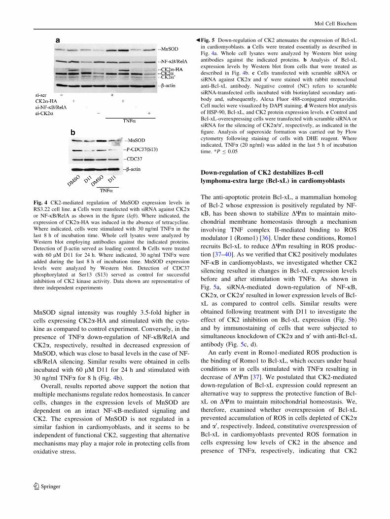
MnSOD signal intensity was roughly 3.5-fold higher in
cells expressing CK2a-HA and stimulated with the cyto-
kine as compared to control experiment. Conversely, in the
presence of TNFa down-regulation of NF-jB/RelA and
CK2a, respectively, resulted in decreased expression of
MnSOD, which was close to basal levels in the case of NF-
jB/RelA silencing. Similar results were obtained in cells
incubated with 60 lM D11 for 24 h and stimulated with
30 ng/ml TNFa for 8 h (Fig. 4b).
Overall, results reported above support the notion that
multiple mechanisms regulate redox homeostasis. In cancer
cells, changes in the expression levels of MnSOD are
dependent on an intact NF-jB-mediated signaling and
CK2. The expression of MnSOD is not regulated in a
similar fashion in cardiomyoblasts, and it seems to be
independent of functional CK2, suggesting that alternative
mechanisms may play a major role in protecting cells from
oxidative stress.
Down-regulation of CK2 destabilizes B-cell
lymphoma-extra large (Bcl-xL) in cardiomyoblasts
The anti-apoptotic protein Bcl-xL, a mammalian homolog
of Bcl-2 whose expression is positively regulated by NF-
jB, has been shown to stabilize DWm to maintain mito-
chondrial membrane homeostasis through a mechanism
involving TNF complex II-mediated binding to ROS
modulator 1 (Romo1) [36]. Under these conditions, Romo1
recruits Bcl-xL to reduce DWm resulting in ROS produc-
tion [37–40]. As we verified that CK2 positively modulates
NF-jB in cardiomyoblasts, we investigated whether CK2
silencing resulted in changes in Bcl-xL expression levels
before and after stimulation with TNFa. As shown in
Fig. 5a, siRNA-mediated down-regulation of NF-jB,CK2a, or CK2a0 resulted in lower expression levels of Bcl-
xL as compared to control cells. Similar results were
obtained following treatment with D11 to investigate the
effect of CK2 inhibition on Bcl-xL expression (Fig. 5b)
and by immunostaining of cells that were subjected to
simultaneous knockdown of CK2a and a0 with anti-Bcl-xL
antibody (Fig. 5c, d).
An early event in Romo1-mediated ROS production is
the binding of Romo1 to Bcl-xL, which occurs under basal
conditions or in cells stimulated with TNFa resulting in
decrease of DWm [37]. We postulated that CK2-mediated
down-regulation of Bcl-xL expression could represent an
alternative way to suppress the protective function of Bcl-
xL on DWm to maintain mitochondrial homeostasis. We,
therefore, examined whether overexpression of Bcl-xL
prevented accumulation of ROS in cells depleted of CK2aand a0, respectively. Indeed, constitutive overexpression of
Bcl-xL in cardiomyoblasts prevented ROS formation in
cells expressing low levels of CK2 in the absence and
presence of TNFa, respectively, indicating that CK2
Fig. 4 CK2-mediated regulation of MnSOD expression levels in
RS3.22 cell line. a Cells were transfected with siRNA against CK2aor NF-jB/RelA as shown in the figure (left). Where indicated, the
expression of CK2a-HA was induced in the absence of tetracycline.
Where indicated, cells were stimulated with 30 ng/ml TNFa in the
last 8 h of incubation time. Whole cell lysates were analyzed by
Western blot employing antibodies against the indicated proteins.
Detection of b-actin served as loading control. b Cells were treated
with 60 lM D11 for 24 h. Where indicated, 30 ng/ml TNFa were
added during the last 8 h of incubation time. MnSOD expression
levels were analyzed by Western blot. Detection of CDC37
phosphorylated at Ser13 (S13) served as control for successful
inhibition of CK2 kinase activity. Data shown are representative of
three independent experiments
bFig. 5 Down-regulation of CK2 attenuates the expression of Bcl-xL
in cardiomyoblasts. a Cells were treated essentially as described in
Fig. 4a. Whole cell lysates were analyzed by Western blot using
antibodies against the indicated proteins. b Analysis of Bcl-xL
expression levels by Western blot from cells that were treated as
described in Fig. 4b. c Cells transfected with scramble siRNA or
siRNA against CK2a and a0 were stained with rabbit monoclonal
anti-Bcl-xL antibody. Negative control (NC) refers to scramble
siRNA-transfected cells incubated with biotinylated secondary anti-
body and, subsequently, Alexa Fluor 488-conjugated streptavidin.
Cell nuclei were visualized by DAPI staining. d Western blot analysis
of HSP-90, Bcl-xL, and CK2 protein expression levels. e Control andBcl-xL-overexpressing cells were transfected with scramble siRNA or
siRNA for the silencing of CK2a/a0, respectively, as indicated in the
figure. Analysis of superoxide formation was carried out by Flow
cytometry following staining of cells with DHE reagent. Where
indicated, TNFa (20 ng/ml) was added in the last 5 h of incubation
time. *P B 0.05
Mol Cell Biochem
123

Mol Cell Biochem
123

antagonizes ROS accumulation by preserving intact Bcl-xL
expression (Fig. 5e and Suppl. Fig. S1).
Previous studies have shown that CK2-mediated phos-
phorylation of the co-chaperone CDC37 at Ser13 is a
necessary event for the stabilization of heat shock protein
90 (HSP-90)–CDC37 heterocomplex with client proteins
including ErbB2, EGFR, Raf, MEK, PTEN, AKT, and
mTOR [41–45]. We postulated that CK2 down-regulation
impaired Bcl-xL expression as a result of indirect disrup-
tion of HSP-90 chaperone function as HSP-90 has been
shown to interact with and stabilize Bcl-xL [46]. To show
this, we analyzed the association between HSP-90 and Bcl-
xL by in situ proximity ligation assay employing antibodies
directed against the aforementioned proteins. As shown in
Fig. 6, cells lacking CK2 resulted in a significant decrease
in signal intensity as compared to si-scr-transfected cells,
suggesting that the observed decrease in the expression
levels of Bcl-xL may result from destabilization of HSP-
90–CDC37 heterocomplex.
Differential gene expression profiling
of cardiomyoblasts in CK2-mediated oxidative stress
induction
Oxidative stress that results from ROS accumulation can
modulate multiple signaling pathways and thus a wide
variety of biological processes by linking cell surface sig-
nals to changes in gene expression. To clarify differential
mRNA expression in cardiomyoblasts resulting from ROS
accumulation induced by CK2 down-regulation, we
examined the expression of 84 genes related to oxidative
stress and oxidative defense using the oxidative stress RT2
profiler PCR array. The differential expression of genes
was plotted on a volcano plot as shown in Fig. 7a. Genes
up-regulated with fold-change C1.5 and P\ 0.05
consisted of flavin-containing monooxygenase 2 (Fmo2)
and prostaglandin-endoperoxide synthase 2 (Ptgs2), while
genes down-regulated with fold-change B-1.5 and
P\ 0.05 included 24-dehydrocholesterol reductase
(Dhcr24) and prostaglandin-endoperoxide synthase 1 (Pt-
gs1). The analysis also identified a number of genes whose
up-regulation was C1.5 although changes were not defined
as statistically significant (Fig. 7a; Suppl. Table S1). Based
on scatter plot analysis where the log of the fold-change in
gene expression in control cells was plotted against log of
the fold-change in CK2-knockdown cells, we could deter-
mine that the aforementioned group of genes had a low
expression profile making any change in their expression
among different datasets not statistically significant
(Fig. 7b). Overall, the observed differences in gene
expression suggest that CK2 modulates a subset of genes
linked to oxidative stress and ROS metabolism.
Discussion
The role of protein kinase CK2 in the regulation of a wide
range of intracellular processes has been mainly studied in
cancer cells (for reviews see [13, 14, 47, 48]). In this
respect, compelling evidence has indicated that CK2 is
markedly elevated both at the protein and mRNA levels
conferring protection from induction of cell death and
supporting cell survival and proliferation. Although
CK2 has been linked to various human diseases including
cancer, its role in non-cancerous cells particularly with
respect to mitochondrial redox homeostasis has remained
largely unexplored. The mitochondrial respiratory chain is
one of the major sources of ROS whose production is
related to oxygen consumption. The latter process is
especially prominent in cardiomyocytes that are equipped
Fig. 6 Analysis of the association between HSP-90 and Bcl-xL. Cells
that were treated as described in Fig. 5c were stained with antibodies
against HSP-90 and Bcl-xL, respectively. Detection of complexes was
revealed by in situ PLA (left). NC: scramble siRNA-transfected cells
subjected to in situ PLA where incubation with one of the primary
antibodies was omitted (negative control). Bar graph refers to the
determination of number of signals per cell as distinct red fluores-
cence spots performed by computer-assisted image analysis (right).
Data are expressed as mean ± standard deviation. Experiments were
repeated three times, obtaining similar results. In all cases, cell
pictures were taken at 409 magnification. *P B 0.001
Mol Cell Biochem
123

with scavenger enzymes and non-enzymatic systems to
prevent oxidative damage. In this study, we have reported
evidence that down-regulation of CK2 expression or its
inhibition, though not affecting the cell viability (data not
shown) significantly, results in the accumulation of ROS
and this effect is accompanied by loss of mitochondrial
membrane potential. Similar to what was previously
reported, changes in redox homeostasis appear to be more
significant when CK2 is inhibited than when its expression
levels are down-regulated, an effect most likely associated
to the long incubation time necessary to induce CK2
silencing [11].
Two main NF-jB-regulated pathways exist in cells: the
canonical pathway mostly induced by NF-jB stimuli and
the non-canonical signaling pathway induced by certain
TNF family cytokines. However, NF-jB can be directly
modulated by a variety of post-translational modifications
including phosphorylation, acetylation, and methylation
[31]. We show here that CK2 modulates the activity of NF-
jB/RelA in cardiomyoblasts and that its down-regulation
results in ROS production in cells left untreated or exposed
to TNFa, suggesting that CK2 modulates redox home-
ostasis through NF-jB/RelA.An important observation that emerged from this study
is that cancer cells promptly responded to TNFa treatment
by inducing MnSOD synthesis. Correspondingly, altered
expression of CK2 resulted in modulation of MnSOD
expression levels most likely through the NF-jB signaling
pathway. Conversely, similar results were not obtained
with cardiomyoblasts (in this respect, down-regulation of
NF-jB/RelA resulted only in a marginal decrease in
MnSOD expression), suggesting that alternative detoxifi-
cation mechanisms regulated by CK2 through or indepen-
dent from NF-jB might be taking place in
cardiomyoblasts. In this respect, NADPH oxidase is a
multi-component complex producing ROS after ischemic
injury. Deactivation of CK2 in mouse brain has been
shown to induce accumulation of ROS and neural cell
death as a result of increased NADPH oxidase activity,
demonstrating that CK2 is a negative modulator of this
enzyme [9].
Consistent with the notion that NF-jB regulates the
expression of genes antagonizing oxidative stress, it has
been shown that c-Rel and RelA directly activate the
expression of the apoptosis inhibitor Bcl-xL [36]. As
Romo1-mediated sequestration of Bcl-xL has been shown
to promote DWm destabilization and subsequent ROS
generation, we examined whether CK2 down-regulation
had an effect on Bcl-xL [37]. We show for the first time
that chemical inhibition or silencing of CK2 results in
decreased expression levels of Bcl-xL in cardiomyoblasts
identifying an alternative strategy to block Bcl-xL function
and induce ROS formation beside Romo1-mediated
sequestration. We failed to observe up-regulation of Bcl-xL
expression levels following treatment with TNFa of cells
transfected with si-scr as compared to control experiment
(Fig. 5a). This may indicate that a longer incubation time
with TNFa is necessary to induce the expression of Bcl-xL.
Results shown in Fig. 6 suggest that impaired expression of
Bcl-xL may result from CK2-mediated disruption of HSP-
90 chaperone function; however, one cannot exclude that
Fig. 7 Down-regulation of CK2 determines the changes in the
expression levels of genes related to oxidative stress. a Total RNA of
cells transfected with scramble siRNA (control) or siRNA against
CK2a and CK2a0 was extracted and purified as described in the
Materials and Methods and subsequently subjected to quantitative
PCR array for the analysis of 84 genes related to oxidative stress.
Log2 fold-changes (x axis) are plotted against -log10 of P-values (y
axis) in the volcano plot. The dashed black line indicates P = 0.05
with values above the line having P\ 0.05 and values below the line
having P[ 0.05. Points outside the dashed red lines indicate values
having a log2 fold-change B-0.58 or C0.58 corresponding to fold-
change B-1.5 and C1.5, respectively. b Scatter plot showing genes
differentially transcribed in cells transfected with siRNA against
CK2a and a0. The fold regulation cut-off was set as 1.5. Results
shown are derived from three independent data sets. (Color
figure online)
Mol Cell Biochem
123

impaired NF-jB transcription activity following CK2
down-regulation may additionally be responsible for the
observed decrease in Bcl-xL expression levels as previ-
ously reported in the case of tumor cells treated with CK2
inhibitors [49].
An additional finding that emerged from our study is the
elevated expression of two ROS-related genes, namely,
Fmo2 and Ptgs2, following simultaneous down-regulation
of CK2a and a0. Flavin-containing monooxygenases
(FMOs) are a family of NADPH-dependent proteins spe-
cialized in the oxidation of xeno-substrates in the presence
of flavin adenine dinucleotide (FAD). FMO2 is the most
abundant of the FMOs and it is particularly expressed in
lung although appreciable amounts are also found in nasal
tissue, heart, and brain (reviewed in [50, 51]). Studies
carried out with yeast FMO showed that its expression is
significantly enhanced as a result of accumulation of mis-
folded proteins in the endoplasmic reticulum as part of an
unfolded protein response (UPR) which triggers up-regu-
lation of endoplasmic reticulum-resident proteins involved
in protein folding [52]. Bearing in mind that CK2-depen-
dent phosphorylation of CDC37 is essential for the stability
of HSP-90–CDC37-containing complex, it is conceivable
that stimulation of FMO gene expression in cardiomy-
oblasts may result from the presence of misfolded proteins
as a result of impaired HSP-90 chaperone function.
Stimulation of ptgs-2-gene expression was also observed
in cells lacking CK2. This is in line with previous studies
showing that the expression of ptgs-2 is induced by
oxidative stress in various cancer cell lines and in cardiac
tissue [53, 54]. The reason why ptgs-2-transcript levels are
increased following ROS accumulation remains to be
determined; however, it has been postulated that this might
be a mechanism to abort cell death as ptgs-2 is known to
reduce apoptotic susceptibility [55].
The analysis of genes related to oxidative stress also
revealed a significant down-regulation of the expression of
Dhcr24 and Ptgs1. Dhcr24 encodes for 24-dehydrocholes-
terol reductase (DHCR24), a key enzyme in cholesterol
biosynthesis. DHCR24 has been recently shown to act as
anti-oxidant through twomechanisms: the first dependent on
cholesterol and the second one through a direct scavenging
of hydrogen peroxide [56, 57]. Interestingly, compelling
evidence has pointed out to a dual role of DHCR24 in pro-
tecting cells against oxidative stress. Its up-regulation during
an acute response is associated with elevated cholesterol
levels. In contrast, DHCR24 levels decline upon prolonged
exposure to oxidative stress and its down-regulation seems to
be part of an alternative pro-survival strategy associatedwith
destabilization of the tumor suppressor protein p53 [57]. We
have obtained results in line with these findings. However, it
remains to be determined whether down-regulation of
DHCR24 correlates with decreased expression levels of the
corresponding protein and whether this event results in
destabilization of p53 as part of a protective mechanism
against cell death.
Taken together, results reported here demonstrate that
CK2 provides protection against oxidative stress in car-
diomyoblasts through cellular mechanisms involving the
NF-jB-mediated signaling pathway and the Bcl-2 mam-
malian homolog Bcl-xL. It has been previously shown that
Romo1 controls redox homeostasis by recruiting Bcl-xL to
reduce DWm. We propose a new mechanism by which
silencing of CK2 correlates with attenuation of HSP-90-
molecular chaperone function and reduced Bcl-xL levels
resulting in oxidative stress. Although changes in the
expression of a gene may not necessarily lead to alteration
in the abundance of the corresponding protein, analysis of a
PCR array for profiling the expression of genes related to
oxidative stress further supports the notion that CK2 exerts
a pro-survival role also in non-cancerous cells. This may
occur by positively regulating the activity of proteins with
chaperone activity and those with a pro-survival function
during a prolonged exposure to oxidative stress.
Acknowledgements The authors would like to thank Dr. Olaf-Georg
Issinger (University of Southern Denmark) for critically reading the
manuscript; Dr. David W. Litchfield (University of Western Ontario)
for the generous gift of the RS3.22 cell line; Dr. Tuula Kallunki
(Danish Cancer Society) for providing the Bcl-xL viral vector; Dr.
Phillip Hallenborg (University of Southern Denmark) for expertise in
establishing Bcl-xL-overexpressing cardiomyoblasts; and Tina H.
Svenstrup for technical assistance. We thank the Drug Synthesis and
Chemistry Branch, Developmental Therapeutics Program, NCI, USA,
for providing us with viable samples. This work was supported by the
Danish Council for Independent Research-Natural Sciences (Grant
1323-00212A to B. Guerra).
References
1. Brown GC (1992) Control of respiration and ATP synthesis in
mammalian mitochondria and cells. Biochem J 284:1–13. doi:10.
1042/bj2840001
2. Taverne YJHJ, Bogers AJJC, Duncker DJ, Merkus D (2013)
Reactive oxygen species and the cardiovascular system. Oxid
Med Cell Longev. doi:10.1155/2013/862423
3. Turrens JF (2003) Mitochondrial formation of reactive oxygen
species. J Physiol 552:335–344. doi:10.1113/jphysiol.2003.
049478
4. Castaldo SA, Freitas JR, Conchinha NV, Madureira PA (2016)
The tumorigenic roles of the cellular REDOX regulatory systems.
Oxid Med Cell Longev 2016:1–17. doi:10.1155/2016/8413032
5. Truong TH, Carroll KS (2013) Redox regulation of protein
kinases. Crit Rev Biochem Mol Biol 48:332–356. doi:10.3109/
10409238.2013.790873
6. Knock GA, Ward JPT (2011) Redox regulation of protein kinases
as a modulator of vascular function. Antioxid Redox Signal
15:1531–1547. doi:10.1089/ars.2010.3614
7. Morgan MJ, Liu Z-G (2010) Crosstalk of reactive oxygen species
and NF-jB signaling. Cell Res 21:103–115. doi:10.1038/cr.2010.
178
Mol Cell Biochem
123

8. Montenarh M (2009) DMAT, an inhibitor of protein kinase CK2
induces reactive oxygen species and DNA double strand breaks.
Oncol Rep 21:1593–1597. doi:10.3892/or_00000392
9. Kim GS, Jung JE, Niizuma K, Chan PH (2009) CK2 is a novel
negative regulator of NADPH oxidase and a neuroprotectant in
mice after cerebral ischemia. J Neurosci 29:14779–14789. doi:10.
1523/JNEUROSCI.4161-09.2009
10. Guerra B (2012) Downregulation of protein kinase CK2 induces
autophagic cell death through modulation of the mTOR and
MAPK signaling pathways in human glioblastoma cells. Int J
Oncol 41:1967–1976. doi:10.3892/ijo.2012.1635
11. Qaiser F, Trembley JH, Kren BT et al (2014) Protein Kinase CK2
Inhibition Induces Cell Death via Early Impact on Mitochondrial
Function. J Cell Biochem 115:2103–2115. doi:10.1002/jcb.24887
12. Guerra B, Issinger O-G (1999) Protein kinase CK2 and its role in
cellular proliferation, development and pathology. Electrophore-
sis 20:391–408. doi:10.1002/(SICI)1522-2683(19990201)20:
2\391:AID-ELPS391[3.0.CO;2-N
13. Guerra B, Issinger O-G (2008) Protein kinase CK2 in human
diseases. Curr Med Chem 15:1870–1886
14. St-Denis NA, Litchfield DW (2009) Protein kinase CK2 in health
and disease. Cell Mol Life Sci 66:1817–1829. doi:10.1007/
s00018-009-9150-2
15. Trembley JH, Chen Z, Unger G et al (2010) Emergence of protein
kinase CK2 as a key target in cancer therapy. BioFactors
36:187–195. doi:10.1002/biof.96
16. Guerra B, Iwabuchi K, Issinger O-G (2014) Protein kinase CK2 is
required for the recruitment of 53BP1 to sites of DNA double-
strand break induced by radiomimetic drugs. Cancer Lett
345:115–123. doi:10.1016/j.canlet.2013.11.008
17. Hallenborg P, Feddersen S, Francoz S et al (2012) Mdm2 controls
CREB-dependent transactivation and initiation of adipocyte dif-
ferentiation. Cell Death Differ 19:1381–1389. doi:10.1038/cdd.
2012.15
18. Olsen BB, Issinger OG, Guerra B (2010) Regulation of DNA-
dependent protein kinase by protein kinase CK2 in human
glioblastoma cells. Oncogene 29:6016–6026. doi:10.1038/onc.
2010.337
19. Olsen BB, Wang S-Y, Svenstrup TH et al (2012) Protein kinase
CK2 localizes to sites of DNA double-strand break regulating the
cellular response to DNA damage. BMC Mol Biol 13:7. doi:10.
1186/1471-2199-13-7
20. Guerra B, Fischer M, Schaefer S, Issinger O-G (2015) The kinase
inhibitor D11 induces caspase-mediated cell death in cancer cells
resistant to chemotherapeutic treatment. J Exp Clin Cancer Res
34:125. doi:10.1186/s13046-015-0234-6
21. Livak KJ, Schmittgen TD (2001) Analysis of relative gene
expression data using real-time quantitative PCR and the 2(-Delta
Delta C(T)) Method. Methods 25:402–408. doi:10.1006/meth.
2001.1262
22. Schmittgen TD, Livak KJ (2008) Analyzing real-time PCR data
by the comparative C(T) method. Nat Protoc 3:1101–1108
23. Guerra B, Hochscherf J, Jensen NB, Issinger O-G (2015) Iden-
tification of a novel potent, selective and cell permeable inhibitor
of protein kinase CK2 from the NIH/NCI Diversity Set Library.
Mol Cell Biochem 406:151–161. doi:10.1007/s11010-015-2433-z
24. Marchi S, Giorgi C, Suski JM et al (2012) Mitochondria-ros
crosstalk in the control of cell death and aging. J Signal Trans-
duct. doi:10.1155/2012/329635
25. Peshavariya HM, Dusting GJ, Selemidis S (2007) Analysis of
dihydroethidium fluorescence for the detection of intracellular
and extracellular superoxide produced by NADPH oxidase. Free
Radic Res 41:699–712. doi:10.1080/10715760701297354
26. Kim I, Rodriguez-Enriquez S, Lemasters JJ (2007) Selective
degradation of mitochondria by mitophagy. Arch Biochem Bio-
phys 462:245–253. doi:10.1016/j.abb.2007.03.034
27. Cottet-Rousselle C, Ronot X, Leverve X, Mayol J-F (2011)
Cytometric assessment of mitochondria using fluorescent probes.
Cytom A 79:405–425. doi:10.1002/cyto.a.21061
28. To M-S, Aromataris EC, Castro J et al (2010) Mitochondrial
uncoupler FCCP activates proton conductance but does not block
store-operated Ca(2?) current in liver cells. Arch Biochem
Biophys 495:152–158. doi:10.1016/j.abb.2010.01.004
29. Sakon S, Xue X, Takekawa M et al (2003) NF-kappaB inhibits
TNF-induced accumulation of ROS that mediate prolonged
MAPK activation and necrotic cell death. EMBO J
22:3898–3909. doi:10.1093/emboj/cdg379
30. Viatour P, Merville M-P, Bours V, Chariot A (2005) Phospho-
rylation of NF-kappaB and IkappaB proteins: implications in
cancer and inflammation. Trends Biochem Sci 30:43–52. doi:10.
1016/j.tibs.2004.11.009
31. Oeckinghaus A, Hayden MS, Ghosh S (2011) Crosstalk in NF-jBsignaling pathways. Nat Immunol 12:695–708. doi:10.1038/ni.
2065
32. Jones PL, Ping D, Boss JM (1997) Tumor necrosis factor alpha
and interleukin-1beta regulate the murine manganese superoxide
dismutase gene through a complex intronic enhancer involving
C/EBP-beta and NF-kappaB. Mol Cell Biol 17:6970–6981
33. Djavaheri-Mergny M, Javelaud D, Wietzerbin J, Besancon F
(2004) NF-kappaB activation prevents apoptotic oxidative stress
via an increase of both thioredoxin and MnSOD levels in
TNFalpha-treated Ewing sarcoma cells. FEBS Lett 578:111–115.
doi:10.1016/j.febslet.2004.10.082
34. Kairisalo M, Korhonen L, Blomgren K, Lindholm D (2007)
X-linked inhibitor of apoptosis protein increases mitochon-
drial antioxidants through NF-kappaB activation. Biochem
Biophys Res Commun 364:138–144. doi:10.1016/j.bbrc.2007.
09.115
35. Vilk G, Saulnier RB, Pierre RS, Litchfield DW (1999) Inducible
Expression of Protein Kinase CK2 in Mammalian Cells: EVI-
DENCE FOR FUNCTIONAL SPECIALIZATION OF CK2
ISOFORMS. J Biol Chem 274:14406–14414. doi:10.1074/jbc.
274.20.14406
36. Chen C, Edelstein LC, Gelinas C (2000) The Rel/NF-kappaB
family directly activates expression of the apoptosis inhibitor
Bcl-x(L). Mol Cell Biol 20:2687–2695
37. Kim JJ, Lee SB, Park JK, Yoo YD (2010) TNF-alpha-induced
ROS production triggering apoptosis is directly linked to Romo1
and Bcl-X(L). Cell Death Differ 17:1420–1434. doi:10.1038/cdd.
2010.19
38. Genestier L, Bonnefoy-Berard N, Rouault JP et al (1995) Tumor
necrosis factor-alpha up-regulates Bcl-2 expression and decreases
calcium-dependent apoptosis in human B cell lines. Int Immunol
7:533–540
39. Vander Heiden MG, Chandel NS, Williamson EK et al (1997)
Bcl-xL regulates the membrane potential and volume home-
ostasis of mitochondria. Cell 91:627–637
40. Shirakata Y, Koike K (2003) Hepatitis B virus X protein induces
cell death by causing loss of mitochondrial membrane potential.
J Biol Chem 278:22071–22078. doi:10.1074/jbc.M301606200
41. Miyata Y (2009) Protein kinase CK2 in health and disease: CK2:
the kinase controlling the Hsp90 chaperone machinery. Cell Mol
Life Sci 66:1840–1849. doi:10.1007/s00018-009-9152-0
42. Pratt WB, Toft DO (2003) Regulation of signaling protein
function and trafficking by the hsp90/hsp70-based chaperone
machinery. Exp Biol Med (Maywood) 228:111–133
43. Echeverrıa PC, Bernthaler A, Dupuis P et al (2011) An interac-
tion network predicted from public data as a discovery tool:
application to the Hsp90 molecular chaperone machine. PLoS
ONE 6:e26044. doi:10.1371/journal.pone.0026044
44. Taipale M, Krykbaeva I, Koeva M et al (2012) Quantitative
analysis of HSP90-client interactions reveals principles of
Mol Cell Biochem
123

substrate recognition. Cell 150:987–1001. doi:10.1016/j.cell.
2012.06.047
45. Fu J, Koul D, Yao J et al (2013) Novel HSP90 inhibitor NVP-
HSP990 targets cell-cycle regulators to ablate Olig2-positive
glioma tumor-initiating cells. Cancer Res 73:3062–3074. doi:10.
1158/0008-5472.CAN-12-2033
46. Caldas-Lopes E, Cerchietti L, Ahn JH et al (2009) Hsp90 inhi-
bitor PU-H71, a multimodal inhibitor of malignancy, induces
complete responses in triple-negative breast cancer models. Proc
Natl Acad Sci USA 106:8368–8373. doi:10.1073/pnas.
0903392106
47. Duncan JS, Litchfield DW (2008) Too much of a good thing: the
role of protein kinase CK2 in tumorigenesis and prospects for
therapeutic inhibition of CK2. Biochim Biophys Acta
1784:33–47. doi:10.1016/j.bbapap.2007.08.017
48. Trembley JH, Wang G, Unger G et al (2009) Protein kinase CK2
in health and disease: CK2: a key player in cancer biology. Cell
Mol Life Sci 66:1858–1867. doi:10.1007/s00018-009-9154-y
49. Ravi R, Bedi A (2002) Sensitization of tumor cells to Apo2
ligand/TRAIL-induced apoptosis by inhibition of casein kinase
II. Cancer Res 62:4180–4185
50. Krueger SK, Williams DE (2005) Mammalian flavin-containing
monooxygenases: structure/function, genetic polymorphisms and
role in drug metabolism. Pharmacol Ther 106:357–387. doi:10.
1016/j.pharmthera.2005.01.001
51. Cashman JR, Zhang J (2006) Human flavin-containing
monooxygenases. Annu Rev Pharmacol Toxicol 46:65–100.
doi:10.1146/annurev.pharmtox.46.120604.141043
52. Suh JK, Robertus JD (2000) Yeast flavin-containing monooxy-
genase is induced by the unfolded protein response. Proc Natl
Acad Sci USA 97:121–126
53. Gilroy DW, Colville-Nash PR, Willis D et al (1999) Inducible
cyclooxygenase may have anti-inflammatory properties. Nat Med
5:698–701. doi:10.1038/9550
54. Sun Y, Chen J, Rigas B (2009) Chemopreventive agents induce
oxidative stress in cancer cells leading to COX-2 overexpression
and COX-2-independent cell death. Carcinogenesis 30:93–100.
doi:10.1093/carcin/bgn242
55. Sun Y, Tang XM, Half E et al (2002) Cyclooxygenase-2 over-
expression reduces apoptotic susceptibility by inhibiting the
cytochrome c-dependent apoptotic pathway in human colon
cancer cells. Cancer Res 62:6323–6328
56. Lu X, Kambe F, Cao X et al (2008) 3beta-Hydroxysteroid-
delta24 reductase is a hydrogen peroxide scavenger, protecting
cells from oxidative stress-induced apoptosis. Endocrinology
149:3267–3273. doi:10.1210/en.2008-0024
57. Kuehnle K, Crameri A, Kalin RE et al (2008) Prosurvival effect
of DHCR24/Seladin-1 in acute and chronic responses to oxidative
stress. Mol Cell Biol 28:539–550. doi:10.1128/MCB.00584-07
Mol Cell Biochem
123


![Amyloid-Beta Disrupts Calcium and Redox Homeostasis in ... · Amyloid-Beta Disrupts Calcium and Redox Homeostasis in Brain Endothelial Cells ... (SERCA) [39]. Although low ... Homeostasis](https://static.cupdf.com/doc/110x72/5c76b20d09d3f2d3778bffa9/amyloid-beta-disrupts-calcium-and-redox-homeostasis-in-amyloid-beta-disrupts.jpg)