CK2 phosphorylation-dependent interaction between aprataxin and MDC1 in the DNA damage response Olivier J. Becherel 1 , Burkhard Jakob 2 , Amy L. Cherry 3 , Nuri Gueven 1 , Markus Fusser 4 , Amanda W. Kijas 1 , Cheng Peng 1 , Sachin Katyal 5 , Peter J. McKinnon 5 , Junjie Chen 6 , Bernd Epe 4 , Stephen J. Smerdon 3, *, Gisela Taucher-Scholz 2 and Martin F. Lavin 1,7, * 1 Queensland Institute of Medical Research, Radiation Biology and Oncology, Brisbane, QLD 4029, Australia, 2 GSI Helmholtzzentrum Schwerionenforschung GmBH, Planckstr. 1, 64291 Darmstadt, Germany, 3 The MRC National Institute for Medical Research, The Ridgeway, Mill Hill, London, NW7 1AA, UK, 4 Institute of Pharmacy, University of Mainz, Mainz, Germany, 5 Department of Genetics and Tumor Cell Biology, St Jude Children’s Research Hospital, Memphis, Tennessee, 6 Department of Experimental Radiation Oncology, The University of Texas M.D. Anderson Cancer Center, Houston, Texas, USA and 7 Central Clinical Division, University of Queensland, Brisbane, Australia Received August 4, 2009; Revised October 26, 2009; Accepted November 20, 2009 ABSTRACT Aprataxin, defective in the neurodegenerative disorder ataxia oculomotor apraxia type 1, resolves abortive DNA ligation intermediates during DNA repair. Here, we demonstrate that aprataxin local- izes at sites of DNA damage induced by high LET radiation and binds to mediator of DNA-damage checkpoint protein 1 (MDC1/NFBD1) through a phosphorylation-dependent interaction. This inter- action is mediated via the aprataxin FHA domain and multiple casein kinase 2 di-phosphorylated S-D-T-D motifs in MDC1. X-ray structural and mutagenic analysis of aprataxin FHA domain, combined with modelling of the pSDpTD peptide interaction suggest an unusual FHA binding mecha- nism mediated by a cluster of basic residues at and around the canonical pT-docking site. Mutation of aprataxin FHA Arg29 prevented its interaction with MDC1 and recruitment to sites of DNA damage. These results indicate that aprataxin is involved not only in single strand break repair but also in the pro- cessing of a subset of double strand breaks presum- ably through its interaction with MDC1. INTRODUCTION Aprataxin, defective in the human autosomal recessive disorder ataxia oculomotor apraxia type 1 (AOA1), is encoded by the APTX gene (1,2). AOA1 is a neuro- degenerative disorder characterized by early onset cerebellar ataxia, oculomotor apraxia, hypoalbuminemia and late peripheral neuropathy (3). It resembles ataxia telangiectasia (A-T) in its neurodegenerative phenotype but lacks the extra-neurological features of A-T that include radiosensitivity, immunodeficiency and cancer predisposition (3,4). ATM, the protein defective in A-T, recognizes and is activated by DNA double strand breaks (DSB) to signal this damage to the DNA repair machinery and activate cell cycle checkpoints (5). Aprataxin contains three functional domains: an N-terminal FHA domain (6), a central histidine triad (HIT) domain (7) and a C-terminal zinc finger motif. Aprataxin interacts with XRCC1 and PARP-1, two key components of the DNA base excision repair machinery (8–10), suggesting a role for aprataxin in DNA single strand break (SSB) repair. Cells derived from AOA1 patients are sensitive to genotoxic agents that induce SSB in DNA (8–12). Consistent with the involvement of aprataxin in DNA repair, defective SSB repair has been reported in AOA1 cells in response to camptothecin, H 2 O 2 and BSO (11–13). More recently aprataxin was shown to catalyze the nucleophilic release of adenylate groups covalently linked to 5 0 -termini of DNA molecules (14,15). Ligation of SSB or DSB involves the formation of an AMP-ligase complex, which subsequently transfers the AMP moiety onto the 5 0 phosphate of the break site from which it is released after the formation of the phosphodiester bond (16). The AMP hydrolase activity of aprataxin appears to be important in resolving *To whom correspondence should be addressed. Tel: +61(0)7 3362 0335; Fax: +61(0)7 3362 0106; Email: [email protected] Correspondence may also be addressed to Dr Stephen J. Smerdon. Tel: +44 (0)20 8816 2533; Fax: +44 (0)208 816 2580; Email: [email protected] The authors wish it to be known that, in their opinion, the first three authors should be regarded as joint First Authors. Nucleic Acids Research, 2009, 1–15 doi:10.1093/nar/gkp1149 ß The Author(s) 2009. Published by Oxford University Press. This is an Open Access article distributed under the terms of the Creative Commons Attribution Non-Commercial License (http://creativecommons.org/licenses/ by-nc/2.5), which permits unrestricted non-commercial use, distribution, and reproduction in any medium, provided the original work is properly cited. Nucleic Acids Research Advance Access published December 14, 2009

Welcome message from author
This document is posted to help you gain knowledge. Please leave a comment to let me know what you think about it! Share it to your friends and learn new things together.
Transcript

CK2 phosphorylation-dependent interaction betweenaprataxin and MDC1 in the DNA damage responseOlivier J. Becherel1, Burkhard Jakob2, Amy L. Cherry3, Nuri Gueven1, Markus Fusser4,
Amanda W. Kijas1, Cheng Peng1, Sachin Katyal5, Peter J. McKinnon5, Junjie Chen6,
Bernd Epe4, Stephen J. Smerdon3,*, Gisela Taucher-Scholz2 and Martin F. Lavin1,7,*
1Queensland Institute of Medical Research, Radiation Biology and Oncology, Brisbane, QLD 4029, Australia,2GSI Helmholtzzentrum Schwerionenforschung GmBH, Planckstr. 1, 64291 Darmstadt, Germany, 3The MRCNational Institute for Medical Research, The Ridgeway, Mill Hill, London, NW7 1AA, UK, 4Institute of Pharmacy,University of Mainz, Mainz, Germany, 5Department of Genetics and Tumor Cell Biology, St Jude Children’sResearch Hospital, Memphis, Tennessee, 6Department of Experimental Radiation Oncology, The University ofTexas M.D. Anderson Cancer Center, Houston, Texas, USA and 7Central Clinical Division, University ofQueensland, Brisbane, Australia
Received August 4, 2009; Revised October 26, 2009; Accepted November 20, 2009
ABSTRACT
Aprataxin, defective in the neurodegenerativedisorder ataxia oculomotor apraxia type 1, resolvesabortive DNA ligation intermediates during DNArepair. Here, we demonstrate that aprataxin local-izes at sites of DNA damage induced by high LETradiation and binds to mediator of DNA-damagecheckpoint protein 1 (MDC1/NFBD1) through aphosphorylation-dependent interaction. This inter-action is mediated via the aprataxin FHA domainand multiple casein kinase 2 di-phosphorylatedS-D-T-D motifs in MDC1. X-ray structural andmutagenic analysis of aprataxin FHA domain,combined with modelling of the pSDpTD peptideinteraction suggest an unusual FHA binding mecha-nism mediated by a cluster of basic residues at andaround the canonical pT-docking site. Mutation ofaprataxin FHA Arg29 prevented its interaction withMDC1 and recruitment to sites of DNA damage.These results indicate that aprataxin is involved notonly in single strand break repair but also in the pro-cessing of a subset of double strand breaks presum-ably through its interaction with MDC1.
INTRODUCTION
Aprataxin, defective in the human autosomal recessivedisorder ataxia oculomotor apraxia type 1 (AOA1),
is encoded by the APTX gene (1,2). AOA1 is a neuro-degenerative disorder characterized by early onsetcerebellar ataxia, oculomotor apraxia, hypoalbuminemiaand late peripheral neuropathy (3). It resembles ataxiatelangiectasia (A-T) in its neurodegenerative phenotypebut lacks the extra-neurological features of A-T thatinclude radiosensitivity, immunodeficiency and cancerpredisposition (3,4). ATM, the protein defective in A-T,recognizes and is activated by DNA double strand breaks(DSB) to signal this damage to the DNA repair machineryand activate cell cycle checkpoints (5).Aprataxin contains three functional domains: an
N-terminal FHA domain (6), a central histidine triad(HIT) domain (7) and a C-terminal zinc finger motif.Aprataxin interacts with XRCC1 and PARP-1, two keycomponents of the DNA base excision repair machinery(8–10), suggesting a role for aprataxin in DNA singlestrand break (SSB) repair. Cells derived from AOA1patients are sensitive to genotoxic agents that induceSSB in DNA (8–12). Consistent with the involvement ofaprataxin in DNA repair, defective SSB repair has beenreported in AOA1 cells in response to camptothecin, H2O2
and BSO (11–13). More recently aprataxin was shown tocatalyze the nucleophilic release of adenylate groupscovalently linked to 50-termini of DNA molecules(14,15). Ligation of SSB or DSB involves the formationof an AMP-ligase complex, which subsequently transfersthe AMP moiety onto the 50 phosphate of the break sitefrom which it is released after the formation of thephosphodiester bond (16). The AMP hydrolase activityof aprataxin appears to be important in resolving
*To whom correspondence should be addressed. Tel: +61(0)7 3362 0335; Fax: +61(0)7 3362 0106; Email: [email protected] may also be addressed to Dr Stephen J. Smerdon. Tel: +44 (0)20 8816 2533; Fax: +44 (0)208 816 2580;Email: [email protected]
The authors wish it to be known that, in their opinion, the first three authors should be regarded as joint First Authors.
Nucleic Acids Research, 2009, 1–15doi:10.1093/nar/gkp1149
� The Author(s) 2009. Published by Oxford University Press.This is an Open Access article distributed under the terms of the Creative Commons Attribution Non-Commercial License (http://creativecommons.org/licenses/by-nc/2.5), which permits unrestricted non-commercial use, distribution, and reproduction in any medium, provided the original work is properly cited.
Nucleic Acids Research Advance Access published December 14, 2009
abortive DNA ligation intermediates that can form at‘dirty’ or ‘complex’ SSB and potentially also at DSBformed by reactive oxygen species attack or clustereddamage (15). The involvement of aprataxin in DNArepair is further supported by recent evidence of elevatedlevels of oxidative DNA damage in AOA1 cells coupledwith a reduced expression of PARP-1, apurinic endo-nuclease 1 (APE1) and OGG1 (17). Furthermore,impaired base excision and gap filling repair efficienciesreveals a synergy between aprataxin, PARP-1, APE-1and OGG1 in the DNA damage response and highlightsboth direct and indirect modulating functions foraprataxin on base excision repair (17). Although a rolefor aprataxin in the repair of abortive ligations at sitesof DSB has been suggested by the interaction betweenaprataxin and XRCC4 (9) and the in vitro binding ofaprataxin to DNA double strand ends (15,18), no directevidence for its involvement in DNA double strand breakrepair has been reported (19).In response to DNA damage, many proteins involved in
DNA damage signalling/repair, such as Mre11, NBS1 and53BP1 quickly re-localize to nuclear foci (20,21). Thesefoci co-localize with the phosphorylated form of histoneH2AX (gH2AX), a well-established marker for DSB (22).While aprataxin interacts with XRCC1, XRCC4 andPARP-1, we have previously shown that in contrast tothe other DNA repair proteins, it does not form detectablenuclear foci after DNA damage exposure induced byeither low linear energy transfer (LET) ionizing radiation(IR) or H2O2 (8). The dynamics of recruitment of DNArepair proteins to sites of DNA damage has beeninvestigated by laser irradiation combined with photo-sensitizers (23–25). Hirano et al. (2007) employed thisapproach to show that aprataxin was recruited in vivo toSSB. While laser-induced DNA damage produces SSBand DSB, the relative distribution and the density of thedamage are greatly influenced by the laser type, energyoutput and the type of photosensitizer (26). Biologicalimaging of charged particle tracks represents an alterna-tive approach for investigating the association of repairproteins with chromatin (27,28). This technology offersthe advantage of defined intrinsic physical properties ofthe heavy ion beams and allows the density of lesionsalong the track to be varied with ion species with differentLET values. Using a remote-controlled microscopecoupled to the beamline that allows the acquisition offluorescence images of living cells, in real-time duringion irradiation, we observed that GFP-aprataxin israpidly localized to DNA damage induced by nickel-ion-irradiation (8,28). To gain further insight how aprataxinis recruited to sites of DNA breaks, we investigated thein vivo recruitment of GFP-tagged aprataxin to localizedDNA damage induced by high LET heavy ion irradia-tion. We demonstrate for the first time that aprataxinco-localizes and interacts with mediator of DNAdamage checkpoint protein 1 (MDC1), a protein thatamplifies ATM-dependent DNA damage signalling(29–35). Interaction between the two proteins ismediated through the FHA domain of aprataxin, whichbinds to a casein kinase II (CK2)-phosphorylatedN-terminal region of MDC1. Mutation of aprataxin
Arg29, a key residue conserved within FHA motifs (36),disrupted the interaction between aprataxin and MDC1,and abolished its recruitment to sites of DNA damage.These results together with those of Harris et al. (17) high-light the importance of the aprataxin FHA domain inregulating protein–protein interactions and targetingaprataxin to sites of DNA damage.
MATERIALS AND METHODS
Cell lines, inhibitors
Control lymphoblastoid cell line (LCL) (C3ABR) andcervical adenocarcinoma cells (HeLa) were culturedin RPMI 1640 (GIBCO BRL) and DMEM (GIBCOBRL), respectively containing 10% foetal calf serum(FCS) (Lonza), 2mM L-glutamine (Life Technologies),100U/ml penicillin (GIBCO BRL), 100U/ml streptomy-cin (GIBCO BRL) and maintained in a humidifiedincubator at 37�C/5% CO2. Wild type, Aptx�/� andMdc1�/� mouse embryonic fibroblasts (MEF), andhuman AOA1 fibroblasts (FD105) hTERT-transformed(Gift from Prof. Keith Caldecott, University of Sussex,UK) were cultured in DMEM as described earlier.CK2 inhibitors 4,5,6,7-tetrabromotriazole (TBB), and2-dimethylamino-4,5,6,7-tetrabromo-1H-benzimidazole(DMAT) were purchased from Calbiochem and dissolvedin DMSO.
Aprataxin-GST and MDC1-GST pull down assays
Aprataxin-GST proteins have been described earlier(8,37). Pull down assays, were performed as describedin Supplementry Data. Proteins were separated on 5%and 10% SDS–PAGE, transferred onto nitrocellulosemembranes (Hybond C, Amersham) and detected usingthe relevant antibody. Aprataxin antibodies have beendescribed earlier (8,37). Sheep polyclonal MDC1antibody was produced against GST-MDC1 fragmentsand purified by affinity column against GST only andthe antigen using standard protocols. For immuno-blotting, antibodies were used at dilutions of: MDC1(1/2000), aprataxin (1/2000), anti-GST (1/2000) followedby the relevant species-specific HRP-conjugated secondaryantibody from Chemicon or Sigma (1/10 000).
In vivo CK2 inhibition and pull downs
HeLa cells were mock treated (DMSO) or treated with arange of concentrations of specific CK2 inhibitor TBB,or DMAT for 8 h in a six well-plate format and thenlysed. Pull down was performed as described inSupplementary Data.
In vitro CK2 phosphorylation and pull down
A region of MDC1 containing CK2 sites (amino acids150–350) was cloned into the bacterial GST expressionvector pGEX-6P-1, expressed in E. coli (BL21DE3) andpurified as described by the manufacturer. Two micro-grams of each GST fusion protein was in vitro phos-phorylated by incubation with 200 ng of humanrecombinant CK2 (New England Biolabs) in buffer P
2 Nucleic Acids Research, 2009
(20mM Tris–HCl pH 7.5, 50mM KCl, 10mM MgCl2,200 mM ATP) for 1 h at 37�C. Once phosphorylated,GST fusion proteins were used in pull-down assaysmixed with 2mg of HeLa whole cell extracts (WCE) asdescribed earlier.
Immunoprecipitation and immunoblotting
Total cell extracts from control (C3ABR) cells wereprepared as described in Supplementary Data and theequivalent of 1mg of total protein was immunopre-cipitated with non-specific serum (1 or 5 mg), aprataxin(1 mg) and MDC1 (5mg) antibodies overnight at 4�C.40 ml of protein G-Sepharose beads (Amersham) wereadded to the immunoprecipitation (IP) and incubatedfor 1 h on a rotating wheel at 4�C. IP were washed threetimes with lysis buffer and the beads were resuspended in20 ml of sample loading buffer. Proteins were separated on5% and 10% SDS�PAGE, transferred onto nitrocellulosemembranes and detected using the relevant primary andsecondary antibodies.
Isothermal titration calorimetry
Phosphopeptides were synthesised by Dr W. Mawby(University of Bristol). Aprataxin FHA-phosphopeptidebinding was quantified by isothermal titration calorimetry(ITC) using a Microcal Omega VP-ITC calorimeter(MicroCal Inc., Northampton, MA, USA). Protein wasdialysed against ITC buffer (50mM HEPES pH 7.5,150mM NaCl, 5mM b-mercaptoethanol) and peptideswere dissolved in the dialysis buffer. Experiments werecarried out at 22�C and involved 30 successive 10 ml injec-tions of 1mM peptide solution into a sample cell contain-ing 100 mM protein solution. Heats of dilution weresubtracted and binding isotherms were plotted andanalysed with MicroCal Origin version 7.0, assuming asingle-site binding model.
Crystallization and structure determination of theaprataxin FHA domain
Aprataxin FHA domain was crystallized by hanging-dropvapour diffusion. Protein at a concentration of10mg/ml in 50mM HEPES pH 7.5, 150mM NaCl,5mM b-mercaptoethanol was mixed with an equalvolume of reservoir solution [0.2M ammonium fluoride,20% (w/v) PEG 3350] and incubated at 18�C. Thin platesgrew within 1 week and were transferred into cryo-protectant (0.1M ammonium fluoride, 10 % PEG 3350,25mM HEPES pH 7.5, 75mM NaCl, 25% PEG 400) andflash frozen in liquid nitrogen. Data were collected at100K on an Raxis-IV detector mounted on a RigakuMicroMax-007HF rotating anode X-ray source, andintegrated and scaled using DENZO and SCALEPACK(38). The structure was solved by molecular replacementusing the coordinates of PDB entry 1YJM as a searchmodel with AmoRe (39,40). Subsequent refinement wascarried out at 1.65 A using REFMAC5 (41) and manualmodel building in Coot (42). Data collection and refine-ment statistics are summarized in SupplementaryTable S2. All structure figures were prepared with
PyMol (http://www.pymol.org). Coordinates have beendeposited in the Protein Databank: Accession code 3KT9.
High LET heavy ion irradiation procedure
For heavy ion irradiation experiments, cells were seeded onsterilized glass coverslips and irradiated at the UNILACbeamline at GSI (Darmstadt, Germany) with high energyxenon ions (4.5 MeV/nucleon; LET 8815 keV/mm),krypton (5.4 MeV/nucleon; LET 5060 keV/mm), nickel(6.0 MeV/nucleon; LET 3430 keV/mm) or uranium ions(4.2 MeV/nucleon; LET 14925 keV/mm), respectively.Different heavy ion species were used for the recruitmentstudies since these experiments were carried out overseveral years and we have had to adhere to the scheduleof radiation exposure at the Darmstadt GSI site inGermany. Directly before the irradiation, the coverslipswere mounted into sample holders and placed in amedium filled tank. For the ion irradiation, the sampleholders were automatically taken from the mediumtank, exposed to the ion beam under a small angle of 15�
between the axis of the ion beam and the plane of the cellmonolayer and placed back again. For the applied fluenceof 3� 106 p/cm2, the whole procedure takes <30 s.
Immunofluorescence and microscopy
For immunostaining, cells grown on coverslips werewashed with PBS, fixed in 2% formaladehyde/PBS for20min and permeabilized with PT-5 solution [PBS/0.5%Triton X-100] for 5min. Non-specific binding was blockedby incubating with PTB-5 [PBS/0.5% BSA] for 20min atroom temperature. Coverslips were then incubated withmouse anti-gH2AX antibody (Upstate 1/500 in PTB-5)for 1 h at room temperature. Cells were stained with5 mg/ml Alexa 568 goat anti-mouse F(ab)2 conjugate(Invitrogen) and the DNA counterstained with 1mMToPro3 (Invitrogen). Microscopy was performed on aLeica TCS NT confocal scanner equipped with anArKr-Laser on the Leica DM IRBE inverted microscope(lens: HCX PlanApo 63x oil/NA1.32). Images wererecorded as confocal stacks with optical sections separatedby about 0.3mm. Confocal images were displayed asmaximum projections and assembled in AdobePhotoshop 7.0. Heavy ion irradiation procedure isdescribed in Supplementary Data.
In vivo real-time beamline microscopy and heavy ionirradiation
Cells were transfected with Amaxa Nucleofection technol-ogy (Amaxa, Koeln, Germany). Transfection of MEFswas performed with Amaxa Nucleofector Kit VDP-1004and protocol A-023 according to the manufacturerinstructions (Amaxa, Germany). Mdc1�/� and Mdc1+/+
cells transiently transfected with GFP-aprataxin wereemployed for real-time fluorescence intensity measure-ments. These cells were cultivated on polycarbonate foil(18-mm diameter; 40-mm thickness) and irradiated at thebeamline microscope as described earlier (28).
Nucleic Acids Research, 2009 3
cH2AX foci repair kinetics
Analysis of gH2AX repair foci kinetics was performed asdescribed earlier (43) and is described in ExperimentalProcedures in Supplementary Data.
Analysis of SSB repair
SSB were induced by exposing confluent cells to low dosesof H2O2 (50�100 mM) in RPMI 1640 medium withoutsupplements for 15min at 37�C. To remove H2O2, cellswere washed two times with PBSCMF (140mM NaCl,3mM KCl, 8mM Na2HPO4 and 1mM KH2PO4). Thenumbers of SSB were determined by an alkaline elutionassay as described earlier (44). The numbers of SSB inuntreated control cells were subtracted in all cases.
RESULTS
Recruitment of aprataxin to DNA damage andco-localization with MDC1 after high LETradiation exposure
A hallmark of DNA repair proteins is their capacity tore-localize in discrete aggregates or foci within the nucleus
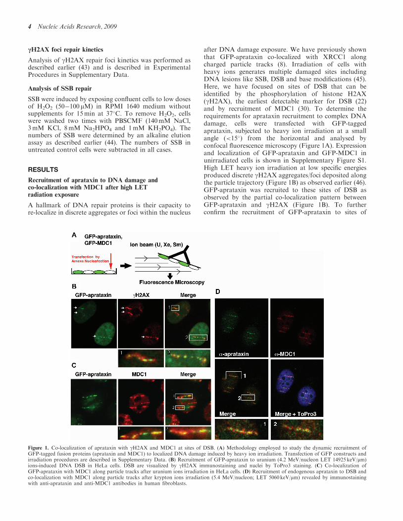
after DNA damage exposure. We have previously shownthat GFP-aprataxin co-localized with XRCC1 alongcharged particle tracks (8). Irradiation of cells withheavy ions generates multiple damaged sites includingDNA lesions like SSB, DSB and base modifications (45).Here, we have focused on sites of DSB that can beidentified by the phosphorylation of histone H2AX(gH2AX), the earliest detectable marker for DSB (22)and by recruitment of MDC1 (30). To determine therequirements for aprataxin recruitment to complex DNAdamage, cells were transfected with GFP-taggedaprataxin, subjected to heavy ion irradiation at a smallangle (<15�) from the horizontal and analysed byconfocal fluorescence microscopy (Figure 1A). Expressionand localization of GFP-aprataxin and GFP-MDC1 inunirradiated cells is shown in Supplementary Figure S1.High LET heavy ion irradiation at low specific energiesproduced discrete gH2AX aggregates/foci deposited alongthe particle trajectory (Figure 1B) as observed earlier (46).GFP-aprataxin was recruited to these sites of DSB asobserved by the partial co-localization pattern betweenGFP-aprataxin and gH2AX (Figure 1B). To furtherconfirm the recruitment of GFP-aprataxin to sites of
Figure 1. Co-localization of aprataxin with gH2AX and MDC1 at sites of DSB. (A) Methodology employed to study the dynamic recruitment ofGFP-tagged fusion proteins (aprataxin and MDC1) to localized DNA damage induced by heavy ion irradiation. Transfection of GFP constructs andirradiation procedures are described in Supplementary Data. (B) Recruitment of GFP-aprataxin to uranium (4.2 MeV/nucleon LET 14925 keV/mm)ions-induced DNA DSB in HeLa cells. DSB are visualized by gH2AX immunostaining and nuclei by ToPro3 staining. (C) Co-localization ofGFP-aprataxin with MDC1 along particle tracks after uranium ions irradiation in HeLa cells. (D) Recruitment of endogenous aprataxin to DSB andco-localization with MDC1 along particle tracks after krypton ions irradiation (5.4 MeV/nucleon; LET 5060 keV/mm) revealed by immunostainingwith anti-aprataxin and anti-MDC1 antibodies in human fibroblasts.
4 Nucleic Acids Research, 2009
DSB, we demonstrated co-immunostaining with MDC1(Figure 1C), another DNA damage response factor thatis recruited to DSB through binding to gH2AX (34).As was the case with gH2AX, co-localization was incom-plete between aprataxin and MDC1 (Figure 1C). To con-firm the recruitment of MDC1 to high LET heavyion-induced DSB, we used a GFP-MDC1 fusion protein.As expected, GFP-MDC1 completely co-localized withgH2AX staining at sites of heavy ion-induced DSB(Supplementary Figure S2). To address possible artefactsdue to either overexpression or the presence of a GFPtag attached to the N-terminus of aprataxin, immuno-staining for endogenous aprataxin was also performed(Figure 1D). In this case also, endogenous aprataxin wasrecruited to sites of DSB as observed by partialco-localization with MDC1 along the particle tracks. Todetermine whether MDC1 mediates the recruitment ofaprataxin to sites of DNA DSB, wild-type and Mdc1�/�
MEFs were transiently transfected with GFP-aprataxinand analysed for protein track formation after heavyion exposure. While aprataxin was recruited to DNAbreaks in wild-type MEFs after heavy ion irradiationat 10min post-irradiation (4.5 MeV/nucleon; LET8815 keV/mm), no recruitment was observed in Mdc1�/�
cells despite the formation of gH2AX tracks (Supple-mentary Figure S3A). Due to poor transfection efficiencyof Mdc1 MEFs, the absence of aprataxin recruitment wasonly observed in a limited number of cells. Accordingly, tomonitor the recruitment of aprataxin in MDC1-deficientcells over shorter time points (up to 4min), we employeda remote-controlled system coupled to a beamline micro-scope that allows measurement of GFP fluorescence inten-sity at sites of DNA breaks in real-time during heavy ionirradiation (28). Both wild-type and Mdc1�/� MEFs weretransiently transfected with GFP-aprataxin and subjectedto high LET heavy ion irradiation as described earlier(28). Real-time imaging of GFP fluorescence revealedfast recruitment of aprataxin to sites of DNA damagewithin 1min after irradiation in both Mdc1�/� and wild-type cells (Supplementary Figure S3B). Interestingly,fluorescence intensity at the sites of DNA damage dropsout rapidly in Mdc1�/� compared with wild-type cells sug-gesting an increased rate of dissociation of GFP-aprataxinfrom sites of DNA DSB in these cells. This indicates thatwhile MDC1 is not necessary for the initial recruitmentof aprataxin to sites of DNA DSB, its absence affects theretention of aprataxin to these sites. On the other hand,loss of XRCC1 prevented any recruitment of aprataxin incells exposed to high LET radiation (SupplementaryFigure S4). Together, these results provide evidence thataprataxin localizes at least in part to sites of DNA DSB,and that MDC1 does not act to recruit aprataxin, butrather as part of a tethering platform for aprataxin andpossibly other repair proteins, to ensure effective DNADSB repair.
Interaction between aprataxin and MDC1
The co-localization of MDC1 and aprataxin to sites ofDNA breaks prompted us to determine whether thesetwo molecules associate. Using a combination of co-IP
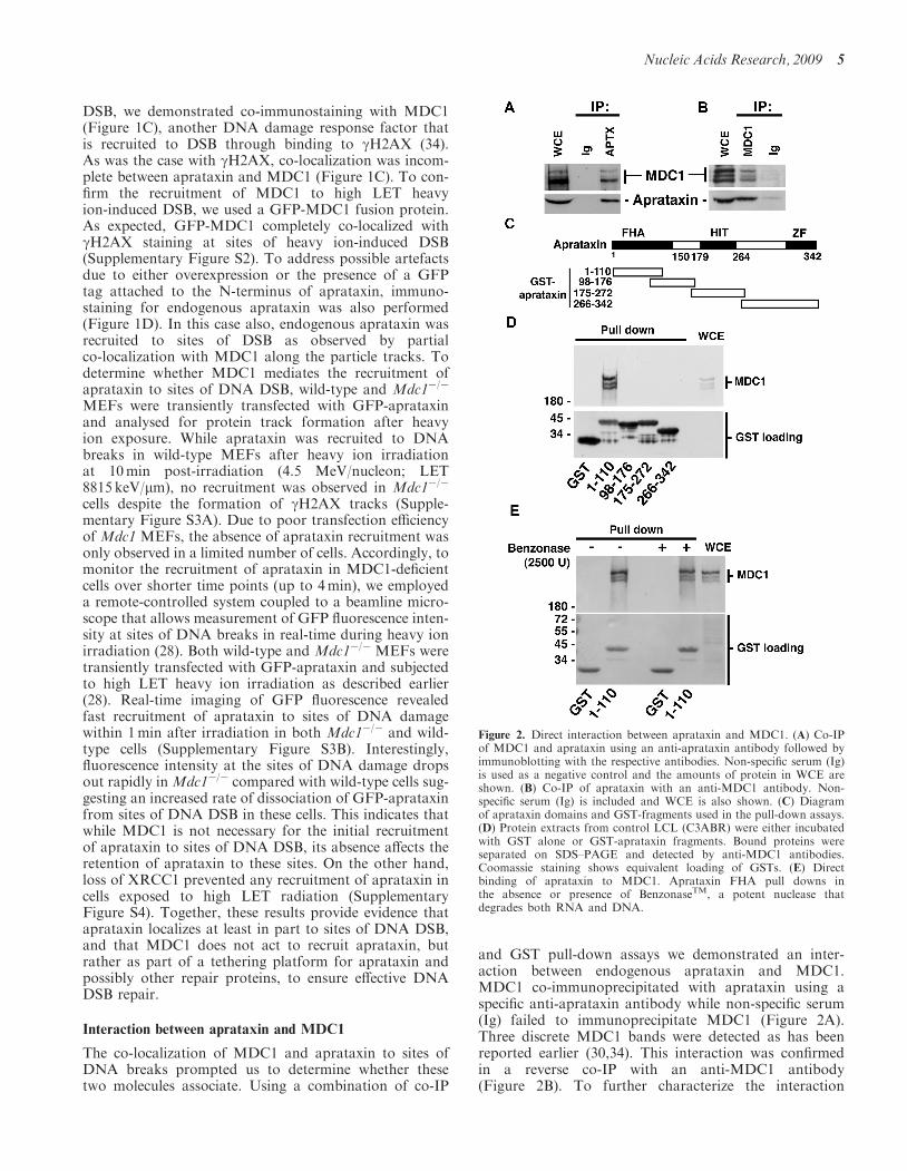
and GST pull-down assays we demonstrated an inter-action between endogenous aprataxin and MDC1.MDC1 co-immunoprecipitated with aprataxin using aspecific anti-aprataxin antibody while non-specific serum(Ig) failed to immunoprecipitate MDC1 (Figure 2A).Three discrete MDC1 bands were detected as has beenreported earlier (30,34). This interaction was confirmedin a reverse co-IP with an anti-MDC1 antibody(Figure 2B). To further characterize the interaction
Figure 2. Direct interaction between aprataxin and MDC1. (A) Co-IPof MDC1 and aprataxin using an anti-aprataxin antibody followed byimmunoblotting with the respective antibodies. Non-specific serum (Ig)is used as a negative control and the amounts of protein in WCE areshown. (B) Co-IP of aprataxin with an anti-MDC1 antibody. Non-specific serum (Ig) is included and WCE is also shown. (C) Diagramof aprataxin domains and GST-fragments used in the pull-down assays.(D) Protein extracts from control LCL (C3ABR) were either incubatedwith GST alone or GST-aprataxin fragments. Bound proteins wereseparated on SDS–PAGE and detected by anti-MDC1 antibodies.Coomassie staining shows equivalent loading of GSTs. (E) Directbinding of aprataxin to MDC1. Aprataxin FHA pull downs inthe absence or presence of BenzonaseTM, a potent nuclease thatdegrades both RNA and DNA.
Nucleic Acids Research, 2009 5
between aprataxin and MDC1 and to map the interactiondomain on aprataxin, we performed aprataxin GSTpull-downs. Using a series of overlapping GST fusionproteins, encompassing aprataxin FHA, HIT and zincfinger domains (Figure 2C), we identified the N-terminalregion (amino acid 1�110) of aprataxin as the MDC1interaction domain (Figure 2D). Binding of MDC1 tothe aprataxin FHA domain was retained in the presence ofBenzonaseTM suggesting that this was not due to bridgingwith nucleic acid but rather a direct protein�protein inter-action (Figure 2E). To confirm the direct interactionbetween aprataxin and MDC1, aprataxin GST pulldowns were carried out in the presence of ethidiumbromide (EtBr) (Supplementary Figure S5). EtBr did notdisrupt the interaction between the two proteins. On thecontrary, an increase in MDC1 binding was observed inthe presence of EtBr which may reflect an increase inavailable soluble MDC1. Thus the co-localization ofaprataxin and MDC1 to sites of DNA damage inducedby high LET radiation is supported by evidence of a directinteraction between these two proteins.
CK2 phosphorylation-dependent interaction of MDC1 withthe aprataxin FHA domain
FHA domains are conserved sequences of 65–150 aminoacid residues found principally within eukaryotic nuclearproteins that have been shown to mediate phospho-specific protein–protein interactions by binding tophosphopeptides (47,48). Previous reports have demon-strated that the interaction between aprataxin andXRCC1 was dependent on phosphorylation of XRCC1by CK2 (9,10). Furthermore, XRCC4 which is involvedin the repair of DNA DSB via the non-homologous endjoining pathway was also shown to bind to the FHAdomain of aprataxin (9). To determine whether XRCC4could also target aprataxin to DNA breaks, we examinedthe recruitment of aprataxin in Xrcc4�/� cells. Normalrecruitment of GFP-aprataxin was observed in Xrcc4�/�
cells indicating that XRCC4, albeit binding to the FHAdomain of aprataxin, is not involved in its targeting toheavy ion-induced DNA breaks (data not shown).Nevertheless, the interaction between aprataxin andXRCC4 may be important for the assembly of therejoining complex and/or to ensure effective DNA end‘clean-up’ during the repair process.To determine whether the interaction between MDC1
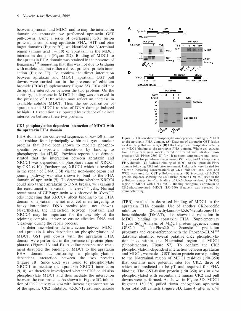
and aprataxin is also dependent on phosphorylation ofMDC1, GST pull downs with the aprataxin FHAdomain were performed in the presence of protein phos-phatase (Figure 3A and B). Alkaline phosphatase treat-ment disrupted the binding of MDC1 to the aprataxinFHA domain demonstrating a phosphorylation-dependent interaction between the two proteins(Figure 3B). Since CK2 was found to phosphorylateXRCC1 to mediate the aprataxin–XRCC1 interaction(9,10), we therefore investigated whether CK2 could alsophosphorylate MDC1 and thus mediate the interactionbetween the two proteins. As shown in Figure 3C, inhibi-tion of CK2 activity in vivo with increasing concentrationof the specific CK2 inhibitor, 4,5,6,7-Tetrabromotriazole
(TBB), resulted in decreased binding of MDC1 to theaprataxin FHA domain. Use of another CK2-specificinhibitor, 2-dimethylamino-4,5,6,7-tetrabromo-1H-benzimidazole (DMAT), also showed a reduction inMDC1 binding to aprataxin FHA (SupplementaryFigure S6). Analysis of MDC1 protein sequence usingGPS2.0 TM, NetPhos2.0TM, ScansiteTM predictionprograms and cross-reference with the Phospho-ELMTM
database identified several putative CK2 phosphoryla-tion sites within the N-terminal region of MDC1(Supplementary Figure S7). To confirm the CK2phosphorylation-dependent interaction between aprataxinand MDC1, we made a GST fusion protein correspondingto the N-terminal region of MDC1 residues (150–350)that contains nine potential sites for CK2, three ofwhich are predicted to be pT and required for FHAbinding. The GST-fusion protein (150–350) was in vitrophosphorylated with recombinant human CK2 and pulldowns were performed. As shown in Figure 3D, MDC1fragment 150–350 pulled down endogenous aprataxinfrom total cell extracts (Figure 3D, Lane 4) after in vitro
Figure 3. CK2-mediated phosphorylation-dependent binding of MDC1to the aprataxin FHA domain. (A) Diagram of aprataxin GST fusionused in the pull-down assays. (B) Effect of protein phosphatase activityon MDC1 binding to the aprataxin FHA domain. Whole cell extractsfrom HeLa cells were mock treated or treated with alkaline phos-phatase (Alk PPase: 2500 U) for 1 h at room temperature and subse-quently used for pull-down assays using GST only, and GST-aprataxinFHA domain. (C) Reduced binding of MDC1 to the aprataxin FHAdomain following CK2 inhibitor treatment. HeLa cells were treated for8 h with increasing concentrations of CK2 inhibitor TBB, lysed andWCE were used for GST pull-down assays. (D) Schematic of MDC1protein sequence showing the GST fusion protein (150–350) used in thepull-down assays. In vitro binding of CK2-phosphorylated (150–350)region of MDC1 with HeLa WCE. Binding endogenous aprataxin toCK2-phosphorylated MDC1 (150–350) fragment was revealed byimmunoblotting.
6 Nucleic Acids Research, 2009
CK2 phosphorylation, indicating that the CK2-phosphorylated 150–350 region is the aprataxin bindingdomain on MDC1.
Binding of aprataxin to CK2-phosphorylated MDC1pSDpTD sites
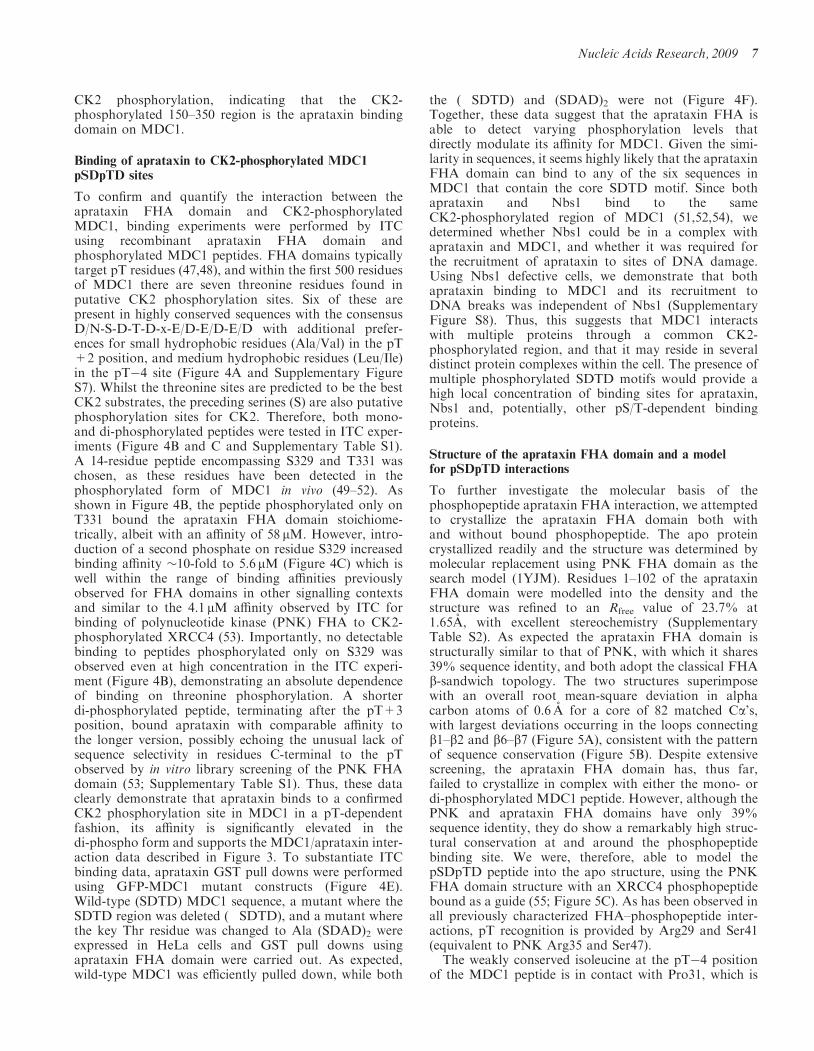
To confirm and quantify the interaction between theaprataxin FHA domain and CK2-phosphorylatedMDC1, binding experiments were performed by ITCusing recombinant aprataxin FHA domain andphosphorylated MDC1 peptides. FHA domains typicallytarget pT residues (47,48), and within the first 500 residuesof MDC1 there are seven threonine residues found inputative CK2 phosphorylation sites. Six of these arepresent in highly conserved sequences with the consensusD/N-S-D-T-D-x-E/D-E/D-E/D with additional prefer-ences for small hydrophobic residues (Ala/Val) in the pT+2 position, and medium hydrophobic residues (Leu/Ile)in the pT�4 site (Figure 4A and Supplementary FigureS7). Whilst the threonine sites are predicted to be the bestCK2 substrates, the preceding serines (S) are also putativephosphorylation sites for CK2. Therefore, both mono-and di-phosphorylated peptides were tested in ITC exper-iments (Figure 4B and C and Supplementary Table S1).A 14-residue peptide encompassing S329 and T331 waschosen, as these residues have been detected in thephosphorylated form of MDC1 in vivo (49–52). Asshown in Figure 4B, the peptide phosphorylated only onT331 bound the aprataxin FHA domain stoichiome-trically, albeit with an affinity of 58 mM. However, intro-duction of a second phosphate on residue S329 increasedbinding affinity �10-fold to 5.6mM (Figure 4C) which iswell within the range of binding affinities previouslyobserved for FHA domains in other signalling contextsand similar to the 4.1mM affinity observed by ITC forbinding of polynucleotide kinase (PNK) FHA to CK2-phosphorylated XRCC4 (53). Importantly, no detectablebinding to peptides phosphorylated only on S329 wasobserved even at high concentration in the ITC experi-ment (Figure 4B), demonstrating an absolute dependenceof binding on threonine phosphorylation. A shorterdi-phosphorylated peptide, terminating after the pT+3position, bound aprataxin with comparable affinity tothe longer version, possibly echoing the unusual lack ofsequence selectivity in residues C-terminal to the pTobserved by in vitro library screening of the PNK FHAdomain (53; Supplementary Table S1). Thus, these dataclearly demonstrate that aprataxin binds to a confirmedCK2 phosphorylation site in MDC1 in a pT-dependentfashion, its affinity is significantly elevated in thedi-phospho form and supports the MDC1/aprataxin inter-action data described in Figure 3. To substantiate ITCbinding data, aprataxin GST pull downs were performedusing GFP-MDC1 mutant constructs (Figure 4E).Wild-type (SDTD) MDC1 sequence, a mutant where theSDTD region was deleted (�SDTD), and a mutant wherethe key Thr residue was changed to Ala (SDAD)2 wereexpressed in HeLa cells and GST pull downs usingaprataxin FHA domain were carried out. As expected,wild-type MDC1 was efficiently pulled down, while both
the (�SDTD) and (SDAD)2 were not (Figure 4F).Together, these data suggest that the aprataxin FHA isable to detect varying phosphorylation levels thatdirectly modulate its affinity for MDC1. Given the simi-larity in sequences, it seems highly likely that the aprataxinFHA domain can bind to any of the six sequences inMDC1 that contain the core SDTD motif. Since bothaprataxin and Nbs1 bind to the sameCK2-phosphorylated region of MDC1 (51,52,54), wedetermined whether Nbs1 could be in a complex withaprataxin and MDC1, and whether it was required forthe recruitment of aprataxin to sites of DNA damage.Using Nbs1 defective cells, we demonstrate that bothaprataxin binding to MDC1 and its recruitment toDNA breaks was independent of Nbs1 (SupplementaryFigure S8). Thus, this suggests that MDC1 interactswith multiple proteins through a common CK2-phosphorylated region, and that it may reside in severaldistinct protein complexes within the cell. The presence ofmultiple phosphorylated SDTD motifs would provide ahigh local concentration of binding sites for aprataxin,Nbs1 and, potentially, other pS/T-dependent bindingproteins.
Structure of the aprataxin FHA domain and a modelfor pSDpTD interactions
To further investigate the molecular basis of thephosphopeptide aprataxin FHA interaction, we attemptedto crystallize the aprataxin FHA domain both withand without bound phosphopeptide. The apo proteincrystallized readily and the structure was determined bymolecular replacement using PNK FHA domain as thesearch model (1YJM). Residues 1–102 of the aprataxinFHA domain were modelled into the density and thestructure was refined to an Rfree value of 23.7% at1.65A, with excellent stereochemistry (SupplementaryTable S2). As expected the aprataxin FHA domain isstructurally similar to that of PNK, with which it shares39% sequence identity, and both adopt the classical FHAb-sandwich topology. The two structures superimposewith an overall root mean-square deviation in alphacarbon atoms of 0.6 A for a core of 82 matched Ca’s,with largest deviations occurring in the loops connectingb1–b2 and b6–b7 (Figure 5A), consistent with the patternof sequence conservation (Figure 5B). Despite extensivescreening, the aprataxin FHA domain has, thus far,failed to crystallize in complex with either the mono- ordi-phosphorylated MDC1 peptide. However, although thePNK and aprataxin FHA domains have only 39%sequence identity, they do show a remarkably high struc-tural conservation at and around the phosphopeptidebinding site. We were, therefore, able to model thepSDpTD peptide into the apo structure, using the PNKFHA domain structure with an XRCC4 phosphopeptidebound as a guide (55; Figure 5C). As has been observed inall previously characterized FHA–phosphopeptide inter-actions, pT recognition is provided by Arg29 and Ser41(equivalent to PNK Arg35 and Ser47).The weakly conserved isoleucine at the pT�4 position
of the MDC1 peptide is in contact with Pro31, which is
Nucleic Acids Research, 2009 7
Figure 4. Aprataxin binds to a conserved di-phosphorylated CK2 motif in MDC1. (A) MDC1 contains six conserved motifs with a core SDTDmotif that is di-phosphorylated in vivo (48–50). (B) ITC binding isotherms for interaction of wild-type aprataxin FHA with pSDTD and SDpTDphosphopeptides (C, D) ITC binding isotherms for the titration of pSDpTD phosphopeptide into wild-type and K38A aprataxin FHA, respectively.(E) Diagram of GFP-MDC1 mutants used in the pull-down assays. (F) Aprataxin FHA GST pull downs from HeLa cells expressing wild-typeGFP-MDC1, a mutant containing a deletion of the SDTD region (�SDTD), and a mutant where the Thr residues contained in the SDTD sequencewere replaced by Ala (SDAD)2.
8 Nucleic Acids Research, 2009
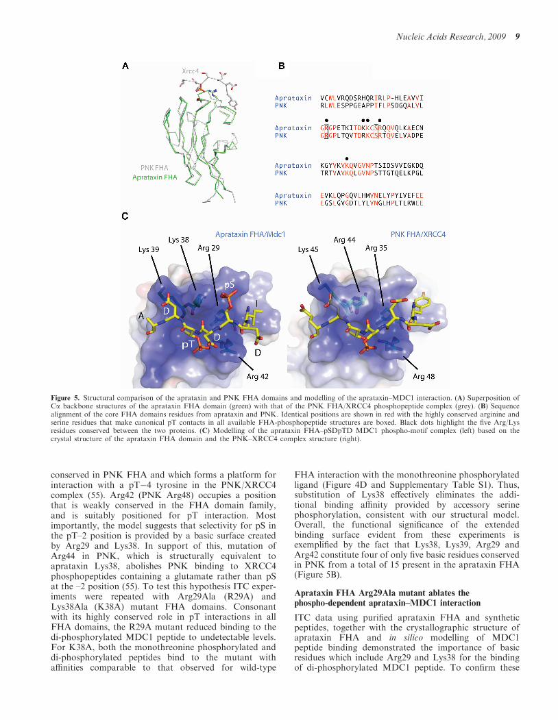
conserved in PNK FHA and which forms a platform forinteraction with a pT�4 tyrosine in the PNK/XRCC4complex (55). Arg42 (PNK Arg48) occupies a positionthat is weakly conserved in the FHA domain family,and is suitably positioned for pT interaction. Mostimportantly, the model suggests that selectivity for pS inthe pT–2 position is provided by a basic surface createdby Arg29 and Lys38. In support of this, mutation ofArg44 in PNK, which is structurally equivalent toaprataxin Lys38, abolishes PNK binding to XRCC4phosphopeptides containing a glutamate rather than pSat the –2 position (55). To test this hypothesis ITC exper-iments were repeated with Arg29Ala (R29A) andLys38Ala (K38A) mutant FHA domains. Consonantwith its highly conserved role in pT interactions in allFHA domains, the R29A mutant reduced binding to thedi-phosphorylated MDC1 peptide to undetectable levels.For K38A, both the monothreonine phosphorylated anddi-phosphorylated peptides bind to the mutant withaffinities comparable to that observed for wild-type
FHA interaction with the monothreonine phosphorylatedligand (Figure 4D and Supplementary Table S1). Thus,substitution of Lys38 effectively eliminates the addi-tional binding affinity provided by accessory serinephosphorylation, consistent with our structural model.Overall, the functional significance of the extendedbinding surface evident from these experiments isexemplified by the fact that Lys38, Lys39, Arg29 andArg42 constitute four of only five basic residues conservedin PNK from a total of 15 present in the aprataxin FHA(Figure 5B).
Aprataxin FHA Arg29Ala mutant ablates thephospho-dependent aprataxin–MDC1 interaction
ITC data using purified aprataxin FHA and syntheticpeptides, together with the crystallographic structure ofaprataxin FHA and in silico modelling of MDC1peptide binding demonstrated the importance of basicresidues which include Arg29 and Lys38 for the bindingof di-phosphorylated MDC1 peptide. To confirm these
Figure 5. Structural comparison of the aprataxin and PNK FHA domains and modelling of the aprataxin–MDC1 interaction. (A) Superposition ofCa backbone structures of the aprataxin FHA domain (green) with that of the PNK FHA/XRCC4 phosphopeptide complex (grey). (B) Sequencealignment of the core FHA domains residues from aprataxin and PNK. Identical positions are shown in red with the highly conserved arginine andserine residues that make canonical pT contacts in all available FHA-phosphopeptide structures are boxed. Black dots highlight the five Arg/Lysresidues conserved between the two proteins. (C) Modelling of the aprataxin FHA–pSDpTD MDC1 phospho-motif complex (left) based on thecrystal structure of the aprataxin FHA domain and the PNK–XRCC4 complex structure (right).
Nucleic Acids Research, 2009 9
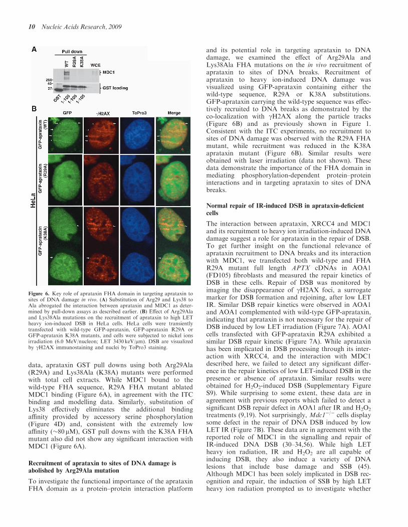
data, aprataxin GST pull downs using both Arg29Ala(R29A) and Lys38Ala (K38A) mutants were performedwith total cell extracts. While MDC1 bound to thewild-type FHA sequence, R29A FHA mutant ablatedMDC1 binding (Figure 6A), in agreement with the ITCbinding and modelling data. Similarly, substitution ofLys38 effectively eliminates the additional bindingaffinity provided by accessory serine phosphorylation(Figure 4D) and, consistent with the extremely lowaffinity (�80 mM), GST pull downs with the K38A FHAmutant also did not show any significant interaction withMDC1 (Figure 6A).
Recruitment of aprataxin to sites of DNA damage isabolished by Arg29Ala mutation
To investigate the functional importance of the aprataxinFHA domain as a protein–protein interaction platform
and its potential role in targeting aprataxin to DNAdamage, we examined the effect of Arg29Ala andLys38Ala FHA mutations on the in vivo recruitment ofaprataxin to sites of DNA breaks. Recruitment ofaprataxin to heavy ion-induced DNA damage wasvisualized using GFP-aprataxin containing either thewild-type sequence, R29A or K38A substitutions.GFP-aprataxin carrying the wild-type sequence was effec-tively recruited to DNA breaks as demonstrated by theco-localization with gH2AX along the particle tracks(Figure 6B) and as previously shown in Figure 1.Consistent with the ITC experiments, no recruitment tosites of DNA damage was observed with the R29A FHAmutant, while recruitment was reduced in the K38Aaprataxin mutant (Figure 6B). Similar results wereobtained with laser irradiation (data not shown). Thesedata demonstrate the importance of the FHA domain inmediating phosphorylation-dependent protein–proteininteractions and in targeting aprataxin to sites of DNAbreaks.
Normal repair of IR-induced DSB in aprataxin-deficientcells
The interaction between aprataxin, XRCC4 and MDC1and its recruitment to heavy ion irradiation-induced DNAdamage suggest a role for aprataxin in the repair of DSB.To get further insight on the functional relevance ofaprataxin recruitment to DNA breaks and its interactionwith MDC1, we transfected both wild-type and FHAR29A mutant full length APTX cDNAs in AOA1(FD105) fibroblasts and measured the repair kinetics ofDSB in these cells. Repair of DSB was monitored byimaging the disappearance of gH2AX foci, a surrogatemarker for DSB formation and rejoining, after low LETIR. Similar DSB repair kinetics were observed in AOA1and AOA1 complemented with wild-type GFP-aprataxin,indicating that aprataxin is not necessary for the repair ofDSB induced by low LET irradiation (Figure 7A). AOA1cells transfected with GFP-aprataxin R29A exhibited asimilar DSB repair kinetic (Figure 7A). While aprataxinhas been implicated in DSB processing through its inter-action with XRCC4, and the interaction with MDC1described here, we failed to detect any significant differ-ence in the repair kinetics of low LET-induced DSB in thepresence or absence of aprataxin. Similar results wereobtained for H2O2-induced DSB (Supplementary FigureS9). While surprising to some extent, these data are inagreement with previous reports which failed to detect asignificant DSB repair defect in AOA1 after IR and H2O2
treatments (9,19). Not surprisingly, Mdc1�/� cells displaysome defect in the repair of DNA DSB induced by lowLET IR (Figure 7B). These data are in agreement with thereported role of MDC1 in the signalling and repair ofIR-induced DNA DSB (30–34,56). While high LETheavy ion radiation, IR and H2O2 are all capable ofinducing DSB, they also induce a variety of DNAlesions that include base damage and SSB (45).Although MDC1 has been solely implicated in DSB rec-ognition and repair, the induction of SSB by high LETheavy ion radiation prompted us to investigate whether
Figure 6. Key role of aprataxin FHA domain in targeting aprataxin tosites of DNA damage in vivo. (A) Substitution of Arg29 and Lys38 toAla abrogated the interaction between aprataxin and MDC1 as deter-mined by pull-down assays as described earlier. (B) Effect of Arg29Alaand Lys38Ala mutations on the recruitment of aprataxin to high LETheavy ion-induced DSB in HeLa cells. HeLa cells were transientlytransfected with wild-type GFP-aprataxin, GFP-aprataxin R29A orGFP-aprataxin K38A mutants, and cells were subjected to nickel ionsirradiation (6.0 MeV/nucleon; LET 3430 keV/mm). DSB are visualizedby gH2AX immunostaining and nuclei by ToPro3 staining.
10 Nucleic Acids Research, 2009
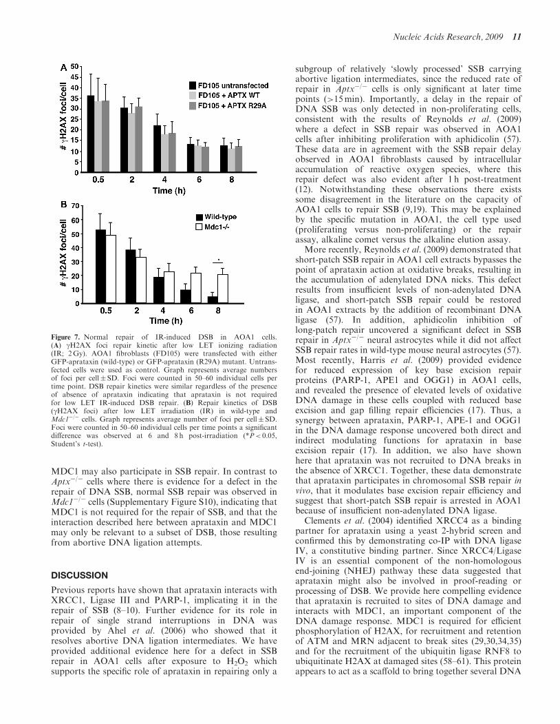
MDC1 may also participate in SSB repair. In contrast toAptx�/� cells where there is evidence for a defect in therepair of DNA SSB, normal SSB repair was observed inMdc1�/� cells (Supplementary Figure S10), indicating thatMDC1 is not required for the repair of SSB, and that theinteraction described here between aprataxin and MDC1may only be relevant to a subset of DSB, those resultingfrom abortive DNA ligation attempts.
DISCUSSION
Previous reports have shown that aprataxin interacts withXRCC1, Ligase III and PARP-1, implicating it in therepair of SSB (8–10). Further evidence for its role inrepair of single strand interruptions in DNA wasprovided by Ahel et al. (2006) who showed that itresolves abortive DNA ligation intermediates. We haveprovided additional evidence here for a defect in SSBrepair in AOA1 cells after exposure to H2O2 whichsupports the specific role of aprataxin in repairing only a
subgroup of relatively ‘slowly processed’ SSB carryingabortive ligation intermediates, since the reduced rate ofrepair in Aptx�/� cells is only significant at later timepoints (>15min). Importantly, a delay in the repair ofDNA SSB was only detected in non-proliferating cells,consistent with the results of Reynolds et al. (2009)where a defect in SSB repair was observed in AOA1cells after inhibiting proliferation with aphidicolin (57).These data are in agreement with the SSB repair delayobserved in AOA1 fibroblasts caused by intracellularaccumulation of reactive oxygen species, where thisrepair defect was also evident after 1 h post-treatment(12). Notwithstanding these observations there existssome disagreement in the literature on the capacity ofAOA1 cells to repair SSB (9,19). This may be explainedby the specific mutation in AOA1, the cell type used(proliferating versus non-proliferating) or the repairassay, alkaline comet versus the alkaline elution assay.More recently, Reynolds et al. (2009) demonstrated that
short-patch SSB repair in AOA1 cell extracts bypasses thepoint of aprataxin action at oxidative breaks, resulting inthe accumulation of adenylated DNA nicks. This defectresults from insufficient levels of non-adenylated DNAligase, and short-patch SSB repair could be restoredin AOA1 extracts by the addition of recombinant DNAligase (57). In addition, aphidicolin inhibition oflong-patch repair uncovered a significant defect in SSBrepair in Aptx�/� neural astrocytes while it did not affectSSB repair rates in wild-type mouse neural astrocytes (57).Most recently, Harris et al. (2009) provided evidencefor reduced expression of key base excision repairproteins (PARP-1, APE1 and OGG1) in AOA1 cells,and revealed the presence of elevated levels of oxidativeDNA damage in these cells coupled with reduced baseexcision and gap filling repair efficiencies (17). Thus, asynergy between aprataxin, PARP-1, APE-1 and OGG1in the DNA damage response uncovered both direct andindirect modulating functions for aprataxin in baseexcision repair (17). In addition, we also have shownhere that aprataxin was not recruited to DNA breaks inthe absence of XRCC1. Together, these data demonstratethat aprataxin participates in chromosomal SSB repair invivo, that it modulates base excision repair efficiency andsuggest that short-patch SSB repair is arrested in AOA1because of insufficient non-adenylated DNA ligase.Clements et al. (2004) identified XRCC4 as a binding
partner for aprataxin using a yeast 2-hybrid screen andconfirmed this by demonstrating co-IP with DNA ligaseIV, a constitutive binding partner. Since XRCC4/LigaseIV is an essential component of the non-homologousend-joining (NHEJ) pathway these data suggested thataprataxin might also be involved in proof-reading orprocessing of DSB. We provide here compelling evidencethat aprataxin is recruited to sites of DNA damage andinteracts with MDC1, an important component of theDNA damage response. MDC1 is required for efficientphosphorylation of H2AX, for recruitment and retentionof ATM and MRN adjacent to break sites (29,30,34,35)and for the recruitment of the ubiquitin ligase RNF8 toubiquitinate H2AX at damaged sites (58–61). This proteinappears to act as a scaffold to bring together several DNA
Figure 7. Normal repair of IR-induced DSB in AOA1 cells.(A) gH2AX foci repair kinetic after low LET ionizing radiation(IR; 2Gy). AOA1 fibroblasts (FD105) were transfected with eitherGFP-aprataxin (wild-type) or GFP-aprataxin (R29A) mutant. Untrans-fected cells were used as control. Graph represents average numbersof foci per cell� SD. Foci were counted in 50–60 individual cells pertime point. DSB repair kinetics were similar regardless of the presenceof absence of aprataxin indicating that aprataxin is not requiredfor low LET IR-induced DSB repair. (B) Repair kinetics of DSB(gH2AX foci) after low LET irradiation (IR) in wild-type andMdc1�/� cells. Graph represents average number of foci per cell� SD.Foci were counted in 50–60 individual cells per time points a significantdifference was observed at 6 and 8 h post-irradiation (*P< 0.05,Student’s t-test).
Nucleic Acids Research, 2009 11
damage response proteins to mediate and enhance therecognition and signalling of DSB (58–66). Our data addan additional DNA repair protein, aprataxin, to the list ofproteins interacting with MDC1. MDC1 interacts withDNA damage response proteins in a number of differentways. Interaction with gH2AX and the anaphase-promoting complex occurs through its C-terminal,BRCT domain (33,34,65), while it binds DNA-PKthrough an internal set of repeat sequences (32). It alsopossesses an FHA domain (amino acids 55–124) thatmediates interaction with Chk2 and Rad51 (33,66).It employs an S/TQ rich region for ATM-dependent inter-action with the ubiquitin ligase RNF8 (58).We have described here a novel interaction between
MDC1 and aprataxin, involving an extended cluster ofCK2 phosphorylation sites (S-D-T-D) located down-stream from the MDC1 FHA domain (amino acids150–350). This was confirmed by the use of CK2 inhibi-tors, binding studies after in vitro CK2 phosphorylation,GST pull downs and modelling studies based on bindingof mono- and di-phosphorylated peptides to the aprataxinFHA domain and the X-ray crystal structure of thisdomain. We provide here compelling evidence for a rolefor CK2 activity in the interaction of this region of MDC1with aprataxin and show that a phosphopeptide contain-ing a core pSDpTD motif corresponding to one of thesesites binds to the FHA domain of aprataxin with highaffinity. While a single threonine phosphorylation isboth necessary and sufficient for binding, affinity is sub-stantially increased by secondary serine phosphorylation.This, in turn, appears to provide for accessory interactionswith an extended FHA phosphopeptide binding surfacenot previously documented. The data presented hereuncover another important role for CK2 in mediatingthe interaction between aprataxin and the DSB repairprotein MDC1.In three recent reports constitutive phosphorylation of
the N-terminal SDT repeats of MDC1 by CK2 has beendemonstrated in the recruitment of the MRN complex tosites of DSB through direct interactions with the FHAdomain of Nbs1 (51,52,54). Spycher et al. (54) reportedthat doubly phosphorylated pSDpT motifs regulate theaccumulation and retention of the MRN complex in theDSB-flanking chromatin, and depletion of CK2 disruptedthe MDC1–MRN complex and DNA damage-inducedNbs1 foci formation. Moreover, disruption of the SDTphosphoacceptor sites on MDC1 prevented therecruitment of Nbs1 to damaged chromatin (51,52,54).We have demonstrated that Nbs1 is not required forthe binding of aprataxin to MDC1 and its recruitmentto sites of DNA damage. Therefore, MDC1 interactswith multiple proteins through a commonCK2-phosphorylated region and may reside in severaldistinct protein complexes within the cell. It is likely thatthe interaction between MDC1/Nbs1 and MDC1/aprataxin is influenced or regulated by the relative abun-dance of these proteins in the cells. These data togetherwith previous findings that identified a direct role for CK2in the repair of chromosomal DNA strand breaks (67), theCK2-dependent aprataxin/XRCC1 (9,10) and aprataxin/XRCC4 (9) interactions provide growing evidence for
a role for CK2 in maintaining genome integrity bycontributing to protein–protein complexes formationand/or recruitment of DNA damage regulators/repairfactors at sites of DNA lesions.
As shown here, aprataxin re-localizes rapidly to discretefoci along charged particle tracks that overlap withgH2AX foci, a marker of DSB after exposure of cells tohigh LET radiation which gives rise to complex damagein DNA (22). The recruitment and co-localization ofaprataxin to sites of gH2AX-labelled DNA breaks haspreviously been observed using different heavy ionspecies (8,17,28, this work). However, while MDC1completely co-localized with gH2AX in the DSB-flanking microenvironment as reported earlier (68),aprataxin only partially co-localized with MDC1 andgH2AX in smaller foci (microfoci). This suggests thataprataxin may be recruited only to the DSB or asub-group of DNA breaks directly within sites ofcomplex or clustered damage or that its association withDNA damaged sites may be more transient. This transientbehaviour has been described for Nbs1 in the absence ofMDC1 (26,52). In addition, we provide evidence here thatthe lack of MDC1 doesn’t affect the rapid recruitment ofaprataxin to sites of high LET-induced DNA DSB, but incontrast affects the retention of aprataxin at sites of DNAbreaks, supporting a role for MDC1 as a scaffold duringthe DNA damage response. Proteins such as SMC1 andDNA-PKcs that are clearly involved in the DNA damageresponse cannot be localized to sites of DNA damageexcept after exposure to extremely high doses of radiation(68). Spatial-co-localization studies revealed that unlikegH2AX and MDC1, phosphorylated DNA-PKcs waslocalized only at very specific regions, presumably repre-senting sites of DSB within the tracks (69). The presenceof microfoci surrounded by regions of gH2AX-modifiedchromatin after laser-induced DNA damage has beenreported (68). Several factors involved in DNA repairincluding Nbs1 and Mre11 are present in microfocilocated in the centre of microirradiated tracks and sur-rounded by vast regions of gH2AX-modified chromatin(68,70). In the present context these microirradiatedtracks coincide with MDC1-modified chromatin. The het-erogeneity of staining may be explained by the presence ofsignificant amounts of ssDNA adjacent to DSB and/or thepresence of clustered or complex DNA damage (45,46).Exposure of cells to low LET radiation (IR) or H2O2
failed to localize aprataxin to sites of damage (8). Thismay be similar to that observed for SMC1 or DNA-PKwhere extremely high levels of damage are required to seesuch localization (68). Low LET radiation is moresparsely ionizing and thus less damaging to DNA thanthe complex forms of damage arising from high LETexposure which could explain the failure to see aprataxinfoci after low LET radiation (45). The difference incapacity of aprataxin to localize to sites of DNAdamage after the two types of IR correlates well withthe extent of repair of DNA DSB. The rate of repair ofDNA DSB in response to low LET radiation was unaf-fected by aprataxin status of the cells. Similar to previousreports (9,19), the failure to detect any significant DSBrepair defect in the presence or absence of aprataxin
12 Nucleic Acids Research, 2009
after low LET irradiation or high doses of H2O2 suggeststhat aprataxin recruitment to DNA breaks may only berelevant for a sub-group of breaks, possiblymore frequently/effectively induced by high energy andhigh LET heavy ions irradiation. This sub-group ofbreaks could represent those where abortive ligation wasoccurring, a property of AOA1 cells (14).
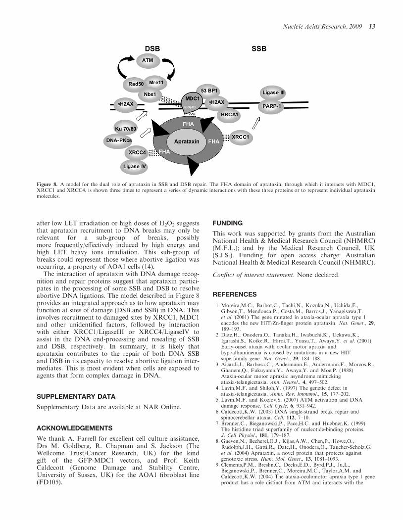
The interaction of aprataxin with DNA damage recog-nition and repair proteins suggest that aprataxin partici-pates in the processing of some SSB and DSB to resolveabortive DNA ligations. The model described in Figure 8provides an integrated approach as to how aprataxin mayfunction at sites of damage (DSB and SSB) in DNA. Thisinvolves recruitment to damaged sites by XRCC1, MDC1and other unidentified factors, followed by interactionwith either XRCC1/LigaseIII or XRCC4/LigaseIV toassist in the DNA end-processing and resealing of SSBand DSB, respectively. In summary, it is likely thataprataxin contributes to the repair of both DNA SSBand DSB in its capacity to resolve abortive ligation inter-mediates. This is most evident when cells are exposed toagents that form complex damage in DNA.
SUPPLEMENTARY DATA
Supplementary Data are available at NAR Online.
ACKNOWLEDGEMENTS
We thank A. Farrell for excellent cell culture assistance,Drs M. Goldberg, R. Chapman and S. Jackson (TheWellcome Trust/Cancer Research, UK) for the kindgift of the GFP-MDC1 vectors, and Prof. KeithCaldecott (Genome Damage and Stability Centre,University of Sussex, UK) for the AOA1 fibroblast line(FD105).
FUNDING
This work was supported by grants from the AustralianNational Health & Medical Research Council (NHMRC)(M.F.L.); and by the Medical Research Council, UK(S.J.S.). Funding for open access charge: AustralianNational Health & Medical Research Council (NHMRC).
Conflict of interest statement. None declared.
REFERENCES
1. Moreira,M.C., Barbot,C., Tachi,N., Kozuka,N., Uchida,E.,Gibson,T., Mendonca,P., Costa,M., Barros,J., Yanagisawa,T.et al. (2001) The gene mutated in ataxia-ocular apraxia type 1encodes the new HIT/Zn-finger protein aprataxin. Nat. Genet., 29,189–193.
2. Date,H., Onodera,O., Tanaka,H., Iwabuchi,K., Uekawa,K.,Igarashi,S., Koike,R., Hiroi,T., Yuasa,T., Awaya,Y. et al. (2001)Early-onset ataxia with ocular motor apraxia andhypoalbuminemia is caused by mutations in a new HITsuperfamily gene. Nat. Genet., 29, 184–188.
3. Aicardi,J., Barbosa,C., Andermann,E., Andermann,F., Morcos,R.,Ghanem,Q., Fukuyama,Y., Awaya,Y. and Moe,P. (1988)Ataxia-ocular motor apraxia: asyndrome mimickingataxia-telangiectasia. Ann. Neurol., 4, 497–502.
4. Lavin,M.F. and Shiloh,Y. (1997) The genetic defect inataxia-telangiectasia. Annu. Rev. Immunol., 15, 177–202.
5. Lavin,M.F. and Kozlov,S. (2007) ATM activation and DNAdamage response. Cell Cycle, 6, 931–942.
6. Caldecott,K.W. (2003) DNA single-strand break repair andspinocerebellar ataxia. Cell, 112, 7–10.
7. Brenner,C., Bieganowski,P., Pace,H.C. and Huebner,K. (1999)The histidine triad superfamily of nucleotide-binding proteins.J. Cell Physiol., 181, 179–187.
8. Gueven,N., Becherel,O.J., Kijas,A.W., Chen,P., Howe,O.,Rudolph,J.H., Gatti,R., Date,H., Onodera,O., Taucher-Scholz,G.et al. (2004) Aprataxin, a novel protein that protects againstgenotoxic stress. Hum. Mol. Genet., 13, 1081–1093.
9. Clements,P.M., Breslin,C., Deeks,E.D., Byrd,P.J., Ju,L.,Bieganowski,P., Brenner,C., Moreira,M.C., Taylor,A.M. andCaldecott,K.W. (2004) The ataxia-oculomotor apraxia type 1 geneproduct has a role distinct from ATM and interacts with the
Figure 8. A model for the dual role of aprataxin in SSB and DSB repair. The FHA domain of aprataxin, through which it interacts with MDC1,XRCC1 and XRCC4, is shown three times to represent a series of dynamic interactions with these three proteins or to represent individual aprataxinmolecules.
Nucleic Acids Research, 2009 13
DNA strand break repair proteins XRCC1 and XRCC4.DNA Repair, 3, 1493–1502.
10. Luo,H., Chan,D.W., Yang,T., Rodriguez,M., Chen,B.P., Leng,M.,Mu,J.J., Chen,D., Songyang,Z., Wang,Y. et al. (2004) A newXRCC1-containing complex and its role in cellular survival ofmethyl methane sulfonate treatment. Mol. Cell. Biol., 24,8356–8365.
11. Mosesso,P., Piane,M., Palitti,F., Pepe,G., Penna,S. and Chessa,L.(2005) The novel human gene aprataxin is directly involved inDNA single-strand break repair. Cell. Mol. Life Sci., 62, 485–491.
12. Hirano,M., Yamamoto,A., Mori,T., Lan,L., Iwamoto,T.A.,Aoki,M., Shimada,K., Furiya,Y., Kariya,S., Asai,H. et al. (2007)DNA single-strand break repair is impaired in aprataxin-relatedataxia. Ann. Neurol., 61, 162–174.
13. Gueven,N., Chen,P., Nakamura,J., Becherel,O.J., Kijas,A.W.,Grattan-Smith,P. and Lavin,M.F. (2007) A subgroup ofspinocerebellar ataxias defective in DNA damage responses.Neuroscience, 145, 1418–1425.
14. Ahel,I., Rass,U., El-Khamisy,S.F., Katyal,S., Clements,P.M.,McKinnon,P.J., Caldecott,K.W. and West,S.C. (2006) Theneurodegenerative disease protein aprataxin resolves abortiveDNA ligation intermediates. Nature, 443, 713–716.
15. Rass,U., Ahel,I. and West,S.C. (2007) Actions of aprataxin inmultiple DNA repair pathways. J. Biol. Chem., 282, 9469–9474.
16. Lehman,I.R. (1974) DNA ligase: structure, mechanism, andfunction. Science, 186, 790–797.
17. Harris,J.L., Jakob,B., Taucher-Scholz,G., Becherel,O.J. andLavin,M.F. (2009) Aprataxin combines with Poly-ADP RibosePolymerase 1 (PARP-1) and Apurinic Endonuclease 1 (APE1) toprotect the genome against oxidative damage. Hum. Mol. Genet.,18, 4102–4117.
18. Kijas,A.W., Harris,J.L., Harris,J.M. and Lavin,M.F. (2006)Aprataxin forms a discrete branch in the HIT (histidine triad)superfamily of proteins with both DNA/RNA bindingand nucleotide hydrolase activities. J. Biol. Chem., 281,13939–13948.
19. El-Khamisy,S.F., Katyal,S., Patel,P., Ju,L., McKinnon,P.J. andCaldecott,K.W. (2009) Synergistic decrease of DNA single-strandbreak repair rates in mouse neural cells lacking both Tdp1 andaprataxin. DNA Repair, 8, 760–766.
20. Paull,T.T., Rogakou,E.P., Yamazaki,V., Kirchgessner,C.U.,Gellert,M. and Bonner,W.M. (2000) A critical role for histoneH2AX in recruitment of repair factors to nuclear foci after DNAdamage. Curr. Biol., 10, 886–895.
21. Rappold,I., Iwabuchi,K., Date,T. and Chen,J. (2001) Tumorsuppressor p53 binding protein 1 (53BP1) is involved in DNAdamage-signaling pathways. J. Cell Biol., 153, 613–620.
22. Rogakou,E.P., Pilch,D.R., Orr,A.H., Ivanova,V.S. andBonner,W.M. (1998) DNA double-stranded breaks induce histoneH2AX phosphorylation on serine 139. J. Biol. Chem., 273,5858–5868.
23. Okano,S., Kanno,S., Nakajima,S. and Yasui,A. (2000) Cellularresponses and repair of single-strand breaks introduced by UVdamage endonuclease in mammalian cells. J. Biol. Chem., 275,32635–32641.
24. Limoli,C.L. and Ward,J.F. (1993) A new method for introducingdouble-strand breaks into cellular DNA. Radiat. Res., 134,160–169.
25. Katsumi,S., Kobayashi,N., Imoto,K. et al. (2001) In situvisualization of ultraviolet-light-induced DNA damage repair inlocally irradiated human fibroblasts. J. Invest. Dermatol., 117,1156–1161.
26. Lukas,C., Melander,F., Stucki,M., Falck,J., Bekker-Jensen,S.,Goldberg,M., Lerenthal,Y., Jackson,S.P., Bartek,J. and Lukas,J.(2004) Mdc1 couples DNA double-strand break recognition byNbs1 with its H2AX-dependent chromatin retention. EMBO J.,23, 2674–2683.
27. Jakob,B., Scholz,M. and Taucher-Scholz,G. (2003) Biologicalimaging of heavy charged-particle tracks. Radiat. Res., 159,676–684.
28. Jakob,B., Rudolph,J.H., Gueven,N., Lavin,M.F. andTaucher-Scholz,G. (2005) Live cell imaging of heavy-ion-inducedradiation responses by beamline microscopy. Radiat. Res., 163,681–690.
29. Stewart,G.S., Wang,B., Bignell,C.R., Taylor,A.M. and Elledge,S.J.(2003) MDC1 is a mediator of the mammalian DNA damagecheckpoint. Nature, 421, 961–966.
30. Goldberg,M., Stucki,M., Falck,J., D’Amours,D., Rahman,D.,Pappin,D., Bartek,J. and Jackson,S.P. (2003) MDC1 is requiredfor the intra-S-phase DNA damage checkpoint. Nature, 421,952–956.
31. Lou,Z., Chini,C.C., Minter-Dykhouse,K. and Chen,J. (2003)Mediator of DNA damage checkpoint protein 1 regulates BRCA1localization and phosphorylation in DNA damage checkpointcontrol. J. Biol. Chem., 278, 13599–13602.
32. Lou,Z., Chen,B.P., Asaithamby,A., Minter-Dykhouse,K.,Chen,D.J. and Chen,J. (2004) MDC1 regulates DNA-PKautophosphorylation in response to DNA damage. J. Biol. Chem.,279, 46359–46362.
33. Lou,Z., Minter-Dykhouse,K., Wu,X. and Chen,J. (2003) MDC1is coupled to activated CHK2 in mammalian DNA damageresponse pathways. Nature, 421, 957–961.
34. Stucki,M., Clapperton,J.A., Mohammad,D., Yaffe,M.B.,Smerdon,S.J. and Jackson,S.P. (2005) MDC1 directly bindsphosphorylated histone H2AX to regulate cellular responses toDNA double-strand breaks. Cell, 123, 1213–1226.
35. Wu,L., Luo,K., Lou,Z. and Chen,J. (2008) MDC1 regulatesintra-S-phase checkpoint by targeting NBS1 to DNAdouble-strand breaks. Proc. Natl Acad. Sci. USA, 105,11200–11205.
36. Mahajan,A., Yuan,C., Lee,H., Chen,E.S., Wu,P.Y. and Tsai,M.D.(2008) Structure and function of the phosphothreonine-specificFHA domain. Sci. Signal., 1, re12.
37. Becherel,O.J., Gueven,N., Birrell,G.W., Schreiber,V.,Suraweera,A., Jakob,B., Taucher-Scholz,G. and Lavin,M.F.(2006) Nucleolar localization of aprataxin is dependent oninteraction with nucleolin and on active ribosomal DNAtranscription. Hum. Mol. Genet., 15, 2239–2249.
38. Otwinowski,Z. and Minor,W. (1997) Processing of X-raydiffraction data collected in oscillation mode. Methods Enzymol.,276, 307–326.
39. Collaborative Computational Project, Number 4. (1994) TheCCP4 suite: programs for protein crystallography. ActaCrystallogr. D. Biol. Crystallogr., 50, 760–763.
40. Navaza,J., Saludjian,P. and Charles,W.C. Jr (1997) AMoRe: anautomated molecular replacement program package. MethodsEnzymol., 276, 581–594.
41. Murshudov,G.N., Vagin,A.A. and Dodson,E.J. (1997) Refinementof macromolecular structures by the maximum-likelihood method.Acta Crystallogr. D. Biol. Crystallogr., 53, 240–255.
42. Emsley,P. and Cowtan,K. (2004) Coot: model-building tools formolecular graphics. Acta Crystallogr. D. Biol. Crystallogr., 60,2126–2132.
43. Suraweera,A., Becherel,O.J., Chen,P., Rundle,N., Woods,R.,Nakamura,J., Gatei,M., Criscuolo,C., Filla,A., Chessa,L. et al.(2007) Senataxin defective in ataxia oculomotor apraxia type 2is involved in the defence against oxidative DNA damage.J. Cell Biol., 177, 969–979.
44. Epe,B. and Hegler,J. (1994) Oxidative DNA damage:endonuclease fingerprinting. Methods Enzymol., 234, 122–131.
45. Ward,J.F. (1994) The complexity of DNA damage:relevance to biological consequences. Int. J. Radiat. Biol., 66,427–432.
46. Jakob,B., Splinter,J. and Taucher-Scholz,G. (2009) Positionalstability of damaged chromatin domains along radiation tracks inmammalian cells. Radiat. Res., 171, 405–418.
47. Durocher,D., Henckel,J., Fersht,A.R. and Jackson,S.P. (1999) TheFHA domain is a modular phosphopeptide recognition motif.Mol. Cell, 4, 387–394.
48. Durocher,D., Taylor,I.A., Sarbassova,D., Haire,L.F.,Westcott,S.L., Jackson,S.P., Smerdon,S.J. and Yaffe,M.B. (2000)The molecular basis of FHA domain:phosphopeptide bindingspecificity and implications for phospho-dependent signalingmechanisms. Mol. Cell, 6, 1169–1182.
49. Beausoleil,S.A., Jedrychowski,M., Schwartz,D., Elias,J.E.,Villen,J., Li,J., Cohn,M.A., Cantley,L.C. and Gygi,S.P. (2004)Large-scale characterization of HeLa cell nuclearphosphoproteins. Proc. Natl Acad. Sci. USA, 101, 12130–12135.
14 Nucleic Acids Research, 2009
50. Olsen,J.V., Blagoev,B., Gnad,F., Macek,B., Kumar,C.,Mortensen,P. and Mann,M. (2006) Global, in vivo, andsite-specific phosphorylation dynamics in signaling networks. Cell,127, 635–648.
51. Melander,F., Bekker-Jensen,S., Falck,J., Bartek,J., Mailand,N.and Lukas,J. (2008) Phosphorylation of SDT repeats in theMDC1 N terminus triggers retention of NBS1 at the DNAdamage-modified chromatin. J. Cell Biol., 181, 213–226.
52. Chapman,J.R. and Jackson,S.P. (2008) Phospho-dependentinteractions between NBS1 and MDC1 mediate chromatinretention of the MRN complex at sites of DNA damage.EMBO Rep., 9, 795–801.
53. Koch,C.A., Agyei,R., Galicia,S., Metalnikov,P., O’Donnell,P.,Starostine,A., Weinfeld,M. and Durocher,D. (2004) Xrcc4physically links DNA end processing by polynucleotide kinase toDNA ligation by DNA ligase IV. EMBO J., 23, 3874–3885.
54. Spycher,C., Miller,E.S., Townsend,K., Pavic,L., Morrice,N.A.,Janscak,P., Stewart,G.S. and Stucki,M. (2008) Constitutivephosphorylation of MDC1 physically links the MRE11–RAD50–NBS1 complex to damaged chromatin. J. Cell Biol., 181,227–240.
55. Bernstein,N.K., Williams,R.S., Rakovszky,M.L., Cui,D.,Green,R., Karimi-Busheri,F., Mani,R.S., Galcia,S., Koch,C.A.,Cass,C.E. et al. (2005) The molecular architecture of themammalian DNA repair enzyme, polynucleotide kinase.Mol. Cell, 17, 657–670.
56. Lou,Z., Minter-Dykhouse,K., Franco,S., Gostissa,M.,Rivera,M.A., Celeste,A., Manis,J.P., van Deursen,J.,Nussenzweig,A., Paull,T.T. et al. (2006) MDC1 maintainsgenomic stability by participating in the amplification ofATM-dependent DNA damage signals. Mol. Cell, 21, 187–200.
57. Reynolds,J.J., El-Khamisy,S.F., Katyal,S., Clements,P.,McKinnon,P.J. and Caldecott,K.W. (2009) Defective DNAligation during short-patch single-strand break repair in ataxiaoculomotor apraxia 1. Mol. Cell. Biol., 29, 1354–1362.
58. Huen,M.S., Grant,R., Manke,I., Minn,K., Yu,X., Yaffe,M.B. andChen,J. (2007) RNF8 transduces the DNA-damage signal viahistone ubiquitylation and checkpoint protein assembly. Cell, 131,901–914.
59. Mailand,N., Bekker-Jensen,S., Faustrup,H., Melander,F.,Bartek,J., Lukas,C. and Lukas,J. (2007) RNF8 ubiquitylateshistones at DNA double-strand breaks and promotes assembly ofrepair proteins. Cell, 131, 887–900.
60. Kolas,N.K., Chapman,J.R., Nakada,S., Ylanko,J., Chahwan,R.,Sweeney,F.D., Panier,S., Mendez,M., Wildenhain,J.,Thomson,T.M. et al. (2007) Orchestration of the DNA-damageresponse by the RNF8 ubiquitin ligase. Science, 318, 1637–1640.
61. Sakasai,R. and Tibbetts,R. (2008) RNF8-dependent andRNF8-independent regulation of 53BP1 in response to DNAdamage. J. Biol. Chem., 283, 13549–13555.
62. Eliezer,Y., Argaman,L., Rhie,A., Doherty,A.J. and Goldberg,M.(2008) The direct interaction between 53BP1 and MDC1 isrequired for the recruitment of 53BP1 to sites of damage. J. Biol.Chem., 284, 426–435.
63. Doil,C., Mailand,N., Bekker-Jensen,S., Menard,P., Larsen,D.H.,Pepperkok,R., Ellenberg,J., Panier,S., Durocher,D., Bartek,J.et al. (2009) RNF168 binds and amplifies ubiquitin conjugates ondamaged chromosomes to allow accumulation of repair proteins.Cell, 136, 435–446.
64. Stewart,G.S., Panier,S., Townsend,K., Al-Hakim,A.K.,Kolas,N.K., Miller,E.S., Nakada,S., Ylanko,J., Olivarius,S.,Mendez,M. et al. (2009) The RIDDLE syndrome proteinmediates a ubiquitin-dependent signaling cascade at sites of DNAdamage. Cell, 136, 420–434.
65. Coster,G., Hayouka,Z., Argaman,L., Strauss,C., Friedler,A.,Brandeis,M. and Goldberg,M. (2007) The DNA damage responsemediator MDC1 directly interacts with the anaphase-promotingcomplex/cyclosome. J. Biol. Chem., 282, 32053–32064.
66. Zhang,J., Ma,Z., Treszezamsky,A. and Powell,S.N. (2005) MDC1interacts with Rad51 and facilitates homologous recombination.Nat. Struct. Mol. Biol., 12, 902–909.
67. Loizou,J.I., El-Khamisy,S.F., Zlatanou,A., Moore,D.J.,Chan,D.W., Qin,J., Sarno,S., Meggio,F., Pinna,L.A. andCaldecott,K.W. (2004) The protein kinase CK2 facilitates repairof chromosomal DNA single-strand breaks. Cell, 117, 17–28.
68. Bekker-Jensen,S., Lukas,C., Kitagawa,R., Melander,F.,Kastan,M.B., Bartek,J. and Lukas,J. (2006) Spatial organizationof the mammalian genome surveillance machinery in response toDNA strand breaks. J. Cell Biol., 173, 195–206.
69. Asaithamby,A., Uematsu,N., Chatterjee,A., Story,M.D., Burma,S.and Chen,D.J. (2008) Repair of HZE-particle-induced DNAdouble-strand breaks in normal human fibroblasts. Radiat. Res.,169, 437–446.
70. Lisby,M., Antunez de Mayolo,A., Mortensen,U.H. andRothstein,R. (2003) Cell cycle-regulated centers of DNAdouble-strand break repair. Cell Cycle, 2, 479–483.
Nucleic Acids Research, 2009 15
Related Documents