Available online at www.worldscientificnews.com
( Received 17 April 2020; Accepted 06 May 2020; Date of Publication 07 May 2020 )
WSN 145 (2020) 222-233 EISSN 2392-2192
Prevalence and antimicrobial susceptibility of Klebsiella pneumoniae isolated from hospitalized
patients at General Hospital, Etim Ekpo, Akwa Ibom State, Nigeria
I. Kelechi Anosike1, U. Okon Edet1,2,*, G. E. Umoafia3, B. E. Agbo2,
V. O. Ejelonu1 and V. Onyesoro1
1Obong University, Faculty of Natural and Applied Sciences, Department of Microbiology, Obong Ntak, Etim Ekpo, Akwa Ibom State, Nigeria
2University of Calabar, Faculty of Biological Sciences, Department of Microbiology, PMB 1115 Calabar, Cross River State, Nigeria
3Arthur Jarvis University, Faculty of Biological Science, Akpabuyo, Microbiology Department, Cross River State, Nigeria
*E-mail address: [email protected]
ABSTRACT
Klebsiella pneumoniae (KP) was investigated in respondents presenting signs and symptoms of
urinary tract infection (UTI) in a rural community. Using simple random sampling, a total of 360
respondents (144 males and 216 females) were recruited into the study following informed consent and
ethical approval. Mid-stream urine samples were collected from all the respondents aseptically using
standard protocol. Isolation of K. pneumoniae was done using morphological characteristics and various
biochemical tests while antibiotic sensitivity testing using various antibiotics was done using guidelines
of Clinical and Laboratory Standard Institute. A total of 16 samples gave positive culture representing
an overall prevalence of 4.4%. Furthermore, prevalence rates of 2.8%, 5.6%, 47.82%, 4.9%, 3.50%, and
8.60% for males, females, diabetics, in-patients, out-patients, and respondents above 60 years of age
respectively were obtained. Resistance to antibiotics ranged from 37.50 to 56.25% for gentamycin and
nalidixic, and tarivid, respectively. There is need for preventive measures aimed at sensitization of
dwellers in community settings.
World Scientific News 145 (2020) 222-233
-223-
Keywords: Klebsiella pneumonia, Nosocomial, Prevalence, Antibiotic susceptibility, Resistance
1. INTRODUCTION
Klebsiella pneumoniae (KP) is a Gram negative bacterium which belongs to the
Enterobacteriaceae family [1]. They are normal flora of the human intestine and important
human pathogens implicated as the causative agents of several infections including pneumonia,
septicaemia, wound infections, surgical site infections, meningitis and urinary tract infections
(UTI) [1-3] and are second to Escherichia coli in causing a wide spectrum of infections [3].
Klebsiella pneumoniae have been reported to cause increase in the colonization rate of
Klebsiella in patients two weeks after hospital admission, up to two- to four-fold due to their
capacity to spread in clinical settings [4]. Being the most medically important Klebsiella
species, Klebsiella pneumoniae, is responsible for a significant amount of nosocomial urinary
tract infections [5-7]. As an opportunistic pathogen, it causes disease easily in
immunocompromised individuals with underlying medical conditions such as diabetes mellitus
and chronic pulmonary obstruction [2].
Klebsiella pneumoniae is the second most frequently isolated species from UTI, after
Escherichia coli [8, 9]. UTI is usually classified according to the infection sites such as kidney
(in case of pyelonephritis), bladder (in case of cystitis), or urine, and also can be symptomatic
or asymptomatic. The infection rate varies depending on the age, sex, catheterization and
hospitalization [10]. Females usually acquire UTI more often than males due to the anatomical
structure and position of the female external genitalia [5, 11]. Symptoms of UTI include
frequent urination, fowl-smelling cloudy urine, bloody urine, painful urination with a burning
sensation, muscle aches, abdominal pains, nausea and vomiting. People with catheters are more
prone to UTI. However, they only experience fever as a symptom, which makes diagnosis more
difficult [5, 11-13].
KP has been described as a “successful” pathogen as a result of its ability to transfer
resistant genes to other strains vertically or horizontally via plasmids and transposons [14, 15].
In the last two decades, KP have been shown to accumulate antibiotics resistant genes (ARGs)
via various means including de novo mutations and in the process harbouring super resistome
thus, positing itself as an extremely drug resistant pathogen (XDR) [15]. Furthermore, it has
been shown to have the ability to develop multi-drug resistance (MDR) and have been placed
with other MDR pathogens as ESKAPE. The other pathogens include Enterobacter spp,
Staphylococcus aureus, Enterococcus faecium, Pseudomonas aeruginosa and Acinetobacter
baumanni. ESKAPE is as group of six pathogens that exhibit multidrug resistance, virulence
and are commonly associated with nosocomial infections [16, 17].
Antibiotics have been in clinical use for over seven decades now. During this period,
pathogens have evolved as a significant public health concern globally [18-20]. The pace at
which pathogens develop resistance is faster than the rate of development of newer antibiotics;
despite advances in sciences [19, 21]. This has lead to an increasing reports of infection due to
these multidrug resistant pathogens even among the Enterobacteriaceae taxa which K.
pneumoniae is a member [20]. KP has also been shown to develop resistance to the
carbapenems and other routinely used antibiotics such as quinolones in the management of KP
infection spectrum [2, 20, 22].
World Scientific News 145 (2020) 222-233
-224-
Several studies exist that have shown that Klebsiella pneumoniae causes UTIs clinical
setting in cities [3, 20, 26, 34, 36]. However, studies aimed at MDR KP implicated in UTIs are
lacking or non-existent in rural areas with poor health facilities in developing countries like
Nigeria.
Furthermore, KP have been shown to have the capacity to become hyper virulent thereby
increasing the net of susceptible persons to its spectrum of diseases [23]. Thus, this study was
aim to establish the MDR profile of KP isolates implicated in UTI infections in rural
community in Akwa Ibom State, South-South, Nigeria amongst hospitalized patients attending
a community general hospital.
2. MATERIAL AND METHODS
2. 1. Study site
This study was conducted at Etim Ekpo General Hospital located in Etim Ekpo Local
Government Area (LGA) of Akwa Ibom State. The LGA is located on 4º51’ - 5º03’ North of
Equator and Longitude 7º 44’ East of the Greenwich Meridian. It occupies a total area of 183.3
Km2 [24] and as at the 2006 Census, it had male and female populations of 105, 418 people and
55,771, respectively [24]. The General Hospital is managed by both the Local and State
Governments. It provides health care services to the people of the LGA and adjoining
communities and villages. The major occupation of the inhabitants remains agriculture [24].
2. 2. Experimental design and ethical approval
The study design employed in this study was a cross-sectional study design, a type of
observational study that measures outcomes and exposures at the same time using a section of
the population [25]. Ethical approval was obtained from the Hospital Management Ethical
Approval Board, and as well as Obong University Research Directorate.
2. 3. Inclusion and exclusion criteria
Participants were only included in the study if they were admitted in the hospital, consent
to fill our questionnaire, and presenting with signs and symptoms of UTIs. Those that could not
meet these criteria were excluded from the study.
2. 4. Design and administration of questionnaires
Open ended questionnaires were designed to obtain sociodemographic and risk factors of
the respondents. The questionnaires were administered to the respondents following their
informed consent and assurance of the confidentiality of their data.
2. 5. Collection of samples
The study was conducted from February to August, 2019. Following informed consent,
simple random sampling was used to recruit respondents. From all the 360 respondents early
morning mid-stream urine samples were collected (144 males and 216 females) aseptically and
at room temperature.
All samples were appropriately labelled and transported to Obong University
microbiology laboratory in an ice pack for microbiological analysis.
World Scientific News 145 (2020) 222-233
-225-
2. 6. Inoculation and identification of the isolates
The samples were aseptically inoculated on MacConkey agar using sterile wire loop and
incubated at 37 ºC for 24 hours. Discrete, mucoid, rose pink colonies (lactose fermenters) seen
on MacConkey agar were picked with a sterile wire loop and sub-cultured onto another freshly
prepared MacConkey agar plates to obtain pure cultures. K. pneumoniae isolates were identified
using cultural characteristics, Gram reaction, microscopic appearance and biochemical tests as
previously described [26-28].
2. 7. Antibiotic susceptibility test
Using disc diffusion method, antimicrobial susceptibility test was performed following
the guidelines established by the Clinical and Laboratory Standard Institute (CLSI) [29].
Colonies of Klebsiella pneumonia isolates were inoculated aseptically in nutrient broth in test
tubes and adjusted to 0.5 McFarland standard which corresponds to 1.5×10⁸ CFU/ml. This was
inoculated on sterile Mueller-Hinton agar by spread plate method. After 3-5 minutes, antibiotic
discs were placed and the plates incubated at 37 ºC for 24 hours. The antibiotics used were
Ciprofloxacin (10 µg/disc), Streptomycin (30 µg/disc), Peflacine (10 µg/disc), Septrin (30
µg/disc), Ampicilin (30 µg/disc), Tarivid (10 µg/disc), Ceporex (10 µg/disc), Gentamycin (10
µg/disc), Augmentin (30 µg/disc) and Nalidixic acid (30 µg/disc).
After incubation, inhibition zone diameters (IZD) around the discs were measured and
recorded to the nearest millimeters. Each isolate was prepared in duplicates and the mean values
were recorded. The values obtained were compared with interpretative criteria of CLSI [29] as
isolates were identified as susceptible, intermediate and resistant. Isolates that showed
resistance to at least one agent in at least three classes of antimicrobial agents were recorded as
multidrug resistant bacteria [30, 31].
2. 8. Statistical analysis
Simple descriptive statistics (%) and Chi-square was used to carry out analysis of data
obtained in this study test using the Vassarsstat tool. Level of significance was set at 95% (0.05).
3. RESULT
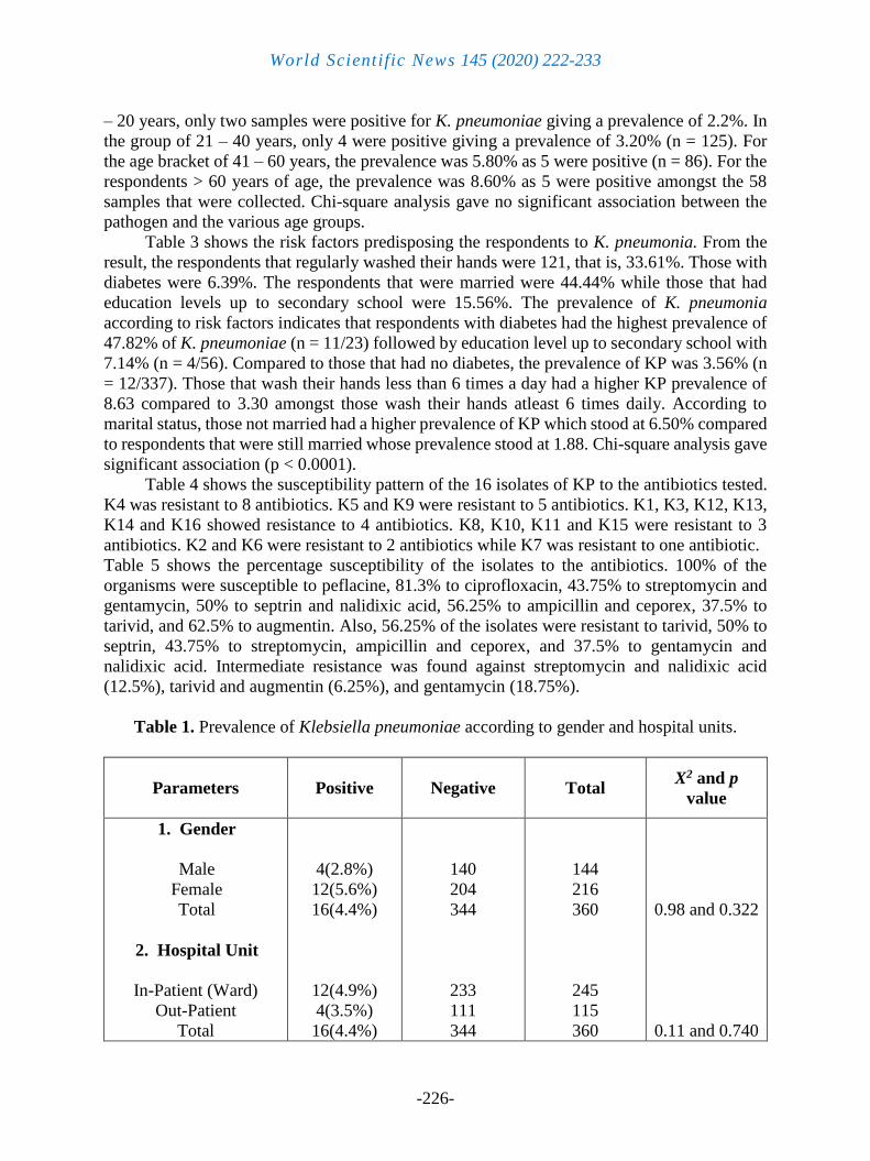
From the 360 urine samples collected and subjected to culture, only 16 samples were
positive for Klebsiella pneumoniae (Table 1). In other words, the overall prevalence of K.
pneumoniae was 4.4%. Distribution of the prevalence according to sex revealed that out of the
144 samples collected from male respondents, 4 were positive giving a prevalence of 2.8%. On
the other hand, from the 216 samples collected from the female respondents, 12 of them tested
positive, giving a prevalence of 5.6%. Furthermore, from the 245 samples obtained from
patients in the ward (in-patients), 12 of them were positive giving a prevalence rate of 4.9%
while for out-patients, only 4 out of the 115 samples collected were positive with 3.5%
prevalence (Table 1). Chi-square analysis gave no significant association between the pathogen
and gender and hospital units.
The presence of K. pneumoniae in the samples was also evaluated based on the age groups
of the patients (Table 2). There was a stepwise increase in the number of Klebsiella pneumoniae
isolates across the age groups. Of the 91 samples collected from patients within the ages of 10
World Scientific News 145 (2020) 222-233
-226-
– 20 years, only two samples were positive for K. pneumoniae giving a prevalence of 2.2%. In
the group of 21 – 40 years, only 4 were positive giving a prevalence of 3.20% (n = 125). For
the age bracket of 41 – 60 years, the prevalence was 5.80% as 5 were positive (n = 86). For the
respondents > 60 years of age, the prevalence was 8.60% as 5 were positive amongst the 58
samples that were collected. Chi-square analysis gave no significant association between the
pathogen and the various age groups.
Table 3 shows the risk factors predisposing the respondents to K. pneumonia. From the
result, the respondents that regularly washed their hands were 121, that is, 33.61%. Those with
diabetes were 6.39%. The respondents that were married were 44.44% while those that had
education levels up to secondary school were 15.56%. The prevalence of K. pneumonia
according to risk factors indicates that respondents with diabetes had the highest prevalence of
47.82% of K. pneumoniae (n = 11/23) followed by education level up to secondary school with
7.14% (n = 4/56). Compared to those that had no diabetes, the prevalence of KP was 3.56% (n
= 12/337). Those that wash their hands less than 6 times a day had a higher KP prevalence of
8.63 compared to 3.30 amongst those wash their hands atleast 6 times daily. According to
marital status, those not married had a higher prevalence of KP which stood at 6.50% compared
to respondents that were still married whose prevalence stood at 1.88. Chi-square analysis gave
significant association (p < 0.0001).
Table 4 shows the susceptibility pattern of the 16 isolates of KP to the antibiotics tested.
K4 was resistant to 8 antibiotics. K5 and K9 were resistant to 5 antibiotics. K1, K3, K12, K13,
K14 and K16 showed resistance to 4 antibiotics. K8, K10, K11 and K15 were resistant to 3
antibiotics. K2 and K6 were resistant to 2 antibiotics while K7 was resistant to one antibiotic.
Table 5 shows the percentage susceptibility of the isolates to the antibiotics. 100% of the
organisms were susceptible to peflacine, 81.3% to ciprofloxacin, 43.75% to streptomycin and
gentamycin, 50% to septrin and nalidixic acid, 56.25% to ampicillin and ceporex, 37.5% to
tarivid, and 62.5% to augmentin. Also, 56.25% of the isolates were resistant to tarivid, 50% to
septrin, 43.75% to streptomycin, ampicillin and ceporex, and 37.5% to gentamycin and
nalidixic acid. Intermediate resistance was found against streptomycin and nalidixic acid
(12.5%), tarivid and augmentin (6.25%), and gentamycin (18.75%).
Table 1. Prevalence of Klebsiella pneumoniae according to gender and hospital units.
Parameters Positive Negative Total X2 and p
value
1. Gender
Male
Female
Total
2. Hospital Unit
In-Patient (Ward)
Out-Patient
Total
4(2.8%)
12(5.6%)
16(4.4%)
12(4.9%)
4(3.5%)
16(4.4%)
140
204
344
233
111
344
144
216
360
245
115
360
0.98 and 0.322
0.11 and 0.740

World Scientific News 145 (2020) 222-233
-227-
Table 2. Prevalence of Klebsiella pneumoniae according to age groups
Age group (years) Positive Negative Total C.I, X2 and p value
10 – 20 2(2.2%) 89 91
21 – 40 4(3.2%) 121 125
40 – 60 5(5.8%) 81 86
>60 5(8.6%) 53 58
Total 16(4.4%) 344 360
0.0-0.105, 2.91
and 0.406
Table 3. Prevalence and risk factors to colonization of respondents to K. pneumoniae.
Risk factors Yes (n, %) No (n, %) C.I, X2 and p value
Regular washing of hands up to
6 times daily
(Prevalence of K. pneumoniae)
121 (4, 3.30) 139 (12, 8.63) 0.106-0.05, 176.85
and <0.0001
Diabetes
(Prevalence of K. pneumoniae) 23 ( 11, 47.82) 337 ( 12, 3.56)
Marital status
(Prevalence of K. pneumoniae) 160 (3, 1.88) 200 (13, 6.50)
Education level up to secondary
and above
(Prevalence of K. pneumoniae)
56 (4, 7.14) 304 (12, 3.95)
Table 4. The Antibiotic susceptibility pattern of K. pneumonia isolates.
Isolate CIP STR PEF SEP AMP TAR CEP GEN AUG NA
K1 S S S R R R S I S R
K2 S I S S S R S R S I
K3 S R S S R S R S S R
K4 R R S R R R R R S R
K5 S R S R R R S S R S
K6 S S S S S S R R I S
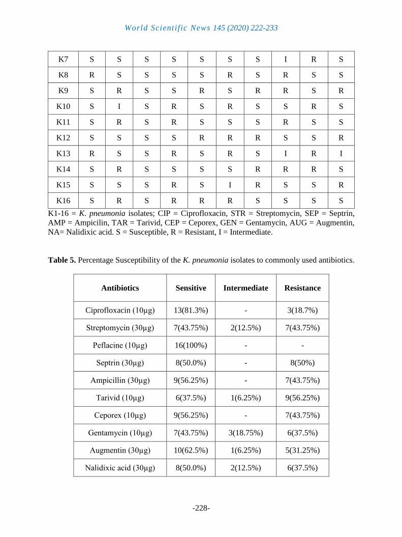
World Scientific News 145 (2020) 222-233
-228-
K7 S S S S S S S I R S
K8 R S S S S R S R S S
K9 S R S S R S R R S R
K10 S I S R S R S S R S
K11 S R S R S S S R S S
K12 S S S S R R R S S R
K13 R S S R S R S I R I
K14 S R S S S S R R R S
K15 S S S R S I R S S R
K16 S R S R R R S S S S
K1-16 = K. pneumonia isolates; CIP = Ciprofloxacin, STR = Streptomycin, SEP = Septrin,
AMP = Ampicilin, TAR = Tarivid, CEP = Ceporex, GEN = Gentamycin, AUG = Augmentin,
NA= Nalidixic acid. S = Susceptible, R = Resistant, I = Intermediate.
Table 5. Percentage Susceptibility of the K. pneumonia isolates to commonly used antibiotics.
Antibiotics Sensitive Intermediate Resistance
Ciprofloxacin (10µg) 13(81.3%) - 3(18.7%)
Streptomycin (30µg) 7(43.75%) 2(12.5%) 7(43.75%)
Peflacine (10µg) 16(100%) - -
Septrin (30µg) 8(50.0%) - 8(50%)
Ampicillin (30µg) 9(56.25%) - 7(43.75%)
Tarivid (10µg) 6(37.5%) 1(6.25%) 9(56.25%)
Ceporex (10µg) 9(56.25%) - 7(43.75%)
Gentamycin (10µg) 7(43.75%) 3(18.75%) 6(37.5%)
Augmentin (30µg) 10(62.5%) 1(6.25%) 5(31.25%)
Nalidixic acid (30µg) 8(50.0%) 2(12.5%) 6(37.5%)

World Scientific News 145 (2020) 222-233
-229-
4. DISCUSSION
The result of this study shows that Klebsiella pneumonia infection was seen more in
females than the males. This suggests that women have a higher tendency to acquire urinary
tract infections than men. Felson et al. (2005) [10] explained this to be a result of the relative
anatomical position of the external genitalia with respect to the anus. Considering the nature
and proximity of the female external genitalia to the anus, faecal materials easily contaminate
the female genitourinary system with a resultant increase in urinary tract infections.
Gastrointestinal tract and hands of hospital personnel are the main sources of transmission of
pathogenic Klebsiella. Cases of UTI are frequent in diarrheic patients because faeces is the most
significant source of patient infection, followed by contact with contaminated instruments. In
Pakistan, from 162 urine samples, 6 were positive representing a total prevalence of 3.96%,
which was within range of our reported 4.4%. Amongst the positive in their study, the ratio of
males to females was the same (1:1). However, in our study more females than males were
positive (3:1) [26]. A 10.70% prevalence (n = 455/4260) for year 2000 to 2006 and 18.10% (n
= 965/5331) from 2007 to 2013 of K. pneumoniae from blood stream infections in a
retrospective study was reported in Brazil [32]. The high prevalence of KP in these studies
confirms that it is a versatile pathogen that is of great public health concern.
Furthermore, our result shows that 4.9% and 3.5% of the in-patients and out-patients
respectively gave positive cultures. The higher rate of occurrence amongst the in-patients is an
indication that the hospitalized immunocompromised patients are more vulnerable to KP. It is
also possible that the rate of infection could also lead to a corresponding increase in hospital
stay. Longer periods of hospitalization increase the chances of acquiring nosocomial infections.
Its infections occur mostly in people with weak immune system. In other words, the infections
are mostly seen in old people and those with debilitating diseases. Our result shows that people
above 60 years of age are the most infected. The majority of people in this group are believed
to have impaired immunity with conditions such as diabetes mellitus, chronic obstructive
pulmonary diseases and other health conditions. This infection can also be obtained when a
person is in the hospital for some other reasons. Parisi et al [33] reported a prevalence of 3.28%
(n = 496/15104) from K. pneumoniae producing carbapenemases isolates from 15,104 rectal
samples in an intensive care unit. Cristea et al [34] obtained 32.60% prevalence in K.
pneumoniae in patients with UTI in Romania. Furthermore, they observed that their MDR
isolates correlated with kidney failure, advanced age, male gender, and diabetes mellitus. In our
study, those with diabetes had a higher KP prevalence of 47.82%.
Our study also shows that almost all the Klebsiella pneumonia isolates were multidrug
resistant strains. About 50% of the isolates showed resistance to antibiotics such as tarivid and
septrin while 43.75% of the isolates were resistant to streptomycin ampicillin and ceporex
among others. However, peflacine was 100% effective. Other antibiotics that were reasonably
effective are ciprofloxacin (81.3%) and augmentin (62.5%). As stated earlier, indiscriminate
use of antimicrobials has often been held responsible for the occurrence of multidrug resistant
Klebsiella strains in hospitals [13]. In a study, a two to fourfold increase in colonization rate
with Klebsiella by the hospital patients receiving antibiotics two weeks after admission to the
hospital has been reported [4]. Most of these organisms are multidrug resistant strains [4][35].
Due to the increased resistance to antibiotics by strains of K. pneumonia, it is becoming
increasingly difficult to treat these infections. Rath and Padhy [36] in an earlier study carried
out in India isolated extended spectrum beta lactamase (ESSL) and cephalosporinase enzymes
World Scientific News 145 (2020) 222-233
-230-
producing K. oxytoca and K. pneumoniae in non-hygienic communities and clinical settings.
The prevalence rates of ESBL resistance were 81.715 and 74.07% for K. oxytoca and K.
pneumoniae respectively. Apart from Peflacine, the isolates in our study showed resistance to
all other antibiotics used and ranged from 18.70 and 56.25% for ciprofloxacin and tarivid,
respectively. Furthermore, Rath and Padhy [36] showed that the isolates K. pneumoniae strains
were highly resistant to several antibiotics such as ampicillin, norfloxacin, ciprofloxacin, and
imipenem. Hou et al [37] characterized 38 MDR K. pneumoniae isolates from China that
possesses atleast 7 antibiotics resistance determinants that drives its resistance to
aminoglycosides, macrolides, quinolones and beta-lactams. This explains the resistance
displayed by our isolates to these classes of antibiotics. Tian et al [38] linked K. pneumoniae
carbapenem resistance to increased mortality of K. pneumoniae in blood stream infection
patients. Despite the fact that Klebsiella species are not a predominant cause of UTI, they are
capable of causing kidney disease even among patients getting infected for the first time [39].
Moreover, infections with multidrug resistant Klebsiella strains are more likely to lead to death
than are infections with most E. coli strains.
5. CONCLUSIONS
The result obtained in this study shows that Klebsiella pneumoniae infection is a common
hospital acquired urinary tract infection in Etim Ekpo general hospital of Akwa Ibom state,
Nigeria. Females are more likely to be infected than their male counterparts. Also, the higher
rate of occurrence amongst the in-patients is an indication that the hospitalized patients with
low immunity are more vulnerable to KP. Furthermore, our study also revealed that multidrug
resistant strains of Klebsiella pneumonia are prevalent in a community clinical setting and this
could further narrow the spectrum of available potent antibiotics in its management and
possibly lead to longer stay in the hospital. Efforts should be made to trace the source of these
MDR KP pathogens and also channeled to hospitals and other healthcare systems to prevent
the nosocomial spread of the organism.
References
[1] Prescott, L. M., Harley, J. P. and Klein, D. N. (1999). Microbiology 4th Edition. New
York: The McGraw-Hill Companies, Inc.
[2] CDC (2020). Klebsiella pneumoniae in Healthcare Settings.
[3] Vading, M., NaucleÂr, P., Kalin, M. and Giske, C. G. (2018). Invasive infection caused
by Klebsiella pneumoniae is a disease affecting patients with high comorbidity and
associated with high long- term mortality. PLoS ONE 13(4), e0195258
[4] Pollack, M., Niemann, R E., Reinhardt, J. A., Charache, P., Jett, M. P. and Hardy, P. H.
Jr. (1972). Factors influencing colonisation and antibiotic-resistance patterns of gram-
negative bacteria in hospital patients. Lancet. 2(7779), 668–671
[5] Casewell, M. and Talsania, H. G. (1979). Predominance of certain Klebsiella capsular
types in hospitals in the United Kingdom. Journal of Infections 1, 77–79

World Scientific News 145 (2020) 222-233
-231-
[6] Horan, T., Culver, D., Jarvis, W., Emori, G., Banerjee, S., Martone, W. and
Thornsberry, C. (2001). Pathogens causing nosocomial infections. Antimicrobic
Newsletter 5, 65-67
[7] Schaberg, D. R., Culver, D. H. and Gaynes, R. P. (2004). Major trends in the microbial
etiology of nosocomial infection. American Journalof Medicine. 91, 72S-75S
[8] Kodner, C. M. and Gupton, E. K. T. (2010).Recurrent Urinary Tract Infections in
Women: Diagnosis and Management. Am Fam Physician. 82(6), 638-643
[9] Bradford, P. A., Urban, C., Mariano, N., Projan, S. J., Rahal, J. J. and Bush, K. (1997).
Imipenem resistance in Klebsiella pneumoniae is associated with the combination of
ACT-1, a plasmid-mediated AmpC β-lactamase, and the loss of an outer membrane
protein. Antimicrobial Agents Chemotherapy 41, 563-569
[10] Felson, B., Rosenberg, L. S. and Hamburger, M. J. (2005) Roentgen findings in acute
Friedländer’s pneumonia. Journal of Radiology 53, 559–565
[11] Casewell M W. and Phillips I. (1977). Hands as a route of transmission for Klebsiella
species. British Medical Journal 2, 1315-1317
[12] Casewell, M. W. and Phillips, I. (1978). Epidemiological patterns of Klebsiella
colonization and infection in an intensive care ward. Journal of Hygiene Cambridge 80,
295-300
[13] Selden, R., Lee, S., Wang, W. L., Bennett, J. V. and Eickhoff, T. C. (1971). Nosocomial
Klebsiella infections: intestinal colonization as a reservoir. Annual Internal Medicince.
74, 657-664
[14] Woodford, N., Turton, J. F. and Livermore, D. M. (2011). Multiresistant Gram-negative
bacteria: the role of high-risk clones in the dissemination of antibiotic resistance. FEMS
Microbiol Review 35(5), 736-755
[15] Navon-Venezia, S., Kondratyeva, K. and Carattoli, A. (2017). Klebsiella pneumoniae: a
major worldwide source and shuttle for antibiotic resistance. FEMS Microbiology
Reviews, 41, 252–275
[16] Mulani, M. S., Kamble, E. E., Kumkar, S. N., Tawre, M. S. and Pardesi, K. R (2019).
Emerging Strategies to Combat ESKAPE Pathogens in the Era of Antimicrobial
Resistance: A Review. Front. Microbiol. 10, 539
[17] Xu, M., Fu, Y., Kong, H., Chen, X., Chen, Y., Li, L. and Yang, Q. (2018). Bloodstream
infections caused by Klebsiella pneumoniae: prevalence of blaKPC, virulence factors
and their impacts on clinical outcome. BMC Infectious Diseases. 18, 358
[18] Ebana, R. U. B., Etok, C. A. and Edet U. O. (2016). Phytochemical screening and
antimicrobial effect of three medicinal plants on urinary tract pathogens (2016). Asian
Journal of medicine and Health 1(2), 1-7
[19] Ebana, R. U. B., Edet, U. O., Anosike, I. K. and Etok, C. A. (2019). Bdellovibrio and
like organisms: The much anticipated magic bullet. World News of Natural Sciences 23,
233-249
World Scientific News 145 (2020) 222-233
-232-
[20] Effah, C. Y., Sun, T., Liu,S. and Wu, Y. (2020). Klebsiella pneumoniae: an increasing
threat to public health. Ann Clin Microbiol Antimicrob. 19, 1.
[21] Ebana, R. U. B., Andy, I. E., Edet, U. O., Benjamin, A. U., Mbim, E. N. and Anosike, I.
K. (2019). Nutritional Studies and Antimicrobial Activities of Jatropha tanjorensis
Leaves Extracts against Escherichia coli isolates. International Journal Innovation
Science and Research Technology, 4(8), 945-955
[22] Heidry, M., Goudarzi, H., Hashemi, A., Eslami, G., Goudarzi, M., Chirani, A. S. and
Amraei, S. (2017). Prevalence of Quinolone Resistance Genes in Klebsiella pneumoniae
Strains Isolated from Hospitalized Patients During 2013 – 2014. Arch Pediatr Infect
Dis. 5(4), e38343
[23] Paczosa, M. K. and Mecsas, J. (2016). Klebsiella pneumoniae: going on the offense
with a strong defense. Microbiol Mol Biol Rev. 80, 629-661
[24] Okonkwo, E. E. and Oguamanam, C. C. (2013). Traditional Crafts and Tourism
Development and Promotion in Etim Ekpo Local Government of Akwa Ibom State,
Nigeria. Research on Humanities and Social Sciences, 3(6), 139-147
[25] Setia, M. S. (2016). Methodology Series Mdoule 3: Cross Sectional Studies. Indian
Journal of Dermatology, 61(3), 261-264
[26] Masood, M. B. E., Saba, N. U. and Samad, A. (2002). Klebsiella pueumoniae Urinary
Tract infections associated with long-term catherterization and spinal cord injuries.
Journal of Medical Science, 2(5-6), 227-229.
[27] Barrow, G. I. and Feltham, R. K. A. (2003). Cowan and Steel᾽s Manual for the
identification of the Medical Bacteria, 3rd Edition. Cambridge University Press,
Cambridge, U. K.
[28] Cheesborough, M. (2009). Medical laboratory manual for tropical countries,
microbiology (ELBS), New York, 2009; 2nd edition: 62-70.
[29] Clinical Laboratory Standard Institute (2012). Performance Standards for Antimicrobial
Susceptibility Testing; Twenty-second Informational Supplement. Clinical Laboratory
Standard Institute. Wayne, Pennsylvania, USA Magiorakos. 32, 70–71
[30] Srinivasan, A. P., Carey, A., Carmeli, R. B., Falagas, Y., M. E, et al. (2012). Multidrug
resistance, extensively drug resistance and pandrug-resistance bacteria: An international
expert proposal for interim standard definations for acquired resistance. Clin Microbiol
Infect. 18, 268-281
[31] Sham, D. F., Thomsberry, C., Mayfield, D. C., Jones, M. E. and Karlowsky, J. A.(2001)
Multidrug resistant urinary tract isolates of Escherichia coli: Prevalence and patient
demographics in united states in 2000. Antimicrob Agents Chemother. 45(5), 1402-1406
[32] Santana, R. D. C., Gaspar, G. G., Vilar, F. C., Bellissimo-Rodrigues, F. and Martinez,
R. (2016). Secular trends in Klebsiella pneumoniae isolated in a tertiary-care hospital:
increasing prevalence and accelerated decline in antimicrobial susceptibility. Revista da
Sociedade Brasileira de Medicina Tropical. 49(2), 177-182
[33] Parisi, S. G., Bartolini, A., Santacatterina, E., Castellani, E., Ghirardo, R., Berto, A.,
Franchin, E., Menegotto, Canale, E. D, TOmmasini, T., Rinaldi, R., Baso, M. and Palu,

World Scientific News 145 (2020) 222-233
-233-
G. (2015). Prevalence of Klebsiella pneumoniae strains producing carbapenemases and
increase ofresistance to colistin in an Italian teaching hospital from January 2012 To
December 2014. BMC Infectious Diseases. 15, 244.
[34] Cristea, O. M., Avramescu, C. S., Balasoiu, M., Popescu, F, D., Popescu, F. and
Amzoiu, M. O. (2017). Urinary tract infection with Klebsiella pneumoniae in Patients
with Chronic Kidney Disease. Current Health Sciences Journal, 43(2), 137-148
[35] Coovadia, Y. M., Johnson, A. P., Bhana, R. H., Hutchinson, G. R., George, R. C. and
Hafferjee, I. E. (1992). Multiresistant Klebsiella pneumoniae in a neonatal nursery: the
importance of maintenance of infection control policies and procedures in the
prevention of outbreaks. Journal of Hospital Infections 22, 197-205
[36] Rath, S. and Padhy, R. N. (2014). Prevalence of two multidrug-resistant Klebsiella
species in an Indian teachinghospital and adjoining community. Journal of Infection
and Public Health 7, 496-507
[37] Hou, X. U., Song, X. Y., Ma, X. B., Zhang, S. Y. and Zhang, .Q. (2015). Molecular
characterization of multidrug-resistant Klebsiella pneumoniae isolates. Brazilian
Journal of Microbiology. 46, 3, 759-768
[38] Tian, L., Tan, R., Chen, Y., Sun, J., Liu, Qu, H. and Wang, X. (2016). Epidemiology of
Klebsiella pneumoniae bloodstream infections in a teaching hospital: factors related to
the carbapenem resistance and patient mortality. Antimicrobial Resistance and Infection
Control 5, 48
[39] Ofek, I., Goldhar, J., Zafriri, D., Lis, H., Adar, R. and Sharon, N. (1991) Anti-
Escherichia coli adhesin activity of cranberry and blueberry juices. New England
Journal of Medicine 324, 1599