1 Vol.:(0123456789) Scientific Reports | (2021) 11:8220 | https://doi.org/10.1038/s41598-021-87428-z www.nature.com/scientificreports Antimicrobial susceptibility, serotype distribution, virulence profile and molecular typing of piliated clinical isolates of pneumococci from east coast, Peninsular Malaysia Nurul Diana Dzaraly 1 , Mohd Nasir Mohd Desa 1* , AbdulRahman Muthanna 1 , Siti Norbaya Masri 2 , Niazlin Mohd Taib 2 , Zarizal Suhaili 1,3 , Nurshahira Sulaiman 1 , Nurul Hana Zainal Baharin 1 , Cheah Yun Shuan 1 , Zarina Ariffin 1 , Nor Iza A. Rahman 4 , Farahiyah Mohd Rani 4 , Navindra Kumari Palanisamy 5 , Tuan Suhaila Tuan Soh 6 & Fatimah Haslina Abdullah 7 Pilus has been recently associated with pneumococcal pathogenesis in humans. The information regarding piliated isolates in Malaysia is scarce, especially in the less developed states on the east coast of Peninsular Malaysia. Therefore, we studied the characteristics of pneumococci, including the piliated isolates, in relation to antimicrobial susceptibility, serotypes, and genotypes at a major tertiary hospital on the east coast of Peninsular Malaysia. A total of 100 clinical isolates collected between September 2017 and December 2019 were subjected to serotyping, antimicrobial susceptibility test, and detection of pneumococcal virulence and pilus genes. Multilocus sequence typing (MLST) and phylogenetic analysis were performed only for piliated strains. The most frequent serotypes were 14 (17%), 6A/B (16%), 23F (12%), 19A (11%), and 19F (11%). The majority of isolates were resistant to erythromycin (42%), tetracycline (37%), and trimethoprim-sulfamethoxazole (24%). Piliated isolates occurred in a proportion of 19%; 47.3% of them were multidrug-resistant (MDR) and a majority had serotype 19F. This study showed ST236 was the most predominant sequence type (ST) among piliated isolates, which was related to PMEN clone Taiwan 19F -14 (CC271). In the phylogenetic analysis, the piliated isolates were grouped into three major clades supported with 100% bootstrap values. Most piliated isolates belonged to internationally disseminated clones of S. pneumoniae, but pneumococcal conjugate vaccines (PCVs) have the potential to control them. Streptococcus pneumoniae (pneumococcus) is a Gram-positive bacterium that frequently colonizes and becomes the normal flora of the upper human respiratory tract. Under favourable conditions, pneumococci can cause a variety of severe diseases, such as pneumonia, meningitis, and bacteremia 1,2 . Worldwide, pneumococcal infection continues to be a major cause of death and morbidity, especially among children, the elderly, and patients with OPEN 1 Department of Biomedical Sciences, Faculty of Medicine and Health Sciences, Universiti Putra Malaysia, UPM Serdang, Selangor, Malaysia. 2 Department of Medical Microbiology, Faculty of Medicine and Health Sciences, Universiti Putra Malaysia, UPM Serdang, Selangor, Malaysia. 3 School of Animal Science, Aquatic Science and Environment, Faculty of Bioresources and Food Industry, Universiti Sultan Zainal Abidin, Besut Campus, Besut, Terengganu, Malaysia. 4 Faculty of Medicine, Universiti Sultan Zainal Abidin, Kuala Terengganu, Terengganu, Malaysia. 5 Department of Medical Microbiology and Parasitology, Faculty of Medicine, Universiti Teknologi MARA (UiTM), Sungai Buloh Campus, Jalan Hospital, Sungai Buloh, Selangor, Malaysia. 6 Department of Pathology, Sungai Buloh Hospital, Ministry of Health Malaysia, Jalan Hospital, Sungai Buloh, Selangor, Malaysia. 7 Department of Pathology, Sultanah Nur Zahirah Hospital, Ministry of Health Malaysia, Jalan Sultan Mahmud, Kuala Terengganu, Terengganu, Malaysia. * email: [email protected]

Welcome message from author
This document is posted to help you gain knowledge. Please leave a comment to let me know what you think about it! Share it to your friends and learn new things together.
Transcript

1
Vol.:(0123456789)
Scientific Reports | (2021) 11:8220 | https://doi.org/10.1038/s41598-021-87428-z
www.nature.com/scientificreports
Antimicrobial susceptibility, serotype distribution, virulence profile and molecular typing of piliated clinical isolates of pneumococci from east coast, Peninsular MalaysiaNurul Diana Dzaraly1, Mohd Nasir Mohd Desa1*, AbdulRahman Muthanna1, Siti Norbaya Masri2, Niazlin Mohd Taib2, Zarizal Suhaili1,3, Nurshahira Sulaiman1, Nurul Hana Zainal Baharin1, Cheah Yun Shuan1, Zarina Ariffin1, Nor Iza A. Rahman4, Farahiyah Mohd Rani4, Navindra Kumari Palanisamy5, Tuan Suhaila Tuan Soh6 & Fatimah Haslina Abdullah7
Pilus has been recently associated with pneumococcal pathogenesis in humans. The information regarding piliated isolates in Malaysia is scarce, especially in the less developed states on the east coast of Peninsular Malaysia. Therefore, we studied the characteristics of pneumococci, including the piliated isolates, in relation to antimicrobial susceptibility, serotypes, and genotypes at a major tertiary hospital on the east coast of Peninsular Malaysia. A total of 100 clinical isolates collected between September 2017 and December 2019 were subjected to serotyping, antimicrobial susceptibility test, and detection of pneumococcal virulence and pilus genes. Multilocus sequence typing (MLST) and phylogenetic analysis were performed only for piliated strains. The most frequent serotypes were 14 (17%), 6A/B (16%), 23F (12%), 19A (11%), and 19F (11%). The majority of isolates were resistant to erythromycin (42%), tetracycline (37%), and trimethoprim-sulfamethoxazole (24%). Piliated isolates occurred in a proportion of 19%; 47.3% of them were multidrug-resistant (MDR) and a majority had serotype 19F. This study showed ST236 was the most predominant sequence type (ST) among piliated isolates, which was related to PMEN clone Taiwan19F-14 (CC271). In the phylogenetic analysis, the piliated isolates were grouped into three major clades supported with 100% bootstrap values. Most piliated isolates belonged to internationally disseminated clones of S. pneumoniae, but pneumococcal conjugate vaccines (PCVs) have the potential to control them.
Streptococcus pneumoniae (pneumococcus) is a Gram-positive bacterium that frequently colonizes and becomes the normal flora of the upper human respiratory tract. Under favourable conditions, pneumococci can cause a variety of severe diseases, such as pneumonia, meningitis, and bacteremia1,2. Worldwide, pneumococcal infection continues to be a major cause of death and morbidity, especially among children, the elderly, and patients with
OPEN
1Department of Biomedical Sciences, Faculty of Medicine and Health Sciences, Universiti Putra Malaysia, UPM Serdang, Selangor, Malaysia. 2Department of Medical Microbiology, Faculty of Medicine and Health Sciences, Universiti Putra Malaysia, UPM Serdang, Selangor, Malaysia. 3School of Animal Science, Aquatic Science and Environment, Faculty of Bioresources and Food Industry, Universiti Sultan Zainal Abidin, Besut Campus, Besut, Terengganu, Malaysia. 4Faculty of Medicine, Universiti Sultan Zainal Abidin, Kuala Terengganu, Terengganu, Malaysia. 5Department of Medical Microbiology and Parasitology, Faculty of Medicine, Universiti Teknologi MARA (UiTM), Sungai Buloh Campus, Jalan Hospital, Sungai Buloh, Selangor, Malaysia. 6Department of Pathology, Sungai Buloh Hospital, Ministry of Health Malaysia, Jalan Hospital, Sungai Buloh, Selangor, Malaysia. 7Department of Pathology, Sultanah Nur Zahirah Hospital, Ministry of Health Malaysia, Jalan Sultan Mahmud, Kuala Terengganu, Terengganu, Malaysia. *email: [email protected]
2
Vol:.(1234567890)
Scientific Reports | (2021) 11:8220 | https://doi.org/10.1038/s41598-021-87428-z
www.nature.com/scientificreports/
comorbid diseases. According to the World Health Organization (WHO), about 808.694 children under five years of age die due to pneumonia, accounting for 15% of total death cases in 20173. Malaysia is a rapidly developing country in Southeast Asia, experiencing extensive industrialization and urbanization, which is concerning from the perspective of public health as respiratory diseases, particularly pneumonia, are one of the primary causes of hospitalization and death. According to the Department of Statistics, pneumonia has been reported as the third leading cause of death in Malaysia among children under five years of age, with an estimated 3.8 per 100 000 cases per year, and among the causative agents of this disease is S. pneumoniae4,5.
The polysaccharide capsule in S. pneumoniae is a major virulence factor with 100 different serotypes6. The 7-valent pneumococcal conjugate vaccine (PCV7) was first implemented for children in the United States of America (USA) in 2000 and Europe in 2001 to reduce the burden of pneumococcal diseases. This vaccine covers serotypes 4, 6B, 9V, 14, 18C, 19F, and 23F. However, pneumococcal infection remains a severe problem due to the replacement of PCV7 serotype by non-PCV7 serotypes. 10-valent conjugate vaccine (PCV10; additional sero-types 1, 5 and 7F) was introduced in 2008 to cover a wider number of serotypes, while in early 2010, a 13-valent conjugate vaccine (PCV13; additional serotypes 3, 6A and 19A) was introduced in the USA primarily to replace PCV7 as a part of the infant immunization initiative. PCV13 was also introduced in Malaysia in 2010, with full support from paediatricians for use in childhood immunization7 but has yet to be widely implemented across the nation. In 2020, it was included in the Malaysia National Immunisation Programme (NIP).
Additionally, pneumococci are also equipped with a wide virulence regiment such as pneumolysin, choline-binding proteins, neuraminidase, hyaluronate lyase, autolysin, and many others. Recently, the discovery of a long filamentous, pilus-like structure in Gram-positive bacteria, specifically in S. pneumoniae, has added another fac-tor to the pneumococcal virulence regiment. The role of pili is to enhance the ability of pneumococci to adhere to epithelial cells; at the same time, the piliated strain was found to be significantly more virulent in a murine model of invasive diseases8. This suggests that pneumococcal pili provide an extra advantage to initiate colonization, leading to the downstream of infection and pathogenesis process9–11. To date, two pili islets have been detected in pneumococci, namely, PI-18 and PI-29.
While a number of studies have addressed the serotype, antimicrobial resistance, and sequence type (ST) of piliated pneumococcal strains, information regarding the piliated pneumococcal isolates in the Malaysian population has been scarce. Furthermore, certain areas are still underreported, particularly on the east coast of Peninsular Malaysia, where the process of urbanization is less extensive than that on the west coast. The emer-gence of a subpopulation carrying the pilus trait which is not common in Gram-positive bacteria should not be underestimated. Therefore, this study was undertaken to determine the serotype distribution, antimicrobial resistance, virulence gene profile, occurrence of pilus genes, and the genotypic characteristics of pneumococci and piliated isolates at a major tertiary hospital on the east coast of Peninsular Malaysia.
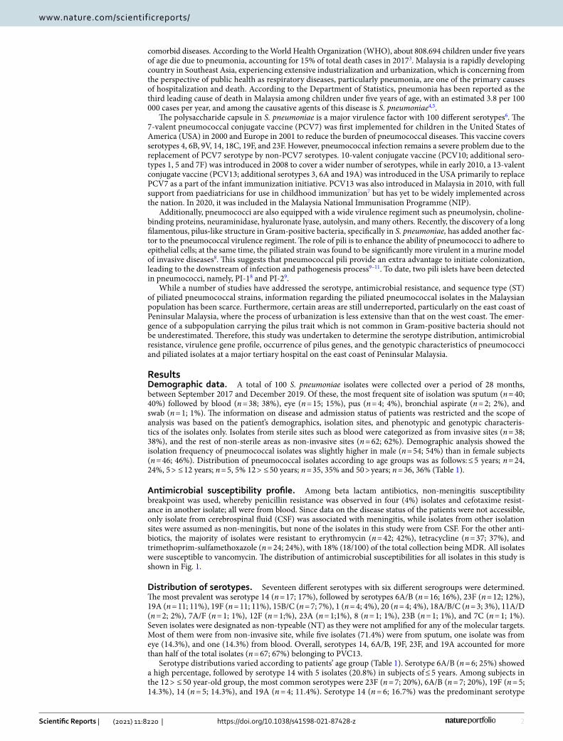
ResultsDemographic data. A total of 100 S. pneumoniae isolates were collected over a period of 28 months, between September 2017 and December 2019. Of these, the most frequent site of isolation was sputum (n = 40; 40%) followed by blood (n = 38; 38%), eye (n = 15; 15%), pus (n = 4; 4%), bronchial aspirate (n = 2; 2%), and swab (n = 1; 1%). The information on disease and admission status of patients was restricted and the scope of analysis was based on the patient’s demographics, isolation sites, and phenotypic and genotypic characteris-tics of the isolates only. Isolates from sterile sites such as blood were categorized as from invasive sites (n = 38; 38%), and the rest of non-sterile areas as non-invasive sites (n = 62; 62%). Demographic analysis showed the isolation frequency of pneumococcal isolates was slightly higher in male (n = 54; 54%) than in female subjects (n = 46; 46%). Distribution of pneumococcal isolates according to age groups was as follows: ≤ 5 years; n = 24, 24%, 5 > ≤ 12 years; n = 5, 5% 12 > ≤ 50 years; n = 35, 35% and 50 > years; n = 36, 36% (Table 1).
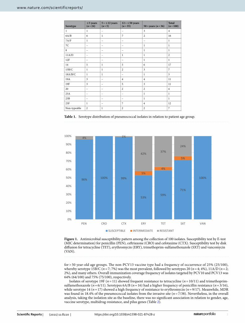
Antimicrobial susceptibility profile. Among beta lactam antibiotics, non-meningitis susceptibility breakpoint was used, whereby penicillin resistance was observed in four (4%) isolates and cefotaxime resist-ance in another isolate; all were from blood. Since data on the disease status of the patients were not accessible, only isolate from cerebrospinal fluid (CSF) was associated with meningitis, while isolates from other isolation sites were assumed as non-meningitis, but none of the isolates in this study were from CSF. For the other anti-biotics, the majority of isolates were resistant to erythromycin (n = 42; 42%), tetracycline (n = 37; 37%), and trimethoprim-sulfamethoxazole (n = 24; 24%), with 18% (18/100) of the total collection being MDR. All isolates were susceptible to vancomycin. The distribution of antimicrobial susceptibilities for all isolates in this study is shown in Fig. 1.
Distribution of serotypes. Seventeen different serotypes with six different serogroups were determined. The most prevalent was serotype 14 (n = 17; 17%), followed by serotypes 6A/B (n = 16; 16%), 23F (n = 12; 12%), 19A (n = 11; 11%), 19F (n = 11; 11%), 15B/C (n = 7; 7%), 1 (n = 4; 4%), 20 (n = 4; 4%), 18A/B/C (n = 3; 3%), 11A/D (n = 2; 2%), 7A/F (n = 1; 1%), 12F (n = 1;%), 23A (n = 1;1%), 8 (n = 1; 1%), 23B (n = 1; 1%), and 7C (n = 1; 1%). Seven isolates were designated as non-typeable (NT) as they were not amplified for any of the molecular targets. Most of them were from non-invasive site, while five isolates (71.4%) were from sputum, one isolate was from eye (14.3%), and one (14.3%) from blood. Overall, serotypes 14, 6A/B, 19F, 23F, and 19A accounted for more than half of the total isolates (n = 67; 67%) belonging to PVC13.
Serotype distributions varied according to patients’ age group (Table 1). Serotype 6A/B (n = 6; 25%) showed a high percentage, followed by serotype 14 with 5 isolates (20.8%) in subjects of ≤ 5 years. Among subjects in the 12 > ≤ 50 year-old group, the most common serotypes were 23F (n = 7; 20%), 6A/B (n = 7; 20%), 19F (n = 5; 14.3%), 14 (n = 5; 14.3%), and 19A (n = 4; 11.4%). Serotype 14 (n = 6; 16.7%) was the predominant serotype
3
Vol.:(0123456789)
Scientific Reports | (2021) 11:8220 | https://doi.org/10.1038/s41598-021-87428-z
www.nature.com/scientificreports/
for > 50 year-old age groups. The non-PCV13 vaccine type had a frequency of occurrence of 25% (25/100), whereby serotype 15B/C (n = 7; 7%) was the most prevalent, followed by serotypes 20 (n = 4; 4%), 11A/D (n = 2; 2%), and many others. Overall immunization coverage frequency of isolates targeted by PCV10 and PCV13 was 64% (64/100) and 75% (75/100), respectively.
Isolates of serotype 19F (n = 11) showed frequent resistance to tetracycline (n = 10/11) and trimethoprim-sulfamethoxazole (n = 6/11). Serotypes 6A/B (n = 16) had a higher frequency of penicillin resistance (n = 3/16), while serotype 14 (n = 17) showed a high frequency of resistance to erythromycin (n = 9/17). Meanwhile, MDR was found in 18.4% of the pneumococcal isolates from the invasive site (n = 7/38). Nevertheless, in the overall analysis, taking the isolation site as the baseline, there was no significant association in relation to gender, age, vaccine serotype, multidrug-resistance, and pilus genes (Table 2).
Table 1. Serotype distribution of pneumococcal isolates in relation to patient age group.
Serotype ≤ 5 years(n = 24)
5 > ≤ 12 years(n = 5)
12 > ≤ 50 years(n = 35) 50 > years (n = 36)
Total(n = 100)
1 1 – – 3 4
6A/B 6 1 7 2 16
7A/F 1 – – – 1
7C – – – 1 1
8 – – – 1 1
11A/D – – 1 1 2
12F – – – 1 1
14 5 1 5 6 17
15B/C 1 1 2 3 7
18A/B/C 1 1 – 1 3
19A 3 – 4 4 11
19F 3 – 5 3 11
20 – – 2 2 4
23A – – – 1 1
23B – – – 1 1
23F 1 – 7 4 12
Non-typeable 2 1 2 2 7
96% 100% 99%
53%59%
71%
100%5%
4%
5%
4% 1%
42%37%
24%
0%
10%
20%
30%
40%
50%
60%
70%
80%
90%
100%
PEN CRO CTX ERY TET SXT VAN
SUSCEPTIBLE INTERMEDIATE RESISTANT
Figure 1. Antimicrobial susceptibility pattern among the collection of 100 isolates. Susceptibility test by E-test (MIC determination) for penicillin (PEN), ceftriaxone (CRO) and cefotaxime (CTX). Susceptibility test by disk diffusion for tetracycline (TET), erythromycin (ERY), trimethoprim-sulfamethoxazole (SXT) and vancomycin (VAN).
4
Vol:.(1234567890)
Scientific Reports | (2021) 11:8220 | https://doi.org/10.1038/s41598-021-87428-z
www.nature.com/scientificreports/
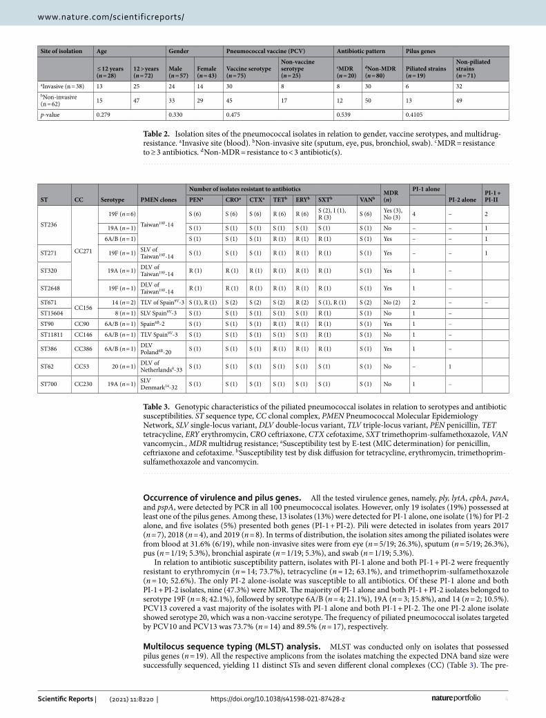
Occurrence of virulence and pilus genes. All the tested virulence genes, namely, ply, lytA, cpbA, pavA, and pspA, were detected by PCR in all 100 pneumococcal isolates. However, only 19 isolates (19%) possessed at least one of the pilus genes. Among these, 13 isolates (13%) were detected for PI-1 alone, one isolate (1%) for PI-2 alone, and five isolates (5%) presented both genes (PI-1 + PI-2). Pili were detected in isolates from years 2017 (n = 7), 2018 (n = 4), and 2019 (n = 8). In terms of distribution, the isolation sites among the piliated isolates were from blood at 31.6% (6/19), while non-invasive sites were from eye (n = 5/19; 26.3%), sputum (n = 5/19; 26.3%), pus (n = 1/19; 5.3%), bronchial aspirate (n = 1/19; 5.3%), and swab (n = 1/19; 5.3%).
In relation to antibiotic susceptibility pattern, isolates with PI-1 alone and both PI-1 + PI-2 were frequently resistant to erythromycin (n = 14; 73.7%), tetracycline (n = 12; 63.1%), and trimethoprim-sulfamethoxazole (n = 10; 52.6%). The only PI-2 alone-isolate was susceptible to all antibiotics. Of these PI-1 alone and both PI-1 + PI-2 isolates, nine (47.3%) were MDR. The majority of PI-1 alone and both PI-1 + PI-2 isolates belonged to serotype 19F (n = 8; 42.1%), followed by serotype 6A/B (n = 4; 21.1%), 19A (n = 3; 15.8%), and 14 (n = 2; 10.5%). PCV13 covered a vast majority of the isolates with PI-1 alone and both PI-1 + PI-2. The one PI-2 alone isolate showed serotype 20, which was a non-vaccine serotype. The frequency of piliated pneumococcal isolates targeted by PCV10 and PCV13 was 73.7% (n = 14) and 89.5% (n = 17), respectively.
Multilocus sequence typing (MLST) analysis. MLST was conducted only on isolates that possessed pilus genes (n = 19). All the respective amplicons from the isolates matching the expected DNA band size were successfully sequenced, yielding 11 distinct STs and seven different clonal complexes (CC) (Table 3). The pre-
Table 2. Isolation sites of the pneumococcal isolates in relation to gender, vaccine serotypes, and multidrug-resistance. a Invasive site (blood). b Non-invasive site (sputum, eye, pus, bronchiol, swab). c MDR = resistance to ≥ 3 antibiotics. d Non-MDR = resistance to < 3 antibiotic(s).
Site of isolation Age Gender Pneumococcal vaccine (PCV) Antibiotic pattern Pilus genes
≤ 12 years(n = 28)
12 > years(n = 72)
Male(n = 57)
Female(n = 43)
Vaccine serotype(n = 75)
Non-vaccine serotype(n = 25)
cMDR(n = 20)
dNon-MDR(n = 80)
Piliated strains(n = 19)
Non-piliated strains(n = 71)
aInvasive (n = 38) 13 25 24 14 30 8 8 30 6 32bNon-invasive (n = 62) 15 47 33 29 45 17 12 50 13 49
p-value 0.279 0.330 0.475 0.539 0.4105
Table 3. Genotypic characteristics of the piliated pneumococcal isolates in relation to serotypes and antibiotic susceptibilities. ST sequence type, CC clonal complex, PMEN Pneumococcal Molecular Epidemiology Network, SLV single-locus variant, DLV double-locus variant, TLV triple-locus variant, PEN penicillin, TET tetracycline, ERY erythromycin, CRO ceftriaxone, CTX cefotaxime, SXT trimethoprim-sulfamethoxazole, VAN vancomycin., MDR multidrug resistance; a Susceptibility test by E-test (MIC determination) for penicillin, ceftriaxone and cefotaxime. b Susceptibility test by disk diffusion for tetracycline, erythromycin, trimethoprim-sulfamethoxazole and vancomycin.
ST CC Serotype PMEN clones
Number of isolates resistant to antibiotics MDR(n)
PI-1 alone
PI-2 alonePI-1 + PI-IIPENa CROa CTXa TETb ERYb SXTb VANb
ST236
CC271
19F (n = 6)
Taiwan19F-14
S (6) S (6) S (6) R (6) R (6) S (2), I (1), R (3) S (6) Yes (3),
No (3) 4 – 2
19A (n = 1) S (1) S (1) S (1) S (1) S (1) S (1) S (1) No – – 1
6A/B (n = 1) S (1) S (1) S (1) R (1) R (1) R (1) S (1) Yes – – 1
ST271 19F (n = 1) SLV of Taiwan19F-14 S (1) S (1) S (1) R (1) R (1) R (1) S (1) Yes – – 1
ST320 19A (n = 1) DLV of Taiwan19F-14 R (1) R (1) R (1) R (1) R (1) R (1) S (1) Yes 1 –
ST2648 19F (n = 1) DLV of Taiwan19F-14 R (1) R (1) R (1) R (1) R (1) R (1) S (1) Yes 1 –
ST671CC156
14 (n = 2) TLV of Spain9V-3 S (1), R (1) S (2) S (2) S (2) R (2) S (1), R (1) S (2) No (2) 2 – –
ST15604 8 (n = 1) SLV Spain9V-3 S (1) S (1) S (1) S (1) S (1) R (1) S (1) No 1 –
ST90 CC90 6A/B (n = 1) Spain6B-2 S (1) S (1) S (1) R (1) R (1) R (1) S (1) Yes 1 –
ST11811 CC146 6A/B (n = 1) TLV Spain9V-3 S (1) S (1) S (1) S (1) S (1) R (1) S (1) No 1 –
ST386 CC386 6A/B (n = 1) DLV Poland6B-20 S (1) S (1) S (1) R (1) R (1) R (1) S (1) Yes 1 –
ST62 CC53 20 (n = 1) DLV of Netherlands8-33 S (1) S (1) S (1) S (1) S (1) S (1) S (1) No – 1
ST700 CC230 19A (n = 1) SLV Denmark14-32 S (1) S (1) S (1) S (1) S (1) S (1) S (1) No 1 –

5
Vol.:(0123456789)
Scientific Reports | (2021) 11:8220 | https://doi.org/10.1038/s41598-021-87428-z
www.nature.com/scientificreports/
dominant one was ST236 (n = 8; 42.1%), followed by ST671 (n = 2; 10.5%), ST271 (n = 1; 5.3%), ST90 (n = 1; 5.3%), ST700 (n = 1; 5.3%), ST11811 (n = 1; 5.3%), ST320 (n = 1; 5.3%), ST386 (n = 1; 5.3%), ST62 (n = 1; 5.3%), ST2648 (n = 1; 5.3%), and a new novel sequence type, ST15604. The novel ST15604 isolate originated from the sputum of a 7-year-old child, was susceptible to all antibiotics, and had serotype 8 carrying PI-1 gene.
Nineteen piliated isolates presented STs similar to 11 of the 43 clones recognized by the Pneumococcal Molecular Epidemiology Network (PMEN), sharing at least five MLST alleles with those PMEN clones. Nine isolates had the same ST as Taiwan 19F-14 and Spain6B-2, three strains were single-locus locus variant (SLV) of Taiwan19F-14, Denmark14-32, and Spain9V-3, four isolates were double-locus variant (DLV) of Taiwan19F-14, Poland6B-20, and Netherlands8-33, and three isolates were triple-locus variant (TLV) of Spain9V-3 and Spain6B-2.
Phylogenetic analysis. All nucleotide sequences of the seven housekeeping genes for the respective iso-lates were aligned in the specified order12 and subjected to phylogenetic analysis among all the 19 piliated isolates (labelled with T followed by numbers) and 11 reference sequences from the MLST database (labelled with REF followed by identity number). The tree was basally rooted to strain numbers T106 and T110, assigned as clade III and branched out into two bigger clades I and II with 100% bootstrap confidence interval values at all branching (Fig. 2). Among the three clades, there was one major group consisting of 12 isolates from this current study (Clade I), while others were small clades comprising two and four isolates (clade II and clade III). The clades also showed clear segregation among the STs, where clade I represented predominantly ST236, followed by ST271, ST2648, ST320, and ST62; clade II for ST700, ST386, ST90, and ST11811; clade III for ST671 for both isolates; and a single ST15604 (novel) in its own lineage. With the exception of the novel ST, all reference sequences of the different STs were clustered according to their respective similar ST of the isolates in this study.
Sources ST Serotype Pilus Genes MDREye 236 19F PI-1 Yes
Sputum 236 19F PI-1 YesEye 236 19F PI-1 Yes
Sputum 236 19F PI-1 NoBronchiole 236 19F PI-1 + PI-2 No
Sputum 236 6A/B PI-1 + PI-2 YesBlood 236 19A PI-1 + PI-2 NoEye 236 19F PI-1 + PI-2 NoEye 236 19F Not stated Not stated
Sputum 236 19F Not stated Not stated
Sputum 271 19F PI-1 + PI-2 YesSputum 271 19F Not stated Not statedSwab 2648 19F PI-1 Yes
Sputum 2648 19F Not stated Not statedBlood 320 19A PI-1 YesBlood 320 19A Not stated Not statedEye 62 20 PI-2 No
Blood 62 11A Not stated Not statedPus 700 19A PI-1 No
Blood 700 19F Not stated Not statedBlood 386 6A/B PI-1 YesBlood 386 6B Not stated Not statedEye 90 6A/B PI-1 YesEye 90 6B Not stated Not stated
Blood 11811 6A/B PI-1 NoBlood 11811 6C Not stated Not stated
Sputum 15604 8 PI-1 NoBlood 671 14 PI-1 NoBlood 671 14 PI-1 NoBlood 671 14 Not stated Not stated
Clade I
Clade II
Clade III
100%
Figure 2. Phylogenetic analysis of the piliated pneumococcal isolates. Phylogenetic analysis revealed three clades among piliated pneumococcal isolates with clade I being predominant and exhibiting mostly serotype 19F of ST236, and all the PI-1 alone and both PI-1+PI-2 isolates. Bootstrap sampling at 1000 replicates showed 100% at all branching. The distribution of sources, sequence type (ST), serotype, pilus genes and multidrug resistance for respective isolates are shown in the right columns. The reference sequences were retrieved from the MLST database comprising identity (ID) numbers 1592 (Taiwan), 345 (Taiwan), 6665 (South Korea), 4312 (Vietnam), 13506 (Malaysia), 40817 (Japan), 30311 (Malawi), 19496 (India), 38687 (China), 32260 (United States) and 1254 (United States); ID number was preceded by REF indicating reference sequence, and ID labelled with T (Terengganu) and numbers represent the isolates in this study. T14 is a newly assigned ST (REF sequence is not available).
6
Vol:.(1234567890)
Scientific Reports | (2021) 11:8220 | https://doi.org/10.1038/s41598-021-87428-z
www.nature.com/scientificreports/
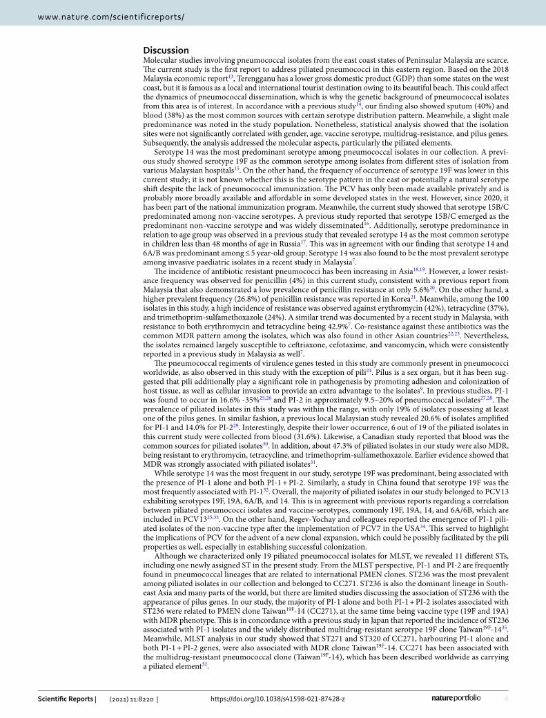
DiscussionMolecular studies involving pneumococcal isolates from the east coast states of Peninsular Malaysia are scarce. The current study is the first report to address piliated pneumococci in this eastern region. Based on the 2018 Malaysia economic report13, Terengganu has a lower gross domestic product (GDP) than some states on the west coast, but it is famous as a local and international tourist destination owing to its beautiful beach. This could affect the dynamics of pneumococcal dissemination, which is why the genetic background of pneumococcal isolates from this area is of interest. In accordance with a previous study14, our finding also showed sputum (40%) and blood (38%) as the most common sources with certain serotype distribution pattern. Meanwhile, a slight male predominance was noted in the study population. Nonetheless, statistical analysis showed that the isolation sites were not significantly correlated with gender, age, vaccine serotype, multidrug-resistance, and pilus genes. Subsequently, the analysis addressed the molecular aspects, particularly the piliated elements.
Serotype 14 was the most predominant serotype among pneumococcal isolates in our collection. A previ-ous study showed serotype 19F as the common serotype among isolates from different sites of isolation from various Malaysian hospitals15. On the other hand, the frequency of occurrence of serotype 19F was lower in this current study; it is not known whether this is the serotype pattern in the east or potentially a natural serotype shift despite the lack of pneumococcal immunization. The PCV has only been made available privately and is probably more broadly available and affordable in some developed states in the west. However, since 2020, it has been part of the national immunization program. Meanwhile, the current study showed that serotype 15B/C predominated among non-vaccine serotypes. A previous study reported that serotype 15B/C emerged as the predominant non-vaccine serotype and was widely disseminated16. Additionally, serotype predominance in relation to age group was observed in a previous study that revealed serotype 14 as the most common serotype in children less than 48 months of age in Russia17. This was in agreement with our finding that serotype 14 and 6A/B was predominant among ≤ 5 year-old group. Serotype 14 was also found to be the most prevalent serotype among invasive paediatric isolates in a recent study in Malaysia7.
The incidence of antibiotic resistant pneumococci has been increasing in Asia18,19. However, a lower resist-ance frequency was observed for penicillin (4%) in this current study, consistent with a previous report from Malaysia that also demonstrated a low prevalence of penicillin resistance at only 5.6%20. On the other hand, a higher prevalent frequency (26.8%) of penicillin resistance was reported in Korea21. Meanwhile, among the 100 isolates in this study, a high incidence of resistance was observed against erythromycin (42%), tetracycline (37%), and trimethoprim-sulfamethoxazole (24%). A similar trend was documented by a recent study in Malaysia, with resistance to both erythromycin and tetracycline being 42.9%7. Co-resistance against these antibiotics was the common MDR pattern among the isolates, which was also found in other Asian countries22,23. Nevertheless, the isolates remained largely susceptible to ceftriaxone, cefotaxime, and vancomycin, which were consistently reported in a previous study in Malaysia as well7.
The pneumococcal regiments of virulence genes tested in this study are commonly present in pneumococci worldwide, as also observed in this study with the exception of pili24. Pilus is a sex organ, but it has been sug-gested that pili additionally play a significant role in pathogenesis by promoting adhesion and colonization of host tissue, as well as cellular invasion to provide an extra advantage to the isolates9. In previous studies, PI-1 was found to occur in 16.6% -35%25,26 and PI-2 in approximately 9.5–20% of pneumococcal isolates27,28. The prevalence of piliated isolates in this study was within the range, with only 19% of isolates possessing at least one of the pilus genes. In similar fashion, a previous local Malaysian study revealed 20.6% of isolates amplified for PI-1 and 14.0% for PI-229. Interestingly, despite their lower occurrence, 6 out of 19 of the piliated isolates in this current study were collected from blood (31.6%). Likewise, a Canadian study reported that blood was the common sources for piliated isolates30. In addition, about 47.3% of piliated isolates in our study were also MDR, being resistant to erythromycin, tetracycline, and trimethoprim-sulfamethoxazole. Earlier evidence showed that MDR was strongly associated with piliated isolates31.
While serotype 14 was the most frequent in our study, serotype 19F was predominant, being associated with the presence of PI-1 alone and both PI-1 + PI-2. Similarly, a study in China found that serotype 19F was the most frequently associated with PI-132. Overall, the majority of piliated isolates in our study belonged to PCV13 exhibiting serotypes 19F, 19A, 6A/B, and 14. This is in agreement with previous reports regarding a correlation between piliated pneumococci isolates and vaccine-serotypes, commonly 19F, 19A, 14, and 6A/6B, which are included in PCV1325,33. On the other hand, Regev-Yochay and colleagues reported the emergence of PI-1 pili-ated isolates of the non-vaccine type after the implementation of PCV7 in the USA34. This served to highlight the implications of PCV for the advent of a new clonal expansion, which could be possibly facilitated by the pili properties as well, especially in establishing successful colonization.
Although we characterized only 19 piliated pneumococcal isolates for MLST, we revealed 11 different STs, including one newly assigned ST in the present study. From the MLST perspective, PI-1 and PI-2 are frequently found in pneumococcal lineages that are related to international PMEN clones. ST236 was the most prevalent among piliated isolates in our collection and belonged to CC271. ST236 is also the dominant lineage in South-east Asia and many parts of the world, but there are limited studies discussing the association of ST236 with the appearance of pilus genes. In our study, the majority of PI-1 alone and both PI-1 + PI-2 isolates associated with ST236 were related to PMEN clone Taiwan19F-14 (CC271), at the same time being vaccine type (19F and 19A) with MDR phenotype. This is in concordance with a previous study in Japan that reported the incidence of ST236 associated with PI-1 isolates and the widely distributed multidrug-resistant serotype 19F clone Taiwan19F-1435. Meanwhile, MLST analysis in our study showed that ST271 and ST320 of CC271, harbouring PI-1 alone and both PI-1 + PI-2 genes, were also associated with MDR clone Taiwan19F-14. CC271 has been associated with the multidrug-resistant pneumococcal clone (Taiwan19F-14), which has been described worldwide as carrying a piliated element32.

7
Vol.:(0123456789)
Scientific Reports | (2021) 11:8220 | https://doi.org/10.1038/s41598-021-87428-z
www.nature.com/scientificreports/
The molecular analysis of S. pneumoniae in this study revealed several genetic lineages, namely, ST90, ST386, and ST671, which were associated with PI-1 alone and the majority of them from the invasive site carrying the MDR phenotype; those STs were also related to the widely disseminating PMEN clones, including Spain 6B-2 (CC90), Poland6B-20 (CC386), and Spain9V-3 (CC156), respectively. A previous study reported that PI-1 was primarily associated with PMEN clones, such as Spain6B-2 (CC156) and Taiwan19F-14 (CC320), with all of them being vaccine-serotype covered by PCV1331. In our collection, the only PI-2 isolate was found in ST62 and associ-ated with Netherland8-33 (CC53) clone. Zahner and colleagues also reported that PI-2 isolates from the invasive site in their US study were linked to Netherland8-33 clone (ST62) but had serotype 11A, which is a non-vaccine type28. This indicated that the presence of pili was attributed to homogenous clonal expansion and the majority of the MDR clones were occupied by those piliated strains.
The phylogenetic tree indicated a close genetic lineage among several piliated isolates, especially those car-rying both pilus genes. The most dominant clade I consisted of isolates that exhibited mainly ST236, ST271, ST320, and ST2648 of CC271 carrying PI-1 alone and both PI-1 + PI-2. Most isolates from this clade showed a similar MDR pattern and vaccine-serotype as well. There were a few strains in the dominant clade I manifesting different serotypes, which could potentially be due to capsular switches of serotype36. A notable observation was two piliated isolates related to ST236 expressing serotypes 19A and 6A/B. The only PI-2 isolate in clade I had serotype 20, which was a non-vaccine type. Clade II consisting of PI-I alone isolates was the second major clade that represented the diversity of STs, including ST700, ST386, ST90, and ST11811. Most of the piliated strains from this clade were of serotype 6A/B, and only one isolate had serotype 19A. ST386 and ST90 isolates had the MDR phenotype. The emergence and expansion of the MDR serotype 6C-CC386 lineage in Brazil was reported after universal use of PCV37, while our study revealed serotype 6A/B for ST386, suggesting a potential capsular switch event. Such an observation suggested that pili might help this lineage to emerge after the selective pres-sure of the vaccination.
Meanwhile, clade III consisted of two ST671 isolates that had PI-I alone. They had similar serotype 14 and non-MDR and they were both from the invasive site (blood), which reflected their close association. The new ST15604 belonged to its own lineage in the phylogenetic tree, having PI-1 alone and serotype 8, which was a non-vaccine type. ST15604 could possibly emerge differently from others as it was located more basally and distinct in the tree structure. All in all, the analysis showed that the majority of the piliated pneumococcal isolates in this study displayed close genetic correlation due to being clustered together, with some exhibiting similar serotypes and STs. It can thus be deduced that they could have initially derived from a common origin and subsequently disseminated within the population.
Nevertheless, the phylogenetic analysis relies on mutation as per the MLST principle. Gene ddl that is used in the S. pneumoniae MLST scheme had been previously linked to sequence replacement due to the ‘hitch hiking’ effect. The reason for this is that it is located near the penicillin-binding protein 2b gene (pbp2b) that is well-known with its mosaic structure due to sequence recombinational replacement leading to penicillin resistance38. Such a mutational event may also affect the nearby ddl gene to end up with similar sequence diversity. Our study did not take into consideration such potential sequence replacement in ddl gene that could affect the phylogenetic output due to supporting data limitations. In addition, because of research scarcity, the frequency of such ddl gene-diversity-related event is unknown. Further analysis utilizing whole genome sequencing is highly war-ranted to elucidate this matter and to provide a better understanding of the genetic organization and evolution of the piliated isolates.
ConclusionThis study carried out a phenotypic and genotypic analysis of piliated pneumococcal isolates at a major tertiary hospital on the east coast of Peninsular Malaysia. This tourist attraction area was chosen as studies on the topic in question were scarce there. We found that the pneumococcal pilus islet was associated with clonal spread involving many serotypes such as 19F, 19A, and 6A/B, which are mostly covered by PCVs. The predominant ST of the piliated isolates was ST236. This was linked to the clone Taiwan19-14. ST236, ST271, ST320, ST90, ST386, and ST2648 of piliated isolates, which have also been associated with a high frequency of MDR. Based on these findings, it can be suggested that global pneumococcal lineages have been disseminated in this area and pili could play a role in the spread of antibiotic resistant clones, as supported by the phylogenetic analysis as well. Fortunately, the vaccine-serotypes exhibited by these clones can be controlled by PCVs, but a potential serotype switch may rule out isolates from the vaccine coverage. This warrants continuous monitoring, particularly when the PCV has been fully implemented.
Materials and methodsBacterial isolates. Clinical S. pneumoniae isolates were collected from the Microbiology Laboratory, Department of Pathology, Hospital Sultanah Nur Zahirah (HSNZ), Kuala Terengganu, Terengganu between September 2017 and December 2019. This is a tertiary hospital with more than 800 beds serving the capital city of the state of Terengganu and nearby areas on the east coast of Peninsular Malaysia. Each clinical isolate was obtained from a different individual from invasive (blood) and non-invasive sites (sputum, eye, pus, bron-chial aspirate and swab of a non-sterile area). The ages of the patients ranged from one month to 82 years old. The information on disease and admission status of patients was not accessible and therefore was excluded from analysis. S. pneumoniae was confirmed by bile solubility and susceptibility to ethylhydrocupreine disc (optochin). S. pneumoniae ATCC 49,619 was included as a reference strain in all analyses.
Antimicrobial susceptibility test. The antimicrobial susceptibility of pneumococcal isolates was assessed by the disk diffusion method (Oxoid, USA) for erythromycin, tetracycline, trimethoprim-sulfamethoxazole, and
8
Vol:.(1234567890)
Scientific Reports | (2021) 11:8220 | https://doi.org/10.1038/s41598-021-87428-z
www.nature.com/scientificreports/
vancomycin. Meanwhile, the minimal inhibitory concentrations (MIC) of penicillin, ceftriaxone, and cefotaxime were determined using the E-test method (BioMérieux, France). Both methods followed the CLSI procedures and interpretation guidelines, taking into consideration the different MIC criteria for isolates from meningitis and non-meningitis39. Both assays were tested on Mueller Hinton Agar (Isolab) with 5% sheep blood, incubated at 37 °C in 5% CO2. Isolates resistant to three or more antimicrobial agents were defined as multidrug-resistant (MDR)40.
Genomic DNA extraction. Overnight pure culture on Columbia agar with 5% sheep blood was used for DNA extraction via the GeneAll Exgene kit (GeneAll Biotechnology Co.Ltd, Korea) as per the manufacturer’s instructions.
Detection of S. pneumoniae virulence and pilus genes. S. pneumoniae virulence (ply, lytA, cbpA, pavA and pspA) and pilus genes (rlrA, rrgA, rrgC and sipA) were amplified using PCR assays with primers and running conditions, as previously described (see Supplementary Table S1)10,28,41–46. All PCR products were elec-trophoresed on 1.7% agarose gel with Etb “Out” Nucleic Acid Staining (Yeastern Bio) for 1 h at 80 V.
Determination of capsular types. Pneumococcal capsular types were deduced by multiplex PCR using published primers recommended by the Center for Disease Control and Prevention (CDC)47. Primers were divided into six multiplex sets named A, B, C, D, E, and F, as previously described29,48. The cpsA gene found in all known pneumococcal serotypes was used as the positive control, while the 100 bp plus DNA ladder marker (GeneDirex) was used for molecular weight reference.
Multilocus sequence typing (MLST). Only pili-carrying pneumococcal isolates were subjected to MLST. The internal fragments of seven housekeeping genes, namely, aroE, gdh, gki, recP, spi, xpt, and ddl, were amplified by PCR, as previously described12. Sequences were submitted to the MLST database (http:// spneu moniae. mlst. net) for the assignment of allelic profiles and sequence type (ST). New alleles and ST were submitted to the cura-tor of the MLST website for verification. The PHYLOViZ software was used for assigning the isolates for clonal complexes (CC) defined as cluster sharing at least five out of seven alleles. ST profiles were inferred to Pneumo-coccal Molecular Epidemiology Network (PMEN) clones in the PMEN database (http:// www. sph. emory. edu/ PMEN/ pmen_ table1. html) to identify the close lineage of circulating clones.
MLST phylogenetic analysis based on the seven housekeeping genes12 was determined by using Molecular Evolutionary Genetics Analysis version 7 (MEGA7). Appropriate reference sequences ID 1592, 345, 6665, 4312, 13506, 40817, 30311, 19496, 38687, 32260, and 1254 for respective STs were retrieved from the MLST database and included in the analysis as control. The phylogenetic tree was constructed by MEGA7 using the maximum-likelihood method based on the Tamura-Nei model, while the reliability of the tree was estimated via bootstrap analysis with 1000 replicates.
Statistical analysis. Chi-square was used to compare the demographic characteristics of the patients with phenotypic and genotypic variables of the pneumococcal isolates. Statistical significance was indicated by p < 0.05.
Ethical considerations. Ethical approval for this study was granted by the Medical Research and Eth-ics Committee of the Malaysian Ministry of Health, National Medical Research Register (approval no. NMRR 17-1025-35696). The study used de-identified pneumococcal isolates collected at the microbiology laboratory of the hospital. Since it did not meet the definition of research involving human subjects, informed consent was not required for this study; only data related to isolation site, age, and gender of the patients associated with the isolates were provided and were not traceable to the sampled individuals. Additionally, all methods were carried out in accordance with the relevant guidelines and regulations.
Received: 4 December 2020; Accepted: 30 March 2021
References 1. Greenwood, D., Slack, R., Barer, M. & Irving, W. Medical Microbiology, A Guide to Microbial Infections: Pathogenesis, Immunity,
Laboratory Investigation and Control 18th edn, 192–193 (Elsevier, Hoboken, 2012). 2. Örtqvist, Å., Hedlund, J. & Kalin, M. Streptococcus pneumoniae: Epidemiology, risk factors, and clinical features. Semin. Respir.
Crit. Care. Med. 26, 563–574 (2005). 3. World Health Organization (WHO). Pneumonia. Preprint at https:// www. who. int/ news room/factsheets/detail/pneumonia (2019). 4. Maimaiti, N. et al. Incidence of pneumococcal meningitis in children less than 5 years age in Malaysia, Singapore, Thailand: A
review. Malays. J. Public Health Med. 15, 25–29 (2015). 5. Department of Statictics Malaysia. Statistics on causes of death Malaysia. Malaysia: The office of Chief Statistician. Peprint at
https:// www. dosm. gov. my/ v1/ index. php?r= column/ cthem eByCa t& cat= 401& bul_ id= Y3psY UI2Vj U0ZzR hZU1k cVFMM ThGUT 09& menu_ id= (2017).
6. Ganaie, F. et al. A new pneumococcal capsule type, 10D, is the 100th serotype and has a large cps fragment from an oral Streptococ-cus. MBio 11, e00937-e1920 (2020).
7. Arushothy, R. et al. Pneumococcal serotype distribution and antibiotic susceptibility in Malaysia: A four-year study (2014–2017) on invasive paediatric isolates. Int. J. Infect. Dis. 80, 129–133 (2019).
9
Vol.:(0123456789)
Scientific Reports | (2021) 11:8220 | https://doi.org/10.1038/s41598-021-87428-z
www.nature.com/scientificreports/
8. Barocchi, M. A. et al. A pneumococcal pilus influences virulence and host inflammatory responses. Proc. Natl. Acad. Sci. USA 103, 2857–2862 (2006).
9. Bagnoli, F. et al. A second pilus type in Streptococcus pneumoniae is prevalent in emerging serotypes and mediates adhesion to host cells. J. Bacteriol. 190, 5480–5492 (2008).
10. Nelson, A. L. et al. RrgA is a pilus-associated adhesin in Streptococcus pneumoniae. Mol. Microbiol. 66, 329–340 (2007). 11. Kreikemeyer, B. et al. Genomic organization, structure, regulation and pathogenic role of pilus constituents in major pathogenic
Streptococci and Enterococci. Int. J. Med. Microbiol. 301, 240–251 (2011). 12. Enright, M. C. & Spratt, B. G. A multilocus sequence typing scheme for Streptococcus pneumoniae: Identification of clones associ-
ated with serious invasive disease. Microbiology 144, 3049–3060 (1998). 13. Malaysia DoS. State Socioeconomic Report 2018 Malaysia. Preprint at https:// www. dosm. gov. my/ v1/ index. php?r= column/ cthem
eByCa t& cat= 102& bul_ id= a0c3U M3MzR HK1N1 WGU5T 3pQNT B3Zz0 9& menu_ id= TE5CR UZCbl h4ZTZ MODZI bmk2a WRRQT 09 (2019).
14. Wang, X., Cong, Z., Huang, W. & Li, C. Molecular characterization of Streptococcus pneumoniae isolated from pediatric patients in Shanghai, China. Pediatr. Pulmonol. 55, 2135–2141 (2020).
15. Nathan, J. J. et al. Genotypic characterization of Streptococcus pneumoniae serotype 19F in Malaysia. Infect. Genet. Evol. 21, 391–394 (2014).
16. Lo, S. W. et al. Pneumococcal lineages associated with serotype replacement and antibiotic resistance in childhood invasive pneumococcal disease in the post-PCV13 era: An international whole-genome sequencing study. Lancet Infect. Dis. 19, 759–769 (2019).
17. Jensen, V. V. S. et al. Epidemiological and molecular characterization of Streptococcus pneumoniae carriage strains in pre-school children in Arkhangelsk, northern European Russia, prior to the introduction of conjugate pneumococcal vaccines. BMC Infect. Dis. 20, 279 (2020).
18. Zhao, C. et al. Serotype distribution and antibiotic resistance of Streptococcus pneumoniae isolates from 17 Chinese cities from 2011 to 2016. BMC Infect. Dis. 17, 804 (2017).
19. Kim, S. H. et al. Changing trends in antimicrobial resistance and serotypes of Streptococcus pneumoniae isolates in Asian countries: An Asian Network for Surveillance of Resistant Pathogens (ANSORP) study. Antimicrob. Agents. Chemother. 56, 1418–1426 (2012).
20. Subramaniam, P. et al. Serotypes & penicillin susceptibility of Streptococcus pneumoniae isolated from children admitted to a tertiary teaching hospital in Malaysia. Indian J. Med. Res. 148, 225–231 (2018).
21. Kim, S. H. et al. Serotype distribution and antimicrobial resistance of Streptococcus pneumoniae isolates causing invasive and noninvasive pneumococcal diseases in Korea from 2008 to 2014. Biomed. Res. Int. 2016, 6950482 (2016).
22. Ganaie, F., Govindan, V., Nagraj, G. & Ravikumar, K. L. Serotype distribution and antimicrobial resistance of invasive S. pneumoniae among Indian children before the introduction of pneumococcal conjugate vaccine. J. Pediatr. Infect. Dis. 11, 118–125 (2016).
23. Li, X. X. et al. Serotype distribution, antimicrobial susceptibility, and multilocus sequencing type (MLST) of Streptococcus pneu-moniae from adults of three hospitals in Shanghai, China. Front. Cell Infect. Microbiol. 9, 407 (2019).
24. Dzaraly, N. D. et al. Pilus islets and the clonal spread of piliated Streptococcus pneumoniae: A review. Int. J. Med. Microbiol. 310, 151449 (2020).
25. Knupp-Pereira, P. A., Marques, N. T. C., Teixeira, L. M., Póvoa, H. C. C. & Neves, F. P. G. Prevalence of PspA families and pilus islets among Streptococcus pneumoniae colonizing children before and after universal use of pneumococcal conjugate vaccines in Brazil. Braz. J. Microbiol. 51, 419–425 (2020).
26. Kawaguchiya, M. et al. Serotype distribution, antimicrobial resistance and prevalence of pilus islets in pneumococci following the use of conjugate vaccines. J. Med. Microbiol. 66, 643–650 (2017).
27. Hjálmarsdóttir, M., PétursdóTtir, B., ErlendsdóTtir, H., Haraldsson, G. & Kristinsson, K. G. Prevalence of pilus genes in pneu-mococci isolated from healthy preschool children in Iceland: Association with vaccine serotypes and antibiotic resistance. J Antimicrobial Chemother. 70, 2203–2208 (2015).
28. Zähner, D., Gudlavalleti, A. & Stephens, D. S. Increase in pilus islet 2-encoded pili among Streptococcus pneumoniae isolates, Atlanta, Georgia, USA. Emerg. Infect. Dis. 16, 955–962 (2010).
29. Shakrin, N. N. S. M. et al. Genotypic characterization of Malaysian human isolates of Streptococcus pneumoniae from carriage and clinical sources. Comp. Immunol. Microbiol. Infect. Dis. 37, 347–354 (2014).
30. Golden, A. R. et al. Molecular characterization of predominant Streptococcus pneumoniae serotypes causing invasive infections in Canada: The SAVE study, 2011–15. J. Antimicrob. Chemother. 73, vii20–vii31 (2018).
31. Moreno, B. Q. & Araque, M. Molecular characterisation of multidrug-resistant pneumococcal clones colonising healthy children in Merida, Venezuela. J. Glob. Antimicrob. Resist. 14, 45–50 (2018).
32. Fu, J. et al. Etiology of acute otitis media and phenotypic-molecular characterization of Streptococcus pneumoniae isolated from children in Liuzhou, China. BMC Infect. Dis. 19, 168 (2019).
33. Safari, D. et al. The prevalence of pilus islets in Streptococcus pneumoniae isolates from healthy children in Indonesia. Access Microbiol. 3, 000184 (2021).
34. Regev-Yochay, G. et al. Re-emergence of the type 1 pilus among Streptococcus pneumoniae isolates in Massachusetts, USA. Vaccine 28, 4842–4846 (2010).
35. Imai, S. et al. High prevalence of multidrug-resistant pneumococcal molecular epidemiology network clones among Streptococcus pneumoniae isolates from adult patients with community-acquired pneumonia in Japan. Clin. Microbiol. Infect. 15, 1039–1045 (2009).
36. Chaguza, C. et al. Population genetic structure, antibiotic resistance, capsule switching and evolution of invasive pneumococci before conjugate vaccination in Malawi. Vaccine 35, 4594–4602 (2017).
37. Neves, F. P. G. et al. Population structure of Streptococcus pneumoniae colonizing children before and after universal use of pneumo-coccal conjugate vaccines in Brazil: Emergence and expansion of the MDR serotype 6C-CC386 lineage. J. Antimicrob. Chemother. 73, 1206–1212 (2018).
38. Enright, M. C. & Spratt, B. G. Extensive variation in the ddl gene of penicillin-resistant Streptococcus pneumoniae results from a hitchhiking effect driven by the penicillin-binding protein 2b gene. Mol. Biol. Evol. 16, 1687–1695 (1999).
39. CLSI. Performance Standards for Antimicrobial Susceptibility Testing. (29th ed.) CLSI supplement M100. Wayne, PA 83–86 (Clinical and Laboratory Standards Institute, 2019).
40. Magiorakos, A. P. et al. Multidrug-resistant, extensively drug-resistant and pandrug-resistant bacteria: An international expert proposal for interim standard definitions for acquired resistance. Clin. Microbiol. Infect. 18, 268–281 (2012).
41. Aguiar, S. I., Serrano, I., Pinto, F. R., Melo-Cristino, J. & Ramirez, M. The presence of the pilus locus is a clonal property among pneumococcal invasive isolates. BMC Microbiol. 8, 41 (2008).
42. Seki, M. et al. Loop-mediated isothermal amplification method targeting the lytA gene for detection of Streptococcus pneumoniae. J. Clin. Microbiol. 43, 1581–1586 (2005).
43. Sheppard, C. L., Harrison, T. G., Morris, R., Hogan, A. & George, R. C. Autolysin-targeted LightCycler assay including internal process control for detection of Streptococcus pneumoniae DNA in clinical samples. J. Med. Microbiol. 53, 189–195 (2004).
44. Hollingshead, S. K. et al. Pneumococcal surface protein A (PspA) family distribution among clinical isolates from adults over 50 years of age collected in seven countries. J. Med. Microbiol. 55, 215–221 (2006).
10
Vol:.(1234567890)
Scientific Reports | (2021) 11:8220 | https://doi.org/10.1038/s41598-021-87428-z
www.nature.com/scientificreports/
45. Regev-Yochay, G. et al. The pneumococcal pilus predicts the absence of Staphylococcus aureus co-colonization in pneumococcal carriers. Clin. Infect. Dis. 48, 760–763 (2009).
46. Imai, S. et al. Distribution and clonal relationship of cell surface virulence genes among Streptococcus pneumoniae isolates in Japan. Clin. Microbiol. Infect. 17, 1409–1414 (2011).
47. Centers for Disease Control and Prevention. PCR deduction of pneumococcal serotypes. www. cdc. gov/ strep lab/ pcr. html (2016). 48. Shakrin, N. N. S. M. et al. Evaluation of PCR-based approach for serotype determination of Streptococcus pneumoniae. Trop. Biomed.
30, 338–344 (2013).
AcknowledgementsWe would like to thank the medical laboratory technicians at the Microbiology Unit, Department of Pathology, Hospital Sultanah Nur Zahirah (HSNZ), for their assistance. We would also like to thank the Director General of Health Malaysia for his permission to publish this article. This work was supported by the Research Univer-sity Grant (GPS-IPS/2018/9613200) of Universiti Putra Malaysia and Fundamental Research Grant Scheme (FRGS/1/2020/SKK0/UPM/02/10) of the Ministry of Higher Education Malaysia.
Author contributionsM.N.M.D. supervised the study. N.D.D. and F.M.R. collected the isolates for the study. N.D.D., C.Y.S., and Z.A. performed the experiment. N.D.D., A.M., N.S. and N.H.Z.B. analyzed the data. N.D.D. and M.N.M.D. wrote the manuscript. Z.S., S.N.M., N.M.T., N.K.P., N.I.A., T.S.T.S. and F.H.A. contributed to the interpretation of the results. All authors read and approved the final manuscript.
Competing interests The authors declare no competing interests.
Additional informationSupplementary Information The online version contains supplementary material available at https:// doi. org/ 10. 1038/ s41598- 021- 87428-z.
Correspondence and requests for materials should be addressed to M.N.M.D.
Reprints and permissions information is available at www.nature.com/reprints.
Publisher’s note Springer Nature remains neutral with regard to jurisdictional claims in published maps and institutional affiliations.
Open Access This article is licensed under a Creative Commons Attribution 4.0 International License, which permits use, sharing, adaptation, distribution and reproduction in any medium or
format, as long as you give appropriate credit to the original author(s) and the source, provide a link to the Creative Commons licence, and indicate if changes were made. The images or other third party material in this article are included in the article’s Creative Commons licence, unless indicated otherwise in a credit line to the material. If material is not included in the article’s Creative Commons licence and your intended use is not permitted by statutory regulation or exceeds the permitted use, you will need to obtain permission directly from the copyright holder. To view a copy of this licence, visit http:// creat iveco mmons. org/ licen ses/ by/4. 0/.
© The Author(s) 2021
Related Documents