NHS Sickle Cell and Thalassaemia
Screening Programme Handbook
Draft version 0.7
SCT programme handbook v0.7 – Consultation/Peer Review 1
Project / Category SCT programme handbook
Document title Programme handbook
Version / Date Version 0.7
Authors Emma Perkin, Cynthia Gill, Cathy Coppinger
Owner Emma Perkin
Type Guidance
Authorised By SCT team including programme advisers
Valid From Date published on Gov.UK
Review Date 3 years after publication
Audience Healthcare and allied professionals
Contents
Chapter 1 - Introduction ............................................................................................................................... 5
1.1 Programme aims: ................................................................................................................................ 5
1.2 Programme objectives are to: ............................................................................................................ 5
Chapter 2 - Support from patient societies .................................................................................................. 7
Chapter 3 - Understanding Haemoglobinopathies ....................................................................................... 8
3.1 Normal haemoglobin .......................................................................................................................... 8
3.2 Haemoglobinopathies: an overview; .................................................................................................. 9
3.3 Inheritance of haemoglobinopathies ................................................................................................ 10
3.4 Sickle cell disease .............................................................................................................................. 12
3.5 Management of individuals with sickle cell disease ......................................................................... 13
3.6 Thalassaemias (Appendix 4).............................................................................................................. 14
3.7 Alpha thalassaemia; .......................................................................................................................... 15
3.8 Alpha (α°) thalassaemia major .......................................................................................................... 15
3.9 Alpha zero (α°) thalassaemia carrier ................................................................................................. 16
3.10 Alpha plus (α+) thalassaemia .......................................................................................................... 17
3.11 Haemoglobin H disease .................................................................................................................. 17
3.12 Beta Thalassaemia major ............................................................................................................... 18
3.13 Management of individuals with beta thalassaemia major ........................................................... 19

SCT programme handbook v0.7 – Consultation/Peer Review 2
3.14 Thalassaemia intermedia ................................................................................................................ 20
3.15 Haemoglobin E/beta thalassaemia ........................................................................................... 20
3.16 References ...................................................................................................................................... 20
Chapter 4 - Antenatal Screening ................................................................................................................. 22
4.1 Prevalence ......................................................................................................................................... 23
4.2 Booking for antenatal care................................................................................................................ 24
4.3 The family origin questionnaire (FOQ) .............................................................................................. 25
4.4 Conditions & carrier states to be detected ....................................................................................... 26
4.5 Screening for haemoglobin variants ................................................................................................. 27
4.6 Screening for beta thalassaemia ....................................................................................................... 27
4.7 Screening for alpha zero thalassaemia ............................................................................................. 27
4.8 Referral of antenatal samples to the DNA laboratories for haemoglobinopathy mutation analysis
................................................................................................................................................................ 28
4.9 Issues which may arise during routine antenatal screening ............................................................. 29
4.10 Testing in subsequent pregnancies ................................................................................................. 29
4.11 Screening results; ............................................................................................................................ 30
4.12 Screening follow up for clinically significant results (carrier, affected, inconclusive, benign
haemoglobin disorder) ........................................................................................................................... 31
4.13 Beta thalassaemia carriers .............................................................................................................. 32
4.14 Screening the baby’s biological father ............................................................................................ 32
4.15 Maximising uptake of father testing ............................................................................................... 32
4.16 Follow up after paternal screening ................................................................................................. 34
4.16.1 Paternal carrier results (baby at risk of inheriting a benign haemoglobin disorder) .................. 34
4.16.2 Paternal carrier results (baby at risk of inheriting a major haemoglobin disorder) .................... 34
4.17 Considerations for the antenatal screening programme ............................................................... 35
4.18 References ...................................................................................................................................... 36
Chapter 5 - Counselling and Referral for Prenatal Diagnosis ...................................................................... 38
5.1 Introduction ...................................................................................................................................... 38
5.2 Carrier women and at risk couples ................................................................................................... 38
5.3 Women and couples who present as known carriers ....................................................................... 39
5.4 Referral for prenatal diagnosis ......................................................................................................... 39
5.5 Parental samples ............................................................................................................................... 40
5.6 Sickle cell disease .............................................................................................................................. 40
5.7 Thalassaemia conditions ................................................................................................................... 40
5.8 Declining prenatal diagnosis ............................................................................................................. 40

SCT programme handbook v0.7 – Consultation/Peer Review 3
5.9 Prenatal Diagnosis: The Procedure ................................................................................................... 41
5.10 Prenatal Diagnosis Follow-up.......................................................................................................... 41
5.11 Termination of pregnancy............................................................................................................... 42
5.12 Pre-implantation Genetic Diagnosis ............................................................................................... 43
5.13 Data Collection ................................................................................................................................ 43
5.14 Websites ......................................................................................................................................... 44
5.15 References ...................................................................................................................................... 44
Chapter 6 Antenatal screening - Special circumstances ............................................................................. 46
6.1 Adoption ........................................................................................................................................... 46
6.2 Women with relevant medical conditions or treatment who book for antenatal care ................... 46
6.2.1 Blood transfusion ........................................................................................................................... 46
6.2.2 Bone marrow transplant ................................................................................................................ 46
6.2.3 Woman with a major haemoglobin disorder ................................................................................. 47
6.3 Women who book late in pregnancy or present un-booked in labour ............................................ 47
6.4 Screening results following miscarriage or termination of pregnancy ............................................. 48
6.5 Woman with a haemoglobinopathy who does not attend or cancels counselling appointment
(DNA) ....................................................................................................................................................... 48
6.6 Couples with assisted pregnancies ................................................................................................... 48
6.7 Linkage between antenatal and newborn screening ........................................................................ 49
6.8 Guidance concerning possible non-paternity ................................................................................... 50
6.9 References ........................................................................................................................................ 51
Chapter 7 - Newborn Screening .................................................................................................................. 52
7.1 Linked antenatal and newborn screening programme .................................................................... 52
7.2 Offer of the newborn blood spot screen .......................................................................................... 52
7.3 Babies born to “at risk” couples........................................................................................................ 53
7.4 Preterm babies .................................................................................................................................. 53
7.5 Transfused babies ............................................................................................................................. 53
7.6 Newborn screening results ............................................................................................................... 54
7.7 Benign haemoglobin conditions ....................................................................................................... 55
7.8 Baby has sickle cell disease ............................................................................................................... 55
7.9 Beta thalassaemia ............................................................................................................................. 56
7.10 Non-Paternity .................................................................................................................................. 57
7.11 References ...................................................................................................................................... 57
Chapter 8 - Failsafe, Quality Assurance and Data Collection ...................................................................... 58
8.1 Newborn blood spot failsafe solution (NBSFS) ................................................................................. 58

SCT programme handbook v0.7 – Consultation/Peer Review 4
8.2 Quality Assurance ............................................................................................................................. 58
8.3 Data collection .................................................................................................................................. 59
8.4 References ........................................................................................................................................ 59
Chapter 9 - Resources & Training ............................................................................................................... 60
Appendix 1 – Haemoglobinopathy Carriers ................................................................................................ 61
Appendix 2 – Benign Haemoglobin Disorders ............................................................................................ 67
Appendix 3 – Sickle Cell Disease ................................................................................................................. 70
Appendix 4 – Thalassaemia Conditions ...................................................................................................... 75
Appendix 5: DNA Laboratory contact details .............................................................................................. 78
Appendix 6 - Antenatal Counselling Form ............................................................................................ 79
Appendix 7: “At risk couples” flow chart .................................................................................................... 81
Appendix 8: At risk pregnancy alert form ................................................................................................... 82
Appendix 9: NHS Sickle Cell and Thalassaemia Screening Programme prenatal diagnosis (PND) outcome
form ............................................................................................................................................................ 83
Appendix 10 Follow up of newborn DNA screening results ....................................................................... 87
Appendix 11 Affected baby counselling form ............................................................................................. 88
SCT programme handbook v0.7 – Consultation/Peer Review 5
Chapter 1 - Introduction
1.1 Programme aims:
Antenatal screening
To offer timely antenatal sickle cell and thalassaemia screening to all women (and
couples) to facilitate informed decision-making.
Newborn screening
To achieve the lowest possible childhood death rate, and to minimise childhood
morbidity from sickle cell disease.
1.2 Programme objectives are to:
ensure a high quality, accessible screening programme throughout England
support people to make informed choices during pregnancy and ensure
timely transition into appropriate follow up and treatment
improve infant health through prompt identification of affected babies
and timely transition into clinical care
promote greater understanding and awareness of the conditions and the value of
screening
This handbook provides supporting guidance for all healthcare professionals throughout
the entire screening journey.
The content is based on evidence, healthcare professional enquiries to the programme,
lessons from patient safety incidents, data collection, assessment of performance
against standards, evaluation of external SCT courses and the programme’s e-learning
resources.
1.3 Resources
This handbook is part of a suite of documents that include:
the service specification which outlines the service and quality indicators
expected by NHS England (NHS E) and which meets the policies,
recommendations and standards of the NHS Screening Programmes
the programme standards (2017) explain the standards for monitoring
the SCT antenatal and newborn screening programme. The generic
newborn blood spot screening standards also apply
the screening pathway a flow chart
checks and audits to improve quality and reduce risks explain the checks
needed at each stage in the screening pathway to ensure the individual moves
seamlessly and safely through the pathway unless they chose not to. If these
SCT programme handbook v0.7 – Consultation/Peer Review 6
checks are not in place there is a risk that an individual does not complete the
pathway or the pathway is unnecessarily delayed
antenatal and prenatal diagnosis and newborn laboratory handbooks these
two handbooks set out policy and standards for laboratories and
includes information on:
o laboratory working standards
o testing algorithms for antenatal screening
o referral guidelines for DNA
o risk assessment procedures
o laboratory support services
1.4 Additional resource
Sickle cell and thalassaemia screening e-learning module. There are 9 units
which can be completed independently. There is an optional quiz to test
knowledge at the end of each unit, with a certificate issued on satisfactory
completion. The resource covers the following topics:
Unit 1 AN and NB screening for sickle cell, thalassaemia and other haemoglobin variants Unit 2 understanding haemoglobinopathies Unit 3 about sickle cell disease Unit 4 about Thalassaemia Unit 5 informed choice and understanding diverse needs in screening Unit 6 understanding the screening test and the FOQ Unit 7 understanding antenatal screening results Unit 8 communicating and responding to screening results Unit 9 screening the newborn infant
1.5 Contacting the screening programme
To ensure the handbook meets your needs we require feedback from everyone
who uses this resource; please send your comments to

SCT programme handbook v0.7 – Consultation/Peer Review 7
Chapter 2 - Support from patient societies
The UK Thalassaemia Society (UKTS) and the Sickle Cell Society (SCS) are the
national charities which represent people affected by thalassaemia and sickle cell
disorders respectively. Both Societies collaborate closely with the NHS Sickle Cell and
Thalassaemia Screening Programme (NHSSCTSP) in areas such as public outreach,
patient engagement, media support, social research, lobbying & campaigning and policy
& resource development.
The Societies fully support the NHSSCTSP in its aim to offer informed choice to all
couples at risk of having a child affected by a haemoglobin disorder; and the right of
parents to exercise this choice. Members of the public can self-refer to both the UKTS
and SCS for support and advice about screening. Information about carrier status, and
the options available to ‘at risk’ couples such as prenatal diagnosis or pre-implantation
genetic diagnosis (PGD) is also available.
The Societies can provide valuable contacts and information for parents who are at risk
of having a child with a haemoglobin disorder; including putting them in touch with other
parents and/or affected adults who are successfully managing their condition. It
provides evidence that people who have a haemoglobin disorder can, with effective
medical management, have similar expectations as other people regarding education,
careers and social relationships.
In most cases the Societies can, where necessary, connect people with their peers in
terms of language and culture. These contacts can help to inform and reassure parents
who have declined PND or who have decided to proceed with an affected pregnancy.
The Societies have a unique ability to be able to provide this kind of peer support; which
is highly valued by service users. Where relevant, the Societies can also signpost
parents to local support groups run by the NHS Sickle Cell and Thalassaemia Centres
(STANMAP.org).
All health professionals coming into contact with individuals affected by a haemoglobin
disorder (whether carriers, couples at risk or affected individuals); should ensure that
they pass on the contact details of the relevant Society and explain that these services
are available on request and free to service users.
Sickle Cell Society UK Thalassaemia Society
54 Station Road, 19 The Broadway, London NW10 4UA Southgate, London N14 6PH Tel: 020 8961 7795 Tel 020 8882 0011 or 01226 765 718 Email: [email protected] Email: [email protected] Website: www.sicklecellsociety.org Website: www.ukts.org Facebook: Sickle Cell Society UK Twitter: @SickleCellUK

SCT programme handbook v0.7 – Consultation/Peer Review 8
Chapter 3 - Understanding Haemoglobinopathies
All healthcare professionals involved in the screening programme for sickle cell and
thalassaemia are advised to keep their knowledge updated; and an eLearning resource1
is provided to support practitioners.
3.1 Normal haemoglobin
Haemoglobin (Hb) is the substance within red blood cells which carries oxygen around
the body.2
Normal haemoglobin is made up of different globin (polypeptide) chains with haem
molecules containing iron. The globin chains combine to make particular types of
haemoglobin. The structure of each globin chain in haemoglobin is genetically
determined.
Normal haemoglobin is called haemoglobin A and consists of:
2 alpha (α) globin chains
2 beta (β) globin chains
Adult red blood cells normally contain the following haemoglobin chain combinations:
haemoglobin A (α2β2) >95%
haemoglobin A2 (α2δ2) 2 to 3.5%
fetal haemoglobin F (α2γ2) <1%
Laboratory tests can quantify normal haemoglobin and identify variants by their different
characteristics.

SCT programme handbook v0.7 – Consultation/Peer Review 9
3.2 Haemoglobinopathies: an overview3; 4
Haemoglobinopathies are a group of recessively inherited genetic conditions affecting
the haemoglobin component of blood. They are caused by a genetic change (mutation)
in the haemoglobin. More than 1,000 mutations5 have been identified that result in
either haemoglobin variants or thalassaemias.
Most of these unusual genes are clinically insignificant. However, there is a genetic
relevance to some haemoglobinopathies which, when combined with other variants or
thalassaemias, may cause a significant clinical condition resulting in illness and
potential death.
The most significant haemoglobinopathies result in either a change in the structure and
quality of the haemoglobin or a reduction in the quantity of haemoglobin produced.
Change in structure and quality of haemoglobin
Haemoglobinopathies, where the mutation results in a change to the structure and
quality of haemoglobin, are known as haemoglobin variants; the most important of
which is sickle cell; Hb S. Other haemoglobin variants which have a genetic
significance, and occur most frequently in the populations in England are Hb C, Hb D
and Hb E*.
Reduction in quantity of haemoglobin
The thalassaemias is the name for a group of related conditions where the amount of
haemoglobin that the body produces is reduced, and this impacts on its oxygen carrying
capacity. These usually affect either the alpha or beta globin chain.
*Haemoglobin (Hb) E is technically a Hb variant, but it also interacts with beta thalassaemia to cause a significant clinical condition, so it can be classified in both categories.

SCT programme handbook v0.7 – Consultation/Peer Review 10
Haemoglobinopathies are
not gender (x) linked
more prevalent in certain parts of the world. For example sickle cell disease is
most common in Africa and India. Thalassaemia major is more common in Asia
and Mediterranean countries.
The likelihood of a person being a carrier of a haemoglobinopathy depends on ancestry
and the type of mutation varies between ethnic groups.
It is possible to inherit mutations in both alpha and beta globin genes at the same time.
It is also possible (although rare) for an individual to have a ‘de novo’ haemoglobin
mutation. This is a genetic mutation that is not directly inherited from parents, but is
present only in that individual.
3.3 Inheritance of haemoglobinopathies6
The genes for haemoglobin are inherited from both parents. Please refer to the
inheritance risk table for further details.
Haemoglobin disorders such as sickle cell disease or beta thalassaemia major are
recessively inherited. If one abnormal beta chain gene is inherited from one parent, the
individual will be a carrier of the condition but will not be affected. This is sometimes
called having a trait.
Carriers of haemoglobin variants are healthy and unless screened are unaware of their
status. A carrier of a haemoglobin variant will usually have approximately:
50% normal haemoglobin A
30-45% unusual haemoglobin (for example Hb S, Hb C or Hb D)
a small amount of haemoglobin A2 and F
SCT programme handbook v0.7 – Consultation/Peer Review 11
Sickle cell carriers have to be careful in certain situations, see the information for adult
haemoglobinopathy carriers - you are a sickle cell carrier7 leaflet for more information.
Beta thalassaemia carriers may be misdiagnosed as having iron deficiency anaemia but
don’t require iron therapy. See information for adult haemoglobinopathy carriers - you
are a beta thalassaemia carrier8 for more information.
An additional range of adult carrier information leaflets can be found at
https://www.gov.uk/government/collections/adult-carriers-sickle-cell-thalassaemia-
unusual-haemoglobin9
If 2 abnormal beta chain genes are inherited, one from each parent, the individual will
have a haemoglobin disorder. The most common clinically significant conditions are
thalassaemia major and sickle cell disease.
It is also possible to inherit a Benign Haemoglobin Disorder, where the individual has
no Hb A, but does not have a clinically significant condition requiring treatment.
However, these conditions are genetically relevant.
For more specific information relating to each condition, please refer to haemoglobin
carrier states (Appendix 1) and benign haemoglobin disorders tables (Appendix 2).
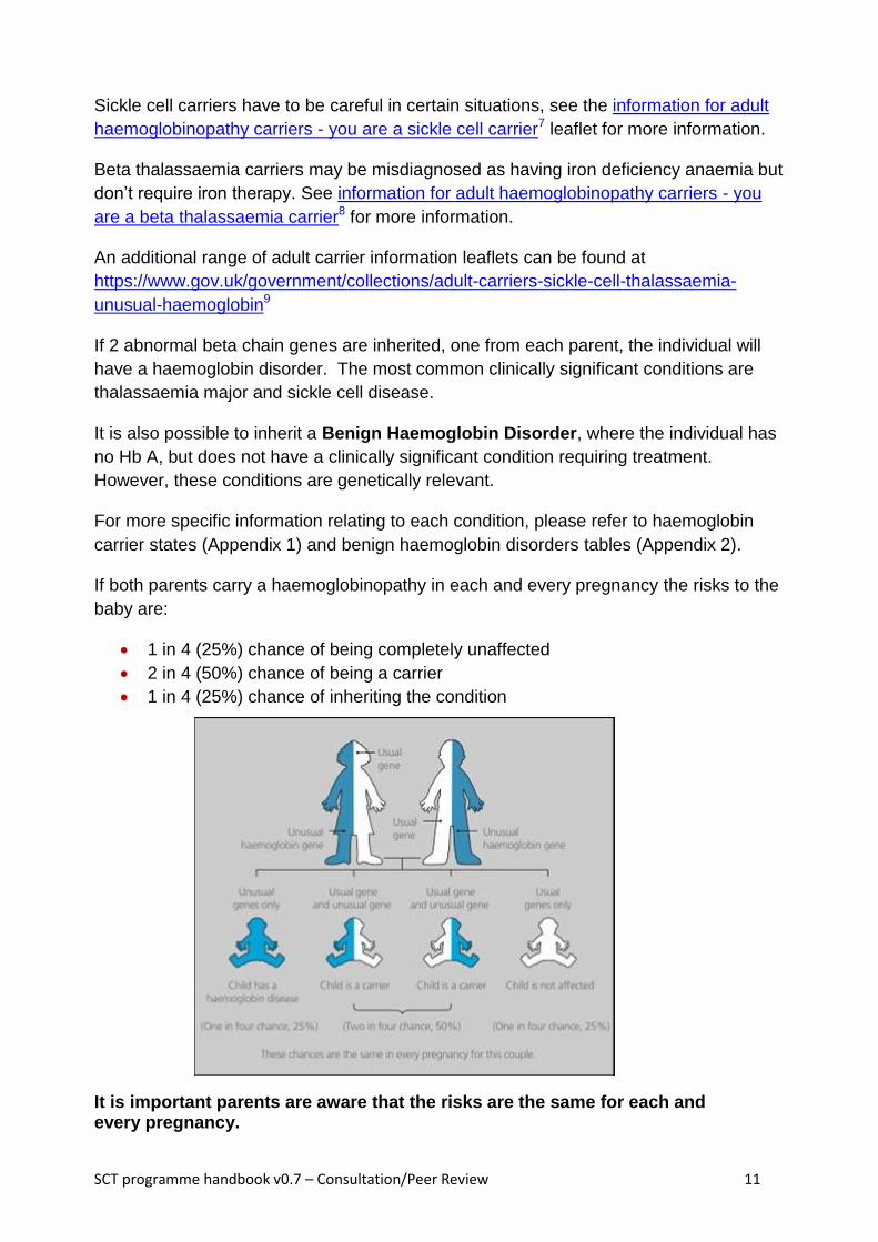
If both parents carry a haemoglobinopathy in each and every pregnancy the risks to the
baby are:
1 in 4 (25%) chance of being completely unaffected
2 in 4 (50%) chance of being a carrier
1 in 4 (25%) chance of inheriting the condition
It is important parents are aware that the risks are the same for each and every pregnancy.
SCT programme handbook v0.7 – Consultation/Peer Review 12
3.4 Sickle cell disease10
Sickle cell disease is a recessively inherited genetic condition of the haemoglobin. It
occurs when both parents pass abnormal haemoglobin genes to the baby, and the baby
has no normal haemoglobin (Hb A). The production of abnormal beta globin chains
affects the quality of the haemoglobin.
The most common types of sickle cell disease seen in England are:
Hb SS, sickle cell anaemia
Hb SC, sickle/Hb C disease
Hb S/beta thalassaemia
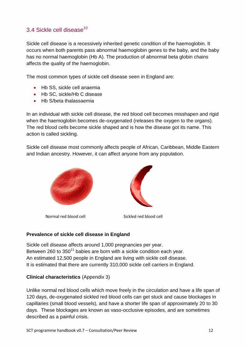
In an individual with sickle cell disease, the red blood cell becomes misshapen and rigid
when the haemoglobin becomes de-oxygenated (releases the oxygen to the organs).
The red blood cells become sickle shaped and is how the disease got its name. This
action is called sickling.
Sickle cell disease most commonly affects people of African, Caribbean, Middle Eastern
and Indian ancestry. However, it can affect anyone from any population.
Normal red blood cell Sickled red blood cell
Prevalence of sickle cell disease in England
Sickle cell disease affects around 1,000 pregnancies per year.
Between 260 to 35011 babies are born with a sickle condition each year.
An estimated 12,500 people in England are living with sickle cell disease.
It is estimated that there are currently 310,000 sickle cell carriers in England.
Clinical characteristics (Appendix 3)
Unlike normal red blood cells which move freely in the circulation and have a life span of
120 days, de-oxygenated sickled red blood cells can get stuck and cause blockages in
capillaries (small blood vessels), and have a shorter life span of approximately 20 to 30
days. These blockages are known as vaso-occlusive episodes, and are sometimes
described as a painful crisis.

SCT programme handbook v0.7 – Consultation/Peer Review 13
A sickle cell crisis can be triggered by:
sudden changes in body temperature
dehydration
shortage of oxygen
infection
‘Sickling’ can result in:
intense pain
severe anaemia
tissue damage
infections
strokes, especially in Hb SS
shortened life expectancy
These adverse consequences are made worse if the individual experiences repeated
crises.
3.5 Management of individuals with sickle cell disease12
Sickle cell disease requires specialist consultant haematologist or paediatric
management. Early diagnosis is vital and screening for sickle cell disease is
incorporated in the newborn blood spot screening programme in England. Children who
are diagnosed with sickle cell disease at birth should be entered into specialist care by 3
months of age. See NHS newborn blood spot (NBS) screening programme for more
information.
Management of individuals with sickle cell disease includes:
regular specialist outpatient reviews
easy, direct access to specialist medical care when unwell
prophylactic penicillin V and regular immunisations to prevent infections
consideration of hydroxycarbamide if the symptoms are significant
general anaesthesia should only be undertaken when full medical support is
available. This also applies to dental treatment
blood transfusions may be required for relevant complications, for example
stroke or severe/acute anaemia
annual transcranial doppler scans are recommended for children and
adolescents
Sickle cell disease can be cured by bone marrow or stem cell transplant but the genetic
profile of the individual does not change.
Much of the care required by individuals with sickle cell disease is preventative and
supportive care. Families need specialist support to understand the condition and learn

SCT programme handbook v0.7 – Consultation/Peer Review 14
how to care for children in a proactive way, to recognise potential problems and try to
prevent or minimise the effects of a sickle cell crisis.
Families are taught to:
ensure a balanced, healthy diet
encourage fluid intake
keep the child warm in cold conditions
comply with immunisations and penicillin V
administer simple analgesia at the start of a sickling episode
seek urgent medical care when required
3.6 Thalassaemias (Appendix 4)
Thalassaemias are recessively inherited genetic conditions which affect the quantity of
haemoglobin produced. This is due to changes in the genetic code responsible for the
production of either the alpha or beta globin chains that are present in normal
haemoglobin. There are 3 types of thalassaemia that have clinical significance. These
are:
alpha thalassaemia major, which is clinically significant to the fetus and mother
beta thalassaemia major, which is clinically relevant after birth
thalassaemia intermedia, which has variable clinical significance
Normal red blood cells Thalassaemic red blood cells

SCT programme handbook v0.7 – Consultation/Peer Review 15
3.7 Alpha thalassaemia13;
Normal haemoglobin A has 2 alpha globin chains, however the production of these
alpha globin chains is controlled by 4 alpha globin genes, 2 genes from each parent. In
alpha thalassaemia there is either reduced or absent production of alpha globin chains,
caused by a defect or mutation in one or more of the alpha globin genes.
Alpha thalassaemia carrier status can only be confirmed by DNA analysis.
Below is a graphic illustration of the alpha globin chains (2), and the full complement of alpha globin genes, (4) that each individual with normal Hb A inherits. This is usually written αα/αα in medical literature.
3.8 Alpha (α°) thalassaemia major
In alpha thalassaemia major, also known as Barts Hydrops Fetalis, there are no
functioning alpha globin genes (--/--). As a result no alpha globin chains are produced,
which results in a severe life-threatening anaemia in the fetus and, without intervention,
is incompatible with extra-uterine life.
The hydropic fetus can usually be diagnosed by ultrasound scan during the 2nd trimester
of pregnancy. Very occasionally, babies survive with intra-uterine transfusions.
However, there is a high risk of significant disabilities.
There are also implications for the mother during pregnancy with a fetus that has Barts
Hydrops Fetails, these include:
pre-eclampsia
ante-partum haemorrhage
retained placenta
possible 50% maternal mortality rate if alpha thalassaemia major in the fetus is
undiagnosed

SCT programme handbook v0.7 – Consultation/Peer Review 16
During antenatal screening it is important to diagnose if both parents are alpha zero (α°)
thalassaemia carriers, which relies on 3 essential components
family origin
blood tests
DNA analysis for confirmation of carrier status
The family origins that are most at risk of alpha zero (α°) thalassaemia carrier status are
from Taiwan, Laos, Vietnam, Malaysia, China, Hong Kong, Burma, Cyprus, Turkey,
Sardinia, Greece, Singapore, Philippines, Thailand, Cambodia and Indonesia.
In the UK around 20 to 30 couples annually are identified as being at high risk of having
a baby with alpha thalassaemia major.
3.9 Alpha zero (α°) thalassaemia carrier
This occurs when an individual has inherited no alpha globin chain genes from one
parent (--/αα). The individual is generally healthy but there is a reduction in alpha
globin chain production and they may have a mild anaemia with a MCH that is usually
less than 25pg. It can be confused with iron deficiency anaemia. Below is a graphic
illustration of alpha zero thalassaemia.
If a couple are both identified as potentially being alpha zero thalassaemia carriers, then
their carrier status must be identified by DNA analysis as there is a 1 in 4 (25%) risk to
all their children of inheriting Barts Hydrops Fetalis, also known as alpha thalassaemia
major.

SCT programme handbook v0.7 – Consultation/Peer Review 17
3.10 Alpha plus (α+) thalassaemia
Individuals with alpha plus thalassaemia have inherited either one or 2 faulty alpha
globin genes (-α/αα) or (-α/-α). Although this can affect alpha globin chain production,
there is usually minimal change to the haemoglobin level. Alpha plus thalassaemia is not clinically significant but can be confused with iron deficiency anaemia in the antenatal period.
Alpha plus thalassaemia is most often seen in African, Caribbean, Indian, Pakistani, Bangladeshi and Middle Eastern populations.
3.11 Haemoglobin H disease
An individual with haemoglobin H disease (--/-α) has only one normal alpha globin gene
but usually retains the ability to produce sufficient haemoglobin for life. The haemoglobin level usually ranges from 7 to 10g/dl, with a MCH of 15 to 25pg.
Haemoglobin H disease is a mild to moderate condition. It does not usually require treatment or lifelong blood transfusions, however short-term transfusions may occasionally be required during critical periods, such as pregnancy or illness or if the individual has an infection.

SCT programme handbook v0.7 – Consultation/Peer Review 18
3.12 Beta Thalassaemia major 14
Beta thalassaemia major is also called ‘Cooley’s Anaemia’ or ‘Mediterranean Anaemia’.
Thalassaemia major is most common in people of Pakistani, Cypriot, Italian, Greek,
Indian, Bangladeshi, Chinese and other East/South East Asian ancestry.
There are 2 main thalassaemia conditions depending on the beta thalassaemia gene
mutation inherited. They are:
thalassaemia major
thalassaemia intermedia
In beta thalassaemia major there is reduced or absent production of the beta globin
chains that make up normal adult haemoglobin, due to defective beta globin genes
which are inherited from both parents. This results in severe, life-threatening anaemia
which usually requires regular blood transfusions for life.
Prevalence in England
It is estimated that thalassaemia major affects about 1 in 27 000 pregnancies.
Approximately 20-305 babies are born with thalassaemia major each year. There are
more than 800 people living with thalassaemia major.
It is estimated there are 300 000 thalassaemia carriers.
Clinical complications of untreated beta thalassaemia major
Clinical complications of untreated beta thalassaemia major include:
failure to thrive in babies and young children
lethargy and fatigue due to severe anaemia
hypersplenism due to impaired breakdown of red blood cells
over activity of the bone marrow
severe anaemia leading to early death

SCT programme handbook v0.7 – Consultation/Peer Review 19
3.13 Management of individuals with beta thalassaemia major
Although beta thalassaemia major is not included in the newborn bloodspot screening
programme, children with this condition are usually identified and should be referred for
specialist care. Early diagnosis helps to monitor a child’s condition until blood
transfusions are required and also gives parents the opportunity to learn about the
condition before complications arise. .
The management of individuals with beta thalassaemia major aims to correct the severe
anaemia and includes
blood transfusions every 3 to 5 weeks, usually starting from approximately 9 to
12 months of age
iron chelation therapy to remove the excess iron (either orally or by
subcutaneous injection)
regular hospital appointments to monitor the condition
splenectomy for hypersplenism
In addition, supportive care is important for people affected by beta thalassaemia major
and they
should be encouraged to avoid iron rich foods and have a daily vitamin C
supplement
may require bone marrow or stem cell transplant to cure their condition, however
their genetic profile does not change
should have psycho-social support in addition to medical care
There is a risk of early death unless patients adhere to the strict regime of blood
transfusions and iron chelation.
Clinical complications of treated beta thalassaemia major
If thalassaemia major is not well managed by transfusion therapy this could result in
anaemia and over-activity of bone marrow. However, the major cause of complications
is usually related to an excess of iron accumulated in the body due to the regular blood
transfusions. This can result in the following
damage to the pituitary glands which could affect
o growth and also delayed puberty
o insulin production, resulting in diabetes
hypothyroidism
cardiac impairment/failure
liver damage
lethargy and fatigue
erectile dysfunction in men and amenorrhoea in women
change in skin colour due to iron deposits

SCT programme handbook v0.7 – Consultation/Peer Review 20
Individuals may also have
hypersplenism due to impaired breakdown of red blood cells and over activity
increased susceptibility to infections such as meningitis and flu, especially if the
spleen has been removed
3.14 Thalassaemia intermedia
Thalassaemia intermedia occurs in an individual when the beta globin chain production
is significantly reduced. Clinical implications vary depending on the gene mutations
inherited from both parents. This can result in a degree of anaemia, but the condition is
not as severe as thalassaemia major.
The individual usually manages without regular blood transfusions but there may be
splenomegaly and the requirement for occasional blood transfusions.
3.15 Haemoglobin E/beta thalassaemia
Haemoglobin E/beta thalassaemia may result in a syndrome similar to either
thalassaemia major or thalassaemia intermedia.
3.16 References
1 NHS Sickle Cell & Thalassaemia Screening Programme (2016) – eLearning Modules http://portal.e-
lfh.org.uk/Catalogue/Index 2 Sickle Cell Information Centre (SCINFO) Tutorial: Normal Blood Cells http://scinfo.org/world-wide-
resources/power-point-presentations 3 Bain BJ (2006) Other significant haemoglobinopathies. Haemoglobinopathy Diagnosis, 2nd Edition Blackwall Publishing
Ltd
4 Weatherall DJ, Clegg JB (2001) Inherited haemoglobin disorders: an increasing global health problem. Bulletin of
the World Health Organisation, 2001, 79 (8) (Article Contains Prevalence Data) http://www.who.int/docstore/bulletin/pdf/2001/issue8/vol79.no.8.704-712.pdf 5 Huisman THJ et al A database of Human Hemoglobin Variants and Thalassemias http://globin.bx.psu.edu/cgi-
bin/hbvar/counter 6 Public Health England (2013) Sickle cell and thalassaemia screening: inheritance risk table
https://www.gov.uk/government/publications/sickle-cell-and-thalassaemia-screening-inheritance-risk-table 7 NHS Sickle Cell & Thalassaemia Screening Programme (2012) Information for adult haemoglobinopathy carriers:
you are a sickle cell carrier. https://www.gov.uk/government/publications/sickle-cell-carrier-description-in-brief

SCT programme handbook v0.7 – Consultation/Peer Review 21
8 NHS Sickle Cell & Thalassaemia Screening Programme (2012) Information for adult haemogloboinopathy
carriers: you are a beta thalassaemia carrier. https://www.gov.uk/government/publications/beta-thalassaemia-carrier-description-in-brief 9 NHS Sickle Cell & Thalassaemia Screening Programme (2012)
Adult carriers: sickle cell, thalassaemia, unusual
haemoglobin https://www.gov.uk/government/collections/adult-carriers-sickle-cell-thalassaemia-unusual-haemoglobin
10 Eleftheriou A, Angastiniotis M About Sickle Cell Disorders. Thalassaemia International Federation (TIF)
http://www.thalassaemia.org.cy/ 11
NHS Sickle Cell & Thalassaemia Screening Programme (2017) Sickle cell and thalassaemia screening: data trends and performance analysis https://www.gov.uk/government/publications/sickle-cell-and-thalassaemia-screening-data-trends-and-performance-analysis 12 Brent Sickle Cell & Thalassaemia Centre (2012) A Parent’s Guide to Managing Sickle Cell Disease
https://www.gov.uk/government/publications/sickle-cell-disease-managing-the-condition
13 Eleftheriou A, Angastiniotis M About Alpha Thalassaemia Thalassaemia International Federation (TIF)
www.thalassaemia.org.cy 14 Eleftheriou A, Angastiniotis M About Beta Thalassaemia Thalassaemia International Federation (TIF)
www.thalassaemia.org.cy

SCT programme handbook v0.7 – Consultation/Peer Review 22
Chapter 4 - Antenatal Screening
Antenatal screening identifies women with a haemoglobinopathy, and provides
screening of consenting biological fathers. When both parents are carriers of a
significant haemoglobinopathy, there is a one in four (25%) chance, in each pregnancy,
that their baby could inherit a condition that needs treatment. The most important
conditions are sickle cell disease and thalassaemia major.
Sickle cell disease and thalassaemia major are serious, inherited blood disorders. They
affect haemoglobin and its oxygen carrying capacity. Individuals who have one of these
conditions need treatment and lifelong care. People who are carriers are healthy and
unaware of their status unless they have a specific blood test.15
Carrier women and couples “at risk” of having a baby with a major haemoglobin
disorder need information, advice and counselling to make choices for the pregnancy.
This includes the decision to have prenatal diagnosis, and to take further action if they
choose to.
This means that screening must occur early in pregnancy, preferably by 10 weeks’ gestation. This allows time for any subsequent actions required. Early screening usually results in a greater uptake of prenatal diagnosis (PND), ideally by 12 weeks + 6 days gestation.16 The antenatal screening programme is a pathway17 and needs all the components in place for the screening test to be effective. The stages in the pathway work most efficiently with coordination from a multidisciplinary team of professionals. These include:
midwife, screening coordinator, & maternity services
laboratory team
counselling services
primary care
voluntary sector To ensure that a quality service is delivered, there must be a named individual who has lead responsibility for each stage of the pathway:
• identification of the eligible population • providing information before screening and completion of the Family Origin
Questionnaire (FOQ),18 along with obtaining a blood sample • processing the blood samples and reporting the results • offer of testing to all biological fathers of babies where the mother has been
identified with a haemoglobinopathy • communicating the blood test results to mother and the baby’s father (where
relevant) • carrying out actions, such as prenatal diagnosis, based on parental decisions • diagnosis (where requested) of babies at risk of inheriting a major haemoglobin
disorder • refer affected individuals for treatment and care
SCT programme handbook v0.7 – Consultation/Peer Review 23
4.1 Prevalence There are two approaches to the delivery of the screening programme based on the geographical prevalence of haemoglobinopathy conditions in the high risk populations living in England.19 A list of high and low prevalence trusts is available for further information. In low prevalence trusts, where less than 1% of the booking bloods received by the laboratory are screen positive:
with consent, the red blood cell indices will screen all women (irrespective of family origins) for thalassaemia
the FOQ is used as an initial screening tool to identify women, or the baby’s biological father, at high risk of being a carrier for sickle cell, and other haemoglobin variants
where either parent falls into a high risk group, a screening blood test for haemoglobin variants must be offered to the woman
In high prevalence trusts, where 2% or greater of the booking bloods received by the laboratory are screen positive
• all women must be offered a screening blood test for sickle cell, thalassaemia and other haemoglobin variants, irrespective of family origins
In high and low prevalence trusts
where a woman is diagnosed with a haemoglobinopathy, the baby’s biological father (irrespective of family origins) must be offered screening for sickle cell, thalassaemia and other haemoglobin variants
it is important to note that not all haemoglobinopathies will be diagnosed and where there is an inconclusive result, systems must be in place to follow up the woman/couple where relevant
a completed paper or electronic FOQ must accompany all blood samples to the laboratory
checks should be in place to ensure that all women have been offered screening, and the results have been followed up appropriately
There are detailed algorithms for processing antenatal samples in both high and low prevalence areas, which are outlined in the Laboratory Handbook.20

SCT programme handbook v0.7 – Consultation/Peer Review 24
4.2 Booking for antenatal care
Choice & Consent21
All women must receive information about antenatal screening tests early in pregnancy, before they are asked to make any screening decision. This should include information on when results will be available following uptake of screening. There must be an opportunity to discuss the screening options with a professional who is informed about the condition(s). The health professional offering the screening test must ensure that the woman understands the test, has given her consent for antenatal screening, and is aware of the choices that will follow if the test is positive. When offering screening for sickle cell and thalassaemia, healthcare professionals (HCPs) must:
• give verbal and written information about the screening test, using the booklet Screening tests for you and your baby22
• offer the woman an opportunity to discuss the screening test and her decision • offer resources to address any specific needs that the woman has such as
literacy, visual impairment, language needs23; 24 • be aware of, and sensitive to, the woman’s values and beliefs and support the
woman to make decisions which are right for her • record consent or non-consent for screening in the woman’s maternity notes • communicate non-consent for screening to appropriate professionals, including
laboratory staff It is only necessary to offer screening for sickle cell and thalassaemia once in the same pregnancy. If a women is screened in a low prevalence area, but chooses to give birth in a maternity unit in a high prevalence area, her current result is sufficient, and there is no need for re-screening. Alternately, if the woman changes NHS provider during the pregnancy, it is not necessary to repeat the blood test if the result is available. In both cases the previous result must be from a laboratory accredited by the UK Accreditation Service (UKAS), and be consistent, unequivocal, well documented and interpreted and reported within the testing algorithms in the laboratory handbook.20 During booking for antenatal care it is important to establish some details which are relevant to the sickle cell and thalassaemia screening programme. This includes information about:
adoption or a lack of awareness of family ancestry from either parent?
fertility treatment, is it a o donor egg? o donor sperm? o both?
history of blood transfusion or currently having regular blood transfusions? (Why? When? Where?)
history of bone marrow or stem cell transplant? (Why? When? Where?)

SCT programme handbook v0.7 – Consultation/Peer Review 25
history of haemoglobin disorders or other inherited conditions? For themselves? In either the maternal or paternal family?
If the woman consents, the screening sample must be taken at first booking appointment. If the woman declines screening, the laboratory team should be aware of this information prior to processing the full blood count sample. All women need to be made aware that routine analysis of blood may be indicative of thalassaemia carrier status. However, further investigations to confirm carrier status should not occur if the woman has not consented to screening.
4.3 The family origin questionnaire (FOQ)
Although people from any population can have these conditions, it is more likely that an
individual will be a genetic carrier if any of their ancestors come from a malarial area of
the world. Being a carrier provides partial protection against malaria.
The aim of the FOQ25 is to identify the population groups at highest risk of sickle cell,
thalassaemia and other haemoglobin variants.
Completion of the FOQ information is the responsibility of the HCP who is booking the
woman for antenatal care. Details are required:
for both the baby’s biological mother and father
in both high and low prevalence areas
to be completed in every pregnancy and sent with the blood sample to the laboratory, or be accessible to the laboratory team if using an electronic system
for all ancestry, as far back as the individual can remember (at least 2 generations, but more if possible); this is particularly important for mixed race individuals
In low prevalence areas the FOQ information is used as an initial screening tool which asks about the family origins of both parents, to assess a woman’s eligibility for haemoglobin variant screening. If the woman falls into a high risk group she should be offered screening for haemoglobin variants If the woman falls into a low risk group, but the baby’s biological father falls into a high risk group, then the woman should be offered screening for a haemoglobin variant (irrespective of her family origins). In high and low prevalence areas the FOQ
must accompany all blood samples to the laboratory, or the relevant information must be accessible to the laboratory team if using electronic requesting
can avoid unnecessary testing of fathers and unnecessary anxiety for parents when accurately completed
SCT programme handbook v0.7 – Consultation/Peer Review 26
is relevant in the interpretation of red blood cell indices, particularly when screening groups at high risk of alpha zero thalassaemia
assists with accurate DNA analysis of prenatal diagnosis samples, ensuring that the relevant genotypes are included in the assay
If the woman declines screening, there must be systems in place to inform the laboratory team of this information. The NHS Sickle Cell and Thalassaemia screening programme produces a paper FOQ form as a template. The integration of the FOQ categories onto local antenatal screening forms or incorporated into an electronic requesting system is encouraged. The versatility of the national template must be reflected locally and the categories kept up to date if there are any changes. The current FOQ form can be downloaded from the programme website and can also be ordered from Harlow Press.
4.4 Conditions & carrier states to be detected26
There are over a thousand haemoglobin variants and thalassaemia mutations, but not all of these are clinically relevant.27 The national programme in England has determined the significant haemoglobinopathies which must be detected by antenatal screening.20 The rationale for choosing these carrier states and conditions is based on the high risk populations living in England. 1. Significant maternal haemoglobin conditions (these are important for maternal
care)
Hb SS
Hb SC
Hb SDPunjab
Hb SE
Hb SOArab
Hb S/Lepore
Hb S/β(0; +) thalassaemia
Hb S/δβ thalassaemia
β thalassaemia major/intermedia
Hb Lepore/β thalassaemia
Hb E/β thalassaemia
Hb H Disease (--/-α)
2. Carrier states in mother
Hb AS
Hb AC
Hb ADPunjab
Hb AE
Hb AOArab
Hb A/Lepore
SCT programme handbook v0.7 – Consultation/Peer Review 27
β thalassaemia carrier
δβ thalassaemia carrier
α0 thalassaemia carrier (--/αα)
Hereditary persistence of fetal haemoglobin (HPFH) carrier
3. Any compound heterozygous state including one or more of the above carrier states.
4. Any homozygous state of the above carrier conditions.
4.5 Screening for haemoglobin variants
In low prevalence areas the information about both the woman and the baby’s biological father on the family origin questionnaire, along with her consent, determines which women must be screened for haemoglobin variants.
In high prevalence areas all consenting women are screened for haemoglobin variants, irrespective of their family origins.
4.6 Screening for beta thalassaemia
All women in both high and low prevalence areas should be offered screening for thalassaemia.
The initial screen for the risk of thalassaemia involves a review of the full blood count:
haemoglobin (Hb) – normal value in pregnancy is equal to, or above =>11g/dl. Low values may indicate anaemia
mean cell volume (MCV) - normal range is 77-95 fl. Low values may indicate deficient haemoglobin production such as iron deficiency anaemia or thalassaemia
mean cell haemoglobin (MCH) – normal range is 27-32 pg. Low values are seen in thalassaemia or iron deficiency anaemia
If the MCH is lower than usual, the Hb A2 is measured. A range between 3.5% - 8% is the usual for a beta thalassaemia carrier. Screening for beta thalassaemia
can sometimes be complex.27
4.7 Screening for alpha zero thalassaemia
There is no straightforward test in the antenatal screening laboratory to
diagnose an alpha thalassaemia carrier, and DNA is required for a definitive
diagnosis. The approved laboratories for DNA testing are listed in the
Laboratory Handbook20 and Appendix 5.

SCT programme handbook v0.7 – Consultation/Peer Review 28
Alpha+ (alpha plus) thalassaemia is not considered clinically significant, and a suspected carrier will not require any further investigations.
Alpha0 (alpha zero) thalassaemia is clinically significant and most commonly found in people with ancestry from
East Mediterranean (Cyprus, Greece, Sardinia or Turkey)
Southeast Asia (China, Hong Kong, Thailand, Taiwan, Cambodia, Laos, Vietnam, Burma, Singapore, Indonesia or Philippines).
The screening policy in England aims to identify couples where both parents are alpha zero thalassaemia carriers (alpha0) and their baby is at risk of inheriting alpha thalassaemia major (Hb Barts Hydrops Fetalis). The screening process to follow for these couples:
if the woman’s initial screening result indicates that she may be an alpha zero thalassaemia carrier, but only one parent is from a high risk group and the other parent is not, then no further investigations are needed
if the woman’s initial screening result indicates that she may be an alpha zero thalassaemia carrier, and both biological parents are from one of the high risk groups (see list above), then the baby’s father should be offered a screening test
if both parental screening results show a possibility of alpha zero thalassaemia carrier status, then a blood sample from each parent must be sent for DNA analysis to confirm whether or not they are alpha zero thalassaemia carriers
if both parents are carriers, then prenatal diagnosis (PND) should be offered
Only a small number of cases of alpha thalassaemia major occur in England each year.28
4.8 Referral of antenatal samples to the DNA laboratories for
haemoglobinopathy mutation analysis20
The majority of carriers are diagnosed in the antenatal screening laboratory. However, on occasion it may be necessary to refer a sample for DNA analysis. It is the responsibility of the antenatal screening laboratory team to decide which samples need to be referred and to inform the maternity or counselling team if any additional blood samples are needed from either the woman or the baby’s biological father.
SCT programme handbook v0.7 – Consultation/Peer Review 29
4.9 Issues which may arise during routine antenatal screening
During screening some carriers may be missed, and it is possible for false positive and false negative results to be reported. Assuming that the FOQ has been completed accurately, below are some examples of carrier states that can be missed20
• Some β-thalassaemia carriers may have o a “silent” or “near silent” genotype, associated with a borderline Hb A2 level
o their carrier status obscured by severe iron deficiency anaemia; a medical condition (B12 or folate deficiency; liver disease); or treatment (such as HIV therapy); or another haemoglobinopathy
• alpha0 thalassaemia occurring outside the defined high risk family origins or in women with anaemia
• any significant haemoglobin masked by an unreported blood transfusion or bone marrow transplant
• any significant haemoglobinopathy present in donor egg or donor sperm where the donor is undeclared or untested
• a second haemoglobin variant may be masked by haemoglobin A or another haemoglobinopathy.
In low prevalence areas, in addition to the above, carrier states that occur in individuals who fall outside the defined high risk family origins, or in individuals who have not disclosed their family origins accurately, may be missed.
4.10 Testing in subsequent pregnancies
If a woman is booked for antenatal care for a subsequent pregnancy, the HCP must:
offer the woman screening for a haemoglobinopathy, irrespective of previous screening
complete the FOQ, or have systems in place to make the information accessible to the laboratory team if using electronic test requesting
take the blood sample and send to the laboratory, if the woman consents to screening
If a carrier or affected woman is identified, the baby’s biological father must be offered a screening test, irrespective of previous screening history. If it is not possible to test the baby’s biological father in every pregnancy and a previous result is being considered for use, please check that this is the same father. This information must be recorded in the woman’s notes for the current pregnancy. If a written copy of the result is available, this should also be included in the woman’s records.
The previous result must be from a laboratory accredited by the UK Accreditation Service (UKAS), and be consistent, unequivocal, well documented and interpreted and reported within the testing algorithms in the Laboratory Handbook.20
SCT programme handbook v0.7 – Consultation/Peer Review 30
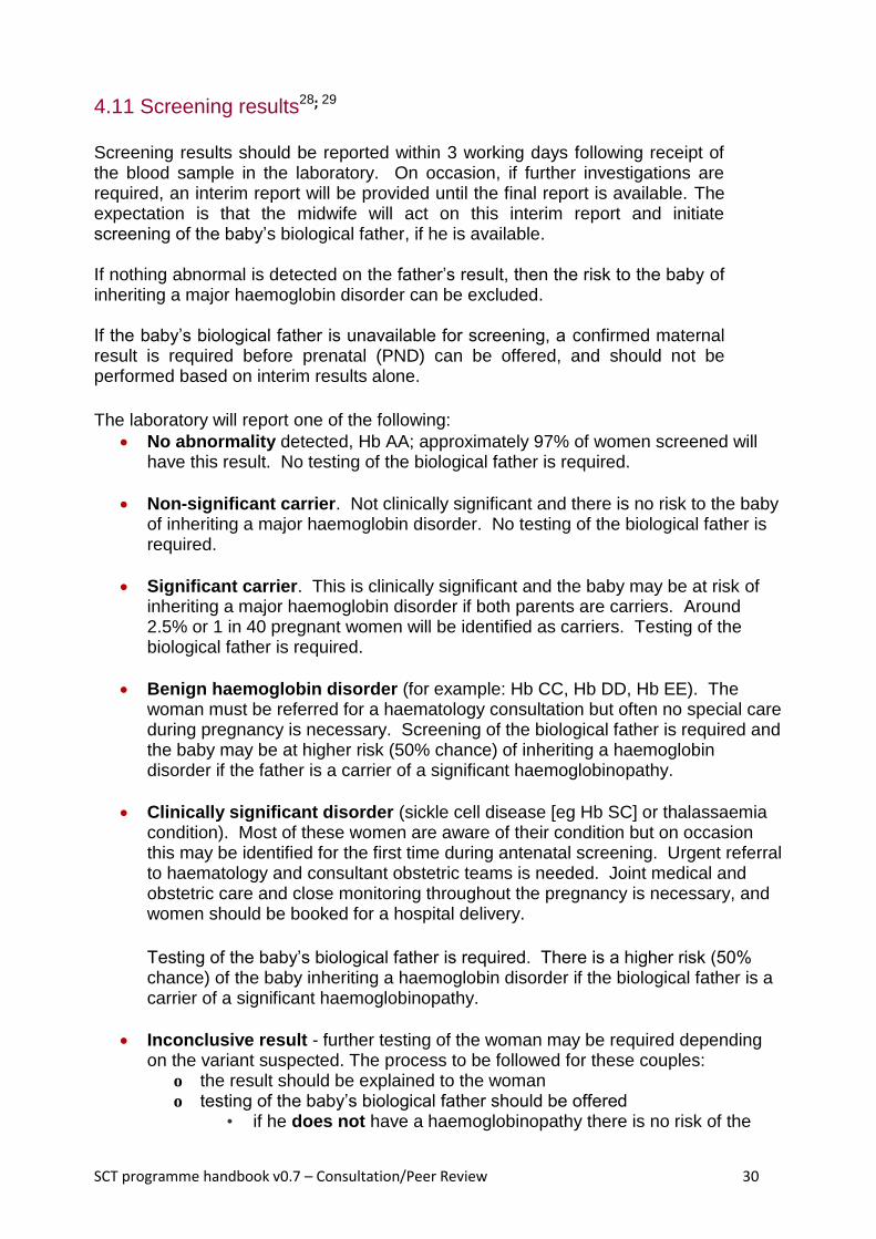
4.11 Screening results28; 29
Screening results should be reported within 3 working days following receipt of the blood sample in the laboratory. On occasion, if further investigations are required, an interim report will be provided until the final report is available. The expectation is that the midwife will act on this interim report and initiate screening of the baby’s biological father, if he is available. If nothing abnormal is detected on the father’s result, then the risk to the baby of inheriting a major haemoglobin disorder can be excluded. If the baby’s biological father is unavailable for screening, a confirmed maternal result is required before prenatal (PND) can be offered, and should not be performed based on interim results alone.
The laboratory will report one of the following:
No abnormality detected, Hb AA; approximately 97% of women screened will have this result. No testing of the biological father is required.
Non-significant carrier. Not clinically significant and there is no risk to the baby of inheriting a major haemoglobin disorder. No testing of the biological father is required.
Significant carrier. This is clinically significant and the baby may be at risk of inheriting a major haemoglobin disorder if both parents are carriers. Around 2.5% or 1 in 40 pregnant women will be identified as carriers. Testing of the biological father is required.
Benign haemoglobin disorder (for example: Hb CC, Hb DD, Hb EE). The woman must be referred for a haematology consultation but often no special care during pregnancy is necessary. Screening of the biological father is required and the baby may be at higher risk (50% chance) of inheriting a haemoglobin disorder if the father is a carrier of a significant haemoglobinopathy.
Clinically significant disorder (sickle cell disease [eg Hb SC] or thalassaemia condition). Most of these women are aware of their condition but on occasion this may be identified for the first time during antenatal screening. Urgent referral to haematology and consultant obstetric teams is needed. Joint medical and obstetric care and close monitoring throughout the pregnancy is necessary, and women should be booked for a hospital delivery.
Testing of the baby’s biological father is required. There is a higher risk (50% chance) of the baby inheriting a haemoglobin disorder if the biological father is a carrier of a significant haemoglobinopathy.
Inconclusive result - further testing of the woman may be required depending on the variant suspected. The process to be followed for these couples:
o the result should be explained to the woman o testing of the baby’s biological father should be offered
• if he does not have a haemoglobinopathy there is no risk of the

SCT programme handbook v0.7 – Consultation/Peer Review 31
baby inheriting a major haemoglobin disorder. There may be no further maternal testing required (this is locally determined). However, if there is no further testing of the woman she will remain unaware of her specific carrier status, and the risks for offspring in a future pregnancy if she changes partners
• if he does have a haemoglobinopathy, then further maternal testing may be required for an accurate assessment of the fetal risk of inheriting a significant haemoglobin condition, and the couple need to be followed up appropriately
All women should be informed of their screening result (normal, carrier, inconclusive, haemoglobin disorder) and a local protocol and pathway must be in place to support this. Women who have inconclusive, carrier or affected results should be offered an opportunity to receive the result in a face to face counselling session, along with written notification of the results. An information leaflet on specific carrier status must be provided.30 Advice regarding issuing haemoglobinopathy cards is given by the British Society of Haematology (BSH)26 Screening results should be accessible to ALL HCPs involved in the screening programme by recording details in the woman’s:
handheld maternity records
electronic maternity record
primary care health record
4.12 Screening follow up for clinically significant results (carrier, affected,
inconclusive, benign haemoglobin disorder)
Results must be communicated to the woman urgently. The mother needs time to organise screening for the baby’s biological father and to consider the implications for the pregnancy and her unborn child. Receiving a positive screening result can be emotionally traumatic for women, as this may not have been anticipated. A trained professional should be available to explain all significant results. Timing is critical in making decisions for further investigations. Women should be given written confirmation of their result along with an explanatory leaflet.30 The woman must be invited for counselling31 (a template letter is available) and made aware of:
the implications for her as an individual of being a carrier or having a haemoglobin condition
the implications for this pregnancy and for future pregnancies
the fact that the baby’s biological father needs to be tested to assess the risk to the baby
the available choices for the pregnancy
the fact that other members of her family could also be carriers and that they can request testing by their GP or at a specialist centre, especially if they are planning to have a baby

SCT programme handbook v0.7 – Consultation/Peer Review 32
4.13 Beta thalassaemia carriers
A beta thalassaemia carrier has inherited an abnormal beta globin gene from
one parent and a normal one from the other. Where an individual is a beta
thalassaemia carrier it is important for them to be aware of the fact that:
the abnormal gene could be passed on to his or her children
even if their child has inherited the gene it cannot be diagnosed at birth by routine newborn blood spot screening
if parents choose, babies can be tested when they are over 9 months of age to confirm their carrier status
An example of a counselling form that could be used is in Appendix 6.
4.14 Screening the baby’s biological father
During organising and encouraging screening of the baby’s father, HCPs should be sensitive to possible paternity issues, and clarify to the woman the importance of screening the baby’s biological father.
The baby’s biological father of all women identified with a haemoglobinopathy, or an inconclusive result, (irrespective of his family origins), should be invited for counselling and a blood test as soon as possible. The leaflet, Tests for Fathers32 and letter33 should be given to the father prior to screening. Fathers must be offered screening in every pregnancy as for mothers.
Where possible the couple should have a joint counselling session to discuss the woman’s results and the implications for the pregnancy, and for the father to be tested. The session should be with a professional trained in giving haemoglobinopathy information.
If a joint appointment is not possible, then the father should be offered an appointment on his own to discuss the screening results and to have a blood test.
The HCP responsible for screening the baby’s biological father should provide the laboratory with information about the woman when the father is screened so that the results can be linked.
The fathers’34 test result should be recorded in the mother’s antenatal handheld records and on the counselling records.
4.15 Maximising uptake of father testing
On occasion it may be difficult for biological fathers to attend for screening, or they may be reluctant to be screened.
Some of the possible barriers to accessing screening include:
an assumption by the man that he has already been tested and has a negative result (based on the fact that he may have had a blood test in the past)

SCT programme handbook v0.7 – Consultation/Peer Review 33
a lack of understanding about the test; the significance of being a carrier; how the conditions are inherited; the risk to their baby
a possible stigma attached to screening
men who think that if they are well they cannot be a carrier
pregnancy and blood tests are seen as being a part of the woman’s world, compounded by systems in the antenatal clinic
difficulty with taking time off work to attend an appointment for a blood test
fear of needles
Some men may have been previously screened, either in the UK or in another country,
and do not recognise the need for re-screening. Explain that
it is necessary for their previous result to be confirmed when having prenatal diagnosis (PND)
previous screening may not include all variants tested for in the English Programme
screening test results need to be from an accredited laboratory
we need to see a copy of the laboratory report with his previous screening result
The HCP who reviewed the father’s previous screening results should document this in the woman’s record and, where possible, keep a copy of the result. Some points to consider
timing - can screening be offered at a convenient time for the father e.g. outside his normal working hours?
location - can screening be offered at a more convenient or neutral location?
if there are socio-cultural barriers to uptake, listen to what the mother says
would direct contact from the HCP to the father support the request for the need to have a blood test, and highlight the importance of screening?
Biological father unavailable for screening
If the baby’s biological father is unavailable, unknown or refuses testing then the HCP should discuss options with the woman:
is she living with the father/in contact during this pregnancy?
has the biological father been tested in the past and is there a confirmed result?
is she willing (or able) to deliver the letter and leaflet to the father?
if she is not living with the father and no longer in contact, can she provide his details so that the information about the test can be sent to him directly?
The responsible HCP should attempt to make direct contact with the baby’s biological
father, with the woman’s consent, if the woman is unwilling or unable to make contact,
in order to offer information and a screening blood test.
SCT programme handbook v0.7 – Consultation/Peer Review 34
4.16 Follow up after paternal screening29
Results should be reported to the designated HCP within 3 working days from the time of blood sample receipt in the laboratory.16 All father screening results must be reviewed and linked to the maternal results. Checks should be in place to ensure that paternal results34 have been received and are followed up. Fathers must be informed of their results, whether or not these are clinically significant. Carriers should receive the information in writing, along with an appropriate carrier leaflet30 where relevant. Advice regarding issuing haemoglobinopathy cards is given by the British Society of Haematology (BSH).26
4.16.1 Paternal carrier results (baby at risk of inheriting a benign haemoglobin disorder) If the man is identified as a haemoglobinopathy carrier then the couple must be invited for a follow up counselling session and the results explained “face to face”. If the couple are at risk of having a baby with a benign haemoglobin disorder which does not require long-term treatment, (for example a condition such as Hb EE; Hb CC; Hb DD; Hb C/Beta thalassaemia), this should be explained and the couple reassured. Confirmation in writing of his carrier status and an appropriate carrier leaflet30 should be given to the father. Prenatal diagnosis is not required for any of these conditions.
4.16.2 Paternal carrier results (baby at risk of inheriting a major haemoglobin disorder)35 Women and couples “at risk” of having an affected baby must be offered prenatal diagnosis (PND) as soon as possible, ideally by 12 weeks + 0 days gestation. They should
be offered an urgent counselling appointment with an appropriately trained professional (for example a professional trained in an approved course such as the genetic risk assessment and counselling course) to explain the risk to their baby, details about the condition that their baby could inherit, and the options for the pregnancy. An explanatory leaflet36; 37 to support the counselling session should be given to the couple
be urgently referred if they do decide to proceed with prenatal diagnosis

SCT programme handbook v0.7 – Consultation/Peer Review 35
4.17 Considerations for the antenatal screening programme
The Sickle Cell and Thalassaemia Screening Programme presents some challenges for
practitioners.
Defining family origins, heritage and ancestry: Identifying family origins, heritage or ancestry is integral to screening for sickle cell and thalassaemia. It is important this is not confused with nationality.
The FOQ identifies the groups at highest risk of sickle cell, thalassaemia and other haemoglobin variants. This includes the Mediterranean population who, under normal circumstances, may be ‘missed’ for screening. Practitioners need to assist parents to complete the FOQ for screening in low prevalence areas and for laboratories to have the correct information for analysis of samples.
Influences of culture during screening: The perception of what carrier status means may affect families’ attitudes to screening. In some of the groups at highest risk of haemoglobinopathies, there may be religious or cultural beliefs that influence decisions about prenatal diagnosis and termination of pregnancy. Research38 confirms this and practitioners need to be aware of the relevant issues.
Linking the antenatal and newborn screening programmes: The national programme in its’ entirety is a linked antenatal and newborn screening programme. The genetic nature of sickle cell disease and thalassaemia major means that it is important to link information from parental results to the baby’s screening result. Local systems need to be in place to facilitate this.

SCT programme handbook v0.7 – Consultation/Peer Review 36
4.18 References
15
NHS Sickle Cell & Thalassaemia Screening Programme (2016) – eLearning Modules http://portal.e-lfh.org.uk/Catalogue/Index 16
NHS Sickle Cell & Thalassaemia Screening Programme (2017) Sickle cell & thalassaemia screening: programme standards https://www.gov.uk/government/publications/standards-for-sickle-cell-and-thalassaemia-screening 17
NHS Sickle Cell & Thalassaemia Screening Programme (2015) Antenatal screening care pathway https://www.gov.uk/government/publications/sickle-cell-and-thalassaemia-screening-care-pathway 18
NHS Sickle Cell & Thalassaemia Screening Programme (2014) Family origin questionnaire https://www.gov.uk/government/publications/family-origin-questionnaire-sickle-cell-and-thalassaemia-screening 19
NHS Sickle Cell & Thalassaemia Screening Programme (2016) Determining prevalence https://www.gov.uk/government/publications/nhs-trusts-area-prevalence-for-sickle-cell-and-thalassaemia 20
NHS Sickle Cell & Thalassaemia Screening Programme (2017 ) Laboratory Handbook 4th
Edition https://www.gov.uk/government/publications/sickle-cell-and-thalassaemia-screening-handbook-for-laboratories 21
Department of Health (2009) Reference guide to consent for examination or treatment 2nd
Editionhttps://www.gov.uk/government/publications/reference-guide-to-consent-for-examination-or-treatment-second-edition 22
Public Health England (2017) Screening tests for you and your baby. https://www.gov.uk/government/publications/screening-tests-for-you-and-your-baby-description-in-brief 23
NHS Sickle Cell & Thalassaemia Screening Programme Large Print information http://webarchive.nationalarchives.gov.uk/20150408175925/http://sct.screening.nhs.uk/easyreadlargeprint 24
NHS Sickle Cell & Thalassaemia Screening Programme Language and Audio informationhttp://webarchive.nationalarchives.gov.uk/20150408175925/http://sct.screening.nhs.uk/audio 25
NHS Sickle Cell & Thalassaemia Screening Programme (2014) Family Origin Questionnaire (FOQ) https://www.gov.uk/government/publications/family-origin-questionnaire-sickle-cell-and-thalassaemia-screening 26 British Society for Haematology (2010) Significant Haemoglobinopathies: Guidelines for Screening and Diagnosis.
http://www.b-s-h.org.uk/guidelines/guidelines/significant-haemoglobinopathies-guidelines-for-screening-and-diagnosis/ 27 Huisman THJ et al A database of Human Hemoglobin Variants and Thalassemias http://globin.bx.psu.edu/cgi-
bin/hbvar/counter 28
NHS Sickle Cell & Thalassaemia Screening Programme (2017) Data report, trends and performance analysis https://www.gov.uk/government/publications/sickle-cell-and-thalassaemia-screening-data-trends-and-performance-analysis 29
NHS Sickle Cell & Thalassaemia Screening Programme (2013) Inheritance risk chart and carrier risk chart combined https://www.gov.uk/government/publications/sickle-cell-and-thalassaemia-screening-inheritance-risk-table 30
NHS Sickle Cell & Thalassaemia Screening Programme (2012 - Revised 2016) Adult carriers: sickle cell, thalassaemia, unusual haemoglobin https://www.gov.uk/government/collections/adult-carriers-sickle-cell-thalassaemia-unusual-haemoglobin

SCT programme handbook v0.7 – Consultation/Peer Review 37
31
NHS Sickle Cell & Thalassaemia Screening Programme (2013) Template letter: sickle cell and thalassaemia screening for parents https://www.gov.uk/government/publications/template-letter-sickle-cell-and-thalassaemia-screening-for-parents 32
NHS Sickle Cell & Thalassaemia Screening Programme (2012) Tests for fathers leaflet https://www.gov.uk/government/publications/tests-for-dads-sickle-cell-and-thalassaemia-screening 33
NHS Sickle Cell & Thalassaemia Screening Programme (2013) Template letter: sickle cell and thalassaemia screening for fathers https://www.gov.uk/government/publications/template-letter-sickle-cell-and-thalassaemia-screening-for-fathers 34
Fatherhood institute (2012) Guidance Contacting fathers for antenatal sickle cell and thalassaemia screening https://www.gov.uk/government/publications/contacting-men-for-sickle-cell-and-thalassaemia-screening/contacting-fathers-for-antenatal-sickle-cell-and-thalassaemia-screening 35
NHS Sickle Cell & Thalassaemia Screening Programme (2017) Counselling and Referral for Prenatal Diagnosis – Chapter 5 Programme Handbook 36
NHS Sickle Cell & Thalassaemia Screening Programme (2015) Information for couples “at risk” of having a baby with sickle cell disease or thalassaemia major https://www.gov.uk/government/publications/baby-at-risk-of-having-sickle-cell-disease-description-in-brief and https://www.gov.uk/government/publications/baby-at-risk-of-having-thalassaemia-description-in-brief 37
Fetal Anomaly Screening Programme (2017) Diagnostic tests https://www.gov.uk/government/publications/cvs-and-amniocentesis-diagnostic-tests 38 Maxwell-Otungo, K (2002) Towards a communication strategy for the NHS Sickle Cell & Thalassaemia Screening
Programme. A planning framework based on preliminary community consultation. https://www.gov.uk/government/publications/sickle-cell-and-thalassaemia-screening-community-outreach-research

SCT programme handbook v0.7 – Consultation/Peer Review 38
Chapter 5 - Counselling and Referral for Prenatal Diagnosis
5.1 Introduction
This guidance has been prepared for healthcare professionals who provide
counselling and referral for prenatal diagnosis (PND) to couples/women at risk
of having a baby with sickle cell disease or thalassaemia major.
Couples/women at risk are defined as:
both biological parents are carriers of a significant haemoglobin gene variant the woman is a carrier of a significant haemoglobin gene variant and the
screening status of the biological father is unknown If it is not possible to test the baby’s biological father in every pregnancy and a previous result is being used then this fact must be recorded in the woman’s notes for the current pregnancy. The previous result must be from a laboratory accredited by the UK Accreditation Service (UKAS), and be consistent, unequivocal, well documented and interpreted and reported within the algorithms of the laboratory handbook. All maternity units should provide a publically available and advertised direct dial number and/or email address for the use of women, GPs and midwives. There must be a clear pathway, with a named individual or team responsible at each step to:
provide direct access to counselling and PND for known at risk couples/known carrier women
check screening results offer testing to the baby’s biological father identify women and couples at risk of having a baby with sickle cell disease or
thalassaemia major in their current pregnancy counsel women/couples and refer for PND obtain the fetal sample and dispatch to the molecular laboratory along with
parental venous blood samples follow up after PND
A flow chart outlining the pathway is in Appendix 7.
5.2 Carrier women and at risk couples
Carrier women with no paternal result available, and known carrier couples, who are “at risk” of having a baby with a major haemoglobin disorder must be:
offered an urgent face to face counselling appointment to explain their carrier status, and given information about the condition that may affect their baby (a sample counselling form is in Appendix 6)

SCT programme handbook v0.7 – Consultation/Peer Review 39
made aware that in pregnancies that result from egg or sperm donation, the genes inherited by the baby are those of the biological donor
provided with an explanatory leaflet39 either before, or after the counselling session
given information regarding the options for the pregnancy given contact details of UK patient societies for the relevant condition (SCS or UKTS) made aware that the decision to have a diagnostic test is an informed choice by
the woman/couple offered an appointment for PND made aware of the intrinsic miscarriage risk associated with PND whether the
baby is affected or not40; 41
5.3 Women and couples who present as known carriers Women/couples with carrier status known prior to the current pregnancy must be fast-tracked and immediately offered a counselling appointment to discuss their options for the pregnancy as soon as they present in pregnancy. Confirmatory carrier screening must still be offered to the couple. To avoid delay in access to PND this should occur at the same time as the referral. If the baby’s biological father is unavailable for screening in this pregnancy, then the woman should still be counselled and offered PND without a paternal screening result. A risk assessment based on the ethnicity of the baby’s father is required to determine the risk of him being a carrier.
5.4 Referral for prenatal diagnosis If there is any doubt about the parental genotypes, or the need for further parental testing prior to fetal sampling, contact the relevant molecular haemoglobinopathy laboratory for advice. There are four haemoglobinopathy DNA laboratories in England which offer analysis of prenatal diagnosis samples for sickle cell disease and thalassaemia disorders.42; 43 Each laboratory has their own specified prenatal diagnosis request form, and a fully completed form must accompany the fetal and parental samples. Please see Appendix 5 for further details.
The following details, in addition to demographic information for both parents, must be included on the PND referral form:
maternal and paternal (if available) haemoglobinopathy results gestational age of the fetus including EDD by LMP maternal antenatal screening booking blood results (where feasible; do not delay
the referral); o full blood count o Hepatitis B status o HIV status o blood group and rhesus factor
whether the woman/couple consent to PND for any other condition, such as Fetal Anomaly

SCT programme handbook v0.7 – Consultation/Peer Review 40
5.5 Parental samples A parental blood sample (10 ml EDTA from each parent), must be sent to the DNA laboratory with the fetal sample, regardless if the woman/couple have had a previous PND. If a paternal sample is unavailable, a maternal blood sample should still be sent to confirm the maternal genotype and to exclude maternal contamination of the fetal sample. If the paternal genotype is
known (i.e. the father has been previously tested) but he is unavailable or declines testing; then a copy of his laboratory result should be sent to the prenatal diagnosis laboratory so that the fetal risk can be assessed. The result should be from a laboratory accredited by UKAS, and be consistent, unequivocal and well documented.
unknown and he is unavailable or declines testing, PND can still be carried out but this makes diagnosis of conditions more complex. If extended testing is required, this could delay the turnaround time of the fetal diagnosis. If the ethnic origin of the father is known, this should be included on the PND referral form.
5.6 Sickle cell disease Fetal diagnosis of a haemoglobin disorder in the case of sickle cell disease is generally a straightforward process, and is usually achieved without the need for molecular confirmation of parental carrier status.
5.7 Thalassaemia conditions In thalassaemia conditions it is possible to inherit one of a large range of genetic mutations. If a couple have not had PND before, send parental bloods for molecular confirmation of carrier status prior to fetal sampling. This will assist with a quicker fetal diagnosis result. If the couple has had PND before, or molecular confirmation, details of the previous PND should be included on the referral form.
5.8 Declining prenatal diagnosis If PND is declined the healthcare professional who has counselled the couple and recorded their decision, must inform:
parents of the process for screening their baby after birth and how they will obtain the result
the maternity department screening coordinator or equivalent the woman’s GP the newborn screening laboratory of the “at risk” couple (see Appendix 3 for an
example of a pregnancy alert form) the couple of contact details for UK patient societies for the relevant condition

SCT programme handbook v0.7 – Consultation/Peer Review 41
5.9 Prenatal Diagnosis: The Procedure
Local arrangements must be in place to obtain an urgent fetal sample for prenatal diagnosis. The diagnostic approach41; 44 usually depends on the gestational age of the fetus, and the sample will be obtained by one of the following methods:
chorionic villus sampling undertaken from 11 weeks of pregnancy amniocentesis performed from 15-16 weeks of pregnancy fetal blood sampling may also be an option
The molecular haemoglobinopathy laboratory must be contacted prior to dispatch of the sample, so that they expect a prenatal diagnosis sample for analysis. This ensures that the failsafe mechanisms to identify samples that do not arrive can be implemented.
5.10 Prenatal Diagnosis Follow-up
Reporting
Prenatal diagnosis results assume that the stated family relationships are true. Any inaccuracies, such as non-paternity, can lead to a misdiagnosis of the fetal condition. Results must be:
issued within 3 working days following receipt of the sample in the DNA laboratory
sent to the named healthcare professional via a secure, confidential method. A copy of the report may also be sent by first class post
communicated to the woman/couple within 5 working days of the PND test
In special circumstances the final result may be delayed beyond 3 working days, for example
no paternal sample or genotype complicated genetics inadequate sample maternal contamination of the sample multiple pregnancies
Results
The pathway for giving the woman/couple the results should be agreed during the counselling session prior to the procedure. Clinicians must ensure there are processes in place for all the PND results, including fetal karyotyping, to be communicated to the parents. The report will show one of the following results; not affected, carrier or affected.
If the baby is not affected, or is a carrier then: no further appointments or follow up is required during pregnancy. The
pregnancy should continue as normal a record of the results and the baby’s due date should be kept by the
maternity/specialist counselling service for review after the baby’s birth the relevant newborn blood spot screening laboratory should be informed of the
PND result (a sample notification form is in Appendix 8) parental screening and PND results should be included on the newborn blood
spot card45

SCT programme handbook v0.7 – Consultation/Peer Review 42
If the baby is affected with a major haemoglobin disorder and the woman/couple choose not to have a termination of pregnancy, then the responsible healthcare professional must:
offer the woman/couple an opportunity for a face to face discussion about the results
offer the parents a paediatric consultation to discuss the implications for their baby
offer counselling offer the couple contact details for UK patient societies for the relevant condition communicate information about the PND results to all healthcare professionals
involved in the ongoing pregnancy notify the newborn screening laboratory of the PND result and the fact that the
couple are continuing the pregnancy (a sample notification form is in Appendix 7) ensure that arrangements are made for parental screening and PND results to be
included on the newborn blood spot card.45
If the baby is affected with a major haemoglobin disorder and the woman/couple consider having a termination of pregnancy, an opportunity for a face to face discussion with the relevant healthcare professional(s) should be provided in order to:
confirm the decision to proceed with a termination of pregnancy
explain the termination procedure, and what happens afterwards
The PND results and subsequent parental decision must be recorded in the maternity and counselling records and communicated to all relevant healthcare professionals involved in the screening pathway.
Termination of pregnancy should be organised and provided within 5 days of making the decision.46
Ongoing counselling support should be offered to the woman/couple by a qualified healthcare professional (HCP).
5.11 Termination of pregnancy47 Arrangements for termination of pregnancy should be organised locally by the Trust where the pregnancy booking occurred. The legal limit generally for termination of pregnancy is 24 weeks gestation.47; 48 However, where the termination of pregnancy is due to a severe fetal abnormality such as a major haemoglobinopathy, this limit on gestation does not apply.49; 50

SCT programme handbook v0.7 – Consultation/Peer Review 43
5.12 Pre-implantation Genetic Diagnosis51
Pre-implantation genetic diagnosis (PGD) enables people with a specific inherited condition in their family to avoid passing it on to their children. It involves checking the genes of embryos created through IVF for specific genetic conditions, avoiding the need for invasive pre-natal diagnostic procedures and possible termination of an affected pregnancy. The process to establish pregnancy is identical to that used for infertility treatment with the genetic status of embryos pre-determined in vitro using assisted reproductive technology. Further information is available from https://www.gov.uk/guidance/sickle-cell-and-thalassaemia-screening-programme-overview
5.13 Data Collection
Annual data on all PNDs performed throughout England is collected and published by the NHS Sickle Cell & Thalassaemia Screening Programme.52 In April 2016, the National Congenital and Rare Disorders Registration Service (NCARDRS)53 began collecting national data on PND on behalf of the NHS Sickle Cell & Thalassaemia Screening Programme. Screening midwives or specialist counselling services are required to complete a pregnancy outcome form for each PND, and return the information to the relevant haemoglobinopathy DNA laboratory, so that a record of the pregnancy outcome can be completed. Each DNA laboratory has modified their form to include their specific details however the Programme has a generic form for reference in Appendix 9.
SCT programme handbook v0.7 – Consultation/Peer Review 44
5.14 Websites
Sickle Cell Society (SCS) http://sicklecellsociety.org/
UK Thalassaemia Society (UKTS) http://www.ukts.org/
Antenatal Results & Choices (ARC) http://www.arc-uk.org/for-parents/publications-2
The Miscarriage Association www.miscarriageassociation.org.uk
Modell’s Haemoglobinopathologist’s Almanac http://www.modell-almanac.net/
National Congenital Anomaly and Rare Disease Registration Service (NCARDRS) https://www.gov.uk/guidance/the-national-congenital-anomaly-and-rare-disease-registration-service-ncardrs
NHS Sickle Cell & Thalassaemia Screening Programme https://www.gov.uk/guidance/sickle-cell-and-thalassaemia-screening-programme-overview
Royal College of Obstetricians & Gynaecologists (RCOG) https://www.rcog.org.uk/
UK Genetics Testing Network (UKGTN) http://ukgtn.nhs.uk/
5.15 References
39
NHS Sickle Cell & Thalassaemia Screening Programme (2015) Information for couples “at risk” of having a baby with sickle cell disease or thalassaemia major https://www.gov.uk/government/publications/baby-at-risk-of-having-sickle-cell-disease-description-in-brief and https://www.gov.uk/government/publications/baby-at-risk-of-having-thalassaemia-description-in-brief 40
Royal College of Obstetricians & Gynaecologists (2011) Chorionic villus sampling and amniocentesis https://www.rcog.org.uk/en/patients/patient-leaflets/amniocentesis-and-chorionic-villus-sampling/ 41
Public Health England (2017) CVS and amniocentesis diagnostic tests: description in brief https://www.gov.uk/government/publications/cvs-and-amniocentesis-diagnostic-tests-description-in-brief 42
UK Genetics Testing Network http://ukgtn.nhs.uk/find-a-test/search-by-laboratory/laboratory/ 43
NHS Sickle Cell & Thalassaemia Screening Programme (2017) Handbook for Antenatal and DNA Laboratories 4th
Edition https://www.gov.uk/government/publications/sickle-cell-and-thalassaemia-screening-handbook-for-laboratories 44
Royal College of Obstetricians & Gynaecologists (2010) Amniocentesis and Chorionic Villus Sampling (Green-top
Guideline No.8) https://www.rcog.org.uk/en/guidelines-research-services/guidelines/gtg8/ 45
Newborn Bloodspot Screening Programme (2016) Guidelines for newborn blood spot sampling https://www.gov.uk/government/publications/newborn-blood-spot-screening-sampling-guidelines 46
UK Thalassaemia Society (2016) Thalassaemia Standards http://ukts.org/standards/Standards-2016final.pdf 47
Government Legislation (1967) Abortion Act 1967 (including updates in force on or before 17th
November 2016) http://www.legislation.gov.uk/ukpga/1967/87/contents 48
Royal College of Obstetricians & Gynaecologists (2011) The care of women requesting induced abortion
(Evidence-based Clinical Guidance #7) https://www.rcog.org.uk/en/guidelines-research-services/guidelines/the-care-of-women-requesting-induced-abortion/ 49
Letter from the Chief Medical Officer (February 2012) Letter to all those involved in providing and commissioning
treatment for termination of pregnancy. https://www.gov.uk/government/publications/abortion-act-1967-as-amended-termination-of-pregnancy 50
Royal College of Obstetricians & Gynaecologists (2010) Termination for Fetal Abnormality in England, Scotland & Wales https://www.rcog.org.uk/en/guidelines-research-services/guidelines/termination-of-pregnancy-for-fetal-abnormality-in-england-scotland-and-wales/ 51
Genetic Alliance Pre-implantation Genetic Diagnosis http://www.geneticalliance.org.uk/information/services-and-testing/preimplantation-genetic-diagnosis-information-for-patients

SCT programme handbook v0.7 – Consultation/Peer Review 45
52
NHS Sickle Cell & Thalassaemia Screening Programme (2017) Data report, trends and performance analysis https://www.gov.uk/government/publications/sickle-cell-and-thalassaemia-screening-data-trends-and-performance-analysis 53
National Congenital Anomaly and Rare Disease Registration Service https://www.gov.uk/guidance/the-national-congenital-anomaly-and-rare-disease-registration-service-ncardrs
SCT programme handbook v0.7 – Consultation/Peer Review 46
Chapter 6 Antenatal screening - Special circumstances
6.1 Adoption
If either biological parent is adopted, they may not have accurate information on
their true family origins. In low prevalence (LP) areas, these individuals should
be treated as high risk and the woman should have full laboratory screening.
6.2 Women with relevant medical conditions or treatment who book for
antenatal care
6.2.1 Blood transfusion Individuals who have had a recent blood transfusion, present misleading data on screening tests, and will not show a true haemoglobinopathy result. During booking for antenatal care ask all women if they have ever had a blood transfusion and if they have, then the healthcare professional (HCP) should record
why did they have a blood transfusion?
when was the last transfusion?
where did they/do they have transfusions?
This information should also be conveyed to the laboratory on the blood test or Family Origin Questionnaire (FOQ) form.
6.2.2 Bone marrow transplant
If either biological parent has had a bone marrow (BMT) or stem cell transplant,
the haemoglobinopathy screen results will usually reflect the BMT donor. This
means that the genetic risk to the fetus will not be clearly identified.
If the biological mother has had a BMT, the baby’s biological father must be
tested to ensure that this is not a high risk pregnancy. If confirmation of the
biological mother’s or father’s status is required, then a DNA test will be needed.
Follow up should be based on both parental results.

SCT programme handbook v0.7 – Consultation/Peer Review 47
6.2.3 Woman with a major haemoglobin disorder
Pregnant women who have a major haemoglobin disorder are considered “high risk” and should always be booked for joint obstetric/haematologist care antenatally and not for midwifery-led care. The HCP responsible for following up screening results should ensure that joint care by Obstetrician and Haematologist is initiated.
Occasionally antenatal screening may identify women with previously undiagnosed sickle cell disease (Hb SC; Hb S/Beta+ Thalassaemia). However, the majority of women with sickle cell disease and all women with thalassaemia major will know they have a condition before they become pregnant. HCPs need to be aware that:
there is a higher risk of having a baby with sickle cell disease or thalassaemia major (2 in 4 or 50% chance) if the baby’s father is a carrier
the woman should be offered an appointment for counselling regarding care of her condition during pregnancy, as well as for genetic counselling and screening of the baby’s biological father
if the woman is being transfused, blood tests may show normal or carrier status, and a careful booking history is required to identify the fact that the woman has a major haemoglobin disorder
even if the woman has had a bone marrow transplant she will be “cured” of the condition but can still pass a haemoglobinopathy gene on to her children
the woman with a major haemoglobin disorder requires specialist care during pregnancy and increased clinic visits may be indicated. The woman and baby require close monitoring and a hospital delivery should always be booked
Guidance on care of pregnant women with sickle cell disease54 or thalassaemia major55 can be accessed on the Royal College of Obstetricians and Gynaecologists website, and in the Programme Centre’s eLearning Module, Units 3 and 4.
6.3 Women who book late in pregnancy or present un-booked in labour
Women who book very late in pregnancy, or who show up at the maternity unit
in labour, must be offered screening for a haemoglobinopathy. If she is in labour
then this could be in either the intra or post-partum period.
Results should be obtained within 3 working days, and the offer of paternal
screening should be conducted in the same way as for routine antenatal
screening early in pregnancy.
Although the focus here is not reproductive choice or the option to have prenatal
diagnosis in this pregnancy, it is important the woman’s knows her genotype
for her personal health
in preparation for newborn screening
for future pregnancies
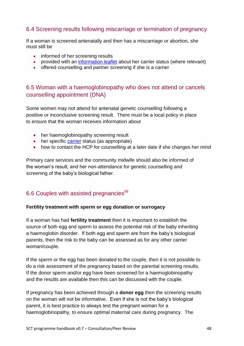
SCT programme handbook v0.7 – Consultation/Peer Review 48
6.4 Screening results following miscarriage or termination of pregnancy If a woman is screened antenatally and then has a miscarriage or abortion, she must still be
informed of her screening results provided with an information leaflet about her carrier status (where relevant) offered counselling and partner screening if she is a carrier
6.5 Woman with a haemoglobinopathy who does not attend or cancels
counselling appointment (DNA)
Some women may not attend for antenatal genetic counselling following a
positive or inconclusive screening result. There must be a local policy in place
to ensure that the woman receives information about
her haemoglobinopathy screening result
her specific carrier status (as appropriate)
how to contact the HCP for counselling at a later date if she changes her mind
Primary care services and the community midwife should also be informed of
the woman’s result, and her non-attendance for genetic counselling and
screening of the baby’s biological father.
6.6 Couples with assisted pregnancies56
Fertility treatment with sperm or egg donation or surrogacy
If a woman has had fertility treatment then it is important to establish the
source of both egg and sperm to assess the potential risk of the baby inheriting
a haemoglobin disorder. If both egg and sperm are from the baby’s biological
parents, then the risk to the baby can be assessed as for any other carrier
woman/couple.
If the sperm or the egg has been donated to the couple, then it is not possible to
do a risk assessment of the pregnancy based on the parental screening results.
If the donor sperm and/or egg have been screened for a haemoglobinopathy
and the results are available then this can be discussed with the couple.
If pregnancy has been achieved through a donor egg then the screening results
on the woman will not be informative. Even if she is not the baby’s biological
parent, it is best practice to always test the pregnant woman for a
haemoglobinopathy, to ensure optimal maternal care during pregnancy. The
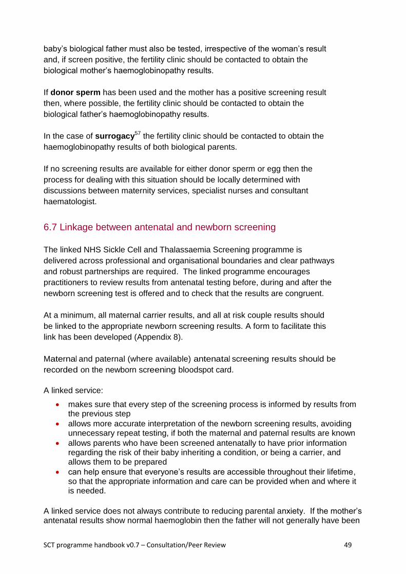
SCT programme handbook v0.7 – Consultation/Peer Review 49
baby’s biological father must also be tested, irrespective of the woman’s result
and, if screen positive, the fertility clinic should be contacted to obtain the
biological mother’s haemoglobinopathy results.
If donor sperm has been used and the mother has a positive screening result
then, where possible, the fertility clinic should be contacted to obtain the
biological father’s haemoglobinopathy results.
In the case of surrogacy57 the fertility clinic should be contacted to obtain the
haemoglobinopathy results of both biological parents.
If no screening results are available for either donor sperm or egg then the
process for dealing with this situation should be locally determined with
discussions between maternity services, specialist nurses and consultant
haematologist.
6.7 Linkage between antenatal and newborn screening
The linked NHS Sickle Cell and Thalassaemia Screening programme is
delivered across professional and organisational boundaries and clear pathways
and robust partnerships are required. The linked programme encourages
practitioners to review results from antenatal testing before, during and after the
newborn screening test is offered and to check that the results are congruent.
At a minimum, all maternal carrier results, and all at risk couple results should
be linked to the appropriate newborn screening results. A form to facilitate this
link has been developed (Appendix 8).
Maternal and paternal (where available) antenatal screening results should be
recorded on the newborn screening bloodspot card.
A linked service:
makes sure that every step of the screening process is informed by results from the previous step
allows more accurate interpretation of the newborn screening results, avoiding unnecessary repeat testing, if both the maternal and paternal results are known
allows parents who have been screened antenatally to have prior information regarding the risk of their baby inheriting a condition, or being a carrier, and allows them to be prepared
can help ensure that everyone’s results are accessible throughout their lifetime, so that the appropriate information and care can be provided when and where it is needed.
A linked service does not always contribute to reducing parental anxiety. If the mother’s antenatal results show normal haemoglobin then the father will not generally have been

SCT programme handbook v0.7 – Consultation/Peer Review 50
tested. If the baby is subsequently found to be a carrier, the risk would not have been identified antenatally. Research shows that prior knowledge of carrier status gives parents a more positive newborn screening experience than discovering test results out of the blue.58
The antenatal report given to all women following the birth of their baby should contain all antenatal screening results, including sickle cell and thalassaemia screening, for communication to primary care.
6.8 Guidance concerning possible non-paternity
All genetic tests undertaken during pregnancy and the newborn period, will include the possible issue of non-paternity. This needs to be considered by all HCPs when offering screening tests, and when inviting the ‘baby’s father’ for testing. It should be highlighted to the mother that the correct person to be screened is the ‘baby’s biological father’ in order to assess the genetic inheritance in the baby correctly. The role of HCPs is to provide clarity and discretion around genetic screening test results, particularly as there is no consensus on the rate of non-paternity in the population.59 Obtaining this unsolicited information during screening creates an ethical dilemma about whether to pass the information on, and to whom. The revelation of non-paternity may be detrimental to established relationships and HCPs must be alert to the need for discretion in pursuing family studies and in discussing results with the mother/parents.
Non-paternity may be suspected when60
a baby with a major haemoglobin disorder has a carrier mother but the ‘father’ is not a carrier
a baby identified as a carrier has neither their mother nor their father identified as a carrier
However, even when screening results seem to suggest non-paternity,
alternative explanations must also be considered, for example:
one parent may carry a variant that cannot be detected by routine screening methods, e.g. an unstable variant or a silent form of beta thalassaemia
there may have been an error a) with labelling a sample b) in the laboratory c) or in reporting the results
a parent’s identity may have been stolen or used by another person
the couple may have used assisted reproductive methods (artificial insemination by donor egg/sperm), which may not have been declared
a very premature baby may not yet have developed any of their adult haemoglobin
the baby may have developed a De Novo mutation, which although rare, is possible

SCT programme handbook v0.7 – Consultation/Peer Review 51
Despite these possibilities, a risk of non-paternity remains and needs to be handled carefully if relationships and family units are not to be disrupted. When discussing results the need for discretion is essential.
The HCP needs to be non-judgemental while considering the following actions:
1) review the antenatal and newborn screening process to establish whether or not an error may have occurred at any stage of the pathway
2) explore the possibility of non-paternity with the mother, preferably on her own in private, without her partner present
3) agree with the baby’s mother how the situation will be dealt with if non-paternity is indeed a possibility
4) offer a re-test (in the first instance) to: a) mother b) baby
5) if indicated, re-screen the father 6) carefully document results and communicate these only to those HCPs who need
the information to support the family.
6.9 References
54
Royal College of Obstetricians and Gynaecologists (2011) Sickle Cell Disease in Pregnancy,
Management of (Green-top Guideline 61) Reviewed December 2014 https://www.rcog.org.uk/en/guidelines-research-services/guidelines/gtg61/
55
Royal College of Obstetricians and Gynaecologists (2014) Thalassaemia in Pregnancy, Management of
Beta (Green-top Guideline 66) https://www.rcog.org.uk/en/guidelines-research-services/guidelines/gtg66/
56 Human fertility and embryology authority Fertility treatments https://www.hfea.gov.uk/treatments/ 57 Human fertility and embryology authority https://www.hfea.gov.uk/treatments/explore-all-
treatments/surrogacy/ 58
Kai, J Ulph, F Cullinan, T and Qureshi, N (2009) Communication of carrier status information following universal newborn screening for sickle cell disorders and cystic fibrosis: qualitative study of experience and practice. Health Technology Assessment 2009; Vol. 13: No. 57 www.hta.ac.uk
59
Voracek M; Haubner T; Fisher ML (2008) Recent decline in nonpaternity rates: a cross-temporal meta-analysis. Psychological Rep. 2008 Dec; 103(3): 799-811 60
Turney L (2005) The Incidental Discovery of Non-paternity Through Genetic Carrier Screening: An
Exploration of Lay Attitudes. Qualitative Health Research 2005; 15(5): 620

SCT programme handbook v0.7 – Consultation/Peer Review 52
Chapter 7 - Newborn Screening
Newborn screening (NBS) is offered as part of the newborn blood spot (heel prick) test61 and identifies babies who have sickle cell disease. It also detects babies who are genetic carriers of some haemoglobin variants. The key reason for offering newborn screening is that babies with sickle cell disease are vulnerable to life-threatening infections. By identifying them promptly after birth, they can be offered potentially life-saving penicillin, and be referred for specialist care.
There is no routine screen for babies at risk of inheriting thalassaemia major - although this is currently under review. However, most cases of beta thalassaemia major should be detected during newborn screening, but beta thalassaemia carriers are not.
Newborn bloodspot screening is performed on day 5 of birth, counting the day of birth as day 0. Additionally, babies up to 12 months of age who become the responsibility of the provider organisation must be offered screening if there is no documented evidence of a conclusive result for the conditions currently recommended by the UK National Screening Committee (UK NSC). The SCT62 and NBS63 screening programmes have published standards for newborn screening, against which screening services will be assessed and monitored.
7.1 Linked antenatal and newborn screening programme
Linking the results of the parents and the babies is particularly important for the sickle cell and thalassaemia screening programme as it helps to make an accurate diagnosis in the baby. All maternal carrier results, and at risk couple results, should be linked to the appropriate newborn screening results. There is a notification form that should be used to inform newborn blood spot screening laboratories of any carrier women and at risk couples (Appendix 8).
7.2 Offer of the newborn blood spot screen
Parents must be offered information about the newborn blood spot screen prior to the offer of screening. The parent’s information leaflet ‘Screening tests for you and your baby64 is available to support information from a healthcare professional. The leaflet is available in a number of languages and an easy read version. Full guidance on performing the NBS is covered in the Newborn Blood Spot Screening guidelines.65

SCT programme handbook v0.7 – Consultation/Peer Review 53
If the screen is accepted:
it should be recorded in the maternity record and the Personal Child Health Record (PCHR).
if the baby is in hospital then the consent should also be recorded in the baby’s hospital records.
If the screen is declined:
this should be recorded on the bloodspot card and sent to the laboratory
each condition for which this is relevant, must be recorded in the maternity records and the PCHR
the baby’s GP and health visitor must be informed of the conditions for which the parents have declined screening
the parents should be informed that the baby can be screened at a later date if they so wish
7.3 Babies born to “at risk” couples Couples at risk of having a baby with a major haemoglobin disorder, where the results of both parents are known, may wish to know their baby’s result before the normal time for reporting from bloodspot screening. Local policies should be in place to have an early newborn bloodspot test, at the parents’ request. Alternately, a liquid capillary blood specimen (not cord blood) can be taken for analysis soon after birth. This is not part of the screening programme, and should be considered as an aspect of parental choice. The blood sample must be analysed in a laboratory which has expertise in haemoglobinopathy analysis in the newborn period. Please refer to the Handbook for Newborn Laboratories66 for further details.
7.4 Preterm babies
Preterm babies or those in neonatal units:
require the sample to be taken on day 5
would benefit from the coordination of the NBS with other blood tests to minimise discomfort
Be aware that preterm babies do not always show their adult haemoglobin clearly, depending on their gestational age.
7.5 Transfused babies
Babies that require a blood transfusion must have a NBS for sickle cell taken prior to the blood transfusion.

SCT programme handbook v0.7 – Consultation/Peer Review 54
The pre-transfusion sample must be clearly labelled ‘pre-transfusion’ on the blood spot card. The remaining conditions are to be screened on day 5 on a separate blood spot card, as routine. Both cards must then be submitted together to the newborn screening laboratory.
In the cases when a baby has had a blood transfusion before a pre-transfusion sample
has been taken, it is possible to perform DNA testing on post-transfusion samples. The
DNA test will detect the presence of the sickle haemoglobin gene. The test is able to differentiate between babies with:
only the sickle gene present
those with the sickle gene and another globin gene (either a normal beta gene or another variant)
no sickle gene present
If the sickle gene is detected the baby must be referred for clinical follow up. Please see Appendix 10 for further information.
7.6 Newborn screening results
Screen negative
Approximately 97% of babies will be screen negative. This means the baby is unaffected or none of the haemoglobin variants that must be identified have been detected. The parents should be informed before the baby’s health check at 6-8 weeks.
Haemoglobin variant carrier
Approximately 9000 babies every year in England are found to be a healthy carrier of a haemoglobin gene variant. This is a prevalence of one in 70 births.67 The haemoglobin gene variants the NBS screening must identify are: Hb S Hb C Hb D Hb E Hb OArab (additional investigations are required to confirm this result).
The results will show the presence of HbF, Hb A and the haemoglobin variant It is important for parents to understand that their child could pass on the gene for unusual haemoglobin to future generations. The parents of babies found to be carrier of a haemoglobin gene variant must be given the opportunity for a face to face discussion with a suitably trained professional to enable the significance of the carrier status to be explained. It is important for the parents to understand that their child could pass on the gene for unusual haemoglobin to future generations when they have their own baby.

SCT programme handbook v0.7 – Consultation/Peer Review 55
There are 2 parent information leaflets to support the information given by a healthcare professional: Information for mums and dads: your baby carries a gene for sickle cell68
Information for mums and dads: your baby carries a gene for unusual haemoglobin69
Both leaflets are available in English, French, Bengali and Urdu.
It is important to remember that it is not possible to identify those babies that are beta thalassaemia carriers, using routine newborn screening methods.
Inconclusive results
For babies found to have a haemoglobin variant other than those stated above, the NBS result will be issued as ‘condition not suspected’. The wording will state that haemoglobin S, C, DPunjab, E, and OArab have not been detected
7.7 Benign haemoglobin conditions
There are a number of benign haemoglobin conditions that may be detected in the NBS. In such cases, the baby should be referred to a named clinician for follow up and counselling. There may be an indication to rescreen these babies. Please see appendix 2 for further details of the conditions below. Benign haemoglobin conditions include: Hb CC and C/βthalassaemia Hb DD and D/βthalassaemia Hb CD Hb CE Hb DE Hb EE
7.8 Baby has sickle cell disease
Results from babies with sickle cell disease show the presence of HbF and HbS in the absence of HbA; or HbF and HbS with another haemoglobin variant. Between 260-350 babies a year in England have a sickle cell disease result; this is a prevalence of one per 2,000-2,500 births. Expected results from babies with sickle cell disease are:
Newborn screening result Disease
FS Hb SS
FS Hb S/β° thalassaemia
FS Hb S/δβ thalassaemia
FS Hb S/Lepore

SCT programme handbook v0.7 – Consultation/Peer Review 56
FS Hb S/HPFH
FSA or FS Hb S/β⁺ thalassaemia
FSC Hb SC
FSD Hb S/DPunjab
FSE Hb S/E
FSOArab Hb S/OArab
The parents of babies who have a sickle cell disease result from the NBS must be informed of the result before the baby is 28 days of age. This is an auditable standard63 in the sickle cell and thalassaemia screening programme.
The parents must be informed by an appropriately trained professional, with sensitivity to the parents’ concerns. Best practice is for the parents to be informed by a healthcare professional they have met in the antenatal period, along with either the family’s health visitor or a haemoglobinopathy paediatric nurse. The family must be given contact numbers, and all relevant healthcare professionals informed of the result. The early identification of affected babies allows:
the opportunity to inform and educate the parents of the condition
early intervention to reduce morbidity and mortality
prophylactic oral penicillin v to be commenced
timely entry into specialist haemoglobinopathy centre The standard is for affected babies to attend a haemoglobinopathy centre (medical) by 90 days of age. There is a parent information book70 available which gives detailed guidance on to have to care for an affected child. This can be obtained from Harlow Press. An example of a form which could be used to support the counselling session, and also as a template of a referral letter is in Appendix 10. Appendix 3 has further details of the clinical impact of each sickle cell disorder.
7.9 Beta thalassaemia
The NBS programme does not specifically screen for beta thalassaemia major, but babies with severe thalassaemia major will generally be detected. Results from a baby with severe Beta thalassaemia major will usually have only HbF present and no HbA. It is important to remember that not all thalassaemia conditions will be detected by the NBS. Babies with a Beta thalassaemia condition must be referred for follow up and care to a haemoglobinopathy centre (medical) by 90 days of age. It is important to remember that it is not possible to identify those babies that are beta thalassaemia carriers. Parents should be informed of this in the antenatal period if relevant.
SCT programme handbook v0.7 – Consultation/Peer Review 57
7.10 Non-Paternity
Healthcare professionals involved in newborn screening must be mindful that there is the potential for the results to identify possible non-paternity of the baby. Such cases must be handled sensitively. Further information is available in Chapter 6 of the programme handbook.
7.11 References
61 NHS Newborn Blood Spot Screening Programme (2017) Programme Overview
https://www.gov.uk/topic/population-screening-programmes/newborn-blood-spot 62
NHS Sickle Cell & Thalassaemia Screening Programme (2017) Sickle cell & thalassaemia screening: programme standards https://www.gov.uk/government/publications/standards-for-sickle-cell-and-thalassaemia-screening 63
NHS Newborn Blood Spot Screening Programme (2017) Newborn blood spot screening programme standards https://www.gov.uk/government/publications/standards-for-nhs-newborn-blood-spot-screening 64
Public Health England (2017) Screening tests for you and your baby. https://www.gov.uk/government/publications/screening-tests-for-you-and-your-baby-description-in-brief 65
NHS Newborn Blood Spot Screening Programme (2016) Newborn blood spot screening: sampling guidelines https://www.gov.uk/government/publications/newborn-blood-spot-screening-sampling-guidelines 66
NHS Sickle Cell & Thalassaemia Screening Programme (2017) Handbook for newborn laboratories https://www.gov.uk/government/publications/sickle-cell-and-thalassaemia-screening-handbook-for-laboratories 67
NHS Sickle Cell & Thalassaemia Screening Programme (2017) Sickle cell and thalassaemia screening: data trends and performance analysis https://www.gov.uk/government/publications/sickle-cell-and-thalassaemia-screening-data-trends-and-performance-analysis 68
NHS Sickle Cell & Thalassaemia Screening Programme (2016) Baby carries a gene for sickle cell: description in
brief
https://www.gov.uk/government/publications/baby-carries-a-gene-for-sickle-cell-description-in-brief 69
NHS Sickle Cell & Thalassaemia Screening Programme (2010)Baby carries a gene for unusual haemoglobin: description in brief https://www.gov.uk/government/publications/baby-carries-a-gene-for-unusual-haemoglobin-description-in-brief 70
NHS Sickle Cell & Thalassaemia Screening Programme (2012) A parent’s guide to Sickle cell disease
https://www.gov.uk/government/publications/sickle-cell-disease-managing-the-condition

SCT programme handbook v0.7 – Consultation/Peer Review 58
Chapter 8 - Failsafe, Quality Assurance and Data Collection
Each NHS screening programme has a defined screening pathway71
The NHS Sickle Cell and Thalassaemia Programme has a second pathway for known at
risk women and couples.
The pathways show how the individual undergoing screening moves from one stage of
the pathway to the next. Checks are needed at each stage to ensure the individual
moves seamlessly and safely through the pathway unless they choose not to.
If these checks are not in place there is a risk that an individual does not complete the
pathway or the pathway is delayed unnecessarily. Quality assurance of screening
programmes includes checking these procedures are in place and operating effectively.
In screening programmes these checks and failsafe processes are in place to ensure if
something goes wrong it can easily be identified at the time it goes wrong and action
can be taken to correct it before any harm occurs. Guidance72 is given by the SCT
programme; an audit template72 is also available.
8.1 Newborn blood spot failsafe73 solution (NBSFS)
The NBSFS is in use by maternity units across England. It identifies babies who have missed newborn blood spot (NBS) screening. This enables screening to be offered and affected babies to be identified early and treatment can be started within an effective timeframe.
8.2 Quality Assurance
The screening quality assurance services (SQAS)74 is the process of checking that
national standards are met (ensuring that screening programmes are safe and effective)
and encouraging continuous improvement.
All maternity services in England are required to submit quarterly Key Performance
Indicators75 (KPIs) to the national screening programmes to demonstrate performance
against set standards.
Each screening programme provider must report KPI data using the appropriate
reporting template. Service users, programme teams and the screening quality
assurance service (SQAS) use KPIs to help measure the success of screening
programmes.

SCT programme handbook v0.7 – Consultation/Peer Review 59
Maternity services are also subject to peer review, led and supported by the SQAS.
It is important that any screening incidents are managed in accordance with national
guidelines76. Incidents must be reported via the local incident reporting systems and to
the relevant SQAS team.
8.3 Data collection
The sickle cell and thalassaemia programme collects data77 to evaluate the delivery of
the programme using data from a variety of sources.
The data report78 is produced annually and reports performance against antenatal and
newborn sickle cell and thalassaemia screening programme standards and trends over
time.
8.4 References
71 Public Health England (2015)
NHS population screening: care pathways
https://www.gov.uk/government/collections/nhs-population-screening-care-pathways 72
NHS sickle cell and thalassaemia screening programme (2017) SCT checks and audit https://www.gov.uk/government/publications/sct-checks-and-audits-to-improve-quality-and-reduce-risks/sct-checks-and-audits
73 NHS Newborn blood spot screening programme (2016)
Newborn blood spot screening: failsafe procedures
https://www.gov.uk/government/publications/newborn-blood-spot-screening-failsafe-procedures 74
NHS population screening programmes (2016) NHS population screening: quality assurance https://www.gov.uk/guidance/nhs-population-screening-quality-assurance 75 NHS Population screening programmes (2017) Key performance indicators
https://www.gov.uk/government/publications/nhs-population-screening-reporting-data-definitions 76
NHS Population screening programme (2015) Managing safety incidents in NHS screening programmes https://www.gov.uk/government/publications/managing-safety-incidents-in-nhs-screening-programmes 77 NHS Sickle cell and thalassaemia screening programme (2017) Sickle cell and thalassaemia screening: data
collection https://www.gov.uk/government/collections/sickle-cell-and-thalassaemia-screening-data-collection
78
NHS Sickle cell and thalassaemia screening programme (2017) Sickle cell and thalassaemia screening: data trends and performance analysis
https://www.gov.uk/government/publications/sickle-cell-and-thalassaemia-
screening-data-trends-and-performance-analysis

SCT programme handbook v0.7 – Consultation/Peer Review 60
Chapter 9 - Resources & Training
Laboratory support service
The NHS Sickle Cell &Thalassaemia Screening Programme funds a support
service via Oxford University Hospitals, for HCPs working in the antenatal and
newborn screening programmes. There is a designated telephone helpline and
secure email address for questions about screening policy and
haemoglobinopathy results.
Contact details
Telephone: 01865572767
Email: [email protected]
Fax: 01865 572 775
NHS Sickle Cell &Thalassaemia Screening Programme enquiries
Telephone: 020 3682 0890
Email: [email protected]
Website: gov.uk/phe/screening
Training Courses & Resources
Kings College London offers 3 courses:
Genetic risk assessment (this is a 4 day course for health professionals
involved in counselling women and couples 'at risk' of having a child with
a haemoglobinopathy)
Specialist Counsellors Update (this is a one day update for practitioners
who have previously undertaken a specialist course)
SCT Screening Programme Update (this is a one day update for non-
specialist nurses, midwives and health visitors so that they can develop
an understanding of the antenatal and newborn sickle cell and
thalassaemia screening programme in England)
Warwick Health Screening Module
NHS Sickle Cell &Thalassaemia Screening Programme
Core competencies in genetics for sickle cell and thalassaemia counselling
Other Training Tools
Sickle Cell Society Family Legacy DVD
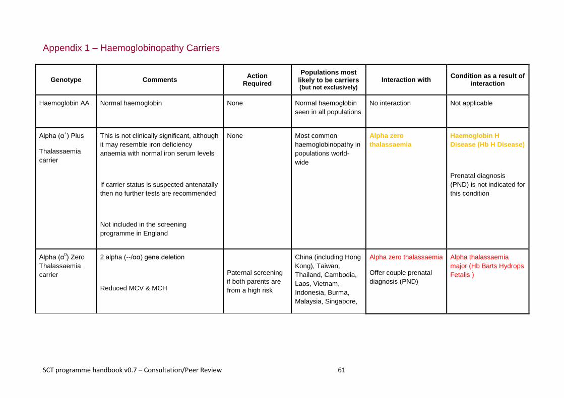
SCT programme handbook v0.7 – Consultation/Peer Review 61
Appendix 1 – Haemoglobinopathy Carriers
Genotype Comments Action
Required
Populations most likely to be carriers (but not exclusively)
Interaction with Condition as a result of
interaction
Haemoglobin AA Normal haemoglobin None Normal haemoglobin
seen in all populations
No interaction Not applicable
Alpha (α+) Plus
Thalassaemia
carrier
This is not clinically significant, although
it may resemble iron deficiency
anaemia with normal iron serum levels
If carrier status is suspected antenatally
then no further tests are recommended
Not included in the screening
programme in England
None Most common
haemoglobinopathy in
populations world-
wide
Alpha zero
thalassaemia
Haemoglobin H
Disease (Hb H Disease)
Prenatal diagnosis
(PND) is not indicated for
this condition
Alpha (α0) Zero
Thalassaemia
carrier
2 alpha (--/αα) gene deletion
Reduced MCV & MCH
Paternal screening
if both parents are
from a high risk
China (including Hong
Kong), Taiwan,
Thailand, Cambodia,
Laos, Vietnam,
Indonesia, Burma,
Malaysia, Singapore,
Alpha zero thalassaemia
Offer couple prenatal
diagnosis (PND)
Alpha thalassaemia
major (Hb Barts Hydrops
Fetalis )

SCT programme handbook v0.7 – Consultation/Peer Review 62
Genotype Comments Action
Required
Populations most likely to be carriers (but not exclusively)
Interaction with Condition as a result of
interaction
May resemble iron deficiency anaemia
with normal iron serum levels
DNA required to confirm carrier status,
as it cannot be diagnosed by routine
screening methods
Confirmation of carrier status as part of
the antenatal screening programme in
England is only indicated if both parents
are from a high risk group
group
Genetic counselling
and DNA to confirm
carrier status ONLY
if results from both
parents indicate
they may be alpha
zero thalassaemia
carriers
Philippines, Cyprus,
Turkey, Greece,
Sardinia, unknown
family origins
Alpha plus thalassaemia (Unlikely to be detected during antenatal screening as it not considered clinically significant)
Haemoglobin H Disease (Hb H Disease) PND is not indicated for this condition
Beta (β) Thalassaemia carrier
Elevated haemoglobin A2 Reduced MCV & MCH Reduced production of ß (beta) globin chains May be misdiagnosed as iron deficiency anaemia Unless iron deficient, then no supplement required There are a range of ß thalassaemia mutations
Genetic counselling & paternal/partner screening is indicated Sometimes requires DNA to confirm carrier status Not diagnosed during routine newborn screening
Mediterranean Middle East South East Asian South Asian (China, Indonesia, Vietnam and other countries in the region) Caribbean African Occurs sporadically in all populations including White British
β thalassaemia Offer couple PND
βthal/βthal β thalassaemia majoror βthalassaemia Intermedia
Hb Lepore Assessment by Specialist - Offer PND if indicated
Lepore/ βthal May present as Thalassaemia Major or Thalassaemia Intermedia
Hb E Assessment by Specialist - Offer PND if indicated
E/βthalassaemia may present as Thalassaemia Major or Thalassaemia Intermedia

SCT programme handbook v0.7 – Consultation/Peer Review 63
Genotype Comments Action
Required
Populations most likely to be carriers (but not exclusively)
Interaction with Condition as a result of
interaction
Delta beta (δβ) thalassaemia Assessment by Specialist - Offer PND if indicated
βthal/δβthal May present as Thalassaemia Major or Thalassaemia Intermedia
O Arab Assessment by Specialist
OArab
/βthalassaemia is usually similar to Thalassaemia Intermedia
Hb S Offer PND
S/βthalassaemia

SCT programme handbook v0.7 – Consultation/Peer Review 64
Genotype Comments Action
Required
Populations most likely to be carriers (but not exclusively)
Interaction with Condition as a result of
interaction
Hb Lepore carrier (Hb A/Lepore)
Red blood cells are usually hypochromic and microcytic
Occasional enlarged spleen
Genetic counselling Partner screening
Mediterranean (Greek, Italian)
βeta thalassaemia Assessment by Specialist - Offer PND if indicated
May present as Thalassaemia Major or Thalassaemia Intermedia
Sickle cell (Hb AS) Assessment by Specialist - Offer PND if indicated
Hb S/Lepore
Delta (δ) Beta (β) thalassaemia carrier (Hb A/δβ thalassaemia)
Red blood cells are usually hypochromic and microcytic
Occasional enlarged spleen
DNA to confirm diagnosis
Genetic counselling Partner screening
Mediterranean (Greek, Italian)
β thalassaemia Assessment by Specialist - Offer PND if indicated
May present as Thalassaemia Major or Thalassaemia Intermedia
Sickle cell (Hb AS) Assessment by Specialist - Offer PND if indicated
Hb S/δβ thalassaemia
Hereditary Persistence of Fetal Haemoglobin carrier (Hb A/HPFH)
Usually 2% or less of total haemoglobin in adults It is not possible to distinguish Hb SS from Hb S/HPFH and S/β
0
thalassaemia in newborn screening
Genetic counselling Partner screening
Africans Caribbean
Sickle cell (Hb AS) Assessment by Specialist
S/HPFH Does not usually require treatment
Haemoglobin E carrier (Hb AE)
Lysine substituted for glutamic acid, 26
th point ß globin chain
Red blood cells may be hypochromic and microcytic
Genetic counselling Partner screening
South East Asia (India, Bangladesh) South Asia (China, Vietnam, Thailand, Indonesia and other countries in the region) Caribbean
β thalassaemia Assessment by Specialist - Offer PND if indicated
E/β thalassaemia may present as Thalassaemia Major or Thalassaemia Intermedia
Sickle cell (Hb AS) Assessment by Specialist
Hb S/E Disease

SCT programme handbook v0.7 – Consultation/Peer Review 65
Genotype Comments Action
Required
Populations most likely to be carriers (but not exclusively)
Interaction with Condition as a result of
interaction
Haemoglobin D
Punjab carrier
(Hb ADPunjab
) also called D
Los Angeles
Other types of Hb D are not usually clinically significant
Glutamine substituted for glutamic acid, 121 point, ß globin chain Important to identify D
Punjab from other
Hb D’s due to clinical interaction with Hb S
Genetic counselling Partner screening
Indian Pakistan Caribbean Occurs sporadically in all populations including White British)
Sickle haemoglobin (Hb S) Offer PND
Hb S/DPunjab
Haemoglobin C carrier (Hb AC)
Lysine substituted for glutamic acid, 6th
point, ß globin chain Genetic Counselling Partner screening
West African Caribbean
Sickle haemoglobin (Hb S) Offer PND
Hb S/C Disorder
Hb OArab
(Hb AO
Arab)
carrier Also known as Hb Egypt
Lysine substituted for glutamine at 121
st point of the ß globin chain
Genetic counselling Partner screening
North Africa Saudi Arabia Bulgaria/Eastern Europe Eastern Mediterranean
β thalassaemia Assessment by Specialist
OArab
/βthalassaemia usually similar to Thalassaemia Intermedia
Sickle haemoglobin (Hb S) Offer PND
S/OArab
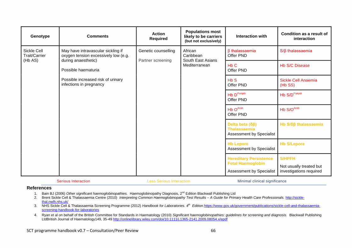
SCT programme handbook v0.7 – Consultation/Peer Review 66
Genotype Comments Action
Required
Populations most likely to be carriers (but not exclusively)
Interaction with Condition as a result of
interaction
Sickle Cell Trait/Carrier (Hb AS)
May have intravascular sickling if oxygen tension excessively low (e.g. during anaesthetic) Possible haematuria Possible increased risk of urinary infections in pregnancy
Genetic counselling Partner screening
African Caribbean South East Asians Mediterranean
β thalassaemia Offer PND
S/β thalassaemia
Hb C Offer PND
Hb S/C Disease
Hb S Offer PND
Sickle Cell Anaemia (Hb SS)
Hb DPunjab
Offer PND Hb S/D
Punjab
Hb OArab
Offer PND Hb S/O
Arab
Delta beta (δβ) Thalassaemia Assessment by Specialist
Hb S/δβ thalassaemia
Hb Lepore Assessment by Specialist
Hb S/Lepore
Hereditary Persistence Fetal Haemoglobin
Assessment by Specialist
S/HPFH
Not usually treated but investigations required
Serious Interaction Less Serious Interaction Minimal clinical significance
References
1. Bain BJ (2006) Other significant haemoglobinopathies. Haemoglobinopathy Diagnosis, 2nd Edition Blackwall Publishing Ltd 2. Brent Sickle Cell & Thalassaemia Centre (2010) Interpreting Common Haemoglobinopathy Test Results – A Guide for Primary Health Care Professionals. http://sickle-
thal.nwlh.nhs.uk/ 3. NHS Sickle Cell & Thalassaemia Screening Programme (2012) Handbook for Laboratories. 4th Edition.https://www.gov.uk/government/publications/sickle-cell-and-thalassaemia-
screening-handbook-for-laboratories
4. Ryan et al on behalf of the British Committee for Standards in Haematology (2010) Significant haemoglobinopathies: guidelines for screening and diagnosis. Blackwall Publishing LtdBritish Journal of Haematology149, 35-49 http://onlinelibrary.wiley.com/doi/10.1111/j.1365-2141.2009.08054.x/epdf

SCT programme handbook v0.7 – Consultation/Peer Review 67
Appendix 2 – Benign Haemoglobin Disorders
Genotype Anaemia Other clinical features Comments
Haemoglobin C Disease
C/β+·Thalassaemia
C/β0·Thalassaemia
or Hb CC
Mild haemolytic
anaemia
Occasional intermittent abdominal pain
Gallstones
Fatigue
Occasional jaundice
Usually a mild condition
Genetic counselling is essential during pregnancy for
parents ‘at risk’ of having a child with this condition but
prenatal diagnosis is not indicated.
Register with haematology/ specialist
clinic decision regarding regularity of
follow-up is made locally
No regular treatment necessary, only
required in relation to symptoms
Genetic counselling and partner testing
for individuals with this condition is
important as they do not have any normal
Hb A genes.
Haemoglobin DPunjab
Disease
Hb DPunjab
/DPunjab
DPunjab
/β0·Thalassaemia
DPunjab
/β+·Thalassaemia
Microcytosis
Hypochromia
Hb at lower end
of normal
Occasional abdominal pain
Symptoms related to haemolytic anaemia
Gallstones
Fatigue
Occasional jaundice
Iron medication not required unless iron deficient
Register with haematology/ specialist
clinic decision regarding regularity of
follow-up is made locally
No regular treatment necessary, only
required in relation to symptoms
Genetic counselling and partner testing
for individuals with this condition is

SCT programme handbook v0.7 – Consultation/Peer Review 68
Genotype Anaemia Other clinical features Comments
Genetic counselling is essential during pregnancy for
parents ‘at risk’ of having a child with this condition but
prenatal diagnosis is not indicated.
important as they do not have any normal
Hb A genes
Haemoglobin E Disease
(Hb EE)
(Hb E/β·Thalassaemia may be
clinically significant, please see
information on thalassaemia
disorders for further information)
Mild haemolytic
anaemia
Hypochromic
Very mild condition
Genetic counselling is essential during pregnancy for
parents ‘at risk’ of having a child with this condition
but prenatal diagnosis is not indicated.
Hb EE and Hb E/β0·Thalassaemia will look similar on the
initial screening test and further investigations will be
needed for the conditions to be differentiated.
Register with haematology/ specialist
clinic decision regarding regularity of
follow-up is made locally
No regular treatment necessary, only
required in relation to symptoms
Common in South East Asian
populations.
Genetic counselling and partner testing
for individuals with this condition is
important as they do not have any normal
Hb A genes.
References Bain BJ (2006) Other significant haemoglobinopathies. Haemoglobinopathy Diagnosis, 2
nd Edition Blackwall Publishing Ltd
Brent Sickle Cell & Thalassaemia Centre (2010) Interpreting Common Haemoglobinopathy Test Results – A Guide for Primary Health Care Professionals. http://sickle-thal.nwlh.nhs.uk/
SCT programme handbook v0.7 – Consultation/Peer Review 69
NHS Sickle Cell & Thalassaemia Screening Programme (2012) Handbook for Laboratories. 4th Edition.
https://www.gov.uk/government/publications/sickle-cell-and-thalassaemia-screening-handbook-for-laboratories Ryan et al on behalf of the British Committee for Standards in Haematology (2010) Significant haemoglobinopathies: guidelines for screening and diagnosis. Blackwall Publishing Ltd British Journal of Haematology149, 35-49 http://onlinelibrary.wiley.com/doi/10.1111/j.1365-2141.2009.08054.x/epdf

SCT programme handbook v0.7 – Consultation/Peer Review 70
Appendix 3 – Sickle Cell Disease
Genotype Anaemia Splenomegaly Clinical features Clinical
severity Remarks/treatment
Sickle Cell Anaemia
(Hb SS)
Severe Yes
Usually auto-
spleenectomy
by age 5-7
years
Vaso-occlusive episodes (painful
crisis)
Bone and joint infarcts
Hepato-renal complications
Bacterial infections
If Hb F is elevated then condition
may be milder than usual
Severe Progressive disability common. Usually shortened
life span. Bone marrow transplant may be
considered (in children) if there is severe disease
and an HLA matched sibling is available.
Treatment: Oral Penicillin is routinely prescribed for
children but is optional for adults
Pneumococcal vaccine (adults & children)
Folic acid depending on diet and/or local protocol
Haemoglobin SC
Disorder
(Hb SC)
Mild to
moderate
Hb level is
just below
normal
Common Intermittent painful crises
Bone and joint infarcts (less
common than in SS)
Retinopathy in adults
May have aseptic necrosis of joints
Variable Severity often increased during pregnancy
May present with any of the same symptoms as
Sickle Cell Anaemia but condition is usually milder.
Treatment: Oral Penicillin is routinely prescribed for
children but is optional for adults
Pneumococcal vaccine (adults & children)
Folic acid depending on diet and/or local protocol

SCT programme handbook v0.7 – Consultation/Peer Review 71
Genotype Anaemia Splenomegaly Clinical features Clinical
severity Remarks/treatment
S/β thalassaemia
This is inclusive of
Hb S/β+, Hb S/δβ,
Hb S/and
Hb S/Lepore
Mild to
moderate
Occasional Intermittent pain crises
Bone and joint infarcts
Variable
Treatment: Oral Penicillin is routinely prescribed for
children but is optional for adults
Pneumococcal vaccine (adults & children)
Folic acid depending on diet and/or local protocol

SCT programme handbook v0.7 – Consultation/Peer Review 72
Genotype Anaemia Splenomegaly Clinical features Clinical
severity Remarks/treatment
S/β0 Thalassaemia
Moderate to
severe
Common Intermittent pain crises
Bone and joint infarcts
Severity depends on type of beta
(β) thalassaemia gene inherited
and % of Hb F produced
Moderate to
severe
May be as severe as Sickle Cell Anaemia, depends
on the thalassaemia mutation inherited. No normal
Hb A.
Treatment: Oral Penicillin is routinely prescribed for
children but is optional for adults
Pneumococcal vaccine (adults & children),
Folic acid depending on diet and/or local protocol
Haemoglobin
S/DPunjab
Disorder
(HbSDPunjab
)
Mild to
moderate
haemolytic
anaemia
Common Severity of clinical picture is
variable but usually milder than
sickle cell anaemia
Variable
Treatment: Oral Penicillin is routinely prescribed for
children but is optional for adults
Pneumococcal vaccine (adults & children),
Folic acid depending on diet and/or local protocol
Haemoglobin S/OArab
Disorder
(HbSOArab
)
Mild to
moderate
haemolytic
anaemia
Usually mild to moderate condition Variable Treatment: Oral Penicillin is routinely prescribed for
children but is optional for adults
Pneumococcal vaccine (adults & children),
Folic acid depending on diet and/or local protocol
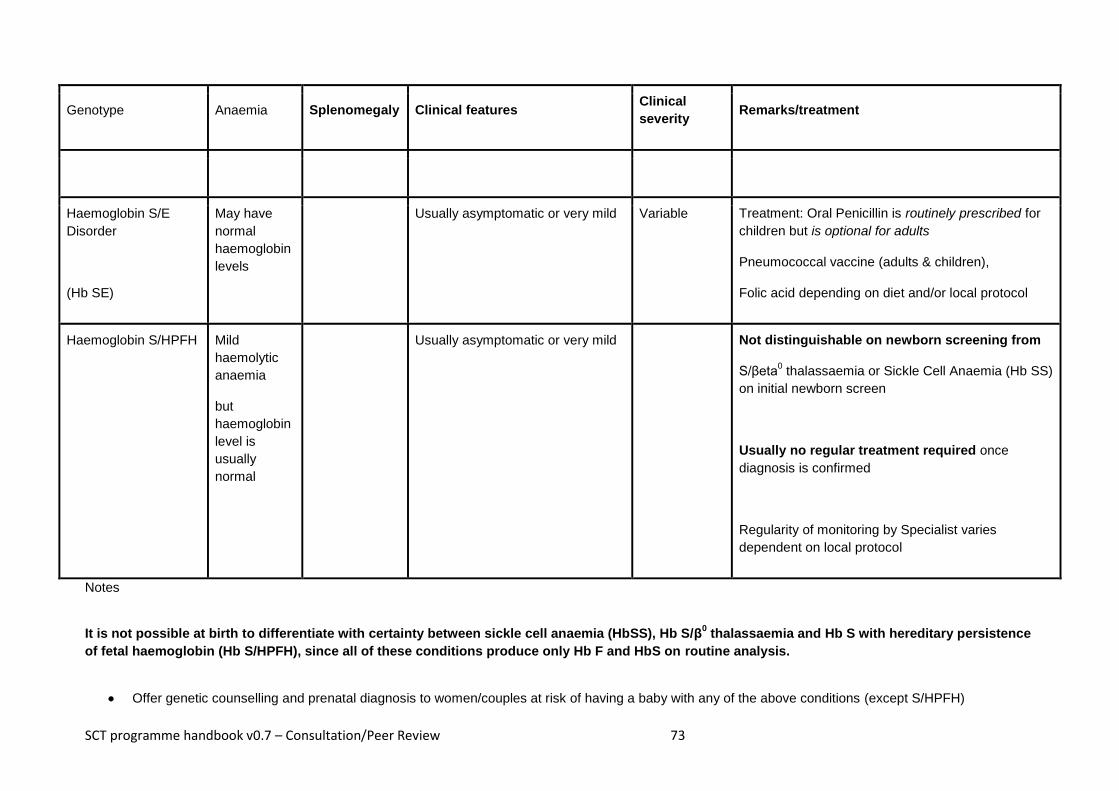
SCT programme handbook v0.7 – Consultation/Peer Review 73
Genotype Anaemia Splenomegaly Clinical features Clinical
severity Remarks/treatment
Haemoglobin S/E
Disorder
(Hb SE)
May have
normal
haemoglobin
levels
Usually asymptomatic or very mild Variable Treatment: Oral Penicillin is routinely prescribed for
children but is optional for adults
Pneumococcal vaccine (adults & children),
Folic acid depending on diet and/or local protocol
Haemoglobin S/HPFH Mild
haemolytic
anaemia
but
haemoglobin
level is
usually
normal
Usually asymptomatic or very mild Not distinguishable on newborn screening from
S/βeta0 thalassaemia or Sickle Cell Anaemia (Hb SS)
on initial newborn screen
Usually no regular treatment required once
diagnosis is confirmed
Regularity of monitoring by Specialist varies
dependent on local protocol
Notes
It is not possible at birth to differentiate with certainty between sickle cell anaemia (HbSS), Hb S/β
0 thalassaemia and Hb S with hereditary persistence
of fetal haemoglobin (Hb S/HPFH), since all of these conditions produce only Hb F and HbS on routine analysis.
Offer genetic counselling and prenatal diagnosis to women/couples at risk of having a baby with any of the above conditions (except S/HPFH)
SCT programme handbook v0.7 – Consultation/Peer Review 74
Individuals with these conditions should be registered with and followed up regularly by a Haematology Clinic
Pregnant women with any of the above sickle cell disorders should be considered as being “high risk” and should be followed up by a Haematologist and Obstetrician during pregnancy with booked hospital delivery.
References Bain BJ (2006) Sickle cell haemoglobin and its interactions with other various haemoglobins and with thalassaemia.Haemoglobinopathy Diagnosis, 2
nd Edition
Blackwall Publishing Ltd Brent Sickle Cell & Thalassaemia Centre (2012) A Parent’s Guide to Managing Sickle Cell Disease https://www.gov.uk/government/publications/sickle-cell-disease-managing-the-condition NHS Sickle Cell & Thalassaemia Screening Programme (2012) Handbook for Laboratories.4
th Edition.
https://www.gov.uk/government/publications/sickle-cell-and-thalassaemia-screening-handbook-for-laboratories Thalassaemia International Federation (TIF) About Sickle Cell Disorders.http://www.thalassaemia.org.cy/
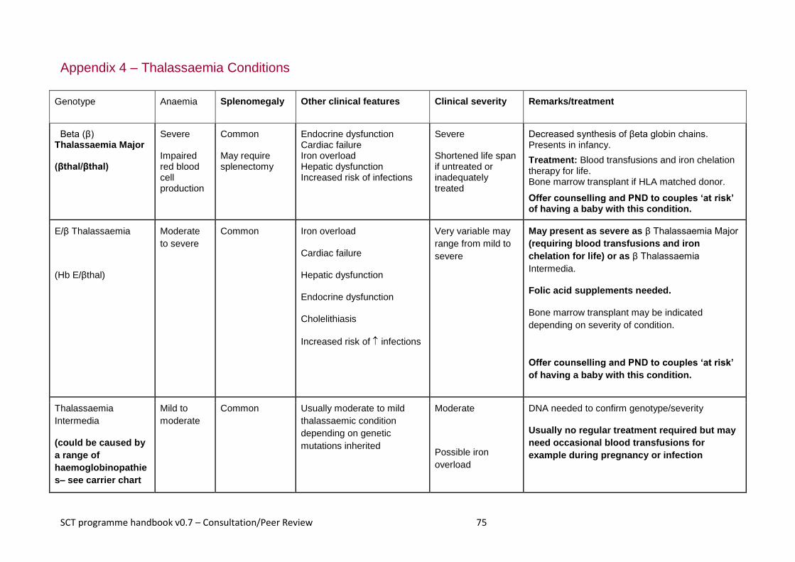
SCT programme handbook v0.7 – Consultation/Peer Review 75
Appendix 4 – Thalassaemia Conditions
Genotype Anaemia Splenomegaly Other clinical features Clinical severity Remarks/treatment
Beta (β) Thalassaemia Major (βthal/βthal)
Severe Impaired red blood cell production
Common May require splenectomy
Endocrine dysfunction Cardiac failure Iron overload Hepatic dysfunction Increased risk of infections
Severe Shortened life span if untreated or inadequately treated
Decreased synthesis of βeta globin chains. Presents in infancy.
Treatment: Blood transfusions and iron chelation therapy for life. Bone marrow transplant if HLA matched donor.
Offer counselling and PND to couples ‘at risk’ of having a baby with this condition.
E/β Thalassaemia
(Hb E/βthal)
Moderate
to severe
Common Iron overload
Cardiac failure
Hepatic dysfunction
Endocrine dysfunction
Cholelithiasis
Increased risk of infections
Very variable may
range from mild to
severe
May present as severe as β Thalassaemia Major
(requiring blood transfusions and iron
chelation for life) or as β Thalassaemia
Intermedia.
Folic acid supplements needed.
Bone marrow transplant may be indicated
depending on severity of condition.
Offer counselling and PND to couples ‘at risk’
of having a baby with this condition.
Thalassaemia
Intermedia
(could be caused by
a range of
haemoglobinopathie
s– see carrier chart
Mild to
moderate
Common Usually moderate to mild
thalassaemic condition
depending on genetic
mutations inherited
Moderate
Possible iron
overload
DNA needed to confirm genotype/severity
Usually no regular treatment required but may
need occasional blood transfusions for
example during pregnancy or infection
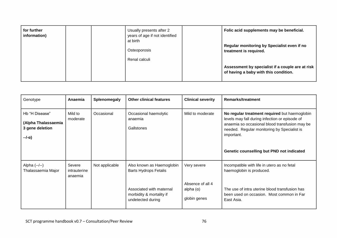
SCT programme handbook v0.7 – Consultation/Peer Review 76
for further
information)
Usually presents after 2
years of age if not identified
at birth
Osteoporosis
Renal calculi
Folic acid supplements may be beneficial.
Regular monitoring by Specialist even if no
treatment is required.
Assessment by specialist if a couple are at risk
of having a baby with this condition.
Genotype Anaemia Splenomegaly Other clinical features Clinical severity Remarks/treatment
Hb “H Disease”
(Alpha Thalassaemia
3 gene deletion
--/-α)
Mild to
moderate
Occasional Occasional haemolytic
anaemia
Gallstones
Mild to moderate No regular treatment required but haemoglobin
levels may fall during infection or episode of
anaemia so occasional blood transfusion may be
needed. Regular monitoring by Specialist is
important.
Genetic counselling but PND not indicated
Alpha (--/--)
Thalassaemia Major
Severe
intrauterine
anaemia
Not applicable Also known as Haemoglobin
Barts Hydrops Fetalis
Associated with maternal
morbidity & mortality if
undetected during
Very severe
Absence of all 4
alpha (α)
globin genes
Incompatible with life in utero as no fetal
haemoglobin is produced.
The use of intra uterine blood transfusion has
been used on occasion. Most common in Far
East Asia.

SCT programme handbook v0.7 – Consultation/Peer Review 77
pregnancy
Offer counselling and PND to couples ‘at risk’
of having a baby with this condition.
Notes
Individuals with these conditions (except alpha thalassaemia major) should be registered with and followed up regularly by a Haematology Clinic
Pregnant women with any of the above disorders should be considered “high risk” and should be followed up by a Haematologist and Obstetrician during pregnancy with booked hospital delivery.
References Eleftheriou A, Angastiniotis M About Alpha ThalassaemiaThalassaemia International Federation (TIF) www.thalassaemia.org.cy Eleftheriou A, Angastiniotis M About Beta ThalassaemiaThalassaemia International Federation (TIF) www.thalassaemia.org.cy NHS Sickle Cell & Thalassaemia Screening Programme (2017) Handbook for Laboratories. 4
th Edition.
https://www.gov.uk/government/publications/sickle-cell-and-thalassaemia-screening-handbook-for-laboratories Ryan et al on behalf of the British Committee for Standards in Haematology (2010) Significant haemoglobinopathies: guidelines for screening and diagnosis. Blackwall Publishing Ltd British Journal of Haematology149, 35-49 http://onlinelibrary.wiley.com/doi/10.1111/j.1365-2141.2009.08054.x/epdf

SCT programme handbook v0.7 – Consultation/Peer Review 78
Appendix 5: DNA Laboratory contact details
John Radcliffe Hospital Oxford Contact Dr Melanie Proven, Principle Clinical Scientist e-mail [email protected] or [email protected] [email protected] Tel 01865572769/ 01865572826 Fax 01865572775
Address: Molecular Haematology Level 4 John Radcliffe Hospital Headington, Oxford, OX39DU
Web site: http://www.oxford-translational-molecular-diagnostics.org.uk/ Link for PND referral form: http://www.oxford-translational-molecular-diagnostics.org.uk/content/forms
King’s College Hospital Contact Dr Barnaby Clark, Principal Clinical Scientist e-mail [email protected] Tel 020 32994337 Tel lab 020 3299 9000 Ext 2265 Fax 020 32991035 Lab Email [email protected]
Address: C /O Central Specimen Reception Blood Sciences Laboratories Ground Floor Bessemer Wing King's College Hospital Denmark Hill London SE5 9RS Link for PND referral form: http://www.viapath.co.uk/our-tests/prenatal-diagnosis
University College London Hospital Contact Dr Mary Petrou, Director Haemoglobinopathy Genetics Centre e-mail [email protected] Tel 020 34479458 Fax 02034479864 E-mail [email protected]
Address: Haemoglobinopathy Genetics Centre Molecular Genetics Laboratory 307 Euston Road London NW1 3AD
Link for PND referral form: https://www.uclh.nhs.uk/OurServices/ServiceA-Z/PATH/PATHHT/PATHHAEMGEN/Pages/Home.aspx
Central Manchester University Hospitals NHS Foundation Trust Contact Steve Keeney, PhD Lead Clinical Scientist, Molecular Haematology Service e-mail [email protected] Tel 0161 276 5990 / 4809 Fax 0161 276 5989 Website http://www.cmft.nhs.uk/haemoglobinopathy
Address: Molecular Diagnostics Centre The Manchester Centre for Genomic Medicine 6th Floor, St Mary's Hospital Oxford Road
Manchester, M13 9WL Link for PND referral form: http://www.cmft.nhs.uk/info-for-health-professionals/laboratorymedicine/haematology/haemoglobinopathy

SCT programme handbook v0.7 – Consultation/Peer Review 79
Appendix 6 - Antenatal Counselling Form

SCT programme handbook v0.7 – Consultation/Peer Review 80

SCT programme handbook v0.7 – Consultation/Peer Review 81
Appendix 7: “At risk couples” flow chart
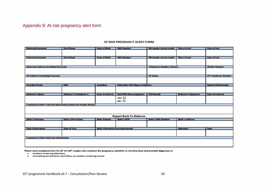
SCT programme handbook v0.7 – Consultation/Peer Review 82
Appendix 8: At risk pregnancy alert form
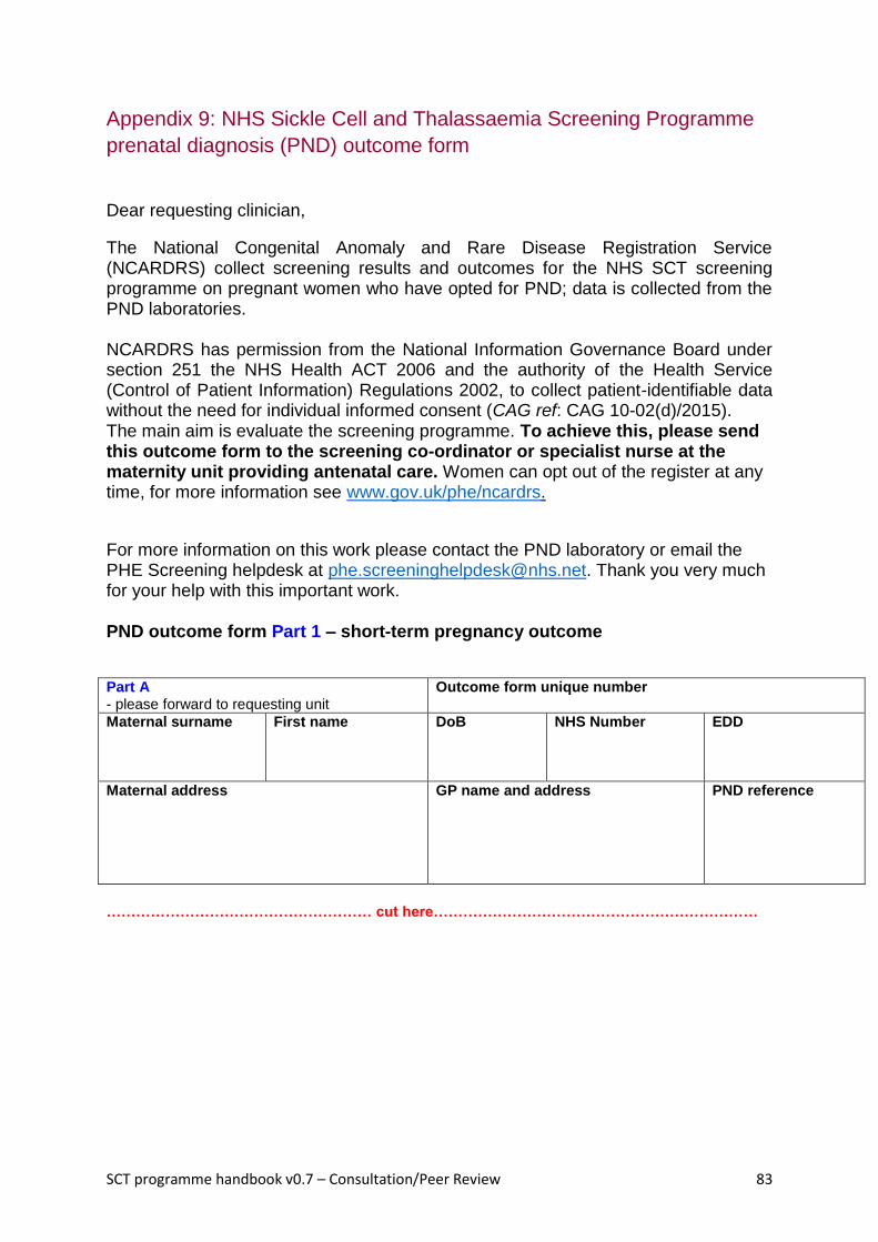
SCT programme handbook v0.7 – Consultation/Peer Review 83
Appendix 9: NHS Sickle Cell and Thalassaemia Screening Programme
prenatal diagnosis (PND) outcome form
Dear requesting clinician,
The National Congenital Anomaly and Rare Disease Registration Service (NCARDRS) collect screening results and outcomes for the NHS SCT screening programme on pregnant women who have opted for PND; data is collected from the PND laboratories. NCARDRS has permission from the National Information Governance Board under section 251 the NHS Health ACT 2006 and the authority of the Health Service (Control of Patient Information) Regulations 2002, to collect patient-identifiable data without the need for individual informed consent (CAG ref: CAG 10-02(d)/2015). The main aim is evaluate the screening programme. To achieve this, please send this outcome form to the screening co-ordinator or specialist nurse at the maternity unit providing antenatal care. Women can opt out of the register at any time, for more information see www.gov.uk/phe/ncardrs.
For more information on this work please contact the PND laboratory or email the PHE Screening helpdesk at [email protected]. Thank you very much for your help with this important work. PND outcome form Part 1 – short-term pregnancy outcome
Part A - please forward to requesting unit
Outcome form unique number
Maternal surname
First name
DoB
NHS Number
EDD
Maternal address
GP name and address
PND reference
……………………………………………… cut here…………………………………………………………

SCT programme handbook v0.7 – Consultation/Peer Review 84
PND outcome form Part 1 – short-term pregnancy outcome
Part B To be completed by the screening coordinator or specialist nurse and returned to PND lab within one month of receiving PND result
Outcome form unique number:
Maternity unit address
Date of referral
Referrer’s name
PND result
Please tick outcome: CONTINUING PREGNANCY [ ] MISCARRIAGE [ ]* TERMINATION OF PREGNANCY [ ]* *If there is a miscarriage or termination of pregnancy, do not complete Part 2 of the outcome form
Completed by (please print) NAME TELEPHONE
Date Part 1 B completed on
PND outcome form Part 2 – final outcome
Part A – please forward to requesting unit
Outcome form unique number
Maternal surname
First name
DoB
NHS Number
EDD
Maternal address
GP name and address
PND reference
Maternity unit address
Date of referral
Referrer’s name
PND result
……………………………………………… cut here…………………………………………………………

SCT programme handbook v0.7 – Consultation/Peer Review 85
PND outcome form Part 2 – final outcome
Part B to be completed within one month of delivery by screening coordinator or specialist nurse and returned to PND lab
Outcome form unique number:
Maternity unit address
Date of referral
Referrer’s name
EDD
Please return to PND lab address
Please return by (one month from EDD)
Baby’s NHS Number
Newborn laboratory that baby’s blood spot was sent to
Baby’s place of birth
If no live birth, please give reason
Completed by (please print) Name: Telephone:
Date Part 2 B completed on
Please complete parts in red, retain the named portion of this form (Part 2 A), and only return Part 2 B Final Outcome Form, with its unique identifying number to the PND laboratory Version 20 June 2017

SCT programme handbook v0.7 – Consultation/Peer Review 86
Flow diagram for use of 2-part pregnancy outcome form
Sample received in PND lab
Result reported to requesting clinician with 2-part outcome form
2 part outcome form given to screening co-ordinator or specialist nurse with result
Part 1 B completed and returned to PND lab by screening coordinator or specialist nurse within one month of receiving PND result
Part 2 retained by screening coordinator or specialist nurse until delivery
PND lab uses information to complete Q19 of PND data return
Screening coordinator or specialist nurse completes Part 2 B within one month of delivery and returns to PND lab PND lab uses information to complete Q 21 of PND data return
PND lab contacts newborn lab with information from Part 2 for baby’s result PND lab uses information to complete Q18 of PND data return
The form should be sent be sent for all PND requests, not just those where PND shows an affected fetus. The screening co-ordinator or specialist nurse is only required to complete parts in red and blue. Version 20 June 2017

SCT programme handbook v0.7 – Consultation/Peer Review 87
Appendix 10 Follow up of newborn DNA screening results
DNA for Transfused Babies Counselling Notes for Health Care Professionals Babies who have had a blood transfusion prior to newborn screening for sickle cell disease do not have a reliable screening result. The NHS Sickle Cell & Thalassaemia Screening Programme supports testing these samples using a DNA process. Although it is not the same quality as the routine screening processes, babies with sickle cell disease who may be missed for timely follow up and care should be identified via the test.
No sickle cell gene (mutation) detected Counselling Notes A negative result means that the individual does not carry the sickle cell gene mutation on any chromosome. The baby does not have sickle cell disease but this does not exclude other haemoglobin variants or conditions such as
Hb C, Hb OArab, Hb DPunjab, Hb E, Hb Lepore or any unstable Hb variant (carrier states)
Beta thalassaemia major
Beta thalassaemia carrier
thalassaemia; thalassaemia; thalassaemia
Hereditary Persistence of Fetal Haemoglobin (HPFH)
Alpha thalassaemia
Benign haemoglobin disorders (for example Hb CC; C/Beta thalassaemia; DD; EE etc) Review parental haemoglobinopathy results and advise accordingly. It is NOT the responsibility of the health care professional to initiate the repeat blood test, but parents should be assisted with this process if required. The sample should be liquid blood and sent to the specialist haematology laboratory linked to the Clinical Network, at least 4 months after the last blood transfusion was given.
Sickle cell gene (mutation) plus another haemoglobin gene Counselling Notes This individual is most likely to be a sickle cell carrier but a compound heterozygous condition with another haemoglobin variant or a beta thalassaemia mutation cannot be excluded. The possible outcomes are listed below. A carrier of Hb S (Hb AS genotype) OR Sickle cell disease
HbSC
HbS/OArab
HbS/DPunjab
HbS/Lepore
HbS/0 thalassaemia
HbS/+ thalassaemia
S/HPFH Please follow up via the appropriate clinical pathway for possible sickle cell disease patients for confirmation of diagnosis and counselling. Parental haemoglobinopathy results should be checked where possible.
Only the sickle cell gene (mutation) detected Counselling Notes Please follow up via the appropriate clinical pathway for newly diagnosed children with sickle cell disease. This individual is likely to be affected by sickle cell disease. Review parental haemoglobinopathy results. Babies should commence penicillin prophylaxis routinely while diagnosis is being confirmed.
If you have any queries please contact the NHS Sickle Cell and Thalassaemia Screening Programme Email: [email protected]

SCT programme handbook v0.7 – Consultation/Peer Review 88
Appendix 11 Affected baby counselling form
Brent Sickle Cell & Thalassaemia Centre (NW London Hospitals NHS Trust)
Neonatal Screening and Counselling Form
DETAILS OF BABY District ……………………………………………..
Birth Surname .…………………………………… Hospital of Birth ………………………………….
Registered Surname ………………………………… D.o.B ….../……/…… Sex M F
First Name …………………………………………… Hb Genotype (final result)……………………………
Address ……………………………………………….. Lab No. ……………... NHS No. …………………
………………………………………………. Date card & leaflet sent …. ….../…..….../………....
………………………………………………. Transfused YES NO
Tel. No. ……………………………………..………… If YES enter date of last transfusion ……/……/……
Affected Baby: Hospital referred………………………………. Name Paediatrician…..………………………...
Notification to GP/ HV /Parents..………………………………... Date of 1st Hospital Appt………………………..
Date 1st prescribed Penicillin:…………………………………… Date 1
st Primary Vac:…….……………………..
HV Details:………………………………………………………………………………………………………………….
……………………………………………………………………………………………………………………………….
DETAILS OF PARENTS (Enter surname first in CAPITAL letters, then first name)
Mother …………………………………………………. Father …………………………………………………
DoB ..……/……/…… NHS No:……………………… DoB ..……/……/…… NHS No:…………………….
Ethnic Origin ………..………………………………… Ethnic Origin …………………………………………
Religion …………………………………..…………… Religion ……………………………………………….
GP …………………………………………………….. GP ………………………………..……………………
Need Interpreter YES NO Language ………………………………….………….
HAEMATOLOGY RESULTS (Parents) PND YES NO Outcome:………………
Date
Tested
Hb
Type
Hb RBC MCV MCHC A2 F Sickle
Test
Lab
confirmed
Yes/No
Screened
At
Mother
Father
Date(s) card(s) sent: Mother ……/……/…… Father……/……/…… GP notified ……/……/……
DETAILS OF SIBLINGS
Name DoB Place of
Birth
Sex Date
Tested
Hb
Type
Comments


![Www.tascunit.com Ethnicity Questions and Antenatal Screening for Sickle Cell and Thalassaemia [EQUANS]: A Randomised Controlled Trial of Two Ethnicity.](https://static.cupdf.com/doc/110x72/56649c995503460f9495502c/wwwtascunitcom-ethnicity-questions-and-antenatal-screening-for-sickle-cell.jpg)