Mutations in the UBIAD1 Gene, Encoding a PotentialPrenyltransferase, Are Causal for Schnyder CrystallineCorneal DystrophyAndrew Orr1,2, Marie-Pierre Dube3, Julien Marcadier4, Haiyan Jiang2, Antonio Federico5, Stanley George1, Christopher Seamone1, DavidAndrews1, Paul Dubord6, Simon Holland6, Sylvie Provost3, Vanessa Mongrain4, Susan Evans4, Brent Higgins7, Sharen Bowman7, DuaneGuernsey2, Mark Samuels2,8¤*
1 Department of Ophthalmology and Visual Sciences, Dalhousie University, Halifax, Nova Scotia, Canada, 2 Department of Pathology, DalhousieUniversity, Halifax, Nova Scotia, Canada, 3 Montreal Heart Institute, University of Montreal, Montreal, Quebec, Canada, 4 Faculty of Medicine,Dalhousie University, Halifax, Nova Scotia, Canada, 5 Dipartimento di Scienze Neurologiche e del Comportamento, Universita degli Studi di Siena,Siena, Italy, 6 Department of Ophthalmology, Faculty of Medicine, University of British Columbia, Vancouver, British Columbia, Canada, 7 GenomeAtlantic, National Research Council of Canada Institute of Marine Biology, Halifax, Nova Scotia, Canada, 8 Department of Medicine, University ofMontreal, Montreal, Quebec, Canada
Schnyder crystalline corneal dystrophy (SCCD, MIM 121800) is a rare autosomal dominant disease characterized by progressiveopacification of the cornea resulting from the local accumulation of lipids, and associated in some cases with systemicdyslipidemia. Although previous studies of the genetics of SCCD have localized the defective gene to a 1.58 Mbp interval onchromosome 1p, exhaustive sequencing of positional candidate genes has thus far failed to reveal causal mutations. We haveascertained a large multigenerational family in Nova Scotia affected with SCCD in which we have confirmed linkage to thesame general area of chromosome 1. Intensive fine mapping in our family revealed a 1.3 Mbp candidate interval overlappingthat previously reported. Sequencing of genes in our interval led to the identification of five putative causal mutations in geneUBIAD1, in our family as well as in four other small families of various geographic origins. UBIAD1 encodes a potentialprenyltransferase, and is reported to interact physically with apolipoprotein E. UBIAD1 may play a direct role in intracellularcholesterol biochemistry, or may prenylate other proteins regulating cholesterol transport and storage.
Citation: Orr A, Dube M-P, Marcadier J, Jiang H, Federico A, et al (2007) Mutations in the UBIAD1 Gene, Encoding a Potential Prenyltransferase, AreCausal for Schnyder Crystalline Corneal Dystrophy. PLoS ONE 2(8): e685. doi:10.1371/journal.pone.0000685
INTRODUCTIONSchnyder crystalline corneal dystrophy (SCCD, MIM 121800) is
an inherited disorder whose most prominent feature is progressive,
symmetrical opacification of the central cornea, the transparent
anterior face of the eye (Fig. 1). Described first in 1924 by van
Went and Wibaut[1], and later in more detail by Schnyder[2],
SCCD is very rare. Until recently, the world literature contained
fewer than 100 cases[3]. SCCD affects both sexes equally, and is
found in multiple ethnic groups around the globe.
SCCD can become manifest as early as in the first few years of
life, although it more commonly presents in the second decade.
Thereafter, the clinical course is somewhat variable, although
surprisingly good vision can be retained long-term despite
significant corneal clouding[4]. Eventually however, reduced visual
acuity and glare often mandate intervention. While photother-
apeutic keratectomy (removal of superficial corneal layers via
excimer laser ablation) can provide temporary relief in selected
cases[5], the definitive treatment is surgical replacement of the
central cornea (penetrating keratoplasty) with cadaveric donor
tissue. SCCD can recur in the corneal graft postoperatively[4].
Pathophysiologically, SCCD appears to result from an abnor-
mality in lipid metabolism in the cells of the cornea[6–14].
Examination of corneal tissue removed from affected patients
during transplantation surgery has revealed a tenfold increase in
mainly unesterified cholesterol levels, and a five- to ninefold
increase in phospholipids[11,12]. Immunohistochemical analysis
of the same tissue is consistent with an underlying defect in HDL
metabolism[11]. Although not a constant finding[7], SCCD has
been associated in some patients with systemic dyslipidemia[9,15–
18] and thus possibly to an elevated risk of cardiovascular events
such as myocardial infarction (heart attack) and stroke[9].
SCCD is inherited as an autosomal dominant trait with age-
dependent penetrance, in which it is possible to assign affection
status unambiguously by 40 years of age[19]. Although strongly
genetic, identification of a causal gene has been elusive. Shearman
et al. performed linkage analysis on a large family originally of
Swedish/Finnish ancestry, localizing the defective gene to the
short arm of chromosome 1, at 1p34–36[20,21]. Theendakara et
al. further refined the SCCD locus using families of multiple
ethnicities, reducing the candidate region to a 2.32 Mbp (million
base pair) interval lying between genetic markers D1S1160 and
Academic Editor: Florian Kronenberg, Innsbruck Medical University, Austria
Received April 23, 2007; Accepted June 13, 2007; Published August 1, 2007
Copyright: � 2007 Orr et al. This is an open-access article distributed under theterms of the Creative Commons Attribution License, which permits unrestricteduse, distribution, and reproduction in any medium, provided the original authorand source are credited.
Funding: AO was supported by the Queen Elizabeth II Health Science CentreResearch Fund. MES was supported by Dalhousie University, the IWK HealthCentre, the Dalhousie Medical Research Foundation, the Capital District HealthAuthority, Genome Atlantic and Genomic Canada. SB was supported by GenomeCanada/Genome Atlantic. MPD was supported by the Fonds de Recherche enSante du Quebec (FRSQ).
Competing Interests: The authors have declared that no competing interestsexist.
* To whom correspondence should be addressed. E-mail: [email protected]
¤ Current address: Department of Medicine, University of Montreal, Montreal,Quebec, Canada
PLoS ONE | www.plosone.org 1 August 2007 | Issue 8 | e685
D1S1635, or possibly a smaller 1.58 Mbp interval between
D1S503 and D1S1635[22]. Recently Aldave et al. and Oleynikov
et al. have reported sequencing of all annotated genes within the
larger interval, finding no pathogenic mutations and tentatively
excluding them as causing SCCD[23,24], a finding proposed to
result from locus heterogeneity, mutations within promoter or
untranslated regions, or the presence of an unannotated gene.
RESULTS
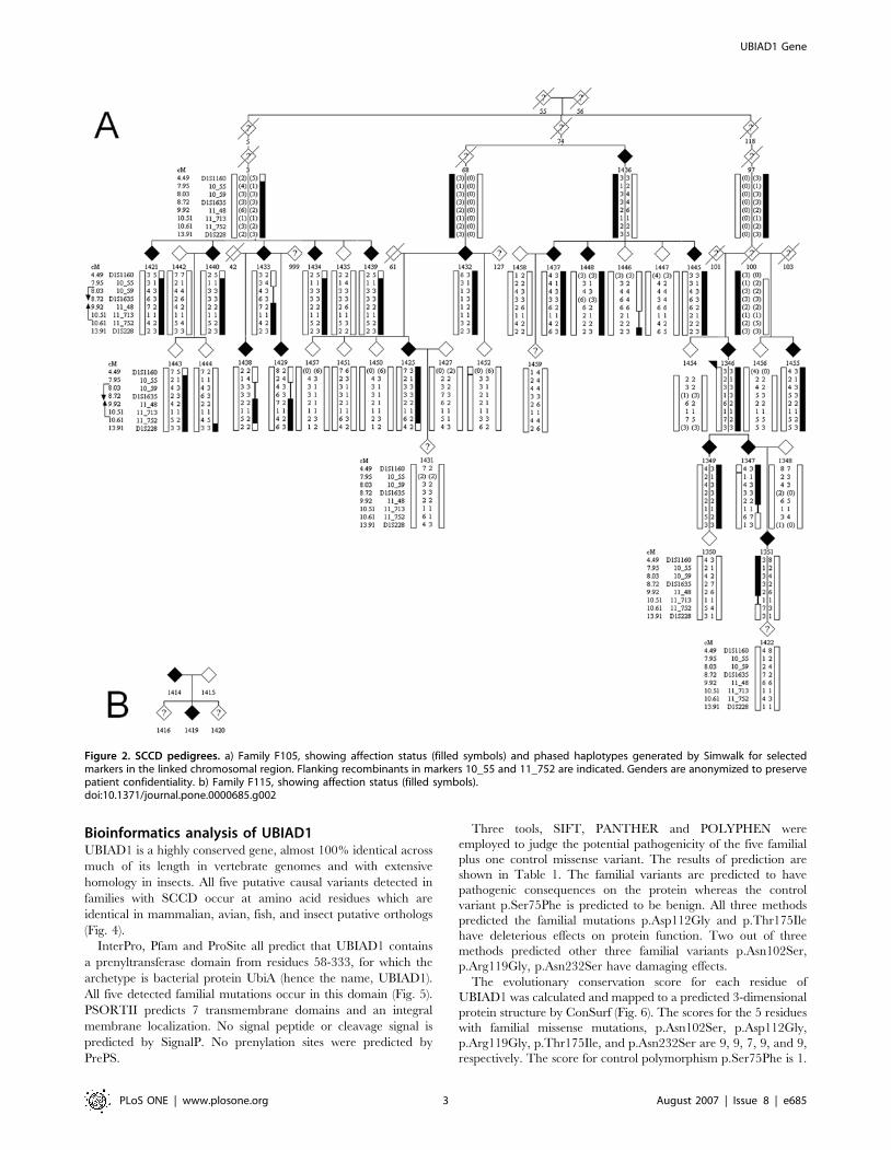
Confirmation of linkage to chromosome 1pInitial microsatellite genotyping using selected markers from the
published linkage interval[22] on chromosome 1 were consistent
with linkage in family F105 and F115 (see Materials and Methods
and Fig. 2 for ascertainment and descriptions of families
segregating SCCD). The two families together generated a multi-
point sumLOD score of 8.7 using the 90% penetrance trans-
mission model, with family F105 providing essentially all the
statistical power (Fig. 3).
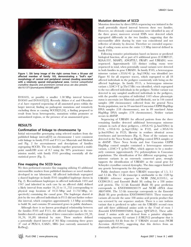
Fine mapping the SCCD locusWe then performed extensive fine mapping utilizing 45 additional
microsatellite markers from published databases or novel markers
developed in our laboratory. All affected individuals segregated
a shared haplotype in family F105 (simplified haplotype shown in
Fig. 2). Recombinant haplotypes in affected individuals 1433 and
1347 (and inherited in each case by an affected child) defined
a likely interval from marker 10_55 to 11_752 (corresponding to
physical map locations of 10.55 Mbp and 11.752 Mbp, re-
spectively) containing the causal gene. The haplotype shared de
facto by the two affected individuals in F115 did not further reduce
this interval, which comprises approximately 1.3 Mbp according
to build 36, and contains 20 annotated genes in public databases.
Although there is no known genealogical relationship between
the families F105 and F115, the affected haplotypes in the two
families shared a small region of three consecutive markers (10_59,
10_70, 10_89) identical by state. These markers defined
a potentially shared interval of 361 Kbp containing three genes
(39 end of PEX14, CASZ1, SRG [not currently annotated by
RefSeq]).
Mutation detection of SCCDMutation detection by direct DNA sequencing was initiated in the
small potentially shared interval between these two families.
However, no obviously causal mutations were identified in any of
the three genes; moreover several SNPs were detected which
segregated differently in the two families, suggesting that the
microsatellite allele sharing by state was coincidental and not
reflective of identity by descent. Therefore we extended sequenc-
ing of coding exons across the entire 1.3 Mbp interval defined in
family F105.
Following tentative prioritization based on known or predicted
biological function, all or part of 6 additional genes (C1ORF127,
KIAA1337, MASP2, ANGPTL7, FRAP1 and UBIAD1) were
sequenced. Approximately 125 distinct coding exons were
sequenced in total, when potentially causal variants were detected
in both families in gene UBIAD1. In family F105, a heterozygous
missense variant c.355A.G (p. Arg119Gly) was identified (see
Figure S1 for all sequence traces), which segregated to all 18
affected individuals in the pedigree consistently with being on the
affected haplotype. In family F115, a heterozygous missense
variant c.524C.T (p.Thr175Ile) was identified, which segregated
to the two affected individuals in the pedigree. Neither variant was
detected in any sampled unaffected individuals in the pedigrees,
with the possible exception of individual 1443 whose phenotypic
status is uncertain (see Materials and Methods), nor in 144 control
samples (288 chromosomes) collected from the general Nova
Scotia population, nor in 59 unrelated Caucasian CEPH HapMap
DNA samples (118 chromosomes), nor in 89 unrelated Asian
HapMap DNA samples (178 chromosomes). Neither variant
occurs in dbSNP.
Sequencing of UBIAD1 for affected patients from the three
remaining families detected additional heterozygous missense
variants in each of these pedigrees, c.695A.G (p.Asn232Ser) in
F118, c.335A.G (p.Asp112Gly) in F122, and c.305A.G
(p.Asn102Ser) in F123, likewise in residues identical across
vertebrates and invertebrates (Fig. 4). None of the variants was
detected in the Nova Scotia or HapMap control DNA samples or
in dbSNP. Three Nova Scotia and four Caucasian CEPH
HapMap control samples contained a heterozygous missense
variant, c.224C.T (p.Ser75Phe), which appears to be a moder-
ately common (approximately 3%) polymorphism in Caucasian
populations. The identification of five different segregating, rare
missense variants in an extremely conserved gene, strongly
supports the identification of UBIAD1 as the causal gene for
Schnyder crystalline corneal dystrophy. The data suggest limited if
any genetic heterogeneity for this phenotype.
Public databases report three UBIAD1 transcripts of 1.5, 3.1
and 3.5 kb. The 1.5 kb transcript is attributable to the 1520 bp
UBIAD1 reference sequence in NCBI (NM_013319.1, or
CCDS129.1) and Ensembl (Build 38) coding for a 338 amino
acid protein. The 3.1 kb Ensembl (Build 38) gene prediction
corresponds to ENST00000240179 and NCBI cDNA clone
AK074890. The 3.5 kb transcript corresponds to the Ensembl
(Build 38) gene prediction ENST00000376810. These transcript
variants all encode the same 338 amino acid protein product that
was screened by our sequence analysis. There is a rare isoform
variant that is predicted to splice out the UBIAD1 second exon
and add three additional amino acids to the 39end of exon 1
(Ensembl ENST00000376804; Expasy Q9Y5Z9-2). These addi-
tional 3 amino acids are derived from a putative ubiguitin-
conjugating enzyme E2 variant 2 (UBE2V2) pseudogene that is
approximately 8.6 kb from the 39 UBIAD1 second exon (NCBI
Accession AL031291), suggesting that this derives from an
aberrantly spliced message.
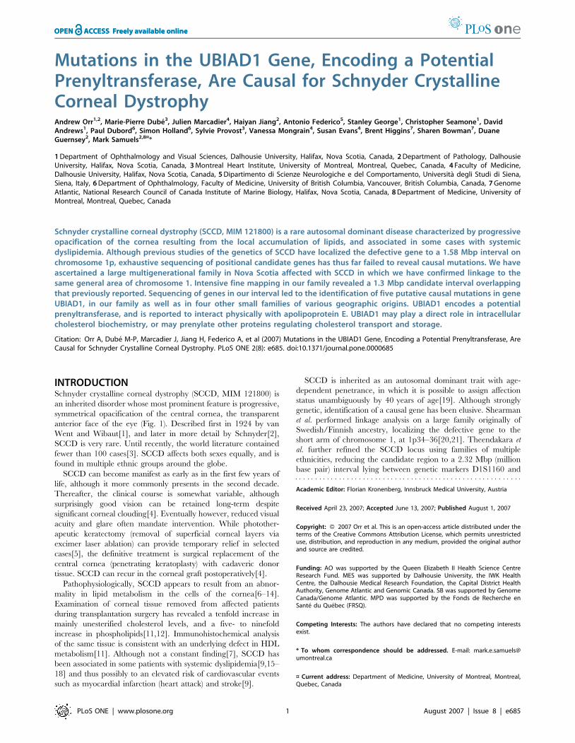
Figure 1. Slit lamp image of the right cornea from a 50-year oldaffected member of family 105, demonstrating a ‘‘bull’s eye’’morphology of central and peripheral corneal clouding associatedwith a relatively spared mid-peripheral zone. Central subepithelialcrystalline deposits and prominent corneal arcus are also present.doi:10.1371/journal.pone.0000685.g001
UBIAD1 Gene
PLoS ONE | www.plosone.org 2 August 2007 | Issue 8 | e685
Bioinformatics analysis of UBIAD1UBIAD1 is a highly conserved gene, almost 100% identical across
much of its length in vertebrate genomes and with extensive
homology in insects. All five putative causal variants detected in
families with SCCD occur at amino acid residues which are
identical in mammalian, avian, fish, and insect putative orthologs
(Fig. 4).
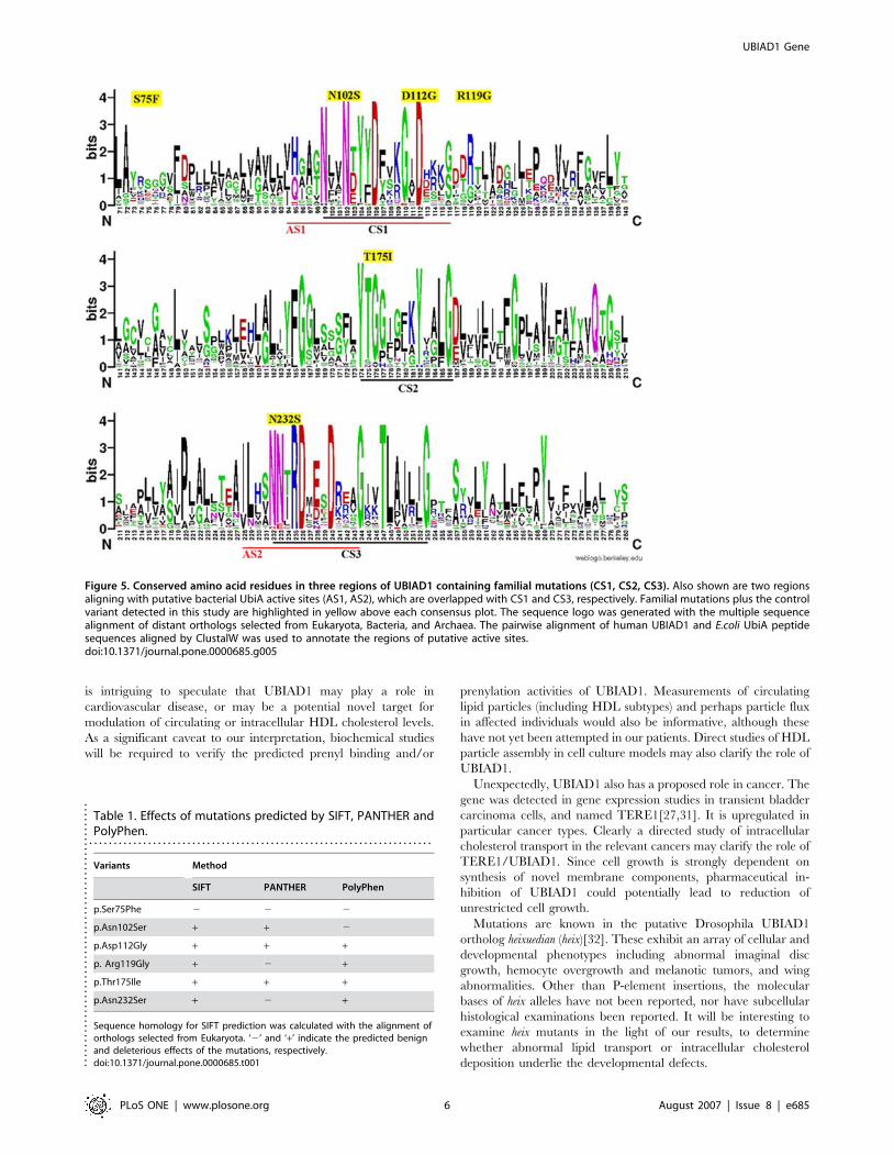
InterPro, Pfam and ProSite all predict that UBIAD1 contains
a prenyltransferase domain from residues 58-333, for which the
archetype is bacterial protein UbiA (hence the name, UBIAD1).
All five detected familial mutations occur in this domain (Fig. 5).
PSORTII predicts 7 transmembrane domains and an integral
membrane localization. No signal peptide or cleavage signal is
predicted by SignalP. No prenylation sites were predicted by
PrePS.
Three tools, SIFT, PANTHER and POLYPHEN were
employed to judge the potential pathogenicity of the five familial
plus one control missense variant. The results of prediction are
shown in Table 1. The familial variants are predicted to have
pathogenic consequences on the protein whereas the control
variant p.Ser75Phe is predicted to be benign. All three methods
predicted the familial mutations p.Asp112Gly and p.Thr175Ile
have deleterious effects on protein function. Two out of three
methods predicted other three familial variants p.Asn102Ser,
p.Arg119Gly, p.Asn232Ser have damaging effects.
The evolutionary conservation score for each residue of
UBIAD1 was calculated and mapped to a predicted 3-dimensional
protein structure by ConSurf (Fig. 6). The scores for the 5 residues
with familial missense mutations, p.Asn102Ser, p.Asp112Gly,
p.Arg119Gly, p.Thr175Ile, and p.Asn232Ser are 9, 9, 7, 9, and 9,
respectively. The score for control polymorphism p.Ser75Phe is 1.
Figure 2. SCCD pedigrees. a) Family F105, showing affection status (filled symbols) and phased haplotypes generated by Simwalk for selectedmarkers in the linked chromosomal region. Flanking recombinants in markers 10_55 and 11_752 are indicated. Genders are anonymized to preservepatient confidentiality. b) Family F115, showing affection status (filled symbols).doi:10.1371/journal.pone.0000685.g002
UBIAD1 Gene
PLoS ONE | www.plosone.org 3 August 2007 | Issue 8 | e685
The familial variants also lie close to each other in contrast to the
control variant on the predicted protein structure model.
Structurally and functionally important regions in the protein
typically appear as patches of evolutionarily conserved residues
that are spatially close to each other[25]. The evolutionary
conservation and the physical proximities of the five familial
variants support that the variants are in a functional region of
UBIAD1 protein.
DISCUSSIONWe have identified the putative causal gene for Schnyder
crystalline corneal dystrophy (SCCD) through a positional candi-
date strategy. We ascertained a large Nova Scotia family, as well as
four small families, segregating SCCD in a dominant transmission
pattern. Within the large family we were able to confirm linkage to
the published locus at chromosome 1p36.2–36.3 with high
statistical significance (maxLOD = 8.7), and we defined a minimal
recombinant interval containing 20 annotated genes. Direct DNA
resequencing of coding exons in the region identified five different
heterozygous mutations in the gene UBIAD1, one mutation in
each family. Each mutation was carried by all affected individuals
within the respective family, and none was found in 144 unaffected
control DNA samples (288 chromosomes) from the Nova Scotia
population, nor 59 Caucasian HapMap DNA samples (118
chromosomes), nor 89 Asian HapMap DNA samples (178
chromosomes), nor are any of these mutations found in the
dbSNP database. With one possible case of incomplete pene-
trance, sampled unaffecteds did not carry any of these mutations.
All five mutations are in highly conserved residues in putative gene
orthologs from other vertebrate (chimp, macaque, dog, mouse, rat,
chicken, clawed toad) and invertebrate (Drosophila, Anopheles)
genomes.
The recombinant interval defined by our family 105 overlaps
with, but is offset slightly centromeric to that previously de-
fined[22]. The location of gene UBIAD1 itself is consistent with
chromosomal haplotypes in 70 of the 71 affected individuals from
13 families described in the previous study, with the exception of
one recombinant individual, affected III:4 in family 9. The source
of the inconsistency is unclear, with potential explanations
including genetic heterogeneity, phenotypic misdiagnosis, micro-
satellite marker mutation or other technical difficulty with
genotyping. Interestingly, a 343 kb copy number variant (CNV)
has been annotated to occur in the genomic region including
marker D1S1635[26], which if found in the key recombinant
individual could have led to misleading definition of the
recombinant boundary. Further suggestion of this possibility is
that Theendakara et al. document segregation of two alleles for
marker D1S3153, which however does not actually contain
a microsatellite repeat but is also within the potential CNV
region. In any case, direct resequencing of UBIAD1 has not been
reported by other groups working on the genetics of SCCD, hence
there is no inconsistency between our results and other published
work at the nucleotide level.
Bioinformatics packages overall agreed in predicting likely
pathogenicity for the five familial mutations, but less so for
a missense variant identified in seven control samples. While
UBIAD1 is ubiquitously expressed[27], the eye has been
identified to have the highest normalized expression distribution
of the 39 tissues reported at the Source (http://genome-www5.
stanford.edu/ ) and of the 47 tissues identified at the Unigene
Expression Profile Viewer (http://www.ncbi.nlm.nih.gov/
UniGene/ESTProfileViewer.cgi?uglist = Hs.522933 ).
Although little has been known until now about UBIAD1
from the perspective of vertebrate genetics, the primary amino
acid sequence is tantalizing. Bioinformatics analysis suggests that
this gene is an intrinsic membrane protein with a prenyltransfer-
ase functional domain. UbiA, the canonical family member, also
known as 4-hydroxybenzoate octaprenyl transferase, catalyzes
1,4-dihydroxy-2-naphthoate –.dimethylmenaquinone, in the
ubiquinone biosynthetic pathway of bacteria (not to be confused
with the UbiA gene in C. elegans which encodes ubiquitin and has
no sequence homology to E. coli UbiA or UBIAD1). Although
there is extensive sequence divergence between human UBIAD1
and E. coli UbiA, nonetheless the sequences can be aligned.
Direct conservation of mutated residues in our families is not
evident across such a large evolutionary divide, but four of the
five familial variants we detected lie near or within predicted
active site regions of the bacterial enzyme based on molecular
modeling and multigenome alignment (Fig. 5)[28]. This
supports, albeit indirectly, a deleterious effect of mutations in
these regions of the protein. UBIAD1 need not be a true
enzymatic prenyltransferase, it might simply contain ligand
binding pockets for related molecules. Interestingly no prenyla-
tion sites were predicted for UBIAD1 by PrePS, suggesting that
UBIAD1 is probably not self-modifying. However, the same
result was found for UbiA of E. coli K12, indicating that
prenylation sites are not automatically found in prenyltrans-
ferases themselves. PrePS did correctly predict farnesyltransferase
and geranylgeranyltransferase modification sites for human c-K-
ras2 protein isoform a.
The role of prenyltransferases or even prenyl binding proteins in
lipid or cholesterol metabolism can be imagined. It seems unlikely
that UBIAD1 plays a direct metabolic role in cholesterol
biosynthesis. However, prenyl binding proteins such as UBIAD1
might play a role in sensing and regulating metabolite levels
intracellularly and/or systemically. UBIAD1 might prenylate
other proteins thereby influencing their intracellular localization.
It is noteworthy that corneal lipid deposition is observed in three
other human genetic disorders of cholesterol, specifically high
density lipoprotein (HDL), metabolism: Niemann-Pick type C[29],
LCAT deficiency (fish-eye disease) and ABCA1/Tangier Disease
(TD)[11]. It is also suggestive that UBIAD1 has been shown to
bind directly to apolipoprotein E, a component of very low density
lipoprotein particles, in protein-protein interaction studies[30]. It
-4
-2
0
2
4
6
8
10
0 2 4 6 8 10 12 14cM
LOD
D1S
2667
LOD = 8.7
ped 1ped 115
Simwalk2 multipoint linkage: Chomosome 1
Figure 3. Multipoint linkage analysis using Simwalk for Family F105(ped 1 in the figure) and F115 (ped 115 in the figure), across thelinked chromosomal interval. In this analysis individual 1443 of family105 was set as phenotype unaffected; the maxLOD increased slightly to9.5 when 1443 was set to phenotype unknown. A maxLOD = 6.6 wasobtained using an affecteds-only model.doi:10.1371/journal.pone.0000685.g003
UBIAD1 Gene
PLoS ONE | www.plosone.org 4 August 2007 | Issue 8 | e685
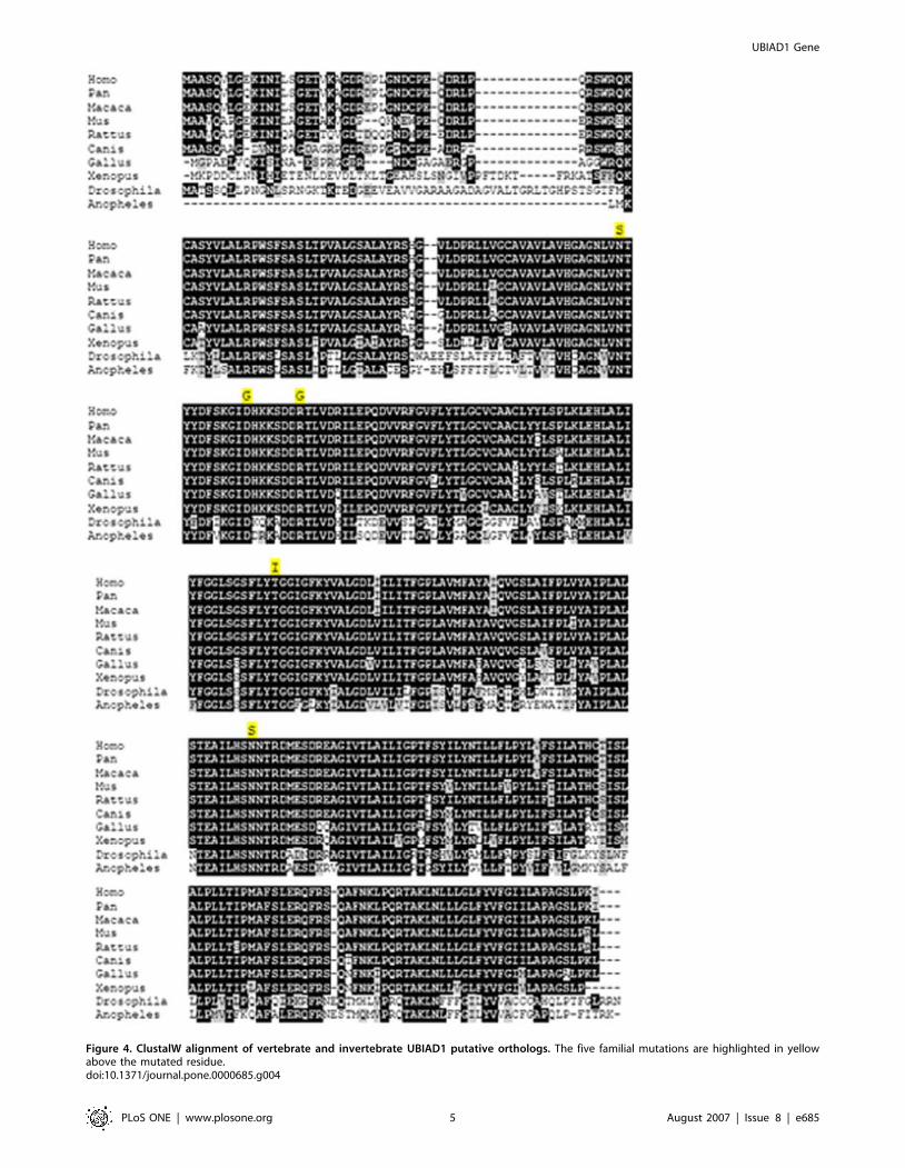
Figure 4. ClustalW alignment of vertebrate and invertebrate UBIAD1 putative orthologs. The five familial mutations are highlighted in yellowabove the mutated residue.doi:10.1371/journal.pone.0000685.g004
UBIAD1 Gene
PLoS ONE | www.plosone.org 5 August 2007 | Issue 8 | e685
is intriguing to speculate that UBIAD1 may play a role in
cardiovascular disease, or may be a potential novel target for
modulation of circulating or intracellular HDL cholesterol levels.
As a significant caveat to our interpretation, biochemical studies
will be required to verify the predicted prenyl binding and/or
prenylation activities of UBIAD1. Measurements of circulating
lipid particles (including HDL subtypes) and perhaps particle flux
in affected individuals would also be informative, although these
have not yet been attempted in our patients. Direct studies of HDL
particle assembly in cell culture models may also clarify the role of
UBIAD1.
Unexpectedly, UBIAD1 also has a proposed role in cancer. The
gene was detected in gene expression studies in transient bladder
carcinoma cells, and named TERE1[27,31]. It is upregulated in
particular cancer types. Clearly a directed study of intracellular
cholesterol transport in the relevant cancers may clarify the role of
TERE1/UBIAD1. Since cell growth is strongly dependent on
synthesis of novel membrane components, pharmaceutical in-
hibition of UBIAD1 could potentially lead to reduction of
unrestricted cell growth.
Mutations are known in the putative Drosophila UBIAD1
ortholog heixuedian (heix)[32]. These exhibit an array of cellular and
developmental phenotypes including abnormal imaginal disc
growth, hemocyte overgrowth and melanotic tumors, and wing
abnormalities. Other than P-element insertions, the molecular
bases of heix alleles have not been reported, nor have subcellular
histological examinations been reported. It will be interesting to
examine heix mutants in the light of our results, to determine
whether abnormal lipid transport or intracellular cholesterol
deposition underlie the developmental defects.
Figure 5. Conserved amino acid residues in three regions of UBIAD1 containing familial mutations (CS1, CS2, CS3). Also shown are two regionsaligning with putative bacterial UbiA active sites (AS1, AS2), which are overlapped with CS1 and CS3, respectively. Familial mutations plus the controlvariant detected in this study are highlighted in yellow above each consensus plot. The sequence logo was generated with the multiple sequencealignment of distant orthologs selected from Eukaryota, Bacteria, and Archaea. The pairwise alignment of human UBIAD1 and E.coli UbiA peptidesequences aligned by ClustalW was used to annotate the regions of putative active sites.doi:10.1371/journal.pone.0000685.g005
Table 1. Effects of mutations predicted by SIFT, PANTHER andPolyPhen.
. . . . . . . . . . . . . . . . . . . . . . . . . . . . . . . . . . . . . . . . . . . . . . . . . . . . . . . . . . . . . . . . . . . . . .
Variants Method
SIFT PANTHER PolyPhen
p.Ser75Phe 2 2 2
p.Asn102Ser + + 2
p.Asp112Gly + + +
p. Arg119Gly + 2 +
p.Thr175Ile + + +
p.Asn232Ser + 2 +
Sequence homology for SIFT prediction was calculated with the alignment oforthologs selected from Eukaryota. ‘2’ and ‘+’ indicate the predicted benignand deleterious effects of the mutations, respectively.doi:10.1371/journal.pone.0000685.t001....
....
....
....
....
....
....
....
....
....
....
....
....
..
UBIAD1 Gene
PLoS ONE | www.plosone.org 6 August 2007 | Issue 8 | e685
MATERIALS AND METHODSEthical approval for this study was obtained from the Research
Ethics Board of the Queen Elizabeth II Health Sciences Centre.
Clinical AssessmentWe ascertained a large family from Nova Scotia known on
a longstanding basis to local corneal specialists. The family, F105,
unilineally segregates SCCD in 18 living affecteds and offered an
excellent opportunity to discover the identity of the defective gene
responsible for this disorder (Fig. 2a). This family is of uncertain,
but possibly Spanish ancestry. Concurrently, we identified another
nuclear family with SCCD, originally from Scotland, that had
recently immigrated to Nova Scotia (F115, two affecteds, Fig. 2b).
Subsequently, two further families were recruited from the clinical
practices of Canadian corneal specialists: family F118, previously
described elsewhere[12], containing two affected members of
unknown ancestry, and family F122, of East Indian descent, with
A
B
Figure 6. Predicted UBIA prenyltransferase domain-containing protein 1 structure from ModBase mapped with evolutionary conservationscores calculated by ConSurf. Five familial mutations plus one control variant detected in this study are indicated. The color scale ranging from blueto red represents the conservation score of residue varies from 9 (most conserved) to 1 (most variable). a, Front view; b, Rear view.doi:10.1371/journal.pone.0000685.g006
UBIAD1 Gene
PLoS ONE | www.plosone.org 7 August 2007 | Issue 8 | e685
one known affected member. Lastly, family F123 with two
available affected members, also previously described else-
where[7], was recruited from colleagues in Italy.
Affection status of participants was determined in the following
manner. Individuals were regarded as affected if typical corneal
features of SCCD were present on slit lamp examination, or in
documentary slit lamp images, or if corneal transplantation had
been performed with SCCD as the underlying diagnosis.
Individuals were regarded as unaffected if, by the age of forty,
no features of SCCD were found. To facilitate phase determina-
tion during subsequent haplotype analysis, we collected DNA
specimens from as many family members as possible. In the case of
participants residing outside of Nova Scotia, in whom direct
examination was not possible, individuals older than forty years of
age without a definite diagnosis of SCCD and a history of normal
routine eye examinations were considered to be probably
unaffected. One individual, 1443 in family 105, is also of uncertain
phenotype; visual exam did not indicate status as affected, however
full slit-lamp examination could not be conducted due to local
circumstances.
Control DNA samples were obtained from a randomized
collection of apparently healthy individuals in the local Nova
Scotia population, biased toward Caucasian ancestry. Additional
controls were derived from the set of generally available HapMap
DNA samples, specifically the 60 CEPH Caucasian parents and 90
East Asian unrelated individuals. Affection status of controls was
unknown, but the possibility that any would be affected by SCCD
seems remote, given its rarity and the likelihood in the case of the
local population controls that they would have come to the
attention of clinicians in the academic hospital system associated
with Dalhousie University.
Following written informed consent, saliva or venous blood
samples were obtained from all ascertained subjects, from which
DNA was extracted according to standard protocols. We used the
Oragene kits from DNA Genotek (Ottawa, Canada) for self-
collection of salivary DNA (particularly in situations where it was
desirable for samples to be collected via mail), with excellent DNA
yields and performance during microsatellite genotyping and
direct sequencing.
Genotyping and Linkage AnalysisMicrosatellite genotyping was performed using fluorescent pri-
mers. 59 tags were added to the reverse, unlabelled primer in each
case to reduce variable non-templated nucleotide addition[33,34].
Products were resolved on ABI 377 electrophoresis instruments
and genotype chromatograms were interpreted using the Gene-
Marker program from SoftGenetics, Inc.[35] See Table S1 for
details of custom marker primers.
Pedigree files and genotype data were imported into Progeny
Lab software version (6.6.01). Mendelian inconsistencies were
identified with Pedcheck version 1.1[36]. Allele calls for in-
consistent markers were set to 0 in the offending nuclear families
involved in the inconsistencies. Genetic positions from the Decode
map were used when available[37]. To calculate genetic position
for markers not on the Decode map, linear interpolation was used
between the two closest common markers flanking the markers to
position, using physical distances provided by human genome
assembly, build 36.
Statistical analyses were conducted with two models.
1. An affected only model in which all individuals not known to
be affected except spouses were set to unknown and using
penetrance set to 0.99, phenocopy rate set to 0.001 for
a dominant disease with allele frequency of 0.001.
2. Penetrance set to 0.90 with a phenocopy rate of 0.001 and
a dominant disease allele frequency of 0.001.
Marker allele frequencies were estimated by maximum likelihood
using Merlin version 1.0.1 (option –fm)[38]. As Merlin can not
handle large pedigrees, pedigree 105 was divided into three
smaller families (branch 2/3, 74/75 and 100/103/101) for this
stage of the analysis. Allelic frequencies from Merlin were
manually incorporated into dat files.
Two-point linkage was carried out using the MLINK routine of
FASTLINK v4.1P on Linux. LOD scores were compiled by
extracting results from the final.out output file using MLINK_-
LODS v2. Multipoint linkage analysis and haplotyping were
carried out using SIMWALK version 2.90 on Linux[39]. The
input files were converted to SIMWALK format using Mega2 v3.0
R4. The haplotype routine converged on the first run for both
pedigrees.
Mutation DetectionPredicted protein coding regions of all examined genes were
amplified using primers designed with Primer3[40] (http://frodo.
wi.mit.edu/) (see Table S2 for sequences) from two affected
individuals from family F105 and one affected individual from
family F115. Coding exons of gene UBIAD1 were subsequently
sequenced in samples from additional affected individuals in all
five families, and from controls. PCR products were sequenced
using ABI 377 or 3700 electrophoresis instruments at the Genome
Atlantic and Institute for Marine Biology TAGC or at the McGill
University and Genome Quebec Centre for Innovation. Sequence
chromatograms were interpreted using the MutationSurveyor
program from SoftGenetics, Inc., with gene annotations from
GenBank[35].
Bioinformatics AnalysisInterPro, Pfam, ProSite, PSORTII, SignalP, and PrePS were run
via the Expasy web site (http://us.expasy.org/tools/ ). The effects
of amino acid substitutions on protein function were predicted
with SIFT[41–43], PolyPhen[44–46], and PANTHER[47,48].
Homologous peptide sequences of human UBIAD1 gene in
Eukaryota, Archaea and Bacteria were retrieved through NCBI
web site using protein-protein BLAST (blastp) against the nr
database. Multiple sequence alignments were computed by
ClustalW and displayed with BoxShade. The sequences of
distantly related orthologs were aligned by MUSCLE[49]. The
sequence logo in Fig. 5 was created by WebLogo[50]. The
evolutionary conservation of amino acid sites with mutations was
analyzed using ConSurf[25,51,52], based on alignments shown in
Fig. S2 and S3. The predicted protein structure from Mod-
Base[53] for the UbiA prenyltransferase domain-containing pro-
tein 1 was used to build a 3D model. Figure 6 was generated using
PyMOL[54].
SUPPORTING INFORMATION
Table S1 Custom microsatellite genotyping marker primer data.
Found at: doi:10.1371/journal.pone.0000685.s001 (0.17 MB
DOC)
Table S2 Primer sequences for mutation detection amplification
of UBIAD1 coding exons (two amplicons for each exon).
Found at: doi:10.1371/journal.pone.0000685.s002 (0.03 MB
DOC)
Figure S1 Mutation detection sequencing traces for affected
patients from each of the five families with SCCD, following
UBIAD1 Gene
PLoS ONE | www.plosone.org 8 August 2007 | Issue 8 | e685
fluorescent sequencing on ABI 377 or 3700 electrophoresis
instruments and alignment to annotated genomic sequences
containing the UBIAD1 gene using MutationSurveyor. Each
panel has 7 lines generated by the software: from top to bottom are
the amino acid translations of consensus and predicted mutation
sequences, forward direction virtual reference trace, forward
direction patient sequence trace, forward direction mutation call,
reverse direction mutation call, reverse direction patient sequence
trace, reverse direction virtual reference trace. a, Family F105; b,
Family F115; c, Family F118; d, Family F122; e, Family F123.
Found at: doi:10.1371/journal.pone.0000685.s003 (0.33 MB TIF)
Figure S2 Multiple sequence alignment of the Eukaryota
orthologs of Human UBIAD1 peptide sequence. The alignment
was used to study the sequence conservation and predict the effects
of mutations.
Found at: doi:10.1371/journal.pone.0000685.s004 (3.89 MB TIF)
Figure S3 Multiple sequence alignment of distant orthologs of
Human UBIAD1 peptide sequence selected from Eukaryota,
Bacteria, and Archaea. The alignment was used to study the
sequence conservation and generate the sequence logo.
Found at: doi:10.1371/journal.pone.0000685.s005 (4.73 MB TIF)
ACKNOWLEDGMENTSWe are grateful to the families who generously contributed their time and
materials for this research.
Author Contributions
Conceived and designed the experiments: MS AO. Performed the
experiments: JM VM SE BH SB DG. Analyzed the data: MS AO MD
JM HJ SP VM SE BH DG. Contributed reagents/materials/analysis tools:
AO AF SG CS DA PD SH. Wrote the paper: MS AO MD DG. Other:
Supervised molecular genetics experiments: DG SB. Senior clinical
investigator: AC. Senior investigator overseeing all molecular genetic
aspects of project: MS.
REFERENCES1. Van Went J, Wibaut F (1924) Een zyeldzame erfelijke hoornvliesaandoening.
Ned Tijdschr Geneesks 68(1st half, B): 2996–2997.
2. Schnyder W (1929) Mitteilung uber einen neuen Typus von familiarer
Hornhauterkrankung. Schweiz Med Wochenschr 59: 559–571.
3. Weiss JS (1992) Schnyder’s dystrophy of the cornea. A Swede-Finn connection.
Cornea 11: 93–101.
4. Bron AJ (1989) Corneal changes in the dislipoproteinaemias. Cornea 8:
135–140.
5. Meier U, Anastasi C, Failla F, Simona F (1998) [Possibilities of therapeuticphotokeratotomy with the excimer laser in treatment of Schnyder crystalline
corneal dystrophy]. Klin Monatsbl Augenheilkd 212: 405–406.
6. Barchiesi BJ, Eckel RH, Ellis PP (1991) The cornea and disorders of lipid
metabolism. Surv Ophthalmol 36: 1–22.
7. Battisti C, Dotti MT, Malandrini A, Pezzella F, Bardelli AM, et al. (1998)
Schnyder corneal crystalline dystrophy: description of a new family withevidence of abnormal lipid storage in skin fibroblasts. Am J Med Genet 75:
35–39.
8. Bron AJ, Williams HP, Carruthers ME (1972) Hereditary crystalline stromal
dystrophy of Schnyder. I. Clinical features of a family with hyperlipoproteinae-mia. Br J Ophthalmol 56: 383–399.
9. Brownstein S, Jackson WB, Onerheim RM (1991) Schnyder’s crystalline cornealdystrophy in association with hyperlipoproteinemia: histopathological and
ultrastructural findings. Can J Ophthalmol 26: 273–279.
10. Burns RP, Connor W, Gipson I (1978) Cholesterol turnover in hereditary
crystalline corneal dystrophy of Schnyder. Trans Am Ophthalmol Soc 76:184–196.
11. Gaynor PM, Zhang WY, Weiss JS, Skarlatos SI, Rodrigues MM, et al. (1996)Accumulation of HDL apolipoproteins accompanies abnormal cholesterol
accumulation in Schnyder’s corneal dystrophy. Arterioscler Thromb Vasc Biol16: 992–999.
12. McCarthy M, Innis S, Dubord P, White V (1994) Panstromal Schnyder cornealdystrophy. A clinical pathologic report with quantitative analysis of corneal lipid
composition. Ophthalmology 101: 895–901.
13. Weiss JS, Rodrigues MM, Kruth HS, Rajagopalan S, Rader DJ, et al. (1992)
Panstromal Schnyder’s corneal dystrophy. Ultrastructural and histochemicalstudies. Ophthalmology 99: 1072–1081.
14. Yamada M, Mochizuki H, Kamata Y, Nakamura Y, Mashima Y (1998)Quantitative analysis of lipid deposits from Schnyder’s corneal dystrophy.
Br J Ophthalmol 82: 444–447.
15. Crispin S (2002) Ocular lipid deposition and hyperlipoproteinaemia. Prog Retin
Eye Res 21: 169–224.
16. Kajinami K, Inazu A, Wakasugi T, Koizumi J, Mabuchi H, et al. (1988) [A case
of familial hypercholesterolemia associated with Schnyder’s corneal dystrophy].Nippon Naika Gakkai Zasshi 77: 1017–1020.
17. Kohnen T, Pelton RW, Jones DB (1997) [Schnyder corneal dystrophy and
juvenile, systemic hypercholesteremia]. Klin Monatsbl Augenheilkd 211:
135–137.
18. Thiel HJ, Voigt GJ, Parwaresch MR (1977) [Crystalline corneal dystrophy(Schnyder) in the presence of familial type IIa hyperlipoproteinaemia (author’s
transl)]. Klin Monatsbl Augenheilkd 171: 678–684.
19. Weiss JS (1996) Schnyder crystalline dystrophy sine crystals. Recommendation
for a revision of nomenclature. Ophthalmology 103: 465–473.
20. Riebeling P, Polz S, Tost F, Weiss JS, Kuivaniemi H, et al. (2003) [Schnyder’s
crystalline corneal dystrophy. Further narrowing of the linkage interval atchromosome 1p34.1-p36?]. Ophthalmologe 100: 979–983.
21. Shearman AM, Hudson TJ, Andresen JM, Wu X, Sohn RL, et al. (1996) The
gene for schnyder’s crystalline corneal dystrophy maps to human chromosome
1p34.1-p36. Hum Mol Genet 5: 1667–1672.
22. Theendakara V, Tromp G, Kuivaniemi H, White PS, Panchal S, et al. (2004)
Fine mapping of the Schnyder’s crystalline corneal dystrophy locus. Hum Genet
114: 594–600.
23. Aldave AJ, Rayner SA, Principe AH, Affeldt JA, Katsev D, et al. (2005) Analysis
of fifteen positional candidate genes for Schnyder crystalline corneal dystrophy.
Mol Vis 11: 713–716.
24. Oleynikov YS, Yellore VS, Bourla N, Khan M, Rayner SA, et al. Exclusion of
the chrosome 1p36 candidate region for Schnyder crystalline corneal dystrophy;
2007.
25. Landau M, Mayrose I, Rosenberg Y, Glaser F, Martz E, et al. (2005) ConSurf
2005: the projection of evolutionary conservation scores of residues on protein
structures. Nucleic Acids Res 33: W299–302.
26. Redon R, Ishikawa S, Fitch KR, Feuk L, Perry GH, et al. (2006) Global
variation in copy number in the human genome. Nature 444: 444–454.
27. McGarvey TW, Nguyen T, Tomaszewski JE, Monson FC, Malkowicz SB (2001)
Isolation and characterization of the TERE1 gene, a gene down-regulated in
transitional cell carcinoma of the bladder. Oncogene 20: 1042–1051.
28. Brauer L, Brandt W, Wessjohann LA (2004) Modeling the E. coli 4-
hydroxybenzoic acid oligoprenyltransferase ( ubiA transferase) and character-
ization of potential active sites. J Mol Model (Online) 10: 317–327.
29. Palmer M, Green WR, Maumenee IH, Valle DL, Singer HS, et al. (1985)
Niemann-Pick disease–type C. Ocular histopathologic and electron microscopic
studies. Arch Ophthalmol 103: 817–822.
30. McGarvey TW, Nguyen TB, Malkowicz SB (2005) An interaction between
apolipoprotein E and TERE1 with a possible association with bladder tumor
formation. J Cell Biochem 95: 419–428.
31. McGarvey TW, Nguyen T, Puthiyaveettil R, Tomaszewski JE, Malkowicz SB
(2003) TERE1, a novel gene affecting growth regulation in prostate carcinoma.
Prostate 54: 144–155.
32. Ashburner M, Misra S, Roote J, Lewis SE, Blazej R, et al. (1999) An exploration
of the sequence of a 2.9-Mb region of the genome of Drosophila melanogaster:
the Adh region. Genetics 153: 179–219.
33. Magnuson VL, Ally DS, Nylund SJ, Karanjawala ZE, Rayman JB, et al. (1996)
Substrate nucleotide-determined non-templated addition of adenine by Taq
DNA polymerase: implications for PCR-based genotyping and cloning.
Biotechniques 21: 700–709.
34. Samuels ME, Dube MP (2005) Linkage Mapping. Encyclopedia of Genetics,
Genomics, Proteomics, and Bioinformatics: John Wiley & Sons, Inc.
35. Samuels M, Higgins B, Provost S, Marcadier J, Blouin C, et al. (2007) New
technologies in human genetic analysis. American Biotechnology Laboratory in
press.
36. O’Connell JR, Weeks DE (1998) PedCheck: a program for identification of
genotype incompatibilities in linkage analysis. Am J Hum Genet 63: 259–266.
37. Kong A, Gudbjartsson DF, Sainz J, Jonsdottir GM, Gudjonsson SA, et al. (2002)
A high-resolution recombination map of the human genome. Nat Genet 31:
241–247.
38. Abecasis GR, Cherny SS, Cookson WO, Cardon LR (2002) Merlin–rapid
analysis of dense genetic maps using sparse gene flow trees. Nat Genet 30:
97–101.
39. Weeks DE, Sobel E, O’Connell JR, Lange K (1995) Computer programs for
multilocus haplotyping of general pedigrees. Am J Hum Genet 56: 1506–1507.
UBIAD1 Gene
PLoS ONE | www.plosone.org 9 August 2007 | Issue 8 | e685
40. Rozen S, Skaletsky H (2000) Primer3 on the WWW for general users and for
biologist programmers. In: SKSM. ed. Bioinformatics Methods and Protocols:
Methods in Molecular Biology. Totowa, NJ: Humana Press. pp 365–386.
41. Ng PC, Henikoff S (2001) Predicting deleterious amino acid substitutions.
Genome Res 11: 863–874.
42. Ng PC, Henikoff S (2002) Accounting for human polymorphisms predicted to
affect protein function. Genome Res 12: 436–446.
43. Ng PC, Henikoff S (2003) SIFT: Predicting amino acid changes that affect
protein function. Nucleic Acids Res 31: 3812–3814.
44. Ramensky V, Bork P, Sunyaev S (2002) Human non-synonymous SNPs: server
and survey. Nucleic Acids Res 30: 3894–3900.
45. Sunyaev S, Ramensky V, Bork P (2000) Towards a structural basis of human
non-synonymous single nucleotide polymorphisms. Trends Genet 16: 198–200.
46. Sunyaev S, Ramensky V, Koch I, Lathe W 3rd, Kondrashov AS, et al. (2001)
Prediction of deleterious human alleles. Hum Mol Genet 10: 591–597.
47. Thomas PD, Campbell MJ, Kejariwal A, Mi H, Karlak B, et al. (2003)
PANTHER: a library of protein families and subfamilies indexed by function.
Genome Res 13: 2129–2141.
48. Thomas PD, Kejariwal A (2004) Coding single-nucleotide polymorphisms
associated with complex vs. Mendelian disease: evolutionary evidencefor differences in molecular effects. Proc Natl Acad Sci U S A 101:
15398–15403.
49. Edgar RC (2004) MUSCLE: multiple sequence alignment with high accuracyand high throughput. Nucleic Acids Res 32: 1792–1797.
50. Crooks GE, Hon G, Chandonia JM, Brenner SE (2004) WebLogo: a sequencelogo generator. Genome Res 14: 1188–1190.
51. Armon A, Graur D, Ben-Tal N (2001) ConSurf: an algorithmic tool for the
identification of functional regions in proteins by surface mapping ofphylogenetic information. J Mol Biol 307: 447–463.
52. Glaser F, Pupko T, Paz I, Bell RE, Bechor-Shental D, et al. (2003) ConSurf:identification of functional regions in proteins by surface-mapping of
phylogenetic information. Bioinformatics 19: 163–164.53. Pieper U, Eswar N, Braberg H, Madhusudhan MS, Davis FP, et al. (2004)
MODBASE, a database of annotated comparative protein structure models, and
associated resources. Nucleic Acids Res 32: D217–222.54. DeLano W (2002) The PyMOL molecular graphics system. San Carlos, CA:
DeLano Scientific.
UBIAD1 Gene
PLoS ONE | www.plosone.org 10 August 2007 | Issue 8 | e685


![A Stilbenoid-Specific Prenyltransferase Utilizes ...A Stilbenoid-Specific Prenyltransferase Utilizes Dimethylallyl Pyrophosphate from the Plastidic Terpenoid Pathway1[OPEN] Tianhong](https://static.cupdf.com/doc/110x72/5eb88723cf80df2b4822048a/a-stilbenoid-specific-prenyltransferase-utilizes-a-stilbenoid-speciic-prenyltransferase.jpg)