12 Global Identification of Protein Prenyltransferase Substrates: Defining the Prenylated Proteome CORISSA L. LAMPHEAR a ELAINA A. ZVERINA b JAMES L. HOUGLAND c,1 CAROL A. FIERKE a,b,c a Department of Biological Chemistry University of Michigan Ann Arbor, Michigan, USA b Chemical Biology Program University of Michigan Ann Arbor, Michigan, USA c Department of Chemistry University of Michigan Ann Arbor, Michigan, USA I. Abstract The protein prenyltransferases, protein farnesyltransferase (FTase) and protein geranylgeranyltransferase-I (GGTase-I), catalyze the attachment of a 15-carbon farnesyl or 20-carbon geranylgeranyl moiety, respectively, to a cysteine near the C-terminus of a substrate protein targeting it to the membrane. Substrates of the prenyltransferases are involved in a myriad of signaling pathways and processes within the cell, therefore inhibitors tar- geting FTase and GGTase-I are being developed as therapeutics for treat- ment of diseases such as cancer, parasitic infection, asthma, and progeria. 1 Present address: Department of Chemistry, Syracuse University, Syracuse, New York, USA 207 THE ENZYMES, Vol. XXIX ISSN NO: 1874-6047 # 2011 Elsevier Inc. All rights reserved. DOI: 10.1016/B978-0-12-381339-8.00012-3

Welcome message from author
This document is posted to help you gain knowledge. Please leave a comment to let me know what you think about it! Share it to your friends and learn new things together.
Transcript
1 Present address
THE ENZYMES,# 2011 Elsevier Inc
12
Global Identification of Protein
Prenyltransferase Substrates:
Defining the Prenylated ProteomeCORISSA L. LAMPHEARa � ELAINA A. ZVERINAb �JAMES L. HOUGLANDc,1 � CAROL A. FIERKEa,b,c
aDepartment of Biological Chemistry
University of Michigan
Ann Arbor, Michigan, USA
bChemical Biology Program
University of Michigan
Ann Arbor, Michigan, USA
cDepartment of Chemistry
University of Michigan
Ann Arbor, Michigan, USA
I. Abstract
The protein prenyltransferases, protein farnesyltransferase (FTase) andprotein geranylgeranyltransferase-I (GGTase-I), catalyze the attachmentof a 15-carbon farnesyl or 20-carbon geranylgeranyl moiety, respectively, toa cysteine near the C-terminus of a substrate protein targeting it to themembrane. Substrates of the prenyltransferases are involved in a myriad ofsignaling pathways and processes within the cell, therefore inhibitors tar-geting FTase and GGTase-I are being developed as therapeutics for treat-ment of diseases such as cancer, parasitic infection, asthma, and progeria.
: Department of Chemistry, Syracuse University, Syracuse, New York, USA
207Vol. XXIX ISSN NO: 1874-6047. All rights reserved. DOI: 10.1016/B978-0-12-381339-8.00012-3
208 CORISSA L. LAMPHEAR, ET AL.
FTase and GGTase-I were proposed to recognize a Ca1a2X motif, where Cis the cysteine where the prenyl group is attached, a1 and a2 are smallaliphatic amino acids, and X confers specificity between FTase andGGTase-I with X being methionine, serine, glutamine, and alanine forFTase and leucine or phenylalanine for GGTase-I. Recent studies indicatethat the Ca1a2X paradigm should be expanded; therefore, further studiesare needed to define the prenylated proteome, to understand normal cellu-lar processes, and to determine the targets of prenyltransferase inhibitors.This review highlights the multiple approaches currently used to identifyand define FTase and GGTase-I substrates. Direct identificationapproaches involve identifying FTase and GGTase-I substrates ‘‘one byone’’ or by using lipid donor analogs. A complementary approach toidentify the prenylated proteome is to define the modes of FTase andGGTase-I substrate recognition using structure–function studies, peptidelibrary studies, and computational methods.
II. Introduction
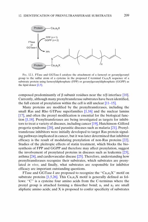
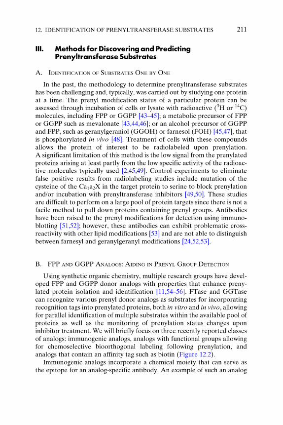
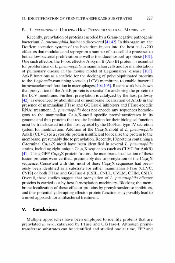
Prenylation is an important posttranslational modification where anisoprenoid moiety is transferred to a cysteine near the C-terminus of asubstrate protein [1,2]. This hydrophobic modification helps to localizeproteins to cellular membranes to carry out their function as well as tofacilitate protein–protein interactions [3,4]. FTase catalyzes the transfer ofthe 15-carbon farnesyl moiety from farnesyldiphosphate (FPP) to a cysteineresidue near the C-terminus of the target protein; protein GGTase-I andGGTase-II (also called RabGGTase) catalyze the analogous attachment ofa 20-carbon geranylgeranyl group from geranylgeranyldiphosphate(GGPP) to cysteine(s) near the C-terminus of the substrate protein(Figure 12.1) [2,5]. Additionally, in vivo, substrates can undergo furthermodification after prenylation. The last three amino acids of prenylatedsubstrates can be proteolyzed by zinc metalloprotease Ste24 (ZMPSTE24)orRas-converting enzyme 1 (RCE1) at the endoplasmic reticulum, followedby methylation of the carboxy terminus, catalyzed by isoprenylcysteinemethyl transferase (ICMT) [6–8]. These additional modifications can aidin membrane localization, but it is not yet known whether these modifica-tions occur on every prenylated substrate. This review will primarily focuson FTase andGGTase-I, the ‘‘CaaXprenyltransferases,’’ which have similarmodes of recognition (see Chapter 8 for a review on GGTase-II). Both ofthese enzymes are heterodimeric, containing identical a subunits and dis-tinct but homologous b subunits [9], with active sites in both enzymes
FPP (n = 1)GGPP (n = 2)
PPi
Ca1a2X
Ca1a2X
S
n
FTaseor
GGTase-I
Protein
Protein
FIG. 12.1. FTase and GGTase-I catalyze the attachment of a farnesyl or geranylgeranyl
group to the sulfur atom of a cysteine in the proposed C-terminal Ca1a2X sequence of a
substrate protein using farnesyldiphosphate (FPP) or geranylgeranyldiphosphate (GGPP) as
the lipid donor [2,5].
12. IDENTIFICATION OF PRENYLTRANSFERASE SUBSTRATES 209
composed predominantly of b subunit residues near the a/b interface [10].Currently, althoughmany prenyltransferase substrates have been identified,the full extent of prenylation within the cell is still unclear [11–15].
Many proteins are modified by the prenyltransferases, including thesmall Ras and Rho GTPase superfamilies [1,16] and the nuclear lamins[17], and often the prenyl modification is essential for the biological func-tion [1,18]. Prenyltransferases are being investigated as targets for inhibi-tors to treat a variety of diseases, including cancer [19], Hutchinson–Gilfordprogeria syndrome [20], and parasitic diseases such as malaria [21]. Prenyl-transferase inhibitors were initially developed to target Ras protein signal-ing pathways implicated in cancer, but it was later determined that inhibitorefficacy is the result of modulating prenylation of non-Ras proteins [22].Studies of the pleitropic effects of statin treatment, which blocks the bio-synthesis of FPP and GGPP and therefore may affect prenylation, suggestthe involvement of prenylated proteins in diseases such as leukemia [23],asthma [24], and cardiovascular disease [25]. Therefore, understanding howprenyltransferases recognize their substrates, which substrates are preny-lated in vivo, and finally, what substrates are responsible for inhibitorefficacy are important outstanding questions.
FTase and GGTase-I are proposed to recognize the ‘‘Ca1a2X’’ motif onsubstrate proteins [1,5,26]. This Ca1a2X motif is generally defined as fol-lows: ‘‘C’’ is a cysteine four amino acids from the C-terminus where theprenyl group is attached forming a thioether bond; a1 and a2 are smallaliphatic amino acids; and X is proposed to confer specificity of substrates
210 CORISSA L. LAMPHEAR, ET AL.
for modification by FTase or GGTase-I, with X being methionine, serine,glutamine, and alanine for FTase and leucine or phenylalanine forGGTase-I [27–30]. Although many substrates are described by this para-digm, recent studies have indicated that the Ca1a2X model should berevised, as many substrates fall outside the traditional Ca1a2X definition[15]. Additionally, there is evidence for a large pool of dual substrates forFTase and GGTase-I, with these proteins potentially modified by bothenzymes [31]. Understanding how FTase and GGTase-I recognize sub-strates would aid in defining the prenylated proteome and in understandingthe biological signaling pathways involving prenylated proteins.
An emerging field is the study of prenyltransferases from pathogenic andparasitic organisms as potential therapeutic targets [32–35]. For instance,the pathogenic yeast strain Candida albicans, known to affect immunocom-promised patients, utilizes both protein FTase and GGTase-I enzymes tocatalyze prenylation of substrate proteins [36,37]. The specificity of thesetwo enzymes is currently being studied to define the complement of pre-nylation of proteins in this organism, since it is possible that inhibitors of theprenyltransferases could be used as antifungal therapeutics [36,38]. Addi-tionally, inhibitors are being developed as therapeutics for the treatment ofparasitic diseases such as malaria, caused by Plasmodium falciparum [39].These FTase inhibitors, based upon ethylenediamine [34] and tetrahydro-quinoline [33] scaffolds, are thought to block P. falciparum FTase activity,which is essential for the organism. Based upon modeling and resistancemutation studies [40], these inhibitors may chelate the catalytic Zn2þ andbind to the enzyme in the lipid substrate grove. Recently, it has also beendiscovered that the Gram-negative bacterium, Legionella pneumophila,hijacks the host prenyltransferase machinery to catalyze prenylation ofbacterial proteins, raising the possibility that current mammalian prenyl-transferase inhibitors could also be used as antibiotics [41,42].
Defining the extent of prenylation within the proteome of an organismcan be approached using two complementary tactics: (1) direct in vivodetermination of proteins that are prenylated or (2) definition of therecognition elements used by FTase and GGTase-I to select peptideand protein substrates. This review covers both approaches. The summaryof the direct in vivo identification modes includes a brief discussionof methods for detecting prenylation on a small or large scale(Sections III.A and III.B). Additionally, methods used to define molecularrecognition in prenyltransferases are described, including structure–function studies (Section III.C), peptide library studies (Section III.D),and computational predictive methods (Section III.E). Finally, a briefdiscussion of pathogenic prenyltransferase enzyme structure and specificityis included in Section IV.
12. IDENTIFICATION OF PRENYLTRANSFERASE SUBSTRATES 211
III. Methods forDiscoveringandPredictingPrenyltransferaseSubstrates
A. IDENTIFICATION OF SUBSTRATES ONE BY ONE
In the past, the methodology to determine prenyltransferase substrateshas been challenging and, typically, was carried out by studying one proteinat a time. The prenyl modification status of a particular protein can beassessed through incubation of cells or lysate with radioactive (3H or 14C)molecules, including FPP or GGPP [43–45]; a metabolic precursor of FPPor GGPP such as mevalonate [43,44,46]; or an alcohol precursor of GGPPand FPP, such as geranylgeraniol (GGOH) or farnesol (FOH) [45,47], thatis phosphorylated in vivo [48]. Treatment of cells with these compoundsallows the protein of interest to be radiolabeled upon prenylation.A significant limitation of this method is the low signal from the prenylatedproteins arising at least partly from the low specific activity of the radioac-tive molecules typically used [2,45,49]. Control experiments to eliminatefalse positive results from radiolabeling studies include mutation of thecysteine of the Ca1a2X in the target protein to serine to block prenylationand/or incubation with prenyltransferase inhibitors [49,50]. These studiesare difficult to perform on a large pool of protein targets since there is not afacile method to pull down proteins containing prenyl groups. Antibodieshave been raised to the prenyl modifications for detection using immuno-blotting [51,52]; however, these antibodies can exhibit problematic cross-reactivity with other lipid modifications [53] and are not able to distinguishbetween farnesyl and geranylgeranyl modifications [24,52,53].
B. FPP AND GGPP ANALOGS: AIDING IN PRENYL GROUP DETECTION
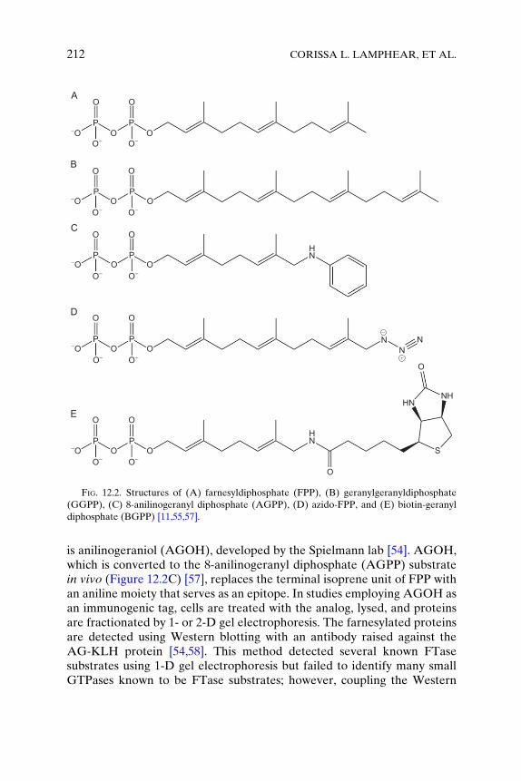
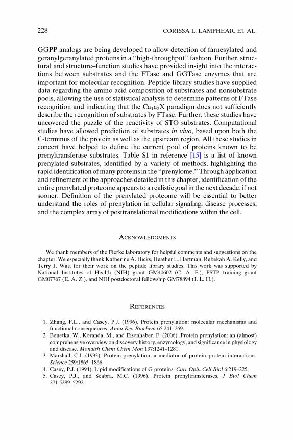
Using synthetic organic chemistry, multiple research groups have devel-oped FPP and GGPP donor analogs with properties that enhance preny-lated protein isolation and identification [11,54–56]. FTase and GGTasecan recognize various prenyl donor analogs as substrates for incorporatingrecognition tags into prenylated proteins, both in vitro and in vivo, allowingfor parallel identification of multiple substrates within the available pool ofproteins as well as the monitoring of prenylation status changes uponinhibitor treatment. We will briefly focus on three recently reported classesof analogs: immunogenic analogs, analogs with functional groups allowingfor chemoselective bioorthogonal labeling following prenylation, andanalogs that contain an affinity tag such as biotin (Figure 12.2).
Immunogenic analogs incorporate a chemical moiety that can serve asthe epitope for an analog-specific antibody. An example of such an analog
OA
B
C
D
E
O
O O-OO-O-
P P
O O
O O-OO-O-
P P
O O
O O
HN
N
O
HN
S
O
HN
NH
-OO-O-
P P
O O
O O-OO-O-
P P
O O
O O-OO-O-
P P
NN
FIG. 12.2. Structures of (A) farnesyldiphosphate (FPP), (B) geranylgeranyldiphosphate
(GGPP), (C) 8-anilinogeranyl diphosphate (AGPP), (D) azido-FPP, and (E) biotin-geranyl
diphosphate (BGPP) [11,55,57].
212 CORISSA L. LAMPHEAR, ET AL.
is anilinogeraniol (AGOH), developed by the Spielmann lab [54]. AGOH,which is converted to the 8-anilinogeranyl diphosphate (AGPP) substratein vivo (Figure 12.2C) [57], replaces the terminal isoprene unit of FPP withan aniline moiety that serves as an epitope. In studies employing AGOH asan immunogenic tag, cells are treated with the analog, lysed, and proteinsare fractionated by 1- or 2-D gel electrophoresis. The farnesylated proteinsare detected using Western blotting with an antibody raised against theAG-KLH protein [54,58]. This method detected several known FTasesubstrates using 1-D gel electrophoresis but failed to identify many smallGTPases known to be FTase substrates; however, coupling the Western
12. IDENTIFICATION OF PRENYLTRANSFERASE SUBSTRATES 213
blot analysis with 2-D gel electrophoresis increases the efficiency ofprenylated protein identification [58].
An emerging class of FPP and GGPP analogs employs bioorthogonalligation methods, wherein analogs containing an azido or alkyne group onthe terminal isoprenoid of FPP or GGPP are attached to substrate proteins.These functional groups are amenable to Staudinger ligation or Huisgen 1,3-dipolar cycloaddition (‘‘click’’ chemistry) [59,60], which allows for chemose-lective attachment of reporter molecules such as a fluorophore or biotinfollowing protein prenylation. The first use of an azido analog for identifica-tion of prenylated proteins was reported byKho et al. [11]. They treatedCOS-1 cells with azido-FPP (Figure 12.2D) or azido-FOH and demonstratedin vivo incorporation of this analog into proteins catalyzed by FTase. Follow-ing cell lysis, the azido-farnesylated proteins were labeled with a biotin-con-taining phosphine capture reagent using the Staudinger ligation, purifiedusing streptavidin beads and identified using mass spectrometry [11]. Thegroup used this method to detect 18 FTase substrates, including knownfarnesylated proteins such as H-Ras and Rheb. More recently, azido-GGalcohol has been used to identify geranylgeranylated substrates using clickchemistry with TAMRA-alkyne, 2-D gels, and mass spectrometry [61] and tolabel substrates in prenylation-deficient mouse tissue [62]. Additionally,alkyne derivatives of FPP andGGPP are gaining popularity for identificationof prenylated proteins in vitro and in vivo [63–65].
An alternative method, the ‘‘affinity tag’’ approach, uses as biotin-geranyldiphosphate analog (BGPP; Figure 12.2E) [55]. This analog serves as anefficient substrate for GGTase-II, but not FTase or GGTase-I. To expandthe utility of this analog, Alexandrov and coworkers ‘‘engineered’’ FTase andGGTase-I enzymes with altered substrate selectivity by mutating residues inthe active site (W102T/Y154T or W102T/Y154T/Y205T for FTase, andF53Y/Y126T or F52Y/F53Y/Y126T for GGTase-I), allowing the engineeredenzymes to catalyze modification of substrate proteins using the BGPPanalog. Compactin was used to block native FPP and GGPP synthesis,increasing the pool of unmodified FTase and GGTase-I substrates. Thecompactin-treated cell lysates were incubated in vitrowith BGPP andmutantor wild-type prenyltransferases, pulled down using streptavidin beads, andmodified proteins were identified using mass spectrometry. Many Rab pro-teins were detected and quantified as GGTase-II substrates, while variousmolecular weight substrates were identified from the engineered FTase- andGGTase-I-treated lysates.
Although there has been much success using analogs to identify preny-lated substrates, there are caveats to these approaches. First of all, the FPPand GGPP analogs may alter the protein specificity of the enzyme [66–68]since the prenyl group forms a portion of the protein substrate-binding site
214 CORISSA L. LAMPHEAR, ET AL.
[14]. In particular, for use of the biotin analogs, the FTase and GGTase-Ienzymes were engineered at positions in the active site that can alter theenzyme specificity [69]. Another concern is the ‘‘hit rate’’ for these analogs,as treatment with several of these analogs did not identify known substratesas prenylated proteins; these issues with false negatives may indicate thatthese analogs may not be incorporated into substrates at a high enough levelto allow identification. One method to obtain higher incorporation of theanalogs is to treat the cells with an inhibitor of FPP or GGPP synthesis, suchas compactin [55,70]; however, this treatment may disrupt the homeostasis ofthe cell, not allowing for true identification of substrates under normalconditions. Additionally, the BGPP analog was incubated with cell lysatesin an in vitro context, identifying potential substrates, but perhaps not iden-tifying substrates that are prenylated under native in vivo conditions.
C. IDENTIFYING PRENYLTRANSFERASE SUBSTRATES THROUGH STRUCTURAL
AND STRUCTURE–FUNCTION BIOCHEMICAL STUDIES
In this section, we review the findings from crystallographic data andstructure–activity studies that combine to provide a functional and struc-tural picture of the interactions important for prenyltransferase selectivity.Such studies have yielded valuable insights that suggest the prenylatedproteome may be much richer in size and diversity than has been previouslyproposed. Future studies, focusing on identifying additional interactionsand quantifying the energetic contribution of each enzyme–substrate inter-action, have the potential to provide the basis of a quantitative model forpredicting the full complement of prenylated proteins within the cell.
1. Structural Studies
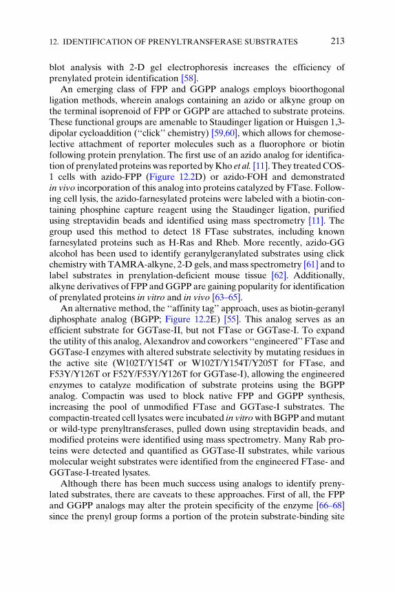
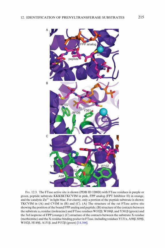
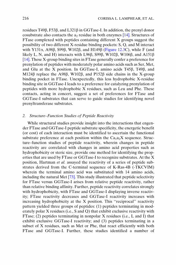
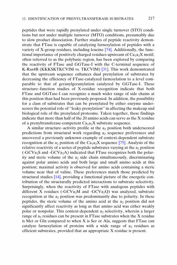
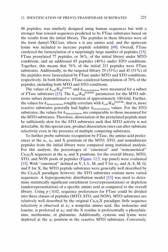
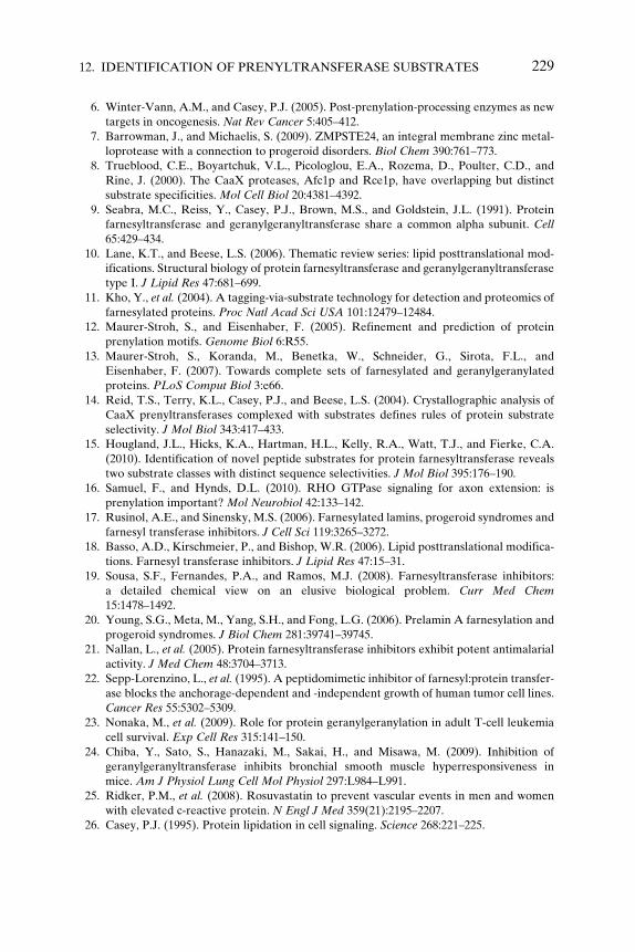
Over the past 15 years, a series of crystallographic structures of mamma-lian FTase and GGTase-I have provided insight into the active site inter-actions and structural context of the peptide substrate-binding site withinprenyltransferases [14,71,72]. Using these structures as a reference, theselectivity for each amino acid within the Ca1a2X motif can be interpretedin light of potential contacts with active site residues. The binding site forthe a1 residue of the peptide substrate is exposed to solvent at the interfaceof the FTase a and b subunits [14], consistent with the relaxed specificity atthe a1 position observed in biochemical studies [13,15,29,30]. In contrast,the a2 and X-residue binding sites lie within the solvent-excluded active site,suggesting that these two positions may be primarily responsible for pre-nyltransferase selectivity. The a2 site in FTase is mainly composed of theside chains of W102b, W106b, and Y361b (Figure 12.3B), with analogous
FPP analog
peptide
A
B
C
W102
W106
H149
XP152
Y131 A151
S99A98
W102
Y361a2
Zn2+
FIG. 12.3. The FTase active site is shown (PDB ID 1D8D) with FTase residues in purple or
green, peptide substrate KKKSKTKCVIM in pink, FPP analog (FPT Inhibitor II) in orange,
and the catalytic Zn2þ in light blue. For clarity, only a portion of the peptide substrate is shown:
TKCVIM in (A) and CVIM in (B) and (C). (A) The structure of the rat FTase active site
showing the positionof the boundFPPanalog andpeptide; (B) structure of the contacts between
the substrate a2 residue (isoleucine) and FTase residuesW102b, W106b, andY361b (green) andthe 3rd isoprene of FPP (orange); (C) structure of the contacts between the substrate X residue
(methionine) and theX-residue binding pocket in FTase, including residuesY131a, A98b, S99b,W102b, H149b, A151b, and P152b (green) [14,106].
12. IDENTIFICATION OF PRENYLTRANSFERASE SUBSTRATES 215
216 CORISSA L. LAMPHEAR, ET AL.
residues T49b, F53b, and L321b in GGTase-I. In addition, the prenyl donorcosubstrate also contacts the a2 residue in both enzymes [14]. Structures ofFTase complexed with peptides containing different X groups suggest thepossibility of two different X-residue binding pockets: S, Q, and M interactwith Y131a, A98b, S99b, W102b, and H149b (Figure 12.3C), while F (andlikely L, N, and H) interacts with L96b, S99b, W102b, W106b, and A151b[14]. These X-group binding sites in FTase generally confer a preference forprenylation of peptides with moderately polar amino acids such as Ser, Met,and Glu at the X position. In GGTase-I, amino acids T45b, T49b, andM124b replace the A98b, W102b, and P152b side chains in the X-groupbinding pocket in FTase. Unexpectedly, this less hydrophobic X-residuebinding site in GGTase-I leads to a preference for catalyzing prenylation ofpeptides with more hydrophobic X residues, such as Leu and Phe. Thesecontacts, acting in concert, suggest a set of preferences for FTase andGGTase-I substrates that can serve to guide studies for identifying novelprenyltransferase substrates.
2. Structure–Function Studies of Peptide Reactivity
While structural studies provide insight into the interactions that engen-der FTase and GGTase-I peptide substrate specificity, the energetic benefit(or cost) of each interaction must be identified to ascertain the functionalsubstrate preference at each position within the Ca1a2X sequence. Struc-ture–function studies of peptide reactivity, wherein changes in peptidereactivity are correlated with changes in amino acid properties such ashydrophobicity or steric size, provide one method for identifying the prop-erties that are used by FTase or GGTase-I to recognize substrates. At the Xposition, Hartman et al. assayed the reactivity of a series of peptide sub-strates derived from the C-terminal sequence of K-Ras-4B (-TKCVIM)wherein the terminal amino acid was substituted with 14 amino acids,including the natural Met [73]. This study illustrated that peptide selectivityfor FTase versus GGTase-I arises from relative peptide reactivity, ratherthan relative binding affinity. Further, peptide reactivity correlates stronglywith hydrophobicity, with FTase and GGTase-I displaying inverse reactiv-ity; FTase reactivity decreases and GGTase-I reactivity increases withincreasing hydrophobicity at the X position. This ‘‘reciprocal’’ reactivitypattern yielded three groups of peptides: (1) peptides terminating in mod-erately polar X residues (i.e., S and Q) that exhibit exclusive reactivity withFTase; (2) peptides terminating in nonpolar X residues (i.e., L and I) thatexhibit exclusive GGTase-I reactivity; and (3) peptides terminating in asubset of X residues, such as Met or Phe, that react efficiently with bothFTase and GGTase-I. Further, these studies identified a number of
12. IDENTIFICATION OF PRENYLTRANSFERASE SUBSTRATES 217
peptides that were rapidly prenylated under single turnover (STO) condi-tions but not under multiple turnover (MTO) conditions, presumably dueto slow product dissociation. Further studies of peptide reactivity demon-strate that FTase is capable of catalyzing farnesylation of peptides with avariety of X-group residues, including leucine [74]. Additionally, the func-tional importance of positively charged residues upstream of Ca1a2X motif,often referred to as the polybasic region, has been explored by comparingthe reactivity of FTase and GGTase-I with the C-terminal sequence ofK-Ras4B (KKKSKTKCVIM vs. TKCVIM) [31]. This work demonstratedthat the upstream sequence enhances dual prenylation of substrates bydecreasing the efficiency of FTase-catalyzed farnesylation to a level com-parable to that of geranylgeranylation catalyzed by GGTase-I. Thesestructure–function studies of X-residue recognition indicate that bothFTase and GGTase-I can recognize a much wider range of side chains atthis position than had been previously proposed. In addition, the possibilityfor a class of substrates that can be prenylated by either enzyme under-scores the potential role of ‘‘leaky prenylation’’ in affecting the makeup andbiological role of the prenylated proteome. Taken together, these findingsindicate that more than half of the 20 amino acids can serve as the X residueof a prenyltransferase-competent Ca1a2X substrate sequence.
A similar structure–activity profile at the a2 position both underscoredpredictions from structural work regarding a2 sequence preferences anduncovered a previously unknown example of context-dependent substraterecognition at the a2 position of the Ca1a2X sequence [75]. Analysis of therelative reactivity of a series of peptide substrates varying at the a2 position(-GCVa2S and -GCVa2A) indicated that FTase recognizes both the polar-ity and steric volume of the a2 side chain simultaneously, discriminatingagainst polar amino acids and both large and small amino acids at thisposition; maximal activity is observed for amino acids containing a stericvolume near that of valine. These preferences match those predicted bystructural studies [14], providing a functional picture of the energetic con-tribution of the structurally predicted interactions to substrate selectivity.Surprisingly, when the reactivity of FTase with analogous peptides withdifferent X residues (-GCVa2M and -GCVa2Q) was analyzed, substraterecognition at the a2 position was predominantly due to polarity. In thesepeptides, the steric volume of the amino acid at the a2 position did notsignificantly affect reactivity as long as that amino acid was either weaklypolar or nonpolar. This context-dependent a2 selectivity, wherein a largerrange of a2 residues can be present in FTase substrates when the X residueis Met or Gln compared to when X is Ser or Ala, suggests that FTase cancatalyze farnesylation of proteins with a wide range of a2 residues asefficient substrates, provided that an appropriate X residue is present.
218 CORISSA L. LAMPHEAR, ET AL.
Structural studies of FTase and GGTase-I have provided an intricatepicture of the interactions and active site microenvironment responsible forrecognizing prenyltransferase substrates from among the milieu of all cel-lular proteins. Structure–function analysis of peptide reactivity indicatesthat both FTase and GGTase-I recognize a much wider range of proteinsequences as substrates than was originally proposed. Further, the specificchemical properties recognized by prenyltransferases at positions withinthe Ca1a2X sequence have been characterized by correlation of peptidereactivity with amino acid properties such as size and polarity. Thesestructural and functional insights will serve as essential foundations fordevelopment of models for comprehensive prediction of prenyltransferasesubstrates based on protein C-terminal sequence data.
D. PEPTIDE LIBRARY STUDIES: A HIGH-THROUGHPUT METHOD OF
IDENTIFYING POTENTIAL PRENYLTRANSFERASE SUBSTRATES AND
DEFINING SUBSTRATE SPECIFICITY
An additional method of determining prenyltransferase substrates is todefine the molecular recognition elements of FTase and GGTase-I usingpeptide library studies. Short peptides, as small as tetrapeptides, are effi-cient substrates for FTase and GGTase-I with comparable affinity andreactivity to full-length proteins [29,30,76–78]. Adding a dansyl group tothe N-terminus of the peptide allows for continuous monitoring of thereaction using a fluorescence assay, as the dansyl group increases in fluo-rescence upon prenylation [77,79]. Conveniently, this assay can be carriedout in a high-throughput manner using 96-well plates in a plate reader, andwith large libraries, statistical analysis of peptide reactivity can be used todetermine patterns of substrate recognition [15]. The use of these peptidelibraries to study prenyltransferase specificity is advantageous, since poten-tial substrates can be screened quickly and efficiently, using wild-typeenzymes and the natural lipid substrates FPP and GGPP, although onelimitation with this method is that, in some cases, the structure of proteinsubstrates may alter recognition. Many research groups have tested small ornonhomogenous libraries of peptides as a means to identify prenyltransfer-ase substrates [67,80,81]. For instance, groups have tested the Ca1a2Xparadigm using GCxxS and GCxxL libraries [74,81], finding that theirresults generally correlate well with structural studies [14] and computa-tional algorithms [12,13] (see Section III.E).
To more completely define the scope of the prenylated proteome, alarge-scale peptide library study of the substrate selectivity of FTase wascarried out, using statistical analysis to analyze FTase preference patterns[15]. The library peptides were screened for reactivity with FTase under
12. IDENTIFICATION OF PRENYLTRANSFERASE SUBSTRATES 219
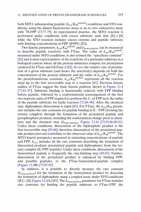
both MTO, subsaturating peptide (kcat/KMpeptide) conditions and STO con-
ditions, using the dansyl fluorescence assay or an in vitro radioactive assaywith 3H-FPP [15,77,79]. In experimental practice, the MTO reaction isperformed under conditions with excess substrate such that [E]� [S]while the STO reaction includes excess enzyme and peptide substratewith limiting concentrations of FPP ([FPP]< [E]).
Two kinetic parameters, kcat/KMpeptide and kfarnesylation, can be measured
to describe peptide reactivity with FTase. The value of kcat/KMpeptide,
measured under MTO conditions, is also termed the ‘‘specificity constant’’[82] and is most representative of the reactivity of a particular substrate in abiological context where all the protein substrates compete for prenylationcatalyzed by FTase and GGTase-I [82]. In vivo the relative rate of prenyla-tion of a given substrate (and hence the selectivity) depends on both theconcentration of the protein substrate and the value of kcat/KM
peptide. Forthe prenyltransferase reactions, kcat/KM
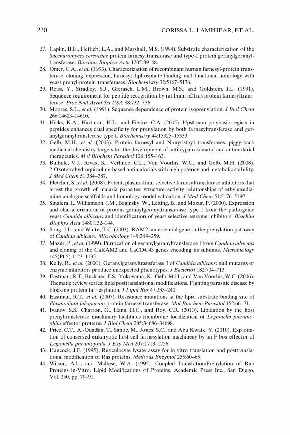
peptide represents all the reactionsteps up to the first irreversible step in a reaction [82]. Previous kineticstudies of FTase suggest the basic kinetic pathway shown in Figure 12.4[77,83–87]. Substrate binding is functionally ordered, with FPP bindingbefore peptide, followed by a conformational rearrangement of the firsttwo isoprene units of FPP required to position the C1 of FPP near the sulfurof the peptide substrate for facile reaction [72,88–90]. After the chemicalstep, diphosphate dissociation is rapid [85]. For FTase, the kcat/KM param-eter includes the rate constants for peptide binding to E � FPP (forming theternary complex) through the formation of the prenylated peptide andpyrophosphate products, including the conformation change prior to chem-istry and the chemical step (kfarnesylation, Figure 12.4) [72,85,86,90,91].Under these conditions, dissociation of the diphosphate product is thefirst irreversible step [85,86]; therefore dissociation of the prenylated pep-tide product does not contribute to the observed value of kcat/KM
peptide. TheMTO kinetic parameter measured at saturating concentrations of peptideand FPP, kcat, includes all the rate constants describing the formation ofdissociated products (prenylated peptide and diphosphate) from the ter-nary complex (E�FPP�peptide). Under these conditions, dissociation of thefarnesylated peptide is frequently the rate-limiting step [85,87]. Further,dissociation of the prenylated product is enhanced by binding FPP,and possibly peptides, to the FTase�farnesylated-peptide complex(Figure 12.4B) [73,87,92].
In addition, it is possible to directly measure the rate constant(kfarnesylation) for the formation of the farnesylated product by detectingthe formation of diphosphate using a coupled assay under STO conditions([E]> [S]; Figure 12.4A) [85]. The kfarnesylation parameter for FTase includesrate constants for binding the peptide substrate to FTase�FPP, the
FPPE
FPPE
Spep FPP
FPP
E
HSpep
FPP
HSpep
H+
H+
FPP
E
A
B
HSpep
kfarnesylation
kfarnesylation
kchem
kchem
H+
HSpep
H+
E
FPPE
SpepE
Spep FPPE F-SpepESpep
PPi
FPPE Spep
[ *[
FPPE Spep
F-Spep
F-Spep
F-SpepHSpep
H+
F-Spep SpepE
F-SpepE
F-Spep
E
FPPPPi
E
FPP
[ *[
FIG. 12.4. The proposed catalytic cycle for FTase under (A) single turnover conditions
(STO, [E]>S) and (B) multiple turnover conditions (MTO, [E]� [S]). The STO-only sub-
strates are proposed to undergo farnesylation, but not product release. FTase catalyzes turn-
over of MTO substrates where binding of either an additional peptide or FPP molecule to the
E�farnesylated-peptide complex may facilitate product release. The rate constant kfarnesylationincludes both the conformational change and chemistry steps [15,83–87,89,107].
220 CORISSA L. LAMPHEAR, ET AL.
conformation change before the chemistry step, the farnesylation step, andthe rapid dissociation of diphosphate, thereby including all the reactionsteps up to but not including release of the prenylated protein [72,85,90,91].At saturating concentrations of FTase, kfarnesylation reflects solely theconformational change and farnesylation steps. Previous studies havedemonstrated that, in some cases, peptide substrates are rapidly farnesy-lated by FTase, as measured under STO conditions, but that productdissociation is so slow that MTO activity is not observable by the currentassays [73,93]. Measurement of both MTO and STO activity providesadditional information to probe the molecular recognition modes of FTase.
The reactivity of FTase with two peptide libraries was measured: an‘‘initial library’’ of 213 peptides was designed based on the Cxxx sequencestaken from the human proteome with a mild bias toward sequences pre-dicted by the Ca1a2X model, and a second library, the ‘‘targeted library’’ of
12. IDENTIFICATION OF PRENYLTRANSFERASE SUBSTRATES 221
88 peptides, was similarly designed using human sequences but with astronger bias toward sequences predicted to be FTase substrates based onthe results from the initial library. The peptides in these libraries were ofthe form dansyl-TKCxxx, where x is any amino acid, and the upstreamlysine was included to increase peptide solubility [89]. Overall, FTasecatalyzed the farnesylation of a surprisingly large number of peptides [15].FTase prenylated 77 peptides, or 36%, of the initial library under MTOconditions, and an additional 85 peptides (40%) under STO conditions.Together, this means that 76% of the initial 213 peptides were FTasesubstrates. Additionally, in the targeted library 29 (33%) and 45 (51%) ofthe peptides were farnesylated by FTase under MTO and STO conditions,respectively. In both libraries, FTase-catalyzed farnesylation of 78% of thepeptides, including both MTO and STO conditions.
The values of kcat/KMpeptide and kfarnesylation were measured for a subset
of FTase substrates [15]. The kcat/KMpeptide parameters for the MTO sub-
strate subset demonstrated a variation of approximately 100-fold. Further,the values for kfarnesylation roughly correlate with kcat/KM
peptide, that is, morereactive substrates generally had higher kfarnesylation values. For the STOsubstrates, the values for kfarnesylation are comparable to those measured fortheMTO substrates. Therefore, dissociation of the prenylated peptide mustbe sufficiently slow for the STO substrates such that MTO activity is notdetectable. In this special case, product dissociation rates can alter substrateselectivity even in the presence of multiple competing substrates.
To further probe substrate recognition by FTase, the amino acid prefer-ences at the a1, a2, and X positions of the MTO, STO, and nonsubstratepeptides from the initial library were compared using statistical analysis.For this analysis, the percentages of ‘‘canonical’’ and ‘‘noncanonical’’Ca1a2X sequences at the a2 and X positions, for the overall library, MTO,STO, and NON pools of peptides (Figure 12.5, top panel) were evaluated[15]. With ‘‘canonical’’ defined as V, I, L, M, and T for a2, and A, S, M, Q,and F for X, the MTO peptide substrates were generally well described bythe Ca1a2X paradigm; however, the STO substrates contain more variedsequences. A hypergeometric distribution model [15] was used to deter-mine statistically significant enrichment (overrepresentation) or depletion(underrepresentation) of a specific amino acid as compared to the overalllibrary. Using p�0.02, sequence preferences for FTase could be dividedinto three classes of peptides (MTO, STO, and NON). MTO substrates arerelatively well described by the original Ca1a2X paradigm: little sequenceselectivity is observed at a1; a nonpolar amino acid, like isoleucine andleucine, is preferred at a2; and the X residue is preferentially a phenylala-nine, methionine, or glutamine. Additionally, cysteine and lysine weredepleted at the a2 position in the reactive MTO substrates. Conversely,
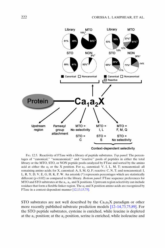
FIG. 12.5. Reactivity of FTase with a library of peptide substrates. Top panel: The percen-
tages of ‘‘canonical,’’ ‘‘noncanonical,’’ and ‘‘reactive’’ pools of peptides in either the total
library or the MTO, STO, or NON peptide pools catalyzed by FTase and sorted by the amino
acid at either the a2 or the X position. For a2, canonical: V, I, L, M, T; noncanonical: all
remaining amino acids; for X, canonical: A, S, M, Q, F; reactive: C, N, T; and noncanonical: I,
L, R, Y, D, V, E, G, H, K, P, W. An asterisk (*) represents percentages which are statistically
different (p<0.02) as compared to the library. Bottom panel: FTase sequence preferences for
MTO and STO substrates at the a1, a2, and X positions. Upstream region selectivity can include
residues that form a flexible linker region. The a2 and X position amino acids are recognized by
FTase in a context-dependent manner [12,13,15,75].
222 CORISSA L. LAMPHEAR, ET AL.
STO substrates are not well described by the Ca1a2X paradigm or othermore recently published substrate prediction models [12–14,73,75,89]. Forthe STO peptide substrates, cysteine is enriched, while leucine is depletedat the a1 position; at the a2 position, serine is enriched, while isoleucine and
12. IDENTIFICATION OF PRENYLTRANSFERASE SUBSTRATES 223
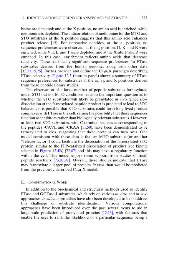
lysine are depleted; and at the X position, no amino acid is enriched, whilemethionine is depleted. The anticorrelation of methionine for theMTO andSTO substrates at the X position suggests that this amino acid enhancesproduct release [15]. For unreactive peptides, at the a1 position, nosequence preferences were observed; at the a2 position, D, K, and R wereenriched, while V, I, L, and T were depleted; and at the X site, P and R wereenriched. In this case, enrichment reflects amino acids that decreasereactivity. These statistically significant sequence preferences for FTasesubstrates derived from the human genome, along with other data[12,13,15,75], further broaden and define the Ca1a2X paradigm describingFTase selectivity. Figure 12.5 (bottom panel) shows a summary of FTasesequence preferences for substrates at the a1, a2, and X positions derivedfrom these peptide library studies.
The observation of a large number of peptide substrates farnesylatedunder STO but not MTO conditions leads to the important question as towhether the STO substrates will likely be prenylated in vivo. Since slowdissociation of the farnesylated-peptide product is predicted to lead to STObehavior, it is possible that STO substrates could form long-lived productcomplexes with FTase in the cell, raising the possibility that these sequencesfunction as inhibitors rather than biologically relevant substrates. However,at least two STO substrates, with C-terminal sequences corresponding tothe peptides -CAVL and -CKAA [11,50], have been demonstrated to befarnesylated in vivo, suggesting that these proteins can turn over. Onemodel consistent with these data is that an MTO substrate (or another‘‘release factor’’) could facilitate the dissociation of the farnesylated-STOprotein, similar to the FPP-catalyzed dissociation of product (see kineticscheme in Figure 12.4B) [72,87] and this may have a regulatory functionwithin the cell. This model enjoys some support from studies of smallpeptide reactivity [73,87,92]. Overall, these studies indicate that FTasemay farnesylate a larger pool of proteins in vivo than would be predictedfrom the previously described Ca1a2X model.
E. COMPUTATIONAL WORK
In addition to the biochemical and structural methods used to identifyFTase and GGTase-I substrates, which rely on various in vitro and in vivoapproaches, in silico approaches have also been developed to help addressthis challenge of substrate identification. Various computationalapproaches have been introduced over the past several years to aid inlarge-scale prediction of prenylated proteins [12,13], with features thatenable the user to rank the likelihood of a particular sequence being a
224 CORISSA L. LAMPHEAR, ET AL.
substrate for FTase, GGTase-I, both, or neither. The methods are itera-tively improved by continuously incorporating new biochemical data as itbecomes available to further refine the predictive power of the computa-tional analyses.
Several computer algorithms have been developed to help predictprotein substrates for mammalian prenyltransferases. Among predictionsoftware, one of the first tools built was Prosite protocol PS00294 whichused the consensus pattern C-{DENQ}-[LIVM]-x> (http://www.expasy.org/prosite/) [94]. However, this tool is unable to distinguish betweenFTase and GGTase-I substrates, nor does it predict prenylation byGGTase-II enzyme. Crystallographic analysis of FTase and GGTase-Icomplexed with eight cross-reactive substrates used interactions with thebinding pocket in the structures of the enzyme–substrate complexes todraw inferences about FTase and GGTase-I substrate recognition ele-ments [14]. The most significant drawback of this approach is that it onlyidentifies a subset of verified substrates, missing key substrate–proteininteractions that are not covered by the peptide diversity in the availablecrystal structures.
PrePS is the most recent algorithm developed to predict prenyltransfer-ase substrates [12]. To define this algorithm, the authors built a learning setof known and homologous substrates (defined by specific rules) whichresulted in a set of 692 FTase and 486 GGTase-I substrates. One of thedifficulties in predicting prenylation substrates is the inherent complexity ofsubstrate recognition motifs, which may extend beyond the Ca1a2X box toinclude the upstream region of the protein. To address this additionalcomplexity, the authors of PrePS included an 11-amino acid upstreamregion of the Ca1a2X motif to refine their algorithm, as well as expandingthe list of acceptable amino acids for the Ca1a2X motif. This upstreamregion typically consists of a flexible linker region that often has a compo-sitional bias toward small or hydrophilic amino acids. Using this learningset, the PrePS algorithm defines a set of rules that is used to predict thelikelihood of a 15-amino acid sequence being an FTase, GGTase-I, and/orGGTase-II substrate. In cross-validation experiments, they were able toestablish 92.6% and 98.6% true positive rate for FTase and GGTase-Isubstrates, respectively, with false positive rates of 0.11% and 0.02% forFTase and GGTase-I, respectively. Consistent with this, analysis of thesubstrates identified in the peptide library screen described above usingthe PrePS algorithm yielded a very low number of false positive results [15].However, the PrePS analysis led to a large number of false negative pre-dictions, around 40%, indicating that the PrePS algorithm potentiallymisses a large number of prenyltransferase substrates.
12. IDENTIFICATION OF PRENYLTRANSFERASE SUBSTRATES 225
IV. Prenylation inPathogenicOrganisms: Structural andBiochemical Insights
A. C. ALBICANS PRENYLTRANSFERASES
Although most studies to date have focused on mammalian prenyltrans-ferases, lower eukaryotic organisms, such as yeast, also contain FTase andGGTase-I and inhibition of these enzymes has been proposed as a possibletreatment for pathogenic yeast infection [36–38,95–97]. C. albicans is adimorphic yeast that is a major opportunistic human fungal pathogenwhich causes life-threatening infection in many immunocompromisedpatients. Although there are currently therapies on the market that combatC. albicans infections, resistance is always a concern, and thus novel thera-pies with unique mechanisms of action are highly sought after. IdentifyingC. albicans prenylation substrates and mining the differences between thesubstrate recognition of the human andC. albicans prenyltransferases couldpave a way for novel drug candidates against this infection.
The first report of sequencing, cloning, and purification of C. albicansGGTase-I was published in 1999 by the Mazur group [37]. The a and bsubunits of GGTase-I are 30% identical to their mammalian homologues,with conservation of the zinc ligands and substrate-binding residues of the bsubunit. Based on sequence alignment and mammalian FTase crystal struc-ture, His231, Arg296, and Lys299 were identified as prenyl donor contacts;and Arg160, Ala165, and Gly344 were implicated in protein substratebinding in C. albicans GGTase. In 2008, the Beese group solved the crystalstructure of C. albicans GGTase-I complexed with GGPP at 1.58 A resolu-tion [98]. A noted difference between the mammalian and C. albicansGGTase-I structures is the size of the ‘‘exit groove.’’ In the structure ofrat GGTase complexed with prenylated peptide and GGPP, reflecting anintermediate in the dissociation of prenylated product during the catalyticcycle (Figure 12.4B), the prenyl group of the product binds in the exitgroove while the new GGPP molecule binds in the active site pocket [71].In the C. albicans crystal structure, the exit groove is narrowed, suggestingthat the geranylgeranyl group of the product could not be accommodated inthe same binding mode as observed for the mammalian GGTase-I.Although no peptide substrate was bound in the solved crystal structureof C. albicansGGTase-I, comparison of the putative Ca1a2X binding site ofC. albicans GGTase-I to that of the mammalian enzyme revealed signifi-cant variation in the identities of amino acids, and most of these arenonconservative changes. This could suggest that the C. albicansGGTase-I has a different substrate recognition pattern than the mamma-lian enzyme.
226 CORISSA L. LAMPHEAR, ET AL.
Biochemical studies of a small number of peptide substrates have beencarried out to investigate fungal prenyltransferase substrate specificity. TheMazur group initially measured the reactivity of partially purified C. albi-cans GGTase-I and FTase with seven peptides containing a Ca1a2Xsequence (CVIL, CVVL, CTIL, CAIL, CVLM, CVLS, and CVIA) fromprenylated proteins in either C. albicans or Saccharomyces cerevisiae withthe upstream sequence from S. cerevisiae Ras [37]. Five of these substrateswere geranylgeranylated upon incubation with C. albicans GGTase-I,although the KM values were considerably higher than those measured formammalian GGTase-I [37]. The two inactive sequences, CVLS and CVIA,are prototypical mammalian FTase substrates. Additionally, C. albicansFTase catalyzed farnesylation of all seven Ca1a2X sequences. Unexpect-edly, the FTase-catalyzed prenylation of the -CaaL substrates was compa-rable to that of GGTase-I, suggesting both that C. albicans FTase has broadsubstrate specificity and that there is significant overlap in substrate speci-ficity between the two enzymes. In a follow-up study in 2000, Mazur andcoworkers explored the function of a polybasic sequence upstream of theCa1a2X using biotinylated peptide substrates [35]. Overall, the upstreampolybasic region had little impact on kcat values, while the KM valuesdecreased; for example, addition of multiple lysine residues lowered thevalue of KM �15-fold for the CVIL and CAIL peptide sequences. Inaddition, the polybasic region broadened the substrate pool for GGTase,enhancing reactivity for peptides, like -CVIM, that contain an amino acidother than L as the terminal amino acid. In general, peptides ending inresidues other than leucine are poor substrates for C. albicans GGTase-Iunless they contain an upstream polybasic region.
Substrate specificity of the fungal prenyltransferases was also investi-gated in vivo. FTase and GGTase-I were shown to be essential forC. albicans growth as determined by knockout of the shared a subunit[36]. The b subunits of both FTase and GGTase-I are essential for growthof S. cerevisiae but not C. albicans, possibly due to cross-prenylation[99,100]. In S. cerevisiae, loss of FTase can be overcome by growth atlower temperatures, and lack of GGTase-I activity can be circumventedby overexpression of the GGTase-I substrates Rho1p and Cdc42p [99,101].To address the issue of in vivo cross-prenylation in C. albicans, the prenyla-tion status of the GGTase-I substrates Rho1p (-CVVL) and Cdc42p(-CTIL) was assessed in the absence of GGTase-I protein expression [38].Under such conditions, Rho1p and Cdc42p were prenylated, as determinedby localization of the proteins in the membrane fraction of the cells. Thisreaction was presumably catalyzed by FTase. These data suggest that aprenyltransferase inhibitor with dual specificity for FTase and GGTase-Iwould be necessary to obtain in vivo antifungal activity against C. albicans.
12. IDENTIFICATION OF PRENYLTRANSFERASE SUBSTRATES 227
B. L. PNEUMOPHILA: UTILIZING HOST PRENYLTRANSFERASE MACHINERY
Recently, prenylation of proteins encoded by aGram-negative pathogenicbacterium,L. pneumophila, has been discovered [41,42]. In this organism, theDot/Icm secretion system of the bacterium injects into the host cell �200effectors that modulate and reprogram a number of host cellular processes toboth allow bacterial proliferation aswell as to induce host cell apoptosis [102].One such effector, the F-box effector Ankyrin B (AnkB) protein, is essentialfor proliferation ofL. pneumophila in mammalian cells and formanifestationof pulmonary disease in the mouse model of Legionnaires’ disease [103].AnkB functions as a scaffold for the docking of polyubiquitinated proteinsto the Legionella-containing vacuole (LCV) membrane to enable bacterialintravacuolar proliferation inmacrophages [104,105]. Recent work has shownthat prenylation of the AnkB protein is essential for anchoring the protein tothe LCV membrane. Further, prenylation is catalyzed by the host pathway[42], as evidenced by abolishment of membrane localization of AnkB in thepresence of mammalian FTase and GGTase-I inhibitors and FTase-specificRNAi treatment. L. pneumophila does not encode any sequences homolo-gous to the mammalian Ca1a2X-motif specific prenyltransferases in itsgenome and thus proteins that require lipidation for their biological functionmust be translocated into the host cytosol by the Dot/Icm type IV secretionsystem for modification. Addition of the Ca1a2X motif of L. pneumophilaAnkB (CLVC) to a cytosolic protein is sufficient to localize the protein to themembrane, presumably due to prenylation.Recently, 10 proteins containing aC-terminal Ca1a2X motif have been identified in several L. pneumophilastrains, including eight unique Ca1a2X sequences (such as CLVC for AnkB)[41]. Using GFP-Ca1a2X protein fusions, the membrane localization of thesefusion proteins were verified, presumably due to prenylation of the Ca1a2Xsequence. Consistent with this, most of these Ca1a2X sequences had previ-ously been identified as a substrate for either mammalian FTase (CLVC,CVIS) or both FTase and GGTase-I (CSIL, CNLL, CVLM, CTIM, CSIL).Overall, these studies suggest that prenylation of L. pneumophila effectorproteins is carried out by host farnesylation machinery. Blocking the mem-brane localization of these effector proteins by prenyltransferase inhibitors,and thus potentially disrupting effector protein function, may possibly lead toa novel approach for antibacterial treatment.
V. Conclusions
Multiple approaches have been employed to identify proteins that areprenylated in vivo, catalyzed by FTase and GGTase-I. Although prenyl-transferase substrates can be identified and studied one at time, FPP and
228 CORISSA L. LAMPHEAR, ET AL.
GGPP analogs are being developed to allow detection of farnesylated andgeranylgeranylated proteins in a ‘‘high-throughput’’ fashion. Further, struc-tural and structure–function studies have provided insight into the interac-tions between substrates and the FTase and GGTase enzymes that areimportant for molecular recognition. Peptide library studies have supplieddata regarding the amino acid composition of substrates and nonsubstratepools, allowing the use of statistical analysis to determine patterns of FTaserecognition and indicating that the Ca1a2X paradigm does not sufficientlydescribe the recognition of substrates by FTase. Further, these studies haveuncovered the puzzle of the reactivity of STO substrates. Computationalstudies have allowed prediction of substrates in vivo, based upon both theC-terminus of the protein as well as the upstream region. All these studies inconcert have helped to define the current pool of proteins known to beprenyltransferase substrates. Table S1 in reference [15] is a list of knownprenylated substrates, identified by a variety of methods, highlighting therapid identification ofmany proteins in the ‘‘prenylome.’’ Throughapplicationand refinement of the approaches detailed in this chapter, identification of theentire prenylated proteome appears to a realistic goal in the next decade, if notsooner. Definition of the prenylated proteome will be essential to betterunderstand the roles of prenylation in cellular signaling, disease processes,and the complex array of posttranslational modifications within the cell.
ACKNOWLEDGMENTS
We thank members of the Fierke laboratory for helpful comments and suggestions on the
chapter. We especially thank Katherine A. Hicks, Heather L. Hartman, RebekahA. Kelly, and
Terry J. Watt for their work on the peptide library studies. This work was supported by
National Institutes of Health (NIH) grant GM40602 (C. A. F.), PSTP training grant
GM07767 (E. A. Z.), and NIH postdoctoral fellowship GM78894 (J. L. H.).
REFERENCES
1. Zhang, F.L., and Casey, P.J. (1996). Protein prenylation: molecular mechanisms and
functional consequences. Annu Rev Biochem 65:241–269.
2. Benetka, W., Koranda, M., and Eisenhaber, F. (2006). Protein prenylation: an (almost)
comprehensive overview on discovery history, enzymology, and significance in physiology
and disease. Monatsh Chem Chem Mon 137:1241–1281.
3. Marshall, C.J. (1993). Protein prenylation: a mediator of protein–protein interactions.
Science 259:1865–1866.
4. Casey, P.J. (1994). Lipid modifications of G proteins. Curr Opin Cell Biol 6:219–225.
5. Casey, P.J., and Seabra, M.C. (1996). Protein prenyltransferases. J Biol Chem
271:5289–5292.
12. IDENTIFICATION OF PRENYLTRANSFERASE SUBSTRATES 229
6. Winter-Vann, A.M., and Casey, P.J. (2005). Post-prenylation-processing enzymes as new
targets in oncogenesis. Nat Rev Cancer 5:405–412.
7. Barrowman, J., and Michaelis, S. (2009). ZMPSTE24, an integral membrane zinc metal-
loprotease with a connection to progeroid disorders. Biol Chem 390:761–773.
8. Trueblood, C.E., Boyartchuk, V.L., Picologlou, E.A., Rozema, D., Poulter, C.D., and
Rine, J. (2000). The CaaX proteases, Afc1p and Rce1p, have overlapping but distinct
substrate specificities. Mol Cell Biol 20:4381–4392.
9. Seabra, M.C., Reiss, Y., Casey, P.J., Brown, M.S., and Goldstein, J.L. (1991). Protein
farnesyltransferase and geranylgeranyltransferase share a common alpha subunit. Cell
65:429–434.
10. Lane, K.T., and Beese, L.S. (2006). Thematic review series: lipid posttranslational mod-
ifications. Structural biology of protein farnesyltransferase and geranylgeranyltransferase
type I. J Lipid Res 47:681–699.
11. Kho, Y., et al. (2004). A tagging-via-substrate technology for detection and proteomics of
farnesylated proteins. Proc Natl Acad Sci USA 101:12479–12484.
12. Maurer-Stroh, S., and Eisenhaber, F. (2005). Refinement and prediction of protein
prenylation motifs. Genome Biol 6:R55.
13. Maurer-Stroh, S., Koranda, M., Benetka, W., Schneider, G., Sirota, F.L., and
Eisenhaber, F. (2007). Towards complete sets of farnesylated and geranylgeranylated
proteins. PLoS Comput Biol 3:e66.
14. Reid, T.S., Terry, K.L., Casey, P.J., and Beese, L.S. (2004). Crystallographic analysis of
CaaX prenyltransferases complexed with substrates defines rules of protein substrate
selectivity. J Mol Biol 343:417–433.
15. Hougland, J.L., Hicks, K.A., Hartman, H.L., Kelly, R.A., Watt, T.J., and Fierke, C.A.
(2010). Identification of novel peptide substrates for protein farnesyltransferase reveals
two substrate classes with distinct sequence selectivities. J Mol Biol 395:176–190.
16. Samuel, F., and Hynds, D.L. (2010). RHO GTPase signaling for axon extension: is
prenylation important? Mol Neurobiol 42:133–142.
17. Rusinol, A.E., and Sinensky, M.S. (2006). Farnesylated lamins, progeroid syndromes and
farnesyl transferase inhibitors. J Cell Sci 119:3265–3272.
18. Basso, A.D., Kirschmeier, P., and Bishop, W.R. (2006). Lipid posttranslational modifica-
tions. Farnesyl transferase inhibitors. J Lipid Res 47:15–31.
19. Sousa, S.F., Fernandes, P.A., and Ramos, M.J. (2008). Farnesyltransferase inhibitors:
a detailed chemical view on an elusive biological problem. Curr Med Chem
15:1478–1492.
20. Young, S.G., Meta, M., Yang, S.H., and Fong, L.G. (2006). Prelamin A farnesylation and
progeroid syndromes. J Biol Chem 281:39741–39745.
21. Nallan, L., et al. (2005). Protein farnesyltransferase inhibitors exhibit potent antimalarial
activity. J Med Chem 48:3704–3713.
22. Sepp-Lorenzino, L., et al. (1995). A peptidomimetic inhibitor of farnesyl:protein transfer-
ase blocks the anchorage-dependent and -independent growth of human tumor cell lines.
Cancer Res 55:5302–5309.
23. Nonaka, M., et al. (2009). Role for protein geranylgeranylation in adult T-cell leukemia
cell survival. Exp Cell Res 315:141–150.
24. Chiba, Y., Sato, S., Hanazaki, M., Sakai, H., and Misawa, M. (2009). Inhibition of
geranylgeranyltransferase inhibits bronchial smooth muscle hyperresponsiveness in
mice. Am J Physiol Lung Cell Mol Physiol 297:L984–L991.
25. Ridker, P.M., et al. (2008). Rosuvastatin to prevent vascular events in men and women
with elevated c-reactive protein. N Engl J Med 359(21):2195–2207.
26. Casey, P.J. (1995). Protein lipidation in cell signaling. Science 268:221–225.
230 CORISSA L. LAMPHEAR, ET AL.
27. Caplin, B.E., Hettich, L.A., and Marshall, M.S. (1994). Substrate characterization of the
Saccharomyces cerevisiae protein farnesyltransferase and type-I protein geranylgeranyl-
transferase. Biochim Biophys Acta 1205:39–48.
28. Omer, C.A., et al. (1993). Characterization of recombinant human farnesyl-protein trans-
ferase: cloning, expression, farnesyl diphosphate binding, and functional homology with
yeast prenyl-protein transferases. Biochemistry 32:5167–5176.
29. Reiss, Y., Stradley, S.J., Gierasch, L.M., Brown, M.S., and Goldstein, J.L. (1991).
Sequence requirement for peptide recognition by rat brain p21ras protein farnesyltrans-
ferase. Proc Natl Acad Sci USA 88:732–736.
30. Moores, S.L., et al. (1991). Sequence dependence of protein isoprenylation. J Biol Chem
266:14603–14610.
31. Hicks, K.A., Hartman, H.L., and Fierke, C.A. (2005). Upstream polybasic region in
peptides enhances dual specificity for prenylation by both farnesyltransferase and ger-
anylgeranyltransferase type I. Biochemistry 44:15325–15333.
32. Gelb, M.H., et al. (2003). Protein farnesyl and N-myristoyl transferases: piggy-back
medicinal chemistry targets for the development of antitrypanosomatid and antimalarial
therapeutics. Mol Biochem Parasitol 126:155–163.
33. Bulbule, V.J., Rivas, K., Verlinde, C.L., Van Voorhis, W.C., and Gelb, M.H. (2008).
2-Oxotetrahydroquinoline-based antimalarials with high potency and metabolic stability.
J Med Chem 51:384–387.
34. Fletcher, S., et al. (2008). Potent, plasmodium-selective farnesyltransferase inhibitors that
arrest the growth of malaria parasites: structure–activity relationships of ethylenedia-
mine-analogue scaffolds and homology model validation. J Med Chem 51:5176–5197.
35. Smalera, I., Williamson, J.M., Baginsky, W., Leiting, B., andMazur, P. (2000). Expression
and characterization of protein geranylgeranyltransferase type I from the pathogenic
yeast Candida albicans and identification of yeast selective enzyme inhibitors. Biochim
Biophys Acta 1480:132–144.
36. Song, J.L., and White, T.C. (2003). RAM2: an essential gene in the prenylation pathway
of Candida albicans. Microbiology 149:249–259.
37. Mazur, P., et al. (1999). Purification of geranylgeranyltransferase I from Candida albicans
and cloning of the CaRAM2 and CaCDC43 genes encoding its subunits. Microbiology
145(Pt 5):1123–1135.
38. Kelly, R., et al. (2000). Geranylgeranyltransferase I of Candida albicans: null mutants or
enzyme inhibitors produce unexpected phenotypes. J Bacteriol 182:704–713.
39. Eastman, R.T., Buckner, F.S., Yokoyama, K., Gelb, M.H., and Van Voorhis, W.C. (2006).
Thematic review series: lipid posttranslational modifications. Fighting parasitic disease by
blocking protein farnesylation. J Lipid Res 47:233–240.
40. Eastman, R.T., et al. (2007). Resistance mutations at the lipid substrate binding site of
Plasmodium falciparum protein farnesyltransferase. Mol Biochem Parasitol 152:66–71.
41. Ivanov, S.S., Charron, G., Hang, H.C., and Roy, C.R. (2010). Lipidation by the host
prenyltransferase machinery facilitates membrane localization of Legionella pneumo-
phila effector proteins. J Biol Chem 285:34686–34698.
42. Price, C.T., Al-Quadan, T., Santic, M., Jones, S.C., and Abu Kwaik, Y. (2010). Exploita-
tion of conserved eukaryotic host cell farnesylation machinery by an F-box effector of
Legionella pneumophila. J Exp Med 207:1713–1726.
43. Hancock, J.F. (1995). Reticulocyte lysate assay for in vitro translation and posttransla-
tional modification of Ras proteins. Methods Enzymol 255:60–65.
44. Wilson, A.L., and Maltese, W.A. (1995). Coupled Translation/Prenylation of Rab
Proteins in-Vitro. Lipid Modifications of Proteins. Academic Press Inc., San Diego,
Vol. 250, pp. 79–91.
12. IDENTIFICATION OF PRENYLTRANSFERASE SUBSTRATES 231
45. Gibbs, B.S., Zahn, T.J., Mu, Y., Sebolt-Leopold, J.S., and Gibbs, R.A. (1999). Novel
farnesol and geranylgeraniol analogues: a potential new class of anticancer agents
directed against protein prenylation. J Med Chem 42:3800–3808.
46. Peter, M., Chavrier, P., Nigg, E.A., and Zerial, M. (1992). Isoprenylation of Rab proteins
on structurally distinct cysteine motifs. J Cell Sci 102:857–865.
47. Corsini, A., Farnsworth, C.C., McGeady, P., Gelb, M.H., and Glomset, J.A. (1999).
Incorporation of radiolabeled prenyl alcohols and their analogs into mammalian cell
proteins. A useful tool for studying protein prenylation. Methods Mol Biol 116:125–144.
48. Andres, D.A., Crick, D.C., Finlin, B.S., andWaechter, C.J. (1999). Rapid identification of
cysteine-linked isoprenyl groups by metabolic labeling with [3H]farnesol and [3H]gera-
nylgeraniol. Methods Mol Biol 116:107–123.
49. Benetka, W., Koranda, M., Maurer-Stroh, S., Pittner, F., and Eisenhaber, F. (2006).
Farnesylation or geranylgeranylation? Efficient assays for testing protein prenylation
in vitro and in vivo BMC Biochem 7:6.
50. Liu, Z.H., et al. (2009). Membrane-associated farnesylated UCH-L1 promotes alpha-
synuclein neurotoxicity and is a therapeutic target for Parkinson’s disease. Proc Natl
Acad Sci USA 106:4635–4640.
51. Baron, R., et al. (2000). RhoB prenylation is driven by the three carboxyl-terminal amino
acids of the protein: evidenced in vivo by an anti-farnesyl cysteine antibody. Proc Natl
Acad Sci USA 97:11626–11631.
52. Lin, H.P., Hsu, S.C., Wu, J.C., Sheen, I.J., Yan, B.S., and Syu, W.J. (1999). Localization of
isoprenylated antigen of hepatitis delta virus by anti-farnesyl antibodies. J Gen Virol 80
(Pt 1):91–96.
53. Liu, X.H., Suh, D.Y., Call, J., and Prestwich, G.D. (2004). Antigenic prenylated peptide
conjugates and polyclonal antibodies to detect protein prenylation. Bioconjug Chem
15:270–277.
54. Troutman, J.M., Roberts, M.J., Andres, D.A., and Spielmann, H.P. (2005). Tools to
analyze protein farnesylation in cells. Bioconjug Chem 16:1209–1217.
55. Nguyen, U.T., et al. (2009). Analysis of the eukaryotic prenylome by isoprenoid affinity
tagging. Nat Chem Biol 5:227–235.
56. Nguyen, U.T.T., Goody, R.S., and Alexandrov, K. (2010). Understanding and exploiting
protein prenyltransferases. Chembiochem 11:1194–1201.
57. Chehade, K.A., Andres, D.A., Morimoto, H., and Spielmann, H.P. (2000). Design and
synthesis of a transferable farnesyl pyrophosphate analogue to Ras by protein farnesyl-
transferase. J Org Chem 65:3027–3033.
58. Onono, F.O., et al. (2010). A tagging-via-substrate approach to detect the farnesylated
proteome using two-dimensional electrophoresis coupled withWestern blotting.Mol Cell
Proteomics 9:742–751.
59. Saxon, E., and Bertozzi, C.R. (2000). Cell surface engineering by a modified Staudinger
reaction. Science 287:2007–2010.
60. Moses, J.E., and Moorhouse, A.D. (2007). The growing applications of click chemistry.
Chem Soc Rev 36:1249–1262.
61. Chan, L.N., et al. (2009). A novel approach to tag and identify geranylgeranylated
proteins. Electrophoresis 30:3598–3606.
62. Berry, A.F., et al. (2010). Rapid multilabel detection of geranylgeranylated proteins by
using bioorthogonal ligation chemistry. Chembiochem 11:771–773.
63. Charron, G., Tsou, L.K., Maguire, W., Yount, J.S., and Hang, H.C. (2011). Alkynyl-
farnesol reporters for detection of protein S-prenylation in cells. Mol Biosyst 7:67–73.
64. DeGraw, A.J., et al. (2010). Evaluation of alkyne-modified isoprenoids as chemical
reporters of protein prenylation. Chem Biol Drug Des 76:460–471.
232 CORISSA L. LAMPHEAR, ET AL.
65. Labadie, G.R., Viswanathan, R., and Poulter, C.D. (2007). Farnesyl diphosphate analo-
gues with omega-bioorthogonal azide and alkyne functional groups for protein farnesyl
transferase-catalyzed ligation reactions. J Org Chem 72:9291–9297.
66. Reigard, S.A., Zahn, T.J., Haworth, K.B., Hicks, K.A., Fierke, C.A., and Gibbs, R.A.
(2005). Interplay of isoprenoid and peptide substrate specificity in protein farnesyltrans-
ferase. Biochemistry 44:11214–11223.
67. Krzysiak, A.J., et al. (2007). Combinatorial modulation of protein prenylation.ACSChem
Biol 2:385–389.
68. Troutman, J.M., Subramanian, T., Andres, D.A., and Spielmann, H.P. (2007). Selective
modification of CaaX peptides with ortho-substituted anilinogeranyl lipids by protein
farnesyl transferase: competitive substrates and potent inhibitors from a library of farne-
syl diphosphate analogues. Biochemistry 46:11310–11321.
69. Terry, K.L., Casey, P.J., and Beese, L.S. (2006). Conversion of protein farnesyltransferase
to a geranylgeranyltransferase. Biochemistry 45:9746–9755.
70. Luckman, S.P., Hughes, D.E., Coxon, F.P., Russell, R.G.G., and Rogers, M.J. (1998).
Nitrogen-containing bisphosphonates inhibit the mevalonate pathway and prevent post-
translational prenylation of GTP-binding proteins, including Ras. J Bone Miner Res
13:581–589.
71. Taylor, J.S., Reid, T.S., Terry, K.L., Casey, P.J., and Beese, L.S. (2003). Structure of
mammalian protein geranylgeranyltransferase type-I. EMBO J 22:5963–5974.
72. Long, S.B., Casey, P.J., and Beese, L.S. (2002). Reaction path of protein farnesyltransfer-
ase at atomic resolution. Nature 419:645–650.
73. Hartman, H.L., Hicks, K.A., and Fierke, C.A. (2005). Peptide specificity of protein
prenyltransferases is determined mainly by reactivity rather than binding affinity. Bio-
chemistry 44:15314–15324.
74. Krzysiak, A.J., Aditya, A.V., Hougland, J.L., Fierke, C.A., and Gibbs, R.A. (2009).
Synthesis and screening of a CaaL peptide library versus FTase reveals a surprising
number of substrates. Bioorg Med Chem Lett 20:767–770.
75. Hougland, J.L., Lamphear, C.L., Scott, S.A., Gibbs, R.A., and Fierke, C.A. (2009).
Context-dependent substrate recognition by protein farnesyltransferase. Biochemistry
48:1691–1701.
76. Goldstein, J.L., Brown, M.S., Stradley, S.J., Reiss, Y., and Gierasch, L.M. (1991). Non-
farnesylated tetrapeptide inhibitors of protein farnesyltransferase. J Biol Chem
266:15575–15578.
77. Pompliano, D.L., Gomez, R.P., and Anthony, N.J. (1992). Intramolecular fluorescence
enhancement—a continuous assay of ras farnesyl—protein transferase. J Am Chem Soc
114:7945–7946.
78. Hightower, K.E., Huang, C.C., Casey, P.J., and Fierke, C.A. (1998). H-Ras peptide and
protein substrates bind protein farnesyltransferase as an ionized thiolate. Biochemistry
37:15555–15562.
79. Cassidy, P.B., Dolence, J.M., and Poulter, C.D. (1995). Continuous fluorescence assay for
protein prenyltransferases. Methods Enzymol 250:30–43.
80. Boutin, J.A., et al. (1999). Investigation of S-farnesyl transferase substrate specificity with
combinatorial tetrapeptide libraries. Cell Signal 11:59–69.
81. Krzysiak, A.J., Scott, S.A., Hicks, K.A., Fierke, C.A., and Gibbs, R.A. (2007). Evaluation
of protein farnesyltransferase substrate specificity using synthetic peptide libraries.
Bioorg Med Chem Lett 17:5548–5551.
82. Fersht, A. (1999). Structure and Mechanism in Protein Science. W.H. Freeman and
Company, New York, NY.
12. IDENTIFICATION OF PRENYLTRANSFERASE SUBSTRATES 233
83. Pompliano, D.L., Schaber, M.D., Mosser, S.D., Omer, C.A., Shafer, J.A., and Gibbs, J.B.
(1993). Isoprenoid diphosphate utilization by recombinant human farnesyl:protein trans-
ferase: interactive binding between substrates and a preferred kinetic pathway. Biochem-
istry 32:8341–8347.
84. Zimmerman, K.K., Scholten, J.D., Huang, C.C., Fierke, C.A., and Hupe, D.J. (1998).
High-level expression of rat farnesyl:protein transferase in Escherichia coli as a transla-
tionally coupled heterodimer. Protein Expr Purif 14:395–402.
85. Pais, J.E., Bowers, K.E., Stoddard, A.K., and Fierke, C.A. (2005). A continuous fluores-
cent assay for protein prenyltransferases measuring diphosphate release. Anal Biochem
345:302–311.
86. Furfine, E.S., Leban, J.J., Landavazo, A., Moomaw, J.F., and Casey, P.J. (1995). Protein
farnesyltransferase: kinetics of farnesyl pyrophosphate binding and product release.
Biochemistry 34:6857–6862.
87. Tschantz, W.R., Furfine, E.S., and Casey, P.J. (1997). Substrate binding is required for
release of product from mammalian protein farnesyltransferase. J Biol Chem
272:9989–9993.
88. Bowers, K.E., and Fierke, C.A. (2004). Positively charged side chains in protein farnesyl-
transferase enhance catalysis by stabilizing the formation of the diphosphate leaving
group. Biochemistry 43:5256–5265.
89. Long, S.B., Hancock, P.J., Kral, A.M., Hellinga, H.W., and Beese, L.S. (2001). The crystal
structure of human protein farnesyltransferase reveals the basis for inhibition by CaaX
tetrapeptides and their mimetics. Proc Natl Acad Sci USA 98:12948–12953.
90. Pickett, J.S., et al. (2003). Kinetic studies of protein farnesyltransferase mutants establish
active substrate conformation. Biochemistry 42:9741–9748.
91. Pais, J.E., Bowers, K.E., and Fierke, C.A. (2006). Measurement of the alpha-secondary
kinetic isotope effect for the reaction catalyzed by mammalian protein farnesyltransfer-
ase. J Am Chem Soc 128:15086–15087.
92. Troutman, J.M., Andres, D.A., and Spielmann, H.P. (2007). Protein farnesyl transferase
target selectivity is dependent upon peptide stimulated product release. Biochemistry
46:11299–11309.
93. Spence, R.A., Hightower, K.E., Terry, K.L., Beese, L.S., Fierke, C.A., and Casey, P.J.
(2000). Conversion of Tyr361 beta to Leu in mammalian protein farnesyltransferase
impairs product release but not substrate recognition. Biochemistry 39:13651–13659.
94. Falquet, L., et al. (2002). The PROSITE database, its status in 2002. Nucleic Acids Res
30:235–238.
95. McGeady, P., Logan, D.A., and Wansley, D.L. (2002). A protein-farnesyl transferase
inhibitor interferes with the serum-induced conversion of Candida albicans from a cellu-
lar yeast form to a filamentous form. FEMS Microbiol Lett 213:41–44.
96. Murthi, K.K., Smith, S.E., Kluge, A.F., Bergnes, G., Bureau, P., and Berlin, V. (2003).
Antifungal activity of a Candida albicans GGTase I inhibitor-alanine conjugate. Inhibi-
tion of Rho1p prenylation in C. albicans. Bioorg Med Chem Lett 13:1935–1937.
97. Nishimura, S., et al. (2003). Massadine, a novel geranylgeranyltransferase type I inhibitor
from the marine sponge Stylissa aff. massa. Org Lett 5:2255–2257.
98. Hast, M.A., and Beese, L.S. (2008). Structure of protein geranylgeranyltransferase-I from
the human pathogen Candida albicans complexed with a lipid substrate. J Biol Chem
283:31933–31940.
99. Trueblood, C.E., Ohya, Y., and Rine, J. (1993). Genetic evidence for in vivo cross-
specificity of the CaaX-box protein prenyltransferases farnesyltransferase and geranyl-
geranyltransferase-I in Saccharomyces cerevisiae. Mol Cell Biol 13:4260–4275.
234 CORISSA L. LAMPHEAR, ET AL.
100. He, B., Chen, P., Chen, S.Y., Vancura, K.L., Michaelis, S., and Powers, S. (1991). RAM2,
an essential gene of yeast, and RAM1 encode the two polypeptide components of the
farnesyltransferase that prenylates a-factor and Ras proteins. Proc Natl Acad Sci USA
88:11373–11377.
101. Ohya, Y., Qadota, H., Anraku, Y., Pringle, J.R., and Botstein, D. (1993). Suppression of
yeast geranylgeranyl transferase I defect by alternative prenylation of two target
GTPases, Rho1p and Cdc42p. Mol Biol Cell 4:1017–1025.
102. Zink, S.D., Pedersen, L., Cianciotto, N.P., and Abu-Kwaik, Y. (2002). The Dot/Icm type
IV secretion system of Legionella pneumophila is essential for the induction of apoptosis
in human macrophages. Infect Immun 70:1657–1663.
103. Price, C.T., Al-Khodor, S., Al-Quadan, T., and Abu Kwaik, Y. (2010). Indispensable role
for the eukaryotic-like ankyrin domains of the ankyrin B effector of Legionella pneumo-
phila within macrophages and amoebae. Infect Immun 78:2079–2088.
104. Price, C.T., et al. (2009). Molecular mimicry by an F-box effector of Legionella pneumo-
phila hijacks a conserved polyubiquitination machinery within macrophages and proto-
zoa. PLoS Pathog 5:e1000704.
105. Al-Khodor, S., Price, C.T., Habyarimana, F., Kalia, A., andAbuKwaik, Y. (2008). ADot/
Icm-translocated ankyrin protein of Legionella pneumophila is required for intracellular
proliferation within human macrophages and protozoa. Mol Microbiol 70:908–923.
106. Long, S.B., Casey, P.J., and Beese, L.S. (2000). The basis for K-Ras4B binding specificity
to protein farnesyltransferase revealed by 2 A resolution ternary complex structures.
Structure 8:209–222.
107. Pompliano, D.L., Rands, E., Schaber, M.D., Mosser, S.D., Anthony, N.J., and Gibbs, J.B.
(1992). Steady-state kinetic mechanism of Ras farnesyl:protein transferase. Biochemistry
31:3800–3807.
Related Documents


