1
Mutant telomere sequences lead to impaired chromosome separation
and a unique checkpoint response
Jue Lin*, Dana L. Smith* and Elizabeth H. Blackburn#
University of California, San Francisco
Department of Biochemistry and Biophysics
San Francisco, California 94143-2200
#: corresponding author
tel: 415-476-4912
fax: 415-514-2913
email: [email protected]
*These authors contributed equally to this work.
Running Title: Mutant telomeres impair chromosome separation
MBC in Press, published on January 23, 2004 as 10.1091/mbc.E03-10-0740
Copyright 2004 by The American Society for Cell Biology.

2
Abstract
Mutation of the template region in the RNA component of telomerase can cause incorporation of mutant DNA sequences at telomeres. We made all 63 mutant sequence combinations at template positions 474-476 of the yeast telomerase RNA, TLC1. Mutants contained faithfully incorporated template mutations, as well as misincorporated sequences in telomeres, a phenotype not previously reported for Saccharomyces cerevisiae telomerase template mutants. Although growth rates and telomere profiles varied widely among the tlc1 mutants, chromosome separation and segregation were always aberrant. The mutants showed defects in sister chromatid separation at centromeres as well as telomeres, suggesting activation of a cell cycle checkpoint. Deletion of the DNA damage response genes DDC1, MEC3 or DDC2/SML1 failed to restore chromosome separation in the tlc1 template mutants. These results suggest that mutant telomere sequences elicit a checkpoint that is genetically distinct from those activated by deletion of telomerase or DNA damage.
Introduction
Telomeres, the ends of linear chromosomes, are DNA-protein complexes required
for the complete replication of DNA and for chromosome stability (Blackburn, 2000c;
Blackburn, 2001). The ribonucleoprotein enzyme telomerase adds DNA repeat
sequences to telomeres (Greider and Blackburn, 1985; Greider and Blackburn, 1989).
Deletion of telomerase causes progressive shortening of telomeres in dividing cells and
eventual cellular senescence (Blackburn, 2000b).
Telomerase contains an enzymatically catalytic protein subunit (Est2p in S.
cerivisiae, TERT in other organisms) and an RNA molecule that contains a short
template sequence (TLC1, TER) (Bryan et al., 1998; Counter et al., 1997; Nakamura et
al., 1997; Weinrich et al., 1997). Like other reverse transcriptases, a triad of aspartates in
the conserved reverse transcriptase (RT) domain directly participates in catalysis and is
essential for telomerase activity (Counter et al., 1997). The templating sequence within

3
the telomerase RNA component not only provides the sequence information used by
telomerase to direct synthesis of new telomeric DNA, but also contributes to other
enzymatic properties. In Tetrahymena, single-base mutations in the template cause
primer slippage, loss of fidelity and premature dissociation of product (Gilley and
Blackburn, 1996; Gilley et al., 1995). More dramatically, a three-base change in the
template region of TLC1 in S. cerevisiae telomerase, tlc1-476gug, completely abolishes
enzyme activity in vitro and in vivo (Prescott and Blackburn, 1997). Single or double
point mutations to the same three bases mutated in 476gug still retained in vitro core
telomerase activity, suggesting that the ablation of activity in the triplet gug mutant
results from the combined effect of all three substitutions (Prescott and Blackburn, 2000).
In addition to its templating and enzymatic properties, the telomerase RNA
template also affects telomere length regulation. Telomere length is maintained within a
tight range characteristic of a given organism (Greider, 1996). The TLC1 template
sequence normally directs the synthesis of telomeric TG1-3 repeats, which contain specific
DNA binding sites for proteins involved in telomere length regulation and protection.
Thus, changes within the templating sequence can have a direct influence on the binding
of these proteins and consequently, can influence telomere length and integrity. In S.
cerevisiae, sequence-specific binding of Rap1p to telomeric DNA nucleates a higher
order DNA-protein complex that controls the accessibility of nucleases, telomerase and
proteins involved in recombination and DNA repair. This structure protects the
telomeres from degradation and maintains a tight, species- and strain-specific length
distribution (Hardy et al., 1992; Kyrion et al., 1992; Marcand et al., 1997; Wotton and
Shore, 1997; Krauskopf and Blackburn, 1998).
4
Telomerase RNA template mutants have been expressed and characterized in
budding yeasts, mammalian cells and Tetrahymena (Blackburn, 2000a). They cause
incorporation of mutant telomeric DNA sequences, in some cases, leading to
uncontrolled elongation, degradation and increased single-strandedness at telomeres
(Blackburn, 2000a). In the budding yeast Kluyveromyces lactis, certain template mutant
cells caused “monster cell” phenotypes, characterized by variable and often increased
DNA content in enlarged and misshapen cells (Smith and Blackburn, 1999). In
Tetrahymena, template mutations cause chromosome fusion, failed chromosomal
separation, and accumulation of cells in late anaphase (Kirk et al., 1997). However, it is
not known if mutant telomere sequences are seen as DNA damage or how template
mutations affect cell cycle progression.
Here we systematically examine the effects of mutating a core 3-base region of
the template sequence of S. cerevisiae RNA. Our collection of 63 mutants, together with
wild type, correspond to every possible sequence of template positions 474, 475 and 476
of TLC1. We examined telomere profile and growth phenotype for all mutants and
classified them into six categories. We chose three representative mutants in which
telomeres were respectively long, very short, or extensively degraded; in each mutant, we
examined cell morphology, budding kinetics, chromosome dynamics and activation of
DNA damage checkpoints. Hence, our results indicate that mutant telomeric sequences
elicit a checkpoint response that is distinct from the DNA damage or telomerase loss
checkpoints.

5
Materials and Methods Yeast strain construction
All yeast strains used in this study (except for intermediate strains yEHB5012,
yEHB5013, yEHB5025 and yEHB5026 described below) are listed in Table 1 and were
constructed using standard genetic techniques. Plasmid and oligo sequences are available
upon request. Diploids were isolated on selective media at 23oC and subsequently
sporulated at 23oC. Strain yEHB4003 was made in the S288C genetic background
(Brachmann et al., 1998) and was constructed by disrupting the TLC1 gene with TRP1
and the RAD52 gene with LEU2. The strain carries pRS316TLC1, a CEN/ARS, URA3
plasmid containing the wild type TLC1 with its endogenous promoter and terminator.
For cytological assays, the W303 genetic background was used and template
mutants were derived from yEHB5001 or yEHB5004 in which chromosome IV was
marked as previously described (Straight et al., 1996) either 12kb from the centromere
(yEHB5001) or 100kb from the telomere (yEHB5004) with 256 tandem repeats of the
lactose repressor operator sequence. Both strains contain copper-inducible pCUP1-
GFP12-lacI12::HIS3. The strains were modified for these experiments in three steps:
First, the HIS3 marker was converted to URA3 using pDS317 to create strains yEHB5012
from yEHB5001 (centromere-marked) and yEHB5013 from yEHB5004 (telomere-
marked). Next, TLC1 was expressed using pRS317(LYS2) while the endogenous TLC1
was deleted and marked with KAN in yEHB5012(cen) and yEHB5013(tel) through PCR
integration to make yEHB5025(cen) and yEHB5026(tel). The integration product was
made using primers oEHB4075 and oEHB4076 to PCR amplify pFA6a-kanMX6
(Longtine et al., 1998). Finally, the tlc1 template mutations, tlc1aCA (D), tlc1Cuc(E) and
6
tlc1Cgg(SS), were introduced on pRS313. Template mutants were passaged six times
after counterselection of TLC1. Cells from the sixth passage were used for subsequent
analyses or further genetic manipulation.
yEHB5029 (cdc13-1) was made by crossing yEHB5025 with yEHB5023. Strains
yEHB5076 and yEHB5077 (top2-4) were a gift from N. Bhalla (University of California,
San Francisco, CA). Centromere-marked (yEHB5092) or telomere-marked (yEHB5094)
strains of cdc13-5 were made by disruption of CDC13 in yEHB5025(cen) and
yEHB5026(tel), using pVL1215 (pEHB5005), a gift of V. Lundblad (Baylor College of
Medicine, Houston, TX). In yEHB5056 (tlc1(D)) TLC1 was deleted and marked with
KAN through PCR integration, using primers oEHB4075 and oEHB4076 to PCR amplify
pFA6a-kanMX6 (as described above). TLC1 was expressed using pRS317(LYS2), and
the tlc1(D) was introduced in pRS303. yEHB5115(∆ddc1) and yEHB5121(∆mec3) were
made by crossing a yEHB5025(cen) with yEHB5072 or yEHB5070 respectively (a.k.a.
YJB4567 and YJB4527, both gifts from J. Berman, University of Minnesota, St Paul,
Minnesota.). yEHB5122(∆ddc1, tlc1(D)) was made by crossing yEHB5056 with
yEHB5072 and yEHB5097(∆mec3, tlc1(D)) was made by crossing yEHB5056 with
yEHB5070. yEHB5150 (∆sml1,∆ddc2,∆tlc1) was made in three steps: First, the deletion
of SML1 was made by transformation using PCR integration. Primers oEHB1100 and
oEHB1101 (a gift from Simon Chan) were used to amplify pRS402(ADE2). Product was
integrated into yEHB5025(cen) to make yEHB5137. The deletion of DDC2 was made by
transformation and PCR-integration into yEHB5137 using primers oEHB5023 and
oEHB5019 for amplification of pAG25-NAT1MX4 to make yEHB5141. Deletion of
TLC1 was carried out as described for yEHB5025 above and subsequent introduction of

7
template mutations, (D), (E) and (SS), was done with pRS313 to create yEHB5158,
yEHB5161 and yEHB5164 respectively.
yEHB10007(Ddc1-GFP) and yEHB10008(Ddc2-GFP) (a gift from Shang Li),
made in the S288C genetic background, were used as the parent strains for the
construction of yEHB5144-5149. In these strains, TLC1 was deleted and tlc1-template
mutations were introduced as for yEHB4003, above.
Construction of template mutants
Plasmid pR313TLC1 contains TLC1 with 614 bp 5' and 222 bp 3' flanking
sequences inserted at BamHI-XhoI of pRS313 as previously reported (Prescott and
Blackburn, 1997). Plasmid pRS313TLC1tempcassette was made by changing nucleotides
456G to C and 458A to T in TLC1 to create an SphI site, and by changing nucleotide
490T to C and inserting G at nucleotide 490 to create a SalI site. Primers
oEHB4031 and oEHB4032 which have randomized nucleotides corresponding to
positions 474-476 of TLC1 were annealed and cloned into the SphI and SalI sites of
pRS313TLC1tempcassette. Transformants were sequenced and each mutant was
identified to create the whole collection of template mutants.
Southern blot analysis of telomeres
Strain yEHB4003, carrying pRS316TLC1, was transformed with various mutant
TLC1 plasmids. Cells were grown in -Ura-His medium to keep both the wild type and
mutant plasmids (streak 0). They were streaked on 5-FOA-His to select against the wild
type TLC1 plasmid (streak 1). Cells were then streaked on -His plates continuously.
Genomic DNA was prepared from cells after certain numbers of streaks as indicated,
8
digested with XhoI and run on 0.8% agarose gels. DNA was transferred from gels to
Hybond N+ membranes and probed with a γ32P end-labeled wild type telomeric repeat
oligonucleotide as described previously (Prescott and Blackburn, 1997). A similar
protocol was used to confirm the telomere profiles of template mutants used for cell cycle
analysis.
Telomere cloning and sequencing
Telomere cloning was done as previously described (Tzfati et al., 2000). Briefly,
genomic DNA was ligated to a 3' end amino-modified oligo RA20. The ligated genomic
DNA was PCR-amplified with oligos RA23 and 1SUBT. The PCR product was gel-
purified, digested with EagI and PstI and cloned into pBluescript KS-. Clones were then
sequenced.
Cytological Techniques and Microscopy
Microscopy to analyse chromosome dynamics was performed using a Nikon
Eclipse E600 microscope (Nikon, Tokyo, Japan) with a 100x PL APO 1.4 NA oil
immersion objective. Data were visualized with a Coolsnap fx CCD camera and software
(Roper Scientific, Tucson, AZ). CuSO4 was added to a final concentration of 0.25mM to
all experiments involving strains with marked chromosomes to induce expression of the
green fluorescent protein (GFP)-LacI fusion. All chromosome analysis experiments were
carried out by arresting cells in 1µg/ml α-factor (Bio-Synthesis, Lewisville, TX) at 23oC
for 4 hr, then washing cells twice in α-factor free media. Cells were then resuspended in
fresh YPD at 23oC, and 1ml samples were collected every twenty minutes and held on ice

9
until a time-course was complete. To fix cells, harvested samples were pelleted and
resuspended in 100µl of 4% paraformaldehyde, 3.4% sucrose at room temperature for 15
minutes. Cells were washed once in 1ml of 0.1M potassium phosphate, 1.2M Sorbitol
buffer and resuspended in the same buffer. Cells were sonicated prior to microscopy.
Only cells that responded to α-factor were scored. Indirect immunofluorescence was
carried out as described (Rose et al., 1990). 4’6-diamidino-2-phenylindole (DAPI) was
obtained from Molecular Probes (Eugene, OR) and used at 1µg/ml final concentration.
Rat anti-alpha tubulin antibodies were obtained from Accurate Chemical (Westbury, NY)
and used at a 1:1000 dilution. Goat anti-rat Texas Red antibodies were obtained from
Jackson Immunoreseach (West Grove, PA) and used at a 1:1000 dilution. For
quantification of Ddc1-GFP and Ddc2-GFP foci, 1ml samples were harvested, held on ice
and visualized live, without fixation. In all microscopy experiments, at least 3 sets of 100
cells for each time point were counted.

10
Results Growth and telomere phenotypes of 63 TLC1 template mutants
We systematically mutated each of the three nucleotides corresponding to TLC1
positions 474-476 to all possible sequences, in order to determine which of these template
bases are required for yeast telomerase activity in vivo. This resulted in a complete
collection of 63 mutants. To prevent generation of telomerase-independent, Rad52p-
mediated survivors (Lundblad and Blackburn, 1993), which might complicate the
interpretation of telomere length profiles, we deleted the RAD52 gene.
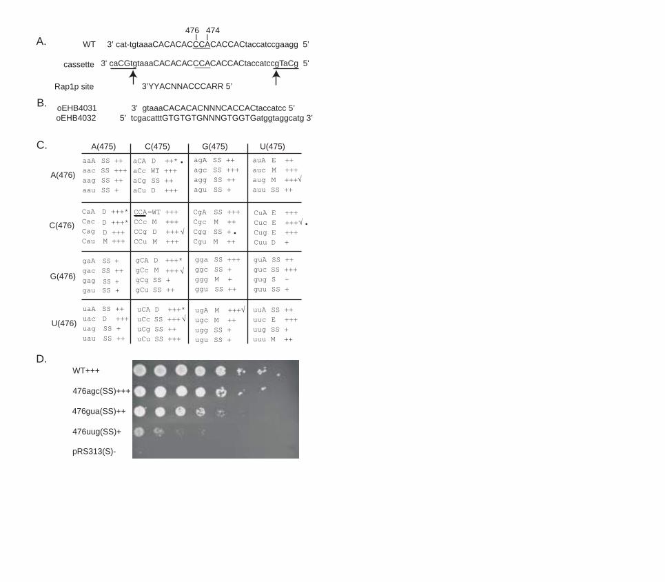
We analyzed the growth phenotypes of all 63 mutants. Only the tlc1-476gug led
to complete loss of telomerase activity and senescence identical to that caused by tlc1
deletion originally described (Prescott and Blackburn, 2000). Cells that express tlc1-
476gug and are ∆rad52 stopped growth completely 50-75 generations after the loss of the
wild type TLC1, with no survivors generated (Prescott and Blackburn, 2000). In contrast,
the other 62 mutants were still able to form colonies 20 streaks (approximately 400-500
generations) after removal of the wild type TLC1. Mutants showed different degrees of
compromised growth, based on colony size. We scored each mutant for growth and the
results are summarized in Figure 1C. Figure 1D shows the growth of one representative
mutant from each growth class at the 6th streak after loss of the wild type TLC1.
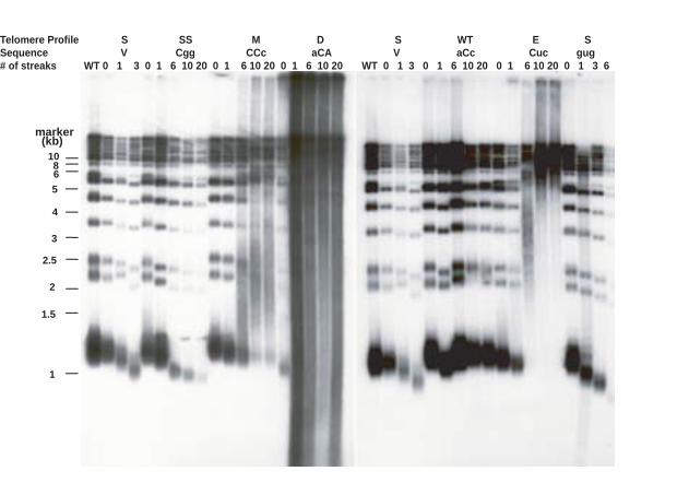
The 63 tlc1 template mutants fell into six classes based on Southern blot analyses
of their telomere length profiles: 1) wild type length (WT); 2) progressively shortened
telomeres which led to senescence at the same rate as telomerase-null cells (S); 3)
elongated telomeres (E); 4) mixed populations of telomeres (short, but tightly regulated
11
plus elongated with a broad size distribution) (M); 5) elongated and degraded telomeres
(D) and 6) short but stably maintained telomeres (SS). The Southern blotting analysis
results are summarized in Figure 1C and representative blots are shown in Figure 2.
Only one template mutant, tlc-476aCc, had a wild type telomere profile with ~350
bp long telomeric repeat tracts, and a normal growth phenotype. The template RNA
directs the synthesis of the Rap1p binding site and Rap1p binding is strongly influenced
by mutations in the 474-476 sequence (Prescott and Blackburn, 2000). This “wild type”
allele has two point mutations, 476C to A and 474A to C. It can potentially copy this
template into TGTGTGTGGGTGG repeats, with a 10/13 nucleotide match to the Rap1p
consensus binding site (see Figure 1A). Apparently sufficient Rap1p binding affinity is
retained by the sequences incorporated into these mutant telomeres to support normal
telomere length regulation.
Telomeres in the five elongated (E) mutants, 476auA, 476CuA, 476Cuc, 476Cug,
and 476uuc were much longer than wild type. All five mutant sequences share a
common C to U change at position 475. This is consistent with previous results that this
position is critical for Rap1p binding (Krauskopf and Blackburn, 1998). The telomeres in
two of these mutants, 476uuc(E), and 476Cuc(E), were over 10 kb at the 10th streak,
longer than any previously reported S. cerevisiae mutant.
Initial shortening of telomeres followed by rapid elongation was a feature
common to three classes of mutants: elongated (E), mixed (M), and elongated°raded
(D). In these mutants, telomeres shortened during the first 50-100 generations after loss
of the wild type TLC1 (Figure 2; see 0 and 1 streak lanes). This was followed by rapid
deregulation of telomere length within the next ~50 generations. In the case of the (E)
12
mutants, the initially shortened telomere population disappeared and the entire population
became lengthened. In contrast, in the mixed population (M) mutants, the shortened
telomere subpopulation became stabilized for at least another 300 generations. A
comparable population of shortened telomeres was also maintained in a subset of the
elongated and degraded (D) mutants with mild degradation, but was not evident in (D)
mutants with severely degraded telomeric DNA (see Figure 2 and below for more
discussion of (D) mutants).
Similar mixed telomere phenotypes were previously reported for telomerase RNA
template mutants in the yeast K. lactis (Krauskopf and Blackburn, 1996; Krauskopf and
Blackburn, 1998). In these mutants, which retained the Rap1p consensus binding site and
normal in vitro Rap1p binding, telomeres were initially well-regulated at shorter-than-
wild type length for many generations, but subsequently underwent rapid lengthening. It
was proposed that this rapid elongation occurred upon the eventual replacement of the
bulk of the wild type telomeric tract by mutant sequence (Krauskopf and Blackburn,
1998). Our mutants may reflect a similar situation.
Three tlc1 template mutants caused high misincorporation rates
The fact that 62 mutants continued to grow for hundreds more generations than
∆tlc1 or tlc1-476gug mutants, in the absence of Rad52p, indicated that these mutant
telomerases are active in vivo. To confirm telomerase activity, we analyzed the telomeres
of three mutants for the incorporation of the predicted mutant nucleotides, using a
previously developed PCR-based technique (Tzfati et al., 2000). As expected, mutant
sequences were found in the telomeres, sometimes as multiple repeats (see Figure 3 for
13
representative clones). A striking finding was that, in addition to the expected mutant
sequences, all three mutants contained significant numbers of misincorporated bases in
their telomeres (indicated as bold and italicized bases in Figure 3). Figure 3D lists all the
misincorporated repeats found. 476agc(SS) and 476Cuc(E) have four and three
misincorporated repeats out of 42 total repeats synthesized by the mutant telomerase
respectively (10% and 7%). 476uug(SS) has 4 misincorporated repeats out of 31 total
repeats (13%). As controls, telomeres were cloned from cells expressing only the wild
type TLC1. These contained no misincorporated bases out of the ~2,200 bases sequenced
(J.L. and E.H.B. unpublished). Thus, like Tetrahymena (Gilley et al., 1995), mutations in
the template region of yeast telomerase also cause reduced fidelity. This is the first report
of this type of base misincorporation by mutant-template telomerases in vivo for S.
cerevisiae.
The telomeres in tlc1-476agc(SS) and tlc1-476uug(SS) were both shorter than
wild type, but stably maintained. However, by the 6th streak after loss of the wild type
TLC1, tlc1-476agc(SS) grew like wild type, while tlc1-476uug(SS) was very sick (Figure
1). Telomeres were cloned from these two mutants at the 6th streak. In both cases, the
telomeres contained tandem repeats of mutant sequence, indicating that the enzyme was
able to copy the template completely (see Figure 3A-C for representative clones).
Therefore, the contrasting growth properties of these two mutants did not reflect any
obvious difference in their efficiency of mutant repeat incorporation or length
maintenance.
Telomeres of tlc1-476Cuc(E) became much longer than wild type 120
generations after removal of wild type TLC1 (see Figure 2). In order to clone full-length

14
telomeres from tlc-476Cuc(E), genomic DNA was extracted from the first streak after
removal of the wild type TLC1, when the bulk telomere length was still similar to wild
type. Out of the 8 cloned tlc1-476Cuc(E) telomeres, two contained only wild type
sequences, but the other six contained up to 14 repeats of the expected mutant sequence
(data not shown). Interestingly, long tracts of wild type telomeric sequence were
interspersed with mutant sequences. Since these telomeres were cloned from rad52∆
cells that contained only mutant tlc1 for ~30 cell generations, we speculate that these
wild type sequences resulted from copying the parts of the template that remained wild
type in 476Cuc(E). The enzyme may have dissociated before it reached the mutant
sequence, or the mutant DNA was cleaved off.
Degraded telomeres are associated with an immediate slow growth phenotype.
Eleven out of the 63 mutants had telomeres that appeared both elongated and
degraded (D). As previously reported for tlc1-476A (here referred to as tlc1-aCA(D))
(Chan et al., 2001), telomeric DNA hybridization signal in Southern blots from these
cells was extremely broad, extending from the wells of the gel to the bottom, with no
discrete bands (see Figure 2). No common pattern of base substitution was discernible
for this telomere profile class: although 476CaA, 476Cac and 476Cag share a common C
to A mutation in position 475, in 476aCA, 476gCA and 476uCA, positions 475 and 474
are still wild type. The severity of the degradation phenotype varied from very severe
(476aCA, 476gCA, 476uCA, 476Cac and 476CCg), or intermediate (476Cgc, 476CaA,
476aCu, 476uac and 476Cag) to least severe, in which isolated bands become
distinguishable (476uCA; see Figure 1C).

15
The four (D) mutants with the most severe telomeric DNA degradation phenotype
showed slower growth within ~20 generations after the loss of the wild type TLC1
(Figure 1C). These mutants had heterogeneous colony sizes and extended population-
doubling times (Figure S1 and data not shown). This immediate slow growth phenotype,
with high percentages of enlarged, misshapen monster cells, was previously reported for
tlc1476aCA(D) (Chan et al., 2001). This mutant telomerase was active and the predicted
mutant sequence was incorporated into telomeres in vivo, likely causing these phenotypes
(Chan et al. 2001). Such behavior contrasts with the delayed phenotype characteristic of
senescence, which only becomes apparent at 50-75 generations after the loss of
functional TLC1. Furthermore, senescent rad52∆ cells stop growing completely 50-75
generations after the loss of the wild type TLC1. In contrast, the (D) mutant cells
apparently adapted, regaining relatively healthy growth after 50 generations and
continuing to grow throughout the rest of the experiment (over 400 generations). Thus,
we conclude that the immediate slow growth in (D) mutants is the consequence of
incorporation of mutant telomeric sequence rather than lack of telomerase activity.
The tlc1 template mutations cause aberrant chromosome separation.
In order to further analyze the cellular consequences of mutant-sequence
telomeres, we examined budding kinetics and chromosome dynamics in representatives
of three distinct tlc1 mutant telomere length classes: tlc1-476aC(D), tlc-4761Cuc(E) and
tlc1-476Cgg(SS). These cause degraded, elongated and short&stable telomeres,
respectively. Although some cellular phenotypes of telomerase RNA template mutants
have been described previously in other eukaryotes (Kim et al., 2001; Kirk et al., 1997;

16
Smith and Blackburn, 1999), direct analysis of their chromosome behavior has not been
reported. In order to visualize chromosome movement in individual cells, we used a
method that marks a single chromosome with a green spot at a specified location
(Straight et al., 1996; Straight et al., 1997) (diagramed in S2A), using a tandem array of
lac operators inserted either 12 kb from the centromere (“centromere-marked”) or 100kb
from one telomere (“telomere-marked”) of chromosome IV, the largest chromosome in S.
cerevisiae (Figure S2A). Lactose repressor fused to GFP (GFP-LacI) expressed in these
cells allows visualization of chromosome IV which, during the course of a normal cell
cycle, is seen as one green spot (unreplicated, or replicated but unseparated) or two spots
(replicated and separated). The three tlc1 template mutations were transformed into
haploid strains containing either the “centromere” or “telomere”-marked chromosome IV.
Following release from an α-factor arrest, we compared the timing of sister chromatid
separation in the centromere and telomere marked strains. Budding kinetics, sister
chromatid separation and chromosome segregation were measured for all strains during
the course of a single cell cycle.
Based on the behavior of template mutants in Tetrahymena (Kirk et al., 1997), our
initial expectation was that these assays might reveal mutant chromosomes that were able
to separate at their centromeres, but had delayed separation, or remained attached, at their
ends. Such behavior is also exhibited by the previously characterized yeast topoisomerase
II mutant, top2-4. Top2p functions to resolve the concatenations between sister
chromatids so they can fully separate from each other at anaphase (DiNardo et al., 1984;
Holm et al., 1989; Shamu and Murray, 1992; Uemura et al., 1987). Using chromosome
IV GFP-marked at the centromere, the telomere, or midway along a chromatid arm
17
(Bhalla et al., 2002), it was shown that in the top2-4 mutant, centromere separation
precedes telomere separation. Therefore, as a control, we first confirmed that our assay
revealed such differential centromere and telomere separation kinetics in top2-4 mutant
cells. Cells were synchronized in G1 by growth in α-factor for 4 hours, followed by
release into fresh medium without α-factor. Samples were collected every 20 minutes
and fixed for later analysis. Only “shmooed” cells, capable of growing and responding to
α-factor, were considered for this analysis. Cells were counted and classified as
unbudded, or small, large or re-budded. We confirmed that the telomerically located
GFP spots in top2-4 cells separated more slowly than centromeric GFP spots in the first
cell cycle after α-factor release, reassuring us that this assay could, in our hands, reveal
defects that lead to telomere fusion, but still allow centromere separation (Figure 4B and
data not shown).
We then carried out the same analysis for the tlc1(D), (E) and (SS) mutants.
Following release from α-factor, budding kinetics for all three mutants were the same as
wild type, suggesting that they proceed with normal kinetics through the cell cycle.
FACS analysis showed that the tlc1 cells accumulate 2N DNA content at rates similar to
wild type for the (E) and (SS) mutants, and with slightly delayed kinetics for the (D)
mutant tlc1-476aCA (Supplementary Material, Figure S2B and C). However, in all three
mutants, chromosome segregation was defective. Chromosome dynamics following
release from α-factor were assayed by counting additional cells every 20 minutes and
sorting them into seven categories based on the number and position of GFP-marked
chromosome spots (Figure 4). In all GFP-chromosome assays, only “shmooed” cells,
those responding to α–factor and still capable of entering the cell cycle, were scored. In
18
“shmooed,” large-budded cells, categories 1-3, the most common, are found during the
course of a normal wild type (WT) cell cycle. Categories 4-7 are rare in WT cells but
were found in significant numbers in the tlc1 mutants. “Sister Chromatid Separation” was
scored as the percentage of cells in categories 2, 3, 4, 5 and 7. “Chromosome
Segregation” was made up of category 3 only, and consists of cells with sister chromatids
properly separated and segregated into mother and daughter cells. “Chromosomes
Unsegregated” was measured as the percentage of cells in all categories except 1, 3 and
6, until the 100 minute timepoint and beyond, when categories 1 and 6 were included.
The (D) mutant, in particular, had 2-10% monster cells, with more in early timepoints,
and the (SS) mutant accumulated about 5% monster cells in some later passages. Total
cell viability (colony forming ability) was quantified and was ~90% of wild type for all
three mutants (data not shown). Monster cells were not included in the GFP-
chromosome assays. Figure 4A summarizes how cells were scored and Figure 4B shows
the distribution of cell categories at the 100 minute time point for several strains.
In all three tlc1 mutants, quantification of the above categories uncovered aberrant
separation of replicated GFP-marked chromosomes and improper segregation of the
marked chromosomes to daughter cells. Figure 5A shows that in wild type cells, sister
chromatid separation began 80 minutes after release from G1 and was complete by 120
minutes. In tlc1 mutant cells, sister separation also began at 80 minutes, but the
percentage of cells in which chromatids separated, or went on to segregate, was never as
high. This low percentage of separation and high degree of unsegregated chromosomes
was a consistent property of all three tlc1 template mutants (Figure 5B). Strikingly, for
all tlc1 template mutants, there was no difference in segregation kinetics between the

19
centromere and telomere-marked strains. This suggested that the lack of separation was
occurring along the entire length of the chromosome, not just at the telomeres.
Furthermore, for cells with a single spot at late time points (classes 1 and 6 in Figure 4A),
the spot appeared in daughter cells almost as often as mother cells. As noted above, the
high viability of the template mutant strains (data not shown) indicated that the 20-30%
of “shmooed” cells with chromosome abnormalities were not simply dead.
The mutant-telomere response is distinct from known DNA damage checkpoints.
One explanation for a whole-chromosome delay in chromatid separation and
segregation could be activation of a cell cycle checkpoint. Therefore, we used our assay
to examine chromosome dynamics using two mutant alleles of CDC13 that cause
telomere perturbations: cdc13-1, which activates a cell-cycle arrest, and cdc13-5, which
does not. When grown at the nonpermissive temperature (30°), the cdc13-1 mutant
contains extensive single stranded DNA at telomeres and arrests in G2/M with a large
bud and a single nucleus, via the RAD9-dependent checkpoint pathway (Burke and
Church, 1991; Garvik et al., 1995; Weinert and Hartwell, 1993). Figure 4B shows the
breakdown of marked chromosome segregation categories at the 100 minute timepoint
for wild type cells, the tlc1 template mutants, cdc13-5 and cdc13-1 mutants at different
temperatures. When grown at 30°, cdc13-1 accumulated large-budded cells with single
chromosome spots, as expected for cells that arrest at G2/M with a single nucleus.
Interestingly, when grown at the permissive temperature (23°), cdc13-1 behaved like the
tlc1 template mutants: the percentage of unsegregated chromatids was high, with no
significant difference in kinetics between telomere and centromere-marked strains

20
(Figure 4B and data not shown). We combined cdc13-1 with deletions of DDC1 and/or
DDC2, components of the DNA damage checkpoint, and found that the missegregation
phenotype of cdc13-1 at 23 degrees is completely relieved by combining cdc13-1 with
∆ddc1, ∆ddc2 or ∆ddc1∆ddc2 (Figure 4B and Supplemental Figure 4). This suggests that
the DNA damage checkpoint is activated in response to the telomere damage created by
the cdc13-1, even at 23 degrees.
The mutant carrying the cdc13-5 allele neither activates the DNA damage
checkpoint nor causes a temperature-sensitive phenotype, even though it has long, G-rich,
single-stranded telomeric overhangs during S phase (Chandra et al., 2001). cdc13-5 also
contained unsegregated chromatids, with equal fractions seen in both the telomere- and
the centromere-marked strains (Figure 4B and data not shown). Hence, mutations of a
telomeric component, Cdc13p, other than in the TLC1 template also lead to a chomosome
segregation defect. Furthermore, in the case of the cdc13-5 mutant, this segregation
defect occurs independently of activation of any previously described known checkpoint.
Complete deletion of TLC1 causes cell cycle arrest, activates a checkpoint
(Enomoto et al., 2002; IJpma and Greider, 2003) and causes chromosomal fusions when
telomeres become short (Hackett et al., 2001). Consistent with this, we examined
chromosome dynamics in an asynchronous ∆tlc1 culture and found that 60% of cells
were large budded (compared with about 30% in an asynchronous wild type culture).
Among the large budded cells, only 17% had properly separated and segregated
chromosomes (class 3). The majority, 75%, had a single spot (classes 1 and 6) and the
rest fell into the aberrant segregation classes (4,5 and 7). Because most of these cells

21
were arrested, they did not respond to α-factor and thus we could not do synchronized
time course analysis.
If the tlc1 template mutations activate a DNA damage checkpoint that blocks
chromosome separation, then disruption of the checkpoint sensing complexes might
restore normal chromosome dynamics. In our assay, the restoration would appear as a
higher percentage of large-budded cells with two fully separated and segregated GFP
spots (class 3 cells). DDC1, MEC3 and DDC2 were shown to be important for activating
the checkpoint in response to loss of telomerase in a ∆tlc1 background (Enomoto et al.,
2002; IJpma and Greider, 2003). Ddc1p is a member of the PCNA-like trimer that loads
directly at sites of DNA damage, and Ddc2p associates with Mec1p (an ATR kinase) in a
complex that is also recruited to sites of damage (Melo and Toczyski, 2002). First, we
combined tlc1(D) with deletion of DDC1, DDC2 or MEC3 and examined budding and
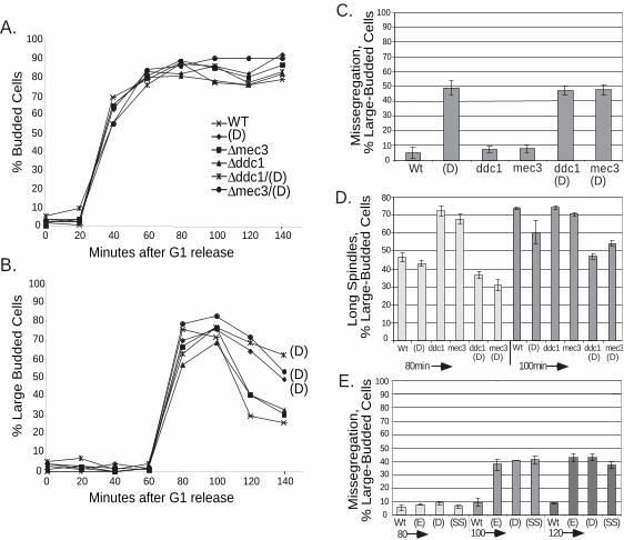
chromosome dynamics for each single and double mutant (Figure 6). Overall budding
kinetics were the same for all strains (Figure 6A). However, in both tlc1 (D) and the
double mutants tlc1(D)/mec3 and tlc1(D)/ddc1, the number of cells with large buds was
higher than wild type, beginning at 100 minutes, and remained high for the rest of the
time course (Figure 6B). Furthermore, like tlc1(D), the double mutants were slow to
separate sister chromatids and showed a high degree of unsegregated chromosomes
(Figure 6C and data not shown).
Spindle staining with anti-tubulin antibody provided further evidence that neither
DDC1 nor MEC3 are required for the checkpoint resulting in the arrest of chromosome
separation in tlc1(D) mutant cells. Following release from G1 arrest, strains deleted for
DDC1 or MEC3 alone accumulated cells with long spindles faster than wild type, as

22
expected for cells that can no longer pause and respond to DNA damage (Figure 6D, 80
min time point bars). However, tlc1(D)/ddc1 and tlc1(D)/mec3 double mutants, like
tlc1(D), all grew long spindles more slowly than wild type cells or ddc1 or mec3 single
mutants. The timing of this delay in spindle formation (Figure 6D) coincided with the
slower chromosome segregation and the persistence of large budded cells described
above.
Next, we combined ∆ddc2/∆sml1 with each of the three tlc1 template mutations
(E, D or SS). Like ∆mec1 lethality, ∆ddc2 lethality is suppressed by ∆sml1. Again, the
fraction of unseparated chromatids was comparable to that seen for the each template
mutation alone (Figure 6E). Thus, in contrast to the telomere defect caused by the cdc13-
1 mutation, mutant-template telomerase RNAs cause a DNA damage checkpoint-
independent response.
Finally, since DDC1 and DDC2 were not required for the checkpoint, we tested
whether the delayed spindle elongation in tlc1 template mutants is due to activation of the
spindle assembly checkpoint pathway. To address this possibility, we combined each of
the three template mutants (E, D or SS) with ∆mad2 and looked for restoration of proper
chromosome segregation. However, chromosome missegregation remained high for all
three mutants. This result suggests that the spindle assembly checkpoint is not solely
responsible for detecting or responding to aberrations at mutant telomeres (Figure 4B and
Supplemental Figure 5).
In summary, our results show that aberrant-sequence telomeres, whether very
short, elongated, or highly degraded, all arrest chromosome separation, and lead to
impaired chromosome segregation and failure to progress into anaphase. Furthermore,

23
disruption of the DNA damage checkpoint or the spindle assembly checkpoint in cells
with these telomere defects, via deletion of MEC3, DDC1, DDC2 or MAD2 does not
restore cell cycle progression. These findings imply that neither of the major DNA
damage sensing complexes, nor the spindle checkpoint, is solely responsible for
activating a cell cycle checkpoint in response to the type of telomere aberration caused by
mutant telomeric DNA sequences.
Discussion
Roles of template bases in telomerase enzymatic activity
Here we have analyzed all possible mutations within an essential 3-base sequence
at the core of the yeast telomerase RNA template sequence. We have shown that among
the 63 substitution mutations of positions 474-476, only one, tlc1-476gug, completely
abolishes enzymatic activity, implying that telomerase enzyme activity will tolerate
almost any sequence at TLC1 positions 474-476 except 476gug. However, while the
remaining mutants do not senesce, many of them (34/63, or 54%) remain compromised
for telomerase function in vivo, as judged by their shortened telomeres.
Further evidence for altered enzymatic activity came from sequencing the
telomeric DNA cloned from several of our mutants. Telomerase normally makes a
faithful copy of its RNA template sequence. Specific mutations of the Tetrahymena
telomerase template cause primer slippage, loss of fidelity and premature dissociation of
products (Gilley et al., 1995; Gilley and Blackburn 1996). Here, we report, for the first
time in S. cerevisiae, that three tlc1 template mutations lead to high levels of base

24
misincorporation. Hence the ability of template bases to affect properties of the
polymerization reaction may be general among telomerases.
Cellular consequences of mutating the telomerase RNA
Thorough mutagenesis of a small essential template region showed that mutant
telomeric sequences led to great variation in telomere integrity, with telomere profiles
ranging from severe shortening to extensive lengthening. Many mutants had highly
degraded telomeres, with no consistently maintained length. This wide variation of bulk
telomere sizes confirms and extends previous work in other systems showing that
template mutations can greatly influence both positive and negative regulation of
telomere length (Krauskopf and Blackburn, 1996; Krauskopf and Blackburn, 1998;
Prescott and Blackburn, 1997; Prescott and Blackburn, 2000; Chan et al., 2001).
The effects of our set of template mutants on cell growth also varied widely in
severity and time of onset. While the senescent phenotype of 476gug was caused by total
loss of telomerase activity, several mutants that retained enzymatic activity grew slowly.
Two types of slow growth were observed: an initial defect that improved with passaging,
or normal growth followed by increased sickness in later passages. Mutants with
severely degraded telomeres fell into the first group, with growth becoming faster ~50
generations after the loss of wild type TLC1. Growth continued for as long as cells were
observed (> 400 generations), despite the accumulation of progressively more degraded
and single-stranded telomeric DNA. It appears that these cells incorporated mutations
but then adapted, in a ∆rad52 background, by unknown mechanism(s). The second
group of slow growers contained short stable telomeres, and the onset of slowing of

25
growth was delayed to ~100 generations after loss of the wild type TLC1. We speculate
that a critical number of mutant repeats had to be added to telomeres, possibly in
combination with a critical degree of shortening, in order to affect cell growth.
Our mutant collection was assembled without stringent growth requirements.
Recently, Forsteman et al. screened libraries of randomly mutagenized tlc1 template
sequences for complementation of the ∆tlc1 senescence phenotype (Forstemann et al.,
2003). Thirty-two clones were recovered from a library containing random substitutions
throughout positions 477-473. Most of them contained C/A-rich sequences, like the wild
type S. cerevisiae sequence. All 32 isolates had wild type growth, and among them, six
template sequences were the same as in the best growers of our collection (Figure 1C).
The difference in our results can be explained by the more stringent growth requirements
of the screen by Forsteman et al (2003).
We found no simple correlation between telomere length and cell growth.
Mutants with identical telomere length profiles in Southern blots grew well or poorly
depending on individual sequence changes, indicating that the specific telomeric
sequences, rather than bulk telomere length, determined their growth properties.
Mutant-sequence telomeres elicit a unique checkpoint response.
Chromosome dynamics had not previously been analyzed in any telomerase
template mutant in any organism. Interestingly, our direct analysis of chromosomes in
three diverse template mutants revealed a common cell cycle arrest response: specifically,
whether telomeres were short, long, or extremely degraded, tlc1 mutations consistently
led to a high level of delayed sister chromatid separation and an increase in cells with

26
unsegregated chromosomes. Furthermore, deletion of the known DNA damage
checkpoint genes MEC3, DDC1 or DDC2 did not restore cell cycle progression or proper
chromosome dynamics to template mutant cells. These findings imply that neither of the
established DNA damage sensing complexes is solely responsible for activating a cell
cycle checkpoint in response to telomere damage caused by mutant telomeric DNA
sequences.
This cellular response to tlc1 mutations, even those that lead to shortened
telomeres, is distinct from the response to telomeric shortening and senescence caused by
TLC1 deletion. Enomoto et al. (Enomoto et al., 2002) and Ijpma and Grieder (IJpma and
Greider, 2003) have independently reported that when telomeres become short following
deletion of TLC1, a G2/M arrest occurs that is dependent on MEC1, MEC3, DDC2, and
RAD24. Despite the short telomeres of the (SS) mutants, there was no activation of the
senescence phenotype, and the cellular response was different, since the components of
the ATR complex Mec1p and Ddc2p were not required to activate a checkpoint in
response to short, long or degraded telomeres in the tlc1(SS), (E) and (D) template
mutants. The Enomoto et al. (Enomoto et al, 2002) study specifically ruled out
involvement of Tel1p the ATM-kinase in S. cerevisiae, in the cell cycle arrest response to
telomerase deficiency. These cell cycle responses imply that yeast has more than one
mechanism for responding to mutant telomeres. Furthermore, response to telomerase
deletion the yeast contrasts with results seen in human cells. Specifically, overexpression
of a dominant-negative form of the human telomere-protective protein TRF2 causes a
pronounced ATM-dependent cellular response (de Lange, 2002). Taken together, these
27
results suggest that ATM and ATR have varied responses to different types of telomeric
lesions, both within and between organisms.
Our results suggest that mutant telomeric sequences may not be seen by the cell
in the same way as general genomic DNA damage, since deletion of DDC1 or DDC2 did
not relieve the chromosome defect. However, all three mutations chosen for cellular
analysis accumulated Ddc1-GFP and Ddc2-GFP foci in mutant cells, a characteristic
associated with activation of a DNA damage checkpoint (Supplementary data Figure S3).
Very early after introduction of mutant template tlc1 alleles (< 20 generations), both
Ddc1-GFP and Ddc2-GFP appeared as bright nuclear foci in a subpopulation of cells
(Figure S3). The Ddc2-GFP foci persisted throughout 6 serial passages (~120
generations). Upon further passaging, the number of Ddc1-GFP foci gradually increased
in (SS) mutant cells, but gradually decreased in the (D) mutant. These findings echoed
the slow onset of growth defects in the (SS) mutants and the immediate growth defect
seen for the (D) mutants, as described above; they may reflect DNA damage foci formed
at sites of secondary DNA damage, such as chromosome breaks following end-to-end
chromosome fusion, rather than foci on the telomeres.
Surveillance of mutant telomere sequences may require a combination of DNA
damage checkpoint proteins and/or participation of more than one checkpoint pathway.
This is the case with deletion of Taz1p, the S. pombe ortholog of hTRF2. Taz1p promotes
proper chromosome segregation, DNA repair, and chromosome end protection: both
DNA damage and spindle assembly checkpoint proteins are required for ∆taz1 cells to
survive (Miller and Cooper, 2003). A similar response is seen in cells with deletions of
the nonhomologous end-joining and telomere-protection proteins, yKu70 and yKu80.

28
∆Ku70 and ∆Ku80 cells are temperature sensitive and have short telomeres and single-
stranded Y’ sub-telomeric repeats (Gravel et al., 1998; Polotnianka et al., 1998; Smith
and Jackson, 1999). Maringele and Lydall recently showed that subsets of both the DNA
damage checkpoint (CHK1, MEC1, and RAD9) and the spindle assembly checkpoint
(MAD2) pathways are required for efficient cell cycle arrest of yKu70∆ mutants grown at
the non-permissive temperature (Maringele and Lydall, 2002). Other alterations of
chromosome structure can activate multiple checkpoints, and many conditions that
activate the DNA damage or DNA replication checkpoints also activate the spindle
checkpoint (Garber and Rine, 2002). Notably, the arrest of cdc13-1 mutants is an
exception; arrest in these cells is dependent on a large group of DNA damage checkpoint
genes: CHK1, MEC1, and RAD9 as well as RAD17, RAD24, MEC3, DDC1 and DUN1,
but does not require the spindle assembly checkpoint pathway (Maringele and Lydall,
2002). In contrast, the tlc1 template mutant response shown here appears to work
independently of the spindle assembly checkpoint. We have shown that mutant telomeric
repeats elicit a response, directly or indirectly, that is distinct in its genetic dependence,
from that induced by cdc13-1 telomerase deficiency or other DNA damaging agents.
Together, these findings suggest that defects at telomeres activate various checkpoint
responses depending on the molecular nature of disruption to telomere integrity. A future
challenge will be to link specific types of telomere damage to precise patterns of
checkpoint activation.

29
Acknowledgments:
We would like to thank J. Berman for ∆mec3 and ∆ddc1 strains; V. Lundblad for
pVL1215, used to construct cdc13-5; Needhi Bhalla, for top2-4 strains; Justine Melo and
Genevieve Vidanes for technical advice with Ddc1-GFP and Ddc2-GFP protein imaging
and Shivani Nautiyal, Dan Levy, Carol Anderson, Sveta Makovets, Dave Toczyski, and
Dave Morgan for critical reading of the manuscript.

30
References
Bhalla, N., Biggins, S., and Murray, A. W. (2002). Mutation of YCS4, a budding yeast
condensin subunit, affects mitotic and nonmitotic chromosome behavior, Mol Biol Cell
13, 632-45.
Blackburn, E. H. (2000a). The end of the (DNA) line, Nat Struct Biol 7, 847-50.
Blackburn, E. H. (2000b). Telomere states and cell fates, Nature 408, 53-6.
Blackburn, E. H. (2000c). Telomeres and telomerase, Keio J Med 49, 59-65.
Blackburn, E. H. (2001). Switching and signaling at the telomere, Cell 106, 661-73.
Brachmann, C. B., Davies, A., Cost, G. J., Caputo, E., Li, J., Hieter, P., and Boeke, J. D.
(1998). Designer deletion strains derived from Saccharomyces cerevisiae S288C: a useful
set of strains and plasmids for PCR-mediated gene disruption and other applications,
Yeast 14, 115-32.
Bryan, T. M., Sperger, J. M., Chapman, K. B., and Cech, T. R. (1998). Telomerase
reverse transcriptase genes identified in Tetrahymena thermophila and Oxytricha
trifallax, Proc Natl Acad Sci U S A 95, 8479-84.
Burke, D. J., and Church, D. (1991). Protein synthesis requirements for nuclear division,
cytokinesis, and cell separation in Saccharomyces cerevisiae, Mol Cell Biol 11, 3691-8.
Chan, S. W., Chang, J., Prescott, J., and Blackburn, E. H. (2001). Altering telomere
structure allows telomerase to act in yeast lacking ATM kinases, Curr Biol 11, 1240-50.
Chandra, A., Hughes, T. R., Nugent, C. I., and Lundblad, V. (2001). Cdc13 both
positively and negatively regulates telomere replication, Genes Dev 15, 404-14.
31
Counter, C. M., Meyerson, M., Eaton, E. N., and Weinberg, R. A. (1997). The catalytic
subunit of yeast telomerase, Proc Natl Acad Sci U S A 94, 9202-7.
de Lange, T. (2002). Protection of mammalian telomeres, Oncogene 21, 532-40.
DiNardo, S., Voelkel, K., and Sternglanz, R. (1984). DNA topoisomerase II mutant of
Saccharomyces cerevisiae: topoisomerase II is required for segregation of daughter
molecules at the termination of DNA replication, Proc Natl Acad Sci U S A 81, 2616-20.
Enomoto, S., Glowczewski, L., and Berman, J. (2002). MEC3, MEC1, and DDC2 are
essential components of a telomere checkpoint pathway required for cell cycle arrest
during senescence in Saccharomyces cerevisiae, Mol Biol Cell 13, 2626-38.
Forstemann, K., Zaug, A. J., Cech, T. R., and Lingner, J. (2003). Yeast telomerase is
specialized for C/A-rich RNA templates, Nucleic Acids Res 31, 1646-55.
Garber, P. M., and Rine, J. (2002). Overlapping roles of the spindle assembly and DNA
damage checkpoints in the cell-cycle response to altered chromosomes in Saccharomyces
cerevisiae, Genetics 161, 521-34.
Garvik, B., Carson, M., and Hartwell, L. (1995). Single-stranded DNA arising at
telomeres in cdc13 mutants may constitute a specific signal for the RAD9 checkpoint,
Mol Cell Biol 15, 6128-38.
Gilley, D., and Blackburn, E. H. (1996). Specific RNA residue interactions required for
enzymatic functions of Tetrahymena telomerase, Mol Cell Biol 16, 66-75.
Gilley, D., Lee, M. S., and Blackburn, E. H. (1995). Altering specific telomerase RNA
template residues affects active site function, Genes Dev 9, 2214-26.
Gravel, S., Larrivee, M., Labrecque, P., and Wellinger, R. J. (1998). Yeast Ku as a
regulator of chromosomal DNA end structure, Science 280, 741-4.
32
Greider, C. W. (1996). Telomere length regulation, Annu Rev Biochem 65, 337-65.
Greider, C. W., and Blackburn, E. H. (1985). Identification of a specific telomere
terminal transferase activity in Tetrahymena extracts, Cell 43, 405-13.
Greider, C. W., and Blackburn, E. H. (1989). A telomeric sequence in the RNA of
Tetrahymena telomerase required for telomere repeat synthesis, Nature 337, 331-7.
Hackett, J. A., Feldser, D. M., and Greider, C. W. (2001). Telomere dysfunction
increases mutation rate and genomic instability, Cell 106, 275-86.
Hardy, C. F., Sussel, L., and Shore, D. (1992). A RAP1-interacting protein involved in
transcriptional silencing and telomere length regulation, Genes Dev 6, 801-14.
Holm, C., Stearns, T., and Botstein, D. (1989). DNA topoisomerase II must act at mitosis
to prevent nondisjunction and chromosome breakage, Mol Cell Biol 9, 159-68.
IJpma, A. S., and Greider, C. W. (2003). Short Telomeres Induce a DNA Damage
Response in Saccharomyces cerevisiae, Mol Biol Cell 14, 987-1001.
Kim, M. M., Rivera, M. A., Botchkina, I. L., Shalaby, R., Thor, A. D., and Blackburn, E.
H. (2001). A low threshold level of expression of mutant-template telomerase RNA
inhibits human tumor cell proliferation, Proc Natl Acad Sci U S A 98, 7982-7.
Kirk, K. E., Harmon, B. P., Reichardt, I. K., Sedat, J. W., and Blackburn, E. H. (1997).
Block in anaphase chromosome separation caused by a telomerase template mutation,
Science 275, 1478-81.
Krauskopf, A., and Blackburn, E. H. (1996). Control of telomere growth by interactions
of RAP1 with the most distal telomeric repeats, Nature 383, 354-7.
Krauskopf, A., and Blackburn, E. H. (1998). Rap1 protein regulates telomere turnover in
yeast, Proc Natl Acad Sci U S A 95, 12486-91.
33
Kyrion, G., Boakye, K. A., and Lustig, A. J. (1992). C-terminal truncation of RAP1
results in the deregulation of telomere size, stability, and function in Saccharomyces
cerevisiae, Mol Cell Biol 12, 5159-73.
Longtine, M. S., McKenzie, A., 3rd, Demarini, D. J., Shah, N. G., Wach, A., Brachat, A.,
Philippsen, P., and Pringle, J. R. (1998). Additional modules for versatile and economical
PCR-based gene deletion and modification in Saccharomyces cerevisiae, Yeast 14, 953-
61.
Lundblad, V., and Blackburn, E. H. (1993). An alternative pathway for yeast telomere
maintenance rescues est1- senescence, Cell 73, 347-60.
Marcand, S., Wotton, D., Gilson, E., and Shore, D. (1997). Rap1p and telomere length
regulation in yeast, Ciba Found Symp 211, 76-93; discussion 93-103.
Maringele, L., and Lydall, D. (2002). EXO1-dependent single-stranded DNA at
telomeres activates subsets of DNA damage and spindle checkpoint pathways in budding
yeast yku70Delta mutants, Genes Dev 16, 1919-33.
Melo, J., and Toczyski, D. (2002). A unified view of the DNA-damage checkpoint, Curr
Opin Cell Biol 14, 237-45.
Miller, K. M., and Cooper, J. P. (2003). The telomere protein Taz1 is required to prevent
and repair genomic DNA breaks, Mol Cell 11, 303-13.
Nakamura, T. M., Morin, G. B., Chapman, K. B., Weinrich, S. L., Andrews, W. H.,
Lingner, J., Harley, C. B., and Cech, T. R. (1997). Telomerase catalytic subunit homologs
from fission yeast and human, Science 277, 955-9.

34
Polotnianka, R. M., Li, J., and Lustig, A. J. (1998). The yeast Ku heterodimer is essential
for protection of the telomere against nucleolytic and recombinational activities, Curr
Biol 8, 831-4.
Prescott, J., and Blackburn, E. H. (1997). Telomerase RNA mutations in Saccharomyces
cerevisiae alter telomerase action and reveal nonprocessivity in vivo and in vitro, Genes
Dev 11, 528-40.
Prescott, J. C., and Blackburn, E. H. (2000). Telomerase RNA template mutations reveal
sequence-specific requirements for the activation and repression of telomerase action at
telomeres, Mol Cell Biol 20, 2941-8.
Rose, M. D., Winston, F., and Heiter, P. (1990). Methods in Yeast Genetics (Cold Spring
Harbor, NY).
Shamu, C. E., and Murray, A. W. (1992). Sister chromatid separation in frog egg extracts
requires DNA topoisomerase II activity during anaphase, J Cell Biol 117, 921-34.
Smith, C. D., and Blackburn, E. H. (1999). Uncapping and deregulation of telomeres lead
to detrimental cellular consequences in yeast, J Cell Biol 145, 203-14.
Smith, G. C., and Jackson, S. P. (1999). The DNA-dependent protein kinase, Genes Dev
13, 916-34.
Straight, A. F., Belmont, A. S., Robinett, C. C., and Murray, A. W. (1996). GFP tagging
of budding yeast chromosomes reveals that protein-protein interactions can mediate sister
chromatid cohesion, Curr Biol 6, 1599-608.
Straight, A. F., Marshall, W. F., Sedat, J. W., and Murray, A. W. (1997). Mitosis in living
budding yeast: anaphase A but no metaphase plate, Science 277, 574-8.

35
Tzfati, Y., Fulton, T. B., Roy, J., and Blackburn, E. H. (2000). Template boundary in a
yeast telomerase specified by RNA structure, Science 288, 863-7.
Uemura, T., Ohkura, H., Adachi, Y., Morino, K., Shiozaki, K., and Yanagida, M. (1987).
DNA topoisomerase II is required for condensation and separation of mitotic
chromosomes in S. pombe, Cell 50, 917-25.
Weinert, T. A., and Hartwell, L. H. (1993). Cell cycle arrest of cdc mutants and
specificity of the RAD9 checkpoint, Genetics 134, 63-80.
Weinrich, S. L., Pruzan, R., Ma, L., Ouellette, M., Tesmer, V. M., Holt, S. E., Bodnar, A.
G., Lichtsteiner, S., Kim, N. W., Trager, J. B., et al. (1997). Reconstitution of human
telomerase with the template RNA component hTR and the catalytic protein subunit
hTRT, Nat Genet 17, 498-502.
Wotton, D., and Shore, D. (1997). A novel Rap1p-interacting factor, Rif2p, cooperates
with Rif1p to regulate telomere length in Saccharomyces cerevisiae, Genes Dev 11, 748-
60.

36
Figure legends
Figure 1
Summary of telomere profile and growth phenotypes of 63 mutants. (A) Sequence of the
template cassette. The wild type TLC1 sequence surrounding the template region is
shown on top. The template region is in capitals and positions 476-474 are underlined.
Mutations that create SphI and SalI sites are in capitals. The SphI and SalI sites are
underlined with arrows pointing to the bases where the restriction enzymes cut.
Sequence of the Rap1p consensus binding site is shown in 3’-5’ direction underneath.
(B) Sequences of the oligos used to construct the template mutant library. (C) Summary
of telomere profile and growth phenotypes of 63 mutants. Sequence is shown in 3’-5’
direction. Telomere length for each mutant is characterized as one of the following: D=
long°raded, E= elongated, M=mixed population (well-regulated and elongated
broader distribution), S=senescence, SS=short&stable, WT=wild type. Cell growth for
each mutant is scored as: wild type or close to wild type growth (+++), distinguishably
sicker than wild type (++), very sick (+) or senescent (-). Stars indicate mutants that
showed immediate slow growth but later recovered. Mutants that were recovered in the
screen in Forstermann et al. (Forstemann et al., 2003) are checked. Mutants that were
further analyzed for their cellular phenotypes as described in the results are marked by
dots. (D) Growth of representative mutants was scored as described in C. Cells were
scored about 100 generations (5 streaks) after the loss of the wild type sequence. Cell
were grown in liquid culture to OD600=1.0 and serially diluted by 1:3. Equal volumes of

37
the diluted cultures were spotted on the plate. The sequence and the telomere profile
class for each mutant are indicated.
Figure 2
Southern blots of one representative mutant from each class. Genomic DNA was
prepared from cells after the indicated number of streaks, then was digested with XhoI
and probed with a wild type telomeric repeat oligonucleotide. The sequences of positions
476-474 of TLC1 for each mutant are shown on the top. For pRS313 and 476gug, cells
for last time point were approximately 10 generation before senescence.
Figure 3 Three mutants have high misincorporation rates.
(A)-(C) Representative telomeric sequences cloned from three mutants. (D) Complete
list of misincorporated sequences seen in three mutants. The TG-rich strand is shown.
Correctly matched nucleotides that are synthesized from the mutant template are
italicized and in lowercases. Misincorporated nucleotides are underlined and in bold.
Figure 4 and ∆mad2 alone or in combination with tlc1cen mutants (D) (E) and (SS).
Template mutations lead to impaired chromosome separation and segregation.
Strains bearing tlc1 template mutations contained Lac operator repeats integrated at the
TRP1 locus near the centromere (yEHB5025-derived) or near the telomere (yEHB5026-
derived). Template mutations were well-established at telomeres before cytological
analysis (6 passages, about 120 generations). (A) Classes 1-7 show all spot-
conformations that were observed during microscopic analysis in either cen or tel-marked

38
strains. Sister chromatid separation and sister chromatid segregation were scored as
indicated. (B) Distribution of marked chromosomes at representative timepoint, t=100,
for: Wt cen; top2-4cen and top2-4tel; tlc1cen mutants (D) (E) and (SS); cdc13-1cen at
30o and 23o; cdc13-5cen; cdc13-1cen in combination with ∆ddc1, ∆ddc2 or both at 23o
and ∆mad2 alone or in combination with tlc1cen (D).
Figure 5
Analysis of chromosome dynamics. (A) Sister chromatid separation is delayed in the tlc1
template mutants but there is no difference between centromere and telomere marked
strains. (B) The number of chromosomes that fail to separate and segregate in the
template mutants is high for all three classes, (D), (E) and (SS) but is there is no
difference between centromere and telomere marked strains.
Figure 6
(A) Overall budding appears normal for tlc1(D), ∆ddc1, ∆mec3, and tlc1(D) double
mutants, but (B) there is an enrichment of large-budded cells in tlc1(D); tlc1(D)∆ddc1;
and tlc1(D) ∆mec3. (C) The number of unsegregated chromosomes is as high in
tlc1(D)∆ddc1 and tlc1(D) ∆mec3 as it is in tlc1-(D). Data for the 100 minute timepoint is
shown, (D) The number of cells with long spindles rapidly increases in DNA-damage
checkpoint mutants, ∆ddc1 and ∆mec3, but remains low in tlc1(D), tlc1(D)∆ddc1 and
tlc1(D) ∆mec3 at the 80 and 100 min timepoints (E) The percentage of unsgegregated
chromosomes remains high for all three template mutants, (D), (E) or (SS), when
combined with ∆ddc2/∆sml1.
Table 1. Strains used in this study. Plasmids are indicated in brackets. Strain Genotype yEHB4003 MATα ade2∆::hisG his3∆200 leu2∆0 lys2∆0 met15∆0 trp1∆63 ura3∆0 tlc1∆::TRP1 rad52∆::LEU2 {pRS316TLC1} yEHB5001 MATa ura3-1 leu2-3,112 his3-11::pCUP1-GFP12-LacI12::HIS3 trp1-1::LacO::TRP1 ade2-1 can1-100 bar1∆ lys2∆ yEHB5004 MATa ura3-1 leu2-3,112 his3-11::pCUP1-GFP12-LacI12::HIS3 trp1-1 ade2-1 can1-100 bar1∆ lys2∆ telIV::LacO::LEU2 yEHB5031 MATa ura3-1 leu2-3,112 his3-11::pCUP1-GFP12-LacI12::URA3 trp1-1::LacO::TRP1 ade2-1 can1-100 bar1∆ lys2∆ tlc1∆::KAN
{pRS313-tlc1-aCA-(D)} yEHB5032 MATa ura3-1 leu2-3,112 his3-11::pCUP1-GFP12-LacI12::URA3 trp1-1 ade2-1 can1-100 bar1∆ lys2∆ telIV::LacO::LEU2 tlc1∆::KAN {pRS313-tlc1-aCA-(D)} yEHB5033 MATa ura3-1 leu2-3,112 his3-11::pCUP1-GFP12-LacI12::URA3 trp1-1::LacO::TRP1 ade2-1 can1-100 bar1∆ lys2∆ tlc1∆::KAN
{pRS313-tlc1-Cuc-(E)} yEHB5034 MATa ura3-1 leu2-3,112 his3-11::pCUP1-GFP12-LacI12::URA3 trp1-1 ade2-1 can1-100 bar1∆ lys2∆ telIV::LacO::LEU2 tlc1∆::KAN
{pRS313-tlc1-Cuc-(E)} yEHB5035 MATa ura3-1 leu2-3,112 his3-11::pCUP1-GFP12-LacI12::URA3 trp1-1::LacO::TRP1 ade2-1 can1-100 bar1∆ lys2∆ tlc1∆::KAN
{pRS313-tlc-1Cgg-(SS)} yEHB5036 MATa ura3-1 leu2-3,112 his3-11::pCUP1-GFP12-LacI12::URA3 trp1-1 ade2-1 can1-100 bar1∆ lys2∆ telIV::LacO::LEU2 tlc1∆::KAN
{pRS313-tlc-1Cgg-(SS)} yEHB5029 MATa ura3-1 leu2-3,112 his3-11::pCUP1-GFP12-LacI12::HIS3 trp1-1::LacO::TRP1 ade2-1 can1-100 bar1∆ lys2∆ cdc13-1 yEHB5076 MATa ura3-1 leu2-3,112 his3-11::pCUP1-GFP12-LacI12::HIS3 trp1-1::LacO::TRP1 ade2-1 can1-100 bar1∆ lys2∆ top2-4 yEHB5077 MATa ura3-1 leu2-3,112 his3-11::pCUP1-GFP12-LacI12::HIS3 trp1-1 ade2-1 can1-100 bar1∆ lys2∆ telIV::LacO::LEU2 top2-4 yEHB5092 MATa ura3-1 leu2-3,112 his3-11::pCUP1-GFP12-LacI12::HIS3 trp1-1::LacO::TRP1 ade2-1 can1-100 bar1∆ lys2∆ cdc13-5 yEHB5094 MATa ura3-1 leu2-3,112 his3-11::pCUP1-GFP12-LacI12::HIS3 trp1-1 ade2-1 can1-100 bar1∆ lys2∆ telIV::LacO::LEU2 cdc13-5 yEHB5056 MATa ura3-1 leu2-3,112 his3-11::pCUP1-GFP12-LacI12::URA3 trp1-1::LacO::TRP1 ade2-1 can1-100 bar1∆ lys2∆ tlc1∆::KAN
TLC1::tlc1-aCA-(D)::HIS3 yEHB5115 MATa ura3-1 leu2-3,112 his3-11::pCUP1-GFP12-LacI12::URA3 trp1-1::LacO::TRP1 ade2-1 can1-100 bar1∆ lys2∆ ddc1∆::KAN yEHB5121 MATa ura3-1 leu2-3,112 his3-11::pCUP1-GFP12-LacI12::URA3 trp1-1::LacO::TRP1 ade2-1can1-100 bar1∆ LYS2 mec3∆::TRP yEHB5122 MATa ura3-1 leu2-3,112 his3-11::pCUP1-GFP12-LacI12::URA3 trp1-1::LacO::TRP1 ade2-1can1-100 bar1∆ LYS2 ddc1∆::KAN
TLC1::tlc1-aCA-(D)::HIS3 yEHB5097 MATa ura3-1 leu2-3,112 his3-11::pCUP1-GFP12-LacI12::URA3 trp1-1::LacO::TRP1 ade2-1 can1-100 bar1∆ lys2∆ mec3∆::TRP
TLC1::tlc1-aCA-(D)::HIS3 yEHB5150 MATa ura3-1 leu2-3,112 his3-11::pCUP1-GFP12-LacI12::HIS3 trp1-1::LacO::TRP1 ade2-1 can1-100 bar1∆ lys2∆ tlc1∆::KAN ddc2∆::NAT
sml1∆::ADE yEHB5158 MATa ura3-1 leu2-3,112 his3-11::pCUP1-GFP12-LacI12::HIS3 trp1-1::LacO::TRP1 ade2-1 can1-100 bar1∆ lys2∆ tlc1∆::KAN ddc2∆::NAT
sml1∆::ADE {pRS313-tlc1-aCA-(D)} yEHB5161 MATa ura3-1 leu2-3,112 his3-11::pCUP1-GFP12-LacI12::HIS3 trp1-1::LacO::TRP1 ade2-1 can1-100 bar1∆ lys2∆ tlc1∆::KAN ddc2∆::NAT
sml1∆::ADE {pRS313-tlc1-Cuc-(E)} yEHB5164 MATa ura3-1 leu2-3,112 his3-11::pCUP1-GFP12-LacI12::HIS3 trp1-1::LacO::TRP1 ade2-1 can1-100 bar1∆ lys2∆ tlc1∆::KAN ddc2∆::NAT
sml1∆::ADE {pRS313-tlc-1Cgg-(SS)} yEHB5144 MATa ade2∆::hisG his3∆200 leu2∆0 lys2∆0 met15∆0 trp1∆63 ura3∆0 ddc1::DDC1-GFP-KanMX6 tlc1∆::TRP1
{pRS313-tlc1-Cuc-(E)} yEHB5145 MATa ade2∆::hisG his3∆200 leu2∆0 lys2∆0 met15∆0 trp1∆63 ura3∆0 ddc1::DDC1-GFP-KanMX6 tlc1∆::TRP1
{pRS313-tlc1-aCA-(D)} yEHB5146 MATa ade2∆::hisG his3∆200 leu2∆0 lys2∆0 met15∆0 trp1∆63 ura3∆0 ddc1::DDC1-GFP-KanMX6 tlc1∆::TRP1
{pRS313-tlc-1Cgg-(SS)} yEHB5147 MATa ade2∆::hisG his3∆200 leu2∆0 lys2∆0 met15∆0 trp1∆63 ura3∆0 ddc1::DDC2-GFP-KanMX6 tlc1∆::TRP1
{pRS313-tlc1-Cuc-(E)} yEHB5148 MATa ade2∆::hisG his3∆200 leu2∆0 lys2∆0 met15∆0 trp1∆63 ura3∆0 ddc1::DDC2-GFP-KanMX6 tlc1∆::TRP1
{pRS313-tlc1-aCA-(D)} yEHB5149 MATa ade2∆::hisG his3∆200 leu2∆0 lys2∆0 met15∆0 trp1∆63 ura3∆0 ddc1::DDC2-GFP-KanMX6 tlc1∆::TRP1
{pRS313-tlc-1Cgg-(SS)} yEHB5141 MATa ura3-1 leu2-3,112 his3-11::pCUP1-GFP12-LacI12::HIS3 trp1-1::LacO::TRP1 ade2-1 can1-100 bar1∆ lys2∆ ddc2∆::NAT sml11∆::ADE yEHB5203 MATa ura3-1 leu2-3,112 his3-11::pCUP1-GFP12-LacI12::HIS3 trp1-1::LacO::TRP1 ade2-1 can1-100 bar1∆ lys2∆ ddc2∆::NAT sml11∆::ADE
ddc1∆::KAN yEHB5204 MATa ura3-1 leu2-3,112 his3-11::pCUP1-GFP12-LacI12::HIS3 trp1-1::LacO::TRP1 ade2-1 can1-100 bar1∆ lys2∆ cdc13-1 yEHB5201 MATa ura3-1 leu2-3,112 his3-11::pCUP1-GFP12-LacI12::HIS3 trp1-1::LacO::TRP1 ade2-1 can1-100 bar1∆ lys2∆ cdc13-1 ddc1∆::KAN yEHB5194 MATa ura3-1 leu2-3,112 his3-11::pCUP1-GFP12-LacI12::HIS3 trp1-1::LacO::TRP1 ade2-1 can1-100 bar1∆ lys2∆ ddc2∆::NAT sml11∆::ADE
cdc13-1 yEHB5195 MATa ura3-1 leu2-3,112 his3-11::pCUP1-GFP12-LacI12::HIS3 trp1-1::LacO::TRP1 ade2-1 can1-100 bar1∆ lys2∆ ddc2∆::NAT sml11∆::ADE
cdc13-1 ddc1∆::KAN yEHB5185 MATa ura3-1 leu2-3,112 his3-11::pCUP1-GFP12-LacI12::HIS3 trp1-1::LacO::TRP1 ade2-1 can1-100 bar1∆ lys2∆ mad2∆::URA yEHB5222 MATa ura3-1 leu2-3,112 his3-11::pCUP1-GFP12-LacI12::HIS3 trp1-1::LacO::TRP1 ade2-1 can1-100 bar1∆ lys2∆ tlc1∆::KAN mad2∆::URA
{pRS313-tlc1-aCA-(D)} yEHB5223 MATa ura3-1 leu2-3,112 his3-11::pCUP1-GFP12-LacI12::HIS3 trp1-1::LacO::TRP1 ade2-1 can1-100 bar1∆ lys2∆ tlc1∆::KAN mad2∆::URA
{pRS313-tlc1-Cuc-(E)} yEHB5224 MATa ura3-1 leu2-3,112 his3-11::pCUP1-GFP12-LacI12::HIS3 trp1-1::LacO::TRP1 ade2-1 can1-100 bar1∆ lys2∆ tlc1∆::KAN mad2∆::URA
{pRS313-tlc-1Cgg-(SS)}
oEHB4031 3’ gtaaaCACACACNNNCACCACtaccatcc 5’oEHB4032 5’ tcgacatttGTGTGTGNNNGTGGTGatggtaggcatg 3’
A.
3’YYACNNACCCARR 5’
WT 3’ cat-tgtaaaCACACACCCACACCACtaccatccgaagg 5’
cassette
476 474
B.
Rap1p site
A(475) C(475) G(475) U(475)
CuA E +++
Cuc E +++√.Cug E +++
Cuu D +
uaA SS ++
uac D +++
uag SS +
uau SS ++
uCA D +++*
uCc SS +++ √uCg SS ++
uCu SS +++
ugA M +++√ugc M ++
ugg SS +
ugu SS +
uuA SS ++
uuc E +++
uug SS +
uuu M ++
auA E ++
auc M +++
aug M +++ √auu SS ++
guA SS ++
guc SS +++
gug S -
guu SS +
C(476)
U(476)
A(476)
aaA SS ++
aac SS +++
aag SS ++
aau SS +
aCA D ++*.aCc WT +++
aCg SS ++
aCu D +++
agA SS ++
agc SS +++
agg SS ++
agu SS +
CaA D +++*
Cac
Cag
Cau M +++
CCA=WT +++
CCc M +++
CCg D +++ √CCu M +++
CgA SS +++
Cgc M ++
Cgg SS +.Cgu M ++
G(476)
gaA SS +
gac SS ++
gag
gau SS +
gCA D +++*
gCc M √gCg SS +
gCu SS ++
gga SS +++
ggc SS +
ggg M +
ggu SS ++
C.
WT+++
476agc(SS)+++
476uug(SS)+
pRS313(S)-
476gua(SS)++
D.
3' caCGtgtaaaCACACACCCACACCACtaccatccgTaCg 5'
D +++*
D +++
SS +
+++
marker (kb)
10865
4
3
2.5
2
1.5
1
Telomere Profile S SS M D S WT E S Sequence V Cgg CCc aCA V aCc Cuc gug# of streaks WT 0 1 3 0 1 6 10 20 0 1 6 10 20 0 1 6 10 20 WT 0 1 3 0 1 6 10 20 0 1 6 10 20 0 1 3 6

A. tlc1-476Cuc(E)clone#15' CTGCAGAATGGAGGGTAAGTTGAGAGACAGGTTGGCCAGGGTTAGATTAGGGCTGTGTTAGGGTAGTGTTAGGATGTGTGTGTGTGGGTGTGGTGTGGTGTGTGGTGTGGTGTGTGTGGGTGTGGTGTGTGGGTGTGGGTGTGGGTGTGGTGTGGGTGTGGTGTGTGTGGTGTGTGTGGGTGTGGTGTGGTGTGTGGTGTGTGGagGTGTGTGGagGTGGTGTGGagGTGGTGGTGGagGTGGTGTGTGGagGTGGagGTGGTGGagGTGGTGTGGagGTGGTGTGTGGagGTGGTGGTGGTGGTGGTGTGGagGTGGTGTGTGTGGTGTGTGGagGTGGTGGTGGTGTGGGTGTGGGTGTGTGGGTGTGGagGTGTGGagGTGGTGTGTGGGTGTGGGTGTGGTGTGTGGagGTGGT 3'
clone#25' CTGCAGAATGGAGGGTAAGTTGAGAGACAGGTTGGCCAGGGTTAGATTAGGGCTGTGTTAGGGTAGTGTTAGGATGTGTGTGTGTGGGTGTGGTGTGGGTGTGGTGTGTGGGTGTGTGGAGTGTGGTGTGTGGGTGTGGTGTGTGGGGTGTGTGGGTGTGGGTGTGGTGTGGGTGTGGGTGTGGTGTGGGTGTGTGTGGGTGTGTGGGTGTGGTGTGGTGTGTGGGTGTGGTGTGTGGGTGTGGGTGTGGTGTGGGTGTGGTGTGTGGGTGTGGTGTGTGTGGGTGTGGTGTGTGGGTGTGTGTGGagGTGTGGGTGTGGCGCGTGGGTGTGTGTGGGTGTGGGTGTGTGGGTGTGGTGTGTGT 3'
B. tlc1-476uug(SS)clone#1CTGCAGAATGGAGGGTAAGTTGAGAGACAGGTTGGCCAGGGTTGGATTAGGGTAGGGTTGAGGTAGTATTAGGGTGTGGGTGTGGTGTGTGGGTGTGGGTGTGGTGGGTGTGGTGTGGGTGTGGTGTGGTGTGGGTGTGGTGTGaacGTGGTGTGTGTGaacGTGGTGTGaacGTGGTGTGTGTGTGTGaacGTGGTGTGTGaacGTGGTG 3'
clone#25' CTGCAGAATGGAGGGTAAGTTGAGAGACAGGATGGTTAGGGTTAAAGTAGGGTAGTGTTAGGGTAGTGTGGTGTGTGGGTGTGGGTGTGGaTGTGGTGTGGaTGTGGTGTGGGTGTGGaTGTGGGTGTGGTGTGTGTGGGTGTGGGTGTGGTGTGTGGGTGTGGTGTGTGGGTGTGTGGTGTGTGTGaacGTGGTGTGTGTGaacGT3'
C. tlc1-476agc(SS)clone#15'CTGCAGAATGGAGGGTAAGTTGAGAGACAGGTTGGCCAGGGTTAGATTAGGGCTGTGTTAGGGTAGTGTTAGGATGTGTGTGTGTGGGTGTGGTGTGGTGTGGTGTGGTGTGTGGGTGTGTGGGTGTGGTGTGGGTGTGGTGTGTGGGTGTGTGGGTGTGGTGTGTGGGTGTGGTGGGGTGtcgGTGGTGTGtcgGTGGtcgGTGGT 3'
clone#25' CTGCAGAATGGAGGGTAAGTTGAGAGACAGGATGGTTAGGGTTAAAGTAGGGTAGTGTTAGGGTAGTGTGGTGTGTGGGTGTGGGTGTGGATGTGGTGTGGATGTGGTGTGGGTGTGGAAGGGTGTGtcgGTGGTGTGCcgGTGGTGtcgGTGGTGtcgGTGGtcgGTGGTGGT 3'
D.tlc1-476Cuc(E)TGGCgCGTGGGTGCGgTGTGTGGGTGCGgTGTGGGTGCGgGTGTGTGTGGG
tlc1-476uug(SS)TGTGGaTGTGG(X2)TGTGGaTGTGGGTGGTGaacGTGGc
tlc1-476agc(SS)TGTGCcgGTGGTGTGGcgTGGTGGTGGtcgGTGGc

B.
71 2 3 4 5 6
Marked Chromosomes at t=100
71 2 3 4 5 6GFP-MarkedChromosomeClasses
Unsegregated
ChromosomesSegregated
ChromosomeSeparatio n
80min 100min +
Cen-markedexcept*
WT
Degraded (D)Elongated (E)Short and Stable (SS)cdc13-1, 30˚cdc13-1, 23˚cdc13-5
2
31173891237
3
1020032
95
54776006684
0
200021
0
212033
0
020911
0
010022
A.
Chromosomes
Figure 4
top2-4top2-4-tel*
112
11
9774
00
13
00
00
3 2 94 0 0 1 03 0 94 0 1 0 1cdc13-1, ddc2, 23˚2 0 97 0 0 0 02 1 97 0 0 1 1
cdc13-1, ddc1, ddc2, 23˚
cdc13-1, ddc1, 23˚
mad231 2 55 2 3 6 2mad2, (D)

0102030405060708090
100
0 20 40 60 80 100 120 140 0102030405060708090
100
40 60 80 100 120 1400 200
102030405060708090
100
0 20 40 60 80 100 120 140
Minutes after G1 release
% M
is-s
egre
gatio
nB.
A. Short & Stable(SS)Degraded (D) Elongated (L)
0 20 40 60 80 100 120 1400102030405060708090
100
0 20 40 60 80 100 120 1400
102030405060708090
100
Minutes after G1 release0
102030405060708090
100
0 20 40 60 80 100 120 140% C
ells
, Sep
arat
ion
WT, cenWT, telDegraded, cenDegraded, tel
WT, cenWT, telLong, cenLong, tel
WT, cenWT, telShort & Stable, cenShort & Stable, tel
∆
A.
B.
0 20 40 60 80 100 120 1400
10
20
30
40
50
60
70
80
90
100
Minutes after G1 release
WT(D)
∆ddc1∆mec3
∆ddc1/(D)mec3/(D)
% B
ud
de
d C
ells
Wt (D) mec3ddc1 mec3(D)
ddc1(D)
0
10
20
30
40
50
60
70
80
90
100C.
D.
M
isse
gre
ga
tion
,%
La
rge
-Bu
dd
ed
Ce
lls
Mis
seg
reg
atio
n,
% L
arg
e-B
ud
de
d C
ells
L
on
g S
pin
dle
s,%
La
rge
-Bu
dd
ed
Ce
llsE.
80min 100min
mec3 mec3ddc1Wt ddc1(D) mec3 ddc1Wt ddc1
0 20 40 60 80 100 120 1400
10
20
30
40
50
60
70
80
90
100
% L
arg
e B
udded C
ells
Minutes after G1 release
80 100 120Wt (E) (D) (SS) Wt (E) (D) (SS) Wt (E) (D) (SS)
0
10
20
3040
50
6070
80
90
100
0
10
20
30
40
50
60
70
80
(D)
(D)(D)
(D)(D)(D) (D) (D)
mec3


![Research Paper MiR-185 targets POT1 to induce telomere ... · and induce telomere fragility, replication fork stalling, and telomere elongation [5, 6]. POT1 is a key protein linking](https://static.cupdf.com/doc/110x72/603d50e8cb3cfc37ff77b2c6/research-paper-mir-185-targets-pot1-to-induce-telomere-and-induce-telomere-fragility.jpg)