Extreme Telomere Length Dimorphism in the Tasmanian Devil and Related Marsupials Suggests Parental Control of Telomere Length Hannah S. Bender 1 *, Elizabeth P. Murchison 2 , Hilda A. Pickett 3,4 , Janine E. Deakin 1 , Margaret A. Strong 5 , Carly Conlan 1 , Daniel A. McMillan 1 , Axel A. Neumann 3,4 , Carol W. Greider 5 , Gregory J. Hannon 2 , Roger R. Reddel 3,4 , Jennifer A. Marshall Graves 1 1 Research School of Biology, The Australian National University, Canberra, Australian Capital Territory, Australia, 2 Cold Spring Harbor Laboratory, Cold Spring Harbor, New York, United States of America, 3 Cancer Research Unit, Children’s Medical Research Institute, Westmead, New South Wales, Australia, 4 Sydney Medical School, University of Sydney, Sydney, New South Wales, Australia, 5 Department of Molecular Biology and Genetics, Johns Hopkins University School of Medicine, Baltimore, Maryland, United States of America Abstract Telomeres, specialised structures that protect chromosome ends, play a critical role in preserving chromosome integrity. Telomere dynamics in the Tasmanian devil (Sarcophilus harrisii) are of particular interest in light of the emergence of devil facial tumour disease (DFTD), a transmissible malignancy that causes rapid mortality and threatens the species with extinction. We used fluorescent in situ hybridisation to investigate telomere length in DFTD cells, in healthy Tasmanian devils and in four closely related marsupial species. Here we report that animals in the Order Dasyuromorphia have chromosomes characterised by striking telomere length dimorphism between homologues. Findings in sex chromosomes suggest that telomere length dimorphism may be regulated by events in the parental germlines. Long telomeres on the Y chromosome imply that telomere lengthening occurs during spermatogenesis, whereas telomere diminution occurs during oogenesis. Although found in several somatic cell tissue types, telomere length dimorphism was not found in DFTD cancer cells, which are characterised by uniformly short telomeres. This is, to our knowledge, the first report of naturally occurring telomere length dimorphism in any species and suggests a novel strategy of telomere length control. Comparative studies in five distantly related marsupials and a monotreme indicate that telomere dimorphism evolved at least 50 million years ago. Citation: Bender HS, Murchison EP, Pickett HA, Deakin JE, Strong MA, et al. (2012) Extreme Telomere Length Dimorphism in the Tasmanian Devil and Related Marsupials Suggests Parental Control of Telomere Length. PLoS ONE 7(9): e46195. doi:10.1371/journal.pone.0046195 Editor: Arthur J. Lustig, Tulane University Health Sciences Center, United States of America Received May 17, 2012; Accepted August 27, 2012; Published September 25, 2012 Copyright: ß 2012 Bender et al. This is an open-access article distributed under the terms of the Creative Commons Attribution License, which permits unrestricted use, distribution, and reproduction in any medium, provided the original author and source are credited. Funding: This research was supported by the Australian Research Council (LP0562190, HSB, JAMG) the American Australian Association (EPM) and in part by grants from the National Institutes of Health (GJH) and generous gifts from Kathryn Davis and Dr. David Middleton (The devils’ angels grant). The funders had no role in study design, data collection and analysis, decision to publish, or preparation of the manuscript. Competing Interests: The authors have declared that no competing interests exist. * E-mail: [email protected] Introduction Eukaryotic chromosome ends are capped by telomeres, specialised nucleoprotein structures that maintain chromosome stability, limit replicative lifespan, and in doing so, impact carcinogenesis and age-related degenerative disease [1–4]. In most cell populations, progressive telomere erosion occurs with each cell division as a result of the end replication problem, until a critically short length is reached and replicative senescence or apoptosis is initiated [5,6]. Highly proliferative and immortal cells maintain their telomere lengths by activating the enzyme telomerase or by a recombina- tion-based pathway known as Alternative Lengthening of Telo- meres (ALT) [7,8]. Telomerase is composed of two essential components, telomerase RNA (TR), which includes a telomere repeat template region, and telomerase reverse transcriptase (TERT), a catalytic component. In humans, mice and yeast, current models propose that telomerase is limiting, and is preferentially targeted to the shortest telomeres [9–11]. Telome- rase-positive cancer cell lines typically display short, homogeneous telomere lengths [12]. In contrast, ALT-positive cancer cells are typified by strikingly heterogeneous telomeres and the presence of ALT-associated promyelocytic leukemia protein nuclear bodies (APBs) [13]. Some human neoplasms have both telomerase activity and chromosomes characterised by telomere length heterogeneity suggestive of ALT [14]. The importance of protecting telomere integrity is evident in the diverse spectrum of human disease associated with telomere dysfunction, from neoplasia to pulmonary fibrosis and bone marrow failure [15–18]. Dramatic differences in phenotypes associated with telomerase deficiency indicate considerable vari- ation in telomere function between species. Whereas modest telomerase deficiency may result in devastating disease in humans, complete loss of telomerase is tolerated for several generations in laboratory mice, C.elegans and plants [19–21]. Such interspecies differences are thought to be related to differences in lifespan and reproductive strategies [22]. Long-lived species invest significant energy in cell maintenance and DNA repair in order to PLOS ONE | www.plosone.org 1 September 2012 | Volume 7 | Issue 9 | e46195

Welcome message from author
This document is posted to help you gain knowledge. Please leave a comment to let me know what you think about it! Share it to your friends and learn new things together.
Transcript

Extreme Telomere Length Dimorphism in the TasmanianDevil and Related Marsupials Suggests Parental Controlof Telomere LengthHannah S. Bender1*, Elizabeth P. Murchison2, Hilda A. Pickett3,4, Janine E. Deakin1, Margaret A. Strong5,
Carly Conlan1, Daniel A. McMillan1, Axel A. Neumann3,4, Carol W. Greider5, Gregory J. Hannon2,
Roger R. Reddel3,4, Jennifer A. Marshall Graves1
1 Research School of Biology, The Australian National University, Canberra, Australian Capital Territory, Australia, 2 Cold Spring Harbor Laboratory, Cold Spring Harbor,
New York, United States of America, 3 Cancer Research Unit, Children’s Medical Research Institute, Westmead, New South Wales, Australia, 4 Sydney Medical School,
University of Sydney, Sydney, New South Wales, Australia, 5 Department of Molecular Biology and Genetics, Johns Hopkins University School of Medicine, Baltimore,
Maryland, United States of America
Abstract
Telomeres, specialised structures that protect chromosome ends, play a critical role in preserving chromosome integrity.Telomere dynamics in the Tasmanian devil (Sarcophilus harrisii) are of particular interest in light of the emergence of devilfacial tumour disease (DFTD), a transmissible malignancy that causes rapid mortality and threatens the species withextinction. We used fluorescent in situ hybridisation to investigate telomere length in DFTD cells, in healthy Tasmaniandevils and in four closely related marsupial species. Here we report that animals in the Order Dasyuromorphia havechromosomes characterised by striking telomere length dimorphism between homologues. Findings in sex chromosomessuggest that telomere length dimorphism may be regulated by events in the parental germlines. Long telomeres on the Ychromosome imply that telomere lengthening occurs during spermatogenesis, whereas telomere diminution occurs duringoogenesis. Although found in several somatic cell tissue types, telomere length dimorphism was not found in DFTD cancercells, which are characterised by uniformly short telomeres. This is, to our knowledge, the first report of naturally occurringtelomere length dimorphism in any species and suggests a novel strategy of telomere length control. Comparative studiesin five distantly related marsupials and a monotreme indicate that telomere dimorphism evolved at least 50 million yearsago.
Citation: Bender HS, Murchison EP, Pickett HA, Deakin JE, Strong MA, et al. (2012) Extreme Telomere Length Dimorphism in the Tasmanian Devil and RelatedMarsupials Suggests Parental Control of Telomere Length. PLoS ONE 7(9): e46195. doi:10.1371/journal.pone.0046195
Editor: Arthur J. Lustig, Tulane University Health Sciences Center, United States of America
Received May 17, 2012; Accepted August 27, 2012; Published September 25, 2012
Copyright: � 2012 Bender et al. This is an open-access article distributed under the terms of the Creative Commons Attribution License, which permitsunrestricted use, distribution, and reproduction in any medium, provided the original author and source are credited.
Funding: This research was supported by the Australian Research Council (LP0562190, HSB, JAMG) the American Australian Association (EPM) and in part bygrants from the National Institutes of Health (GJH) and generous gifts from Kathryn Davis and Dr. David Middleton (The devils’ angels grant). The funders had norole in study design, data collection and analysis, decision to publish, or preparation of the manuscript.
Competing Interests: The authors have declared that no competing interests exist.
* E-mail: [email protected]
Introduction
Eukaryotic chromosome ends are capped by telomeres,
specialised nucleoprotein structures that maintain chromosome
stability, limit replicative lifespan, and in doing so, impact
carcinogenesis and age-related degenerative disease [1–4]. In
most cell populations, progressive telomere erosion occurs with
each cell division as a result of the end replication problem, until a
critically short length is reached and replicative senescence or
apoptosis is initiated [5,6].
Highly proliferative and immortal cells maintain their telomere
lengths by activating the enzyme telomerase or by a recombina-
tion-based pathway known as Alternative Lengthening of Telo-
meres (ALT) [7,8]. Telomerase is composed of two essential
components, telomerase RNA (TR), which includes a telomere
repeat template region, and telomerase reverse transcriptase
(TERT), a catalytic component. In humans, mice and yeast,
current models propose that telomerase is limiting, and is
preferentially targeted to the shortest telomeres [9–11]. Telome-
rase-positive cancer cell lines typically display short, homogeneous
telomere lengths [12]. In contrast, ALT-positive cancer cells are
typified by strikingly heterogeneous telomeres and the presence of
ALT-associated promyelocytic leukemia protein nuclear bodies
(APBs) [13]. Some human neoplasms have both telomerase
activity and chromosomes characterised by telomere length
heterogeneity suggestive of ALT [14].
The importance of protecting telomere integrity is evident in the
diverse spectrum of human disease associated with telomere
dysfunction, from neoplasia to pulmonary fibrosis and bone
marrow failure [15–18]. Dramatic differences in phenotypes
associated with telomerase deficiency indicate considerable vari-
ation in telomere function between species. Whereas modest
telomerase deficiency may result in devastating disease in humans,
complete loss of telomerase is tolerated for several generations in
laboratory mice, C.elegans and plants [19–21]. Such interspecies
differences are thought to be related to differences in lifespan and
reproductive strategies [22]. Long-lived species invest significant
energy in cell maintenance and DNA repair in order to
PLOS ONE | www.plosone.org 1 September 2012 | Volume 7 | Issue 9 | e46195

successfully propagate germline DNA over longer reproductive
intervals. This may provide no selective advantage for short-lived
species.
Telomere homeostasis is relatively well understood in humans
and model species such as the mouse, C. elegans and yeast, yet less is
known about telomere regulation in non-model organisms. Here
we describe what appears to be a novel cycle of intergenerational,
sex-specific telomere lengthening and shortening in the Tasma-
nian devil and four related species in the marsupial Family
Dasyuridae.
The endangered Tasmanian devil (Sarcophilus harrisii) is the
world’s largest carnivorous marsupial, endemic to the island state
of Tasmania. Wild devil populations suffer from critically low
genetic diversity, which is thought to have played a role in the
recent emergence of a contagious neoplasm known as devil facial
tumour disease (DFTD) [23]. DFTD is characterised by
unchecked proliferation of a soft tissue neoplasm in the facial
subcutis and oral cavity. Affected devils rapidly succumb to
starvation due to the locally destructive effects of the tumour, or
due to complications associated with thoracic and abdominal
metastasis [24,25].
Efforts to identify the causative agent have demonstrated that
DFTD is a stable, immortalised cell line that is propagated when
tumour cells are transplanted between devils, probably by biting.
This hypothesis is supported by cytogenetic studies [26,27],
microsatellite genotyping [28], mitochondrial DNA sequencing
[29,30], MHC allele typing [28], molecular cytogenetics [31] and
whole genome sequencing [30]. Even with significant advances in
our understanding of the molecular pathogenesis of DFTD, wild
devil numbers have collapsed and at the current rate of decline,
extinction of wild devil populations is predicted to occur within
25–35 years [32]. Intensive efforts are therefore underway to
elucidate the mechanisms of DFTD transmission and progression,
and to translate findings into meaningful conservation strategies.
One of the hallmarks of neoplastic cells is replicative immor-
tality [33]. This requires a telomere length maintenance mecha-
nism such as telomerase activity, which is detected in more than
85% of human neoplasms [34]. The role of telomere length and
telomerase activity in DFTD carcinogenesis is therefore of
significant interest.
As part of a comparative investigation of telomere length in
Tasmanian devil and DFTD chromosomes, we performed
quantitative fluorescent in situ hybridisation (Q-FISH) on meta-
phase spreads from healthy devil cells, and cultured DFTD cells.
In doing so, we observed a surprising distribution of telomere
length within cells from healthy animals. Here we report that the
chromosomes of the Tasmanian devil are characterised by
unprecedented dimorphism that suggests parental control of
telomere length. We propose a model for parental control of
telomere length whereby telomere amplification occurs in the male
germline and telomere shortening occurs in the female germline.
Similar findings in closely related marsupials indicate that
telomere length dimorphism evolved at least 50 million years
ago and has been conserved across the marsupial Family
Dasyuridae. DFTD cells, in contrast, are characterised by
uniformly short telomeres and detectable telomerase, consistent
with telomerase-mediated telomere length maintenance.
Results
We characterised telomere length and maintenance mecha-
nisms in healthy male and female Tasmanian devils and in DFTD
cells, followed by comparative studies in closely and distantly
related marsupial species.
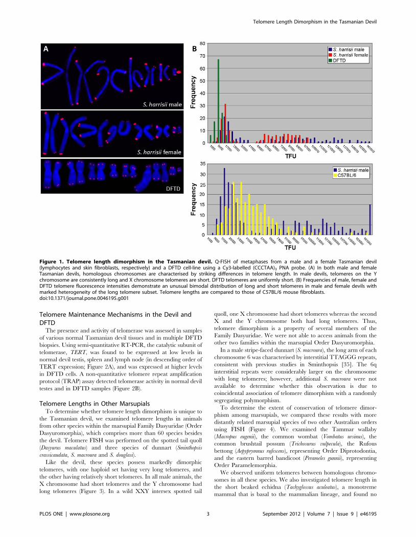
Telomere Length in the Tasmanian DevilThe telomere lengths of a male devil and female devil were
compared using quantitative fluorescent in situ hybridisation (Q-
FISH) with a Cy3-labelled (CCCTAA)3 peptide nucleic acid
(PNA) oligonucleotide. We found that chromosomes in both
animals had a striking pattern of long and short telomeres. One
homologue from each of the seven chromosome pairs had very
long telomeres, whereas the other had considerably shorter
telomeres. In images of metaphase chromosomes this correlates
with large, bright hybridisation signals on the seven chromosomes
with long telomeres. The seven homologues with short telomeres
have much smaller, more discrete fluorescence signals (Figure 1A).
This result suggested that the two haploid chromosome sets had
different telomere lengths.
To test the possibility that these two devils were hybrids between
two populations with disparate telomere length, we performed
non-quantitative telomere FISH on an additional four animals (2
females, 2 males) from different locations in Tasmania, and on two
healthy devils (2 males) from mainland Australian zoos. In all eight
animals, haploid chromosome sets were distinguished by strikingly
disparate telomere lengths, indicating that telomere length is not a
heritable trait segregating in the population. Rather, telomere
length dimorphism between homologues is a characteristic feature
of Tasmanian devil chromosomes.
The lengths of telomeres on sex chromosomes in male and
female devils were examined to investigate the possibility that long
and short telomeres were inherited differentially from male and
female parents. In all four female devils, X chromosome pairs
showed the same striking dimorphism as the autosome pairs. In all
four male devils examined, the Y chromosome was characterised
by particularly long telomeres, with short telomeres on the X
chromosome. This finding is consistent with inheritance of a
haploid set of chromosomes with long telomeres from the male
parent, and inheritance of a haploid set with short telomeres from
the female parent.
Terminal restriction fragment (TRF) analysis of Tasmanian
devil telomeres revealed that devil telomeric repeats are probably
interspersed with non-repeat sequences (Figure S1), as has been
reported for other marsupials [22]. Tasmanian devil telomere
length was therefore inferred by comparing hybridisation signals in
Q-FISH experiments using a mixture of devil and C57BL/6
mouse fibroblast cell lines (Figure 1B). Comparison of mouse and
Tasmanian devil telomere lengths highlighted the dimorphic devil
hybridisation signals and demonstrated that short Tasmanian devil
telomeres are substantially shorter than the approximately 50 kb
long C57BL/6 mouse telomeres. The haploid subset of long devil
telomeres is considerably longer than the mouse and is much more
heterogeneous than the short telomere subset, with a broader
range in telomere fluorescence.
Telomere Length in DFTD CellsQ-FISH with the same probe on cultured DFTD cell lines
demonstrated that tumour cells were characterised by short,
uniform telomeres, with small hybridisation signals on all
chromosomes (Figure 1). This was confirmed in an additional
four DFTD cell cultures that were prepared from tumour biopsies
collected over a period of four years from various locations in
Tasmania. Devil facial tumour disease is a clonal somatic cell line
that is propagated between host animals [26–31], so these results
indicate that telomere length has been maintained in the tumour
over several generations.
Telomere Length Dimorphism in the Tasmanian Devil
PLOS ONE | www.plosone.org 2 September 2012 | Volume 7 | Issue 9 | e46195
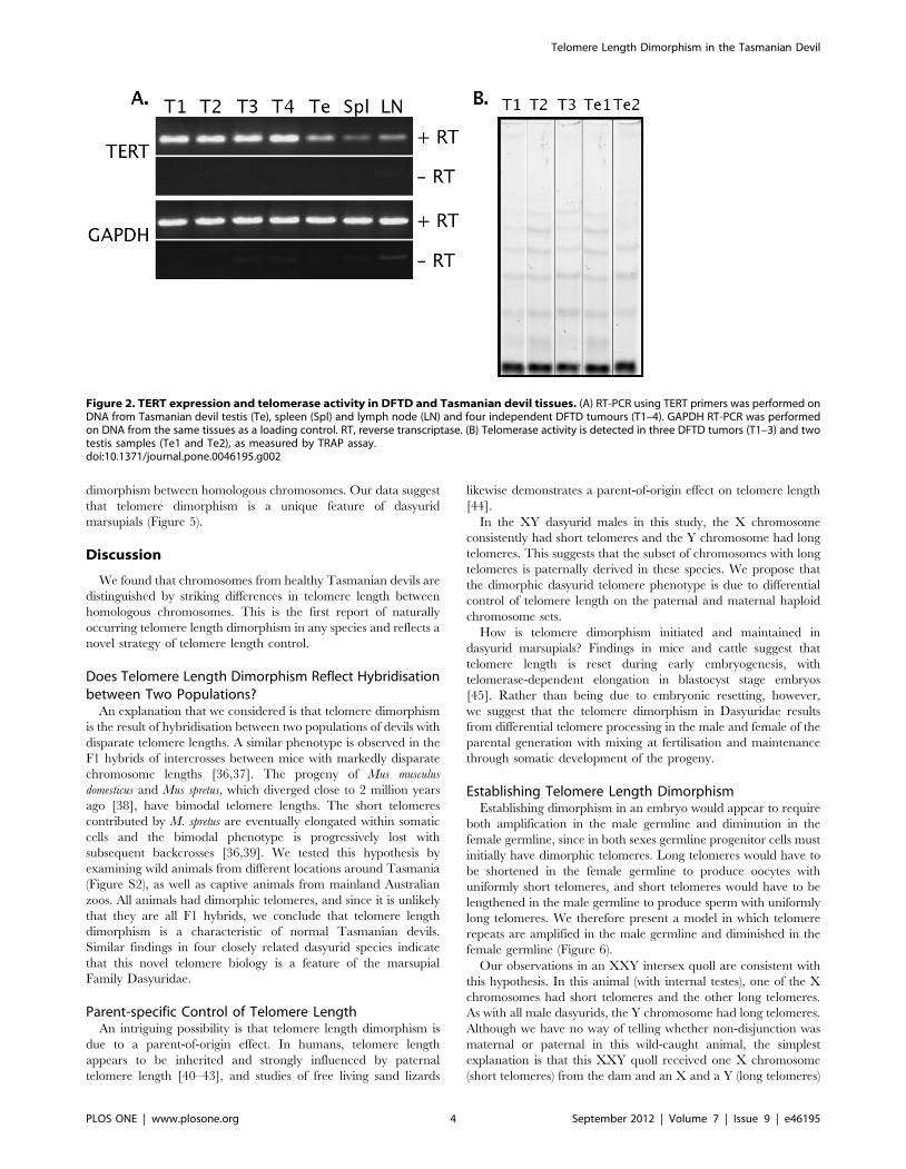
Telomere Maintenance Mechanisms in the Devil andDFTD
The presence and activity of telomerase was assessed in samples
of various normal Tasmanian devil tissues and in multiple DFTD
biopsies. Using semi-quantitative RT-PCR, the catalytic subunit of
telomerase, TERT, was found to be expressed at low levels in
normal devil testis, spleen and lymph node (in descending order of
TERT expression; Figure 2A), and was expressed at higher levels
in DFTD cells. A non-quantitative telomere repeat amplification
protocol (TRAP) assay detected telomerase activity in normal devil
testes and in DFTD samples (Figure 2B).
Telomere Lengths in Other MarsupialsTo determine whether telomere length dimorphism is unique to
the Tasmanian devil, we examined telomere lengths in animals
from other species within the marsupial Family Dasyuridae (Order
Dasyuromorphia), which comprises more than 60 species besides
the devil. Telomere FISH was performed on the spotted tail quoll
(Dasyurus maculatus) and three species of dunnart (Sminthopsis
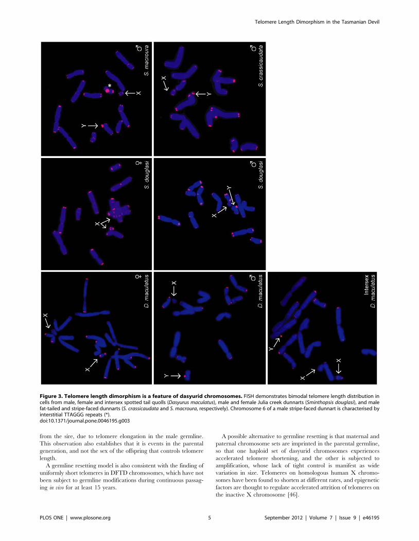
crassicaudata, S. macroura and S. douglasi).
Like the devil, these species possess markedly dimorphic
telomeres, with one haploid set having very long telomeres, and
the other having relatively short telomeres. In all male animals, the
X chromosome had short telomeres and the Y chromosome had
long telomeres (Figure 3). In a wild XXY intersex spotted tail
quoll, one X chromosome had short telomeres whereas the second
X and the Y chromosome both had long telomeres. Thus,
telomere dimorphism is a property of several members of the
Family Dasyuridae. We were not able to access animals from the
other two families within the marsupial Order Dasyuromorphia.
In a male stripe-faced dunnart (S. macroura), the long arm of each
chromosome 6 was characterised by interstitial TTAGGG repeats,
consistent with previous studies in Sminthopsis [35]. The 6q
interstitial repeats were considerably larger on the chromosome
with long telomeres; however, additional S. macroura were not
available to determine whether this observation is due to
coincidental association of telomere dimorphism with a randomly
segregating polymorphism.
To determine the extent of conservation of telomere dimor-
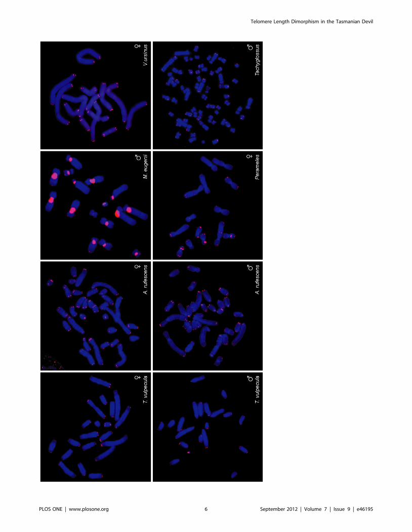
phism among marsupials, we compared these results with more
distantly related marsupial species of two other Australian orders
using FISH (Figure 4). We examined the Tammar wallaby
(Macropus eugenii), the common wombat (Vombatus ursinus), the
common brushtail possum (Trichosurus vulpecula), the Rufous
bettong (Aepyprymnus rufescens), representing Order Diprotodontia,
and the eastern barred bandicoot (Perameles gunnii), representing
Order Paramelemorphia.
We observed uniform telomeres between homologous chromo-
somes in all these species. We also investigated telomere length in
the short beaked echidna (Tachyglossus aculeatus), a monotreme
mammal that is basal to the mammalian lineage, and found no
Figure 1. Telomere length dimorphism in the Tasmanian devil. Q-FISH of metaphases from a male and a female Tasmanian devil(lymphocytes and skin fibroblasts, respectively) and a DFTD cell-line using a Cy3-labelled (CCCTAA)3 PNA probe. (A) In both male and femaleTasmanian devils, homologous chromosomes are characterised by striking differences in telomere length. In male devils, telomeres on the Ychromosome are consistently long and X chromosome telomeres are short. DFTD telomeres are uniformly short. (B) Frequencies of male, female andDFTD telomere fluorescence intensities demonstrate an unusual bimodal distribution of long and short telomeres in male and female devils withmarked heterogeneity of the long telomere subset. Telomere lengths are compared to those of C57BL/6 mouse fibroblasts.doi:10.1371/journal.pone.0046195.g001
Telomere Length Dimorphism in the Tasmanian Devil
PLOS ONE | www.plosone.org 3 September 2012 | Volume 7 | Issue 9 | e46195
dimorphism between homologous chromosomes. Our data suggest
that telomere dimorphism is a unique feature of dasyurid
marsupials (Figure 5).
Discussion
We found that chromosomes from healthy Tasmanian devils are
distinguished by striking differences in telomere length between
homologous chromosomes. This is the first report of naturally
occurring telomere length dimorphism in any species and reflects a
novel strategy of telomere length control.
Does Telomere Length Dimorphism Reflect Hybridisationbetween Two Populations?
An explanation that we considered is that telomere dimorphism
is the result of hybridisation between two populations of devils with
disparate telomere lengths. A similar phenotype is observed in the
F1 hybrids of intercrosses between mice with markedly disparate
chromosome lengths [36,37]. The progeny of Mus musculus
domesticus and Mus spretus, which diverged close to 2 million years
ago [38], have bimodal telomere lengths. The short telomeres
contributed by M. spretus are eventually elongated within somatic
cells and the bimodal phenotype is progressively lost with
subsequent backcrosses [36,39]. We tested this hypothesis by
examining wild animals from different locations around Tasmania
(Figure S2), as well as captive animals from mainland Australian
zoos. All animals had dimorphic telomeres, and since it is unlikely
that they are all F1 hybrids, we conclude that telomere length
dimorphism is a characteristic of normal Tasmanian devils.
Similar findings in four closely related dasyurid species indicate
that this novel telomere biology is a feature of the marsupial
Family Dasyuridae.
Parent-specific Control of Telomere LengthAn intriguing possibility is that telomere length dimorphism is
due to a parent-of-origin effect. In humans, telomere length
appears to be inherited and strongly influenced by paternal
telomere length [40–43], and studies of free living sand lizards
likewise demonstrates a parent-of-origin effect on telomere length
[44].
In the XY dasyurid males in this study, the X chromosome
consistently had short telomeres and the Y chromosome had long
telomeres. This suggests that the subset of chromosomes with long
telomeres is paternally derived in these species. We propose that
the dimorphic dasyurid telomere phenotype is due to differential
control of telomere length on the paternal and maternal haploid
chromosome sets.
How is telomere dimorphism initiated and maintained in
dasyurid marsupials? Findings in mice and cattle suggest that
telomere length is reset during early embryogenesis, with
telomerase-dependent elongation in blastocyst stage embryos
[45]. Rather than being due to embryonic resetting, however,
we suggest that the telomere dimorphism in Dasyuridae results
from differential telomere processing in the male and female of the
parental generation with mixing at fertilisation and maintenance
through somatic development of the progeny.
Establishing Telomere Length DimorphismEstablishing dimorphism in an embryo would appear to require
both amplification in the male germline and diminution in the
female germline, since in both sexes germline progenitor cells must
initially have dimorphic telomeres. Long telomeres would have to
be shortened in the female germline to produce oocytes with
uniformly short telomeres, and short telomeres would have to be
lengthened in the male germline to produce sperm with uniformly
long telomeres. We therefore present a model in which telomere
repeats are amplified in the male germline and diminished in the
female germline (Figure 6).
Our observations in an XXY intersex quoll are consistent with
this hypothesis. In this animal (with internal testes), one of the X
chromosomes had short telomeres and the other long telomeres.
As with all male dasyurids, the Y chromosome had long telomeres.
Although we have no way of telling whether non-disjunction was
maternal or paternal in this wild-caught animal, the simplest
explanation is that this XXY quoll received one X chromosome
(short telomeres) from the dam and an X and a Y (long telomeres)
Figure 2. TERT expression and telomerase activity in DFTD and Tasmanian devil tissues. (A) RT-PCR using TERT primers was performed onDNA from Tasmanian devil testis (Te), spleen (Spl) and lymph node (LN) and four independent DFTD tumours (T1–4). GAPDH RT-PCR was performedon DNA from the same tissues as a loading control. RT, reverse transcriptase. (B) Telomerase activity is detected in three DFTD tumors (T1–3) and twotestis samples (Te1 and Te2), as measured by TRAP assay.doi:10.1371/journal.pone.0046195.g002
Telomere Length Dimorphism in the Tasmanian Devil
PLOS ONE | www.plosone.org 4 September 2012 | Volume 7 | Issue 9 | e46195
from the sire, due to telomere elongation in the male germline.
This observation also establishes that it is events in the parental
generation, and not the sex of the offspring that controls telomere
length.
A germline resetting model is also consistent with the finding of
uniformly short telomeres in DFTD chromosomes, which have not
been subject to germline modifications during continuous passag-
ing in vivo for at least 15 years.
A possible alternative to germline resetting is that maternal and
paternal chromosome sets are imprinted in the parental germline,
so that one haploid set of dasyurid chromosomes experiences
accelerated telomere shortening, and the other is subjected to
amplification, whose lack of tight control is manifest as wide
variation in size. Telomeres on homologous human X chromo-
somes have been found to shorten at different rates, and epigenetic
factors are thought to regulate accelerated attrition of telomeres on
the inactive X chromosome [46].
Figure 3. Telomere length dimorphism is a feature of dasyurid chromosomes. FISH demonstrates bimodal telomere length distribution incells from male, female and intersex spotted tail quolls (Dasyurus maculatus), male and female Julia creek dunnarts (Sminthopsis douglasi), and malefat-tailed and stripe-faced dunnarts (S. crassicaudata and S. macroura, respectively). Chromosome 6 of a male stripe-faced dunnart is characterised byinterstitial TTAGGG repeats (*).doi:10.1371/journal.pone.0046195.g003
Telomere Length Dimorphism in the Tasmanian Devil
PLOS ONE | www.plosone.org 5 September 2012 | Volume 7 | Issue 9 | e46195
Telomere Length Dimorphism in the Tasmanian Devil
PLOS ONE | www.plosone.org 6 September 2012 | Volume 7 | Issue 9 | e46195
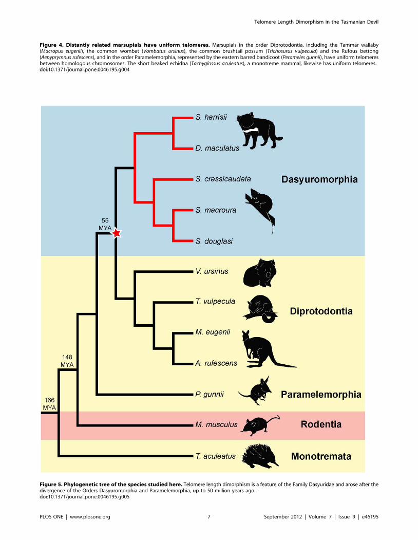
Figure 4. Distantly related marsupials have uniform telomeres. Marsupials in the order Diprotodontia, including the Tammar wallaby(Macropus eugenii), the common wombat (Vombatus ursinus), the common brushtail possum (Trichosurus vulpecula) and the Rufous bettong(Aepyprymnus rufescens), and in the order Paramelemorphia, represented by the eastern barred bandicoot (Perameles gunnii), have uniform telomeresbetween homologous chromosomes. The short beaked echidna (Tachyglossus aculeatus), a monotreme mammal, likewise has uniform telomeres.doi:10.1371/journal.pone.0046195.g004
Figure 5. Phylogenetic tree of the species studied here. Telomere length dimorphism is a feature of the Family Dasyuridae and arose after thedivergence of the Orders Dasyuromorphia and Paramelemorphia, up to 50 million years ago.doi:10.1371/journal.pone.0046195.g005
Telomere Length Dimorphism in the Tasmanian Devil
PLOS ONE | www.plosone.org 7 September 2012 | Volume 7 | Issue 9 | e46195

Maintenance of Telomere Length DimorphismHow dasyurids develop and maintain two distinct telomere size
classes is of great interest. There are four known elements of
telomere length control in mammals [47]. There are two telomere
shortening mechanisms, namely the gradual telomere attrition
which accompanies cellular proliferation and results in part from
the end replication problem, and the rapid, regulated telomere
shortening known as telomere trimming, and two lengthening
mechanisms - telomerase and ALT. TERT is expressed at low
levels in normal devil testis, spleen and lymph node, and we found
telomerase active in testes, suggestive of telomerase activity in the
germline and some somatic tissues (haematopoietic precursors and
lymphocytes), consistent with findings in humans [48]. It is
therefore possible that the massive lengthening of maternal-origin
telomeres that we presume occurs in the male germline may result
from sustained, high levels of telomerase activity, most likely
unopposed by telomere trimming. The heterogeneous telomere
lengths on chromosomes of paternal origin would also be
consistent with ALT-mediated lengthening in the male germline.
The extent of shortening of paternally-derived telomeres that
occurs in the female germline greatly exceeds the shortening that
would be expected to occur through gradual replication-associated
attrition so it is likely that the rapid telomere trimming mechanism
is involved here. The persistence of this dimorphism in somatic
cells suggests that the rapid shortening and lengthening mecha-
nisms are mostly inactive in these cells, whereas continued gradual
telomere attrition would be compatible with the dimorphism being
maintained.
Evolution of Telomere Length DimorphismWhen did this unusual telomere dimorphism evolve, and what
were the conditions that selected for it? The observation of
telomere dimorphism in all species of the Family Dasyuridae
(Order Dasyuromorphia), but not other marsupial orders, or other
mammal groups, provides an estimate of the time that this
mechanism evolved. This feature must have been present in the
common ancestor at least of dunnarts, quolls and devils, and
possibly the common ancestor of the whole Order Dasyuromor-
phia. The absence of telomere dimorphism in other marsupial
orders implies it must have evolved after the divergence of Order
Dasyuromorphia and Order Diprotodontia 50 million years ago
[49].
What might be the functional consequences or selective
advantage of telomere length dimorphism in dasyurids? Little is
known about the overall prevalence of neoplasia in dasyurid
species, although Tasmanian devils at the San Diego zoo were
reported to develop tumours at a higher frequency than other
species [50] and it is at least conceivable that this could be
associated with aspects of telomere length control in these animals.
However, although it is interesting to speculate whether some
aspect of the unusual telomere biology of dasyurids predisposes to
carcinogenesis, at present we have no evidence for this or that it
predisposed to the development of DFTD.
Interestingly, many dasyurids are well known for their
semelparous reproductive strategies. The species in this study are
iteroparus; however, some Antechinus species trade longevity for
intense and physiologically costly mating efforts [51] that result in
death of males at one year of age due to the systemic effects of
markedly elevated testosterone and cortisol levels. Stress has been
Figure 6. Proposed mechanism of telomere length regulation in the dasyurid germline. Primordial germ cells contain a mixture of longand short telomeres which would be expected to segregate randomly, followed by lengthening and diminution in the paternal and maternalgermlines, respectively. Following fertilisation, the zygote contains haploid chromosome sets with long and short telomeres which are maintained insomatic cells.doi:10.1371/journal.pone.0046195.g006
Telomere Length Dimorphism in the Tasmanian Devil
PLOS ONE | www.plosone.org 8 September 2012 | Volume 7 | Issue 9 | e46195

shown to contribute to telomere attrition [52] and it is interesting
to contemplate the significance of telomere lengthening in
dasyurid sperm. Perhaps remarkable telomere lengthening during
spermatogenesis provides a means to counter the effects of the
severe stress males succumb to during mating.
Our findings in the Tasmanian devil and related marsupials
suggest a novel system of germline regulation of telomere length.
These results provide an exciting avenue for future investigations
of the mechanisms that govern parental control of dimorphic
telomeres in dasyurid marsupials.
Materials and Methods
Ethics StatementSample collection was approved by the Australian National
University Animal Experimentation Ethics Committee (AEECP
R.CG.11.06).
Animals and Cell LinesTasmanian devil tumour, skin and blood samples were obtained
from biopsies of live, wild-caught Tasmanian devils (Tables S1 and
S2, Figure S2). Animals were trapped for the purposes of disease
surveillance and, while under general anaesthesia, were biopsied
by a veterinarian. Where euthanasia was indicated for welfare
reasons, complete necropsies were performed, allowing more
extensive tissue sampling, including testis.
Primary tumour, fibroblast and lymphocyte cultures were
initiated following published protocols [26]. Briefly, tumour and
skin samples were washed in 10 mL phosphate buffered saline
(Invitrogen, Mulgrave, VIC, Australia) with 0.1 mL penicillin-
streptomycin solution (Sigma-Aldrich, Castle Hill, NSW, Austra-
lia). Cultures were established by disaggregating tissue samples
using a scalpel and resuspending cells in 8 mL GIBCO
AmnioMAXTM-C100 (Invitrogen). Lymphocyte cultures were
initiated by isolating the buffy coat and stimulating cells with
phytohaemagglutinin (Gibco). Remaining marsupial and mono-
treme skin fibroblast cell lines were obtained from frozen stocks
maintained by the Comparative Genomics Group at the ANU
Research School of Biology (Table S3). All cultures were
incubated at 35uC in 5% CO2.
Telomere Length AnalysisMetaphase preparation. Cell lines were cultured in RPMI
1640 medium with Glutamax, HEPES buffer and 10% foetal
bovine serum. Cells were arrested with colcemid (0.1 mg/ml) and
harvested 4 hours later by treating with 0.075 M KCl for 20
minutes at 37uC and fixing in chilled methanol/acetic acid (3:1).
Fluorescent in situ hybridization. Telomere specific Q-
FISH analysis was performed on normal metaphases (fibroblasts
and lymphocytes) from two disease-free devils, a tumour cell line
and fibroblasts from a C57BL/6 mouse using a Cy3-labelled
(CCCTAA)3 PNA oligonucleotide as previously described [50].
Chromosomes were counterstained with DAPI and images were
acquired using a Zeiss Axioskope microscope and IP-lab Spectrum
acquisition software. A minimum of ten metaphases was assessed for
each experiment. Cells from an additional four Tasmanian devils,
the remaining eight marsupial species and one monotreme were
examined by non-quantitative FISH, also using a Cy3-labelled
(CCCTAA)3 PNA oligonucleotide. These images were captured
using Leica DMRXA fluorescence microscope and IP-lab software.
Telomere restriction fragment analysis and in-gel
hybridization. Agarose plugs containing genomic DNA were
prepared from samples of S. harrisii spleen and kidney according to
kit instructions (Bio-Rad), digested overnight with one or more
restriction enzymes (MboI, HinfI, RsaI, MspI, BstUI, and AluI) and
electrophoresed on a 1% agarose gel with 0.56TBE using a Bio-
Rad CHEF MAPPER at 6V/cm using a linear ramped factor,
switch times from 12.55 seconds to 1 minute, 8.67 seconds and
120u angle for 28.4 hours at 14uC. After electrophoresis the gel
was ethidium bromide stained and dried for 1 hour at 50uC. The
dehydrated gel was hybridised to [c-32P]ATP end-labelled
oligonucleotide (CCCTAA)5, washed and analysed by Phosphor-
Imager (Fuji) as previously described [10].
Telomerase ActivityRNA preparation and RT-PCR. RNA was extracted using
standard Trizol procedure from tissues stored in RNAlater. RNA was
treated with DNaseI and RT-PCR was performed for TERT
and GAPDH using primers TertF 59-CTGGCAAAAGGTATTC
CTGAG-39, TertR 59-CAAAACACGTTTAGGGTCCTTG-39,
GapdhF 59 GACTCAACCACGTATTCGGCTC-39 and GapdhR
59- ATATGATTCCACCCATGGCAAGTTCAA-39. Annealing
temperatures were 58uC and 60uC respectively.
Telomerase repeat amplification protocol (TRAP).
Telomerase activity was assessed using a commercial telomerase
repeat amplification protocol (TRAP) assay (TRAPeze kit, Chemicon).
Extracts from tumour and testis samples were prepared following the
manufacturer’s instructions. Following PCR amplification of three
serial lysate dilutions, PCR products were electrophoresed in a 10%
acrylamide gel, stained with ethidium bromide and imaged using a
Bio-Rad PharosFX system.
Supporting Information
Figure S1 Tasmanian devil telomeres contain non-repeat sequences. (A) Pulse-field gel electrophoresis and in-gel
hybridisation of MboI digested genomic DNA from Tasmanian devil
spleen using an end-labelled (CCCTAA)3 probe reveals discontin-
uous telomere fragments. A C57BL/6 mouse has a single TRF band
measuring 25–50 kb. (B) Tasmanian devil kidney DNA samples
digested with MboI (1), HinfI and RsaI (2), MspI, BstUI, AluI (3) and
HinfI, RsaI, MspI, BstUI, AluI (4) produce fragments of varying sizes.
(PDF)
Figure S2 Trapping locations in Tasmania. Tasmanian
devil and tumour samples were collected from various sites in
Tasmania. Samples were also obtained from two mainland zoos.
(PDF)
Table S1 Tasmanian devil (Sarcophilus harrisii) fibro-blast and lymphocyte preparations.
(DOCX)
Table S2 Devil facial tumour disease samples.
(DOCX)
Table S3 Marsupial and monotreme cell lines.
(DOCX)
Acknowledgments
We thank Amber Alsop (Walter and Eliza Hall Institute), Anne-Maree
Pearse and Kate Swift (Tasmanian Department of Primary Industries,
Parks, Water and Environment) for laboratory assistance and Elizabeth
Blackburn and Denis O’Meally for helpful discussion. For their assistance
collecting or providing tissue samples we acknowledge Drs Colette
Harmsen, Jemma Bergfeld and Alan Marcus, as well as the staff and
veterinarians at Taronga Zoo in Sydney and the National Zoo and
Aquarium in Canberra. HSB was supported by the Australian Research
Council and EPM by the American Australian Association. Thanks also to
Kathryn Davis and Dr David Middleton (The devils’ angels grant) for their
generous contributions to this project.
Telomere Length Dimorphism in the Tasmanian Devil
PLOS ONE | www.plosone.org 9 September 2012 | Volume 7 | Issue 9 | e46195

Author Contributions
Conceived and designed the experiments: HB EPM JAMG RRR GJH.
Performed the experiments: HB EPM HP CC DAM MS AAN. Analyzed
the data: HB GJH RRR JAMG. Contributed reagents/materials/analysis
tools: MS CWG. Wrote the paper: HB EPM HAP JED RRR JAMG.
References
1. Blackburn EH (1991) Structure and function of telomeres. Nature 350: 569–573.
doi:10.1038/350569a0.2. Martınez P, Blasco MA (2011) Telomeric and extra-telomeric roles for
telomerase and the telomere-binding proteins. Nat Rev Cancer 11: 161–176.doi:10.1038/nrc3025.
3. Shay JW, Wright WE (2011) Role of telomeres and telomerase in cancer. Semin
Cancer Biol 21: 349–353. doi:10.1016/j.semcancer.2011.10.001.4. Armanios M, Alder JK, Parry EM, Karim B, Strong MA, et al. (2009) Short
telomeres are sufficient to cause the degenerative defects associated with aging.Am J Hum Genet 85: 823–832. doi:10.1016/j.ajhg.2009.10.028.
5. Harley CB, Futcher AB, Greider CW (1990) Telomeres shorten during ageing of
human fibroblasts. Nature 345: 458–460. doi:10.1038/345458a0.6. Hastie ND, Dempster M, Dunlop MG, Thompson AM, Green DK, et al. (1990)
Telomere reduction in human colorectal carcinoma and with ageing. Nature346: 866–868. doi:10.1038/346866a0.
7. Hug N, Lingner J (2006) Telomere length homeostasis. Chromosoma 115: 413–425. doi:10.1007/s00412-006-0067-3.
8. Cesare AJ, Reddel RR (2008) Telomere uncapping and alternative lengthening
of telomeres. Mech Ageing Dev 129: 99–108. doi:10.1016/j.mad.2007.11.006.9. Britt-Compton B, Capper R, Rowson J, Baird DM (2009) Short telomeres are
preferentially elongated by telomerase in human cells. FEBS Lett 583: 3076–3080. doi:10.1016/j.febslet.2009.08.029.
10. Hemann MT, Greider CW (2000) Wild-derived inbred mouse strains have short
telomeres. Nucleic Acids Res 28: 4474–4478.11. Teixeira MT, Arneric M, Sperisen P, Lingner J (2004) Telomere length
homeostasis is achieved via a switch between telomerase- extendible and -nonextendible states. Cell 117: 323–335.
12. Shay JW, Zou Y, Hiyama E, Wright WE (2001) Telomerase and cancer. Hum
Mol Genet 10: 677–685.13. Henson JD, Hannay JA, McCarthy SW, Royds JA, Yeager TR, et al. (2005) A
robust assay for alternative lengthening of telomeres in tumors shows thesignificance of alternative lengthening of telomeres in sarcomas and astrocyto-
mas. Clin Cancer Res 11: 217–225.14. Bryan TM, Englezou A, Dalla-Pozza L, Dunham MA, Reddel RR (1997)
Evidence for an alternative mechanism for maintaining telomere length in
human tumors and tumor-derived cell lines. Nat Med 3: 1271–1274.15. Lansdorp PM (2009) Telomeres and disease. EMBO J 28: 2532–2540.
doi:10.1038/emboj.2009.172.16. Alder JK, Chen JJ-L, Lancaster L, Danoff S, Su S, et al. (2008) Short telomeres
are a risk factor for idiopathic pulmonary fibrosis. Proc Natl Acad Sci USA 105:
13051–13056. doi:10.1073/pnas.0804280105.17. Calado RT, Young NS (2008) Telomere maintenance and human bone marrow
failure. Blood 111: 4446–4455. doi:10.1182/blood-2007-08-019729.18. Savage SA, Bertuch AA (2010) The genetics and clinical manifestations of
telomere biology disorders. Genet Med 12: 753–764.19. Blasco MA, Lee HW, Hande MP, Samper E, Lansdorp PM, et al. (1997)
Telomere shortening and tumor formation by mouse cells lacking telomerase
RNA. Cell 91: 25–34.20. Cheung I, Schertzer M, Rose A, Lansdorp PM (2006) High incidence of rapid
telomere loss in telomerase-deficient Caenorhabditis elegans. Nucleic Acids Res34: 96–103. doi:10.1093/nar/gkj417.
21. Fitzgerald MS, Riha K, Gao F, Ren S, McKnight TD, et al. (1999) Disruption of
the telomerase catalytic subunit gene from Arabidopsis inactivates telomeraseand leads to a slow loss of telomeric DNA. Proc Natl Acad Sci USA 96: 14813–
14818.22. Gomes NMV, Ryder OA, Houck ML, Charter SJ, Walker W, et al. (2011)
Comparative biology of mammalian telomeres: hypotheses on ancestral statesand the roles of telomeres in longevity determination. Aging Cell 10: 761–768.
doi:10.1111/j.1474–9726.2011.00718.x.
23. Siddle HV, Marzec J, Cheng Y, Jones M, Belov K (2010) MHC gene copynumber variation in Tasmanian devils: implications for the spread of a
contagious cancer. Proc Biol Sci 277: 2001–2006. doi:10.1098/rspb.2009.2362.24. Loh R, Bergfeld J, Hayes D, O’hara A, Pyecroft S, et al. (2006) The pathology of
devil facial tumor disease (DFTD) in Tasmanian Devils (Sarcophilus harrisii).
Vet Pathol 43: 890–895. doi:10.1354/vp.43-6-890.25. Pyecroft SB, Pearse A-M, Loh R, Swift K, Belov K, et al. (2007) Towards a Case
Definition for Devil Facial Tumour Disease: What Is It? EcoHealth 4: 346–351.doi:10.1007/s10393-007-0126-0.
26. Pearse A-M, Swift K (2006) Allograft theory: transmission of devil facial-tumourdisease. Nature 439: 549. doi:10.1038/439549a.
27. Pearse A-M, Swift K, Hodson P, Hua B, McCallum H, et al. (2012) Evolution in
a transmissible cancer: a study of the chromosomal changes in devil facial tumor(DFT) as it spreads through the wild Tasmanian devil population. Cancer Genet
205: 101–112. doi:10.1016/j.cancergen.2011.12.001.28. Siddle HV, Kreiss A, Eldridge MDB, Noonan E, Clarke CJ, et al. (2007)
Transmission of a fatal clonal tumor by biting occurs due to depleted MHC
diversity in a threatened carnivorous marsupial. Proc Natl Acad Sci USA 104:
16221–16226. doi:10.1073/pnas.0704580104.29. Miller W, Hayes VM, Ratan A, Petersen DC, Wittekindt NE, et al. (2011)
Genetic diversity and population structure of the endangered marsupialSarcophilus harrisii (Tasmanian devil). Proc Natl Acad Sci USA 108: 12348–
12353. doi:10.1073/pnas.1102838108.
30. Murchison EP, Schulz-Trieglaff OB, Ning Z, Alexandrov LB, Bauer MJ, et al.(2012) Genome sequencing and analysis of the tasmanian devil and its
transmissible cancer. Cell 148: 780–791. doi:10.1016/j.cell.2011.11.065.31. Deakin JE, Bender HS, Pearse A-M, Rens W, O’Brien PCM, et al. (2012)
Genomic restructuring in the tasmanian devil facial tumour: chromosome
painting and gene mapping provide clues to evolution of a transmissible tumour.PLoS Genet 8: e1002483. doi:10.1371/journal.pgen.1002483.
32. McCallum H (2008) Tasmanian devil facial tumour disease: lessons forconservation biology. Trends Ecol Evol (Amst) 23: 631–637. doi:10.1016/
j.tree.2008.07.001.33. Hanahan D, Weinberg RA (2011) Hallmarks of cancer: the next generation. Cell
144: 646–674. doi:10.1016/j.cell.2011.02.013.
34. Shay JW, Bacchetti S (1997) A survey of telomerase activity in human cancer.Eur J Cancer 33: 787–791. doi:10.1016/S0959-8049(97)00062-2.
35. Metcalfe CJ, Eldridge MDB, Johnston PG (2004) Mapping the distribution ofthe telomeric sequence (T2AG3)n in the 2n = 14 ancestral marsupial
complement and in the macropodines (Marsupialia: Macropodidae) by
fluorescence in situ hybridization. Chromosome Res 12: 405–414.doi:10.1023/B:CHRO.0000034133.77878.88.
36. Zhu L, Hathcock KS, Hande P, Lansdorp PM, Seldin MF, et al. (1998)Telomere length regulation in mice is linked to a novel chromosome locus. Proc
Natl Acad Sci USA 95: 8648–8653.
37. Ding H, Schertzer M, Wu X, Gertsenstein M, Selig S, et al. (2004) Regulation ofmurine telomere length by Rtel: an essential gene encoding a helicase-like
protein. Cell 117: 873–886. doi:10.1016/j.cell.2004.05.026.38. Dejager L, Libert C, Montagutelli X (2009) Thirty years of Mus spretus: a
promising future. Trends Genet 25: 234–241. doi:10.1016/j.tig.2009.03.007.39. Hathcock KS, Hemann MT, Opperman KK, Strong MA, Greider CW, et al.
(2002) Haploinsufficiency of mTR results in defects in telomere elongation. Proc
Natl Acad Sci USA 99: 3591–3596. doi:10.1073/pnas.012549799.40. Vasa-Nicotera M, Brouilette S, Mangino M, Thompson JR, Braund P, et al.
(2005) Mapping of a major locus that determines telomere length in humans.Am J Hum Genet 76: 147–151. doi:10.1086/426734.
41. Njajou OT, Cawthon RM, Damcott CM, Wu S-H, Ott S, et al. (2007) Telomere
length is paternally inherited and is associated with parental lifespan. Proc NatlAcad Sci USA 104: 12135–12139. doi:10.1073/pnas.0702703104.
42. Chiang YJ, Calado RT, Hathcock KS, Lansdorp PM, Young NS, et al. (2010)Telomere length is inherited with resetting of the telomere set-point. Proc Natl
Acad Sci USA 107: 10148–10153. doi:10.1073/pnas.0913125107.43. Atzmon G, Cho M, Cawthon RM, Budagov T, Katz M, et al. (2010) Evolution
in health and medicine Sackler colloquium: Genetic variation in human
telomerase is associated with telomere length in Ashkenazi centenarians. ProcNatl Acad Sci USA 107 Suppl 1: 1710–1717. doi:10.1073/pnas.0906191106.
44. Olsson M, Pauliny A, Wapstra E, Uller T, Schwartz T, et al. (2011) Sexdifferences in sand lizard telomere inheritance: paternal epigenetic effects
increases telomere heritability and offspring survival. PLoS ONE 6: e17473.
doi:10.1371/journal.pone.0017473.45. Schaetzlein S, Lucas-Hahn A, Lemme E, Kues WA, Dorsch M, et al. (2004)
Telomere length is reset during early mammalian embryogenesis. Proc NatlAcad Sci USA 101: 8034–8038. doi:10.1073/pnas.0402400101.
46. Surralles J, Hande MP, Marcos R, Lansdorp PM (1999) Accelerated telomereshortening in the human inactive X chromosome. Am J Hum Genet 65: 1617–
1622. doi:10.1086/302665.
47. Pickett HA, Reddel RR (2012) The role of telomere trimming in normaltelomere length dynamics. Cell Cycle 11: 1309–1315. doi:10.4161/cc.19632.
48. Rufer N, Brummendorf TH, Kolvraa S, Bischoff C, Christensen K, et al. (1999)Telomere fluorescence measurements in granulocytes and T lymphocyte subsets
point to a high turnover of hematopoietic stem cells and memory T cells in early
childhood. J Exp Med 190: 157–167.49. Nilsson MA, Arnason U, Spencer PBS, Janke A (2004) Marsupial relationships
and a timeline for marsupial radiation in South Gondwana. Gene 340: 189–196.doi:10.1016/j.gene.2004.07.040.
50. Griner L (1979) Neoplasms in Tasmanian devils (Sarcophilus harrisii). Journal ofthe National Cancer Institute 62: 589–589–95.
51. Oakwood M, Bradley AJ, Cockburn A (2001) Semelparity in a large marsupial.
Proc Biol Sci 268: 407–411. doi:10.1098/rspb.2000.1369.52. Tomiyama AJ, O’Donovan A, Lin J, Puterman E, Lazaro A, et al. (2012) Does
cellular aging relate to patterns of allostasis? An examination of basal and stressreactive HPA axis activity and telomere length. Physiol Behav 106: 40–45.
doi:10.1016/j.physbeh.2011.11.016.
Telomere Length Dimorphism in the Tasmanian Devil
PLOS ONE | www.plosone.org 10 September 2012 | Volume 7 | Issue 9 | e46195
Related Documents