P/N:ENG-M7-420285x8P-20100225
M7 Diagnostic Ultrasound System
Mindray Building, Keji 12th Road South, High-tech Industrial Park,Nanshan, Shenzhen 518057, P.R. China
Tel: +86 755 86140388, 26582888 Fax: +86 755 26582680E-mail: [email protected] Website: www.mindray.com
2010
Mindray is listed on the NYSE under the symbol”MR”
DISTRIBUTOR:
Hand-Carried Color Doppler
M7 Diagnostic Ultrasound System
Hand-Carried Color Doppler
Clarity‧Accuracy‧PerformanceBring Your Crystal Ball to Point-of-Care
Confidence for DiagnoseRaising the level on image quality
iTOUCH with parasternal long axis and display cardiac structures
Anatomical M mode with three sample lines shows motions of three Cardiac regions simultaneously
Transvaginal transducer with high definition displays minute fetus clearly
Distinct fetal aortic arch with C5-2s convex transducer
Quality volume transducer shows vivid fetal facial organs
Sensitive Color Doppler displays plentiful renal blood flow
Dedicated software measures carotid IMT automatically and accurately
Vertebral artery and vein blood flow reflects enough penetration and good spatial resolution
Definite median nerve using L14-6s high frequency linear transducer

Multi-SpecialtyHigh Performance Ultrasound can let you scan more.
The M7 Diagnostic Ultrasound System is designed to fulfill clinicians' busy, challenging point of care environments. With M7's crystal clarity, crisp, clear image quality, it can perform any exam, from abdominal to vascular to cardiac, with efficiency and accuracy. Just choosing a transducer, the M7 brings you more benefits in more way than ever with wellness within reach.
Cardiovascular:
Obstetrics/Gynecology
Anesthesiology/ Emergency Medicine/ Musculosketal
Free Xros™ Imaging (Anatomic M mode)Tissue Doppler ImagingEmbedded IMT (Intima Media Thickness) software detects edge with mean and maximum thickness value.
Abundant and dedicated clinical measurement and analysis packagesWide range of broadband transducers including convex, linear, transvaginal, phased array and 4D transducer
4D Imaging on top of PortabilityNew transducer design: the ergonomic design and the light weight allow the users to scan in 2D as with a standard convex probe.New 4D transducer: With its ergonomic design and the light weight allow clinicians elevate speed of scanning and provide ease of 3D/4D acquisitions.Abundant clinical measurement and analysis packages

From Cart-Based Configuration to
Anywhere, Anytime
M7 Diagnostic Ultrasound System is a powerful imaging tool with superior image quality to assist you in meeting your clinical challenges today and tomorrow. The M7 is designed for use in all point of care environments. It delivers premium imaging performance across a broad range of specialties. Providing accurate data with speed, the M7 enables clinicians to achieve enhanced level of diagnostic confidence and efficiency.
High capacity Li-ion batteries support continuous scanning more than 1.5 hours. Robust magnesium with anti shock and anti splash ability can perform diagnostic exams whatever inside hospital or outdoor harsh environment.Comfortable grab and go backpack and artistic traveling case for easy transportationiRoam™, 802.11b/g wireless data transfer solutionDICOM 3.0 and M-Scan Pack providing Point-of-Care and field scan support
the Point-Of-Care Environment

It's obviously designed with the power of leadership MINDRAY technology available to all clinicians. As a world-class medical equipment solution provider, M7 is a multiplying power station with innovation for the future. With its ergonomic mobile trolley same with performance and features comparable to that of conventional cart-based systems, provide you mobility with power and improve your productivity. To sum it up, the M7 delivers you the power and productivity of a full-sized system in a hand-carried size.
Make your M7 uniquely yours with one of six colorful trackball
15-inch medical grade LCD monitor with integrated stereo speakers
Intel® Processor with Microsoft® brings the stability, security and connectivity of the system
iTouch™ Intelligent Image Optimization
iRoam™, 802.11b/g wireless data transfer solution
iZoom™ Automatically expand the image to full screen
iClear™: Adaptive Speckle Suppression
iBeam™: Spatial Compounding Imaging
Phase Inversion THI for all probes
Capture high quality cine up to 480 seconds long
Increased flexibility with the optional Transducer Extended Connector
Fast Power Up for quickly field scanning
Gorgeous design with Innovative technology

Designed for echocardiology environment
l Power up in seconds
l One key image optimization
l 15" LCD display
l Anti-shock magnesium case
l TDI
l TDIQ
l Free Xros M and CM
l LV Auto Calc
Premium performance through advanced technologies
l Multiple transducer connectors
(optional)
l Premium performance through
advanced technologies
l Multiple frequency broadband
imaging
l iZoom™: enables accurate viewing
of image for users from distance
l iClear™: speckle suppression
technology
l iBeam™: spatial compounding
l iStation™: on board workstation for
patient information management
and connectivity
l Wired and wireless connectivity:
DICOM PACS and PC
A premium performance procedural tool for echocardiology.
With high speed response, exceptional image quality and a sealed surface for easy
cleaning/infection control, the M7 is a top choice premium class imaging tool for
cardiology professionals. Mindray engineering team employs the System On Chip
(SOC) design to enable complex technologies to be built into the M7’s compact
chassis. The M7 elevates the performance standard of hand-carried ultrasound
imaging tool.
speed | clarity | flexibility
M7 AdvancedHand-carried Diagnostic Ultrasound System
Mindray™ is a trademark or registered trademark of Shenzhen Mindray Bio-Medical Electronics Co., Ltd. ©2012 Mindray DS USA, Inc. Subject to change. P/N: 0002-08-40041 Rev A
Mindray DS USA, Inc.
800 MacArthur Blvd., Mahwah, NJ 07430
Tel: 1.800.288.2121 Tel: 201.995.8000 Fax: 1.800.926.4275 www.na.mindray.com
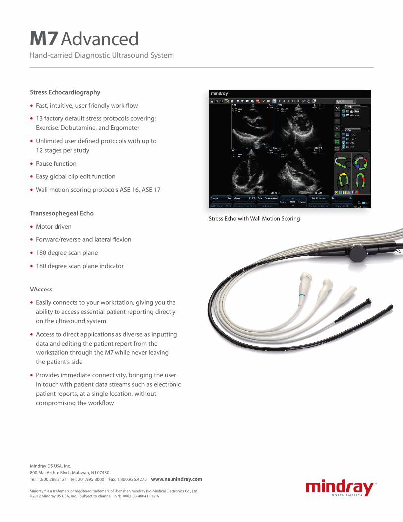
Stress Echocardiography
lFast, intuitive, user friendly work flow
l13 factory default stress protocols covering: Exercise, Dobutamine, and Ergometer
lUnlimited user defined protocols with up to 12 stages per study
lPause function
lEasy global clip edit function
lWall motion scoring protocols ASE 16, ASE 17
Transesophegeal Echo
lMotor driven
lForward/reverse and lateral flexion
l180 degree scan plane
l180 degree scan plane indicator
VAccess
lEasily connects to your workstation, giving you the ability to access essential patient reporting directly on the ultrasound system
lAccess to direct applications as diverse as inputting data and editing the patient report from the workstation through the M7 while never leaving the patient’s side
lProvides immediate connectivity, bringing the user in touch with patient data streams such as electronic patient reports, at a single location, without compromising the workflow
Stress Echo with Wall Motion Scoring
M7 AdvancedHand-carried Diagnostic Ultrasound System
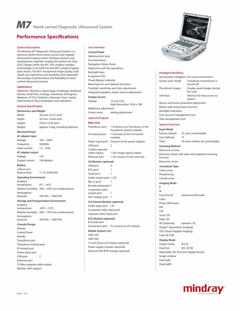
System DescriptionThe Mindray M7 Diagnostic Ultrasound System is a premium performance hand-carried color doppler ultrasound imaging system. Mindray research and development engineers employ the System On Chip (SOC) design within the M7. SOC enables complex technologies to be built into the M7’s compact laptop style chassis. The M7’s exceptional image quality, high speed user experience and versatility have expanded the envelop of performance and flexibility for hand carried ultrasound systems.
ApplicationsAbdomen, Obstetrics, Gynecology, Cardiology, Peripheral Vessels, Small Parts, Urology, Anesthesia, Emergency Medicine, IC/CCU, Pediatrics, Neonate, Trans-cranial, Interventional, Musculoskeletal, Intra-operative.
General Specification
Dimensions and WeightWidth: 361mm (14.21 inch)Depth: 357mm (14.06 inch)Height: 75mm (2.95 inch)Weight: Approx. 6.5kg, including batteries.
Electrical Power
AC adapter inputVoltage: 100 – 240V~Frequency: 50/60HzInput current: 1.5 – 0.6A
AC adapter outputVoltage: 12VOutput current: 10A Battery
BatteryLithium-Ion Battery Pack: 11.1V, 4500mAh
Operating EnvironmentAmbient temperature: 0°C ~ 40°CRelative humidity: 30% ~ 85% (no condensation)Atmospheric pressure: 700 hPa ~ 1060 hPa
Storage and Transportation EnvironmentAmbient temperature: -20°C ~ 55°CRelative humidity: 30% ~ 95% (no condensation)Atmospheric pressure: 700 hPa ~ 1060 hPa
Console DesignDisplayControl PanelHandleTransducer portTransducer locking leverIO extend portPower input portUSB port: 2Ethernet portS-Video separate video outputWireless LAN support
User Interface
Control PanelAlphanumeric keysFunctional keysNavigation Rotary KnobErgonomic soft key operationsBacklight keys8 segment TGCPower/Battery indicatorBlank keys for user-defined functionsTrackball, sensitivity and color adjustmentIntegrated speakers, audio volume adjustment
Display ScreenDisplay: 15 inch LCD,
High-Resolution 1024 x 768Brightness adjustmentScreen saver: setting adjustment
Inputs & Outputs
Main UnitTransducer port: 1 (Connect to a Transducer or the
Transducer extend module)I/O extend port: 1 (Connect to the I/O extend
module)Power input port: Connect to the power adapterUSB port: 2S-Video separate video output: 1 (For image signal output)Ethernet port: 1 (To connect to the network)
I/O Module (optional)USB port: 2ECG port: 1Serial port: 1Audio output port: 1 L/RMic In port: 1Remote control port: 1Composite video output port: 1DVI-I output port: 1
V/A Extend Module (optional)Audio input port: L/RComposite video input portSeparate video input port
ECG Module (optional)ECG lead portConnection port: To connect to I/O module
Mobile System CartUMT-200UMT-30015 inch Extra LCD Display (optional)Power supply module (optional)External DVD R/W storage (optional)
Intelligent WorkflowSynchronous navigation: On-screen instructionsScreen saver mode: Transducer transmission is
turned offThumbnail images: Display saved images during
live scanSoft keys: Shortcut for easy access to
system Menus and active parameter adjustmentReport edit and preview functionBacklight indicationUser account management toolTask management tool
System Overview
Exam ModeFactory default: 35, user customizableUser Defined: 15 Total: 50 exam modes, all customizable
Scanning MethodElectronic convexElectronic linear with steer and trapezoid scanning functionElectronic sector
Transducer Type Linear arrayPhased arrayConvex array
Imaging ModeBMFree Xros M: Anatomical M modeColorPower (DirPower)PWCWSmart 3DStatic 3D4D (optional): Dynamic 3DiScape™ (panoramic imaging)TDI (Tissue Doppler imaging)Color M (CM)
Display ModeTriplex mode: B/C/DDual live: B/C, B/TDIAdjustable 2D/ time line display formatSingle windowDual-split:Quad-split:
Hand-carried Diagnostic Ultrasound System
Performance Specifications
M7
Page 1 of 6

Imaging TechnologyTissue harmonic imagingTissue doppler ImagingSteer scanning for linear transducers (B, Color/Power, PW/CW independent)Trapezoid imaging for linear transducersiBeam™: Spatial compounding imaging for
linear transduceriClear™: Adaptive speckle suppression
imaging for all transducersiTouch™: Quick optimization for B or PW/CW
image with one button controlHPRF for PWMulti-frequency Transducers for 2D and Doppler imaging modes
Imaging FeatureZoom: Magnification factor 1 – 10Full screen (iZoom): Zoom in the image areaSystem dynamic range: 30 – 160dBFrame rate (Max.): 643 frames/sAdjustable focus positions (Max.): 16Maximum frame rate in 4D: 30 volumes/s
LanguagesSoftware display, control panel overlay and electronic copy of operation manuals including: Chinese, English, French, German, Italian, Portuguese, Russian, Spanish, Polish, Czech, Turkish, Finnish, Danish, Icelandic, Norwegian, and Swedish.
System Configuration
Standard ConfigurationDisplay: 15 inch LCD display,
High resolutionPWHPRFColor doppler flow imagingPower doppler flow imagingDirectional power doppler flow imagingTissue harmonic imagingTrapezoid imagingiBeam™iTouch™iStation™160G Integrated hard diskMulti-language screen display and control panel overlayCarrying case with telescopic handle
Software OptionsiClear™CWD moduleiScape™ moduleFree Xros M (Anatomical M)Smart 3D module4D moduleTDI (Tissue Doppler imaging) module
Application packages, including exam mode, comments, measurements, body marks and report. Abdominal package Obstetrical package Gynecological package Cardiac package Small parts package Urological package Vascular package Pediatric package Nerve blocks package Emergency medicine DICOM basic function module (including: task management, DICOM storage, DICOM print, DICOM storage commitment, DICOM media storage (including DICOM DIR)DICOM WorklistDICOM MPPSDICOM OB/GYN structured reportDICOM vascular structured reportDICOM cardiac structured reportDICOM Query/Retrieve
Hardware OptionsExternal USB DVD-RW: SE-S224QIO extend module: IOM-21Transducer extend module: PEM-21V/A extend module: VAM-11ECG module: ECG-21ECG leadFootswitch: 971-SWNOMMobile trolley: UMT-300PackDust-proof coverBattery Pack (LI23I001A)Wireless-LAN adapterTransducersNeedle-guided brackets
Peripherals SupportedBlack/white video printerSONY UP-D897Color video printer: SONY UP-D23MDGraph/text printerHP Deskjet D2568HP OfficeJet J3600 (HP Officejet J3608 All-in-One)HP Color LaserJet CM1015
Display AnnotationsManufacturer logoHospital name: Display up to 64 charactersExam date: 3 types selectable, YY/MM/DD,
MM/DD/YY, DD/MM/YYExam time: 2 time formatsAcoustic output indices: MI, TIC, TIS, TIBFreeze iconGenderAge
ID: Display up to 64 characters Name display up to 64 characters
Transducer modelCurrent exam modeECG icon (displays when connects with a physiology module)Accession#Operator: display up to 64 charactersMenuImageECG traceTransducer orientation markTime lineCoordinate axis, including depth, time, velocity/frequencyTGC curveFocusCommentBody MarkMeasure caliperGray/color scale barThumbnailCine iconTrackball functionality status iconHelp informationSoft MenuStatus iconsBiopsy guidelineMeasure result window (up to 8 results can be displayed)Image parameters B mode (including iScape™) Frequency (F) Depth (D) Gain (G) Frame rate (FR) B IP (IP) Dynamic range (DR)Color mode Frequency (F) Gain (G) IP (IP) Pulse repeated frequency (PRF) Wall filter (WF) M mode M Speed (V) M IP (IP) Dynamic range (DR)Power mode Frequency (F) Gain (G) IP (IP) Pulse repeated frequency (PRF) Wall filter (WF)
Performance Specifications
Page 2 of 6
Hand-carried Diagnostic Ultrasound SystemM7

System Configuration (cont'd)PW mode Frequency (F) Gain (G) Pulse repeated frequency (PRF) Wall filter (WF) Sample volume depth (SVD) Sample volume (SV)CW mode Frequency (F) Gain (G) Pulse repeated frequency (PRF) Wall filter (WF) Sample volume depth (SVD) Free Xros M (anatomical M) Gain (G) Velocity (V)TVI mode Frequency (F) Gain (G) TVI IP (IP) Pulse repeated frequency (PRF) Wall filter (WF)TEI mode Frequency (F) Gain (G) TEI IP(IP) Pulse repeated frequency (PRF) Wall filter (WF)TVD mode Frequency (F) Gain (G) Pulse repeated frequency (PRF) Wall filter (WF) Sample volume depth (SVD) Sample volume (SV)3D/4D Brightness (B) Contrast (C) Scan method (only for Smart 3D) Quality (Q, for Static 3D and 4D) Angle (A) Parameter pack: default or user-defined
SetupGeneral settingsUser-defined functional keys: Print, Save, F1-F6, footswitchCustomize user-defined exam modes in: Exam selection of each Transducer Configuration of measurement packages, body
mark and comment libraries Imaging parameters setting as well as
layout of menus and soft keys in imaging mode 15 User-defined exam modes Create new measurement items, body marks and comments Preset data manage: to save, load, export and defaultPeripheral devices installation and setting
DICOM settings and network settingSystem Maintenance (network updating, remote desktop, system test, log operation and preset)System information viewing
Imaging and Processing
Display DepthMinimum: 18mm, Transducer dependentMaximum: 388mm, Transducer dependent
B modeGain: 0 – 100TGC: 8 segments, with re-mapping
functionality at any depthiTouch™: -12dB – 12dBiTouch™ Bright: -2, -1, 0, 1, 2FOV positionB IP: 1 – 8, combination of dynamic
range, iClear™, persistence, smoothTHI IP: 1 – 8, combination of dynamic
range, iClear™, persistence, smoothRotation: 0°, 90°, 180°, 270°Colorize/ Colorize Map: On/Off, 1 – 10A. power: 10% – 100%, in increments of 6FOV: N, W, M1, M2Line Density: L, M, H, UHL/R FlipiClear™: 1 – 4, OffPersistence: 0 – 7U/D FlipTSI: General, Muscle, Fat, FluidSmooth: 1 – 4Gray Rejection: 0 – 5y: 0 – 3 CurveHigh FR: On, OffFrequency: Transducer dependentFocus PositionDyn Ra.: 30dB – 160dB, in increments of 5dBGray Map: 1 – 8Focus Number: 1 – 4B Steer: -6°, 0°, 6°Trapezoid: On, OffiBeam™: On, OffImg Merge: On, Off
M modeGain: 0 – 100TGC: 8 segments, with re-mapping
functionality at any depthIP: 1 – 8, combination of dynamic
range, M soften, edge enhanceA power: 10% – 100%, in increments of 6Display Format: L/R, 1:1, 1:2, FullM Soften: 0 – 4Gray Rejection: 0 – 5y: 0 – 3 CurveColorize/ Colorize Map: On/Off, 1 – 10
Time Mark: On, OffFocus PositionFrequency: Transducer dependentSpeed: 1 – 6Dyn Ra.: 30dB – 160dB, in increments of 5dBEdge Enhance: 0 – 3Gray Map: 1 – 8
Color modeGain: 0 – 100Color IP: 1 – 8, combination of Smooth and
PersistenceA. power: 10% – 100%, in increments of 6Line Density: L, M, H, UHB Display: On, OffSmooth: 0 – 4Persistence: 0 – 4Baseline: -8 – +8Focus Position: 0% – 100%Packet Size: 0 – 3B/C Wide: On, OffDual Live: On, OffMap: V0 – V10, VV0 – VV9Priority: 0 – 100%WF: 0 – 7Frequency: Transducer dependentScale: Frequency, Transducer and depth
dependentSteer: Transducer dependentInvert: On, OffFlow State: L, M, H
Power (DirPower)Gain: 0 – 100Packet Size: 0 – 3Flow State: L, M, HDyn Ra.: 10dB – 70dB, in increments of 5dBPower IP: 1 – 8, combination of Smooth and
PersistenceA. power: 10% – 100%, in increments of 6Line Density: L, M, H, UHSmooth: 0 – 4Persistence: 0 – 4Focus Position: 0% – 100%B Display: On, OffB/C Wide: On, OffDual Live: On, OffMap: P0-3 (Power), dP0-3 (DirPower)Priority: 0% – 100%Frequency: Transducer dependentScale: Frequency, Transducer and depth
dependentInvert: On, OffWF: 0 – 7Steer: -12°, 0°, 12°
Performance Specifications
Page 3 of 6
Hand-carried Diagnostic Ultrasound SystemM7
Imaging and Processing (cont'd)
PW/CWGain: 0 – 100V Max: On, OffV Mean: On, OffColorize/ Colorize Map: On/Off, 1 – 10Dyn Ra.: 24dB – 72dB, in increments of 2Audio: 0 – 100%, in increments of 2Trace Area: Above, Below, AllA. power: 10% – 100%, in increments of 6Trace Sensitivity: 0 – 5Trace Smooth: Off, 1 – 4Time Mark: On, OffDisplay Format: L/R, 1:1, 1:2, FullT/F Res: 0 – 3Auto Calc Param: On, OffHPRF: On, OffFrequency: Transducer dependentScale: Frequency, Transducer and depth
dependentBaseline: -4 – +4Invert: On, OffQuick Angle: -60, 0, 60Angle: -80 – 80°, in increments of 1°SV: 0.5mm – 20mmSVDWF: 0 – 6Auto Calc: On, OffSpeed: 1 – 6Duplex/Triplex: On, OffGray Map: 1 – 8Post Process: Curve, Gray Rejection, yPW Steer: Maximum ±20°
(Transducer dependent)
Free Xros MGain: 0 – 100TGC: 8 segments, with re-mapping
functionality at any depthColorize/ Colorize Map: On/Off, 1 – 10Post Process: N, curve, gray rejectionDisplay Format: L/R, 1:1, 1:2, FullDisplay: Cur., FullMark Adjustment: Show A, Show B, Show CTime Mark: On, OffAngleSpeed: 1 – 6Gray Map: 1 – 8
CMFor parameter details in CM mode, please refer to relevant sections of B, Color and M modes.
TVIGain: 0 – 100Baseline: -8 – +8TVI IP: 1 – 8, combination of Smooth and
PersistenceA. power: 10% – 100%, in increments of 6
Line Density: L, M, H, UHB Display: On, OffSmooth: 0 – 4Persistence: 0 – 4Focus Position: 0% – 100%Packet Size: 0 – 3B/C Wide: On, OffDual Live: On, OffMap: V0 – V10Priority: 0% – 100%WF: 0 – 7Frequency: Transducer dependentScale: Frequency, transducer and depth
dependentInvert: On, OffTissue State: L, M, H
TEIGain: 0 – 100Dual Live: On, OffTEI IP: 1 – 8, combination of Smooth and
PersistenceFocus Position: 0% – 100%Frequency: Transducer dependentScale: Frequency, Transducer and depth
dependentTissue State: L, M, HInvert: On, OffWF: 0 – 7Persistence: 0 – 4Smooth: 0 – 4Dyn Ra.: 10 – 70dB, in increments of 5B/C Wide: On, OffMap: P0 – P3, dP0 – dP3Packet Size: 0 – 3B Display: On, OffPriority: 0 – 100%Line Density: L, M, H, UHA. power: 10% – 100%, in increments of 6
TVDGain: 0 – 100Quick Angle: -60°, 0°, 60°WF: 0 – 6Trace Sensitivity: 0 – 5Auto Calc ParamV Max: On, OffV Mean: On, OffTrace Area: Above, Below, AllDuplex/Triplex: On, OffColorize/ Colorize Map: On/Off, 1 – 10Gray Map: 1 – 8Invert: On, OffSpeed: 1 – 6Angle: -80° – 80°, in increments of 1SV: 0.5mm – 20mmSVDA. power: 10% – 100%, in increments of 6Display Format: L/R, 1:1, 1:2, Full
Audio: 0% – 100%, in increments of 2%Frequency: Transducer dependentScale: Frequency, Transducer and depth
dependentBaseline: -4 – +4Dyn Ra.: 24 – 72dBTrace Smooth: Off, 1 – 4Time Mark: On, OffT/F Res: 0 – 3Post Process: Curve, Gray Rejection, y
TVMFor parameter details in TVM mode, please refer to relevant sections of B, M and TVI modes.
3D/4DMethod (only for Smart 3D): Fan, LinearDirection: Up/Down, Down/Up, Back/Front,
Front/Back, Left/Right, Right/LeftDisplay Format: Single, Dual, QuadDistance (for Smart 3D only): 10 – 200mm, in increments of 10mmAngleSmart 3D: 10 – 80°, in increments of 2°Static 3D/4D: Transducer dependentQuality (for Static 3D/4D only): Low 1, Low2, Mid, High 1, High2Inversion: On, OffPara pack: 5Auto Rot.: On, OffReset ROI (For Smart 3D only)Adjusting VOI: On, OffAccept VOI: On, OffColorize/ Colorize Map: Off, 1 – 5Reset: On, OffQuick rotate angle: 0°, 90°, 180°, 270°Current image: A/B/C/3DBrightness: 0 – 100%, in increments of 2Contrast: 0 – 100%, in increments of 2Smooth: 0 – 20, in increments of 1Threshold: 0 – 100%, in increments of 1Transparency: 0 – 100%, in increments of 5Render mode: Surface, Min, Max, X RayMPR Line: Partial, None, EntireEdit Type: Inside Contour, Outside Contour,
Big Contour, Big Eraser, Small Eraser, Inside Rect, Outside Rect, Inside Polygon, Outside Polygon
Edit Depth: Full Depth, User Defined (0 – 100%) Reset Curve
iScape™ ViewActual SizeFit SizeRuler: On, OffColorize/ Colorize Map: Off, 0 – 10Rotation: 0 – 360°, in increments of 5°
Performance Specifications
Page 4 of 6
Hand-carried Diagnostic Ultrasound SystemM7
Comments and Body Mark
Text commentComment text (option)Abdomen: 89OB: 97Cardiology: 80GYN: 69Vascular: 110Urology: 61SMP: 124Pediatrics: 35Nerve blocks: 52EM: 126User-defined CommentsAddDelete
ArrowArrow SizeArrow positionArrow orientation
TraceControl panel operation
Body Mark
Application package (Option)Abdomen: 13OB: 25Cardiology: 13GYN: 7Vascular: 17Urology: 7SMP: 46Nerve blocks: 32EM: 38
User-definedNewCopyExportLoadDeleteEdit
Storage/Connectivity320G integrated hard diskExternal DVD-R/W (Optional)USB portsImage archive on hard disk and DVD, temporary saving in cine memoryLive capture: Retrospective (1 – 120s, or 1 – 120 cycles) Prospective (1 – 120s, or 1 – 120 cycles)ThumbnailSingle image formats: BMP, JPG, DCM, FRM, supports
off-line analysis
Multi-frame images formats: AVI, DCM, CIN, supports off-line
analysisClip length: 1 – 60s, 1 – 16 cyclesStorage area: Image area: 640×480 Standard area: 800×600 Full-screen: 1024×768iVision™Cine review: Auto, Manual (auto review
segment can be set), supports linked cine review for 2D, M/D images, 8380 frames (Max.).
Send/print image after End ExamDICOM: DICOM Storage DICOM print DICOM Worklist Query/Retrieve Structured Report (SR) Storage Commitment MPPS Media review
iStation™
Intelligent patient data management platformIntegrated search engine for patient dataDetailed patient information viewIntelligent data backup/ restorePatient data/ image sendingPatient data deletingExam managing: create new exam, activate exam and continue examRecycle Bin
Measure/Calc/Study
Caliper2D modeM modeDoppler mode
ApplicationOptional package for specific clinical usesClinical PackagesAbdomenObstetricsCardiologyVascularGynecologyUrologySmall PartsPediatrics
Diagnostic ReportView/add imagesEdit reportObstetric/vascular analysis
Fetal growth curvePrint reportImport/export reportView history report
Physio Input/ Output
ECGDisplay: On, OffPosition: 0% – 100%, in increments of 5Display HR: On, OffGain: 0 – 30
Transducer Specifications
C5-2sArray type: Convex-wideApplications: Gynecology and obstetrics,
abdomen, vascular, pediatricsB mode imaging frequency: 2.5/3.5/5.0MHzHarmonic frequency: 5.0/6.0MHzDoppler frequency C: 2.5 /3.0MHz PW: 2.5 /3.0MHzConvex radius: 49.57mmBiopsy guide: NGB-015, 25°/35°/45°
7L4sArray type: LinearApplications: Small parts, vascular,
musculoskeletal, pediatrics, abdomen
B mode imaging frequency: 5.0/7.5/10MHzHarmonic frequency: 8.0/10MHzDoppler frequency C: 5.0/5.7MHz PW: 5.0/5.7MHzSteer angle: ±6°/12°Biopsy guide: NGB-007, 40°/50°/60°
L14-6sArray type: LinearApplications: Small parts, vascular,
musculoskeletal, pediatricsB mode imaging frequency: 8.0/10.0/12.0MHzHarmonic frequency: 10.0/11.0MHzDoppler frequency C: 5.7 /6.6MHz PW: 5.7 /6.6MHzSteer angle: ±6°/20°Biopsy guide: NGB-016, 30°/40°/50°
Performance Specifications
Hand-carried Diagnostic Ultrasound SystemM7
Page 5 of 6
Transducer Specifications (cont'd)
L14-6NsArray type: LinearApplications: Small parts, vascular,
musculoskeletal, pediatricsB mode imaging frequency: 8.0/10.0/11.0MHzHarmonic frequency: 10.0/14.0MHzDoppler frequency C: 5.7 /11MHz PW: 5.7 /6.6MHzSteer angle: ±6°/20°
L12-4sArray type: Linear Applications: Small parts, vascular,
musculoskeletal, pediatricsB mode imaging frequency: 6.0/7.5/10.0MHzHarmonic frequency: 10.0/11.0MHzDoppler frequency C: 5.0 /5.7MHz PW: 5.0/5.7MHzSteer angle: ±6°/12°Biopsy guide: NGB-007
P4-2sArray type: Sector phasedApplications: Cardiology, abdomen,
transcranial, pediatricsB mode imaging frequency: 2.0/2.5/3.0MHzHarmonic frequency: 3.2/3.6MHzDoppler frequency C: 2.0/2.3MHz PW: 2.0/2.5MHz CW: 2.0MHz TVI: 2.5/3.0MHz TVD: 2.0/2.5MHz TEI: 2.5/3.0MHzBiopsy guide: NGB-011, 11°/23°
P7-3sArray type: Sector phasedApplications: Cardiology, abdomen,
transcranial, pediatricsB mode imaging frequency: 3.6/5.0/6.6MHzHarmonic frequency: 6.0/7.0MHzDoppler frequency C: 3.3/4.0MHz PW: 3.2/4.0MHz CW: 3.3MHz TVI: 3.3/4.0MHz TVD: 3.2/4.0MHz TEI: 3.3/4.0MHzBiopsy guide: None
4CD4sArray type: ConvexApplications: Abdomen, gynecology, obstetricsB mode imaging frequency: 2.5/4.5/6.0MHzHarmonic frequency: 5.0/6.0MHzDoppler frequency C: 2.5/3.0MHz PW: 2.5/3.0MHzConvex radius: 40mmSwing angle (Max.): 70°Biopsy guide: None
V10-4sArray type: ConvexApplications: Gynecology, obstetrics, urologyB mode imaging frequency: 5.0/6.5/8.0MHzHarmonic frequency: 8.0/9.0MHzDoppler frequency C: 4.0/5.0MHz PW: 4.0/5.0MHzConvex radius: 10mmBiopsy guide: NGB-004
V10-4BsArray type: ConvexApplications: Gynecology, obstetrics, urologyB mode imaging frequency: 5.0/6.5/8.0MHzHarmonic frequency: 8.0/9.0MHzDoppler frequency C: 4.0/5.0MHz PW: 4.0/5.0MHzConvex radius: 10mmBiopsy guide: NGB-004
Safety & Conformance
Quality StandardsISO 9001:2000ISO 13485:2003
Design StandardsUL 60601-1CSA C22.2 No. 601-1EN 60601-1 and IEC 60601-1EN 60601-1-1 and IEC 60601-1-1EN 60601-1-2 and IEC 60601-1-2EN 60601-2-37 and IEC60601-2-37EN60601-1-4 and IEC60601-1-4EN60601-1-6 and IEC60601-1-6
CE DeclarationM7 system is fully in conformance with the Council Directive Concerning Medical Devices 93/42/EEC. The number adjacent to the CE marking (0123) is the number of the EU-notified body that certified meeting the requirements of Annex II of the Directive.
M7™ is a trademark of Mindray DS USA, Inc. Mindray™ is a trademark of Shenzhen Mindray Bio-Medical Electronics Co., Ltd.© 2011 Mindray DS USA, Inc. Subject to change. P/N: 0002-08-40011 Rev A
Mindray DS USA, Inc.800 MacArthur Blvd., Mahwah, NJ 07430Tel: 1.800.288.2121 Tel: 201.995.8000 Fax: 1.800.926.4275 www.na.mindray.com
Performance Specifications
Page 6 of 6
Hand-carried Diagnostic Ultrasound SystemM7

Power
iStation (Patient management)
Setup
Report
Patient (Input patient information)
Exam (Choose probe and exam)
Review
End Exam
Body Mark
Cine
Zoom
A B C Comment & Arrow
iTouch (Gain & iTouch)
Image Mode & 2D/B Mode Select
Multi-function Knob
Depth
Update (Shift exam status)
Measure
Cursor
Save (Frame or cine)
Set
Soft Keys
1
2
3
4
5
6
7
8
9
10
11
12
13
14
15
16
17
18
19
20
21
22
1
4
5
6 9
7 10
8 11
2 3
M7Quick Reference Guide
22
2117
18
13
19
2016
12
14
15

Getting StartedNew Patient1. Press [Patient], fill in basic patient information.2. Press [Exam].3. Move [Trackball] to choose probe and exam mode.
ScanningB Mode1. Press [B] to enter B mode.2. Rotate [Gain & iTouch] knob to adjust gain.
CDFI/Power Mode1. Press [Color] to enter Color mode.2. Move [Trackball] to change the position of ROI.3. Press [Set], and then move [Trackball] to change the size of ROI, press [Set].
PW/CW Mode1. Press [PW] to enter PW mode.2. Move [Trackball] to change the position of sample volume.3. Use related [Function Key] to change the size and angle of sample volume, press
[Update] or [PW] to get the Pulse Wave Doppler.
Image Adjustment
The [Soft Key] is corresponding to the soft menu. Use left/right keys to switch the modes, use up/down keys to choose parameters and turn pages up or down.The parameters in B mode: Frequency, iBeam, Focus Position, iClear, ExFOV, Line Density, Gray Map, Focus Number, Colorize, FOV, iTouch, Persistence, L/R Flip, Colorize Map, Dynamic Range, Acoustic Power, IP, U/D Flip, Magnify, FOV position.The parameters in Color mode: Scale, Flow State, WF, Invert, Packet Size, Frequency, Priority, Baseline, Line Density, Dual Live, Color IP, Map, B/C Wide, Focus Position, Acoustic Power.
Special Imaging Mode (Optional)3D/4D1. Select the volume probe and choose the exam mode. Press [3D/4D] key on the
control panel or click [3D/4D] in the soft menu of B mode to enter.2. Move [Trackball] and press [Set] to adjust the ROI and curve VOI.3. Press [Update] to enter the 4D image real-time status.
M7Quick Reference Guide
Free Xros M mode (Anatomical M mode)1. Under B mode, click [Free Xros M] on the screen to enter.2. Move [Trackball] to set the position of the sample line, and rotate [Multi-functional
Knob] to change the angle of the sample line.
Tissue Doppler Imaging1. Under B mode with phased array probe, click [TDI] on the screen to enter TDI mode.2. Move [Trackball] to change the position and size of the ROI.3. Press [M] to enter TVM mode; press [PW] to enter TVD mode; press [Power] to
enter TEI mode.
Measurement1. Press [Freeze] to freeze the image before the measurement.2. Press [Measure] to enter the application measurement status.3. Move [Trackball] to choose the measurement tool, and then go to the desired
position to measure.
Post ScanningComments and Body MarksComment1. Press [Comment] to choose the comment setting position and then add the
comment to the image.
Body Mark1. Press [Body Mark] and rotate it to choose the desired one.2. Move [Trackball] to place the probe marker, and rotate the [Multi-functional Knob]
to adjust the orientation of the probe.
Save Images or Cine1. Press [Save] to save a single-frame image or cine to the system.2. Press [Review] or select an exam of a patient in the [iStation] screen, and click
[Review] to see the image of cine.
Report and Print1. Press [Report], move the cursor to the comment text box and type the text.2. Click [Image select] on the report page to add images, and then click [Print View]
to preview the report, click [Print] to print the report out.
End ExamPress [End Exam] to end one examination. You may start a new exam by repeating the instructions above.
Image ManagementImage Transfer1. Press [iStation] to enter the image management system and choose the images.2. Click [Send to] to send the images to USB, DICOM, etc.
Note: For detailed information, please refer to the operation manual.
Mindray DS USA, Inc. 800 MacArthur Blvd., Mahwah, NJ 07430 Tel: 1.800.288.2121 or 201.995.8000 www.na.mindray.comP/N
: 046
-003
456-
00 R
ev A