13
Ischaemic Postconditioning Reduces Reperfusion Injury After Aortic
Revascularization Surgery
Gabor Jancso, Endre Arató and Lászlo Sinay University of Pécs, Faculty of Medicine,
Department of Vascular Surgery, Hungary
1. Introduction
In vascular surgery during the manipulation on the vessels the periferial tissues always suffer from a more or less severe ischaemia. In acute ischaemia the time of ischaemia could be also serious, thus after reconstruction we always have to face with reperfusion injury. The aim to reduce these extent of these reperfusion injury associated pathways has real clinical importance in vascular surgery.
Reperfusion injury is an inherent response to the restoration of blood flow after ischaemia, and is initiated at the very early moments of reperfusion, lasting potentially for days. The extent of the oxidative stress and the consecutive generalized inflammatory response depends on the ischaemic-time, the ischaemic tissue volume, and the general state of the endothelium-leukocyte-tissue functional complex (diabetes, chronic ischaemia, drugs). The pathogenesis of reperfusion injury is a complex process involving numerous mechanisms exerted in the intracellular and extracellular environment.
2. Pathophysiology of reperfusion injury
Reperfusion injury is not a mere worsening of the ischaemia-induced damage, but it is secondary to events that are specifically induced by reperfusion. In fact, reperfusion injury is due to complex mechanisms involving mechanical, extra-cellular and intracellular processes. The modern hypothesis of the pathogenesis of reperfusion injury has been reviewed by Piper and al.1 In patients with acute periferial ischaemia, it is now widely accepted that periodically reopening the occluded artery is accompanied by a reduction of the extent of necrosis and a major reduction of short- and long-term mortality. However, together with a definite protective effect on ischaemic tissues, post-ischaemic reperfusion may bring with it unwanted consequences that may partly counteract its beneficial effects. This phenomenon has thus been named reperfusion injury.
2.1 Causes of reperfusion injury
It seems that in the tissue ischaemia/reperfusion (I/R) can induce various forms of cell death, such as programmed cell death, apoptosis, oncosis and necrosis2. Apoptosis can be
www.intechopen.com

Vascular Surgery
238
caused by both prolonged ischaemia/hypoxia and by reperfusion3. In contrast to programmed cell death, apoptosis and oncosis, which are pre-mortal processes, necrosis is a post-mortal event. According to this viewpoint necrosis is not a form of cell death but the end stage of cell death processes.
The mechanisms of reperfusion-induced cell death are not completely understood, but it
seems that the occurrence of oxidative stress related to the generation of ROS may play an
important role4. ROS have downstream effects, which results in the initiation of a highly
orchestrated acute inflammatory response through the release of cytokines, activation of
vascular endothelial cells and leukocytes with expression of cell surface adhesion molecules,
and up-regulation of a program of pro-inflammatory genes, which contribute to the onset
and maintenance of post-ischaemic inflammation5. When the occlusion of the artery branch
that perfuse the ischaemic tissue is removed, the superoxide anion (O2-) production
increases as a result of the activation of various enzymatic complexes. The superoxide anion
and other ROS strongly oxidize the myocardial fibres already damaged by the ischaemia,
thus favouring the apoptosis6. It reacts with the nitric oxide, forming peroxynitrite (ONOO–).
Therefore, ONOO-, represents a sign of a reduced availability of nitric oxide7 and it
participates with O2- in the lesion of tissues8. Superoxide anion dependent damages are
reduced if O2- is transformed to hydrogen peroxide (H2O2) by the superoxide-dismutase.
However, since in the presence of Fe2+ or Cu2+, the H2O2 can be transformed in hydroxyl
anion (HO-), which is more toxic than O2- and H2O2, an increase in toxicity can occur.
Reperfusion injury is also due to cellular Ca2+ overload. The Ca2+-overload, which starts
during ischaemia, is further increased during reperfusion.
The overload of Ca2+ increases the cellular osmolarity favouring swelling (explosive
swelling) of skeletal muscle cells; it can also favour the expression of proapoptotic elements
from mitochondria9. It is noteworthy that altered cytosolic Ca2+-handling during ischaemia
may induce structural fragility and excessive contractile activation upon reperfusion, as also
indicated from a progressive increase of ventricular diastolic pressure and contraction band
necrosis10.
Ca2+-overload is also considered to be responsible for the opening of mPTP. Although,
mPTP opening is strongly inhibited by acidosis during ischaemia, it is favoured by ATP
depletion, oxidative stress and high intramitochondrial Ca2+ concentrations, conditions all
occurring during myocardial reperfusion11.
Intriguingly, the nuclear factor kappa B (NFkB) plays a double-edge sword role in tissue-
protection. Activation of NFkB is essential for late preconditioning, in which NFkB is
involved in the up-regulation of iNOS and COX-2 genes. However, in the longer time the
role NFkB is also important in reperfusion injury. It contributes to the exacerbation of the
tissues lesions sustaining inflammatory reactions. The activation of NFkB is induced from
several agents included hydrogen peroxide12. NFkB determine an up-regulation of the genes
responsible of the production of molecules of cellular adhesion. These molecules favour the
adhesion of leukocytes to the endothelium and possibly the migration within the cells13.
Moreover, the reduced nitric oxide availability determined by I/R participates to the
activation of transcription codifying for molecules of cellular adhesion14. Therefore, tissue
damages during reperfusion among others can be due to the cellular/mitochondrial
www.intechopen.com

Ischaemic Postconditioning Reduces Reperfusion Injury After Aortic Revascularization Surgery
239
overload of Ca2+, to the liberation of ROS, to the activation of mPTP, to the reduced
availability of nitric oxide and to the activation of the NFkB. The nitric oxide deficiency can
also cause vasoconstriction and formation of micro-thrombi into the lumen of the small
vessels15. These mechanisms, combined with the adhesion of the leucocytes to the
endothelium, can lead to the so-called ‘no-reflow phenomenon`16. In summary, reperfusion
injury is due to several mechanisms that include Ca2+ overload, ROS generation, reduced
availability of nitric oxide, mPTP opening and to the activation of the NFkB, which lead to
the augmented expression of molecules of cellular adhesion, leukocyte infiltration and no-
reflow phenomenon.
3. Effects of reperfusion injury
Among the outcomes of reperfusion injury are included: (1) endothelial and vascular
dysfunction and the sequels of impaired arterial flow, which may concur with the ‘no-
reflow phenomenon’; (2) metabolic and contractile dysfunction; (3) arrhythmias in case of
myocardial I/R; (4) cellular death by cellular swelling, and apoptosis. One may anticipate
that effective treatment during reperfusion may reduce tissue injury. However, the
complexity of mechanisms suggests that one single intervention aimed to contrast just one
or two of these mechanisms may not be sufficient. (Figure 1)
4. Definition of postconditioning
The concept of ‘Ischaemic PostC’ was first described by Vinten-Johansen’s group17. This study was performed in a canine model of 1 hr coronary occlusion and 3 hrs reperfusion. In this study the PostC algorithm was 30 sec. of reperfusion followed by 30 sec. of coronary occlusion, which were repeated for three cycles at the onset of reperfusion. Although this seminal study used the term ‘Ischaemic PostC’, subsequent studies of these and other authors omit the term ‘Ischaemic’ because it is not clear whether the brief periods of ischaemia, the preceding and/or the subsequent periods of reperfusion, or their combination, provide the key stimulus for cardioprotection. In general, PostC can be defined as intermittent interruption of coronary flow in the very early phase of a reperfusion, which leads to protection against reperfusion injury. The duration and number of these stuttering periods of reperfusion and ischaemia has been one of the aims of early studies on this topic.
5. Protective effects of postconditioning
Ischaemic PostC was already examined mostly in myocardium, thus the protective effects
are known in the myocardium. Our study was the first to examine the effect of PostC in
peripheral tissues.
Depending on species, models and other factors, PostC reduces the infarct size by ~20–70%
versus matched controls with matched risk areas. There is an emerging agreement across
multiple models and species that PostC may reduce endothelial dysfunction and endothelial
activation, thus leading to a reduced endothelial/leukocyte interaction and to a reduced
ROS formation. Reduced incidence of apoptosis and arrhythmias has also been observed.
Whether PostC reduces post-ischaemic stunning it has not yet been clarified.
www.intechopen.com

Vascular Surgery
240
Fig. 1. Simplified presentation of the mechanism of ischaemic-reperfusion injury. Emphasizing, that the engine of reperfusion injury is the ROI – cytokine – leukocyte positive feedback circle. Hypoxia leads to intracellular ATP depletion with a consecutive hypoxanthine elevation. In the early seconds of reperfusion, when the molecular oxygen appears in the cell, the - xanthine oxidase catalised – hypoxanthine-xanthine conversion will produce a mass of superoxide radicals. Superoxide radical and the other reactive oxygen intermediates will damage the membrane-lipids (through lipidperoxidation), the proteins (causing enzyme defects and ion channel injury) and the DNA. These are the main pathways of the cellular oxidant injury. The endogenous antioxidant system defends against these radical injuries. 18
Reactive oxygen species (ROS) will also induce local and systemic inflammatory responses through the inducing of cytokine expression and leukocyte activation. Inflammatory process leads to increased microvascular permeability, interstitial edema, and capillary perfusion depletion. The oxidative and inflammatory pathways will lead to a complex reperfusion injury. 19 (ROI – reactive oxygen intermediers; ATP – adenosine triphosphate; DNA – deoxyribonucleic acid)
6. Reduction of necrosis
In their seminal study Vinten-Johansen and coworkers 20 reported that PostC causes massive
salvage of the myocardium. The infarct size was reduced by ~45% when the initial minutes
of reperfusion were ‘stuttered’ compared to an abrupt and complete reperfusion. These
findings have been confirmed by several laboratories as well21. As mentioned, in multiple
HYPOXIA
ATP↓
Na/K transport ↓
Ca influx Calmodulin ↑
Proteolitic enzymes↑Xanthine dehydr→ xanthine oxydase
hypoxanthine↑
pH ↓
Cellular deformability ↓
Cellular swelling hemoconcentration
STASIS
microthrombus
CAPILLARY PERFUSION ↓
ISCHAEMIA
xanthine
O2
Superoxide ROI
Leukocyte activation
Mikrovasc. permeability↑ Interstitial edema
Lipidperoxidation Protein oxidation DNA lesion
Membran lesion Ion channel, enzyme defect
Genetic injury
HYPOXIA
REPERFUSION
Cytokines Inflammatory mediators
Local and systemic inflammatory response
TISSUE INJURY
ANTIOXIDANT
www.intechopen.com

Ischaemic Postconditioning Reduces Reperfusion Injury After Aortic Revascularization Surgery
241
species and models, PostC reduces infarct size by ~20–70% versus matched controls with
matched risk areas22. Studies from Przyklenk’s laboratory and other laboratories confirmed
the infarct size reduction in rat isolated heart model23. It has been showed that in hearts
perfused with constant flow the infarct size reduction by PostC is greater than that observed
in the same model perfused at constant pressure 24.
7. Reduction of apoptosis
Apoptosis is a genetically programmed cell death that occurs in reperfusion injury25. The
reduction in apoptosis may involve the inhibition of caspase-3 and caspase-9 and
preservation of Bcl- 2/Bax ratio. So far the only study that reported a reduction of apoptosis
by PostC is of Zhao et al. 26 in which a reduced apoptosis was detected with TUNEL assay
and the presence of DNA ladders in a model of isolated neonatal cardiomyocytes that
underwent hypoxic PostC. We also examined anti- and proapoptotic factors in our periferial
PostC model.
8. Reduction of endothelial dysfunction
The endothelial cell dysfunction is a common characteristic of various heart pathologies27. In
their seminal study Zhao et al.28 reported that postischaemic endothelial dysfunction was
attenuated by PostC. In this study, incremental doses of acetylcholine were used to evaluate
the endothelium dependent vasodilatation of coronary vessels isolated from the post-
ischaemic region. The authors demonstrated that vasodilatation of postconditioned vessels
was improved with respect to that observed in post-ischaemic control vessels. The
vasodilator response was similar to that observed in preconditioned vessels and to that
observed in vessels from non-ischaemic region.
9. Reduction of endothelial activation and neutrophil adherence
PostC decreases the expression of P-selectin, an adhesion molecule on the surface of
endothelial cells. Moreover, it has been observed both a reduction in neutrophils adhesion
on the post-conditioned coronary artery endothelium and accumulation of neutrophils in
the area at risk29. A reduction in superoxide anion generation in the perivascular area has
also been observed in the proximity of risk area of postconditioned hearts30. Whether the
reduced neutrophil accumulation, the subsequent ROS production and the pro-
inflammatory response is a cause or consequence of necrosis, apoptosis and vascular injury
is not clear. In fact, PostC exerts marked cardioprotection in leukocyte-free models (isolated
buffer perfused hearts and isolated cardiomyocytes)31.
10. Possibilities of postconditioning
It has been reported that PostC-induced necrosis reduction persists up to 72 hrs32. These are
important studies because they demonstrate that the protection by PostC represents a long-
term protective effect and not a mere attenuation of event involved in early reperfusion injury.
In some studies the protocol of classical preconditioning and PostC were combined in order to
see whether or not the protection by these two protocols was additive, relative to the
www.intechopen.com

Vascular Surgery
242
protection of each protocol alone. The results are inconsistent. In a canine model, Halkos et al. 33
showed that the combination of protocols is neither additive for infarct size reduction, ROS
production nor for post-ischaemic endothelial dysfunction. Similar results were obtained by
Tsang et al. 34 and by us 35 in isolated perfused rat hearts. However, Yang et al. 36 demonstrated
in an in vivo rabbit model that the combination of the two protocols reduced infarct size
significantly more than either manoeuvre alone. The different results may be due to species
difference and/or different I/R and PostC protocols. Recently, Bolli’s group reported that
cardioprotection induced by late preconditioning is enhanced by PostC via a COX-2-mediated
mechanism in conscious rats37. It remains to de ascertained whether such additive effect
between late preconditioning and PostC can be observed in other species and/or models.
Very few studies tested the differences between male and female hearts with regard to PostC effectiveness. In a specifically designed study it has been reported that while the PostC protective effect against stunning was observed in isolated male rat hearts after both 20 min. and 25 min. ischaemia, the protective effect was present in female rat hearts exposed to 20 min of ischaemia, but absent in those exposed to 25 min. ischaemia38. In a preliminary study, it has been observed that after 30 min. ischaemia the PostC protective effect against infarction is less effective in female than in male rat hearts. The importance of PostC warrants further studies to elucidate the signal pathways and differences in males and female hearts. It has been reported that cardioprotection by PostC is dependent on the PostC algorithm in aged and STAT3 (signal transducer and activator of transcription 3)-deficient hearts. Moreover it seems that the reduced levels of STAT3 with increasing age may contribute to the age-related loss of PostC protection39.
In clinical practice ischaemic postconditioning seems even as effective as ischaemic preconditioning. Furthermore, PostC could be used after ischaemia, thus it coud be used in acute ischaemia as well. Threre are many more details in the pathogenesis and clinical applicability of PostC, it seems to be an effective tool in cardiology and vascular surgery to reduce reperfusion injury.
11. Effects of ischaemic postconditioning in human vascular surgery
11.1 Aims
Ischaemic postconditioning was found effective to reduce reperfusion injury not only in experimental animal models, but in humans as well in cardiac interventions. In our investigations we focused on the effect of ischaemic PostC in human revascularization operations. After aorto-bifemoral bypass surgery we applied ischaemic PostC and observed the protective effect.
To describe the oxidative stress we measured the serum malondialdehyde level – to quantify the rate of lipidperoxidation, and the antioxidant enzymes (SOD, GSH, SH). To see the inflammatory changes we measured serum MPO levels, free radical production of leukocytes, and the expression of leukocyte CD11a and 18 adhesion molecules.
11.2 Patients and methods
Patient selection for this prospective randomized study performed according to the Helsinki Declaration (1996), considering the statute of Hungarian Ministry of Health
www.intechopen.com
Ischaemic Postconditioning Reduces Reperfusion Injury After Aortic Revascularization Surgery
243
(35/2005.(VIII:26.)) with the permission of local ethical board of the Pécs University Medical School (No of permission: 2498). Blood samples were collected in three Vacutainer tube containing trisodium citrate (3.8%) and one containing K3-EDTA (7.5%; Becton Dickinson, UK; blue or purple, respectively), before, and two and 24 hours, then one week after the surgery. All human subjects provided formal informed consent.
12. Aorto-bifemoral bypass surgery
In general anaesthesia median laparatomy was performed. After physical examination of
the abdominal organs we prepared the distal abdominal aorta. Intravenous 7500 IU
unfractionated heparine was given. After occlusion of the aorta a 3 cm longitudinal
aortotomy was made. High pressure inflow could be detected from the central aorta. Dacron
Y-graft (size depending on the diameter of the vessels) proximal end-to-side anastomosis
was sutured with 4/0 Premilene (polypropilene monophylament, B-Braun Aesculap,
Tuttlingen, Germany).
We isolated the common femoral artery and its sidebranches (deep and superficial femoral artery). 3 cm longitudinal arteriotomy was made on the common femoral artery. Exploration of distal flow was checked Fogarty catheter. The distal branches of Y-graft are tunneled under the inguinal ligament, and on both sides an end-to-side anastomosis was performed to the common femoral artery with 5/0 Premilene running suture. Followed by haemostasis, drain was placed, and the wound was closed.
All patients completing the study suffered from general atherosclerosis with distal aortic or
aorto-biiliac occlusion. All patients received antiplatelet therapy (at least 75 mg Aspirin)
before the recruitment. Low molecular weight heparin was administered in the
perioperative period. Ten healthy blood donors served as controls for the measurements
(Control group). The patients with other chronic inflammatory disease, or gangrene were
excluded from the study. After intragroup analysis the patients with significantly deviating
results (caused by polytransfusion, extreme intraoperative blood loss, or any postoperative
complication) we excluded from the study.
13. Human ischaemic postconditioning protocol
In the postconditioned group (10 patients) after the completion of the distal anastomosis,
before starting the reperfusion we made two cycles of 30 sec reperfusion-reocclusion on the
graft. After this two cycles of reperfusion-reocclusion we let the continuous reperfusion to
the distal artery.
In the ischaemia-reperfusion group (10 patients) after the distal anastomosis we started the continuous perfusion.
14. The measurement of oxidative stress parameters
Measurement of malondialdehyde (MDA):
Malondialdehyde was determined in anticoagulated whole blood, by photometric method40.
Measurement of reduced glutathione (GSH) and plasma thiol (SH) groups:
www.intechopen.com

Vascular Surgery
244
GSH and plasma SH levels were determined from anticoagulated whole blood (ethylene diamine tetraacetic acid (EDTA)) by Ellman’s reagent according to the method of Sedlak and Lindsay41.
Measurement of Superoxide dismutase (SOD) activity in washed red blood cell (RBC):
The main principle of this measurement was that adrenaline is able to spontaneously
transform to adrenochrome (a detectable colorful complex). This transformation can be
blocked by SOD, and SOD containing cells or tissues. The difference in the rate of rise of
control and sample curves obtained at 415 nm, are proportional to SOD activity42.
15. Measurement of inflammatory response, leukocyte activation
Determination of free radical production from whole blood:
Free radical production was induced by 30 µl phorbol-12 myristate 13-acetate (PMA;
0,2µg/ml) (Sigma Aldrich Budapest); in the mixture of whole blood (20 µl), phosphate
buffered saline (1400 µ) and 50 µl luminol (3.33 µg/ml; Boehringer Mannheim Gmbh
Germany), and was detected by Chrono-Log Lumino-aggregometer.
16. Serum myeloperoxidase assay
Anticoagulated blood was centrifuged with 2000g, and 200 µl plasma was mixed with 1 ml
working solution (0,1 M sodium-citrate 10,9 ml, 0,05% Triton-X 100 5 µl, 1mM H2O2 1 ml,
0,1% o-dianisidine 100 µl). The mixture was incubated at 37 ºC for 5 minutes, then 1 ml 35%
perchloric acid was added. Photometry were done at 560 nm. Plasma myeloperoxidase was
expressed as nM/l. Hematologic measurement: Red blood cell count, white blood cell count,
platelet numbers, haemoglobin concentration, haematocrit level were measured by Minitron
automatic analysator (Diatron Ltd, Budapest, Hungary).
17. Leukocyte adhesion molecule measurement
The leukocytes were marked with fluorescein isotiocianide (FITC) labeled antibodies for
adhesion molecules (CD11, CD11b, CD18, CD49d, és CD97) (Becton Dickinson Biosciences,
Pharmingen, USA), and measurements were performed on BD FacsCalibur (Becton
Dickinson Biosciences, Pharmingen, USA) flowcytometer. 43 44
18. Hematology test
Red blood cell count, white blood cell count, platelet numbers, haemoglobin concentration
and haematocrit level were measured by Minitron automatic analysator (Diatron Ltd,
Budapest, Hungary).
19. Statistical analysis
Data are expressed as mean ± SE, or percentage. For analysis of data, paired and unpaired
Student’s t-test, and one-way analysis of variance (ANOVA) were used. Statistical
significance was established at p<0.05.
www.intechopen.com

Ischaemic Postconditioning Reduces Reperfusion Injury After Aortic Revascularization Surgery
245
20. Results
Plasma malondialdehyde concentration before surgery was similar to the control group. A
significant increase was detected in both group right after the reconstruction, but this
elevation was significantly higher in the non-conditioned group. Same results were
measured 24 hours later and the MDA plasma concentration decreased to the initial values
after 7 days. (Figure 2)
Fig. 2. Changes in plasma malondialdehyde concentration in the patients following the operations. (# p<0.05 vs before surgery; * p<0.05 vs non-conditioned group)
Measuring the antioxidant enzyme plasma levels we observed that the thiol group
concentration in non-conditioned group significantly decreased in the early reperfusion
period. The 24 hours values did not show significant changes compared to control and
initial values, but after a week in the non-conditioned group a slight decrease was detectable
(the second waves of reperfusion injury: mediated by not the ischaemia-reperfusion, but the
inflammatory response activated leukocytes45).
In the plasma level of reduced glutathion, a significant decrease was detectable in the
early reperfusion in both groups. From the first day a continuous elevation was observed
until the 7th day and the plasma level in both groups returned to the values before
surgery. (Figure 3)
The activity of superoxide dismutase before surgery was lower in both groups compared to
the control group, and did not show any changes right after the operation. 24 hours later in
the non-conditioned group we detected a significant decrease, which disappeared at the end
of the week. (Figure 4)
0,2
0,4
0,6
0,8
1,0
nmol/ml
before surgery
after surgery 24th hours 7th days
PLASMA MALONDIALDEHYDE (MDA) CONCENTRATION
CONTROL
RECONSTRUCTION
RECONSTRUCTION + POSTCONDITIONING
**
#
#
#
www.intechopen.com

Vascular Surgery
246
Fig. 3. Changes in antioxidant compounds (thiol group, reduced glutahtion) plasma levels during the examined perioperative period. (# p<0.05 vs before surgery; * p<0.05 vs non-conditioned group)
Fig. 4. Changes in blood superoxide dismutase activity during the examined perioperative period. (# p<0.05 vs before surgery; * p<0.05 vs non-conditioned group)
PLASMA LEVELS OF ANTIOXIDANT COMPOUNDS
µmol / ml
10
20
30
40
50
60
*
#
control Before surgery
After surgery
24 hours
7 days
Thiol (-SH) group
#
nmol / ml
100
200
300
400
#
control Before surgery
After surgery
24 hours
7 days
Reduced glutathion (GSH)
500
600
700
800
900
#
control
reconstructi
reconstr + postcond
*
#
µU / ml
200
400
600
800
1000
1200
control Before surgery
After surgery
24 hours 7 days
ACTIVITY OF SUPEROXIDE DISMUTASE (SOD)
CONTROL
RECONSTR
RECONST + POSTCOND
www.intechopen.com
Ischaemic Postconditioning Reduces Reperfusion Injury After Aortic Revascularization Surgery
247
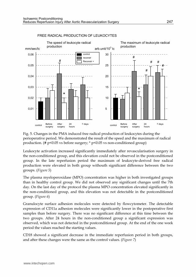
Fig. 5. Changes in the PMA induced free radical production of leukocytes during the perioperative period. We demonstrated the result of the speed and the maximum of radical production. (# p<0.05 vs before surgery; * p<0.05 vs non-conditioned group)
Leukocyte activation increased significantly immediately after revascularisation surgery in
the non-conditioned group, and this elevation could not be observed in the postconditioned
group. In the late reperfusion period the maximum of leukocyte-derived free radical
production were elevated in both group withouth significant difference between the two
groups. (Figure 5)
The plasma myeloperoxidase (MPO) concentration was higher in both investigated groups
than in healthy control group. We did not observed any significant changes until the 7th
day. On the last day of the protocol the plasma MPO concentration elevated significantly in
the non-conditioned group, and this elevation was not detectable in the postconditioned
group. (Figure 6)
Granulocyte surface adhesion molecules were detected by flowcytometer. The detectable
expression of CD11a adhesion molecules were significantly lower in the postoperative first
samples than before surgery. There was no significant difference at this time between the
two groups. After 24 hours in the non-conditioned group a significant expression was
observed, which was not detected in the postconditioned group. At the end of the one week
period the values reached the starting values.
CD18 showed a significant decrease in the immediate reperfusion period in both groups,
and after these changes were the same as the control values. (Figure 7)
mm/sec/lc
0,01
0,02
0,03
0,04
0,05
0,06
*
#
control Before surgery
After surgery
24 hours
7 days
The speed of leukocyte radical production
arb.unit/103 lc
5
10
15
20
25
30
*
#
#
#
control Before surgery
After surgery
24 hours
7 days
The maximum of leukocyte radical production
# #
FREE RADICAL PRODUCTION OF LEUKOCYTES
control
reconstr
Reconstr +
www.intechopen.com

Vascular Surgery
248
Fig. 6. Changes in plasma myeloperoxidase following operation (# p<0.05 vs before surgery; * p<0.05 vs non-conditioned group)
Fig. 7. The graphs show the changes in expression of granulocyte adhesion during the examined perioperative period. (AU= arbitary unit) (# p<0.05 vs before surgery; * p<0.05 vs non-conditioned group)
0,2
0,4
0,6
0,8
1,0
nmol/ml
Before surgery After surgery 24 hours 7 days
PLASMA MYELOPEROXIDASE (MPO) CONCENTRATION
CONTROL
RECONSTRUCTION
RECONSTRUCTION + POSTCONDITIONING
*
#
1,2
control
EXPRESSION OF GRANULOCYTE ADHESION MOLECULES
AU
100
200
300
400
500
# #
control Before surgery
After surgery
24 hours
7 days
Granulocyte CD18 expression
control
reconstruction
Granulocyte CD11a expression
AU
200
400
600
800
#
kontroll Before surgery
After surgery
24 hours
7 days
1000
1200
1400
1600
#
*
#
Reconstruction + postcond
www.intechopen.com
Ischaemic Postconditioning Reduces Reperfusion Injury After Aortic Revascularization Surgery
249
In the results of the red blood cell count, white blood cell count, platelet numbers,
haemoglobin concentration and haematocrit level we did not detected any difference
between the two groups of patient.
21. Discussion
In the last 3 years the literature of ischaemic postconditioning exponentially increased in the
experimental cardiology. The beneficial effects of the manoeuver has been confirmed in
various models, including human results as well, and the cellular and biochemical
background is intensively examined. Until now this is the first study to evaluate the effect of
ischaemic postconditioning on peripheral tissues in abdominal aortic surgery.
Our results demonstrated that after a prolonged ischaemia, postconditioning can reduce free
radical production, TNF-alpha expression and leukocyte activation in the early phase of
reperfusion in an animal model of abdominal aortic surgery. In this model we also
confirmed that PostC could induce antiapoptotic signaling pathways in the skeletal muscle
and in far organs as well.
In our human model we could confirm that ischaemic PostC could decrease in some points
the revascularization surgery evoked oxidative stress and inflammatory response.
We have demonstrated that the protective effect of postconditioning is a complex process,
involving many cell types, the generation of oxidants, cytokines, and inflammatory
pathways, has not only one target, but acts on a diverse site. This complexity, the powerful
protective effect and the simplicity in the surgery (lasts for a few minutes) can make the
manoeuver really a powerful tool of surgeons.
22. Mechanisms involved in postconditioning
The mechanisms of protection by PostC were initially attributed mainly to improved
endothelial function and to the events reducing the detrimental effects of lethal
reperfusion injury, such as reduced edema, reduced oxidative stress, reduced
mitochondrial calcium accumulation, reduced endothelium damage and reduced
inflammation. However, subsequent studies suggest that protection is mediated through
the recruitment of signal transduction pathways as in the case of ischaemic
preconditioning. Therefore, a distinction in passive and active mechanisms can
be proposed. Of course an intricate cross-talk among these events/mechanisms exists,
thus this distinction can be useful for a better understanding of the phenomenon, but we
must not forget that a single event/mechanism may not be effective if it occurs alone.
(Figure 8)
23. Passive mechanisms
Among passive mechanisms we can consider those strictly related with hydrostatic force –
hereafter named as ‘Mechanical mechanisms’ – and those related with reduced endothelial
adhesion of leucocytes and subsequent reduction of inflammatory process that we call
‘Cellular mechanisms’.
www.intechopen.com

Vascular Surgery
250
Fig. 8. A schematic figure on the mechanisms of ischaemic postconditioning.
24. Mechanical mechanisms
With regard to mechanical or haemodynamic mechanisms, it has been suggested that the
stuttering of reperfusion and pressure during PostC manoeuvres may limit the hydrostatic
forces in a very important moment, thus limiting early edema and consequent damages. In
experiments performed in isolated heart models, the effect of the PostC on the infarct area
has been studied perfusing the hearts either with constant pressure or with constant flow. It
has been compared the role of these two types in perfusion in affecting the infarct area
during PostC. In the constant pressure model the infarct area was less reduced by PostC
than it was with the model of the constant flow reperfusion after PostC46. Considering that
during the short period of restoration of flow in the PostC manoeuvres the capillary
pressure increases less in the constant flow model, than in the constant pressure model (i.e.
at the beginning of reperfusion in the constant flow model there is smaller hydrostatic
pressure and so smaller transcapillary pressure), it was argued that in the constant-flow
model a reduced edema and consequent reduced damages may explain the increased
effectiveness of PostC. In other words, in the constant flow model the effectiveness of PostC
is greater than in the constant pressure model supports the idea that the reduction of
hydrostatic forces during PostC manoeuvres may play an important role in determining the
protective effects.
25. Cellular mechanisms
Among the cellular mechanisms we consider acute inflammatory response. It occurs through the release of cytokines, activation of vascular endothelial cells and leukocytes with expression of cell surface adhesion molecules, and up-regulation of a program of
CELLULAR PROTECTION
K+
KATP mPTP APOPTOSIS↓
NECROSIS↓
EDEMA CELLULAR
SWELLING
Citokines
Ca2+ ↓
endothelium
MECHANICAL CELLULAR MOLECULAR
H2O
adenosine
GPCR
PMN(
.O2)
↑PI3 K
↑ P-Akt
↑ ERK 1/2
↓ P38, JNK
↑ Bcl2/BAX (-)
(-)
(-)
(-)
(-)
MITOCHONDRIUM
(.O2)
NO.
(.O2)
(-)
(.O2) ↓ IC
(-)
J.Vinten-Johansen et al. Basic Res Cardiol 2005
pH↑
www.intechopen.com

Ischaemic Postconditioning Reduces Reperfusion Injury After Aortic Revascularization Surgery
251
proinflammatory genes. PostC delays the onset and reduces the maintenance of post-ischaemic inflammation47. As stated before, whether this is a cause or an effect of PostC protection remains to be elucidated.
26. Active mechanisms (intracellular mechanisms)
Studies have identified a signalling pathway that is recruited at the time of reperfusion and
which is similar in ischaemic preconditioning and PostC. This pathway includes the
survival kinases phosphatidylinositol 3-kinase (PI3K)-Akt and Erk1/2, the major
components of the reperfusion-injury salvage kinase pathway, termed the RISK- pathway,
which may influence the mPTP, a non-specific pore of the mitochondrial membrane whose
opening in the first few minutes of myocardial reperfusion promotes cell death48. Delayed
washout of endogenously produced adenosine and activation of the adenosine receptor also
seems to be required for PostC protection49, by activating the survival pathway.
Thus delayed washout of adenosine in the setting of PostC might recruit RISK at the time of
reperfusion through the activation of adenosine responsive G-protein-coupled receptors. It
seems that adenosine receptors are repopulated during PostC manoeuvres. While in murine
hearts adenosine A2a and A3 subtypes 50 have been seen to be involved, in rabbit hearts
PostC seems to depend on A2b subtype 51. An important role of the redox environment has
also been observed52.
Therefore, similar to preconditioning, PostC has been proposed to be triggered by receptor
stimulation, mediated by one or more complex and interrelated signal transduction
pathways, and, ultimately, achieved via phosphorylation of one or more end-effectors of
cardioprotection53.
27. Triggers of postconditioning
Ligands, such as adenosine 54 and bradykinine 55 what accumulate during PostC
manoeuvres may initiate the cascade that lead to PostC protection. It has been recently
reported that inhibition of opioid receptors with opioid antagonists administered 5 min.
before reperfusion in the absence or presence of PostC, reversed the infarct sparing effect of
PostC in an in vivo rat model56. The activation of protein kinase C and G (PKC and PKG)
and opening of mitochondrial KATP channels after PostC (see below) would be consistent
with the involvement of BK and endogenous opioids. Nitric oxide and ROS may be
included among the triggers. Nitric oxide is demonstrated to act both as a trigger and as a
mediator of the preconditioning response in a variety of species. The role of endogenous NO
in classic ischaemic preconditioning was controversial. Cohen and Downey’s group
suggested that exogenously administered NO could trigger the preconditioned state
through a free radical-mediated process not shared by endogenous NO. Very recently these
authors questioned whether their observation was due to a bias in the experimental model.
These authors are now on the opinion that endogenous NO participates in triggering in vivo
preconditioning57. Among the autocaids released by the ischaemic heart there is BK that
may induce nitric oxide release (Figure 3). It has been suggested that the mechanism
whereby NO protects myocardium includes the activation of guanylate-cyclase58. As an
inducer of the protection, nitric oxide may also directly open the mitochondrial KATP
www.intechopen.com

Vascular Surgery
252
channels59. Therefore, nitric oxide acting on mitochondria may play a relevant role in
protection both through activation of these channels and via modulation of respiratory
chain; both mechanisms favor ROS signalling, which can trigger protection60. A relevant role
of nitric oxide may also be attributed to the endothelial protection brought by this molecule 61 or to its role as antioxidant under certain conditions 62.
The one-electron-reduction product of nitric oxide, HNO/NO– (nitrosyl hydride/nitroxyl
anion), has been scarcely studied in an I/R scenario. In our laboratory low doses of
Angeli’s salt, a donor of HNO/NO–, have been seen to induce early/classical
preconditioning against myocardial damages63. Intriguingly, the protective effects of
HNO/NO– generated by Angeli’s salt were more potent than the protective effects
induced by equimolar concentration of the pure nitric oxide donor diethylamine/nitric
oxide (DEA/NO). While the HNO/NO– donor seems deleterious in reperfusion64, there is
evidence that NO may also be involved in the cardioprotection by ischaemic PostC. When
the nitric oxide synthase (NOS) inhibitor N-omega-nitro-L-arginine methyl esther
(LNAME) was given 5 min. before start of reperfusion of in vivo rabbit hearts, the infarct
limiting effect was abolished65. We have shown that nitric oxide participates in PostC, but
NOS inhibitors given for the entire reperfusion period only blunted the protective effect
of PostC66 . Paradoxically, the same inhibitor, given only during PostC manoeuvres
completely blocked the protective effects67. At the moment, we do not have an
explanation for this apparent paradox. In a previous study, we argued that nitric oxide
may be produced in post-conditioned heart both by NOS and by non-enzymatic
mechanisms. Nitric oxide can then activate the guanyl cyclase to produce cyclic guanosine
monophosphate (cGMP), which mediates protection 68 (see also below). The infusion of a
NOS inhibitor only during PostC manoeuvres may alter the equilibrium between ROS
and nitric oxide thus leading to the production of the wrong kind of radical which does
not trigger the protective pathway. It can be argued that in the absence of this protection
the stronger limitation of nitric oxide production by NOS may be protective during
reperfusion. In fact, data have demonstrated that NOS inhibitors can attenuate I/R
damage69. Also, the different doses of nitric oxide inhibitors applied and the different
basal levels of nitric oxide endogenously produced may explain these disparities.
The beneficial and deleterious effects of nitric oxide and nitrite in pathophysiological conditions and contradictory results about the effects of nitric oxide during reperfusion have been reviewed by Bolli in 200170, Wink et al. in 200371, Pagliaro in 200372 and Schulz et al. in 2004 73. ROS could also be included among the triggers of PostC. In fact, ROS scavengers such as N-acetylcysteine and 2-mercapto-propionylglycine given during PostC manoeuvres prevent the protective effects74. It is possible that the low pH during the PostC cycles prevents mPTP opening, while the intermittent oxygen bursts allow mitochondria to make enough ROS in a moment in which other enzymes, able to produce massive quantity of ROS, are not yet re-activated. Then mitochondrial ROS may activate PKC and put the heart into a protected state. The importance of the role of acidosis in the triggering of PostC protection has been recently confirmed by two independent laboratories75. Acidosis may also prevent mPTP opening in the early reperfusion (see below). Recently, it has been reported that redox signaling and a low pH at the time of myocardial reperfusion are also required to mediate the cardioprotection triggered by ischaemic preconditioning76.
www.intechopen.com

Ischaemic Postconditioning Reduces Reperfusion Injury After Aortic Revascularization Surgery
253
28. Mediators of postconditioning
We considered ROS among triggers as they are necessary during PostC manoeuvres.
Nevertheless, PostC activated the RISK pathway, with increased expression of the
phosphorylated form of endothelial nitric oxide synthase (eNOS) as one of the results77.
Thus it is likely that after NOS activation the cGMP is produced and PKG is activated; then
mitochondrial ATP-dependent potassium (mKATP) channels are opened and ROS produced.
Therefore cGMP, PKG, mKATP and ROS may be considered as mediators of PostC
protection, which are likely to be upstream to PKC activation. We demonstrated that cGMP
production is increased during reperfusion of postconditioned hearts78. Moreover we
showed that in these hearts mKATP and PKC must also be active (i.e. they should not be
blocked) during late reperfusion79. Regarding the role of mKATP channels a couple of papers
indicate that the mKATP channel is important for PostC80. In these studies two different
mKATP channel blockers (glibenclamide and 5-hydroxidecanoate) abolished the protective
effect of PostC 81.
It is interesting that many of the RISK elements (e.g. PI3K/Akt and MEK1/2-ERK) involved
in the signaling pathway in preconditioning and protection against reperfusion injury have
recently been documented also in PostC82. Some differences, however, may exist between
pre- and PostC (see also Table 1 and Table 2). Darling et al. 83 showed an increase of
phospho-ERK, but not of PI3K/Akt in PostC, while Yang et al.84 showed that ERK is
involved in PostC, but not in preconditioning. These findings may explain a certain degree
of additive protection between ischaemic preconditioning and PostC, as observed by Yang
et al.85. Yet in contrast with Yang et al.86, Cao et al.87 reported that ERK is present in
preconditioning trigger pathway. The reasons for the differences are not clear. Different
species and/or protocols may play a role88. Different methods of tissue sampling also be
may play a role89. Besides protein kinase C, the possible roles for tyrosine kinase, and
members of the MAPK family other than ERK1/2 in PostC has been suggested90. (Figure 9)
Focal disorganization of gap junction distribution and down-regulation of connexin 43
(Cx43) are typical features of myocardial remodelling 91 and Cx43 – especially Cx43
localized in mitochondria – has been indicated as one key element of the signal transduction
cascade of the protection by preconditioning. However, Cx43 does not seem to be important
for infarct size reduction by PostC 92. These results, together with the above reported
differences on kinase activation by pre- and PostC, suggest a certain degree of differences
between the protective pathways activated by these two procedures.
29. End-effectors of postconditioning
Mitochondrial PTPs opening represents a fundamental step of reperfusion injury. Among
the potential mechanisms responsible for mPTP opening during reperfusion, Ca2+-overload
has received particular attention. In particular, mitochondrial Ca2+-overload occurring
during ischaemia must bring mitochondria closer to the threshold at which mPTP opening
takes place, favouring the occurrence of mPTP opening during reperfusion, a phenomenon
described as mitochondrial priming93. Additionally, reduced mitochondrial Ca2+ overload
during ischaemia has been pointed out as a potentially important mechanism of ischaemic
and pharmacological preconditioning94.
www.intechopen.com
Vascular Surgery
254
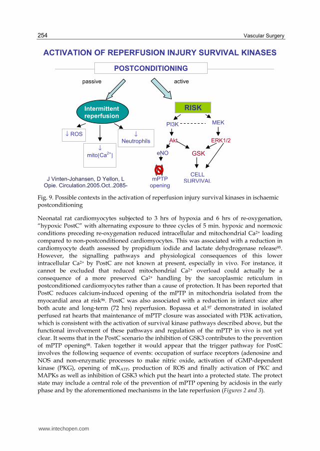
Fig. 9. Possible contexts in the activation of reperfusion injury survival kinases in ischaemic postconditioning
Neonatal rat cardiomyocytes subjected to 3 hrs of hypoxia and 6 hrs of re-oxygenation, “hypoxic PostC” with alternating exposure to three cycles of 5 min. hypoxic and normoxic conditions preceding re-oxygenation reduced intracellular and mitochondrial Ca2+ loading compared to non-postconditioned cardiomyocytes. This was associated with a reduction in cardiomyocyte death assessed by propidium iodide and lactate dehydrogenase release95. However, the signalling pathways and physiological consequences of this lower intracellular Ca2+ by PostC are not known at present, especially in vivo. For instance, it cannot be excluded that reduced mitochondrial Ca2+ overload could actually be a consequence of a more preserved Ca2+ handling by the sarcoplasmic reticulum in postconditioned cardiomyocytes rather than a cause of protection. It has been reported that PostC reduces calcium-induced opening of the mPTP in mitochondria isolated from the myocardial area at risk96. PostC was also associated with a reduction in infarct size after both acute and long-term (72 hrs) reperfusion. Bopassa et al.97 demonstrated in isolated perfused rat hearts that maintenance of mPTP closure was associated with PI3K activation, which is consistent with the activation of survival kinase pathways described above, but the functional involvement of these pathways and regulation of the mPTP in vivo is not yet clear. It seems that in the PostC scenario the inhibition of GSK3 contributes to the prevention of mPTP opening98. Taken together it would appear that the trigger pathway for PostC involves the following sequence of events: occupation of surface receptors (adenosine and NOS and non-enzymatic processes to make nitric oxide, activation of cGMP-dependent kinase (PKG), opening of mKATP, production of ROS and finally activation of PKC and MAPKs as well as inhibition of GSK3 which put the heart into a protected state. The protect state may include a central role of the prevention of mPTP opening by acidosis in the early phase and by the aforementioned mechanisms in the late reperfusion (Figures 2 and 3).
POSTCONDITIONING
passive active
↓ ROS ↓ Neutrophils
↓
mito[Ca2+]
RISKIntermittent reperfusion
PI3K MEK
Akt ERK1/2
eNO
mPTP opening
GSK
CELL SURVIVAL
ACTIVATION OF REPERFUSION INJURY SURVIVAL KINASES
J Vinten-Johansen, D Yellon, L Opie. Circulation.2005.Oct..2085-
2088
www.intechopen.com

Ischaemic Postconditioning Reduces Reperfusion Injury After Aortic Revascularization Surgery
255
30. Cytoprotection by pre- and postconditioning is redox-sensitive
It has been already established that preconditioning triggering, that is the period that precedes the index ischaemia, is redox sensitive. This was demonstrated by both avoiding preconditioning with ROS scavengers and inducing preconditioning with ROS generators given before the index ischaemia 99. Also, several metabolites, including acetylcholine, BK, opioids and phenylephrine, trigger preconditioning-like protection via a mKATP-ROS dependent mechanism100. As stated in the case of reperfusion injury, ROS are also implicated in the sequel of myocardial reperfusion injury101. These studies supported the paradigm that ROS may be protective in pre-ischaemic phase, but are deleterious in the post-ischaemic phase. Thus the main idea was that ROS play an essential, though double-edged, role in cardioprotection: they may participate reperfusion injury or may play a role as signaling elements of protection in pre-ischaemic phase102. The importance of ROS signalling (as opposed to excess ROS in the development of injury) has been examined closely in great detail in recent years103. Intriguingly, and in contrast to the above-described theory of ROS as an obligatory part of reperfusion induced damage, some studies suggest the possibility that some ROS species at low concentrations could protect ischaemic hearts104. Yet, from the above reported mechanisms of PostC, it appears that also ischaemic PostC is a cardioprotective phenomenon that requires the intervention of redox signaling to be protective105. Moreover, as mentioned, very recently it has been shown that redox signalling is also required at the time of myocardial reperfusion to mediate the cardioprotection elicited by ischaemic preconditioning106. Therefore, the role of ROS in reperfusion may be reconsidered as they are not only deleterious. This fact may help to understand the variability in the results of studies aimed at proving a role of ROS in reperfusion injury. For instance, negative results came from trials in which free radical scavengers such as recombinant human superoxide dismutase or vitamin E were administered to patients with coronary artery disease or risk factors for cardiovascular events107. In addition to the dual role of ROS (beneficial versus deleterious), among the reasons why these scavengers did not show any consistent benefit in these human studies may be: (1) the type of ROS generated (e.g. superoxide dismutase only removes the superoxide and not the hydroxyl radical); (2) the site of ROS generation (e.g. most scavengers scarcely enter into the cells) and (3) the rate of reaction between two ROS and/or scavengers. The importance of the rate of reaction can be understood if we consider that, despite a five times lower concentration of nitric oxide with respect to superoxide dismutase, 50% or more of the available superoxide will react with nitric oxide to form ONOO– instead of reacting with superoxide dismutase108. Notwithstanding the evidence of a protective role of ROS signalling in reperfusion, we were unable to reproduce cardioprotection with ROS generation by purine/xanthine oxidase given at reperfusion109.
Since ROS scavengers (N-acetyl-L-cysteine or 2-mercapto-propionylglycine), given at the beginning of reperfusion, abolished both IP- and PostC-induced protection110 it is likely that the type, the concentration and/or the compartmentalization of ROS may play a pivotal role in triggering protection at reperfusion time. We are performing studies in the attempt to clarify this issue.
31. Conclusion
Postconditioning has the advantage of being a way to influence and modify reperfusion injury after it has occurred. This may open a therapeutic alternative in situations of
www.intechopen.com

Vascular Surgery
256
unexpected and uncontrolled ischaemic-reperfusion injury, for instance in the situation where technical complications occur during surgery, making a simple procedure into a complicated one, and making aortic cross-clamping longer than anticipated.
We think, that many more examinations are needed to describe and understand in details the mechanism of ischaemic PostC. We are sure, that this manoeuver is easy to perform, quick, and does not any expensive instruments, so it may have a place in the therapeutic arsenal of vascular surgeons.
32. References
[1] Piper HM, Meuter K, Schafer C. Cellular mechanisms of ischaemia-reperfusion injury. Ann Thorac Surg. 2003; 75: S644–8.
[2] Takemura G, Fujiwara H. Morphological aspects of apoptosis in heart diseases. J Cell Mol Med. 2006; 10: 56–75.
[3] Van Cruchten S, Van Den Broeck W. Morphological and biochemical aspects of apoptosis, oncosis and necrosis. Anat Histol Embryol. 2002; 31: 214–23.
[4] Tritto I, Ambrosio G. Role of oxidants in the signaling pathway of preconditioning. Antioxid Redox Signal. 2001; 3: 3–10.
[5] Zhao ZQ, Vinten-Johansen J. Myocardial apoptosis and ischemic preconditioning. Cardiovasc Res. 2002;55: 438–55.
[6] Ambrosio G, Flaherty JT, Duilio C,Tritto I, Santoro G, Elia PP, Condorelli M, Chiariello M. Oxygen radicals generated at reflow induce peroxidation of membrane lipids in reperfused hearts. J Clin Invest. 1991; 87: 2056–66.
[7] Kaeffer N, Richard V, Thuillez C. Delayed coronary endothelial protection 24 hours after preconditioning: role of free radicals. Circulation. 1997; 96: 2311–6
[8] Lefer AM, Lefer DJ. Endothelial dysfunction in myocardial ischaemia and reperfusion: role of oxygenderived free radicals. Basic Res Cardiol. 1991; 86: 109–16.
[9] Zhao ZQ. Oxidative stress-elicited myocardial apoptosis during reperfusion. Curr Opin Pharmacol. 2004; 4: 159–65.
[10] Hoffman JW Jr, Gilbert TB, Poston RS, Silldorff EP. Myocardial reperfusion injury: etiology, mechanisms, and therapies. J Extra Corpor Technol. 2004; 36: 391–411.
[11] Gateau-Roesch O, Argaud L, Ovize M. Mitochondrial permeability transition pore and postconditioning. Cardiovasc Res. 2006; 70: 264–73.
[12] Schreck R, Albermann K, Baeuerle PA. Nuclear factor kappa B: an oxidative stress-responsive transcription factor of eukariotic cells. Free Radical Res Commun. 1992; 17: 221–37.
[13] Baldwin AS. The transcription factor NFkB and human disease. J Clin Invest. 2001; 107: 3–6. [14] Lefer AM, Lefer DJ. The role of nitric oxide and cell adhesion molecules on the
microcirculation in ischaemia-reperfusion. Cardiovasc Res. 1996; 32: 743–51. [15] Radomski MW, Palmer RM, Moncada S. Comparative pharmacology of endothelium-
derived relaxing factor, nitric oxide and prostacyclin in platelets. Br J Pharmacol. 1987; 92: 181–7.
[16] Reffelmann T, Kloner RA. The “no-reflow” phenomenon: basic science and clinical correlates. Heart. 2002; 87: 162–8.
[17] Zhao ZQ, Corvera J, Halkos ME, Kerendi F,Wang NP, Guyton RA, Vinten-Johansen J. Inhibition of myocardial injury by ischemic postconditioning during reperfusion:
www.intechopen.com

Ischaemic Postconditioning Reduces Reperfusion Injury After Aortic Revascularization Surgery
257
comparison with ischemic preconditioning. Am J Physiol Heart Circ Physiol. 2003; 285: H579–88.
[18] LB Becker , New concepts in reactive oxygen species and cardiovascular reperfusion physiology, Cardiovasc Res, 15 (2004), 461-70.
[19] KA Kaminski, Bonda TA, Korecki J, Musial WJ, Oxidative stress and neutrophil activation-the two keystones of ischaemia/reperfusion injury, Int J Cardiol, 86 (2002), 41-59. Review.
[20] Zhao ZQ, Corvera J, Halkos ME, Kerendi F,Wang NP, Guyton RA, Vinten-Johansen J. Inhibition of myocardial injury by ischemic postconditioning during reperfusion: comparison with ischemic preconditioning. Am J Physiol Heart Circ Physiol. 2003; 285: H579–88.
[21] Darling CE, Jiang R, Maynard M, Whittaker P, Vinten-Johansen J, Przyklenk K. ‘Postconditioning’ via stuttering reperfusion limits myocardial infarct size in rabbit hearts: role of ERK 1/2. Am J Physiol Heart Circ Physiol. 2005; 289: H1618–26.
[22] Yang XM, Philipp S, Downey JM, Cohen MV. Postconditioning’s protection is not dependent on circulating blood factors or cells but involves adenosine receptors and requires PI3-kinase and guanylyl cyclase activation. Basic Res Cardiol. 2005; 100: 57–63.
[23] Kerendi F, Kin H, Halkos ME, Jiang R, Zatta AJ, Zhao ZQ, Guyton RA, Vinten-Johansen J. Remote postconditioning. Brief renal ischaemia and reperfusion applied before coronary artery reperfusion reduces myocardial infarct size via endogenous activation of adenosine receptors. Basic Res Cardiol. 2005; 100: 404–12.
[24] Penna C, Cappello S, Mancardi D, Raimondo S, Rastaldo R, Gattullo D, Losano G, Pagliaro P. Post-conditioning reduces infarct size in the isolated rat heart: role of coronary flow and pressure and the nitric oxide/cGMP pathway. Basic Res Cardiol. 2006; 101: 168–79.
[25] Gottlieb RA, Burleson KO, Kloner RA, Babior BM, Engler RL. Reperfusion injury induces apoptosis in rabbit cardiomyocytes. J Clin Invest. 1994; 94: 1621–8.
[26] Sun HY, Wang NP, Halkos M, Kerendi F, Kin H, Guyton RA, Vinten-Johansen J, Zhao ZQ. Postconditioning attenuates cardiomyocyte apoptosis via inhibition of JNK and p38 mitogen-activated protein kinase signaling pathways. Apoptosis. 2006; 11: 1583–93.
[27] Heltianu C, Costache G, Gafencu A, Diaconu M, Bodeanu M, Cristea C, Azibi K, Poenaru L, Simionescu M. Relationship of eNOS gene variants to diseases that have in common an endothelial cell dysfunction. J Cell Mol Med. 2005; 9: 135–42.
[28] Zhao ZQ, Corvera J, Halkos ME, Kerendi F,Wang NP, Guyton RA, Vinten-Johansen J. Inhibition of myocardial injury by ischemic postconditioning during reperfusion: comparison with ischemic preconditioning. Am J Physiol Heart Circ Physiol. 2003; 285: H579–88.
[29] Zhao ZQ, Corvera J, Halkos ME, Kerendi F,Wang NP, Guyton RA, Vinten-Johansen J. Inhibition of myocardial injury by ischemic postconditioning during reperfusion: comparison with ischemic preconditioning. Am J Physiol Heart Circ Physiol. 2003; 285: H579–88.
[30] Schwartz LM, Lagranha CJ. Ischemic postconditioning during reperfusion activates Akt and ERK without protecting against lethal myocardial ischaemia-reperfusion injury in pigs. Am J Physiol Heart Circ Physiol. 2006; 290: H1011–8.
www.intechopen.com

Vascular Surgery
258
[31] Sun HY, Wang NP, Kerendi F, Halkos M, Kin H, Guyton RA, Vinten-Johansen J, Zhao ZQ. Hypoxic postconditioning reduces cardiomyocyte loss by inhibiting ROS generation and intracellular Ca2+ overload. Am J Physiol Heart Circ Physiol. 2005; 288: H1900–8.
[32] Mykytenko J, Kerendi F, Reeves JG, Kin H, Zatta AJ, Jiang R, Guyton RA, Vinten-Johansen J, Zhao ZQ. Long-term inhibition of myocardial infarction by postconditioning during reperfusion. Basic Res Cardiol. 2007; 102: 90–100.
[33] Halkos ME, Kerendi F, Corvera JS,Wang NP, Kin H, Payne CS, Sun HY, Guyton RA,Vinten-Johansen J, Zhao ZQ. Myocardial protection with postconditioning is not enhanced by ischemic preconditioning.Ann Thorac Surg. 2004; 78: 961–9.
[34] Tsang A, Hausenloy DJ, Mocanu MM, Yellon DM. Postconditioning: a form of “modified reperfusion” protects the myocardium by activating the phosphatidylinositol 3-kinase-Akt pathway. Circ Res. 2004; 95: 230–2.
[35]Penna C, Cappello S, Mancardi D, Raimondo S, Rastaldo R, Gattullo D, Losano G, Pagliaro P. Post-conditioning reduces infarct size in the isolated rat heart: role of coronary flow and pressure and the nitric oxide/cGMP pathway. Basic Res Cardiol. 2006; 101: 168–79.
[36] Yang XM, Proctor JB, Cui L, Krieg T, Downey JM, Cohen MV. Multiple, brief coronary occlusions during early reperfusion protect rabbit hearts by targeting cell signaling pathways. J Am Coll Cardiol. 2004; 44: 1103–10.
[37] Sato H, Bolli R, Rokosh GD, Bi Q, Dai S, Shirk G, Tang XL. The cardioprotection of the late phase of ischemic preconditioning is enhanced by postconditioning via a COX-2-mediated mechanism in conscious rats. Am J Physiol Heart Circ Physiol. 2007; 293: H2557–64.
[38] Crisostomo PR,Wang M,Wairiuko GM,Terrell AM, Meldrum DR. Postconditioning in females depends on injury severity. J Surg Res. 2006; 134: 342–7.
[39] Boengler K, Buechert A, Heinen Y, Roeskes C, Hilfiker-Kleiner D, Heusch G, Schulz R. Cardioprotection by ischemic postconditioning is lost in aged and STAT3-deficient mice. Circ Res. 2008; 102: 131–5.
[40] Ohakawa HN, Okishi N, Yagi K: Assay for lipid peroxides in animal tissues by thiobarbituric acid reaction. Anal Biochem, 1979; 95: 351-8
[41] Sedlak J, Lindsay RH: Estimation of total protein-bound and non-protein sulphydryl groups in tissue with Ellman’sreagent, Anal Biochem, 1968; 25: 192-205
[42] Misra HP, Fridovich I. The role of superoxide anion in the autooxidation of epinephrine and a simple assay for superoxide dismutase. J Biol Chem 1972. 247: 3170-3175.
[43] Albelda SM, Smith CW, Ward PA. Adhesion molecules and inflammatory injury. FASEB J. 1994, 8: 504-512.
[44] Menger MD, Vollmar B. Adhesion molecules as determinations disease: from molecular biology to surgical research. BR. J. Surg. 1996, 83: 588-601.
[45] Arató E. PhD Thesis 2006 Univ of Pécs [46] Penna C, Cappello S, Mancardi D, Raimondo S, Rastaldo R, Gattullo D, Losano G,
Pagliaro P. Post-conditioning reduces infarct size in the isolated rat heart: role of coronary flow and pressure and the nitric oxide/cGMP pathway. Basic Res Cardiol. 2006; 101: 168–79.
[47] Zhao ZQ, Corvera J, Halkos ME, Kerendi F,Wang NP, Guyton RA, Vinten-Johansen J. Inhibition of myocardial injury by ischemic postconditioning during reperfusion:
www.intechopen.com

Ischaemic Postconditioning Reduces Reperfusion Injury After Aortic Revascularization Surgery
259
comparison with ischemic preconditioning. Am J Physiol Heart Circ Physiol. 2003; 285: H579–88.
[48] Hausenloy DJ, Yellon DM. The mitochondrial permeability transition pore: its fundamental role in mediating cell death during ischaemia and reperfusion. J Mol Cell Cardiol. 2003; 35: 339–41.
[49] Y Itoh, Takaoka R, Ohira M, Abe T, Tanahashi N, Suzuki N. Reactive oxygen species generated by mitochondrial injury in human brain microvessel endothelial cells. Clin Hemorheol Microcirc. 2006;34(1-2):163-8.
[50] Kin H, Zatta AJ, Lofye MT, Amerson BS, Halkos ME, Kerendi F, Zhao ZQ, Guyton RA, Headrick JP, Vinten-Johansen J. Postconditioning reduces infarct size via adenosine receptor activation by endogenous adenosine. Cardiovasc Res. 2005; 67: 124–33.
[51] Philipp S,Yang XM, Cui L, Davis AM, Downey JM, Cohen MV. Postconditioning protects rabbit hearts through a protein kinase C-adenosine A2b receptor cascade. Cardiovasc Res. 2006; 70: 308–14.
[52] Penna C, Rastaldo R, Mancardi D, Raimondo S, Cappello S, Gattullo D, Losano G, Pagliaro P. Postconditioning induced cardioprotection requires signalling through a redox-sensitive mechanism, mitochondrial ATP-sensitive K+ channel and protein kinase C activation. Basic Res Cardiol. 2006; 101: 180–9.
[53] Hausenloy DJ, Tsang A, Yellon, DM. The reperfusion injury salvage kinase pathway: a common target for both ischemic preconditioning and postconditioning. Trends Cardiovasc Med. 2005; 15: 69–75.
[54] Philipp S,Yang XM, Cui L, Davis AM, Downey JM, Cohen MV. Postconditioning protects rabbit hearts through a protein kinase C-adenosine A2b receptor cascade. Cardiovasc Res. 2006; 70: 308–14.
[55] Penna C, Mancardi D, Rastaldo R, Losano G, Pagliaro P. Intermittent activation of bradykinin B2 receptors and mitochondrial KATP channels trigger cardiac postconditioning through redox signaling. Cardiovasc Res. 2007; 75: 168–77.
[56] Kin H, Zatta AJ, Jiang R, Reeves JG. Activation of opioid mediates the infarct size reduction by postconditioning. J Mol Cell Cardiol. 2005; 38: 827.
[57] Cohen MV, Yang XM, Downey JM. Nitric oxide is a preconditioning mimetic and cardioprotectant and is the basis of many available infarct-sparing strategies. Cardiovasc Res. 2006; 70: 231–9.
[58] Dawn B, Bolli R. Role of nitric oxide in myocardial preconditioning. Ann N Y Acad Sci. 2002; 962: 18–41.
[59] Sasaki N, Sato T, Ohler A, O’Rourke B, Marban E. Activation of mitochondrial ATP-dependent potassium channels by nitric oxide. Circulation. 2000; 101: 439–45.
[60] Moncada S, Erusalimsky JD. Does nitric oxide modulate mitochondrial energy generation and apoptosis? Nat Rev Mol Cell Biol. 2002; 3: 214–20.
[61] Gattullo D, Linden RJ, Losano G, Pagliaro P, Westerhof N. Ischaemic preconditioning changes the pattern of coronary reactive hyperaemia in the goat: role of adenosine and nitric oxide. Cardiovasc Res. 1999; 42: 57–64.
[62] Ridnour LA, Thomas DD, Mancardi D, Espey MG, Miranda KM, Paolocci N, Feelisch M, Fukuto J, Wink DA. The chemistry of nitrosative stress induced by nitric oxide and reactive nitrogen oxide species. Putting perspective on stressful biological situations. Biol Chem. 2004; 385: 1–10.
www.intechopen.com
Vascular Surgery
260
[63] Pagliaro P, Mancardi D, Rastaldo R, Penna C, Gattullo D, Miranda KM, Feelisch M, Wink DA, Kass DA, Paolocci N. Nitroxyl affords thiol-sensitive myocardial protective effects akin to early preconditioning. Free Radic Biol Med. 2003; 34: 33–43.
[64] Ma XL, Gao F, Liu GL, Lopez BL, Christopher TA, Fukuto JM, Wink DA, Feelisch M. Opposite effects of nitric oxide and nitroxyl on postischemic myocardial injury. Proc Natl Acad Sci USA. 1999; 96: 14617–22.
[65] Yang XM, Proctor JB, Cui L, Krieg T, Downey JM, Cohen MV. Multiple, brief coronary occlusions during early reperfusion protect rabbit hearts by targeting cell signaling pathways. J Am Coll Cardiol. 2004; 44: 1103–10.
[66] Penna C, Cappello S, Mancardi D, Raimondo S, Rastaldo R, Gattullo D, Losano G, Pagliaro P. Post-conditioning reduces infarct size in the isolated rat heart: role of coronary flow and pressure and the nitric oxide/cGMP pathway. Basic Res Cardiol. 2006; 101: 168–79.
[67] Penna C, Mancardi D, Rastaldo R, Losano G, Pagliaro P. Intermittent activation of bradykinin B2 receptors and mitochondrial KATP channels trigger cardiac postconditioning through redox signaling. Cardiovasc Res. 2007; 75: 168–77.
[68] Pagliaro P, Rastaldo R, Penna C, Mancardi D, Cappello S, Losano G. Nitric oxide (NO)-cyclic guanosine monophosphate (cGMP) pathway is involved in ischemic postconditioning in the isolated rat heart. Circulation. 2004; 110: III 136.
[69] Patel VC,Yellon DM, Singh KJ, Neild GH,Woolfson RG. Inhibition of nitric oxide limits infarct size in the in situ rabbit heart. Biochem Biophys Res Commun. 1993; 194: 234–8.
[70] Bolli R. Cardioprotective function of inducible nitric oxide synthase and role of nitric oxide in myocardial ischaemia and preconditioning: an overview of a decade of research. J Mol Cell Cardiol. 2001; 33: 1897–918.
[71] Wink DA, Miranda KM, Katori T, Mancardi D, Thomas DD, Ridnour L, Espey MG, Feelisch M, Colton CA, Fukuto JM, Pagliaro P, Kass DA, Paolocci N. Orthogonal properties of the redox siblings nitroxyl and nitric oxide in the cardiovascular system: a novel redox paradigm. Am J Physiol Heart Circ Physiol. 2003; 285: H2264–76.
[72] Pagliaro P. Differential biological effects of products of nitric oxide (NO) synthase: it is not enough to say NO. Life Sci. 2003; 73: 2137–49.
[73] Schulz R, Kelm M, Heusch G. Nitric oxide in myocardial ischaemia/reperfusion injury. Cardiovasc Res. 2004; 61: 402–13.
[74] Downey JM, Cohen MV. A really radical observation–a comment on Penna et al. in Basic Res Cardiol (2006) 101:180–189. Basic Res Cardiol. 2006; 101: 190–1.
[75] Cohen MV,Yang XM, Downey JM. The pH hypothesis of postconditioning: staccato reperfusion reintroduces oxygen and perpetuates myocardial acidosis. Circulation. 2007; 115: 1895–903.
[76] Hausenloy DJ, Wynne AM, Yellon DM. Ischemic preconditioning targets the reperfusion phase. Basic Res Cardiol. 2007; 102: 445–52.
[77] Tsang A, Hausenloy DJ, Mocanu MM, Yellon DM. Postconditioning: a form of “modified reperfusion” protects the myocardium by activating the phosphatidylinositol 3-kinase-Akt pathway. Circ Res. 2004; 95: 230–2.
[78] Pagliaro P, Rastaldo R, Penna C, Mancardi D, Cappello S, Losano G. Nitric oxide (NO)-cyclic guanosine monophosphate (cGMP) pathway is involved in ischemic postconditioning in the isolated rat heart. Circulation. 2004; 110: III 136.
www.intechopen.com

Ischaemic Postconditioning Reduces Reperfusion Injury After Aortic Revascularization Surgery
261
[79] Penna C, Rastaldo R, Mancardi D, Raimondo S, Cappello S, Gattullo D, Losano G, Pagliaro P. Postconditioning induced cardioprotection requires signalling through a redox-sensitive mechanism, mitochondrial ATP-sensitive K+ channel and protein kinase C activation. Basic Res Cardiol. 2006; 101: 180–9.
[80]Yang XM, Philipp S, Downey JM, Cohen MV. Postconditioning’s protection is not dependent on circulating blood factors or cells but involves adenosine receptors and requires PI3-kinase and guanylyl cyclase activation. Basic Res Cardiol. 2005; 100: 57–63.
[81] Penna C, Rastaldo R, Mancardi D, Raimondo S, Cappello S, Gattullo D, Losano G, Pagliaro P. Postconditioning induced cardioprotection requires signalling through a redox-sensitive mechanism, mitochondrial ATP-sensitive K+ channel and protein kinase C activation. Basic Res Cardiol. 2006; 101: 180–9.
[82] Hausenloy DJ, Tsang A, Yellon, DM. The reperfusion injury salvage kinase pathway: a common target for both ischemic preconditioning and postconditioning. Trends Cardiovasc Med. 2005; 15: 69–75.
[83] Darling CE, Jiang R, Maynard M, Whittaker P, Vinten-Johansen J, Przyklenk K. ‘Postconditioning’ via stuttering reperfusion limits myocardial infarct size in rabbit hearts: role of ERK 1/2. Am J Physiol Heart Circ Physiol. 2005; 289: H1618–26.
[84] Yang XM, Philipp S, Downey JM, Cohen MV. Postconditioning’s protection is not dependent on circulating blood factors or cells but involves adenosine receptors and requires PI3-kinase and guanylyl cyclase activation. Basic Res Cardiol. 2005; 100: 57–63.
[85] Yang XM, Philipp S, Downey JM, Cohen MV. Postconditioning’s protection is not dependent on circulating blood factors or cells but involves adenosine receptors and requires PI3-kinase and guanylyl cyclase activation. Basic Res Cardiol. 2005; 100: 57–63.
[86] Yang XM, Philipp S, Downey JM, Cohen MV. Postconditioning’s protection is not dependent on circulating blood factors or cells but involves adenosine receptors and requires PI3-kinase and guanylyl cyclase activation. Basic Res Cardiol. 2005; 100: 57–63.
[87] Cao Z, Liu L, Van Winkle DM. Met5-enkephalininduced cardioprotection occurs via transactivation of EGFR and activation of PI3K. Am J Physiol Heart Circ Physiol. 2005; 288: 1955–64.
[88] Vinten-Johansen J, Zhao ZQ, Zatta AJ, Kin H, Halkos ME, Kerendi F. Postconditioning A new link in nature’s armor against myocardial ischaemiareperfusion injury. Basic Res Cardiol. 2005; 100: 295–310.
[89] Darling CE, Jiang R, Maynard M, Whittaker P, Vinten-Johansen J, Przyklenk K. ‘Postconditioning’ via stuttering reperfusion limits myocardial infarct size in rabbit hearts: role of ERK 1/2. Am J Physiol Heart Circ Physiol. 2005; 289: H1618–26.
[90] Zhao ZQ, Vinten-Johansen J. Postconditioning: reduction of reperfusion-induced injury. Cardiovasc Res. 2006; 70: 200–11.
[91] Kostin S. Zonula occludens-1 and connexin 43 expression in the failing human heart. J Cell Mol Med. 2007; 11: 892–5.
[92] Heusch G, Büchert A, Feldhaus S, Schulz R. No loss of cardioprotection by postconditioning in connexin 43-deficient mice. Basic Res Cardiol. 2006; 101: 354–6.
[93] Weiss JN, Korge P, Honda HM, Ping P. Role of the mitochondrial permeability transition in myocardial disease. Circ Res. 2003; 93: 292–301.
www.intechopen.com
Vascular Surgery
262
[94] Murata M, Akao M, O’Rourke B, Marban E. Mitochondrial ATP-sensitive potassium channels attenuate matrix Ca(2+) overload during simulated ischaemia and reperfusion: possible mechanism of cardioprotection. Circ Res. 2001; 89: 891–8.
[95] Sun HY, Wang NP, Kerendi F, Halkos M, Kin H, Guyton RA, Vinten-Johansen J, Zhao ZQ. Hypoxic postconditioning reduces cardiomyocyte loss by inhibiting ROS generation and intracellular Ca2+ overload. Am J Physiol Heart Circ Physiol. 2005; 288: H1900–8.
[96] Argaud L, Gateau-Roesch O, Raisky O, Loufouat J, Robert D, Ovize M. Postconditioning inhibits mitochondrial permeability transition. Circulation. 2005; 111: 194–7.
[97] Bopassa JC, Ferrera R, Gateau-Roesch O, Couture-Lepetit E, Ovize M. PI 3-kinase regulates the mitochondrial transition pore in controlled reperfusion and postconditioning. Cardiovas Res. 2006; 69: 178–85.
[98] Gateau-Roesch O, Argaud L, Ovize M. Mitochondrial permeability transition pore and postconditioning. Cardiovasc Res. 2006; 70: 264–73.
[99] Tritto I, Ambrosio G. Role of oxidants in the signaling pathway of preconditioning. Antioxid Redox Signal. 2001; 3: 3–10.
[100] Zhao ZQ. Oxidative stress-elicited myocardial apoptosis during reperfusion. Curr Opin Pharmacol. 2004; 4: 159–65.
[101] Pagliaro P. Differential biological effects of products NO. Life Sci. 2003; 73: 2137–49. [102] Zhao ZQ. Oxidative stress-elicited myocardial apoptosis during reperfusion. Curr
Opin Pharmacol. 2004; 4: 159–65. [103] Yao Z, Tong J, Tan X, Li C, Shao Z, Kim WC, vanden Hoek TL, Becker LB, Head CA,
Schumacker PT. Role of reactive oxygen species in acetylcholineinduced preconditioning in cardiomyocytes. Am J Physiol Heart Circ Physiol. 1999; 277: H2504–9.
[104] Urschel WC. Cardiovascular effects of hydrogen peroxide: current status. Dis Chest. 1967; 51: 180–92.
[105] Tsutsumi YM, Yokoyama T, Horikawa Y, Roth DM, Patel HH. Reactive oxygen species trigger ischemic and pharmacological postconditioning: In vivo and in vitro characterization. Life Sci. 2007; 81: 1223–7.
[106] Hausenloy DJ, Wynne AM, Yellon DM. Ischemic preconditioning targets the reperfusion phase. Basic Res Cardiol. 2007; 102: 445–52.
[107] Flaherty JT, Pitt B, Gruber JW, Heuser RR, Rothbaum DA, Burwell LR, George BS, Kereiakes DJ, Deitchman D, Gustafson N. Recombinant human superoxide dismutase (h-SOD) fails to improve recovery of ventricular function in patients undergoing coronary angioplasty for acute myocardial infarction. Circulation. 1999; 89: 1982–91.
[108] Kloner RA, Jennings RB. Consequences of brief ischaemia: stunning, preconditioning and their clinical implications. Circulation. 2001; 104: 2981–9.
[109] Penna C, Mancardi D, Rastaldo R, Losano G, Pagliaro P. Intermittent activation of bradykinin B2 receptors and mitochondrial KATP channels trigger cardiac postconditioning through redox signaling. Cardiovasc Res. 2007; 75: 168–77.
[110] Hausenloy DJ, Wynne AM, Yellon DM. Ischemic preconditioning targets the reperfusion phase. Basic Res Cardiol. 2007; 102: 445–52.
www.intechopen.com

Vascular SurgeryEdited by Dr. Dai Yamanouchi
ISBN 978-953-51-0328-8Hard cover, 262 pagesPublisher InTechPublished online 04, April, 2012Published in print edition April, 2012
InTech EuropeUniversity Campus STeP Ri Slavka Krautzeka 83/A 51000 Rijeka, Croatia Phone: +385 (51) 770 447 Fax: +385 (51) 686 166www.intechopen.com
InTech ChinaUnit 405, Office Block, Hotel Equatorial Shanghai No.65, Yan An Road (West), Shanghai, 200040, China
Phone: +86-21-62489820 Fax: +86-21-62489821
This book aims to provide a brief overview of conventional open vascular surgery, endovascular surgery andpre- and post-operative management of vascular patients. The collections of contributions from outstandingvascular surgeons and scientists from around the world present detailed and precious information about theimportant topics of the current vascular surgery practice and research. I hope this book will be used worldwideby young vascular surgeons and medical students enhancing their knowledge and stimulating theadvancement of this field.
How to referenceIn order to correctly reference this scholarly work, feel free to copy and paste the following:
Gabor Jancso, Endre Arató and Lászlo Sinay (2012). Ischaemic Postconditioning Reduces Reperfusion InjuryAfter Aortic Revascularization Surgery, Vascular Surgery, Dr. Dai Yamanouchi (Ed.), ISBN: 978-953-51-0328-8, InTech, Available from: http://www.intechopen.com/books/vascular-surgery/ischaemic-postconditioning-reduces-reperfusion-injury-after-aortic-revascularization-surgery

© 2012 The Author(s). Licensee IntechOpen. This is an open access articledistributed under the terms of the Creative Commons Attribution 3.0License, which permits unrestricted use, distribution, and reproduction inany medium, provided the original work is properly cited.