Cell Reports
Article
Hrq1, a Homolog of the Human RecQ4 Helicase,Acts Catalytically and Structurallyto Promote Genome IntegrityMatthew L. Bochman,1,2,3,* Katrin Paeschke,1,2,4 Angela Chan,1 and Virginia A. Zakian11Department of Molecular Biology, Princeton University, Princeton, NJ 08544, USA2These authors contributed equally to this work3Present address: Molecular and Cellular Biochemistry Department, Indiana University, Bloomington, IN 47405, USA4Present address: Department of Biochemistry, Theodor Boveri-Institute, University of Wurzburg, Am Hubland, 97074 Wurzburg, Germany
*Correspondence: [email protected]://dx.doi.org/10.1016/j.celrep.2013.12.037
This is anopen-access article distributed under the termsof theCreativeCommonsAttribution-NonCommercial-NoDerivativeWorks License,
which permits non-commercial use, distribution, and reproduction in any medium, provided the original author and source are credited.
SUMMARY
Human RecQ4 (hRecQ4) affects cancer and agingbut is difficult to study because it is a fusion betweena helicase and an essential replication factor.Budding yeast Hrq1 is homologous to the disease-linked helicase domain of RecQ4 and, like hRecQ4,is a robust 30-50 helicase. Additionally, Hrq1 has theunusual property of forming heptameric rings. Cellslacking Hrq1 exhibited two DNA damage pheno-types: hypersensitivity to DNA interstrand crosslinks(ICLs) and telomere addition to DNA breaks. Bothactivities are rare; their coexistence in a single pro-tein is unprecedented. Resistance to ICLs requireshelicase activity, but suppression of telomere addi-tion does not. Hrq1 also affects telomere length bya noncatalytic mechanism, as well as telomerase-independent telomere maintenance. Because Hrq1binds telomeres in vivo, it probably affects themdirectly. Thus, the tumor-suppressing activity ofRecQ4 could be due to a role in ICL repair and/orsuppression of de novo telomere addition.
INTRODUCTION
Helicases are motor proteins that use the energy of nucleotide
hydrolysis to separate duplex nucleic acids into their component
single strands (Abdelhaleem, 2010). RecQ family helicases are
involved in many aspects of DNA replication, recombination,
and repair (Bernstein et al., 2010). Humans encode five RecQs
(hRecQ1, hBLM, hWRN, hRecQ4, and hRecQ5), and mutations
in three of these enzymes (hBLM, hWRN, and hRecQ4) are linked
to cancers and/or premature aging. This article presents in vitro
and in vivo studies of the Saccharomyces cerevisiae Hrq1 heli-
case, a homolog of hRecQ4.
Mutation of hRecQ4 is linked to three distinct diseases with
related and overlapping symptoms and which are all character-
ized by genome instability, premature aging, and increased
346 Cell Reports 6, 346–356, January 30, 2014 ª2014 The Authors
cancer risk (Capp et al., 2010; Larizza et al., 2010). However,
determining how loss of hRecQ4 promotes human disease is
complicated because its N terminus is homologous to the essen-
tial S. cerevisiae Sld2 DNA replication factor (Figure 1A) (Liu,
2010). Given that 95% of the known disease-causing alleles of
hRecQ4 are found C-terminal to its Sld2-like domain (Larizza
et al., 2010), these diseases are probably due to loss of its heli-
case activities rather than loss of its replication function, which
would presumably be lethal. Thus, a simple model to determine
the nonreplication functions of RecQ4 would be useful.
Fungi such as S. cerevisiae and Schizosaccharomyces pombe
were previously described as encoding only one RecQ helicase
(Sgs1 and Rqh1, respectively) that is functionally homologous to
hBLM (Mirzaei et al., 2011). However, computational analyses
recently identified the product of the S. cerevisiae YDR291W
gene as a homolog of hRecQ4 (Lee et al., 2005) and found similar
RecQ4 homologs in many fungal and plant genomes, naming
these proteins Hrq1 (Barea et al., 2008).
Here, we purified S. cerevisiae Hrq1 and showed that it is a 30-50 DNA helicase. Mutation of the S. cerevisiae HRQ1 resulted in
strong sensitivity to DNA interstrand crosslinks (ICLs), a pheno-
type also reported for hRecQ4-deficient fibroblasts (Jin et al.,
2008). In addition, Hrq1, like other RecQ helicases, had multiple
telomere functions. HRQ1 suppressed telomere addition (TA) to
DSBs, an activity it shares with Pif1, a yeast DNA helicase whose
human counterpart is proposed to be a tumor suppressor gene
(Chisholm et al., 2012). HRQ1 also suppressed telomere hyper-
elongation in pif1 mutant cells. However, unlike Pif1, which acts
catalytically at both DSBs and telomeres (Boule et al., 2005;
Myung et al., 2001a; Zhou et al., 2000), neither of these telomeric
functions required the helicase activity of Hrq1. Like hBLM (Stav-
ropoulos et al., 2002) and Sgs1 (Huang et al., 2001; Johnson
et al., 2001), Hrq1 was also important for telomerase-indepen-
dent telomere maintenance.
RESULTS
Purified Hrq1 Displays Robust Helicase ActivityTo compare the biochemical functions of Hrq1 and RecQ4, full-
length S. cerevisiae Hrq1 and hRecQ4, as well as catalytically
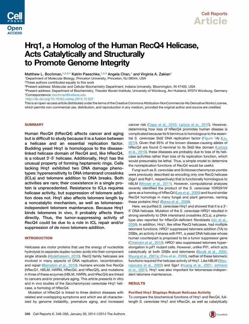
Figure 1. Purified Hrq1 Is an Active 30-50 Helicase(A) Domain schematics of Hrq1, hRecQ4, and Sgs1. The amino acid length of each is given on the right. The black bars in the helicase domain correspond to
conserved ATPase/helicase motifs. RQC, RecQ C-terminal domain; RHCD, RecQ4/Hrq1-conserved domain; Sld2-like, portion of hRecQ4 homologous to
S. cerevisiae Sld2.
(B) Coomassie-stained gel of purified S. cerevisiae Hrq1. The expected molecular weight is �130 kDa.
(C) hRecQ4 (Q4) and Hrq1 (WT) (both 50 nM) unwind a fork substrate; 100 nM Hrq1-KA (KA) does not.
(D) Hrq1 and hRecQ4 unwind the fork with similar apparent KMs ([protein] necessary to unwind 50% of the DNA).
(E) The rates of fork unwinding by 50 nM Hrq1 and hRecQ4 were similar (t1/2 = time necessary to unwind 50% of the DNA).
(F) Hrq1 and Hrq1-KA bind ssDNA by gel shifts. Hrq1 also preferentially bound ss- versus dsDNA, as well as telomeric repeat ssDNA (TG1-3).
(G) Directionality of Pif1 (lane 1), hRecQ4 (lane 2), and Hrq1 (lane 3) unwinding. The fastest migrating band corresponds to 50-30 unwinding; the slower migrating
band indicates 30-50 activity.(H) Sypro-orange-stained native gradient PAGE gel of Hrq1.
(I) TEM image of negative-stained Hrq1; white bar = 200 A.
(J) Two-dimensional reconstruction of the Hrq1 heptamer. The inner and outer diameters of the ring are shown. All gel images are representative of three or more
independent experiments, plotted data represent the average of three or more independent experiments, and error bars correspond to the SD.
See also Figures S1 and S5.
inactive Hrq1-KA, were purified from E. coli (Figure 1B; data not
shown). In Hrq1-KA, the invariant lysine (K318) in the Walker
A box was mutated to alanine (hereafter called KA alleles). The
identity of the purified proteins was verified by western blotting
and mass spectrometry (data not shown). Two earlier studies
on fungal Hrq1 found that the S. pombe Hrq1 has minimal un-
winding activity (Groocock et al., 2012), whereas S. cerevisiae
Hrq1 requires a long (R70 nt) 30 tail for activity (Kwon et al.,
2012). In contrast, our recombinant S. cerevisiae Hrq1 displayed
robust helicase activity, similar to that of hRecQ4 (Suzuki et al.,
2009) (Figures 1C–1E and 1G), on a fork substrate with 25-nt sin-
gle-stranded DNA (ssDNA) tails. Hrq1-KA had no activity (Fig-
ure 1C; data not shown), but it did bind ssDNA almost as well
C
as wild-type (WT) Hrq1 (Figure 1F). We also tested the ability of
WT Hrq1 to bind double-stranded DNA (dsDNA) and a ssDNA
substrate comprised of the S. cerevisiae telomeric repeat
sequence TG1-3. Hrq1 did not bind dsDNA (Figure 1F) but did
bind TG1-3 with weaker affinity (Kd = 48 ± 2 nM) than for a poly(dT)
substrate (Kd = 800 ± 69 pM) but stronger than that for a poly(dG)
or random sequence substrate (apparent Kd = 260 ± 60 and
560 ± 20 nM, respectively; Figure 1F; data not shown).
All tested RecQ family helicases unwind DNA in the 30-50direction. Using a universal directionality substrate (Shin and
Kelman, 2006), hRecQ4 and Hrq1 produced only the expected
30-50 unwinding product, whereas purified S. cerevisiae Pif1
(a 50-30 DNA helicase) yielded only the 50-30 unwinding product
ell Reports 6, 346–356, January 30, 2014 ª2014 The Authors 347
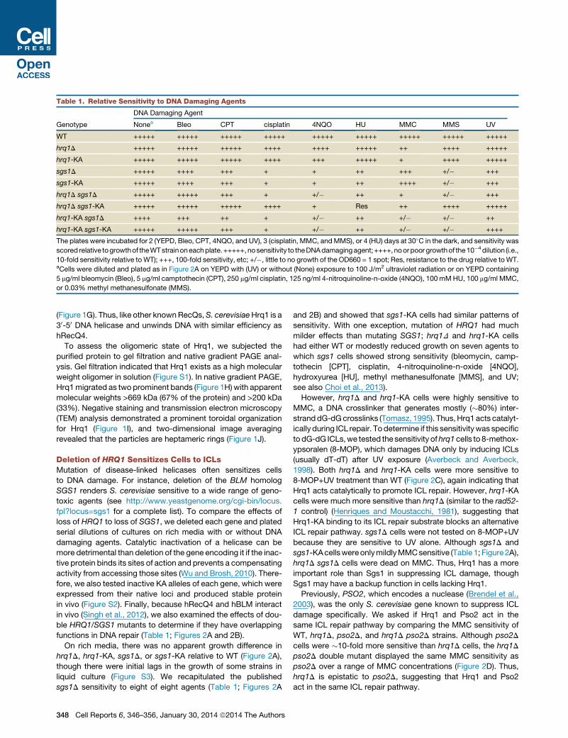
Table 1. Relative Sensitivity to DNA Damaging Agents
Genotype
DNA Damaging Agent
Nonea Bleo CPT cisplatin 4NQO HU MMC MMS UV
WT +++++ +++++ +++++ +++++ +++++ +++++ +++++ +++++ +++++
hrq1D +++++ +++++ +++++ ++++ ++++ +++++ ++ ++++ +++++
hrq1-KA +++++ +++++ +++++ ++++ +++ +++++ + ++++ +++++
sgs1D +++++ ++++ +++ + + ++ +++ +/� +++
sgs1-KA +++++ ++++ +++ + + ++ ++++ +/� +++
hrq1D sgs1D +++++ +++++ +++ + +/� ++ + +/� +++
hrq1D sgs1-KA +++++ +++++ +++++ ++++ + Res ++ ++++ +++++
hrq1-KA sgs1D ++++ +++ ++ + +/� ++ +/� +/� ++
hrq1-KA sgs1-KA +++++ +++++ +++ + +/� ++ +/� +/� ++++
The plates were incubated for 2 (YEPD, Bleo, CPT, 4NQO, and UV), 3 (cisplatin, MMC, andMMS), or 4 (HU) days at 30�C in the dark, and sensitivity was
scored relative togrowthof theWTstrainoneachplate.+++++,nosensitivity to theDNAdamagingagent;++++,noorpoorgrowthof the10�4dilution (i.e.,
10-fold sensitivity relative to WT); +++, 100-fold sensitivity, etc; +/�, little to no growth of the OD660 = 1 spot; Res, resistance to the drug relative toWT.aCells were diluted and plated as in Figure 2A on YEPD with (UV) or without (None) exposure to 100 J/m2 ultraviolet radiation or on YEPD containing
5 mg/ml bleomycin (Bleo), 5 mg/ml camptothecin (CPT), 250 mg/ml cisplatin, 125 ng/ml 4-nitroquinoline-n-oxide (4NQO), 100 mMHU, 100 mg/ml MMC,
or 0.03% methyl methanesulfonate (MMS).
(Figure 1G). Thus, like other knownRecQs,S. cerevisiaeHrq1 is a
30-50 DNA helicase and unwinds DNA with similar efficiency as
hRecQ4.
To assess the oligomeric state of Hrq1, we subjected the
purified protein to gel filtration and native gradient PAGE anal-
ysis. Gel filtration indicated that Hrq1 exists as a high molecular
weight oligomer in solution (Figure S1). In native gradient PAGE,
Hrq1migrated as two prominent bands (Figure 1H) with apparent
molecular weights >669 kDa (67% of the protein) and >200 kDa
(33%). Negative staining and transmission electron microscopy
(TEM) analysis demonstrated a prominent toroidal organization
for Hrq1 (Figure 1I), and two-dimensional image averaging
revealed that the particles are heptameric rings (Figure 1J).
Deletion of HRQ1 Sensitizes Cells to ICLsMutation of disease-linked helicases often sensitizes cells
to DNA damage. For instance, deletion of the BLM homolog
SGS1 renders S. cerevisiae sensitive to a wide range of geno-
toxic agents (see http://www.yeastgenome.org/cgi-bin/locus.
fpl?locus=sgs1 for a complete list). To compare the effects of
loss of HRQ1 to loss of SGS1, we deleted each gene and plated
serial dilutions of cultures on rich media with or without DNA
damaging agents. Catalytic inactivation of a helicase can be
more detrimental than deletion of the gene encoding it if the inac-
tive protein binds its sites of action and prevents a compensating
activity from accessing those sites (Wu and Brosh, 2010). There-
fore, we also tested inactive KA alleles of each gene, which were
expressed from their native loci and produced stable protein
in vivo (Figure S2). Finally, because hRecQ4 and hBLM interact
in vivo (Singh et al., 2012), we also examined the effects of dou-
ble HRQ1/SGS1 mutants to determine if they have overlapping
functions in DNA repair (Table 1; Figures 2A and 2B).
On rich media, there was no apparent growth difference in
hrq1D, hrq1-KA, sgs1D, or sgs1-KA relative to WT (Figure 2A),
though there were initial lags in the growth of some strains in
liquid culture (Figure S3). We recapitulated the published
sgs1D sensitivity to eight of eight agents (Table 1; Figures 2A
348 Cell Reports 6, 346–356, January 30, 2014 ª2014 The Authors
and 2B) and showed that sgs1-KA cells had similar patterns of
sensitivity. With one exception, mutation of HRQ1 had much
milder effects than mutating SGS1; hrq1D and hrq1-KA cells
had either WT or modestly reduced growth on seven agents to
which sgs1 cells showed strong sensitivity (bleomycin, camp-
tothecin [CPT], cisplatin, 4-nitroquinoline-n-oxide [4NQO],
hydroxyurea [HU], methyl methanesulfonate [MMS], and UV;
see also Choi et al., 2013).
However, hrq1D and hrq1-KA cells were highly sensitive to
MMC, a DNA crosslinker that generates mostly (�80%) inter-
stranddG-dGcrosslinks (Tomasz, 1995). Thus,Hrq1acts catalyt-
ically during ICL repair. Todetermine if this sensitivitywas specific
todG-dG ICLs,we tested the sensitivity ofhrq1cells to 8-methox-
ypsoralen (8-MOP), which damages DNA only by inducing ICLs
(usually dT-dT) after UV exposure (Averbeck and Averbeck,
1998). Both hrq1D and hrq1-KA cells were more sensitive to
8-MOP+UV treatment than WT (Figure 2C), again indicating that
Hrq1 acts catalytically to promote ICL repair. However, hrq1-KA
cells were much more sensitive than hrq1D (similar to the rad52-
1 control) (Henriques and Moustacchi, 1981), suggesting that
Hrq1-KA binding to its ICL repair substrate blocks an alternative
ICL repair pathway. sgs1D cells were not tested on 8-MOP+UV
because they are sensitive to UV alone. Although sgs1D and
sgs1-KAcellswereonlymildlyMMCsensitive (Table1; Figure2A),
hrq1D sgs1D cells were dead on MMC. Thus, Hrq1 has a more
important role than Sgs1 in suppressing ICL damage, though
Sgs1 may have a backup function in cells lacking Hrq1.
Previously, PSO2, which encodes a nuclease (Brendel et al.,
2003), was the only S. cerevisiae gene known to suppress ICL
damage specifically. We asked if Hrq1 and Pso2 act in the
same ICL repair pathway by comparing the MMC sensitivity of
WT, hrq1D, pso2D, and hrq1D pso2D strains. Although pso2D
cells were �10-fold more sensitive than hrq1D cells, the hrq1D
pso2D double mutant displayed the same MMC sensitivity as
pso2D over a range of MMC concentrations (Figure 2D). Thus,
hrq1D is epistatic to pso2D, suggesting that Hrq1 and Pso2
act in the same ICL repair pathway.

Figure 2. Comparison of hrq1 and sgs1 DNA Damage Sensitivity
(A) Growth of the indicated strains on YEPD and YEPD containing 0.03% MMS, 100 mg/ml MMC, or 100 mM HU. Cells of the indicated genotype were grown in
liquid culture, diluted to OD660 = 1, and 10-fold serial dilutions were spotted onto plates, which were then incubated for 2 (YEPD), 3 (MMS and MMC), or 4 (HU)
days in the dark.
(B) Growth curves of the indicated strains displaying relative sensitivity to cisplatin. Cells were grown overnight in YEPD, diluted to OD660 = 0.1 in a 96-well plate,
and incubated at 30�C in a BioTek EON plate reader with shaking. The OD660 was thenmeasured every 15min for 24 hr. The plotted values are themeans of three
or more independent experiments per strain.
(C) 8-MOP+UV sensitivity of the indicated strains. Cells were grown, diluted, and spotted (as in A on YEPD plates or YEPD plates containing 20 mM 8-MOP and
either placed in an opaque container (YEPD and YEPD+8-MOP) or exposed to 365 nm UV for 5 (YEPD+8-MOP+UV) or 15 min (YEPD+UV) and then incubated in
the dark for 2 days. The images are representative of results from triplicate experiments.
(D) hrq1D and pso2D are epistatic for MMC sensitivity. Cells were grown, diluted, and spotted (as in A on YEPD or YEPD+MMC plates and incubated for 2 days.
See also Figures S2 and S3.
A reverse pattern of sensitivities was seen for cisplatin, which
induces mostly (90%) 1,2-intrastrand crosslinks (Table 1; Fig-
ure 2B). More rarely, cisplatin generates dG-dG ICLs (Brabec,
2002). As reported, sgs1D cells were highly cisplatin sensitive
(Liao et al., 2007), as were sgs1-KA cells. In contrast, hrq1 and
C
hrq1-KA cells had only modest cisplatin sensitivity (see also
Choi et al., 2013; Dittmar et al., 2013). Remarkably, deletion of
HRQ1 in the sgs1-KA strain strongly suppressed the sgs1-KA
cisplatin sensitivity (Figure 2B). This suppression was not seen
for otherhrq1 sgs1combinations, and itsmechanism isunknown.
ell Reports 6, 346–356, January 30, 2014 ª2014 The Authors 349

Figure 3. Deletion of HRQ1 Affects De Novo
Telomere Addition at DSBs
(A) Schematic of Chr V-L in the GCR strain. The
numbered bars indicate the positions of the multi-
plex PCR products used to determine the relative
locations of the GCR events. ‘‘A’’ denotes AlwNI
restriction sites. URA3 and CAN1, counter-
selectable markers; PCM1, first essential gene to
the right of the V-L telomere.
(B) Southern blot analysis of representative hrq1D
GCR clones. The blot was probed with the CIN8
PCR product (primer pair 3 from A). TAs are denoted
with arrows.
In general, deletion and KA alleles had similar effects on DNA
damage sensitivity (Table 1). However, hrq1D sgs1-KA cells
were more HU resistant than WT cells (Figure 2A). Although we
did not explore the reason for these differences, they may reflect
the dual roles of Sgs1 in activating the intra-S phase checkpoint,
only one of which is helicase dependent (Frei and Gasser, 2000).
Deleting HRQ1 Inhibits De Novo TAIn addition to drug sensitivities, helicase mutations often result in
high rates of spontaneous DNA damage, such as gross-chromo-
somal rearrangements (GCRs), which are common in many
cancers (Bernstein et al., 2010). The S. cerevisiae GCR assay
provides a quantitative measure of such events by selecting for
the simultaneous loss of expression of two counterselectable
markers on the left arm of chromosome V (Chr V-L): URA3 and
CAN1 (Figure 3A). Mutation of most yeast genes that are homo-
logs of human tumor suppressor genes (e.g., SGS1) results in
increased GCR rates (Myung et al., 2001b).
The sgs1D GCR rate was 16-fold higher than in WT cells (p =
0.025), similar to the �20-fold increase reported previously
(Myung et al., 2001b). The sgs1-KA strain had the same high
GCR rate as sgs1D (p = 0.020), suggesting that the number of
DNA lesions in the two strains was similar (Table 2). In contrast,
the GCR rates in both hrq1D and hrq1-KA cells were only
modestly higher than WT (p = 0.043 and 0.048, respectively)
(Table 2). The low GCR rate in hrq1 mutants is consistent with
our finding that these strains were insensitive to most types of
DNA damage except ICLs (Table 1; Figure 3), which are rare
for cells grown in laboratory conditions (Scharer, 2005). Indeed,
pregrowth of hrq1D cells in media containing MMC prior to
plating resulted in an �58-fold increase in the GCR rate relative
to cells treated with solvent alone (Table 2; data not shown). The
GCR rate of WT cells grown under the same conditions also
increased but only 5-fold.
If breaks occur between CAN1 and PCM1 (the most distal
essential gene on Chr V-L; Figure 3A), FOAR CanR cells can theo-
retically be generated by TA to DSBs. However, in WT cells and
virtually all single mutants, GCR events arise from recombination
events that delete or move URA3 and CAN1 to new locations
(Kolodner et al., 2002). The one published exception is pif1 cells,
350 Cell Reports 6, 346–356, January 30, 2014 ª2014 The Authors
where most GCR events are due to TA
(Myung et al., 2001a). To determine if
GCR events were due to TA or recombina-
tion, we determined the structure of Chr V-L in multiple indepen-
dent GCR clones from each mutant (Figures 3A and 3B; Table 2;
data not shown).
As anticipated (Myung et al., 2001b), none of the WT (0/10) or
sgs1D (0/15) GCR clones were due to TA, whereas 93% of the
GCR events in pif1-m2 (an allele that eliminates most of the
nuclear Pif1 [Schulz and Zakian, 1994]) cells were TAs (52/56).
Remarkably, �80% (20/26) of the hrq1D GCR events were TAs
(Table 2; Figure 3B). Also surprising, the events leading to the
GCRs were different in hrq1-KA (4.5% TA, 1/22) and hrq1D
(77% TAs) cells, even though the GCR rates were the same
in these strains (p = 0.264). Thus, unlike Pif1, which requires
helicase activity to inhibit telomerase (Myung et al., 2001a;
Zhou et al., 2000), Hrq1 inhibited TA noncatalytically. In addition,
HRQ1 and PIF1 did not act synergistically to suppress TAs,
because the two helicases did not have additive effects on TA:
the fraction of TAs in hrq1D pif1-m2 cells was <50%, lower
than in either single mutant. (See Discussion for a model
explaining these data.)
We also determined the structure of Chr V-L in sgs1-KA GCR
clones. Although TAs are not detected in sgs1D cells (Myung
et al., 2001b) (Table 2),�50%of the GCR events in sgs1-KA cells
were TAs (11/24; Table 2). TAs are also detected in sgs1D exo1D
cells, presumably because both pathways for DSB resection
are blocked (Lydeard et al., 2010; Marrero and Symington,
2010). Because Sgs1-KA binds ssDNA (Cejka and Kowalczy-
kowski, 2010), we suggest that it reduces Exo1 access (Fig-
ure 5A). Thus, both pathways to generate the ssDNA for strand
invasion are inhibited, shifting the recombination-TA equilibrium
toward TAs.
Hrq1 Limits Telomere Lengthening in pif1-m2 CellsThe ability of Hrq1 to suppress TAs to DSBs suggested that, like
Pif1, it might also inhibit telomerase lengthening of existing telo-
meres. However, telomere length was indistinguishable fromWT
in hrq1D and hrq1-KA cells (Figure 4A). In contrast, telomeres in
hrq1D pif1-m2 cells were even longer than in pif1-m2.
The hyperlengthening of telomeres in hrq1D pif1-m2 cells
could be due to recombination or telomerase. To distinguish be-
tween these possibilities, we analyzed telomere length in spore
Table 2. Gross-Chromosomal Rearrangement Rates and
Analysis of Events in Independent Clones Grown in the Absence
or Presence of Mitomycin C
Genotype GCR Ratea,b95% Confidence
Interval
Telomere Addition
(Clones Analyzed)
WT 1.0 ± 0.5c 0.18–2.27 0% (10)c
WT + MMC 5 ± 4 2.52–11.5 ND
sgs1D 16 ± 5c 5.15–37.4 0% (15)c
sgs1-KA 14 ± 3 13.1–40.1 46% ± 14% (24)
hrq1D 4 ± 2 3.19–12.6 77% ± 2.9% (26)
hrq1D + MMC 115 ± 30 89.6–188 ND
hrq1-KA 5 ± 3 3.48–16.7 4.5% ± 3.9% (22)
hrq1-KA + MMC 132 ± 42 46.5–246 ND
pif1-m2 76 ± 8c 36.4–125 93% ± 7.6% (56)c
ND, not determined.aGross-chromosomal rearrangement (GCR) rates are the average of
three or more independent experiments and are normalized to the WT
rate (1.5 ± 0.73 10�10 events/generation). ± denotes SD. +MMCdenotes
growth in media containing mitomycin C.bp values were calculated for the GCR rates for all pairwise combinations
of strains. All mutant rates are significantly different from WT (p = 0.025,
sgs1D; p = 0.020, sgs1-KA; p = 0.043, hrq1D; p = 0.048, hrq1-KA; and p <
0.00005, pif1-m2). All rates are significantly different from pif1-m2 (all p%
0.008). The hrq1D and hrq1-KA GCR rates are not significantly different
from either the sgs1D or sgs1-KA rates (all p R 0.078). For telomere ad-
ditions, the frequency in pif1-m2 cells was significantly different from all
other strains (all p < 0.030), and the frequencies between hrq1D and
hrq1-KA were also significantly different (p = 0.0002).cData are from Paeschke et al., 2013, though it was collected at the same
time as the other data.
clones from a pif1-m2/WT hrq1D/WT rad52D/WT diploid (Rad52
is a protein required for virtually all homologous recombination
[HR] [Hiom, 1999]). Telomeres were even longer in hrq1D pif1-
m2 rad52D cells than in recombination-proficient cells (Fig-
ure 4A), demonstrating that the hyperlengthening was not
recombination dependent. Telomeres are also longer in rad52
versions of other mutants with long telomeres (e.g., rif1 and
rif2), suggesting that HR suppresses elongation in cells that
already have long telomeres (Teng et al., 2000; Zhou et al.,
2000). Thus, Hrq1 limits telomerase, not recombination, at pif1-
m2 telomeres.
Hrq1 did not act catalytically to inhibit telomerase at DSBs
(Table 2). To determine if it acts catalytically to affect telomere
length in pif1-m2 cells, we analyzed telomeres in hrq1-KA pif1-
m2 and hrq1-KA pif1-m2 rad52D cells. Telomere length was un-
affected in the hrq1-KA pif1-m2 background relative to pif1-m2
(Figure 4A) (as expected, deletion of RAD52 did increase hrq1-
KA pif1-m2 telomere length as seen for pif1-m2 versus rad52D
cells [Zhou et al., 2000]). Thus, Hrq1 acts structurally to inhibit
lengthening of pif1-m2 telomeres, just as it does during TA
at DSBs.
Telomerase-Independent Telomere Maintenance by theType I Pathway Is Hrq1 DependentIn telomerase-deficient yeast and human cells, survivors arise
that maintain telomeres by mechanisms that usually involve HR
C
(i.e., alternative lengthening of telomeres or ALT) (Wellinger and
Zakian, 2012). In S. cerevisiae, telomerase-independent survi-
vors come in two types: type I, which have tandem copies of
the subtelomeric Y’ element and very short tracts of telomeric
DNA; and type II, which have heterogeneous length telomeres.
Because hBLM (Stavropoulos et al., 2002) and Sgs1 (Huang
et al., 2001; Johnson et al., 2001) both influence ALT, we asked
if Hrq1 does as well. We sporulated an hrq1D/WT sgs1D/WT
tlc1D/WT diploid strain (TLC1 encodes telomerase RNA), serially
restreaked spore clones until survivors appeared, and examined
telomere structure in 20 survivors from each strain. As previously
reported, sgs1D tlc1D spore clones generated only type I survi-
vors. In contrast, all hrq1D tlc1D survivors had type II telomeres;
sgs1Dwas epistatic to hrq1D because all hrq1D sgs1D tlc1D sur-
vivors were type I (Figure 4B). Thus, Hrq1 is required to generate
type I survivors in the presence of Sgs1.
Hrq1 Binds Telomeres In VivoIf Hrq1 affects telomeres directly, it will bind telomeres, which
might be detectable by chromatin immunoprecipitation (ChIP).
In WT cells, Hrq1-13xMyc bound significantly to telomere VI-R
(relative to a control site; p < 0.001); binding to the VII-L telomere
was detectable but not significant (p = 0.0567; Figure 4C). How-
ever, in pif1-m2 cells, Hrq1 binding was significant at both (p <
0.001; �2.5-fold higher to pif1-m2 versus WT telomeres). These
results suggest that Hrq1 is telomere associated in WT cells
but that Pif1 can displace it from telomeres. This interpretation
also explains why telomeres in hrq1D cells are of WT length
(Figure 4A).
DISCUSSION
Hrq1 Protects against ICLsUnlike sgs1 cells, hrq1 cells were resistant to most DNA
damaging agents (Table 1). This pattern is reminiscent of
the damage sensitivities for their human homologs, because
hRecQ4-deficient cells are less sensitive than hBLMmutant cells
to many DNA damaging agents, including cisplatin (see Mao
et al., 2010 and references therein). However, a different pattern
was seen for two ICL-inducing agents: hrq1D and hrq1-KA cells
were highly sensitive to both MMC and 8-MOP. Thus, Hrq1
acts catalytically to promote ICL repair. Cells expressing Hrq1-
KA were much more sensitive to these agents than hrq1D cells
(Figures 2A and 2C). This finding suggests that Hrq1 is the main
helicase in ICL repair, and its backup is hindered from accessing
theDNAbecauseHrq1-KA is bound to sites of damage. The even
higher MMC sensitivity of hrq1 sgs1 cells (Figure 2A) suggests
that Sgs1 is the backup for Hrq1 at ICLs. Even the modest sensi-
tivity of hrq1 cells to cisplatin, a predominantly intrastrand cross-
linker (Table 1; Figure 2B), can be explained by its role in ICL
repair, because�5%of cisplatin lesions are ICLs (Brabec, 2002).
Our study reports MMC and 8-MOP sensitivity for hrq1
S. cerevisiae, but S. pombe hrq1D cells have previously been re-
ported as highly sensitive to both cisplatin and MMC (Groocock
et al., 2012). However, because 8-MOP was not tested in that
study, it is possible that the S. pombe hrq1D MMC sensitivity is
due to the low level of intrastrand crosslinks generated by
MMC. In any case, becauseS. cerevisiaeSgs1 has quite different
ell Reports 6, 346–356, January 30, 2014 ª2014 The Authors 351
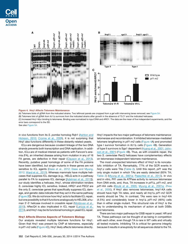
Figure 4. Hrq1 Affects Telomere Maintenance
(A) Telomere blots of gDNA from the indicated strains. Two leftmost panels are cropped from a gel with intervening lanes removed; see Figure S4.
(B) Telomere blot of gDNA from tlc1D survivors from the indicated strains after growth in the absence of TLC1 and the indicated helicases.
(C) Increased Hrq1-Myc binding to telomeres. Binding was normalized to input DNA and ARO1. The data are the mean of five independent experiments, and the
error bars correspond to the SD.
See also Figure S4.
in vivo functions from its S. pombe homolog Rqh1 (Ashton and
Hickson, 2010; Cromie et al., 2008), it is not surprising that
Hrq1 also functions differently in these distantly related yeasts.
ICLs are dangerous because covalent linkage of the two DNA
strands prevents both transcription and DNA replication. In addi-
tion, ICLs are of medical interest as patients with Fanconi’s ane-
mia (FA), an inherited disease arising from mutation in any of 16
FA genes, are defective in their repair (Clauson et al., 2013).
Recently, putative yeast homologs of some of the FA proteins
have been identified, but single mutants in these genes are not
sensitive to ICL agents (Daee et al., 2012; Daee and Myung,
2012; Ward et al., 2012). Whereas mammals have multiple heli-
cases that suppress ICL damage (e.g., HELQ acts in a pathway
parallel to FA to suppress ICL damage [Adelman et al., 2013]),
our study identifies a helicase, Hrq1, whose elimination renders
S. cerevisiae highly ICL sensitive. Indeed, HRQ1 and PSO2 are
the only S. cerevisiae genes that specifically suppress ICL dam-
age, and genetic data indicate that they act in the same pathway
(Figure 2D).Wedonot knowhowHrq1acts to facilitate ICL repair,
but onepossibility is that it functions analogously toHEL308, ahu-
man 30-50 helicase involved in crosslink repair (Moldovan et al.,
2010). hRecQ4 is also implicated in ICL repair (Larizza et al.,
2010), andHrq1maybe its functional homolog in yeast ICL repair.
Hrq1 Affects Diverse Aspects of Telomere BiologyOur analysis revealed multiple telomere functions for Hrq1.
Given that Hrq1 was present by ChIP at telomeres, especially
in pif1-m2 cells (Figure 4C), Hrq1 likely affects telomeres directly.
352 Cell Reports 6, 346–356, January 30, 2014 ª2014 The Authors
Hrq1 impacts the two major pathways of telomere maintenance:
telomerase and recombination. It inhibited telomerase-mediated
telomere lengthening in pif1-m2 cells (Figure 4A) and promoted
type I survivor formation in tlc1D cells (Figure 4B). Generation
of type II survivors is Sgs1 dependent (Huang et al., 2001; John-
son et al., 2001) (Figure 4B). Thus, as with crosslink repair, the
two S. cerevisiae RecQ helicases have complementary effects
on telomerase-independent telomere maintenance.
The most unexpected telomere effect of Hrq1 is its noncata-
lytic inhibition of TA. Remarkably, 77% of the GCR events in
hrq1D cells were TAs (Table 2). Until this report, pif1 was the
only single mutant in which TAs are easily detected (93% TA;
Table 2) (Myung et al., 2001a; Paeschke et al., 2013). In vivo
and in vitro, Pif1 uses its ATPase activity to remove telomerase
from DNA ends, and, thus, TA increases in pif1D, pif1-m2, and
pif1-KA cells (Boule et al., 2005; Myung et al., 2001a; Zhou
et al., 2000). If Hrq1 also removes telomerase, hrq1-KA cells
should have high TA rates, and nearly all hrq1D pif1-m2 GCR
events should be TAs. In contrast, TAs were rare in hrq1-KA
(4.5%) and considerably lower in hrq1D pif1-m2 (46%) cells
than in either single mutant. This structural role of Hrq1 is the
key to understanding its mechanism of action at both DSBs
and telomeres.
There are two major pathways for DSB repair in yeast: HR and
TA. These pathways can be thought of as being in competition
with each other, even though TA is rare in WT cells and virtually
all tested mutants. Inhibiting TA is critical for genome integrity
because it results in aneuploidy for all sequences distal to the TA.
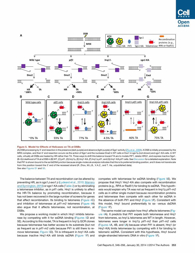
Figure 5. Model for Effects of Helicases on TA at DSBs
(A) DSBprocessing by 50 end resection in the presence (dark purple) and absence (light purple) of Sgs1 activity (Zhu et al., 2008). A DSB is initially processed by the
MRX complex, and then 50 end resection occurs via the action of Sgs1 and the nuclease Dna2 in WT cells or Exo1 in sgs1D (not shown) and sgs1-KA cells. In WT
cells, virtually all DSBs are healed by HR rather than TA. Three ways to shift this balance toward TA are to mutate PIF1, delete HRQ1, and express inactive Sgs1.
(B–G) Likelihood of TA at a DSB in (B)WT, (C) pif1, (D) hrq1D, (E) hrq1-KA, (F) hrq1D pif1, and (G) hrq1-KA pif1 cells. SeeDiscussion for a detailed explanation. Note
that Pif1 is shown bound to the ss/dsDNA junction because single-molecule analysis indicates that this is its preferred binding position, and it does not translocate
from this position toward the 30 end of the recessed strand (R. Zhou, M.L.B., V.A.Z., and T. Ha, unpublished data).
See also Figures S1 and S5.
The balance between TA and recombination can be altered by
preventing HR, as in sgs1D exo1D (Lydeard et al., 2010; Marrero
and Symington, 2010) or sgs1-KA cells (Table 2) or by eliminating
a telomerase inhibitor, as in pif1 cells. Hrq1 is unlikely to affect
the HR-TA balance by promoting recombination, because it
has not been recovered in the large number of screens for genes
that affect recombination. Its binding to telomeres (Figure 4C)
and inhibition of telomerase at pif1-m2 telomeres (Figure 4A)
also argue that it affects telomerase, not recombination, at
DSBs.
We propose a working model in which Hrq1 inhibits telome-
rase by competing with it for ssDNA binding (Figures 5D and
5E). According to this model, TA is frequent in hrq1DGCR clones
because telomerase has better access to its substrate (but not
as frequent as in pif1-m2 cells because Pif1 is still there to re-
move telomerase; Figure 5D). TA is infrequent in hrq1-KA cells
because inactive Hrq1-KA still binds ssDNA (Figure 1F) and
C
competes with telomerase for ssDNA binding (Figure 5E). We
propose that Hrq1/ Hrq1-KA also compete with recombination
proteins (e.g., RPA or Rad51) for binding to ssDNA. This hypoth-
esis would explain why TA was not as frequent in hrq1D pif1-m2
cells as in either single mutant because recombination proteins
and telomerase then compete with each other for ssDNA in
the absence of both Pif1 and Hrq1 (Figure 5F). Consistent with
this model, Hrq1 bound preferentially to ss- versus dsDNA
(Figure 1F).
The same model can explain how Hrq1 affects telomeres (Fig-
ure 4A). It predicts that Pif1 expels both telomerase and Hrq1
from telomeres, so hrq1D telomeres are WT in length. However,
telomeres were longer in hrq1D pif1-m2 than pif1-m2 cells
(Figures 4A, 4B, and 5A) because when Pif1 is absent, Hrq1 (or
Hrq1-KA) limits telomerase by competing with it for binding to
telomeric ssDNA. Consistent with this hypothesis, Hrq1 bound
single-stranded telomeric DNA in vitro (Figure 1F).
ell Reports 6, 346–356, January 30, 2014 ª2014 The Authors 353
In summary, our studies show that Hrq1 has two roles that pro-
mote genome integrity: (1) It acts catalytically to promote ICL
repair. Indeed, the very strong ICL sensitivity of hrq1-KA cells
suggests that Hrq1 may be the first line of defense against these
dangerous lesions. (2) We also found that Hrq1 affects several
aspects of telomere biology, including inhibition of telomerase
at DSBs and telomeres. Two of the defects in hrq1 cells, sensi-
tivity to ICLs and frequent TAs, are rare phenotypes; the demon-
stration of both functions in one protein is unprecedented. If
hRecQ4 has either or both activities, this could explain why its
mutation results in genomic instability and disease.
EXPERIMENTAL PROCEDURES
Yeast Strains, Media, and Other Reagents
All strains (Table S1) were created by standard methods and are derivatives of
the YPH background (Sikorski and Hieter, 1989). Cells were grown in standard
S. cerevisiae media at 30�C unless indicated. 32P-dCTP and 32P-ATP were
purchased from PerkinElmer, and unlabeled ATP was from GE Healthcare.
DNA damaging agents were from Sigma. All restriction enzymes were from
NEB, and all oligonucleotides were from IDT.
Protein Purification
Details on HRQ1 and hrq1-KA cloning and expression vector construction, as
well as the complete protein purification protocol, can be found in the Supple-
mental Experimental Procedures. Briefly, expression plasmids were trans-
formed into Rosetta 2(DE3) pLysS cells, and recombinant protein was
expressed using the autoinduction method (Studier, 2005). Cells were har-
vested by centrifugation. The pellets were resuspended in buffer and lysed
by adding n-dodecyl b-D-maltoside (DM; Sigma) to a final concentration of
0.05% (w/v) and 1 3 FastBreak (Promega).
The soluble fraction was clarified by centrifugation and filtering the through a
0.22 mmmembrane. This mixture was then loaded onto a Strep-Tactin Sephar-
ose column (IBA), and protein was eluted with three column volumes (CVs) of
desthiobiotin buffer after extensive washing. Peak fractions were pooled and
loaded onto a His60 Ni column (Clontech). After washing, protein was eluted
with six CVs of imidazole buffer, and peak fractions were pooled and concen-
trated by ultrafiltration. The protein was then buffer-exchanged into storage
buffer using a desalting column.
The hRecQ4 expression plasmid pGEX-RecQ4-His9 was a gift from Patrick
Sung, Yale University. hRecQ4 was purified as described (Macris et al., 2006).
The protein concentration and purity of the final preparations were determined
on SYPRO orange-stained SDS-PAGE gels using known amounts of a stan-
dard protein for comparison. In all cases, protein purity was R95%.
Helicase Assays
The fork substrate was constructed by end labeling oligonucleotide MB733
(Table S2) with T4 PNK and g32P-ATP and separating the ssDNA from free
label using a G-50 micro column (GE Healthcare). Labeled MB733 was then
annealed to oligonucleotide MB734 in 13 NEB buffer #2 by boiling and slowly
cooling to room temperature. The directionality substrate was similarly con-
structed by end labeling an equimolar mixture of oligonucleotides MB453
and 454 (Table S2) and removing free label using a G-50 micro column.
The labeled oligonucleotides were then annealed to MB452 as above. Both
substrates were separated from contaminating ssDNA by gel purification,
followed by electroelution into a dialysis membrane.
Helicase reactions were performed in 13 binding buffer (25 mMNa-HEPES
[pH 8.0], 5% glycerol, 50 mM NaOAc, 150 mM NaCl, 7.5 mM MgOAc, and
0.01%DM) and contained 0.1 nM radiolabeled substrate, protein as indicated,
5 mM ATP, and 15 nM unlabeled oligonucleotide MB733 (fork) or MB453 and
MB454 (directionality). Hrq1 helicase activity was unaffected by omitting these
unlabeled ssDNA traps (e.g., Figure S5). Reactions containing the direction-
ality substrate additionally contained 100 mg/ml Neutravidin (Pierce). The reac-
tions were incubated at 37�C for 30 min and stopped by the addition of 1 3
stop-load buffer (25% [w/v] Ficoll (type 400), 100 mM EDTA, 0.1% SDS,
354 Cell Reports 6, 346–356, January 30, 2014 ª2014 The Authors
0.25% bromophenol blue, and 0.25% xylene cyanol). The samples were
then separated on 8% 29:1 acrylamide:bis-acrylamide gels in 1 3 TBE buffer
at 100 V/cm for 30–45min, dried, and imaged/quantified using a Typhoon 9410
scanner and Image Gauge software.
Native Gradient Gel Electrophoresis
Native gradient gels were poured following the protocol in the Supplemental
Experimental Procedures. Protein (R0.5 mg) was loaded into the wells in the
absence of loading dye and run at 17 V/cm for 3–6 hr in 20 mM Tris-HCl
(pH 8.8) and 200 mM glycine. After electrophoresis, gels were incubated in
0.05% SDS for 15 min, rinsed with water, and stained with SYPRO orange
overnight prior to analysis as described above for protein concentration.
TEM
Hrq1was analyzed by TEM essentially as previously described for theMcm2-7
helicase (Bochman and Schwacha, 2007). Briefly, the protein was diluted to
25 mg/ml in storage buffer, absorbed to glow-discharged copper grids (Ted
Pella), and negatively stained with 2% uranyl acetate. Grids were visualized
with a LEO OMEGA 912 electron microscope (Carl Zeiss) at 80 kV and 40,000
3 magnification. Micrographs were taken with a 7.5 megapixel EMCCD cam-
era (Peltier-cooled Hamamatsu ORCA) and visualized with AMT (v.602) soft-
ware. Two-dimensional image averaging of Hrq1 complexes was performed
using EMAN2 (Tang et al., 2007).
GCR Assays
GCR assays were performed essentially as described by Putnam and
Kolodner (2010). Briefly, three or more sets of five or more 5 ml cultures of
each S. cerevisiae GCR strain (Table 1) were grown to saturation in YEPD
medium ± 25 mg/ml MMC at 30�C for 36–48 hr. Cells (2 ml) from each culture
were pelleted, resuspended in sterile water, plated on dropoutmedium lacking
uracil and arginine (USBiologicals) supplemented with 1 g/l 5-FOA and 60mg/l
canavanine sulfate (FOA+Can), and incubated at 30�C for 4 days. GCR rates
(per 10�9 mutations/generation) and 95% confidence intervals were calcu-
lated using the FALCOR web server and MMS Maximum Likelihood Method
(Hall et al., 2009). The rates presented are the means ± SDs of three or more
experiments per strain. p values were calculated using Student’s t test.
We define GCR clones as colonies that grew on the FOA+Can plates. Such
FOAR CanR clones were selected for post-GCR analyses (multiplex PCR
[Supplemental Experimental Procedures] and Southern blotting, below).
Southern Blotting
When colonies arose after GCR events, they were restreaked onto FOA+Can
plates to verify the FOAR CanR phenotype and grown overnight in YEPD liquid
media at 30�C, and genomic DNA (gDNA) was isolated. The gDNA was
analyzed by multiplex PCR and Southern blotting as described (Paeschke
et al., 2013). Briefly, gDNA from clones retaining the CIN8 PCR product was
digested overnight at 37�C with AlwNI, run on 0.7% agarose gels, and blotted
on Hybond-XL membranes (GE Life Sciences). For the Southerns, the 400 bp
CIN8 PCR product was used a probe (hybridization sites are shown in Fig-
ure 3A), resulting in a 3.2 kb background band in all lanes. A non-GCR event
yields a 6.9 kb band, TAs produce fuzzy bands <5.5 kb, and non-TAs result
in sharp bands.
For telomere blots, gDNA was isolated from cells as described above. For
the experiments examining the effect of recombination on telomere length,
heterozygous diploids (Table S1) were sporulated, tetrads were dissected,
and three spore clones of the desired genotypes were serially restreaked on
YEPD for �100 generations prior to gDNA isolation. For standard telomere
length detection, gDNA was digested overnight with PstI and XhoI, whereas
for telomere blots from tlc1D survivors, the gDNA was digested overnight
with XhoI. Digests were separated on 1% agarose gels and transferred as
described above. In both types of telomere blots, the probe used was a previ-
ously described C1–3A/TG1–3 restriction fragment (Runge and Zakian, 1989).
tlc1D Survivor Analysis
The generation and analysis of telomeric survivors were performed as
described previously (Teng and Zakian, 1999). Briefly, a HRQ1/hrq1D SGS1/
sgs1D TLC1/tlc1D diploid strain (KP386) was generated, sporulated, and
hrq1D tlc1D and sgs1D tlc1D spores were identified by plating. Spores where
restreaked four to five times (�25 generations/streak) on YC plates until survi-
vors formed. For each spore clone, ten colonies where picked for the next re-
streak. In total, gDNA was isolated from 20 different spores for each strain
background and analyzed as described above.
Chromatin Immunoprecipitation
Chromatin immunoprecipitation (ChIP) of asynchronous yeast cells growing in
YEPDwas performed as described (Azvolinsky et al., 2009) and analyzed using
an iCycleriQ Real-Time PCR detection system (Bio-Rad). Hrq1 was C-termi-
nally tagged with 13 Myc epitopes, and ChIP was performed using a Myc
monoclonal antibody (Clontech #631206). The amount of DNA in the immuno-
precipitate was normalized to the amount in input samples. The ChIP experi-
ment was analyzed by quantitative PCR (qPCR) in duplicate or triplicate to
obtain an average value for each sample. The ChIP experiment was repeated
three times at each locus. For each qPCR experiment, the amount of signal
in the Hrq1 immunoprecipitate was normalized to input and to the immunopre-
cipitated signal from ARO1.
SUPPLEMENTAL INFORMATION
Supplemental Information includes Supplemental Experimental Procedures,
five figures, and two tables and can be found with this article online at http://
dx.doi.org/10.1016/j.celrep.2013.12.037.
ACKNOWLEDGMENTS
M.L.B. dedicates this article to the memory of Megan Davey who passed away
during the course of this work. We thank Kathy Friedman, Margaret Platts,
Kristina Schmidt, Raymund Wellinger, and Nadine Guenther for sharing their
methods, suggesting experiments, for useful discussions, and assistance
with experiments. Research in the Zakian laboratory is supported by grants
from the National Institutes of Health (GM26938 and GM GM43265) and
postdoctoral fellowships from the American Cancer Society (to M.L.B.; PF-
10-145-01-DMC), the Deutsche Forschungsgemeinschaft (to K.P.), and the
New Jersey Commission on Cancer Research (to K.P.). Research in the
Paeschke laboratory is supported by the Emmy-Noether Program of the Deut-
sche Forschungsgemeinschaft.
Received: October 14, 2013
Revised: November 27, 2013
Accepted: December 24, 2013
Published: January 16, 2014
REFERENCES
Abdelhaleem, M. (2010). Helicases: an overview. Methods Mol. Biol. 587,
1–12.
Adelman, C.A., Lolo, R.L., Birkbak, N.J., Murina, O., Matsuzaki, K., Horejsi, Z.,
Parmar, K., Borel, V., Skehel, J.M., Stamp, G., et al. (2013). HELQ promotes
RAD51 paralogue-dependent repair to avert germ cell loss and tumorigenesis.
Nature 502, 381–384.
Ashton, T.M., and Hickson, I.D. (2010). Yeast as amodel system to study RecQ
helicase function. DNA Repair (Amst.) 9, 303–314.
Averbeck, D., and Averbeck, S. (1998). DNA photodamage, repair, gene induc-
tion and genotoxicity following exposures to 254 nm UV and 8-methoxypsor-
alen plus UVA in a eukaryotic cell system. Photochem. Photobiol. 68, 289–295.
Azvolinsky, A., Giresi, P.G., Lieb, J.D., and Zakian, V.A. (2009). Highly tran-
scribed RNA polymerase II genes are impediments to replication fork progres-
sion in Saccharomyces cerevisiae. Mol. Cell 34, 722–734.
Barea, F., Tessaro, S., and Bonatto, D. (2008). In silico analyses of a new group
of fungal and plant RecQ4-homologous proteins. Comput. Biol. Chem. 32,
349–358.
Bernstein, K.A., Gangloff, S., and Rothstein, R. (2010). The RecQ DNA heli-
cases in DNA repair. Annu. Rev. Genet. 44, 393–417.
C
Bochman, M.L., and Schwacha, A. (2007). Differences in the single-stranded
DNA binding activities of MCM2-7 and MCM467: MCM2 and MCM5 define
a slow ATP-dependent step. J. Biol. Chem. 282, 33795–33804.
Boule, J.B., Vega, L.R., and Zakian, V.A. (2005). The yeast Pif1p helicase
removes telomerase from telomeric DNA. Nature 438, 57–61.
Brabec, V. (2002). DNA modifications by antitumor platinum and ruthenium
compounds: their recognition and repair. Prog. Nucleic Acid Res. Mol. Biol.
71, 1–68.
Brendel, M., Bonatto, D., Strauss, M., Revers, L.F., Pungartnik, C., Saffi, J.,
and Henriques, J.A. (2003). Role of PSO genes in repair of DNA damage of
Saccharomyces cerevisiae. Mutat. Res. 544, 179–193.
Capp, C., Wu, J., and Hsieh, T.S. (2010). RecQ4: the second replicative
helicase? Crit. Rev. Biochem. Mol. Biol. 45, 233–242.
Cejka, P., and Kowalczykowski, S.C. (2010). The full-length Saccharomyces
cerevisiae Sgs1 protein is a vigorous DNA helicase that preferentially unwinds
holliday junctions. J. Biol. Chem. 285, 8290–8301.
Chisholm, K.M., Aubert, S.D., Freese, K.P., Zakian, V.A., King, M.C., and
Welcsh, P.L. (2012). A genomewide screen for suppressors of Alu-mediated
rearrangements reveals a role for PIF1. PLoS ONE 7, e30748.
Choi, D.H., Lee, R., Kwon, S.H., and Bae, S.H. (2013). Hrq1 functions indepen-
dently of Sgs1 to preserve genome integrity in Saccharomyces cerevisiae.
J. Microbiol. 51, 105–112.
Clauson, C., Scharer, O.D., and Niedernhofer, L. (2013). Advances in under-
standing the complex mechanisms of DNA interstrand cross-link repair.
Cold Spring Harbor perspectives in medicine 3, a012732.
Cromie, G.A., Hyppa, R.W., and Smith, G.R. (2008). The fission yeast BLM
homolog Rqh1 promotes meiotic recombination. Genetics 179, 1157–1167.
Daee, D.L., Ferrari, E., Longerich, S., Zheng, X.F., Xue, X., Branzei, D., Sung,
P., and Myung, K. (2012). Rad5-dependent DNA repair functions of the
Saccharomyces cerevisiae FANCM protein homolog Mph1. J. Biol. Chem.
287, 26563–26575.
Daee, D.L., and Myung, K. (2012). Fanconi-like crosslink repair in yeast.
Genome integrity 3, 7.
Dittmar, J.C., Pierce, S., Rothstein, R., and Reid, R.J. (2013). Physical and
genetic-interaction density reveals functional organization and informs signif-
icance cutoffs in genome-wide screens. Proc. Natl. Acad. Sci. USA 110, 7389–
7394.
Frei, C., and Gasser, S.M. (2000). The yeast Sgs1p helicase acts upstream of
Rad53p in the DNA replication checkpoint and colocalizes with Rad53p in
S-phase-specific foci. Genes Dev. 14, 81–96.
Groocock, L.M., Prudden, J., Perry, J.J., and Boddy, M.N. (2012). The RecQ4
orthologue Hrq1 is critical for DNA interstrand cross-link repair and genome
stability in fission yeast. Mol. Cell. Biol. 32, 276–287.
Hall, B.M., Ma, C.X., Liang, P., and Singh, K.K. (2009). Fluctuation analysis
CalculatOR: a web tool for the determination of mutation rate using Luria-Del-
bruck fluctuation analysis. Bioinformatics 25, 1564–1565.
Henriques, J.A., and Moustacchi, E. (1981). Interactions between mutations
for sensitivity to psoralen photoaddition (pso) and to radiation (rad) in Saccha-
romyces cerevisiae. J. Bacteriol. 148, 248–256.
Hiom, K. (1999). Dna repair: Rad52 - the means to an end. Curr. Biol. 9, R446–
R448.
Huang, P., Pryde, F.E., Lester, D., Maddison, R.L., Borts, R.H., Hickson, I.D.,
and Louis, E.J. (2001). SGS1 is required for telomere elongation in the absence
of telomerase. Curr. Biol. 11, 125–129.
Jin, W., Liu, H., Zhang, Y., Otta, S.K., Plon, S.E., and Wang, L.L. (2008). Sensi-
tivity of RECQL4-deficient fibroblasts from Rothmund-Thomson syndrome
patients to genotoxic agents. Hum. Genet. 123, 643–653.
Johnson, F.B., Marciniak, R.A., McVey, M., Stewart, S.A., Hahn, W.C., and
Guarente, L. (2001). The Saccharomyces cerevisiae WRN homolog Sgs1p
participates in telomere maintenance in cells lacking telomerase. EMBO J.
20, 905–913.
ell Reports 6, 346–356, January 30, 2014 ª2014 The Authors 355
Kolodner, R.D., Putnam, C.D., and Myung, K. (2002). Maintenance of genome
stability in Saccharomyces cerevisiae. Science 297, 552–557.
Kwon, S.H., Choi, D.H., Lee, R., and Bae, S.H. (2012). Saccharomyces
cerevisiae Hrq1 requires a long 30-tailed DNA substrate for helicase activity.
Biochem. Biophys. Res. Commun. 427, 623–628.
Larizza, L., Roversi, G., and Volpi, L. (2010). Rothmund-Thomson syndrome.
Orphanet J. Rare Dis. 5, 2.
Lee, W., St Onge, R.P., Proctor, M., Flaherty, P., Jordan, M.I., Arkin, A.P., Da-
vis, R.W., Nislow, C., and Giaever, G. (2005). Genome-wide requirements for
resistance to functionally distinct DNA-damaging agents. PLoS Genet. 1, e24.
Liao, C., Hu, B., Arno, M.J., and Panaretou, B. (2007). Genomic screening
in vivo reveals the role played by vacuolar H+ ATPase and cytosolic acidifica-
tion in sensitivity to DNA-damaging agents such as cisplatin. Mol. Pharmacol.
71, 416–425.
Liu, Y. (2010). Rothmund-Thomson syndrome helicase, RECQ4: on the cross-
road between DNA replication and repair. DNA Repair (Amst.) 9, 325–330.
Lydeard, J.R., Lipkin-Moore, Z., Jain, S., Eapen, V.V., and Haber, J.E. (2010).
Sgs1 and exo1 redundantly inhibit break-induced replication and de novo
telomere addition at broken chromosome ends. PLoS Genet. 6, e1000973.
Macris, M.A., Krejci, L., Bussen, W., Shimamoto, A., and Sung, P. (2006).
Biochemical characterization of the RECQ4 protein, mutated in Rothmund-
Thomson syndrome. DNA Repair (Amst.) 5, 172–180.
Mao, F.J., Sidorova, J.M., Lauper, J.M., Emond,M.J., andMonnat, R.J. (2010).
The human WRN and BLM RecQ helicases differentially regulate cell prolifer-
ation and survival after chemotherapeutic DNA damage. Cancer Res. 70,
6548–6555.
Marrero, V.A., and Symington, L.S. (2010). Extensive DNA end processing by
exo1 and sgs1 inhibits break-induced replication. PLoS Genet. 6, e1001007.
Mirzaei, H., Syed, S., Kennedy, J., and Schmidt, K.H. (2011). Sgs1 truncations
induce genome rearrangements but suppress detrimental effects of BLM
overexpression in Saccharomyces cerevisiae. J. Mol. Biol. 405, 877–891.
Moldovan, G.L., Madhavan, M.V., Mirchandani, K.D., McCaffrey, R.M., Vinci-
guerra, P., and D’Andrea, A.D. (2010). DNA polymerase POLN participates
in cross-link repair and homologous recombination. Mol. Cell. Biol. 30,
1088–1096.
Myung, K., Chen, C., and Kolodner, R.D. (2001a). Multiple pathways cooperate
in the suppression of genome instability in Saccharomyces cerevisiae. Nature
411, 1073–1076.
Myung, K., Datta, A., Chen, C., and Kolodner, R.D. (2001b). SGS1, the Saccha-
romyces cerevisiae homologue of BLM and WRN, suppresses genome insta-
bility and homeologous recombination. Nat. Genet. 27, 113–116.
Paeschke, K., Bochman, M.L., Garcia, P.D., Cejka, P., Friedman, K.L.,
Kowalczykowski, S.C., and Zakian, V.A. (2013). Pif1 family helicases suppress
genome instability at G-quadruplex motifs. Nature 497, 458–462.
Putnam, C.D., and Kolodner, R.D. (2010). Determination of gross chromo-
somal rearrangement rates. Cold Spring Harb. Protoc. Published online
September 1, 2010. http://dx.doi.org/10.1101/pdb.prot5492.
Runge, K.W., and Zakian, V.A. (1989). Introduction of extra telomeric DNA
sequences into Saccharomyces cerevisiae results in telomere elongation.
Mol. Cell. Biol. 9, 1488–1497.
356 Cell Reports 6, 346–356, January 30, 2014 ª2014 The Authors
Scharer, O.D. (2005). DNA interstrand crosslinks: natural and drug-induced
DNA adducts that induce unique cellular responses. ChemBioChem 6, 27–32.
Schulz, V.P., and Zakian, V.A. (1994). The saccharomyces PIF1 DNA helicase
inhibits telomere elongation and de novo telomere formation. Cell 76, 145–155.
Shin, J.H., and Kelman, Z. (2006). DNA unwinding assay using streptavidin-
bound oligonucleotides. BMC Mol. Biol. 7, 43.
Sikorski, R.S., and Hieter, P. (1989). A system of shuttle vectors and yeast host
strains designed for efficient manipulation of DNA in Saccharomyces cerevi-
siae. Genetics 122, 19–27.
Singh, D.K., Popuri, V., Kulikowicz, T., Shevelev, I., Ghosh, A.K., Ramamoor-
thy, M., Rossi, M.L., Janscak, P., Croteau, D.L., and Bohr, V.A. (2012). The
human RecQ helicases BLM and RECQL4 cooperate to preserve genome
stability. Nucleic Acids Res. 40, 6632–6648.
Stavropoulos, D.J., Bradshaw, P.S., Li, X., Pasic, I., Truong, K., Ikura, M.,
Ungrin, M., and Meyn, M.S. (2002). The Bloom syndrome helicase BLM inter-
acts with TRF2 in ALT cells and promotes telomeric DNA synthesis. Hum. Mol.
Genet. 11, 3135–3144.
Studier, F.W. (2005). Protein production by auto-induction in high density
shaking cultures. Protein Expr. Purif. 41, 207–234.
Suzuki, T., Kohno, T., and Ishimi, Y. (2009). DNA helicase activity in purified
human RECQL4 protein. J. Biochem. 146, 327–335.
Tang, G., Peng, L., Baldwin, P.R., Mann, D.S., Jiang, W., Rees, I., and Ludtke,
S.J. (2007). EMAN2: an extensible image processing suite for electron micro-
scopy. J. Struct. Biol. 157, 38–46.
Teng, S.-C., and Zakian, V.A. (1999). Telomere-telomere recombination is
an efficient bypass pathway for telomere maintenance in Saccharomyces
cerevisiae. Mol. Cell. Biol. 19, 8083–8093.
Teng, S.-C., Chang, J., McCowan, B., and Zakian, V.A. (2000). Telomerase-in-
dependent lengthening of yeast telomeres occurs by an abrupt Rad50p-
dependent, Rif-inhibited recombinational process. Mol. Cell 6, 947–952.
Tomasz, M. (1995). Mitomycin C: small, fast and deadly (but very selective).
Chem. Biol. 2, 575–579.
Ward, T.A., Duda�sova, Z., Sarkar, S., Bhide, M.R., Vlasakova, D., Chovanec,
M., and McHugh, P.J. (2012). Components of a Fanconi-like pathway control
Pso2-independent DNA interstrand crosslink repair in yeast. PLoS Genet. 8,
e1002884.
Wellinger, R.J., and Zakian, V.A. (2012). Everything you ever wanted to
know about Saccharomyces cerevisiae telomeres: beginning to end. Genetics
191, 1073–1105.
Wu, Y., and Brosh, R.M., Jr. (2010). Helicase-inactivating mutations as a basis
for dominant negative phenotypes. Cell Cycle 9, 4080–4090.
Zhou, J.-Q., Monson, E.M., Teng, S.-C., Schulz, V.P., and Zakian, V.A. (2000).
The Pif1p helicase, a catalytic inhibitor of telomerase lengthening of yeast telo-
meres. Science 289, 771–774.
Zhu, Z., Chung, W.H., Shim, E.Y., Lee, S.E., and Ira, G. (2008). Sgs1 helicase
and two nucleases Dna2 and Exo1 resect DNA double-strand break ends. Cell
134, 981–994.
![MULTICOMPONENT CATALYSTS FOR SYNTHESIS OF ...vestnik.tstu.ru/rus/t_19/pdf/19_1_014.pdfparticles of catalytically active metals [45–50]. Particularly, obtaining of catalytically active](https://static.cupdf.com/doc/110x72/60ed79eb310f493ed8060d4e/multicomponent-catalysts-for-synthesis-of-particles-of-catalytically-active.jpg)


![Characterization of DNA Helicase II from a uvrD252 Mutant of · Purification of DNA helicase HI. To overproduce DNA helicase II, 6 liters of SK8118 (SK707 [uvrD+] containing pBWK58[uvrD+]](https://static.cupdf.com/doc/110x72/5ff8a53c2b681343f2207317/characterization-of-dna-helicase-ii-from-a-uvrd252-mutant-of-purification-of-dna.jpg)