An Atlas of Investigation and Management
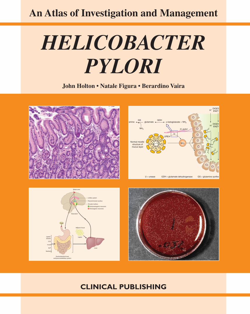
HELICOBACTER PYLORIThis comprehensive illustrated guide sets out the pathology, clinical presentation and options for treatment of Helicobacter pylori infection. Presented in a clinical context, instructive chapters cover key topics, including: genomics, metabolism, taxonomy, epidemiology and pathogenesis. There are also detailed sections on diagnosis and management of patients with gastric disease.
An important reference not only for clinicians but also microbiologists, it provides an up-to-date summary of our current knowledge of this organism and the many ways in which it impacts upon public health in all parts of the world.
Related titles:
Esophageal Diseases: an Atlas of Investigation and Management, MF VaeziISBN 978 1 904392 55 2
Inflammatory Bowel Disease: an Atlas of Investigation and Management, TR Orchard, RD Goldin, PP Tekkis, HRT WilliamsISBN 978 1 84692 013 4
Paediatric Gastroenterology: an Atlas of Investigation and Management, JM Moreno-Villares, I PolancoISBN 978 1 84692 009 7
Website: www.clinicalpublishing.co.uk
ISBN: 978 1 904392 89 7
An Atlas of Investigation and Management
HELICOBACTER PYLORI
CLINICAL PUBLISHING
HE
LIC
OB
AC
TE
R P
YL
OR
IC
LIN
ICA
L
PU
BL
ISHIN
G
Helicobacter pylori HPAG1 plasmid pHPAG1, complete genome
Accession: NC_008087 Topology circular, Length: 9,370 bp; Genes: 8
8500 bp1000 bp
1500 bp
2000 bp
2500 bp
3000 bp
3500 bp
4000 bp
4500 bp5000 bp
5500 bp
6000 bp
6500 bp
7000 bp
7500 bp
8000 bp
9000 bp 500 bp
Motor area
Limbic system
Paraventricular nucleus
Arcuate nucleus
Antiorexiogenic neuronesOrexiogenic neurones
Brainstem
Food
LeptinLeptin/Ghrelin
CCK
Insulin
GLP
Nutrients
Gastrointestinal tractand pancreatobiliary system
Liver
Adipose tissue
tamine glutamate α-ketoglutarate +GDH
H. pylori
GS
NH3
UREA
Normal micelle structure ofmucus layer
DIGESENZYM
DIGESENZYM
H+
H+
U = urease GDH = glutamate dehydrogenase GS = glutamine synthet
U
NH3
NH3
H_Pylori cover.indd 1 03/02/2012 13:19
An Atlas of Investigation and Management
HELICOBACTER
PYLORIBy
John Holton, BSc, MB ChB, PhD, MRCPath
Reader Clinical Microbiology
Centre for Infectious Diseases and International Health
Windeyer Institute of Medical Sciences
Royal Free and University College London Medical School
London, UK
Natale Figura, MD
Associate Professor in Gastroenterology
Department of Internal Medicine, Endocrine-Metabolic Sciences and Biochemistry
University of Siena and Policlinico S. Maria alle Scotte
Siena, Italy
Berardino Vaira, MD
Associate Professor, Department of Internal Medicine and Gastroenterology
University of Bologna
Bologna, Italy
CLINICAL PUBLISHINGOXFORD
AIM-Helicobacter_Pylori.indb 3 16/01/2012 10:15
Clinical Publishing an imprint of Atlas Medical Publishing Ltd
Oxford Centre for Innovation
Mill Street, Oxford OX2 0JX, UK
Tel: +44 1865 811116
Fax: +44 1865 251550
E mail: [email protected]
Web: www.clinicalpublishing.co.uk
Distributed in USA and Canada by:
Clinical Publishing
30 Amberwood Parkway
Ashland OH 44805, USA
Tel: 800-247-6553 (toll free within US and Canada)
Fax: 419-281-6883
Email: [email protected]
Distributed in UK and Rest of World by:
Marston Book Services Ltd
PO Box 269
Abingdon
Oxon OX14 4YN, UK
Tel: +44 1235 465500
Fax: +44 1235 465555
Email: [email protected]
© Atlas Medical Publishing Ltd 2012
First published 2012
All rights reserved. No part of this publication may be reproduced, stored in a retrieval system, or transmitted,
in any form or by any means, without the prior permission in writing of Clinical Publishing or Atlas Medical
Publishing Ltd
Although every effort has been made to ensure that all owners of copyright material have been acknowledged
in this publication, we would be glad to acknowledge in subsequent reprints or editions any omissions brought
to our attention
Clinical Publishing and Atlas Medical Publishing Ltd bear no responsibility for the persistence or accuracy of
URLs for external or third-party internet websites referred to in this publication, and does not guarantee that
any content on such websites is, or will remain, accurate or appropriate.
A catalogue record for this book is available from the British Library
ISBN-13 978 1 904392 89 7
ISBN e-book 978 1 84692 630 3
The publisher makes no representation, express or implied, that the dosages in this book are correct.
Readers must therefore always check the product information and clinical procedures with the most
up-to-date published product information and data sheets provided by the manufacturers and the
most recent codes of conduct and safety regulations. The authors and the publisher do not accept any
liability for any errors in the text or for the misuse or misapplication of material in this work
Project manager: Gavin Smith, GPS Publishing Solutions, Herts, UK
Typeset by Phoenix Photosetting, Chatham, Kent, UK
Printed and bound by Marston Book Services Ltd, Abingdon, Oxon, UK
AIM-Helicobacter_Pylori.indb 4 16/01/2012 10:15
Contents
Abbreviations vi
General note viii
1. Discovery, metabolism, genome and taxonomy 1
2. Epidemiology and colonization 23
3. Clinical features 37
4. Pathogenesis 63
5. Diagnosis 95
6. Management 113
7. Case studies 139
Index 143
AIM-Helicobacter_Pylori.indb 5 16/01/2012 10:15
vi
Abbreviations
ABC ATP-binding cassette
ACh acetylcholine
ADH alcohol dehydrogenase
AG atrophic gastritis
Alp adherence-associated lipoprotein
AMP adenosine monophosphate
AP-PCR Arbitrarily primed PCR
AS aphthous stomatitis
ATP adenosine triphosphate
BabA2 Lewis blood group antigen-binding
protein
BHIA brain heart infusion agar
BLAST Basic Local Alignment Search Tool
BMI body mass index
bp base pairs
BRENDA bacteria restriction endonuclease
analysis
CAD coronary artery disease
CagA cytotoxin-associated gene A protein
cAMP cyclic-AMP
CARD caspase recruitment domains
CD Crohn’s disease
cDNA complementary DNA
Che chemotaxis protein
CLO Campylobacter-like organism
CM cytoplasmic membrane
CoA coenzyme A
COPD chronic obstructive pulmonary disease
COX cyclo-oxygenase
ECG electrocardiogram
EGF epidermal growth factor
EGFR EGF receptor
EHSG European Helicobacter Study Group
Erk extracellular signal regulated kinase
EYA egg yolk agar
FADH flavin adenine dinucleotide (reduced)
FD functional dyspepsia
Fec ferric citrate transporter
FlaA/-B flagellin protein subunit A or B
Flg flagellar basal body rod protein
Fli flagellar motor switch protein
Frp NAD(P)H-flavin oxidoreductase
GH growth hormone
GlcNAc N-acetyl glucosamine
GORD gastro-oesophageal reflux disease
GroEL a chaperonin
HLA human leukocyte antigen
HopZ Helicobacter outer membrane adhesin
protein
Hpa flagellar sheath adhesin
HSP heat shock protein
IBD inflammatory bowel disease
ICAM intracellular adhesion molecule
IceA restriction endonuclease (induced by
contact with epithelium)
Ig immunoglobulin
IL interleukin
iNOS inducible nitric oxide synthase
InvA invasion protein
ITP idiopathic thrombocytopenic purpura
ITT intention to treat
Le Lewis antigen
LPS lipopolysaccharide
MALDI-TOF matrix-assisted laser desorption/
ionization time-of-flight
MALT mucosal-associated lymphoid tissue
Mbp megabase pairs
MCA MacConkey agar
MCP macrophage chemoattractant protein
MLEE multilocus enzyme electrophoresis
MLST multilocus sequence typing
MLVA multiple loci VNTR analysis
Mot flagellar motor protein
MTM modified Thayer–Martin agar
MurNAc N-acetyl muramic acid
NAD nicotinamide adenine dinucleotide
NADH nicotinamide adenine dinucleotide
(reduced)
NADPH nicotinamide adenine dinucleotide
phosphate (reduced)
NFAT nuclear factor of activated T cell
NixA nickel-transport protein
AIM-Helicobacter_Pylori.indb 6 16/01/2012 10:15
vii
NOD1/-2 nucleotide-binding o ligomerization
domain-containing protein 1 or -2
NSAID non-steroidal anti-infl ammatory drug
NSF N-ethylmaleimide-sensitive factor
OipA outer infl ammatory protein A
PAF platelet-activating factor
PAI pathogenicity island
PAMP pathogen associated molecular pattern
Pbp penicillin-binding protein
PCR polymerase chain reaction
PG peptidoglycan
PGE2 prostaglandin E
2
PLA phospholipase A
PPIs proton pump inhibitors
PSGN pepsinogen
PUD peptic ulcer disease
RecG ATP-dependent DNA helicase
RecN DNA repair protein
RFLP restriction fragment length
polymorphism
RT-PCR reverse transcription PCR
RUT rapid urease test
RuvABC Holliday junction resolvase
SabA sialic acid-binding adhesin protein
SCC squamous cell carcinoma
Sec preprotein translocase subunit
SHP-2 Src homology-2 domain
s sigma factor (RNA polymerase factor)
SNARE soluble NSF attachment protein
receptor
spp. species (note: sp. singular)
Src proto-oncogene tyrosine-protein kinase
Src
SST somatostatin
TNF tumour necrosis factor
TonB T-one (bacteriophage T1) ferric
hydroxamate transporter B
TSA trypticase soy agar
TTI ‘test and treat’ intervention
UKCRC UK Clinical Research Collaboration
Uvr excision endonuclease subunit
VCAM vascular cell adhesion molecule
VNTR variable-number tandem repeat
AIM-Helicobacter_Pylori.indb 7 16/01/2012 10:15

viii
Acknowledgement
The authors are grateful to the contribution made to this book by Dr Carla Vindigni, Pathology Unit, Department of
Oncology, Policlinico Santa Maria alle Scotte, Siena, Italy
AIM-Helicobacter_Pylori.indb 8 16/01/2012 10:15
1
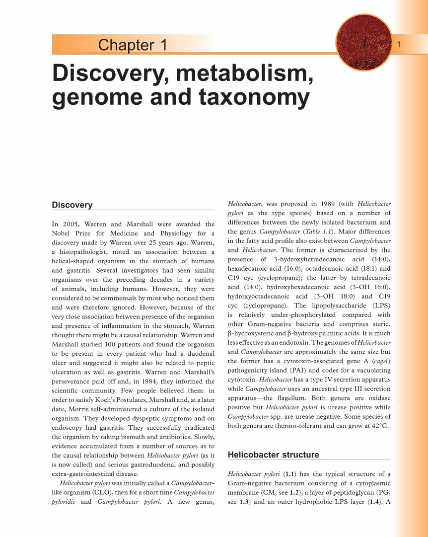
Discovery
In 2005, Warren and Marshall were awarded the
Nobel Prize for Medicine and Physiology for a
discovery made by Warren over 25 years ago. Warren,
a histopathologist, noted an association between a
helical-shaped organism in the stomach of humans
and gastritis. Several investigators had seen similar
organisms over the preceding decades in a variety
of animals, including humans. However, they were
considered to be commensals by most who noticed them
and were therefore ignored. However, because of the
very close association between presence of the organism
and presence of inflammation in the stomach, Warren
thought there might be a causal relationship. Warren and
Marshall studied 100 patients and found the organism
to be present in every patient who had a duodenal
ulcer and suggested it might also be related to peptic
ulceration as well as gastritis. Warren and Marshall’s
perseverance paid off and, in 1984, they informed the
scientific community. Few people believed them: in
order to satisfy Koch’s Postulates, Marshall and, at a later
date, Morris self-administered a culture of the isolated
organism. They developed dyspeptic symptoms and on
endoscopy had gastritis. They successfully eradicated
the organism by taking bismuth and antibiotics. Slowly,
evidence accumulated from a number of sources as to
the causal relationship between Helicobacter pylori (as it
is now called) and serious gastroduodenal and possibly
extra-gastrointestinal disease.
Helicobacter pylori was initially called a Campylobacter-like organism (CLO), then for a short time Campylobacter pyloridis and Campylobacter pylori. A new genus,
Helicobacter, was proposed in 1989 (with Helicobacter pylori as the type species) based on a number of
differences between the newly isolated bacterium and
the genus Campylobacter (Table 1.1). Major differences
in the fatty acid profile also exist between Campylobacter and Helicobacter. The former is characterized by the
presence of 3-hydroxyltetradecanoic acid (14:0),
hexadecanoic acid (16:0), octadecanoic acid (18:1) and
C19 cyc (cyclopropane); the latter by tetradecanoic
acid (14:0), hydroxyhexadecanoic acid (3-OH 16:0),
hydroxyoctadecanoic acid (3-OH 18:0) and C19
cyc (cyclopropane). The lipopolysaccharide (LPS)
is relatively under-phosphorylated compared with
other Gram-negative bacteria and comprises steric,
b-hydroxysteric and b-hydroxy palmitic acids. It is much
less effective as an endotoxin. The genomes of Helicobacter and Campylobacter are approximately the same size but
the former has a cytotoxin-associated gene A (cagA)
pathogenicity island (PAI) and codes for a vacuolating
cytotoxin. Helicobacter has a type IV secretion apparatus
while Campylobacter uses an ancestral type III secretion
apparatus—the flagellum. Both genera are oxidase
positive but Helicobacter pylori is urease positive while
Campylobacter spp. are urease negative. Some species of
both genera are thermo-tolerant and can grow at 42°C.
Helicobacter structure
Helicobacter pylori (1.1) has the typical structure of a
Gram-negative bacterium consisting of a cytoplasmic
membrane (CM; see 1.2), a layer of peptidoglycan (PG;
see 1.3) and an outer hydrophobic LPS layer (1.4). A
Discovery, metabolism, genome and taxonomy
Chapter 1
AIM-Helicobacter_Pylori.indb 1 16/01/2012 10:15
Discovery, metabolism, genome and taxonomy2
typical Gram-negative cell wall consists of the inner
layer of phospholipid known as the CM, containing
proteins necessary for respiration and permeability and
covered by a PG layer. On the external surface of the PG
layer is a second hydrophobic phospholipid membrane,
the LPS (or endotoxin). On electron microscopy, a
Gram-negative cell wall has a tri-laminar appearance
with a periplasmic space between the CM-PG and
the PG-LPS. This space contains enzymes required
for cell wall synthesis—transpeptidases (proteins that
bind b-lactam antibiotics called penicillin-binding
proteins), which are the target for inhibition by b-lactam
antibiotics.
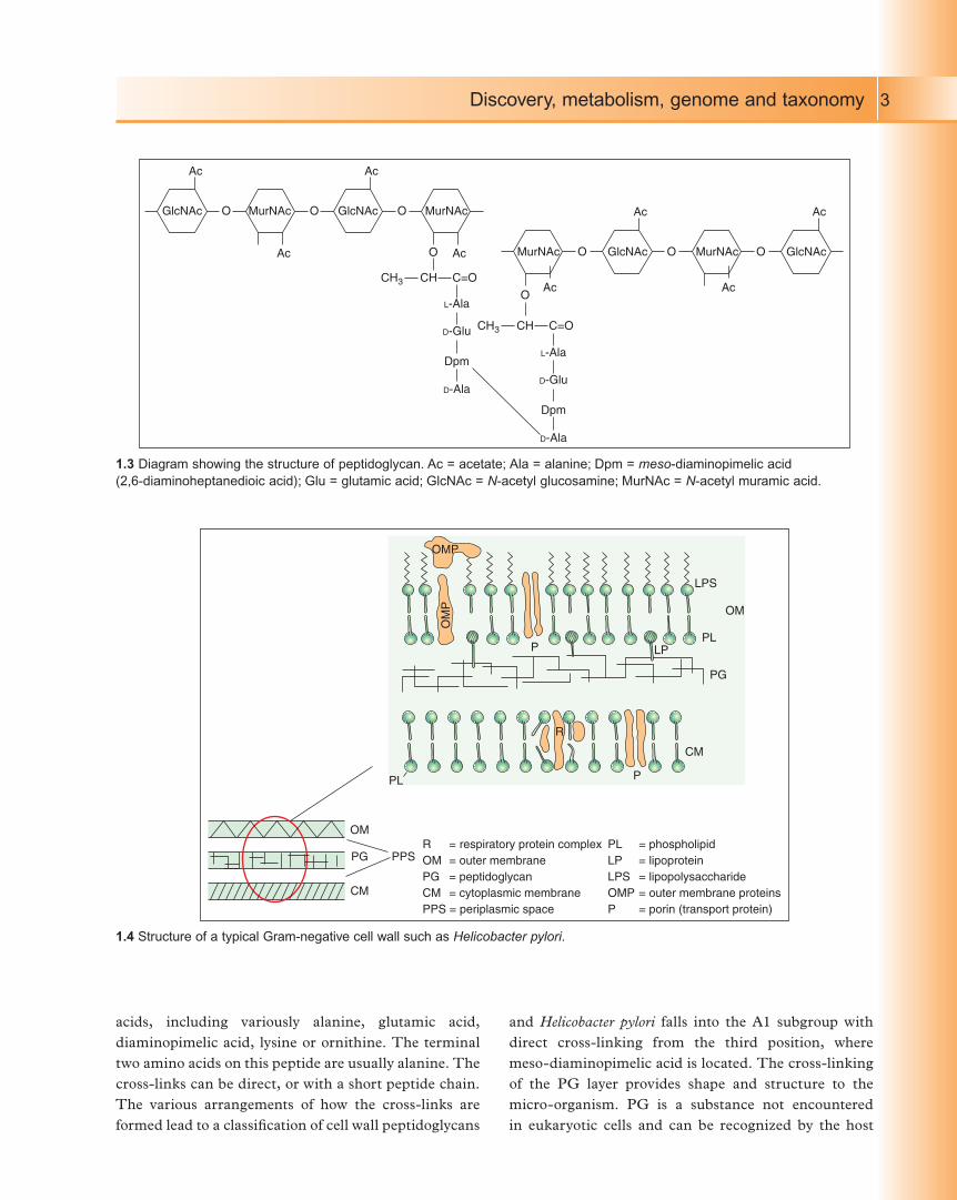
Peptidoglycan is a heteropolymer of N-acetyl
glucosamine (GlcNAc) and N-acetyl muramic acid
(MurNAc) in which adjacent glycan chains are cross-
linked by peptides. The amino acid composition of
these peptides varies between bacteria. The amino
acids in H. pylori are meso-diaminopimelic acid, alanine
and glutamic acid. The short peptide chain attached to
the glycan units consist of alternating D - and L - amino
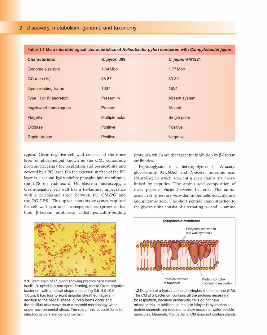
Table 1.1 Main microbiological characteristics of Helicobacter pylori compared with Campylobacter jejuni
Characteristic H. pylori J99 C. jejuni RM1221
Genome size (bp) 1.64 Mbp 1.77 Mbp
GC ratio (%) 38.87 30.30
Open reading frame 1637 1654
Type III or IV secretion Present IV Absent system
cagA/vacA homologues Present Absent
Flagella Multiple polar Single polar
Oxidase Positive Positive
Rapid urease Positive Negative
1.1 Gram stain of H. pylori showing predominant curved
bacilli. H. pylori is a non-spore forming, motile Gram-negative
bacterium with a helical shape measuring 2.5–4.5 ¥ 0.5–
1.0 mm. It has four to eight unipolar sheathed flagella. In
addition to the helical shape, curved forms occur and
the bacillus also converts to a coccoid morphology when
under environmental stress. The role of this coccoid form in
infection or persistence is uncertain.
Cytoplasmic membrane
Enzymes involved incell wall synthesis
Protein complexinvolved in respiration
Proteins involved in transport
1.2 Diagram of a typical bacterial cytoplasmic membrane (CM).
The CM of a bacterium contains all the proteins necessary
for respiration, because prokaryotic cells do not have
mitochondria. In addition, as the lipid bilayer is hydrophobic,
protein channels are required to allow access of water-soluble
molecules. Generally, the bacterial CM does not contain sterols.
AIM-Helicobacter_Pylori.indb 2 16/01/2012 10:15
Discovery, metabolism, genome and taxonomy 3
acids, including variously alanine, glutamic acid,
diaminopimelic acid, lysine or ornithine. The terminal
two amino acids on this peptide are usually alanine. The
cross-links can be direct, or with a short peptide chain.
The various arrangements of how the cross-links are
formed lead to a classification of cell wall peptidoglycans
and Helicobacter pylori falls into the A1 subgroup with
direct cross-linking from the third position, where
meso-diaminopimelic acid is located. The cross-linking
of the PG layer provides shape and structure to the
micro-organism. PG is a substance not encountered
in eukaryotic cells and can be recognized by the host
GlcNAc O
Ac
Ac
O O
O
MurNAc GlcNAc MurNAc
Ac
Ac Ac
Ac
AcL-Ala
D-Glu
Dpm
D-Ala
CH3 CH C=O
L-Ala
D-Glu
Dpm
D-Ala
Ac
O
O
CH3 CH C=O
MurNAc GlcNAc GlcNAcOMurNAcO
OMP
LPS
LPPL
PL
OM
CM
PG PPS
R
OM
PG
CM
P
P
OM
P
R = respiratory protein complexOM = outer membranePG = peptidoglycanCM = cytoplasmic membranePPS = periplasmic space
PL = phospholipidLP = lipoproteinLPS = lipopolysaccharideOMP = outer membrane proteinsP = porin (transport protein)
1.3 Diagram showing the structure of peptidoglycan. Ac = acetate; Ala = alanine; Dpm = meso-diaminopimelic acid
(2,6-diaminoheptanedioic acid); Glu = glutamic acid; GlcNAc = N-acetyl glucosamine; MurNAc = N-acetyl muramic acid.
1.4 Structure of a typical Gram-negative cell wall such as Helicobacter pylori.
AIM-Helicobacter_Pylori.indb 3 16/01/2012 10:15
Discovery, metabolism, genome and taxonomy4
innate immune system as foreign, thus initiating the
host response. These and similar microbial molecules
are called pathogen-associated molecular patterns. PG
is found in nearly all cell walls and is most abundant
in Gram-positive cell walls. Helicobacter pylori is Gram-
negative and thus has an outer hydrophobic membrane
containing LPS.
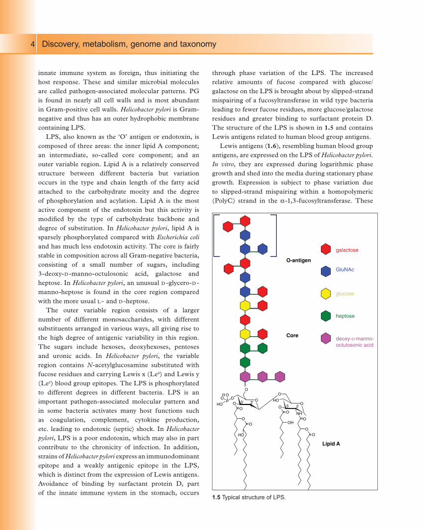
LPS, also known as the ‘O’ antigen or endotoxin, is
composed of three areas: the inner lipid A component;
an intermediate, so-called core component; and an
outer variable region. Lipid A is a relatively conserved
structure between different bacteria but variation
occurs in the type and chain length of the fatty acid
attached to the carbohydrate moeity and the degree
of phosphorylation and acylation. Lipid A is the most
active component of the endotoxin but this activity is
modified by the type of carbohydrate backbone and
degree of substitution. In Helicobacter pylori, lipid A is
sparsely phosphorylated compared with Escherichia coli and has much less endotoxin activity. The core is fairly
stable in composition across all Gram-negative bacteria,
consisting of a small number of sugars, including
3-deoxy-D -manno-octulosonic acid, galactose and
heptose. In Helicobacter pylori, an unusual D -glycero-D -
manno-heptose is found in the core region compared
with the more usual L - and D -heptose.
The outer variable region consists of a larger
number of different monosaccharides, with different
substituents arranged in various ways, all giving rise to
the high degree of antigenic variability in this region.
The sugars include hexoses, deoxyhexoses, pentoses
and uronic acids. In Helicobacter pylori, the variable
region contains N-acetylglucosamine substituted with
fucose residues and carrying Lewis x (Lex) and Lewis y
(Ley) blood group epitopes. The LPS is phosphorylated
to different degrees in different bacteria. LPS is an
important pathogen-associated molecular pattern and
in some bacteria activates many host functions such
as coagulation, complement, cytokine production,
etc. leading to endotoxic (septic) shock. In Helicobacter pylori, LPS is a poor endotoxin, which may also in part
contribute to the chronicity of infection. In addition,
strains of Helicobacter pylori express an immunodominant
epitope and a weakly antigenic epitope in the LPS,
which is distinct from the expression of Lewis antigens.
Avoidance of binding by surfactant protein D, part
of the innate immune system in the stomach, occurs
through phase variation of the LPS. The increased
relative amounts of fucose compared with glucose/
galactose on the LPS is brought about by slipped-strand
mispairing of a fucosyltransferase in wild type bacteria
leading to fewer fucose residues, more glucose/galactose
residues and greater binding to surfactant protein D.
The structure of the LPS is shown in 1.5 and contains
Lewis antigens related to human blood group antigens.
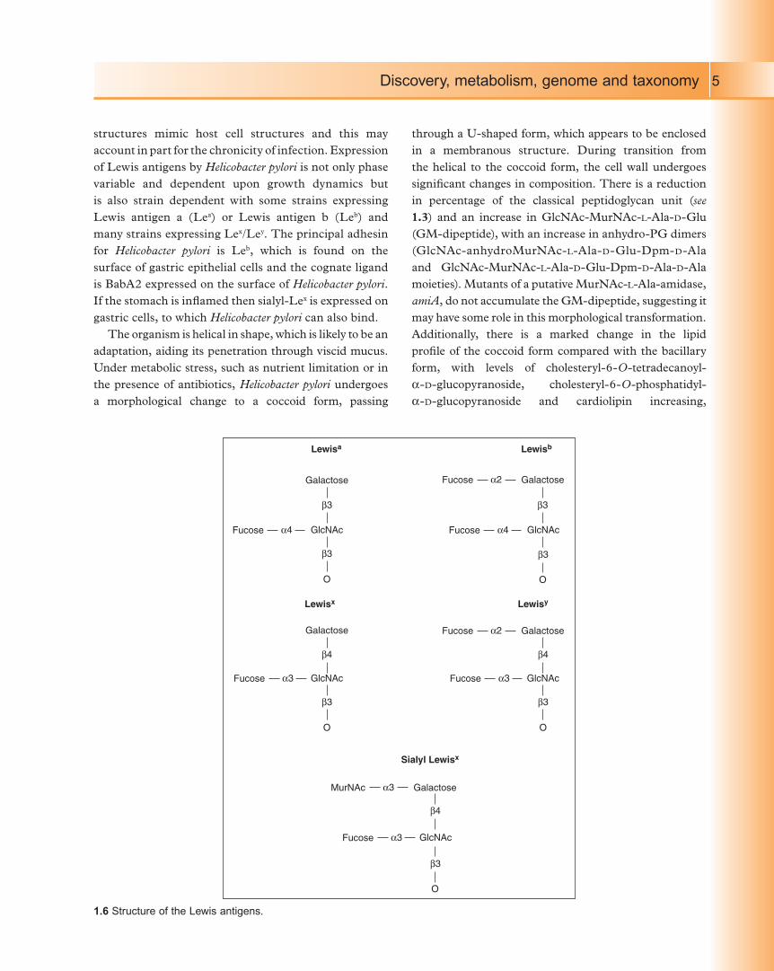
Lewis antigens (1.6), resembling human blood group
antigens, are expressed on the LPS of Helicobacter pylori. In vitro, they are expressed during logarithmic phase
growth and shed into the media during stationary phase
growth. Expression is subject to phase variation due
to slipped-strand mispairing within a homopolymeric
(PolyC) strand in the a-1,3-fucosyltransferase. These
O-antigen
galactose
GluNAc
glucose
heptose
deoxy-D-manno-octulosonic acid
Core
Lipid A
O
OO
OOH
OO
O
O
O
O
NH
3 23’ 2’ HO
O
OO
OOΘP
O
HO
HO
1.5 Typical structure of LPS.
AIM-Helicobacter_Pylori.indb 4 16/01/2012 10:15
Discovery, metabolism, genome and taxonomy 5
structures mimic host cell structures and this may
account in part for the chronicity of infection. Expression
of Lewis antigens by Helicobacter pylori is not only phase
variable and dependent upon growth dynamics but
is also strain dependent with some strains expressing
Lewis antigen a (Lea) or Lewis antigen b (Leb) and
many strains expressing Lex/Ley. The principal adhesin
for Helicobacter pylori is Leb, which is found on the
surface of gastric epithelial cells and the cognate ligand
is BabA2 expressed on the surface of Helicobacter pylori. If the stomach is inflamed then sialyl-Lex is expressed on
gastric cells, to which Helicobacter pylori can also bind.
The organism is helical in shape, which is likely to be an
adaptation, aiding its penetration through viscid mucus.
Under metabolic stress, such as nutrient limitation or in
the presence of antibiotics, Helicobacter pylori undergoes
a morphological change to a coccoid form, passing
through a U-shaped form, which appears to be enclosed
in a membranous structure. During transition from
the helical to the coccoid form, the cell wall undergoes
significant changes in composition. There is a reduction
in percentage of the classical peptidoglycan unit (see 1.3) and an increase in GlcNAc-MurNAc-L-Ala-D-Glu
(GM-dipeptide), with an increase in anhydro-PG dimers
(GlcNAc-anhydroMurNAc-L-Ala-D-Glu-Dpm-D-Ala
and GlcNAc-MurNAc-L-Ala-D-Glu-Dpm-D-Ala-D-Ala
moieties). Mutants of a putative MurNAc-L-Ala-amidase,
amiA, do not accumulate the GM-dipeptide, suggesting it
may have some role in this morphological transformation.
Additionally, there is a marked change in the lipid
profile of the coccoid form compared with the bacillary
form, with levels of cholesteryl-6-O-tetradecanoyl-
a-D-glucopyranoside, cholesteryl-6-O-phosphatidyl-
a-D-glucopyranoside and cardiolipin increasing,
Lewisa
Lewisx Lewisy
Lewisb
Galactose
Galactose
β3
β3
O
β3
β4
β3
GlcNAcα4Fucose
β3
GlcNAcα3
O
Fucose
β4
β3
GlcNAcα3
O
Fucose
GlcNAcα4Fucose
α2Fucose Galactose
O
α2Fucose Galactose
Sialyl Lewisx
β4
β3
GlcNAcα3Fucose
α3MurNAc Galactose
O
1.6 Structure of the Lewis antigens.
AIM-Helicobacter_Pylori.indb 5 16/01/2012 10:15
Discovery, metabolism, genome and taxonomy6
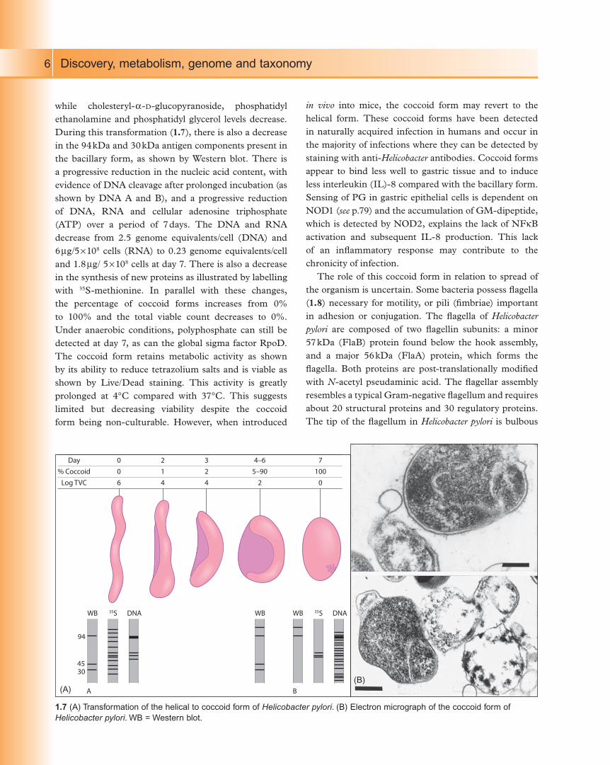
while cholesteryl-a-D-glucopyranoside, phosphatidyl
ethanolamine and phosphatidyl glycerol levels decrease.
During this transformation (1.7), there is also a decrease
in the 94 kDa and 30 kDa antigen components present in
the bacillary form, as shown by Western blot. There is
a progressive reduction in the nucleic acid content, with
evidence of DNA cleavage after prolonged incubation (as
shown by DNA A and B), and a progressive reduction
of DNA, RNA and cellular adenosine triphosphate
(ATP) over a period of 7 days. The DNA and RNA
decrease from 2.5 genome equivalents/cell (DNA) and
6 mg/5 ¥ 108 cells (RNA) to 0.23 genome equivalents/cell
and 1.8 mg/ 5 ¥ 108 cells at day 7. There is also a decrease
in the synthesis of new proteins as illustrated by labelling
with 35S-methionine. In parallel with these changes,
the percentage of coccoid forms increases from 0%
to 100% and the total viable count decreases to 0%.
Under anaerobic conditions, polyphosphate can still be
detected at day 7, as can the global sigma factor RpoD.
The coccoid form retains metabolic activity as shown
by its ability to reduce tetrazolium salts and is viable as
shown by Live/Dead staining. This activity is greatly
prolonged at 4°C compared with 37°C. This suggests
limited but decreasing viability despite the coccoid
form being non-culturable. However, when introduced
in vivo into mice, the coccoid form may revert to the
helical form. These coccoid forms have been detected
in naturally acquired infection in humans and occur in
the majority of infections where they can be detected by
staining with anti-Helicobacter antibodies. Coccoid forms
appear to bind less well to gastric tissue and to induce
less interleukin (IL)-8 compared with the bacillary form.
Sensing of PG in gastric epithelial cells is dependent on
NOD1 (see p.79) and the accumulation of GM-dipeptide,
which is detected by NOD2, explains the lack of NFkB
activation and subsequent IL-8 production. This lack
of an inflammatory response may contribute to the
chronicity of infection.
The role of this coccoid form in relation to spread of
the organism is uncertain. Some bacteria possess flagella
(1.8) necessary for motility, or pili (fimbriae) important
in adhesion or conjugation. The flagella of Helicobacter pylori are composed of two flagellin subunits: a minor
57 kDa (FlaB) protein found below the hook assembly,
and a major 56 kDa (FlaA) protein, which forms the
flagella. Both proteins are post-translationally modified
with N-acetyl pseudaminic acid. The flagellar assembly
resembles a typical Gram-negative flagellum and requires
about 20 structural proteins and 30 regulatory proteins.
The tip of the flagellum in Helicobacter pylori is bulbous
Day
% Coccoid
Log TVC
006
214
324
4–65–90
2
7100
0
94
4530
WB WB35S DNA WB 35S DNA
A B
1.7 (A) Transformation of the helical to coccoid form of Helicobacter pylori. (B) Electron micrograph of the coccoid form of
Helicobacter pylori. WB = Western blot.
(A)(B)
AIM-Helicobacter_Pylori.indb 6 16/01/2012 10:15