Genome-Wide Screen for Modifiersof Ataxin-3 Neurodegeneration in DrosophilaJulide Bilen
1¤, Nancy M. Bonini
1,2*
1 Department of Biology, University of Pennsylvania, Philadelphia, Pennsylvania, United States of America, 2 Howard Hughes Medical Institute, University of Pennsylvania,
Philadelphia, Pennsylvania, United States of America
Spinocerebellar ataxia type-3 (SCA3) is among the most common dominantly inherited ataxias, and is one of ninedevastating human neurodegenerative diseases caused by the expansion of a CAG repeat encoding glutamine withinthe gene. The polyglutamine domain confers toxicity on the protein Ataxin-3 leading to neuronal dysfunction and loss.Although modifiers of polyglutamine toxicity have been identified, little is known concerning how the modifiersfunction mechanistically to affect toxicity. To reveal insight into spinocerebellar ataxia type-3, we performed a geneticscreen in Drosophila with pathogenic Ataxin-3-induced neurodegeneration and identified 25 modifiers defining 18genes. Despite a variety of predicted molecular activities, biological analysis indicated that the modifiers affectedprotein misfolding. Detailed mechanistic studies revealed that some modifiers affected protein accumulation in amanner dependent on the proteasome, whereas others affected autophagy. Select modifiers of Ataxin-3 also affectedtau, revealing common pathways between degeneration due to distinct human neurotoxic proteins. These findingsprovide new insight into molecular pathways of polyQ toxicity, defining novel targets for promoting neuronal survivalin human neurodegenerative disease.
Citation: Bilen J, Bonini NM (2007) Genome-wide screen for modifiers of Ataxin-3 neurodegeneration in Drosophila. PLoS Genet 3(10): e177. doi:10.1371/journal.pgen.0030177
Introduction
Spinocerebellar ataxia type 3 (SCA3) is the most commondominantly inherited ataxia worldwide and is caused by aCAG repeat expansion encoding glutamine within the ATXN3gene [1,2]. The expanded polyglutamine (polyQ) is thought toconfer toxicity on the protein Ataxin-3, leading to neuraldysfunction and loss [3]. At least nine human diseases,including additional spinocerebellar ataxias and Hunting-ton’s disease, share this mechanism. Studies on suchpathogenic proteins reveal that the long polyQ domain altersprotein conformation, causing an enriched beta sheetstructure [4]. Mutant polyQ protein also dynamically asso-ciates with chaperones and colocalizes with proteasomesubunits, indicating that the protein is misfolded orabnormally folded [5,6]. Such accumulation of misfoldedprotein is a common pathology of human degenerativedisorders, including Alzheimer, Parkinson, and prion diseases[7–9], indicating that these diseases may share molecular andcellular mechanisms.
Models for human neurodegenerative diseases in simplesystems have provided valuable tools to dissect molecularmechanisms of disease pathology [10–13]. Directed expres-sion of pathogenic human Ataxin-3 in Drosophila recapitulateskey features of disease, with late-onset neuronal dysfunctionand degeneration accompanied by ubiquitinated inclusions[14,15]. Neurotoxicity is more severe with increasing length ofthe polyQ repeat, similar to the human disease where longerrepeats are associated with more severe and earlier onsetdisease [16,17]. In the fly, increased levels of expression of thedisease protein leads to more severe degeneration and earlieronset protein accumulation, suggesting that abnormal accu-mulation of the mutant protein is central to disease anddegeneration.
A number of modifiers of select polyQ disease proteins
have been identified using animal models, including chaper-ones, transcriptional coregulators, and microRNAs [18–22].Although these approaches have revealed genes that modu-late polyQ toxicity, little is known regarding how themodifiers act biologically to modulate polyQ degeneration.Among polyQ proteins, Ataxin-3 is unique because it hasbeen implicated in ubiquitin pathways, and its normal activitymay impinge on protein degradation pathways [23–26].Truncation of Ataxin-3 to remove the ubiquitin proteasedomain, or mutation of the ubiquitin protease activity,dramatically enhances toxicity [15], indicating that thenormal activity of Ataxin-3 may be critical in Ataxin-3-induced degeneration. To reveal insight into pathways thatmodulate Ataxin-3 neurodegeneration, we performed agenetic modifier screen in Drosophila. These studies revealeda range of modifiers that, despite some broadly diversepredicted molecular functions, converge on protein misfold-ing with a subset mitigating toxicity through proteasome and/or autophagy pathways. These findings underscore the criticalrole of protein quality control in SCA3 pathogenesis andprovide potential new targets toward therapeutics.
Editor: Harry Orr, University of Minnesota, United States of America
Received February 8, 2007; Accepted August 30, 2007; Published October 19,2007
Copyright: � 2007 Bilen and Bonini. This is an open-access article distributedunder the terms of the Creative Commons Attribution License, which permitsunrestricted use, distribution, and reproduction in any medium, provided theoriginal author and source are credited.
Abbreviations: GFP, green fluorescent protein; NI, nuclear inclusion; polyQ,polyglutamine; SCA3, spinocerebellar ataxia type-3; UPS, ubiquitin-proteasomalsystem
* To whom correspondence should be addressed. E-mail: [email protected]
¤ Current address: Howard Hughes Medical Institute, Janelia Farm ResearchCampus, Ashburn Virginia, United States of America
PLoS Genetics | www.plosgenetics.org October 2007 | Volume 3 | Issue 10 | e1771950

Results
A Genetic Screen Defines Modifiers of Pathogenic Ataxin-3 Toxicity
To define modifiers that may reveal new insight intomechanisms of human SCA3 disease, we performed anoverexpression screen for modifiers of Ataxin-3-inducedneurodegeneration in Drosophila. SCA3trQ78 causes lateonset progressive degeneration characterized by loss ofpigmentation and collapse of the eye (Figure 1A and 1B)[14]. We initially screened a subset of 2,300 available EP-element insertion lines, each carrying a transposon engi-neered to direct expression of the downstream gene in thepresence of the yeast GAL4 protein [27]. Because reprodu-cibility was variable with this collection, we then performed ascreen de novo, selecting for new EP-insertions that modifiedSCA3trQ78 toxicity. This approach identified 17 suppressorsand one enhancer (Figures 1C–1J and S1), which affectedboth external and internal retinal degeneration.
Plasmid rescue was performed to identify the genesaffected. BLAST searches with genomic sequence from theintegration sites revealed that the lines bore insertions in the59 regulatory region of select genes, and all were in anorientation to direct GAL4-dependent gene expression (TextS1; Table S1). Northern and reverse transcription PCRanalysis confirmed upregulation of the targeted genes; usinga variety of tests we confirmed that the modifiers did notappear to affect transcription of the Ataxin-3 transgene orgeneral GAL4-UAS transgene expression (Figure S2). Bothmolecular and genetic analyses confirmed that the insertionswere single insertions (see Materials and Methods). Reversionanalysis proved that the EP-elements were causal in mod-ification. Taken together, these data indicated that the EPmodifiers resulted in increased expression of the targeteddownstream genes, which modulated SCA3trQ78 toxicity andneurodegeneration.
Molecular Analysis Implicates Diverse Biological PathwaysAnalysis of the targeted genes revealed that the majority
fell into two major classes of chaperones and ubiquitin-
pathway components (Table 1; Figures S1 and S3). Theremaining modifiers were placed in a third category ofmiscellaneous functions. Class 1 (Figure 1C–1E) included anHsp70 family member, Hsp68E407; two Hsp40 genes, DnaJ-1B345.2 and mrjE1050; a small heat shock protein aB crystallineCG14207EP1348; and the cochaperone Tpr2EB7-1A. The class 2ubiquitin-pathway components included: polyubiquitinCR11700EP1384; a ubiquitin-specific protease Ubp64EE213-1A;two ubiquitin ligases, CG8209B3-Sa and Faf E659; and an F-boxprotein CG11033EP3093, the only enhancer of the group. Class3 (Figure 1F and 1G) included genes with a variety ofpredicted molecular functions: the nuclear export proteinEmbargoed, embE2-1A and embE128-1A; three transcriptionalregulators Sin3AB9-E, NFAT (NFATEP1335 and NFATEP1508),and debra (dbrEP456 and dbrEP9); three translational regulators,including four alleles of the lin-41 homologue dappled(dpld JM120, dpld JM265, dpldEP546, and dpldEP2594), a polyA bind-ing protein orb2B8-S, and insulin growth factor II mRNAbinding protein ImpEP1433; and finally a fatty acid oxidationenzyme palmitoyl co-A oxidase CG5009B227.2. This indicatedthat, although chaperone and ubiquitin-pathway componentsare major modifier categories, a variety of functional path-ways are implicated in Ataxin-3 pathogenesis.The screen selected for modifiers that, upon upregulation,
affected toxicity. To determine whether the activity of thesegenes may normally play a role in SCA3 toxicity, we examinedwhether reduction in the level of the modifier genes had aneffect. To do this, we reduced gene expression by 50% usingloss-of-function alleles where available or deficiency lines.Among these, reduction with a deficiency of DnaJ-1 andUbp64E (within the same deficiency; reduction of DnaJ-1 haspreviously been shown to enhance with a dominant-negativeconstruct [16]), Trp2, emb (with an allele), and dbr dramaticallyenhanced degeneration (Figure1K–1O; Table S2). Althoughthe deficiencies reduce the level of a number of genes, thesedata suggest that the endogenous activity of these genes maynormally help to protect against degeneration.To address whether morphological rescue correlated with
functional rescue, we determined the ability of selectsuppressors to restore function in a phototaxis assay. Whenflies bearing the SCA3trQ78 protein were given a choicebetween a light and dark chamber, they distributed randomly,indicating that they are functionally blind (Figure 1P).However, when mrjE1050, which dramatically rescued degen-eration, was co-expressed, normal vision was restored. Amilder suppressor that anatomically restored less retinaltissue, CG14207EP1348, restored vision partially. Thus, ana-tomical rescue correlated with functional rescue. These andother studies indicated that the modifiers not only modulatedtoxicity of the external eye, but also that of the neuronal cells.To confirm this in another situation, we tested selectmodifiers for the ability to mitigate polyQ toxicity whendirected exclusively to the nervous system with elav-GAL4(Figure S4). These studies confirmed that the modifiersmitigated neuronal toxicity of the Ataxin-3 protein.We performed select additional experiments with dpld, for
which we obtained many independent EP overexpressionalleles. These detailed studies confirmed activity of the EPalleles of dpld with independent UAS-dpld transgenic lines.Further, suppression by dpld was not limited to developmentbut also extended to the adult timeframe (Figures S2, S5, andS6).
PLoS Genetics | www.plosgenetics.org October 2007 | Volume 3 | Issue 10 | e1771951
Modifiers of Ataxin-3 Neurodegeneration
Author Summary
Spinocerebellar ataxia type-3 is the most common dominantlyinherited movement disorder and is caused by a CAG repeatexpansion within the gene ATXN3, encoding the Ataxin-3 protein.This leads to a protein with an expanded polyglutamine domain,which confers a dominant toxicity on the protein, leading to lateonset, progressive neural degeneration in the brain. Although somemodifiers of Ataxin-3 toxicity have been defined, little was knownabout their molecular mechanisms of action. The fruit fly Drosophilarecapitulates fundamental aspects of the human disease. Here, weperformed a genome-wide screen for new modifiers of Ataxin-3toxicity using the fly and defined 25 modifiers in 18 genes. Themajority of the genes belong to chaperone and ubiquitinproteasome pathways, which modulate protein folding anddegradation, but the remaining modifiers have a broad range ofpredicted molecular functions. Assays in vivo revealed that thebiological activity of all modifiers converge on aiding in situations ofprotein misfolding, despite distinct predicted molecular functions.Select modifiers of Ataxin-3 toxicity also modulated tau toxicityassociated with Alzheimer disease. These findings underscore theimportance of protein homeostasis pathways to disease and providethe foundation for new therapeutic insight.

PLoS Genetics | www.plosgenetics.org October 2007 | Volume 3 | Issue 10 | e1771952
Modifiers of Ataxin-3 Neurodegeneration

Because the genes were isolated as modifiers of a truncatedAtaxin-3 protein, we tested whether they could mitigatetoxicity of full-length Ataxin-3, which is largely a neuronaltoxicity [15]. Modifiers that phenotypically strongly sup-pressed toxicity of truncated Ataxin-3 also strongly sup-pressed full-length Ataxin-3 toxicity; however, a number ofmodifiers that were weak or moderate suppressors of thetruncated protein were, in contrast, strong suppressors offull-length Ataxin-3: CG14207EP1348 (aB crystalline),CR11700EP1384 (polyubiquitin), CG8209B3-Sa (putative ubiqui-tin ligase), and Sin3AB9-E (Figure S7 and unpublished data).Consistent with strong anatomical rescue, CG14207EP1348 alsorobustly suppressed functional vision defects of full-lengthAtaxin-3 (Figure 1Q). The enhancer, however, CG11033EP3093
had a minimal effect on toxicity of full-length Ataxin-3(unpublished data). This indicated that the modifiers variedin strength and selectivity depending upon whether theAtaxin-3 protein was intact or truncated. As truncation maybe a feature of SCA3 disease [28–30], the effectiveness ofmodifiers against different forms of Ataxin-3 has implicationsfor disease pathogenesis.
Ataxin-3 Modifiers Affect General Protein MisfoldingPrevious studies have shown that the molecular chaperone
Hsp70 is a potent suppressor of SCA3 toxicity [31]. Therefore,we considered that the class 1 modifiers may function similarto Hsp70 to help cells handle the misfolded disease protein,whereas those of class 2 likely have a role in ubiquitin-dependent pathways that process misfolded proteins. How-ever, class 3 presented a range of potential activities. Toaddress how the modifiers were functioning biologically, wetested whether the modifiers could affect a general proteinmisfolding phenotype: compromised chaperone activity witha dominant-negative form of Hsp70 (Hsp70.K71E). Thissituation results in an eye phenotype that resembles severepolyQ degeneration (Figure 2A and [32]). Thus, modifiers thataffected both SCA3 and Hsp70.K71E toxicity would likelyinclude those whose mode of action was to modulate proteinmisfolding.
Strikingly, we found that most of the suppressors of polyQtoxicity also mitigated the Hsp70.K71E phenotype, as well asor better than directed expression of Hsp70 itself (Figure 2;
Table 1). Interestingly, DnaJ-1B345.2 and Tpr2EB7-1A enhancedthis phenotype; we interpreted this to indicate that thesegenes, which encode proteins thought to act as cochaperonesof Hsp70, may compromise residual Hsp70 in the dominant-negative situation. The chaperone mrjE1050, although anHsp40 class chaperone, acted in a manner distinct fromDnaJ-1B345.2, as it suppressed rather than enhanced theHsp70.K17E phenotype. Moreover, the enhancer ofSCA3trQ78 toxicity, CG11033EP3093, suppressed the misfold-ing phenotype. Only one modifier did not affect thisphenotype, the ubiquitin protease Ubp64EE213-1A; interest-ingly, normal Ataxin-3 also has ubiquitin protease activitythat mitigates its own pathogenicity, and similarly, has noeffect on Hsp70.K71E [15]. We considered that one mecha-nism by which the modifiers may mitigate the Hsp70.K71Ephenotype is by upregulating Hsp70/Hsc70 chaperones;however, none of the modifiers appeared to act in this way(Figure 2K and unpublished data). These results indicatedthat the majority of modifiers of SCA3trQ78 toxicityappeared to function biologically by aiding in situations ofcompromised chaperone activity and/or protein misfolding.
Reduced Accumulation of the Pathogenic ProteinReduces ToxicityThe degree of neurodegeneration induced by pathogenic
polyQ protein is typically correlated with the level ofaccumulation of the protein in animals in vivo. We reasoned,therefore, that the modifiers may affect protein levels. Wetherefore examined protein accumulation by immunohisto-chemistry and western analysis. Although nuclear inclusionsmay not be causal in disease [33], later onset, smaller nuclearinclusions are typically reflective of reduced pathogenicity ofthe protein in vivo. Hsp70 and Hsp40 have also been shownto increase the solubility of pathogenic polyQ protein bywestern blots, concomitant with reducing toxicity [16]. Wetherefore examined protein accumulation using rh1-GAL4 orthe full-length Ataxin-3 protein—both situations that allowsensitive analysis of protein accumulation [15,22]. In thesestudies, we limited analysis to the strong and moderatemodifiers.Immunohistochemical analysis revealed that select modi-
fiers had striking effects to reduce NIs. These included the
Figure 1. Modifiers of SCA3trQ78-Induced Neurodegeneration in Drosophila Eye and Internal Retinal Sections of 1-d Flies
(A) Control with normal eye and retinal thickness (white arrow). Genotype w; gmr-GAL4/þ.(B) Expression of strong SCA3trQ78 causes external and internal degeneration, with pigment loss and severely reduced retinal thickness. Genotype w;gmr-GAL4 UAS-SCA3trQ78/þ.(C–E) Class 1 suppressors Hsp68E407, mrjE1050, and CG14207EP1348 rescued external eye pigmentation and restored the internal eye towards normal.Genotypes w; gmr-GAL4 UAS-SCA3trQ78 in trans to alleles indicated.(F and G) Class 3 suppressors embE128-1A and dbrEP9 have strong and weak rescue. Genotypes w; gmr-GAL4 UAS-SCA3trQ78 in trans to alleles indicated.(H–J) Co-expression of CG11033EP3093 with SCA3trQ61 severely enhances pigment loss and internal retinal degeneration; expression of CG11033EP3093
alone has no effect. Genotypes (H) w; gmr-GAL4 UAS-SCA3trQ61/þ, (I) w; gmr-GAL4 UAS-SCA3trQ61 in trans to CG11033EP3093, and (J) w; gmr-GAL4 in transto CG11033EP3093.(K–O) Deletions for regions that uncover select modifier genes enhance Ataxin-3 degeneration, suggesting dose sensitivity of these modifiers. (K)Pathogenic Ataxin-3 causes neuronal degeneration. (L) Ataxin-3 in trans to Df(3L)CH20, which uncovers both DnaJ-1 and Ubp64E, is lethal and causessevere degeneration (pupal eye shown). (M–O) Ataxin-3 in trans to deficiencies uncovering Tpr2 (Df(2L)r10), emb (Df(2L)TE29A), or dbr (Df(2L)PM44).Genotypes (K) w; gmr-GAL4 UAS-SCA3trQ78/þ, (L) w; gmr-GAL4 UAS-SCA3trQ78 /þ; Df(3L)CH10/þ, (M) w;gmr-GAL4 UAS-SCA3trQ78/ Df(2L)r10, (N) w;gmr-GAL4 UAS-SCA3trQ78/ Df(2L)TE29A, and (O) w;gmr-GAL4 UAS-SCA3trQ78/ Df(2L)PM44.(P and Q) Modifiers functionally restore vision. (P) Flies with normal vision chose light when given a choice between light and dark chambers; fliesexpressing SCA3tr-Q78 are blind, choosing light and dark in equal numbers (1-d flies). Co-expression of mrjE1050 or CG14207EP1348 with SCA3trQ78restored vision toward normal. Genotypes: Canton-S, w; gmr-GAL4 UAS-SCA3trQ78 in trans to þ, mrjE1050 or CG14207EP1348.(Q) Modifiers mitigate vision loss induced by full-length pathogenic Ataxin-3. Flies expressing SCA3-Q84 have strikingly compromised vision (7-d flies).Coexpression of CG14207EP1348 or mrjE1050 suppressed visual defects and restored phototaxis. Mean 6 standard error of the mean of three experiments.Genotypes w; gmr-GAL4 UAS-SCA3Q84 in trans to þ, mrjE1050 or CG14207EP1348.Bar in (A), 100 lm for (A–J); bar in (K), 100 lm for (K–O).doi:10.1371/journal.pgen.0030177.g001
PLoS Genetics | www.plosgenetics.org October 2007 | Volume 3 | Issue 10 | e1771953
Modifiers of Ataxin-3 Neurodegeneration

Ta
ble
1.
Act
ivit
ies
of
Ge
ne
tic
Mo
dif
iers
of
Ata
xin
-3N
eu
rod
eg
en
era
tio
n
Cla
ssN
um
be
rN
am
e/
CG
nu
mb
er/
Ge
ne
ID
All
ele
(s)
Hu
ma
n
Ort
ho
log
ue
/
Ge
ne
ID
GO
term
Ph
en
oty
pic
Eff
ect
so
fM
od
ifie
rN
ote
s
Eff
ect
on
SC
A3
aE
ffe
cto
n
Mis
fold
ing
by
Hsp
70
.K7
1E
a
Eff
ect
on
Ata
xin
-3P
rote
in
Incl
usi
on
s
Cla
ss1
:
Ch
ap
ero
ne
s
1H
sp6
8/C
G5
43
6/4
28
52
E40
7H
SPA
1A
/33
03
AT
Pb
ind
ing
;u
nfo
lde
dp
rote
inb
ind
ing
Sup
(S)
Sup
De
cre
ase
Par
tial
lyd
ep
en
de
nt
on
pro
teas
om
e;
Hsp
70
sup
pre
sse
sau
top
hag
y
2D
naJ
-1/C
G1
05
78
/38
64
3B
34
5.2
EP4
11
DN
AJB
4/1
10
80
He
atsh
ock
pro
tein
bin
din
g;
un
fold
ed
pro
tein
bin
din
g
Sup
(S)
EnD
ecr
eas
eIn
du
ces
auto
ph
agy
3m
rj/C
G8
44
8/3
67
97
E10
50
Dn
aJB
6/1
00
49
AT
Pas
eac
tivi
ty,
cou
ple
d;
he
atsh
ock
pro
tein
bin
din
g
Sup
(S)
Sup
De
cre
ase
Ind
uce
sau
top
hag
y
4C
G1
42
07
/32
95
5EP
13
48
CR
YA
B/1
41
0Su
p(M
)Su
pN
oe
ffe
ct
5T
pr2
/CG
45
99
/34
98
4EB
7-1
AD
NA
JC7
/72
66
Bin
din
g;
he
atsh
ock
pro
tein
bin
din
gSu
p(S
)En
No
eff
ect
No
td
ep
en
de
nt
on
pro
teas
om
e;
sup
pre
sse
sta
uto
xici
ty
Cla
ss2
:
Ub
iqu
itin
pa
thw
ay
6C
R1
17
00
/35
16
4EP
13
84
Ub
c/7
31
6C
ellu
lar
ph
ysio
log
ical
pro
cess
;p
rote
in
me
tab
olis
m;
pro
tein
mo
dif
icat
ion
;
pro
teo
lysi
sb
Sup
(M)
Sup
De
cre
ase
Act
ivit
yd
ep
en
de
nt
on
pro
teas
om
e;
sup
pre
sse
sta
uto
xici
ty
7U
bp
64
E/C
G5
48
6/3
86
44
E21
3-1
AU
SP4
7/5
50
31
Cys
tein
e-t
ype
en
do
pe
pti
das
eac
tivi
ty;
ub
iqu
itin
thio
lest
era
seac
tivi
ty;
ub
iqu
itin
spe
cifi
cp
rote
ase
acti
vity
Sup
(S)
No
eff
ect
De
cre
ase
Act
ivit
yd
ep
en
de
nt
on
pro
teas
om
e
8C
G8
20
9/3
88
88
B3
-Sa
UB
XD
2/2
31
90
Sup
(M)
Sup
No
eff
ect
Act
ivit
yd
ep
en
de
nt
on
pro
teas
om
e
9Fa
f/C
G1
03
72
/35
12
9E6
59
UB
XD
8/2
31
97
Mo
lecu
lar
fun
ctio
nu
nkn
ow
nSu
p(M
)Su
pN
oe
ffe
ctA
ctiv
ity
de
pe
nd
en
to
np
rote
aso
me
10
CG
11
03
3/4
10
90
EP3
09
3FB
XL1
1/2
29
92
DN
Ab
ind
ing
;tr
ansc
rip
tio
nre
gu
lato
r
acti
vity
;zi
nc
ion
bin
din
g
EnSu
pN
oe
ffe
ct
Cla
ss3
:
Mis
cell
an
eo
us
fun
ctio
ns
11
em
b/C
G1
33
87
/34
16
7E2
-1A
E12
8–
1A
XP
O1
/75
14
nu
cle
are
xpo
rtsi
gn
alre
cep
tor
acti
vity
;
pro
tein
bin
din
g;
pro
tein
tran
spo
rte
r
acti
vity
Sup
(M)
Sup
De
cre
ase
No
td
ep
en
de
nt
on
pro
teas
om
e;
en
han
ces
tau
toxi
city
;e
nh
ance
s
hid
-ce
lld
eat
h
12
Sin
3A
/CG
88
15
/36
38
2B
9-E
SIN
3A
/25
94
2A
TP
bin
din
g;
RN
Ap
oly
IItr
ansc
rip
tio
n
acti
vity
;ch
rom
atin
bin
din
g;
de
ace
tyla
se
acti
vity
;tr
ansc
rip
tin
fact
or/
cofa
cto
r;
tran
scri
pti
on
alre
pre
sso
rac
tivi
ty
Sup
(W)
Sup
No
eff
ect
13
NFA
T/C
G1
11
72
/32
32
1EP
13
35
EP1
50
8N
FAT
5/1
07
25
RN
Ap
oly
IItr
ansc
rip
tio
nfa
cto
rac
tivi
ty;
tran
scri
pti
on
fact
or
acti
vity
Sup
(M)
Sup
De
cre
ase
Act
ivit
yd
ep
en
de
nt
on
pro
teas
om
e;
en
han
ces
tau
toxi
city
;e
nh
ance
s
hid
-ce
lld
eat
h
14
db
r/C
G1
13
71
/33
16
1EP
45
6EP
9Z
inc
ion
bin
din
gSu
p(W
)Su
pN
oe
ffe
ctA
ctiv
ity
de
pe
nd
en
to
np
rote
aso
me
;
en
han
ces
tau
toxi
city
;e
nh
ance
s
hid
-ce
lld
eat
h
15
dp
ld/C
G1
62
4/4
46
53
JM1
20
JM2
65
E54
6EP
25
94
TR
IM7
1/1
31
40
5T
ran
scri
pti
on
reg
ula
tor
acti
vity
;zi
nc
ion
bin
din
g
Sup
(S)
Sup
De
cre
ase
No
td
ep
en
de
nt
on
pro
teas
om
e;
sup
pre
sse
sta
uto
xici
ty;
sup
pre
sse
s
hid
-ce
lld
eat
h
16
orb
2/C
G5
73
5/3
90
19
B8
-SC
REB
2/1
38
6m
RN
Ab
ind
ing
;n
ucl
eo
tid
eb
ind
ing
;
po
ly(A
)b
ind
ing
Sup
(W)
Sup
De
cre
ase
17
Imp
/CG
16
91
/32
00
9EP
14
33
IGF2
BP
2/1
06
44
mR
NA
bin
din
gSu
p(S
)Su
pN
oe
ffe
ctN
ot
de
pe
nd
en
to
np
rote
aso
me
;
sup
pre
sse
sau
top
hag
y;su
pp
ress
es
tau
toxi
city
;su
pp
ress
es
hid
-ce
lld
eat
h
18
CG
50
09
/37
02
8B
22
7.2
AC
OX
1/5
1FA
Db
ind
ing
;ac
yl-C
oA
de
hyd
rog
en
ase
acti
vity
;p
alm
ito
yl-C
oA
oxi
das
eac
tivi
ty
Sup
(S)
EnD
ecr
eas
eN
ot
de
pe
nd
en
to
np
rote
aso
me
;
sup
pre
sse
sta
uto
xici
ty;
en
han
ces
hid
-ce
lld
eat
h
aSu
p,
sup
pre
sso
r;S,
stro
ng
;M
,m
od
era
te;
W,
we
ak;
En,
en
han
cer.
bG
Ote
rmre
fers
tob
iolo
gic
al,
rath
er
than
mo
lecu
lar,
fun
ctio
n.
do
i:10
.13
71
/jo
urn
al.p
ge
n.0
03
01
77
.t0
01
PLoS Genetics | www.plosgenetics.org October 2007 | Volume 3 | Issue 10 | e1771954
Modifiers of Ataxin-3 Neurodegeneration

class I chaperones Hsp68E407, DnaJ-1B345.2, mrjE1050, but did notinclude aB-crystalline CG14207EP1348 or Tpr2EB7-1A (Figure 3;Table 1). Of the class 2 ubiquitin-pathway components,polyubiquitin CR11700EP1384 and the ubiquitin proteaseUbp64EE213-1A reduced NIs, but the other strong modifiersof this class did not. Of class 3 modifiers tested, all reduced NIexcept ImpEP1433 (Figure 3B and 3F). We then analyzedsolubility of the pathogenic protein by immunoblot. Thisapproach revealed that, although the modifiers had no effecton protein levels at early time points prior to inclusionformation, all suppressors increased the level of monomericprotein over time, thus all increased the solubility propertiesof the pathogenic protein (Figure 3E). The effect was specific,as co-expression of a control protein (green fluorescentprotein [GFP]) had no effect (unpublished data). Similarly, theenhancer reduced monomer levels (unpublished data). Thesefindings indicate that the modifiers either affected patho-genic protein accumulation or altered the solubility proper-
ties of the toxic protein, concomitant with altering proteinpathogenicity.
Select Modifiers Are Sensitive to In Vivo Inhibition of theProteasomeGiven that many modifiers mitigated protein accumula-
tion, we asked whether there were interactions with genes ofprotein degradation pathways. A key pathway thought tomodulate the pathogenic polyQ toxicity is the ubiquitin-proteasomal system (UPS) [34]. We therefore tested whethersuppression by modifiers that lowered protein levels wasdependent upon a fully functional proteasome. Proteasomeactivity can be reduced by a dominant temperature-sensitivemutation in a proteasome protein subunit (DTS5) [35]. In asituation where limiting proteasome activity using thismutation had no effect on SCA3 toxicity on its own, wefound that a striking number of modifiers still suppressedpolyQ toxicity, thus indicating that they were not sensitive toinhibition of the proteasome by this assay; these included dpldalleles (Figure 4; Table 1). In contrast, a striking exception wasthe class 2 ubiquitin-pathway suppressors: all of thesemodifiers lost the ability to suppress upon proteasomeinhibition with the DTS mutation.
Pathogenic Ataxin-3 Stimulates Autophagy, with SelectModifiers Promoting and Others Preventing AutophagyAutophagy, or lysosome-mediated protein degradation, has
also been implicated in polyQ toxicity and cell survival insituations of stress [36,37]. We therefore asked whether themodifier genes had an effect on this process. First, wedetermined whether normal or pathogenic Ataxin-3 itselfinduced lysosomal accumulation reflective of autophagy, byexamining the fat body tissue from larvae, a standard assayfor autophagy [38]. Normally, well-fed animals show minimallysosomal induction, whereas starved animals show a dramat-ic increase, reflected by the uptake of dye (Figure 4F and 4G).Whereas expression of normal Ataxin-3 (SCA3Q27) hadminimal effect, expression of pathogenic Ataxin-3-inducedautophagy (Figure 4J). To further address the role ofautophagy, we determined whether limiting the activity ofautophagy genes affected SCA3 toxicity. Key genes to whichRNA interference transgenic lines are available include Atg5.Whereas reduction of Atg5 activity on its own had little effect,reducing Atg5 in the presence of pathogenic SCA3 proteinappeared to enhance toxicity, with increased loss of retinalintegrity (Figure 4L–4N). This suggests that, normally,autophagy may mitigate toxicity of the pathogenic protein.Reduction of autophagy also enhanced aggregation of theprotein by western immunoblot and enhanced cytoplasmicprotein accumulation along photoreceptor axons (Figure4O–4Q).We then determined whether strong modifier genes also
modulated autophagy. We examined two situations: (1) todetermine whether strong modifier genes could induceautophagy in well-fed animals when autophagy is normallyminimal; (2) to determine whether they could block autoph-agy under starvation, when autophagy is stimulated as aprotective mechanism. We tested these as we considered thata modifier may affect Ataxin-3 pathogenicity either byinducing autophagy-mediated lysosomal degradation of thepathogenic protein, or alternatively, by blocking autophagy ifautophagy contributes to loss of the cells in response to
Figure 2. EP Modifiers Strongly Affect General Protein Misfolding Defects
(A) Compromised endogenous Hsp70 via expression of a dominantnegative Hsp70 transgene (UAS-Hsp70.K71E) leads to a degenerate eye.Genotype w; gmr-GAL4 UAS-Hsp70.K71E/þ.(B–H) The chaperone modifiers (B) Hsp68E407 and (C) CG14207EP1348
partially rescue Hsp70.K71E. (D) An enhancer of Ataxin-3 toxicity(CG11033EP3093) also suppresses general misfolding, suggesting thatAtaxin-3 toxicity and misfolding do not have identical molecularmechanisms. (E) embE2-1A, (F) Sin3AB9-E, (G) NFATEP1335, and (H) ImpEP1433
also suppress the Hsp70.K71E phenotype suggesting a role of thesemodifiers in protein quality control. Genotypes w; gmr-GAL4 UAS-Hsp70.K71E in trans to (B) Hsp68E407, (C) CG14207EP1348, (D) CG11033EP3093,(E) embE2-1A, (F) Sin3AB9-E, (G) NFAT EP1335, and (H) Imp EP1433.(I and J) Cochaperones DnaJ-1B345.2 and Tpr2EB7-1A are pupal lethal uponexpression with Hsp70.K71E, causing more severe misfolding phenotype.Genotype w; gmr-GAL4 UAS-Hsp70.K71E in trans to EP lines (I) B345.2 and(J) EB7-1A. Bar in (A), 100 lm for (A–J).(K) Western blot indicates that the modifiers do not induce expression ofHsp70. Protein samples from the 1-d fly head, with indicated genotypes,gmr-GAL4 driver. Positive control, 7-d flies expressing UAS-SCA3trQ78.doi:10.1371/journal.pgen.0030177.g002
PLoS Genetics | www.plosgenetics.org October 2007 | Volume 3 | Issue 10 | e1771955
Modifiers of Ataxin-3 Neurodegeneration

mutant polyQ protein. These studies revealed that selectmodifiers induced, whereas others mitigated, autophagy(Figure 4H, 4I, and 4K). Of the class 1 chaperone modifiers,the two Hsp40 genes (DnaJ-1B345.2 and mrj E1050) increasedautophagy, whereas Hsp70 and the class 3 modifier ImpEP1433
reduced autophagy. Although Dpld showed no effect in theseassays, limiting autophagy by reduction of Atg5, Atg7, or Atg12mitigated Dpld suppression, suggesting that its activity wasdependent on autophagy (Figure 4R, 4S, and unpublisheddata). These and other data (Figures S8 and S9) suggested thatDpld may act upstream of autophagy genes to activateautophagy in select situations. We also tested available GFPprotein trap lines to examine localization of modifierproteins. Although none of these lines showed GFP immu-nostaining, one line with a protein trap insertion in Hsc70Cbenhanced SCA3 neuronal toxicity and increased proteinaccumulation in the neurophil similar to autophagy genes(Figure S8G–S8K). Taken together, these studies indicatedthat the modifiers, despite broad molecular nature, mitigatedsituations of protein misfolding; in some cases their activityappeared dependent on the proteasome, whereas others mayinvolve autophagy-based protein clearance or autophagy-related cell loss.
Select Modifiers Affect Other Pathogenic Human DiseaseModelsThe modifiers were selected based on ability to modulate
SCA3 degeneration; however, our studies suggested that themodifiers may have broader functions in protein misfolding.We therefore determined whether they could modulatetoxicity of tau. Abnormal accumulation of tau in neuro-fibrillary tangles or mutations in tau are associated withAlzheimer disease and frontotemporal dementia [39]. Tau-induced degeneration is mitigated by the caspase inhibitorP35 and DIAPs, implicating programmed cell death pathwaysin tau toxicity [40]. Expression of normal (tau.wt) or mutant(tau.R406) tau causes toxicity reflected in a reduced anddegenerate eye [41]. Co-expression of the class 1 chaperoneTpr2EB7-1A and the Hsc70Cb line class 2 modifier polyubiquitinCR11700EP1384, and class 3 modifiers dpld JM265, ImpEP1433, andCG5009B227.2 strikingly suppressed toxicity of tau (Figure 5;Figure S8). The class 3 modifier embE2-1A enhanced tau (Figure5), whereas NFAT EP1335 enhanced tau.wt, but had no effect onmutant tau.R406W (unpublished data). We then examined theability of the modifiers to affect programmed cell death.Several class 3 modifiers had an effect on hid-induced eye loss:co-expression of embE2-1A and NFATEP1508 enhanced hid,whereas ImpEP1433 and dpldJM265 alleles suppressed hid-inducedcell death (Figure 5 and unpublished data). Alleles of thosegenes that modulated tau and programmed cell deathsimilarly (emb, dpld, NFAT, and Imp) may modulate tau toxicityby altering cell death. In contrast, the others (Tpr2,polyubiquitin CR11700, and Hsc70Cb) likely modulate tautoxicity through other means. Taken together, these dataindicate that select modifiers that influence cell survival andprotein misfolding may be common to both SCA3 and tau-induced degeneration.
Discussion
We present a detailed functional analysis of geneticmodifiers of pathogenic Ataxin-3 induced neuronal toxicity.
Figure 3. Modifiers Affect Accumulation or Solubility of Ataxin-3 Protein
(A–D) Horizontal cryosections of 1-d flies indicate two modes of activityof suppressors on disease protein accumulations: reduction or no effect.(A) Control flies showing NI of full-length SCA3Q84 protein. Genotype w;gmr-GAL4 UAS-SCA3Q84 /þ.(B) ImpEP1433 has no effect on NI. Genotype ImpEP1433; gmr-GAL4 UAS-SCA3Q84/þ.(C) Hsp68E407 and (D) CG5009B227.2 decrease NI. Genotypes (C) w; gmr-GAL4 UAS-SCA3Q84/þ; E407/þ and (D) w; gmr-gal4 UAS-SCA3Q84 /B227.2.(E) Western blot shows that all suppressors increase the solubility of thedisease protein. Normally, no monomer is seen in the line expressingstrong SCA3trQ78 (lane 1). Upon co-expression of suppressors, monomerlevel of disease protein increases, indicative of increased solubility.Genotype w; gmr-GAL4 UAS-SCA3Q84 in trans to indicated suppressors.(F and H) Horizontal retinal sections of 7-d flies.(F) NI in flies expressing SCA3trQ78 are strongly reduced by Dpld.(H) Endogenous Hsp70 stress response is also strongly reduced by Dpld.Genotypes (F–H left panel) w; UAS-SCA3trQ78/þ; rh1-GAL4 /þ and (F–Hright panel) w; UAS-SCA3trQ78/þ; rh1-GAL4 / dpld J990-P2.(I) Western blots of 7-d flies showing that reduced stress response, asdetermined by levels of endogenous Hsp70, correlates with increasedsolubility of disease protein. Dpld increases disease protein monomerlevel and reduces stress-induced Hsp70. Genotype w; UAS-SCA3trQ78;rh1-GAL4 in trans to þ, dpldJ990-P2, dpldJM265, dpldJM265ex, and hHsp70.doi:10.1371/journal.pgen.0030177.g003
PLoS Genetics | www.plosgenetics.org October 2007 | Volume 3 | Issue 10 | e1771956
Modifiers of Ataxin-3 Neurodegeneration
PLoS Genetics | www.plosgenetics.org October 2007 | Volume 3 | Issue 10 | e1771957
Modifiers of Ataxin-3 Neurodegeneration

The majority of modifiers fell into chaperone and ubiquitinpathways, consistent with the proposed function of Ataxin-3in the ubiquitin-proteasome pathway, as well as pathwaysimplicated in polyQ toxicity. Strikingly, however, biologicalassays for the activity of modifiers, some of which arepredicted to function in diverse biological processes, in-dicated that their activities also converge on proteinmisfolding. Several protein quality control pathways appearinvolved, including the UPS and autophagy. These findingsunderscore the significance of protein quality control path-ways to SCA3 and potentially other age-dependent proteinconformation diseases.
A Genetic Screen for Modifiers That Mitigate PolyQDegeneration
Our screen was designed to identify upregulation modi-fiers, targeting genes whose increased activity could modulateneuronal toxicity and degeneration of a pathogenic Ataxin-3protein. This screen identified activities that may becomecompromised or normally insufficient in the disease proteinsituation. A number of modifiers appeared dosage sensitive,in that reduction of the genetic region encoding the geneenhanced degeneration. This suggests that, at least for selectmodifiers, their normal activity is also critical for main-tenance of neurons in disease.
Because of the severity of the degenerate eye, we may haveselected for particularly strong modifiers. Modifiers were alsoinitially selected for the ability to mitigate an external eyedegeneration, rather than direct neuronal toxicity. Despitethis, all of the modifiers mitigated internal neural degener-ation. This indicates that the degree of external eyedegeneration is a reasonable predictor of internal neuronalintegrity. A number of modifiers were identified multipletimes (dpld, NFAT, emb, and dbr), although most were foundonly once, indicating that the screen is not saturated.Additional screens using different mutagens or differenttypes of screening approaches may reveal additional and
different classes of modifiers. Although the modifiers wereselected for mitigation of toxicity of a truncated Ataxin-3protein, nearly all effectively mitigated toxicity of the full-length pathogenic protein. Our previous and other studiessuggest that truncation may normally occur in disease toremove the N-terminal ubiquitin protease domain and woulddramatically enhance degeneration [15,29,30]. Therefore,these are efficacious modifiers that mitigate toxicity ofvarious forms of the pathogenic protein.
Chaperones and Ubiquitin-Pathway Components AreCritical Regulators of SCA3The majority of modifiers were either chaperones or, by
sequence and functional tests, modulated ubiquitin-mediatedprotein quality control pathways. Whereas those that havesequence implications in this pathway might be anticipated, asurprise was that many genes with widely divergent molecularactivities were functionally implicated in this process. Thesedata suggest that many genes can modulate situations ofprotein misfolding, and moreover, suggest that proteinmisfolding is central to polyQ pathogenesis. It is also possiblethat, given the normal function of Ataxin-3 in ubiquitin-modulated pathways, the screen selected for modifiers thatimpinge on normal activities of Ataxin-3. Among themodifiers, some decreased protein accumulation, whereasothers suppressed with no apparent change in Ataxin-3inclusions. Of those that decreased Ataxin-3 accumulation,some appeared dependent on functional proteasome activity,whereas others stimulated autophagy, or both, implicatingvarious mechanisms that attenuate Ataxin-3 degeneration.Among the modifiers was polyubiquitin, arguing that the levelof ubiquitin itself may normally be insufficient to handle themisfolded protein over prolonged periods. This is consistentwith the lack of a robust stress response to pathogenic polyQ[42]. In a Caenorhabditis elegans of model of polyQ, Gidalevitz etal. [43] reveal that polyQ expansions can cause temperature-sensitive alleles of various unrelated genes (paramyosin,
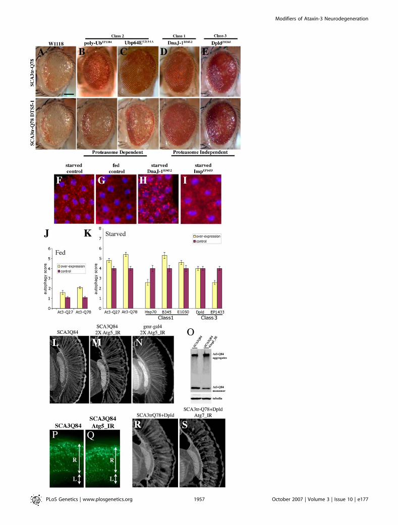
Figure 4. Degradation Pathways of Ataxin-3-Induced Neurodegeneration
(A–E) External eyes of 1-d flies showing suppression of SCAtrQ78 toxicity by modifiers only (upper panel), and suppression by modifiers in limitedproteasome background (lower panel) by expression of a dominant negative form of a 20S proteasome subunit [35].(A) Limiting proteasome activity alone does not enhance Ataxin-3 degeneration. Genotype (upper panel) w; gmr-GAL4 UAS-SCA3trQ78 /þ and (lowerpanel) w; gmr-GAL4 UAS-SCA3trQ78/þ ; UAS-DTS5/þ.(B and C) Polyubiquitin CR11700EP1384 (B) and Ubp64EE213-1A (C) cannot suppress when proteasome activity is limited. Genotypes (B) EP1384; gmr-GAL4UAS-SCA3trQ78 /þ; UAS-DTS5/þ and (C) w; gmr-GAL4 UAS-SCA3trQ78/þ; UAS-DTS5/E213-1A.(D and E) DnaJ-1B345.2 and dpldJM265 suppression remain largely unaffected when proteasome activity is limited. Genotypes (D) w; gmr-GAL4 UAS-SCA3trQ78/þ; UAS-DTS5/ B345.2 and (E) w; gmr-GAL4 UAS-SCA3trQ78/JM265; UAS-DTS5/þ.(F–I) Fat body tissues from larvae under starved or fed conditions, stained by Lysotraker Red (red) to highlight lysosomes and Hoescht (blue) for nuclei.Starvation strongly induces lysosomes (F), whereas fed animals normally have limited lysosomes (G). Ubiquitous expression of DnaJ-1 upregulates theinduction of lysosomes in the fat bodies in starvation conditions (H), suggesting that DnaJ-1 may reduce NI in a disease situation by facilitatingautophagy. Imp suppresses starvation-induced autophagy (I) although has no effect on NI, raising the possibility that it may modulate autophagic celldeath. Genotypes (F–G) w, (H) w; da-GAL4/B345.2, and (I) EP1433;da-GAL4/þ.(J and K) Scores of autophagy. Average values were obtained from at least 20 larvae per genotype. Error bar represents standard error of the mean ofmultiple experiments.(J) In fed conditions, pathogenic Ataxin-3 induces autophagy but normal Ataxin-3 has little effect.(K) In starvation conditions, expression of Ataxin-3, DnaJ-1, and Mrj further induce autophagy; on the contrary, Hsp70 and Imp suppress starvation-induced autophagy.(L–N) Retinal sections. (L) Flies (1 d) expressing SCA3Q84 have some loss of retinal integrity, which is mildly enhanced by reduction of (M) Atg5. (N)Reduction of atg5 activity alone has normal retinal morphology. Genotypes (L) w; gmr-GAL4 UAS-SCA3Q84/þ, (M) UAS-Atg5_IR/UAS-Atg5_IR; gmr-GAL4UAS-SCA3Q84/þ, and (N) gmr-GAL4/þ; UAS-Atg5_IR/UAS-Atg5_IR.(O) Western blot showing that reduction of Atg5 decreases Ataxin-3 monomer level, concomitant with enhanced toxicity.(P and Q) Cryosections for disease protein (1-d flies, anti-myc). Reduction of autophagy increases disease protein accumulation in the lamina (Q)compared to disease protein alone control (P). Hsc70.K71S also enhances, but it does not cause similar protein accumulations in the retina (unpublisheddata). Genotype in (P) is same as (L), and (Q) is same as (M).(R and S) Paraffin sections (1-d flies) showing suppression of Ataxin-3 degeneration by Dpld and that Dpld suppression is reduced by limitingautophagy. Genotypes (R) w; gmr-GAL4 UAS-SCA3trQ78 /JM265 and (S) UAS-Atg7_IR; gmr-GAL4 UAS-SCA3trQ78/JM265.doi:10.1371/journal.pgen.0030177.g004
PLoS Genetics | www.plosgenetics.org October 2007 | Volume 3 | Issue 10 | e1771958
Modifiers of Ataxin-3 Neurodegeneration

dynamin, and ras) to display the mutant effect at what wouldnormally be permissive conditions. This finding suggests thatlong polyQ runs can cause a general disruption of the cellularprotein-folding environment to affect the temperature-sensitive mutant proteins. Our work used a pathogenichuman disease protein Ataxin-3 and revealed that manymodifiers that mitigate Ataxin-3 toxicity also modulategeneral protein misfolding. Taken together, these resultssuggest that, in the absence of upregulation of the variouscomponents needed, the cell may become overwhelmed,triggering deleterious effects [32]. Our detailed analysis ofmodifiers of Ataxin-3 have revealed a variety of biologicalprocesses that can help manage pathogenic polyQ protein, aswell as specific genes of interest (Figure 6A).
The precise molecular pathways by which those modifiersthat are not clearly within protein folding pathways canmodulate misfolding to affect neurodegeneration requiresfurther study. We envisage, however, that they function bytheir predicted molecular mechanisms, but on targets thatimpinge on cellular protein homeostasis. For example,chromatin modifiers may tweak the expression of a varietyof genes in such processes, whereas translational regulatorsmay translationally modify such genes. The finding that avariety of cellular functions are involved would be consistentwith RNA interference screens in C. elegans that identified avariety of genes that impinge on cellular protein homeostasisas enhancers of the aggregation of polyQ protein [20]. Thesefindings, along with other studies including those we presenthere, underscore the critical importance of proper proteinhomeostasis to most—if not all—cellular functions, such thata variety of genes can influence this process.
Among our modifiers, some that might have been expectedto act in a similar manner, appeared to have distinctbiological effects. The class 2 modifiers showed completedependence on proper proteasome activity, while othermodifiers, including alleles of DnaJ-1 and Imp, appeared tobe insensitive to limiting proteasome activity, but ratheraffected autophagy in stress conditions. Hsp70 suppressesmultiple stress conditions including protein misfolding,starvation-induced autophagy, and paraquat-induced oxida-tive stress [11], suggesting it may both facilitate UPS activity,but also block autophagy-dependent degeneration. In vivo,these pathways interplay to maintain proper neuronalfunction in the face of disease, thus further study of themodifiers may allow greater molecular identification of theindividual pathways, as well as their integration.Although only select modifiers affected protein accumu-
lation as assayed by immunohistochemistry, all affected thesolubility of the disease protein by biochemical analysis. Therelationship between protein inclusions and proposed toxicoligomers is still under investigation, but it seems reasonableto suggest that the change in solubility may reflect activity ofthe modifiers to buffer or alter toxic conformations of theprotein. Our efforts to detect toxic oligomers of Ataxin-3using available antibodies [33,44,45] are still in progress. Wenote that, despite gross similarity, SCA3 degeneration is notidentical to general misfolding: some suppressors of Ataxin-3toxicity strikingly enhanced dominant-negative Hsp70 (upre-gulation of DnaJ-1 and Tpr2), whereas the enhancerCG11033E3093 suppressed it.The UPS and autophagy pathways function in either
degeneration or polyQ protein pathogenicity [46]. Our
Figure 5. Select Ataxin-3 Modifiers also Modulate Tau Toxicity
(A) Top: expression of pathogenic human tau.R406W causes a rough and reduced eye [41]. Genotype w; gmr-gal4/þ; UAS-tau.R406W/þ. Bottom: Ectopicexpression of apoptotic gene hid ablates the eye. Genotype w; gmr-GAL4 gmr-hid /þ. Genes shown modulated tau.wt and tau.R406W similarly.(B–D) Tpr2EB7-1A (B), polyubiquitin CR11700EP1384 (C), and CG5009B227.2 (D) suppress pathogenic tau. Tpr2EB7-1A (B) and CR11700EP1384 (C) have no effect onHid-induced programmed cell death. Genotypes (upper panel B) w; gmr-GAL4/þ; UAS-tau.R406W /EB7-1A, (C) EP1384; gmr-GAL4/þ; UAS-tau.R406W /þ, andw; gmr-gal4/B227.2; UAS-tau.R406W /þ. (Lower panel, B) w; gmr-GAL4 gmr-hid /EB7-1A, (C) EP1384; gmr-GAL4 gmr-hid /þ, and (D) w; gmr-GAL4 gmr-hid/B227.2.(E–G) dpldJM265 (E), ImpEP1433 (F), and embE2-1A (G) modulate tau toxicity and programmed cell death in a similar manner, suggesting that their effect ontau may be due to modulation of cell death. Genotypes (upper panel, E) w; gmr-GAL4/þ; UAS-tau.R406W /JM265, (F) EP1433; gmr-GAL4/þ; UAS-tau.R406W /þ,and (G) w; gmr-GAL4/E2-1A; UAS-tau.R406W /þ. (Lower panel, E) w; gmr-GAL4 gmr-hid /JM265, (F) EP1433; gmr-GAL4 gmr-hid /þ, and (G) w; gmr-GAL4 gmr-hid/E2-1A.doi:10.1371/journal.pgen.0030177.g005
PLoS Genetics | www.plosgenetics.org October 2007 | Volume 3 | Issue 10 | e1771959
Modifiers of Ataxin-3 Neurodegeneration

findings indicate that these pathways at least in part functionin the removal or decrease of the toxic protein, therebyreducing degeneration. It is also possible that activities ofthese pathways may be inhibited during pathogenesis; suchinhibition of normal activity would contribute to diseasepathology [47–49]. The normal function of Ataxin-3 is inubiquitin-modulated pathways [23–26]; our data suggest thepossibility that Ataxin-3 may also modulate autophagy.Normal physiological levels of autophagy are critical tointegrity of neurons, as loss of autophagy causes degenerationassociated with ubiquitinated inclusions [50,51]. Our studiesalso reveal that compromise of autophagy pathways strikinglyincreased cytoplasmic Ataxin-3 accumulations in the neuro-phil without an obvious change in NIs. This may indicate thatautophagy normally modulates cytoplasmic accumulation ofthe disease protein. Previous studies show that cytoplasmicpolyQ protein is very toxic and blocks axonal transport [52].These findings suggest that perturbations in autophagy mayenhance cytoplasmic toxicity further.
In addition to regulation of the disease protein level andmisfolding, results with select modifiers (Hsp70 and Imp)suggest that autophagy may also regulate loss of the cells.Given that our previous findings failed to reveal a clear roleof caspase-dependent cell death in SCA3 [22], autophagy maybe a mechanism of cell loss in this situation. Identification ofother components of UPS and autophagy pathway will givefurther insights into how the disease protein is degraded andmechanisms of neuronal loss.
Relationship to Other Modifier Pathways and ScreensGenetic screens have been performed for modifiers of
Ataxin-1 [19] and of pure polyQ domains [18], revealingsimilar components of protein folding and degradation:DnaJ-1 [18,19] and Tpr2 [18], but also RNA binding proteins
and transcription factors. Although components of ubiquitinpathways that modulate Ataxin-1 appear distinct fromAtaxin-3, this may reflect different regulators required formodifying different proteins or lack of saturation of thescreens. Similarly several RNA binding proteins identified asmodifiers of Ataxin-1 were suggested to reflect Ataxin-1function as a putative RNA binding protein; our data suggestthat it is also possible that these modifiers work morefundamentally to modulate protein solubility or levels. Ourother work, however, has revealed a role for microRNAs inmodulating neuronal survival in neurodegenerative situa-tions [22,53], which has recently been extended to vertebrateneuronal integrity as well [54]. A C. elegans screen revealed alarge number of modifiers of a pure polyQ aggregationphenotype [20]. Several modifiers identified in that screenaffect RNA synthesis, processing, and protein synthesis asmodifiers of misfolding. It will be important to test thesemodifiers against various specific disease proteins, as theaction of a pure polyQ repeat may be distinct from apathogenic repeat within a host protein.One reason for global commonality among modifiers of
polyQ disease proteins may be that the proteins themselvesfall within a common interacting protein network. Recentstudies using the yeast two-hybrid approach and proteomicdatabases reveal a protein interaction network of the ataxiaproteins [55]. A surprise was that many different ataxiaproteins, including Ataxin-3, fall within a few interactionsteps from one another, suggesting that their commonphenotypes may be a reflection of common interactions,which, when perturbed, contribute to disease. In that study[55], seven proteins were identified as direct interactionpartners of Ataxin-3; however none of these appeared in ourscreen. Another direct binding protein–VCP [56]—was alsonot identified. However, the human orthologues of the
Figure 6. Activities of Modifier Genes of Ataxin-3-Associated Neurodegeneration
(A) Modifiers of pathogenic Ataxin-3 toxicity may (1) reduce disease protein accumulation into NI in a manner sensitive to proteasome activity and/or bymodulating autophagy, (2) promote cellular functionality in situations of misfolded protein, and/or (3) promote neuronal survival by regulatingautophagic cell loss.(B) Select modifiers of Ataxin-3 toxicity reveal genetic links to tau toxicity and Hid-induced programmed cell death. Tpr2EB7-1A, polyubiquitinCR11700EP1384, and CG5009B227.2 suppress both Ataxin-3 and tau toxicity, implicating chaperone and ubiquitin proteasome activity as modifiers of taueffects. Modifiers with similar effects on tau and hid-induced cell death may be modulating tau by modulating cell death; however, these modifierslikely act in a distinct way on Ataxin-3-associated degeneration, which is not sensitive to programmed cell death genes but rather appears modulatedby the microRNA bantam [22].doi:10.1371/journal.pgen.0030177.g006
PLoS Genetics | www.plosgenetics.org October 2007 | Volume 3 | Issue 10 | e1771960
Modifiers of Ataxin-3 Neurodegeneration

majority of the genetic modifiers we defined fit into theprotein interaction network at various points; some aredirect interactors of other ataxia disease proteins, and othersare one or more steps removed (Table S3).
Alzheimer disease and polyQ diseases are two unrelatedhuman neurodegenerative diseases that cause neuronaldegeneration in distinct brain regions [1,2,57]. Therefore,no overlap was expected between modifiers of these diseaseproteins in flies; indeed previous studies suggest minimaloverlapping genes [58]. Surprisingly, we found that selectmodifiers of Ataxin-3 suppressed tau-degeneration, includingthe cochaperone Tpr2 and polyubiquitin (Figure 6B). Thisfinding is consistent with cell culture studies and C. elegansRNA interference screens that implicate chaperones asmodifiers of tau degeneration [59,60]. Further study of thesemodifiers, especially those that may be in common amongdifferent disease proteins, should provide the foundation fornew therapeutic insight.
Materials and Methods
Drosophila stocks and crosses. Fly lines were grown in standardcornmeal molasses agar with dry yeast at 25 8C. Transgenic lines UAS-SCA3trQ78, UAS-SCAtrQ61, UAS-SCA3Q78, and UAS-SCA3Q84 aredescribed [14–16]. General stock lines, deficiency lines, and specificalleles of the EP modifiers were obtained from the BloomingtonDrosophila stock center. The emb mutant lines were from C.Samakovlis (Wenner-Gren Institute Stockholm University, Stock-holm, Sweden) [61]. rh1-GAL4, gmr-hid, UAS-DTS5, autophagyinverted repeat lines (UAS-Atg5_IR and UAS-Atg7_IR), tau trans-genic lines (UAS-htau and UAS-htau.R406W ), UAS-brat, and Hsc70Cprotein trap line were kindly provided by C. Desplan (New YorkUniversity, New York, New York, United States), H. Steller (Rock-efeller University, New York, New York, US), J.M. Belote (SyracuseUniversity, Syracuse, New York, US), T. Neufeld (University ofMinnesota, Minneapolis, Minnesota, US), M. Feany (Harvard MedicalSchool, Boston, Massachusetts, US), R. Wharton (Duke UniversityMedical Center, Durham, North Carolina, US), M. Buszczakl (JohnsHopkins University, Baltimore, Maryland, US), respectively. Thedominant-negative Hsp70 line is described [32]. UAS-dpld FLAG,untagged, and gfp fusion lines were generated from cDNA cloneLD02463 by subcloning into the pUAST vector [62]. Excision lines forreversion were made by crossing the EP insertions to lines bearingtransposase, then screening for loss of the EP element.
For the genetic screen, virgin females of the starter line EP55,bearing an EP insertion on the X chromosome [27], was crossed tomales with transposase. Males were selected and crossed to virginfemales that expressed the disease gene (w/w; gmr-GAL4 UAS-SCA3trQ78 Tft/CyO). F1 progeny were screened for males with eithersuppressed or enhanced eye phenotypes as compared to controls; anymodifiers were then outcrossed and balanced. EP insertions wereconfirmed to be single insertions through outcrossing, as well asplasmid rescue.
Histochemistry. Epon sections, paraffin sections, and cryosectionsof adult heads were performed as described [14,16]. Primaryantibodies for immunostaining were anti-HA primary antibody (Y-11, 1:50, Santa Cruz Biotechnology; and 12CA5, 1:100, Roche), anti-Myc (9E10, 1:100, Santa Cruz Biotechnology), anti-Hsp70 (7FB,1:1,000,) [63], and anti-Gfp (A6455, 1:50, Molecular Probes). Secon-dary antibodies included anti-mouse or anti-rabbit conjugated toAlexa Fluor 594 or 488 (1:200 or 1:100, Molecular Probes). Westernimmunoblots were performed as described [16]. Primary antibodiesused were HA-HRP (3F10, 1:500, Roche), rat anti-Hsp70 (7FB,1:2,000), and mouse anti-tubulin (E7, 1:2,000, Developmental StudiesHybridoma Bank), with the secondary goat anti-rat IgG (1:2,000,Roche) and goat anti-mouse IgG (1:2,000, Chemicon International).
Molecular biology. To define the sites of EP insertion, plasmidrescue was performed [27]. To confirm single hits in the genome,multiples of clones from each EP line were sequenced with an EP 39 P-end specific primer (59-CAA TCA TAT CGC TGT CTC ACT CA-39).The flanking DNA sequence was used to query flybase BLAST todefine the nearby gene and exact site of insertion. Upregulation ofthe genes by the EP element was confirmed by northern analysis,
comparing the EP line alone, to wild-type fly controls with the EP linein the presence of a GAL4 driver, using gene-specific probesgenerated by reverse-transcription PCR using an EP-specific primerand gene-specific primers to the target genes. That the EP insertionswere single insertions was independently verified genetically bymolecular and phenotypic analysis of lines.
The effect of the modifiers on transgene expression levels wasexamined by real-time PCR (see Text S1 and Figure S2) and byreverse-transcription PCR. For the latter, total RNA was extractedwith the RNAeasy kit (74104, QIAGEN) following manufacturer’sinstructions. cDNA synthesis was done using SuperScript First StrandSynthesis for reverse-transcription PCR (12371–019, Invitrogen). PCRwas performed using primers: for the SCA3 transcript, 59-CTAT-CAGGACAGAGTTCACAT-39 (forward) and 59-CAGATAAAGTGT-GAAGGTAGC-39 (reverse); for the GAL4 transcript, 59-GTCTTCTATCGAACAAGCATGCGA-39 (forward) and 59-TGACCTTTGTTACTACTCTCTTCC-39 (reverse) and for rp49 con-trol, 59-CCAGTCGGATCGATATGCTAA-39 (forward) and 59-AC-CGTTGGGGTTGGTGAG-39 (reverse).
Behavioral assays. Phototaxis was performed as described [15]. Thepercentage of flies in the light and dark chambers represent anaverage of three independent groups of flies. At least 100 flies weretested for each genotype, 20 flies were used in each experiment.
Autophagy assays. Third instar larval fat body tissues were stainedwith Lyso Tracker Red DND-99 (L-7528, Molecular Probes) andHoechst (H3570, Molecular Probes) as described [38]. For eachgenotype, well-fed and starved larvae were used as negative andpositive controls for the assay conditions. A score of 4 was given tocontrol larvae grown under starvation condition; autophagy for othergenotypes was scored relative to this. Controls (driver line alone and/or modifier alone, in starvation and well-fed conditions) wereperformed for each modifier genes in parallel to the experimentalsituation. The final autophagy score represents the average of 20larvae from each genotype.
Supporting Information
Figure S1. Modifiers of SCA3trQ78-Associated Neurodegeneration
External eye and internal retinal sections of 1-d flies.(A) Control external and internal eye. Normal retinal thickness isindicated with yellow arrow. Genotype w; gmr-GAL4/þ.(B) Expression of strong SCA3trQ78 in the eye causes external andinternal degeneration with severe pigment loss and markedly reducedretinal thickness. Genotype w; gmr-GAL4 UAS-SCA3trQ78/þ.(C–T) Suppressors of SCA3tr-Q78 associated neurodegeneration intrans to w; gmr-gal4 UAS-SCA3trQ78. EP lines are (C) E407, (D) B345.2,(E) E1050, (F) EP1348, (G) EB7-1A, (H) EP1384, (I) E213-1A, ( J) B3-Sa,(K) E659, (M) E2-1A, (N) B9-E, (O) EP1335, (P) EP456, (Q) JM265, (R)B8-S, (S) EP1433 and (T) B227.2. (L) EP3093 enhances SCA3trQ78-associated neurodegeneration and lethality.
Found at doi:10.1371/journal.pgen.0030177.sg001 (1.9 MB JPG).
Figure S2. Control Experiments for SCA3 Modifiers
(A) (Top) Western analysis for SCA3trQ78 protein level (detectedwith anti-HA) in 1-d fly heads. Experiments were performed using thelate onset gene expression driver rh1-GAL4. At 1 d, the pathogenicprotein is soluble by immunocytochemistry and western analysis. TheAtaxin-3 protein level was similar in the various situations, indicatingthat the modifiers do not affect expression from the GAL4-UASsystem. Genotypes: w; UAS-SCA3trQ78; rh1-GAL4 in trans to þ or thevarious EP modifier lines as indicated.(A) (Bottom) Real-time PCR for level of transgenes shown areTpr2EB7-1A, Faf E659, Sin3AB9-E, dbrEP456, dpld JM265, orb2B8-S, ImpEP1433;CG5009B227.2 was confirmed by reverse transcription PCR (unpub-lished data). Genotypes: w; UAS-SCA3trQ78; rh1-GAL4 in trans toþ orEP modifier lines indicated.(B–F) Examples of modifiers that failed to affect tau toxicity. Allmodifiers were tested for ability to modulate the deleterious effectsof a number of additional genes, including tau, hid, and others(below). Although select modifiers affected tau toxicity (either toenhance or suppress, see Figure 5), others had no effect. Theinability to modulate such additional gene activities was additionalevidence that the modifiers did not affect the GAL4-UAS system.Tau-induced degenerate eye is not modulated upon co-expressionof EP alleles of (C) Hsp68, (D) Ubp64E, (E) CG11103, or (F) Sin3A.Genotypes: (B) w; gmr-GAL4/þ; UAS-tau.R406W/þ , (C) w; gmr-GAL4/þ;UAS-tau.R406W/ E407, (D) w; gmr-GAL4/þ; UAS-tau.R406W/ E213-1A,
PLoS Genetics | www.plosgenetics.org October 2007 | Volume 3 | Issue 10 | e1771961
Modifiers of Ataxin-3 Neurodegeneration

(E) w; gmr-GAL4/þ; UAS-tau.R406W/EP3093, and (F) w; gmr-GAL4/þ;UAS-tau.R406W/ B9-E.(G–K) Detailed analysis of controls for alleles of the dpld gene.(G)Western analysis for SCA3trQ78 protein expression in 1-d fly heads.The EP allele of dpld (dpld JM265), generated transgenic lines of dpld (UAS-dpld, dpld J990), and the precise excision line of the dpld JM265 allele (dpldJM265-ex) showed similar levels of disease protein at early time pointscompared to the control (þ). This indicated that upregulation of dplddid not affect expression from the GAL4-UAS system; rather furtherstudies showed that Dpld affected long-term accumulation of theprotein (see Figure 3F and 3G). Genotypes: lane 1, w; UAS-SCA3trQ78/þ; rh1-GAL/þ; lane 2, w; UAS-SCA3trQ78/dpld JM265; rh1-GAL4/þ; lane 3, w; UAS-SCA3trQ78/þ; rh1-GAL4/UAS-dpld J990; lane 4, w;UAS-SCA3trQ78/dpld JM265-ex; rh1-GAL4/þ.(H–K) Effect of dpld expression on various deleterious GAL4/UAS-induced eye phenotypes.(H and I) dpld expression had no effect on eyeless-induced disruptionof the eye. Genotypes: (H) w; ey-GAL4 UAS-eyeless/ dpld JM265 and (I) w/þ;ey-GAL4 UAS-eyeless/þ.( J and K) dpld expression did not affect the rough eye phenotypeinduced by expression of MAP2. Genotypes: ( J) w; gmr-GAL4 UAS-MAP2/ dpld JM265 and (K) w/þ; gmr-GAL4 UAS-MAP2/þ.Found at doi:10.1371/journal.pgen.0030177.sg002 (1.0 MB JPG).
Figure S3. Conserved Domains of Modifier Genes
The modifiers fall into three major classes based on conservedsequence domains: genes involved in chaperone pathways, inubiquitin pathways, and miscellaneous functions. The conserveddomains for the first category include Hsp70, DnaJ, DNAJ-C, Hsp20,and tetratricopeptide repeat (TPR). The second category hasubiquitin, ubiquitin carboxy hydrolase (UCH), ubiquitin associated(UBA), ubiquitin regulatory (UBX), and F-box domains. Theconserved domains for the third category include Importin-beta Nterminus (IBNN), paired amphipathic helix repeat (PAH), relhomology domain (RHD), transcription factor/cell surface receptorimmunoglobulin-like fold (TIG), RNA recognition motif (RRM), KH,and acyl-CoA oxidase (ACOX) domain.
Found at doi:10.1371/journal.pgen.0030177.sg003 (665 KB JPG).
Figure S4. Ataxin-3 Modifier Genes Mitigate Toxicity of PolyQProtein in the Nervous System
(A) Animals expressing SCA3trQ78 in the nervous system do notdevelop to adults, with lethality of larvae and pupae that fail to formcompletely (pupa shown).(B) With co-expression of EP1050, now the animals develop to thepharate pupal stage, with eyes, wings, and legs fully formed, thoughthe animals fail to emerge from the pupal case. Genotypes: (A) elav-GAL4; UAS-SCA3trQ78(S)/þ and (B) elav-GAL4; UAS-SCA3trQ78(S)/EP1050.(C) Table of lethality with or without added select modifier genes. *,Lethality assessed with neuronal expression by the elav-GAL4 driver,which is expressed in all neurons through development and in theadult.
Found at doi:10.1371/journal.pgen.0030177.sg004 (901 KB JPG).
Figure S5. Genomic Organization of dpld(A) Genomic organization of dpld with insertion sites of EP allelesindicated. The A form of the dpld transcript is shown (three splicevariants are predicted), black represents exons, and blue indicates the59 UTR and 39 UTR. EP insertions are in the same orientation as geneexpression. dpld is on 2R, cytological position 43C5. The centromereis to the right. Restriction enzyme sites are: X, XbaI; H, HindIII; R,EcoRI; B, BamHI; S, SacII; P, PstI.(B) Schematic representation of Dpld protein and select homologs(Dm Brat, Ce Lin-41, and Homo sapiens Lin-41). Blue is the B-box zincfinger, yellow is the coiled-coil, red is NHL repeats, and pink is a ringfinger domain. The percent identity of the NHL domain to that ofDpld is noted.(C) NHL domain based evolutionary tree showing relatedness of Dpldto that of other RBCC-NHL family members in nematode, fly, andhuman. Dpld is most homologous to Lin-41; the other studied flyNHL protein Brat is distantly related.(D) Sequence alignment of the B1 and B2 boxes, and the NHL domainof Dpld to those of Brat, human Lin-41, and C. elegans Lin-41. Aminoacid identities in black; similarities and identities in three of the foursequences are in gray. The cysteine and histidine residues that definethe B-box subclass of Zn finger are marked with an asterisk (*). Thepositions of NHL repeats are noted.
Found at doi:10.1371/journal.pgen.0030177.sg005 (806 KB JPG).
Figure S6. Dpld Suppresses Ataxin-3-Associated Neurodegeneration
External eye and internal retinal sections of 1-d flies.(A) Control fly bearing only the driver line. Genotype w; gmr-GAL4/þ.(B) Ataxin-3 induces severe retinal degeneration. Genotype w; gmr-GAL4 UAS-SCA3trQ78/þ.(C) Co-expression of dpld JM265 with SCA3trQ78 results in normalpigmentation and improved retinal structure. Genotype w; gmr-GAL4 UAS-SCA3trQ78/dpld JM265.(D) Expression of dpld from a UAS-dpld transgene improves retinalstructure, indicating that suppression of polyQ toxicity is due toincreasedDpld activity.Genotypew; gmr-GAL4UAS-SCA3trQ78 /þ; UAS-dpld J990-P2/þ.(E–G) Tangential retinal sections of fly expressing SCA3trQ78 aloneor with Dpld. Flies expressing SCA3trQ78 have a normal retina (E) at1 d, which undergoes severe progressive degeneration by (F) 21 d.Genotype w; UAS-SCA3trQ78/þ; rh1-GAL4/þ.(G) Flies with SCA3trQ78 and UAS-dpldJ990-P2 are normal. GenotypeUAS-SCA3trQ78/þ; rh1-GAL4 /UAS-dpld.(H) Rhabdomere counts in individual ommatidia by optical neutral-ized corneal pseudopupil (see reference contained in Text S1). Fliesexpressing SCA3trQ78 show degeneration in the number of rhab-domeres per ommatidial unit at 21 d. Flies expressing SCA3trQ78 anddpld J990-P2 retain rhabdomere numbers over 21 d; dpldJM265 has partialrescue compared to controls.
Found at doi:10.1371/journal.pgen.0030177.sg006 (1.5 MB JPG).
Figure S7. Select Modifiers More Effectively Mitigate Toxicity of FullLength Ataxin-3
(A) Expression of full-length SCA3Q84 causes amild eye degeneration.Genotype w; gmr-GAL4 UAS-SCA3Q84 /þ.(B–C) The modifiers (B) CG14207EP1348 and (C) polyubiquitinCR11700EP1384 suppress full-length disease toxicity stronger thanthey do the truncated disease protein. Genotypes w; gmr-GAL4 UAS-SCA3Q84 in trans to EP1348 and EP1384.Found at doi:10.1371/journal.pgen.0030177.sg007 (482 KB JPG).
Figure S8. Additional Details of Dpld and Hsc70C
(A–C) Horizontal eye section of a fly eye expressing dpld-eYFP shows(B) nonhomogenous distribution and localization to membranousstructures. (A) Hoechst staining. (C) Overlay shows that Dpld does notlocalize to nucleus but is concentrated around it. Genotype w; UAS-dpld-eYFP in trans to gmr-GAL4.(D–F) Horizontal retinal section showing Dpld localization in thepresence of SCA3trQ78. (D) eYFP staining for Dpld and (E) HAstaining for SCA3trQ78. (F) Overlay shows that Dpld is not recruitedto the SCA3trQ78 aggregates. Genotype w; gmr-GAL4 UAS-SCA3trQ78in trans to UAS-dpld-eYFP.(G and H) GFP insertion allele of Hsc70Cb enhances Ataxin-3neurotoxicity. Hsc70CbGFP fusion protein was confirmed by Westernimmunoblot (unpublished data). The line is homozygous lethalindicating a potential loss of function of allele of Hsc70Cb. Genotypesw; gmr-GAL4 UAS-SCA3trQ78/þ (H) and w; gmr-GAL4 UAS-SCA3trQ78/þ; Hsc70CbCB02656/þ.(I–K) Hsc70Cb protein trap line strikingly increases Ataxin-3inclusions in the lamina and medulla, indicating enhanced cytoplas-mic disease protein. (I) Hoescht staining for nuclei. Genotypes gmr-GAL4 UAS-SCA3Q84/þ (I and J), w; gmr-GAL4 UAS-SCA3Q84/þ;Hsc70CbCB02656/þ.(L–O) Hsc70Cb suppresses (L and M) normal and (N and O) mutantR406W tau-induced degeneration. Genotypes (L) w; gmr-GAL4/þ; UAS-tau/þ, (N) w; gmr-GAL4/þ; UAS-tau/ Hsc70CbCB02656, (M) w; gmr-GAL4/þ;UAS-tau.R406W/þ, and (N) w; gmr-GAL4/þ; UAS-tau.R406W/Hsc70CbCB02656.Found at doi:10.1371/journal.pgen.0030177.sg008 (2.1 MB JPG).
Figure S9. Dpld Modulates Body Size
(A–F) dpld and brat share a function in growth regulation (seereference contained in Text S1), but the activities of dpld to suppresspolyQ toxicity and cell death are special to dpld. The effect of dpld oneye and body size were mitigated by reducing autophagy gene activity(not shown).(A right, and C) Dpld expression during development reduces bodysize (A, fly on the right) and (C) eye size, similar to brat over-expression. Genotype in (A) w; act5C-GAL4 in trans to þ or UAS-dpldand (C) w; ey-GAL4/UAS-dpld.
PLoS Genetics | www.plosgenetics.org October 2007 | Volume 3 | Issue 10 | e1771962
Modifiers of Ataxin-3 Neurodegeneration

(A left, B) Normal fly body and eye size. Genotype (A) w; act5C-GAL4/þand (B) w; ey-GAL4/þ.(D, E) Directed expression of brat does not mitigate degenerationinduced by SCA3trQ78 either (D) externally or (E) internally.Genotype w; gmr-GAL4 UAS-SCA3trQ78 in trans to UAS-brat.(F) Brat does not mitigate hid-induced programmed cell death.Genotype w; gmr-hid gmr-GAL4 in trans to UAS-brat.Found at doi:10.1371/journal.pgen.0030177.sg009 (769 KB JPG).
Table S1. Summary of Genetic Modifiers of Ataxin-3
The location and orientation of the EP insertions are indicated,including the method used to confirm that the insertions causedoverexpression of the targeted gene. Note that insertions into novelgenes were reverted to confirm that the EP insertion was causal inmodification.
Found at doi:10.1371/journal.pgen.0030177.st001 (98 KB DOC).
Table S2. Loss-of-Function Deficiency Regions and Alleles ofModifiers of Ataxin-3 Neurodegeneration That Were Tested
Alleles and deficiency lines of Ataxin-3 modifiers were tested formodulation of polyQ toxicity. See also Figure 1K–1O.
Found at doi:10.1371/journal.pgen.0030177.st002 (89 KB DOC).
Table S3. Protein Interactions of Ataxin-3 Genetic Modifiers
List of human orthologues of the fly modifiers found. The degree ofinteraction from Ataxin-3 is from Table 5 of Lim et al. (see referencecontained in Text S1). Degree of interaction is based on the distanceeach orthologue is from the seven primary interactors defined forAtaxin-3 (ARHGAP19, EWSR1, LIN10, RAD23A, RAD23B, TEX11,and PRKCABP) (see reference contained in Text S1) and VCP/TER94(see reference contained in Text S1).
Found at doi:10.1371/journal.pgen.0030177.st003 (122 KB DOC).
Text S1. Supplemental Methods
Found at doi:10.1371/journal.pgen.0030177.sd001 (62 KB DOC).
Accession Numbers
The accession numbers of the proteins used in the analysis in FigureS5 from the National Center for Biotechnology Information (NCBI)(http://www.ncbi.nlm.nih.gov) are Dm Dappled (NM_165533),DmCG15105 (NM_137546), Dm Brat (NM_057597), Dm Mei-P26(NM_143765), Ce Lin-41 (NM_060086), Hs Lin-41 (XM_067369),Hs TRIM2 (NM_015271), Hs TRIM3 (NM_006458), and Hs TRIM32(NM_012210).
Acknowledgments
We thank R. Pittman and D. Lessing for critical comments. We thankJ.M. Belote, C. Desplan, M. Feany, T. Neufeld, H. Steller, R. Wharton,M. Buszczak, H. Zoghbi, and S. Lindquist, and the Bloomington StockCenter and Developmental Studies Hybridoma Bank (funding fromNICHD) for fly lines, reagents, or advice. We thank R. Bernstein, A.Bovell, L. Faust, L. Morabito, and X. Teng for outstanding technicalassistance.
Author contributions. JB and NMB conceived and designed theexperiments, performed the experiments, analyzed the data, andwrote the paper.
Funding. This work was funded by the National Institute ofNeurological Disorders and Stroke and the Packard Foundation (toNMB). NMB is an investigator of the Howard Hughes MedicalInstitute.
Competing interests. The authors have declared that no competinginterests exist.
References1. Zoghbi HY, Orr HT (2000) Glutamine repeats and neurodegeneration.
Annu Rev Neurosci 23: 217–247.2. Gusella JF, MacDonald ME (2000) Molecular genetics: unmasking polyglut-
amine triggers in neurodegenerative disease. Nat Rev Neurosci 1: 109–115.3. Ikeda H, Yamaguchi M, Sugai S, Aze Y, Narumiya S, et al. (1996) Expanded
polyglutamine in the Machado-Joseph disease protein induces cell death invitro and in vivo. Nat Genet 13: 196–202.
4. Perutz MF (1999) Glutamine repeats and neurodegenerative diseases:molecular aspects. Trends Biochem Sci 24: 58–63.
5. Kim S, Nollen EA, Kitagawa K, Bindokas VP, Morimoto RI (2002)Polyglutamine protein aggregates are dynamic. Nat Cell Biol 4: 826–831.
6. Cummings CJ, Mancini MA, Antalffy B, DeFranco DB, Orr HT, et al. (1998)Chaperone suppression of aggregation and altered subcellular proteasomelocalization imply protein misfolding in SCA1. Nat Genet 19: 148–154.
7. Lansbury PT, Lashuel HA (2006) A century-old debate on proteinaggregation and neurodegeneration enters the clinic. Nature 443: 774–779.
8. Lee VM, Trojanowski JQ (2006) Mechanisms of Parkinson’s disease linkedto pathological alpha-synuclein: new targets for drug discovery. Neuron 52:33–38.
9. Aguzzi A, Heikenwalder M (2006) Pathogenesis of prion diseases: currentstatus and future outlook. Nat Rev Microbiol 4: 765–775.
10. Marsh JL, Thompson LM (2006) Drosophila in the study of neuro-degenerative disease. Neuron 52: 169–178.
11. Bilen J, Bonini NM (2005) Drosophila as a model for human neuro-degenerative disease. Annu Rev Genet 39: 153–171.
12. Driscoll M, Gerstbrein B (2003) Dying for a cause: invertebrate geneticstakes on human neurodegeneration. Nat Rev Genet 4: 181–194.
13. Outeiro TF, Muchowski PJ (2004) Molecular genetics approaches in yeast tostudy amyloid diseases. J Mol Neurosci 23: 49–60.
14. Warrick JM, Paulson HL, Gray-Board GL, Bui QT, Fischbeck KH, et al.(1998) Expanded polyglutamine protein forms nuclear inclusions andcauses neural degeneration in Drosophila. Cell 93: 939–949.
15. Warrick JM, Morabito LM, Bilen J, Gordesky-Gold B, Faust LZ, et al. (2005)Ataxin-3 suppresses polyglutamine neurodegeneration in Drosophila by aubiquitin-associated mechanism. Mol Cell 18: 37–48.
16. Chan HY, Warrick JM, Gray-Board GL, Paulson HL, Bonini NM (2000)Mechanisms of chaperone suppression of polyglutamine disease: selectivity,synergy and modulation of protein solubility in Drosophila. Hum MolGenet 9: 2811–2820.
17. Morley JF, Brignull HR, Weyers JJ, Morimoto RI (2002) The threshold forpolyglutamine-expansion protein aggregation and cellular toxicity isdynamic and influenced by aging in Caenorhabditis elegans. Proc NatlAcad Sci U S A 99: 10417–10422.
18. Kazemi-Esfarjani P, Benzer S (2000) Genetic suppression of polyglutaminetoxicity in Drosophila. Science 287: 1837–1840.
19. Fernandez-Funez P, Nino-Rosales ML, de Gouyon B, She WC, Luchak JM, et
al. (2000) Identification of genes that modify ataxin-1-induced neuro-degeneration. Nature 408: 101–106.
20. Nollen EA, Garcia SM, van Haaften G, Kim S, Chavez A, et al. (2004)Genome-wide RNA interference screen identifies previously undescribedregulators of polyglutamine aggregation. Proc Natl Acad Sci U S A 101:6403–6408.
21. Willingham S, Outeiro TF, DeVit MJ, Lindquist SL, Muchowski PJ (2003)Yeast genes that enhance the toxicity of a mutant huntingtin fragment oralpha-synuclein. Science 302: 1769–1772.
22. Bilen J, Liu N, Burnett BG, Pittman RN, Bonini NM (2006) MicroRNApathways modulate polyglutamine-induced neurodegeneration. Mol Cell24: 157–163.
23. Doss-Pepe EW, Stenroos ES, Johnson WG, Madura K (2003) Ataxin-3interactions with rad23 and valosin-containing protein and its associationswith ubiquitin chains and the proteasome are consistent with a role inubiquitin-mediated proteolysis. Mol Cell Biol 23: 6469–6483.
24. Burnett B, Li F, Pittman RN (2003) The polyglutamine neurodegenerativeprotein ataxin-3 binds polyubiquitylated proteins and has ubiquitinprotease activity. Hum Mol Genet 12: 3195–3205.
25. Donaldson KM, Li W, Ching KA, Batalov S, Tsai CC, et al. (2003) Ubiquitin-mediated sequestration of normal cellular proteins into polyglutamineaggregates. Proc Natl Acad Sci U S A 100: 8892–8897.
26. Chow MK, Mackay JP, Whisstock JC, Scanlon MJ, Bottomley SP (2004)Structural and functional analysis of the Josephin domain of the polyglut-amine protein ataxin-3. Biochem Biophys Res Commun 322: 387–394.
27. Rorth P (1996) A modular misexpression screen in Drosophila detectingtissue-specific phenotypes. Proc Natl Acad Sci U S A 93: 12418–12422.
28. Wellington CL, Ellerby LM, Hackam AS, Margolis RL, Trifiro MA, et al.(1998) Caspase cleavage of gene products associated with triplet expansiondisorders generates truncated fragments containing the polyglutaminetract. J Biol Chem 273: 9158–9167.
29. Berke SJ, Schmied FA, Brunt ER, Ellerby LM, Paulson HL (2004) Caspase-mediated proteolysis of the polyglutamine disease protein ataxin-3. JNeurochem 89: 908–918.
30. Mauri PL, Riva M, Ambu D, De Palma A, Secundo F, et al. (2006) Ataxin-3 issubject to autolytic cleavage. FEBS J 273: 4277–4286.
31. Warrick JM, Chan HY, Gray-Board GL, Chai Y, Paulson HL, et al. (1999)Suppression of polyglutamine-mediated neurodegeneration in Drosophilaby the molecular chaperone HSP70. Nat Genet 23: 425–428.
32. Bonini NM (2002) Chaperoning brain degeneration. Proc Natl Acad Sci U SA 99 Suppl 4: 16407–16411.
33. Arrasate M, Mitra S, Schweitzer ES, Segal MR, Finkbeiner S (2004) Inclusionbody formation reduces levels of mutant huntingtin and the risk ofneuronal death. Nature 431: 805–810.
34. Ross CA, Pickart CM (2004) The ubiquitin-proteasome pathway inParkinson’s disease and other neurodegenerative diseases. Trends CellBiol 14: 703–711.
PLoS Genetics | www.plosgenetics.org October 2007 | Volume 3 | Issue 10 | e1771963
Modifiers of Ataxin-3 Neurodegeneration

35. Saville KJ, Belote JM (1993) Identification of an essential gene, l(3)73Ai,with a dominant temperature-sensitive lethal allele, encoding a Drosophilaproteasome subunit. Proc Natl Acad Sci U S A 90: 8842–8846.
36. Ravikumar B, Vacher C, Berger Z, Davies JE, Luo S, et al. (2004) Inhibitionof mTOR induces autophagy and reduces toxicity of polyglutamineexpansions in fly and mouse models of Huntington disease. Nat Genet36: 585–595.
37. Baehrecke EH (2005) Autophagy: dual roles in life and death? Nat Rev MolCell Biol 6: 505–510.
38. Scott RC, Schuldiner O, Neufeld TP (2004) Role and regulation ofstarvation-induced autophagy in the Drosophila fat body. Dev Cell 7:167–178.
39. Lee VM, Goedert M, Trojanowski JQ (2001) Neurodegenerative tauopa-thies. Annu Rev Neurosci 24: 1121–1159.
40. Jackson GR, Wiedau-Pazos M, Sang TK, Wagle N, Brown CA, et al. (2002)Human wild-type tau interacts with wingless pathway components andproduces neurofibrillary pathology in Drosophila. Neuron 34: 509–519.
41. Wittmann CW, Wszolek MF, Shulman JM, Salvaterra PM, Lewis J, et al.(2001) Tauopathy in Drosophila: neurodegeneration without neurofibril-lary tangles. Science 293: 711–714.
42. Satyal SH, Schmidt E, Kitagawa K, Sondheimer N, Lindquist S, et al. (2000)Polyglutamine aggregates alter protein folding homeostasis in Caenorhab-ditis elegans. Proc Natl Acad Sci U S A 97: 5750–5755.
43. Gidalevitz T, Ben-Zvi A, Ho KH, Brignull HR, Morimoto RI (2006)Progressive disruption of cellular protein folding in models of polyglut-amine diseases. Science 311: 1471–1474.
44. Brooks E, Arrasate M, Cheung K, Finkbeiner SM (2004) Using antibodies toanalyze polyglutamine stretches. Methods Mol Biol 277: 103–128.
45. Kayed R, Head E, Thompson JL, McIntire TM, Milton SC, et al. (2003)Common structure of soluble amyloid oligomers implies commonmechanism of pathogenesis. Science 300: 486–489.
46. Rubinsztein DC (2006) The role of intracellular protein degradationpathways in neurodegeneration. Nature 443: 780–786.
47. Bence NF, Sampat RM, Kopito RR (2001) Impairment of the ubiquitin-proteasome system by protein aggregation. Science 292: 1552–1555.
48. Cuervo AM, Stefanis L, Fredenburg R, Lansbury PT, Sulzer D (2004)Impaired degradation of mutant alpha-synuclein by chaperone-mediatedautophagy. Science 305: 1292–1295.
49. Bennett EJ, Bence NF, Jayakumar R, Kopito RR (2005) Global impairmentof the ubiquitin-proteasome system by nuclear or cytoplasmic proteinaggregates precedes inclusion body formation. Mol Cell 17: 351–365.
50. Komatsu M, Waguri S, Chiba T, Murata S, Iwata J, et al. (2006) Loss ofautophagy in the central nervous system causes neurodegeneration in mice.Nature 441: 880–884.
51. Hara T, Nakamura K, Matsui M, Yamamoto A, Nakahara Y, et al. (2006)Suppression of basal autophagy in neural cells causes neurodegenerativedisease in mice. Nature 441: 885–889.
52. Gunawardena S, Her LS, Brusch RG, Laymon RA, Niesman IR, et al. (2003)Disruption of axonal transport by loss of huntingtin or expression ofpathogenic polyQ proteins in Drosophila. Neuron 40: 25–40.
53. Bilen J, Liu N, Bonini NM (2006) A new role for microRNA pathways:modulation of degeneration induced by pathogenic human diseaseproteins. Cell Cycle 5: 2835–2838.
54. Schaefer A, O’Carroll D, Tan CL, Hillman D, Sugimori M, et al. (2007)Cerebellar neurodegeneration in the absence of microRNAs. J Exp Med204: 1553–1558.
55. Lim J, Hao T, Shaw C, Patel AJ, Szabo G, et al. (2006) A protein-proteininteraction network for human inherited ataxias and disorders of Purkinjecell degeneration. Cell 125: 801–814.
56. Boeddrich A, Gaumer S, Haacke A, Tzvetkov N, Albrecht M, et al. (2006) Anarginine/lysine-rich motif is crucial for VCP/p97-mediated modulation ofataxin-3 fibrillogenesis. EMBO J 25: 1547–1558.
57. Goedert M, Spillantini M (2006) A century of Alzheimer’s disease. Science314: 777–781.
58. Shulman JM, Feany MB (2003) Genetic modifiers of tauopathy inDrosophila. Genetics 165: 1233–1242.
59. Richter-Landsberg C, Bauer NG (2004) Tau-inclusion body formation inoligodendroglia: the role of stress proteins and proteasome inhibition. Int JDev Neurosci 22: 443–451.
60. Kraemer BC, Burgess JK, Chen JH, Thomas JH, Schellenberg GD (2006)Molecular pathways that influence human tau-induced pathology inCaenorhabditis elegans. Hum Mol Genet 15: 1483–1496.
61. Roth P, Xylourgidis N, Sabri N, Uv A, Fornerod M, et al. (2003) TheDrosophila nucleoporin DNup88 localizes DNup214 and CRM1 on thenuclear envelope and attenuates NES-mediated nuclear export. J Cell Biol163: 701–706.
62. Brand AH, Perrimon N (1993) Targeted gene expression as a means ofaltering cell fates and generating dominant phenotypes. Development 118:401–415.
63. Velazquez JM, Lindquist S (1984) Hsp70: nuclear concentration duringenvironmental stress and cytoplasmic storage during recovery. Cell 36:655–662.
PLoS Genetics | www.plosgenetics.org October 2007 | Volume 3 | Issue 10 | e1771964
Modifiers of Ataxin-3 Neurodegeneration