Fig. 31-1

Fig. 31-3
(b) Coenocytic hypha
Septum
(a) Septate hypha
Pore
Nuclei
Nuclei Cell wallCell wall
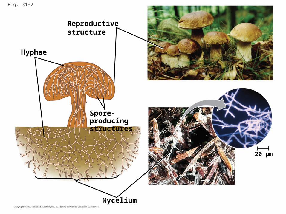
Fig. 31-2
Reproductive structure
Spore-producingstructures
Hyphae
Mycelium
20 µm
Fig. 31-4
(b) Haustoria
(a) Hyphae adapted for trapping and killing prey
Nematode
Plantcellwall
Haustorium
Plant cellplasmamembrane
Plant cell
Fungal hypha
Hyphae25 µm

Basidium
Fig. 31-19-4
SEXUALREPRODUCTION
Diploid (2n)
Haploid (n)Dikaryotic (n +n)
Key
PLASMOGAMY
Matingtype (+)
Haploid myceliaDikaryotic mycelium
Matingtype (–)
Basidia(n+n)
Gills linedwith basidia
Basidiocarp(n+n)
KARYOGAMY
Diploidnuclei
MEIOSIS
Basidium containingfour haploid nuclei
Dispersal andgermination
Basidiospores(n)
Basidium withfour basidiospores
Basidiospore1 µm
Haploid mycelia

Fig. 31-18
Shelf fungi, importantdecomposers of wood
Maiden veil fungus(Dictyphora), afungus with anodor like rottingmeat
Puffballs emittingspores
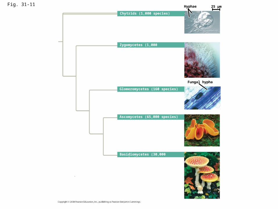
Fig. 31-11
Chytrids (1,000 species)
Zygomycetes (1,000 species)
Hyphae 25 µm
Glomeromycetes (160 species)
Fungal hypha
Ascomycetes (65,000 species)
Basidiomycetes (30,000 species)
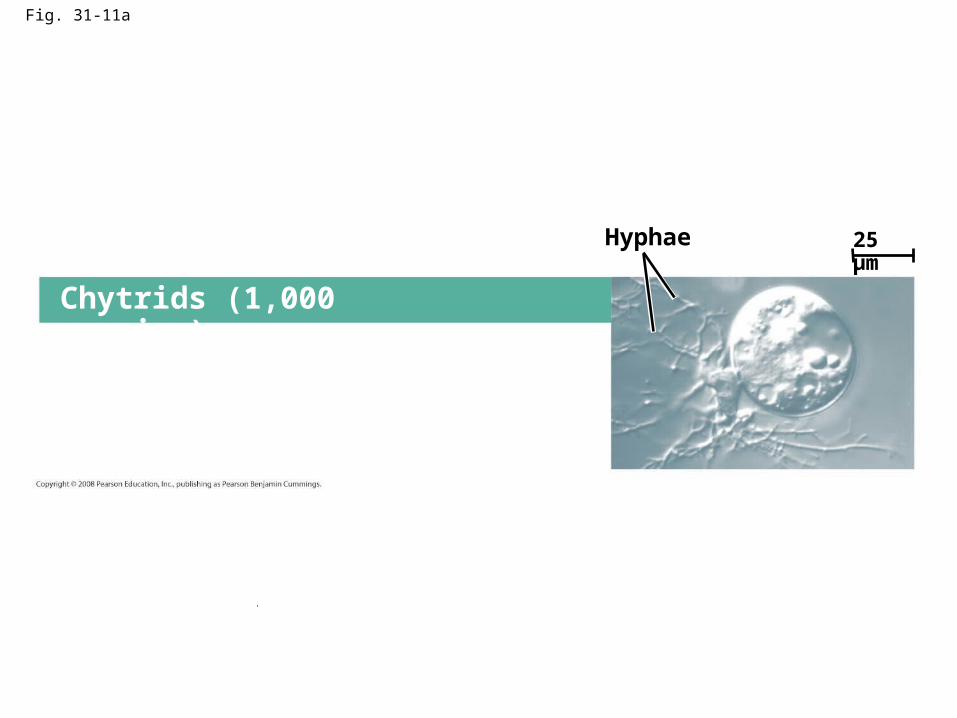
Fig. 31-11a
Chytrids (1,000 species)
Hyphae 25 µm

Fig. 31-12
Flagellum
4 µm
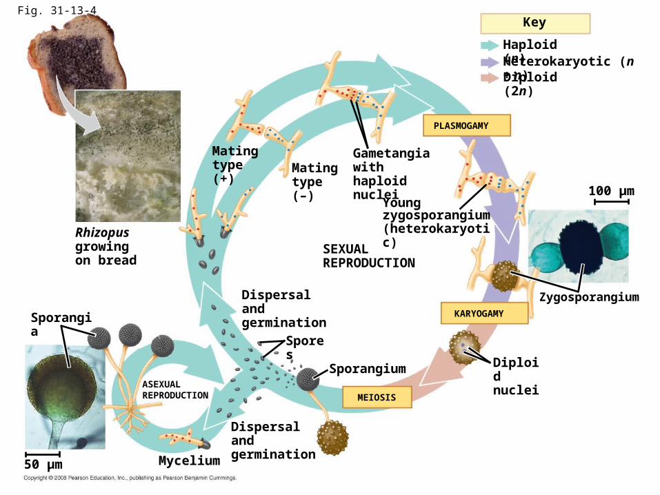
Fig. 31-13-4
Rhizopusgrowingon bread
SEXUALREPRODUCTION
Youngzygosporangium(heterokaryotic)
Gametangia withhaploid nucleiMating
type (–)
Matingtype (+)
Diploid (2n)
Haploid (n)Heterokaryotic (n + n)
PLASMOGAMY
Key
Diploidnuclei
Zygosporangium
100 µm
KARYOGAMY
MEIOSIS
Sporangium
Spores
Dispersal andgermination
ASEXUALREPRODUCTION
Dispersal andgermination
Sporangia
Mycelium50 µm

Fig. 31-14
0.5 mm

Fig. 31-15
2.5 µm
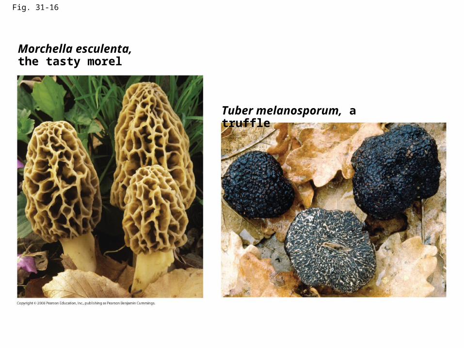
Fig. 31-16
Tuber melanosporum, a truffle
Morchella esculenta,the tasty morel
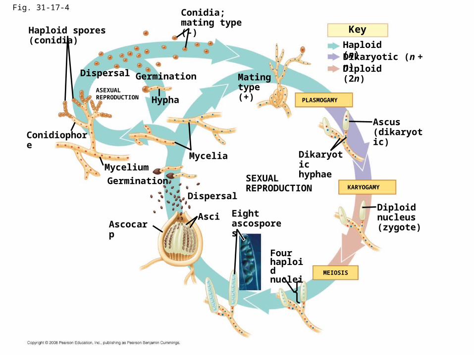
Fig. 31-17-4
Key
Haploid (n)
Diploid (2n)Dikaryotic (n + n)
Conidiophore
Mycelium
ASEXUALREPRODUCTION
Germination
Hypha PLASMOGAMY
Haploid spores (conidia)
Conidia;mating type (–)
Matingtype (+)
SEXUALREPRODUCTION
Dikaryotichyphae
Ascus(dikaryotic)
Mycelia
KARYOGAMY
Diploid nucleus(zygote)
Germination
Asci
Dispersal
Dispersal
AscocarpEightascospores
Fourhaploidnuclei MEIOSIS
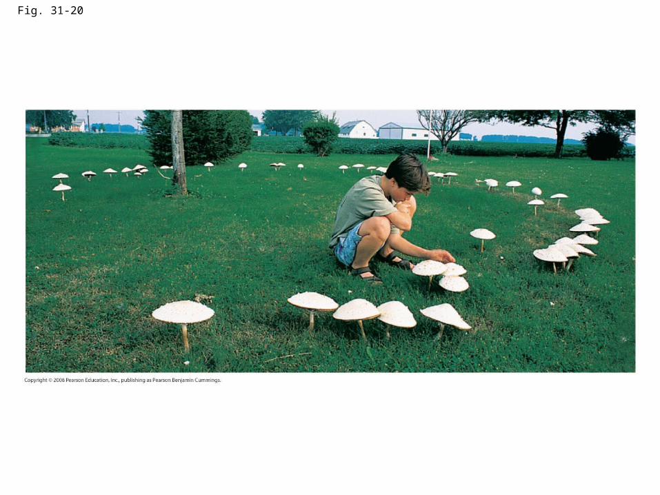
Fig. 31-20
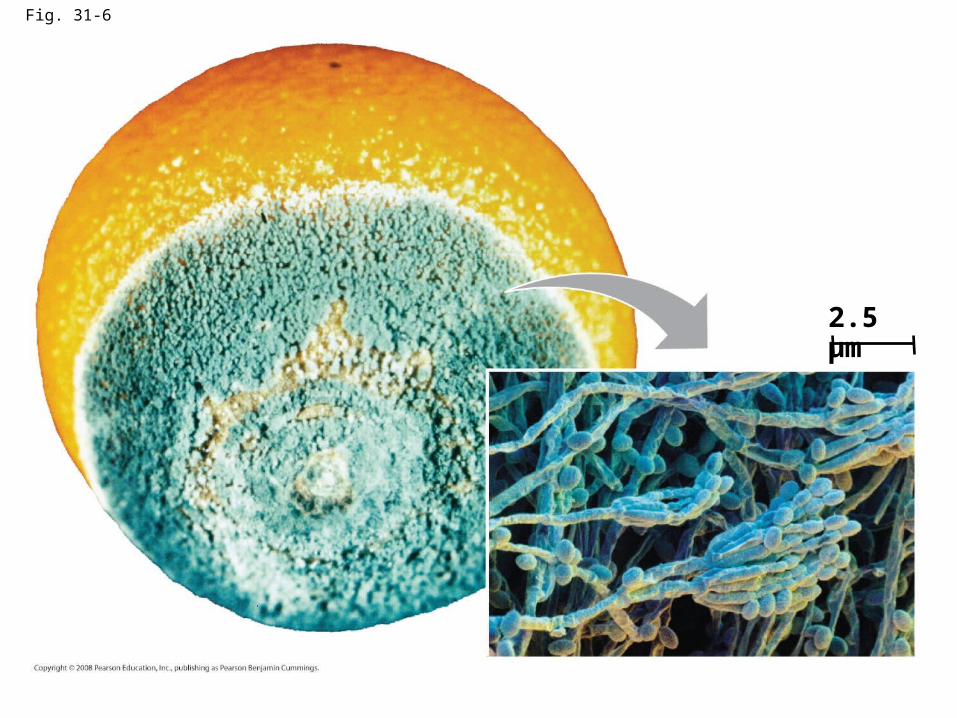
Fig. 31-6
2.5 µm

Fig. 31-26
Staphylococcus
Zone ofinhibitedgrowth
Penicillium

Fig. 31-7
10 µm
Parentcell
Bud
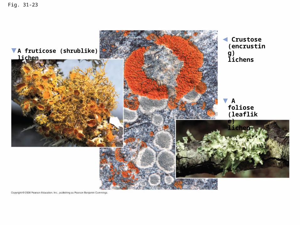
Fig. 31-23
A foliose(leaflike)lichen
A fruticose (shrublike) lichen
Crustose(encrusting)lichens
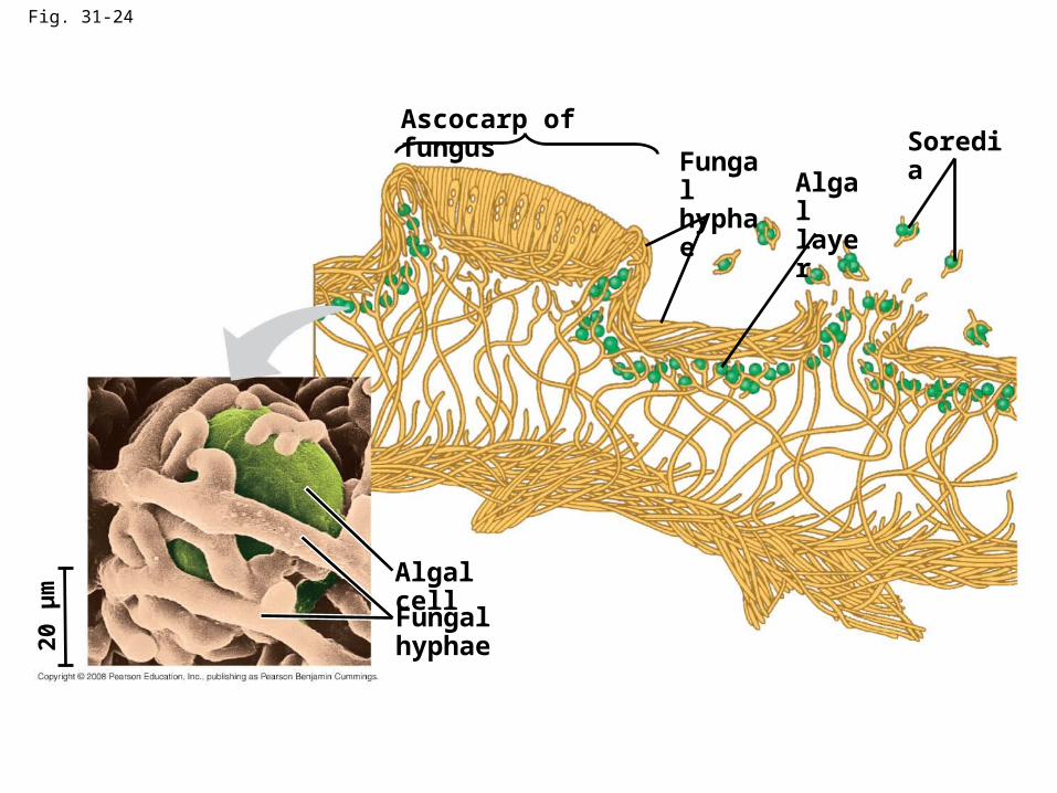
Fig. 31-24
Algal cell
Ascocarp of fungusSoredia
Fungal hyphae
Fungalhyphae Algal
layer
20 µ
m