Exercise Arrhythmias, Pt 2Tachyarrhythmias, Asystole, PEA, Pulseless
VT/VF• Atrial tachyarrhythmias• Ventricular tachyarrhythmias• Treatment of tachyarrhythmias
• Asystole• Pulseless Electrical Activity• Defibrillation
SVT: Case StudyN Eng J Med: 354:1039-51, 2006
• 28 yr-old women suddenly has rapid palpitations and chest pain while playing her cello
• In the emergency room, she has– HR 190– BP 82/54
• EKG shows regular tachycardia with a narrow QRS and no apparent P waves
Treatments
• Try cardiac sinus pressure or other vagalmaneuvers
• Try intravenous adenosine• If all fails, and tachycardia is recurrent and causes
symptoms, treatment may be catheter ablation to destroy an accessory pathway
Ablation Treatment
Causes of Tachycardias
• Supraventricular tachycardia • PACs• Atrial flutter/atrial fibrillation• Ventricular tachycardia• PVCs
Atrial Arrhythmias
• Tend to “go away” with vagal withdrawal at the start of exercise
• Re-appear during recovery• Occurs in 4-18% of patients
– 5 % in normals– 40% in CAD
• Reduces “atrial kick” to increase stroke volume

Premature Atrial Contractions
• Occur at low exercise intensity and have little clinical significance
PACs
What else doyou see here?
Where’s the PAC?
Atrial Flutter or Fibrillation
• Transient Atrial flutter or fibrillation occur frequently in patients
• Associated with– CAD– rheumatic heart disease– thyrotoxicosis– myocarditis– sometimes in normal people with no disease
Exercise Response with AtrialFlutter or Fibrillation
• Cardiac output is compromised– 5-30% lower stroke volume– elevated heart rates– greater incidence of ischemia (inadequate perfusion
time)• Atrial flutter rate 220-300• Atrial fibrillation, rate indeterminant
Fib or Flutter?
A
B
Paroxysmal SupraventricularTachycardia (PSVT or PAT)
• 2-3 beats of PAT or junctional tachycardia occasionally occur with exercise
• rate of ~160 to 220 • Not associated with increased mortality• Sustained PAT is rare• Sometimes, but not always associated with
ischemia with ST depression

Sustained PAT
Intermittent PAT
Premature Ventricular Contractions
• PVCs at Rest– controversy over significance– most agree that PVCs at rest are not significant in
healthy people– Patients with CAD who have PVCs have a “small”
increase in mortality– PVCs during recovery, usually are
not significant
Single PVC
PVC and compensatory pause
Exercise-Induced PVCs
• Caused by excess catecholamines and vagal withdrawal• May be caused by electrical re-entry and ectoptic beats• Occur in 36-42% of normal subjects during intense
exercise• Occur in 50-60% of CAD patients
and at lower HR• not significant, if asymptomatic
Ominous PVCs
• Multi-focal, multiform, repetitive• Moderate increase in mortality in
CAD patients
Bigeminy Trigeminy
Couplet

Exercise Guidelines and PVCs?
• Relative contra-indications to stop exercise– sustained VT (4 or more PVCs)– multi-focal PVCs– Triplets of PVCs
Non-Sustained Ventricular Tachycardia
• 4 or less = non-sustained• usually not a problem unless accompanied by
other signs or symptoms
Sustained VT
• Relatively rare• Usually portray serious underlying cardiac disease• Often deteriorates to VF
VT vs. V flutter
• VT rate is 140 to 250• VF > 250
Torsades de Pointes
Often related to hypoxia, electrolyte disturbances such as hypokalemia, or drugs
Tachycardia Algorithm
• Immediate assessment: stable or unstable?• Unstable= chest pain, shortness of breath, shock,
heart failure, pulmonary congestion
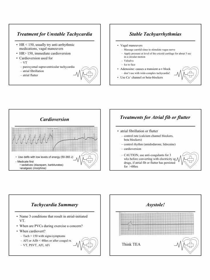
Treatment for Unstable Tachycardia
• HR < 150, usually try anti-arrhythmic medications, vagal maneuvers
• HR> 150, immediate cardioversion• Cardioversion used for
– VT– paroxysmal supraventricular tachycardia– atrial fibrillation– atrial flutter
Stable Tachyarrhythmias
• Vagal maneuvers– Massage carotid sinus to stimulate vagus nerve– Apply pressure at level of the cricoid cartilage for about 5 sec
in a circular motion– Valsalva– Ice to face
• Adenosine: causes a transient a-v block– don’t use with wide-complex tachycardia!
• Use Ca+ channel or beta-blockers
Cardioversion
• Use defib with low levels of energy (50-360 J)
• Medicate first• sedatives (diazepam, barbiturates)•analgesic (morphine)
Treatments for Atrial fib or flutter
• atrial fibrillation or flutter– control rate (calcium channel blockers,
beta blockers)– control rhythm (amidodarone, lidocaine)– cardioversion
– CAUTION, use anti-coagulants for 3 wks before converting with electricity or drugs, if atrial fib or flutter has persisted for >48hrs
Tachycardia Summary
• Name 3 conditions that result in atrial-initiated VT.
• When are PVCs during exercise a concern?• When cardiovert?
– Tach > 150 with signs/symptoms– AFl or Afib < 48hrs or after coagul rx– VT, PSVT, AFl, AFi
Asystole!
Think TEA

Asystole Algorithm
• Confirm non-responsiveness and asystole
Pulseless Electrical Activity
• Presence of some type of electrical activity but no detectable pulse
• VF/VT and PEA are “rhythms of survival” if– VF/VT--resuscitated with a defibrillator– PEA--cause is treated in time
• PEA treatment, think PEA– Problem, Epinephrine, Atropine
PEA Algorithm The 5 Hs
• Hypovolemia– volume infusion, vasoconstrictor
• Hypoxia– oxygen
• Hydrogen ion– bicarbonate infusion
• Hyper/hypokalemia• Hypothermia
The 5 Ts
• Tablets (antidepressants, beta blockers, ca channel blockers, digitalis)
• Tamponade• Tension Pneumothorax• Thrombosis, coronary• Thrombosis, pulmonary embolism
VF/VT
• Survivable rhythm if defibrillation is performed quickly
• Use CPR skills• Use AED or get defibrillator

Primary ABCs
Secondary ABCD
Conclusions
• When do you cardiovert, when do you use a defibrillator?
• What are the 5 Hs and the 5 Ts?• Name 2 times you would consider using
a pacer– bradycardia and asystole