Electron-transfer capacity of catechin derivatives andinfluence on the cell cycle and apoptosis in HT29 cellsCarles Lozano1,2, Lluıs Julia1, Aurora Jimenez1, Sonia Tourino1, Josep J. Centelles2,Marta Cascante2 and Josep Lluıs Torres1
1 Institute for Chemical and Environmental Research (IIQAB-CSIC), Barcelona, Spain
2 Department of Biochemistry and Molecular Biology, Associated Unit to CSIC, University of Barcelona, Spain
Polyphenols of plant origin are potent free-radical
scavengers [1,2] and are increasingly appreciated as
chemopreventive agents against conditions such as can-
cer and cardiovascular diseases [3,4]. They appear to
minimize the number of oxidative DNA mutations and
protein modifications by scavenging harmful reactive
oxygen species (ROS) [5]. Moreover, some polyphenols
of the flavonoid type show antiproliferative and pro-
apoptotic activities [6]. In particular, flavanols (cate-
chins) from tea, grape and other sources may exert
their beneficial action by a combination of prophylac-
tic and therapeutic effects related to both their radical-
scavenging capacity and their influence on the cell
machinery [7,8]. The gallate moiety appears to be
behind the influence of some catechins on the cell cycle
and the induction of apoptosis in tumour cells [9],
probably via enzyme–ligand interactions with some
key protein domains [10,11]. Another line of evidence
suggests that some catechins induce apoptosis via the
formation of the superoxide radical from molecular
oxygen by electron transfer [12]. The superoxide anion
may participate directly in the apoptotic toxic response
Keywords
apoptosis; catechins; electron transfer; free
radicals; gallate ester
Correspondence
J. L. Torres, Institute for Chemical and
Environmental Research (IIQAB-CSIC), Jordi
Girona 18–26, 08034 Barcelona, Spain
Fax: +34 93 204 5904
Tel: +34 93 400 6112
E-mail: [email protected]
(Received 30 January 2006, revised 27
March 2006, accepted 31 March 2006)
doi:10.1111/j.1742-4658.2006.05255.x
Galloylated and nongalloylated catechin conjugates with cysteine deriva-
tives have been synthesized and evaluated for their capacity to scavenge
free radicals and to influence crucial functions (cell cycle, apoptosis) in
HT29 colon carcinoma cells. We show that the nonphenolic part of the
molecule modified the capacity of catechins to donate hydrogen atoms and
to transfer electrons to free radicals. Nongalloylated derivatives did not sig-
nificantly influence either the cell cycle or apoptosis. Among the galloylated
species, 4b-[S-(O-ethyl-cysteinyl)]epicatechin 3-O-gallate, which showed a
high electron-transfer capacity (5 e– per molecule), arrested the cell cycle
and induced apoptosis as expected for galloylated catechins such as tea
(–)-epigallocatechin 3-O-gallate. 4b-[S-(N-Acetyl-O-methyl-cysteinyl)]epicate-
chin 3-O-gallate, which showed the highest hydrogen-donating capacity
(10 H per molecule) while keeping the electron-transfer capacity low
(2.9 e– per molecule), did not trigger any significant apoptosis. The gallate
moiety did not appear to be sufficient for the pro-apoptotic effect of the
catechin derivatives in HT29 cells. Instead, a high electron-transfer capacity
is more likely to be behind this effect. The use of stable radicals sensitive
exclusively to electron transfer may help to design molecules with either pre-
ventive scavenging action (high hydrogen donation, low electron transfer)
or therapeutic pro-apoptotic activity (high electron transfer).
Abbreviations
AMCys-Cat, 4b-[S-(N-acetyl-O-methyl-cysteinyl)]catechin; AMCys-Ec, 4b-[S-(N-acetyl-O-methyl-cysteinyl)]epicatechin; AMCys-EcG, 4b-
[S-(N-acetyl-O-methyl-cysteinyl)]epicatechin 3-O-gallate; ARP, antiradical power; Cys-Ec, 4b-(S-cysteinyl)epicatechin; DPPH, 1,1-diphenyl-
2-picrylhydrazyl free radical; Ec, (–)-epicatechin; ECys-Cat, 4b-[S-(O-ethyl-cysteinyl)]catechin; ECys-Ec, 4b-[S-(O-ethyl-cysteinyl)]epicatechin;
ECys-EcG, 4b-[S-(O-ethyl-cysteinyl)]epicatechin 3-O-gallate; EgcG, (–)-epigallocatechin 3-O-gallate; HNTTM, tris(2,4,6-trichloro-3,5-
dinitrophenyl)methyl radical; PI, propidium iodide; ROS, reactive oxygen species.
FEBS Journal 273 (2006) 2475–2486 ª 2006 The Authors Journal compilation ª 2006 FEBS 2475
or be involved in the regulation of apoptotic pathways
[13]. Pro-apoptotic tea (–)-epigallocatechin 3-O-gallate
(EgcG) which includes two trihydroxybenzene moieties
(ring B and gallate ester) appears to be a particularly
efficient reducing (electron donating) agent [14].
Catechin conjugates with thiols have been described
[15–17]. The derivatives are obtained by acid depolym-
erization of grape polymeric procyanidins in the pres-
ence of the thiol and show higher antiradical capacity
than their underivatized counterparts in the 1,1-diphe-
nyl-2-picrylhydrazyl free radical (DPPH) assay [16,17].
Interestingly, the nonphenolic part of the molecule
appears to influence the capacity of the conjugates to
penetrate biological membranes, particularly the skin
layers [18]. We present evidence that these nonphenolic
moieties may also modulate the redox behaviour of
molecules and their capacity to induce apoptosis by a
mechanism involving electron transfer, whereas the
gallate moiety may be a necessary, but not sufficient,
condition to explain the pro-apoptotic effect.
Results
Synthesis and purification
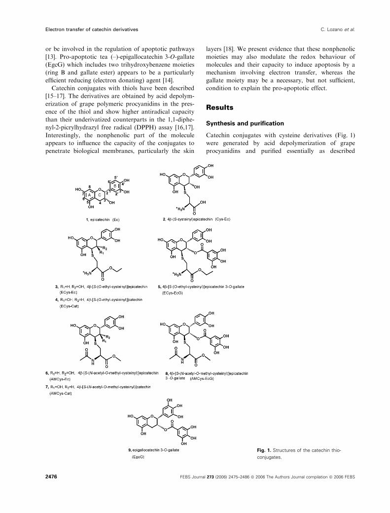
Catechin conjugates with cysteine derivatives (Fig. 1)
were generated by acid depolymerization of grape
procyanidins and purified essentially as described
Fig. 1. Structures of the catechin thio-
conjugates.
Electron transfer of catechin derivatives C. Lozano et al.
2476 FEBS Journal 273 (2006) 2475–2486 ª 2006 The Authors Journal compilation ª 2006 FEBS
previously [17]. 4b-[S-(O-Ethyl-cysteinyl)]epicatechin
(ECys-Ec), 4b-[S-(O-ethyl-cysteinyl)]catechin (ECys-
Cat) and 4b-[S-(O-ethyl-cysteinyl)]epicatechin 3-O-gal-
late (ECys-EcG) (3–5) were obtained from the ethyl
ester of cysteine and separated from the crude depo-
lymerization mixture using a strong cation-exchange
resin (MacroPrepTM High S 50 lm) by taking advant-
age of the free amino function on the cysteinyl
moiety. 4b-[S-(N-Acetyl-O-methyl-cysteinyl)]epicatechin
(AMCys-Ec), 4b-[S-(N-acetyl-O-methyl-cysteinyl)]cate-
chin (AMCys-Cat) and 4b-[S-(N-acetyl-O-methyl-
cysteinyl)]epicatechin 3-O-gallate (AMCys-EcG) (6–8)
were obtained from N-acetyl-cysteine. Under the depo-
limerization conditions (60 �C, HCl, methanol, 15 min)
the methyl ester was readily obtained from the free
carboxylic acid.
The stereochemistry at C-2, C-3 and C-4 of com-
pounds 3–8 was assigned from the hydrogen-coupling
constants measured using 1H NMR and following
Thompson et al. [19]. In agreement with the literature,
the 4b derivatives were the major isomers obtained
irrespective of the 2,3-stereochemistry [19,20].
Free-radical scavenging activity
The new cysteinyl catechin derivatives were potent
free-radical scavenging agents in the DPPH assay and
moderate scavengers in the tris(2,4,6-trichloro-3,5-dini-
trophenyl)methyl radical (HNTTM) assay. DPPH is a
widely used stable free radical that is converted to the
reduced form by incorporating a hydrogen atom via a
mechanism that may involve direct hydrogen donation
and ⁄or electron transfer with subsequent proton incor-
poration [21,22]. HNTTM is a newly introduced stable
radical that is exclusively quenched by electron transfer
to give a stable anion with subsequent slow proton
incorporation [23]. By comparing the results from both
assays, information is obtained about the differential
capacity of a given molecule to donate hydrogen atoms
and transfer electrons.
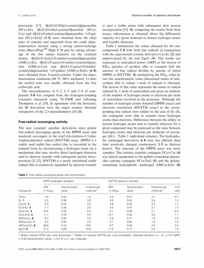
Table 1 summarizes the values obtained for the new
compounds 3–8 with both free radicals in comparison
with the unprotected cysteine derivative Cys-Ec (2) and
underivatized Ec (1) and EgcG (9). The results are
expressed as antiradical power (ARP) or the inverse of
ED50 (lmoles of product able to consume half the
amount of free radical divided by lmoles of initial
DPPH or HNTTM). By multiplying the ED50 value by
two the stoichiometric value (theoretical moles of anti-
oxidant able to reduce 1 mole of radical) is obtained.
The inverse of this value represents the moles of radical
reduced by 1 mole of antioxidant and gives an estimate
of the number of hydrogen atoms or electrons per mole
of antioxidant involved in the processes. Although the
number of hydrogen atoms donated (DPPH assay) and
electrons transferred (HNTTM assay) to the corres-
ponding free radical were similar in the case of Ec (1),
the conjugates were able to transfer more hydrogen
atoms than electrons. Differences between the ability to
donate hydrogen atoms and to transfer electrons for a
given compound may be expressed as the ratio between
hydrogen atoms and electrons per molecule of scaven-
ger (H ⁄ e–, Table 1 right-hand column). The hydropho-
bic uncharged derivatives 6–8 were less efficient than
their positively charged counterparts 3–5 as electron
donors. The outcome of the DPPH assay was more
complex. The cationic catechin conjugate ECys-Cat (4)
was almost equipotent to the gallate-containing epicate-
chin cationic conjugate ECys-EcG (5) and the gallate-
containing hydrophobic uncharged AMCys-EcG (8)
Table 1. Free radical scavenging power and stoichiometry.
Compound
DPPH (hydrogen donation) HNTTM (electron transfer)
ARP
(1 ⁄ ED50)
Stoichiometric
value
H atoms per
moleculea
ARP
(1 ⁄ ED50)
Stoichiometric
value
Electrons per
moleculeb
H ⁄ e–
ratio
Trolox 3.9 0.52 1.9 5.4 0.37 2.7 0.7
Ec, 1 5.5 0.36 2.8 4.8 0.42 2.4 1.2
Cys-Ec, 2 8.3 0.24 4.2 4.8 0.42 2.4 1.7
ECys-Ec, 3 7.1 0.28 3.6 4.3 0.47 2.1 1.7
ECys-Cat, 4 10.0 0.20 5.0 4.1 0.48 2.1 2.4
ECys-EcG, 5 11.1 0.18 5.6 10.1 0.20 5.0 1.1
AMCys-Ec, 6 6.7 0.30 3.3 2.8 0.71 1.4 2.3
AMCys-Cat, 7 5.0 0.40 2.5 3.1 0.64 1.6 1.6
AMCys-EcG, 8 20.0 0.10 10.0 5.7 0.35 2.9 3.4
EgcG, 9 21.3 0.09 10.6 11.3 0.17 5.9 1.8
a Moles reduced DPPH per mole antioxidant. b Moles of reduced HNTTM per mole antioxidant. Standard deviation (n ¼ 3): 6 0.3 (ARP),
6 0.04 (stoichiometric value), 6 0.2 (H or e– per molecule).
C. Lozano et al. Electron transfer of catechin derivatives
FEBS Journal 273 (2006) 2475–2486 ª 2006 The Authors Journal compilation ª 2006 FEBS 2477
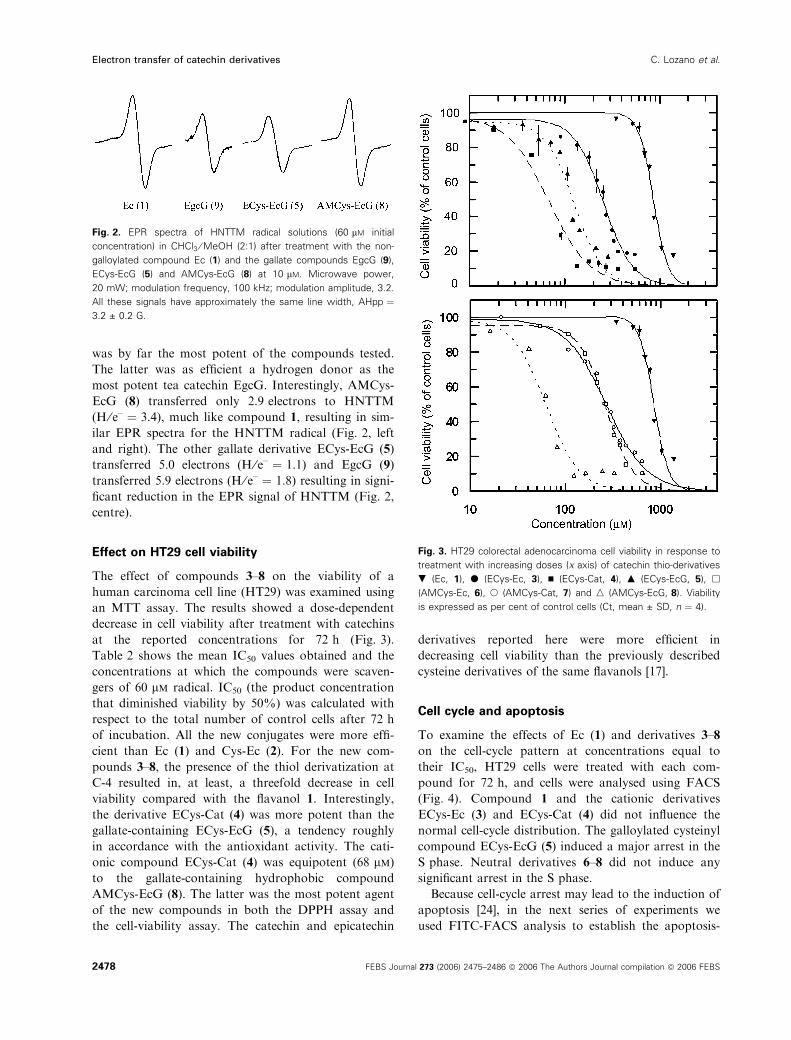
was by far the most potent of the compounds tested.
The latter was as efficient a hydrogen donor as the
most potent tea catechin EgcG. Interestingly, AMCys-
EcG (8) transferred only 2.9 electrons to HNTTM
(H ⁄ e– ¼ 3.4), much like compound 1, resulting in sim-
ilar EPR spectra for the HNTTM radical (Fig. 2, left
and right). The other gallate derivative ECys-EcG (5)
transferred 5.0 electrons (H ⁄ e– ¼ 1.1) and EgcG (9)
transferred 5.9 electrons (H ⁄ e– ¼ 1.8) resulting in signi-
ficant reduction in the EPR signal of HNTTM (Fig. 2,
centre).
Effect on HT29 cell viability
The effect of compounds 3–8 on the viability of a
human carcinoma cell line (HT29) was examined using
an MTT assay. The results showed a dose-dependent
decrease in cell viability after treatment with catechins
at the reported concentrations for 72 h (Fig. 3).
Table 2 shows the mean IC50 values obtained and the
concentrations at which the compounds were scaven-
gers of 60 lm radical. IC50 (the product concentration
that diminished viability by 50%) was calculated with
respect to the total number of control cells after 72 h
of incubation. All the new conjugates were more effi-
cient than Ec (1) and Cys-Ec (2). For the new com-
pounds 3–8, the presence of the thiol derivatization at
C-4 resulted in, at least, a threefold decrease in cell
viability compared with the flavanol 1. Interestingly,
the derivative ECys-Cat (4) was more potent than the
gallate-containing ECys-EcG (5), a tendency roughly
in accordance with the antioxidant activity. The cati-
onic compound ECys-Cat (4) was equipotent (68 lm)to the gallate-containing hydrophobic compound
AMCys-EcG (8). The latter was the most potent agent
of the new compounds in both the DPPH assay and
the cell-viability assay. The catechin and epicatechin
derivatives reported here were more efficient in
decreasing cell viability than the previously described
cysteine derivatives of the same flavanols [17].
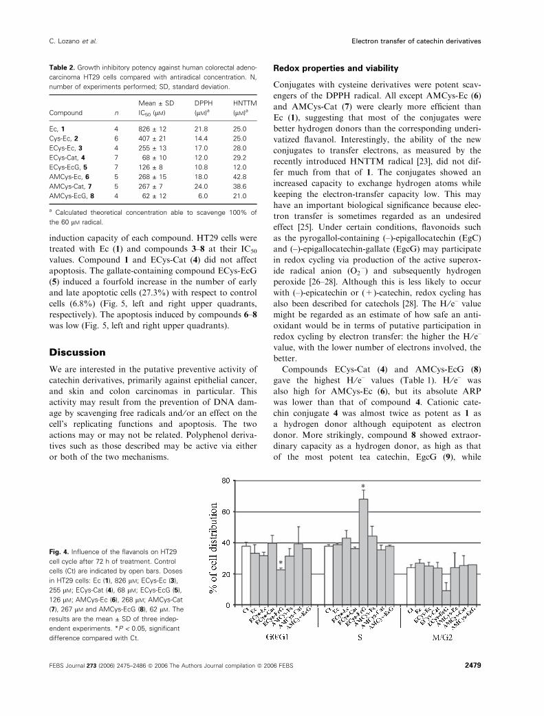
Cell cycle and apoptosis
To examine the effects of Ec (1) and derivatives 3–8
on the cell-cycle pattern at concentrations equal to
their IC50, HT29 cells were treated with each com-
pound for 72 h, and cells were analysed using FACS
(Fig. 4). Compound 1 and the cationic derivatives
ECys-Ec (3) and ECys-Cat (4) did not influence the
normal cell-cycle distribution. The galloylated cysteinyl
compound ECys-EcG (5) induced a major arrest in the
S phase. Neutral derivatives 6–8 did not induce any
significant arrest in the S phase.
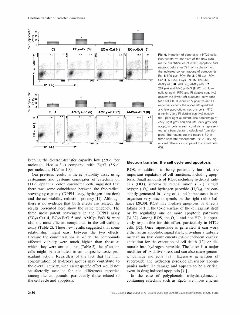
Because cell-cycle arrest may lead to the induction of
apoptosis [24], in the next series of experiments we
used FITC-FACS analysis to establish the apoptosis-
Fig. 2. EPR spectra of HNTTM radical solutions (60 lM initial
concentration) in CHCl3 ⁄MeOH (2:1) after treatment with the non-
galloylated compound Ec (1) and the gallate compounds EgcG (9),
ECys-EcG (5) and AMCys-EcG (8) at 10 lM. Microwave power,
20 mW; modulation frequency, 100 kHz; modulation amplitude, 3.2.
All these signals have approximately the same line width, AHpp ¼3.2 ± 0.2 G.
Fig. 3. HT29 colorectal adenocarcinoma cell viability in response to
treatment with increasing doses (x axis) of catechin thio-derivatives
. (Ec, 1), d (ECys-Ec, 3), n (ECys-Cat, 4), m (ECys-EcG, 5), h
(AMCys-Ec, 6), s (AMCys-Cat, 7) and n (AMCys-EcG, 8). Viability
is expressed as per cent of control cells (Ct, mean ± SD, n ¼ 4).
Electron transfer of catechin derivatives C. Lozano et al.
2478 FEBS Journal 273 (2006) 2475–2486 ª 2006 The Authors Journal compilation ª 2006 FEBS
induction capacity of each compound. HT29 cells were
treated with Ec (1) and compounds 3–8 at their IC50
values. Compound 1 and ECys-Cat (4) did not affect
apoptosis. The gallate-containing compound ECys-EcG
(5) induced a fourfold increase in the number of early
and late apoptotic cells (27.3%) with respect to control
cells (6.8%) (Fig. 5, left and right upper quadrants,
respectively). The apoptosis induced by compounds 6–8
was low (Fig. 5, left and right upper quadrants).
Discussion
We are interested in the putative preventive activity of
catechin derivatives, primarily against epithelial cancer,
and skin and colon carcinomas in particular. This
activity may result from the prevention of DNA dam-
age by scavenging free radicals and ⁄or an effect on the
cell’s replicating functions and apoptosis. The two
actions may or may not be related. Polyphenol deriva-
tives such as those described may be active via either
or both of the two mechanisms.
Redox properties and viability
Conjugates with cysteine derivatives were potent scav-
engers of the DPPH radical. All except AMCys-Ec (6)
and AMCys-Cat (7) were clearly more efficient than
Ec (1), suggesting that most of the conjugates were
better hydrogen donors than the corresponding underi-
vatized flavanol. Interestingly, the ability of the new
conjugates to transfer electrons, as measured by the
recently introduced HNTTM radical [23], did not dif-
fer much from that of 1. The conjugates showed an
increased capacity to exchange hydrogen atoms while
keeping the electron-transfer capacity low. This may
have an important biological significance because elec-
tron transfer is sometimes regarded as an undesired
effect [25]. Under certain conditions, flavonoids such
as the pyrogallol-containing (–)-epigallocatechin (EgC)
and (–)-epigallocatechin-gallate (EgcG) may participate
in redox cycling via production of the active superox-
ide radical anion (O2Æ–) and subsequently hydrogen
peroxide [26–28]. Although this is less likely to occur
with (–)-epicatechin or (+)-catechin, redox cycling has
also been described for catechols [28]. The H ⁄ e– value
might be regarded as an estimate of how safe an anti-
oxidant would be in terms of putative participation in
redox cycling by electron transfer: the higher the H ⁄ e–
value, with the lower number of electrons involved, the
better.
Compounds ECys-Cat (4) and AMCys-EcG (8)
gave the highest H ⁄ e– values (Table 1). H ⁄ e– was
also high for AMCys-Ec (6), but its absolute ARP
was lower than that of compound 4. Cationic cate-
chin conjugate 4 was almost twice as potent as 1 as
a hydrogen donor although equipotent as electron
donor. More strikingly, compound 8 showed extraor-
dinary capacity as a hydrogen donor, as high as that
of the most potent tea catechin, EgcG (9), while
Table 2. Growth inhibitory potency against human colorectal adeno-
carcinoma HT29 cells compared with antiradical concentration. N,
number of experiments performed; SD, standard deviation.
Compound n
Mean ± SD
IC50 (lM)
DPPH
(lM)aHNTTM
(lM)a
Ec, 1 4 826 ± 12 21.8 25.0
Cys-Ec, 2 6 407 ± 21 14.4 25.0
ECys-Ec, 3 4 255 ± 13 17.0 28.0
ECys-Cat, 4 7 68 ± 10 12.0 29.2
ECys-EcG, 5 7 126 ± 8 10.8 12.0
AMCys-Ec, 6 5 268 ± 15 18.0 42.8
AMCys-Cat, 7 5 267 ± 7 24.0 38.6
AMCys-EcG, 8 4 62 ± 12 6.0 21.0
a Calculated theoretical concentration able to scavenge 100% of
the 60 lM radical.
Fig. 4. Influence of the flavanols on HT29
cell cycle after 72 h of treatment. Control
cells (Ct) are indicated by open bars. Doses
in HT29 cells: Ec (1), 826 lM; ECys-Ec (3),
255 lM; ECys-Cat (4), 68 lM; ECys-EcG (5),
126 lM; AMCys-Ec (6), 268 lM; AMCys-Cat
(7), 267 lM and AMCys-EcG (8), 62 lM. The
results are the mean ± SD of three indep-
endent experiments. *P < 0.05, significant
difference compared with Ct.
C. Lozano et al. Electron transfer of catechin derivatives
FEBS Journal 273 (2006) 2475–2486 ª 2006 The Authors Journal compilation ª 2006 FEBS 2479
keeping the electron-transfer capacity low (2.9 e– per
molecule, H ⁄ e– ¼ 3.4) compared with EgcG (5.9 e–
per molecule, H ⁄ e– ¼ 1.8).
Our previous results in the cell-viability assay using
cysteamine and cysteine conjugates of catechins on
HT29 epithelial colon carcinoma cells suggested that
there was some coincidence between the free-radical
scavenging capacity (DPPH assay, hydrogen donation)
and the cell viability reduction potency [17]. Although
there is no evidence that both effects are related, the
results presented here show the same tendency. The
three most potent scavengers in the DPPH assay
(ECys-Cat 4, ECys-EcG 5 and AMCys-EcG 8) were
also the most efficient compounds in the cell-viability
assay (Table 2). These new results suggested that some
relationship might exist between the two effects.
Because the concentrations at which the compounds
affected viability were much higher than those at
which they were antioxidants (Table 2) the effect on
cells might be attributed to an unspecific toxic pro-
oxidant action. Regardless of the fact that the high
concentration of hydroxyl groups may contribute to
the overall activity, such an unspecific effect would not
satisfactorily account for the differences recorded
among the compounds, particularly those related to
the cell cycle and apoptosis.
Electron transfer, the cell cycle and apoptosis
ROS, in addition to being potentially harmful, are
important regulators of cell functions, including apop-
tosis. Small amounts of ROS, including hydroxyl radi-
cals (HOÆ), superoxide radical anion (O2Æ–), singlet
oxygen (1O2) and hydrogen peroxide (H2O2), are con-
stantly generated in living cells and homeostasis in an
organism very much depends on the right redox bal-
ance [29,30]. ROS may mediate apoptosis by directly
taking part in the toxic warfare of the cell against itself
or by regulating one or more apoptotic pathways
[31,32]. Among ROS, the O2Æ–, and not HOÆ, is appar-
ently responsible for this effect, particularly in HT29
cells [32]. Once superoxide is generated it can work
either as an apoptotic signal itself, providing a fail-safe
mechanism that complements cyt-c-dependent caspase
activation for the execution of cell death [13], or dis-
mutate into hydrogen peroxide. The latter is a major
mediator of oxidative stress and can also cause genom-
ic damage indirectly [33]. Excessive generation of
superoxide and hydrogen peroxide invariably accom-
panies molecular damage and appears to be a critical
event in drug-induced apoptosis [31].
In the case of polyphenols, trihydroxybenzene-
containing catechins such as EgcG are more efficient
Fig. 5. Induction of apoptosis in HT29 cells.
Representative dot plots of the flow cyto-
metric quantification of intact, apoptotic and
necrotic cells after 72 h of incubation with
the indicated concentrations of compounds
Ec (1, 826 lM), ECys-Ec (3, 255 lM), ECys-
Cat (4, 68 lM), ECys-EcG (5, 126 lM),
AMCys-Ec (6, 268 lM), AMCys-Cat (7,
267 lM) and AMCys-EcG (8, 62 lM). Live
cells (annexin-FITC and PI double negative)
occupy the lower left quadrant, early apop-
totic cells (FITC-annexin V positive and PI
negative) occupy the upper left quadrant
and late apoptotic or necrotic cells (FITC-
annexin V and PI double positive) occupy
the upper right quadrant. The percentage of
early (light grey bar) and late (dark grey bar)
apoptotic cells in each condition is represen-
ted as a bars diagram, calculated from dot
plots. The results are the mean ± SD of
three separate experiments. *P < 0.05, sig-
nificant difference compared to control cells
(Ct).
Electron transfer of catechin derivatives C. Lozano et al.
2480 FEBS Journal 273 (2006) 2475–2486 ª 2006 The Authors Journal compilation ª 2006 FEBS
producers of the superoxide radical anion than cate-
chol-containing catechins such as Ec (1) [14,29,34], and
they inhibit cancer cell growth via cell-cycle arrest and
apoptosis induction by mechanisms involving the gal-
late moiety [12,35–37]. In agreement with the litera-
ture, our results show that the gallate-containing
compound ECys-EcG (5) induced apoptosis (threefold
compared with control cells, see Fig. 5) and triggered a
significant arrest in the S phase of the cell cycle
(Fig. 4). Several authors have reported that the gallate
group decreases cyclin-dependent kinase 2 (Cdk2)
expression and activity, and upregulates the expression
of p21, a Cdk inhibitor [38–40] essential for progres-
sion from the G1 to the S phase of the cell cycle.
Alternatively, the catechin gallates, acting as pro-oxi-
dants, may damage the DNA directly through ROS
[41]. All these events lead to stoppage of the cell-cycle
progression at the S phase and may potentially result
in inhibition of proliferation, cytostasis and possibly
apoptosis in human tumours. It seems increasingly evi-
dent that the duality antioxidant ⁄pro-oxidant of cate-
chins may play a crucial role in their interactions with
the cell machinery, most probably via formation of the
superoxide anion radical by electron transfer.
ECys-EcG (5), together with EgcG (9), possessed a
high electron-transfer capacity (five and six electrons
per molecule, respectively). Compounds 1–4 and 6, 7,
all lacking the gallate moiety and showing low elec-
tron-transfer capacity, did not induce any significant
cell-cycle arrest or apoptosis. Surprisingly, the gallate-
containing compound AMCys-EcG (8) did not induce
cell-cycle arrest or apoptosis. Curiously, the electron-
transfer capacity of 8 (2.9 electrons per molecule) as
measured by the stable radical HNTTM was lower
than expected, similar to those of the nongalloylated
species (approximately 2 electrons per molecule, see
Fig. 2). This finding appears to corroborate the obser-
vation from other authors that links the induction of
apoptosis to the electron-transfer capacity and forma-
tion of the superoxide radical anion. The presence of
the gallate moiety does not appear to be a sufficient
condition for the induction of apoptosis in HT29 cells.
Whether cell-cycle arrest and apoptosis are due to
redox cycling with the production of hydrogen perox-
ide (pro-oxidant effect) or other ROS-mediated events
is something that must be explored further.
Conclusions
The conjugation of catechins with both cationic and
neutral cysteine derivatives produced compounds with
an improved capacity to donate hydrogen atoms while
keeping their capacity to participate in electron-trans-
fer reactions low. One of the new molecules, AMCys-
EcG (8), the most efficient DPPH scavenger of the
flavanol thio-conjugates described to date, was the
most effective derivative against colon carcinoma cell
viability. Despite including a gallate moiety, this com-
pound showed a low electron-transfer capacity and
neither arrested the cell cycle nor induced apoptosis.
This result, together with the observation that pro-
apoptotic ECys-EcG (5) and EgcG (9) possessed
higher electron-transfer capacity is suggesting that the
gallate moiety may not be a sufficient condition to
trigger apoptosis, which would be more directly related
to the ability of the flavanol derivatives to transfer
electrons. Our newly introduced scavenging assay using
the stable radical HNTTM, which is exclusively sensi-
tive to electron transfer, may be a valuable tool for
predicting the pro-apoptotic activity of polyphenols
and other putative drugs. The electron-transfer capa-
city of exogenous plant phenolics and its influence on
the delicate balance between the antioxidant and pro-
oxidant events governing cell functions may help to
explain the putative cancer-preventive properties of
catechins and their derivatives.
Experimental procedures
Materials
Analytical grade methanol (MeOH, Panreac, Montcada i
Reixac, Spain) was used for the acid cleavage reaction and
DPPH assay, deionized water and bulk ethanol (EtOH,
Momplet y Esteban, Barcelona, Spain) for semipreparative
and preparative cation-exchange chromatography, Milli-Q�
water and HPLC grade acetonitrile (CH3CN, Merck,
Darmstadt, Germany) for analytical RP-HPLC, and
deionized water and preparative grade CH3CN (Scharlau,
Barcelona, Spain) for preparative and semipreparative
RP-HPLC. Deuterated solvents for NMR were from SDS
(Peypin, France). Cysteine hydrochloride, l-cysteine ethyl
ester hydrochloride and N-acetyl-l-cysteine (Aldrich, Stein-
heim, Germany) were of synthesis grade. (–)-Epicatechin
(Ec, 1) (–)-epigallocatechin 3-O-gallate (EgcG, 9), MTT,
dimethylsulfoxide, Trypan Blue solution 0.4%, propidium
iodide (PI), the nonionic surfactant Igepal CA-630, a,a,a-tris(hydroxymethyl)aminomethane and NaCl ⁄Pi were from
Sigma (Steinheim, Germany). Acetic acid, 37% HCl
(Merck) and NaCl (Carlo Erba, Milan, Italy) were of ana-
lytical grade. Triethylamine (Merck) was of buffer grade.
Trifluoroacetic acid (Fluorochem, Glossop, UK) biotech
grade was distilled in-house. DPPH (95%) was from Ald-
rich (Gillingham, UK), 6-hydroxy-2,5,7,8-tetramethyl-chro-
man-2-carboxylic acid (Trolox) (97%) was from Aldrich
(Milwaukee, MN). Fetal bovine serum was purchased from
Gibco (Invitrogen, Carlsbad, CA). Trypsin–EDTA solution
C. Lozano et al. Electron transfer of catechin derivatives
FEBS Journal 273 (2006) 2475–2486 ª 2006 The Authors Journal compilation ª 2006 FEBS 2481
C (0.05% trypsin and EDTA 1:5000 in NaCl ⁄Pi) was from
Biological Industries (Beit Haemek, Israel). RNase was
from Roche Diagnostics (Mannheim, Germany). FITC–
annexin V kit and binding buffer 4· for apoptosis assay
were purchased from Bender MedSystems (MedSystems
Diagnostics GmbH, Vienna, Austria).
Chromatographic equipment and columns
Analytical RP-HPLC was performed on a Kontron Analyt-
ical system (Kontron Instruments, Basel, Switzerland) fitted
with a VYDACTM (The Separations Group, Hesperia,
USA) C18, 300 A pore size, 5 lm particle size,
250 · 4.6 mm i.d. column. Cation-exchange chromatogra-
phy was performed on a flash chromatography-type glass
column (21 · 2.5 cm i.d., � 105 mL bed volume) packed
in-house with MacroPrepTM High S 50 lm (Bio-Rad
Laboratories, Hercules, CA). Preparative RP-HPLC chro-
matography was performed on a Waters (Milford, USA)
Prep LC 4000 pumping system with a Waters PrepPack�
1000 module fitted with a PrepPack� Waters cartridge
(30 · 4.7 cm i.d) filled with VYDACTM (The Separations
Group) C18, 300 A pore size, 15–20 lm particle size station-
ary phase. Detection was carried out using an analytical
Merck-Hitachi (Darmstadt, Germany) L-4000 UV detector.
MS, NMR and EPR measurements
ES-MS analyses were recorded on a VG-Quattro� system
from Fisons Instruments (Altrincham, UK). The carrier
solution was Milli-Q water ⁄CH3CN (1:1) containing 1%
(v ⁄ v) formic acid. 1H NMR spectra were acquired on a
Varian (Palo Alto, CA) Unity 300 spectrometer in the deu-
terated solvents (CD3)2CO and D2O.
EPR measurements were performed on a Varian E-109
spectrometer working in the X-band (microwave power,
20 mW; modulation amplitude, 3.2 G).
Preparation of the conjugates
Conjugates were obtained by acid depolymerization of
plant procyanidins essentially as described previously [16].
To obtain the thio-conjugates 3–8 (Fig. 1) the solvent was
eliminated from an aqueous fraction (400 mL, 6 g estima-
ted polyphenols by mass, from 3.2 kg of grape byproduct)
of polymeric procyanidins. The pellet was then dissolved in
MeOH (400 mL) and dried. The resulting syrupy residue
was dissolved in MeOH (400 mL) and a solution of the
appropriate cysteine derivative (20 g) and 37% HCl
(10 mL) in MeOH (400 mL) was added. The mixture was
kept at 65 �C for 20 min under stirring. The reaction was
then quenched with cold water (3.2 L).
Conjugates were separated from the whole mixture using
the MacroPrepTM High S resin. The eluents were: (A)
20 mm sodium phosphate, pH 2.3 buffer ⁄EtOH (13:7, v ⁄ v)and (B) 20 mm sodium phosphate, pH 2.3 buffer ⁄EtOH
(3:2, v ⁄ v), 100 mm NaCl. The column was equilibrated with
eluent (A), loaded with the quenched depolymerized mix-
ture (500 mL) and washed with (A) (500 mL, 4.75 bed vol-
umes). The retained catechin derivatives were released with
500 mL (4.75 bed volumes) of eluent (B). The operation
was repeated until the whole mixture was consumed. The
separation process was monitored by analytical RP-HPLC
on a VYDACTM C18 column eluted with a binary system:
(C) 0.10% (v ⁄ v) aqueous trifluoroacetic acid, (D) 0.09%
(v ⁄ v) trifluoroacetic acid in water ⁄CH3CN (1:4, v ⁄ v) underisocratic conditions 19% (D) at a flow rate of 1.5 mLÆmin)1
and detection at 214 nm. The eluates containing the corres-
ponding conjugate were pooled (3.5 L).
The mixture containing the O-ethyl-cysteinyl conjugates
3–5 was fractionated on a preparative RP-HPLC cartridge
filled with VYDACTM C18 stationary phase by a CH3CN
gradient in 0.10% (v ⁄ v) aqueous trifluoroacetic acid (4–20%
CH3CN over 45 min). Fractions enriched in each of the three
compounds were obtained: fraction I, 9–11% CH3CN, com-
pound ECys-Cat (4); fraction II, 12–16% CH3CN, com-
pound ECys-Ec (3); fraction III, 17–19% CH3CN,
compound ECys-EcG (5).
4b-[S-(O-ethyl-cysteinyl)]epicatechin (ECys-Ec, 3)
Fraction II from reversed-phase fractionation was concen-
trated, loaded onto the preparative cartridge and the target
compound purified by CH3CN gradient in triethylamine
phosphate buffer and aqueous trifluoroacetic acid. Analysis
of the fractions was accomplished under isocratic condi-
tions in 0.10% (v ⁄ v) aqueous trifluoroacetic acid ⁄CH3CN
using the VYDACTM C18 column, solvent system, flow rate
and detection as described above. ECys-Ec (3) (354 mg)
was obtained as the trifluoroacetate by lyophilization.
dH(300 MHz; (CD3)2CO +3 drops D2O) 1.24 (3 H, t, J
7.2 Hz, O-CH2-CH3), 3.93 (1 H, d, J3,4 2.1 Hz, 4-H 3,4-
trans configuration), 4.06 (1 H, dd, J 2.4 and 0.9 Hz, 3-H),
4.26 (2 H, q, J 7.2 and 1.5 Hz, O-CH2-CH3), 4.71 (1 H, m,
S-CH2-CH <), 5.09 (1 H, s, 2-H 2,3-cis configuration),
5.90 (1 H, d, J 2.4 Hz, 8-H), 6.09 (1 H, d, J 2.4 Hz, 6-H),
6.80–6.81 (2 H, m, 5¢-H, 6¢-H), 7.04 (1 H, d, J 1.8 Hz,
2¢-H). m ⁄ z 438.1 (M + 1)+, calculated for C20H24N1O8S1(M + H)+ 438.1. Purity (> 95%) was ascertained by
RP-HPLC.
4b-[S-(O-ethyl-cysteinyl)]catechin (ECys-Cat, 4)
Fraction I from reversed-phase fractionation was concen-
trated, loaded onto the preparative cartridge, purified and
characterized as stated for compound 3. ECys-Cat (4)
(68 mg) was obtained as the trifluoroacetate. dH(300 MHz;
(CD3)2CO +3 drops D2O) 1.24 (3 H, t, J 7.0 Hz, O-CH2-
Electron transfer of catechin derivatives C. Lozano et al.
2482 FEBS Journal 273 (2006) 2475–2486 ª 2006 The Authors Journal compilation ª 2006 FEBS
CH3), 4.06 (1 H, 2d, J2,3 9.6 and 2.4 Hz, 3-H 2,3-trans con-
figuration), 4.23 (1 H, d, J3,4 2.4 Hz, 4-H 3,4-cis configur-
ation), 4.26 (2 H, q, J 7.0 and 2.4 Hz, O-CH2-CH3), 4.68–
4.72 (1 H, m, S-CH2-CH <), 4,78 (1 H, d, J 8.6 Hz; 2-H),
5.89 (1 H, d, J 2.4 Hz, 8-H), 6.10 (1 H, d, J 2.4 Hz, 6-H),
6.62 (2 H, m, 5¢-H, 6¢-H), 6.91 (1 H, s, 2¢-H). m ⁄ z 438.1
(M + 1)+, calculated for C20H24N1O8S1 (M + H)+ 438.1.
Purity (> 93%) was ascertained by RP-HPLC.
4b-[S-(O-ethyl-cysteinyl)]epicatechin3-O-gallate (ECys-EcG, 5)
Fraction III from reversed-phase fractionation was concen-
trated, loaded onto the preparative cartridge, purified and
characterized as stated for compound 3. ECys-EcG (5)
(33 mg) was obtained as the trifluoroacetate. dH(300 MHz;
(CD3)2CO +3 drops D2O) 1.28 (3 H, t, J 7.0 Hz, O-CH2-
CH3), 4.15 (1 H, d, J3,4 1.8 Hz, 4-H 3,4-trans configur-
ation), 4.29 (2H, q, J 7.0 and 1.8 Hz, O-CH2-CH3), 4.77
(1 H, m, S-CH2-CH <), 5.28 (1 H, m, 3-H), 5.36 (1 H, bs,
2-H 2,3-cis configuration), 6.01 (1 H, d, J 2.1 Hz, 6-H),
6.13 (1 H, d, J 2.1 Hz, 8-H), 6.79 (1 H, d, J 8.1 Hz, 5¢-H),
6.88 (1 H, dd, J 8.4 and 2.1 Hz, 6¢-H), 6.96 (2 H, s, galloyl-
H), 7.10 (1 H, d, J 1.8 Hz, 2¢-H). m ⁄ z 590.1 (M + 1)+ cal-
culated for C27H28N1O12S1 (M + H)+ 590.1. Purity
(> 96%) was ascertained by RP-HPLC.
The preparative RP-HPLC fractionation of the N-acetyl-
O-methyl-cysteinyl conjugates 6–8 was performed directly
from the depolymerized mixture under chromatographic con-
ditions (6–20% CH3CN over 54 min) similar to the condi-
tions described for the ethyl-cysteine conjugates. Fractions of
interest: fraction IV, 13–14% CH3CN, compound AMCys-
Cat (7); fraction V, 15–18% CH3CN, compound AMCys-Ec
(6); fraction VI, 18–19% CH3CN, compound AMCys-EcG
(8).
4b-[S-(N-Acetyl-O-methyl-cysteinyl)]epicatechin
(AMCys-Ec, 6)
Fraction V from reversed-phase fractionation was concen-
trated, loaded onto the preparative cartridge and the target
compound purified by CH3CN gradient in triethylamine
phosphate buffer and aqueous trifluoroacetic acid. Analysis
of the fractions was accomplished as described above.
AMCys-Ec (6) (818 mg) was obtained by lyophilization.
dH(300 MHz; (CD3)2CO +3 drops D2O) 2.05 (3 H, s,
CO-CH3), 3.69 (3 H, s, O-CH3), 4.02 (1 H, dd, J2,3 2.4 and
1.2 Hz, 3-H 2,3-cis configuration), 4.06 (1 H, d, J3,4 2.4 Hz,
4-H 3,4-trans configuration), 4.94 (1 H, m, S-CH2-CH <),
5.22 (1 H, s, 2-H), 5.89 (1 H, d, J 2.4 Hz, 8-H), 6.06 (1 H,
d, J 2.4 Hz, 6-H), 6.81–6.83 (2 H, m, 5¢-H, 6¢-H), 7.06
(1 H, d, J 2.1 Hz, 2¢-H). m ⁄ z 464.7 (M ) 1)–, calculated for
C21H23N1O9S1 (M ) H)– 464.5. Purity (> 99%) was ascer-
tained by RP-HPLC.
4b-[S-(N-Acetyl-O-methyl-cysteinyl)]catechin
(AMCys-Cat, 7)
Fraction IV from reversed-phase fractionation was concen-
trated, loaded onto the preparative cartridge, purified and
characterized as stated for compound 6. After lyophiliza-
tion, AMCys-Cat (7) (64 mg) was obtained. dH(300 MHz;
(CD3)2CO +3 drops D2O) 2.11 (3 H, s, CO-CH3), 3.65
(3 H, s, O-CH3), 4.15 (1 H, 2d, J 9.6 and 3.9 Hz, 3-H),
4.38 (1 H, d, J3,4 3.9 Hz, 4-H 3,4-cis configuration), 4.82
(1 H, m, S-CH2-CH <), 4.95 (1 H, d, J2,3 9.6 Hz, 2-H 2,3-
trans configuration), 5.78 (1 H, d, J 2.4 Hz, 8-H), 6.06
(1 H, d, J 2.4 Hz, 6-H), 6.78 (2 H, m, 5¢-H, 6¢-H), 6.92
(1 H, s, 2¢-H). m ⁄ z 464.9 (M ) 1)–, calculated for
C21H23N1O9S1 (M-H)– 464.5. Purity (99%) was ascertained
by RP-HPLC.
4b-[S-(N-Acetyl-O-methyl-cysteinyl)]epicatechin
3-O-gallate (AMCys-EcG, 8)
Fraction VI from reversed-phase fractionation was concen-
trated, loaded onto the preparative cartridge, purified and
characterized as stated for compound 6. After lyophiliza-
tion, AMCys-EcG (8) (88 mg) was obtained. dH (300 MHz;
(CD3)2CO +3 drops D2O) 2.09 (3H s, CO-CH3), 3.71
(3 H, s, O-CH3), 4.26 (1 H, d, J3,4 2.4 Hz, 4-H 3,4-trans
configuration), 5.01 (1 H, m, S-CH2-CH <), 5.21 (1 H, m,
3-H), 5.48 (1 H, bs, 2-H), 6.01 (1 H, d, J 2.4 Hz, 8-H), 6.07
(1 H, d, J 2.4 Hz, 6-H), 6.78 (1 H, d, J 8.1 Hz, 5¢-H), 6.89
(1 H, dd, J 8.1 and 2.1 Hz, 6¢-H), 6.96 (2 H, s, galloyl-H),
7.08 (1 H, d, J 2.1 Hz, 2¢-H). m ⁄ z (EI) 616.3 (M ) 1)– cal-
culated for C28H27N1O13S1 (M ) H)– 616.6. Purity (95%)
was ascertained by RP-HPLC.
Free-radical scavenging
Hydrogen atom donation capacity was evaluated using the
DPPH assay [42,43]. The samples (0.1 mL) were added to
aliquots (3.9 mL) of a solution made up with 4.8 mg DPPH
in 200 mL of MeOH and the mixture incubated for 1 h at
room temperature. The initial concentration of DPPH,
� 60 lm, was calculated for every experiment from a calib-
ration curve made by measuring the absorbance at 517 nm
of standard samples of DPPH at different concentrations.
The equation of the curve was A517 ¼ 11345 · CDPPH as
determined by linear regression. The electron-transfer capa-
city was evaluated using the HNTTM assay. The radical
scavengers were dissolved in CHCl3 ⁄MeOH (2:1, v ⁄ v) at
different concentrations. Aliquots (1 mL) were added to a
solution (1 mL) of HNTTM (120 lm in CHCl3 ⁄MeOH 2:1,
v ⁄ v) [23] and the mixture incubated for 30 min. The initial
concentration of radical, � 60 lm, was calculated for every
experiment from calibration curves made by measuring the
intensity (I0) of the EPR signal (peak-to-peak line distance)
C. Lozano et al. Electron transfer of catechin derivatives
FEBS Journal 273 (2006) 2475–2486 ª 2006 The Authors Journal compilation ª 2006 FEBS 2483
of standard samples of the radical at different concentra-
tions. The equations of the curves were I ¼ 1980 · Cradical
or I ¼ 2262 · Cradical depending on the experiment. For
both assays the initial concentration of the scavengers ran-
ged from 1 to 40 lm. The results were plotted as the degree
of disappearance of absorbance ([(1 ) A ⁄A0) · 100], DPPH
assay) or signal intensity ([(1 ) I ⁄ I0) · 100], HNTTM
assay) against lmoles of the sample divided by the initial
lmoles of the radical. Each point was acquired in triplicate.
A dose–response curve was obtained for every product. The
results were expressed as the efficient dose ED50 given as
lmoles of product able to consume half the amount of free
radical divided by lmoles of initial free radical.
Cell culture
HT29 cells (colorectal adenocarcinoma) were purchased
from American Type Culture Collection (ATCC) and cul-
tured in Dulbecco’s modified Eagle’s medium supplemented
with 10% (v ⁄ v) heat-inactivated fetal bovine serum, 2 mm
l-glutamine and antibiotics: 100 UÆmL)1 penicillin and
100 lgÆmL)1 streptomycin. Cells were grown in an isolated
37 �C)5% CO2 tissue incubator compartment and the med-
ium was changed every 3 days.
Cell-viability assay
Cell growth was determined using a variation of the MTT
assay described by Mosmann [44]. HT29 cells were counted
using Trypan Blue solution 4% in a Neubauer cell counter
chamber (Brand, Wertheim, Germany) by observing viable
(nonstained) and nonviable (stained) cells under a micro-
scope [45]. Cells were seeded into 96-well plates at
2.5 · 103 cells per well and incubated for 24 h prior to
addition of the compounds (dissolved in NaCl ⁄Pi). After
3 days of culture, the supernatant was aspirated and
100 lL of filtered MTT (0.5 mgÆmL)1 in cell culture med-
ium) was added. The cell plates were incubated during 1 h
and metabolically active cells reduced the dye to purple
formazan. The supernatant was removed, and the dark blue
MTT formazan precipitated was dissolved in dimethylsulf-
oxide (100 lL) and optical density (OD) measured at
550 nm on a multiwell reader (Merck ELISA System
MIOS�).
The IC50 or compound concentration causing a 50%
reduction in the mean OD value relative to the control was
estimated using grafit 3.00 (Data Analysis and Graphics
Program, Erithacus Software Ltd. Microsoft Corp., Surrey,
UK) curve option: IC50 curve – start at 0.
Cell-cycle analysis
Cell-cycle analysis and apoptosis quantification were con-
ducted by flow cytometry. HT29 cells were seeded into six-
well plates at 8 · 104 cells per well and incubated for 24 h
prior to addition of the compounds. The IC50 concentration
values of each sample were measured after 3 days of sub-
culturing. Both treated and untreated cells were resuspend-
ed in ice-cold TBS 1· buffer (1 mL of 10 mm Tris and
150 mm NaCl, pH 7.4). PI (50 lL, 50 lg) and 1 mL Vinde-
lov buffer at pH 7.4 containing 10 mm Tris, 10 mm NaCl,
PI (50 lL, 50 lg), RNase (1 lL, 10 lg) and Igepal CA-630
(1 lL) were added to each sample, and cells were incubated
for 1 h at 4 �C in the dark [46]. Cell-cycle distribution was
analysed by flow cytometry using a FACS system. DNA
histograms were collected with an Epics XL flow cytometer
(Coulter Corporation, Miami, FL) and analysed using
multicycle software (Phoenix Flow Systems, San Diego,
CA).
Assessment of apoptosis
After 3 days of subculturing the cell plates as described for
the cell-cycle treatment, cells were washed once in ice-cold
binding buffer (10 mm Hepes sodium hydroxide pH 7.4,
140 mm NaCl, 2.5 mm calcium chloride) and resuspended
in the same buffer (95 lL) at a maximum of 0.8 · 106 cell-
sÆmL)1 in the presence of FITC–annexin V binding (3 lL).After 30 min of incubation at room temperature in the
dark, PI (20 lL, 20 lg) was added [47]. Cells, double-
stained with PI and FITC–annexin V were processed by
flow cytometry and laser-scanning cytometry, which collec-
ted green (525 nm) fluorescence for FITC-conjugated anti-
body and red (675 nm) fluorescence for PI, under 488 nm
excitation.
Statistics
For statistical analysis Student’s t-test was used. For each
compound, a minimum of four duplicate experiments for
the growth inhibition test and a minimum of three experi-
ments for both cell-cycle analysis and assessment of apop-
tosis were conducted. Data are given as the mean ± SD.
P-values of <0.05 were considered significant.
Acknowledgements
Financial support (research grants PPQ2003-06602-
C04-01 and -04, BQU2002-0485-C02-01 and doctoral
fellowships to CL and ST from the Spanish Ministry
of Education and Science; grant 2001SGR00048 and
ITT program of the Work Community of Pyrenees
from Generalitat de Catalalunya) is acknowledged. We
thank Dr Irene Fernandez from the Servei d’Espec-
trometria de Masses of the University of Barcelona
for the mass spectrometry analyses, Ms Avencia Dıez
from the Servei d’Espectrometria de Ressonancia
Electron transfer of catechin derivatives C. Lozano et al.
2484 FEBS Journal 273 (2006) 2475–2486 ª 2006 The Authors Journal compilation ª 2006 FEBS
Paramagnetica Electronica at IIQAB-CSIC for the
EPR analyses and Dr Francisco Jose Sanchez-Baeza
and Ms Montserrat Sindreu from the Servei d’Espec-
trometria de Ressonancia Magnetica Nuclear at
IIQAB-CSIC for the NMR analyses.
References
1 Rice-Evans CA, Miller NJ & Paganga G (1996) Struc-
ture–antioxidant activity relationships of flavonoids and
phenolic acids. Free Radical Biol Med 20, 933–956.
2 Yokozawa T, Chen CP, Dong E, Tanaka T, Nonaka
GI & Nishioka I (1998) Study on the inhibitory effect
of tannins and flavonoids against the 1,1-diphenyl-2-
picrylhydrazyl radical. Biochem Pharmacol 56, 213–222.
3 Katiyar SK & Mukhtar H (1997) Tea antioxidants in
cancer chemoprevention. J Cell Biochem (Suppl) 27,
59–67.
4 Yang CS, Landau JM, Huang MT & Newmark HL
(2001) Inhibition of carcinogenesis by dietary polyphe-
nolic compounds. Ann Rev Nutr 21, 381–406.
5 Diplock AT, Charleux JL, Crozier-Willi G, Kok FJ,
Rice-Evans C, Roberfroid M, Stahl W & Vina-Ribes J
(1998) Functional food science and defence against reac-
tive oxidative species. Br J Nutr 80 (Suppl. 1), S77–
S112.
6 Mertens-Talcott SU, Talcott ST & Percival SS (2003)
Low concentrations of quercetin and ellagic acid syner-
gistically influence proliferation, cytotoxicity and apop-
tosis in MOLT-4 human leukemia cells. J Nutr 133,
2669–2674.
7 Chung FL, Schwartz J, Herzog CR & Yang YM (2003)
Tea and cancer prevention: studies in animals and
humans. J Nutr 133, 3268S–3274S.
8 Hsu S, Lewis J, Singh B, Schoenlein P, Osaki T, Athar
M, Porter AG & Schuster G (2003) Green tea polyphe-
nol targets the mitochondria in tumor cells inducing
caspase 3-dependent apoptosis. Anticancer Res 23,
1533–1539.
9 Gupta S, Hussain T & Mukhtar H (2003) Molecular
pathway for (–)-epigallocatechin-3-gallate-induced cell
cycle arrest and apoptosis of human prostate carcinoma
cells. Arch Biochem Biophys 410, 177–185.
10 Ahmad N, Gali H, Javed S & Agarwal R (1998) Skin
cancer chemopreventive effects of a flavonoid antioxi-
dant silymarin are mediated via impairment of receptor
tyrosine kinase signaling and perturbation in cell cycle
progression. Biochem Biophys Res Commun 247, 294–
301.
11 Liang YC, Lin-shiau SY, Chen CF & Lin JK (1997)
Suppression of extracellular signals and cell prolifera-
tion through EGF receptor binding by (–)-epigallocate-
chin gallate in human A431 epidermoid carcinoma cells.
J Cell Biochem 67, 55–65.
12 Alanko J, Riutta A, Holm P, Mucha I, Vapaatalo H &
Metsa-Ketela T (1999) Modulation of arachidonic acid
metabolism by phenols: relation to their structure and
antioxidant ⁄ prooxidant properties. Free Radical Biol
Med 26, 193–201.
13 Cai J & Jones DP (1998) Superoxide in apoptosis.
Mitochondrial generation triggered by cytochrome c
loss. J Biol Chem 273, 11401–11404.
14 Azam S, Hadi N, Khan NU & Hadi SM (2004) Prooxi-
dant property of green tea polyphenols epicatechin and
epigallocatechin-3-gallate: implications for anticancer
properties. Toxicol Vitro 18, 555–561.
15 Tanaka T, Kusano R & Kouno I (1998) Synthesis and
antioxidant activity of novel amphipathic derivatives of
tea polyphenol. Bioorg Med Chem Lett 8, 1801–1806.
16 Torres JL & Bobet R (2001) New flavanol derivatives
from grape (Vitis vinifera) byproducts. Antioxidant ami-
noethylthio-flavan-3-ol conjugates from a polymeric
waste fraction used as a source of flavanols. J Agric
Food Chem 49, 4627–4634.
17 Torres JL, Lozano C, Julia L, Sanchez-Baeza FJ,
Anglada JM, Centelles JJ & Cascante M (2002)
Cysteinyl-flavan-3-ol conjugates from grape procyani-
dins. Antioxidant and antiproliferative properties.
Bioorg Med Chem 10, 2497–2509.
18 Alonso C, Ramon E, Lozano C, Parra JL, Torres JL &
Coderch L (2004) Percutaneous absorption of flavan-
3-ol conjugates from plant procyanidins. Drugs Exp Clin
Res 30, 1–10.
19 Thompson RS, Jacques D & Haslam E (1972) Plant
proanthocyanidins. Part I. Introduction: the isolation,
structure, and distribution in nature of plant procyani-
dins. J Chem Soc Perkin Trans I, 1387–1399.
20 Kolodziej H (1990) Thiolysis of birch bark procyani-
dins: structural dependence in formation of 2,3-cis-
3,4-cis-flavan-4-benzylthioethers from procyanidins.
Phytochemistry 29, 1671–1674.
21 Dangles O, Fargeix G & Dufour C (2000) Antioxidant
properties of anthocyanins and tannins: a mechanistic
investigation with catechin and the 3’,4’,7-trihydroxy-
flavylium ion. J Chem Soc Perkin Trans 2, 1653–1663.
22 Foti MC, Daquino C & Geraci C (2004) Electron-trans-
fer reaction of cinnamic acids and their methyl esters
with the DPPH center dot radical in alcoholic solutions.
J Org Chem 69, 2309–2314.
23 Torres JL, Varela B, Brillas E & Julia L (2003)
Tris(2,4,6-trichloro-3,5-dinitrophenyl) methyl radical: a
new stable coloured magnetic species as a chemosensor
for natural polyphenols. Chem Commun 1, 74–75.
24 Pucci B, Kasten M & Giordano A (2000) Cell cycle and
apoptosis. Neoplasia 2, 291–299.
25 Kovacic P, Sacman A & Wu-Weis M (2002) Nephrotox-
ins: widespread role of oxidative stress and electron
transfer. Curr Med Chem 9, 823–847.
C. Lozano et al. Electron transfer of catechin derivatives
FEBS Journal 273 (2006) 2475–2486 ª 2006 The Authors Journal compilation ª 2006 FEBS 2485
26 Kondo K, Kurihara M, Miyata N, Suzuki T & Toyoda
M (1999) Scavenging mechanisms of (–)-epigallocatechin
gallate and (–)-epicatechin gallate on peroxyl radicals
and formation of superoxide during the inhibitory
action. Free Radical Biol Med 27, 855–863.
27 Kondo K, Kurihara M, Miyata N, Suzuki T & Toyoda
M (1999) Mechanistic studies of catechins as antioxi-
dants against radical oxidation. Arch Biochem Biophys
362, 79–86.
28 Long LH, Clement MV & Halliwell B (2000) Artifacts
in cell culture: rapid generation of hydrogen peroxide
on addition of (–)-epigallocatechin, (–)-epigallocatechin
gallate, (+)-catechin, and quercetin to commonly used
cell culture media. Biochem Biophys Res Commun 273,
50–53.
29 Kovacic P & Jacintho JD (2001) Mechanisms of carci-
nogenesis: focus on oxidative stress and electron trans-
fer. Curr Med Chem 8, 773–796.
30 Pelicano H, Feng L, Zhou Y, Carew JS, Hileman EO,
Plunkett W, Keating MJ & Huang P (2003) Inhibition
of mitochondrial respiration: a novel strategy to
enhance drug-induced apoptosis in human leukemia
cells by a reactive oxygen species-mediated mechanism.
J Biol Chem 278, 37832–37839.
31 Wardman P (2001) Electron transfer and oxidative
stress as key factors in the design of drugs selectively
active in hypoxia. Curr Med Chem 8, 739–761.
32 Wenzel U, Nickel A, Kuntz S & Daniel H (2004) Ascor-
bic acid suppresses drug-induced apoptosis in human
colon cancer cells by scavenging mitochondrial superox-
ide anions. Carcinogenesis 25, 703–712.
33 Konat GW & Cochrane CG (2003) H2O2-induced
higher order chromatin degradation: a novel mechanism
of oxidative genotoxicity. J Biosci 28, 57–60.
34 Khan AU & Wilson T (1995) Reactive oxygen species
as cellular messengers. Chem Biol 2, 437–445.
35 Ahmad N, Cheng P & Mukhtar H (2000) Cell cycle
dysregulation by green tea polyphenol epigallocatechin-
3-gallate. Biochem Biophys Res Commun 275, 328–334.
36 Jung YD & Ellis LM (2001) Inhibition of tumour inva-
sion and angiogenesis by epigallocatechin gallate
(EGCG), a major component of green tea. Int J Exp
Pathol 82, 309–316.
37 Valcic S, Timmermann BN, Alberts DS, Wachter GA,
Krutzsch M, Wymer J & Guillen JM (1996) Inhibitory
effect of six green tea catechins and caffeine on the
growth of four selected human tumor cell lines. Anti-
cancer Drugs 7, 461–468.
38 Kim CH & Moon SK (2005) Epigallocatechin-3-gallate
causes the p2l ⁄WAF1-mediated G(1)-phase arrest of cell
cycle and inhibits matrix metalloproteinase-9 expression
in TNF-alpha-induced vascular smooth muscle cells.
Arch Biochem Biophys 435, 264–272.
39 Ahmad N, Adhami VM, Gupta S, Cheng PY & Mukh-
tar H (2002) Role of the retinoblastoma (pRb)-E2F ⁄DP
pathway in cancer chemopreventive effects of green tea
polyphenol epigallocatechin-3-gallate. Arch Biochem
Biophys 398, 125–131.
40 Liberto M & Cobrinik D (2000) Growth factor-depen-
dent induction of p21 (CIP1) by the green tea polyphe-
nol, epigallocatechin gallate. Cancer Lett 154, 151–161.
41 Shackelford RE, Kaufmann WK & Paules RS (2000)
Oxidative stress and cell cycle checkpoint function. Free
Radical Biol Med 28, 1387–1404.
42 Blois MS (1958) Antioxidant determinations by the use
of a stable free radical. Nature 181, 1199–1200.
43 Brand-Williams W, Cuvelier ME & Berset C (1995) Use
of a free-radical method to evaluate antioxidant activity.
Lebensm-Wiss Technol 28, 25–30.
44 Mosmann T (1983) Rapid colorimetric assay for cellular
growth and survival: application to proliferation and
cytotoxicity assays. J Immunol Methods 65, 55–63.
45 Freshney R (1987) Culture of Animal Cells: A Manual
of Basic Technique. Liss, New York.
46 Comın-Anduix B, Agell N, Bachs O, Ovadi J &
Cascante M (2001) A new bis-indole, KARs, induces
selective M arrest with specific spindle aberration in
neuroblastoma cell line SH-SY5Y. Mol Pharmacol 60,
1235–1242.
47 Comın-Anduix B, Boros LG, Marın S, Boren J, Callol-
Massot C, Centelles JJ, Torres JL, Agell N, Bassilian S
& Cascante M (2002) Fermented wheat germ extract
inhibits glycolysis ⁄ pentose cycle enzymes and induces
apoptosis through poly(ADP-ribose) polymerase activa-
tion in Jurkat T-cell leukemia tumor cells. J Biol Chem
277, 46408–46414.
Electron transfer of catechin derivatives C. Lozano et al.
2486 FEBS Journal 273 (2006) 2475–2486 ª 2006 The Authors Journal compilation ª 2006 FEBS