Copyright © 2010 Pearson Education, Inc.
Chapter 21
Brain Structure and Function
Attention Deficit Disorder

Copyright © 2010 Pearson Education, Inc.
21.1 The Nervous System
The nervous system Neurons carry electrical and chemical
messages to and from the brain Neurons receive, process, and respond to
stimuli.
Three general categories of neurons1. Sensory neurons
2. Interneurons
3. Motor neurons

Copyright © 2010 Pearson Education, Inc.
21.1 The Nervous System
Figure 21.2
See andsmellcookies
Smile andsalivate
Sensoryneurons(to brain)
Interneurons(within brain orspinal cord)
Motorneurons(from brain)

Copyright © 2010 Pearson Education, Inc.
21.1 The Nervous System
Table 21.1
Sensory detectors can be either:
1.Sensory neurons
2.Specialized cells that communicate with neurons
General Senses•Temperature•Pain•Pressure•Touch•Proprioception

Copyright © 2010 Pearson Education, Inc.
21.1 The Nervous System
Table 21.1
Special Senses•Smell•Taste•Vision•Hearing•Equilibrium

Copyright © 2010 Pearson Education, Inc.
21.1 The Nervous System
Table 21.1
Special Senses•Smell•Taste•Vision•Hearing•Equilibrium

Copyright © 2010 Pearson Education, Inc.
21.1 The Nervous System
The nervous system is divided into two parts:
1. Central nervous system brain and spinal cord
2. Peripheral nervous system nerves extending from
vertebrae out to body
Reflex arc sensory neuron that synapses
to an interneuron and then motor neuron
action without higher processing (e.g., knee jerk reflex)
Figure 21.4
Hot stimulus
Motor neuronwithdraws handfrom heat
Sensory neuronsenses heatInterneuron
relays signal

Copyright © 2010 Pearson Education, Inc.
21.1 The Nervous System
Focus on Evolution
Muscle & Nervous tissue is unique to the animal kingdom Enables animals to
sense environment & move in search of food
All animal nervous systems have similar properties.
Figure 21.1
Brain
Senseorgans
Spinalcord
Nerves
Copyright © 2010 Pearson Education, Inc.
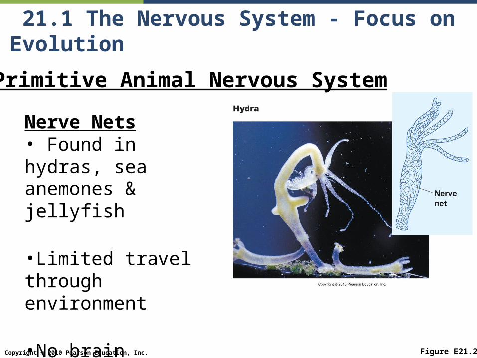
21.1 The Nervous System - Focus on Evolution
Figure E21.2
Primitive Animal Nervous System
Nerve Nets• Found in hydras, sea anemones & jellyfish
•Limited travel through environment
•No brain

Copyright © 2010 Pearson Education, Inc.
21.1 The Nervous System - Focus on Evolution
Figure E21.2
Nerve Cords•Found in insects
•Processing of information centralized in CNS
•Simple brain plus many enlarged ‘ganglia’

Copyright © 2010 Pearson Education, Inc.
21.1 The Nervous System - Focus on Evolution
Figure E21.3
Vertebrate Nervous Systems• Greater degree of centralization
•Single large brain
•Regions of brain become specialized for specific tasks

Copyright © 2010 Pearson Education, Inc.
21.2 The Brain
Human Brain The brain rests in the skull in cerebrospinal fluid, which bathes and
cushions it.
Two major cell types in the brain1. Neurons transmit nervous impulses
Brain has 100-200 billion neurons2. Glial cells support neurons by providing protection & nutrients
Almost 10 times as many glial cells as neurons
Figure 21.6

Copyright © 2010 Pearson Education, Inc.
21.2 The Brain
The brain is divided into 5 regions
1.Cerebrum
2.Cerebellum
3.Thalamus
4.Hypothalamus
5.Brain stem

Copyright © 2010 Pearson Education, Inc.
21.2 The Brain - Cerebrum
Figure 21.7
Lobes of the Cerebrum1.Frontal lobe2.Temporal lobe3.Parietal lobe4.Occipital lobe
Other Important Structures•Right & Left Hemispheres•Central fissure•Corpus callosum•Caudate nuclei

Copyright © 2010 Pearson Education, Inc.
21.2 The Brain - Cerebrum
Figure 21.7
Brain Hemispheres•Many nerves cross over, so left brain controls right side of body, and visa versa
•Left Hemispheres •Controls speech, reading, & solving math
•Right Hemispheres •Interprets spatial relationships, music & art

Copyright © 2010 Pearson Education, Inc.
21.2 The Brain - Thalamus and Hypothalamus
Thalamus and hypothalamus lie deep in the brain between the hemispheres
and act as control center.
Thalamus relays information from spinal cord to brain. Thalamus suppresses some information and
enhances other.
Hypothalamus is the control center for sex drive, pleasure, pain, hunger, and other basic drives.

Copyright © 2010 Pearson Education, Inc.
21.2 The Brain - Cerebellum
Cerebellum Control of balance Coordination of muscular movement Damage to the cerebellum results in jerky,
awkward movements

Copyright © 2010 Pearson Education, Inc.
21.2 The Brain - Brainstem
Brainstem Controls involuntary activity. The brainstem is composed of the midbrain,
pons, and medulla oblongata
Figure 21.8

Copyright © 2010 Pearson Education, Inc.
21.2 The Brain
ADD and Brain Structure and Function
Some researchers suggest there are differences between brains of people with ADD and people without. Corpus callosum smaller in individuals with
ADD Decreased folding on cerebrum in ADD
individuals
Differences could be a result of genetics, or development and life experiences

Copyright © 2010 Pearson Education, Inc.
21.3 Neurons
Neurons Neurons are highly
specialized cells
Parts of Neuron Dendrites Cell Body Axon
Terminal Boutons
Figure 21.9

Copyright © 2010 Pearson Education, Inc.
21.3 Neurons - Neuron Structure
Myelin speeds up nervous impulses Many neurons have their axons covered by a
myelin sheath made by Schwann cells the unmyelinated patches are the nodes of
Ranvier.
Figure 21.10

Copyright © 2010 Pearson Education, Inc.
21.3 Neurons - Neuron Function
Neuron Function Cell accumulates K+ ions inside and Na+ ions
outside
Figure 21.11aNerve cell
All channels are closed.The inside of the cell hasa more negative chargethan the outside ofthe cell.
Nodes ofRanvier
Outside cell Inside cell
(a) Resting nerve cell
K+ channel
Na+ channel

Copyright © 2010 Pearson Education, Inc.
21.3 Neurons - Neuron Function
Nervous Impulse = Action Potential Stimulation of a neuron causes ion gates to open, and
Na+ rushes in, changing polarity (depolarization) Action potential (nervous Impulse) – a brief change in
polarity of the surface membrane, which moves down the length of an axon
Figure 21.11b

Copyright © 2010 Pearson Education, Inc.
21.3 Neurons - Neuron Function
Animation—Communication Within Neurons: The AxonPLAY

Copyright © 2010 Pearson Education, Inc.
21.3 Neurons - Neuron Function
How Neurons Work

Copyright © 2010 Pearson Education, Inc.
21.3 Neurons - Neuron Function
Synapse = junction between neurons Terminal boutons, space, & dendrites or
cell body
Synaptic transmission = Transmission of impulses between neurons
neurons use neurotransmitters to communicate chemically across the synapse

Copyright © 2010 Pearson Education, Inc.
21.3 Neurons - Neuron Function
Figure 21.12
Synaptic Transmission1.Action potential reaches terminal bouton of presynaptic cell2.Calcium gates open, allowing Ca2+ to rush in3.Ca2+ causes synaptic vesicles to release neurotransmitters4.Neurotransmitter binds to receptors on postsynaptic cell5.Opening or ion channels triggers action potential in postsynpatic cell

Copyright © 2010 Pearson Education, Inc.
21.3 Neurons - Neuron Function
Figure 21.12
Two ways to stop synaptic transmission1.Neurotransmitter is digested by enzymes2.Reuptake of neurotransmitters by presynatpic cell

Copyright © 2010 Pearson Education, Inc.
21.3 Neurons - Neuron Function
Animation—Communication Within Neurons: The SynapsePLAY

Copyright © 2010 Pearson Education, Inc.
21.3 Neurons
Alzheimer’s, Depression, Parkinson’s, and ADD Many mental diseases are linked to problems
with neurotransmitters. Alzheimer’s and Parkinson’s diseases seem
to be related to impaired neurotransmitter production.
Depression appears to be related to an imbalance in several neurotransmitters, but its unclear if this is a cause or a result of depression.
ADD may be result of lower levels of neurotransmitter dopamine.

Copyright © 2010 Pearson Education, Inc.
21.3 Neurons - ADD Perscription Drug Action
Figure 21.13
Ritalin •Blocks reuptake receptors on presynaptic cells•Increases dopamine in synapse
Adderall & Dexedrine•Both are amphetamines•Increase levels of dopamine in synapse