Apoptosis ReagentsNucView® caspase-3 substrates ... p. 2Other caspase substrates ... p. 3Apoptosis inducers ... p. 3Annexin V conjugates ... p. 4Tunel assays ... p. 5Mitochondrial membrane potential dyes ... pp. 6-7
Viability AssaysLive-or-Dye™ fixable dead cell stains ... p. 8Cell viability assay kits ... p. 9
Cell ProliferationViaFluor® SE cell proliferation kits ... p. 9
Cell Viability and Cell Death
Microbial ViabilityPMAxx™ and PMA for viability PCR ... p. 10Bacterial viability assay kits ... p. 11Yeast viability assay kits ... p. 11

2 • www.biotium.com
NucView® Substrates
Cat. # Size Description
10402-T 10 uLNucView® 488 Caspase-3 Substrate, 1 mM in DMSO
10402 100 uL10403-T 10 uL
NucView® 488 Caspase-3 Substrate, 1 mM in PBS10403 100 uL10405-T 10 uL
NucView® 405 Caspase-3 Substrate, 1 mM in DMSO10405 100 uL10407-T 10 uL
NucView® 405 Caspase-3 Substrate, 1 mM in PBS10407 100 uL10406-T 10 uL
NucView® 530 Caspase-3 Substrate, 1 mM in DMSO10406 100 uL10408-T 10 uL
NucView® 530 Caspase-3 Substrate, 1 mM in PBS10408 100 uL
NucView® Caspase-3 Substrates
Figure 1. Schematic showing the principle of intracellular caspase-3/7 detection using NucView® caspase-3 substrates.
Fluorescent caspase-3 detection in living cellsNucView® caspase-3 substrates are novel fluorescent probes that allow detection of caspase-3/7 activity in intact cells in real-time. In contrast to other fluorogenic caspase substrates or fluorescent caspase inhibitor based (FLICA) assays, NucView® substrates can be used to detect caspase-3/7 activity in cells without inhibiting apoptosis progression.
NucView® is made by attaching a nucleic acid binding dye to the caspase-3/7 substrate peptide sequence DEVD. This uncleaved substrate dye is unable to bind to DNA and remains non-fluorescent. Once the substrate is cleaved by caspase-3/7 in apoptotic cells, it releases the high-affinity fluorescent DNA dye, which stains the cell nucleus with bright and stable fluorescence signal.
NucView® features
• Real-time monitoring of caspase-3/7 activity
• Rapid, no-wash assay
• Available in 3 colors
• For flow cytometry, microscopy or microplate reader
• Formaldehyde fixable
NucView® 405CF®488A Annexin V
NucView® 405
NucView® 488CF®594 Annexin V
NucView® 488
NucView® 530CF®488A Annexin V
NucView® 530 Proven technology
NucView® caspase detection technology has been extensively tested. NucView® 488 Substrate has been:
• Published in over 200 scientific papers
• Validated in more than 70 different cultured cell lines
• Validated in more than 30 different primary cell types

www.biotium.com • 3
NucView® kits and other caspase assays
Cat. # Size Description
30067 50 assays Dual Apoptosis Assay with NucView® 488 Caspase-3 Substrate and CF®594 Annexin V
30073 50 assays Dual Apoptosis Assay with NucView® 488 Caspase-3 Substrate and CF®640R Annexin V
30062 100 assays NucView® 488 and MitoView™ 633 Apoptosis Assay Kit
30072 100 assays NucView® 488 and RedDot™2 Apoptosis & Necrosis Kit
30029-T 25 assays NucView® 488 Caspase-3 Assay Kit for Live Cells30029 100 assays
30008-1 25 assays Caspase-3 DEVD-R110 Fluorometric & Colorimetric Assay Kit30008-2 100 assays
30009-1 10 assaysCaspase-3 DEVD-R110 Fluorometric HTS Assay30009-2 100 assays
30009-3 1000 assays30011-1 25 assays Caspase-8 IETD-R110 Fluorometric &
Colorimetric Assay Kit30011-2 100 assays30012-1 10 assays
Caspase-8 IETD-R110 Fluorometric HTS Assay30012-2 100 assays30012-3 1000 assays10404-1 1 mg
Ac-DEVD-CHO Caspase-3 Inhibitor10404 5 mg10202 5 mg Ac-DEVD-AMC00025 100 ug Staurosporine59007 1 mg Ionomycin, calcium salt
Additional caspase substratesIn addition to our patented NucView® technology for detecting caspase-3 activity in live cells, Biotium also offers rhodamine 110 (R110)-based assay kits for fluorescence- or absorbance-based detection of caspase-3 or caspase-8 activity in cell lysates. The HTS versions of the R110-based homogenous caspase-3 and caspase-8 assay kits are optimized for high throughput screening by fluorescence microplate reader.
Biotium also offers a coumarin (AMC)-based blue fluorogenic substrate (Ac-DEVD-AMC) for measuring caspase-3 activity in cell lysates by fluorescence microplate reader.
Caspase inhibitorAc-DEVD-CHO is a competitive inhibitor of caspase-3 for use in cultured cells or cell lysates.
Apoptosis inducersStaurosporine is a broad range protein kinase inhibitor that induces apoptosis in cultured cells. It is useful as a positive control for many apoptosis assays. We also offer ionomycin, a calcium ionophore that has been shown to induce apoptosis through calpain activation.
NucView® combination staining kits
Biotium also offers kits containing the NucView® 488 substrate together with other types of apoptosis and viability dyes for convenient multi-parameter experiments.
• Dual Apoptosis Kit: NucView® 488 + Annexin V labeled with red or far-red dyes for co-detection of two apoptotic events, caspase cleavage and phosphatidylserine (PS) translocation. For more Annexin V conjugates see p. 4.
• Dual Apoptosis Kit: NucView® 488 + MitoView™ 633 for co-detection of two apoptotic events, caspase cleavage and loss of mitochondrial membrane potential. For more information on MitoView™ see p. 6.
• Apoptosis/Necrosis Kit: NucView® 488 + RedDot™2 for concurrent measurement of caspase cleavage (apoptosis) and loss of membrane integrity (necrosis).
NucView® Combination Kits and Other Caspase Substrates and Inhibitors
Figure 2. Apoptotic HeLa cell stained with CF®488A Annexin V (green) and NucView® 405 (cyan). See p. 4 for more information on Annexin V conjugates.

4 • www.biotium.com
Annexin V conjugates
Annexin V is a 35-36 kDa protein that has a high affinity for phosphatidylserine (PS). During apoptosis, PS is translocated from the inner to the outer leaflet of the plasma membrane, where it can be stained by fluorescent conjugates of Annexin V, for detection of apoptotic cells by flow cytometry (Fig. 1) or fluorescence microscopy (Fig. 2). Biotium offers Annexin V conjugates and kits featuring our exceptionally bright and photostable CF® dyes. For example, our CF®488A green fluorescent Annexin V conjugate (Fig. 2) is much brighter and more photostable than the traditional FITC-Annexin V, allowing the use of 10-fold less conjugate in staining. Our near-infrared CF® dye conjugates of Annexin V are supplied lyophilized and preservative-free, and are suitable for in vivo imaging.
CF®488A Annexin V Dual Apoptosis & Necrosis Assay Kits Biotium offers several staining kits that allow concurrent identification of late apoptotic and membrane-compromised necrotic cells by fluorescence microscopy or flow cytometry. These dual staining kits all include green fluorescent CF®488A Annexin V paired with a dead cell-specific nucleic acid dye: either red fluorescent Ethidium Homodimer III (EthD-III), red fluorescent propidium iodide (PI), or far-red fluorescent 7-AAD. EthD-III is a novel membrane-impermeant nucleic acid dye developed at Biotium with higher affinity for DNA and higher fluorescence quantum yield than propidium iodide.
The Apoptotic, Necrotic, and Healthy Cells Quantitation Kit also includes blue fluorescent Hoechst 33342 DNA dye for visualizing the healthy cells and dead cells (Fig. 2).
Figure 1. Jurkat cells were treated with staurosporine to induce apoptosis (pink), or with DMSO as a negative control (blue) for the times indicated, then stained for 15 minutes at room temperature with NucView® 488 Caspase-3 Substrate (FL1-H, x-axis) and CF®640R Annexin V (FL4-H, y-axis) in cell culture medium prior to analysis using a BD LSRII flow cytometer. See pp. 2-3 for more information on NucView® caspase-3 substrates.
DMSO 1 hrStaurosporine 1 hr
DMSO 2 hrStaurosporine 2 hr
DMSO 4 hrStaurosporine 4 hr
FL1-H: NucView 488
FL4-
H: C
F640
R An
nexin
V
FL1-H: NucView 488
FL4-
H: C
F640
R An
nexin
V
FL1-H: NucView 488
FL4-
H: C
F640
R An
nexin
V
Annexin V Conjugate Ex/Em (nm) Cat. # SizeCF®350 Annexin V, 50 ug/mL 347/448 29012 0.5 mLCF®405M Annexin V, 50 ug/mL 408/452 29009 0.5 mLCF®488 Annexin V, 50 ug/mL 490/515 29005 0.5 mLCF®555 Annexin V, 50 ug/mL 555/565 29004 0.5 mLCF®568 Annexin V, 50 ug/mL 562/583 29010 0.5 mLCF®594 Annexin V, 50 ug/mL 593/614 29011 0.5 mLCF®633 Annexin V, 50 ug/mL 630/650 29008 0.5 mLCF®640R Annexin V, 50 ug/mL 642/662 29014 0.5 mLCF®647 Annexin V, 50 ug/mL 650/665 29003 0.5 mLCF®660R Annexin V, 50 ug/mL 663/682 29069 0.5 mLCF®680R Annexin V, lyophilized 680/701 29070 25 ugCF®680 Annexin V, lyophilized 681/698 29007 25 ugCF®750 Annexin V, lyophilized 755/777 29006 25 ugCF®770 Annexin V, lyophilized 770/797 29046 25 ugCF®790 Annexin V, lyophilized 784/806 29047 25 ugCF®800 Annexin V, lyophilized 797/816 29078 25 ugFITC Annexin V, 50 ug/mL 490/525 29001 0.5 mLTexas Red Annexin V, 50 ug/mL 583/603 29002 0.5 mL
R-PE Annexin V 496, 546, 565/578
29045-100 uL 20 assays29045-500 uL 100 assays
APC Annexin V 633, 640/66029057-100 uL 20 assays29057-500 uL 100 assays
Biotin Annexin V, 50 ug/mL N/A 29013 0.5 mL5X Annexin V Binding Buffer N/A 99902 15 mL
Kit Name Cat. # Size
Dual Apoptosis Assay with NucView® 488 and CF®594 Annexin V 30067 50 assays
Dual Apoptosis Assay with NucView® 488 and CF®640R Annexin V 30073 50 assays
Figure 2. Jurkat cells stained using the Apoptotic, Necrotic & Healthy Cells Quantitation Kit Plus after apoptosis induction with staurosporine. Apoptotic cells stain with CF®488A Annexin V (green), necrotic/late apoptotic cells stain with EthDIII (red). All cells are stained with Hoechst (blue).
Annexin V Conjugates and Kits
Kit Name/Description Cat. # Size
Apoptosis & Necrosis Quantitation Kit Plus with CF®488A Annexin V and EthD-III 30065 50 assays
CF®488A Annexin V and 7-AAD Apoptosis Kit 30060 100 assaysCF®488A Annexin V and PI Apoptosis Kit 30061 100 assaysApoptotic, Necrotic & Healthy Cells Quantitation Kit Plus with CF®488A Annexin V, EthD-III and Hoechst 30066 50 assays
Apoptosis and Necrosis Combination Kits
Dual apoptosis assay kitsAnnexin V conjugated to our deep red CF®594 or far-red CF®640R dyes is offered together with NucView® 488 Caspase-3 Substrate for simultaneous detection of caspase-3 activity and phosphatidylserine exposure by fluorescence microscopy or flow cytometry (see p. 2 for more information on NucView® substrates).
Dual Apoptosis Kits
Annexin V Conjugates

www.biotium.com • 5
CF® dye TUNEL kits and dUTP conjugatesTUNEL (terminal deoxynucleotidyl transferase (TdT) mediated dUTP nick-end labeling) is highly selective for the detection of apoptotic cells, but not necrotic cells or cells with DNA strand breaks resulting from irradiation or drug treatment. In this assay, TdT enzyme catalyzes the addition of labeled dUTP to the 3’ ends of cleaved DNA fragments. Fluorescent dye-conjugated dUTP can be used for direct detection of fragmented DNA by fluorescence microscopy or flow cytometry.
Biotium offers dUTP conjugated to a range of CF® dye colors for fluorescent TUNEL labeling, as well as direct TUNEL kits with green fluorescent CF®488A, red fluorescent CF®594, and far-red fluorescent CF®640R. We also supply dUTP conjugated to classic fluorophores and biotin. Visit www.biotium.com to see our selection of CF® dye conjugated streptavidin, as well as other nucleotide conjugates for probe labeling.
Product Name Ex/Em (nm) Cat. # Size
CF®488A TUNEL Assay Kit 490/515 30063 50 reactionsCF®594 TUNEL Assay Kit 593/614 30064 50 reactionsCF®640R TUNEL Assay Kit 642/662 30074 50 reactions
CF®405S-dUTP 404/43140004-T 5 nmol40004 25 nmol
CF®405M-dUTP 408/45240100-T 5 nmol40100 25 nmol
CF®488A-dUTP 490/51540100-T 5 nmol40008 25 nmol
CF®543-dUTP 541/56040008-T 5 nmol40002 25 nmol
CF®568-dUTP 562/58340002-T 5 nmol40005 25 nmol
CF®594-dUTP 593/61440005-T 5 nmol40006 25 nmol
CF®640R-dUTP 642/66240006-T 5 nmol40007 25 nmol
CF®680R-dUTP 680/70140007-T 5 nmol40003 25 nmol
Cyanine 555-dUTP 550/570 40064 25 nmolCyanine 647-dUTP 650/670 40065 25 nmolDEAC-dUTP 426/480 40059 25 nmol5-TAMRA-dUTP, 1 mM solution 553/577 40001 25 uLFluorescein-12-dUTP 494/521 40063 25 nmol5-AA-dUTP, 10 mM N/A 40020 100 uL5-AA-dUTP, lyophilzed N/A 40020-1 1 mgDigoxigenin-dUTP N/A 40078 25 nmol5-Bromo-dUTP, 10 mM solution N/A 40025 25 uLBiotin-11-dUTP, 1 mM solution N/A 40029 50 uLBiotin-11-dUTP, lyophilized N/A 40029-1 50 ugBiotin-16-dUTP, 1 mM solution N/A 40022 50 uLBiotin-16-dUTP, lyophilized N/A 40022-1 50 ugBiotin-20-dUTP, 1 mM solution N/A 40030 50 uLBiotin-20-dUTP, lyophilized N/A 40030-1 50 ug
Figure 2. TUNEL staining of paraffin sections of rat mammary gland five days post-weaning (ApopTag® positive control slides, MilliporeSigma) using CF®594-dUTP (red). Nuclei are stained with DAPI (blue).
Figure 1. Jurkat cells labeled using the CF®488A TUNEL Assay Apoptosis Detection Kit after no treatment (A) or apoptosis induction with 1 uM staurosporine for 3 hours (B). Nuclei are counterstained with DAPI (blue).
A. Control B. Staurosporine
TUNEL Assays and dUTP conjugates
TUNEL Assays and dUTP Conjugates
6 • www.biotium.com
MitoView™ DyesLoss of mitochondrial membrane potential is a hallmark for apoptosis. Biotium offers the MitoView™ 633 dye for membrane potential-sensitive staining of mitochondria by microscopy or flow cytometry. We also offer MitoView™ Blue, which changes localization upon mitochondrial depolarization, and MitoView™ Green, a membrane-potential independent mitochondrial dye that can be used to image mitochondria following mitochondrial depolarization, or after fixation.
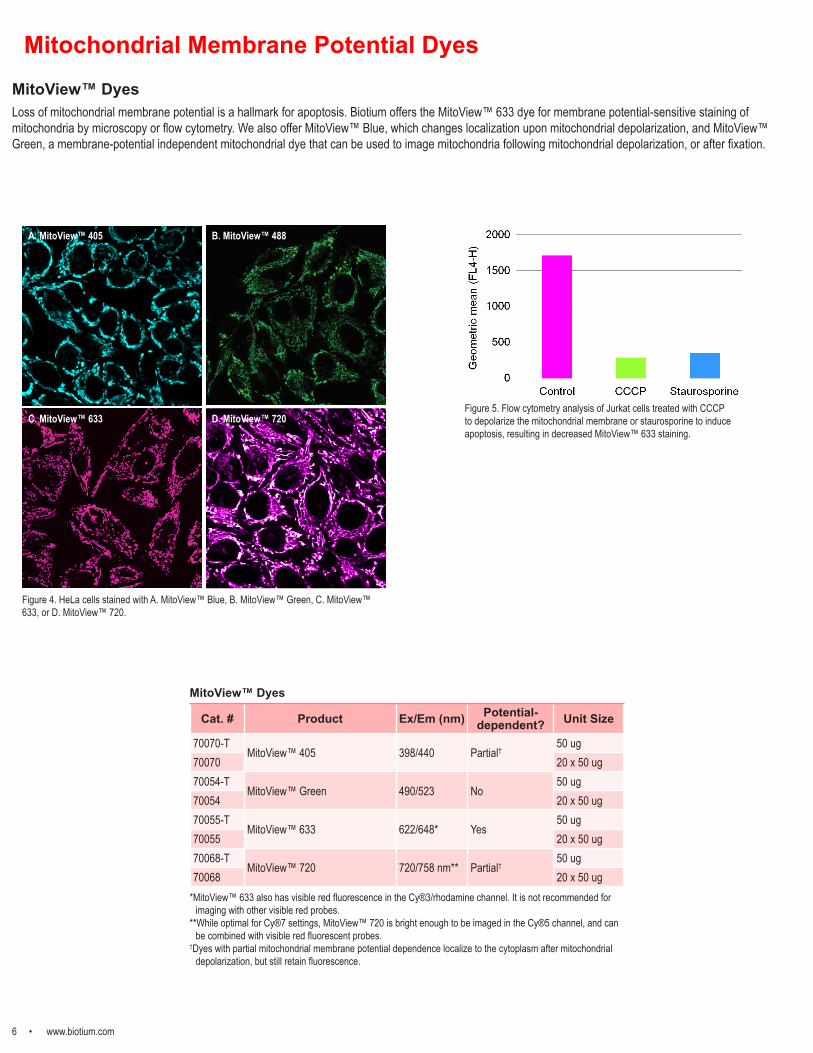
Figure 5. Flow cytometry analysis of Jurkat cells treated with CCCP to depolarize the mitochondrial membrane or staurosporine to induce apoptosis, resulting in decreased MitoView™ 633 staining.
Mitochondrial Membrane Potential Dyes
Figure 4. HeLa cells stained with A. MitoView™ Blue, B. MitoView™ Green, C. MitoView™ 633, or D. MitoView™ 720.
A. MitoView™ 405 B. MitoView™ 488
C. MitoView™ 633 D. MitoView™ 720
Cat. # Product Ex/Em (nm) Potential-dependent? Unit Size
70070-TMitoView™ 405 398/440 Partial†
50 ug70070 20 x 50 ug70054-T
MitoView™ Green 490/523 No50 ug
70054 20 x 50 ug70055-T
MitoView™ 633 622/648* Yes50 ug
70055 20 x 50 ug70068-T
MitoView™ 720 720/758 nm** Partial†50 ug
70068 20 x 50 ug*MitoView™ 633 also has visible red fluorescence in the Cy®3/rhodamine channel. It is not recommended for
imaging with other visible red probes.**While optimal for Cy®7 settings, MitoView™ 720 is bright enough to be imaged in the Cy®5 channel, and can
be combined with visible red fluorescent probes.†Dyes with partial mitochondrial membrane potential dependence localize to the cytoplasm after mitochondrial
depolarization, but still retain fluorescence.
MitoView™ Dyes

www.biotium.com • 7
JC-1 and other mitochondrial dyesIn healthy cells, JC-1 dye aggregates in mitochondria as a function of membrane potential, resulting in red fluorescence with brightness proportional to the membrane potential. Conversely, in apoptotic and necrotic cells with diminished mitochondrial membrane potential, JC-1 exists in a green fluorescent monomeric form in the cytosol, allowing of cell viability to be assessed by measuring the ratio of red to green fluorescence by flow cytometry or fluorescence plate reader.
We also offer a selection of classic potentiometric mitochondrial stains, including TMRE, TMRM, and DiIC1(5), in a variety of fluorescent colors.
MCB Glutathione Detection KitDiminished cellular glutathione (GSH) level occurs during apoptosis due to GSH efflux from mitochondria. Monochlorobimane (MCB), which reacts with thiols to form a blue fluorescent product, allowing fluorometric quantitation of GSH in cell lysates.
MCBnon-fluorescent
MCB-glutathione conjugateEx/Em: 380/461 nm
Figure 6. MCB glutathione assay principle.
Figure 7. Jurkat cells were treated with DMSO (Control) or induced to undergo apoptosis by treatment with 1 uM staurosporine for 5 hours. Glutathione levels were measured using the MCB Glutathione Detection Kit by fluorescence microplate reader.
Dye Color Ex/Em Mitochondrial Membrane Potential Dependent? Cat. # Size
JC-1, chloride salt Green/Red
510/527 nm (cytoplasm)585/590 nm (polarized mitochondria)
Two-color detection mitochondria polarization/depolarization
70011 5 mg
JC-1, iodide salt Green/Red
510/527 nm (cytoplasm)585/590 nm (polarized mitochondria)
Two-color detection mitochondria polarization/depolarization
70014 5 mg
Rhodamine 123 Green 505/534 nm Yes 70010 50 mgTMRE Red 548/573 nm Yes 70016 25 mgTMRE, 2 mM in DMSO Red 548/573 nm Yes 70005 0.5 mLTMRM Red 548/573 nm Yes 70017 25 mgDASPEI Red 461/589 nm Yes 70018 100 mgDiIC1(5) Far-red 638/658 nm Yes 70015 100 mg
Kit Name and Components Color Ex/Em Assay Cat. # Size
NucView® 488 and MitoView™ 633 Apoptosis Kit
Green/Red
500/530 nm (caspase-3)622/648 nm (polarized mitochondria)
Two color detection of caspase-3 activity and mitochondrial potential
30062 100 assays
JC-1 Mitochondrial Membrane Potential Detection Kit
Green/Red
510/527 nm (cytoplasm)585/590 nm (polarized mitochondria)
Two-color detection mitochondria polarization/depolarization
30001 100 assays
MCB Glutathione Detection Kit Blue 394/490 nm Detection of cellular glutathione 30019 100 assays
Mitochondrial Membrane Potential and Cellular Glutathione
Other Mitochondrial Dyes
Assay Kits

8 • www.biotium.com
Figure 2. Discrimination of live and dead Jurkat cells by flow cytometry using Live-or-Dye™ Fixable Viability Stains. Heat killed cells (solid peaks) showed much higher fluorescence intensity compared to live cells (white peaks), allowing the two populations to be clearly distinguished.
Figure 1. HeLa cells were treated with ethanol to kill a fraction of the cells. The cells were stained with A) Live-or-Dye™ 488/515 or B) Live-or-Dye™ NucFix Red. Nuclei were counterstained with Hoechst.
Live-or-Dye™ Fixable Stains for Dead Cells
• Affordable: Lower cost than Thermo Fisher LIVE/DEAD® stains
• Choice: 8 bright colors across the spectrum• Specific: No staining of live cells• Fixable: No loss of brightness after fixation• Versatile: For flow cytometry and microscopy
Live-or-Dye™ Live-or-Dye™ Fixable Viability Staining Kits are designed for discrimination between live and dead cells by flow cytometry and microscopy. Dead cell stains are useful probes to include when analyzing cell surface protein expression by flow cytometry (Fig. 2), because they allow intracellular fluorescence signal from dead cells with permeable plasma membranes to be excluded from analysis. Live-or-Dye™ Fixable Viability Stains are cell membrane impermeable; they enter dead cells that have compromised membrane integrity and covalently label free amines on intracellular proteins. Live-or-Dye™ Fixable Viability Staining Kits can also be used to discriminate live from dead cells by microscopy (Fig. 1A). Live-or-Dye™ labeling is extremely stable, allowing the cells to be fixed and permeabilized without loss of fluorescence or dye transfer between cells.
Live-or-Dye™ NucFix Red Live-or-Dye NucFix™ Red is a unique, cell membrane impermeable dye that specifically stains the nuclei of dead cells (Fig. 1B). The dye is able to enter into dead cells that have compromised membrane integrity and covalently label the cell nucleus, allowing for clear differentiation of live and dead cells by either microscopy or flow cytometry. Unlike other commonly used nuclear stains such as propidium iodide or DRAQ®7, NucFix labeling is extremely stable, allowing the cells to be fixed and permeabilized without loss of fluorescence or dye transfer between cells.
Kit Name Cat. # 200 Reactions
Cat. #50 Reactions Laser Line Emission
FilterAbs/EmMaxima
Validated applications (FC=flow cytometry;
M=microscopy)Live-or-Dye™ 350/448 32002 32002-T 355 nm DAPI or Violet 347/448 nm FCLive-or-Dye™ 405/452 32003 32003-T 405 nm Pacific Blue 408/452 nm FCLive-or-Dye™ 405/545 32009 32009-T 405 nm AmCyan 395/545 nm FC
Live-or-Dye™ 488/515 32004 32004-T 488 nm FITC 490/515 nm FC, M
Live-or-Dye™ 568/583 32005 32005-T 488 or 561 nm PE 562/583 nm FC, MLive-or-Dye™ 594/614 32006 32006-T 488 or 561 nm PE-Texas Red® 593/614 nm FC, MLive-or-Dye™ 640/662 32007 32007-T 633 or 640 nm APC 642/662 nm FC, MLive-or-Dye™ 750/777 32008 32008-T 633 or 640 nm APC-Cy7 755/777 nm FCLive-or-Dye™ NucFix Red 32010 32010-T 488 or 532 nm PE-Texas Red® 520/610 nm FC, M
Live-or-Dye™ Fixable Viability Stains
Live-or-Dye™ NucFix Red
• Unique, nuclear dead cell stain
• Fixable, unlike other commonly used nuclear stains such as Propidium Iodide or DRAQ®7
LIVE/DEAD and Texas Red are registered trademarks of Thermo Fisher Scientific. DRAQ is a registered trademark of Biostatus, Ltd.
A. Live-or-Dye™ 488/515 B. Live-or-Dye™ NucFix Red
Live-or-Dye™ Kits

www.biotium.com • 9
Calcein AM Cell Viability AssayCalcein AM is a non-fluorescent, membrane permeable compound. Esterase activity in the cytoplasm of viable cells converts calcein AM to the green fluorescent, membrane-impermeant compound calcein, which is retained in viable cells with intact plasma membranes. The Viability/Cytotoxicity Assay Kit for Animal Live & Dead Cells pairs calcein AM with the dead cell dye Ethidium Homodimer III for quantitation of live and dead cells.
ATP-Glo™ Bioluminometric Cell Viability AssayThis assay takes advantage of the ATP-dependent oxidation of D-Luciferin by Firefly luciferase and the resulting production of light in order to assess the amount of ATP in a cell culture, which is proportional to the number of viable cells. The ATP-Glo™ kit can be used to detect as little as a single cell or 0.01 picomole of ATP, with signal linearity for ATP detection within 6 orders of magnitude. This is a flash-type assay designed for detection using a single sample luminometer or a luminometer with an injector in 96-well plate format. The luminescent signal is stable for up to one minute.
1
10
100
1000
10000
1 10 100 1000 10000
Lum
ines
cenc
e
Cell Number
Figure 3. Quantitation of 10-fold serial dilutions of Jurkat cells in suspension using ATP-Glo™ Bioluminetric Cell Viability Assay using a single-sample luminometer.
1
10
100
1000
200 600 1800 5400 16200
Cell Number
Fluo
resc
ence
Figure 1. A. Quantitation of HeLa cell numbers in a 96-well assay plate using the Calcein AM Cell Viability Assay Kit. B. Live and dead HeLa cells stained with the Viability/Cytotoxicity Assay for Animal Live & Dead Cells. Live cells are stained green, dead cells are stained red.
Resazurin, MTT, and XTT Viability AssaysMTT, XTT, and resazurin (alamarBlue®) are reduced by mitochondrial metabolic activity to yield colored or fluorescent products, and thus are useful for assaying cell viability and quantitating cell number. MTT and XTT are reduced to colored formazin salts that can be measured by absorbance. MTT generates an insoluble formazin salt, requiring cell lysis before the absorbance can be measured, while XTT does not require cell lysis for measurement. Resazurin is a non-fluorescent blue dye that is reduced to the pink fluorescent compound resorufin, which can be measured by fluorescence or absorbance.
alamarBlue is a registered trademark of Morphosys UK Ltd. Fluoro-Jade is a registered trademark of Histo-Chem, Inc.
Cellular Viability and Proliferation Assays
PathoGreen™ Histofluorescent StainPathoGreen™ Histofluorescent Stain is an anionic green fluorescent dye functionally similar to Fluoro-Jade® dyes. These dyes stain degenerating neurons and their processes in fixed brain sections and cultured neurons. The dyes stain apoptotic and necrotic neurons after exposure to a variety of neurotoxic insults. The mechanism of neuronal staining by anionic fluorescent dyes has not been determined. It has been proposed that the negatively charged dyes bind to positively charged polyamines or other molecules specifically generated in dying neurons.
Figure 2. A section of mouse hippocampus stained with PathoGreen™ Histofluorescent Stain. Degenerating neurons are stained green.
Kit Name Cat. # Unit Size
Viability/Cytotoxicity Assay Kit for Animal Live & Dead Cells
30002-T 150 assays30002 300 assays
Calcein AM Cell Viability Assay Kit 30026 1000 assaysPathoGreen™ Histofluorescent Stain, 1000X in water
80027-5mL 5 mL80027-50mL 50 mL
Resazurin Cell Viability Assay Kit30025-1 25 mL (2500 assays)30025 100 mL (10,000 assays)
MTT Cell Viability Assay Kit 30006 1000 assaysXTT Cell Viability Assay Kit 30007 1000 assays
ATP-Glo™ Bioluminometric Cell Viability Assay Kit
30020-T 50 assays30020-1 200 assays30020-2 1000 assays
ViaFluor® 405 SE Cell Proliferation Kit 30068 1 kitViaFluor® 488 SE Cell Proliferation Kit 30086 1 kitViaFluor® CFSE Cell Proliferation Kit 30050 1 kit
A. B.
Cellular Viability Assays
ViaFluor® SE Cell Proliferation Kits ViaFluor® SE Cell Proliferation Kits diffuse passively into cells covalently label intracellular proteins throughout the cell. They can be used as cell-filling stains for imaging morphology, or to track cell division by dye dilution. With each cell division, daughter cells inherit roughly half of the fluorescent label, allowing the number of cell divisions to be detected by the appearance of successively dimmer fluorescent peaks on a flow cytometry histogram. Staining is formaldehyde fixable. ViaFluor® CFSE is the classic cell proliferation dye, detected in the FITC channel. Biotium created ViaFluor® 488, a new improved green dye that is less toxic, less leaky and more fixable than CFSE. We also offer blue fluorescent ViaFluor® 405 for the violet laser. ViaFluor® 405 has improved brightness and less toxicity than CFSE.
Figure 3. Cell division tracking in Jurkat cells over successive days. Cells were labeled with ViaFluor® 405 (left) or ViaFluor® 488 (right) on day 0, and analyzed by flow cytometry on each following day. Each successively dimmer peak represents one cell division. Unstained cells are in gray.

10 • www.biotium.com
PMAxx™ and PMA Dyes for Viability PCRViability PCR (v-PCR)Viability PCR is a powerful technology for the sensitive and rapid detection of viable microorganisms. Unlike time-consuming culturing procedures, qPCR is a fast and sensitive method of detection. However, normal qPCR does not distinguish between live and dead cells. With v-PCR using PMAxx™ or PMA, you get the speed, sensitivity and specificity of PCR, plus quantifiable viability. And because no culturing is required, you can even detect viable but not culturable (VBNC) bacteria.
How does v-PCR work?PMAxx™ and PMA are photoreactive dyes with high affinity for DNA. The dyes intercalate into dsDNA and form a covalent linkage upon exposure to intense visible light. PMAxx™ and PMA inhibit PCR amplification of modified DNA templates by a combination of removal of modified DNA during purification and inhibition of template amplification by DNA polymerases. Because PMAxx™ and PMA are cell membrane-impermeable, when a sample containing both live and dead bacteria is treated with dye, only dead bacteria with compromised cell membranes are susceptible to DNA modification (Fig. 1). In a real-time PCR reaction, dead cell DNA will show delayed amplification and higher Ct than live cells. In a mixed population, v-PCR permits quantitation of cell viability. The v-PCR technology can be applied not only to bacteria but to other cell types as well.
PMAxx™ vs PMASince Biotium developed PMA in 2006, it has been used extensively in many applications and in hundreds of publications. However there are cell types and conditions in which dead cell DNA inactivation by PMA is incomplete, which could lead to false positive results. After extensive testing, the scientists at Biotium have invented a new dye called PMAxx™ that has the same spectral properties and is even more effective than PMA at live/dead discrimination by viability PCR (Fig. 2).
Figure 1. Mechanism of PMA and PMAxx™. The cell membrane-impermeable dyes PMA and PMAxx™ selectively and covalently modify DNA from dead bacteria with compromised membranes while leaving DNA from viable cells intact.
Figure 2. Live or heat-killed Bacillus subtilis were treated with PMA or PMAxx™, followed by exposure with the PMA-Lite™ and DNA purification. Fast EvaGreen® qPCR Master Mix was used to amplify a 500-bp fragment of B. subtilis DNA. qPCR of dead cells treated with PMAxx™ showed a significant further delay (>7 Ct) compared to dead cells treated with PMA.
Strain-specific v-PCR kits available for:• Salmonella enterica• Escherichia coli• Escherichia coli O157:H7• Listeria monocytogenes• Legionella pneumophila• Mycobacterium tuberculosis• Staphylococcus aureus• Methicilin resistant Staphylococcus aureus (MRSA)
Cat. # Product Name Unit Size
40069 PMAxx™ dye, 20 mM in dH2O 100 uL40013 PMA dye 1 mg40019 PMA dye, 20 mM in dH2O 100 uLE90002 PMA-Lite™ LED Photolysis Device 1 deviceE90004 Glo-Plate™ Blue 1 device31038 PMA Enhancer for Gram-Negative Bacteria 16 mL31033 Real-Time Bacterial Viability Kit-Salmonella (InvA) 200 assays31034 Real-Time Bacterial Viability Kit-M. tuberculosis (groEL2) 200 assays31035 Real-Time Bacterial Viability Kit-Staph. aureus (nuc) 200 assays31036 Real-Time Bacterial Viability Kit-MRSA (mecA) 200 assays31050 Real-Time Bacterial Viability Kit-E. coli (uidA) 200 assays31037 Real-Time Bacterial Viability Kit-E. coli O157:H7 (Z3276) 200 assays
31051 Real-Time Bacterial Viability Kit-Listeria monocytogenes (hly) 200 assays
31053 Real-Time Bacterial Viability Kit-Legionella pneumophila (mip) 200 assays
Ordering Information
PMA-Lite™:• Holds up to 18
microcentrifuge tubes• Bright, long-lasting blue
LED lights• Fan ensures temperatures
lower than 37oC.
Glo-Plate™ Blue:• Flat illumination surface fits
microplates• Bright, long-lasting blue
LED lights• Surface stays cool during
light exposure.
v-PCR LED Photolysis Devices

www.biotium.com • 11
Microbial Viability AssaysBacteria Viability Dyes and KitsLive-or-Dye™ Fixable Viability Staining Kits utilize dead-cell-specific fixable dyes (Fig. 1). They are good for flow cytometry and microscopy and available in 9 bright, photostable colors. See p. 8 for more information on Live-or-Dye™ stains. CTC is a fluorescent dye that has been used to evaluate the respiratory activity in bacteria. Healthy cells will reduce CTC into an insoluble red product. The Viability/Cytotoxicity Assay Kit for Bacteria Live and Dead Cells features dual staining: DMAO for live cells, and EthD-III for dead cells (Fig. 2).
Combination Gram Stain and Viability KitsIt can be useful to distinguish live bacteria from dead, as well as Gram+ from Gram-, in the same sample. Our combination bacterial viability and fluorescent gram staining kits can help (Fig. 3). Our fluorescent gram stains utilize fluorescently-labeled wheat germ agglutinin (WGA) to selectively stain the cell walls of gram-positive bacteria.
Cat. # Product Description
10063 CTC (5-Cyano-2,3-ditolyl tetrazolium chloride) Forms insoluble red product in respiring cells
30027 Viability/Cytotoxicity Assay Kit for Bacteria Live and Dead Cells DMAO to stain all cells and EthD-III for dead cells
32001 Bacterial Viability and Gram Stain Kit WGA for gram stain, EthD-III for dead cells, and DAPI for all cells
Yeast Viability Dyes and KitsIt is often useful to distinguish live yeast cells from dead, or identify cells that are metabolically active. Our selection of yeast viability dyes and kits can help.• Live-or-Dye™ Fixable Viability Staining Kits: Fixable and dead-cell-specific. Good for flow cytometry and microscopy. Available in 9 bright, photostable colors.
Note: the NucFix™ Red variation is not nucleus-specific in yeast. See p. 8 for more information on Live-or-Dye™ stains.• ViaVac™ Red/Green: A vacuolar cell vitality dye. Passively diffuses into cells and gives a nonspecific green staining pattern. In metabolically active yeast, the
dye is actively transported into the vacuole where it stains intravacuolar tubules bright red.
Figure 1. Live and heat-killed E. coli stained with Live-or-Dye™ 568/583 (red) and DAPI (blue).
Figure 4. Yeast Vitality Staining Kit,ViaVac™ Red/Green (red, healthy vacuolar tubules) and Calcofluor White (blue, cell wall).
Figure 3. Bacterial Viability and Gram Stain Kit. CF®488A-WGA, EthD-III and DAPI.
Figure 6. Yeast Fixable Live/Dead Staining Kit, Thiazole Orange (green, all cells) and Live-or-Dye™ 568/583 (red, dead cell cytoplasm). Overlapping signal appears yellow.
Figure 2. Live and heat-killed E. coli stained with DMAO, marking live cells (green) and EthD-III, marking dead cells (red).
Figure 5. Yeast Viability Staining Kit,CF®-ConA (red, cell wall) and Live-or-Dye™ (green, dead cell cytoplasm).
Cat. # Product Description
29068 ViaVac™ Red/Green, 10 mM in DMSO Yeast vital dye
31062 Yeast Vitality Staining Kit ViaVac™ Red/Green and Calcofluor White
31063 Yeast Viability Staining Kit CF®-ConA and Live-or-Dye™ combinations
31064 Yeast Fixable Live/Dead Staining Kit Thiazole Orange and Live-or-Dye™ 568/583
Bacteria Viability Stains
Yeast Viability Stains

Biotium, Inc.
Toll Free: 800-304-5357Phone: 510-265-1027
Fax: 510-265-1352
General [email protected]
Quotes and [email protected]
Technical [email protected]