Biological markers of stress in pediatric acute burninjury
Nadia J. Brown a,*, Roy M. Kimble b, Sylvia Rodger c, Robert S. Ware d,e,Brett C. McWhinney f, Jacobus P.J. Ungerer f, Leila Cuttle g,h
aCentre for Children’s Burns and Trauma Research, Queensland Children’s Medical Research Institute, and the School
of Medicine, The University of Queensland, Royal Children’s Hospital, Brisbane, AustraliabCentre for Children’s Burns and Trauma Research, Queensland Children’s Medical Research Institute, Department of
Paediatrics and Child Health, The University of Queensland, Royal Children’s Hospital, Brisbane, AustraliacThe University of Queensland, School of Health & Rehabilitation Sciences, Division of Occupational Therapy,
Brisbane, AustraliadThe University of Queensland, School of Population Health and Queensland Children’s Medical Research Institute,
Brisbane, AustraliaeQueensland Children’s Medical Research Institute, The University of Queensland, AustraliafDepartment of Chemical Pathology, Pathology Queensland, Royal Brisbane and Women’s Hospital, QLD, AustraliagCentre for Children’s Burns & Trauma Research, Queensland Children’s Medical Research Institute, and University of
Queensland, AustraliahTissue Repair and Regeneration Program, Institute of Health and Biomedical Innovation, Queensland University of
Technology, Australia
b u r n s 4 0 ( 2 0 1 4 ) 8 8 7 – 8 9 5
a r t i c l e i n f o
Article history:
Accepted 4 December 2013
Keywords:
Stress
Salivary cortisol
Salivary alpha-amylase
Child
Burns
a b s t r a c t
Background: Burns and their associated wound care procedures evoke significant stress and
anxiety, particularly for children. Little is known about the body’s physiological stress
reactions throughout the stages of re-epithelialization following an acute burn injury.
Previously, serum and urinary cortisol have been used to measure stress in burn patients,
however these measures are not suitable for a pediatric burn outpatient setting.
Aim: To assess the sensitivity of salivary cortisol and sAA in detecting stress during acute
burn wound care procedures and to investigate the body’s physiological stress reactions
throughout burn re-epithelialization.
Methods: Seventy-seven participants aged four to thirteen years who presented with an
acute burn injury to the burn center at the Royal Children’s Hospital, Brisbane, Australia,
were recruited between August 2011 and August 2012.
Results: Both biomarkers were responsive to the stress of burn wound care procedures. sAA
levels were on average 50.2 U/ml higher ( p < 0.001) at 10 min post-dressing removal com-
pared to baseline levels. Salivary cortisol levels showed a blunted effect with average levels
at ten minutes post dressing removal decreasing by 0.54 nmol/L ( p < 0.001) compared to
baseline levels. sAA levels were associated with pain ( p = 0.021), no medication ( p = 0.047)
and Child Trauma Screening Questionnaire scores at three months post re-epithelialization
Available online at www.sciencedirect.com
ScienceDirect
journal homepage: www.elsevier.com/locate/burns
* Corresponding author at: Centre for Children’s Burns and Trauma Research, Queensland Children’s Medical Research Institute, Level 4,Foundation Building, Royal Children’s Hospital, Brisbane 4029, QLD, Australia. Tel.: +61 7 3636 4249; fax: +61 7 3636 5578.
E-mail address: [email protected] (N.J. Brown).
0305-4179/$36.00 # 2013 Elsevier Ltd and ISBI. All rights reserved.http://dx.doi.org/10.1016/j.burns.2013.12.001
( p = 0.008). Similarly, salivary cortisol was associated with no medication ( p < 0.001), pain
scores ( p = 0.045) and total body surface area of the burn ( p = 0.010).
Conclusion: Factors which support the use of sAA over salivary cortisol to assess stress
during morning acute burn wound care procedures include; sensitivity, morning clinic
times relative to cortisol’s diurnal peaks, and relative cost.
# 2013 Elsevier Ltd and ISBI. All rights reserved.
b u r n s 4 0 ( 2 0 1 4 ) 8 8 7 – 8 9 5888
1. Background
Burns are a traumatic event and both the injury itself and the
associated wound care procedures evoke high levels of stress
and anxiety [1]. Despite this, there are very few studies which
measure biological markers of stress in patients with acute
burns. Catecholamines and cortisol are the two most
commonly used biomarkers of stress [2]. The hypothalamus
is alerted to both physical and emotional threats and controls
the stress response by activating the central hypothalamic–
pituitary–adrenal (HPA) axis which secretes glucocorticoids,
and the peripheral locus ceruleus–norepinephrine (LC–NE)
stress systems which secrete epinephrine/norepinephrine (E/
NE) [3]. The degree of activation is proportional to the stress
experienced.
The steroid hormone cortisol (also known as hydrocorti-
sone), is the primary glucocorticoid in humans. Cortisol is
historically used in research as a substantiated physiological
measure of stress and anxiety. Several studies in severe burns
of large total body surface area (TBSA) have measured serum
cortisol [4–7] and urinary cortisol [5,8,9]. Salivary cortisol is
often considered as a better measure of adrenocortical
function than serum cortisol, as it is not only a less invasive
measure, but also free cortisol (the predominant form in
saliva) is the biologically active fraction of the hormone rather
than bound cortisol [10–12]. To the best of our knowledge,
there is only one study that has measured salivary cortisol [13]
for acute burn injury patients, however, this study had high
attrition rates, highlighting the need for further studies.
Plasma blood analysis of catecholamines (E/NE), is not only
an invasive measure, but also requires immediate processing
following blood draw. These challenges make it almost
impossible to include as a measure in clinical trials [14].
Furthermore, difficulty in maintaining stability of salivary
catecholamines due to oxidative decay [2,15], together with
their delayed appearance rate (peaks occur 60 min post stress)
[16], highlight the need for alternate measures of sympathetic
nervous system (SNS) activity.
Growing literature supports salivary alpha-amylase (sAA)
as a surrogate marker of SNS activity, providing evidence that
sAA is responsive to stress and reflects the fast activation
pattern of the SNS [17–21]. sAA is one of the major proteins in
saliva and accounts for 40–50% of protein produced by the
salivary glands [18,22]. Activation of the autonomic nervous
system has a strong influence over the salivary glands and
controls the secretion of sAA [22]. Mastication activates
salivary production, however salivary flow is not the primary
determinant of stress-induced increases in sAA and therefore
unlikely to significantly confound results [23]. Additionally
age, medication, food, caffeine, alcohol, smoking, medical
drugs, exercise and somatic or psychiatric diseases can alter
sAA activity [18]. No studies of burn injury have been
published which measure sAA as a biomarker of SNS activity.
The aim of this study was to establish if salivary cortisol and
sAA were sensitive to detecting stress during acute burn
wound care procedures. Furthermore, this study compared
the utility of the biomarkers and identified wound manage-
ment factors or patient/wound demographics associated with
salivary cortisol and sAA levels.
1.1. Design
This is a prospective longitudinal study assessing salivary
cortisol and sAA as biological markers of stress, based on data
collected from a randomized controlled trial (RCT) on burn re-
epithelialization [24–26]. The Queensland Children’s Health
Services (Royal Children’s Hospital) Human Research Ethics
Committee and The University of Queensland Ethics Com-
mittee approved this RCT and it was registered with the
Australian New Zealand Clinical Trials Registry
(ACTRN12611000913976).
2. Methods
2.1. Setting & participants
Data were collected from August 2011 to August 2012 at the
Stuart Pegg Pediatric Burns Center (SPPBC) at the Royal
Children’s Hospital (RCH), Brisbane, Australia. The RCH is a
tertiary pediatric burn referral center servicing approximately
800 new burn patients per year. Inclusion criteria were; (1)
children aged 4–13 years, (2) acute burn injury, and (3) burn
total body surface area (TBSA) less than 15%. Exclusion criteria
were; (1) non-English speaking, (2) a diagnosed condition/
illness/developmental delay/psychological condition in addi-
tion to a burns injury, (3) prior history of suspected child abuse
and (4) grafting of burns. Data collection did not alter the
standard medical treatment received.
Participants were recruited and consented at the first
change of dressing (COD), with data repeatedly collected at
every dressing change until discharge from the outpatient
burns clinic. Demographic questionnaires were completed by
caregivers and charts were reviewed to obtain pertinent
clinical characteristics about the patient and their burn injury.
2.2. Sample & data collection
Prior to the administration of pharmacological pain relief pre-
procedurally, Saliva Sample 1 was obtained in the waiting
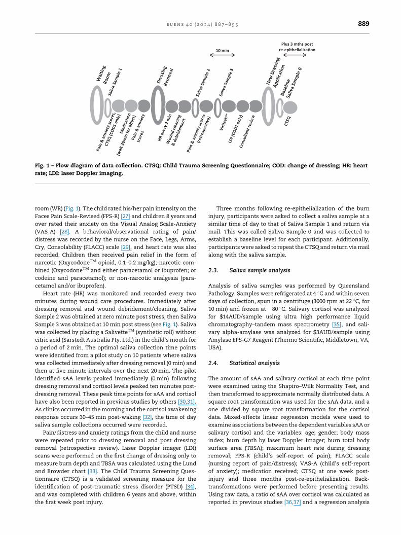
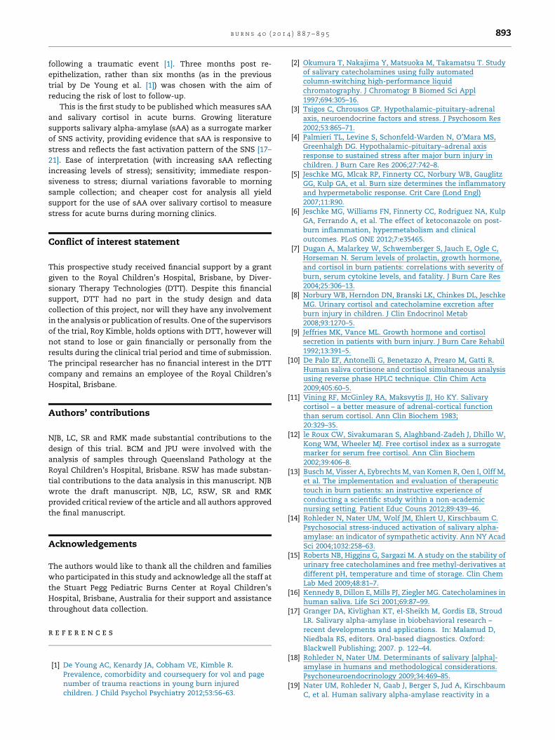
10 minPlus 3 mths post
re-epitheli aliza �on
Fig. 1 – Flow diagram of data collection. CTSQ: Child Trauma Screening Questionnaire; COD: change of dressing; HR: heart
rate; LDI: laser Doppler imaging.
b u r n s 4 0 ( 2 0 1 4 ) 8 8 7 – 8 9 5 889
room (WR) (Fig. 1). The child rated his/her pain intensity on the
Faces Pain Scale-Revised (FPS-R) [27] and children 8 years and
over rated their anxiety on the Visual Analog Scale-Anxiety
(VAS-A) [28]. A behavioral/observational rating of pain/
distress was recorded by the nurse on the Face, Legs, Arms,
Cry, Consolability (FLACC) scale [29], and heart rate was also
recorded. Children then received pain relief in the form of
narcotic (OxycodoneTM opioid, 0.1–0.2 mg/kg); narcotic com-
bined (OxycodoneTM and either paracetamol or ibuprofen; or
codeine and paracetamol); or non-narcotic analgesia (para-
cetamol and/or ibuprofen).
Heart rate (HR) was monitored and recorded every two
minutes during wound care procedures. Immediately after
dressing removal and wound debridement/cleaning, Saliva
Sample 2 was obtained at zero minute post stress, then Saliva
Sample 3 was obtained at 10 min post stress (see Fig. 1). Saliva
was collected by placing a SalivetteTM (synthetic roll) without
citric acid (Sarstedt Australia Pty. Ltd.) in the child’s mouth for
a period of 2 min. The optimal saliva collection time points
were identified from a pilot study on 10 patients where saliva
was collected immediately after dressing removal (0 min) and
then at five minute intervals over the next 20 min. The pilot
identified sAA levels peaked immediately (0 min) following
dressing removal and cortisol levels peaked ten minutes post-
dressing removal. These peak time points for sAA and cortisol
have also been reported in previous studies by others [30,31].
As clinics occurred in the morning and the cortisol awakening
response occurs 30–45 min post-waking [32], the time of day
saliva sample collections occurred were recorded.
Pain/distress and anxiety ratings from the child and nurse
were repeated prior to dressing removal and post dressing
removal (retrospective review). Laser Doppler imager (LDI)
scans were performed on the first change of dressing only to
measure burn depth and TBSA was calculated using the Lund
and Browder chart [33]. The Child Trauma Screening Ques-
tionnaire (CTSQ) is a validated screening measure for the
identification of post-traumatic stress disorder (PTSD) [34],
and was completed with children 6 years and above, within
the first week post injury.
Three months following re-epithelialization of the burn
injury, participants were asked to collect a saliva sample at a
similar time of day to that of Saliva Sample 1 and return via
mail. This was called Saliva Sample 0 and was collected to
establish a baseline level for each participant. Additionally,
participants were asked to repeat the CTSQ and return via mail
along with the saliva sample.
2.3. Saliva sample analysis
Analysis of saliva samples was performed by Queensland
Pathology. Samples were refrigerated at 4 8C and within seven
days of collection, spun in a centrifuge (3000 rpm at 22 8C, for
10 min) and frozen at �80 8C. Salivary cortisol was analyzed
for $14AUD/sample using ultra high performance liquid
chromatography–tandem mass spectrometry [35], and sali-
vary alpha-amylase was analyzed for $3AUD/sample using
Amylase EPS-G7 Reagent (Thermo Scientific, Middletown, VA,
USA).
2.4. Statistical analysis
The amount of sAA and salivary cortisol at each time point
were examined using the Shapiro–Wilk Normality Test, and
then transformed to approximate normally distributed data. A
square root transformation was used for the sAA data, and a
one divided by square root transformation for the cortisol
data. Mixed-effects linear regression models were used to
examine associations between the dependent variables sAA or
salivary cortisol and the variables: age; gender; body mass
index; burn depth by laser Doppler Imager; burn total body
surface area (TBSA); maximum heart rate during dressing
removal; FPS-R (child’s self-report of pain); FLACC scale
(nursing report of pain/distress); VAS-A (child’s self-report
of anxiety); medication received; CTSQ at one week post-
injury and three months post-re-epithelialization. Back-
transformations were performed before presenting results.
Using raw data, a ratio of sAA over cortisol was calculated as
reported in previous studies [36,37] and a regression analysis
b u r n s 4 0 ( 2 0 1 4 ) 8 8 7 – 8 9 5890
was performed on the ratio to examine stress system
dysregulation. All regression models contained three hier-
archical levels: (1) participant identification, (2) burn site on
each participant, (3) the change of dressing number (e.g. 1st,
2nd etc.). Analyses were conducted using Stata statistical
software version 12 (StataCorp, College Station, TX, USA).
3. Results
Data were collected on a total of 77 participants aged from 4
years 1 month to 12 years 9 months (Table 1). Laser Doppler
Images were able to be performed on 59 out of the 77
participants. The average blood flow of each burn wound was
calculated, and the median value was 1138 perfusion units
(PU), with the deepest PU reading in wounds ranging from 97
PU to 977 PU (Table 1). This indicates burn wounds were
primarily superficial partial thickness, with many wounds
identified to also have deeper areas. Reasons for not
performing scans on every participant included: movement
by the child; pain and anxiety experienced by the child; and
the hectic flow of burn clinics. Complete data sets of saliva
samples (i.e. three sample time points collected at every
dressing change until discharge from the burn clinic) were
collected on 72 of the 77 participants. Baseline saliva samples
(sample 0: collected three months post re-epithelialization)
relied on participants returning samples via mail and were
received from 57 participants (74%).
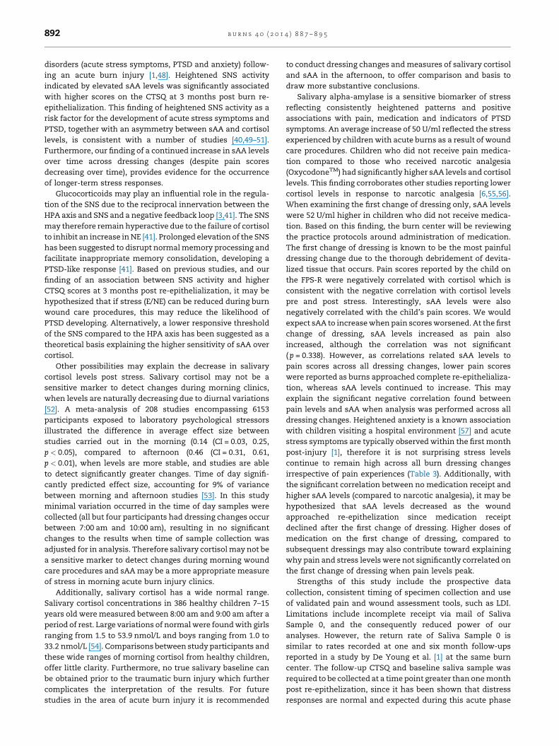
3.1. Biomarker responses to wound care procedures
Salivary cortisol and sAA were sensitive to stress, reflecting
significant changes during burn wound care procedures. Table
2 displays the association between all dressing changes and
Table 1 – Demographics and burn clinical characteristics.
Median (range)
Age (months) 97(50–153)
LDI average of wound area (PU) 1138(472–1699)
LDI deepest wound reading (PU) 624(97–997)
n (%)
Gender Male 46(60)
Ethnicity Caucasian 68(88)
Mechanism Scald 41(53)
Contact 25(33)
Flame 4(5)
Friction 6(8)
Chemical 1(1)
Site Axilla/upper limb 32(41)
Lower limb 23(30)
Chest/torso/back 16(21)
Genitals/buttocks 3(4)
Head/face 3(4)
Medication (COD 1 only) Narcotic 42(55)
Narcotic combined 20(26)
Non-narcotic 4(5)
Nil 11(14)
LDI, laser Doppler imaging; PU: perfusion units; and COD: change
of dressing.
stress biomarkers. For sAA, all recordings taken at each
dressing change were significantly higher ( p < 0.001) than
readings taken at three months post re-epithelialization.
Similar results were observed for salivary cortisol and the ratio
of sAA over cortisol, except for the waiting room measure
(Table 2).
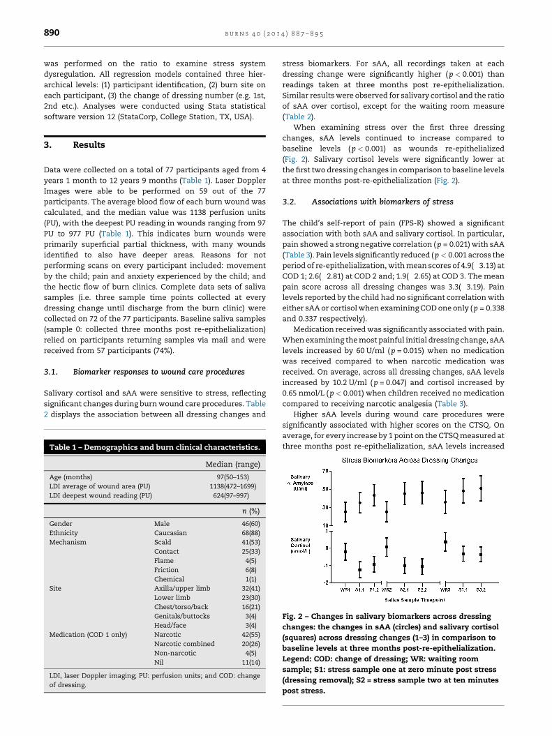
When examining stress over the first three dressing
changes, sAA levels continued to increase compared to
baseline levels ( p < 0.001) as wounds re-epithelialized
(Fig. 2). Salivary cortisol levels were significantly lower at
the first two dressing changes in comparison to baseline levels
at three months post-re-epithelialization (Fig. 2).
3.2. Associations with biomarkers of stress
The child’s self-report of pain (FPS-R) showed a significant
association with both sAA and salivary cortisol. In particular,
pain showed a strong negative correlation ( p = 0.021) with sAA
(Table 3). Pain levels significantly reduced ( p < 0.001 across the
period of re-epithelialization, with mean scores of 4.9(�3.13) at
COD 1; 2.6(�2.81) at COD 2 and; 1.9(�2.65) at COD 3. The mean
pain score across all dressing changes was 3.3(�3.19). Pain
levels reported by the child had no significant correlation with
either sAA or cortisol when examining COD one only ( p = 0.338
and 0.337 respectively).
Medication received was significantly associated with pain.
When examining the most painful initial dressing change, sAA
levels increased by 60 U/ml ( p = 0.015) when no medication
was received compared to when narcotic medication was
received. On average, across all dressing changes, sAA levels
increased by 10.2 U/ml ( p = 0.047) and cortisol increased by
0.65 nmol/L ( p < 0.001) when children received no medication
compared to receiving narcotic analgesia (Table 3).
Higher sAA levels during wound care procedures were
significantly associated with higher scores on the CTSQ. On
average, for every increase by 1 point on the CTSQ measured at
three months post re-epithelialization, sAA levels increased
Fig. 2 – Changes in salivary biomarkers across dressing
changes: the changes in sAA (circles) and salivary cortisol
(squares) across dressing changes (1–3) in comparison to
baseline levels at three months post-re-epithelialization.
Legend: COD: change of dressing; WR: waiting room
sample; S1: stress sample one at zero minute post stress
(dressing removal); S2 = stress sample two at ten minutes
post stress.
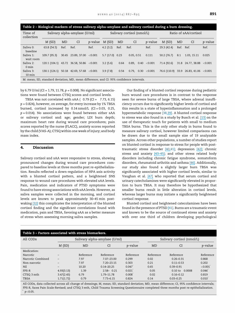
Table 2 – Biological markers of stress salivary alpha-amylase and salivary cortisol during a burn dressing.
Time ofcollection
Salivary alpha-amylase (U/ml) Salivary cortisol (nmol/L) Ratio of sAA/cortisol
M (SD) MD CI p-value M (SD) MD CI p-value M (SD) MD CI p-value
Saliva 0:
baseline
63.8 (54.5) Ref. Ref. Ref. 4.2 (3.2) Ref. Ref. Ref. 29.3 (42.4) Ref. Ref. Ref.
Saliva 1:
wait room
109.7 (95.3) 30.45 23.89, 37.00 <0.001 5.7 (17.0) 0.23 �0.05, 0.51 0.111 50.2 (76.7) 8.1 1.03, 15.11 0.025
Saliva 2:
0 min
120.1 (104.1) 43.72 36.58, 50.86 <0.001 3.2 (5.6) �0.64 �0.89, �0.40 <0.001 71.4 (92.6) 31.8 24.77, 38.88 <0.001
Saliva 3:
10 min
130.1 (124.1) 50.18 42.69, 57.68 <0.001 3.9 (7.8) �0.54 �0.79, �0.30 <0.001 76.6 (110.9) 33.9 26.83, 41.06 <0.001
M: mean; SD, standard deviation; MD, mean difference; and CI: 95% confidence intervals.
b u r n s 4 0 ( 2 0 1 4 ) 8 8 7 – 8 9 5 891
by 6.79 U/ml (CI = 1.79, 11.78, p = 0.008). No significant associa-
tions were found between CTSQ scores and cortisol levels.
TBSA was not correlated with sAA (�0.79 (CI = �7.73, 6.15)
p = 0.824), however, on average, for every increase by 1% TBSA
burned, cortisol increased by 0.14 nmol/L (CI = 0.03, 0.25,
p = 0.014). No associations were found between either sAA
or salivary cortisol and: age; gender; LDI burn depth;
maximum heart rate during wound care procedures; pain
scores reported by the nurse (FLACC), anxiety scores reported
by the child (VAS-A); CTSQ within one week of injury; and body
mass index.
4. Discussion
Salivary cortisol and sAA were responsive to stress, showing
pronounced changes during wound care procedures com-
pared to baseline levels at three months post re-epithelializa-
tion. Results reflected a down-regulation of HPA axis activity
with a blunted cortisol pattern, and a heightened SNS
response to wound care procedures with elevated sAA levels.
Pain, medication and indicators of PTSD symptoms were
found to have strong associations with sAA levels. However, as
saliva samples were collected in the morning, and cortisol
levels are known to peak approximately 30–45 min post-
waking [32] this complicates the interpretation of the blunted
cortisol finding and the significant correlations found with
medication, pain and TBSA, favoring sAA as a better measure
of stress when assessing morning saliva samples.
Table 3 – Factors associated with stress biomarkers.
All CODs Salivary alpha-amylase (U/ml)
M (SD) MD CI
Medication:
Narcotic – Reference Reference
Narcotic Combined – 7.97 �7.07–23.00
Non-narcotic – 7.97 �7.20–23.15
Nil – 10.20 0.14–20.25
FPS-R 4.93(3.13) �1.39 �2.58–�0.21
CTSQ 3 mth 3.47(2.45) 6.79 1.79–11.78
TBSA 1.71(1.72) �0.79 �7.73–6.15
All CODs, data collected across all change of dressings; M, mean; SD, sta
FPS-R, faces Pain Scale-Revised; and CTSQ 3 mth, Child Trauma Screenin* p<0.05.
Our finding of a blunted cortisol response during pediatric
burn wound care procedures is in contrast to the response
seen for severe burns of large TBSA, where adrenal insuffi-
ciency occurs due to significantly higher levels of cortisol and
this results in a state of hyperinflammation and a prolonged
hypermetabolic response [38,39]. A blunted cortisol response
to stress was also found in a study by Busch et al. [13] on the
use of therapeutic touch for patients with small to medium
TBSA burns. This is the only other study in burns found to
measure salivary cortisol, however limited comparisons can
be drawn due to the small sample size of 19 analyzable
samples. Across other populations, a number of studies report
on blunted cortisol in response to stress for people with post-
traumatic stress disorder [40,41]; depression [42]; chronic
stress and anxiety [43–45]; and other stress related body
disorders including chronic fatigue syndrome, somatoform
disorders, rheumatoid arthritis and asthma [46]. Additionally,
our study also found a slightly larger burn TBSA was
significantly associated with higher cortisol levels, similar to
Vaughan et al. [47] who reported that serum cortisol and
urinary catecholamines were significantly elevated in propor-
tion to burn TBSA. It may therefore be hypothesized that
smaller burns result in little alteration in cortisol levels,
whereas larger burns may initiate a significantly heightened
cortisol response.
Blunted cortisol and heightened catecolamines have been
found in the presence of PTSD [41]. Burns are a traumatic event
and known to be the source of continued stress and anxiety
with over one third of children developing psychological
Salivary cortisol (nmol/L)
p-value MD CI p-value
Reference Reference Reference Reference
0.299 0.02 �0.26–0.31 0.868
0.303 0.21 �0.11–0.53 0.202
0.047* 0.65 0.39–0.91 <0.001*
0.021* �0.05 �0.10 to �0.0008 0.046*
0.008* �0.02 �0.16–0.12 0.819
0.824 0.14 0.03–0.25 0.010*
ndard deviation; MD, mean difference; CI, 95% confidence intervals;
g Questionnaire completed three months post re-epithelialization.
b u r n s 4 0 ( 2 0 1 4 ) 8 8 7 – 8 9 5892
disorders (acute stress symptoms, PTSD and anxiety) follow-
ing an acute burn injury [1,48]. Heightened SNS activity
indicated by elevated sAA levels was significantly associated
with higher scores on the CTSQ at 3 months post burn re-
epithelialization. This finding of heightened SNS activity as a
risk factor for the development of acute stress symptoms and
PTSD, together with an asymmetry between sAA and cortisol
levels, is consistent with a number of studies [40,49–51].
Furthermore, our finding of a continued increase in sAA levels
over time across dressing changes (despite pain scores
decreasing over time), provides evidence for the occurrence
of longer-term stress responses.
Glucocorticoids may play an influential role in the regula-
tion of the SNS due to the reciprocal innervation between the
HPA axis and SNS and a negative feedback loop [3,41]. The SNS
may therefore remain hyperactive due to the failure of cortisol
to inhibit an increase in NE [41]. Prolonged elevation of the SNS
has been suggested to disrupt normal memory processing and
facilitate inappropriate memory consolidation, developing a
PTSD-like response [41]. Based on previous studies, and our
finding of an association between SNS activity and higher
CTSQ scores at 3 months post re-epithelialization, it may be
hypothesized that if stress (E/NE) can be reduced during burn
wound care procedures, this may reduce the likelihood of
PTSD developing. Alternatively, a lower responsive threshold
of the SNS compared to the HPA axis has been suggested as a
theoretical basis explaining the higher sensitivity of sAA over
cortisol.
Other possibilities may explain the decrease in salivary
cortisol levels post stress. Salivary cortisol may not be a
sensitive marker to detect changes during morning clinics,
when levels are naturally decreasing due to diurnal variations
[52]. A meta-analysis of 208 studies encompassing 6153
participants exposed to laboratory psychological stressors
illustrated the difference in average effect size between
studies carried out in the morning (0.14 (CI = 0.03, 0.25,
p < 0.05), compared to afternoon (0.46 (CI = 0.31, 0.61,
p < 0.01), when levels are more stable, and studies are able
to detect significantly greater changes. Time of day signifi-
cantly predicted effect size, accounting for 9% of variance
between morning and afternoon studies [53]. In this study
minimal variation occurred in the time of day samples were
collected (all but four participants had dressing changes occur
between 7:00 am and 10:00 am), resulting in no significant
changes to the results when time of sample collection was
adjusted for in analysis. Therefore salivary cortisol may not be
a sensitive marker to detect changes during morning wound
care procedures and sAA may be a more appropriate measure
of stress in morning acute burn injury clinics.
Additionally, salivary cortisol has a wide normal range.
Salivary cortisol concentrations in 386 healthy children 7–15
years old were measured between 8:00 am and 9:00 am after a
period of rest. Large variations of normal were found with girls
ranging from 1.5 to 53.9 nmol/L and boys ranging from 1.0 to
33.2 nmol/L [54]. Comparisons between study participants and
these wide ranges of morning cortisol from healthy children,
offer little clarity. Furthermore, no true salivary baseline can
be obtained prior to the traumatic burn injury which further
complicates the interpretation of the results. For future
studies in the area of acute burn injury it is recommended
to conduct dressing changes and measures of salivary cortisol
and sAA in the afternoon, to offer comparison and basis to
draw more substantive conclusions.
Salivary alpha-amylase is a sensitive biomarker of stress
reflecting consistently heightened patterns and positive
associations with pain, medication and indicators of PTSD
symptoms. An average increase of 50 U/ml reflected the stress
experienced by children with acute burns as a result of wound
care procedures. Children who did not receive pain medica-
tion compared to those who received narcotic analgesia
(OxycodoneTM) had significantly higher sAA levels and cortisol
levels. This finding corroborates other studies reporting lower
cortisol levels in response to narcotic analgesia [6,55,56].
When examining the first change of dressing only, sAA levels
were 52 U/ml higher in children who did not receive medica-
tion. Based on this finding, the burn center will be reviewing
the practice protocols around administration of medication.
The first change of dressing is known to be the most painful
dressing change due to the thorough debridement of devita-
lized tissue that occurs. Pain scores reported by the child on
the FPS-R were negatively correlated with cortisol which is
consistent with the negative correlation with cortisol levels
pre and post stress. Interestingly, sAA levels were also
negatively correlated with the child’s pain scores. We would
expect sAA to increase when pain scores worsened. At the first
change of dressing, sAA levels increased as pain also
increased, although the correlation was not significant
( p = 0.338). However, as correlations related sAA levels to
pain scores across all dressing changes, lower pain scores
were reported as burns approached complete re-epithelializa-
tion, whereas sAA levels continued to increase. This may
explain the significant negative correlation found between
pain levels and sAA when analysis was performed across all
dressing changes. Heightened anxiety is a known association
with children visiting a hospital environment [57] and acute
stress symptoms are typically observed within the first month
post-injury [1], therefore it is not surprising stress levels
continue to remain high across all burn dressing changes
irrespective of pain experiences (Table 3). Additionally, with
the significant correlation between no medication receipt and
higher sAA levels (compared to narcotic analgesia), it may be
hypothesized that sAA levels decreased as the wound
approached re-epithelization since medication receipt
declined after the first change of dressing. Higher doses of
medication on the first change of dressing, compared to
subsequent dressings may also contribute toward explaining
why pain and stress levels were not significantly correlated on
the first change of dressing when pain levels peak.
Strengths of this study include the prospective data
collection, consistent timing of specimen collection and use
of validated pain and wound assessment tools, such as LDI.
Limitations include incomplete receipt via mail of Saliva
Sample 0, and the consequently reduced power of our
analyses. However, the return rate of Saliva Sample 0 is
similar to rates recorded at one and six month follow-ups
reported in a study by De Young et al. [1] at the same burn
center. The follow-up CTSQ and baseline saliva sample was
required to be collected at a time point greater than one month
post re-epithelization, since it has been shown that distress
responses are normal and expected during this acute phase
b u r n s 4 0 ( 2 0 1 4 ) 8 8 7 – 8 9 5 893
following a traumatic event [1]. Three months post re-
epithelization, rather than six months (as in the previous
trial by De Young et al. [1]) was chosen with the aim of
reducing the risk of lost to follow-up.
This is the first study to be published which measures sAA
and salivary cortisol in acute burns. Growing literature
supports salivary alpha-amylase (sAA) as a surrogate marker
of SNS activity, providing evidence that sAA is responsive to
stress and reflects the fast activation pattern of the SNS [17–
21]. Ease of interpretation (with increasing sAA reflecting
increasing levels of stress); sensitivity; immediate respon-
siveness to stress; diurnal variations favorable to morning
sample collection; and cheaper cost for analysis all yield
support for the use of sAA over salivary cortisol to measure
stress for acute burns during morning clinics.
Conflict of interest statement
This prospective study received financial support by a grant
given to the Royal Children’s Hospital, Brisbane, by Diver-
sionary Therapy Technologies (DTT). Despite this financial
support, DTT had no part in the study design and data
collection of this project, nor will they have any involvement
in the analysis or publication of results. One of the supervisors
of the trial, Roy Kimble, holds options with DTT, however will
not stand to lose or gain financially or personally from the
results during the clinical trial period and time of submission.
The principal researcher has no financial interest in the DTT
company and remains an employee of the Royal Children’s
Hospital, Brisbane.
Authors’ contributions
NJB, LC, SR and RMK made substantial contributions to the
design of this trial. BCM and JPU were involved with the
analysis of samples through Queensland Pathology at the
Royal Children’s Hospital, Brisbane. RSW has made substan-
tial contributions to the data analysis in this manuscript. NJB
wrote the draft manuscript. NJB, LC, RSW, SR and RMK
provided critical review of the article and all authors approved
the final manuscript.
Acknowledgements
The authors would like to thank all the children and families
who participated in this study and acknowledge all the staff at
the Stuart Pegg Pediatric Burns Center at Royal Children’s
Hospital, Brisbane, Australia for their support and assistance
throughout data collection.
r e f e r e n c e s
[1] De Young AC, Kenardy JA, Cobham VE, Kimble R.Prevalence, comorbidity and coursequery for vol and pagenumber of trauma reactions in young burn injuredchildren. J Child Psychol Psychiatry 2012;53:56–63.
[2] Okumura T, Nakajima Y, Matsuoka M, Takamatsu T. Studyof salivary catecholamines using fully automatedcolumn-switching high-performance liquidchromatography. J Chromatogr B Biomed Sci Appl1997;694:305–16.
[3] Tsigos C, Chrousos GP. Hypothalamic–pituitary–adrenalaxis, neuroendocrine factors and stress. J Psychosom Res2002;53:865–71.
[4] Palmieri TL, Levine S, Schonfeld-Warden N, O’Mara MS,Greenhalgh DG. Hypothalamic–pituitary–adrenal axisresponse to sustained stress after major burn injury inchildren. J Burn Care Res 2006;27:742–8.
[5] Jeschke MG, Mlcak RP, Finnerty CC, Norbury WB, GauglitzGG, Kulp GA, et al. Burn size determines the inflammatoryand hypermetabolic response. Crit Care (Lond Engl)2007;11:R90.
[6] Jeschke MG, Williams FN, Finnerty CC, Rodriguez NA, KulpGA, Ferrando A, et al. The effect of ketoconazole on post-burn inflammation, hypermetabolism and clinicaloutcomes. PLoS ONE 2012;7:e35465.
[7] Dugan A, Malarkey W, Schwemberger S, Jauch E, Ogle C,Horseman N. Serum levels of prolactin, growth hormone,and cortisol in burn patients: correlations with severity ofburn, serum cytokine levels, and fatality. J Burn Care Res2004;25:306–13.
[8] Norbury WB, Herndon DN, Branski LK, Chinkes DL, JeschkeMG. Urinary cortisol and catecholamine excretion afterburn injury in children. J Clin Endocrinol Metab2008;93:1270–5.
[9] Jeffries MK, Vance ML. Growth hormone and cortisolsecretion in patients with burn injury. J Burn Care Rehabil1992;13:391–5.
[10] De Palo EF, Antonelli G, Benetazzo A, Prearo M, Gatti R.Human saliva cortisone and cortisol simultaneous analysisusing reverse phase HPLC technique. Clin Chim Acta2009;405:60–5.
[11] Vining RF, McGinley RA, Maksvytis JJ, Ho KY. Salivarycortisol – a better measure of adrenal-cortical functionthan serum cortisol. Ann Clin Biochem 1983;20:329–35.
[12] le Roux CW, Sivakumaran S, Alaghband-Zadeh J, Dhillo W,Kong WM, Wheeler MJ. Free cortisol index as a surrogatemarker for serum free cortisol. Ann Clin Biochem2002;39:406–8.
[13] Busch M, Visser A, Eybrechts M, van Komen R, Oen I, Olff M,et al. The implementation and evaluation of therapeutictouch in burn patients: an instructive experience ofconducting a scientific study within a non-academicnursing setting. Patient Educ Couns 2012;89:439–46.
[14] Rohleder N, Nater UM, Wolf JM, Ehlert U, Kirschbaum C.Psychosocial stress-induced activation of salivary alpha-amylase: an indicator of sympathetic activity. Ann NY AcadSci 2004;1032:258–63.
[15] Roberts NB, Higgins G, Sargazi M. A study on the stability ofurinary free catecholamines and free methyl-derivatives atdifferent pH, temperature and time of storage. Clin ChemLab Med 2009;48:81–7.
[16] Kennedy B, Dillon E, Mills PJ, Ziegler MG. Catecholamines inhuman saliva. Life Sci 2001;69:87–99.
[17] Granger DA, Kivlighan KT, el-Sheikh M, Gordis EB, StroudLR. Salivary alpha-amylase in biobehavioral research –recent developments and applications. In: Malamud D,Niedbala RS, editors. Oral-based diagnostics. Oxford:Blackwell Publishing; 2007. p. 122–44.
[18] Rohleder N, Nater UM. Determinants of salivary [alpha]-amylase in humans and methodological considerations.Psychoneuroendocrinology 2009;34:469–85.
[19] Nater UM, Rohleder N, Gaab J, Berger S, Jud A, KirschbaumC, et al. Human salivary alpha-amylase reactivity in a
b u r n s 4 0 ( 2 0 1 4 ) 8 8 7 – 8 9 5894
psychosocial stress paradigm. Int J Psychophysiol2005;55:333–42.
[20] van Stegeren A, Rohleder N, Everaerd W, Wolf OT. Salivaryalpha amylase as marker for adrenergic activity duringstress: effect of betablockade. Psychoneuroendocrinology2006;31:137–41.
[21] Nater UM, La Marca R, Florin L, Moses A, Langhans W,Koller MM, et al. Stress-induced changes in human salivaryalpha-amylase activity – associations with adrenergicactivity. Psychoneuroendocrinology 2006;31:49–58.
[22] Nater UM, Rohleder N. Salivary alpha-amylase as a non-invasive biomarker for the sympathetic nervous system:current state of research. Psychoneuroendocrinology2009;34:486–96.
[23] Rohleder N, Wolf J, Maldonado E, Kirschbaum C. Thepsychosocial stress induced increase in salivary alphaamylase is independent of saliva flow rate.Psychophysiology 2006;43:645–52.
[24] Brown NJ, Rodger S, Ware RS, Kimble RM, Cuttle L. Efficacyof a children’s procedural preparation and distractiondevice on healing in acute burn wound care procedures:study protocol for a randomized controlled trial. Trials2012;13:238.
[25] Brown N, Rodger S, Ware R, Kimble R, Cuttle L. Play andheal randomized controlled trial of DittoTM interventionefficacy on improving re-epithelialization in pediatric burninjuries. Burns 2013, http://dx.doi.org/10.1016/j.burns.2013.11.024.
[26] Brown NJ, Kimble RM, Gramotnev G, Rodger S, Cuttle L.Predictors of re-epithelialization in pediatric burn. Burns2013, http://dx.doi.org/10.1016/j.burns.2013.09.027.
[27] Hicks CL, von Baeyer CL, Spafford PA, van Korlaar I,Goodenough B. The faces pain scale – revised: toward acommon metric in pediatric pain measurement. Pain2001;93:173–83.
[28] Bringuier S, Dadure C, Raux O, Dubois A, Picot MC, CapdevilaX. The perioperative validity of the visual analog anxietyscale in children: a discriminant and useful instrument inroutine clinical practice to optimize postoperative painmanagement. Anesth Analg 2009;109:737.
[29] Nilsson S, Finnstrom B, Kokinsky E, Nilsson S, et al. TheFLACC behavioral scale for procedural pain assessmentin children aged 5–16 years. Paediatr Anaesth 2008;18:767–74.
[30] Plessow F, Kiesel A, Kirschbaum C. The stressed prefrontalcortex and goal-directed behaviour: acute psychosocialstress impairs the flexible implementation of task goals.Exp Brain Res 2012;216:397–408.
[31] Gordis EB, Granger DA, Susman EJ, Trickett PK. Asymmetrybetween salivary cortisol and alpha-amylase reactivity tostress: relation to aggressive behavior in adolescents.Psychoneuroendocrinology 2006;31:976–87.
[32] Steptoe A, van Jaarsveld CHM, Semmler C, Plomin R,Wardle J. Heritability of daytime cortisol levels and cortisolreactivity in children. Psychoneuroendocrinology2009;34:273–80.
[33] Lund CC, Browder NC. The estimation of areas of burns.Surg Gynecol Obstet 1944;79:352–8.
[34] Kenardy JA, Spence SH, Macleod AC. Screening forposttraumatic stress disorder in children after accidentalinjury. Pediatrics 2006;118:1002–9.
[35] McWhinney BC, Briscoe SE, Ungerer JPJ, Pretorius CJ.Measurement of cortisol, cortisone, prednisolone,dexamethasone and 11-deoxycortisol with ultra highperformance liquid chromatography–tandem massspectrometry: application for plasma, plasma ultrafiltrate,urine and saliva in a routine laboratory. J Chromatogr BAnalyt Technol Biomed Life Sci 2010;878:2863–9.
[36] Pruessner JC, Kirschbaum C, Meinlschmid G, HellhammerDH. Two formulas for computation of the area under thecurve represent measures of total hormone concentrationversus time-dependent change. Psychoneuroendocrinology2003;28:916–31.
[37] Ali N, Pruessner JC. The salivary alpha amylase overcortisol ratio as a marker to assess dysregulations of thestress systems. Physiol Behav 2012;106:65–72.
[38] Jeschke MG, Gauglitz GG, Kulp GA, Finnerty CC, WilliamsFN, Kraft R, et al. Long-term persistance of thepathophysiologic response to severe burn injury. PLoS ONE2011;6:e21245.
[39] Williams FN, Herndon DN, Jeschke MG. Thehypermetabolic response to burn injury and interventionsto modify this response. Clin Plast Surg 2009;36:583–96.
[40] McFarlane AC, Barton CA, Yehuda R, Wittert G. Cortisolresponse to acute trauma and risk of posttraumaticstress disorder. Psychoneuroendocrinology 2010;36:720–7.
[41] Yehuda R, McFarlane AC, Shalev AY. Predicting thedevelopment of posttraumatic stress disorder from theacute response to a traumatic event. Biol Psychiatry1998;44:1305–13.
[42] Burke HM, Davis MC, Otte C, Mohr DC. Depression andcortisol responses to psychological stress: a meta-analysis.Psychoneuroendocrinology 2005;30:846–56.
[43] Spies LA, Margolin G, Susman EJ, Gordis EB. Adolescents’cortisol reactivity and subjective distress in response tofamily conflict: the moderating role of internalizingsymptoms. J Adolesc Health 2011;49:386–92.
[44] Tomiyama AJ, Dallman MF, Epel ES. Comfort food iscomforting to those most stressed: evidence of the chronicstress response network in high stress women.Psychoneuroendocrinology 2011;36:1513–9.
[45] Elzinga BM, Roelofs K, Tollenaar MS, Bakvis P, van Pelt J,Spinhoven P. Diminished cortisol responses topsychosocial stress associated with lifetime adverse eventsa study among healthy young subjects.Psychoneuroendocrinology 2008;33:227–37.
[46] Heim C, Ehlert U, Hellhammer DH. The potential role ofhypocortisolism in the pathophysiology of stress-relatedbodily disorders. Psychoneuroendocrinology 2000;25:1–35.
[47] Vaughan GM, Becker RA, Allen JP, Goodwin Jr CW, Pruitt JrBA, Mason Jr AD. Cortisol and corticotrophin in burnedpatients. J Trauma 1982;22:263–73.
[48] Stoddard FJ, Saxe G, Ronfeldt H, Drake JE, Burns J, Edgren C,et al. Acute stress symptoms in young children with burns.J Am Acad Child Adolesc Psychiatry 2006;45:87–93.
[49] Yehuda R, Southwick SM, Nussbaum G, Wahby VS. Lowurinary cortisol excretion in patients with posttraumaticstress disorder. J Nerv Ment Dis; 1990;178(6):366–9.
[50] Brown MR, Fisher LA. Glucocorticoid suppression of thesympathetic nervous system and adrenal medulla. Life Sci1986;39:1003–12.
[51] Kosten TR, Mason JW, Giller EL, Ostroff RB, Harkness L.Sustained urinary norepinephrine and epinephrineelevation in post-traumatic stress disorder.Psychoneuroendocrinology 1987;12:13–20.
[52] Levine A, Zagoory-Sharon O, Feldman R, Lewis JG, Weller A.Measuring cortisol in human psychobiological studies.Physiol Behav 2007;90:43–53.
[53] Dickerson SS, Kemeny ME. Acute stressors and cortisolresponses: a theoretical integration and synthesis oflaboratory research. Psychol Bull 2004;130:355.
[54] Tornhage CJ. Reference values for morning salivary cortisolconcentrations in healthy school-aged children. J PediatrEndocrinol 2002;15:197–204.
b u r n s 4 0 ( 2 0 1 4 ) 8 8 7 – 8 9 5 895
[55] Buckingham J. Secretion of corticotrophin and itshypothalamic releasing factor in response to morphineand opioid peptides. Neuroendocrinology 1982;35:111–6.
[56] Delitala G, Grossman A, Besser M. Differential effects ofopiate peptides and alkaloids on anterior pituitary
hormone secretion. Neuroendocrinology 1983;37:275–9.
[57] Rasnake LK, Linscheid TR. Anxiety reduction in childrenreceiving medical care: developmental considerations. JDev Behav Pediatr 1989;10:169–75.