ACDP/99/P6
1
Lyme Disease and services in the HPA
Contents Introduction ........................................................................................................................................... 3
Background ........................................................................................................................................ 3
Pathogenesis of Lyme disease ............................................................................................................... 3
Table 1: Properties of selected Borrelia antigens and appearance of antibodies in European patients .......................................................................................................................................... 6
Epidemiology ......................................................................................................................................... 7
Clinical features of Lyme Disease .......................................................................................................... 7
Initial presentation ............................................................................................................................ 7
Case definitions ................................................................................................................................. 7
Arthritis .............................................................................................................................................. 8
Table 2: Clinical case definitions for European Lyme Borreliosis ................................................... 9
Carditis ............................................................................................................................................. 10
Neuroborreliosis .............................................................................................................................. 10
Classification of neuroborreliosis (Mygland et al 2010) .............................................................. 10
Post Lyme syndrome ....................................................................................................................... 11
Differential diagnosis of Lyme: ........................................................................................................ 11
Human Granulocytic Anaplasmosis (HGA) ................................................................................... 11
Babesiosis .................................................................................................................................... 11
Louping ill ..................................................................................................................................... 12
Tick borne encephalitis and CCHF ............................................................................................... 12
Diagnosis of Lyme ................................................................................................................................ 12
Serological Tests .............................................................................................................................. 12
ELISA ............................................................................................................................................ 13
Immunoblot ................................................................................................................................. 13
Two Tier Testing .......................................................................................................................... 14
New approaches to serodiagnosis ................................................................................................... 16
VIsE C6 Peptide Elisa .................................................................................................................... 16
Open Government Status: Open

ACDP/99/P6
2
When to perform serological tests .............................................................................................. 16
Other Diagnostic Tests ..................................................................................................................... 16
Culture ......................................................................................................................................... 16
Molecular methods ..................................................................................................................... 17
Testing practice at RIPL Porton Down ............................................................................................. 17
Treatment of Lyme disease ................................................................................................................. 18
Post Lyme Disease Syndromes, Chronic Lyme disease and related issues .......................................... 21
Chronic Lyme disease ...................................................................................................................... 21
Alternative tests for Lyme disease ................................................................................................... 22
Lyme disease without supporting evidence .................................................................................... 23
Meeting the public need: the HPA future plans .................................................................................. 23
Summary .............................................................................................................................................. 24
References ........................................................................................................................................... 25

ACDP/99/P6
3
Introduction The service for Lyme Disease testing in the UK was transferred form Southampton HPA laboratory to Porton Down in June 2012. The HPA, in common with other national laboratories and healthcare providers in Europe and the US, has been the subject of considerable criticism by protagonists for Lyme disease support groups in maintaining a scientifically based approach to the condition and ignoring “alternative” therapies. This antipathy is also directed against infectious disease physicians, neurologists, rheumatologists and others who do not offer the answers that some individuals seek. This paper sets out the current position and plans for the service in its new home.
Background The National Lyme Disease service was transferred to the Rare & Imported Pathogens Laboratory (RIPL) at HPA Porton Down on 1 June 2012. Lyme Disease has become an emotive subject in many countries, and this paper sets out the approach taken by RIPL to address the needs for an accurate and timely diagnostic service, taking into account the tests available and their interpretation and limitations at different stages of the disease. There is a very vocal group of Lyme “sufferers” who have significant concerns about Lyme diagnostics in general, and the efforts made to meet their demands. This has led to a flourishing market in alternative tests and treatment regimens available through private providers, many of which are highly questionable in their accuracy, effectiveness and even safety. A recent publication on the increasing lobby for unproven treatments in Lyme Disease in the Lancet (Auwerter, 2012) drew a vociferous response from Lyme lobbyists (Tuttle, Perronne, Huyshe-‐Shires).
RIPL staff intend that the Service should address valid issues as far as is technically possible, acknowledging that there is room for improvement, and at the same time maintain the best possible scientific analysis and clinical advice based on the available evidence. As a significant proportion of self-‐acclaimed Lyme sufferers are self diagnosed, with no objective evidence of infection, it is essential to develop protocols that identify true cases, and refer those with other conditions sympathetically but firmly to appropriate practitioners for their problems.
Pathogenesis of Lyme disease Lyme disease in Europe is caused by spirochetes of three species: Borrelia burgdorferi sensu stricto, B. garinii and B. afzelii. The term B. burgdorferi sensu lato covers all three species and a number of other related organisms. The latter two predominate in Europe, and there may be considerable heterogeneity even in localised geographical areas. B. burgdorferi s.l. infect sheep ticks (Ixodes ricinus) and related hard ticks when they feed on infected animals. Larvae and nymphs feed mainly on rodents, and adults on larger animals; some also feed on migratory birds which act not only as reservoirs but can transport the ticks across wide geographical areas. There is some association between different sub-‐species of Borrelia and their preferred reservoir host, B. afzelii having a predilection for small rodents and B. garinii for birds. (Wilske, 2005)

ACDP/99/P6
4
In the midgut of the tick the spirochetes bind complement regulators and prevent the formation of toxic activation products, predominantly expressing the ospA protein (outer surface protein A) on their surface. When the tick feeds on mammalian or avian blood, the organism switches to expressing the ospC surface protein which binds mammalian plasminogen, facilitating the organism’s migration from the gut to its new host. This process generally takes >36 hours in the Amercian vector, I. scapularis, but Borrelia can be transmitted by I. ricinus nymphs in as little as 17 hours. Larvae are rarely implicated in transmission because although transovarial transmission occurs within ticks, the majority of tick infections arise when nymphs and adults feed on infected hosts.
During the migration through the tick’s gut, ospC binds a tick salivary protein which coats the Borrelia, protecting them against the non-‐specific immune defence in their new host. The spirochetes reproduce around the bite area, and interact with the local dendritic cells inducing a specific immune response. Antibodies against ospC are therefore amongst the first to appear as the immune response develops (Wagner et al.). The local infection causes an inflammatory response which manifests as the skin rash, erythema migrans (EM), characteristic of Lyme disease. As the infection progresses, additional antibodies appear to other components of the organism (Table 1). The different species give rise to different patterns of antibody responses. The combined innate and adaptive immune response may clear the infection, or it may progress to disseminated infection. Treatment during the early stages prevents the later antigens appearing, and effectively abolishes the immune reaction against them. For this reason, many patients with EM who have been treated do not react in standard laboratory tests.
Within days to weeks, spirochetes may disseminate through the blood to many sites, and during this phase the organism can be detected in the blood and sometimes the CSF. Small numbers of organisms have been seen in myocardium, retina, muscle, bone, spleen, liver, meninges and brain (Duray and Steere, 1988). The dispersion pattern is reflected in the clinical manifestations of this stage, mainly neurological symptoms, arthralgia and occasionally cardiac. Amongst the binding proteins expressed decorin binding proteins A and B (DpbA and DpbB) produced by Borrelia bind decorin, a proteoglycan found on the surface of collagen fibrils in the heart, nervous system and in joints. The pathological damage appears to be largely immune mediated, and the affected tissues show vasculitis, and an infiltration of plasma cells and lymphocytes. Borrelia have not been identified inside cells in tissue samples, despite reports that they can be cultured intracellularly in vitro.
Over time, the immune system usually eliminates the pathogens, although recovery can be prolonged. In a few cases, the spirochetes may linger in affected tissues and cause late Lyme disease, typically in the CNS or joints. In this situation, there is usually a strong immune response, often to multiple antigens and in addition to IgG antibodies IgM may be present in severe cases for over a year after the initial infection. The intensity of the immune reaction depends on the duration and extent of the clinical symptoms,. being stronger in disseminated disease and those patients with large numbers of EM lesions (Wormser et al., 2008). The prolonged course of infection in some cases reflects the ability of the Lyme Borrelia to change their antigenic expression and to inhibit the complement system. Two plasmids, lp25 encoding a nicotinamidase (Purser et al., 2003) and lp28-‐1 coding the VlsE surface protein are essential for this activity. VlsE varies substantially (there is less than 50% homology between genospecies), is a key antigen in the immune response and hence

ACDP/99/P6
5
antibodies to it are used in diagnosis based on a highly conserved region of the main protein (Bankhead and Chaconas, 2007). B. afzelii and B. burgdorferi also have complement regulating proteins that inactivate C3b and reduce complement mediated killing of the bacteria .

ACDP/99/P6
6
Table 1: Properties of selected Borrelia antigens and appearance of antibodies in European patients Antigen or peptide
Range % positive in each Lyme Borreliosis stage Comments EM Neurological Late
IgM IgG IgM IgG IgM IgG OspC 35-‐44 5-‐35 47-‐53 6-‐33 7-‐53 3-‐60 Early antigen; short duration in resolving infection pepC10 36 5 45 8 0 0 P41 † † 49 34-‐60 Not specific ‡C6 (VlsE) *Acute:
45-‐74 Conv: 70-‐90
64 Acute: 94-‐100 Conv: 83-‐98
Develops later than ospC and persist for much longer in convalescent or treated patients
DbpA 9 17 100 96-‐100 Collagen binding ag; see text FlaA 27 58 74 37 79 Flagellar antigen; cross-‐reacts with T pallidum$
P39 (BmpA) Early antigen, highly specific@ The table illustrates the variability of antigens at different stages of the disease. It is apparent that any single antigen e.g. C6 or OspC in an ELisA test will miss a small proportion of true cases depending on the time the specimen is taken.
Notes:
†In RIPL experience this is commonly positive in early infections. The antigen is seen in many other organisms and is not specific
‡C6 is the recombinant antigen for a highly conserved portion of the VlsE antigen, and represents all Lyme Borrelia
*US data given as no separate EU data
$ (Ge et al., 1998)
@ (Aguero-‐Rosenfeld et al., 1993)
Table derived in part modified from (Aguero-‐Rosenfeld et al., 2005)

ACDP/99/P6
7
Epidemiology Lyme disease occurs over most of the UK, although specific areas including the New Forest, Norfolk (Thetford Forest) and parts of the Lake District and Scottish Highlands have higher incidences of infection. Essentially most areas of heath, parkland or forests can harbour the vectors and animal host, and occasional cases appear in “non-‐Lyme areas”. The true incidence in the UK is unknown, as many cases appear as EM and are treated, and an un-‐definable number are not diagnosed, many of which will self-‐limit. The number of cases in England and Wales has risen slowly to around 900 confirmed cases a year as awareness increases. Epidemiological data is collected by Rob Smith at Public health Wales for the whole UK. Appendix 1 contains his complete review.
Clinical features of Lyme Disease Borrelia burgdorferi infection is acquired by a tick bite. However all ticks do not carry the organism and hence the chance of developing the disease varies with the regional prevalence of Borrelia burgdorferi in ticks. Since the UK has a low prevalence, not all tick bites will lead to infection.
Transmission can occur from infected ticks in less than a day, although prompt removal within a few hours will usually prevent this.
The disease can be arbitrarily divided into early manifestations which include a typical rash (erythema migrans), acute neuroborreliosis or Lyme carditis and late manifestations like arthritis, acrodermatitis chronic atrophicans and some rare manifestations of neurological disease. However this distinction is not always seen in clinical practice as some patients may not remember an acute phase of illness (Stanek et al, 2012).
Initial presentation If infection has been transmitted, most patients (>85%) develop a rash (erythema migrans) 3-‐30 days after the bite. This rash is typically annular and expands over a few days to weeks, though atypical rashes are also seen. This rash must be differentiated from the allergic reaction after a bite and other rashes like fungal infections and contact dermatitis. If a rash is noticed by a patient, it should be shown to a GP to establish whether it is erythema migrans and an appropriate decision for treatment should be made.
Along with erythema migrans, other symptoms and signs may be present including fatigue, myalgia, arthralgia, headache, fever, neck stiffness or lymphadenopathy. However, erythema migrans is the only feature which can enable a reliable clinical diagnosis, the others having a low diagnostic value (Stanek and Strle, 2003).
Case definitions A specialist group comprising of members from all over Europe has recently published clinical case definitions for the diagnosis and management of Lyme borreliosis with details on evidence needed to define each aspect of the presentation (Stanek et al 2011) . Table 2 has been adapted from their recommendations.

ACDP/99/P6
8
Arthritis Lyme arthritis generally manifests as recurrent or long standing synovitis. Arthralgia, myalgia or fibromyalgia may accompany joint swelling but alone are not sufficient for diagnosis. The commonest joint affected is the knee. Polyarthritis , although described in literature is not common and other diagnoses must be ruled out first. Other differential diagnoses of arthritis like osteoarthritis, spondyloarthritis, trauma, septic arthritis and crystal induced arthritis must be ruled out before a diagnosis of Lyme arthritis can be made.
Some patients may have prolonged joint inflammation which is postulated to be due to post infectious immunological derangement rather than active infection; hence antibiotics are unlikely to be of use (Steere et al 1994) in such a situation.

ACDP/99/P6
9
Table 2: Clinical case definitions for European Lyme Borreliosis Term Clinical case definition Essential lab
evidence Supporting evidence
Erythema migrans Red/ bluish red patch with or without central clearing Distinct edge intensely coloured, not markedly elevated
None Positive culture/PCR of skin biopsy
Borrelial lymphocytoma Painless bluish red nodule/plaque greater incidence in children, usually on ear lobe, ear helix, nipple or scrotum.
Seroconversion Histology
Positive culture /PCR of skin biopsy Recent EM Histology
Acrodermatitis chronica atrophicans
Long standing red or bluish-‐red lesions, usually on the extensor surface of extremities, Lesions may eventually atrophy. Possible skin indurations and fibroid nodules over bony prominences
High level IgG Positive culture /PCR of skin biopsy Histology
Lyme neuroborreliosis Meningo radiculitis, meningitis, encephalitis, cerebral vasculitis in children. Meningitis and facial palsy more frequent symptoms in children
CSF pleocytosis Intrathecal antibodies to Borrelia burgdorferi
Positive culture /PCR on CSF Intrathecal production of antibodies Specific serum antibodies Recent EM
Lyme arthritis Recurrent or persisting objective joint swelling in one or a few large joints. Other possible diagnoses excluded
Specific serum IgG antibodies usually in high concentrations
Positive culture /PCR on synovial fluid
Lyme carditis Av conduction disturbances of acute onset(I-‐III), rhythm disturbances, myocarditis, pancarditis. Other possible diagnoses excluded
Specific serum antibodies
Positive culture /PCR on endomyocardial biopsy Recent EM Concomitant neurological disorders
Ocular manifestation Conjunctivitis, uveitis, papillitis, episcleritis, keratitis
Specific serum antibodies
Positive culture /PCR on ocular fluid Other manifestations of Lyme disease
Adapted from Stanek et al (2011)
ACDP/99/P6
10
Carditis Lyme carditis is generally an early manifestation of the disease and may be seen in association with other features like early LNB and after erythema migrans. The typical patient will be young with conduction abnormalities usually after a history of tick bites and with no other risk factors.Other features like rhythm disturbances, endomyocarditis and pericarditis have been reported in literature. Other causes of such cardiac abnormalities must be ruled out even if serology is positive. Chronic heart conditions like dilated cardiomyopathy have been reported but the causative association with Lyme disease has not yet been proven (Stanek et al 2010).
Neuroborreliosis Lyme neuroborreliosis (LNB) can be classified as early or late (see below). However the majority (about 95%) of patients present with early manifestations 1-‐12 weeks after a tick bite and only 5% with late manifestations.
Classification of neuroborreliosis (Mygland et al 2010)
Early neuroborreliosis Neurological symptoms for <6 months
With manifestations confined to PNS (cranial nerves, spinal roots or peripheral nerves) (Bannwarth syndrome) With CNS manifestations
Late (chronic) neuroborreliosisi Neurological symptoms for more than 6 months
With PNS manifestations With CNS manifestations
Bannworth’s syndrome (painful meningoradiculitis with pain and paraesis) and lymphocytic meningitis are the commonest presentations of early LNB. The facial nerve is the commonest cranial nerve affected though the oculomotor and abducens is also known to be involved. Headache is present in about 43% patients but is unlikely to be the only presenting symptom, usually being accompanied by other objective symptoms and signs. Central nervous system manifestations are rarer than peripheral but may occur in the form of myelitis or encephalitis. Confusions, ataxia, hemiparaesis vasculitis and polio-‐like symptoms have also been rarely reported .
Late LNB may present with single or multiple nerve involvement in the form of neuropathy or radiculopathy. The European form of the disease is generally accompanied by acrodermatitis atrophicans. However since upto 25% of the healthy population may carry Borrelial antibodies and there are other causes of polyneuropathy, the diagnosis of Borrelial polyneuropathy cannot be based only on the presence neuropathy and of Borrelial antibodies. Central nervous system involvement of LNB may present as cerebral vasculitis, progressive Lyme encephalitis, and spastic gait disorders.

ACDP/99/P6
11
As already discussed, there are some essential differences between the European and American form of Lyme disease which are largely due to the differences in prevalent species of Borrelia. It is worth noting that the American form of LNB has a predominance of erythema migrans, arthritis and meningitis, whereas radicular symptoms and cranial nerve involvement are fewer (except Facial nerve).
Other cutaneous symptoms Acrodermatitis chronic atrophicans is a seen mostly in adults and develops on the extremities many years after the initial infection. In the beginning the lesions look like blue swellings but may increase in size accompanied by skin atrophy. Nodular swellings may appear on the bony areas of like elbows. This condition is typically seen in association with other neurological manifestations like peripheral neuropathy. Women are affected more often than men and B.afzelii is thought to be the pathogen associated with this condition. (Wilske, 2005)
Post Lyme syndrome See controversies
Differential diagnosis of Lyme: Other infections which may be transmitted by the same species of tick as Borrelia burgdorferi include Babesia microti and Anaplasma phagocytophilum which are causative agents of babesiosis and human granulocytic anaplasmosis respectively. Coinfection may exist though very rarely. Although there are other tick borne infections like tick borne encephalitis, louping ill, Crimean Congo haemorrhagic fever(CCHF) and typhus fever, their presentation and epidemiology is different from the former three.
Human Granulocytic Anaplasmosis (HGA) This infection presents with fever, chills, headache which may be accompanied by low platelets, low white cells or increased liver enzymes. Diagnosis is made by blood smear examination for intragranulocytic inclusions or serum antibodies (with a paired serum to demonstrate seroconversion).Most infections resolve within 30 days even without treatment, however in special circumstances like immunosuppression, malignancy or chronic inflammatory states, the disease may have more serious manifestations and fatal cases have been reported. However chronic infection has not been reported. The recommended treatment is doxycycline (which is the same as for cutaneous early Lyme), with rifampcin as an alternative. For asymptomatic patients, no treatment is required.
Babesiosis This is a parasitic infection caused by a protozoan which lives within human red blood cells. The clinical features resemble malaria but can vary from very mild or asymptomatic infection to fulminant disease. Patients may present with fever, chills, sweats, muscle pains, joint pains, tiredness, nausea, vomiting or lack of appetite. On examination, enlarged liver, enlarged spleen, anaemia due to damage to RBCs, elevated liver enzymes and deranged kidney functions. Complications like respiratory failure, disorders in coagulations, heart failure and renal failure may be seen in immunocompromised patients, or those above 50yrs of age.

ACDP/99/P6
12
The presence of parasites in the blood may continue to exist regardless of treatment and hence some patients may get repeat episode weeks after the initial episode. However approximately 1/3 patients remain asymptomatic in spite of infection.
The definitive diagnosis is made by microscopic examination of a Giemsa stained peripheral blood film stained. Acute and convalescent serum samples can demonstrate the presence of antibodies and a PCR for babesiosis has been described. Treatment is recommended only if peripheral blood films confirm presence of the parasite.
Louping ill Human cases are very uncommon, and most of the recorded cases have occurred in laboratory or abattoir workers. The incubation period is thought to be 4 to 7 days. Clinical disease resembles the European form of tick-‐borne encephalitis, and is usually biphasic. There is an initial 'influenza-‐like' illness lasting a few days with a moderately raised temperature, headache, muscle pains and malaise, followed by a remission of about a week. Fever then recurs with signs of meningoencephalitis or paralytic poliomyelitis. Blood white cell counts are depressed in the initial phase and raised in the second. Convalescence in humans may be prolonged, but after recovery most patients have no long-‐term ill effects. In laboratory and slaughterhouse workers the disease may be limited to the first phase and mistaken for influenza. The fact that both clinical and sub-‐clinical infections are rare in rural areas but are more frequent among laboratory and slaughterhouse workers, may indicate that handling infected viscera and needle-‐stick injuries may be more important routes of transmission to man than tick bites.
Tick borne encephalitis and CCHF Both these disease have clinical presentations very different from Lyme disease. They are not endemic in the UK.
Diagnosis of Lyme The diagnosis of Lyme disease should be based upon:
• Epidemiological risk: Known tick bite or history of exposure to an environment where ticks are likely to be present;
• Appropriate clinical presentation; • Positive laboratory test results, except in specific circumstances such as early disease.
The principal and best validated method of laboratory diagnosis is to demonstrate antibodies to B. burgdorferi in the patient’s serum. Detection of B. burgdorferi DNA by polymerase chain reaction (PCR) is also available and of use in limited circumstances.
Serological Tests Serological testing is the most commonly used and available laboratory method to support a diagnosis of Lyme disease. The tests are rapid and automated and are performed on serum samples which are easily acquired with little risk to the patient. In addition, serology is generally more sensitive than methods of direct spirochete detection (including culture and PCR).

ACDP/99/P6
13
However, designing effective serological tests presents an ongoing challenge. The heterogeneity of B. burgdorferi makes the selection of antigens for immunological testing difficult. Not only is there antigenic variation between B. burgdorferi species and geographical variation within species but the spirochete is able to vary its antigens in response to its microenvironment. Once a person is infected, antibodies to differing antigens appear at varying times. For instance, the flagella protein flagellin (FlaB, 41 kDa) is highly immunogenic and there are strong IgM and IgG responses within the first week of infection. However, these are non specific and cross react with antigens of other bacteria. The plasmid encoded OspC protein (21-‐25kDa) is also highly immunogenic but is heterogeneous both between and within species (Aguero-‐Rosefeld et al 2005). The choice of antigens for serological testing is thus difficult but is essential in establishing an effective test. (Wilske et al 2007)
In addition, there are intrinsic problems with serological tests. Lyme serology is usually negative in early disease and convalescent serology may be necessary to confirm acute infection. If treatment has been given during this time, seroconversion may not occur. Once a serological response has occurred, it remains positive for a variable but prolonged period after treatment (Feder H 1992) In areas of lyme endemicity, a significant percentage of individuals may have positive lyme serology. As a result, serological tests support a clinical diagnosis of Lyme but cannot diagnose the disease when used alone. In addition, serological tests cannot be used as a test of cure (Wilske 2007)
The main methods of immunological testing are enzyme immunoassay (ELISA) and immunoblot. Due to performance characteristics of the available tests, American and European authorities advise that they are used in tandem in a two tier testing regimen.
ELISA This is the most commonly used serological test for Lyme disease and there are a large number of commercially available tests. First generation tests used antigen derived from whole cell sonicates. These lacked specificity as B burgdorferi shares antigens with other common bacteria. More recently purified antigens, recombinent antigens and synthetic peptides have been used in an attempt to increase specificity.
Sensitivity varies depending upon the stage of disease and the test used. In erythema migrans, sensitivity is usually less than 50% whilst sensitivity in late disease approaches 100%. (Aguero-‐Rosenfeld et al. 2005). False positive are common, especially in patients with other bacterial or viral infections and autoimmune disease. (Magnerelli et al 1988)
The great advantage of ELISA is that it is sensitive, easy to perform, can be automated and does not require subjective interpretation. One major disadvantage is the lack of standardisation amongst commercially available tests. Tests vary in antigenic composition and there are few well designed studies comparing tests in the various stages of disease. In particular, there are few studies in which culture positivity is used as a gold standard. (Aguero-‐Rosenfeld et al 2005)
Immunoblot Immunoblot is used most frequently as confirmatory test following a positive ELISA.
There has been much investigation to determine the immunodominant antigens at varying stages of disease. Unfortunately this is heavily influenced by both species and strain as well as the strain used

ACDP/99/P6
14
to prepare the blot antigen (Hauser et al. 1997). In the United States, where there is only one species of B. burgdorferi causing Lyme disease, this has been relatively straightforward and the CDC have issued guidelines for the interpretation of immunoblots (Centers for disease control and prevention 1995). In Europe, where there exists at least 3 species of Borrelia burgdoferi capable of causing disease and much intraspecies variation, this is more difficult.
The difficulty lies in choosing the appropriate antigen bands together with an appropriate type and source of antigen. A EUCLAB study in 2000 tested samples from around Europe in 6 laboratories and identified 8 significant bands which were used to formulate 5 rules for interpretation. However, they stress that local rules tailored to the sera of local population are likely to out perform these rules due to the geographical heterogeneity of the spirochete. Validation of each western blot test is thus essential within a local population (Robertson et al. 2000). Differing criteria applicable to each B. burgdorferi species may be required in Europe (Aguero-‐Rosenfeld et al.2005).
In general, western blots are more specific than ELISAs due to the detection of specific immunoreactive bands. False positive do still occur, particularly in IgM immunoblots, in patients with autoimmune disease and other infections (Goossens et al 1999). For this reason, IgM immunoblots should only be used in patients with symptoms or signs present for less than 30 days. In addition, immunoblots should not be used alone or following a negative ELISA as this may reduce specificity (Centre for Disease Control 1995)
Immunoblots can also give information about duration of infection. The number of detectable bands as well as band intensity increases with time since infection and with disseminated disease (Aguero-‐Rosefeld et al 1996).
A disadvantage of western blot is that some laboratories score them visually resulting in subjective interpretation of band intensity. This may result in false positives due to interpretation of weak bands. Newer blots are available with automated standardised reading mechanisms.
Two Tier Testing In an attempt to improve the accuracy of serological testing, infection authorities in Europe and the USA recommend a two tier testing regimen consisting of an ELISA and immunoblot. A positive or equivocal ELISA is followed by a confirmatory, specific immunoblot. If this is positive, then a positive result is recorded. If negative, the western blot result, given that the test is more specific, supercedes the ELISA result and a negative result is recorded. A negative ELISA does not require a confirmatory western blot and is recorded as negative (Centre for Disease Control 1995).
Two tier testing increases the specificity of serological testing whilst slightly reducing sensitivity, particularly in early disease (Aguero-‐Rosenfeld et al. 2005). However, it is difficult to formally evaluate two tier testing due to large differences between tests and studies. Differing antigens are used in various ELISAs and Immunoblots, studies have varying inclusion criteria and methodology and the number of available serological tests results in a large number of possible combinations of ELISAs and Immunoblots once coupled into a two-‐tier test (Ang et al. 2011).

ACDP/99/P6
15
AWestern blot should not be performed alone or when ELISA is negative as this will reduce specificity (Centre for Disease Control 1995, Aguero-‐Rosenfeld et al. 1996)
BConsider alternative diagnosis or, if signs or symptoms are present for less than 30 days, consider convalescent serology at two to four weeks
Adapted from Two-‐tiered testing for Lyme disease decision tree. Centers for Disease Control and Prevention
Figure 1: Two tier screening
Stage of disease Symptoms Test Sensitivity of two tier testing
Specificity of two teir testing
Early localised disease
Erythema migrans Clinical diagnosis
IgM-‐10-‐50%
IgG 4-‐25%
IgM 99%
IgG 99%
Early disseminated disease
Disseminated erythema migrans
Early neurological disease
Cardiac disease
IgM
IgG
85%
85%
99%
99%
Late disease Intermittent or persistent arthritis
Subtle encephalopathy or polyneuropathy
IgM not indicated
IgG
(
100%
99%
Adapted from Wilske et al. 2007

ACDP/99/P6
16
Table 1: Sensitivity and specificity of serological testing in various stages of Lyme disease
New approaches to serodiagnosis
VIsE C6 Peptide Elisa The C6 peptide is derived from the variable major protein like sequence expressed (VlsE) sixth variant region. This peptide is highly immunogenic and is conserved between B. burgdorferi species. IgG antibodies appear within the first week, giving a C6 ELISA similar sensitivities to an IgM ELISA test (Wilske et al. 2007). Given these properties there has been interest in using the C6 ELISA as a stand alone test.
A prospective study comparing traditional two tier testing and C6 peptide ELISA found similar sensitivities in early, early disseminated and late disease. However, the C6 ELISA was slightly less specific than two tier testing (96% vs 99%) although this was not statistically significant (Steere et al. 2008). A further study comparing a two tier ELISA system (EIA followed by C6 EIA) with a traditional two tier regimen (EIA followed by immunoblot) found the former had improved sensitivity with similar specificity (Branda et al. 2011).
When to perform serological tests Lyme serology should only be performed in patients where there is a strong clinical suspicion of early disseminated or late disseminated Lyme disease together with travel or residence in a Lyme endemic area and a risk factor for tick exposure (BIA 2011, Wormser et al. 2006).
Lyme serology should not be performed in the following circumstances
1. In patients with erythema migrans. This is a clinical diagnosis and patients should be empirically treated. Serology is usually negative in early disease which may falsely reassure both physician and patient. In cases of diagnostic uncertainty, patients should be treated empirically and convalescent serology performed 2-‐4 weeks later. Antibiotics may prevent seroconversion (Wormser et al. 2006).
2. To screen asymptomatic people living in endemic areas: a significant proportion of people residing in lyme endemic regions will have serological evidence of prior lyme disease.
3. In patients with non specific symptoms to “rule out” lyme disease: The lower the a priori suspicion of lyme disease, the lower the predictive value of serological tests (Stanek et al. 2010)
Other Diagnostic Tests
Culture B burgdorferi can be cultured directly from skin, blood and CSF. However, this is rarely used outside research facilities as it is labour intensive and time consuming process with little clinical application. Culture requires specialist media and prolonged incubation time of up to 12 weeks. Aliquots of culture material must be examined periodically with dark field microscopy for the presence of spirochete like structures, which once detected, must be confirmed by PCR(Aguero-‐ Rosenfeld et al. 2005)

ACDP/99/P6
17
Sensitivity varies with specimen and disease stage. It is highest in erythema migrans when skin biopsy achieves sensitivity of at least 40% in untreated patients (Aguero-‐Rosenfeld et al. 2005). However, erythema migrans is usually sufficiently distinctive to enable a clinical diagnosis without laboratory confirmation (Wormser et al, 2006). Blood cultures are positive in around 10% of patients with erythema migrans but are almost always negative in patients with extracutaneous lyme disease (Aguero-‐Rosenfeld et al, 2005) This leaves little role for culture in clinical diagnosis although it remains useful in research as a diagnostic gold standard.
Molecular methods Polymerase chain reaction for identification of B. burgdorferi has been evaluated on skin, blood, CSF and joint fluid.
Once again, sensitivity and specificity depend upon specimen type, stage of disease and whether or not treatment has been given. In addition, selection of an appropriate gene target and primer set for PCR amplification is crucial. Given the genetic heterogeneity of Borrelia species this may be challenging. Commercially available targets include RRNA genes, fla-‐ B and the plasmid carried gene osp-‐A. However, there are no large comparative trials identifying the best methods and targets (Wilske et al, 2007).
PCR performs well on skin biopsy in erythema migrans (sensitivity 36-‐88%) and joint fluid in lyme arthritis (sensitivity 46-‐100%). Specificity is maintained at 98-‐100% (Aguero-‐Rosenfeld et al, 2005)
PCR is much less useful in the evaluation of CSF specimens. Sensitivity ranges from 13-‐50% depending upon the symptoms, duration of disease and use of antibiotics. The EFNS only recommends CSF PCR in the unusual scenario of suspected early neuroborreliosis with negative serology (Mygland et al, 2010).
Testing practice at RIPL Porton Down RIPL follows current practice as recommended in Stanek’s review (see Table 3). At present, the laboratory uses a commercial C6 ELISA with a combined IgG and IgM assay, followed by blotting with a ViraMed Virastripe system. This uses purified and recombinant antigens printed in precise locations on the blot substrate, giving a very low background. A built in control stripe allows he automatic reader to give accurate readings of the blots. The assay is designed for European use and covers all three subspecies in the B. burgdorferi s.l. complex. All assay are completely automated to ensure consistency and reproducibility of measurement, and to avoid difficulties from subjective interpretation of line density when humans read blots. Currently, PCR is offered through Southampton.
Experience using this system suggest that sensitivity could be improved using a second screening assay as C6 assay do not reliably detect very early cases, and individual response varies. The laboratory is evaluating alternative approaches (see section on future plans). In addition, when key differential diagnoses are identified from clinical details or by discussion with the referring clinician, these are either performed at the behest of the laboratory physician or mentioned in the advice section of the laboratory report.
ACDP/99/P6
18
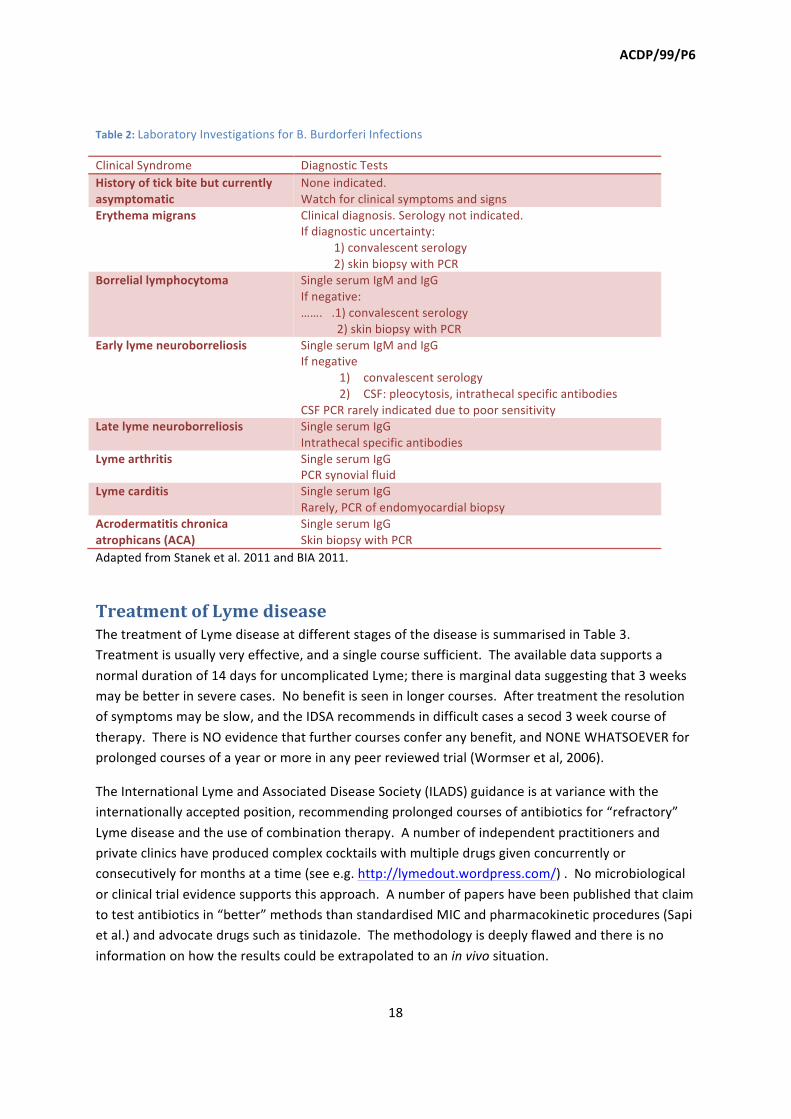
Table 2: Laboratory Investigations for B. Burdorferi Infections
Clinical Syndrome Diagnostic Tests History of tick bite but currently asymptomatic
None indicated. Watch for clinical symptoms and signs
Erythema migrans Clinical diagnosis. Serology not indicated. If diagnostic uncertainty: 1) convalescent serology 2) skin biopsy with PCR
Borrelial lymphocytoma Single serum IgM and IgG If negative: ……. .1) convalescent serology 2) skin biopsy with PCR
Early lyme neuroborreliosis Single serum IgM and IgG If negative
1) convalescent serology 2) CSF: pleocytosis, intrathecal specific antibodies
CSF PCR rarely indicated due to poor sensitivity Late lyme neuroborreliosis Single serum IgG
Intrathecal specific antibodies Lyme arthritis Single serum IgG
PCR synovial fluid Lyme carditis Single serum IgG
Rarely, PCR of endomyocardial biopsy Acrodermatitis chronica atrophicans (ACA)
Single serum IgG Skin biopsy with PCR
Adapted from Stanek et al. 2011 and BIA 2011.
Treatment of Lyme disease The treatment of Lyme disease at different stages of the disease is summarised in Table 3. Treatment is usually very effective, and a single course sufficient. The available data supports a normal duration of 14 days for uncomplicated Lyme; there is marginal data suggesting that 3 weeks may be better in severe cases. No benefit is seen in longer courses. After treatment the resolution of symptoms may be slow, and the IDSA recommends in difficult cases a secod 3 week course of therapy. There is NO evidence that further courses confer any benefit, and NONE WHATSOEVER for prolonged courses of a year or more in any peer reviewed trial (Wormser et al, 2006).
The International Lyme and Associated Disease Society (ILADS) guidance is at variance with the internationally accepted position, recommending prolonged courses of antibiotics for “refractory” Lyme disease and the use of combination therapy. A number of independent practitioners and private clinics have produced complex cocktails with multiple drugs given concurrently or consecutively for months at a time (see e.g. http://lymedout.wordpress.com/) . No microbiological or clinical trial evidence supports this approach. A number of papers have been published that claim to test antibiotics in “better” methods than standardised MIC and pharmacokinetic procedures (Sapi et al.) and advocate drugs such as tinidazole. The methodology is deeply flawed and there is no information on how the results could be extrapolated to an in vivo situation.

ACDP/99/P6
19
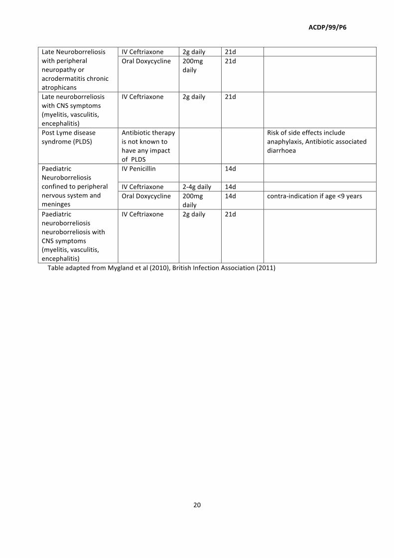
Table 3: Treatment of Lyme Disease
Condition Antibiotic Dosage Duration Notes Prophylaxis following tick bite in the UK
None recommended
If the tick bite occurred in the United States doxy 200mg single dose for adults and 4mg/kg in children>/= 8 years and careful follow up for development of symptoms
Erythema migrans Doxycycline or amoxicillin or cefuroxime axetil
100 mg bd PO 500 mg tds PO 500 mg bd PO
14-‐21d
Lyme arthritis Doxycycline or amoxicillin Ceftriaxone (if not resolved with oral agents )
100 mg bd PO 500 mg tds PO 2gOD
28d 14-‐28d
Post-‐treatment persistence of joint inflammation with negative PCR on synovial fluid and/or synovium biopsies may sometimes occur (termed antibiotic-‐refractory Lyme arthritis). This condition is best managed by a rheumatologist. Arthroscopic synovectomy and steroids may be considered
Acrodermatitis chronica atrophicans (ACA)
Doxycycline
100 mg bd PO
21-‐28 ACA occurs predominantly in older adults.
Lyme carditis with: a) 1st degree block with prolonged (>300 ms) PR interval b) 2nd or 3rd degree block
Ceftriaxone 2gOD IV 14-‐21d
Borrelial lymphocytoma As for EM 14-‐21d If intolerant to 1st line agents , a macrolide (azithromycin, clarithromycin or erythromycin)may be administered
Early Neuroborreliosis confined to peripheral nervous system and meninges
IV Penicillin
20 million U per day
10-‐28d
IV Ceftriaxone 2-‐4g daily 14d Oral Doxycycline 200mg
daily 14d Relative contra-‐indication in
pregnancy /lactation Early neuroborreliosis with CNS symptoms (myelitis, vasculitis, encephalitis)
IV Ceftriaxone 14-‐21d Not enough evidence to
ACDP/99/P6
20
Late Neuroborreliosis with peripheral neuropathy or acrodermatitis chronic atrophicans
IV Ceftriaxone 2g daily 21d Oral Doxycycline 200mg
daily 21d
Late neuroborreliosis with CNS symptoms (myelitis, vasculitis, encephalitis)
IV Ceftriaxone 2g daily 21d
Post Lyme disease syndrome (PLDS)
Antibiotic therapy is not known to have any impact of PLDS
Risk of side effects include anaphylaxis, Antibiotic associated diarrhoea
Paediatric Neuroborreliosis confined to peripheral nervous system and meninges
IV Penicillin
14d
IV Ceftriaxone 2-‐4g daily 14d Oral Doxycycline 200mg
daily 14d contra-‐indication if age <9 years
Paediatric neuroborreliosis neuroborreliosis with CNS symptoms (myelitis, vasculitis, encephalitis)
IV Ceftriaxone 2g daily 21d
Table adapted from Mygland et al (2010), British Infection Association (2011)
ACDP/99/P6
21
Post Lyme Disease Syndromes, Chronic Lyme disease and related issues Since its discovery in the late eighties, Lyme Disease has attracted interest from those who have acquired the disseminated forms of the disease, many of whom have suffered significant morbidity for a prolonged period. It has also been used as a repository for a wide range of other symptoms in a much wider group, in whom there is little or no objective evidence of infection. The leading infectious disease physicians, microbiologists, neurologist and others in Europe and the USA have produced guidelines on best practice in the diagnosis and treatment of Lyme Disease, most recently as the IDSA guidance in the USA (Wormser et al., 2006), the British Infection Association position statement on Lyme Disease (British Infection, 2012) and the EFNS guidelines for neuroborreliosis (Mygland et al., 2010). These are backed up by the excellent reviews by Stanek and others already cited. The IDSA guidance recognises that recovery from a significant attack of neuroborreliosis or arthralgia may lead to prolonged recovery over a period of 6 months or more, which it defines as Post Lyme Disease Syndrome. The guidance makes it clear that this syndrome is independent of active infection, and that treatment should be supportive and symptomatic rather than involving further courses of antibiotics. This positions reflected by the other authorities cited.
A significant number of individuals with a wide range of non-‐specific symptoms see Lyme disease as the cause of their problems. These divide into two groups:
1. Those with symptoms following a complete course of treatment for Lyme disease of any of the forms. For this group, activists have used the term “chronic Lyme disease”.
2. Those with no clinical history (e.g. EM rash) or laboratory evidence by conventional tests of Lyme disease.
Chronic Lyme disease The evidence from both treated cases of Lyme disease, and from treated cases of other spirochetal infections, is that no viable organisms exist after a full course of treatment (Lantos). The majority of patients recover fully, although in some cases it may take several months for symptoms to fully resolve. Despite this, a number of papers have been published claiming to have found evidence of Lyme Borrelia after treatment. Most of these rely on discredited methodology or artefacts (e.g. see http://www.youtube.com/watch?v=gLGfhRuDSio). This has led to a demand for long-‐term treatment with up to 1 year of intravenous therapy with ceftriaxone or even complex mixtures of antibiotics for which no theoretical or laboratory exists. These therapies are usually supplied by private clinics such as the Breakspear clinic in the UK (http://www.breakspearmedical.com/files/lyme-‐disease.html), and such centres are referred to as “Lyme-‐literate” in pressure group literature. “Lyme literate” practitioners and laboratories also offer a range of dedicated diagnostics tests that support their diagnoses (see section on alternative Lyme assays).
Lantos’ review considers the clinical data in detail; no difference was found in between post-‐Lyme disease patients and other controls in the incidence of long term symptoms, and no evidence was

ACDP/99/P6
22
found to support long term therapy. The issues surrounding the use of poor scientific data to support unorthodox therapies and the potential harm to patients have been extensively reviewed in the Lancet Infectious Diseases journal (Auwaerter et al.) and in Brian Dureden’s review for DH (http://www.dh.gov.uk/assetRoot/04/13/89/17/04138917.pdf) .
Alternative tests for Lyme disease A range of alternative test have been proposed for diagnosing Lyme Disease, and many of these are offered in various forms by private laboratories as definitive methods for confirming the infection. The CDC has produced a list of tests that do not meet the quality and reproducibility standards for clinical use based on the available evidence of these assays in comparative trials (http://www.cdc.gov/lyme/diagnosistreatment/LabTest/OtherLab/). Unfortunately, but predictably, the more non-‐specific are the symptoms a patient describes, the lower is the positive predictive value of an assay and there is a considerable incidence of false positives. (Wilske, 2003) Briefly these assays include the following types:
1. Urinary antigen test. This test was developed as an antigen capture test in the early 1990’s using a collection of body fluids from mice, domestic animals and man together with various preparations of Borrelia as antigen sources. Rabbit polyclonal antibodies were used to capture the antigens which were detected as “spirochete proteins or whole spirochetes” by electron microscopy (Dorward et al., 1991). The results have not been shown in the peer-‐reviewed literature to correlate with infection in man.
2. PCR of urine. PCR of urine will detect nucleic acid of Lyme borrelia, but at a much lower rate than PCR of a skin biopsy of erythema migrans or CSF in neuroborreliosis (Lebech et al., 2000). It has been used to suggest that in patients with chronic symptoms after extensive treatment for Lyme Disease organisms persist as PCR signals remain positive in a handful of cases (Bayer et al., 1996). However, similar results have also been reported in healthy patients who are seropositive (Karch et al., 1994) and asymptomatic patients after treatment(Aberer et al., 2007). The predictive value of urinary PCR compared to tissue or CSF PCR is unreliable (Brettschneider et al., 1998) and there is insufficient data to evaluate it adequately (Wilske et al., 2007).
3. Lymphocyte transformation tests. Lymphocyte transformation tests, notably interferon-‐γ release assays e.g. ELISPOT, have been widely used to examine T cell responses in vaccinees before and after vaccination in clinical trials for a number of infectious diseases and cancer.. In this setting, it is possible to demonstrate a change in lymphocyte activity reasonably reliably within a given trial, although there is no generally standardised methodology. The FDA has approved commercial ELSIPOT assays for the diagnosis of TB, but even in this case the interpretation relies on the context from other data. In human borreliosis, ELISPOT assays have been applied for research purposes (Ekerfelt et al., 1999) and unvalidated commercial assays are available for research use. In the absence of standardised tests, different methods of stimulating cells can give wide variations in results, and when tests are applied to the general population the results can be very hard to interpret as prior exposure to a wide variety of antigens can give false positive reactions to the specific antigen used in the test. As some laboratories use crude preparations of whole organisms as the stimulus in the test, the specificity is even worse. The few peer-‐reviewed studies that exist suggest

ACDP/99/P6
23
there is no correlation between T cell reactivity and the stage of Borreliosis (Ekerfelt et al., 1999) and the test has little value in the clinical setting (Horowitz et al., 1994) (Nordberg et al., 2012)
The use of these tests and the dangers inherent therein has been critically reviewed by Brian Duerden on behalf of the Department of Health (http://www.dh.gov.uk/assetRoot/04/13/89/17/04138917.pdf).
Lyme disease without supporting evidence In addition to the groups described above, a significant minority of patients with chronic symptoms now attribute these to Lyme disease. Some of these patients may recall being bitten by a tick in the past (this may be over twenty years previously), but for most there is no clear history of EM, tick bites or definitive symptoms of Lyme Disease. However, there is a plethora of internet sites blaming Lyme Borrelia for chronic symptoms and a network of practitioners who will treat for it accordingly. The International Lyme and Associated Disease Society (ILADS) has produced its own guidelines (www.ilads.org/lyme_disease/treatment_guidelines.html) giving a range of presentations from jaw or testicular pain to dizziness as indicative of Lyme disease, although interestingly the characteristic EM rash is NOT included.
The disease progression and demographics of this group of patients is similar to that seen in patients with “ME” and chronic fatigue syndrome. Lyme disease forms a convenient container in which to place their symptoms, and once this conviction is established, any attempt to refute the cause is met with hostility and anger. The symptoms experienced are genuine, but the cause may be another treatable condition (from tumours to rheumatoid arthritis), or may be somatisation of symptoms. It is important to exclude other physical causes and reassure the patient. If the individual can be persuaded to attend a specialist, psychological therapy can be effective if started early (Sigal and Hassett, 2002).
Meeting the public need: the HPA future plans In view of the interest from public self-‐help groups and politicians, RIPL and HPA staff have met with the Lyme Disease Action group (LDA) and with The Countess of Mar and her advisors. Lady Mar is an elected hereditary peer who campaigns for better treatment for chronic illness. From these meetings, it is clear that there are some genuine issues to be addressed in terms of the information on Lyme disease, awareness of the disease, and some limitations in the available tests, both for Lyme disease itself and in the differential diagnosis. UK physicians use the IDSA and EFNS guidance, together with the BIA statement on Lyme disease as the basis for the investigation and management of Lyme disease, which whilst accurate is not UK specific. There is a clear perceived need for a UK set of guidance. In discussion with these groups, RIPL and the HPA have proposed the following actions:
1. The HPA will produce an information leaflet for GPs on the recognition and management of Lyme disease and distribute it through the Royal College of GP’s and other networks as widely as possible. The LDA and lady Mar will be given the opportunity to comment before the document is circulated..

ACDP/99/P6
24
2. HPA will review the advice on the distribution of Lyme to include general rural areas and parks nationwide to reflect the chance of being infected in the whole country rather than simply Lyme “hotspots”.
3. HPA plan to convene a meeting with GP’s, ID physicians, neurologists and others to compile a UK-‐centric set of guidance for the diagnosis and management of Lyme Disease.
4. As part of point 3, we will critically consider the value of prophylaxis for Lyme disease after tick bites (this may take longer than a first draft of the guidance as the mathematics and analysis are quite complex). (Unless the incidence of Borrelia in ticks is high, prophylaxis is not normally offered after tick bites).
5. RIPL will offer an extended test panel in relevant cases to cover the differential diagnosis of Lyme infection in patients with tick bites. We hope to introduce this in time for the next season.
6. Based on data from our first few months, RIPL staff will review the performance of our assays and modify the testing schedules to improve the sensitivity of testing. As part of this review we are sourcing alternative ELISA tests to evaluate. Depending on their performance we may move to a first and second line ELISA covering different stages of disease, to improve the detection rate. A negative would be required in both tests to record a negative screen; a positive in either assay would lead to an automatic evaluation by western blot.
7. In the longer term, we hope to use the Western blot data to further improve the diagnostic accuracy and perhaps to derive better algorithms for interpreting the test data.
8. RIPL staff are planning a prospective clinical trial to study the diagnosis and clinical progression of Lyme disease in conjunction with the Clinical Research Network through Professor Rob Reid, who is the Infectious Disease lead for CRN. The trial would involve GPS as well as hospital specialists and would be nationwide, and would be subject to obtaining suitable funding.
9. Regular update meetings will be held with LDA and Lady Mar to discuss progress. 10. RIPL and HPA staff will discuss with Simon Wesseley’s group and other interested parties the
development of guidance for clinicians on dealing with the disaffected group with un-‐provable Lyme disease. This will cover the therapeutic approach, investigation of cases and “disengagement” strategies when further investigation is counter-‐productive.
Summary 1. The Lyme disease service was transferred to RIPL Porton Down in June 2011. 2. Present testing is based on a sensitive screening ELISA (C6) followed by a confirmatory
Western blot for IgG and IgM. The analysis is totally automated. 3. There is room for even better screening assays and RIPL is investigating the available
systems to reduce the possibility of missing cases, as well as offering an extended panel to cover important vector borne diseases in the differential diagnosis.
4. RIPL and HPA staff have met with interest groups and agreed to take their views into account, whilst maintaining an evidence based approach from scientific and clinical studies. Regular update meetings will be held.
5. With relevant clinicians, RIPL will seek to produce UK specific guidance for the management of Lyme disease.
6. The present information on the HPA website will be reviewed and updated, and the HPA will launch an active campaign to raise awareness of tick-‐borne diseases and Lyme disease.
ACDP/99/P6
25
7. A significant minority of patients have symptoms that are not related to Lyme infection, and an approach to their management needs to be developed.
Contributors Tim Brooks HPA Meera Chand HPA/UCLH Laura Nabarro HPA/UCLH Amanda Semper HPA Robert Smith Public health Wales Surabhi Taori HPA/Edinburgh
References ABERER, E., BERGMANN, A. R., DERLER, A. M. & SCHMIDT, B. (2007) Course of Borrelia burgdorferi
DNA shedding in urine after treatment. Acta Derm Venereol, 87, 39-‐42. AGUERO-‐ROSENFELD, M. E., NOWAKOWSKI, J., MCKENNA, D. F., CARBONARO, C. A. & WORMSER, G.
P. (1993) Serodiagnosis in early Lyme disease. J Clin Microbiol, 31, 3090-‐5. AGUERO-‐ROSENFELD, M. E., WANG, G., SCHWARTZ, I. & WORMSER, G. P. (2005) Diagnosis of lyme
borreliosis. Clin Microbiol Rev, 18, 484-‐509. AUWAERTER, P. G., BAKKEN, J. S., DATTWYLER, R. J., DUMLER, J. S., HALPERIN, J. J., MCSWEEGAN, E.,
NADELMAN, R. B., O'CONNELL, S., SHAPIRO, E. D., SOOD, S. K., STEERE, A. C., WEINSTEIN, A. & WORMSER, G. P. Antiscience and ethical concerns associated with advocacy of Lyme disease. Lancet Infect Dis, 11, 713-‐9.
BANKHEAD, T. & CHACONAS, G. (2007) The role of VlsE antigenic variation in the Lyme disease spirochete: persistence through a mechanism that differs from other pathogens. Mol Microbiol, 65, 1547-‐58.
BAYER, M. E., ZHANG, L. & BAYER, M. H. (1996) Borrelia burgdorferi DNA in the urine of treated patients with chronic Lyme disease symptoms. A PCR study of 97 cases. Infection, 24, 347-‐53.
BRETTSCHNEIDER, S., BRUCKBAUER, H., KLUGBAUER, N. & HOFMANN, H. (1998) Diagnostic value of PCR for detection of Borrelia burgdorferi in skin biopsy and urine samples from patients with skin borreliosis. J Clin Microbiol, 36, 2658-‐65.
BRITISH INFECTION, A. (2012) The epidemiology, prevention, investigation and treatment of Lyme borreliosis in United Kingdom patients: a position statement by the British Infection Association. J Infect, 62, 329-‐38.
DORWARD, D. W., SCHWAN, T. G. & GARON, C. F. (1991) Immune capture and detection of Borrelia burgdorferi antigens in urine, blood, or tissues from infected ticks, mice, dogs, and humans. J Clin Microbiol, 29, 1162-‐70.
DURAY, P. H. & STEERE, A. C. (1988) Clinical pathologic correlations of Lyme disease by stage. Ann N Y Acad Sci, 539, 65-‐79.
GE, Y., LI, C., CORUM, L., SLAUGHTER, C. A. & CHARON, N. W. (1998) Structure and expression of the FlaA periplasmic flagellar protein of Borrelia burgdorferi. J Bacteriol, 180, 2418-‐25.
HOROWITZ, H. W., PAVIA, C. S., BITTKER, S., FORSETER, G., COOPER, D., NADELMAN, R. B., BYRNE, D., JOHNSON, R. C. & WORMSER, G. P. (1994) Sustained cellular immune responses to Borrelia burgdorferi: lack of correlation with clinical presentation and serology. Clin Diagn Lab Immunol, 1, 373-‐8.
HUYSHE-‐SHIRES, S. Lyme disease antiscience. Lancet Infect Dis, 12, 361; author reply 362-‐3.

ACDP/99/P6
26
KARCH, H., HUPPERTZ, H. I., BOHME, M., SCHMIDT, H., WIEBECKE, D. & SCHWARZKOPF, A. (1994) Demonstration of Borrelia burgdorferi DNA in urine samples from healthy humans whose sera contain B. burgdorferi-‐specific antibodies. J Clin Microbiol, 32, 2312-‐4.
LANTOS, P. M. Chronic Lyme disease: the controversies and the science. Expert Rev Anti Infect Ther, 9, 787-‐97.
LEBECH, A. M., HANSEN, K., BRANDRUP, F., CLEMMENSEN, O. & HALKIER-‐SORENSEN, L. (2000) Diagnostic value of PCR for detection of Borrelia burgdorferi DNA in clinical specimens from patients with erythema migrans and Lyme neuroborreliosis. Mol Diagn, 5, 139-‐50.
MYGLAND, A., LJOSTAD, U., FINGERLE, V., RUPPRECHT, T., SCHMUTZHARD, E. & STEINER, I. (2010) EFNS guidelines on the diagnosis and management of European Lyme neuroborreliosis. Eur J Neurol, 17, 8-‐16, e1-‐4.
PERRONNE, C. Lyme disease antiscience. Lancet Infect Dis, 12, 361-‐2; author reply 362-‐3. PURSER, J. E., LAWRENZ, M. B., CAIMANO, M. J., HOWELL, J. K., RADOLF, J. D. & NORRIS, S. J. (2003)
A plasmid-‐encoded nicotinamidase (PncA) is essential for infectivity of Borrelia burgdorferi in a mammalian host. Mol Microbiol, 48, 753-‐64.
SAPI, E., KAUR, N., ANYANWU, S., LUECKE, D. F., DATAR, A., PATEL, S., ROSSI, M. & STRICKER, R. B. Evaluation of in-‐vitro antibiotic susceptibility of different morphological forms of Borrelia burgdorferi. Infect Drug Resist, 4, 97-‐113.
SIGAL, L. H. & HASSETT, A. L. (2002) Contributions of societal and geographical environments to "chronic Lyme disease": the psychopathogenesis and aporology of a new "medically unexplained symptoms" syndrome. Environ Health Perspect, 110 Suppl 4, 607-‐11.
TUTTLE, C. Lyme disease antiscience. Lancet Infect Dis, 12, 362; author reply 362-‐3. WAGNER, B., FREER, H., ROLLINS, A., GARCIA-‐TAPIA, D., ERB, H. N., EARNHART, C., MARCONI, R. &
MEEUS, P. Antibodies to Borrelia burgdorferi OspA, OspC, OspF, and C6 antigens as markers for early and late infection in dogs. Clin Vaccine Immunol, 19, 527-‐35.
WILSKE, B. (2003) Diagnosis of lyme borreliosis in europe. Vector Borne Zoonotic Dis, 3, 215-‐27. WILSKE, B. (2005) Epidemiology and diagnosis of Lyme borreliosis. Ann Med, 37, 568-‐79. WILSKE, B., FINGERLE, V. & SCHULTE-‐SPECHTEL, U. (2007) Microbiological and serological diagnosis
of Lyme borreliosis. FEMS Immunol Med Microbiol, 49, 13-‐21. WORMSER, G. P., DATTWYLER, R. J., SHAPIRO, E. D., HALPERIN, J. J., STEERE, A. C., KLEMPNER, M. S.,
KRAUSE, P. J., BAKKEN, J. S., STRLE, F., STANEK, G., BOCKENSTEDT, L., FISH, D., DUMLER, J. S. & NADELMAN, R. B. (2006) The clinical assessment, treatment, and prevention of lyme disease, human granulocytic anaplasmosis, and babesiosis: clinical practice guidelines by the Infectious Diseases Society of America. Clin Infect Dis, 43, 1089-‐134.
WORMSER, G. P., NOWAKOWSKI, J., NADELMAN, R. B., VISINTAINER, P., LEVIN, A. & AGUERO-‐ROSENFELD, M. E. (2008) Impact of clinical variables on Borrelia burgdorferi-‐specific antibody seropositivity in acute-‐phase sera from patients in North America with culture-‐confirmed early Lyme disease. Clin Vaccine Immunol, 15, 1519-‐22.