A Retrospective Review of the Outcome of Children Presenting to
Tygerberg Children’s Hospital with Biliary Atresia
By
OR Karangwa
Thesis Presented in partial fulfilment of the requirement for the degree MMed (Paediatrics) Faculty of Health Sciences at the University of Stellenbosch
Supervisor: Dr E D Nel
Department of Paediatrics and Child Health
Faculty of Health Sciences
March 2016

i
Declaration
By submitting this thesis electronically, I declare that the entirety of the work contained therein is my own, original work, that I am the sole author thereof (save to the extent explicitly otherwise stated), that reproduction and publication thereof by Stellenbosch University will not infringe any third party rights and that I have not previously in its entirety or in part submitted it for obtaining any qualification.
March 2016
Copyright © 2016 Stellenbosch UniversityAll rights reserved
Stellenbosch University https://scholar.sun.ac.za

ii
Abbreviations and acronyms
BA biliary atresia
EHBA extra hepatic BA
HPE hepaticoportoenterostomy
KPE Kasai portoenterostomy
K P Kasai procedure
LTx liver transplantation
BARC BA Research Consortium
TCH Tygerberg Children’s Hospital
RCCH Red Cross Children’s Hospital
UK United Kingdom
USA United States of America
BASM BA splenic Malformations
UDCA UrsodeoxyCholic Acid
SA South Africa
Stellenbosch University https://scholar.sun.ac.za

3
Abstract
Background
Biliary atresia (BA) is the end result of an inflammatory process leading to fibrosis and
obliteration of the biliary tract with the development of biliary cirrhosis. It is a leading cause of
end-stage liver disease in children. It accounts for more than 50% of pediatric liver
transplantations (LTx). Surgical treatment with Kasai portoenterostomy (PE) has improved the
prognosis for patients with BA, even if most eventually need LTx. A key determinant for the
post- Kasai PE patient survival is patient age at surgery as well as peri and post-operative
management.
Objectives
This study reviews the short and long-term outcome and identifies prognostic factors of children
with BA
Study design
A retrospective descriptive study
Methods
Folder review of all children born between May 1997 and May 2011 with biliary atresia as
extracted from Tygerberg Children Hospital records database. Data collected included
demographic details, clinical characteristics, liver biochemistry and liver biopsy results and
outcome post Kasai PE.
Results
Thirty seven patients with confirmed extra hepatic BA (EHBA) , 23 females and 14 males, mean
age 89 days (12.7 weeks), range 7 - 227 days, were identified. Twenty three patients underwent
Stellenbosch University https://scholar.sun.ac.za
4
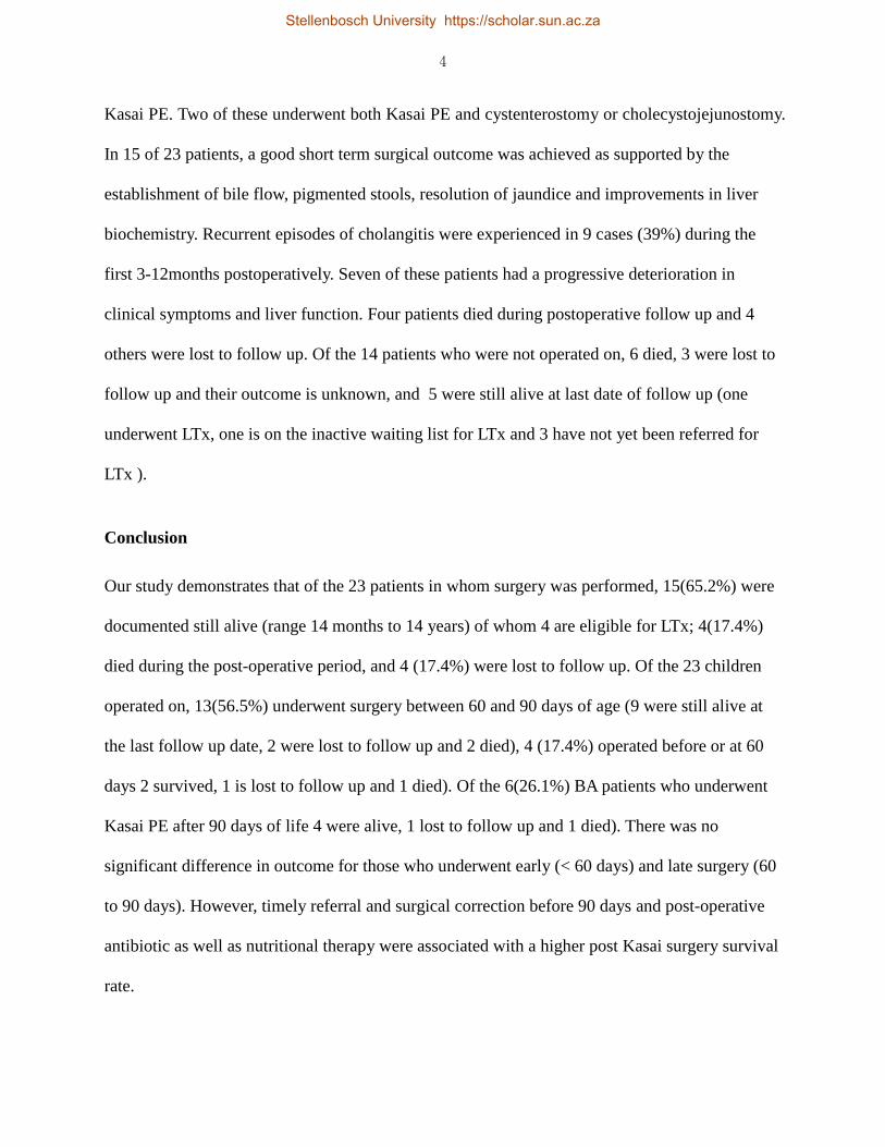
Kasai PE. Two of these underwent both Kasai PE and cystenterostomy or cholecystojejunostomy.
In 15 of 23 patients, a good short term surgical outcome was achieved as supported by the
establishment of bile flow, pigmented stools, resolution of jaundice and improvements in liver
biochemistry. Recurrent episodes of cholangitis were experienced in 9 cases (39%) during the
first 3-12months postoperatively. Seven of these patients had a progressive deterioration in
clinical symptoms and liver function. Four patients died during postoperative follow up and 4
others were lost to follow up. Of the 14 patients who were not operated on, 6 died, 3 were lost to
follow up and their outcome is unknown, and 5 were still alive at last date of follow up (one
underwent LTx, one is on the inactive waiting list for LTx and 3 have not yet been referred for
LTx ).
Conclusion
Our study demonstrates that of the 23 patients in whom surgery was performed, 15(65.2%) were
documented still alive (range 14 months to 14 years) of whom 4 are eligible for LTx; 4(17.4%)
died during the post-operative period, and 4 (17.4%) were lost to follow up. Of the 23 children
operated on, 13(56.5%) underwent surgery between 60 and 90 days of age (9 were still alive at
the last follow up date, 2 were lost to follow up and 2 died), 4 (17.4%) operated before or at 60
days 2 survived, 1 is lost to follow up and 1 died). Of the 6(26.1%) BA patients who underwent
Kasai PE after 90 days of life 4 were alive, 1 lost to follow up and 1 died). There was no
significant difference in outcome for those who underwent early (< 60 days) and late surgery (60
to 90 days). However, timely referral and surgical correction before 90 days and post-operative
antibiotic as well as nutritional therapy were associated with a higher post Kasai surgery survival
rate.
Stellenbosch University https://scholar.sun.ac.za
Table of Contents
Abstract .......................................................................................................................................... 3
Literature review ........................................................................................................................... 1
Patients and Methods ................................................................................................................... 8
Results .......................................................................................................................................... 12
Clinical features ....................................................................................................................... 15
Kasai Portoenterostomy and age at surgery ......................................................................... 16
Management post Kasai .......................................................................................................... 16
Early post-operative complications ........................................................................................ 17
Late post-operative complications ......................................................................................... 18
Nutritional complications........................................................................................................ 19
Infections .................................................................................................................................. 19
Success ...................................................................................................................................... 19
Liver biopsy and other investigations .................................................................................... 21
Growth parameters ................................................................................................................. 21
Survival and outcomes of patients with biliary atresia ........................................................ 22
Patients lost to follow up ......................................................................................................... 27
Discussion..................................................................................................................................... 28
Conclusion ................................................................................................................................... 32
Appendices ................................................................................................................................... 37
Stellenbosch University https://scholar.sun.ac.za

List of Tables
Table 1 Demography of patients .................................................................................................................................. 13
Table 2 Gender ............................................................................................................................................................ 13
Table 3 Ethnic group ................................................................................................................................................... 14
Table 4 Age at presentaion on by urban/rural of origine ............................................................................................. 14
Table 5 Main post operative complication .................................................................................................................. 17
Table 6 Correlation between patient’sage and percentage of biliary drainage after Kasai PE (%) and numbers ........ 20
Table 7 Cause of death in patients who underwent Kasai or not ................................................................................ 25
Table 8 Outcome of patients who underwent Kasai surgery ....................................................................................... 25
Table 9 Results after Kasai procedure ........................................................................................................................ 26
Table 10 Results without Kasai procedure .................................................................................................................. 26
Stellenbosch University https://scholar.sun.ac.za

List of Figures
Figure 1 Comparison of change in Z-score .................................................................................. 22
Figure 2 Survival for full sample .................................................................................................. 23
Figure 3 Comparison according to Kasai surgery/no surgery ...................................................... 24
Stellenbosch University https://scholar.sun.ac.za

1
Literature review
Biliary atresia (BA) is a serious but relatively rare congenital disease of the bile ducts and liver
of unknown aetiology. It is an important cause of severe cholestatic jaundice in infancy1 and is
also the most common indication for liver transplantation in South African children2. It is
associated with severe complications and is consistently fatal if a Kasai portoenterostomy (KPE)
is not performed in the first weeks of life3 . The reported incidence of biliary atresia varies from
5/100000 births per year in Holland to 32/ 100000 in French Polynesia4. Individual studies
suggest an overall incidence in the United States of 1 per 10,000-15,000 live births. The
incidence of BA is highest in Asian populations. The incidence and outcome of biliary atresia in
South Africa is unknown as well as in African and developing countries in general.
No single unifying hypothesis has explained the varying presentations of BA indicating that BA
is probably not a single disease entity. In 1885, BA was reported as an autopsy finding and,
despite various studies since then, its aetiopathogenesis has not been fully determined5 . It is a
condition unique to infancy that is characterized by progressive fibrotic obliteration and
obstruction of the extra-hepatic biliary tree ultimately leading to liver cirrhosis and liver failure.
Two forms of biliary atresia occur: a syndromic form that is associated with congenital
abnormalities such as situs inversus, polysplenia, and severe congenital malformations of the
heart; and a more common non-syndromatic form that is not associated with other congenital
abnormalities. The latter form accounts for approximately 80% of all cases of biliary atresia. It is
postulated that the syndromatic form (also known as embryonic type) results from an insult
during the differentiation of the hepatic diverticulum from the foregut of the embryo. The non-
syndromatic form is thought to result from an insult late in gestation6.
Stellenbosch University https://scholar.sun.ac.za

2
The pathology of the extra hepatic biliary system widely varies in these patients, and the
following classification is based on the predominant site of atresia: Type I involves obliteration
of the common duct, the proximal ducts are patent; Type II is characterized by atresia of the
hepatic duct, with cystic structures found in the porta hepatis; Type III (>90% of patients)
involves atresia of the right and left hepatic ducts to the level of the porta hepatis. These variants
should not be confused with intrahepatic biliary hypoplasia, which comprises a group of distinct
and surgically non correctable disorders.
Various aetiologies have been suggested. These include intrauterine or early postnatal viral
infections, metabolic insults, abnormalities in bile duct morphogenesis or pancreaticobiliary
ductal malformation, genetic factors, vascular insults, and immunological factors. No single
unifying hypothesis has explained the varying presentations of biliary atresia indicating that BA
is probably not a single disease entity. Several studies have identified elevated antibody titers to
reovirus type 3 in patients with biliary atresia when compared with controls. They reported a
high prevalence of positive serologic results for reovirus in patients with atresia, but this finding
was not replicated in a subsequent study7. Some other studies failed to demonstrate evidence of
viral (reovirus) infection in infants with cholestasis8. Other viruses, including rotavirus and
cytomegalovirus (CMV), have also been implicated. No single agent has been identified as
causative for biliary atresia. With regard to CMV, Tarr et al, assessed 23 patients with BA using
liver histology, serology and culture and found five (24%) CMV positive patients9. Fischler
reported CMV infection in almost 25% of affected infants using immunoglobulin M (IgM)
serology10. They also detected CMV DNA in the liver of 50% of children with atresia whose
mothers were CMV-positive. Similarly, a Brazilian study identified positive IgM for CMV in
28.5% of patients with BA or a choledochal cyst. Interestingly, an even higher frequency of
Stellenbosch University https://scholar.sun.ac.za

3
CMV infection has been found in cases of idiopathic neonatal hepatitis, providing support to the
concept that both disorders are ends of the same pathological spectrum, originally described by
Landing as infantile obstructive cholangiopathy11. The role of immune dysfunction has been
considered. Sokol et al, suggested that, from the molecular position, viral antigens may cross-
react with biliary antigens, activating an immune response against the virus, and also against
biliary antigens. Therefore, persistence of immune injury to bile duct cells may lead to disease
progression. Several researchers have studied the potential effects of other aetiological agents,
including teratogens and immunological factors. Again, no clear correlations with BA have been
established.
Most children present within four to six weeks after birth with conjugated jaundice, acholic
stools and dark urine. If BA is diagnosed within the first two to three months after birth, a KPE
can successfully restore bile flow into the intestinal tract in up to 90% of patients leading to
improvement of jaundice and delayed progression of liver disease12. However, if untreated, liver
cirrhosis develops within the first months of life and the majority of children will die before the
age of two years in the absence of liver transplantation. In addition, children who have had an
unsuccessful portoenterostomy also require liver transplantation, usually before 2-5 years13.
In 1959, Kasai and Suzuki14 introduced hepatic portoenterostomy to reestablish bile flow in BA
and it was the first breakthrough in the treatment of BA. It is now accepted that early diagnosis
and timely surgery have a significant impact on long-term prognosis. While an early KPE
improvess the outcome of infants with BA, complications are still common. These include
ascending cholangitis, portal hypertension, pruritis, malnutrition and chronic liver failure.
The most frequent early complication is ascending cholangitis, and its treatment is key for the
preservation of bile flow and to improve the prognosis of the patient. Ascending cholangitis
Stellenbosch University https://scholar.sun.ac.za

4
occurs in 40 to 90% of surgically treated children15 and recurrent post-operative cholangitis is
associated with obliteration of the intra-hepatic bile ducts and progression to liver cirrhosis.
Portal hypertension is the second most frequent complication of atresia. Its presence depends on
the degree of liver fibrosis at KPE.
Longer term complications include fat malabsorption, malnutrition and fat soluble vitamin
deficiency. Vitamin K dependent coagulopathy may occur as may vitamin D deficiency, which
leads to a reduction in bone density and rickets. Clinical deficiencies of vitamin A and E are
unusual with suitable supplementation, but peripheral neuropathy secondary to vitamin E
deficiency in cholestasis is well recognized. Close follow-up after surgery is therefore required.
Delayed psychomotor development is observed in patients who present with progressive chronic
liver disease, apparently associated with malnutrition.
The most important long-term prognostic factors in predicting outcome after the Kasai procedure
appear to be age at time of surgery, achievement of biliary drainage, experience of the centre,
occurrence of ascending cholangitis, as well as pre-operative needle liver biopsy results such as
the degree of liver fibrosis and of intrahepatic bile duct injury16.
Age at the time of the Kasai procedure is perhaps the most important prognostic indicator. Most
reports state that the best outcomes, in both initial success and long-term survival, occur in
patients younger than 60–70 days at the time of operation (68% 10 year survival). The Japanese
Biliary Atresia Registry records favourable outcomes up to 90 days of age, with a rapid drop in
success after that point. Many other series report that performance of a Kasai procedure in
infants after 70 days of age is a risk factor for a poor outcome; however, some do not agree with
this. Hanmin et al proved that Kasai may be performed successfully in all infants presenting up
Stellenbosch University https://scholar.sun.ac.za

5
to 120 days of age17.
Even if it is a very important postoperative prognostic factor, primary biliary drainage is not a
guarantee of the long-term success after a KPE. However, patients who do not achieve primary
biliary drainage after a KPE will never do so. Surgical treatment via a second (redo) Kasai
procedure for patients who did not achieve primary drainage as well as for those who had initial
drainage but subsequent cessation, was met with initial interest following case reports and a
small series that demonstrated successful bile drainage. Recent reports, however, suggest that
redo KPE is not a useful strategy in the vast majority of patients18.
If KPE is not performed, fibrosis will progress to end-stage cirrhosis and death in the first year of
life in 50 to 80% of children and in 90 to 100% of patients by the third year of life.
The survival of infants born with BA has improved over the last 30 past years. Of eleven infants
born with BA in one region in England during the period 1971-73 only one child survived to 10
years19. .In contrast about 90% of children in England and Wales with BA are presently surviving
as a result of KPE and liver transplantation20. There was a similar improvement in the USA.
Prior to the introduction of the Kasai procedure the mean survival time in the USA was
approximately 19 months21. A later multi-centre retrospective study of 104 infants with BA,
diagnosed between 1997 and 200022, showed that at age 24 months 56% of children were alive
with native livers, 40% had undergone liver transplantation and 4% had died without undergoing
liver transplantation (attributable to congenital heart diseases). These data led to a UK
Department of Health directive indicating that all infants with suspected biliary atresia should be
referred to one of three designated centres (St James Hospital in Leeds, Birmingham Children’s
Stellenbosch University https://scholar.sun.ac.za

6
Hospital and King’s College Hospital, London) where both the Kasai operation and, if necessary,
liver transplantation could be offered.
The impact of the centralisation of biliary atresia surgery in the UK has recently been assessed. A
total of 148 infants with biliary atresia were treated between January 1999 and June 2002. A
Kasai PE was carried out in 142 and primary liver transplant in five (3%). Early clearance of
jaundice after KPE was achieved in 57% of infants, compared to 55% in an earlier series. Of 135
children who survived, 62% still had their native liver and 38% had received transplants
compared to 40% in the earlier series. The overall 4year estimated survival was 89% (95% CI
82% to 94%) compared to 85% (95% CI 77% to 92%) in the pre-centralised era, suggesting that
centralisation of surgery did not lead to improved survival.
In a Canadian study23, 230 patients with BA were identified. The median age at KPE was 64
days. The overall 4 year post-Kasai PE native liver survival was 39%.
Tohoku University, a Japanese centre with the world’s longest running series of
portoenterostomies performed between 1951 and 1998 analysed the development in long-term
survival patterns24. It included a comparison of hematologic and biochemical data as well as
quality of life analysis in a group of Japanese patients. Long-term survival rate in the Japanese
BA patients increased dramatically from 1 to 17 years after 1975. The quality of life of survivors
was comparable in Japan and England25. The best outcomes were achieved when HPE was
performed before three months of age.
In developed countries, the outlook for patients with BA has improved remarkably owing to
early referral, good diagnostic facilities and access to surgery and liver transplantation. In the UK,
USA, Taiwan, Thailand and Argentine the use of a cheap, non-invasive screening method for
cholestatic infants has proven that an infant stool colour card is helpful for parents and primary
Stellenbosch University https://scholar.sun.ac.za

7
caregivers; it increases early referrals of BA infants to specialised centres and early surgery with
an increased chance of success and a good outcome. In Japan parents receive written advice on
the serious implications of pale stools and dark urine associated with jaundice in early age. This
advice is reinforced with posters in infant welfare clinics as well as the ‘well baby’ review at 4
weeks of age26.
In developing countries, especially in Sub-Saharan Africa, there is little information on the
current outlook for these children. In Nigeria27, a study of 14 patients with BA managed from
1991 to 2004 at Ahmadou University Hospital, Zaria, with a median age of 16 weeks (between 6
and 24 weeks) showed that 11 patients at presentation already had liver cirrhosis and raised LFTs
and could not have corrective surgery. Three had a Kasai PE but of these three patients, one died
from acute gastroenteritis at 2 years, one from cholangitis after 8 weeks, and the third one died
from anaesthestic complications. Of the 11 patients who did not have surgery, 10 were lost to
follow up and their outcome is unknown. The remaining patient had a laparotomy and liver
biopsy and subsequently died from peritonitis.
Mabogunje (1987) reported 36 histologically confirmed cases of BA in infants and children aged
2-20 months from one of the tertiary health centres in Nigeria within a 13 year study period,
where almost half of them died, 19 were lost to follow-up and 2 survived with persistent jaundice.
In this same report, a KPE was performed on four of the children, however bile flow was not
established in any of the children.
In South Africa, a study of 71 infants with BA (from 1993 to 1998) was performed at
Baragwanah Hospital, Soweto28. The findings appear to confirm a high incidence and poorer
outcome of BA in Black infants from Soweto; of the 71, only 10(14%) were jaundice free.
In Egypt, Ahmad (2009) found that 15 of 30 cases (50%) had a successful Kasai, one died 3
Stellenbosch University https://scholar.sun.ac.za

8
weeks after surgery from fulminant hepatic necrosis and multiorgan failure, 4 were lost to follow
up, 2 were transplanted, 8 were awaiting LTx29.
In South Africa no recent studies have evaluated the long-term outcome of biliary atresia. This
study describes the outcome of infants and children with biliary atresia who have been treated at
our tertiary hospital. In this study we describe factors that potentially influence prognosis (e.g.
age of referral, nutrition, and post-operative cholangitis), the frequency of complications (e.g.
intestinal hemorrhage, spontaneous bacterial peritonitis, and liver failure) and the survival of
children with biliary atresia. These data are necessary to improve the management of biliary
atresia in this setting. The study will unfortunately not be able to assess the incidence of biliary
atresia in South Africa.
Patients and Methods
Study Site
Tygerberg Children’s Hospital (TCH), Cape Town is a large tertiary centre that accepts referrals
from the Eastern, Northern and Western Cape Provinces in South Africa. Facilities for Kasai
surgery (porto/ hepatico enterostomy) are available.
Patients were enrolled in the study if they met the following criteria:
Inclusion Criteria 1. Admitted to TCH between January 1997 and December 2011.
2. Presenting to TCH with cholestatic liver disease:
Diagnosis of BA based on:
a. Clinical features (persistent acholic stools and no other aetiology for the
cholestasis),
Stellenbosch University https://scholar.sun.ac.za

9
b. Biochemical (pronounced elevation of the GGT, greater than ALT)
c. Compatible histology
d. Ultrasound compatible with biliary atresia
e. Operative cholangiogram (where available)
f. Hepatobiliary scintiscanning (where available)
Exclusion Criteria
Where the diagnosis of BA could not confidently be confirmed (as described in the inclusion
criteria), patients were excluded from the study.
Ultrasonography was used to exclude specific anomalies of the extrahepatic biliary system such
as choledocal cyst. BASM (BA splenic malformations) was defined as the presence of
malformations (polyplenia, asplenia) including situs inversus and cardiac anomalies in
conjunction with BA.
The study was performed according to the guidelines of the Medical Ethical Committee of the
University of Stellenbosch.
Data collected included birth date, date of presentation to health centre with first symptoms,
whether the Kasai operation was performed or not, date of Kasai operation, biochemical
evaluation of liver function ( total/ conjugated serum bilirubin, prothrombin time and
transaminases) also bile duct enzymesbefore and after Kasai operation, abdominal ultrasound to
assess cirrhosis, oesophagoscopy to assess oesophageal varices and portal hypertension,
medication used at follow-up, date of last follow-up and final outcome, date and cause of death
and reasons why Kasai operation was not performed. Due to the retrospective nature of the study,
data collection in some cases was incomplete.
Early success of the Kasai operation was defined by complete clearance of jaundice and with
Stellenbosch University https://scholar.sun.ac.za
10
conjugated bilirubin level normal or less than 20mmoles per liter within six months post-surgery.
The bile flow was evaluated on the basis of stool colour. Patients had good bile drainage if they
had pigmented stools and their jaundice resolved; patients with partial bile clearance had
pigmented stools but persistent jaundice; and those with failed bile drainage had persistent
acholic stools and jaundice. Portal hypertension was considered present when at least one of the
following physical signs was present: splenomegaly, ascites and collateral veins or esophageal
varices at oesophagoscopy. Cholangitis was diagnosed if the patient suffered from high fever,
with worsening jaundice, acholic stools, or deranged LFTs and bilirubin as well as positive blood
culture.
In terms of post-operative management, the aim of antibiotic therapy was for the prevention or
treatment of ascending cholangitis. Ursodeoxycholic acid (UDCA) for the promotion of
choleresis and prevention of fibrosis and progression of the liver disease was seldom used. The
most common therapeutic options to treat pruritis included Rifampicin, Phenobarbital and anti
histaminic drugs. Maintaining the nutritional status was essential to a good outcome.
Macronutrient, micronutrient and fat-soluble vitamins were given to prevent deficiencies
especially vitamin A, D, E and K deficiencies. Breastfeeding was acceptable after KPE. Infant
formula with medium-chain triglycerides was given to all non-breastfed infants.
All patients were assessed for other causes of cholestatic liver disease. This included looking at
clinical features (for Alagille syndrome), laboratory investigations (TORCHES screen for some
congenital infections), urine reducing substances for galactosaemia, alpha 1 antitrypsin level for
alpha1 antitrypsin deficiency), sepsis screen, as well as an ultrasound to exclude a choledocal
cyst.
The date of diagnosis was the first date that the diagnosis of BA was recorded for the patients.
Stellenbosch University https://scholar.sun.ac.za

11
On the statistical analysis, survival rates were analyzed with the Kaplan-Meier method and
compared using the log-rank test. A p value of less than 0.05 was considered statistically
significant. Analyses were performed using SPSS and Stata software.
Stellenbosch University https://scholar.sun.ac.za

12
Results
We found 37 cases of BA from 1997 to 2011 at our Children’s Hospital. The median age of the
study patients at age of presentation was 10.4 weeks. The diagnosis of BA was based on the
clinical features (persistent acholic stools and jaundice), biochemical, and radiological data and
was confirmed by hepatobiliary scintiscanning, operative cholangiogram findings, and
histological examination of liver biopsy and resected biliary remnants.
Thirty two cases were isolated BA, 4 cases were BA associated with a choledocal/ liver cyst, and
1 case had BASM. Of the 37 patients, 7(18.9%) were lost to follow-up with their native liver in
situ. Of these 7 patients, Kasai surgery was performed on 4, the remaining 3 had no surgery. A
Kasai procedure was carried out in 23 patients (62.2%); The early and late postoperative
complications were evaluated retrospectively. Referrals for LTx for those who did not improve
after Kasai procedure or those who did not have Kasai were documented. One patient who did
not have a Kasai PE underwent LTx and three are on the waiting list for LTx. All patients were
followed up by our paediatric gastroenterology service.
Stellenbosch University https://scholar.sun.ac.za

13
Demography of patients
Table 1
Variables Overall cohort
Number of patients 37
Male: female 1:1.6
Median age on admission at TCH(weeks) 9.96
Median age at Kasai operation(weeks) 10.85
Percentage of patients who underwent Kasai operation 62.2%
<60 days 17.4%
61-90 days 56.5%
>90 days 26.1%
BASM 3%
BA with choledocal or intrahepatic bile cyst 10.8%
Two patients file’s data were incomplete. There were more females than males, the female-to-ma
le ratio was 1.6: 1.
Gender
Table 2
Gender Frequency Percent
female 23 62.2
male 14 37.8
Total 37 100.0
Stellenbosch University https://scholar.sun.ac.za

14
Ethnic group
In terms of ethnicity, the study group included 75.6% Coloured, 18.9% Black and 5.4% Whites.
Among the Blacks, two were immigrants from Zimbabwe, the remainder were South Africans.
Table 3
Ethnics Group Frequency Percentage
Black 7 18.9
Coloured 28 75.6
White 2 5.4
Total 37 100.0
Referral age, delay to Kasai before and after admission to TCH
The mean age at presentation to the primary health services was 10.9 weeks (76.8 days), standard
deviation 6.7 weeks (47 days). The mean patient age at presentation to our centre (TCH) was
12.9 weeks (90.5 days), standard deviation 7.7 weeks (54.3 days).
Age of presentation by Urban/Rural origin
Table 4
Origin
Age presentation
to TBH mean
Age presentation
to TBH - Std.Dev
Age presentation
to
TBH - -95.00%
Age presentation
to TBH +95.00%
N
Rural 100.6000 67.38567 63.28306 107.3773 15
Urban
81.2727 44.28391 61.63834 100.9071 22
P value 0.0797
Stellenbosch University https://scholar.sun.ac.za

15
The majority of referrals 22(59.40%) were from urban and suburban areas while 15(40.6%) were
from rural areas. We defined rural and urban areas based on the clinic / primary health care
centre of the area of origin where the patient has presented for the first onset of his illness.
Of the 15 patients from rural areas, only 7 were eligible for surgery of whom 4 underwent
surgery between 8 and 12 weeks, and 3 after 12 weeks of age. Of the 22 patients from urban and
suburban areas, 16 were eligible for Kasai surgery of whom 4 underwent surgery at/ before 8
weeks, 9 between 8 and 12 weeks and 3 after 12 weeks of age. Compared to urban patients, rural
patients presented to TCH 19 days later. The age at presentation to TCH for rural patients was
100.60 while it was 81.27 days for urban patients (P= 0.079). Of the 23 patients, 15(65.2%) were
diagnosed before 12 weeks of age and 8 (34.8%) diagnosed after 12 weeks. There were several
reasons for delayed presentation to TCH. In 10 of 15 patients, parents did not realize their child
was unwell until failure to thrive and abdominal distension were identified. Several children
were misdiagnosed (breast milk jaundice, physiological neonatal jaundice) and parents were
repeatedly reassured at referring health care centres that jaundice was physiological. Three
patients presented timeously but had a delay in surgery- the reason for this delay was not
determined. There were no delays due to parental reluctance to accept the diagnosis of serious
liver disease or urgent surgery. Another reason for delayed presentation of rural infants was
geographical (far from TCH) and poor infrastructure in terms of patient transport. The mean
time to surgery from admission to our centre was 21.6 days with standard deviation 14.620 days.
Clinical features
Jaundice, pale stools and dark urine were present in all 37 patients, with hepatomegaly in 27
(72.9%), hepatosplenomegaly in 10 (27%) and abdominal distension in 6 patients (16.2%). Three
patients had choledocal cysts and one a liver cyst. The BASM phenotype (associated anomalies
Stellenbosch University https://scholar.sun.ac.za

16
such as cardiac anomalies) was diagnosed in one patient (2.7%).Three had galactosuria but no
galactosemia. Two were CMV IgM or pp65 positive.
Kasai Portoenterostomy and age at surgery
Of the 37 cases, 23(62.2%) were eligible for Kasai surgery. The median patient age at surgery
was 10.85 weeks (range 5.5 and 19 weeks). Of the 23 patients, 15(65.2%) were diagnosed before
12 weeks of age and 8(34.8%) diagnosed after 12 weeks. Most of the cases were clinically
diagnosed in primary or secondary health services (district hospital and clinics), and diagnosis
was confirmed at TCH. Fourteen (37.8%) other patients did not undergo a Kasai PE or any of its
variants (hepatico jejunostomy) because they had advanced liver disease with signs of liver
cirrhosis, chronic liver failure or severe failure to thrive. Two of these patients, with multiple
liver or choledocal cysts, underwent a cystenterostomy and drainage by pigtail catheter at 24 and
43 weeks of age respectively (no Kasai performed). Seventeen (73.9%) of the Kasai PE were
performed before or at age of 12 weeks while the remaining 6 (26.1 %) were operated after 12
weeks of age.
Management post Kasai
Of the 23 Kasai PEs performed, 2 patients (with a choledochal cyst and intrahepatic bile cyst
respectively required a cystenterostomy or cholecystojejunostomy. After surgery, antibiotics e.g.
Bactrim or IV Ampicillin and IV Gentamycin (in 82% of patients) were prescribed; steroids (IV
Methylprednisolone or oral Prednisone) were given for 10 to 14 days after surgery (in 43.5% of
patients). Rifampicin was given to most of the patients for variable duration after surgery. All
patients received Vitamin A, D and E supplements from admission. Nutritional support was
given to provide a diet rich in medium chain fatty acids and increased caloric and protein content.
Stellenbosch University https://scholar.sun.ac.za

17
Early post-operative complications (occurring within 3 to 6 months after surgery)
Confirmed cholangitis occurred in 9 (39. 1%) out of 23 patients, most commonly in the first
three to six months post Kasai PE. The risk was less in those patients with good bile flow
compared to those with partial bile drainage (Table 6). Cholangitis was difficult to differentiate
from a generalized viral illness in which a similar exacerbation in jaundice and deranged LFTs
may occur. In our patients, blood cultures were obtained prior to starting antibiotics. A number
of organisms were cultured: E. Coli, Staph epidermidis, Proteus, Klebsiella pneumonia, H.
influenza, Serratia morascens. The patients were treated with intravenous broad spectrum
antibiotics- first line Cefotaxime/Gentamycin + Ampicillin and the third line Meropenem,
Vancomycin or Ciprofloxacin. The treatment duration was 7-14 days. Cases of abdominal wall
surgical wound sepsis were noted in 2 patients, umbilical hernia in 2 patients, and umbilical
sepsis in one. A bowel obstruction in one patient due to adhesions required adhesiolysis and a
case of anastomosis breakdown needed a repair. Two patients developed pneumomediastinand
right lower lobe collapse requiring PICU admission. Three patients developed sepsis which was
treated adequately with broad spectrum antibiotics (Table 5).
Stellenbosch University https://scholar.sun.ac.za

18
Table 5
Main post-operative complications No of patients
Cholangitis 9(39%)
GI bleeding 10(43.4%)
Portal hypertension 11(47.8%)
Sepsis 6(26%)
Hepatic encephalopathy 2(8.6%)
Liver cirrhosis 12(52.1%)
Infections (CMV, HSV, Mumps) 6(26%)
Vitamin Deficiencies 5(21.7%)
Pruritus 8(34.8%)
Late post-operative complications (occurring after 6 months of surgery)
Portal hypertension was detected in 11 patients (47.8%) in the late post-operative period. The age
of onset of portal hypertension was not documented.
Gastrointestinal bleeding occurred in 10 patients (43.4%) in the postoperative period early bleeds
in 4 months and later bleeds at 12 months. An endoscopic examination was performed for all of
them. Two had both oesophageal and rectal bleeds. Four underwent injection sclerotherapy and
were started on Octreotide.
Cases of liver cirrhosis (characterized by hard liver), splenomegaly, abdominal distention,
histological and /or ultrasonography signs, were present in 11(47.8%) patients, 5 of whom
presented as such prior to surgery.
There were 5 cases of chronic liver failure, and one of hepatic encephalopathy. Eight (34.8%)
Stellenbosch University https://scholar.sun.ac.za

19
patients complained of pruritus. Five of these patients had worsening jaundice and deranged
LFTs.. In the remainder, the bilirubin and LFTs were normal. Almost all the children with
pruritus were started on Rifampicin and in a few patients, Phenobarbital and UDCA were added.
Nutritional complications
Cases of avitaminosis (A, D, E, and K) were established in five patients. Three cases of
malnutrition were identified. Malnutrition may have been secondary to fat soluble vitamins
malabsorption caused by the absence of bile salts or to chronic infection/ illness. Two cases of
peripheral neuropathy were documented and may have been related to vitamin E deficiency.
Most patients received Multivitaminsm Folate and elemental iron were added depending on the
patient’s clinical status. Baseline fat soluble vitamin levels were performed in our laboratory
(vitamin A, D, E) before or after surgery. Apart from two, all the patients tested had normal blood
vitamin levels within the normal ages. Patients with vitamin D deficiency corrected rapidly with
supplements while those with vitamin E deficiency did not respond well to supplements.
Infections
Congenital infection (TORCHES), viral hepatitis (A, B, C) and septic screens were performed in
most of the patients. HIV screening was done in all patients and all were HIV negative. Three of
them were HIV exposed but PCR negative. A few patients developed nosocomial infections.
There were two confirmed cases of CMV and one of HSV infection. One patient developed
mumps. Three had sepsis and severe adinoviral infection (adenovirus) which required treatment
in PICU. Two had pulmonary tuberculosis and two developed acute gastroenteritis.
Success
Kasai procedure was performed in 23 patients; it was successful in most of the operations (19
Stellenbosch University https://scholar.sun.ac.za

20
cases) but failed in 4 cases. Surgery was considered successful if bile flow was established with
pigmented stools and resolution of jaundice. Thirteen of our patients had successful bile drainage
within 3 months of surgery. Six had only partial success with pigmented stools but persistent
jaundice. Four procedures failed to establish bile flow with secondary persistent pale stools and
jaundice.
Table 6 showing correlation between patient’s age (days) and grading of biliary drainage after K
asai PE (%) and numbers ( ).
Table 6
Patient’s age (days)
Grading of biliary drainage after Kasai PE (%) and number of pat
ients involved ( ).
Good Partial Failed
<60 50%(2) 25%(1) 25%(1)
60-90 42.8%(6) 28.5%(4) 28.5%(4)
>90 60%(3) 20%(1) 20%(1)
Of the patients in whom partial bile drainage was initially achieved, most improved and their
jaundice cleared at 6 to 12 months. Those with failed bile drainage developed complications such
as portal hypertension, liver cirrhosis, progressive chronic liver failure, malnutrition and end-
stage liver disease.
Liver biochemistry
Success of surgery was evaluated performing liver biochemistry. Total serum bilirubin levels
returned to normal limits (below 20mmol/l) in 16 patients; 12 of these patients also normalised
their serum GGT and ALP. Four patients initially normalised their total serum bilirubin levels
but then deteriorated again. Eight patients had deterioration in liver function with rising bilirubin
Stellenbosch University https://scholar.sun.ac.za

21
levels and deranged liver biochemistry. Cirrhosis and portal hypertension were accompanied
with raised liver enzymes (GGT, ALP) and prolonged INR, while bleeding complications were
associated with prolonged INR, PTT and low platelets.
Serum alpha-fetoprotein levels were normal in all 18 (48.6%) patients studied.
Liver biopsy and other investigations
Liver histology was used to assess eligibility for KPE. Liver biopsies were performed on 36
patients (either prior or during Kasai surgery) while in one patient, the histology results could not
be traced but HIDA and operative cholangiogram were conclusive for BA. In all 36 patients, the
diagnosis of BA was confirmed. The portal plate showed atretic, fibrosed bile ducts, bile duct
proliferation and bridging fibrosis in those patients (23) who had KPE. Liver biopsies (wedge or
core needle) results in 36 patients were consistent with features of BA such as bile duct
proliferation, supportive of extrahepatic bile duct obstruction. Six biopsies showed features of
liver cirrhosis. Operative cholangiogram was performed on 22 patients. The surgeon was unable
to canalise the bile duct in the remainder of the patients who had a KPE. The cholangiogram
helped to confirm the diagnosis of BA as well as of choledochal and liver cysts in 4 cases. HIDA
scan performed in 24 babies demonstrated the absence of excretion of bile into the gut with
dilated gall bladders which are in keeping with BA.
Growth parameters
Growth parameters were assessed on admission date, before and after Kasai PE at monthly to 2
monthly follow up visits. The mean weight on admission was 4.7kg (95% CI 4-4.5) and the
median 4.6kg (standard deviation 0.95), the mean Z-score at admission was 0 to -2 Z-score.
Stellenbosch University https://scholar.sun.ac.za

22
Using the Weight for Age Z-score, we compared good and poor outcome patients. Of the 37
cases with BA 21(56.7%) had good weight gain, 12(32.4%) poor weight gain, 2(5.4%) weight
loss and 2(5.4%) had missing data. With respect to length, 23(65.7%) grew well, 5(14.3%) had
faltering growth and in 7(20%) no data was available. Below is the z-score graphics expressing
the weight of these patients.
Comparison of change in Z score (last weight vs admission WAZ)
Figure 1
Patients who had a Kasai PE showed a weight gain in Z scores because their mean weight was
higher than that of the patients who did not undergo Kasai surgery.
Survival and outcome of patients with biliary atresia
Patient survival was defined as the time period between date of birth and the censoring date (last
date known alive) or patient death. Mean follow-up in survivors was 3.15 years, standard
deviation 3.02 with median 2.16 years (range 0.5-14) for all the patients.
Survival to all patients.
Censored on loss to follow-up and survival with last follow-up .Survival for full sample
With surgery No surgery
Surgery
-2.0
-1.5
-1.0
-0.5
0.0
0.5
1.0
1.5
WAZ change
Stellenbosch University https://scholar.sun.ac.za
23
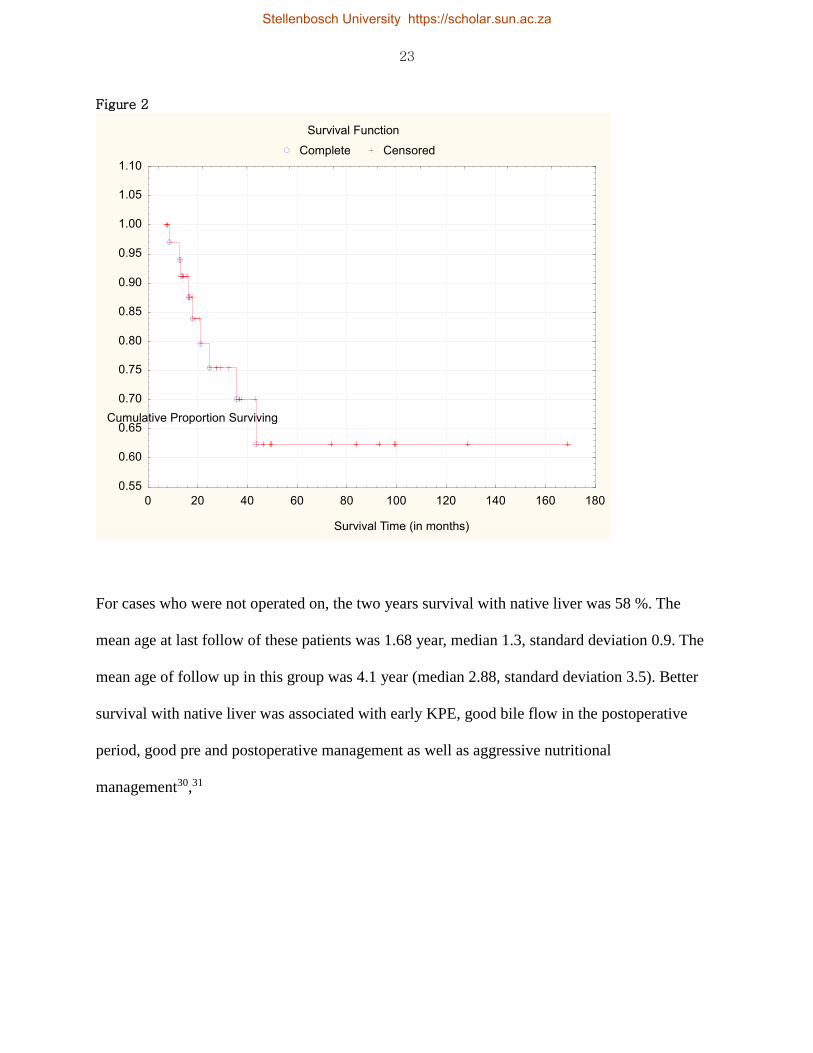
Figure 2
For cases who were not operated on, the two years survival with native liver was 58 %. The
mean age at last follow of these patients was 1.68 year, median 1.3, standard deviation 0.9. The
mean age of follow up in this group was 4.1 year (median 2.88, standard deviation 3.5). Better
survival with native liver was associated with early KPE, good bile flow in the postoperative
period, good pre and postoperative management as well as aggressive nutritional
management30,31
Survival Function
Complete Censored
0 20 40 60 80 100 120 140 160 180
Survival Time (in months)
0.55
0.60
0.65
0.70
0.75
0.80
0.85
0.90
0.95
1.00
1.05
1.10
Cumulative Proportion Surviving
Stellenbosch University https://scholar.sun.ac.za

24
Survival comparing the KPE/non-KPE groups. Figure 3
Log-Rank Test (Kaplan Meier data) WW = -2.744 Sum = 8.5419 Var = 2.0649 Test statistic = -1
.90933 p = 0.05622
The Log Rank test did not reach statistical significance possibly due to the small sample size
Patients who underwent Kasai surgery had a longer survival time compared to those who were
not operated on. Most of the non-operated patients died at an early age. The mean age at time of
death for the 10 deceased was 25.3 months, median 19.5, standard deviation 16.2 (range 8.5 to 5
9) months.
In those who had Kasai surgery, deaths were due to sepsis in 3 patients and to an unknown cause
in one (who deceased at home). For those who did not undergo surgery, hepatic encephalopathy
Cumulative Proportion Surviving (Kaplan-Meier)
Complete Censored
With surgery
Without surgery
0 20 40 60 80 100 120 140 160 180 200
Time (in months
0.3
0.4
0.5
0.6
0.7
0.8
0.9
1.0
Cumulative Proportion Surviving
Stellenbosch University https://scholar.sun.ac.za

25
in one, end stage liver disease and severe failure to thrive in three patients were the cause of
death (2 died at home - one had KPE and one not). Four were not referred for LTx. Six patients
were lost to follow up and their outcome is unknown.
Causes of death in all patients irrespective whether KPE was done
Table 7
Main causes of death No of patients
Sepsis 3
Hepatic encephalopathy 1
End-stage liver disease 3
Excessive esophageal bleeding 1
Unknown cause(died at home) 2
The median survival time in patients who underwent KPE surgery was longer (4.1 years)
compared to the survival time in patients without KPE which was much shorter (1.68 year).
Outcome of patients who underwent Kasai surgery
Table 8
Age (days) at surgery Alive Lost to follow up Died
<60 2 1 1
60-90 9 2 2
>90 4 1 1
Of the 23 patients in whom surgery was performed, 15 were documented still alive at last follow
up date, 4 were lost to follow up and their outcome is unknown since last seen at 8, 21 and 37
months of age respectively. Four were dead at 12, 13, 21months and 4 year of age. Of the 15 still
Stellenbosch University https://scholar.sun.ac.za

26
alive, 4 are on the active/inactive waiting list for LTx. Of the 14 patients who did not have KPE,
6 died, 3 are lost to follow up and 4 are documented alive (one of whom underwent LTx, one is
on the inactive waiting list for LTx and 3 are being investigated for referral for LTx).
Results after Kasai PE
Table 9
Result Number Percentage
Successful KPE 19 82.6%
Known alive at last follow up 15 65.2%
Lost to follow up 4 17.4%
Death 4 17.4%
Candidates on Liver transplant waiting list (included in the 15 aliv
e patients)
4 17.4%
Results without Kasai PE
Table 10
Result Number Percentage
Patients who did not have KPE 14 37.8%(of the 37)
Death 6 42.8%(of the 14)
Lost to follow up 3 21.4%(of the 14)
Alive after Liver transplant 1 7%
Awaiting Liver transplant 1 7%
Alive not yet referred for LTx 3 21.4%
Stellenbosch University https://scholar.sun.ac.za

27
Patients lost to follow up
In our study, 7 patients were lost to follow up. Of the 7 patients, 6(85.7%) were from rural areas.
Attempts to trace patients by means of the hospital information system (clinicom), telephonic
calls to children’s parents or relatives, and contact with RXH liver transplant unit have been tried
several times. Of the 7 patients, 3 patients were traced of whom 2 died (the information was
abstained from the relatives of the deceased children) and one patient, 14 years old is on the
inactive waiting list for liver transplant at RXH. In the 4 remaining patients the outcome is
unknown, but they are more likely deceased than alive.
Stellenbosch University https://scholar.sun.ac.za

28
Discussion
BA is a rare but important and potentially treatable disorder as shown initially by Kasai and later
by other medical workers. For decades, the Kasai portoenterostomy (KPE) has been the regular
surgical procedure for BA worldwide. We analysed the presentation and outcome of BA patients
treated in our centre from 1997 to 2011, with the aim of identifying prognostic factors in these
patients. Many previous studies have analysed the prognostic factors associated with the Kasai
PE and it is controversial as to which factors will predict a successful or failed KPE. The age at
time of Kasai surgery is a potential factor that has been shown in multiple studies to correlate
negatively with survival after KPE. The issue is whether the Kasai procedure is done within 60
or 90 days of life. In our study the survival and outcome of patients was not significantly
impacted by the age at time of surgery. We believe that the numbers were too small to show a
statistically significant difference. There was early jaundice clearance and drainage of bile in
50% of patients who underwent Kasai before or at 60 days of age, in 43.8% of those with surgery
between 60 and 90 days; and in 60% of those in whom surgery was performed after 90 days of
life. Of the 23 patients in whom surgery was performed, 15 were documented still alive (range
14 months to 14 years) at the last follow up date, 4 were lost to follow up and their outcome is
unknown and 4 died. Of the 15 children still alive, 9 underwent surgery between 60 and 90 days
(of whom 2 were lost to follow up and 2 died); 2 had surgery before or at 60 days (of whom one
was lost to follow up and 1 died) and 4 had a KPE after 90 days of life (one was lost to follow up
and 1 died). There was no significant difference in outcome for those who presented early at 60
days or late at 90 days of life. A factor which impacts time of surgery is timing of presentation of
patients to the referral centre. In our study, we found that 32% were referred late with consequent
Stellenbosch University https://scholar.sun.ac.za

29
delay in surgery to after 90 days. The reasons for delayed referral were parents’ ignorance of
their child’s clinical condition and awareness of need to seek health care as well as failure to
appreciate the importance of jaundice in a young infant by medical staff . In a survey done by
Howard ER et al, the reasons for late referral for effective surgery were almost identical to those
found in our study 32. The introduction of a newborn screening programme for BA in Western
(UK, France), Asian (Japan, Taiwan) and American (Argentina) countries contributed to
diagnosis within the first weeks of life with Kasai surgery subsequently performed at an early
age 30-60 days of life. In the UK and Japan, a yellow alert educational programme was initiated
to expedite the referral of the infants with BA to specialized centres. In Asia (Thailand and
Taiwan), a stool colour card has been introduced to identify jaundiced infants with BA. It may be
beneficial to introduce such health care policies in SA to improve the timing of referral for
patients with BA.
In a review by Tan (Tan C, 1994), it is stated that the time of onset, rate of progression, and the
severity of the disease process in BA varies from case to case and the author argues against
determining prognosis purely on age at surgery.
Several recent studies have suggested that the expertise of the site and the experience of the
surgeon performing the Kasai surgery are major prognostic factors; centers with greater case
experience show evidence of better survival rates. In the UK where they register annually around
2000 to 3000 cases of BA, a centralisation policy was implemented in 1999 restricting the
referral and management of all BA patients throughout the country to 3 skilled pediatric
hepatology units. In France, a decentralisation policy was maintained with efforts to promote
collaboration and ensure high standards of care among all the treatment centers. These different
policies led to an increase in 4-year patient survival rates from 85 to 89% (UK) and 74% to 87%
Stellenbosch University https://scholar.sun.ac.za
30
(France) between 1997 and 2002.
In SA KPE surgery is mostly restricted to tertiary university hospitals and LTx are only done in
two centres- Red Cross Children’s Hospital in Cape Town and Donald Gordon Hospital in
Gauteng. Within TCH, only senior paediatric surgeons perform KPE surgery, and the post-
operative care is similar to that of other institutions.
Other factors which influence survival include postoperative complications. We were unable to
report on the number of episodes of post-operative cholangitis as consistent diagnostic criteria
were not used.
The effect of steroids (introduced after 2007 as part of post-operative treatment) on the outcome
post-surgery could not be assessed due to the limited sample and lack of a uniform dosing
regimen for all post-operative patients. In fact, these liver diseases trigger fat soluble vitamins (A,
D, E and K) deficiency and consequently a malabsorption of the nutrients.
Our study unfortunately was not able to assess the incidence of BA in our tertiary centre. Our BA
patients were mostly born in the drainage areas of our tertiary centre with the problem of
obstructive jaundice. It was not possible to establish the total number of babies born at various
clinics, day and district hospitals over the same period as our study.
The few studies from developing countries which assessed outcome differ from those from
developed countries. In Nigeria, one report33 of 36 histologically confirmed cases of BA in
infants (range 2-20 months) over a 13 year study period, found that almost half of the patients
died, 19 were lost to follow up and 2 survived with persistent jaundice. An Egyptian study
showed that of 30 patients who underwent Kasai PE, 15 (50%) had a successful Kasai, one died
3 weeks after surgery from fulminant hepatic necrosis and multiorgan failure, 4 were lost to
Stellenbosch University https://scholar.sun.ac.za
31
follow up and 8 were candidates for LTx.
During our study period, 23 patients underwent Kasai surgery of whom 19 had a successful
outcome. The study showed that 15 (65.2%) patients were still alive at the time of conclusion of
the study, four (17.4%) died during the postoperative period from sepsis, end-stage liver disease,
hepatic encephalopathy and multiorgan failure and 4 (17.4%) were lost to follow up. Of the 15
patients (out of the 23) still alive (range 1-14years), 4 are eligible for LTx and are on the active
or inactive waiting lists for LTx and 11 are still followed up in our clinic.
Based on our data, we believe the KPE is a useful initial step even for those who present at an
older age, but without established liver cirrhosis. Our results are encouraging but still not as good
as those of Western countries (UK, USA, France) and Asian countries (Japan, Taiwan). It is
remarkable that general health care underwent many developments in SA especially with public
health systems with which all the South Africans with low income, adult and children, have
access to health care. The Kasai patient’s survival rate has increased over the ten years.
Stellenbosch University https://scholar.sun.ac.za

32
Conclusion
In the last five decades, BA has gone from a fatal disease to one that is treatable for most
children. The Kasai PE is the optimal initial management for patients with BA, even for selected
cases presenting after 90 days. Early PE is more likely to result in improved outcomes. Surgery
performed by an experienced paediatric surgeon could result in a better outcome (and prevent a
number of failed PE performed at any age). Long-term survival for patients with BA may exceed
90% if some conditions are fulfilled. Policies to ensure timely diagnosis and intervention,
therapeutic surgery and good nutrition are all needed to enhance the outcome of patients with BA.
Patients who had successful KPE surgery grow and develop better and are ultimately better liver
transplant candidates than malnourished children. Early identification and referral of babies with
obstructive jaundice is essential to offer Kasai PE at the most favourable time. Based on the
literature, the introduction of the newborn screening programme for BA patients helped to
diagnose patients within the first weeks of life with earlier Kasai surgery (at 30-60 days of life)
and with subsequent outcome and survival rate. Health care policies to improve the timing of
referral for patients with BA should be introduced in SA to achieve this.
This study is one of the first to document the outcomes of children with BA in SA. Further
studies are needed to analyse incidence and long-term outcomes of children with BA in the
Western Cape and in SA.
Despite all improvements in terms of early diagnosis and treatment made in the past 50 years,
BA remains a poorly understood disease. Its cause remains unknown, yet if found it may hold the
key to advancement in the treatment and possible prevention of the disease. Knowledge gained
in understanding the role of the immune system in the aetiology of BA may provide new and
Stellenbosch University https://scholar.sun.ac.za

33
promising therapeutic strategies. This will allow more significant data to guide additional
potential improvements in the care and hopefully look for much needed light on this dark chapter
of paediatrics.
Stellenbosch University https://scholar.sun.ac.za

34
References
1 Motala, 1990. Cholestatic Disorders of Infancy- Etiology and Outcome
2 Spearman, 2006. Liver transplantation at Red Cross War Memorial Children’s Hospital. S Afr
Med, pp. 96:960-963
3 Balistreri, 1996. Biliary atresia: current concepts and research directions. Hepatology , pp.
23(6): 1682-92.
4 Chardot, 1999. Epidemiology of biliary atresia in France: a national study 1986–96. J Hepatol,
pp. 1006-1013
5 Tan, 1994. The developing human biliary system at the porta hepatis level between 11 and 25
weeks of gestation: a way to understanding biliary atresia. Part 2.. Pathol Int, p. 44:600–610.
6 Chardot, 1999. Epidemiology of biliary atresia in France: a national study 1986–96. J Hepatol,
pp. 1006-1013
7 Moreski, 1983. Extrahepatic biliary atresia in a rhesus monkey (Macaca mulatta). Hepatology ,
Volume 3, pp. 577-580.
8 Steele, 1995. Reovirus 3 not detected by reverse transcriptase-mediated polymerase chain reaction
analysis of preserved tissue from infants with cholestatic liver disease. Hepatology, Volume 21, p. 697–
702.
9 Tarr, 1996. Biliary atresia, cytomegalovirus, and age at referral. Pediatrics, Volume 97, pp.
828-31.
10Fischler, 1998. The viral association of neonatal cholestasis in Sweden: a possible link between
cytomegalovirus infection and extrahepatic biliary atresia.. J Pediatr Gastroenterol Nutr, Volume
27, p. 57–64.
11 Yoon, 1997. Epidemiology of BA: A population based study. Pediatric, Volume 25, pp. 376-
382
12 Balistreri, 1996. Biliary atresia: current concepts and research directions. Hepatology , pp.
23(6): 1682-92.
13 McKieman, 2000. Patrick J , Alastair J Baker, Deirdre A Kelly. The frequency and outcome of
biliary atresia in UK and Ireland.. Lancet , Volume 355, pp. 25-29
Stellenbosch University https://scholar.sun.ac.za
35
14 Kasai M, S. S., 1959. A new operation for “noncorrectable” biliary. Hepatic Portoenterostomy,
Volume 13, pp. 733-739
15 Luo Y, Zheng S. Current concepts about postoperative cholangitis in BA. World J Pediatr
2008; 4:14
16 Kenneth, 1997. Biliary atresia: Should all patients undergo a portoenterostomy?. J Pediatr
Surgery, 32(February), pp. 168-174
17 Hanmin, K. e. a., 2001. The Kasai PE: When is it too late?. J Pediatr Surg , pp. 36:97-99
18 Hasegawa T, S. T. O. A., 2003 Apr. Indication for redo hepatic portoenterostomy for
insufficient bile drainage in BA: re-evaluation in the era of liver transplantation. Pediatr Surg Int
19 Davenport, 2004. l: The outcome of the older(more than 100 days) infant with BA. J.Pediatr
Surg, Volume 39, pp. 575-581
20 Mark Davenport, J. d. V. d. G. G. M.-V. D. K. P. M. C., 2004. Seamless management of biliary
atresia in England and Wales (1999-2002). Lancet , Volume 363, pp. 1354-57
21 Howard, 2003. Late referral for BA- Missed opportunities for effective PE surgery.. Pediatr
Surg.
22 Sokol RJ, S. B., 2006. A Multicenter study of the outcome of Biliary Atresia in the United
States, 1997 to 2000. J Pediatr, Volume 148, pp. 467-474
23 Schreiber, 2002. Biliary atresia in Canada: the effect of centre caseload experience on outcome
24 Masaki Nio, N. S., 2006. et al: Long-term outcome in type 1 biliary atresia. .. Journal of
Pediatric Surgery, Volume 41, pp. 1973-5
25Tan C, D. M., 1994. Does the morphology of the extrahepatic remnants in BA influence
survival? A review of 205 cases.. J Pedatr Surg, Volume 29, pp. 1459-1464
26 Hayes DH, D. R. C. R. W. G. H. C. A. R., 1992. . Extrahepatic biliary atresia from diagnosis to
liver transplantation.. Pediatr Surg Int , Volume 7, pp. 737-40
27 Mshelbwala PM, S. L. L. C. A. E., 2007. Management of biliary atresia in Nigeria: the
ongoing challenge.. Ann Trop Paediatr , Volume 27, p. 69–73
28Zuckerman M, H. C., 1998. Incidence and outcome of biliary atresia in black infants in Soweto
(South Africa): review of cases from 1993-1996 [abstract]. J Pediatr Gastroenterol Nutr, Volume
26, p. 587
29 Elsadat, A. M., Oct 2009. BA: Experience with 30 consecutive cases in a single institute.
Stellenbosch University https://scholar.sun.ac.za

36
Annals of Pediatr Surgery, Volume 5, No 4, pp. 233-240
30 Pediatric Clinics of North America, Volume 56,Issue 5, page 1161-1183, October 2009
31 De Russo PA, Shepherd R, et al. Growth failure and outcomes in infants with BA: A report
from the BARC. Hepatology. 2007, 46(5): 1632-1638
32 Howard, 2003. Late referral for BA- Missed opportunities for effective PE surgery.. Pediatr
Surg. 33 Mabogunje O.A. 1987. Biliary atresia in Zaria, Niger: A review Ann Trop Paediatr 1987:7-204
Stellenbosch University https://scholar.sun.ac.za

Appendices
Appendix 1: Protocol
A Retrospective Review of the Outcome of Children Presenting to Tyger
berg Children’s Hospital with Biliary Atresia
OR Karangwa; E Nel
Tygerberg Hospital
University of Stellenbosch
Cape Town, South Africa.
Stellenbosch University https://scholar.sun.ac.za
Introduction
Biliary atresia (BA) is a serious but relatively rare congenital disease of the bile ducts and liver and freq
uently has fatal complications. The reported incidence varies from 5/100000 births per year in Holland
to 6/100000 in UK to 32/100000 in French Polynesia (Christophe et al, 2001). The incidence in South
Africa is unknown. The precise mechanism underlying the pathogenesis of the BA remains unclear. It i
s characterized by progressive fibrotic obliteration and obstruction of the extra hepatic biliary tree ultim
ately leading to liver cirrhosis and liver failure. Most children present within four to six weeks with co
njugated jaundice, pale, acholic stools and dark urine.
If BA is diagnosed within the first two to three months after birth, a Kasai portoenterostomy (KPE) whi
ch connects the bile drainage from the liver to the intestinal tract, can successfully restore bile flow into
the intestinal tract in 50% to 90% of patients. In some infants jaundice resolves and the progression of
liver disease is delayed. However, if untreated, liver cirrhosis gradually develops within the first month
s of life and the majority of children die before the age of two years. After a KPE, complications such a
s ascending cholangitis (bacterial), poor growth and malnutrition, portal hypertension and liver failure
may develop. Children who have not had a KPE performed within the first 3 months of life or who hav
e had an unsuccessful KPE require liver transplantation (LTx), usually before 2-5 years of life.
The survival of infants born with BA has improved over the last 30 past years. Of eleven infants born w
ith BA in one region in England during the period 1971-73 only one child survived to 10 years (Purrice
li et al, 2004). In contrast to this, at present, in England and in Wales about 90% of children with BA ar
e surviving, at least in the short-term through KPE and LTx. (Davenport et al, 2006
Stellenbosch University https://scholar.sun.ac.za

In South Africa (SA), the incidence of extra hepatic biliary atresia (EHBA) is still unknown and few stu
dies have evaluated the long-term outcome after surgery. It is the most frequent indication for LTx in c
hildren in SA (Spearman et al, 2006). The outcome of all children with BA is however not well docume
nted. This study will describe the outcome of infants and children with BA who have been treated at Ty
gerberg Children’s Hospital (TCH). We will describe the parameters that influence prognosis (e.g. age
of referral, nutrition and postoperative cholangitis); the frequency of complications (e.g. intestinal hem
orrhage, spontaneous bacterial peritonitis, and liver failure) and the survival of children with BA in our
institution. These data are necessary to identify factors leading to the delayed diagnosis of BA; to impro
ve the management of biliary atresia and to optimize the selection of children for LTx.
Aim
The aim of this study is to review the presentation and long-term outcome of the infants with BA presenting to
Tygerberg Children’s Hospital.
Objectives
The objectives of the study are to describe the following features of children with BA who present to TCH:
1. Demographic details.
2. Clinical features at presentation.
3. Causes of delayed presentation.
4. Short term complications after surgery.
5. Long-term outcome
Stellenbosch University https://scholar.sun.ac.za

Research methodology
Study design
A retrospective descriptive study will be performed using data sourced from patient records.
Sample
Patients will be traced from a database of outpatients and inpatients seen at the Paediatric Gastroenterology out
patient clinic and Pediatric Gastroenterology inpatient service. The data will be collected from the clinical recor
ds of children identified. Information regarding outcome of referral for liver transplant will be requested from th
e paediatric liver transplant unit at Red Cross Children’s Hospital.
Inclusion criteria
The following inclusion criteria will be used:
1. Children who presented to Tygerberg Hospital 1997 to 2011
2. Children with a diagnosis of biliary atresia as confirmed by clinical and biochemical findings and liver
histology. When performed an operative cholangiogram will serve as further confirmation of the diagnosis.
Sample Size
It is estimated that the sample size will be 37 children.
Methodology
A datasheet will be used to record the applicable information from TCH file, folders, and clinical pathology resul
ts database. (Appendix 7). All the information will be analyzed using standard statistical methods.
Variables
The following variables will be recorded:
Stellenbosch University https://scholar.sun.ac.za
3. The demographic characteristics of the patients (gender, ethnicity, residential address). (Appendix 2)
4. The date of birth and age of presentation,
5. The delay from presentation to the health service and delay at Tygerberg Hospital to confirmation of the
diagnosis, and causes of delayed presentation and confirmation.
6. Complications of surgery. (Appendix 6)
a. Cholangitis in the post-operative period.
b. Surgical complications.
c. Bile flow after surgery (determined by the presence of bile stained stool).
7. Histology of preoperative and/or intra-operative liver biopsies.
8. Measures of morbidity occurring after diagnosis:
a. Growth (weight and length). (Appendix 3)
b. Presence of fat soluble vitamin deficiencies (clinical and biochemical signs).
c. Liver biochemistry. (Appendix 4)
d. The presence of signs of portal hypertension.(Appendix 5)
e. Intestinal hemorrhage. (Appendix 5)
i. Age of first hemorrhage.
ii. Etiology of hemorrhage.
9. Number of admissions required
10. Therapy given (all drugs, vitamin supplements, nutritional support, surgical treatment, sclerotherapy)
11. Death and cause of death.
12. The following aspects regarding liver transplantation will be recorded:
a. Date referred for transplantation.
b. Date accepted for transplantation.
c. Date and outcome of liver transplant performed.
Stellenbosch University https://scholar.sun.ac.za

Statistical Methods
All the informations will be analyzed using standard statistical methods:
1. Variables will be summarized as medians and interquartile ranges.
2. Outcome data (survival, time to first variceal bleed, time to transplantation) will be described by
survival curves (Kaplan Meier Curve).
Ethical considerations
Importance of this study to the community
There is no recent data available describing the outcome of children with BA in the Western Cape, South Africa
. This data aims to identify areas where the management of children can potentially be improved (e.g. causes of
late presentation, quality of nutritional support, and the effective management of complications such as variceal
hemorrhage).
Anonymity
Patients will be assigned a study number in the database. The names and hospital numbers of the patients linked
to the study numbers will be held in a separate database once data entry has been completed. This database will
be kept secure in an alternative area. Patients who have been lost to follow-up will be recalled for review. The
consent of the legal guardians will not be sought.
Budget
All costs will be wholly carried by the Department of Paediatrics and the principal researcher himself.
Stellenbosch University https://scholar.sun.ac.za

Appendix
Data sheet: Anthropometry
Patient ID Date Weight Height
12345676 0 0
Stellenbosch University https://scholar.sun.ac.za

Biochemistry data
Patient ID
Date
Tprot
Alb
Glob
Br tot
Br conj
Cholesterol
AST
ALT
LDH
ALP
GGT
Ca
Phosphate
INR
PTT
WBC
Hb
MCV
Platelets
RBC
Stellenbosch University https://scholar.sun.ac.za
Stellenbosch University https://scholar.sun.ac.za
5
Stellenbosch University https://scholar.sun.ac.za