DMD # 61804
1
Title Page
A Novel Reaction Mediated by Human Aldehyde Oxidase: Amide Hydrolysis of
GDC-0834
Jasleen K. Sodhi, Susan Wong, Donald S. Kirkpatrick, Lichuan Liu, S. Cyrus Khojasteh,
Cornelis E. C. A. Hop, John T. Barr, Jeffrey P. Jones, and Jason S. Halladay*
Department of Drug Metabolism and Pharmacokinetics (J.K.S., S.W., S.C.K., C.E.C.A.H.
and J.S.H.), Department of Clinical Pharmacology (L.L.), and Department of Protein
Chemistry (D.S.K.), Genentech, Inc., 1 DNA Way, South San Francisco, CA 94080, USA;
Department of Chemistry (J.T.B. and J.P.J.), Washington State University, Pullman, WA
99164, USA.
This article has not been copyedited and formatted. The final version may differ from this version.DMD Fast Forward. Published on April 6, 2015 as DOI: 10.1124/dmd.114.061804
at ASPE
T Journals on A
pril 3, 2016dm
d.aspetjournals.orgD
ownloaded from

DMD # 61804
2
Running Title Page
Running Title: Amide Hydrolysis Mediated by Human Aldehyde Oxidase
Corresponding Author: Jasleen K. Sodhi, Genentech, Inc., 1 DNA Way, MS 412a,
South San Francisco, CA 94080. USA. Tel.: +1 650 225 8190; Fax: +1 650 467 3487; e-
mail address: [email protected]
Text Pages ................................ 31
Tables ....................................... 1
Figures ..................................... 6
References ................................ 34
Words in Abstract ................... 214
Words in Introduction ............ 424
Words in Discussion ................ 1,396
ABBREVIATIONS: AO, aldehyde oxidase; BNPP, bis-(p-nitrophenyl) phosphate;
BTK, Bruton’s tyrosine kinase; CES, carboxylesterase; CPT-11, irinotecan; DACA, N-
[(2-dimethylamino) ethyl]acridine-4-carboxamide; DCPIP, dichlorophenolindophenol;
DLC, dog liver cytosol; HLC, human liver cytosol; MoCo, molybdenum cofactor; XO,
xanthine oxidase.
This article has not been copyedited and formatted. The final version may differ from this version.DMD Fast Forward. Published on April 6, 2015 as DOI: 10.1124/dmd.114.061804
at ASPE
T Journals on A
pril 3, 2016dm
d.aspetjournals.orgD
ownloaded from

DMD # 61804
3
Abstract
GDC-0834, a Bruton’s tyrosine kinase inhibitor investigated as a potential treatment for
rheumatoid arthritis, was previously reported to be extensively metabolized by amide
hydrolysis such that no measurable levels of this compound was detected in human
circulation following oral administration. In vitro studies in human liver cytosol
determined GDC-0834 was rapidly hydrolyzed with CLint of 0.511 mL/min/mg protein.
Aldehyde oxidase (AO) and carboxylesterase (CES) were putatively identified as the
enzymes responsible following cytosolic fractionation and MS-proteomics analysis of the
enzymatically active fractions. Results were confirmed by a series of kinetic experiments
with inhibitors of AO, CES, and xanthine oxidase (XO), which implicated AO and CES,
but not XO, as mediating GDC-0834 amide hydrolysis. Further supporting the
interaction between GDC-0834 and AO, GDC-0834 was shown to be a potent reversible
inhibitor of six known AO substrates with IC50 values ranging from 0.86 to 1.87 μM.
Additionally, in silico modelling studies suggest that GDC-0834 is capable of binding in
the active site of AO with the amide bond of GDC-0834 near the molybdenum cofactor
(MoCo), orientated in such a way to enable potential nucleophilic attack on the carbonyl
of the amide bond by the hydroxyl of MoCo. Together, the in vitro and in silico results
suggest the involvement of AO in the amide hydrolysis of GDC-0834.
This article has not been copyedited and formatted. The final version may differ from this version.DMD Fast Forward. Published on April 6, 2015 as DOI: 10.1124/dmd.114.061804
at ASPE
T Journals on A
pril 3, 2016dm
d.aspetjournals.orgD
ownloaded from

DMD # 61804
4
Introduction
GDC-0834 is a potent, selective, and reversible ATP-competitive small molecule
inhibitor of Bruton’s tyrosine kinase (BTK) that was under consideration as a therapeutic
agent for rheumatoid arthritis (Liu et al., 2011a; Liu et al., 2011b). One liability for
GDC-0834 was the low confidence in the human pharmacokinetic prediction based on
the species-dependent metabolism, with amide hydrolysis being more predominant in
human than other pre-clinical species. However, despite the uncertainty in the human
clearance prediction, there was a high level of interest in a BTK inhibitor for clinical
evaluation. An investigational new drug strategy was initiated in which GDC-0834 was
rapidly advanced to a single-dose human clinical trial study. Exploratory clinical studies
established that after oral administration of 35 and 105 mg GDC-0834 to healthy
volunteers, limited exposure of this drug was observed in circulation (<1 ng/mL in most
plasma samples). This was attributed primarily to metabolism, with the majority of drug-
related circulating material being the aniline metabolite M1 (Fig. 1). At 35 mg, the mean
highest observed plasma concentration (Cmax) of M1 was 142 ng/mL; at 105 mg, the
mean Cmax of M1 was 390 ng/mL (Liu et al., 2011b).
In contrast to humans, the extensive metabolism of GDC-0834 was not evident in
preclinical species, as GDC-0834 was orally bioavailable following oral administration to
mice, rats, dogs, and monkeys. In plasma of preclinical species, M1 was minor and the
percent exposure ratios of M1 AUC / parent AUC were 9.3% (SCID mice), 1.5% (rats),
26% (dogs), and negligible (monkeys). It is interesting that in PXB mice with humanized
livers, the ratio increased to 74.1% (Liu et al., 2011b). In vitro metabolic stability studies
of GDC-0834 in liver microsomes (in the presence and absence of NADPH) and
This article has not been copyedited and formatted. The final version may differ from this version.DMD Fast Forward. Published on April 6, 2015 as DOI: 10.1124/dmd.114.061804
at ASPE
T Journals on A
pril 3, 2016dm
d.aspetjournals.orgD
ownloaded from

DMD # 61804
5
hepatocytes predicted hydrolysis as the route of metabolism in humans and indicated
significant differences in amide hydrolysis rates between humans and other preclinical
species (Liu et al., 2011b). This specific GDC-0834 to M1 biotransformation was much
more pronounced in human liver fractions than those of preclinical species.
Here we investigated the enzyme(s) involved in the amide hydrolysis of GDC-0834.
Preliminary in vitro metabolism experiments using various liver fractions revealed that
soluble enzyme(s) present in human liver cytosol (HLC) mediated the amide hydrolysis
of GDC-0834. Therefore, HLC was chosen over human liver microsomes and
hepatocytes since it contained the soluble enzyme(s) to facilitate fractionation and MS-
proteomics analysis. A series of kinetic experiments with probe substrates and chemical
inhibitors of aldehyde oxidase (AO), carboxylesterase (CES), and xanthine oxidase (XO),
as well as in silico modelling studies of GDC-0834 were conducted.
Materials and Methods
Chemicals. GDC-0834 ((R)-N-(3-(6-(4-(1,4-dimethyl-3-oxopiperazin-2-
yl)phenylamino)-4-methyl-5-oxo- 4,5-dihydropyrazin-2-yl)-2-methylphenyl)-4,5,6,7-
tetrahydrobenzo[b] thiophene-2-carboxamide), M1, M2 (acid metabolite; Fig. 1), and an
internal standard (structural analog of GDC-0834 and a proprietary compound) were
synthesized at Genentech, Inc. (South San Francisco, CA). Phosphate buffered saline
(PBS; pH 7.4), potassium phosphate buffer (KPi; 100 mM; pH 7.4), and 4-
morpholinepropanesulfonic acid (MOPS; 2.5 mM; pH 7.4) were provided by the Media
Preparation Facility at Genentech, Inc. High-performance liquid chromatography-grade
solvents (acetonitrile, methanol, and water) were purchased from EMD Chemicals, Inc.
This article has not been copyedited and formatted. The final version may differ from this version.DMD Fast Forward. Published on April 6, 2015 as DOI: 10.1124/dmd.114.061804
at ASPE
T Journals on A
pril 3, 2016dm
d.aspetjournals.orgD
ownloaded from

DMD # 61804
6
(Gibbstown, NJ). Formic acid (FA) was purchased from Mallinckrodt Baker, Inc.
(Phillipsburg, NJ). Allopurinol, O6-benzylguanine, bis-(p-nitrophenyl) phosphate
(BNPP), 2,6-dichlorophenolindophenol (DCPIP), β-estradiol, 7-ethyl-10-
hydroxycamptothecin (SN-38), irinotecan (7-ethyl-10-[4-(1-piperidino)-1-
piperidino]carbonyloxycampthothecin; CPT-11), loperamide, zaleplon, and zoniporide
were purchased from Sigma Aldrich (St. Louis, MO). Raloxifene, 4-hydroxycarbazeran,
8-oxobenzylguanine, 5-oxozaleplon, and 2-oxozoniporide were purchased from Toronto
Research Chemicals Inc. (North York, ON, Canada). Phthalazine was purchased from
Alfa Aesar (Ward Hill, MA). Menadione and phthalazinone were purchased from Acros
Organics (Geel, Belgium). Carbazeran was purchased from Chemoraga, Inc. (Oakland,
CA). HLC from mixed male and female donor pools (n=150 donors; protein content of
20 mg/mL) and dog liver cytosol (DLC) from a pool of male beagle dogs (n=4 dogs;
protein content of 20 mg/mL) were purchased from BD Biosciences (San Jose, CA) and
stored at -80oC. Fresh whole blood and plasma collected from male humans, male
Sprague-Dawley rats, female CD-1 mice, male beagle dogs, and male cynomologus
monkeys were purchased from BioreclamationIVT (Westbury, NY). The synthesis of N-
[(2-dimethylamino) ethyl]acridine-4-carboxamide (DACA) was performed as described
by Barr and Jones (2013).
Protein Separation and LC-MS/MS Proteomic Analysis. HLC was fractionated
using an Agilent 1100 Series pump and a Varian 701 fraction collector. HLC (50 µL; 20
mg/mL) was manually injected via a 100 µL loop onto a Xenix SEC-300, 3 µm, 300 Å
size-exclusion column from Sepax Technologies (Neward, DE) and separated at a flow
rate of 0.5 mL/min using PBS (pH 7.4) as the mobile phase. The eluent was collected as
This article has not been copyedited and formatted. The final version may differ from this version.DMD Fast Forward. Published on April 6, 2015 as DOI: 10.1124/dmd.114.061804
at ASPE
T Journals on A
pril 3, 2016dm
d.aspetjournals.orgD
ownloaded from
DMD # 61804
7
500 µL fractions every min for one hr into glass test tubes. A portion of each fraction (45
µL) was assessed for enzymatic activity by incubating with GDC-0834 (0.8 µM) for 30
min. Reactions were terminated using acetonitrile containing the internal standard (100
µL) and centrifuged for 10 min at 2,000g. The supernatants (100 µL) were removed,
combined with water (200 µL), and analyzed by liquid chromatography-tandem mass
spectrometry (LC-MS/MS) to quantify the relative abundance of M1 formed from GDC-
0834.
Mass Spectrometry Proteomics and Correlation Profiling. Mass spectrometry
proteomics and correlation profiling were used to help identify potential candidate
enzymes responsible for conversion of GDC-0834 to M1. A series of seven fractions
(fractions 25 – 31) containing measurable amide hydrolytic activity and one fraction
(fraction 24) lacking activity were loaded onto a series of lanes on a sodium dodecyl
sulfate polyacrylamide gel electrophoresis (SDS-PAGE) on a 4-12% Bis/Tris gradient gel
run in MOPS buffer. Proteins were partially separated, subjected to in-gel trypsin
digestion, and peptides analyzed by LC-MS/MS on an LTQ-Orbitrap XL mass
spectrometer as described previously (Phu et al., 2011; Sheng et al, 2012). Precursor ions
were analyzed in high resolution (resolving power of 60K) in the Orbitrap and MS/MS
spectra were collected in the ion trap using data-dependent acquisition. MS/MS were
searched using Mascot against a concatenated target-decoy database comprised of
forward and reverse sequence of human proteins from Uniprot and common
contaminants with a 50 ppm precursor ion tolerance. Peptide spectral matches were
filtered to a 1% false discovery rate using linear discriminant analysis. Pearson
correlations were determined for each protein using the spectral count data relative to the
This article has not been copyedited and formatted. The final version may differ from this version.DMD Fast Forward. Published on April 6, 2015 as DOI: 10.1124/dmd.114.061804
at ASPE
T Journals on A
pril 3, 2016dm
d.aspetjournals.orgD
ownloaded from

DMD # 61804
8
metabolic activity observed across the series of fractions. P-values were determined for
each correlation and corrected for multiple hypothesis testing by the FDR based approach
using the “qvalue” library available through Bioconductor.
Kinetic Formation of M1 in HLC and DLC. Enzyme kinetic studies of M1
formation were performed in HLC and DLC. Assay conditions were optimized with
regard to protein concentration and incubation time, based on linear formation of M1
(data not shown). All incubations containing GDC-0834 (0.05 – 100 μM for human and
1 – 100 μM for dog) were incubated at 37°C in triplicate and initiated by the addition of
cytosol, with final protein concentrations of 0.05 mg/mL (HLC) and 3.0 mg/mL (DLC).
Optimized incubation times in HLC and DLC were 10 min and 60 min, respectively.
Incubations (100 µL) were terminated by protein precipitation with addition of
acetonitrile containing the internal standard (200 µL). All samples were centrifuged for
10 min at 2,000g, the supernatants removed, diluted 1:2 (v/v) with 0.1% FA in water, and
analyzed by LC-MS/MS. Standard curves of the M1 standard prepared in each matrix
were used, with a lower limit of quantitation 0.01 µM, to quantify the amount of M1
formed and estimate the maximum rate of M1 formation (Vmax) and the Michaelis-
Menten constant (Km) using nonlinear regression analysis within GraphPad Prism
(GraphPad Software Inc., La Jolla, CA) using the Michaelis-Menten equation:
Y = Vmax*X/(Km + X)
where Y is enzyme velocity and X is substrate concentration.
Formation of M1 in Fresh Blood and Plasma. Metabolic stability studies with
GDC-0834 were conducted with fresh whole blood and plasma collected from human,
rat, mouse, dog, and monkey. Discrete incubations (100 µL) were conducted in triplicate
This article has not been copyedited and formatted. The final version may differ from this version.DMD Fast Forward. Published on April 6, 2015 as DOI: 10.1124/dmd.114.061804
at ASPE
T Journals on A
pril 3, 2016dm
d.aspetjournals.orgD
ownloaded from

DMD # 61804
9
at 37°C and initiated with addition of GDC-0834 (0.8 µM final concentration). Discrete
incubations corresponding to each time point were terminated at selected times with the
addition of methanol (500 µL) containing the internal standard and samples prepared for
LC-MS/MS analysis as described above. Standard curves of the M1 standard prepared in
each matrix were used to quantify the amount of M1 formed. Samples were analyzed by
LC-MS/MS for GDC-0834 and M1 formation.
Inhibition of GDC-0834 Metabolism Using Chemical Inhibitors. Chemical
inhibition studies with HLC, DLC, and human plasma were conducted using the AO
inhibitors β-estradiol, DCPIP, menadione, and raloxifene, the AO/CES inhibitor
loperamide, the CES inhibitor BNPP, and the XO inhibitor allopurinol. The inhibitors (0
- 50 μM in HLC and DLC and 0 - 10 µM in human plasma) were co-incubated with
GDC-0834 (0.8 μM with 0.05 mg/mL HLC for 10 min, 63 μM with 3.0 mg/mL DLC for
60 min, or 0.8 μM in human plasma (pH 7.4) for 2 hr). At the end of the incubation
period, the reactions were terminated and samples prepared for LC-MS/MS analysis as
described above. Samples were analyzed by LC-MS/MS for M1 formation. Estimations
of the 50% inhibitory concentration (IC50) values of the inhibitors were performed by
nonlinear regression analysis by GraphPad Prism using the following equation:
Y=100/(1+10^((LogIC50-X)*HillSlope)))
where Y is response and X is the logarithm of the concentration of the inhibitor.
Inhibition of AO- and CES-mediated Metabolism by M1 and M2. Inhibition
studies with M1 or M2 (0 – 10 μM; Fig. 1) were conducted in HLC using either an AO
probe substrate (phthalazine; 8 μM, 2.5 min with 0.05 mg/mL HLC) or a CES probe
substrate (CPT-11; 5 μM, 5 min with 1 mg/mL HLC). Incubation conditions were
This article has not been copyedited and formatted. The final version may differ from this version.DMD Fast Forward. Published on April 6, 2015 as DOI: 10.1124/dmd.114.061804
at ASPE
T Journals on A
pril 3, 2016dm
d.aspetjournals.orgD
ownloaded from

DMD # 61804
10
determined in our laboratory for linear formation of SN-38 with respect to CPT-11
concentration, incubation time, and protein concentration (data not shown). Similar
conditions were previously reported (Tabata et al., 2004). At the end of the incubation
period, the reactions were terminated and samples prepared for LC-MS/MS analysis as
described above. Samples were analyzed by LC-MS/MS for either the AO-mediated
metabolite of phthalazine (phthalazinone) or the CES-mediated metabolite of CPT-11
(SN-38). Estimations of the IC50 values of M1 and M2 were performed by nonlinear
regression analysis by GraphPad Prism using the following equation:
Y=100/(1+10^((LogIC50-X)*HillSlope)))
where Y is response and X is the logarithm of the concentration of the inhibitor.
Inhibition of AO-mediated Metabolism by GDC-0834. GDC-0834 (either 0 - 50
or 0 - 100 μM) and an AO probe substrate were coincubated in HLC at 37°C (n=3).
Incubation conditions for each of the AO probe substrates were determined in our
laboratory. The conditions at which the formation of the AO-mediated metabolite of
each probe was linear with respect to incubation time and protein concentration were
used (data not shown). AO probe substrates (and incubation conditions) were carbazeran
(1 μM, 3 min with 1 mg/mL HLC), DACA (6.3 μM, 2.5 min with 0.05 mg/mL HLC), O6-
benzylguanine (1 μM, 10 min with 1 mg/mL HLC), phthalazine (8 μM, 2.5 min with 0.05
mg/mL HLC), zaleplon (1 μM, 30 min with 1 mg/mL HLC), or zoniporide (5 μM, 30 min
with 1 mg/mL HLC). At the end of the incubation period, the reactions were terminated
and samples prepared for LC-MS/MS analysis as described above. Samples were
analyzed by LC-MS/MS for the AO-mediated metabolites of carbazeran (4-
hydroxycarbazeran), DACA (DACA-9(10H)-acridone), O6-benzylguanine (8-oxo
This article has not been copyedited and formatted. The final version may differ from this version.DMD Fast Forward. Published on April 6, 2015 as DOI: 10.1124/dmd.114.061804
at ASPE
T Journals on A
pril 3, 2016dm
d.aspetjournals.orgD
ownloaded from

DMD # 61804
11
benzylguanine), phthalazine (phthalazinone), zaleplon (5-oxozaleplon), or zoniporide (2-
oxozoniporide). Estimations of the IC50 values for GDC-0834 were performed by
nonlinear regression analysis by GraphPad Prism.
LC-MS/MS Analysis. All analytes were monitored by MS multiple-reaction
monitoring using an AB Sciex Triple Quad 5500 coupled to an Ultra High Pressure
Liquid Chromatography (UHPLC) pump and a CTC PAL autosampler from LEAP
Technologies (Carrboro, NC). A Kinetex phenyl-hexyl column (2.6 μm, 30 x 2.1 mm)
from Phenomenex (Torrance, CA) was used with mobile phases consisting of solvent A
(0.1% FA in water) and solvent B (0.1% FA in acetonitrile). The flow rate was 1 mL/min
and the injection volume was 25 μL. The gradient started at 1% solvent B for 0.4 min,
ramped up to 10% solvent B in 0.08 min, ramped up to 63% solvent B in 1.7 min, ramped
up to 95% solvent B in 0.3 min and held for 0.3 min, and then stepped down to the initial
conditions of 1% solvent B and held at these conditions for 0.7 min to equilibrate the
column prior to the next injection. The total LC-MS/MS run time was 3.48 min. The
multiple reaction monitoring (MRM) transitions in positive ion mode were: m/z 361 �
272 (carbazeran), m/z 587 � 124 (CPT-11), m/z 294 � 249 (DACA), m/z 310 � 265
(DACA-9(10H)-acridone), m/z 597.4 � 127.1 (GDC-0834), m/z 377 � 288.1 (4-
hydroxycarbazeran), m/z 433.4 � 127.1 (M1), m/z 183.1 � 139.1 (M2), m/z 242 � 91
(O6-benzylguanine), m/z 258 � 91 (8-oxobenzylguanine), m/z 322 � 252 (5-
oxozaleplon), 337.1 � 278.2 (2-oxozoniporide), m/z 147.0 � 90.0 (phthalazinone), m/z
393.0 � 349.0 (SN-38), m/z 306.0 � 236.0 (zaleplon), and m/z 321.1 � 262.2
(zoniporide). The positive ion mode transitions for phthalazine in single ion monitoring
(SIM) mode were m/z 131.1 � 131.1.
This article has not been copyedited and formatted. The final version may differ from this version.DMD Fast Forward. Published on April 6, 2015 as DOI: 10.1124/dmd.114.061804
at ASPE
T Journals on A
pril 3, 2016dm
d.aspetjournals.orgD
ownloaded from

DMD # 61804
12
Docking of GDC-0834 in AO Active Site. A homology model for AO was
produced using the human sequence and the crystal structure of mouse AOX3 (PDB:
3ZYV) (Coelho et al., 2012). Induced-fit docking was used to place DACA into the
active site of AO. Sequence alignment used the ClustalW program and required no user
input due to the high homology (79% homology) of the two primary sequences. For the
portions of the mouse structure lacking sufficient electron density, the human enzyme
was modelled using the energy-based method in Prime. However, residues 168-200 were
not able to be replaced and were excluded in the model. Modelling was done with
Schrödinger’s Prime module to generate a protein structure followed by the induced fit
docking workflow using DACA as a ligand to refine amino acid residues within 5 Å of
the DACA ligand. Glide docking energies were assessed for DACA, zaleplon, RS-8359,
XK-469, and GDC-0834. The structure for each of these compounds (except GDC-0834)
has been reported (Alfaro and Jones, 2008; Jones and Korzekwa, 2013).
Results
Mass Spectrometry Proteomics and Correlation Profiling (or Identification of
Aldehyde Oxidase in HLC by Correlation Profiling). Database search results
identified over 900 proteins, including 563 proteins with spectra matching at least two
unique peptides. Among the most abundant proteins in the sample series were the
metabolic enzymes carbamoyl-phosphate synthase (CPSM_HUMAN; 468 total peptide-
to-spectrum matches (PSMs) /77 unique peptides), fructose-bisphosphate aldolase B
(ALDOB_HUMAN; 420/21), retinal dehydrogenase 1 (AL1A1_HUMAN; 347/32),
aldehyde dehydrogenase (ALDH2_HUMAN; 292/31), liver carboxylesterase 1
This article has not been copyedited and formatted. The final version may differ from this version.DMD Fast Forward. Published on April 6, 2015 as DOI: 10.1124/dmd.114.061804
at ASPE
T Journals on A
pril 3, 2016dm
d.aspetjournals.orgD
ownloaded from

DMD # 61804
13
(EST1_HUMAN; 275/33), and aldehyde oxidase (AO) (ADO_HUMAN; 265/67) (Fig.
2A). Pearson correlations were determined for each identified protein relative to
normalized metabolic activity (Fig. 2A-B). Among the top candidates, AO was identified
and displayed a pattern of peptide spectral matches that tightly correlated with amide
hydrolysis activity (r2=0.9708) (Fig. 2A-B).
Kinetic Formation of M1 in HLC and DLC. The kinetic parameters describing the
metabolism GDC-0834 to M1 were determined by incubating parent compound with
HLC or DLC. Amide hydrolysis in humans was found to be more efficient than in dogs,
with Vmax of 409 ± 29 compared to 20.3 ± 9.6 pmol/min/mg protein, Km of 0.800 ± 0.027
compared to 63 ± 5 μM, and intrinsic clearance (Vmax/Km) of 0.511 compared to 0.00025
mL/min/mg protein in humans and dogs, respectively (Supplemental Fig. 1A and 1B).
Formation of M1 in Fresh Blood and Plasma. The formation of M1 over time
from GDC-0834 (0.8 μM initial concentration) was determined in fresh blood and plasma
collected from human, rat, mouse, dog, and monkey. A time-dependent formation of M1
was observed in human, rat, and mouse blood and plasma. In dog and monkey, a time-
dependent formation of M1 was observed in plasma, but levels in blood were below the
limit of quantification (<0.001 μM) (Fig. 3). The amounts of formation of M1 were more
pronounced in mouse blood and plasma with 0.016 and 0.120 μM formed by 180 min,
respectively. In contrast, the amounts of M1 formed were lower in blood and plasma of
human, rat, dog, and monkey, with 0.003 and 0.010 μM (human), 0.002 and 0.006 μM
(rat), <0.001 and 0.003 μM (dog), and <0.001 and 0.001 μM (monkey) present,
respectively. The loss of GDC-0834 was minimal in blood and plasma in all species,
with T1/2 values >500 min in both matrices (data not shown).
This article has not been copyedited and formatted. The final version may differ from this version.DMD Fast Forward. Published on April 6, 2015 as DOI: 10.1124/dmd.114.061804
at ASPE
T Journals on A
pril 3, 2016dm
d.aspetjournals.orgD
ownloaded from

DMD # 61804
14
Inhibition of GDC-0834 Metabolism Using Chemical Inhibitors. The inhibitory
properties of AO inhibitors (β-estradiol, DCPIP, menadione, and raloxifene), an AO/CES
inhibitor (loperamide), a CES inhibitor (BNPP), or an XO inhibitor (allopurinol) on
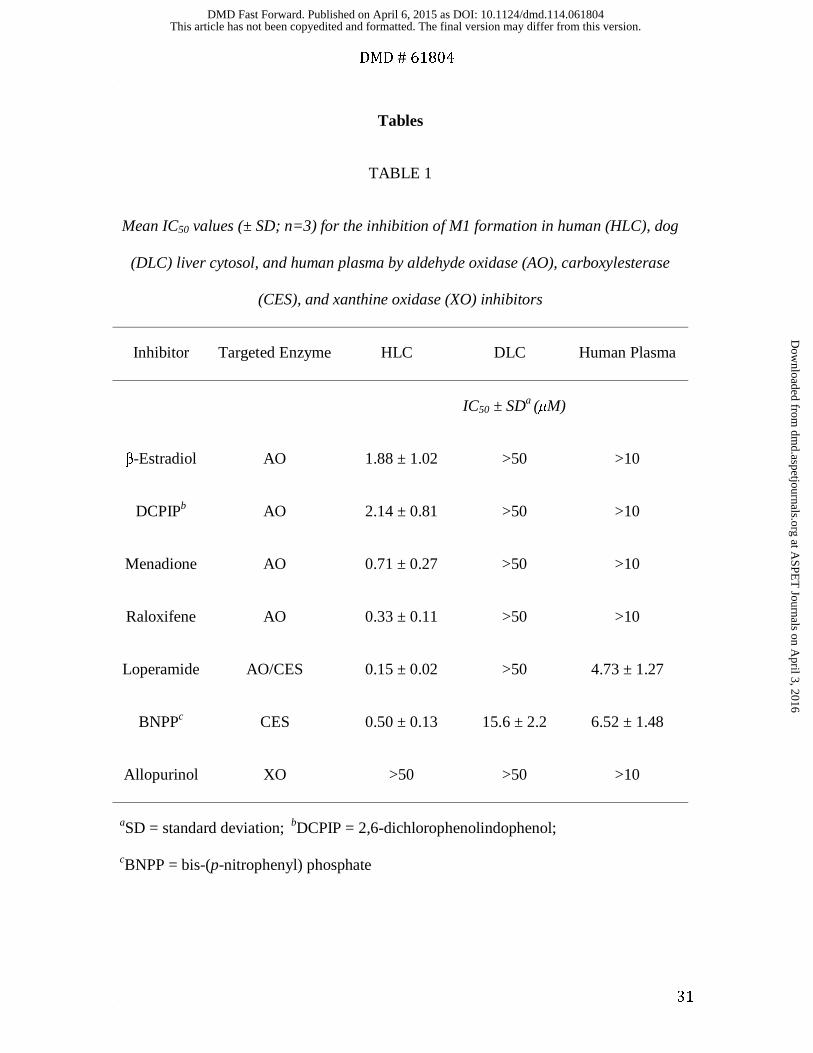
GDC-0834 metabolism in HLC, DLC, and human plasma were examined (Table 1).
Concentration-dependent inhibition of M1 formation in HLC was observed for the AO,
AO/CES, and CES inhibitors tested, with IC50 values ranging from 0.33 to 2.14 μM for
the AO inhibitors, 0.15 μM for loperamide, and 0.50 μM for BNPP. Allopurinol did not
inhibit the metabolism of GDC-0834 and displayed an IC50 >50 μM. In DLC, inhibition
of GDC-0834 metabolism and M1 formation was observed only with the CES inhibitor
BNPP (IC50 = 15.6 μM); all other inhibitors, including loperamide, had IC50 >50 μM. In
human plasma, inhibition of M1 formation was observed only with the CES inhibitor
BNPP (IC50 = 6.52 μM) and the AO/CES inhibitor loperimide (IC50 = 4.73 μM); the AO
inhibitors did not inhibit the formation of M1.
Inhibition of AO- and CES-mediated Metabolism by M1 and M2. The inhibitory
properties of M1 and M2 were examined in HLC. M1 and M2 were weak inhibitors of
AO- and CES-mediated metabolism of phthalazine and CPT-11, respectively. The IC50
values were >10 μM (Supplemental Fig. 2A-D).
Inhibition of AO-mediated Metabolism by GDC-0834. Inhibitory properties of
GDC-0834 on AO probe substrates carbazeran, DACA, O6-benyzlguanine, phthalazine,
zaleplon, and zoniporide were investigated in HLC. GDC-0834 inhibited the formation
of the AO-mediated metabolites with IC50 values ranging from 0.86 to 1.87 μM (Fig. 4A-
4F).
This article has not been copyedited and formatted. The final version may differ from this version.DMD Fast Forward. Published on April 6, 2015 as DOI: 10.1124/dmd.114.061804
at ASPE
T Journals on A
pril 3, 2016dm
d.aspetjournals.orgD
ownloaded from

DMD # 61804
15
Docking of GDC-0834 in AO Active Site. Structural modelling was used to dock
GDC-0834 into the active site of AO (Fig. 5A). GDC-0834 filled the active site and part
of the molecule remained solvated. GDC-0834 bound near the molybdenum cofactor
(MoCo) group in an orientation that would suggest nucleophilic attack by the hydroxyl-
molybdenum (Mo) on the carbonyl of the amide bond. The -5.7 kcal/mol binding energy
of GDC-0834 was similar to the other known AO substrates DACA (-6.6 kcal/mol),
zaleplon (-5.0 kcal/mol), RS-8359 (-5.8 kcal/mol), and XK-469 (-5.0 kcal/mol). Fig. 5B
illustrates the putative interactions of GDC-0834 within the active site of AO.
This article has not been copyedited and formatted. The final version may differ from this version.DMD Fast Forward. Published on April 6, 2015 as DOI: 10.1124/dmd.114.061804
at ASPE
T Journals on A
pril 3, 2016dm
d.aspetjournals.orgD
ownloaded from

DMD # 61804
16
Discussion
Despite the uncertainties surrounding the human clearance prediction (Liu et al., 2011b),
GDC-0834 was rapidly advanced to a single-dose human clinical trial to investigate its
pharmacokinetic parameters due to other advantageous properties. Unfortunately, following oral
administration of GDC-0834 to humans, little to no parent compound was detected due to
extensive metabolism. Amide hydrolysis of GDC-0834 and subsequent M1 formation was the
major metabolic pathway responsible for its high clearance in the clinic (Fig. 1). The enzyme(s)
responsible for the amide hydrolysis was (were) unknown prior to clinical pharmacokinetic
studies. Using HLC, GDC-0834 was observed to be a substrate for enzyme(s) in this subcellular
fraction with a relatively low Km (0.8 µM) and relatively high rate of amide hydrolysis
(approximately 400 pmol/min/mg).
The most obvious enzymes considered as candidates for metabolizing GDC-0834 were
esterases and amidases, which are capable of hydrolyzing amide bonds. These enzymes include
carboxylesterases, cholinesterases, organophosphatases, and amidases/peptidases, where
cholinesterases and aminopeptidases are most efficient in hydrolyzing the amide bonds of
marketed drugs (Uetrecht and Trager, 2007) and the most active hydrolases in human small
intestine and liver (Taketani et al., 2007). In vitro inhibition studies in human liver microsomes,
using general inhibitors of esterases and amidases, failed to implicate these as the hydrolytic
enzymes responsible for the metabolism of GDC-0834 (Liu et al., 2011b).
Preliminary in vitro metabolism experiments using various liver fractions revealed that
soluble enzyme(s) present in HLC mediated the amide hydrolysis of GDC-0834. Therefore,
HLC was chosen over human liver microsomes and hepatocytes since it contained the soluble
enzyme(s) to facilitate fractionation and MS-proteomics analysis. In an attempt to identify the
This article has not been copyedited and formatted. The final version may differ from this version.DMD Fast Forward. Published on April 6, 2015 as DOI: 10.1124/dmd.114.061804
at ASPE
T Journals on A
pril 3, 2016dm
d.aspetjournals.orgD
ownloaded from
DMD # 61804
17
enzyme(s) responsible for the amide hydrolysis of GDC-0834, HLC was fractionated and
subjected to proteomic correlation profiling analogous to methods employed for matching
kinases with their respective substrates (McAllister and Gygi, 2013). Proteomic analysis of the
fractions was then correlated to the rate of hydrolysis of GDC-0834 (Fig. 2). The abundances of
AO and CES and their tight correlations with hydrolytic activity (Pearson Correlations of 0.97
and 0.87, respectively) led us to pursue in vitro chemical inhibition and enzyme activity studies
in HLC to further investigate the role of AO and CES in the amide hydrolysis of GDC-0834.
Data from the chemical inhibition studies confirmed AO-mediated contribution to the amide
hydrolysis of GDC-0834. A panel of widely used AO inhibitors with different chemical
structures and properties, including raloxifene (Obach, 2004), DCPIP (Barr and Jones, 2011),
menadione (Johns, 1967; Sahi et al., 2008; Barr and Jones, 2011), and β-estradiol (Johns et al.,
1969; Barr and Jones, 2011), inhibited GDC-0834 metabolism (formation of M1) with low
single-digit micromolar IC50 values (Table 1). There was also evidence for CES-mediated amide
hydrolysis in HLC (and DLC and human blood and plasma; see below). The AO/CES inhibitor
loperamide (Satoh et al., 1994; Rivory et al., 1996; Williams et al., 2011) and CES inhibitor
BNPP (Satoh et al., 1994) both inhibited M1 formation in HLC, while the XO inhibitor
allopurinol (Baker and Wood, 1967) failed to inhibit GDC-0834 metabolism in HLC.
Dogs have been shown to lack AO activity (Beedham et al., 1987), yet amide hydrolysis of
GDC-0834 was observed in vivo and in vitro (Liu et al., 2011b), albeit less than in humans. In
vitro enzyme identification studies using chemical inhibitors in DLC suggest that CES mediates
the hydrolysis of GDC-0834 in dog. However, the hydrolysis rate was much lower in DLC than
in HLC. In order to detect and quantify the hydrolysis reaction in DLC, a 60-fold larger amount
of cytosolic protein, 6-fold longer incubation time, and 79-fold greater concentration of GDC-
This article has not been copyedited and formatted. The final version may differ from this version.DMD Fast Forward. Published on April 6, 2015 as DOI: 10.1124/dmd.114.061804
at ASPE
T Journals on A
pril 3, 2016dm
d.aspetjournals.orgD
ownloaded from

DMD # 61804
18
0834 were used compared to that of HLC. These data highlight the extensive capability and
efficiency of GDC-0834 hydrolysis in humans.
GDC-0834 was metabolically stable in whole blood and plasma, which suggested limited
contribution of these matrices to the total clearance in vivo (Liu et al., 2011b). In addition, these
studies allowed for the assessments of 1) hydrolysis of GDC-0834 by a hydrolase (presumably
CES) since this enzyme (but not AO) is reported to be present in plasma and blood (McCracken
et al., 1993; Sharma et al., 2011; Beedham 2002) and 2) the specificities of the various AO
inhibitors. In blood and plasma, the loss of GDC-0834 was minimal coupled with little M1
formation (<2% by 180 min) in all species, except in mouse plasma (15% by 180 min) (Fig. 3).
Chemical inhibition studies in human plasma confirmed the specificities of the AO inhibitors
used to implicate AO in HLC. In human plasma, which contains CES but not AO, the AO
inhibitors did not inhibit the formation of M1, whereas BNPP and loperamide did. These data
support the findings of AO involvement in HLC using AO chemical inhibitors, and implicates
AO-mediated hydrolysis in HLC, a matrix which contains both AO and CES.
In addition to the potent inhibition of GDC-0834 metabolism by AO inhibitors, GDC-0834
also inhibits the metabolism of several known AO substrates carbazeran, DACA, O6-
benzylguanine, phthalazine, zaleplon, and zoniporide (Beedham et al., 1990; Beedham et al.,
1995; Kawashima et al., 1999; Schofield et al., 2000; Obach et al., 2004; Dalvie et al., 2012;
Hutzler et al., 2012) (Fig. 4A-4F). The measured IC50 values for the inhibition of six known AO
substrates range between 0.86 and 1.87 μM and are close to the measured Km of 0.8 μM for the
cytosolic hydrolysis of GDC-0834. These results provide additional evidence that GDC-0834
likely interacts at the active site of AO to serve as a substrate and potent AO inhibitor. The
This article has not been copyedited and formatted. The final version may differ from this version.DMD Fast Forward. Published on April 6, 2015 as DOI: 10.1124/dmd.114.061804
at ASPE
T Journals on A
pril 3, 2016dm
d.aspetjournals.orgD
ownloaded from

DMD # 61804
19
metabolites M1 and M2 resulting from hydrolysis were not AO or CES inhibitors and therefore,
the inhibition of AO was due to GDC-0834.
The inhibition of M1 formation by AO inhibitors and the inhibition of AO activity by GDC-
0834 supports AO involvement in the amide hydrolysis reaction and interaction between GDC-
0834 and the active site of AO. The in vitro data surrounding AO prompted us to investigate the
AO active site using docking experiments. GDC-0834, DACA, zaleplon, RS-8359, and XK-469
were docked in the active site of AO and the induced fit homology model was able to bind each
of the substrates. For DACA, zaleplon, RS-8359, and XK-469, a metabolically active orientation
was observed for the top 2-3 binding scores. The docking score for GDC-0834 was within the
same range as the known AO substrates. In the docking experiment, the amide bond of GDC-
0834 appears to be oriented near the MoCo group in close proximity to the reactive hydroxyl
moiety of the MoCo group. The enzyme is able to accommodate a large substrate such as GDC-
0834 because the binding pocket is close to the surface of the enzyme. A large part of the
substrate remains exposed to solvent as proposed in Fig. 5.
With the use of in silico modelling, it is possible to speculate that the amide bond of GDC-
0834 could be coordinated in such a way to be attacked by the hydroxymolybdenum for the
initial step to form a tetrahedral intermediate (Fig. 6). This step requires a nucleophilic hydroxyl
moiety. While the oxidized hydroxymolybdenum species is capable of nucleophilic attack as is
evident by the nucleophilic aromatic substitution normally catalyzed by this reaction,, we
propose that MoIV oxidative state is more suited for this reaction due to higher electron density.
This would require build-up of the MoIV oxidative state in much the same way that AO-mediated
reductions of compounds, such as nitrites, need a substrate to initially be oxidized (Weidert et al.,
2014). If this is the case, the substrate that is oxidized is presently unknown but could be an
This article has not been copyedited and formatted. The final version may differ from this version.DMD Fast Forward. Published on April 6, 2015 as DOI: 10.1124/dmd.114.061804
at ASPE
T Journals on A
pril 3, 2016dm
d.aspetjournals.orgD
ownloaded from

DMD # 61804
20
endogenous aldehyde or azaheterocyclic substrate in the cytosol. The tetrahedral intermediate
formed by either the MoIV or MoVI oxidative state collapses to form M1 and possibly a bound
ester to molybdenum, which then could be further hydrolyzed to release the corresponding acid
metabolite. This reaction does not require any electron transfer and therefore, the catalytic cycle
for amide hydrolysis would be complete after breakdown of the tetrahedral intermediate to
release product.
In conclusion, these data show that AO is involved in the amide hydrolysis of GDC-0834 and
suggest that in addition to AO-mediated oxidative and reductive metabolism of xenobiotics
(Kitamura et al., 2006), hydrolysis may represent an additional metabolic activity mediated by
this enzyme. Therefore, it is prudent to recognize the role of AO in metabolism, including amide
hydrolysis reactions, to avoid poor pharmacokinetics in drug discovery and development stages.
This article has not been copyedited and formatted. The final version may differ from this version.DMD Fast Forward. Published on April 6, 2015 as DOI: 10.1124/dmd.114.061804
at ASPE
T Journals on A
pril 3, 2016dm
d.aspetjournals.orgD
ownloaded from

DMD # 61804
21
Acknowledgements
The authors gratefully acknowledge the assistance of Amish Karanjit, Daisy Bustos,
Robert Cass, Kathryn Kassa, Hoa Le, Jane Lovelidge, Sharmin Jaffer, Chenghong Zhang,
Qinghua Song, Chris Nelson, Richard Vandlen, and James Driscoll. Thanks are also
extended to Wendy Young, Kevin Ford, and Fabio Broccatelli for helpful comments and
discussions.
This article has not been copyedited and formatted. The final version may differ from this version.DMD Fast Forward. Published on April 6, 2015 as DOI: 10.1124/dmd.114.061804
at ASPE
T Journals on A
pril 3, 2016dm
d.aspetjournals.orgD
ownloaded from

DMD # 61804
22
Authorship Contributions
Participated in research design: Sodhi, Barr, Jones, and Halladay.
Conducted experiments: Sodhi, Wong, Barr, Jones, Kirkpatrick, and Halladay.
Contributed new reagents or analytic tools: Sodhi, Barr, Jones, Kirkpatrick, and Halladay.
Performed data analysis: Sodhi, Wong, Liu, Barr, Jones, and Halladay.
Wrote or contributed to the writing of the manuscript: Sodhi, Khojasteh, Hop, Jones, and
Halladay.
This article has not been copyedited and formatted. The final version may differ from this version.DMD Fast Forward. Published on April 6, 2015 as DOI: 10.1124/dmd.114.061804
at ASPE
T Journals on A
pril 3, 2016dm
d.aspetjournals.orgD
ownloaded from

DMD # 61804
23
References
Alfaro JF and Jones JP (2008) Studies on the mechanism of aldehyde oxidase and
xanthine oxidase. J Org Chem 73(23): 9469-9472.
Baker BR and Wood WF (1967) Irreversible enzyme inhibitors. CII. On the mode of
phenyl binding of 9-phenylguanine to guanine deaminase and xanthine
oxidase. J Med Chem 10(6): 1101-1105.
Barr JT and Jones JP (2011) Inhibition of human liver aldehyde oxidase: implications
for potential drug-drug interactions. Drug Metab Dispos 39(12): 2381-2386.
Barr JT and Jones JP (2013) Evidence for substrate-dependent inhibition profiles for
human liver aldehyde oxidase. Drug Metab Dispos 41(1): 24-29.
Beedham C (2002) Molybdenum hydroxylases. In Enzyme Systems that Metabolise
Drugs and Other Xenobiotics (Ionnides C); 147-187.
Beedham C, Bruce SE, Critchley DJ, al-Tayib Y, and Rance DJ (1987) Species variation
in hepatic aldehyde oxidase activity. Eur J Drug Metab Pharmacokinet 12(4):
307-310.
Beedham C, Bruce SE, Critchley DJ, and Rance DJ (1990) 1-substituted phthalazines
as probes of the substrate-binding site of mammalian molybdenum
hydroxylases. Biochem Pharmacol 39(7): 1213-1221.
Beedham C, Critchley DJ, and Rance DJ (1995) Substrate specificity of human liver
aldehyde oxidase toward substituted quinazolines and phthalazines: a
comparison with hepatic enzyme from guinea pig, rabbit, and baboon. Arch
Biochem Biophys 319(2): 481-490.
This article has not been copyedited and formatted. The final version may differ from this version.DMD Fast Forward. Published on April 6, 2015 as DOI: 10.1124/dmd.114.061804
at ASPE
T Journals on A
pril 3, 2016dm
d.aspetjournals.orgD
ownloaded from
DMD # 61804
24
Coelho C, Mahro M, Trincao J, Carvalho AT, Ramos MJ, Terao M, Garattini E,
Leimkuhler S, and Romao MJ (2012) The first mammalian aldehyde oxidase
crystal structure: insights into substrate specificity. J Biol Chem 287(48):
40690-40702.
Dalvie D, Sun H, Xiang C, Hu Q, Jiang Y, and Kang P (2012) Effect of structural
variation on aldehyde oxidase-catalyzed oxidation of zoniporide. Drug Metab
Dispos 40(8): 1575-1587.
Hutzler JM, Yang YS, Albaugh D, Fullenwider CL, Schmenk J, and Fisher MB (2012)
Characterization of aldehyde oxidase enzyme activity in cryopreserved
human hepatocytes. Drug Metab Dispos 40(2): 267-275.
Johns DG (1967) Human liver aldehyde oxidase: differential inhibition of oxidation
of charged and uncharged substrates. J Clin Invest 46(9): 1492-1505.
Johns DG, Spector T, and Robins RK (1969) Studies on the mode of oxidation of
pyrazolo(3,4-d)pyrimidine by aldehyde oxidase and xanthine oxidase.
Biochem Pharmacol 18(10): 2371-2383.
Jones JP and Korzekwa KR (2013) Predicting intrinsic clearance for drugs and drug
candidates metabolized by aldehyde oxidase. Mol Pharm 10(4): 1262-1268.
Kawashima K, Hosoi K, Naruke T, Shiba T, Kitamura M, and Watabe T (1999)
Aldehyde oxidase-dependent marked species difference in hepatic
metabolism of the sedative-hypnotic, zaleplon, between monkeys and rats.
Drug Metab Dispos 27(3): 422-428.
Kitamura S, Sugihara K, and Ohta S (2006) Drug-metabolizing ability of
molybdenum hydroxylases. Drug Metab Pharmacokinet 21(2): 83-98.
This article has not been copyedited and formatted. The final version may differ from this version.DMD Fast Forward. Published on April 6, 2015 as DOI: 10.1124/dmd.114.061804
at ASPE
T Journals on A
pril 3, 2016dm
d.aspetjournals.orgD
ownloaded from

DMD # 61804
25
Liu L, Di Paolo J, Barbosa J, Rong H, Reif K, and Wong H (2011a) Antiarthritis effect
of a novel Bruton's tyrosine kinase (BTK) inhibitor in rat collagen-induced
arthritis and mechanism-based pharmacokinetic/pharmacodynamic
modeling: relationships between inhibition of BTK phosphorylation and
efficacy. J Pharmacol Exp Ther 338(1): 154-163.
Liu L, Halladay JS, Shin Y, Wong S, Coraggio M, La H, Baumgardner M, Le H, Gopaul S,
Boggs J, Kuebler P, Davis JC, Jr., Liao XC, Lubach JW, Deese A, Sowell CG,
Currie KS, Young WB, Khojasteh SC, Hop CE, and Wong H (2011b) Significant
species difference in amide hydrolysis of GDC-0834, a novel potent and
selective Bruton's tyrosine kinase inhibitor. Drug Metab Dispos 39(10): 1840-
1849.
McAllister FE and Gygi SP (2013) Correlation profiling for determining kinase-
substrate relationships. Methods.
McCracken NW, Blain PG, Williams FM (1993) Human xenobiotic metabolizing
esterases in liver and blood. Biochem Pharmacol 46 (7): 1125-1129.
Obach RS (2004) Potent inhibition of human liver aldehyde oxidase by raloxifene.
Drug Metab Dispos 32(1): 89-97.
Obach RS, Huynh P, Allen MC, and Beedham C (2004) Human liver aldehyde oxidase:
inhibition by 239 drugs. J Clin Pharmacol 44(1): 7-19.
Phu L, Izrael-Tomasevic A, Matsumoto ML, Bustos D, Dynek JN, Fedorova AV,
Bakalarski CE, Arnott D, Deshayes K, Dixit VM, Kelley RF, Vucic D, and
Kirkpatrick DS (2011) Improved Quantitative Mass Spectrometry Methods
for Characterizing Complex Ubiquitin Signals. Mol Cell Proteomics 10: 1-19.
This article has not been copyedited and formatted. The final version may differ from this version.DMD Fast Forward. Published on April 6, 2015 as DOI: 10.1124/dmd.114.061804
at ASPE
T Journals on A
pril 3, 2016dm
d.aspetjournals.orgD
ownloaded from

DMD # 61804
26
Rivory LP, Bowles MR, Robert J, and Pond SM (1996) Conversion of irinotecan (CPT-
11) to its active metabolite, 7-ethyl-10-hydroxycamptothecin (SN-38), by
human liver carboxylesterase. Biochem Pharmacol 52(7): 1103-1111.
Sahi J, Khan KK, and Black CB (2008) Aldehyde oxidase activity and inhibition in
hepatocytes and cytosolic fractions from mouse, rat, monkey and human.
Drug Metab Lett 2(3): 176-183.
Satoh T, Hosokawa M, Atsumi R, Suzuki W, Hakusui H, and Nagai E (1994) Metabolic
activation of CPT-11, 7-ethyl-10-[4-(1-piperidino)-1-
piperidino]carbonyloxycamptothecin, a novel antitumor agent, by
carboxylesterase. Biol Pharm Bull 17(5): 662-664.
Schofield PC, Robertson IG, and Paxton JW (2000) Inter-species variation in the
metabolism and inhibition of N-[(2'-dimethylamino)ethyl]acridine-4-
carboxamide (DACA) by aldehyde oxidase. Biochem Pharmacol 59(2): 161-
165.
Sharma R, Eng H, Walker GS, Barreiro G, Stepan AF, McClure KF, Wolford A, Bonin
PD, Cornelius P, and Kalgutkar AS (2011) Oxidative metabolism of a
quinoxaline derivative by xanthine oxidase in rodent plasma. Chem Res
Toxicol 24: 2207-2216.
Sheng Z, Zhang S, Bustos D, Kleinheinz T, Le Pichon CE, Dominguez SL, Solanoy HO,
Drummond J, Zhang X, Ding X, Cai F, Song Q, Li X, Yue Z, van der Brug MP,
Burdick DJ, Gunzner-Toste J, Chen H, Liu X, Estrada AA, Sweeny ZK, Scearce-
Levie K, Moffat JG, Kirkpatrick DS, Zhu H (2012) Ser1292
This article has not been copyedited and formatted. The final version may differ from this version.DMD Fast Forward. Published on April 6, 2015 as DOI: 10.1124/dmd.114.061804
at ASPE
T Journals on A
pril 3, 2016dm
d.aspetjournals.orgD
ownloaded from
DMD # 61804
27
autophosphorylation is an indicator of LRRK2 kinase activity and contributes
to the cellular effects of PD mutations. Sci Transl Med 4(164): 164ra161.
Tabata T, Katoh M, Tokudome S, Nakajima M, and Yokoi T (2004) Identification of
the cytosolic carboxylesterase catalyzing the 5'-deoxy-5-fluorocytidine
formation from the capecitabine in human liver. Drug Metab Dispos 32 (10):
1103-1110.
Taketani M, Shii M, Ohura K, Ninomiya S, and Imai T (2007) Carboxylesterase in the
liver and small intestine of experimental animals and human. Life Sci 81(11):
924-932.
Uetrecht JP and Trager W (2007) Drug Metabolism Chemical and Enzymatic Aspects,
Informa Healthcare USA, New York.
Weidert ER, Schoenborn SO, Cantu-Medellin N, Choughule KV, Jones JP, and Kelley
EE (2014) Inhibition of xanthine oxidase by the aldehyde oxidase inhibitor
raloxifene: Implications for identifying molybdopterin nitrite reductases.
Nitric Oxide 37: 41-45.
Williams ET, Bacon JA, Bender DM, Lowinger JJ, Guo WK, Ehsani ME, Wang X, Wang
H, Qian YW, Ruterbories KJ, Wrighton SA, and Perkins EJ (2011)
Characterization of the expression and activity of carboxylesterases 1 and 2
from the beagle dog, cynomolgus monkey, and human. Drug Metab Dispos
39(12): 2305-2313.
This article has not been copyedited and formatted. The final version may differ from this version.DMD Fast Forward. Published on April 6, 2015 as DOI: 10.1124/dmd.114.061804
at ASPE
T Journals on A
pril 3, 2016dm
d.aspetjournals.orgD
ownloaded from
DMD # 61804
28
Footnotes
The in silico modelling work was supported in part by the National Institutes of
Health National Institute of General Medical Sciences [Grant GM100874] (JPJ, JTB)
*Current affiliation: Anacor Pharmaceuticals, Inc., 1020 East Meadow Circle, Palo Alto,
CA 94303, USA.
This article has not been copyedited and formatted. The final version may differ from this version.DMD Fast Forward. Published on April 6, 2015 as DOI: 10.1124/dmd.114.061804
at ASPE
T Journals on A
pril 3, 2016dm
d.aspetjournals.orgD
ownloaded from

DMD # 61804
29
Legends for Figures
FIG. 1. Amide hydrolysis pathway of GDC-0834 to form M1 and M2.
FIG. 2. Proteomic correlation profiling revealed that aldehyde oxidase (AO/ADO) is an
enzyme present in cytosolic fractions containing the hydrolytic enzyme activity involved
in the metabolism of GDC-0834. A) Table of top 15 most abundant proteins (Uniprot
Reference) identified in fractions 24-31 based on total peptide spectral matches (PSMs);
AO/ADO is shown in red. The hydrolytic enzyme activity (GDC-0834�M1) is shown
in blue. Pearson correlations of each protein relative to GDC-0834�M1 metabolic
activity are reported. Adjusted P-values were corrected for multiple hypothesis testing
using ‘qvalue’ FDR based approach. B) Proteins ranked by Pearson Correlation. The red
diamond highlights AO/ADO.
FIG. 3. Formation of M1 following incubation of GDC-0834 (0.8 µM) in (A) fresh
whole blood and (B) plasma in human (○), rat (■), mouse (▲), dog (♦), and monkey (●).
FIG. 4. IC50 curves for the inhibition by GDC-0834 (0 - 50 or 0 - 100 μM) of aldehyde
oxidase (AO)-mediated metabolism of AO probe substrates in human liver cytosol (A)
carbazeran (formation of 4-hydroxycarbazeran), (B) DACA (formation of DACA-
9(10H)-acridone), (C) O6-benzylguanine (formation of 8-oxo-benzylguanine), (D)
phthalazine (formation of phthalazinone), (E) zaleplon (formation of 5-oxozaleplon), and
(F) zoniporide (formation of 2-oxozoniporide). Data are the mean ± standard deviation
This article has not been copyedited and formatted. The final version may differ from this version.DMD Fast Forward. Published on April 6, 2015 as DOI: 10.1124/dmd.114.061804
at ASPE
T Journals on A
pril 3, 2016dm
d.aspetjournals.orgD
ownloaded from
DMD # 61804
30
of triplicate determinations. The lines represent the best fit to the data using nonlinear
regression. DACA = N-[(2-dimethylamino) ethyl]acridine-4-carboxamide.
FIG. 5. Homology model for aldehyde oxidase (AO) using the human sequence and the
mouse crystal structure (PDB code 3ZYV). (A) Induced fit docking was used to dock
GDC-0834 into the active site of AO near the MoCo group in an orientation that would
suggest nucleophilic attack by the hydroxyl on the carbonyl of the amide bond. (B)
Putative interactions of GDC-0834 within the active site of AO.
FIG. 6. Proposed reaction for the amide hydrolysis mediated by aldehyde oxidase.
This article has not been copyedited and formatted. The final version may differ from this version.DMD Fast Forward. Published on April 6, 2015 as DOI: 10.1124/dmd.114.061804
at ASPE
T Journals on A
pril 3, 2016dm
d.aspetjournals.orgD
ownloaded from
DMD # 61804
31
Tables
TABLE 1
Mean IC50 values (± SD; n=3) for the inhibition of M1 formation in human (HLC), dog
(DLC) liver cytosol, and human plasma by aldehyde oxidase (AO), carboxylesterase
(CES), and xanthine oxidase (XO) inhibitors
Inhibitor Targeted Enzyme HLC DLC Human Plasma
IC50 ± SDa (μM)
β-Estradiol AO 1.88 ± 1.02 >50 >10
DCPIPb AO 2.14 ± 0.81 >50 >10
Menadione AO 0.71 ± 0.27 >50 >10
Raloxifene AO 0.33 ± 0.11 >50 >10
Loperamide AO/CES 0.15 ± 0.02 >50 4.73 ± 1.27
BNPPc CES 0.50 ± 0.13 15.6 ± 2.2 6.52 ± 1.48
Allopurinol XO >50 >50 >10
aSD = standard deviation; bDCPIP = 2,6-dichlorophenolindophenol;
cBNPP = bis-(p-nitrophenyl) phosphate
This article has not been copyedited and formatted. The final version may differ from this version.DMD Fast Forward. Published on April 6, 2015 as DOI: 10.1124/dmd.114.061804
at ASPE
T Journals on A
pril 3, 2016dm
d.aspetjournals.orgD
ownloaded from

FIG. 1.
This article has not been copyedited and formatted. The final version may differ from this version.DMD Fast Forward. Published on April 6, 2015 as DOI: 10.1124/dmd.114.061804
at ASPE
T Journals on A
pril 3, 2016dm
d.aspetjournals.orgD
ownloaded from

A
B
FIG. 2.
Reference Description fr24 fr25 fr26 fr27 fr28 fr29 fr30 fr31Pearson
Corr
adjusted
p-value
CPSMCarbamoyl-phosphate
synthase, mitochondrial14 62 86 80 68 66 65 27 0.7396 0.0407
ALDOBFructose-bisphosphate
aldolase B13 44 122 84 59 43 32 23 0.9342 0.0027
AL1A1 Retinal dehydrogenase 1 0 10 116 78 55 42 27 19 0.9604 0.0010
ALDH2Aldehyde dehydrogenase,
mitochondrial6 12 88 60 43 34 27 22 0.9525 0.0014
EST1 Liver carboxylesterase 1 1 1 57 64 59 38 34 21 0.8671 0.0097
ADO Aldehyde oxidase 0 9 73 78 41 22 30 12 0.9708 0.0007
IDHCIsocitrate dehydrogenase
[NADP] cytoplasmic0 3 55 57 39 32 24 13 0.9455 0.0018
BHMT1Betaine--homocysteine S-
methyltransferase 11 1 47 75 37 29 12 13 0.9294 0.0030
THIM3-ketoacyl-CoA thiolase,
mitochondrial6 27 54 35 28 23 20 13 0.8577 0.0106
ANXA6 Annexin A6 0 0 57 53 39 30 17 9 0.9625 0.0010
AK1C1Aldo-keto reductase family 1
member C11 1 2 16 36 50 49 48 -0.2604 0.1670
AL1L1Cytosolic 10-
formyltetrahydrofolate 1 59 58 32 22 14 11 2 0.4984 0.0936
CATA Catalase 1 3 41 56 37 22 17 18 0.9258 0.0034
PGM1 Phosphoglucomutase-1 0 0 0 30 68 45 26 19 0.1523 0.1984
SBP1 Selenium-binding protein 1 0 0 0 25 62 37 37 25 0.0472 0.2337
ACTIVITYGDC-0834 --> M1
(arbitrary units x 1000)0 2 195 185 100 50 20 10 1.0000 0.0000
# peptide spectral matches (PSMs)
-1.0
-0.5
0.0
0.5
1.0
0 200 400 600
Pea
rson
Corre
lati
on
Protein by Rank
Aldehyde Oxidase
This article has not been copyedited and formatted. The final version may differ from this version.DMD Fast Forward. Published on April 6, 2015 as DOI: 10.1124/dmd.114.061804
at ASPE
T Journals on A
pril 3, 2016dm
d.aspetjournals.orgD
ownloaded from

A
B
FIG. 3
This article has not been copyedited and formatted. The final version may differ from this version.DMD Fast Forward. Published on April 6, 2015 as DOI: 10.1124/dmd.114.061804
at ASPE
T Journals on A
pril 3, 2016dm
d.aspetjournals.orgD
ownloaded from

This article has not been copyedited and formatted. The final version may differ from this version.DMD Fast Forward. Published on April 6, 2015 as DOI: 10.1124/dmd.114.061804
at ASPE
T Journals on A
pril 3, 2016dm
d.aspetjournals.orgD
ownloaded from

A
B
FIG. 5.
This article has not been copyedited and formatted. The final version may differ from this version.DMD Fast Forward. Published on April 6, 2015 as DOI: 10.1124/dmd.114.061804
at ASPE
T Journals on A
pril 3, 2016dm
d.aspetjournals.orgD
ownloaded from

FIG. 6.
This article has not been copyedited and formatted. The final version may differ from this version.DMD Fast Forward. Published on April 6, 2015 as DOI: 10.1124/dmd.114.061804
at ASPE
T Journals on A
pril 3, 2016dm
d.aspetjournals.orgD
ownloaded from