Guided Infra-Zygomatic Screws: Reliable Maxillary Arch Retraction Drs. John Jin-Jong Lin & W. Eugene Roberts Class II Crowded Malocclusion Treated Conservatively with a Passive Self Ligating Appliance: Expansion, Stability and Adaptation Drs. Shih-Yung Lin, Chris Chang & W. Eugene Roberts Trans-Alveolar Uprighting of a Horizontally Impacted Lower Canine with a Mandibular Buccal Shelf Bone Screw Drs. Szu Rou Yeh, Chris Chang & W. Eugene Roberts Archwire Sequence for Insignia® : a Custom Bracket System with a Bright Future Drs. Angle Lee, Chris Chang & W. Eugene Roberts I J OI International Journal of Orthodontics & Implantology Vol. 46 Apr 1, 2017 CC (left) and DD (right) with the new Trumpology book at the Damon Forum Speakers Dinner, Orlando, FL, USA. They are smiling in agreement having decided to drain the swamp! International Journal of Orthodontics & Implantology is an experience sharing magazine for worldwide orthodontists and Implantologists. Download it at http://iaoi.pro. 《僅供牙科專業人士參閱》

Welcome message from author
This document is posted to help you gain knowledge. Please leave a comment to let me know what you think about it! Share it to your friends and learn new things together.
Transcript
Guided Infra-Zygomatic Screws: Reliable Maxillary Arch Retraction Drs. John Jin-Jong Lin & W. Eugene Roberts
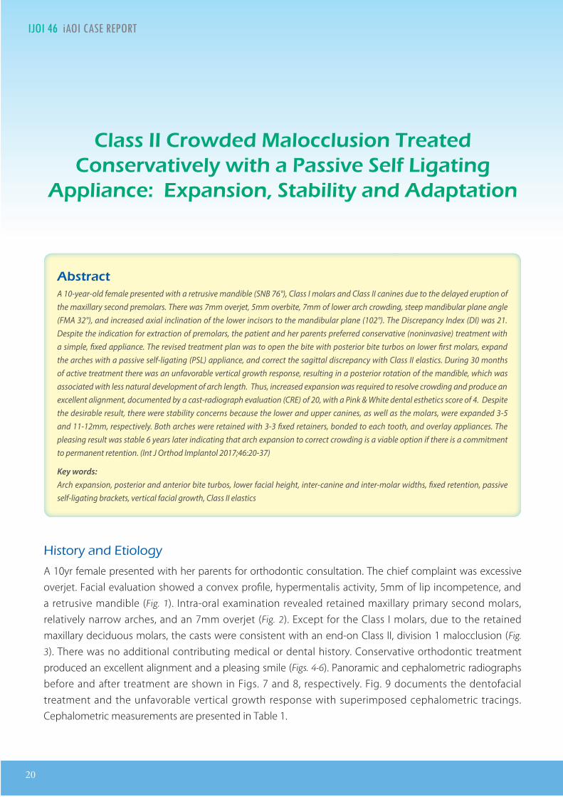
Class II Crowded Malocclusion Treated Conservatively with a Passive Self Ligating Appliance: Expansion, Stability and AdaptationDrs. Shih-Yung Lin, Chris Chang & W. Eugene Roberts
Trans-Alveolar Uprighting of a Horizontally Impacted Lower Canine with a Mandibular Buccal Shelf Bone ScrewDrs. Szu Rou Yeh, Chris Chang & W. Eugene Roberts
Archwire Sequence for Insignia® : a Custom Bracket System with a Bright FutureDrs. Angle Lee, Chris Chang & W. Eugene Roberts
IJOIInternational Journal of
Orthodontics & Implantology
Vol. 46 Apr 1, 2017
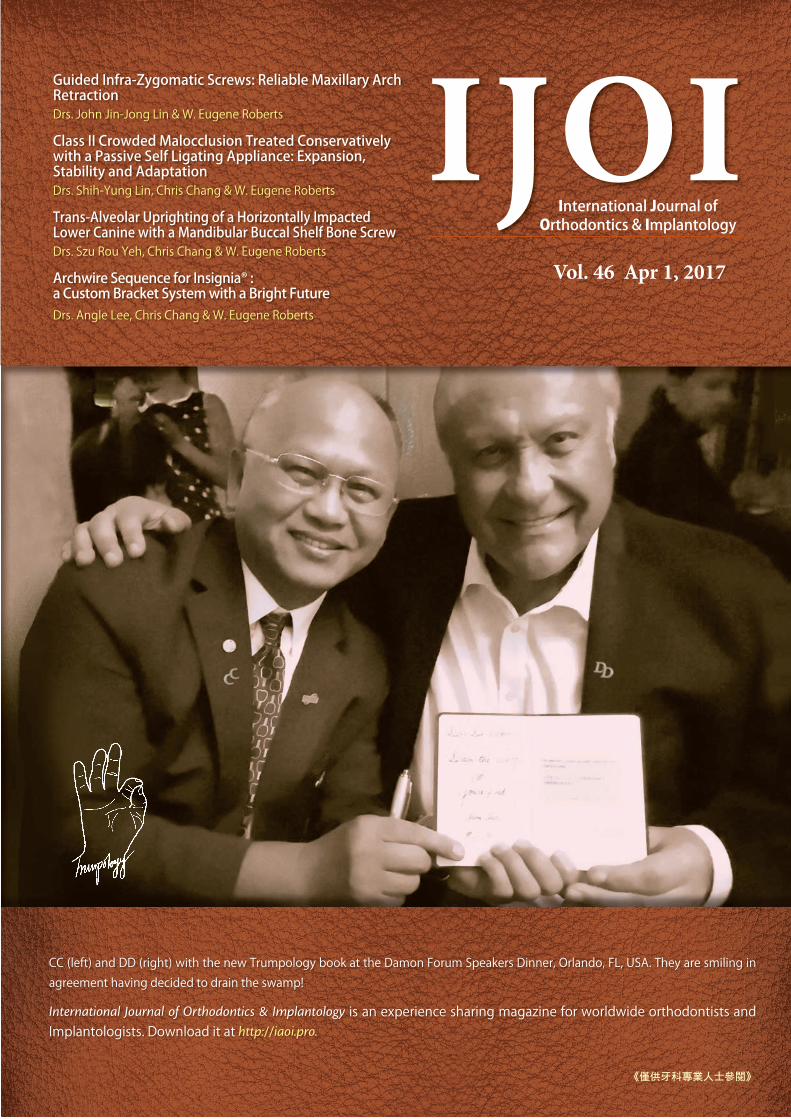
CC (left) and DD (right) with the new Trumpology book at the Damon Forum Speakers Dinner, Orlando, FL, USA. They are smiling in agreement having decided to drain the swamp!
International Journal of Orthodontics & Implantology is an experience sharing magazine for worldwide orthodontists and Implantologists. Download it at http://iaoi.pro.
《僅供牙科專業人士參閱》
10/211/
7/
/10/2/21 23
//
10/111/2212/13
//20
/11//13
7/11
2/13/1
12/21
1/2/1
/22-2
/1 -1 11/2 -12/1
11/27-30
A good sentence can change one’s life.
A classic in your pocket. Available in Newton’s A, Inc.
�e first collection of Steve Jobs’ famous quote, selected by an Apple businessman.
A handy companion for your reading on the go.
Including Job’s special bookmarks to carry his spirit around.
�e other classic created by Dr. Chris Chang, author of the bestseller, Jobsology.
Understand the secrets of Trump’s success and improve your English ability simultaneously.
Collected more than 200 famous quotes of the President Trump.
Pocket-size, perfect for reading on the go.
We owe him(Trump) an open mind and a chance to lead.
Newton’s A
—Hilary Clinton
Chris Chang
Now available in countries:
Available at Amazon now.
IAOI welcomes general submissions and inquiries to be sent to [email protected]
BE OPEN TO NEW INFORMATION AND IDEAS Trumpology p. 72
Featured on the cover of this issue is a photo of Dr. Dwight Damon (DD) and myself at the Damon Forum 2017 Speakers Dinner. I was not sure whether or not DD would be so welcoming after my Keynote speech this year as I tackled a rather controversial, almost taboo, subject for the Damon Forum: extraction vs. non-extraction. However, as those of you who have read my new Trumpology book know: “Be willing to take on new challenges.” (Trumpology p. 73)
I was very hesitant to take on this subject, as for the last 10 years in which I have participated in the Damon Forums, I have never heard anyone talk about extraction issues and DD has an extraction rate of 1% (compared to my 65%). He is considered to be a non-extraction Dr and I have also been repeatedly labelled so (I have been asked “You don’t extract, do you?” for 6 consecutive years at a prestigious organisation’s meeting), as have probably most Orthodontists who use Damon braces.
Extraction is very common in Asia, due to profile and the desired results of our patients. If Asian doctors tried to replicate DD’s 1% extraction rate, it would only end in disaster!! My speech could well have been a disaster, I almost didn’t have the guts to present it, but…….
At the Speakers Dinner I was cornered by DD and for a whole hour we spoke about my speech. He told me he considered it to be the best speech of all the Damon Forums and that somebody should have talked about this important subject a long time ago, to make it clear to the participants and worldwide audience that extraction is OK. DD fully understood that Caucasians and Asians are different and although he advocates minimally invasive treatments, he also has to extract when necessary (as do I!).
How we interpret information is as important as the information itself and DD proved to me that he is very open-minded to new information. Therefore, I also presented him with a copy of my new Trumpology book and discussed with him how we can “drain the swamp” (Trumpology p.166) of our profession to allow all Orthodontists to practise to their fullest potential without a perceived opinion being pushed onto them.
I sincerely hope that all of you will help to “drain the swamp” and break down perceived opinions in our profession and continue to march with us along the path to glory.
Wishing you every success in your practice and a Happy Easter,
s n DDS, PhD, Publisher of IJOI.
Dr. Baldwin W. Marchack
Dr. Thomas HanDr. FernandoRojas-Vizcaya
Dr. Homa ZadehDr. Larry WhiteDr. J. Michael Steffen
Dr. W. EugeneRoberts
Dr. Tucker Haltom
Consultants
Dr. Frank Chang Dr. Johnny Liaw
Examiners
Dr. John J. J. Lin
Dr. Hong Po Chang
Dr. Yu Lin Hsu Dr. Yu-Hsin Huang
Dr. Bill Su Dr. Ming-Jen Chang
Associate editors
Dr. Chris LinDr. Ariel Chang
Editorial Board
Editor-in-chief
Dr. W. Eugene Roberts
Publisher
Dr. Chris ChangResearch
Dr. Chi HuangCase ReportDr. Angle Lee
SurgeryDr. Shih-Yung
Lin
Editors
Desk editorBella Chu
English editing
Paul Head
ProofreaderTzu Han Huang
IllustrationDr. Rungsi
Thavarungkul
3 Editorial
LIVE FROM THE MASTER
4 Guided Infra-Zygomatic Screws: Reliable Maxillary Arch Retraction
iAOI CASE REPORT
20 Class II Crowded Malocclusion Treated Conservatively with a Passive Self Ligating Appliance: Expansion, Stability and Adaptation
40 Trans-Alveolar Uprighting of a Horizontally Impacted Lower Canine with a Mandibular Buccal Shelf Bone Screw
Clinical Tips
60 Archwire Sequence for Insignia®: a Custom Bracket System with a Bright Future
Damon Workbook
72 Class I Anterior Crossbite
86 Crowded Class II Division 2 Malocclusion with Class I Molars Due to Blocked In Lower Second Premolars
iAOI OTHERS
100 In memory of Charles Burstone: He has been missing almost two years, but the void is stillfresh.
FEEDBACK FROM THE WORLD
106 Feedback from the world
EDITORIAL IJOI 46
4
IJOI 46 LI E RO T E A TER I IJOI 46
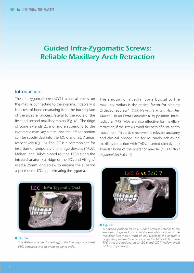
█ Fig. 1A: The skeletal anatomy (osteology) of the Infrazygomatic Crest (IZC) is marked with an ovoid magenta circle.
█ Fig. 1B: A practical position for an IZC bone screw is anterior to the anatomic ridge and buccal to the mesiobuccal root of the maxillary first molar (MBR of U6). Distal to the anatomic ridge, the preferred site is buccal to the MBR of U7. These TAD sites are designated as IZC 6 and IZC 7 (yellow ovoid circles), respectively.
i n ra o atic cria a i ar rc traction
Introduction
The infra-zygomatic crest (IZC) is a buccal process on the maxilla, connecting to the zygoma. Intraorally it is a crest of bone emanating from the buccal plate of the alveolar process, lateral to the roots of the first and second maxillary molars (Fig. 1A). The ridge of bone extends 2cm or more superiorly to the zygomatic-maxillary suture, and the inferior portion can be subdivided into the IZC 6 and IZC 7 areas, respectively (Fig. 1B). The IZC is a common site for insertion of temporary anchorage devices (TADs). Melsen1 and Uribe2 placed routine TADs along the intraoral anatomical ridge of the IZC, and Villegas3 used a 25mm long screw to engage the superior aspects of the IZC, approximating the zygoma.
The amount of a lveolar bone buccal to the maxillary molars is the critical factor for placing OrthoBoneScrew® (OBS, Newton’s A Ltd, Hsinchu,
Taiwan) in an Extra-Radicular (E-R) position. Inter-radicular (I-R) TADs are also effective for maxillary retraction, if the screws avoid the path of distal tooth movement. This article reviews the relevant anatomy and clinical procedures for routinely achieving maxillary retraction with TADs, inserted directly into alveolar bone of the posterior maxilla. (Int J Orthod
Implantol 2017;46:4-16)
IJOI 46 LI E RO T E A TER
5
I IJOI 46
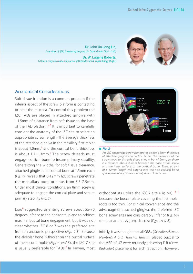
█ Fig. 2: An IZC anchorage screw penetrates about a 3mm thickness of attached gingiva and cortical bone. The clearance of the screw head to the soft tissue should be ~1.5mm, so there is a distance about 4.5mm between the base of the screw and the inner surface of the cortical bone. Thus, screws of 8-12mm length will extend into the non-cortical bone space (medullary bone or sinus) about 3.5-7.5mm.
Dr. John Jin-Jong Lin, Examiner of IJOI, Director of Jin-Jong Lin Orthodontic Clinic (Left)
Dr. W. Eugene Roberts,Editor-in-chief, International Journal of Orthodontics & Implantology (Right)
Anatomical onsiderations
Soft tissue irritation is a common problem if the inferior aspect of the screw platform is contacting or near the mucosa. To control this problem the IZC TADs are placed in attached gingiva with ~1.5mm of clearance from soft tissue to the base of the TAD platform.4,5 It is important to carefully consider the anatomy of the IZC site to select an appropriate screw length. The average thickness of the attached gingiva in the maxillary first molar is about 1.0mm,6 and the cortical bone thickness is about 1.1-1.3mm.7 The screw threads must engage cortical bone to insure primary stability. Generalizing the widths, for soft tissue clearance, attached gingiva and cortical bone at 1.5mm each (Fig. 2), reveals that 8-12mm IZC screws penetrate the medullary bone or sinus from 3.5-7.5mm. Under most clinical conditions, an 8mm screw is adequate to engage the cortical plate and secure primary stability (Fig. 2).
Liou8 suggested orienting screws about 55-70 degrees inferior to the horizontal plane to achieve maximal buccal bone engagement, but it was not clear whether IZC 6 or 7 was the preferred site from an anatomic perspective (Figs. 1-3). Because the alveolar bone is thicker on the buccal surface of the second molar (Figs. 4 and 5), the IZC 7 site is usually preferable for TADs.9 In Taiwan, most
orthodontists utilize the IZC 7 site (Fig. 6A),10,11 because the buccal plate covering the first molar roots is too thin. For clinical convenience and the advantage of attached gingiva, the preferred IZC bone screw sites are considerably inferior (Fig. 6B) to the anatomic zygomatic crest (Figs. 1A & B).
Initially, it was thought that all OBSs (OrthoBoneScrews,
Newton’s A Ltd, Hsinchu, Taiwan) placed buccal to the MBR of U7 were routinely achieving E-R (Extra-
Radicular) placement for arch retraction. However,
6
IJOI 46 LI E RO T E A TER I IJOI 46
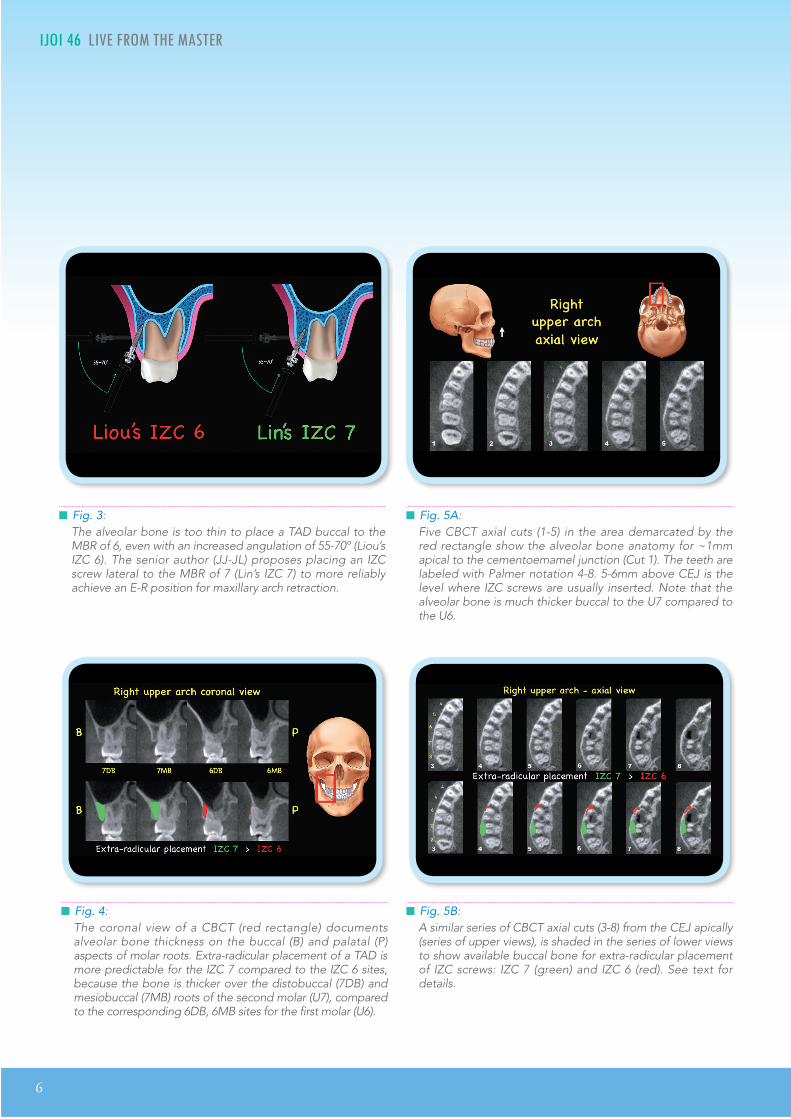
█ Fig. 5B: A similar series of CBCT axial cuts (3-8) from the CEJ apically (series of upper views), is shaded in the series of lower views to show available buccal bone for extra-radicular placement of IZC screws: IZC 7 (green) and IZC 6 (red). See text for details.
█ Fig. 4: The coronal view of a CBCT (red rectangle) documents alveolar bone thickness on the buccal (B) and palatal (P) aspects of molar roots. Extra-radicular placement of a TAD is more predictable for the IZC 7 compared to the IZC 6 sites, because the bone is thicker over the distobuccal (7DB) and mesiobuccal (7MB) roots of the second molar (U7), compared to the corresponding 6DB, 6MB sites for the first molar (U6).
█ Fig. 3: The alveolar bone is too thin to place a TAD buccal to the MBR of 6, even with an increased angulation of 55-70º (Liou’s IZC 6). The senior author (JJ-JL) proposes placing an IZC screw lateral to the MBR of 7 (Lin’s IZC 7) to more reliably achieve an E-R position for maxillary arch retraction.
█ Fig. 5A: Five CBCT axial cuts (1-5) in the area demarcated by the red rectangle show the alveolar bone anatomy for ~1mm apical to the cementoemamel junction (Cut 1). The teeth are labeled with Palmer notation 4-8. 5-6mm above CEJ is the level where IZC screws are usually inserted. Note that the alveolar bone is much thicker buccal to the U7 compared to the U6.
IJOI 46 LI E RO T E A TER
7
I IJOI 46
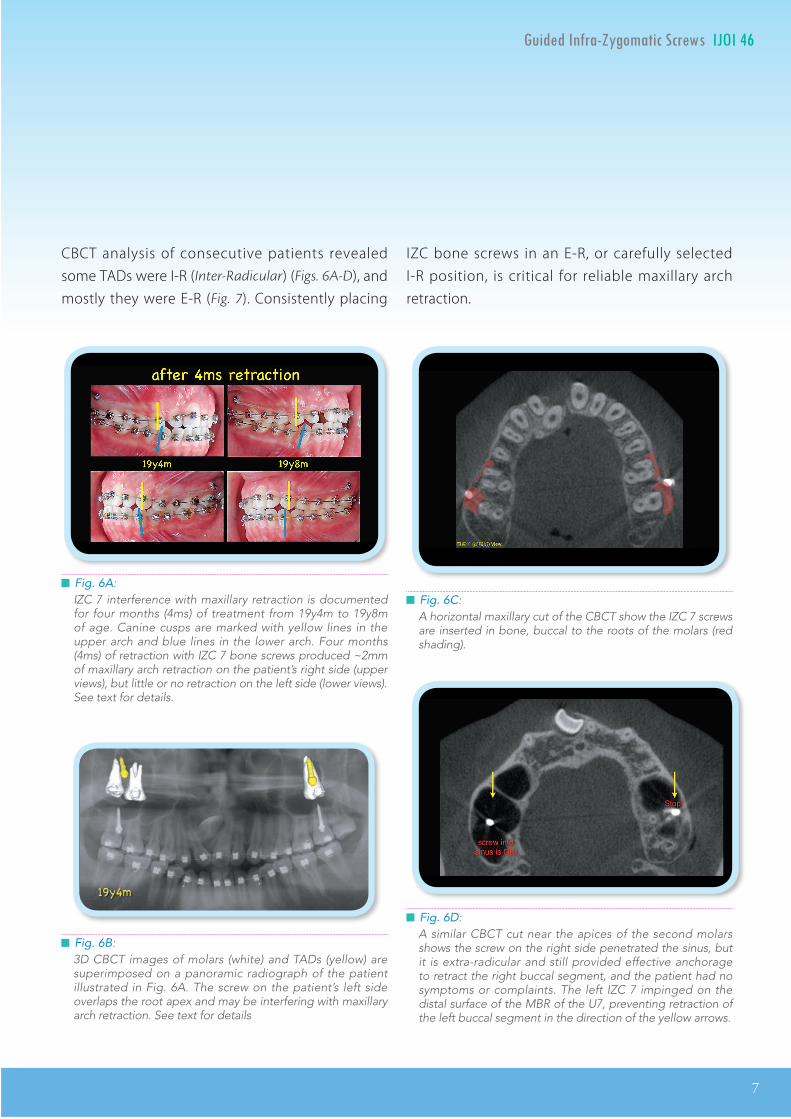
█ Fig. 6A: IZC 7 interference with maxillary retraction is documented for four months (4ms) of treatment from 19y4m to 19y8m of age. Canine cusps are marked with yellow lines in the upper arch and blue lines in the lower arch. Four months (4ms) of retraction with IZC 7 bone screws produced ~2mm of maxillary arch retraction on the patient’s right side (upper views), but little or no retraction on the left side (lower views). See text for details.
█ Fig. 6B: 3D CBCT images of molars (white) and TADs (yellow) are superimposed on a panoramic radiograph of the patient illustrated in Fig. 6A. The screw on the patient’s left side overlaps the root apex and may be interfering with maxillary arch retraction. See text for details
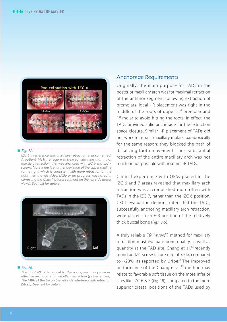
█ Fig. 6C: A horizontal maxillary cut of the CBCT show the IZC 7 screws are inserted in bone, buccal to the roots of the molars (red shading).
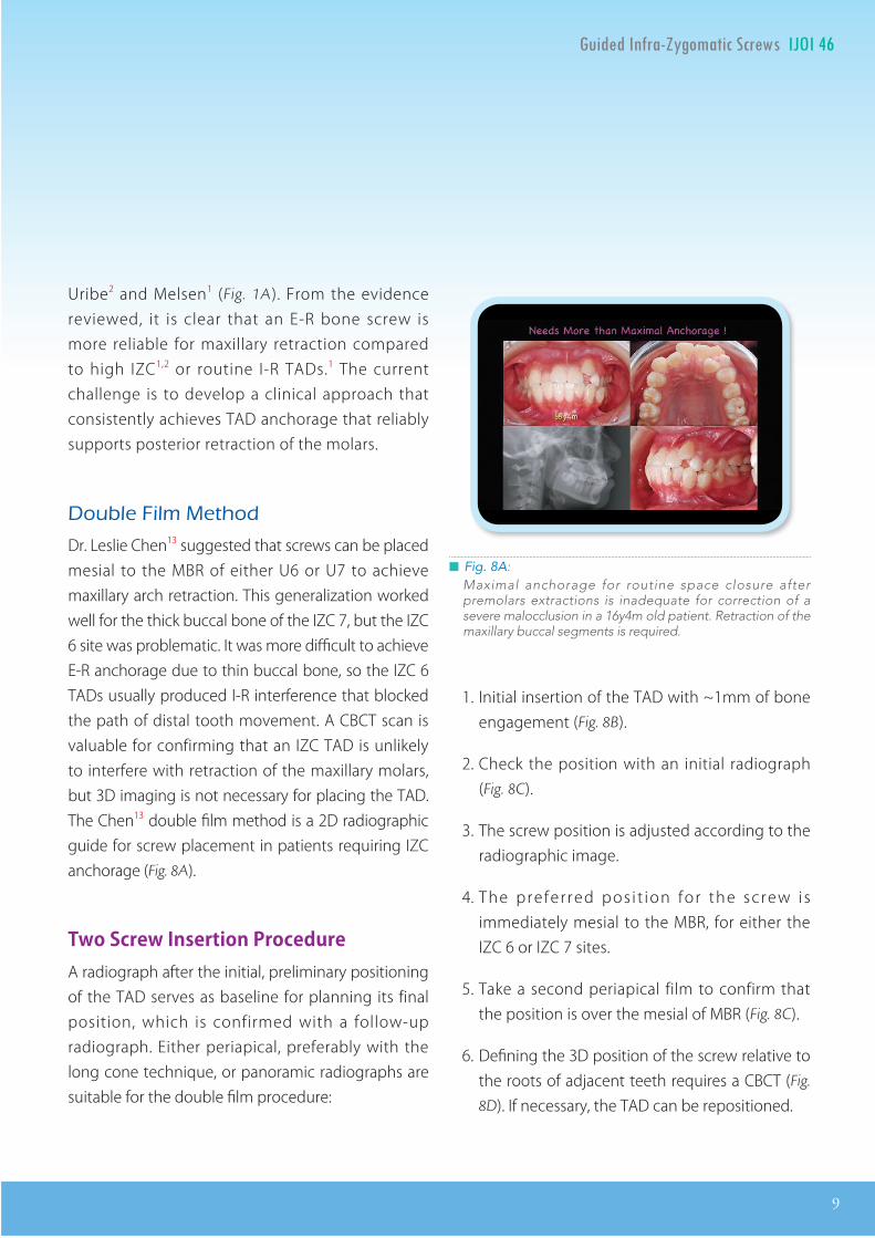
█ Fig. 6D: A similar CBCT cut near the apices of the second molars shows the screw on the right side penetrated the sinus, but it is extra-radicular and still provided effective anchorage to retract the right buccal segment, and the patient had no symptoms or complaints. The left IZC 7 impinged on the distal surface of the MBR of the U7, preventing retraction of the left buccal segment in the direction of the yellow arrows.
CBCT analysis of consecutive patients revealed some TADs were I-R (Inter-Radicular) (Figs. 6A-D), and mostly they were E-R (Fig. 7). Consistently placing
IZC bone screws in an E-R, or carefully selected I-R position, is critical for reliable maxillary arch retraction.
8
IJOI 46 LI E RO T E A TER I IJOI 46
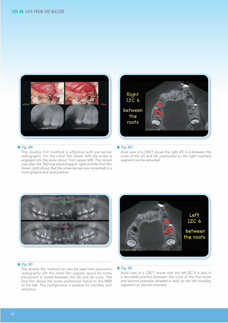
█ Fig. 7A: IZC 6 interference with maxillary retraction is documented. A patient 14y1m of age was treated with nine months of maxillary retraction, that was anchored with IZC 6 and IZC 7 screws. Note there is a further deviation of the upper midline to the right, which is consistent with more retraction on the right than the left sides. Little or no progress was noted in correcting the Class II buccal segment on the left side (lower views). See text for details.
█ Fig. 7B: The right IZC 7 is buccal to the roots, and has provided effective anchorage for maxillary retraction (yellow arrows). The MBR of the U6 on the left side interfered with retraction (Stop!). See text for details.
Anc orage equirements
Originally, the main purpose for TADs in the posterior maxillary arch was for maximal retraction of the anterior segment following extraction of premolars. Ideal I-R placement was right in the middle of the roots of upper 2nd premolar and 1st molar to avoid hitting the roots. In effect, the TADs provided solid anchorage for the extraction space closure. Similar I-R placement of TADs did not work to retract maxillary molars, paradoxically for the same reason: they blocked the path of distalizing tooth movement. Thus, substantial retraction of the entire maxillary arch was not much or not possible with routine I-R TADs.
Cl inical experience with OBSs placed in the IZC 6 and 7 areas revealed that maxillary arch retraction was accomplished more often with TADs in the IZC 7, rather than the IZC 6 position. CBCT evaluation demonstrated that the TADs, successfully anchoring maxillary arch retraction, were placed in an E-R position of the relatively thick buccal bone (Figs. 3-5).
A truly reliable (“fail-proof ”) method for maxillary retraction must evaluate bone quality as well as quantity at the TAD site. Chang et al.12 recently found an IZC screw failure rate of <7%, compared to ~20%, as reported by Uribe.2 The improved performance of the Chang et al.12 method may relate to favorable soft tissue on the more inferior sites like IZC 6 & 7 (Fig. 1B), compared to the more superior crestal positions of the TADs used by
IJOI 46 LI E RO T E A TER
9
I IJOI 46
█ Fig. 8A: Maximal anchorage for routine space closure after premolars extractions is inadequate for correction of a severe malocclusion in a 16y4m old patient. Retraction of the maxillary buccal segments is required.
Uribe2 and Melsen1 (Fig. 1A). From the evidence reviewed, it is clear that an E-R bone screw is more reliable for maxillary retraction compared to high IZC1,2 or routine I-R TADs.1 The current challenge is to develop a clinical approach that consistently achieves TAD anchorage that reliably supports posterior retraction of the molars.
ouble ilm et od
Dr. Leslie Chen13 suggested that screws can be placed mesial to the MBR of either U6 or U7 to achieve maxillary arch retraction. This generalization worked well for the thick buccal bone of the IZC 7, but the IZC 6 site was problematic. It was more difficult to achieve E-R anchorage due to thin buccal bone, so the IZC 6 TADs usually produced I-R interference that blocked the path of distal tooth movement. A CBCT scan is valuable for confirming that an IZC TAD is unlikely to interfere with retraction of the maxillary molars, but 3D imaging is not necessary for placing the TAD. The Chen13 double film method is a 2D radiographic guide for screw placement in patients requiring IZC anchorage (Fig. 8A).
Two Screw Insertion ProcedureA radiograph after the initial, preliminary positioning of the TAD serves as baseline for planning its final position, which is confirmed with a follow-up radiograph. Either periapical, preferably with the long cone technique, or panoramic radiographs are suitable for the double film procedure:
1. Initial insertion of the TAD with ~1mm of bone engagement (Fig. 8B).
2. Check the position with an initial radiograph (Fig. 8C).
3. The screw position is adjusted according to the radiographic image.
4. The prefer red pos i t ion for the screw is immediately mesial to the MBR, for either the IZC 6 or IZC 7 sites.
5. Take a second periapical film to confirm that the position is over the mesial of MBR (Fig. 8C).
6. Defining the 3D position of the screw relative to the roots of adjacent teeth requires a CBCT (Fig.
8D). If necessary, the TAD can be repositioned.
10
IJOI 46 LI E RO T E A TER I IJOI 46
█ Fig. 8D: Axial view of a CBCT shows the right IZC 6 is between the roots of the U5 and U6, positioned so the right maxillary segment can be retracted.
█ Fig. 8E: Axial view of a CBCT shows that the left IZC 6 is also in a favorable position between the roots of the first molar and second premolar (shaded in red), so the left maxillary segment can also be retracted.
█ Fig. 8B: The double film method is effective with periapical radiographs. For the initial film (lower left) the screw is engaged into the bone about 1mm (upper left). The clinical view after the TAD was placed (upper right) and the final film (lower right) shows that the screw (arrow) was reinserted in a more gingival and axial position.
█ Fig. 8C: The double film method can also be used with panoramic radiography. On the initial film (upper), space for screw placement is noted between the U5 and U6 roots. The final film shows the screw positioned mesial to the MBR of the U6s. This configuration is suitable for maxillary arch retraction.
IJOI 46 LI E RO T E A TER
11
I IJOI 46
Advantages:
1. There is usually enough I-R space between the U5 root and the MBR of the U6 for an IZC 6 TAD. Placing the screw in this area is suitable for maxillary arch retraction if it’s mesial to the MBR of the U6 and there is adequate clearance for distal movement of the root of the U5.
2. If it is desirable to position the TAD over the mesial of MBR of U7 (IZC 7), the chance of extra radicular is much higher.
3. Local infiltration analgesia is recommended to control pain, and fortunately this form of anesthesia does not interfere with the patient's perception of a screw contacting the root. If the patient feels the screw touching the root of a tooth, the TAD can be repositioned.
Disadvantages:
1. Additional wounds occur if screw positioning is changed.
2. Repositioning of the screw is associated with saliva contamination.
Pin Head Soft Tissue Penetration MethodDr. Mala Ram Manohar14 presented this innovative method at the 8th World Implant Orthodontics Congress in Goa in 2016. The distinct advantage over the Chen13 double film method is the lack of saliva contamination associated with reinserting the TAD. The procedure is as follows:
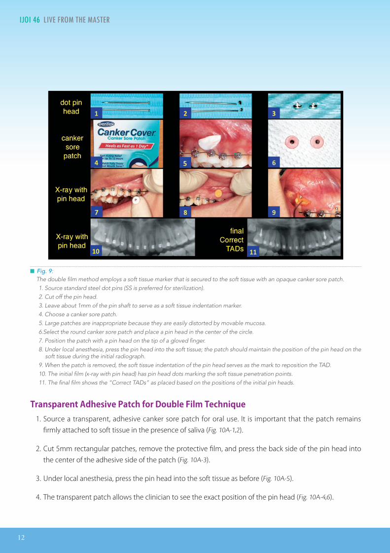
1. Use stainless steel dot pins (Fig. 9-1); cut off the heads (Fig. 9-2) leaving about a 1mm piece of the shank (Fig. 9-3).
2. Canker sore patches (Fig. 9-4) used to cover aphthous ulcers (Fig. 9-5) are thin, opaque strips or circular patches; and position a pin head in the center of the patch, with the point up (Fig.
9-6).
3. Following topical and then local infiltration anesthesia, the circular patch covering the pin-head point is pressed firmly into place, penetrating the soft tissue in the anticipated position of a TAD.
4. Image the area with a 2D radiograph, and re insert the TAD i f needed into a more desirable position; use the soft tissue mark left by the pin head as a landmark.
5. Take a follow-up radiograph to check the final position of the screw, and adjust the TAD position as needed.
Advantages:
1. No saliva contamination of the screw, unless it must be repositioned after the follow-up radiograph.
2. Avoids multiple screw placement wounds.
12
IJOI 46 LI E RO T E A TER I IJOI 46
Transparent Adhesive Patch for Double Film Technique1. Source a transparent, adhesive canker sore patch for oral use. It is important that the patch remains
firmly attached to soft tissue in the presence of saliva (Fig. 10A-1,2).
2. Cut 5mm rectangular patches, remove the protective film, and press the back side of the pin head into the center of the adhesive side of the patch (Fig. 10A-3).
3. Under local anesthesia, press the pin head into the soft tissue as before (Fig. 10A-5).
4. The transparent patch allows the clinician to see the exact position of the pin head (Fig. 10A-4,6).
█ Fig. 9: The double film method employs a soft tissue marker that is secured to the soft tissue with an opaque canker sore patch. 1. Source standard steel dot pins (SS is preferred for sterilization). 2. Cut off the pin head. 3. Leave about 1mm of the pin shaft to serve as a soft tissue indentation marker. 4. Choose a canker sore patch. 5. Large patches are inappropriate because they are easily distorted by movable mucosa. 6.Select the round canker sore patch and place a pin head in the center of the circle. 7. Position the patch with a pin head on the tip of a gloved finger. 8. Under local anesthesia, press the pin head into the soft tissue; the patch should maintain the position of the pin head on the
soft tissue during the initial radiograph. 9. When the patch is removed, the soft tissue indentation of the pin head serves as the mark to reposition the TAD.10. The initial film (x-ray with pin head) has pin head dots marking the soft tissue penetration points. 11. The final film shows the “Correct TADs” as placed based on the positions of the initial pin heads.
IJOI 46 LI E RO T E A TER
13
I IJOI 46
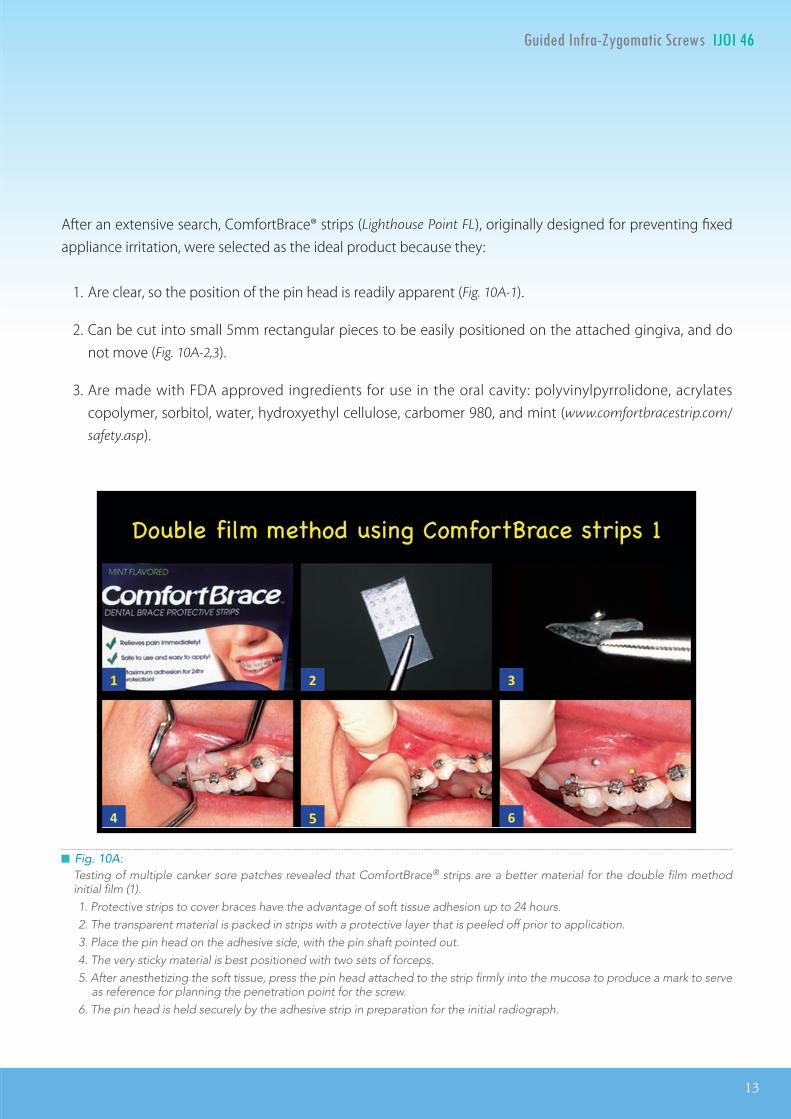
█ Fig. 10A: Testing of multiple canker sore patches revealed that ComfortBrace® strips are a better material for the double film method initial film (1). 1. Protective strips to cover braces have the advantage of soft tissue adhesion up to 24 hours. 2. The transparent material is packed in strips with a protective layer that is peeled off prior to application. 3. Place the pin head on the adhesive side, with the pin shaft pointed out. 4. The very sticky material is best positioned with two sets of forceps. 5. After anesthetizing the soft tissue, press the pin head attached to the strip firmly into the mucosa to produce a mark to serve
as reference for planning the penetration point for the screw. 6. The pin head is held securely by the adhesive strip in preparation for the initial radiograph.
After an extensive search, ComfortBrace® strips (Lighthouse Point FL), originally designed for preventing fixed appliance irritation, were selected as the ideal product because they:
1. Are clear, so the position of the pin head is readily apparent (Fig. 10A-1).
2. Can be cut into small 5mm rectangular pieces to be easily positioned on the attached gingiva, and do not move (Fig. 10A-2,3).
3. Are made with FDA approved ingredients for use in the oral cavity: polyvinylpyrrolidone, acrylates copolymer, sorbitol, water, hydroxyethyl cellulose, carbomer 980, and mint (www.comfortbracestrip.com/
safety.asp).
14
IJOI 46 LI E RO T E A TER I IJOI 46
Indication or I
1. At least a 5mm gap is required between the roots of the U5 and the MBR of U6 to avoid root contact with an I-R TAD.
2. Small oral cavities are often more convenient to place IZC 6 rather than placing the IZC 7.
3. A buccal frenum on or near the site can induce laceration, inflammation and screw failure; fortunately, there is usually no buccal frenum at the IZC 6 site.
4. The 5mm width of attached gingiva is adequate for most IZC 6 TADs.
5. Avoid placing TADs between the roots of teeth where the sinus floor is low because these areas usually have low density bone and a thin cortical plate.
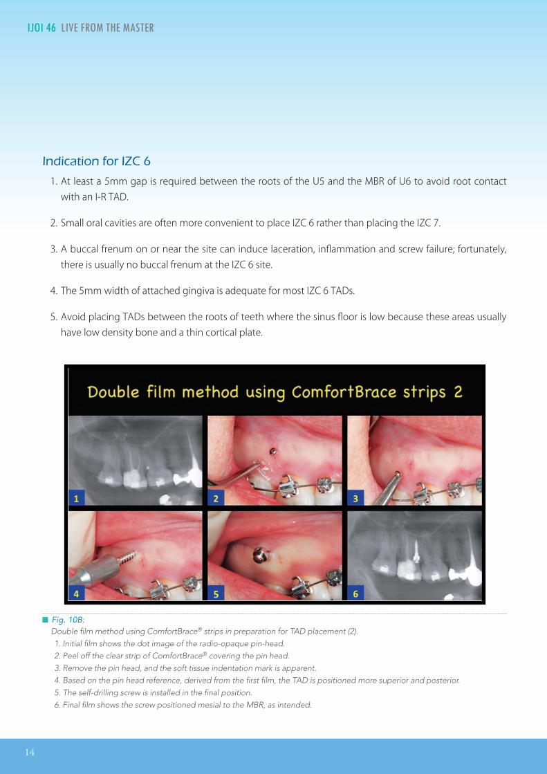
█ Fig. 10B: Double film method using ComfortBrace® strips in preparation for TAD placement (2). 1. Initial film shows the dot image of the radio-opaque pin-head. 2. Peel off the clear strip of ComfortBrace® covering the pin head. 3. Remove the pin head, and the soft tissue indentation mark is apparent. 4. Based on the pin head reference, derived from the first film, the TAD is positioned more superior and posterior. 5. The self-drilling screw is installed in the final position. 6. Final film shows the screw positioned mesial to the MBR, as intended.
IJOI 46 LI E RO T E A TER
15
I IJOI 46
Indication o I
1. At least 5mm width of attached gingiva is advantageous.
2. Access to the IZC 7 area requires a large oral cavity, as well as lip reflection for adequate access to the screw insertion site.
3. Avoid placing between the roots of teeth where the sinus floor is low because this area has a thin cortical plate.
ailure rate
According to a recent study by Chang et al.12 the IZC screw failure rate is <7%. Most of the failures are due to:
1.Poorbonequality: Unfortunately there is no reliable method for evaluating bone quality. The sensation for poor bone quality, beneath a sound layer of cortical bone, is like punching through an egg shell, followed by a lack of screw stability. Unless the TAD can be stabilized by deeper penetration, it is best to remove it and try another site.
2.Immediate loading: SS screws are excellent TADs because they do not osseointegrate and are easily repositioned to another site, if necessary.
3.Sinusfloor: A low sinus between the roots of teeth is undesirable for an IZC TAD site.
4.Movablemucosa: Unattached alveolar mucosa at the TAD site is usually undesirable.4,5 However,
Chang et al.12 found no significant difference in the failure rate between movable mucosa and attached gingiva if the platform of the screw is at least 5mm away from the soft tissue surface. The disadvantages of the latter approach are a longer screw is required (~12mm) and it must be carefully positioned for patient comfort.
uccal el one crews
The senior author (JJ- JL) previously introduced mandibular buccal shelf bone screws, which were usually placed by periodontists or oral surgeons, using the apically positioned flap to provide attached gingiva at the TAD site.15 When the mandibular buccal shelf is steep and if patients require an extra-radicular placement, with an apically repositioned flap of attached gingiva, an experienced surgeon is needed. Flap surgery is more expensive and tends to be painful postoperatively, particularly if a TAD must be repositioned. Currently, a skillful orthodontist can produce a good result with a self drilling screw by using the double film method to place the I-R buccal shelf screw (right mesial to the MBR of L7). Reliable retraction of the mandibular arch can be done.
onclusions
1. The double film method is advantageous for installing TADs in the three most common sites: IZC 6, IZC 7, and mandibular buccal shelf.
2. ComfortBrace® strips have proven superior for maintaining a pin head in a stable position relative to the soft tissue.
16
IJOI 46 LI E RO T E A TER
3. The double f i lm method is indicated for selecting the appropriate point of entry for IZC or buccal shelf screws.
4. Extensive experience with the double film method has demonstrated it is an advantageous approach for reliably placing IZC 6 & IZC 7 bone screws to retract the dentition.
5. The double f i lm method has s ignif icant advantages for both the clinician and the patient.
Ac nowledgement
1. Thanks to Dr. Leslie Yen-Peng Chen for the innovative idea leading to the current double film method for accurately placing TADs for orthodontic anchorage.
2. Thanks to Dr. Mala Ram Manohar for the practical idea of using pin heads with a 1mm shaft to mark the initial point of tissue penetration for a TAD.
3. Thanks to Dr. Po-Jung Chen for the CBCT cross-sectional evaluation of the IZC 6 vs. IZC 7 sites as shown in Fig. 4.
4. Thanks to Dr. Po-Jan Kuo for the CBCT information in Fig. 5 which illustrates the buccal anatomy of IZC 6 & 7 sites.
e erences
1. Melsen B. Overview of mini-implants: where are we? J Clin Ortho 2005;39(9):539-47.
2. Uribe F, Mehr R , Mother A, Janakiraman N, Allareddy V. Failure rates of mini-implants placed in the infrazygomatic region. Prog Orthod 2015;16:31.
3. Villegas C. 100% successful TAD system. Presentation at the 2016 AAO Meeting, Orlando FL.
4. Chang MJ, Lin JJ, Roberts WE. Probable airway etiology for a severe Class III openbite malocclusion: conservative treatment with extra-alveolar bone screws and intermaxillary elastics. Int J Orthod Implantol 2017;45:4-20.
5. Shih YH, Lin JJ, Roberts WE. Conservative management of Class I crowded malocclusion complicated by severe maxillary protrusion, facial convexity and deepbite. Int J Orthod Implantol 2016;44:4-16.
6. Goaslind GD, et al . Thickness of facial gingiva. J Perio 1977;12:768-71.
7. Chen MY, et al. Eastern and western studies of interdental cortical bone thickness for temporary anchorage devices. J Taiwan Periodontal 2013;18(1):33-43.
8. Liou EJ, Chen PH, Wang YC, Lin JC. A computed tomographic image study on the thickness of the infrazygomatic crest of the maxilla and its clinical implications for mini-screw insertion. Am J Orthod Dentofacial Orthop 2007;131:352-6.
9. Chen PJ, et al. Bone thickness of the infrazygomatic crest for orthodontic implant placement. Poster, Annual Meeting of Taiwan Association of Orthodontists, 2008.
10. Lin JJ. Mini-screw or mini-plate, which is better for whole upper arch distalization. News and Trends in Orthodontics 2007;5:1-2.
11. Lin JJ. Creative orthodontics: Blending the Damon System and TADs to Manage Di�cult Malocclusions. Taipei: Yong Chien Ltd; 2010.
12. Chris CH, Hsu E, Lin JSY, Yeh HY, Roberts WE. Comparison of the failure rate for infrazygomatic bone screws placed in movable mucosa or attached gingiva. Eur J Orthod 2017 (In Preparation).
13. Personal communication with Dr. Leslie Chen, 2012.
14. Manohar MR. Versatility of miniscrews-implants & implants guides. 8th Goa, India: 8th WIOC; 2016.
15. Lin JJ, et al. Treatment of Severe Class III with Buccal Shelf Mini-Screws. News and Trends in Orthodontics 2010;18:4-15.
專題演講
Int ucingT th ntics in the a n ste
Dr. Elizabeth Menzel
國立交通大學-浩然圖書館B1國際會議廳
( n)
Dr. Diego Peydro Herrero金牛頓藝術科技
( h )
合報價
e n th ntics with lea ligne s The a
匯款資訊:
03-5711377 蔡小姐 o a iaoi.pro
台灣顱顎障礙症學會 中華民國兒童牙科醫學會
iaoi.pro
好康雙響炮
元000
日盛國際商業銀行815光復分行0347 帳號:105-27376210-000 戶名:國際矯正植牙學會
專題演講
報名2017兩場大會
專題演講
Int ucingT th ntics in the a n ste
Dr. en el 來自德國,於法蘭克福大學完成矯正專科訓練,專長在以矯正方式處理下顎周圍疼痛與疾病,具有方式處理下顎周圍疼痛與疾病,具有 年以上的臨床治療經驗。年以上的臨床治療經驗。
自 年以來經常在國際上發表相關演講,使用以來經常在國際上發表相關演講,使用 Damon system 已超已超過 年的時間,講學和臨床治療的經驗都非常豐富。講學和臨床治療的經驗都非常豐富。 年來台演講,
大獲好評。 應邀再次來台,內容豐富,精彩可期。
Dr. Elizabeth Menzel
國立交通大學-浩然圖書館B1國際會議廳(新竹市大學路1001號)
( n)
Dr. Diego Peydro Herrero金牛頓藝術科技(新竹市建中一路25號2樓)
( h )
合報價
e n th ntics with lea ligne s The auccess ul t ategies t lve the st i icult ases
with Invisalign Technique
Dr. Diego Peydro Herrero 為西班牙的矯正專科醫師,並於馬德里和瓦倫西亞大學開設矯正專業課程。此外,他也是隱適美系統的專家,在歐洲
和中東地區開設隱適美系統的國際繼續教育課程-Clear Ortho Inter-national Program(COIP)。本次的演講他將著重利用 。本次的演講他將著重利用 aligners aligners 和骨釘來和骨釘來
處理複雜處理複雜的案例,透過分解步驟的解說,來闡述他的治療原理和方式。
匯款資訊:
03-5711377 蔡小姐
台灣顱顎障礙症學會 中華民國兒童牙科醫學會
iaoi.pro
好康雙響炮
元000
*4/21前因故退費需扣除一成行政費用、4/21後因故退費需扣除三成行政費用。
日盛國際商業銀行815光復分行0347 帳號:105-27376210-000 戶名:國際矯正植牙學會
專題演講
報名2017兩場大會
(特價至2017/4/21前 額滿為止)(特價至2017/4/21前 額滿為止)
20
IJOI 46 AOI A E RE ORT
istor and tiolog
A 10yr female presented with her parents for orthodontic consultation. The chief complaint was excessive overjet. Facial evaluation showed a convex profile, hypermentalis activity, 5mm of lip incompetence, and a retrusive mandible (Fig. 1). Intra-oral examination revealed retained maxillary primary second molars, relatively narrow arches, and an 7mm overjet (Fig. 2). Except for the Class I molars, due to the retained maxillary deciduous molars, the casts were consistent with an end-on Class II, division 1 malocclusion (Fig.
3). There was no additional contributing medical or dental history. Conservative orthodontic treatment produced an excellent alignment and a pleasing smile (Figs. 4-6). Panoramic and cephalometric radiographs before and after treatment are shown in Figs. 7 and 8, respectively. Fig. 9 documents the dentofacial treatment and the unfavorable vertical growth response with superimposed cephalometric tracings. Cephalometric measurements are presented in Table 1.
a ro a occ ion r aton r ati it a a i i atin
pp ianc pan ion ta i it an aptation
tract A 10-year-old female presented with a retrusive mandible (SNB 76°), Class I molars and Class II canines due to the delayed eruption of the maxillary second premolars. There was 7mm overjet, 5mm overbite, 7mm of lower arch crowding, steep mandibular plane angle (FMA 32°), and increased axial inclination of the lower incisors to the mandibular plane (102°). The Discrepancy Index (DI) was 21. Despite the indication for extraction of premolars, the patient and her parents preferred conservative (noninvasive) treatment with a simple, �xed appliance. The revised treatment plan was to open the bite with posterior bite turbos on lower �rst molars, expand the arches with a passive self-ligating (PSL) appliance, and correct the sagittal discrepancy with Class II elastics. During 30 months of active treatment there was an unfavorable vertical growth response, resulting in a posterior rotation of the mandible, which was associated with less natural development of arch length. Thus, increased expansion was required to resolve crowding and produce an excellent alignment, documented by a cast-radiograph evaluation (CRE) of 20, with a Pink & White dental esthetics score of 4. Despite the desirable result, there were stability concerns because the lower and upper canines, as well as the molars, were expanded 3-5 and 11-12mm, respectively. Both arches were retained with 3-3 �xed retainers, bonded to each tooth, and overlay appliances. The pleasing result was stable 6 years later indicating that arch expansion to correct crowding is a viable option if there is a commitment to permanent retention. (Int J Orthod Implantol 2017;46:20-37)
Key words:Arch expansion, posterior and anterior bite turbos, lower facial height, inter-canine and inter-molar widths, �xed retention, passive self-ligating brackets, vertical facial growth, Class II elastics
Dr. Shih-Yung Lin,Editor, International Journal of Orthodontics & Implantology (Left)
Dr. Chris Chang, Founder, Beethoven Orthodontic Center
Publisher, International Journal of Orthodontics & Implantology (Center)
Dr. W. Eugene Roberts,Editor-in-chief, International Journal of Orthodontics & Implantology (Right)

IJOI 46 AOI A E RE ORT
21
T IJOI 46
█ Fig. 2: Pre-treatment intraoral photographs
█ Fig. 1: Pre-treatment facial photographs, 10yr female
█ Fig. 3: Pre-treatment study models
█ Fig. 4: Post-treatment facial photographs, after 30 months of active treatment
█ Fig. 5: Post-treatment intraoral photographs
█ Fig. 6: Post-treatment study models
Dr. Shih-Yung Lin,Editor, International Journal of Orthodontics & Implantology (Left)
Dr. Chris Chang, Founder, Beethoven Orthodontic Center
Publisher, International Journal of Orthodontics & Implantology (Center)
Dr. W. Eugene Roberts,Editor-in-chief, International Journal of Orthodontics & Implantology (Right)
22
IJOI 46 AOI A E RE ORT
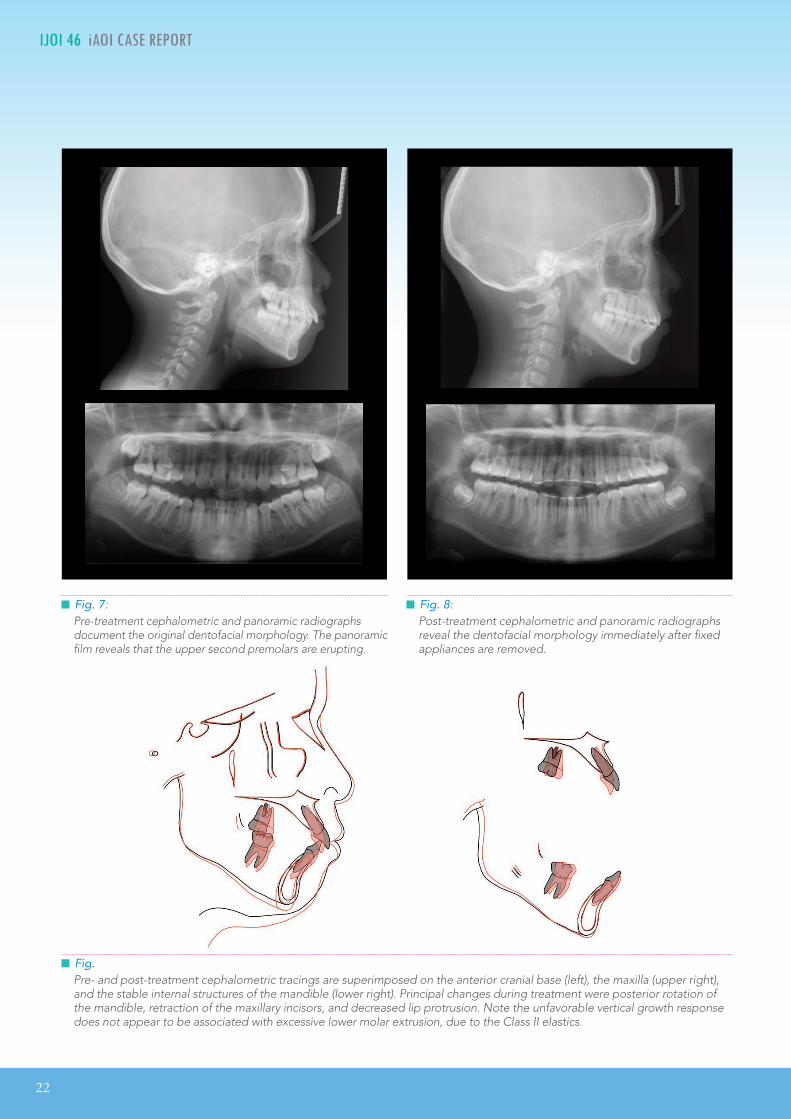
█ Fig. 7: Pre-treatment cephalometric and panoramic radiographs document the original dentofacial morphology. The panoramic film reveals that the upper second premolars are erupting.
█ Fig. 8: Post-treatment cephalometric and panoramic radiographs reveal the dentofacial morphology immediately after fixed appliances are removed.
█ Fig. Pre- and post-treatment cephalometric tracings are superimposed on the anterior cranial base (left), the maxilla (upper right), and the stable internal structures of the mandible (lower right). Principal changes during treatment were posterior rotation of the mandible, retraction of the maxillary incisors, and decreased lip protrusion. Note the unfavorable vertical growth response does not appear to be associated with excessive lower molar extrusion, due to the Class II elastics.
IJOI 46 AOI A E RE ORT
23
T IJOI 46
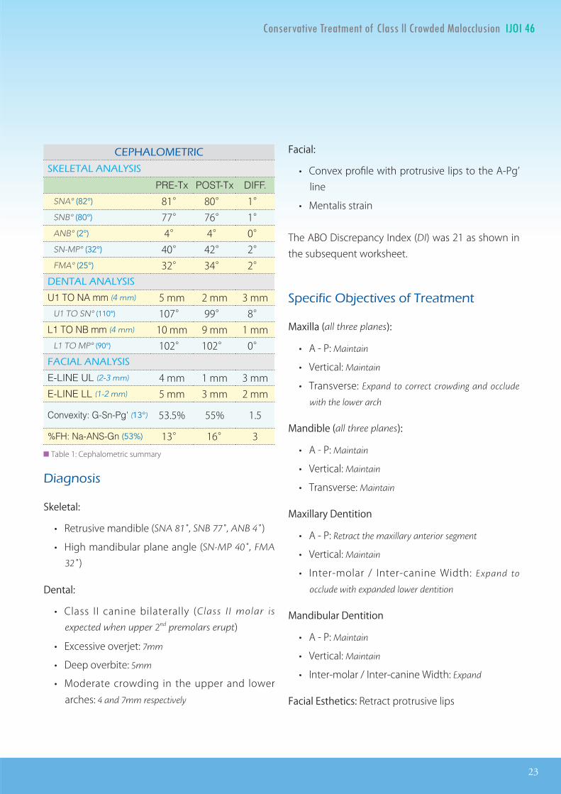
iagnosis
Skeletal:
• Retrusive mandible (SNA 81˚, SNB 77˚, ANB 4˚)
• High mandibular plane angle (SN-MP 40˚, FMA
32˚)
Dental:
• Class II canine bilaterally (Class II molar is
expected when upper 2nd premolars erupt)
• Excessive overjet: 7mm
• Deep overbite: 5mm
• Moderate crowding in the upper and lower arches: 4 and 7mm respectively
Facial:
• Convex profile with protrusive lips to the A-Pg’ line
• Mentalis strain
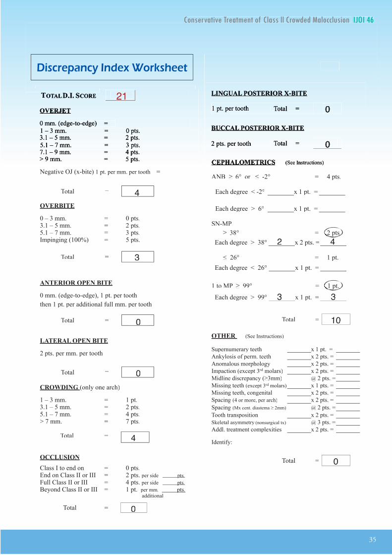
The ABO Discrepancy Index (DI) was 21 as shown in the subsequent worksheet.
eci ic Ob ecti es o reatment
Maxilla (all three planes):
• A - P: Maintain
• Vertical: Maintain
• Transverse: Expand to correct crowding and occlude
with the lower arch
Mandible (all three planes):
• A - P: Maintain
• Vertical: Maintain
• Transverse: Maintain
Maxillary Dentition
• A - P: Retract the maxillary anterior segment
• Vertical: Maintain
• Inter-molar / Inter-canine Width: Expand to
occlude with expanded lower dentition
Mandibular Dentition
• A - P: Maintain
• Vertical: Maintain
• Inter-molar / Inter-canine Width: Expand
Facial Esthetics: Retract protrusive lips
A O I
A A A I
PRE-Tx POST-Tx DIFF.
SNA° (82°) 81° 80° 1° SNB° (80°) 77° 76° 1° ANB° (2°) 4° 4° 0° SN-MP° (32°) 40° 42° 2° FMA° (25°) 32° 34° 2°
A A A I
U1 TO NA mm (4 mm) 5 mm 2 mm 3 mm U1 TO SN° (110°) 107° 99° 8°
L1 TO NB mm (4 mm) 10 mm 9 mm 1 mm L1 TO MP° (90°) 102° 102° 0° A IA A A I
E-LINE UL (2-3 mm) 4 mm 1 mm 3 mm E-LINE LL (1-2 mm) 5 mm 3 mm 2 mm
Convexity: G-Sn-Pg' (13°) 53.5% 55% 1.5
%FH: Na-ANS-Gn (53%) 13° 16° 3██ Table 1: Cephalometric summary
24
IJOI 46 AOI A E RE ORT
reatment lan
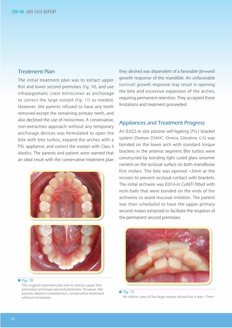
The initial treatment plan was to extract upper first and lower second premolars (Fig. 10), and use infrazygomatic crest miniscrews as anchorage to correct the large overjet (Fig. 11) as needed. However, the parents refused to have any teeth removed except the remaining primary teeth, and also declined the use of miniscrews. A conservative, non-extraction approach without any temporary anchorage devices was formulated to open the bite with bite turbos, expand the arches with a PSL appliance, and correct the overjet with Class II elastics. The parents and patient were warned that an ideal result with the conservative treatment plan
they desired was dependent of a favorable (forward) growth response of the mandible. An unfavorable (vertical) growth response may result in opening the bite and excessive expansion of the arches, requiring permanent retention. They accepted these limitations and treatment proceeded.
A liances and reatment rogress
An 0.022-in slot passive self-ligating (PSL) bracket system (Damon D3MX®, Ormco, Glendora, CA) was bonded on the lower arch with standard torque brackets in the anterior segment. Bite turbos were constructed by bonding light cured glass ionomer cement on the occlusal surface on both mandibular first molars. The bite was opened ~3mm at the incisors to prevent occlusal contact with brackets. The initial archwire was 0.014-in CuNiTi fitted with resin balls that were bonded on the ends of the archwires to avoid mucosal irritation. The patient was then scheduled to have the upper primary second molars extracted to facilitate the eruption of the permanent second premolars.
█ Fig. 10: The original treatment plan was to extract upper first premolars and lower second premolars. However, the parents desired nonextraction, conservative treatment without miniscrews.
█ Fig. 11: An inferior view of the large overjet shows that it was ~7mm.
IJOI 46 AOI A E RE ORT
25
T IJOI 46
for Class II elastics (Fox 1/4”, 3.5-oz) to correct the sagittal discrepancy as the arches were expanding (Fig. 13).
In the 15th month, an upper 0.019x0.025-in stainless steel archwire was placed with vertical hooks mesial to the canines, to continue the Class II elastics (Fox
1/4”, 3.5-oz). Two months later, both mandibular second molars erupted with a lingual inclination. Each second molar was bonded with a buccal bracket and two lingual buttons, and a 0.016-in CuNiT lower archwire was placed, that extended to the tubes of the second molars. The height of the bite turbos on the lower first molars was increased to accommodate posterior cross elastics (Chipmunk
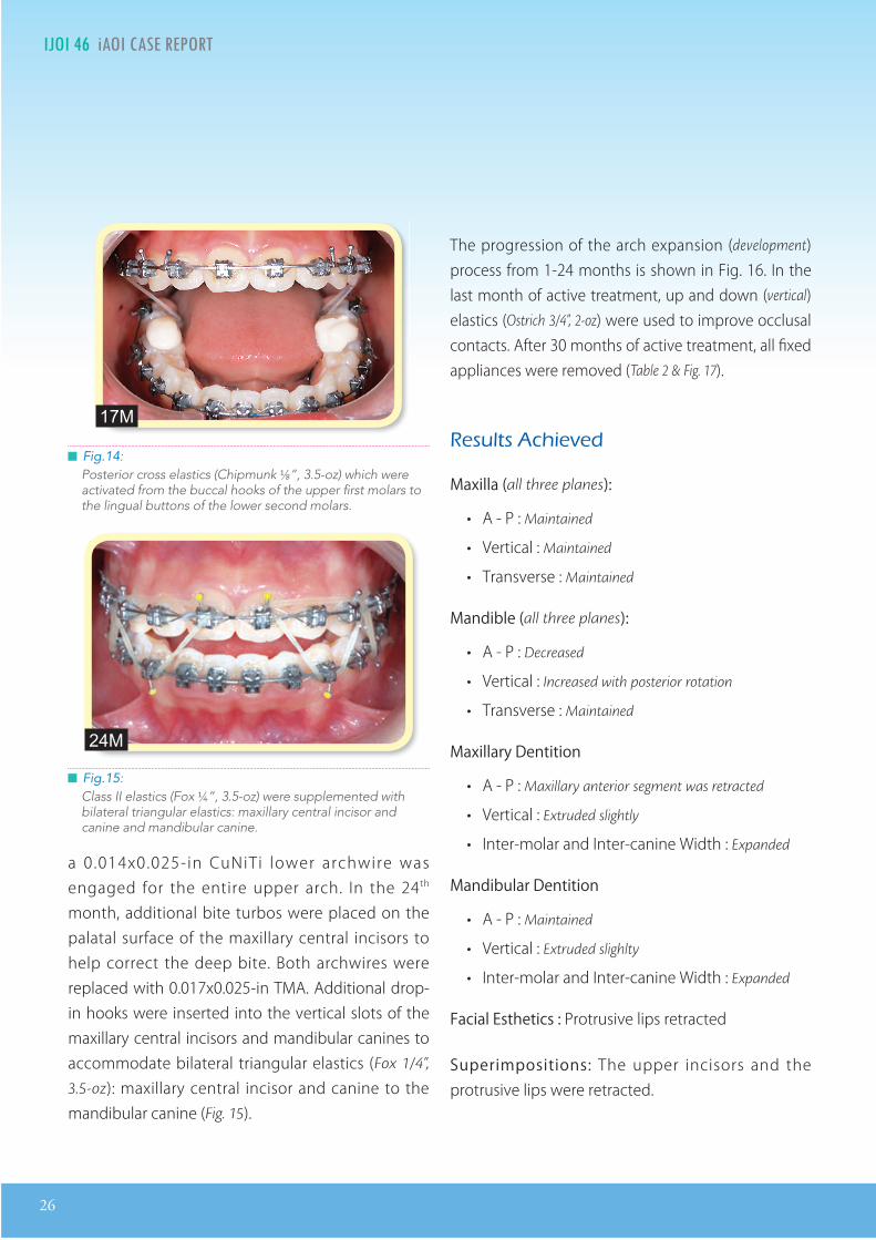
1/8”, 3.5-oz), which were applied from the buccal hooks of the upper first molars to the lingual buttons of the lower second molars (Fig. 14). After one month, the lingually tipped mandibular second molars were corrected, so the bite turbos were removed and a 0.014x0.025-in CuNiTi lower archwire was placed.
In the 21st month of treatment, the maxillary second molars erupted. They were bonded and
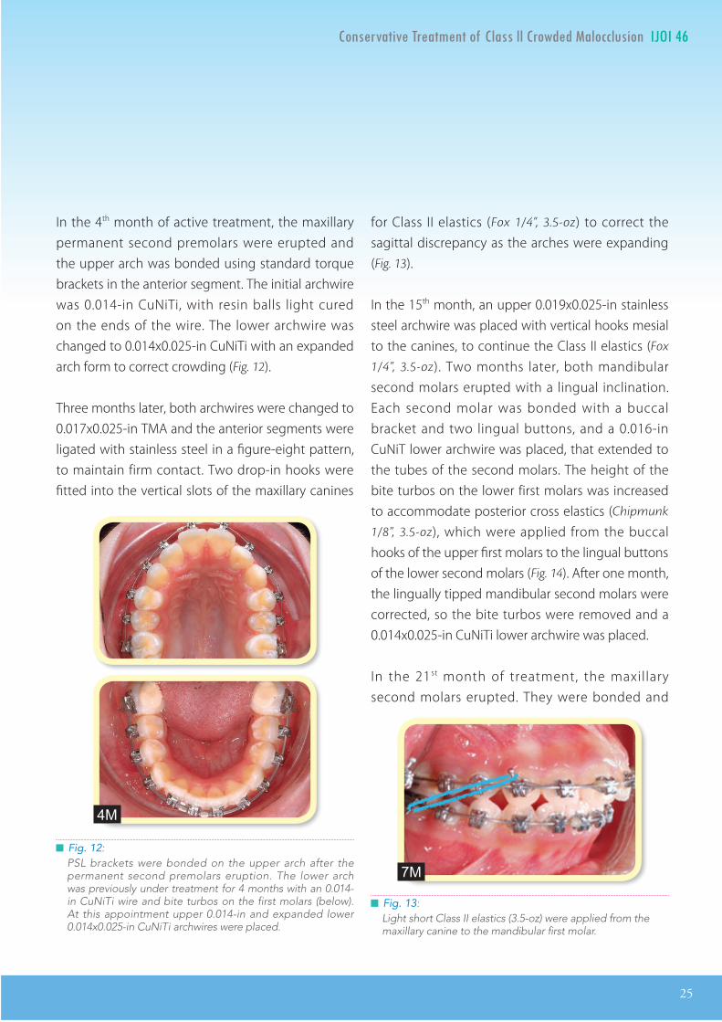
█ Fig. 12: PSL brackets were bonded on the upper arch after the permanent second premolars eruption. The lower arch was previously under treatment for 4 months with an 0.014-in CuNiTi wire and bite turbos on the first molars (below). At this appointment upper 0.014-in and expanded lower 0.014x0.025-in CuNiTi archwires were placed.
In the 4th month of active treatment, the maxillary permanent second premolars were erupted and the upper arch was bonded using standard torque brackets in the anterior segment. The initial archwire was 0.014-in CuNiTi, with resin balls light cured on the ends of the wire. The lower archwire was changed to 0.014x0.025-in CuNiTi with an expanded arch form to correct crowding (Fig. 12).
Three months later, both archwires were changed to 0.017x0.025-in TMA and the anterior segments were ligated with stainless steel in a figure-eight pattern, to maintain firm contact. Two drop-in hooks were fitted into the vertical slots of the maxillary canines
█ Fig. 13: Light short Class II elastics (3.5-oz) were applied from the maxillary canine to the mandibular first molar.
4M
7M
26
IJOI 46 AOI A E RE ORT
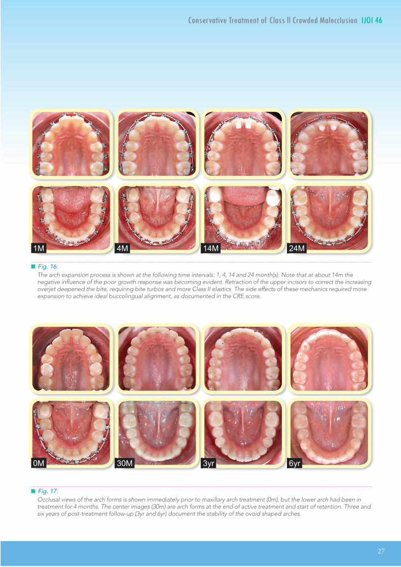
The progression of the arch expansion (development) process from 1-24 months is shown in Fig. 16. In the last month of active treatment, up and down (vertical) elastics (Ostrich 3/4”, 2-oz) were used to improve occlusal contacts. After 30 months of active treatment, all fixed appliances were removed (Table 2 & Fig. 17).
esults Ac ie ed
Maxilla (all three planes):
• A - P : Maintained
• Vertical : Maintained
• Transverse : Maintained
Mandible (all three planes):
• A - P : Decreased
• Vertical : Increased with posterior rotation
• Transverse : Maintained
Maxillary Dentition
• A - P : Maxillary anterior segment was retracted
• Vertical : Extruded slightly
• Inter-molar and Inter-canine Width : Expanded
Mandibular Dentition
• A - P : Maintained
• Vertical : Extruded slighlty
• Inter-molar and Inter-canine Width : Expanded
Facial Esthetics : Protrusive lips retracted
Superimpositions: The upper incisors and the protrusive lips were retracted.
█ Fig.14: Posterior cross elastics (Chipmunk ⅛”, 3.5-oz) which were activated from the buccal hooks of the upper first molars to the lingual buttons of the lower second molars.
█ Fig.15: Class II elastics (Fox ¼”, 3.5-oz) were supplemented with bilateral triangular elastics: maxillary central incisor and canine and mandibular canine.
a 0.014x0.025-in CuNiTi lower archwire was engaged for the entire upper arch. In the 24th month, additional bite turbos were placed on the palatal surface of the maxillary central incisors to help correct the deep bite. Both archwires were replaced with 0.017x0.025-in TMA. Additional drop-in hooks were inserted into the vertical slots of the maxillary central incisors and mandibular canines to accommodate bilateral triangular elastics (Fox 1/4”,
3.5-oz): maxillary central incisor and canine to the mandibular canine (Fig. 15).
17M
24M
IJOI 46 AOI A E RE ORT
27
T IJOI 46
█ Fig. 16:The arch expansion process is shown at the following time intervals: 1, 4, 14 and 24 month(s). Note that at about 14m the negative influence of the poor growth response was becoming evident. Retraction of the upper incisors to correct the increasing overjet deepened the bite, requiring bite turbos and more Class II elastics. The side effects of these mechanics required more expansion to achieve ideal buccolingual alignment, as documented in the CRE score.
█ Fig. 17: Occlusal views of the arch forms is shown immediately prior to maxillary arch treatment (0m), but the lower arch had been in treatment for 4 months. The center images (30m) are arch forms at the end of active treatment and start of retention. Three and six years of post-treatment follow-up (3yr and 6yr) document the stability of the ovoid shaped arches.
24M14M4M1M
6yr3yr30M0M
28
IJOI 46 AOI A E RE ORT
11mm 5mm 12mm
3mm
etention
Anterior fixed retainers were bonded on both arches from canine to canine (3-3). Removable clear overlay retainers were delivered for both arches, and the patient was instructed to wear them full time for the first 6 months and nights only thereafter.
Instructions were provided for home hygiene and maintenance of the retainers (Fig. 17).
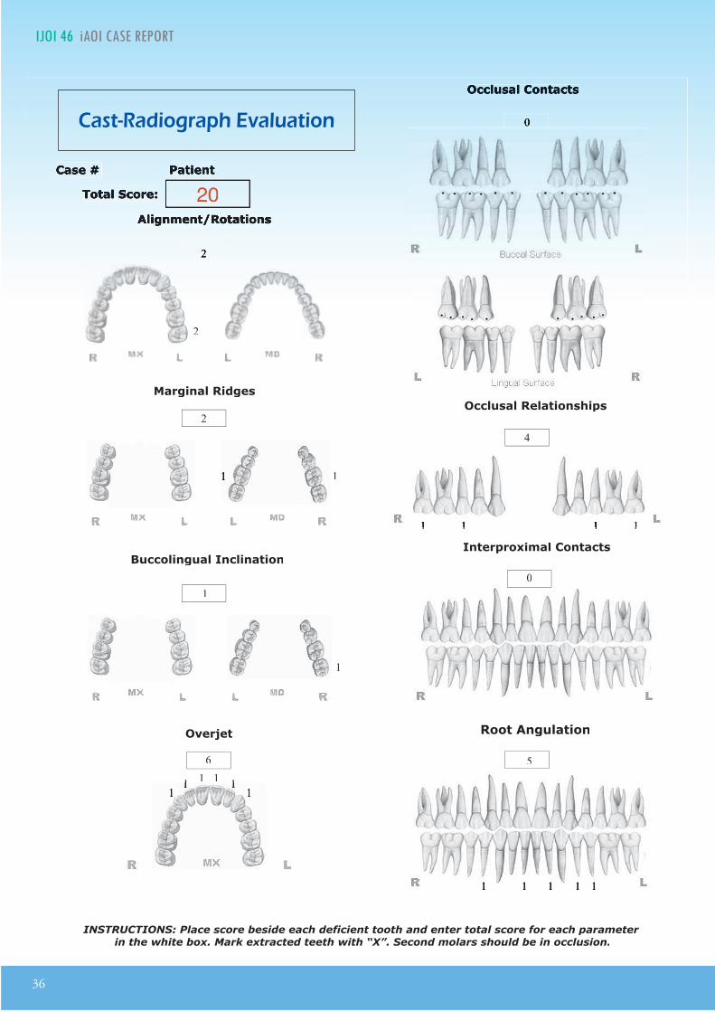
inal aluation o reatment
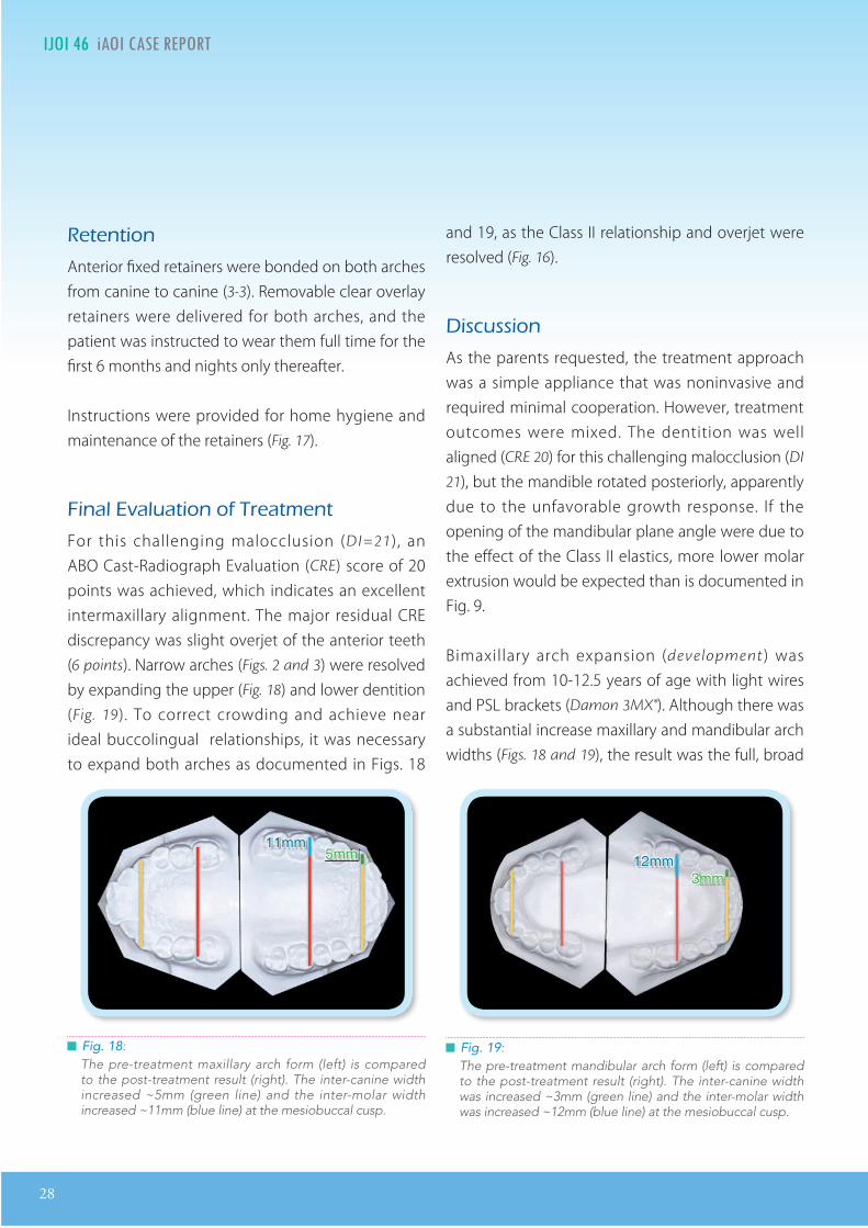
For this challenging malocclusion (DI=21), an ABO Cast-Radiograph Evaluation (CRE) score of 20 points was achieved, which indicates an excellent intermaxillary alignment. The major residual CRE discrepancy was slight overjet of the anterior teeth (6 points). Narrow arches (Figs. 2 and 3) were resolved by expanding the upper (Fig. 18) and lower dentition (Fig. 19). To correct crowding and achieve near ideal buccolingual relationships, it was necessary to expand both arches as documented in Figs. 18
and 19, as the Class II relationship and overjet were resolved (Fig. 16).
iscussion
As the parents requested, the treatment approach was a simple appliance that was noninvasive and required minimal cooperation. However, treatment outcomes were mixed. The dentition was well aligned (CRE 20) for this challenging malocclusion (DI
21), but the mandible rotated posteriorly, apparently due to the unfavorable growth response. If the opening of the mandibular plane angle were due to the effect of the Class II elastics, more lower molar extrusion would be expected than is documented in Fig. 9.
Bimaxillary arch expansion (development) was achieved from 10-12.5 years of age with light wires and PSL brackets (Damon 3MX®). Although there was a substantial increase maxillary and mandibular arch widths (Figs. 18 and 19), the result was the full, broad
█ Fig. 18: The pre-treatment maxillary arch form (left) is compared to the post-treatment result (right). The inter-canine width increased ~5mm (green line) and the inter-molar width increased ~11mm (blue line) at the mesiobuccal cusp.
█ Fig. 19: The pre-treatment mandibular arch form (left) is compared to the post-treatment result (right). The inter-canine width was increased ~3mm (green line) and the inter-molar width was increased ~12mm (blue line) at the mesiobuccal cusp.
IJOI 46 AOI A E RE ORT
29
T IJOI 46
smile which was the objective of the patient and her parents. They realized that it would be necessary to permanently retain both arches with 3-3 fixed and clear overlay retainers at night. Three years after the completion of treatment, the result is stable because it is well retained (Fig. 17). Is it possible for the facial musculoskeletal system of a preadolescent to adapt to this degree of expansion or is retention required indefinitely?
Although the arches were narrow at the onset (Figs. 2 and 3), resolving ~7mm of lower crowding resulted in 3mm of mandibular canine expansion, which is usually a stability concern. Regarding the stability of arch expansion, many studies have reported that there is a strong tendency for the arch to return to its original shape after appliances have been removed.1,2 Lee and Kirschen3 concluded that there is no evidence for longterm stability when the upper first molars are expanded more than 5mm. The results for current patient were quite stable after 3 years (Fig. 17), but the desired outcome was
permanently retained. Previous stability studies1-3
used retention for a limited period of time or not at all. It is not physiologically valid to compare stability between patients who are retained and not retained.
The important stability issues for extensive arch development (expansion ) are the mechanism of expansion, retention, as well as the long-term satisfaction and cooperation of the patient, particularly if removable retainers are involved. The objectives of the patient and parents are important considerations, but all concerned must understand the consequences of their choices. Aligning teeth over the apical base of bone is the best choice for long-term stability if there is no commitment to permanent retention.1-3 The degree of arch development shown in Figs. 9, 16-19 is the expectation for conservative alignment, when there are no extractions or interproximal enamel reduction (IPR). For the present patient (Figs. 4-6), the desired result is expected to be stable as long as the permanent retention scheme is maintained.
Tongue
CheekCheek
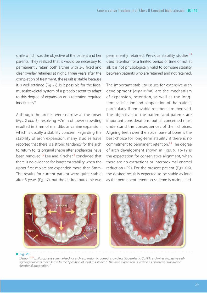
█ Fig. 20: Damon15,16 philosophy is summarized for arch expansion to correct crowding. Superelastic CuNiTi archwires in passive self-ligating brackets move teeth to the “position of least resistance.” The arch expansion is viewed as “posterior transverse functional adaptation.”
30
IJOI 46 AOI A E RE ORT
So from the initial consultation, it is very important that the patient and her parents understand that reality of their choice. It is possible over time that the facial musculoskeletal system (tongue, lips and
cheeks) will adapt to the expanded arch form (Fig. 20), but that mechanism has not been established with randomized clinical trails.
Arch expansion is only one of the nonextraction possibilities for delivering a pleasing smile. There are conservative treatment alternatives for correcting the malocclusion with little or no arch expansion. First, interproximal enamel reduction (IPR) is an excellent option because 0.25mm reduction of each approximating surface could have produced over 7mm of arch length in both arches. This is more than enough to allow ideal alignment with light wires in a PSL appliance, without producing any arch expansion. Even in the absence of arch expansion, a 3-3 fixed retainer bonded to all of the lower teeth is still indicated to prevent incisor crowding and arch collapse. However, a removable retainer such as a Hawley at night is all that would be needed to retain the upper arch. Avoiding fixed retention in the maxillary anterior region is advantageous because retainer debonding is common on the palatal surfaces of maxillary incisors. Second, retraction of the upper and/or lower molars to create arch length is readily accomplished with extra-alveolar (E-A) OrthoBoneScrews® (OBS) (Newton’s A Ltd., Hsinchu,
Taiwan). Seven millimeters of arch length can be easily achieved with OBS anchorage, particularly if there is IPR simultaneously. For the present patient, some expansion was indicated, but it was not necessary to expand to the degree shown in Fig. 18 to correct the crowding. Although IPR was an attractive alternative for treating the present patient, it is an invasive procedure which was undesirable
to the patient and her parents. When marked arch expansion is the outcome, whether it was planned to or not, some degree of permanent retention is indicated.
on entional ansion A liances
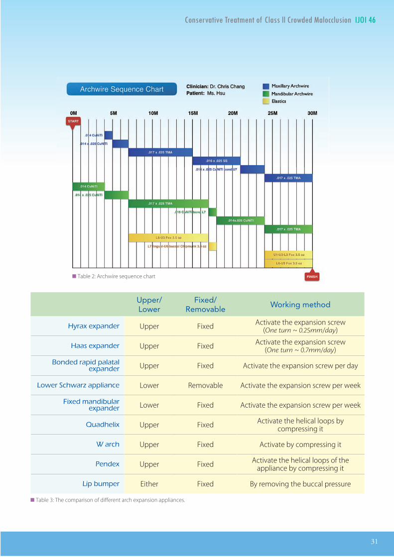
For the present patient, PSL brackets with light wires was an effective and relatively comfortable arch expansion appliance (Figs. 16-19). There are many different types of expansion appliances: Hyrax, Haas, bonded rapid palatal expander, Schwarz appliance, lingual arches, quadhelix, W arch, pendex (pendulum) appliance, lip bumper, and conventional fixed appliances with arch wires.4 The rapid palatal or maxillary expanders (RPE or RME) have long been among the most popular expansion appliances. Table 3 compares some of the most popular expansion appliances.
a id a illar ansion A liances om ared to ig t ires wit rac ets
The most common undesirable effects of rapid expansion are pain and discomfort, from the time of activation up to several days later. The size of the appliance is uncomfortable and soft tissue impingement may result in irritation and/or ulceration. The most significant longterm complication is compromised periodontal health and gingival recession.5,6 If rapid expansion is performed after the mid-palatal suture begins to fuse (~14-16 years of age), there may be a delayed risk of recession of the buccal gingival tissue in the maxillary buccal segments.5 Garib6 reported that RME exerts a high level of force (up to 20-40 lb.), reducing the buccal bone plate thickness from 0.6 to 0.9mm, and it may even result in dehiscence.
IJOI 46 AOI A E RE ORT
31
T IJOI 46
Upper/ Lower
Fixed/Removable Working method
ra e ander Upper Fixed Activate the expansion screw (One turn ~ 0.25mm/day)
aas e ander Upper Fixed Activate the expansion screw (One turn ~ 0.7mm/day)
onded ra id alatal e ander Upper Fixed Activate the expansion screw per day
ower c war a liance Lower Removable Activate the expansion screw per week
i ed mandibular e ander Lower Fixed Activate the expansion screw per week
uad eli Upper Fixed Activate the helical loops by compressing it
arc Upper Fixed Activate by compressing it
ende Upper Fixed Activate the helical loops of the appliance by compressing it
i bum er Either Fixed By removing the buccal pressure
██ Table 3: The comparison of different arch expansion appliances.
██ Table 2: Archwire sequence chart
32
IJOI 46 AOI A E RE ORT
bone screws were a viable option for preventing the mesial molar drift, that is a natural consequence of arch expansion (Fig. 9). Bone screws would help correct the crowding with less expansion, and reduce the overjet without as much tipping of the maxillary incisors. This option was not available for the present patient because her parents had rejected the use of miniscrews.
ongterm tabilit o Arc ansion
In 1969, Riedel2 reviewed stability studies of arch form without retention, concluding that changes in inter-canine and inter-molar width during orthodontic treatment tend to return to their pre-treatment position. In 1988, Sandstrom et al.12
observed that the average amount of increased lower inter-canine width was about 1.1mm and the inter-molar width was 2.9mm; these small changes result in a negligible increase in arch
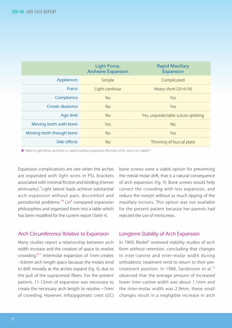
Expansion complications are rare when the arches are expanded with light wires in PSL brackets associated with minimal friction and binding (Damon
philosophy).7 Light lateral loads achieve substantial arch expansion without pain, discomfort and periodontal problems.7,8 Lin8 compared expansion philosophies and organized them into a table which has been modified for the current report (Table 4).
Arc ircum erence elati e to ansion
Many studies report a relationship between arch width increase and the creation of space to resolve crowding.9-11 Intermolar expansion of 1mm creates ~0.6mm arch length space because the molars tend to drift mesially as the arches expand (Fig. 9), due to the pull of the supracrestal fibers. For the present patient, 11-12mm of expansion was necessary to create the necessary arch length to resolve ~7mm of crowding. However, infrazygomatic crest (IZC)
Light Force, Archwire Expansion
Rapid Maxillary Expansion
A liances Simple Complicated
orce Light continue Heavy short (20-40 lb)
om liance No Yes
reate diastema No Yes
Age limit No Yes, unpredictable suture splitting
o ing teet wit bone Yes No
o ing teet t oug bone No Yes
ide e ects No Thinning of buccal plate
██ Table 4: Light force, archwire vs. rapid maxillary expansion (Revision of Dr. John Lin’s table).8
IJOI 46 AOI A E RE ORT
33
T IJOI 46
of treatment is an essential aspect of informed consent. The present patient and her parents were well pleased with both the treatment and the outcome.
Ac nowledgement
Thanks to Mr. Paul Head for proofreading this article.
e erences
1. Felton MJ, Sinclair PM, Jones DL, Alexander RG, A computerized analysis of the shape and stability of mandibular arch form. Am J Orthod Dentofacial Orthop 1987;92:478-483.
2. Riedel RA 1969 In: Graber TM (ed) Current orthodontic concepts and techniques, Saunders, Philadelphia.
3. Lee R , Kirschen R. Space planning for the dentition (space analysis). Orthodontics Principle and Pracice 2011;10:88-98.
4. Bayirli B, Riolo CS, �ornberg M, Riolo ML. Treatment ta�ics for problems related to dentofacial discrepancies in three planes of space. In: Mosby’s Orthodontic Review, eds: English J, Peltomaki T, Pham-Litschel K; 2009. Chapter 10. p.128-136.
5. Graber TM, Vanarsdall RL. Orthodontics: current principles and techniques, 2nd ed. St. Louis: Mosby Year Book; 1994. p. 719-749.
6. Garib DG, Henriques JF, Janson G, de Freitas MR, Fernandes AY. Periodontal effects of rapid maxillary expansion with tooth-tissue-borne and tooth-borne expanders: A computed tomography evaluation. Am J Orthod Dentofacial Orthop 2006;129:749-58.
7. Bagden AA. Conversation the Damon system: questions and answers. Clinical Impression 2005;14:4-13.
8. Lin JJ. Creative Orthodontics: Blending the Damon system & TADs to manage difficult malocclusion. 2nd ed. Taipei: Yong Chieh Enterprise Co, Ltd; 2010.
9. O’Higgins EA, Lee RT. How much space is created from expansion or premolar extracion? J Orthod 2000;27:11-13.
10. Adkins MD, Nanda RS, Currier GF. Arch perimeter changes on rapid palatal expansion. Am J Orthod Dentofacial Orthod 1990;97:194-199.
11. Akkaya S, Lorenzon S, Ucem T. Comparison of dental arch and arch perimeter changes between bonded rapid and slow maxillary expansion procedures. Eur J Orthod 1998;20:255-261.
length to correct crowding. Haas13 studied the long-term outcomes of RME and reported a few cases with an increase in inter-canine width of 3-4mm. The predictability of the latter result is unclear, particularly for archwire expansion.
In contrast to expansion of the mid palatal suture (RPE or RME), numerous authors7,8,15,16 have proposed that expansion with very light wires in PSL brackets results in a more physiologically determined tooth positions (Fig. 20). The present case study confirms that a major malocclusion (DI=21) can be treated to a very good functional (CRE 20) and esthetic (P&W
Esthetic Score 4) result, that is stable for at least three years, with permanent retention: fixed 3-3 in both arches and clear overlay retainers at night. However, there are no longterm studies indicating that large increases in dental arch width, achieved with light wires and PSL brackets, are stable without retention.
onclusion
A challenging malocclusion (DI 21), was treated non-invasively to an excellent alignment (CRE 20) with simple mechanics. Unfortunately there was an unfavorable (vertical) growth response and the mandible rotated posteriorly. Because of failure to grow anteriorly and develop arch length with natural expansion, it was necessary to over-expand the arches to conservatively correct the crowding. Patients may choose “simple treatment” options that produce good functional and esthetic results, but they should be informed of the potential for adverse outcomes, particularly with respect to unpredictable growth patterns and stability. Accepting the limitations for a desired course
34
IJOI 46 AOI A E RE ORT
12. Sandstrom RA, Klapper L, Papaconstantinou S Expansion of the lower arch concurrent with rapid maxillary expansion. Am J Orthod Dentofacial Orthop 1988;94:296-302.
13. Haas AJ. Long-term post-treatment evaluation of rapid palatal expansion. Angle Orthod 1980 Jul;50:189-217.
14. Pollard AP. Capturing the essence of the Damon approach. Clinical Impression 2003;12:4-11.
15. Damon D. Damon system. �e workbooks. Ormco Corporation; 2004.
16. Chang CH. Advanced Damon Course No. 1: Crowding: Ext. vs. Non-ext., Beethoven Podcast Encyclopedia in Orthodontics [podcast]. Hsinchu: Newton’s A Ltd; 2011.
IJOI 46 AOI A E RE ORT
35
T IJOI 46
OVERJET
0 mm. (edge-to-edge) = 1 pt.1 Ð 3 mm. = 0 pts.3.1 Ð 5 mm. = 2 pts.5.1 Ð 7 mm. = 3 pts.7.1 Ð 9 mm. = 4 pts.> 9 mm. = 5 pts.
Negative OJ (x-bite) 1 pt. per mm. per tooth =
OVERBITE
0 Ð 3 mm. = 0 pts.3.1 Ð 5 mm. = 2 pts.5.1 Ð 7 mm. = 3 pts.Impinging (100%) = 5 pts.
ANTERIOR OPEN BITE
0 mm. (edge-to-edge), 1 pt. per tooth
then 1 pt. per additional full mm. per tooth
LATERAL OPEN BITE
2 pts. per mm. per tooth
CROWDING (only one arch)
1 Ð 3 mm. = 1 pt.3.1 Ð 5 mm. = 2 pts.5.1 Ð 7 mm. = 4 pts.> 7 mm. = 7 pts.
OCCLUSION
Class I to end on = 0 pts.End on Class II or III = 2 pts. per side pts.
Full Class II or III = 4 pts. per side pts.
Beyond Class II or III = 1 pt. per mm. pts.pts. additional
TotalTotalT =
TotalTotalT =
TotalTotalT =
TotalTotalT =
TotalTotalT =
Total =
TOTAL D.I.D.I. SCORECORELINGUAL POSTERIOR X-BITE
1 pt. per tooth Total =
BUCCAL POSTERIOR X-BITE
2 pts. per tooth Total =
CEPHALOMETRICS (See Instructions)
ANB ≥ 6¡ or ≤ -2¡ = 4 pts.
SN-MP
≥ 38¡ = 2 pts.
Each degree > 38¡ Each degree > 38¡ x 2 pts. =x 2 pts. =
≤ 26¡ = 1 pt.
Each degree < 26¡ Each degree < 26¡ x 1 pt. =x 1 pt. =
1 to MP ≥ 99¡ = 1 pt.
Each degree > 99¡ Each degree > 99¡ x 1 pt. =x 1 pt. =
OTHER (See Instructions)
Supernumerary teeth x 1 pt. =
Ankylosis of perm. teeth x 2 pts. =
Anomalous morphology x 2 pts. =
Impaction (except 3rd molars)rd molars)rd x 2 pts. =
Midline discrepancy (≥3mm) @ 2 pts. =
Missing teeth (except 3rd molars)rd molars)rd x 1 pts. =
Missing teeth, congenital x 2 pts. =
Spacing (4 or more, per arch) x 2 pts. =
Spacing (Mx cent. diastema ≥ 2mm) @ 2 pts. =
Tooth transposition x 2 pts. =
Skeletal asymmetry (nonsurgical tx) @ 3 pts. =
Addl. treatment complexities Addl. treatment complexities x 2 pts. =x 2 pts. =
Identify:
Each degree > 6¡ x 1 pt. =
Each degree < -2¡ Each degree < -2¡ x 1 pt. =x 1 pt. =
Total =
Total =
2121
4
33
00
00
44
00
0
1010
00
= 1 pt. = 1 pt. = 1 pt. = 1 pt.
2
33
38¡ = 2 pts.38¡ = 2 pts.38¡ = 2 pts.38¡ = 2 pts.
4
33
0
i cr panc n or t
36
IJOI 46 AOI A E RE ORT
Total Score:
Case # Patient
2
2
111
11
10
6
0
11
4
5
1
Alignment/Rotations
Marginal Ridges
Buccolingual Inclination
Overjet
Occlusal Contacts
Occlusal Relationships
Interproximal Contacts
INSTRUCTIONS: Place score beside each deficient tooth and enter total score for each parameter in the white box. Mark extracted teeth with ÒXÓ. Second molars should be in occlusion.
20
Root Angulation
2
1111
11
1
1111
1 1 11
11 11
a t a io rap a ation
IJOI 46 AOI A E RE ORT
37
T IJOI 46
12 34
4
1 2
3 1
2
34
12 341
2 34
4
1 2
3 1
2
34
12 3412 34
4
1 2
3 1
2
34
12 34
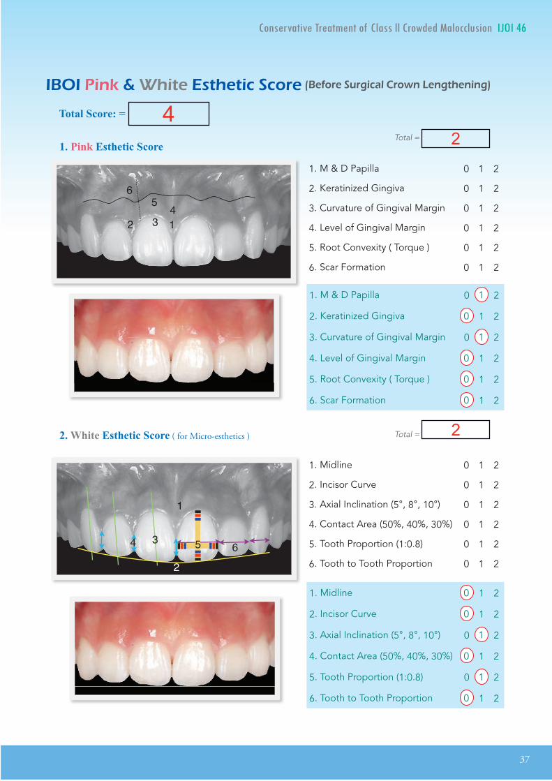
1. Pink Esthetic Score
in it t tic cor or r ica ro n n t nin
Total Score: = 4
2. White Esthetic Score ( for Micro-esthetics )
12 34
4
1 2
3 1
2
34
12 34
1. M & D Papilla 0 1 2
2. Keratinized Gingiva 0 1 2
3. Curvature of Gingival Margin 0 1 2
4. Level of Gingival Margin 0 1 2
5. Root Convexity ( Torque ) 0 1 2
6. Scar Formation 0 1 2
1. Midline 0 1 2
2. Incisor Curve 0 1 2
3. Axial Inclination (5°, 8°, 10°) 0 1 2
4. Contact Area (50%, 40%, 30%) 0 1 2
5. Tooth Proportion (1:0.8) 0 1 2
6. Tooth to Tooth Proportion 0 1 2
Total = 2
Total = 2
International Workshop
Damon
, OBS
& VISTA
Dr. Chris Chang Dr. John Lin
Day 123
work-shophands-on
lecture
OBS Damonsurgeries
Class IIImini screws chair-side observation
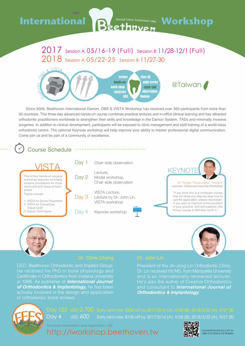
in e eetho en International Damon O I or sho has re ei ed o er arti i ants rom more than
o ntries. his three-day ad an ed hands-on o rse om ines ra ti al le t res and in-o i e lini al learning and has attra ted orthodonti ra titioners orld ide to strengthen their s ills and no ledge in the Damon ystem Ds and minimally in asi e
s rgeries. In addition to lini al de elo ment arti i ants ill e e osed to lini management and sta training o a orld- lass orthodonti entre. he o tional eynote or sho ill hel im ro e yo r a ility to master ro essional digital omm ni ation.
Come oin s and e art o a omm nity o e ellen e.
Day 4 USD 600 USD 2,700 Early bird rate: $200 off by 2017/3/16 (A), 9/28 (B); 2018/3/22 (A), 9/27 (B)
Early bird rate: $100 off by 2017/3/16 (A), 9/28 (B); 2018/3/22 (A), 9/27 (B)
http://iworkshop.beethoven.tw [email protected] more information and registration, visit
@Taiwan
05/16-19 (Full) (Full)11/28-12/1Session A: Session B:
+886-3-5735676 #218 Bella
201705/22-25 11/27-30Session A: Session B:2018
o r o o i a p a ro p.r i i i o p io o ar i i a i r o o i ro ia a i r i
i . p i r o International Journal of Orthodontics & Implantology, aa i i o i i a app i a ioo or o o i o r .
r i o i o i r o o i i i .r. i r i i . ro ar i r i
a i a i r a io a r o r r.a o a or o r a i r o o i
a o a o International Journal of Orthodontics & Implantology.
KEYNOTEVISTA Day 1
C rs h
Chair-side observation
Day 2Lecture,Model workshop, Chair-side observation
Day 3VISTA Lecture,Lecture by Dr. John Lin, VISTA workshop
Day 4 Keynote workshop
i o r a o r i aor op a r i i a
i a i pro r or i paio a o i a
.opi i
. or r a
. or o ii ra
. r i
o i i i a o p r o ra i o o p p o o
app i a io p a r o i r.o a o i pro o i a io
i o r pra i a i pa i io r o r i i i or i .
r. i a ar ai ar r a o or op
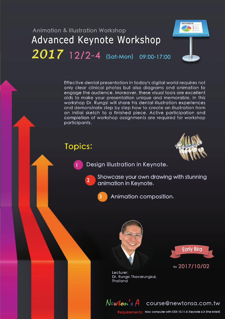
Advanced Keynote Workshop
1
2
Design illustration in Keynote.
Animation & Illustration Workshop
12/2-4 (Sat-Mon) 09:00-17:00
Topics:
Requirements:
Effective dental presentation in today’s digital world requires not only clear clinical photos but also diagrams and animation to engage the audience. Moreover, these visual tools are excellent aids to make your presentation unique and memorable. In this workshop Dr. Rungsi will share his dental illustration experiences and demonstrate step by step how to create an illustration from an initial sketch to a finished piece. Active participation and completioncompletion of workshop assignments are required for workshop participants.
Lecturer: Dr. Rungsi Thavarungkul,Thailand
by
Showcase your own drawing with stunning animation in Keynote.
3 Animation composition.
Mac computer with OSX 10.11.X; Keynote 6.X (the latest)
International Workshop
Damon
, OBS
& VISTA
Dr. Chris Chang Dr. John Lin
Day 123
work-shophands-on
lecture
OBS Damonsurgeries
Class IIImini screws chair-side observation
Since 2009, Beethoven International Damon, OBS & VISTA Workshop has received over 350 participants from more than
30 countries. This three-day advanced hands-on course combines practical lectures and in-office clinical learning and has attracted orthodontic practitioners worldwide to strengthen their skills and knowledge in the Damon System, TADs and minimally invasive
surgeries. In addition to clinical development, participants will be exposed to clinic management and staff training of a world-class orthodontic centre. The optional Keynote workshop will help improve your ability to master professional digital communication.
Come join us and be part of a community of excellence.
Day 4 USD 600 USD 2,700 Early bird rate: $200 off by 2017/3/16 (A), 9/28 (B); 2018/3/22 (A), 9/27 (B)
Early bird rate: $100 off by 2017/3/16 (A), 9/28 (B); 2018/3/22 (A), 9/27 (B)
http://iworkshop.beethoven.tw [email protected] more information and registration, visit
@Taiwan
05/16-19 (Full) (Full)11/28-12/1Session A: Session B:
+886-3-5735676 #218 Bella
201705/22-25 11/27-30Session A: Session B:2018
CEO, Beethoven Orthodontic and Implant Group. He received his PhD in bone physiology and Certificate in Orthodontics from Indiana University in 1996. As publisher of International Journal of Orthodontics & Implantology, he has been actively involved in the design and application of orthodontic bone screws.
President of the Jin-Jong Lin Orthodontic Clinic. Dr. Lin received his MS. from Marquette University and is an internationally renowned lecturer. He’s also the author of Creative Orthodontics and consultant to International Journal of Orthodontics & Implantology.
KEYNOTEVISTA Day 1
Course Schedule
Chair-side observation
Day 2Lecture,Model workshop, Chair-side observation
Day 3VISTA Lecture,Lecture by Dr. John Lin, VISTA workshop
Day 4 Keynote workshop
This 4-hour hands-on surgical workshop features minimally invasive procedures for impa-ctions and soft tissue enhanc-ement.Topics include:
1. VISTA for Screw Placement2. VISTA for Connective Tissue Graft3. Suture Techniques
“If you think this is a computer course that will show you step-by-step how to use the application, please reconsider. If you want to improve communication in your practice, and with patients, this 8-hour course is definitely worth it."
Dr. Rungsi Thavarungkul, ThailandLecturer, Advanced Keynote Workshop
Advanced Keynote Workshop
1
2
Design illustration in Keynote.
Animation & Illustration Workshop
12/2-4 (Sat-Mon) 09:00-17:00
Topics:
Requirements:
Effective dental presentation in today’s digital world requires not only clear clinical photos but also diagrams and animation to engage the audience. Moreover, these visual tools are excellent aids to make your presentation unique and memorable. In this workshop Dr. Rungsi will share his dental illustration experiences and demonstrate step by step how to create an illustration from an initial sketch to a finished piece. Active participation and completioncompletion of workshop assignments are required for workshop participants.
Lecturer: Dr. Rungsi Thavarungkul,Thailand
by
Showcase your own drawing with stunning animation in Keynote.
3 Animation composition.
Mac computer with OSX 10.11.X; Keynote 6.X (the latest)
40
IJOI 46 AOI A E RE ORT I L IJOI 46
Dr. Szu Rou Yeh,Lecturer, Beethoven Orthodontic Course (Left)
Dr. Chris Chang, Founder, Beethoven Orthodontic Center
Publisher, International Journal of Orthodontics & Implantology (Center)
Dr. W. Eugene Roberts,Editor-in-chief, International Journal of Orthodontics & Implantology (Right)
ran o ar pri tin o a ori ontapact o r anin it a an i ar
cca on cr
tract A 10yr 6m female presented with an unerupted mandibular left canine and crowding of the maxillary incisors. Cone-beam computer tomography (CBCT) revealed the unerupted cuspid was a deep transalveolar impaction, positioned lingual to the roots of the left mandibular incisors and buccal to the root of the adjacent �rst premolar. Extraction posed serious surgical risks to the mental nerve, sublingual artery, and periodontium. So a carefully sequenced treatment plan was devised to reverse the etiology of the aberrant development, and recover the cuspid by uprighting it in an oblique plane corresponding to the long axis of the impaction. Two stages of conservative surgery exposed and progressively bonded the impaction as it was uprighted. To help avoid root resorption, the adjacent lateral incisor was not bonded and engaged on the archwire. The precise mechanics to upright the cuspid in the prepared oblique plane was provided by a rectangular lever arm anchored by a mandibular buccal shelf miniscrew (OrthoBoneScrew®). This very di�cult malocclusion with a Discrepancy Index (DI) of 30 was treated to an excellent result in 36 months, as documented a Cast-Radiograph Evaluation (CRE) of 20 and Pink & White esthetic score of 2. (Int J Orthod Implantol 2017;46:40-56)
Key words:Sublingual trans-alveolar impacted cuspid, 3-D lever arm, minimally invasive surgery, progressive bracket bonding, moment to force ratio, buccal shelf screw, horizontal cuspid impaction
istor and tiolog
This 10y6m female was in good general health. The initial clinical examination revealed Class I molars and an edge to edge incisal relationship. The mandibular midline was 3mm to the left of the facial and maxillary midlines. The mandibular left canine was unerupted and there was space between the left premolars (Figs.
1-3). The apparent etiology for the impaction was an aberrant path of eruption. An innovative treatment plan was devised to reverse the aberrant development by: 1. creating an oblique space in the arch form that corresponded to the plane of the aberrant path of eruption (long axis of the impaction), and 2. uprighting the cuspid in the prepared oblique plane with mechanics designed to rotate the tooth at its apex. The patient was treated to an excellent outcome as documented in Figs. 4-6. A pre-treatment cone beam computed tomography (CBCT) documented the position of the impacted canine (Fig. 7). Panoramic and cephalometric radiographs before and after treatment are illustrated in Figs. 8 and 9, respectively. Superimposed cephalometric tracings are show in Fig. 10.
IJOI 46 AOI A E RE ORT
41
I L IJOI 46
Dr. Szu Rou Yeh,Lecturer, Beethoven Orthodontic Course (Left)
Dr. Chris Chang, Founder, Beethoven Orthodontic Center
Publisher, International Journal of Orthodontics & Implantology (Center)
Dr. W. Eugene Roberts,Editor-in-chief, International Journal of Orthodontics & Implantology (Right)
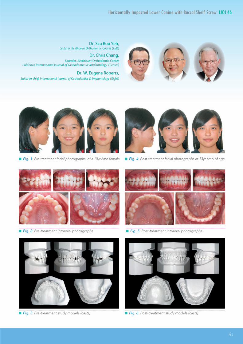
█ Fig. 2: Pre-treatment intraoral photographs
█ Fig. 1: Pre-treatment facial photographs of a 10yr 6mo female
█ Fig. 3: Pre-treatment study models (casts)
█ Fig. 4: Post-treatment facial photographs at 13yr 6mo of age
█ Fig. 5: Post-treatment intraoral photographs
█ Fig. 6: Post-treatment study models (casts)
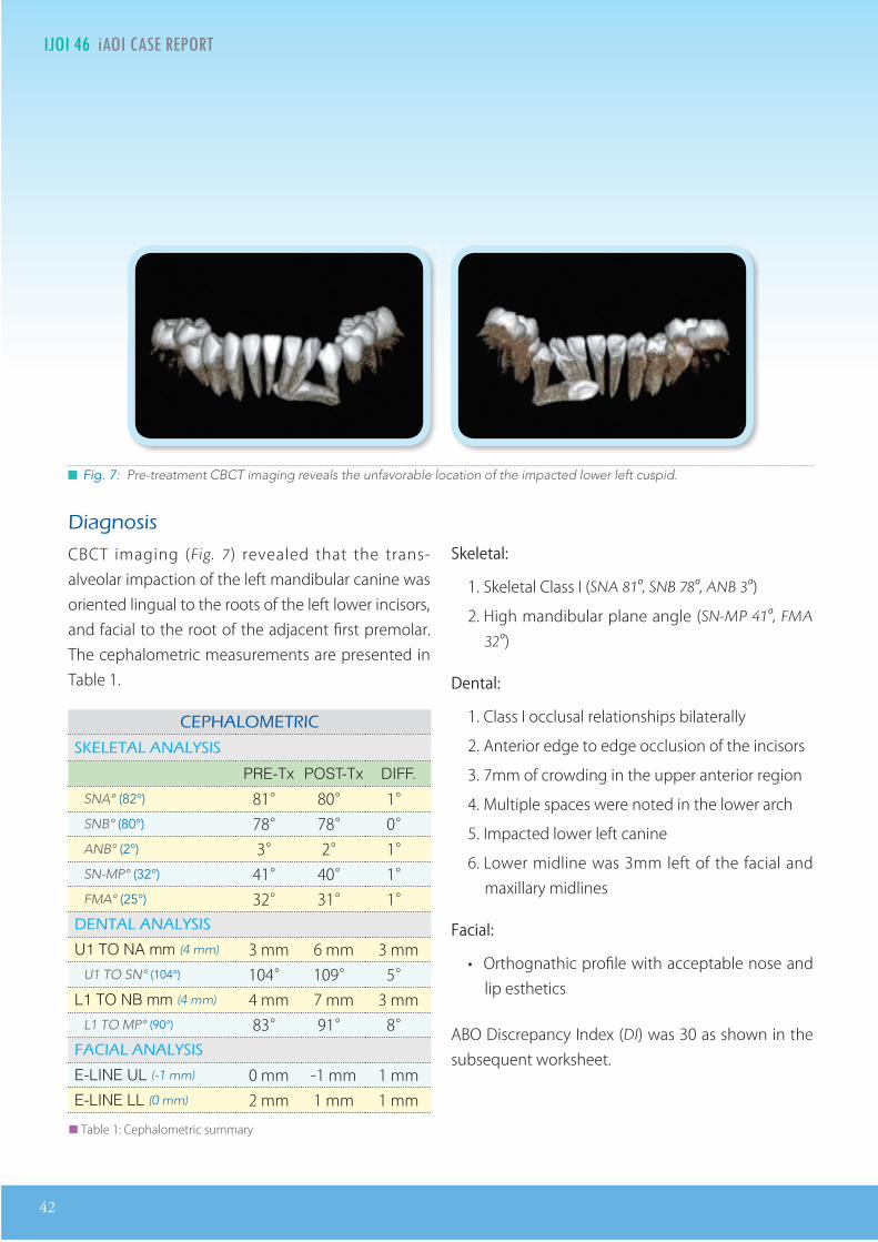
42
IJOI 46 AOI A E RE ORT I L IJOI 46
█ Fig. 7: Pre-treatment CBCT imaging reveals the unfavorable location of the impacted lower left cuspid.
iagnosis
CBCT imaging (Fig. 7) revealed that the trans-alveolar impaction of the left mandibular canine was oriented lingual to the roots of the left lower incisors, and facial to the root of the adjacent first premolar. The cephalometric measurements are presented in Table 1.
A O I
A A A I
PRE-Tx POST-Tx DIFF.
SNA° (82°) 81° 80° 1°SNB° (80°) 78° 78° 0°ANB° (2°) 3° 2° 1°SN-MP° (32°) 41° 40° 1°FMA° (25°) 32° 31° 1°
A A A I
U1 TO NA mm (4 mm) 3 mm 6 mm 3 mmU1 TO SN° (104°) 104° 109° 5°
L1 TO NB mm (4 mm) 4 mm 7 mm 3 mmL1 TO MP° (90°) 83° 91° 8°A IA A A I
E-LINE UL (-1 mm) 0 mm -1 mm 1 mmE-LINE LL (0 mm) 2 mm 1 mm 1 mm
██ Table 1: Cephalometric summary
Skeletal:
1. Skeletal Class I (SNA 81º, SNB 78º, ANB 3º)
2. High mandibular plane angle (SN-MP 41º, FMA
32º)
Dental:
1. Class I occlusal relationships bilaterally
2. Anterior edge to edge occlusion of the incisors
3. 7mm of crowding in the upper anterior region
4. Multiple spaces were noted in the lower arch
5. Impacted lower left canine
6. Lower midline was 3mm left of the facial and maxillary midlines
Facial:
• Orthognathic profile with acceptable nose and lip esthetics
ABO Discrepancy Index (DI) was 30 as shown in the subsequent worksheet.
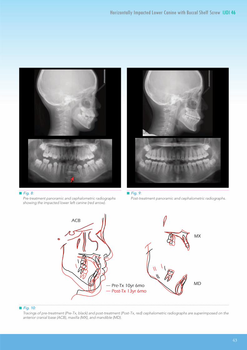
IJOI 46 AOI A E RE ORT
43
I L IJOI 46
█ Fig. 8: Pre-treatment panoramic and cephalometric radiographs showing the impacted lower left canine (red arrow).
█ Fig. 9: Post-treatment panoramic and cephalometric radiographs.
A
— re r mo — ost r mo
█ Fig. 10: Tracings of pre-treatment (Pre-Tx, black) and post-treatment (Post-Tx, red) cephalometric radiographs are superimposed on the anterior cranial base (ACB), maxilla (MX), and mandible (MD).
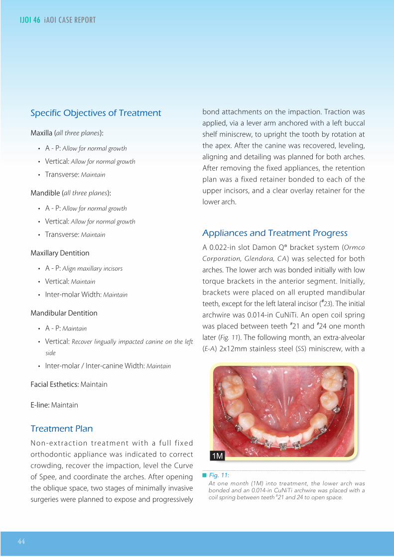
44
IJOI 46 AOI A E RE ORT I L IJOI 46
█ Fig. 11:At one month (1M) into treatment, the lower arch was bonded and an 0.014-in CuNiTi archwire was placed with a coil spring between teeth #21 and 24 to open space.
eci ic Ob ecti es o reatment
Maxilla (all three planes):
• A - P: Allow for normal growth
• Vertical: Allow for normal growth
• Transverse: Maintain
Mandible (all three planes):
• A - P: Allow for normal growth
• Vertical: Allow for normal growth
• Transverse: Maintain
Maxillary Dentition
• A - P: Align maxillary incisors
• Vertical: Maintain
• Inter-molar Width: Maintain
Mandibular Dentition
• A - P: Maintain
• Vertical: Recover lingually impacted canine on the left
side
• Inter-molar / Inter-canine Width: Maintain
Facial Esthetics: Maintain
E-line: Maintain
reatment lan
Non-extract ion t reatment with a fu l l f i xed orthodontic appliance was indicated to correct crowding, recover the impaction, level the Curve of Spee, and coordinate the arches. After opening the oblique space, two stages of minimally invasive surgeries were planned to expose and progressively
1M
bond attachments on the impaction. Traction was applied, via a lever arm anchored with a left buccal shelf miniscrew, to upright the tooth by rotation at the apex. After the canine was recovered, leveling, aligning and detailing was planned for both arches. After removing the fixed appliances, the retention plan was a fixed retainer bonded to each of the upper incisors, and a clear overlay retainer for the lower arch.
A liances and reatment rogress
A 0.022-in slot Damon Q® bracket system (Ormco
Corporation, Glendora, CA) was selected for both arches. The lower arch was bonded initially with low torque brackets in the anterior segment. Initially, brackets were placed on all erupted mandibular teeth, except for the left lateral incisor (#23). The initial archwire was 0.014-in CuNiTi. An open coil spring was placed between teeth #21 and #24 one month later (Fig. 11). The following month, an extra-alveolar (E-A) 2x12mm stainless steel (SS) miniscrew, with a

IJOI 46 AOI A E RE ORT
45
I L IJOI 46
█ Fig. 12: A 2x12mm OBS with a rectangular hole through the head was inserted in the mandibular left buccal shelf, and a power chain from the OBS was used to retract tooth #21 along the archwire. Note that the lateral incisor (#23) was not bonded and engaged on the archwire, so that it can act as a “free body” to avoid root resorption, as the arch is aligned and space is opened.
0.019x0.025-in auxiliary hole (OrthoBoneScrew® (OBS) Newton's A Ltd., Hsinchu City, Taiwan), was inserted in the mandibular buccal shelf, lateral to the mesial root of the lower left first molar (#19). An elastomeric power chain, anchored by the OBS, was used to retract tooth #21, to help open space for the impaction (Fig. 12).
Three months later, the upper dentition (except tooth #10) was bonded with low torque brackets on the anterior teeth. An 0.014-in CuNiTi wire was engaged with an open coil spring between teeth #9 to 11 to open space, and tooth #10, was bonded with a low torque bracket one month later. At the following appointment an 0.014x0.025-in CuNiTi was placed, and in the sixth
1M
█ Fig. 13: In the seventh month (7M), CBCT images from the labial (a) and lingual (b) show the 3D morphology. A 7M panoramic radiograph (c) and eighth month (8M) intraoral photograph (d) show that there is adequate arch length to upright the tooth, but no information is provided on the 3D relationship of tooth roots to the impaction, mental foramen or sublingual area. The CBCT scans (a and b) show that an oblique transalveolar space has been created that corresponds to the long axis of the impaction. NOTE: red arrows mark the structures that present the greatest surgical risk: the mental foramen position (a) and sublingual foramen (b).
7M
7M M
7M
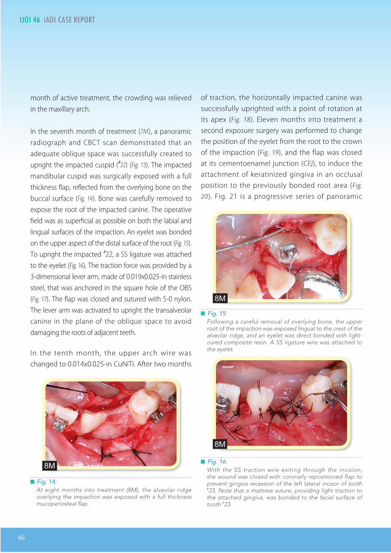
46
IJOI 46 AOI A E RE ORT I L IJOI 46
█ Fig. 16: With the SS traction wire exiting through the incision, the wound was closed with coronally repositioned flap to prevent gingiva recession of the left lateral incisor of tooth #23. Note that a mattress suture, providing light traction to the attached gingiva, was bonded to the facial surface of tooth #23.
█ Fig. 14: At eight months into treatment (8M), the alveolar ridge overlying the impaction was exposed with a full thickness mucoperiosteal flap.
M
█ Fig. 15: Following a careful removal of overlying bone, the upper root of the impaction was exposed lingual to the crest of the alveolar ridge, and an eyelet was direct bonded with light-cured composite resin. A SS ligature wire was attached to the eyelet.
M
M
month of active treatment, the crowding was relieved in the maxillary arch.
In the seventh month of treatment (7M), a panoramic radiograph and CBCT scan demonstrated that an adequate oblique space was successfully created to upright the impacted cuspid (#22) (Fig. 13). The impacted mandibular cuspid was surgically exposed with a full thickness flap, reflected from the overlying bone on the buccal surface (Fig. 14). Bone was carefully removed to expose the root of the impacted canine. The operative field was as superficial as possible on both the labial and lingual surfaces of the impaction. An eyelet was bonded on the upper aspect of the distal surface of the root (Fig. 15). To upright the impacted #22, a SS ligature was attached to the eyelet (Fig. 16). The traction force was provided by a 3-dimensional lever arm, made of 0.019x0.025-in stainless steel, that was anchored in the square hole of the OBS (Fig. 17). The flap was closed and sutured with 5-0 nylon. The lever arm was activated to upright the transalveolar canine in the plane of the oblique space to avoid damaging the roots of adjacent teeth.
In the tenth month, the upper arch wire was changed to 0.014x0.025-in CuNiTi. After two months
of traction, the horizontally impacted canine was successfully uprighted with a point of rotation at its apex (Fig. 18). Eleven months into treatment a second exposure surgery was performed to change the position of the eyelet from the root to the crown of the impaction (Fig. 19), and the flap was closed at its cementoenamel junction (CEJ), to induce the attachment of keratinized gingiva in an occlusal position to the previously bonded root area (Fig.
20). Fig. 21 is a progressive series of panoramic
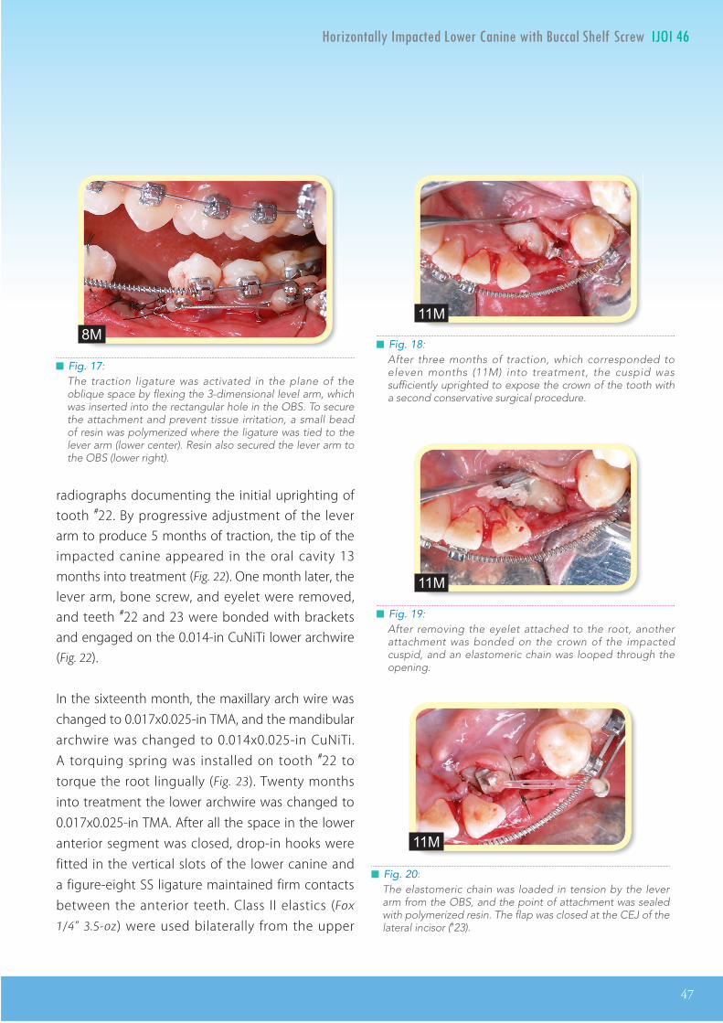
IJOI 46 AOI A E RE ORT
47
I L IJOI 46
█ Fig. 17: The traction ligature was activated in the plane of the oblique space by flexing the 3-dimensional level arm, which was inserted into the rectangular hole in the OBS. To secure the attachment and prevent tissue irritation, a small bead of resin was polymerized where the ligature was tied to the lever arm (lower center). Resin also secured the lever arm to the OBS (lower right).
M █ Fig. 18: After three months of traction, which corresponded to eleven months (11M) into treatment, the cuspid was sufficiently uprighted to expose the crown of the tooth with a second conservative surgical procedure.
11M
█ Fig. 19: After removing the eyelet attached to the root, another attachment was bonded on the crown of the impacted cuspid, and an elastomeric chain was looped through the opening.
11M
█ Fig. 20: The elastomeric chain was loaded in tension by the lever arm from the OBS, and the point of attachment was sealed with polymerized resin. The flap was closed at the CEJ of the lateral incisor (#23).
11M
radiographs documenting the initial uprighting of tooth #22. By progressive adjustment of the lever arm to produce 5 months of traction, the tip of the impacted canine appeared in the oral cavity 13 months into treatment (Fig. 22). One month later, the lever arm, bone screw, and eyelet were removed, and teeth #22 and 23 were bonded with brackets and engaged on the 0.014-in CuNiTi lower archwire (Fig. 22).
In the sixteenth month, the maxillary arch wire was changed to 0.017x0.025-in TMA, and the mandibular archwire was changed to 0.014x0.025-in CuNiTi. A torquing spring was installed on tooth #22 to torque the root lingually (Fig. 23). Twenty months into treatment the lower archwire was changed to 0.017x0.025-in TMA. After all the space in the lower anterior segment was closed, drop-in hooks were fitted in the vertical slots of the lower canine and a figure-eight SS ligature maintained firm contacts between the anterior teeth. Class II elastics (Fox
1/4” 3.5-oz) were used bilaterally from the upper
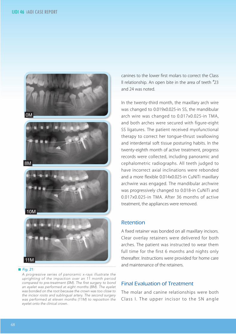
48
IJOI 46 AOI A E RE ORT I L IJOI 46
█ Fig. 21: A progressive series of panoramic x-rays illustrate the uprighting of the impaction over an 11 month period compared to pre-treatment (0M). The first surgery to bond an eyelet was performed at eight months (8M). The eyelet was bonded on the root because the crown was too close to the incisor roots and sublingual artery. The second surgery was performed at eleven months (11M) to reposition the eyelet onto the clinical crown.
canines to the lower first molars to correct the Class ll relationship. An open bite in the area of teeth #23 and 24 was noted.
In the twenty-third month, the maxillary arch wire was changed to 0.019x0.025-in SS, the mandibular arch wire was changed to 0.017x0.025-in TMA, and both arches were secured with figure-eight SS ligatures. The patient received myofunctional therapy to correct her tongue-thrust swallowing and interdental soft tissue posturing habits. In the twenty-eighth month of active treatment, progress records were collected, including panoramic and cephalometric radiographs. All teeth judged to have incorrect axial inclinations were rebonded and a more flexible 0.014x0.025-in CuNiTi maxillary archwire was engaged. The mandibular archwire was progressively changed to 0.018-in CuNiTi and 0.017x0.025-in TMA. After 36 months of active treatment, the appliances were removed.
etention
A fixed retainer was bonded on all maxillary incisors. Clear overlay retainers were delivered for both arches. The patient was instructed to wear them full time for the first 6 months and nights only thereafter. Instructions were provided for home care and maintenance of the retainers.
inal aluation o reatment
The molar and canine relationships were both C lass I . The upper inc i sor to the SN angle
0M
M
10M
11M
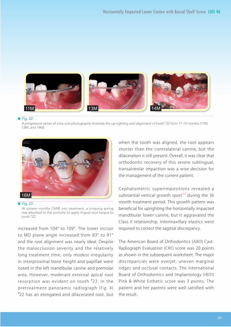
IJOI 46 AOI A E RE ORT
49
I L IJOI 46
█ Fig. 22: A progressive series of intra-oral photographs illustrate the up-righting and alignment of tooth #22 from 11-14 months (11M, 13M, and 14M).
█ Fig. 23: At sixteen months (16M) into treatment, a torquing spring was attached to the archwire to apply lingual root torque to tooth #22.
increased from 104° to 109°. The lower incisor to MD plane angle increased from 83° to 91° and the root alignment was nearly ideal. Despite the malocclusion severity and the relatively long treatment time, only modest irregularity in interproximal bone height and papillae were noted in the left mandibular canine and premolar area. However, moderate external apical root resorption was evident on tooth #22. In the pretreatment panoramic radiograph (Fig . 8 ) #22 has an elongated and dilacerated root, but
when the tooth was aligned, the root appears shorter than the contralateral canine, but the dilaceration is still present. Overall, it was clear that orthodontic recovery of this severe sublingual, transalveolar impaction was a wise decision for the management of the current patient.
Cephalometric superimposit ions revealed a substantial vertical growth spurt1-3 during the 36 month treatment period. This growth pattern was beneficial for uprighting the horizontally impacted mandibular lower canine, but it aggravated the Class II relationship. Intermaxillary elastics were required to correct the sagittal discrepancy.
The American Board of Orthodontics (ABO) Cast-Radiograph Evaluation (CRE) score was 20 points as shown in the subsequent worksheet. The major discrepancies were overjet, uneven marginal ridges and occlusal contacts. The International Board of Orthodontics and Implantology (IBOI) Pink & White Esthetic score was 3 points. The patient and her parents were well satisfied with the result.
11M 13M 14M
16M
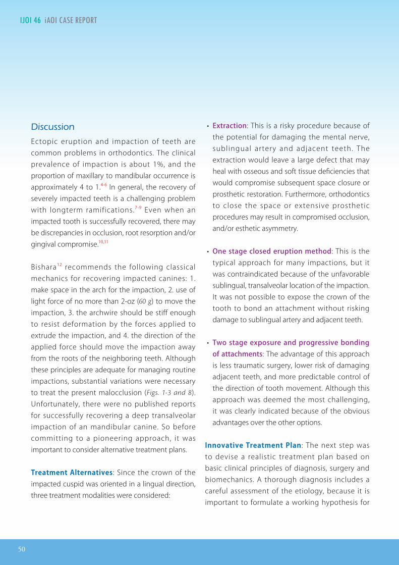
50
IJOI 46 AOI A E RE ORT I L IJOI 46
iscussion
Ectopic eruption and impaction of teeth are common problems in orthodontics. The clinical prevalence of impaction is about 1%, and the proportion of maxillary to mandibular occurrence is approximately 4 to 1.4-6 In general, the recovery of severely impacted teeth is a challenging problem with longterm ramifications.7-9 Even when an impacted tooth is successfully recovered, there may be discrepancies in occlusion, root resorption and/or gingival compromise.10,11
Bishara12 recommends the following classical mechanics for recovering impacted canines: 1. make space in the arch for the impaction, 2. use of light force of no more than 2-oz (60 g) to move the impaction, 3. the archwire should be stiff enough to resist deformation by the forces applied to extrude the impaction, and 4. the direction of the applied force should move the impaction away from the roots of the neighboring teeth. Although these principles are adequate for managing routine impactions, substantial variations were necessary to treat the present malocclusion (Figs. 1-3 and 8). Unfortunately, there were no published reports for successfully recovering a deep transalveolar impaction of an mandibular canine. So before committing to a pioneering approach, it was important to consider alternative treatment plans.
TreatmentAlternatives: Since the crown of the impacted cuspid was oriented in a lingual direction, three treatment modalities were considered:
• Extraction: This is a risky procedure because of the potential for damaging the mental nerve, subl ingual artery and adjacent teeth. The extraction would leave a large defect that may heal with osseous and soft tissue deficiencies that would compromise subsequent space closure or prosthetic restoration. Furthermore, orthodontics to close the space or extensive prosthetic procedures may result in compromised occlusion, and/or esthetic asymmetry.
• One stage closed eruption method: This is the typical approach for many impactions, but it was contraindicated because of the unfavorable sublingual, transalveolar location of the impaction. It was not possible to expose the crown of the tooth to bond an attachment without risking damage to sublingual artery and adjacent teeth.
• Two stage exposure and progressive bonding of attachments: The advantage of this approach is less traumatic surgery, lower risk of damaging adjacent teeth, and more predictable control of the direction of tooth movement. Although this approach was deemed the most challenging, it was clearly indicated because of the obvious advantages over the other options.
InnovativeTreatmentPlan : The next step was to devise a realistic treatment plan based on basic clinical principles of diagnosis, surgery and biomechanics. A thorough diagnosis includes a careful assessment of the etiology, because it is important to formulate a working hypothesis for
IJOI 46 AOI A E RE ORT
51
I L IJOI 46
the aberrant development. The most predictable treatment plan may be to reverse the aberrant developmental process. Four key points were proposed for achieving an optimal correction of the malocclusion without assuming unnecessary surgical risk or damaging the adjacent teeth: 1. do not bond a bracket on any teeth with roots near the impaction, 2. create a plane for uprighting the impaction, 3. perform surgery as conservatively as possible, and 4. use precise mechanics to upright the tooth in the prepared plane.13 Each of these four steps requires a detailed discussion.
AvoidBondingAdjacentTeeth: Not bonding the incisor adjacent to the impaction (#23) prior to uprighting the cuspid allowed it to act as a free body. This was a very important step for several reasons: 1. leveling #23 with the initial archwire would torque its root into the impaction, probably resulting in severe root resorption, 2. moving the root of #23 over the impaction would have blocked the oblique plane for uprighting #22, and 3. attempting to extrude the impaction with no clear path for uprighting it would probably damage the roots of multiple teeth.
CreateanUprightingPlane: For the present patient (Figs. 1-3 and 8), the critical presurgical step was to open an oblique space in the arch that corresponds to the desired plane for uprighting the cuspid (Fig.
13). The next consideration was the least invasive surgery for bonding the initial attachment on the impaction. Several factors are important in this decision: 1. proximity of surgical risks such as the mental nerve, sublingual artery and tooth roots,
2. biomechanics for uprighting the impaction in the prepared oblique plane, and 3. trauma to periodontal tissue. Since the crown of the canine was oriented away from the arch form, the goal was to determine an accessible, superficial area on the impaction for bonding the attachment. It was critical for the attachment to be within the oblique plane prepared for the uprighting. The CBCT image (Fig, 7) was carefully analyzed to select a convenient point of attachment occlusal to the center of resistance (Cr). The latter is typically about a third of the distance down the root in an apical direction. If the load to upright the impaction is a single force, such as a spring activating a SS ligature, the actual center of rotation (Crot) is apical to the Cr. If the eyelet attachment on the tooth is occlusal to the Cr of the tooth, and within the plane of uprighting, the application of a single force is reasonably predictable for uprighting the tooth in the desired oblique plane. For the present patient the optimal area for bonding the attachment was the upper root area in the plane for impaction uprighting (Fig. 15).
Two-StageSurgicalProcedure: Critical surgical and biomechanics considerations dictate appropriate 3D imaging prior to init iat ing treatment on impactions that are risks to adjacent teeth. CBCT is a relatively low dose method for determining the 3D morphology. For the current patient, CBCT imaging showed that the impacted cuspid was located below the roots of the adjacent incisors and and first premolar (Figs. 7 and 12).
The initial surgical exposure required location and
52
IJOI 46 AOI A E RE ORT I L IJOI 46
protection of the mental nerve and sublingual area.14 Damage to the mental nerve may result in permanent facial paresthesia and bleeding from the sublingual artery can cause swelling of the floor of the mouth and tongue. The latter can lead to respiratory obstruction, or a life-threatening hemorrhage.15
After the impaction was exposed and bonded, the direction of traction was selected. All bone in the path of enamel (crown of the impaction) was removed within the plane of uprighting. The bone covering the root of the impaction should be retained unless it is essential to bond an attachment in the upper root area (Fig. 15). The bonding procedure on the root can result in a loss of attachment, unless the attached gingiva is carefully approximated at the CEJ of the cuspid, after the root attachment is removed (Fig. 20).
PreciseBiomechanics: Typically, there are three sources of anchorage: 1. the main archwire, 2. other teeth, and 3. miniscrews. When attempting to recover an impaction that is near the roots of other teeth (Fig. 7), it is important to first open an appropriate space for the uprighting (Fig. 13) and then carefully consider the moment(s) that will be generated by that line of traction force. If not carefully controlled, the rotation of the impaction as it extrudes can cause severe damage to adjacent teeth.
The line of force from the attachment (eyelet) to the activated lever arm determines the path of tooth movement. If the force is applied along the long axis
of the impaction (within the prepared oblique plane), it will tend to upright the canine by rotating it near its root apex because of the resistance of the root to intrusion (Fig. 21). The rotation of the impaction as it extrudes is determined by the applied moment(s) generated in al l three planes of space. The magnitude, direction and plane of the moment(s) depends on the distance from the line of force to the center of resistance (Cr) of the portion of the tooth that is embedded in bone. If the force is not applied in the uprighting plane, moments may be generated that will rotate the impaction as it uprights and cause severe damage to the roots of adjacent teeth.
The 3D lever arm was a 0.019x0.025-in SS wire inserted in the rectangular hole in the head of the OBS. The firm fit of the wire in the OBS provided a stable base for applying the desired force system at any point in the extrusion and alignment of the impacted cuspid. For the present application, the lever arm was used as a spring, carefully configured to activate in the uprighting plane. The 3-D lever arm can either be used as a spring or directly engaged in the bracket.16 Once the cuspid erupted, the lever arm and OBS were removed, and a low torque bracket was bonded in an ideal position on the crown for final alignment with routine archwires (Fig. 22). The final correction was lingual root torque delivered with a torquing auxiliary (Fig. 23).
Treating impacted teeth requires an interdisciplinary approach that invo lves var ious aspects o f periodontal management. Hence, patients with impacted teeth usually require a longer period of treatment and may have a compromised result if
IJOI 46 AOI A E RE ORT
53
I L IJOI 46
their soft tissue is not carefully managed. There were two surgical keys to the successful management of the present impaction: 1. two stages of minimally invasive procedures to bond attachments, and 2. avoiding vertical incisions that might cut the sublingual artery and/or mental nerve.
onclusion
Horizontal oblique (trans-alveolar) lower cuspid impactions are very difficult to manage because of their proximity to the mental nerve, sub-lingual artery, and roots of adjacent teeth. Surgical removal of the tooth is a risky surgical procedure, that may produce periodontal tissue deficiencies which can limit space closure and restorative options. Orthodontic recovery of transalveolar impactions is an attractive option, but very challenging. The present case report describes the diagnosis and innovative treatment sequence for managing a very difficult impaction to a near ideal result.
e erences
1. Anderson DL, �ompson GW, Popovich F. Interrelationship of dental maturity, skeletal maturity, height and weight from age 4 to 14 years. Growth 1975;39:453-462.
2. Sisk CL, Foster DL. The neural basis of puberty and adolescence. Nature Neurosci 2004;7:1040-1047.
3. Agronin KJ, Kokich VG. Displacement of the glenoid fossa: acephalo-metric evaluation of growth during treatment. Am J Orthod Dentofacial Orthop 1987;91:42-48.
4. Grover PS, Lorton L. �e incidence of unerupted permanent teeth and related clinical cases. Oral Surg Oral Med Oral Pathol 1985;59:420–425.
5. Kramer RM, Williams AC. �e incidence of impa�ed teeth. A survey at Harlem hospital. Oral Surg Oral Med Oral Pathol 1970;29:237–241.
6. Dachi SF, Howell FV. A survey of 3,874 routine full-mouth radiographs: II. A study of impa�ed teeth. Oral Surg Oral Med Oral Pathol 1961;14:1165–1169.
7. Leite HR, Oliviera GS, Bri�o HH. Labially displaced ectopically erupting maxillary permanent canine: interceptive treatment and long-term results. Am J Orthod Dentofacial Orthop 2005;128:241-51.
8. Kokich VG. Surgical and orthodontic management of impa�ed maxillary canine. Am J Orthod Dentofacial Orthop 2004;126:278-83.
9. Nagaraj K, Upadhyay M, Yadav S. Impa�ed maxillary central incisor, canine, and second molar with 2 supernumerary teeth and an odontoma. Am J Orthod Dentofacial Orthop 2009;135:390-9.
10. Crescini A, Clauser C, George�i R, Corellini P, Pini Prato GP. Tunnel traction of infraosseous impacted maxillary canines. A three-year periodontal follow-up. Am J Orthod Dentofacial Orthop 1994;105:61-72.
11. Frank CA, Long M. Periodontal concerns. Periodontal concerns associated with the orthodontic treatment of impa�ed teeth. Am J Orthod Dentofacial Orthop 2002;121:639-649.
12. Bishara SE. Clinical management of impacted maxillary canines. Semin Orthod 1998 Jun;4(2):87-98.
13. Chang CH, Roberts WE. Orthodontics Vol. l l [E-reader version]. Hsinchu: Newton’s A Ltd; 2012. Available from: https ://itunes. apple .com/book/orthodontics-vol . -2/id520504691?mt=11.
14. McDonnell D, Reza Nouri M, Todd ME. The mandibular lingual foramen: a consistent arterial foramen in the middle of the mandible. Journal of Anatomy 1994;184:363-9.
15. Misch CL. Contemporary Implant Dentistry 3rd ed. St. Louis: Mosby Elsevier. p. 713-715.
16. Chang CH, Roberts WE. Orthodontics Vol. l [E-reader version]. Hsinchu: Newton’s A Ltd; 2012. Available from: h�ps://itunes.apple.com/book/orthodontics-vol.-i/id520098562?mt=11.
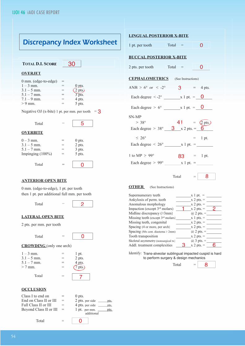
54
IJOI 46 AOI A E RE ORT I L IJOI 46
OVERJET
0 mm. (edge-to-edge) = 1 pt.1 Ð 3 mm. = 0 pts.3.1 Ð 5 mm. = 2 pts.5.1 Ð 7 mm. = 3 pts.7.1 Ð 9 mm. = 4 pts.> 9 mm. = 5 pts.> 9 mm. = 5 pts.
Negative OJ (x-bite) 1 pt. per mm. per tooth =
OVERBITE
0 Ð 3 mm. = 0 pts.3.1 Ð 5 mm. = 2 pts.5.1 Ð 7 mm. = 3 pts.Impinging (100%) = 5 pts.
ANTERIOR OPEN BITE
0 mm. (edge-to-edge), 1 pt. per tooth
then 1 pt. per additional full mm. per tooth
LATERAL OPEN BITE
2 pts. per mm. per tooth
CROWDING (only one arch)
1 Ð 3 mm. = 1 pt.3.1 Ð 5 mm. = 2 pts.5.1 Ð 7 mm. = 4 pts.> 7 mm. = 7 pts.
OCCLUSION
Class I to end on = 0 pts.End on Class II or III = 2 pts. per side pts.
Full Class II or III = 4 pts. per side pts.
Beyond Class II or III = 1 pt. per mm. pts.pts. additional
Total =
Total =
Total =
Total =
Total =
Total =
TOTAL D.I.D.I. SCORECORECORECORECORE
LINGUAL POSTERIOR X-BITE
1 pt. per tooth Total =
BUCCAL POSTERIOR X-BITE
2 pts. per tooth Total =
CEPHALOMETRICS (See Instructions)
ANB ≥ 6¡ or ≤ -2¡ = 4 pts.
SN-MP
≥ 38¡ = 2 pts.
Each degree > 38¡ Each degree > 38¡ x 2 pts. =x 2 pts. =
≤ 26¡ = 1 pt.
Each degree < 26¡ Each degree < 26¡ x 1 pt. =x 1 pt. =
1 to MP ≥ 99¡ = 1 pt.
Each degree > 99¡ Each degree > 99¡ x 1 pt. =x 1 pt. =
OTHER (See Instructions)
Supernumerary teeth x 1 pt. =
Ankylosis of perm. teeth x 2 pts. =
Anomalous morphology x 2 pts. =
Impaction (except 3rd molars)rd molars)rd x 2 pts. =
Midline discrepancy (≥3mm) @ 2 pts. =
Missing teeth (except 3rd molars)rd molars)rd x 1 pts. =
Missing teeth, congenital x 2 pts. =
Spacing (4 or more, per arch) x 2 pts. =
Spacing (Mx cent. diastema ≥ 2mm) @ 2 pts. =
Tooth transposition x 2 pts. =
Skeletal asymmetry (nonsurgical tx) @ 3 pts. =
Addl. treatment complexities Addl. treatment complexities x 2 pts. =x 2 pts. =
Identify:
Each degree > 6¡ Each degree > 6¡ x 1 pt. =x 1 pt. =
Each degree < -2¡ Each degree < -2¡ x 1 pt. =x 1 pt. =
Total =
Total =
3030
55
00
2
0
0
0
0
8
8
2
00 0
00
1
33 66
7
3 3 6 6
Trans-alveolar sublingual impacted cuspid is hard to perform surgery & design mechanics.
3
3 -2¡ = 4 pts.3 -2¡ = 4 pts.
4138¡ = 2 pts.4138¡ = 2 pts.
83 83
i cr panc n or t

IJOI 46 AOI A E RE ORT
55
I L IJOI 46
Total Score:
Case # Patient
1
1 1 1
11
40
3
3
1
0
Alignment/Rotations
Marginal Ridges
Buccolingual Inclination
Overjet
Occlusal Contacts
Occlusal Relationships
Interproximal Contacts
INSTRUCTIONS: Place score beside each deficient tooth and enter total score for each parameter in the white box. Mark extracted teeth with ÒXÓ. Second molars should be in occlusion.
20
Root Angulation
5
211
1
4
1
1
11
2 2
11111 11 1
11
a t a io rap a ation
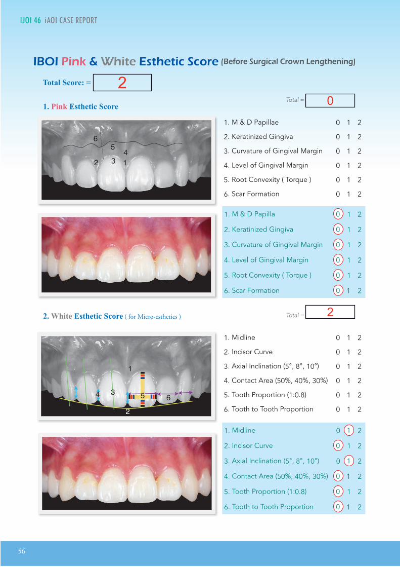
56
IJOI 46 AOI A E RE ORT
12 35 4
4
1 2
3
5
1
2
34 6
12 34
56
12 35 4
4
1 2
3
5
1
2
34 6
12 34
56 12 3
5 4
4
1 2
3
5
1
2
34 6
12 34
56
1. Pink Esthetic Score
in it t tic cor or r ica ro n n t nin
Total Score: = 2
2. White Esthetic Score ( for Micro-esthetics )
12 35 4
4
1 2
3
5
1
2
34 6
12 34
56
1. M & D Papillae 0 1 2
2. Keratinized Gingiva 0 1 2
3. Curvature of Gingival Margin 0 1 2
4. Level of Gingival Margin 0 1 2
5. Root Convexity ( Torque ) 0 1 2
6. Scar Formation 0 1 2
1. Midline 0 1 2
2. Incisor Curve 0 1 2
3. Axial Inclination (5°, 8°, 10°) 0 1 2
4. Contact Area (50%, 40%, 30%) 0 1 2
5. Tooth Proportion (1:0.8) 0 1 2
6. Tooth to Tooth Proportion 0 1 2
1. M & D Papilla 0 1 2
2. Keratinized Gingiva 0 1 2
3. Curvature of Gingival Margin 0 1 2
4. Level of Gingival Margin 0 1 2
5. Root Convexity ( Torque ) 0 1 2
6. Scar Formation 0 1 2
1. Midline 0 1 2
2. Incisor Curve 0 1 2
3. Axial Inclination (5°, 8°, 10°) 0 1 2
4. Contact Area (50%, 40%, 30%) 0 1 2
5. Tooth Proportion (1:0.8) 0 1 2
6. Tooth to Tooth Proportion 0 1 2
Total = 0
Total = 2
本程式相容于iPad、iPhone 和 iPod touchBDE
i o o a. o .p or o o r . op or o o r . o
*部分课程,包含基础正畸与进阶正畸,为每年度制作新版本,凡购买皆安装该年度之最新版本, 若要加购其他年度,可享半价优惠。
课程系列
本程式相容于本程式相容于本程式相容于iPad、iPhoneiPad、iPhoneiPad、iPhone和 iPod touch和 iPod touchBDEBDE
丰富的学习体验
课程
程课程 系列 课
系课程
课
正畸完工(7年度)
诊所行销-正畸 诊所行销-种植 诊所行销-儿牙诊所行销-助理训练
进阶正畸基础正畸 正畸支抗钉
中文演讲精选
正畸 种植 儿牙
植牙论坛(6年度)
* 有声版。
免 费 取 得
o a

拉勾
不锈钢口镜
Bite Turbo 2.0
2.0
医疗产品
拉勾
不锈钢口镜
Bite Turbo 2.0
2.0
医疗产品拉勾
不锈钢口镜
Bite Turbo 2.0
2.0
医疗产品
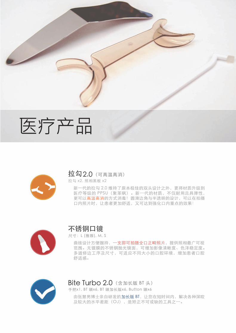
拉勾
不锈钢口镜
Bite Turbo 2.0
2.0
医疗产品
拉勾
不锈钢口镜
Bite Turbo 2.0
2.0
医疗产品拉勾
不锈钢口镜
Bite Turbo 2.0
2.0
医疗产品
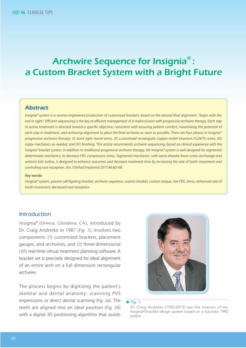
60
IJOI 46 LI I AL TI A I IJOI 46
█ Fig. 1: Dr. Craig Andreiko (1950-2013) was the inventor of the Insignia® bracket design system based on a futuristic 1992 patent.
Dr. Angle Lee,Director, Bell Dental Clinic, Taiwan
Editor, International Journal of Orthodontics & Implantology (Left)
Dr. Chris Chang, Founder, Beethoven Orthodontic Center
Publisher, International Journal of Orthodontics & Implantology (Center)
Dr. W. Eugene Roberts,Editor-in-chief, International Journal of Orthodontics & Implantology (Right)
rc ir nc or n i niaa to rac t t it a ri t t r
tract Insignia® system is a reverse-engineered production of customized brackets, based on the desired �nal alignment: “begin with the end in sight.” E�cient sequencing is the key to e�cient management of a malocclusion with progressive archwire therapy. Each step in active treatment is directed toward a specific objective, consistent with ensuring patient comfort, maximizing the potential of each step in treatment, and achieving alignment to place the �nal archwire as soon as possible. There are four phases in Insignia®
progressive archwire therapy: (I) stock light round wires, (II) customized rectangular copper-nickel-titanium (CuNiTi) wires, (III) major mechanics as needed, and (IV) �nishing. This article recommends archwire sequencing, based on clinical experience with the Insignia® bracket system. In addition to traditional progressive archwire therapy, the Insignia® system is well designed for segmental determinate mechanics, to decrease PDL compressive stress. Segmental mechanics, with extra-alveolar bone screw anchorage and anterior bite turbos, is designed to enhance outcomes and decrease treatment time by increasing the rate of tooth movement and controlling root resorption. (Int J Orthod Implantol 2017;46:60-69)
Key words:Insignia® system, passive self-ligating bracket, archwire sequence, custom bracket, custom torque, low PDL stress, enhanced rate of tooth movement, decreased root resorption
Introduction
Insignia® (Ormco, Glendora, CA), introduced by Dr. Craig Andreiko in 1987 (Fig. 1), involves two components: (1) customized brackets, placement gauges, and archwires, and (2) three-dimensional (3D) real-time virtual treatment planning software. A bracket set is precisely designed for ideal alignment of an entire arch on a full dimension rectangular archwire.
The process begins by digitizing the patient's skeletal and dental anatomy : scanning PVS impressions or direct dental scanning (Fig. 2a). The teeth are aligned into an ideal position (Fig. 2b) with a digital 3D positioning algorithm that assists
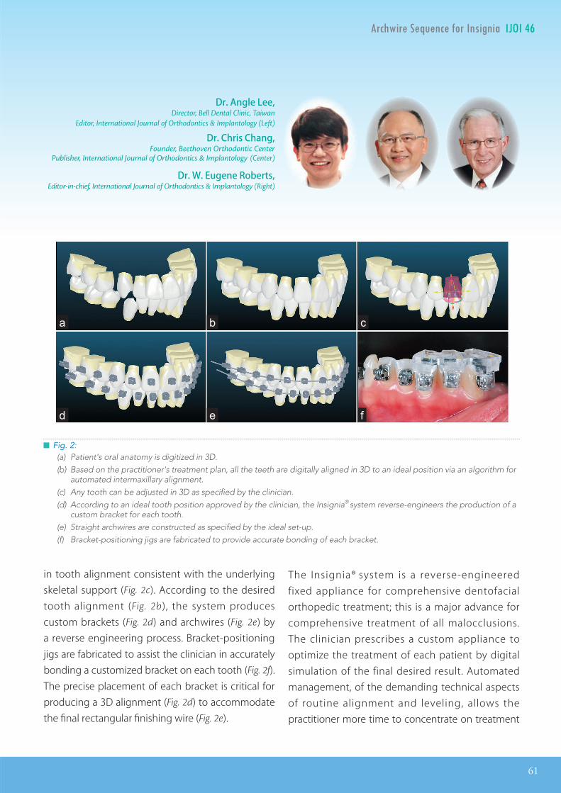
IJOI 46 LI I AL TI
61
A I IJOI 46
Dr. Angle Lee,Director, Bell Dental Clinic, Taiwan
Editor, International Journal of Orthodontics & Implantology (Left)
Dr. Chris Chang, Founder, Beethoven Orthodontic Center
Publisher, International Journal of Orthodontics & Implantology (Center)
Dr. W. Eugene Roberts,Editor-in-chief, International Journal of Orthodontics & Implantology (Right)
in tooth alignment consistent with the underlying skeletal support (Fig. 2c). According to the desired tooth alignment (Fig. 2b), the system produces custom brackets (Fig. 2d) and archwires (Fig. 2e) by a reverse engineering process. Bracket-positioning jigs are fabricated to assist the clinician in accurately bonding a customized bracket on each tooth (Fig. 2f). The precise placement of each bracket is critical for producing a 3D alignment (Fig. 2d) to accommodate the final rectangular finishing wire (Fig. 2e).
█ Fig. 2: (a) Patient's oral anatomy is digitized in 3D.(b) Based on the practitioner's treatment plan, all the teeth are digitally aligned in 3D to an ideal position via an algorithm for
automated intermaxillary alignment.(c) Any tooth can be adjusted in 3D as specified by the clinician.(d) According to an ideal tooth position approved by the clinician, the Insignia® system reverse-engineers the production of a
custom bracket for each tooth.(e) Straight archwires are constructed as specified by the ideal set-up.(f) Bracket-positioning jigs are fabricated to provide accurate bonding of each bracket.
The Insignia® system is a reverse-engineered fixed appliance for comprehensive dentofacial orthopedic treatment; this is a major advance for comprehensive treatment of all malocclusions. The clinician prescribes a custom appliance to optimize the treatment of each patient by digital simulation of the final desired result. Automated management, of the demanding technical aspects of routine alignment and leveling, allows the practitioner more time to concentrate on treatment
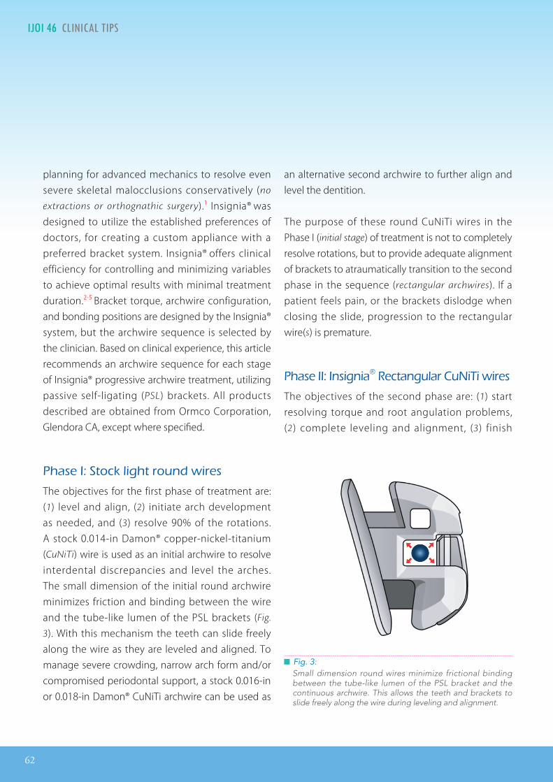
62
IJOI 46 LI I AL TI A I IJOI 46
█ Fig. 3: Small dimension round wires minimize frictional binding between the tube-like lumen of the PSL bracket and the continuous archwire. This allows the teeth and brackets to slide freely along the wire during leveling and alignment.
planning for advanced mechanics to resolve even severe skeletal malocclusions conservatively (no
extractions or orthognathic surgery).1 Insignia® was designed to utilize the established preferences of doctors, for creating a custom appliance with a preferred bracket system. Insignia® offers clinical efficiency for controlling and minimizing variables to achieve optimal results with minimal treatment duration.2-5 Bracket torque, archwire configuration, and bonding positions are designed by the Insignia®
system, but the archwire sequence is selected by the clinician. Based on clinical experience, this article recommends an archwire sequence for each stage of Insignia® progressive archwire treatment, utilizing passive self-ligating (PSL) brackets. All products described are obtained from Ormco Corporation, Glendora CA, except where specified.
ase I toc lig t round wires
The objectives for the first phase of treatment are: (1) level and align, (2) initiate arch development as needed, and (3) resolve 90% of the rotations. A stock 0.014-in Damon® copper-nickel-titanium (CuNiTi) wire is used as an initial archwire to resolve interdental discrepancies and level the arches. The small dimension of the initial round archwire minimizes friction and binding between the wire and the tube-like lumen of the PSL brackets (Fig.
3). With this mechanism the teeth can slide freely along the wire as they are leveled and aligned. To manage severe crowding, narrow arch form and/or compromised periodontal support, a stock 0.016-in or 0.018-in Damon® CuNiTi archwire can be used as
an alternative second archwire to further align and level the dentition.
The purpose of these round CuNiTi wires in the Phase I (initial stage) of treatment is not to completely resolve rotations, but to provide adequate alignment of brackets to atraumatically transition to the second phase in the sequence (rectangular archwires). If a patient feels pain, or the brackets dislodge when closing the slide, progression to the rectangular wire(s) is premature.
ase II Insignia ectangular u i i wires
The objectives of the second phase are: (1) start resolving torque and root angulation problems, (2) complete leveling and alignment, (3) finish
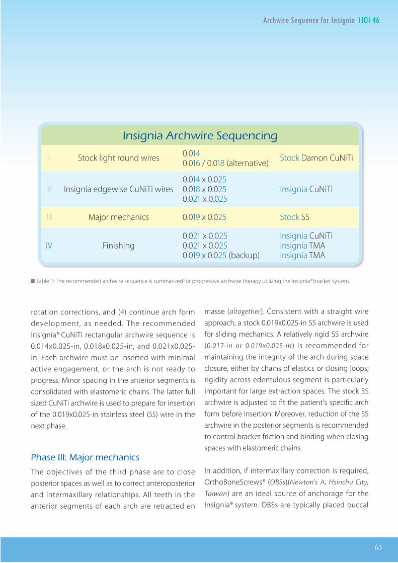
IJOI 46 LI I AL TI
63
A I IJOI 46
rotation corrections, and (4) continue arch form development, as needed. The recommended Insignia® CuNiTi rectangular archwire sequence is 0.014x0.025-in, 0.018x0.025-in, and 0.021x0.025-in. Each archwire must be inserted with minimal active engagement, or the arch is not ready to progress. Minor spacing in the anterior segments is consolidated with elastomeric chains. The latter full sized CuNiTi archwire is used to prepare for insertion of the 0.019x0.025-in stainless steel (SS) wire in the next phase.
ase III a or mec anics
The objectives of the third phase are to close posterior spaces as well as to correct anteroposterior and intermaxillary relationships. All teeth in the anterior segments of each arch are retracted en
masse (altogether). Consistent with a straight wire approach, a stock 0.019x0.025-in SS archwire is used for sliding mechanics. A relatively rigid SS archwire (0.017-in or 0.019x0.025-in) is recommended for maintaining the integrity of the arch during space closure, either by chains of elastics or closing loops; rigidity across edentulous segment is particularly important for large extraction spaces. The stock SS archwire is adjusted to fit the patient's specific arch form before insertion. Moreover, reduction of the SS archwire in the posterior segments is recommended to control bracket friction and binding when closing spaces with elastomeric chains.
In addition, if intermaxillary correction is required, OrthoBoneScrews® (OBSs)(Newton's A, Hsinchu City,
Taiwan) are an ideal source of anchorage for the Insignia® system. OBSs are typically placed buccal
██ Table 1: The recommended archwire sequence is summarized for progressive archwire therapy utilizing the Insignia® bracket system.
Insignia Archwire SequencingInsignia Archwire SequencingInsignia Archwire SequencingInsignia Archwire Sequencing
I Stock light round wires 0.0140.016 / 0.018 (alternative) Stock Damon CuNiTi
II Insignia edgewise CuNiTi wires0.014 x 0.0250.018 x 0.0250.021 x 0.025
Insignia CuNiTi
III Major mechanics 0.019 x 0.025 Stock SS
IV Finishing0.021 x 0.0250.021 x 0.0250.019 x 0.025 (backup)
Insignia CuNiTiInsignia TMAInsignia TMA
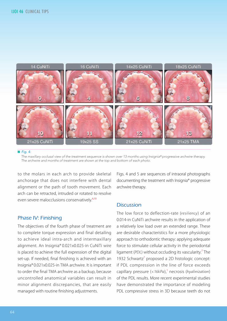
64
IJOI 46 LI I AL TI A I IJOI 46
█ Fig. 4: The maxillary occlusal view of the treatment sequence is shown over 13 months using Insignia® progressive archwire therapy. The archwire and months of treatment are shown at the top and bottom of each photo.
to the molars in each arch to provide skeletal anchorage that does not interfere with dental alignment or the path of tooth movement. Each arch can be retracted, intruded or rotated to resolve even severe malocclusions conservatively.6,10
ase I inis ing
The objectives of the fourth phase of treatment are to complete torque expression and final detailing to achieve ideal intra-arch and intermaxillary alignment. An Insignia® 0.021x0.025-in CuNiTi wire is placed to achieve the full expression of the digital set-up. If needed, final finishing is achieved with an Insignia® 0.021x0.025-in TMA archwire. It is important to order the final TMA archwire as a backup, because uncontrolled anatomical variables can result in minor alignment discrepancies, that are easily managed with routine finishing adjustments.
Figs. 4 and 5 are sequences of intraoral photographs documenting the treatment with Insignia® progressive archwire therapy.
iscussion
The low force to deflection-rate (resiliency) of an 0.014-in CuNiTi archwire results in the application of a relatively low load over an extended range. These are desirable characteristics for a more physiologic approach to orthodontic therapy: applying adequate force to stimulate cellular activity in the periodontal ligament (PDL) without occluding its vascularity.7 The 1932 Schwartz7 proposed a 2D histologic concept: if PDL compression in the line of force exceeds capillary pressure (<16kPa),1 necrosis (hyalinization) of the PDL results. More recent experimental studies have demonstrated the importance of modeling PDL compressive stress in 3D because teeth do not
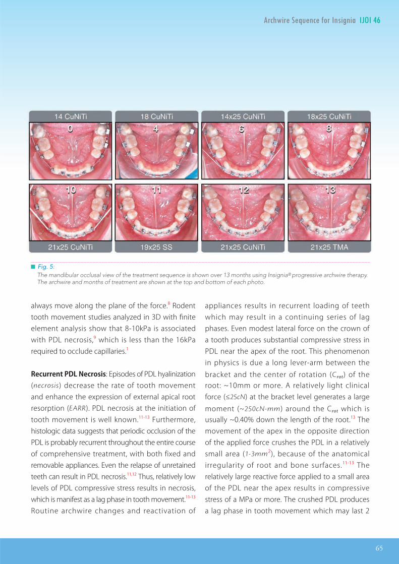
IJOI 46 LI I AL TI
65
A I IJOI 46
█ Fig. 5: The mandibular occlusal view of the treatment sequence is shown over 13 months using Insignia® progressive archwire therapy. The archwire and months of treatment are shown at the top and bottom of each photo.
always move along the plane of the force.8 Rodent tooth movement studies analyzed in 3D with finite element analysis show that 8-10kPa is associated with PDL necrosis,9 which is less than the 16kPa required to occlude capillaries.1
RecurrentPDLNecrosis: Episodes of PDL hyalinization (necrosis) decrease the rate of tooth movement and enhance the expression of external apical root resorption (EARR). PDL necrosis at the initiation of tooth movement is well known.11-13 Furthermore, histologic data suggests that periodic occlusion of the PDL is probably recurrent throughout the entire course of comprehensive treatment, with both fixed and removable appliances. Even the relapse of unretained teeth can result in PDL necrosis.11,12 Thus, relatively low levels of PDL compressive stress results in necrosis, which is manifest as a lag phase in tooth movement.11-13
Routine archwire changes and reactivation of
appliances results in recurrent loading of teeth which may result in a continuing series of lag phases. Even modest lateral force on the crown of a tooth produces substantial compressive stress in PDL near the apex of the root. This phenomenon in physics is due a long lever-arm between the
bracket and the center of rotation (Crot) of the root: ~10mm or more. A relatively light clinical force (≤25cN) at the bracket level generates a large
moment (~250cN-mm) around the Crot which is usually ~0.40% down the length of the root.13 The movement of the apex in the opposite direction of the applied force crushes the PDL in a relatively small area (1-3mm2), because of the anatomical irregularity of root and bone surfaces.11-13 The relatively large reactive force applied to a small area of the PDL near the apex results in compressive stress of a MPa or more. The crushed PDL produces a lag phase in tooth movement which may last 2
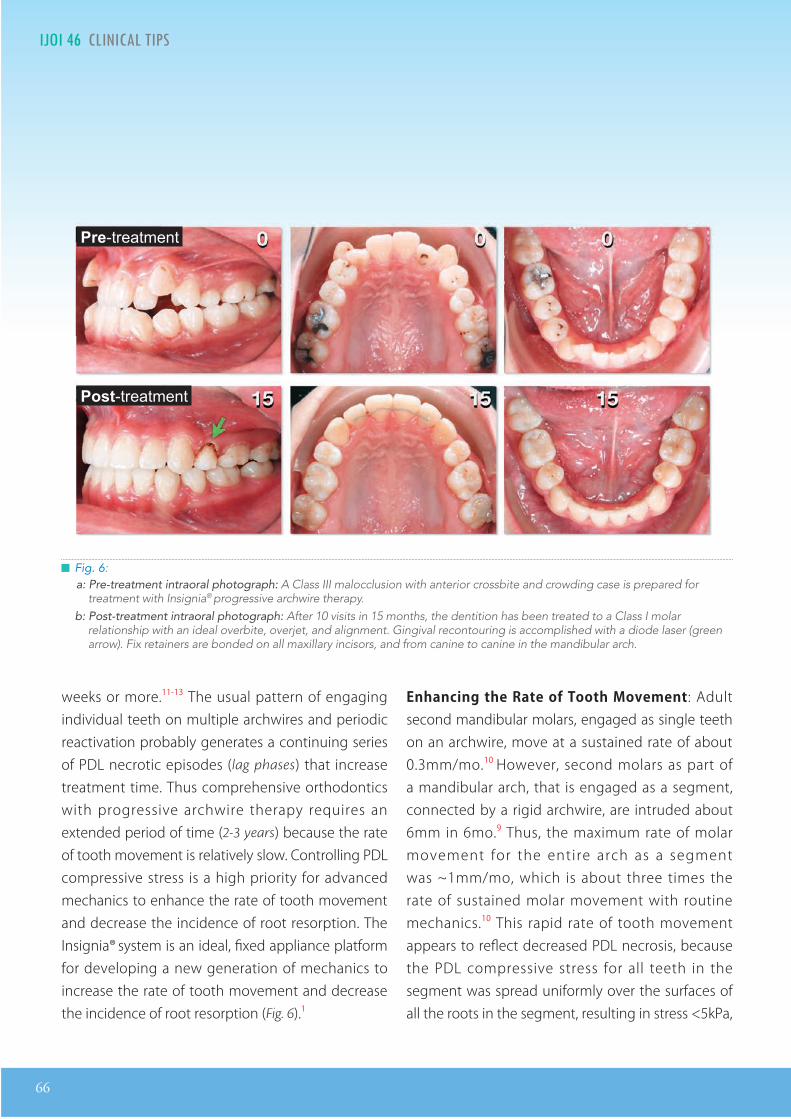
66
IJOI 46 LI I AL TI A I IJOI 46
█ Fig. 6: a: Pre-treatment intraoral photograph: A Class III malocclusion with anterior crossbite and crowding case is prepared for
treatment with Insignia® progressive archwire therapy. b: Post-treatment intraoral photograph: After 10 visits in 15 months, the dentition has been treated to a Class I molar
relationship with an ideal overbite, overjet, and alignment. Gingival recontouring is accomplished with a diode laser (green arrow). Fix retainers are bonded on all maxillary incisors, and from canine to canine in the mandibular arch.
weeks or more.11-13 The usual pattern of engaging individual teeth on multiple archwires and periodic reactivation probably generates a continuing series of PDL necrotic episodes (lag phases) that increase treatment time. Thus comprehensive orthodontics with progressive archwire therapy requires an extended period of time (2-3 years) because the rate of tooth movement is relatively slow. Controlling PDL compressive stress is a high priority for advanced mechanics to enhance the rate of tooth movement and decrease the incidence of root resorption. The Insignia® system is an ideal, fixed appliance platform for developing a new generation of mechanics to increase the rate of tooth movement and decrease the incidence of root resorption (Fig. 6).1
EnhancingtheRateofToothMovement: Adult second mandibular molars, engaged as single teeth on an archwire, move at a sustained rate of about 0.3mm/mo.10 However, second molars as part of a mandibular arch, that is engaged as a segment, connected by a rigid archwire, are intruded about 6mm in 6mo.9 Thus, the maximum rate of molar movement for the entire arch as a segment was ~1mm/mo, which is about three times the rate of sustained molar movement with routine mechanics.10 This rapid rate of tooth movement appears to reflect decreased PDL necrosis, because the PDL compressive stress for all teeth in the segment was spread uniformly over the surfaces of all the roots in the segment, resulting in stress <5kPa,
Pre r
Post r
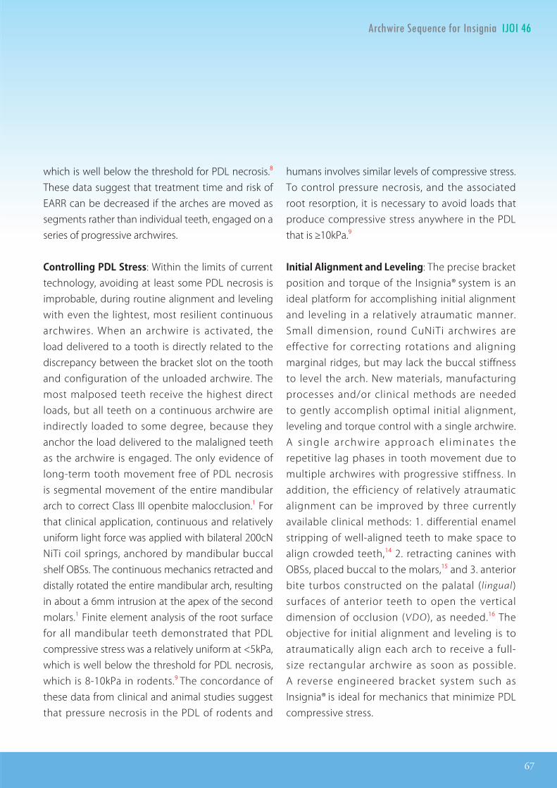
IJOI 46 LI I AL TI
67
A I IJOI 46
which is well below the threshold for PDL necrosis.8 These data suggest that treatment time and risk of EARR can be decreased if the arches are moved as segments rather than individual teeth, engaged on a series of progressive archwires.
ControllingPDLStress: Within the limits of current technology, avoiding at least some PDL necrosis is improbable, during routine alignment and leveling with even the lightest, most resilient continuous archwires. When an archwire is activated, the load delivered to a tooth is directly related to the discrepancy between the bracket slot on the tooth and configuration of the unloaded archwire. The most malposed teeth receive the highest direct loads, but all teeth on a continuous archwire are indirectly loaded to some degree, because they anchor the load delivered to the malaligned teeth as the archwire is engaged. The only evidence of long-term tooth movement free of PDL necrosis is segmental movement of the entire mandibular arch to correct Class III openbite malocclusion.1 For that clinical application, continuous and relatively uniform light force was applied with bilateral 200cN NiTi coil springs, anchored by mandibular buccal shelf OBSs. The continuous mechanics retracted and distally rotated the entire mandibular arch, resulting in about a 6mm intrusion at the apex of the second molars.1 Finite element analysis of the root surface for all mandibular teeth demonstrated that PDL compressive stress was a relatively uniform at <5kPa, which is well below the threshold for PDL necrosis, which is 8-10kPa in rodents.9 The concordance of these data from clinical and animal studies suggest that pressure necrosis in the PDL of rodents and
humans involves similar levels of compressive stress. To control pressure necrosis, and the associated root resorption, it is necessary to avoid loads that produce compressive stress anywhere in the PDL that is ≥10kPa.9
InitialAlignmentandLeveling: The precise bracket position and torque of the Insignia® system is an ideal platform for accomplishing initial alignment and leveling in a relatively atraumatic manner. Small dimension, round CuNiTi archwires are effective for correcting rotations and aligning marginal ridges, but may lack the buccal stiffness to level the arch. New materials, manufacturing processes and/or clinical methods are needed to gently accomplish optimal initial alignment, leveling and torque control with a single archwire. A s ingle archwire approach e l iminates the repetitive lag phases in tooth movement due to multiple archwires with progressive stiffness. In addition, the efficiency of relatively atraumatic alignment can be improved by three currently available clinical methods: 1. differential enamel stripping of well-aligned teeth to make space to align crowded teeth,14 2. retracting canines with OBSs, placed buccal to the molars,15 and 3. anterior bite turbos constructed on the palatal (lingual) surfaces of anterior teeth to open the vertical dimension of occlusion (VDO), as needed.16 The objective for initial alignment and leveling is to atraumatically align each arch to receive a full-size rectangular archwire as soon as possible. A reverse engineered bracket system such as Insignia® is ideal for mechanics that minimize PDL compressive stress.
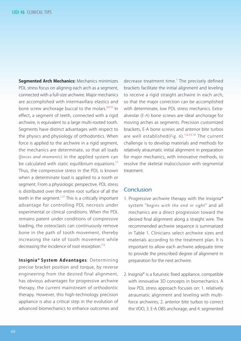
68
IJOI 46 LI I AL TI A I IJOI 46
SegmentedArchMechanics: Mechanics minimizes PDL stress focus on aligning each arch as a segment, connected with a full-size archwire. Major mechanics are accomplished with intermaxillary elastics and bone screw anchorage buccal to the molars.5,9,15 In effect, a segment of teeth, connected with a rigid archwire, is equivalent to a large multi-rooted tooth. Segments have distinct advantages with respect to the physics and physiology of orthodontics. When force is applied to the archwire in a rigid segment, the mechanics are determinate, so that all loads (forces and moments) in the applied system can be calculated with static equilibrium equations.17 Thus, the compressive stress in the PDL is known when a determinate load is applied to a tooth or segment. From a physiologic perspective, PDL stress is distributed over the entire root surface of all the teeth in the segment.1,17 This is a critically important advantage for controlling PDL necrosis under experimental or clinical conditions. When the PDL remains patent under conditions of compressive loading, the osteoclasts can continuously remove bone in the path of tooth movement, thereby increasing the rate of tooth movement while decreasing the incidence of root resorption.7-9
Insignia® SystemAdvantages : Determining precise bracket position and torque, by reverse engineering from the desired final alignment, has obvious advantages for progressive archwire therapy, the current mainstream of orthodontic therapy. However, this high-technology precision appliance is also a critical step in the evolution of advanced biomechanics to enhance outcomes and
decrease treatment time.1 The precisely defined brackets facilitate the initial alignment and leveling to receive a rigid straight archwire in each arch, so that the major correction can be accomplished with determinate, low PDL stress mechanics. Extra-alveolar (E-A) bone screws are ideal anchorage for moving arches as segments. Precision customized brackets, E-A bone screws and anterior bite turbos are well established(Fig . 6 ) .1,6,15,16 The current challenge is to develop materials and methods for relatively atraumatic initial alignment in preparation for major mechanics, with innovative methods, to resolve the skeletal malocclusion with segmental treatment.
onclusion
1. Progressive archwire therapy with the Insignia®
system “begins with the end in sight” and all mechanics are a direct progression toward the desired final alignment along a straight wire. The recommended archwire sequence is summarized in Table 1. Clinicians select archwire sizes and materials according to the treatment plan. It is important to allow each archwire adequate time to provide the prescribed degree of alignment in preparation for the next archwire.
2. Insignia® is a futuristic fixed appliance, compatible with innovative 3D concepts in biomechanics. A low PDL stress approach focuses on: 1. relatively atraumatic alignment and leveling with multi-force archwires, 2. anterior bite turbos to correct the VDO, 3. E-A OBS anchorage, and 4. segmented
IJOI 46 LI I AL TI
69
A I IJOI 46
determinate mechanics to move entire arches en masse. These methods promise to expand the scope of treatment, enhance outcomes, decrease treatment time, and control root resorption.
Ac nowledgment
Thanks to Mr. Paul Head for proofreading this article.
e erences
1. Roberts WE, Viecilli RF, Chang CH, Katona TR, Paydar NH. Biology of biomechanics: �nite element analysis of a statically determinate system to rotate the occlusal plane for correction of skeletal Class III malocclusion. Am J Orthod Dentofacial Orthop 2015;148:943-955.
2. Johnson E. Selecting custom torque prescriptions for the straight-wire appliance. Am J Orthod Dentofacial Orthop 2013;143:S161-7.
3. Weber DJ 2nd, Koroluk LD, Phillips C, Nguyen T, Proffit WR. Clinical effectiveness and efficiency of customized vs. conventional preadjusted bracket systems. J Clin Orthod 2013;47:261-266. quiz 268.
4. Brown MW, Koroluk L, Ko CC, Zhang K , Chen M, Nguyen T. Effectiveness and efficiency of a CAD/CAM orthodontic bracket system. Am J Orthod Dentofacial Orthop 2015;148:1067-1074.
5. Scholz RP, Sarver DM. Interview with an Insignia® doctor: David M. Sarver. Am J Orthod Dentofacia l Orthop 2009;136:853-856.
6. Tseng LY, Chang CH, Roberts WE. Diagnosis and conservative treatment of skeletal Class III malocclusion with anterior crossbite and asymmetric maxillary crowding. Am J Orthod Dentofacial Orthop 2016;149:555-66.
7. Schwarz AM. Tissue changes incidental to orthodontic tooth movement. Int J Orthod Oral Surg and Radiog 1932;18:331-52.
8. Viecilli RF, Katona TR, Chen J, Harts�eld JK Jr, Roberts WE. Three-dimensional mechanical environment of orthodontic tooth movement and root resorption. Am J Orthod Dentofacial Orthop 2008;133:791. e. 11-26.
9. Viecilli RF, Kar-Kuri MH, Varriale J, Budiman A, Janal M. E�ects of initial stresses and time on orthodontic external root resorption. J Dent Res 2013;92:346-51.
10. Roberts WE, Arbuckle GR , Analoui M. Rate of mesial translation of mandibular molars using implant-anchored mechanics. Angle Orthod 1996;66(5):331-338.
11. Reitan K. Initial tissue behavior during apical root resorption. Angle Orthod 1974;44(1):68-82.
12. Reitan K. Clinical and histologic observations on tooth movement during and after orthodontic treatment. Am J Orthod 1967;53(10):721-45.
13. Roberts WE. Bone physiology, metabolism and biomechanics in orthodontic pra�ice. In: Orthodontics: Current Principles and Techniques, Chapter 10, 5th ed. Graber LW, Vanarsdall RL Jr, Vig KWL (Eds). St. Louis: Elsevier Mosby; 2012. pp. 287-343.
14. Livas C, Jongsma AC, Ren Y. Enamel reduction techniques in orthodontics: a literature review. Open Dent J 2013 Oct 31;7:146-51.
15. Chang CH, Liu SY, Roberts WE. Primary failure rate for 1680 extra-alveolar mandibular buccal shelf miniscrews placed in movable mucosa or a�ached gingiva. Angle Orthod 2015;85:905-910.
16. Lee AS, Chang CH, Roberts WE. Severe unilateral scissors-bite with a constricted mandibular arch: bite turbos and extra-alveolar bone screws in the infra-zygomatic crests and mandibular buccal shelf. Am J Orthod Dentofacial Orthop 2017 (Submi�ed).
17. Burstone CJ, Choy K. �e biomechanical foundation of clinical Orthodontics. Chicago: Quintessence Publishing Co; 2015.

贈
30人份 或 60盒 20人份 或40盒
50人份 或 100盒
演講報名費 USD$500 + 7/12-14三晚單人標準房
10套7 77 7
5 55 5
5 55 5
5 55 5
(定價NT$7,700 /10顆/盒,特價NT$5,000/盒)
(定價NT$6,600 /10顆/盒,特價NT$3,300/盒) (定價NT$33,000/人份/盒)
(定價NT$14,000 /10顆/盒,特價NT$7,000/盒)
BANGKOKTHAILAND
JULY 12–14
・ 本文宣品僅供牙科診所及牙醫師參考,禁止張貼或擺置於公眾可瀏覽及取閱之處 ・ 本優惠專案僅適用於牙醫診所 ・ 本公司保留贈品更換之權利 ・ 本公司保留更改及終止活動辦法與條款之權利
Dr. John Lin The Latest on Simple and E�cient Class III TAD TreatmentCombining TADs and PSL for Non-Extraction Treatment inClass II and Class I Crowding Cases
Dr. David Birnie Digital Orthodontics Made EasyManaging Posterior Crossbites without Expanders
Dr. Sonia Palleck Solving the Puzzle: Using Insignia to Help Simplify Treatment Mechanics
Dr. Sabrina HuangDr. Park Chang Suk Case Presentations
Dr. Frank Bogdan PSL Trouble-Shooting: Reading and Reacting During Treatment
Dr. Chris Chang Extraction vs. Non-Extraction: A Reasonable Treatment Plan
Dr. Chris Chang Non-Surgical Treatment for Challenging Cases with Damon System and TAD
THURSDAY, JULY 13
FRIDAY, JULY 14
5 55 5
(定價NT$7,700 /10顆/盒,
好康一
演講報名費
5 55 5
(定價NT$6,600 /10
(定價NT$7,700 /10顆/盒,
好康三
205 55 5
特價NT$5,000特價NT$5,000/盒) (定價NT$14,000 /10顆/盒,
好康二
盒
USD$500USD$500 7/12-14三晚單人標準房
107 77 7
特價NT$3,300特價NT$3,300/盒) (定價NT$33,000/人份/盒)
特價NT$5,000特價NT$5,000/盒) (定價NT$14,000 /10顆/盒,
好康四
專案ASIA DAMON FORUM
Register & more information forum.ormco.com/asia
任選一
(註1: 此優惠方案不再加贈任何贈品,註2: 此Damon Forum 專案至5/25止)
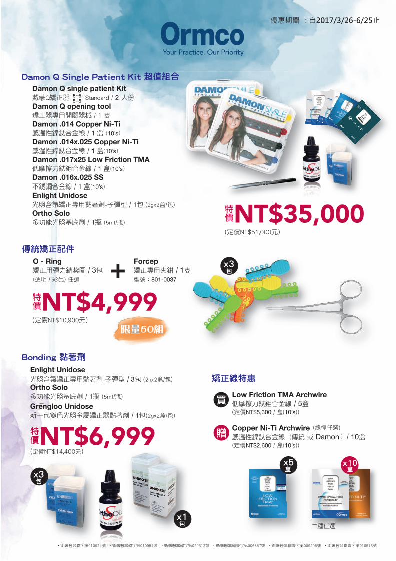
贈
30人份 或 60盒 20人份 或40盒
50人份 或 100盒
演講報名費 USD$500 + 7/12-14三晚單人標準房
10套7 77 7
5 55 5
5 55 5
5 55 5
(定價NT$7,700 /10顆/盒,特價NT$5,000/盒)
(定價NT$6,600 /10顆/盒,特價NT$3,300/盒) (定價NT$33,000/人份/盒)
(定價NT$14,000 /10顆/盒,特價NT$7,000/盒)
BANGKOKTHAILAND
JULY 12–14
・ 本文宣品僅供牙科診所及牙醫師參考,禁止張貼或擺置於公眾可瀏覽及取閱之處 ・ 本優惠專案僅適用於牙醫診所 ・ 本公司保留贈品更換之權利 ・ 本公司保留更改及終止活動辦法與條款之權利
Dr. John Lin The Latest on Simple and E�cient Class III TAD TreatmentCombining TADs and PSL for Non-Extraction Treatment inClass II and Class I Crowding Cases
Dr. David Birnie Digital Orthodontics Made EasyManaging Posterior Crossbites without Expanders
Dr. Sonia Palleck Solving the Puzzle: Using Insignia to Help Simplify Treatment Mechanics
Dr. Sabrina HuangDr. Park Chang Suk Case Presentations
Dr. Frank Bogdan PSL Trouble-Shooting: Reading and Reacting During Treatment
Dr. Chris Chang Extraction vs. Non-Extraction: A Reasonable Treatment Plan
Dr. Chris Chang Non-Surgical Treatment for Challenging Cases with Damon System and TAD
THURSDAY, JULY 13
FRIDAY, JULY 14
好康一
好康三
好康二
好康四
專案ASIA DAMON FORUM
Register & more information forum.ormco.com/asia
任選一
(註1: 此優惠方案不再加贈任何贈品,註2: 此Damon Forum 專案至5/25止)
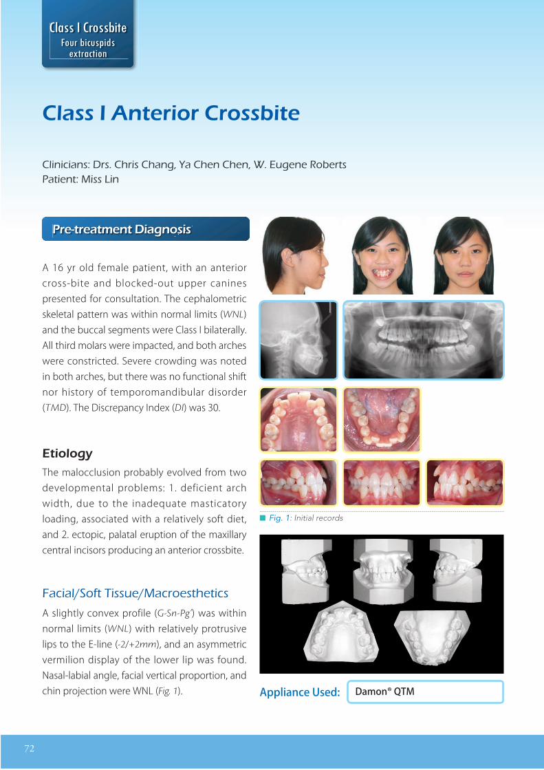
72
a nt rior ro it
linicians rs ris ang a en en ugene obertsatient iss in
Appliance Used: Damon® QTM
r tr at nt ia no i
A 16 yr old female patient, with an anterior cross-bite and blocked-out upper canines presented for consultation. The cephalometric skeletal pattern was within normal limits (WNL) and the buccal segments were Class I bilaterally. All third molars were impacted, and both arches were constricted. Severe crowding was noted in both arches, but there was no functional shift nor history of temporomandibular disorder (TMD). The Discrepancy Index (DI) was 30.
tio oThe malocclusion probably evolved from two developmental problems: 1. deficient arch width, due to the inadequate masticatory loading, associated with a relatively soft diet, and 2. ectopic, palatal eruption of the maxillary central incisors producing an anterior crossbite.
acial o t issue acroest etics
A slightly convex profile (G-Sn-Pg’) was within normal limits (WNL) with relatively protrusive lips to the E-line (-2/+2mm), and an asymmetric vermilion display of the lower lip was found. Nasal-labial angle, facial vertical proportion, and chin projection were WNL (Fig. 1).
I
█ Fig. 1: Initial records
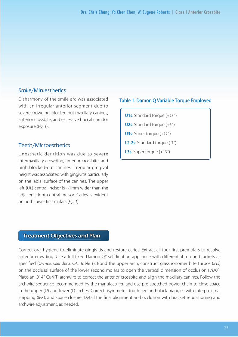
73
D E R I A
U1s: Standard torque (+15˚)
U2s: Standard torque (+6˚)
U3s: Super torque (+11˚)
L2-2s: Standard torque (-3˚)
L3s: Super torque (+13˚)
Table 1: Damon Q Variable Torque Employed
mile iniest etics
Disharmony of the smile arc was associated with an irregular anterior segment due to severe crowding, blocked out maxillary canines, anterior crossbite, and excessive buccal corridor exposure (Fig. 1).
eet icroest etics
Unesthetic dentit ion was due to severe intermaxillary crowding, anterior crossbite, and high blocked-out canines. Irregular gingival height was associated with gingivitis particularly on the labial surface of the canines. The upper left (UL) central incisor is ~1mm wider than the adjacent right central incisor. Caries is evident on both lower first molars (Fig. 1).
r at nt cti an an
Correct oral hygiene to eliminate gingivitis and restore caries. Extract all four first premolars to resolve anterior crowding. Use a full fixed Damon Q® self ligation appliance with differential torque brackets as specified (Ormco, Glendora, CA, Table 1). Bond the upper arch, construct glass ionomer bite turbos (BTs) on the occlusal surface of the lower second molars to open the vertical dimension of occlusion (VDO). Place an .014” CuNiTi archwire to correct the anterior crossbite and align the maxillary canines. Follow the archwire sequence recommended by the manufacturer, and use pre-stretched power chain to close space in the upper (U) and lower (L) arches. Correct asymmetric tooth size and black triangles with interproximal stripping (IPR), and space closure. Detail the final alignment and occlusion with bracket repositioning and archwire adjustment, as needed.
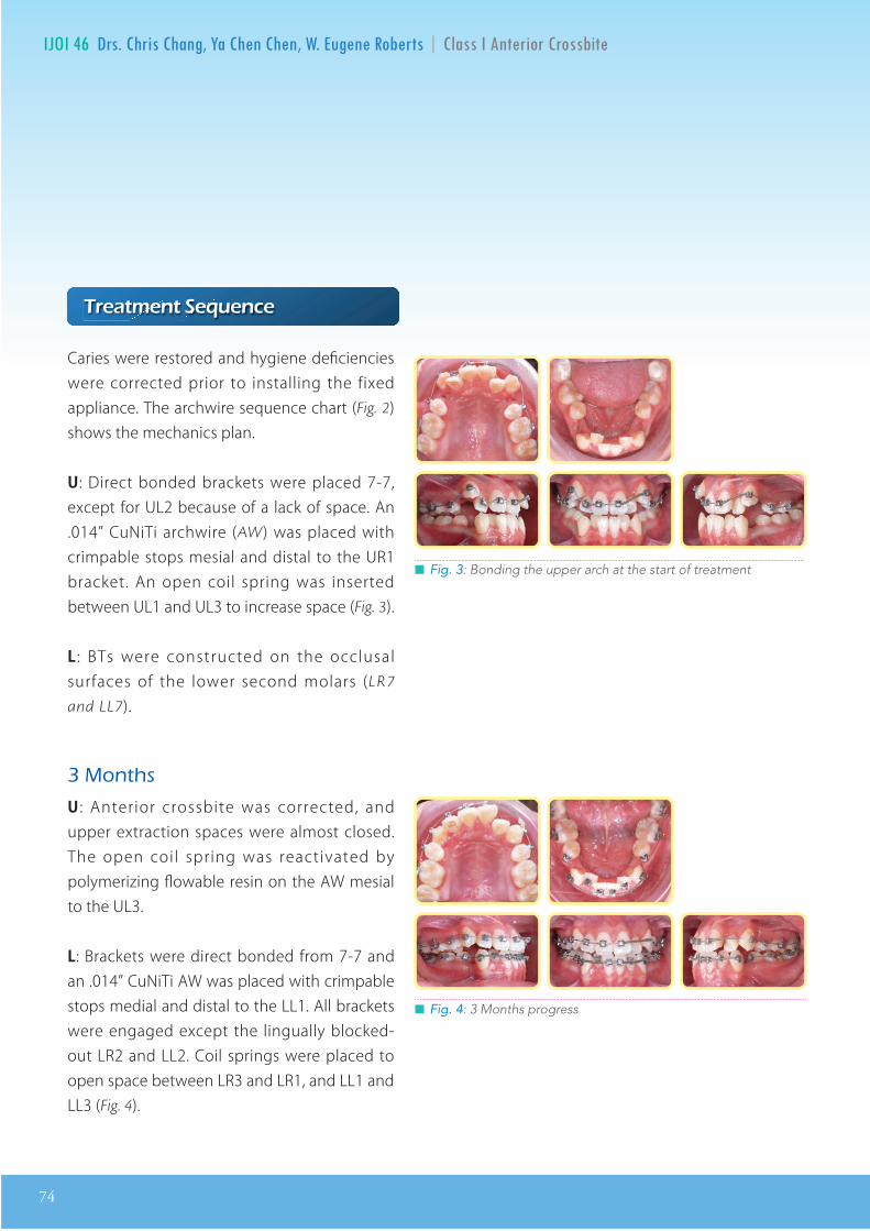
74
IJOI 46 D E R I A
█ Fig. 3: Bonding the upper arch at the start of treatment
█ Fig. 4: 3 Months progress
r at nt nc
Caries were restored and hygiene deficiencies were corrected prior to installing the fixed appliance. The archwire sequence chart (Fig. 2) shows the mechanics plan.
U: Direct bonded brackets were placed 7-7, except for UL2 because of a lack of space. An .014” CuNiTi archwire (AW) was placed with crimpable stops mesial and distal to the UR1 bracket. An open coil spring was inserted between UL1 and UL3 to increase space (Fig. 3).
L : BTs were constructed on the occlusal surfaces of the lower second molars (LR7
and LL7).
ont s
U : Anterior crossbite was corrected, and upper extraction spaces were almost closed. The open coil spring was reactivated by polymerizing flowable resin on the AW mesial to the UL3.
L: Brackets were direct bonded from 7-7 and an .014” CuNiTi AW was placed with crimpable stops medial and distal to the LL1. All brackets were engaged except the lingually blocked-out LR2 and LL2. Coil springs were placed to open space between LR3 and LR1, and LL1 and LL3 (Fig. 4).
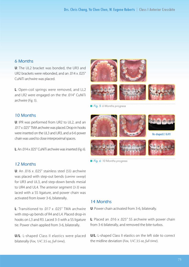
IJOI 46 D E R I A
75
D E R I A
█ Fig. 5: 6 Months progress
Re-shaped L1 & R1
█ Fig. 6: 10 Months progress
ont s
U: The UL2 bracket was bonded, the UR3 and UR2 brackets were rebonded, and an .014 x .025” CuNiTi archwire was placed.
L: Open-coil springs were removed, and LL2 and LR2 were engaged on the the .014” CuNiTi archwire (Fig. 5).
ont s
U: IPR was performed from UR2 to UL2, and an.017 x .025” TMA archwire was placed. Drop-in hooks were inserted on the UL3 and UR3, and a 6-6 power chain was used to close interproximal spaces.
L: An .014 x .025” CuNiTi archwire was inserted (Fig. 6).
ont s
U: An .016 x .025” stainless steel (SS) archwire was placed with step-out bends (canine sweep) for UR3 and UL3, and step-down bends mesial to UR4 and UL4. The anterior segment (3-3) was laced with a SS ligature, and power chain was activated from lower 3-6, bilaterally.
L: Transitioned to .017 x .025” TMA archwire with step-up bends of R4 and L4. Placed drop-in hooks on L3 and R3. Laced 3-3 with a SS ligature tie. Power chain applied from 3-6, bilaterally.
U/L: L-shaped Class II elastics were placed bilaterally (Fox, 1/4”, 3.5 oz, full time).
ont s
U: Power chain activated from 3-6, bilaterally.
L: Placed an .016 x .025” SS archwire with power chain from 3-6 bilaterally, and removed the bite-turbos.
U/L: L-shaped Class II elastics on the left side to correct the midline deviation (Fox, 1/4”, 3.5 oz, full time).
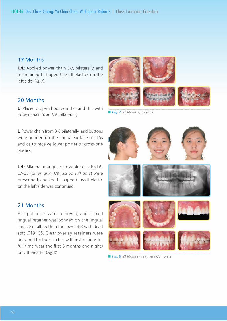
76
IJOI 46 D E R I A
█ Fig. 7: 17 Months progress
█ Fig. 8: 21 Months-Treatment Complete
ont s
U/L: Applied power chain 3-7, bilaterally, and maintained L-shaped Class II elastics on the left side (Fig. 7).
ont s
U: Placed drop-in hooks on UR5 and UL5 with power chain from 3-6, bilaterally.
L: Power chain from 3-6 bilaterally, and buttons were bonded on the lingual surface of LL5s and 6s to receive lower posterior cross-bite elastics.
U/L: Bilateral triangular cross-bite elastics L6-L7-U5 (Chipmunk, 1/8”, 3.5 oz. full time) were prescribed, and the L-shaped Class II elastic on the left side was continued.
ont s
All appliances were removed, and a fixed lingual retainer was bonded on the lingual surface of all teeth in the lower 3-3 with dead soft .019” SS. Clear overlay retainers were delivered for both arches with instructions for full time wear the first 6 months and nights only thereafter (Fig. 8).
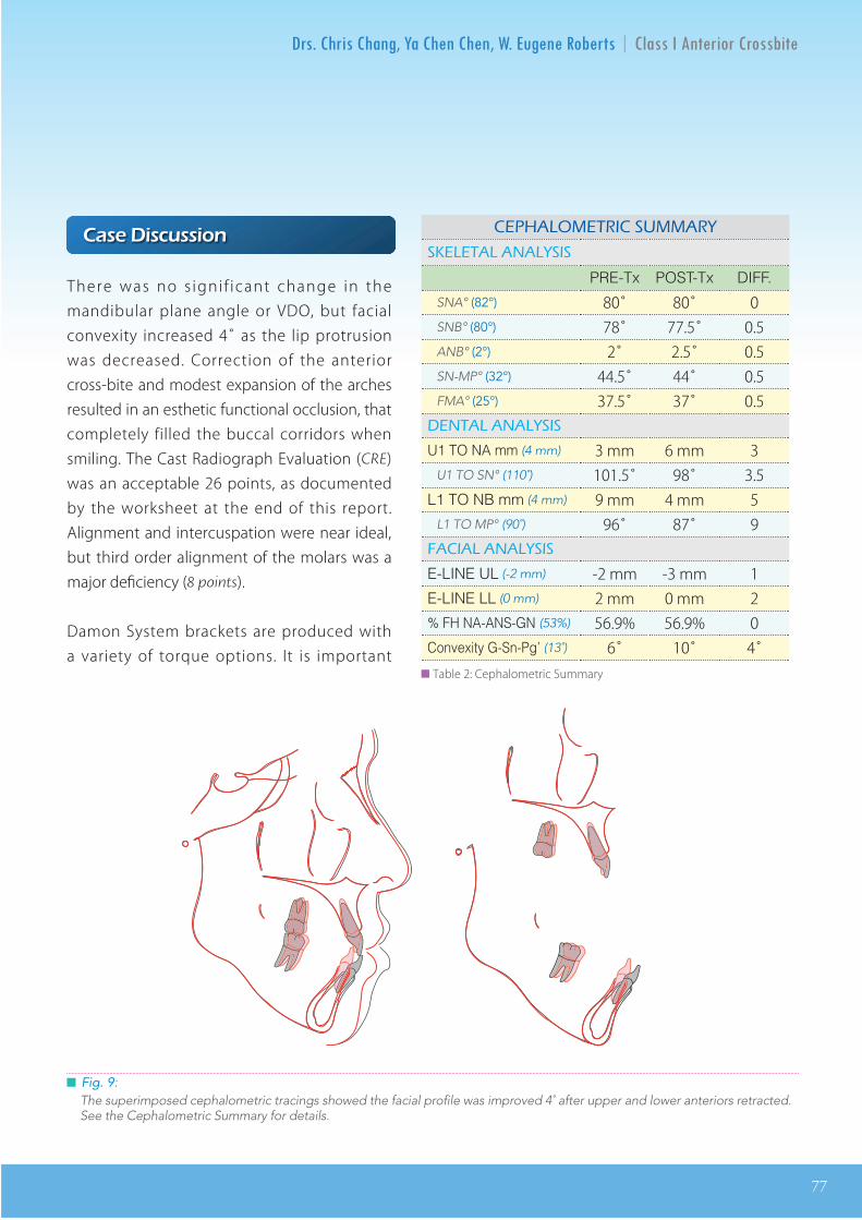
IJOI 46 D E R I A
77
D E R I A
Drs. Chris Chang, Ya Chen Chen, W. Eugene Roberts∣Class I Anterior Crossbite
There was no significant change in the mandibular plane angle or VDO, but facial convexity increased 4¼ as the lip protrusion was decreased. Correction of the anterior cross-bite and modest expansion of the arches resulted in an esthetic functional occlusion, that completely filled the buccal corridors when smiling. The Cast Radiograph Evaluation (CRE) was an acceptable 26 points, as documented by the worksheet at the end of this report. Alignment and intercuspation were near ideal, but third order alignment of the molars was a major deficiency (8 points).
Damon System brackets are produced with a variety of torque options. It is important to carefully select the required torque for each tooth based on crown morphology and the applied mechanics. If torque is not appropriately
Case Discussion
CEPHALOMETRIC SUMMARYCEPHALOMETRIC SUMMARYCEPHALOMETRIC SUMMARYCEPHALOMETRIC SUMMARYSKELETAL ANALYSISSKELETAL ANALYSISSKELETAL ANALYSISSKELETAL ANALYSIS
PRE-TX POST-TX DIFF.SNA¡ (82¼) 80û 80û 0
SNB¡ (80¼) 78û 77.5û 0.5
ANB¡ (2¼) 2û 2.5û 0.5
SN-MP¡ (32¼) 44.5û 44û 0.5
FMA¡ (25¼) 37.5û 37û 0.5
DENTAL ANALYSISDENTAL ANALYSISDENTAL ANALYSISDENTAL ANALYSISU1 TO NA mm (4 mm) 3 mm 6 mm 3
U1 TO SN¡ (110¼) 101.5û 98û 3.5
L1 TO NB mm (4 mm) 9 mm 4 mm 5
L1 TO MP¡ (90¼) 96û 87û 9
FACIAL ANALYSISFACIAL ANALYSISFACIAL ANALYSISFACIAL ANALYSISE-LINE UL (-2 mm) -2 mm -3 mm 1
E-LINE LL (0 mm) 2 mm 0 mm 2
% FH NA-ANS-GN (53%) 56.9% 56.9% 0
Convexity G-Sn-PgÕ (13¼) 6¼ 10¼ 4¼
Fig. 9: The superimposed cephalometric tracings showed the facial proÞle was improved 4¼ after upper and lower anteriors retracted. See the Cephalometric Summary for details.
a i c ion
There was no s igni f icant change in the mandibular plane angle or VDO, but facial convexity increased 4̊ as the lip protrusion was decreased. Correction of the anterior cross-bite and modest expansion of the arches resulted in an esthetic functional occlusion, that completely filled the buccal corridors when smiling. The Cast Radiograph Evaluation (CRE) was an acceptable 26 points, as documented by the worksheet at the end of this report. Alignment and intercuspation were near ideal, but third order alignment of the molars was a major deficiency (8 points).
Damon System brackets are produced with a variety of torque options. It is important
A O I A
A A A I
PRE-Tx POST-Tx DIFF.
SNA° (82°) 80̊ 80̊ 0 SNB° (80°) 78̊ 77.5̊ 0.5 ANB° (2°) 2̊ 2.5̊ 0.5 SN-MP° (32°) 44.5̊ 44̊ 0.5 FMA° (25°) 37.5̊ 37̊ 0.5
A A A I
U1 TO NA mm (4 mm) 3 mm 6 mm 3 U1 TO SN° (110˚) 101.5̊ 98̊ 3.5
L1 TO NB mm (4 mm) 9 mm 4 mm 5 L1 TO MP° (90˚) 96̊ 87̊ 9A IA A A I
E-LINE UL (-2 mm) -2 mm -3 mm 1 E-LINE LL (0 mm) 2 mm 0 mm 2 % FH NA-ANS-GN (53%) 56.9% 56.9% 0 Convexity G-Sn-Pg’ (13˚) 6̊ 10̊ 4̊
█ Fig. 9: The superimposed cephalometric tracings showed the facial profile was improved 4˚ after upper and lower anteriors retracted. See the Cephalometric Summary for details.
██ Table 2: Cephalometric Summary
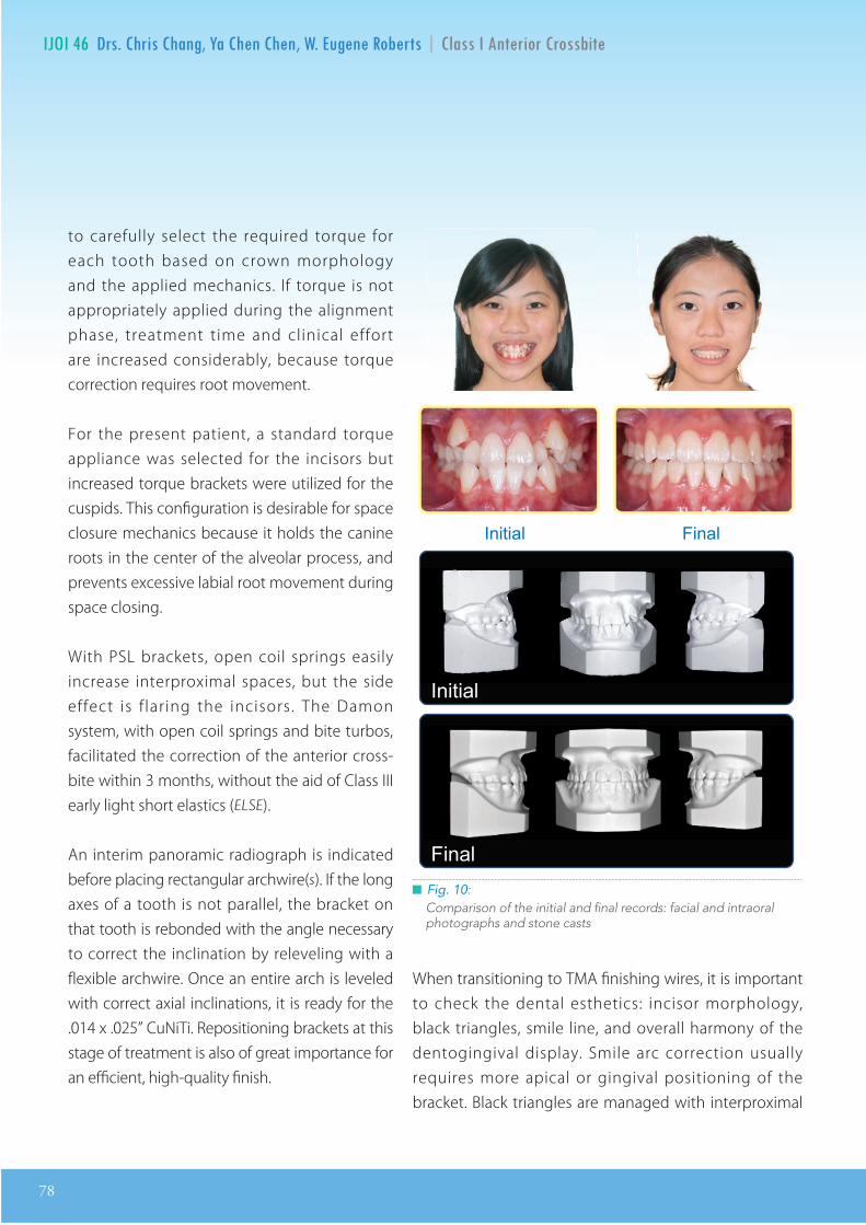
78
IJOI 46 D E R I A
to carefully select the required torque for each tooth based on crown morphology and the applied mechanics. If torque is not appropriately applied during the alignment phase, treatment time and clinical effort are increased considerably, because torque correction requires root movement.
For the present patient, a standard torque appliance was selected for the incisors but increased torque brackets were utilized for the cuspids. This configuration is desirable for space closure mechanics because it holds the canine roots in the center of the alveolar process, and prevents excessive labial root movement during space closing.
With PSL brackets, open coil springs easily increase interproximal spaces, but the side effect is f laring the incisors. The Damon system, with open coil springs and bite turbos, facilitated the correction of the anterior cross-bite within 3 months, without the aid of Class III early light short elastics (ELSE).
An interim panoramic radiograph is indicated before placing rectangular archwire(s). If the long axes of a tooth is not parallel, the bracket on that tooth is rebonded with the angle necessary to correct the inclination by releveling with a flexible archwire. Once an entire arch is leveled with correct axial inclinations, it is ready for the .014 x .025” CuNiTi. Repositioning brackets at this stage of treatment is also of great importance for an efficient, high-quality finish.
█ Fig. 10: Comparison of the initial and final records: facial and intraoral photographs and stone casts
When transitioning to TMA finishing wires, it is important to check the dental esthetics: incisor morphology, black triangles, smile line, and overall harmony of the dentogingival display. Smile arc correction usually requires more apical or gingival positioning of the bracket. Black triangles are managed with interproximal

IJOI 46 D E R I A
79
D E R I A
reduction (IPR) and space closure. Dental contour changes in the esthetic zone are best accomplished with fine diamond burs to achieve a more natural tooth morphology.
In etros ect
It is important to carefully assess the finish (Figs. 8-10). CRE scores alignment, and the Pink & White assessment critically evaluates dental esthetics. Some of the problems detected retrospectively can be corrected during retention, but even if the patient does not benefit from the post-treatment analysis, it is an excellent learning opportunity for avoiding similar problems on future patients.
The deficiencies detected for the present patient were asymmetry in the size of the maxil lary central incisors, s l ight midline deviation, and excessive buccal tipping of the maxillary molars. IPR could be performed on the distal of the UL1, followed by space closure to correct midline, if there is adequate overjet. Since there was no anterior overjet, it would be necessary to strip the lower incisors and close space to create overjet for the maxillary incisor correction. It is not wise to attempt such an extensive correction after the fixed appliances are removed, because there is no control over the axial inclinations of the teeth involved.
The Damon system is effective for expanding arches, but it is a slow process requiring light forces to generate counter-moments for
█ Fig. 11 : 1 year follow-up photographs
buccal segment translation as the cusps of the teeth are tipped into the cheek mucosa. Each light archwire must be used for a long period of time in a carefully prescribed sequence. For the present patient (Fig. 8), the buccal segments were excessively tipped, resulting in third order axial inclination problems, and decreased cusp contacts. In retrospect, 10-15̊ of progressive buccal root torque in the upper posterior segments of the TMA archwire was needed to avoid buccal tipping. This amount of torque must usually be accompanied with additional arch expansion to avoid a posterior crossbite tendency.
Some minor rotations were correctable with detailing adjustments in the SS archwire. Better arch coordination was needed to improve the occlusal relationship and dental contacts. At the debonding appointment, the gingival papilla at the maxillary midline was swollen, but the patient declined a modest gingivectomy with a Diode Laser. Despite these modest problems the CRE score for dental alignment was an acceptable 26, and dentofacial esthetics were good. One year follow-up records (Fig. 11) show the result was not only stable but the occlusion continued to improve.
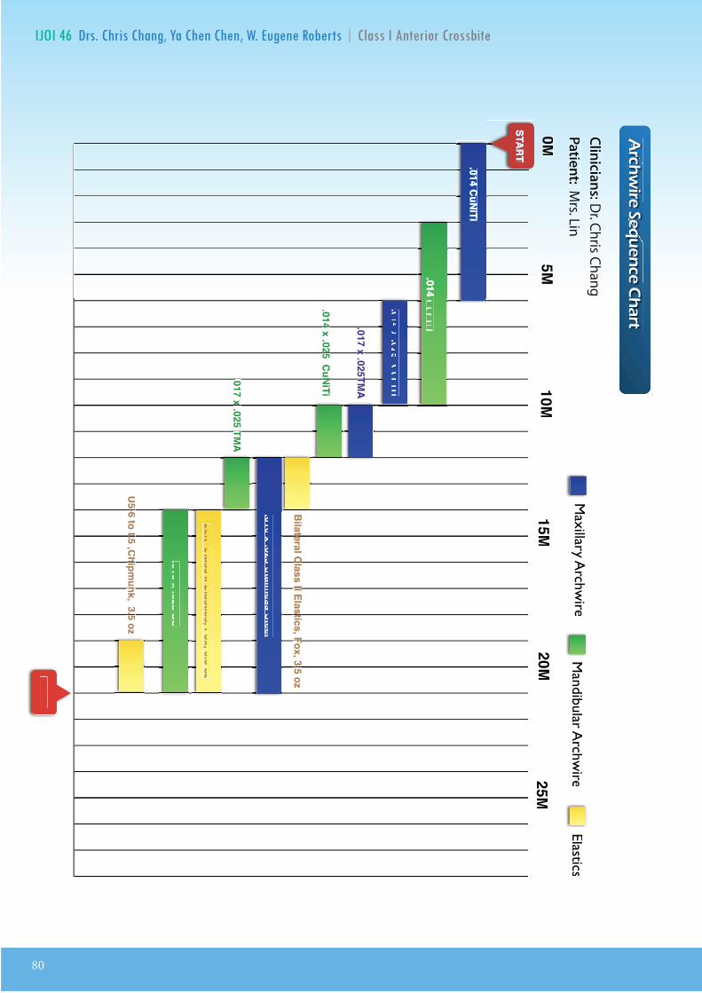
80
IJOI 46 D E R I A
rcir
nc
art
START
FINISH
0M5M
10M15M
20M25M
.014 x .025 CuNiTi
.017 x .025TMA
.017 x .025TMA
.017 x .025TMA
.016 x .025 Stainless Steel
.014 x .025 CuNiTi.014 x .025 CuNiTi.014 x .025 CuNiTi.014 x .025 CuNiTi
.017 x .025 TMA
.017 x .025 TMA
.017 x .025 TMA
.017 x .025 TMA
.016 x .025 SS
.014 CuNiTi
.014 CuNiTi.014 CuNiTi
Bilateral Class II Elastics, Fox, 3.5 ozBilateral Class II Elastics, Fox, 3.5 ozBilateral Class II Elastics, Fox, 3.5 ozBilateral Class II Elastics, Fox, 3.5 ozBilateral Class II Elastics, Fox, 3.5 ozBilateral Class II Elastics, Fox, 3.5 ozBilateral Class II Elastics, Fox, 3.5 oz
U5-6 to L5 ,Chipmunk, 3.5 oz
U5-6 to L5 ,Chipmunk, 3.5 oz
U5-6 to L5 ,Chipmunk, 3.5 oz
U5-6 to L5 ,Chipmunk, 3.5 oz
U5-6 to L5 ,Chipmunk, 3.5 oz
U5-6 to L5 ,Chipmunk, 3.5 oz
Left Class II Elastics, Fox, 3.5 oz
Clinicians:Dr. Chris ChangPatient: M
rs. Lin M
axillary Archw
ireM
andibular Archw
ireElastics
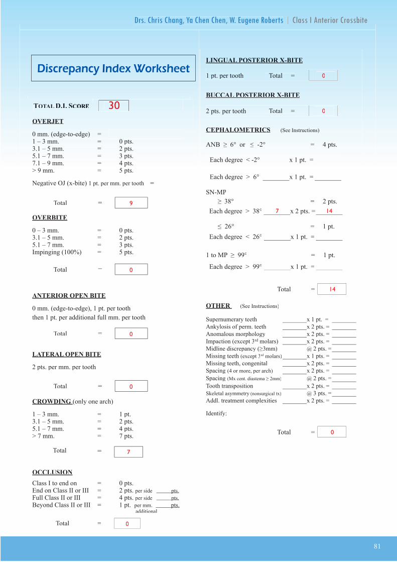
IJOI 46 D E R I A
81
D E R I A D E R
OVERJET
0 mm. (edge-to-edge) = 1 pt.1 Ð 3 mm. = 0 pts.3.1 Ð 5 mm. = 2 pts.5.1 Ð 7 mm. = 3 pts.7.1 Ð 9 mm. = 4 pts.> 9 mm. = 5 pts.> 9 mm. = 5 pts.
Negative OJ (x-bite) 1 pt. per mm. per tooth =
OVERBITE
0 Ð 3 mm. = 0 pts.3.1 Ð 5 mm. = 2 pts.5.1 Ð 7 mm. = 3 pts.Impinging (100%) = 5 pts.
ANTERIOR OPEN BITE
0 mm. (edge-to-edge), 1 pt. per tooth
then 1 pt. per additional full mm. per tooth
LATERAL OPEN BITE
2 pts. per mm. per tooth
CROWDING (only one arch)
1 Ð 3 mm. = 1 pt.3.1 Ð 5 mm. = 2 pts.5.1 Ð 7 mm. = 4 pts.> 7 mm. = 7 pts.
OCCLUSION
Class I to end on = 0 pts.End on Class II or III = 2 pts. per side pts. pts.
Full Class II or III = 4 pts. per side pts. pts.
Beyond Class II or III = 1 pt. per mm. pts.pts. additional
TotalTotalT =
TotalTotalT =
TotalTotalT =
TotalTotalT =
TotalTotalT =
Total =
TOTAL D.I.D.I. SCORECORECORECORECORE
LINGUAL POSTERIOR X-BITE
1 pt. per tooth Total =
BUCCAL POSTERIOR X-BITE
2 pts. per tooth Total =
CEPHALOMETRICS (See Instructions)
ANB ≥ 6¡ or ≤ -2¡ = 4 pts.
SN-MP
≥ 38¡ = 2 pts.
Each degree > 38¡ Each degree > 38¡ x 2 pts. =x 2 pts. =
≤ 26¡ = 1 pt.
Each degree < 26¡ Each degree < 26¡ x 1 pt. =x 1 pt. =
1 to MP ≥ 99¡ = 1 pt.
Each degree > 99¡ Each degree > 99¡ x 1 pt. =x 1 pt. =
OTHER (See Instructions)
Supernumerary teeth x 1 pt. =
Ankylosis of perm. teeth x 2 pts. =
Anomalous morphology x 2 pts. =
Impaction (except 3rd molars)rd molars)rd x 2 pts. =
Midline discrepancy (≥3mm) @ 2 pts. =
Missing teeth (except 3rd molars)rd molars)rd x 1 pts. =
Missing teeth, congenital x 2 pts. =
Spacing (4 or more, per arch) x 2 pts. =
Spacing (Mx cent. diastema ≥ 2mm) @ 2 pts. = 2
Tooth transposition x 2 pts. =
Skeletal asymmetry (nonsurgical tx) @ 3 pts. =
Addl. treatment complexities Addl. treatment complexities x 2 pts. =x 2 pts. =
Identify:
Each degree > 6¡ Each degree > 6¡ x 1 pt. =x 1 pt. =
Each degree < -2¡ Each degree < -2¡ x 1 pt. =x 1 pt. =
Total =
Total =
3030
9
0
0
0
7
0
0
0
7 14
14
0
i cr panc n or t
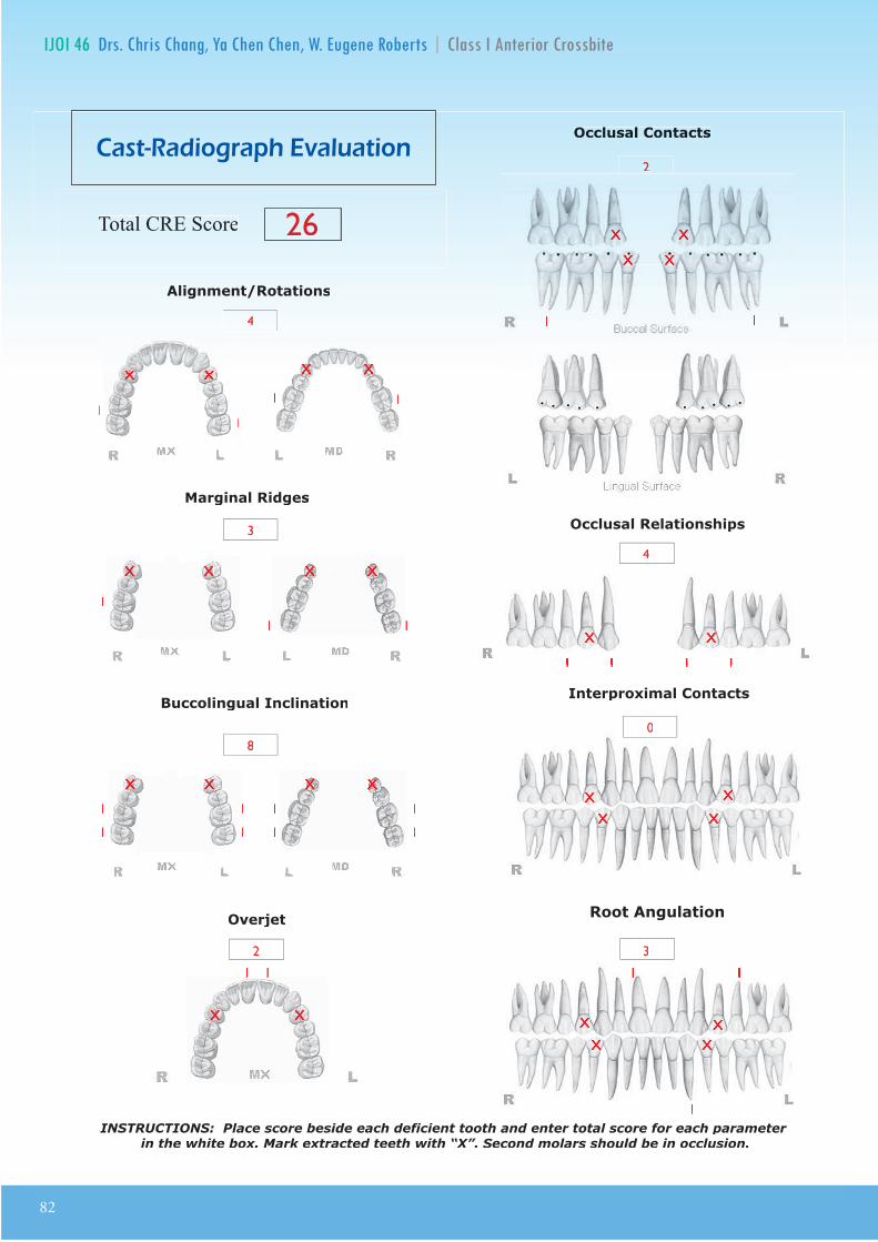
82
IJOI 46 D E R I A
����� Alignment/Rotations
Marginal Ridges
Buccolingual Inclination
Overjet
Occlusal Contacts
Occlusal Relationships
Interproximal Contacts
INSTRUCTIONS: Place score beside each deficient tooth and enter total score for each parameter in the white box. Mark extracted teeth with ÒXÓ. Second molars should be in occlusion.
Root Angulation
Total CRE Score
4
3
1
1
1
1
1
1
1
1
1
1 1
11
1 1
1 11 1
1 1
4
1 1 1 11 1 1 1
0
3
1 1
11
a t a io rap a ation
子彈型銀膠針
無線雙功能根管充填機
丁香油根管充填劑氫氧化鈣根管充填劑
根管擴大慢速安全手機
根管冠部擴大器械
馬來膠熱融機
長效型氧化鋅根管充填劑
長效型氧化鋅根管充填劑
Elementsfree 1 台
Pulp Canal Sealer EWT 1 盒
LA AXXESS KIT 2.0
Sealapex Tubli-Seal
Pulp Canal Sealer EWT
手動根管銼針系列
(23, 25 Gauge任選)GP Cartridge 10 盒
(定價NT$252,800/套)
NT$129,999特價 /套
(定價NT$185,000/套)
NT$99,999特價 /套
(定價NT$13,000-13,200/套)
NT$7,200特價 /套
(定價NT$71,000-75,500/套)
NT$38,000特價 /套
E N DODON T I C S
TM
.衛部醫器輸壹字第016766號.衛部醫器輸壹字第015275號.衛署醫器輸字第011611號
(定價NT$71,000-75,500/套)
NT$38,000.衛署醫器輸字第010982號.衛署醫器輸壹字第009148號.衛部醫器輸壹字第013962號
K-Files #06~#40 Triple-Flex #08~#40Reamers #06~#40K-Flex #06~#40Hedstrom #15~#40Pathfinder 21mm/25mm
LA AXXESS KIT 2.0
Sealapex Tubli-Seal
.衛部醫器輸壹字第014069號 .衛署醫器輸壹字第009148號
限量50個
3盒 3盒
1盒長效型氧化鋅根管充填劑
.衛署醫器輸字第013794號.衛署醫器輸字第013633號.衛署醫器輸字第011611號
・ 本文宣品僅供牙科診所及牙醫師參考,禁止張貼或擺置於公眾可瀏覽及取閱之處 ・ 本優惠專案僅適用於牙醫診所 ・ 本公司保留贈品更換之權利 ・ 本公司保留更改及終止活動辦法與條款之權利
2017/3/26-6/25
子彈型銀膠針
無線雙功能根管充填機
丁香油根管充填劑氫氧化鈣根管充填劑
根管擴大慢速安全手機
根管冠部擴大器械
馬來膠熱融機
長效型氧化鋅根管充填劑
長效型氧化鋅根管充填劑
Elementsfree 1 台
Pulp Canal Sealer EWT 1 盒
LA AXXESS KIT 2.0
Sealapex Tubli-Seal
Pulp Canal Sealer EWT
手動根管銼針系列
(23, 25 Gauge任選)GP Cartridge 10 盒
(定價NT$252,800/套)
NT$129,999特價 /套
(定價NT$185,000/套)
NT$99,999特價 /套
(定價NT$13,000-13,200/套)
NT$7,200特價 /套
(定價NT$71,000-75,500/套)
NT$38,000特價 /套
E N DODON T I C S
TM
.衛部醫器輸壹字第016766號.衛部醫器輸壹字第015275號.衛署醫器輸字第011611號
.衛署醫器輸字第010982號.衛署醫器輸壹字第009148號.衛部醫器輸壹字第013962號
K-Files #06~#40 Triple-Flex #08~#40Reamers #06~#40K-Flex #06~#40Hedstrom #15~#40Pathfinder 21mm/25mm
.衛部醫器輸壹字第014069號 .衛署醫器輸壹字第009148號
限量50個
3盒 3盒
1盒
.衛署醫器輸字第013794號.衛署醫器輸字第013633號.衛署醫器輸字第011611號
・ 本文宣品僅供牙科診所及牙醫師參考,禁止張貼或擺置於公眾可瀏覽及取閱之處 ・ 本優惠專案僅適用於牙醫診所 ・ 本公司保留贈品更換之權利 ・ 本公司保留更改及終止活動辦法與條款之權利
2017/3/26-6/25
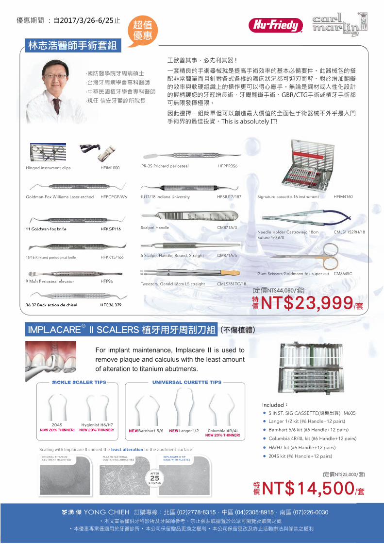
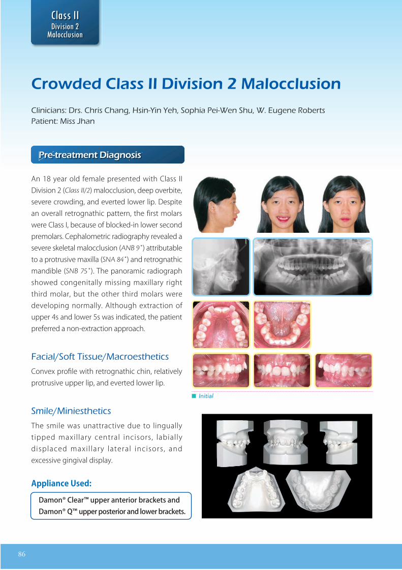
86
D E R II D
r tr at nt ia no i
An 18 year old female presented with Class II Division 2 (Class II/2) malocclusion, deep overbite, severe crowding, and everted lower lip. Despite an overall retrognathic pattern, the first molars were Class I, because of blocked-in lower second premolars. Cephalometric radiography revealed a severe skeletal malocclusion (ANB 9˚) attributable to a protrusive maxilla (SNA 84˚) and retrognathic mandible (SNB 75˚). The panoramic radiograph showed congenitally missing maxillary right third molar, but the other third molars were developing normally. Although extraction of upper 4s and lower 5s was indicated, the patient preferred a non-extraction approach.
acial o t issue acroest etics
Convex profile with retrognathic chin, relatively protrusive upper lip, and everted lower lip.
mile iniest etics
The smile was unattractive due to lingually tipped maxil lary central incisors, labially displaced maxi l lary lateral incisors , and excessive gingival display.
█ Initial
Damon® Clear™ upper anterior brackets andDamon® Q™ upper posterior and lower brackets.
ApplianceUsed:
ro a i i ion a occ ion
linicians rs ris ang sin in e o ia ei en u ugene obertsatient iss an
II D
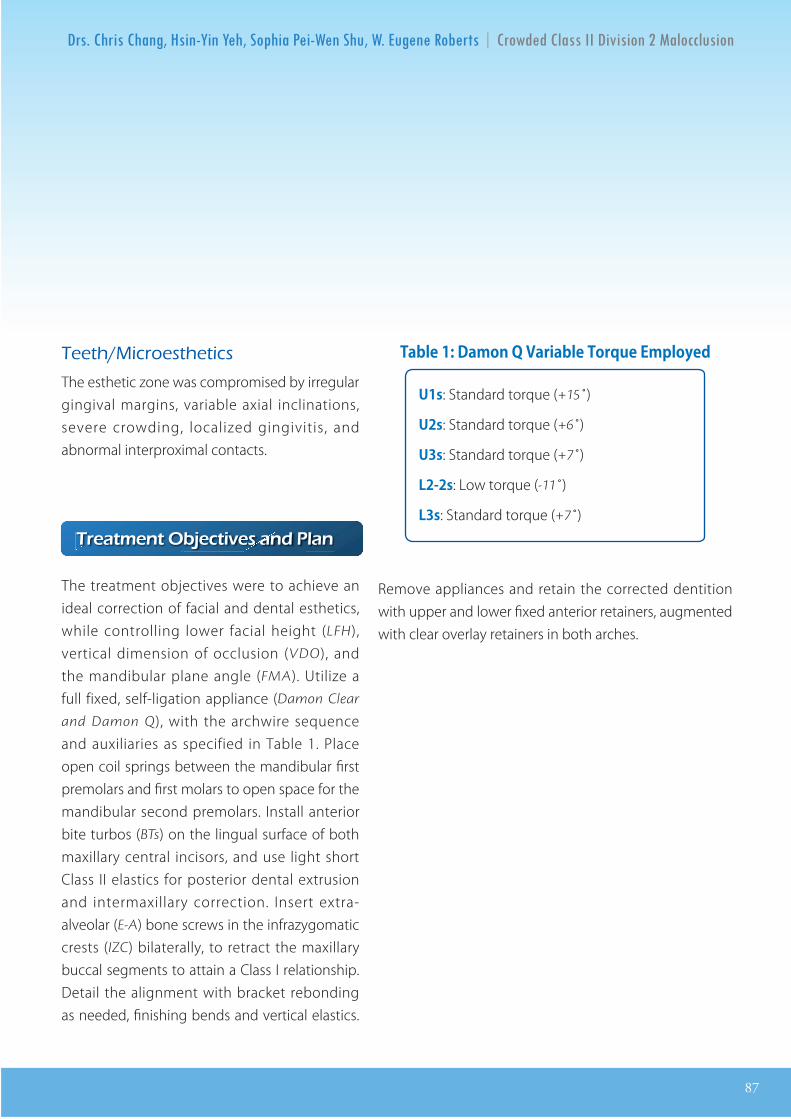
87
D E R II D
eet icroest etics
The esthetic zone was compromised by irregular gingival margins, variable axial inclinations, severe crowding, localized gingivitis, and abnormal interproximal contacts.
r at nt cti an an
The treatment objectives were to achieve an ideal correction of facial and dental esthetics, while controlling lower facial height (LFH), vertical dimension of occlusion (VDO), and the mandibular plane angle (FMA). Utilize a full fixed, self-ligation appliance (Damon Clear
and Damon Q), with the archwire sequence and auxiliaries as specified in Table 1. Place open coil springs between the mandibular first premolars and first molars to open space for the mandibular second premolars. Install anterior bite turbos (BTs) on the lingual surface of both maxillary central incisors, and use light short Class II elastics for posterior dental extrusion and intermaxillary correction. Insert extra-alveolar (E-A) bone screws in the infrazygomatic crests (IZC) bilaterally, to retract the maxillary buccal segments to attain a Class I relationship. Detail the alignment with bracket rebonding as needed, finishing bends and vertical elastics.
U1s: Standard torque (+15˚)
U2s: Standard torque (+6˚)
U3s: Standard torque (+7˚)
L2-2s: Low torque (-11˚)
L3s: Standard torque (+7˚)
Table1:DamonQVariableTorqueEmployed
Remove appliances and retain the corrected dentition with upper and lower fixed anterior retainers, augmented with clear overlay retainers in both arches.
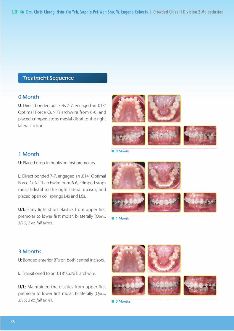
88
IJOI 46 D E R II D D E R II D
r at nt nc
ont
U: Direct bonded brackets 7-7, engaged an .013” Optimal Force CuNiTi archwire from 6-6, and placed crimped stops mesial-distal to the right lateral incisor.
ont
U: Placed drop-in hooks on first premolars.
L: Direct bonded 7-7, engaged an .014” Optimal Force CuNi-Ti archwire from 6-6, crimped stops mesial-distal to the right lateral incisor, and placed open coil springs L4s and L6s.
U/L: Early light short elastics from upper first premolar to lower first molar, bilaterally (Quail,
3/16”, 2 oz, full time).
ont s
U: Bonded anterior BTs on both central incisors.
L: Transitioned to an .018” CuNiTi archwire.
U/L: Maintained the elastics from upper first premolar to lower first molar, bilaterally (Quail,
3/16”, 2 oz, full time). █ 3 Months
█ 1 Month
█ 0 Month
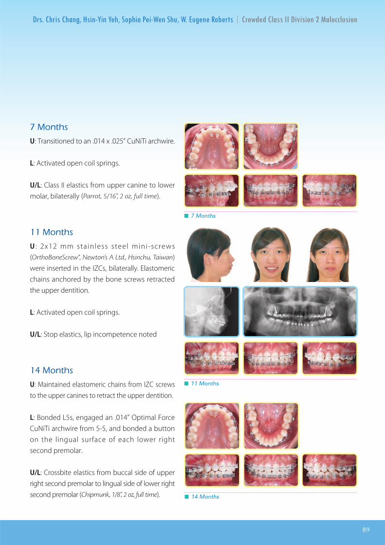
IJOI 46 D E R II D
89
D E R II D
ont s
U: Transitioned to an .014 x .025” CuNiTi archwire.
L: Activated open coil springs.
U/L: Class II elastics from upper canine to lower molar, bilaterally (Parrot, 5/16”, 2 oz, full time).
ont s
U : 2x12 mm stainless steel mini-screws (OrthoBoneScrew®, Newton’s A Ltd., Hsinchu, Taiwan) were inserted in the IZCs, bilaterally. Elastomeric chains anchored by the bone screws retracted the upper dentition.
L: Activated open coil springs.
U/L: Stop elastics, lip incompetence noted
ont s
U: Maintained elastomeric chains from IZC screws to the upper canines to retract the upper dentition.
L: Bonded L5s, engaged an .014” Optimal Force CuNiTi archwire from 5-5, and bonded a button on the lingual surface of each lower right second premolar.
U/L: Crossbite elastics from buccal side of upper right second premolar to lingual side of lower right second premolar (Chipmunk, 1/8”, 2 oz, full time).
█ 11 Months
█ 7 Months
█ 14 Months
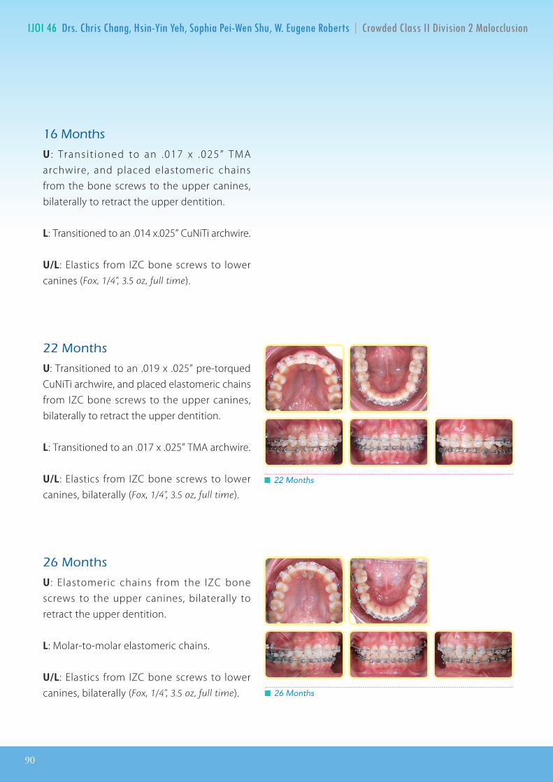
90
IJOI 46 D E R II D D E R II D
ont s
U : Trans i t ioned to an .017 x .025” TMA archwire, and placed elastomeric chains from the bone screws to the upper canines, bilaterally to retract the upper dentition.
L: Transitioned to an .014 x.025” CuNiTi archwire.
U/L: Elastics from IZC bone screws to lower canines (Fox, 1/4”, 3.5 oz, full time).
ont s
U: Transitioned to an .019 x .025” pre-torqued CuNiTi archwire, and placed elastomeric chains from IZC bone screws to the upper canines, bilaterally to retract the upper dentition.
L: Transitioned to an .017 x .025” TMA archwire.
U/L: Elastics from IZC bone screws to lower canines, bilaterally (Fox, 1/4”, 3.5 oz, full time).
ont s
U : Elastomeric chains from the IZC bone screws to the upper canines, bilaterally to retract the upper dentition.
L: Molar-to-molar elastomeric chains.
U/L: Elastics from IZC bone screws to lower canines, bilaterally (Fox, 1/4”, 3.5 oz, full time).
█ 22 Months
█ 26 Months
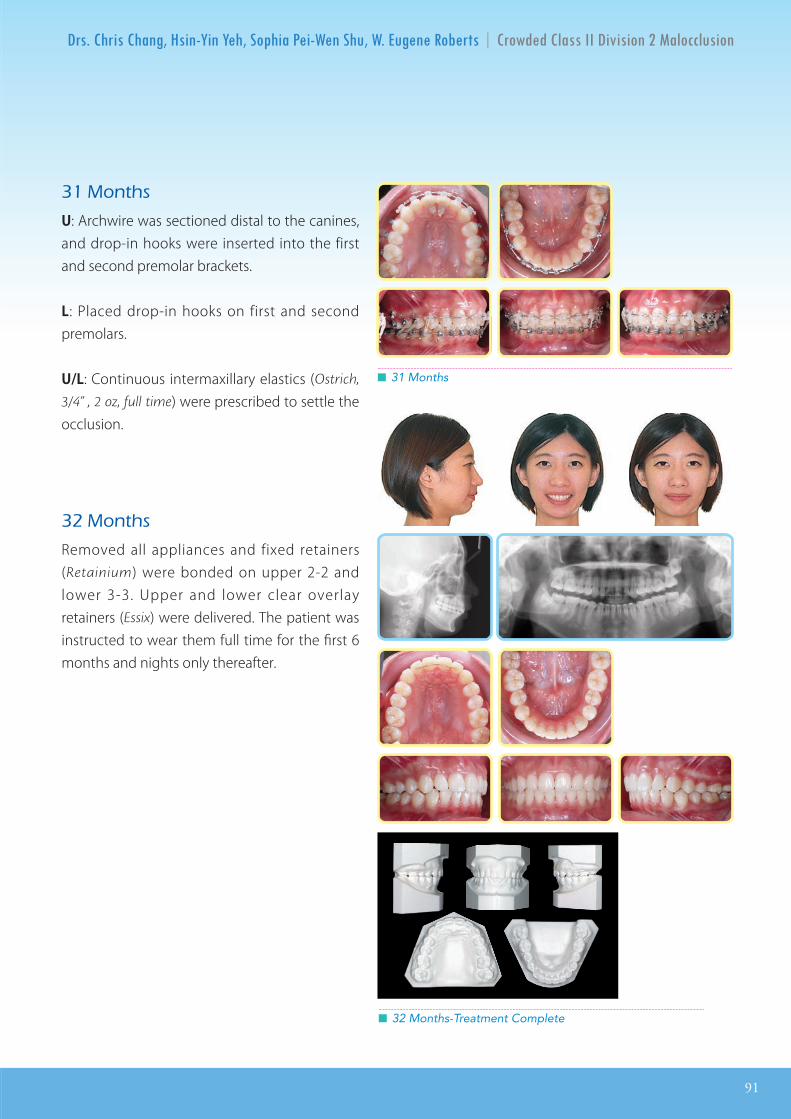
IJOI 46 D E R II D
91
D E R II D
ont s
U: Archwire was sectioned distal to the canines, and drop-in hooks were inserted into the first and second premolar brackets.
L: Placed drop-in hooks on first and second premolars.
U/L: Continuous intermaxillary elastics (Ostrich,
3/4” , 2 oz, full time) were prescribed to settle the occlusion.
ont s
Removed all appliances and fixed retainers (Retainium) were bonded on upper 2-2 and lower 3-3. Upper and lower clear overlay retainers (Essix) were delivered. The patient was instructed to wear them full time for the first 6 months and nights only thereafter.
█ 31 Months
█ 32 Months-Treatment Complete
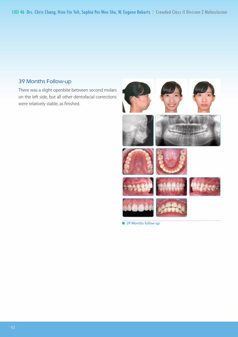
92
IJOI 46 D E R II D D E R II D
ont s ollow u
There was a slight openbite between second molars on the left side, but all other dentofacial corrections were relatively stable, as finished.
█ 39 Months follow-up
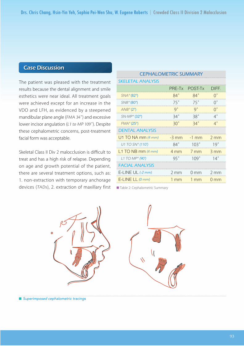
IJOI 46 D E R II D
93
D E R II D
a i c ion
The patient was pleased with the treatment results because the dental alignment and smile esthetics were near ideal. All treatment goals were achieved except for an increase in the VDO and LFH, as evidenced by a steepened mandibular plane angle (FMA 34˚) and excessive lower incisor angulation (L1 to MP 109˚). Despite these cephalometric concerns, post-treatment facial form was acceptable.
Skeletal Class II Div 2 malocclusion is difficult to treat and has a high risk of relapse. Depending on age and growth potential of the patient, there are several treatment options, such as: 1. non-extraction with temporary anchorage devices (TADs), 2. extraction of maxillary first
█ Superimposed cephalometric tracings
A O I A
A A A I
PRE-Tx POST-Tx DIFF.
SNA° (82°) 84̊ 84̊ 0̊ SNB° (80°) 75̊ 75̊ 0̊ ANB° (2°) 9̊ 9̊ 0̊ SN-MP° (32°) 34̊ 38̊ 4̊FMA° (25°) 30̊ 34̊ 4̊
A A A I
U1 TO NA mm (4 mm) -3 mm -1 mm 2 mmU1 TO SN° (110˚) 84̊ 103̊ 19̊
L1 TO NB mm (4 mm) 4 mm 7 mm 3 mmL1 TO MP° (90˚) 95̊ 109̊ 14̊ A IA A A I
E-LINE UL (-2 mm) 2 mm 0 mm 2 mmE-LINE LL (0 mm) 1 mm 1 mm 0 mm
██ Table 2: Cephalometric Summary
94
IJOI 46 D E R II D D E R II D
premolars and mandibular second premolars, and 3. orthognathic surgery for mandible advancement. The patient preferred nonsurgical orthodontic treatment to minimize the risk of facial compromise. Extraction treatment for Class II Div 2 may flatten the facial profile and deepen the bite. The patient’s pre-treatment cephalometric radiograph showed a slightly protruded upper lip due to the flaring of the upper lateral incisors and relatively narrow arches. A non-extraction treatment plan was the patient’s choice, so the Damon self-ligating system with extra-alveolar (E-A) TADs was indicated.
Variable torque brackets were particularly advantageous for aligning the upper arch, but the low torque brackets failed to prevent flaring of the lower incisors. The BTs and Class II elastics opened the LFH 4̊. Transient incompetence lips were noted at 9 months into treatment. The bite opening facilitated the alignment of the severe skeletal malocclusion (ANB 9˚ ) , but resulted in intermaxi l lary alignment problems. It was necessary to increase the axial inclination of the lower incisors to prevent an opening of the overjet because the upper incisors were flared 19̊.
Anter ior BTs are excel lent tools for the correction of deep bite if opening the bite and posterior mandibular rotation are acceptable m e c h a n i c s . F o r t u n a t e l y , t h e t r a n s i e n t incompetent lips noted at 9 months resolved
by the end of treatment at 32 months. They are easy to use and decrease the treatment time for many patients. Early light Class II elastics play an important role in correcting deep bite without decreasing the smile arc. The light force reduces the side effects of the horizontal component of force, which can produce unnecessary tipping of the teeth. For the present patient, anterior BTs and Class II elastics resolved the overjet problem, but extruded the mandibular molars, which increased the mandibular plane angle due to posterior rotation of the mandible.
Anchorage is challenging for skeletal Class II Div 2 malocclusion. En masse movement of the anterior segment and improvement in the facial profile was accomplished with E-A anchorage via IZC bone screws. This minimally invasive method reduced treatment time and simplified the mechanics, but failed to control the LFH and VDO. Permanent retention is indicated for maintaining the camouflage correction of this severe skeletal Class II Div 2 malocclusion.
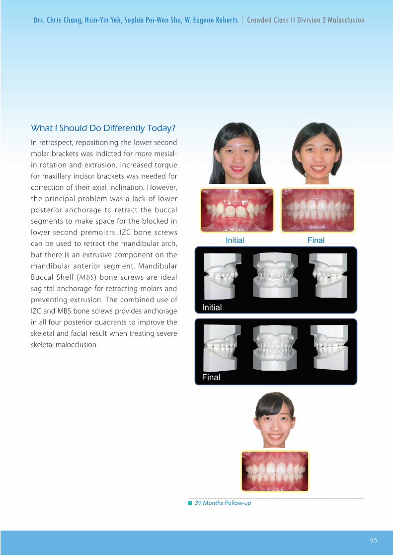
IJOI 46 D E R II D
95
D E R II D
█ 39 Months Follow-up
at I ould o i erentl oda
In retrospect, repositioning the lower second molar brackets was indicted for more mesial-in rotation and extrusion. Increased torque for maxillary incisor brackets was needed for correction of their axial inclination. However, the principal problem was a lack of lower posterior anchorage to retract the buccal segments to make space for the blocked in lower second premolars. IZC bone screws can be used to retract the mandibular arch, but there is an extrusive component on the mandibular anterior segment. Mandibular Buccal Shelf (MBS) bone screws are ideal sagittal anchorage for retracting molars and preventing extrusion. The combined use of IZC and MBS bone screws provides anchorage in all four posterior quadrants to improve the skeletal and facial result when treating severe skeletal malocclusion.
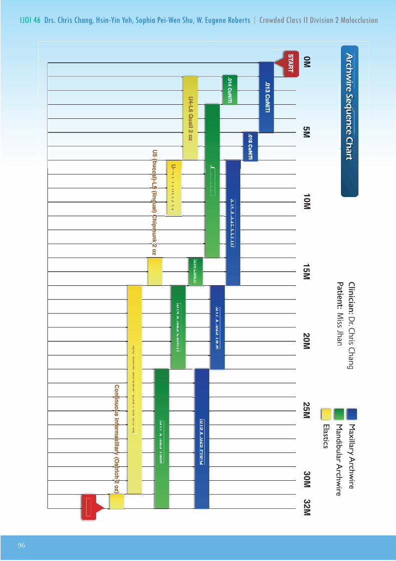
96
IJOI 46 D E R II D
Archwire Sequence Chart
Archwire Sequence Chart
Clinician:Dr. Chris ChangPatient: M
iss JhanM
axillary Archw
ire
Mandibular A
rchwire
Elastics
START
FINISH
0M5M
10M15M
20M25M
U4-L6 Quail 2 oz
Continuous intermaxillary (
Continuous intermaxillary (
Continuous intermaxillary (
Continuous intermaxillary (
Continuous intermaxillary (
Continuous intermaxillary (
Continuous intermaxillary (
Continuous intermaxillary (O
strich 2 oz)O
strich 2 oz)O
strich 2 oz)O
strich 2 oz)O
strich 2 oz)
30M32M
.013 CuNiTi
.014 CuNiTi
.018 CuNiTi
.018 CuNiTi.018 CuNiTi .014 x .025 CuNiTi
U4-L6 Parrot 2 oz U4-L6 Parrot 2 oz
U5 (buccal)-L5 U5 (buccal)-L5 U5 (buccal)-L5 U5 (buccal)-L5 U5 (buccal)-L5 U5 (buccal)-L5 U5 (buccal)-L5 U5 (buccal)-L5 U5 (buccal)-L5 U5 (buccal)-L5 (lingual) Chipm
unk 2 oz(lingual) Chipm
unk 2 oz(lingual) Chipm
unk 2 oz(lingual) Chipm
unk 2 oz(lingual) Chipm
unk 2 oz(lingual) Chipm
unk 2 oz
.014 CuNiTi
.017 x .025 TMA
.014 x .025 CuNiTi
.019 x .025 Pre-Q
.017 x .025 TMA
IZC bone screws- L3s Fox 3.5 oz
rcir
nc
art

高效簡報學習法
(四)
(四)
(六∼一)
強化你的 簡報力
賈伯斯的簡報秘訣與設計要素
精修繪圖及動畫技巧
2017
Keynote Workshop
講師:Dr. Rungsi Thavarungkul
報名 2017 K456 課程即贈送 2016 及 2017 課程視訊。
名額有限,以繳費順序為依據。
舊生享特惠優惠價,限額6名。
1-2
3
456
K
K
K
03-573-5676 [email protected] 新竹市建中一路25號2樓
全新設計!
11/23
11/16
12/2-4
高效簡報學習法
(四)
(四)
(六∼一)
由國際知名的牙科講師張慧男醫師主講的一天課程,將以他幽默風趣的演講風格來剖析賈柏斯的美學概念以及演講秘訣,利用實例來說明如何設計出視覺優美且知識性豐富的專業演講。
數位化潮流下的牙科簡報,不僅需要清晰的臨床照片,也需要精確的圖表和流暢的動畫來吸引觀眾,而優秀的視覺化工具更使您的演講獨樹一格且令人難忘。
Keynote 課程中, n 將分享他利用 Keynote 軟體繪製精美牙科插圖的經驗,並一步步教會您如何從構想和草圖創建出令人驚艷的成果。跟隨簡報美學大師的腳步,您也可以秀出創意、站上世界舞台!
強化你的 簡報力
賈伯斯的簡報秘訣與設計要素
精修繪圖及動畫技巧
2017
Keynote Workshop
講師:Dr. Rungsi Thavarungkul
講師:張慧男醫師
講師:金牛頓工程師
報名 2017 K456 課程即贈送 2016 及 2017 課程視訊。
名額有限,以繳費順序為依據。
舊生享特惠優惠價,限額6名。
若取消報名,30天前退款將扣除10%行政手續費,30天內退款將扣除30%行政手續費。
1-2
3
456
K
K
K
03-573-5676 [email protected] 新竹市建中一路25號2樓
全新設計!
無論是 Keynote 新手或是略有經驗的使用者,在一天的簡報課程中,將精選介紹 Keynote 的重要工具,幫助你設計合適的動畫,分享設計資料視覺化的秘訣,並搭配 Mac 內建常用的影音工具來編修圖片和影像剪輯,金牛頓的教學團隊將教你如何使用Keynote 創造出動人的專業簡報。
11/23
11/16
12/2-4
This app is designed for iPad, iPhone and iPad touch
Get
[email protected]+886-3-573-5676 http://orthobonescrew.com http://orthobonescrew.com
ote 1. Most video courses are available in both English and Chinese and are sold separately. 2. Customers who purchase one set of courses will have access to both the narrated and non-narrated version. 3. Some courses, including Damon Q and Advanced, are renewed annually and each renewal is to be purchased separately with a 50% discount.
e-Courses
Series
TThis app is designed forhis app is designed foriPad, iPhone and iPad touchiPad, iPhone and iPad touchiPad, iPhone and iPad touch
Get
ich ea nin e e ienceThese are all Dr. Chang’s course sl ides turnedinto videos with lots of pictures, videos and clear headings. So it’s very easy to understand whenyou can see how the procedures are done. It’s much more interesting and practical than reading a textbook . I t’s a ver y easy and efficient way of learning and constantly refreshing your memory.
E E E
D B E
oiced oiced oicedoiced
Newton’s Ap or o o r . o
⒈Go to App Store on your device.
⒉ "Sign in" with your Apple ID. ⒊ "Create Apple ID" if you don't have one.
⒋ Search for "BDE.” ⒌ Click "FREE" and “INSTALL APP.”
⒍ Download completed! ⒎ Once downloaded, click "BDE" to launch the app.
o ar i r i o r pai i o or i apro o a [email protected] or i or a io .
Step-by-step Instructions
. o i o o r ar a ai a i o i a ia ar o para .
. o r o p r a o o o r i a ao o arra a o arra r io .
. o o r i i a o a a ar r
o
a a a a r a i o p r a parai a i o .
Knowledge in your pocket
o r pro io a or o o i a i p a i o o r . r a aa i r r a ro o or o ir o . o r ar i rr. ri a . i p p pra i a o r i a o r ar i a
i or a i a p ri . o a a o a a o r a io a o r ao r o o i a p a o o ro pp.
Online accessThis app is designed for iPad, iPhone and iPad touch
Offline download and unlimited viewing
Your personal mobile learning device
Voiced in English
Get
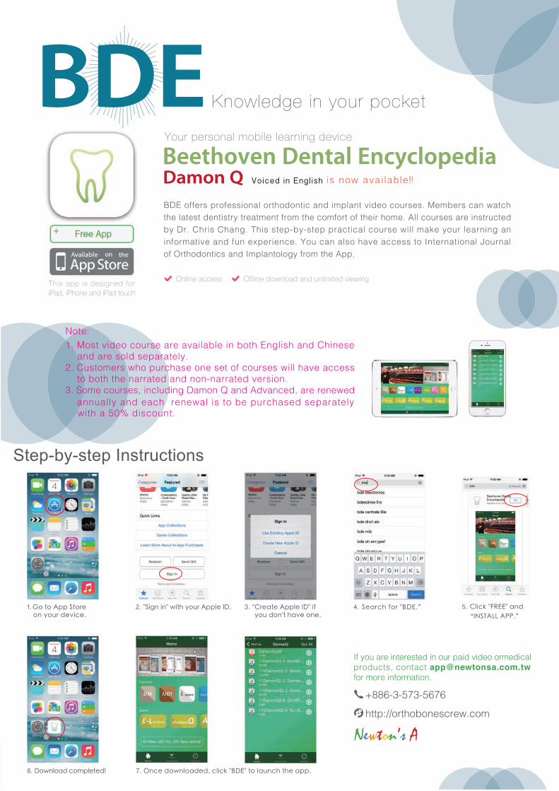
This app is designed for iPad, iPhone and iPad touch
Get
[email protected]+886-3-573-5676 http://orthobonescrew.com
ote 1. Most video courses are available in both English and Chinese and are sold separately. 2. Customers who purchase one set of courses will have access to both the narrated and non-narrated version. 3. Some courses, including Damon Q and Advanced, are renewed annually and each renewal is to be purchased separately with a 50% discount.
e-Courses
Series
E E E
D B E
Newton’s Ap or o o r . op or o o r . o
⒈Go to App Store on your device.
⒉ "Sign in" with your Apple ID. ⒊ "Create Apple ID" if you don't have one.
⒋ Search for "BDE.” ⒌ Click "FREE" and “INSTALL APP.”
⒍ Download completed! ⒎ Once downloaded, click "BDE" to launch the app.
o ar i r i o r pai i o or i apro o a [email protected] or i or a io .
Step-by-step Instructions
. o i o o r ar a ai a i o i a ia ar o para .
. o r o p r a o o o r i a ao o arra a o arra r io .
. o o r i i a o a a ar r
o
a a a a r a i o p r a parai a i o .
Knowledge in your pocket
o r pro io a or o o i a i p a i o o r . r a aa i r r a ro o or o ir o . o r ar i rr. ri a . i p p pra i a o r i a o r ar i a
i or a i a p ri . o a a o a a o r a io a o r ao r o o i a p a o o ro pp.
Online accessThis app is designed for iPad, iPhone and iPad touch
Offline download and unlimited viewing
Your personal mobile learning device
Voiced in English is now available!!
Get
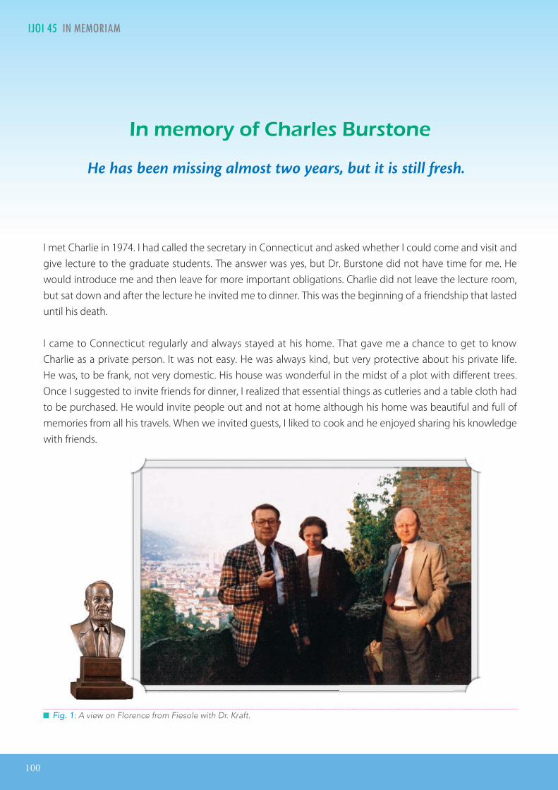
100
IJOI 4 I E ORIA I IJOI 4
I met Charlie in 1974. I had called the secretary in Connecticut and asked whether I could come and visit and give lecture to the graduate students. The answer was yes, but Dr. Burstone did not have time for me. He would introduce me and then leave for more important obligations. Charlie did not leave the lecture room, but sat down and after the lecture he invited me to dinner. This was the beginning of a friendship that lasted until his death.
I came to Connecticut regularly and always stayed at his home. That gave me a chance to get to know Charlie as a private person. It was not easy. He was always kind, but very protective about his private life. He was, to be frank, not very domestic. His house was wonderful in the midst of a plot with different trees. Once I suggested to invite friends for dinner, I realized that essential things as cutleries and a table cloth had to be purchased. He would invite people out and not at home although his home was beautiful and full of memories from all his travels. When we invited guests, I liked to cook and he enjoyed sharing his knowledge with friends.
█ Fig. 1: A view on Florence from Fiesole with Dr. Kraft.
n or o ar r ton
He has been missing almost two years, but it is still fresh.

IJOI 4 I E ORIA
101
I IJOI 4
█ Fig. 2: Preparing for the five star dinner.
Charlie came to Aarhus, Europe the first time together with Dr. Nanda who had just joined the faculty. They gave a typodont course. In comparison with all the other courses I had taken: Tweed, Jarabak, Ricketts, Begg, the segmented approach made sense. I could use the mathematical logic and soon our department was dominated by the Burstone’s thoughts and the six Geometries were eagerly discussed. But we shared not only orthodontics, but also philosophy and religion. He came to Italy (Figures 1-4) where we gave courses together and where he enjoyed the museums and the mountains. Sometimes he over-estimated himself and my younger son almost had to carry him
█ Fig. 4: In Florence with friends: professor Houstone and professor Moss and wife are all no longer with us. Also Professor Prahl and professor Wenzel and Dr Fotis joined us.
█ Fig. 3: On the street with the beloved Timberland accompanied by Dr. Fotis who also invited Charlie for a vacation on Rhodes.
Dr. Birte Melsen, DDSAffiliate, Universities of Lexington Kentucky, Perth West Australia and Hannover Germany
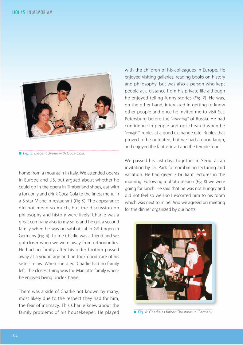
102
IJOI 4 I E ORIA I IJOI 4
with the children of his colleagues in Europe. He enjoyed visiting galleries, reading books on history and philosophy, but was also a person who kept people at a distance from his private life although he enjoyed telling funny stories (Fig. 7). He was, on the other hand, interested in getting to know other people and once he invited me to visit Sct. Petersburg before the “opening” of Russia. He had confidence in people and got cheated when he “bought” rubles at a good exchange rate. Rubles that proved to be outdated, but we had a good laugh, and enjoyed the fantastic art and the terrible food.
We passed his last days together in Seoul as an invitation by Dr. Park for combining lecturing and vacation. He had given 3 brilliant lectures in the morning. Following a photo session (Fig. 8) we were going for lunch. He said that he was not hungry and did not feel so well so I escorted him to his room which was next to mine. And we agreed on meeting for the dinner organized by our hosts.
█ Fig. 5: Elegant dinner with Coca-Cola.
home from a mountain in Italy. We attended operas in Europe and US, but argued about whether he could go in the opera in Timberland shoes, eat with a fork only and drink Coca-Cola to the finest menu in a 3 star Michelin restaurant (Fig. 5). The appearance did not mean so much, but the discussion on philosophy and history were lively. Charlie was a great company also to my sons and he got a second family when he was on sabbatical in Göttingen in Germany (Fig. 6). To me Charlie was a friend and we got closer when we were away from orthodontics. He had no family, after his older brother passed away at a young age and he took good care of his sister-in-law. When she died, Charlie had no family left. The closest thing was the Marcotte family where he enjoyed being Uncle Charlie.
There was a side of Charlie not known by many; most likely due to the respect they had for him, the fear of intimacy. This Charlie knew about the family problems of his housekeeper. He played █ Fig. 6: Charlie as father Christmas in Germany.
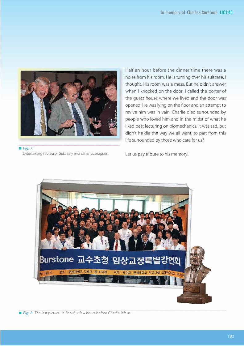
IJOI 4 I E ORIA
103
I IJOI 4
Half an hour before the dinner time there was a noise from his room. He is turning over his suitcase, I thought. His room was a mess. But he didn’t answer when I knocked on the door. I called the porter of the guest house where we lived and the door was opened. He was lying on the floor and an attempt to revive him was in vain. Charlie died surrounded by people who loved him and in the midst of what he liked best lecturing on biomechanics. It was sad, but didn’t he die the way we all want, to part from this life surrounded by those who care for us?
Let us pay tribute to his memory! █ Fig. 7: Entertaining Professor Subtelny and other colleagues.
█ Fig. 8: The last picture. In Seoul, a few hours before Charlie left us.
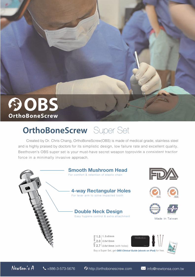
OBSOrthoBoneScrew
[email protected] http://orthobonescrew.com+886-3-573-5676
OrthoBoneScrew Super Set
Buy a Super Set, get OBS Clinical Guide (ebook on iPad) for free.
Smooth Mushroom Head
4-way Rectangular Holes
Double Neck Design
1.52.02.7
Sc
re
w T
yp
e
1 . 5 x 8 m m
2.0x12mm
2.0x14mm (with holes)
Made in TaiwanOBSOrthoBoneScrew
[email protected] http://orthobonescrew.com+886-3-573-5676
OrthoBoneScrew Super Set Created by Dr. Chris Chang, OrthoBoneScrew(OBS) is made of medical grade, stainless steel and is highly praised by doctors for its simplistic design, low failure rate and excellent quality.Beethoven's OBS super set is your must-have secret weapon toprovide a consistent tractionforce in a minimally invasive approach.
Buy a Super Set, get OBS Clinical Guide (ebook on iPad) for free.
Smooth Mushroom Heador com ort & retent ion o e last ic chain
4-way Rectangular Holesor le er arm to sol e im acte tooth
Double Neck DesignEasy hygiene contro l & extra at tachment
1.52.02.7
Screw Type
1 . 5 x 8 m m
2.0x12mm
2.0x14mm (with holes)
Made in Taiwan
ental oducts Essential Kit
Double Retractors
Stainless Steel MirrorSize : L (Recommanded), M, S
Bite Turbo 2.0Handle x1, BT molds x6, BT extended molds x6, Button molds x6
Autoclavable!NEW
OBSOrthoBoneScrew
[email protected] http://orthobonescrew.com+886-3-573-5676
OrthoBoneScrew Super Set
Buy a Super Set, get OBS Clinical Guide (ebook on iPad) for free.
Smooth Mushroom Head
4-way Rectangular Holes
Double Neck Design
1.52.02.7
Screw Type
1 . 5 x 8 m m
2.0x12mm
2.0x14mm (with holes)
Made in Taiwan
ental oducts Essential Kit
Double Retractors
Stainless Steel MirrorSize : L (Recommanded), M, S
Bite Turbo 2.0Handle x1, BT molds x6, BT extended molds x6, Button molds x6
Autoclavable!NEW
ental oducts Essential Kit
Double Retractors
Stainless Steel MirrorSize : L (Recommanded), M, S
Bite Turbo 2.0Handle x1, BT molds x6, BT extended molds x6, Button molds x6
Autoclavable!NEW
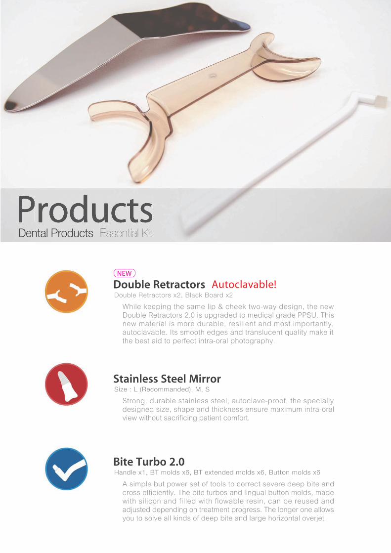
OBSOrthoBoneScrew
[email protected] http://orthobonescrew.com+886-3-573-5676
OrthoBoneScrew Super Set
Buy a Super Set, get OBS Clinical Guide (ebook on iPad) for free.
Smooth Mushroom Head
4-way Rectangular Holes
Double Neck Design
1.52.02.7
Sc
re
w T
yp
e
1 . 5 x 8 m m
2.0x12mm
2.0x14mm (with holes)
Made in TaiwanOBSOrthoBoneScrew
[email protected] http://orthobonescrew.com+886-3-573-5676
OrthoBoneScrew Super Set
Buy a Super Set, get OBS Clinical Guide (ebook on iPad) for free.
Smooth Mushroom Head
4-way Rectangular Holes
Double Neck Design
1.52.02.7
Screw Type
1 . 5 x 8 m m
2.0x12mm
2.0x14mm (with holes)
Made in Taiwan
ental oducts Essential Kit
Double Retractors
Stainless Steel MirrorSize : L (Recommanded), M, S
Bite Turbo 2.0Handle x1, BT molds x6, BT extended molds x6, Button molds x6
Autoclavable!NEW
OBSOrthoBoneScrew
[email protected] http://orthobonescrew.com+886-3-573-5676
OrthoBoneScrew Super Set
Buy a Super Set, get OBS Clinical Guide (ebook on iPad) for free.
Smooth Mushroom Head
4-way Rectangular Holes
Double Neck Design
1.52.02.7
Screw Type
1 . 5 x 8 m m
2.0x12mm
2.0x14mm (with holes)
Made in Taiwan
ental oducts Essential Kit
Double Retractorsou le Ret acto s x , Blac Boa d x
Stainless Steel MirrorSize : L (Recommanded), M, S
Bite Turbo 2.0Handle x1, BT molds x6, BT extended molds x6, Button molds x6
hile keeping the same lip cheek two way design, the new
ouble etractors . is upgraded to medical grade . This
new material is more durable, resilient and most importantly,
autoclavable. ts smooth edges and translucent uality make it
the best aid to perfect intra oral photography.
trong, durable stainless steel, autoclave proof, the specially
designed size, shape and thickness ensure ma imum intra oral
view without sacrificing patient comfort.
A simple but power set of tools to correct severe deep bite and
cross efficiently. The bite turbos and lingual button molds, made
with silicon and filled with flowable resin, can be reused and
adjusted depending on treatment progress. The longer one allows
you to solve all kinds of deep bite and large horizontal overjet.
Autoclavable!NEW
Productsental oducts Essential Kit
Double Retractors
Stainless Steel MirrorSize : L (Recommanded), M, S
Bite Turbo 2.0Handle x1, BT molds x6, BT extended molds x6, Button molds x6
Autoclavable!NEW
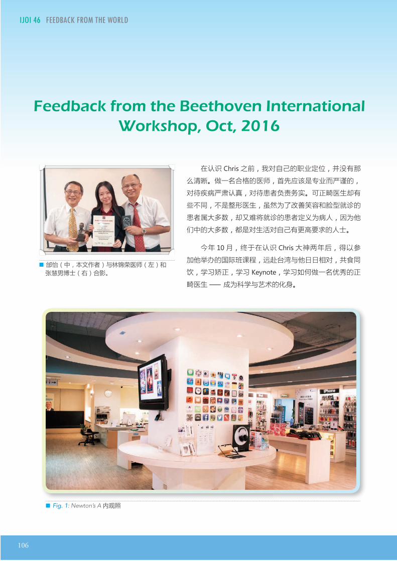
ac ro t t o n nt rnationaor op ct
██ 邰怡(中,本文作者)与林锦荣医师(左)和█ 张慧男博士(右)合影。
在认识 Chris 之前,我对自己的职业定位,并没有那
么清晰。做一名合格的医师,首先应该是专业而严谨的,
对待疾病严肃认真,对待患者负责务实。可正畸医生却有
些不同,不是整形医生,虽然为了改善笑容和脸型就诊的
患者属大多数,却又难将就诊的患者定义为病人,因为他
们中的大多数,都是对生活对自己有更高要求的人士。
今年 10 月,终于在认识 Chris 大神两年后,得以参
加他举办的国际班课程,远赴台湾与他日日相对,共食同
饮,学习矫正,学习 Keynote,学习如何做一名优秀的正
畸医生 — 成为科学与艺术的化身。
█ Fig. 1: Newton’s A 内观照
IJOI 46 EED A RO T E ORLD
106
EED A RO T E ORLD IJOI 46
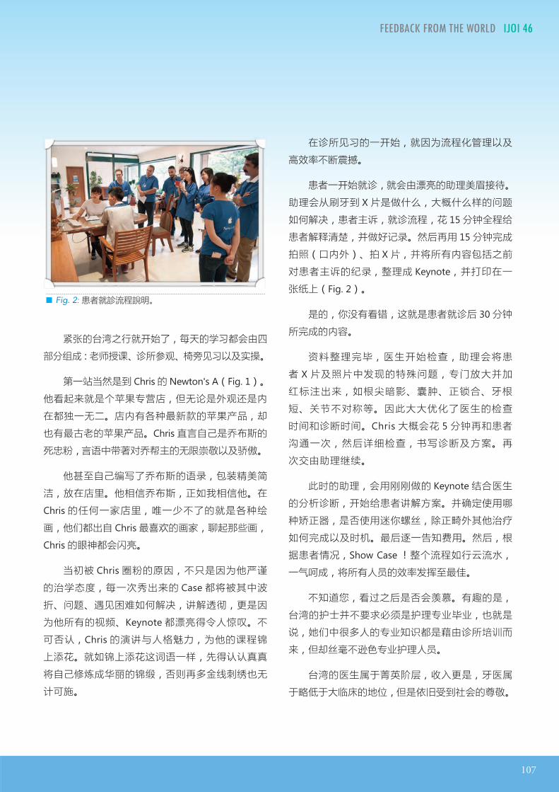
█ Fig. 2:█患者就診流程說明。
在诊所见习的一开始,就因为流程化管理以及
高效率不断震撼。
患者一开始就诊,就会由漂亮的助理美眉接待。
助理会从刷牙到 X 片是做什么,大概什么样的问题
如何解决,患者主诉,就诊流程,花 15 分钟全程给
患者解释清楚,并做好记录。然后再用 15 分钟完成
拍照(口内外)、拍 X 片,并将所有内容包括之前
对患者主诉的纪录,整理成 Keynote,并打印在一
张纸上(Fig. 2)。
是的,你没有看错,这就是患者就诊后 30 分钟
所完成的内容。
资料整理完毕,医生开始检查,助理会将患
者 X 片及照片中发现的特殊问题,专门放大并加
红标注出来,如根尖暗影、囊肿、正锁合、牙根
短、关节不对称等。因此大大优化了医生的检查
时间和诊断时间。Chris 大概会花 5 分钟再和患者
沟通一次,然后详细检查,书写诊断及方案。再
次交由助理继续。
此时的助理,会用刚刚做的 Keynote 结合医生
的分析诊断,开始给患者讲解方案。并确定使用哪
种矫正器,是否使用迷你螺丝,除正畸外其他治疗
如何完成以及时机。最后逐一告知费用。然后,根
据患者情况,Show Case !整个流程如行云流水,
一气呵成,将所有人员的效率发挥至最佳。
不知道您,看过之后是否会羡慕。有趣的是,
台湾的护士并不要求必须是护理专业毕业,也就是
说,她们中很多人的专业知识都是藉由诊所培训而
来,但却丝毫不逊色专业护理人员。
台湾的医生属于菁英阶层,收入更是,牙医属
于略低于大临床的地位,但是依旧受到社会的尊敬。
紧张的台湾之行就开始了,每天的学习都会由四
部分组成:老师授课、诊所参观、椅旁见习以及实操。
第一站当然是到 Chris 的 Newton's A(Fig. 1)。
他看起来就是个苹果专营店,但无论是外观还是内
在都独一无二。店内有各种最新款的苹果产品,却
也有最古老的苹果产品。Chris 直言自己是乔布斯的
死忠粉,言语中带著对乔帮主的无限崇敬以及骄傲。
他甚至自己编写了乔布斯的语录,包装精美简
洁,放在店里。他相信乔布斯,正如我相信他。在
Chris 的任何一家店里,唯一少不了的就是各种绘
画,他们都出自 Chris 最喜欢的画家,聊起那些画,
Chris 的眼神都会闪亮。
当初被 Chris 圈粉的原因,不只是因为他严谨
的治学态度,每一次秀出来的 Case 都将被其中波
折、问题、遇见困难如何解决,讲解透彻,更是因
为他所有的视频、Keynote 都漂亮得令人惊叹。不
可否认,Chris 的演讲与人格魅力,为他的课程锦
上添花。就如锦上添花这词语一样,先得认认真真
将自己修炼成华丽的锦缎,否则再多金线刺绣也无
计可施。
IJOI 46 EED A RO T E ORLD EED A RO T E ORLD IJOI 46
107
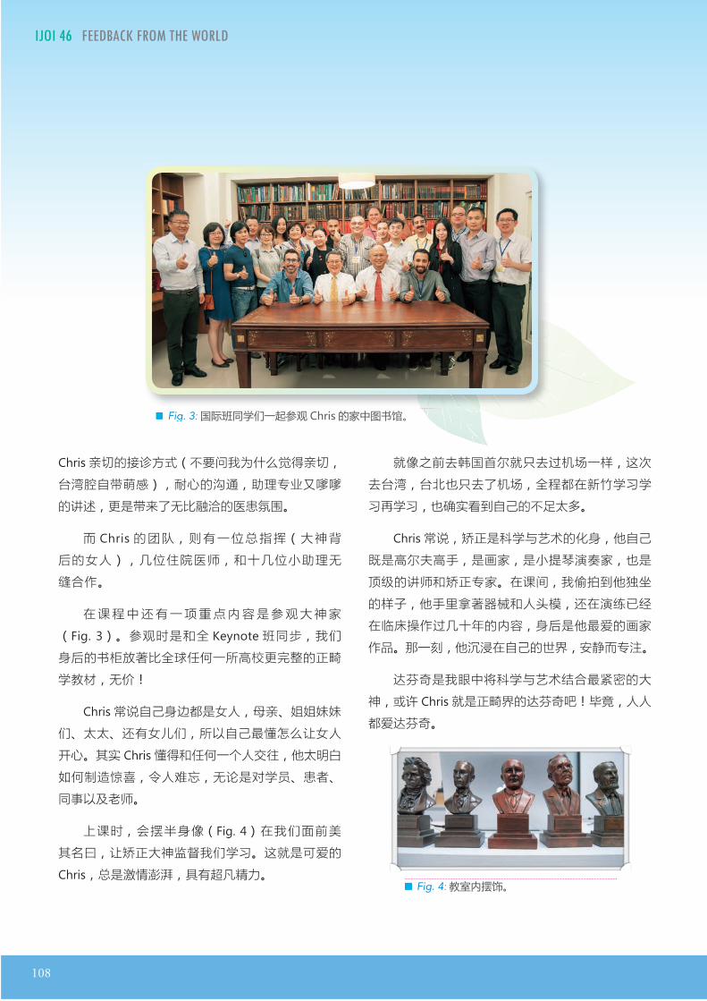
就像之前去韩国首尔就只去过机场一样,这次
去台湾,台北也只去了机场,全程都在新竹学习学
习再学习,也确实看到自己的不足太多。
Chris 常说,矫正是科学与艺术的化身,他自己
既是高尔夫高手,是画家,是小提琴演奏家,也是
顶级的讲师和矫正专家。在课间,我偷拍到他独坐
的样子,他手里拿著器械和人头模,还在演练已经
在临床操作过几十年的内容,身后是他最爱的画家
作品。那一刻,他沉浸在自己的世界,安静而专注。
达芬奇是我眼中将科学与艺术结合最紧密的大
神,或许 Chris 就是正畸界的达芬奇吧!毕竟,人人
都爱达芬奇。
Chris 亲切的接诊方式(不要问我为什么觉得亲切,
台湾腔自带萌感),耐心的沟通,助理专业又嗲嗲
的讲述,更是带来了无比融洽的医患氛围。
而 Chris 的团队,则有一位总指挥(大神背
后的女人),几位住院医师,和十几位小助理无
缝合作。
在 课 程 中 还 有 一 项 重 点 内 容 是 参 观 大 神 家
(Fig. 3)。参观时是和全 Keynote 班同步,我们
身后的书柜放著比全球任何一所高校更完整的正畸
学教材,无价!
Chris 常说自己身边都是女人,母亲、姐姐妹妹
们、太太、还有女儿们,所以自己最懂怎么让女人
开心。其实 Chris 懂得和任何一个人交往,他太明白
如何制造惊喜,令人难忘,无论是对学员、患者、
同事以及老师。
上课时,会摆半身像(Fig. 4)在我们面前美
其名曰,让矫正大神监督我们学习。这就是可爱的
Chris,总是激情澎湃,具有超凡精力。 █ Fig. 4: 教室内摆饰。
█ Fig. 3: 观 中 。
IJOI 46 EED A RO T E ORLD
108
EED A RO T E ORLD IJOI 46
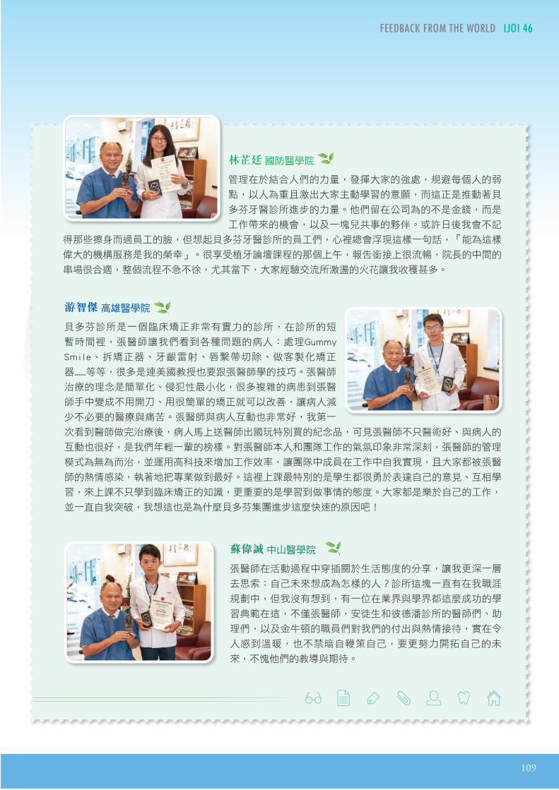
林芷廷 國防醫學院
管理在於結合人們的力量,發揮大家的強處,規避每個人的弱
點,以人為重且激出大家主動學習的意願,而這正是推動著貝
多芬牙醫診所進步的力量。他們留在公司為的不是金錢,而是
工作帶來的機會,以及一塊兒共事的夥伴。或許日後我會不記
得那些擦身而過員工的臉,但想起貝多芬牙醫診所的員工們,心裡總會浮現這樣一句話,「能為這樣
偉大的機構服務是我的榮幸」。很享受植牙論壇課程的那個上午,報告銜接上很流暢,院長的中間的
串場很合適,整個流程不急不徐,尤其當下,大家經驗交流所激盪的火花讓我收穫甚多。
游智傑 高雄醫學院
貝多芬診所是一個臨床矯正非常有實力的診所,在診所的短
暫時間裡,張醫師讓我們看到各種問題的病人:處理Gummy
Smile、拆矯正器、牙齦雷射、唇繫帶切除、做客製化矯正
器......等等,很多是連美國教授也要跟張醫師學的技巧。張醫師
治療的理念是簡單化、侵犯性最小化,很多複雜的病患到張醫
師手中變成不用開刀、用很簡單的矯正就可以改善,讓病人減
少不必要的醫療與痛苦。張醫師與病人互動也非常好,我第一
次看到醫師做完治療後,病人馬上送醫師出國玩特別買的紀念品,可見張醫師不只醫術好、與病人的
互動也很好,是我們年輕一輩的榜樣。對張醫師本人和團隊工作的氣氛印象非常深刻,張醫師的管理
模式為無為而治,並運用高科技來增加工作效率,讓團隊中成員在工作中自我實現,且大家都被張醫
師的熱情感染,執著地把專業做到最好。這裡上課最特別的是學生都很勇於表達自己的意見、互相學
習,來上課不只學到臨床矯正的知識,更重要的是學習到做事情的態度。大家都是樂於自己的工作,
並一直自我突破,我想這也是為什麼貝多芬集團進步這麼快速的原因吧!
蘇偉誠 中山醫學院
張醫師在活動過程中穿插關於生活態度的分享,讓我更深一層
去思索:自己未來想成為怎樣的人?診所這塊一直有在我職涯
規劃中,但我沒有想到,有一位在業界與學界都這麼成功的學
習典範在這,不僅張醫師,安徒生和彼德潘診所的醫師們、助
理們,以及金牛頓的職員們對我們的付出與熱情接待,實在令
人感到溫暖,也不禁暗自鞭策自己,要更努力開拓自己的未
來,不愧他們的教導與期待。
IJOI 46 EED A RO T E ORLD EED A RO T E ORLD IJOI 46
109
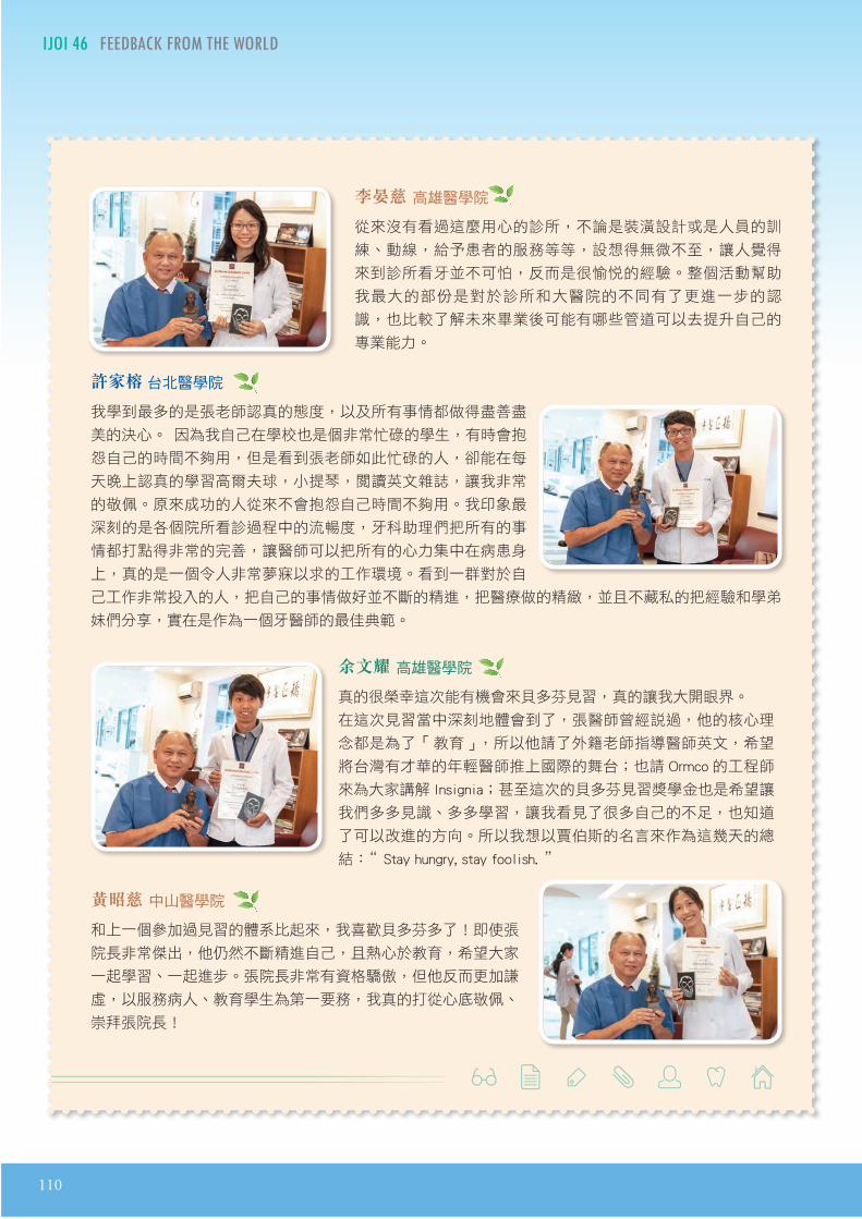
李晏慈 高雄醫學院
從來沒有看過這麼用心的診所,不論是裝潢設計或是人員的訓
練、動線,給予患者的服務等等,設想得無微不至,讓人覺得
來到診所看牙並不可怕,反而是很愉悅的經驗。整個活動幫助
我最大的部份是對於診所和大醫院的不同有了更進一步的認
識,也比較了解未來畢業後可能有哪些管道可以去提升自己的
專業能力。
許家榕 台北醫學院
我學到最多的是張老師認真的態度,以及所有事情都做得盡善盡
美的決心。 因為我自己在學校也是個非常忙碌的學生,有時會抱
怨自己的時間不夠用,但是看到張老師如此忙碌的人,卻能在每
天晚上認真的學習高爾夫球,小提琴,閱讀英文雜誌,讓我非常
的敬佩。原來成功的人從來不會抱怨自己時間不夠用。我印象最
深刻的是各個院所看診過程中的流暢度,牙科助理們把所有的事
情都打點得非常的完善,讓醫師可以把所有的心力集中在病患身
上,真的是一個令人非常夢寐以求的工作環境。看到一群對於自
己工作非常投入的人,把自己的事情做好並不斷的精進,把醫療做的精緻,並且不藏私的把經驗和學弟
妹們分享,實在是作為一個牙醫師的最佳典範。
黃昭慈 中山醫學院
和上一個參加過見習的體系比起來,我喜歡貝多芬多了!即使張
院長非常傑出,他仍然不斷精進自己,且熱心於教育,希望大家
一起學習、一起進步。張院長非常有資格驕傲,但他反而更加謙
虛,以服務病人、教育學生為第一要務,我真的打從心底敬佩、
崇拜張院長!
余文耀 高雄醫學院
真的很榮幸這次能有機會來貝多芬見習,真的讓我大開眼界。
在這次見習當中深刻地體會到了,張醫師曾經說過,他的核心理
念都是為了「教育」,所以他請了外籍老師指導醫師英文,希望
將台灣有才華的年輕醫師推上國際的舞台;也請 Ormco 的工程師
來為大家講解 Insignia;甚至這次的貝多芬見習獎學金也是希望讓
我們多多見識、多多學習,讓我看見了很多自己的不足,也知道
了可以改進的方向。所以我想以賈伯斯的名言來作為這幾天的總
結:“ Stay hungry, stay foolish. ”
IJOI 46 EED A RO T E ORLD
110
๏๏
3๏๏๏
๏๏๏3
๏๏๏๏๏๏ 申請
3๏ 文件...
...
申請3. 其他有利申請之文件
.
.
3.
+
+
買
買
任選
ake Advanced Cartridge Wash(Light body) (50mlx24) 1箱(24支裝) +ake Advanced Volume tray (380mlx2) 2盒
NT$5,000特價
O iBond All In One(6ml) 或 O iBond S(6ml) 5瓶
e Bond 或 e Bond N 5盒
三合一自酸蝕黏著劑 / 二合一全酸蝕黏著劑
.衛署醫器輸字第021479號
.衛署醫器輸字第018552號、010932號
(定價NT$3,800/ ) (定價NT$2, 00/ )
(定價NT$ , 00/ )
NT$20,000特價 /組
/組
(定價NT$17,000/ )
NT$5,000特價 /組
加成型矽膠印模材
Bonding 黏著劑
冠橋用暫時黏著劑(含/不含丁香油)
奈米變色龍樹脂
(顏色限A2或A3)
‧衛署醫器輸壹字第001563號 ‧衛署醫器輸字第013776號 ‧衛署醫器輸字第022166號 ‧衛部醫器輸壹字015889號
Herculi e Precis Syringe 5支 +
・ 本文宣品僅供牙科診所及牙醫師參考,禁止張貼或擺置於公眾可瀏覽及取閱之處 ・ 本優惠專案僅適用於牙醫診所 ・ 本公司保留贈品更換之權利 ・ 本公司保留更改及終止活動辦法與條款之權利
送 無線LED光聚機
(定價NT$1,800/ / )
(定價NT$1,800/ / )
(定價NT$50,000/ ) (定價NT$55,000/ )
(定價NT$2,100/2 2 / )(定價NT$1,100/2 / )
(定價NT$1,500/2 / )
(定價NT$2,200/2 2 / )
(定價NT$1,300/2 / )(定價NT$1,300/2 / )
(定價NT$1,800/ / )(定價NT$1,800/ / )(定價NT$1,800/ / )
(定價NT$1,800/ / )(定價NT$1,100/2 / )
(定價NT$1,500/2 / )
(定價NT$2,200/2 2 / )
(定價NT$1,300/2 / )
Dyad Flow 免酸蝕自黏式流動性樹脂
(定價NT$5,000/ )
Maxcem Elite二合一多功能樹脂黏著劑Revolution Formula 2
流動性複合樹脂
輕巧無線式LED光聚機 / 電容式無線LED光聚機
Herculite Precis Flow 新一代流動性樹脂
Herculite Precis 奈米變色龍樹脂
Premisa 奈米三態聚合樹脂
Herculite Precis奈米變色龍樹脂
Premisa 奈米三態聚合樹脂
.衛署醫器輸字第022220號.衛署醫器輸字第022166號.衛署醫器輸字第010953號
.衛部醫器輸字第028594號.衛署醫器輸字第011616號
.衛署醫器輸字第023384號.衛部醫器陸輸字第000752號
補綴用流動性樹脂 / 補綴樹脂
(定價NT$1,300/2 / )(定價NT$1,300/2 / )
Revolution Formula 2 流動性複合樹脂
(定價NT$1,500/2 / )
(定價NT$1,800/ / )Premisa 奈米三態聚合樹脂
Maxcem Elite滿 盒
2017/3/26-6/25
+
+
買
買
任選
ake Advanced Cartridge Wash(Light body) (50mlx24) 1箱(24支裝) +ake Advanced Volume tray (380mlx2) 2盒
NT$5,000特價
O iBond All In One(6ml) 或 O iBond S(6ml) 5瓶
e Bond 或 e Bond N 5盒
三合一自酸蝕黏著劑 / 二合一全酸蝕黏著劑
.衛署醫器輸字第021479號
.衛署醫器輸字第018552號、010932號
(定價NT$3,800/ ) (定價NT$2, 00/ )
(定價NT$ , 00/ )
NT$20,000特價 /組
/組
(定價NT$17,000/ )
NT$5,000特價 /組
加成型矽膠印模材
Bonding 黏著劑
冠橋用暫時黏著劑(含/不含丁香油)
奈米變色龍樹脂
(顏色限A2或A3)
‧衛署醫器輸壹字第001563號 ‧衛署醫器輸字第013776號 ‧衛署醫器輸字第022166號 ‧衛部醫器輸壹字015889號
Herculi e Precis Syringe 5支 +
・ 本文宣品僅供牙科診所及牙醫師參考,禁止張貼或擺置於公眾可瀏覽及取閱之處 ・ 本優惠專案僅適用於牙醫診所 ・ 本公司保留贈品更換之權利 ・ 本公司保留更改及終止活動辦法與條款之權利
+
+
買
買
任選
ake Advanced Cartridge Wash(Light body) (50mlx24) 1箱(24支裝) +ake Advanced Volume tray (380mlx2) 2盒
NT$5,000特價
O iBond All In One(6ml) 或 O iBond S(6ml) 5瓶
e Bond 或 e Bond N 5盒
三合一自酸蝕黏著劑 / 二合一全酸蝕黏著劑
.衛署醫器輸字第021479號
.衛署醫器輸字第018552號、010932號
+
(定價NT$3,800/ ) (定價NT$2, 00/ )
(定價NT$ , 00/ )
NT$20,000特價 /組
/組
(定價NT$17,000/ )
NT$5,000特價 /組
加成型矽膠印模材
Bonding 黏著劑
冠橋用暫時黏著劑(含/不含丁香油)
奈米變色龍樹脂
+
e Bond冠橋用暫時黏著劑(含/不含丁香油) 冠橋用暫時黏著劑(含/不含丁香油)
(顏色限A2或A3)
‧衛署醫器輸壹字第001563號 ‧衛署醫器輸字第013776號 ‧衛署醫器輸字第022166號 ‧衛部醫器輸壹字015889號
Herculi e Precis Syringe 5支 +
・ 本文宣品僅供牙科診所及牙醫師參考,禁止張貼或擺置於公眾可瀏覽及取閱之處 ・ 本優惠專案僅適用於牙醫診所 ・ 本公司保留贈品更換之權利 ・ 本公司保留更改及終止活動辦法與條款之權利
送 無線LED光聚機
(定價NT$1,800/ / )
(定價NT$1,800/ / )
(定價NT$50,000/ ) (定價NT$55,000/ )
(定價NT$2,100/2 2 / )(定價NT$1,100/2 / )
(定價NT$1,500/2 / )
(定價NT$2,200/2 2 / )
(定價NT$1,300/2 / )
Dyad Flow 免酸蝕自黏式流動性樹脂
(定價NT$5,000/ )
Maxcem Elite二合一多功能樹脂黏著劑Revolution Formula 2
流動性複合樹脂
輕巧無線式LED光聚機 / 電容式無線LED光聚機
Herculite Precis Flow 新一代流動性樹脂
Herculite Precis 奈米變色龍樹脂
Premisa 奈米三態聚合樹脂
Herculite Precis奈米變色龍樹脂
Premisa 奈米三態聚合樹脂
.衛署醫器輸字第022220號.衛署醫器輸字第022166號.衛署醫器輸字第010953號
.衛部醫器輸字第028594號.衛署醫器輸字第011616號
.衛署醫器輸字第023384號.衛部醫器陸輸字第000752號
補綴用流動性樹脂 / 補綴樹脂
Maxcem Elite滿 盒
2017/3/26-6/25
+
+
買
買
任選
ake Advanced Cartridge Wash(Light body) (50mlx24) 1箱(24支裝) +ake Advanced Volume tray (380mlx2) 2盒
NT$5,000特價
O iBond All In One(6ml) 或 O iBond S(6ml) 5瓶
e Bond 或 e Bond N 5盒
三合一自酸蝕黏著劑 / 二合一全酸蝕黏著劑
.衛署醫器輸字第021479號
.衛署醫器輸字第018552號、010932號
(定價NT$3,800/ ) (定價NT$2, 00/ )
(定價NT$ , 00/ )
NT$20,000特價 /組
/組
(定價NT$17,000/ )
NT$5,000特價 /組
加成型矽膠印模材
Bonding 黏著劑
冠橋用暫時黏著劑(含/不含丁香油)
奈米變色龍樹脂
(顏色限A2或A3)
‧衛署醫器輸壹字第001563號 ‧衛署醫器輸字第013776號 ‧衛署醫器輸字第022166號 ‧衛部醫器輸壹字015889號
Herculi e Precis Syringe 5支 +
・ 本文宣品僅供牙科診所及牙醫師參考,禁止張貼或擺置於公眾可瀏覽及取閱之處 ・ 本優惠專案僅適用於牙醫診所 ・ 本公司保留贈品更換之權利 ・ 本公司保留更改及終止活動辦法與條款之權利
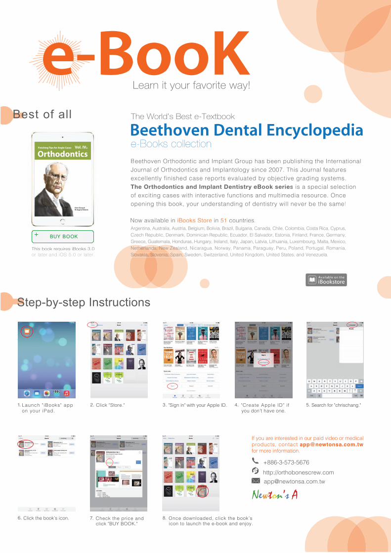
Newton’s A
+886-3-573-5676 http://orthobonescrew.com
a nch i oo s app on o r i a .
ic tore. i n in with o r pp e . reate pp e i f o on t ha e one.
earch for chrischan .
ic the boo s icon. hec the price an
f o are intereste in o r pai i eo or me ica pro cts contact [email protected] for more information.
This oo re uires i oo s or ater and i or ater
Chris Chang & W. Eugene Roberts
OrthodonticsFinishing Tips for Angle Cases Vol. IV(i)
This oo re uires i oo s
BUY BOOK
est o
c ic .o oa i oo
i o o a oo a o .
earn it our favorite wa
o r o o i a p a ro p a p i i r a io ao r a o r o o i a p a o o i . i o r a a r
i i a r por a a o i ra i .The Orthodontics and Implant Dentistry eBook serie i a p ia ioo i i a i i ra i io a i ia r o r .op i i oo o r r a i o i r i r a
e ook collection
The orld s est e Te tboo
r i a ra ia ria i o i ia ra i aria a a a i o o ia o a i a prp i ar o i i a p i a or a a or o ia i a ra r a
r a a a o ra ar r a a apa a ia i a ia o r a a i or a a a i ara a or a a a a ara a r o a or a o a iar a a a i ara a or a a a a ara a r o a or a o a iar a a a i ara a or a a a a ara a r o a or a o a iar a a a i ara a or a a a a ara a r o a or a o a iar a a a i ara a or a a a a ara a r o a or a o a iar a a a i ara a or a a a a ara a r o a or a o a ia
o a ia o ia pai i r a i i oo a ia o ia pai i r a i i oo a ia o ia pai i r a i i oo a ia o ia pai i r a i i oo a ia o ia pai i r a i i oo a ia o ia pai i r a i i o i a a a.
o a ai a i i oo s tore i o ri
Step-by-step Instructions
A good sentence can change one’s life.
A classic in your pocket. Available in Newton’s A, Inc.
�e first collection of Steve Jobs’ famous quote, selected by an Apple businessman.
A handy companion for your reading on the go.
Including Job’s special bookmarks to carry his spirit around.
�e other classic created by Dr. Chris Chang, author of the bestseller, Jobsology.
Understand the secrets of Trump’s success and improve your English ability simultaneously.
Collected more than 200 famous quotes of the President Trump.
Pocket-size, perfect for reading on the go.
We owe him(Trump) an open mind and a chance to lead.
Newton’s A
—Hilary Clinton
Argentina, Australia, Austria, Belgium, Bolivia, Brazil, Bulgaria, Canada, Chile, Colombia, Costa Rica, Cyprus, Czech Republic, Denmark, Dominican Republic, Ecuador, El Salvador, Estonia, Finland, France, Germany, Greece, Guatemala, Honduras, Hungary, Ireland, Italy, Japan, Latvia, Lithuania, Luxembourg, Malta, Mexico, Netherlands, New Zealand, Nicaragua, Norway,Panama, Paraguay, Peru, Poland, Portugal, Romania, Slovakia, Slovenia, Spain, Sweden, Switzerland, United Kingdom, United States, and Venezuela.
Chris Chang
Now available on iBooks Store in 51 countries:
Available at Amazon now.
A good sentence can change one’s life.
A classic in your pocket. Available in Newton’s A, Inc.
�e first collection of Steve Jobs’ famous quote, selected by an Apple businessman.
A handy companion for your reading on the go.
Including Job’s special bookmarks to carry his spirit around.
�e other classic created by Dr. Chris Chang, author of the bestseller, Jobsology.
Understand the secrets of Trump’s success and improve your English ability simultaneously.
Collected more than 200 famous quotes of the President Trump.
Pocket-size, perfect for reading on the go.
We owe him(Trump) an open mind and a chance to lead.
Newton’s A
—Hilary Clinton
Chris Chang
Now available in countries:
Available at Amazon now.
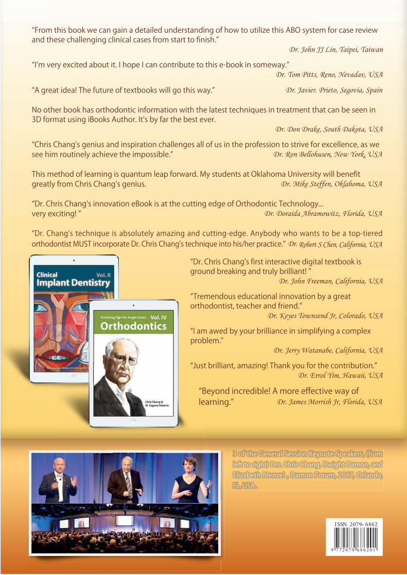
3 of the General Session Keynote Speakers, (from left to right) Drs. Chris Chang, Dwight Damon, and Elizabeth Menzel , Damon Forum, 2017, Orlando, FL, USA.
“From this book we can gain a detailed understanding of how to utilize this ABO system for case review and these challenging clinical cases from start to finish.”
Dr. John JJ Lin, Taipei, Taiwan
“I’m very excited about it. I hope I can contribute to this e-book in someway.”Dr. Tom Pitts, Reno, Nevadav, USA
“A great idea! The future of textbooks will go this way.” Dr. Javier. Prieto, Segovia, Spain
No other book has orthodontic information with the latest techniques in treatment that can be seen in 3D format using iBooks Author. It's by far the best ever.
Dr. Don Drake, South Dakota, USA
“Chris Chang's genius and inspiration challenges all of us in the profession to strive for excellence, as we see him routinely achieve the impossible.” Dr. Ron Bellohusen, New York, USA
This method of learning is quantum leap forward. My students at Oklahoma University will benefit greatly from Chris Chang's genius. Dr. Mike Steffen, Oklahoma, USA
“Dr. Chris Chang's innovation eBook is at the cutting edge of Orthodontic Technology... very exciting! ” Dr. Doraida Abramowitz, Florida, USA
“Dr. Chang's technique is absolutely amazing and cutting-edge. Anybody who wants to be a top-tiered orthodontist MUST incorporate Dr. Chris Chang's technique into his/her practice.” Dr. Robert S Chen, California, USA
“Dr. Chris Chang's first interactive digital textbook is ground breaking and truly brilliant! ”
Dr. John Freeman, California, USA
“Tremendous educational innovation by a great orthodontist, teacher and friend.”
Dr. Keyes Townsend Jr, Colorado, USA
“I am awed by your brilliance in simplifying a complex problem.”
Dr. Jerry Watanabe, California, USA
“Just brilliant, amazing! Thank you for the contribution.” Dr. Errol Yim, Hawaii, USA
“Beyond incredible! A more effective way of learning.” Dr. James Morrish Jr, Florida, USA
Related Documents