UC San Diego UC San Diego Electronic Theses and Dissertations Title Exploring structural and functional features of enzymes across isoprenoid biosynthesis : from archaeal isopentenyl phosphate kinase of primary metabolism to plant terpene cyclases of specialized metabolism Permalink https://escholarship.org/uc/item/4vq9p9n7 Author Dellas, Nikki Publication Date 2010 Peer reviewed|Thesis/dissertation eScholarship.org Powered by the California Digital Library University of California

Welcome message from author
This document is posted to help you gain knowledge. Please leave a comment to let me know what you think about it! Share it to your friends and learn new things together.
Transcript
UC San DiegoUC San Diego Electronic Theses and Dissertations
TitleExploring structural and functional features of enzymes across isoprenoid biosynthesis : from archaeal isopentenyl phosphate kinase of primary metabolism to plant terpene cyclases of specialized metabolism
Permalinkhttps://escholarship.org/uc/item/4vq9p9n7
AuthorDellas, Nikki
Publication Date2010 Peer reviewed|Thesis/dissertation
eScholarship.org Powered by the California Digital LibraryUniversity of California
UNIVERSITY OF CALIFORNIA, SAN DIEGO
Exploring Structural and Functional Features of Enzymes Across Isoprenoid Biosynthesis: From Archaeal Isopentenyl Phosphate Kinase of Primary Metabolism to Plant Terpene
Cyclases of Specialized Metabolism
A dissertation submitted in partial satisfaction of the requirements for the degree Doctor of Philosophy
in
Chemistry
by
Nikki Dellas
Committee in Charge:
Professor Joseph P. Noel, chair Professor Elizabeth Komives, co-chair Professor Michael Burkart Professor Ronald Burton Professor Gourisankar Ghosh
2010

©
Nikki Dellas, 2010
All rights reserved
iii
The Dissertation of Nikki Dellas is approved, and it is acceptable in quality and form for
publication on microfilm and electronically:
Co-Chair
Chair
University of California, San Diego
2010

iv
DEDICATION
for Anderson
v
TABLE OF CONTENTS
Signature Page............................................................................................................................iii
Dedication ..................................................................................................................................iv
Table of Contents ........................................................................................................................v
List of Abbreviations.................................................................................................................xii
List of Figures ...........................................................................................................................xv
List of Tables..........................................................................................................................xviii
Acknowledgements ...................................................................................................................xx
Vita .........................................................................................................................................xxiv
Abstract of the Dissertation.....................................................................................................xxv
Chapter 1 Introduction ...............................................................................................................1
1.1. Isoprenoid biosynthetic pathways ...........................................................................2
1.1.1. The DXP pathway ...................................................................................3
1.1.2. The MVA pathway..................................................................................5
1.1.3. Isoprenoid biosynthesis across the three domains of life........................7
1.2. Short-chain prenyl diphosphate synthases ..............................................................8
1.3. Terpene synthases: function and mechanism ........................................................10
1.3.1. Monoterpene synthases .........................................................................13
1.3.2. Sesquiterpene synthases ........................................................................16
1.3.3. Diterpene synthases...............................................................................19
1.4. Terpene synthases: structure .................................................................................22
1.4.1. The terpene cyclase fold........................................................................22
1.4.2. Crystal structures of monoterpene and sesquiterpene synthases...........23
1.4.3. Divalent metal ion coordination............................................................26
vi
1.4.4. Ligand-induced structural changes .......................................................29
1.4.5. Substrate analogs...................................................................................30
1.5. Emergence of terpene synthases from primary metabolism .................................31
1.6. Conclusions ...........................................................................................................32
REFERENCES.............................................................................................................33
Chapter 2 Quantitative exploration of the catalytic landscape separating divergent plant
sesquiterpene synthases................................................................................................42
2.1. Abstract .................................................................................................................43
2.2. Introduction ...........................................................................................................43
2.3. Results and Discussion..........................................................................................50
2.3.1. Creation and characterization of the M9 lineage ..................................50
2.3.2. Biosynthetic tree of the M9 lineage ......................................................52
2.3.3. Chemical distances of mutational effects..............................................55
2.3.4. Quantifying mutational context.............................................................55
2.3.5. Discussion .............................................................................................57
1.4. Methods .................................................................................................................61
2.4.1. Library construction ..............................................................................61
2.4.2. Biosynthetic tree construction...............................................................61
2.4.3. Sequencing ............................................................................................62
2.4.4. Vial assay characterization....................................................................62
2.4.5. Protein expression and purification.......................................................62
2.4.6. Purification of library proteins ..............................................................63
2.4.7. Kinetic measurements ...........................................................................63
2.5. Supporting Information .........................................................................................64
vii
ACKNOWLEDGEMENTS .........................................................................................81
REFERENCES.............................................................................................................82
Chapter 3 Structural Elucidation of Cisoid and Transoid Cyclization Pathways of a
Sesquiterpene Synthase Using 2-Fluorofarnesyl Diphosphates ................................................85
3.1. Abstract .................................................................................................................86
3.2. Introduction ...........................................................................................................86
3.3. Results and Discussion..........................................................................................92
3.3.1. TEAS-directed cisoid cyclization with (cis,trans)-FPP ........................92
3.3.2. Stereochemical mechanism of cyclization ............................................97
3.3.3. Computational analysis of the TEAS cisoid mechanism ......................98
3.3.4. Structure of wild-type TEAS and M4 TEAS with 2-fluoro analogues .....
.......................................................................................................................101
3.3.5. Spatial reconstruction of cisoid and transoid reaction pathways in TEAS
.......................................................................................................................106
3.3.6. Cisoid cyclase activities with (trans,trans)-FPP..................................109
3.3.7. Structural picture of catalytic promiscuity ..........................................110
3.3.8. Conclusions .........................................................................................111
3.4. Methods ...............................................................................................................114
3.4.1. Organic synthesis ................................................................................114
3.4.2. Protein expression and purification.....................................................114
3.4.3. Kinetic measurement...........................................................................116
3.4.4. Protein crystallization and data collection ..........................................117
3.4.5. Computational methods ......................................................................117
3.4.6. Product elucidation..............................................................................118
viii
3.5 Supporting Information ........................................................................................118
3.5.1. Preparation and Characterization of (2-cis, 6-trans)-2-Fluorofarnesyl
Diphosphate...................................................................................................118
ACKNOWLEDGEMENTS .......................................................................................125
REFERENCES...........................................................................................................126
Chapter 4 A Conserved Amino Terminal Motif in Patchouli Alcohol Synthase Controls
Product Distribution ................................................................................................................131
4.1. Abstract ...............................................................................................................132
4.2. Introduction .........................................................................................................133
4.3. Results and Discussion........................................................................................136
4.3.1. RP motif in PAS..................................................................................136
4.3.2. RP motif mutants in other sesquiterpene cyclases ..............................141
4.3.3. Salt bridge mutants in PAS and TEAS ...............................................143
4.3.4. Conclusions .........................................................................................148
4.4 Methods ................................................................................................................150
4.4.1. Mutant construction, overexpression, and purification.......................150
4.4.2. Specific activity measurements and product profile quantification by
GCMS ...........................................................................................................151
4.5. Supporting Information .......................................................................................153
ACKNOWLEDGEMENTS .......................................................................................160
REFERENCES...........................................................................................................160
Chapter 5 Mutation of Archaeal Isopentenyl Phosphate Kinase Highlights Mechanism and
Guides Phosphorylation of Additional Isoprenoid Monophosphates......................................162
5.1. Abstract ...............................................................................................................163
ix
5.2. Introduction .........................................................................................................164
5.3. Results and Discussion........................................................................................166
5.3.1. Three-dimensional architecture...........................................................166
5.3.2. Active site architecture........................................................................169
5.3.3. Multiple conformations of IPP in a single active site .........................174
5.3.4. Product-bound active site containing IPPβS .......................................176
5.3.5. His60 plays a key role in binding and catalysis ..................................176
5.3.6. IPK mutants can phosphorylate oligoprenyl monophosphates ...........178
5.3.7. Conclusions .........................................................................................181
5.4. Methods ...............................................................................................................182
5.4.1. Activity assays and steady-state kinetic analyses ...............................182
5.4.2. Kinase/terpene synthase coupled assay for chain-length mutants ......183
5.4.3. Structure solution and refinement .......................................................183
5.4.4. Accession codes ..................................................................................186
5.5. Supporting Information .......................................................................................187
5.5.1. Cloning of IPK genes and mutant construction ..................................187
5.5.2. Protein expression and purification.....................................................187
5.5.3. Crystallization and data collection ......................................................188
ACKNOWLEDGEMENTS .......................................................................................190
REFERENCES...........................................................................................................190
Chapter 6 Isopentenyl Phosphate Kinase Homologs Outside of Archaea Suggest a
Bifurcating Mevalonate Pathway in a Diversity of Eukaryotes ..............................................194
6.1. Abstract ...............................................................................................................195
6.2. Introduction .........................................................................................................195
x
6.3. Results and Discussion........................................................................................197
6.3.1. Phylogenetic diversity of IPK .............................................................197
6.3.2. Catalytic activity of IPK homologs.....................................................199
6.3.3. Role for IPK in other kingdoms of life ...............................................200
6.3.4 Conclusions ..........................................................................................200
6.4. Methods ...............................................................................................................201
6.4.1. Cloning of IPK homologs ...................................................................201
6.4.2. Protein expression and purification.....................................................201
6.4.3. Steady-state kinetic analysis ...............................................................202
6.4.4. Bioinformatics.....................................................................................202
6.4.5. Phylogenetic distribution of IPK.........................................................203
6.5. Supporting Information .......................................................................................204
6.5.1. Supporting information on the phylogenetic distribution of IPK .......204
6.5.2. Ultra-conserved residues .....................................................................207
6.5.3. Additional sequences ..........................................................................207
ACKNOWLEDGEMENTS .......................................................................................227
REFERENCES...........................................................................................................227
Chapter 7 Conclusions ...........................................................................................................229
7.1. Overview .............................................................................................................230
7.2. Terpene synthases of specialized metabolism.....................................................231
7.3. IPK of primary metabolism.................................................................................233
7.3.1. Overview .............................................................................................233
7.3.2. Applications for IPK chain-length mutants.........................................234
7.3.3. Implications for active eukaryotic IPKs..............................................235

xi
REFERENCES...........................................................................................................236
xii
LIST OF ABBREVIATIONS
2F. 2-fluoro
4EE. 4-epi eremophilene
5-EA. 5-epi aristolochene
Å. Angstroms
AAK. Amino acid kinase
ADP. Adenosine diphosphate
ADS. Abietadiene synthase
AID. Average interneighbor distance
AMPPNP. Adenylyl imidodiphosphate
ATP. Adenosine triphosphate
ATPγS. Adenosine 5'-(gamma-thiotriphosphate)
C2F. Cis-2-fluoro
CCW. Counterclockwise
CDP. Copalyl diphosphate
CDS. Copalyl diphosphate synthase
CW. Clockwise
DMAPP. Dimethylallyl diphosphate
DNA. Deoxyribonucleic acid
DTT. Dithiothrietol
DXP. 1-deoxy-D-xylulose 5-phosphate
EES. Epi-eremophilene synthase
EST. Expressed sequence tag
FARM. First aspartate-rich motif
xiii
FHP. Farnesyl hydroxyphosphonate
FomA. Fosfomycin resistance A
FP. Farnesyl phosphate
FPP. Farnesyl diphosphate
FPPS. Farnesyl diphosphate synthase
GCMS. Gas chromatography mass spectrometry
GGPP. Geranylgeranyl diphospahte
GGPPS. Geranylgeranyl diphosphate synthase
GP. Geranyl phosphate
GPP. Geranyl diphosphate
GPPS. Geranyl disphosphate synthase
HPS. Hyoscyamus premnaspirodiene synthase
IP. Isopentenyl phosphate
IPK. Isopentenyl phosphate kinase
IPP. Isopentenyl diphosphate
IPPβS. Isopentenyl β-thiodiphosphate
KS. Ent-kaurene synthase
mg. Milligrams
Mg. Magnesium
MgCl2. Magnesium chloride
ml. Milliliters
Mn. Manganese
MVA. Mevalonate
NaCl. Sodium Chloride
xiv
NADH. Nicotinamide adenine dinucleotide, reduced
NAGK. N-acetylglutamate kinase
NMR. Nuclear magnetic resonance
NPP. Nerolidyl diphosphate
PAS. Patchouli alcohol synthase
PCR. Polymerase chain reaction
PDB. Protein data bank
PEG. Polyethylene glycol
PSD. Premnaspirodiene
SARM. Second aspartate-rich motif
SCOPE. Structure-based combinatorial protein engineering
SDS-PAGE. Sodium dodecyl sulfate polyacrylamide gel electrophoresis
SIM. Single ion mode
TEAS. Tobacco 5-epi aristolochene synthase
TIC. Total ion count
UDP. Uridine diphosphate
µ l. Microliters
UMPK. Uridine monophosphate kinase
WT. Wild type

xv
LIST OF FIGURES
Figure 1.1. The DXP pathway....................................................................................................4
Figure 1.2. The MVA pathway...................................................................................................6
Figure 1.3. Proposed alternative mevalonate pathway in Archaea ............................................8
Figure 1.4. General mechanism for short-chain prenyl diphosphate synthases .........................9
Figure 1.5. Geranyl cation cyclization .....................................................................................15
Figure 1.6. Farnesyl cation cyclization pathways ....................................................................17
Figure 1.7. Geranylgeranyl cation cyclization pathways .........................................................21
Figure 1.8. Global Structure of monoterpene and sesquiterpene cyclases from various
kingdoms of life.........................................................................................................................24
Figure 1.9. The catalytic C-terminal domain of terpene cyclases ............................................25
Figure 1.10. Magnesium ion coordination in the active site of 5-epi-aristolochene synthase .27
Figure 2.1. Terminal cyclization steps of TEAS and HPS terpene synthases..........................46
Figure 2.2. Overall structure of TEAS and location and identity of M9 residues....................48
Figure 2.3. Phylogenetic distribution of solanaceous TEAS- and HPS-like terpene synthases...
...................................................................................................................................................50
Figure 2.4. Activities of the M9 lineage...................................................................................52
Figure 2.5. Biosynthetic tree of the M9 library ........................................................................54
Figure 2.6. AID in chemical and sequence space.....................................................................57
Figure 2.7. Similarity-based cluster diagram of the EES-like and HPS-like mutant clades ....65
Figure 3.1. Mechanism of TEAS-catalyzed cyclization of (cis,trans)-FPP to (+)-2-epi-
prezizaene..................................................................................................................................90
Figure 3.2. Gas chromatograms of products from incubations of wild-type TEAS and the M4
mutant with (cis, trans)- and (trans, trans)-FPP.........................................................................93
xvi
Figure 3.3. Computational analysis of the TEAS cisoid cyclization pathway .......................100
Figure 3.4. Crystallographic analysis of wild-type and M4 TEAS bound to fluoro-FPPs.....105
Figure 3.5. Spatial reconstruction of the transoid and cisoid cyclization pathways in TEAS .....
.................................................................................................................................................108
Figure 3.6. Annotation of global structure using B-factors....................................................121
Figure 3.7. Disorder in the J-K loop of experimental crystal structures ................................122
Figure 3.8. Spatial distribution of M4 mutations and closest distances to the farnesyl chain......
.................................................................................................................................................123
Figure 3.9. Farnesyl chain topology of wild-type TEAS from fluorofarnesyl analogues ......124
Figure 3.10. Spatial depiction of mutational effects in M4 TEAS.........................................125
Figure 4.1. Reaction Mechanism of patchoulol synthase accounting for all thirteen
sesquiterpene products ............................................................................................................135
Figure 4.2. Truncation mutant constructs in patchoulol synthase ..........................................137
Figure 4.3. Percent compositions of all products in the truncation mutants of PAS..............138
Figure 4.4. Percent compositions of all products in PAS RP motif mutants..........................140
Figure 4.5. Conservation of a salt bridge interaction with the RP or RR motif .....................143
Figure 4.6. Percent compositions of all products in PAS salt bridge mutants .......................145
Figure 4.7. Percent compositions of all products in TEAS mutants ......................................147
Figure 4.8. Total peak areas for all mutant and wild type enzymes.......................................153
Figure 5.1. The amino acid kinase (AAK) family members ..................................................166
Figure 5.2. Primary sequence, tertiary architecture, and active site snapshots of IPK ..........168
Figure 5.3. Comparative close-up views of the nucleotide phosphate-binding region of the
IPK and fomA active sites.......................................................................................................170
Figure 5.4. N-terminal domain and dual loop conformations in IPK.....................................172
xvii
Figure 5.5. IPK in complex with IP and IPP ..........................................................................173
Figure 5.6. Farnesyl phosphate (FP) phosphorylation by IPK chain length mutants.............180
Figure 6.1. The Bifurcating Mevalonate Pathway..................................................................196
Figure 6.2. IPK phylogeny .....................................................................................................198
Figure 6.3. Steady-State Kinetics ...........................................................................................204
Figure 6.4. Alignment of IPKs from the three domains of life ..............................................213
xviii
LIST OF TABLES
Table 2.1. Ionization efficiencies of 5-EA, 4-EE, and PSD .....................................................64
Table 2.2. Sequences of Solanaceous putative and characterized 5-EA and PSD terpene
synthases....................................................................................................................................66
Table 2.3. SCOPE library construction statistics......................................................................67
Table 2.4. Gas chromatography – mass spectrometry data of M9 mutant proteins .................68
Table 2.5. Kinetic measurements of selected library mutants ..................................................79
Table 2.6. Average chemical distances for each position.........................................................80
Table 2.7. Influence of active site substitutions on product specificity....................................80
Table 2.8. Minimal combinations of mutations converting TEAS to HPS-like product
specificity ..................................................................................................................................81
Table 3.1. Enzymatic products from incubations of TEAS wild-type and the M4 mutant with
(cis,trans)- or (trans,trans)-FPP................................................................................................96
Table 3.2. Kinetic parameters of TEAS wild-type and the M4 enzyme...................................96
Table 3.3. Crystallographic data and refinement statistics .....................................................102
Table 3.4. Global Comparison of TEAS WT and M4 crystal structures...............................121
Table 4.1. PAS truncation mutant % compositions................................................................154
Table 4.2. PAS truncation mutant standard deviations for % compositions ..........................154
Table 4.3. PAS Arg15 mutant % compositions......................................................................155
Table 4.4. PAS Arg15 mutant standard deviations for % compositions ................................155
Table 4.5. PAS Pro16 mutant % compositions.......................................................................156
Table 4.6. PAS Pro16 mutant standard deviations for % compositions.................................156
Table 4.7. PAS salt bridge mutant % compositions ...............................................................157
Table 4.8. PAS salt bridge mutant standard deviations of % compositions ...........................157
xix
Table 4.9. TEAS mutant % compositions ..............................................................................158
Table 4.10. TEAS standard deviations for % compositions...................................................158
Table 4.11. ADS mutant % compositions ..............................................................................158
Table 4.12. ADS mutant standard deviations of % compositions ..........................................159
Table 4.13. HPS mutant % compositions ...............................................................................159
Table 4.14. HPS mutant standard deviations of % compositions...........................................159
Table 5.1. Kinetic Data for IPK-Mj Wild-Type and H60Q at 25°C.......................................177
Table 5.2. X-ray diffraction data processing and refinement statistics ..................................185
Table 5.3. Primer pairs for PCR reactions..............................................................................189
Table 6.1. Kinetic constants for characterized IPKs...............................................................199
Table 6.2. Gene identifier (GI) numbers for MVA pathway gene orthologs in organisms with
an active IPK ...........................................................................................................................204

xx
ACKNOWLEDGEMENTS
I would like to thank my advisor, Joseph P. Noel, for all of his guidance, support, and
generosity both inside and outside of the lab throughout the past five years. His good nature,
positive outlook, and enthusiasm will continue to inspire me throughout life.
I would also like to thank all of my labmates including those in both the Noel lab and
the Wang lab, especially Paul O’maille, Marianne Bowman, Gordon Louie, and Jeffrey
Takimoto. Paul O’maille was a postdoc in the Noel lab whom I worked closely with when I
first joined the lab and continued to work with for several years thereafter. He was a great
friend with a very unique and creative sense of humor. Marianne Bowman is the lab manager
in the Noel lab; she was instrumental in maintaining lab functionality on a variety of levels,
but was also a wonderful person to talk to for support and advice. Gordon Louie is a staff
scientist in the lab who helped me with all aspects of crystallography: from data collection to
crystallographic refinement. Jeffrey Takimoto is a graduate student in the Wang lab who has
been an incredibly supportive and loyal friend, and was always there for me, especially during
more recent struggles.
I would like to thank my friends and family for their love and support. Specifically, I
would like to thank my mom for teaching me persistence and encouraging me to indulge not
only my scientific side but also my creative site throughout most of my life. I would like to
thank my dad for teaching me that life is not only about hard work, but also about generosity,
selflessness, and having fun with the people that are close to you. I would like to thank my
brother Tim for all of the hilarious memories we have together, doing the random things that
we end up doing, and for being someone I could always laugh with, share music with, and be
myself around. I would like to thank my sister Meg for her positive attitude, compassion for
others, and love for life. She has an amazing ability to inspire happiness in those around her

xxi
and, as we have grown up, I no longer think of her as my younger sister, but instead as one of
my peers. In more recent years, I have confided in her and she has become an incredibly
important person in my life, despite the fact that we haven’t lived in the same city for nearly
ten years. I would also like to thank Uncle Tom, Aunt Lauren, and their three kids Sophie,
Jonas, and Drew, who been infinitely hospitable since I moved to San Diego.
I would like to thank Court Heller for his love and support throughout the bulk of my
graduate career. I would especially like to thank him for enduring the pains and celebrating the
successes of graduate school with me.
I would like to thank The National for their music.
Ultimately, I would like to thank Greg Macias (a.k.a. Percy Robinson). He has made
planets align.
The text of chapter 2, in full, is a reprint of the material as it appears in Nature
Chemical Biology 2008, Vol. 18, pp. 3039-3042. Permission was obtained from the co-
authors. I was the third author of this work. As mentioned in the manuscript, Paul O’Maille
designed the study, conducted experiments, analyzed data and wrote the manuscript, Arthur
Malone conducted experiments and developed small-scale protein purification, I conducted
experiments, analyzed data and contributed revision to the manuscript, B Andes Hess Jr.
conducted quantum mechanics calculations and contributed revisions to the manuscript, Lidia
Smentek conducted quantum mechanics calculations, Iseult Sheehan conducted experiments,
Bryan Greenhagen and Joseph Chappell designed the study and contributed revisions to the
manuscript, Gerard Manning analyzed data, developed the biosynthetic tree and chemical
distance analysis, and contributed revisions to the manuscript, and Joseph P. Noel designed
the study, analyzed the data and wrote the manuscript. This research was performed under the
supervision of Joseph P. Noel.

xxii
The text of chapter 3, in full, is a reprint of material as it appears in ACS Chemical
Biology 2010, 5 (4), pp 377–392, with the exception of the section under supporting
information entitled “computational details” which was excluded. Permission was obtained
from all co-authors. I am second author of this work. Paul O’Maille wrote the manuscript, and
was also involved with protein purification, GCMS data analysis, crystallization experiments,
and crystallographic data processing, structure solution and refinement. I was responsible for
protein purification, GCMS data analysis, crystallization experiments, crystallographic data
processing, structure solution, refinement, and contributed revisions to the manuscript. Juan
Faraldos was responsible for organic synthesis, NMR characterization of sesquiterpenes, and
contributed revisions to the manuscript. Yuxin (Marilyn) Zhao was responsible for chemical
synthesis of cis-FPP. B. Andes Hess Jr. and Lidia Smentek were responsible for all
computational studies. The research included in the manuscript was performed under the
supervision of Robert Coates and Joseph P. Noel (who also contributed revisions and helped
write the manuscript).
The text of chapter 4, in part, is currently being prepared for submission for
publication of the material. Dellas, Nikki; Noel, Joseph P. I am the first author of this material.
All experiments were performed under the supervision of Joseph P. Noel.
The text of chapter 5, in full, is a reprint of the material as it appears in ACS Chemical
Biology 2010, 5(6), pp 589-601. I am the primary author of this paper. The research was
performed under the supervision of Joseph P. Noel.
The text of chapter 6, in part, has been submitted for publication of the material as it
may appear in Chemical Communications, 2010, Dellas, Nikki; Manning, Gerard, Noel,
Joseph P. I am the first author of this paper. Gerard Manning and Joseph P. Noel are the
corresponding authors. I was responsible for all gene cloning, enzyme expression, purification,

xxiii
and kinetic characterization of IPK and its homologs. Gerard Manning was responsible for the
bioinformatic and phylogenetic analysis of IPK and its homologs. All experiments were
performed under the supervision of Joseph P. Noel.
xxiv
VITA
Education
2010 Ph.D., Chemistry University of California, San Diego
2007 M.S., Chemistry University of California, San Diego
2005 B.S., Chemistry Carnegie Mellon University
Publications
1. Dellas, N.; Noel, J.P. A Conserved Amino Terminal Motif in Patchouli Alcohol Synthase Controls Product Distribution. Manuscript in preparation.
2. Dellas, N.; Manning, G.; Noel, J.P. Isopentenyl Phosphate Kinase Homologs Outside
of Archaea Suggest a Bifurcating Mevalonate Pathway in a Diversity of Eukaryotes. Submitted to Chem Commun.
3. Dellas, N.; Noel, J. P., Mutation of archaeal isopentenyl phosphate kinase highlights
mechanism and guides phosphorylation of additional isoprenoid monophosphates. ACS Chem Biol 2010, 5 (6), 589-601.
4. Noel, J. P.; Dellas, N.; Faraldos, J. A.; Zhao, M.; Hess, B. A., Jr.; Smentek, L.;
Coates, R. M.; O'Maille, P. E., Structural elucidation of cisoid and transoid cyclization pathways of a sesquiterpene synthase using 2-fluorofarnesyl diphosphates. ACS Chem Biol 2010, 5 (4), 377-392.
5. Faraldos, J. A.; O'Maille, P. E.; Dellas, N.; Noel, J. P.; Coates, R. M., Bisabolyl-
derived sesquiterpenes from tobacco 5-epi-aristolochene synthase-catalyzed cyclization of (2Z,6E)-farnesyl diphosphate. J Am Chem Soc 2010, 132 (12), 4281-9.
6. O'Maille, P. E.; Malone, A.; Dellas, N.; Andes Hess, B., Jr.; Smentek, L.; Sheehan, I.;
Greenhagen, B. T.; Chappell, J.; Manning, G.; Noel, J. P., Quantitative exploration of the catalytic landscape separating divergent plant sesquiterpene synthases. Nature Chem Biol 2008, 4 (10), 617-623.
7. Dasgupta, R.; Hirschmann, M. M.; Dellas, N., The effect of bulk composition on the
solidus of carbonated eclogite from partial melting experiments at 3 GPa. Contrib. Mineral. Petrol. 2005, 149 (3), 288-305.
xxv
ABSTRACT OF THE DISSERTATION
Exploring Structural and Functional Features of Enzymes Across Isoprenoid Biosynthesis:
From Archaeal Isopentenyl Phosphate Kinase of Primary Metabolism to Plant Terpene
Cyclases of Specialized Metabolism
by
Nikki Dellas
Doctor of Philosophy in Chemistry
University of California, San Diego, 2010
Professor Joseph P. Noel, Chair
Professor Elizabeth Komives, Co-Chair
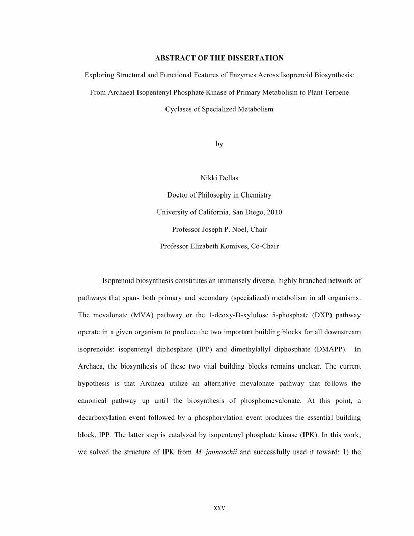
Isoprenoid biosynthesis constitutes an immensely diverse, highly branched network of
pathways that spans both primary and secondary (specialized) metabolism in all organisms.
The mevalonate (MVA) pathway or the 1-deoxy-D-xylulose 5-phosphate (DXP) pathway
operate in a given organism to produce the two important building blocks for all downstream
isoprenoids: isopentenyl diphosphate (IPP) and dimethylallyl diphosphate (DMAPP). In
Archaea, the biosynthesis of these two vital building blocks remains unclear. The current
hypothesis is that Archaea utilize an alternative mevalonate pathway that follows the
canonical pathway up until the biosynthesis of phosphomevalonate. At this point, a
decarboxylation event followed by a phosphorylation event produces the essential building
block, IPP. The latter step is catalyzed by isopentenyl phosphate kinase (IPK). In this work,
we solved the structure of IPK from M. jannaschii and successfully used it toward: 1) the
xxvi
design of a deeper active site pocket for binding and catalysis of longer chained isoprenoid
monophosphates; 2) the identification and characterization of active IPK homologs in other
kingdoms of life. This work contributes towards the design of a synthetic metabolic pathway
and reveals new information about the potential existence of a bifurcated mevalonate pathway
in all plants and certain other eukaryotic organisms.
Farnesyl diphosphate is directly derived from the building blocks IPP and DMAPP
and is an essential metabolic intermediate for a variety of downstream primary and secondary
metabolic pathways including cholesterol biosynthesis and terpenoid biosynthesis,
respectively. Sesquiterpene cyclases (synthases) are part of terpenoid biosynthesis and
catalyze the cyclization of farnesyl diphosphate into one or more sesquiterpene products; these
chemicals play important biological roles in defense and communication, especially in plants.
Here, we explore a variety of mutant and wild type plant sesquiterpene cyclases in attempt to
understand several concepts: 1) how these enzymes traverse a defined catalytic landscape to
biosynthesize disparate products without compromising their catalytic activities; 2) the
structural and functional differences associated with turnover of cis- and trans-FPP by wild
type and promiscuous cyclase mutants; 3) how certain sesquiterpene synthases utilize an Arg-
Pro motif within the amino terminal domain to interact with the catalytic C-terminal domain
and modulate product profile complexity.

1
Chapter 1
Introduction

2
1.1. Isoprenoid biosynthetic pathways Isoprenoid biosynthesis constitutes a complex series of branched pathways that results
in the production of a variety of essential and specialized metabolites across all kingdoms of
life. These essential metabolites include (but are not limited to) squalene, hopanoids, and
steroids (important for membrane structure in Archaea, Bacteria, and Eukarya, respectively),1,2
dolichols (N-linked glycosylation and membrane anchorage of sugars in eukaryotes and
archaea,3 terpenes (plant defense and communication), carotenoids (photoprotection for
certain prokaryotes and plants4, prenylquinones (mitochondrial electron transport),5 and
gibberellins (plant growth and development, for review see Hedden et al 1997).6
All metabolites discussed above originate from the two essential five-carbon building
blocks of isoprenoid biosynthesis: isopentenyl diphosphate (IPP) and its stereoisomer,
dimethylallyl diphosphate (DMAPP). One molecule of DMAPP reacts with one, two, or three
molecules of IPP via a prenyltransferase (isoprenoid diphosphate synthase) to generate geranyl
diphosphate (GPP), farnesyl diphosphate (FPP), or geranylgeranyl diphosphate (GGPP),
respectively. These three compounds are then utilized in different ways by a variety of
enzymes to biosynthesize a repertoire isoprenoid products. DMAPP can be produced either in
conjunction with IPP or is made through isomerization of IPP via an IPP isomerase (IPPI).
The current hypothesis is that IPP (and DMAPP) biosynthesis occurs through one of
two distinct metabolic pathways: the mevalonate (MVA) pathway or the more recently
discovered 1-deoxy-D-xylulose 5-phosphate (DXP) pathway (also known as the 2-C-methyl-
D-erythritol 4-phosphate (MEP) pathway).7-9

3
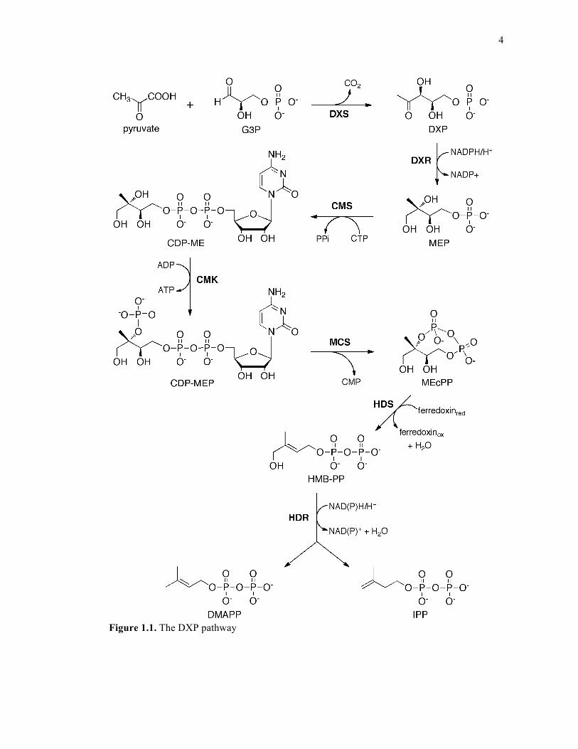
1.1.1. The DXP pathway
The DXP pathway consists of the following steps: 1) Condensation of glyceraldehyde
3-phosphate (G3P) and the “activated acetaldehyde” of pyruvate (Pyr) catalyzed by the
enzyme DXP synthase (DXPS) to produce DXP10; 2) reduction of DXP to 2-C-
methylerythritol-4-phosphate (MEP) by the enzyme DXP reductoisomerase (DXR); 3)
coupling of MEP and cytidine triphosphate (CTP) by the enzyme 4-diphosphocytidyl-2-C-
methyl-D-erythritol synthase (CMS) to generate 4-diphosphocytidyl-2-C-methyl-D-erythritol
(CDP-ME); 4) phosphorylation of CDP-ME by the enzyme 4-diphosphocytidyl-2-C-methyl-
D-erythritol kinase (CMK) to produce 4-diphosphocytidyl-2-C-methyl-D-erythritol 2-
phosphate (CDP-MEP); 5) conversion of CDP-MEP to 2-C-methyl-D-erythritol 2,4-
cyclopyrophosphate (MEcPP) by the enzyme 2-C-methyl-D-erythritol 2,4-cyclodiphosphate
synthase (MCS); 6) ring-opening reduction of MEcPP to (E)-4-Hydroxy-3-methyl-but-2-enyl
pyrophosphate (HMB-PP) by the enzyme HMP-PP synthase (HDS)11,12 and 7) reductive
dehydration of HMB-PP to a mixture of IPP and DMAPP by the enzyme HMB-PP reductase
(HDR).11,13 These steps are detailed in Figure 1.1.
4
Figure 1.1. The DXP pathway
5
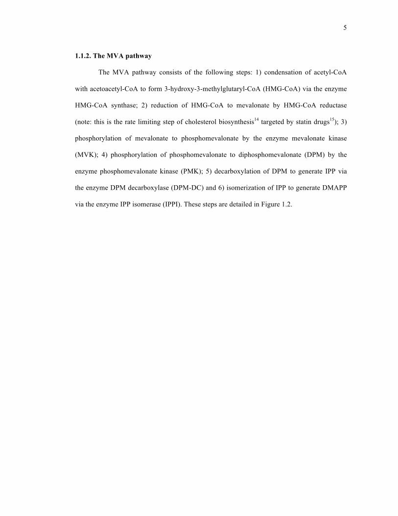
1.1.2. The MVA pathway
The MVA pathway consists of the following steps: 1) condensation of acetyl-CoA
with acetoacetyl-CoA to form 3-hydroxy-3-methylglutaryl-CoA (HMG-CoA) via the enzyme
HMG-CoA synthase; 2) reduction of HMG-CoA to mevalonate by HMG-CoA reductase
(note: this is the rate limiting step of cholesterol biosynthesis14 targeted by statin drugs15); 3)
phosphorylation of mevalonate to phosphomevalonate by the enzyme mevalonate kinase
(MVK); 4) phosphorylation of phosphomevalonate to diphosphomevalonate (DPM) by the
enzyme phosphomevalonate kinase (PMK); 5) decarboxylation of DPM to generate IPP via
the enzyme DPM decarboxylase (DPM-DC) and 6) isomerization of IPP to generate DMAPP
via the enzyme IPP isomerase (IPPI). These steps are detailed in Figure 1.2.
6
Figure 1.2. The MVA pathway

7
1.1.3. Isoprenoid biosynthesis across the three domains of life
Most organisms contain gene orthologs for either one or both pathways. In general,
the MVA pathway is found in eukaryotes (and certain bacteria) while the DXP pathway is
found in most bacteria and plastid-bearing eukaryotes. Plants contain both pathways: the DXP
pathway operates in the plastids while the MVA pathway operates in the cytosol (although
recent results suggest sub-cellular localization of certain MVA pathway enzymes to the ER,
mitochondria, and peroxisomes).16,17,18 Archaea contain gene orthologs for the first four listed
steps of the MVA pathway, but are missing the last three steps, including those catalyzed by
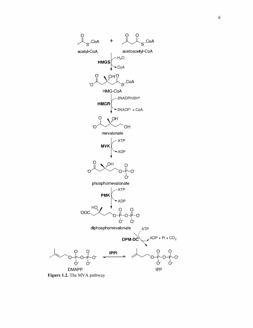
PMK, DPM-DC, and IPPI19. The current view is that Archaea use a modified mevalonate
pathway to generate IPP and DMAPP19, 20. One proposal for evolutionary modification entails
a reversal of the phosphorylation and decarboxylation events that follow PMK biosynthesis in
the classical mevalonate pathway. This modification would include decarboxylation of PMK
to isopentenyl phosphate (IP), followed by phosphorylation of IP to IPP, generating the same
end product as in the classical MVA pathway (Figure 1.3). The recent successful isolation and
characterization of an archaeal isopentenyl phosphate kinase (IPK) that can perform the latter
reaction circumstantially support the proposed modified pathway.19, 20 Chapter 5 details more
recent findings with regard to this modified MVA pathway, particularly with regard to the
unexpected discovery of its existence outside of the Archaeal domain of life. These findings
challenge our current understanding on what was thought to be a widely accepted biosynthetic
pathway.
8
Figure 1.3. Proposed alternative mevalonate pathway in Archaea
1.2. Short-Chain Prenyl Diphosphate Synthases
GPP synthase (GPPS), FPP synthase (FPPS), and GGPP synthase (GGPPS) belong to
a division of prenyltransferases known as “short-chain prenyl diphosphate synthases” that
catalyze the iterative transfer of one, two, or three molecules of IPP, respectively, to either
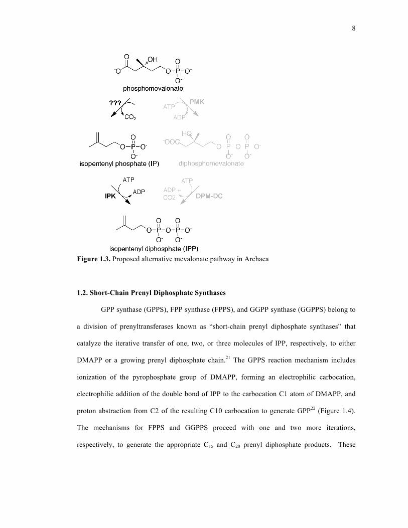
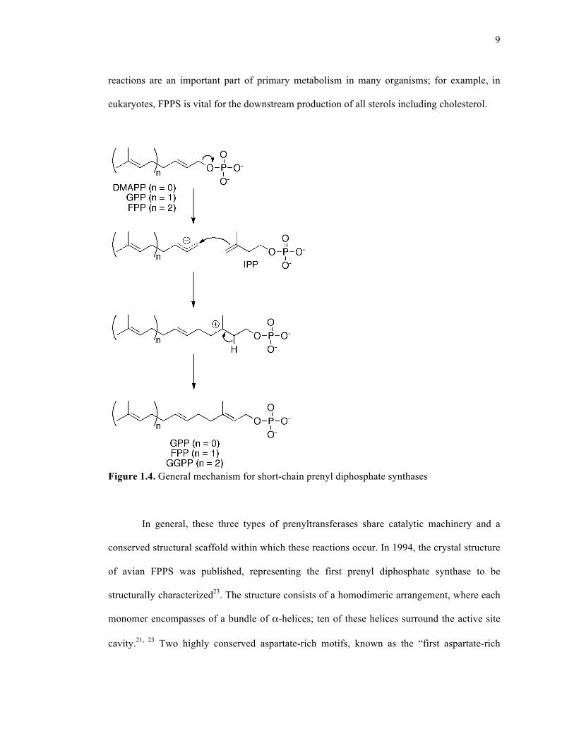
DMAPP or a growing prenyl diphosphate chain.21 The GPPS reaction mechanism includes
ionization of the pyrophosphate group of DMAPP, forming an electrophilic carbocation,
electrophilic addition of the double bond of IPP to the carbocation C1 atom of DMAPP, and
proton abstraction from C2 of the resulting C10 carbocation to generate GPP22 (Figure 1.4).
The mechanisms for FPPS and GGPPS proceed with one and two more iterations,
respectively, to generate the appropriate C15 and C20 prenyl diphosphate products. These
9
reactions are an important part of primary metabolism in many organisms; for example, in
eukaryotes, FPPS is vital for the downstream production of all sterols including cholesterol.
Figure 1.4. General mechanism for short-chain prenyl diphosphate synthases
In general, these three types of prenyltransferases share catalytic machinery and a
conserved structural scaffold within which these reactions occur. In 1994, the crystal structure
of avian FPPS was published, representing the first prenyl diphosphate synthase to be
structurally characterized23. The structure consists of a homodimeric arrangement, where each
monomer encompasses of a bundle of α-helices; ten of these helices surround the active site
cavity.21, 23 Two highly conserved aspartate-rich motifs, known as the “first aspartate-rich
10
motif” (FARM, represented as DDx2-4D) and the “second aspartate-rich motif” (SARM,
represented as DDXXD), lie on opposite ends of the active site.24, 25 More recently published
crystal structures of FPPS from E. coli complexed with DMAPP or DMAPP and IPP
demonstrate conformational changes associated with different phases of the elongation
reaction26. In the presence of DMAPP and Mg2+, two Mg2+ ions coordinate to FARM and the
diphosphate group on the allylic substrate.23, 26 The binding of IPP triggers secondary
structural changes that close the active site and squeeze out water; These changes are
accompanied by the coordination of a third Mg2+ ion to SARM26.
Most short-chain prenyl diphosphate synthases do not demonstrate a high degree of
product promiscuity.24, 25 There are several features that govern chain-length specificity in
short-chain prenyl diphosphate synthases. One hallmark is size of the active site pocket: a
larger pocket can accommodate longer-chained products24 Another attribute is the presence or
absence of amino acids at specific locations directly upstream of FARM;23, 24, 27, 28 in some
cases these residues may protrude into the active site tunnel, marking the floor of the active
site and preventing further chain elongation.23, 24 A third notable feature that modulates chain-
length specificity is the presence of extra residues in FARM (DDXXXXD compared to
DDXXD); this structural component results in products with shorter chain lengths.23, 29
1.3. Terpene synthases: function and mechanism
Terpene synthases (cyclases) encompass a family of enzymes playing critical roles in
the secondary metabolism and chemical ecology of plants, bacteria, fungi and marine
organisms.30 These enzymes catalyze the cyclization of their respective isoprenyl diphosphate
substrate (either GPP, FPP or GGPP) into a variety of chemically complex products that often
contain a number of chiral centers. There are three distinct classes of terpene synthases:
11
monoterpene, sesquiterpene, and diterpene synthases. Monoterpene synthases catalyze the
cyclization of GPP (a C10 prenyl diphosphate) into one or more monoterpene products. One
example is S-linalool synthase, which produces the fragrant monoterpene S-linalool that is
used to attract a moth pollinator to a specific plant species.31 Sesquiterpene synthases catalyze
the cyclization of FPP (a C15 prenyl diphosphate substrate) into one or more sesquiterpene
products. One example is (E)-beta-caryophyllene synthase, which produces the sesquiterpene
(E)-beta-caryophyllene as its major product; this molecule contributes to the airborne defense
response for certain plants against herbivores.32 Diterpene synthases catalyze the cyclization of
GGPP (a C20 prenyl diphosphate) into one or more diterpene products. One example is
taxadiene synthase, which produces the hydrocarbon core, taxadiene, of the pharmaceutically
relevant anti-cancer agent known as Taxol™.33
Although many monoterpenes and sesquiterpenes function as signaling molecules to
attract pollinators, ward off enemies, or communicate with their external environment, other
sesquiterpenes (and diterpenes) can additionally be either directly or indirectly used for
medicinal purposes. One popular example of a sesquiterpene synthase that produces such a
precursor is amorpha-4,11-diene synthase, whose product can be derivatized to the anti-
malarial drug known as artemisinin.34
For this reason, the search for ways to overproduce such valuable compounds is
ongoing.35 Overexpression of MVA pathway enzymes in S. cerevisiae,36, 37 overexpression of
the DXP pathway in E. coli,38 or heterologous expression of the MVA pathway in E. coli39, 40
are three common methodologies that have effectively produced significant quantities of such
terpenes. However, each method has certain drawbacks. For example, heterologous
expression of the MVA pathway in E. coli has lead to difficulties associated with metabolic
flux through the pathway and with cell growth,39 while overexpression of MVA pathway
12
enzymes in S. cerevisiae causes the non-productive accumulation of farnesol, which is usually
considered an unwanted byproduct.36 FPP-induced feedback inhibition of mevalonate kinase
of the MVA pathway has also been reported.41 Nevertheless, some of these methods have
successfully produced concentrations of terpenes at over 100mg/liter of culture and continuing
efforts will most likely improve this number.37
In general, the terpene cyclase reaction begins with Mg2+ or Mn2+ assisted ionization
of the pyrophosphate group on the substrate, which is usually accompanied by electrophilic
cyclization to generate a secondary or tertiary carbocation intermediate.42 The highly reactive
acyclic or cyclic carbocation intermediate can then undergo further transformations including
ring closures and hydride shifts within the hydrophobic active site through other closures and
migrations until proton abstraction or hydroxylation quenches this cycle by means of water or
an active site side chain. This reaction, termed “ionization-dependent cyclization” takes place
in the C-terminal catalytic domain of terpene cyclases.
A highly conserved aspartate-rich motif termed the “DDXX(D/E)” motif coordinates
two of the three divalent metal cations (in the case of Mg2+, these are usually denoted MgA2+
and MgC2+)43, 44 that are responsible for lowering the activation barrier for pyrophosphate
ionization and subsequent allylic carbocation stabilization; this motif is structurally and
functionally conserved with the FARM motif in prenyl diphosphate synthases (residues in
bold denote those involved with metal ion coordination)45. Another conserved motif present in
all terpene cyclases that coordinates the third divalent cation (often referred to as MgB2+) is the
(N,D)DXX(S,T)XXX(E,D) motif, abbreviated as the NSE/DTE motif46. This motif is found
as NDXXSXXXE in most fungal and bacterial terpene cyclases, and as DDXXTXXXE in
most plant terpene cyclases46.
13
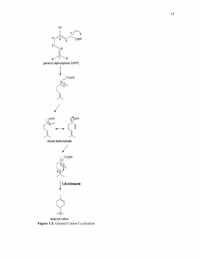
1.3.1. Monoterpene synthases
Monoterpene synthases (cyclases) are a division of terpene synthases that turn over
the C10 isoprenoid GPP using an “ionization-dependent cyclization” mechanism. Plant
monoterpene cyclases usually contain a plastid localization sequence, which consists of
approximately fifty additional residues flanking the amino-terminus.47 Given that monoterpene
synthases accept the shorter C10 substrate, GPP, the double bonds are not initially oriented
properly to enable electrophilic cyclization of the nascent carbocation. Therefore, following
initial pyrophosphate ionization, an isomerization event must occur, which generates the stable
intermediate linalyl diphosphate via a two-step reaction entailing reattachment of the
pyrophosphate to C3 and accompanying rotation about the C2-C3 bond48 (Figure 1.5). Since
roughly one-third of all characterized monoterpene synthases produce acyclic products,49 this
isomerization event is not always necessary; however it is a prerequisite for the generation of
any cyclic monoterpene. A pair of Arg residues located directly C-terminal to the plastid
localization sequence have been implicated in the isomerization mechanism. For example, in
limonene synthase, truncation or mutation of the arginine pair renders the protein inactive
towards geranyl diphosphate (the native substrate) however the enzyme catalyzes the reaction
to completion when provided with the isomerized version of the substrate, linalyl
diphosphate.47 Nevertheless, there is debate with regard to the precise function of this motif,
especially since it exists in certain sesquiterpene synthases (as either an Arg-Arg pair or an
Arg-Pro pair) that do not require an isomerization event. A mutational analysis of both the
Arg-Pro and Arg-Arg pairs in several sesquiterpene synthases (detailed in Chapter 6)
implicates a broader role for this motif in reaction modulation.
The recent discovery of a cis-GPP synthase (called neryl diphosphate synthase, or
NPPS) capable of producing cis-derived neryl diphosphate (NPP) suggests an alternative

14
mechanism for derivation of cyclic monoterpenes which would not involve isomerization of
GPP;50 In fact, successful characterization of the NPP-utilizing β-phellandrene synthase fully
supports this hypothesis.50 Another recent publication analogous to this in a sesquiterpene
cyclase reports utilization of the cis-derivative of FPP, (Z,E)-FPP, as its substrate51.
15
Figure 1.5. Geranyl Cation Cyclization
16
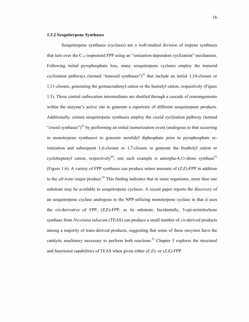
1.3.2 Sesquiterpene Synthases
Sesquiterpene synthases (cyclases) are a well-studied division of terpene synthases
that turn over the C15 isoprenoid FPP using an “ionization-dependent cyclization” mechanism.
Following initial pyrophosphate loss, many sesquiterpene cyclases employ the transoid
cyclization pathways (termed “transoid synthases”)52 that include an initial 1,10-closure or
1,11-closure, generating the germacradienyl cation or the humulyl cation, respectively (Figure
1.5). These central carbocation intermediates are shuttled through a cascade of rearrangements
within the enzyme’s active site to generate a repertoire of different sesquiterpene products.
Additionally, certain sesquiterpene synthases employ the cisoid cyclization pathway (termed
“cisoid synthases”)52 by performing an initial isomerization event (analogous to that occurring
in monoterpene synthases) to generate nerolidyl diphosphate prior to pyrophosphate re-
ionization and subsequent 1,6-closure or 1,7-closure to generate the bisabolyl cation or
cycloheptenyl cation, respectively49; one such example is amorpha-4,11-diene synthase53
(Figure 1.6). A variety of FPP synthases can produce minor amounts of (Z,E)-FPP in addition
to the all-trans major product.54 This finding indicates that in some organisms, more than one
substrate may be available to sesquiterpene cyclases. A recent paper reports the discovery of
an sesquiterpene cyclase analogous to the NPP-utilizing monoterpene cyclase in that it uses
the cis-derivative of FPP, (Z,E)-FPP, as its substrate. Incidentally, 5-epi-aristolochene
synthase from Nicotiana tabacum (TEAS) can produce a small number of cis-derived products
among a majority of trans-derived products, suggesting that some of these enzymes have the
catalytic machinery necessary to perform both reactions.55 Chapter 3 explores the structural
and functional capabilities of TEAS when given either (E,E)- or (Z,E)-FPP.
17
Figure 1.6. Farnesyl cation cyclization pathways
18
Although bacterial sesquiterpene synthases usually produce only one product, plant
sesquiterpene synthases exhibit varying degrees of product diversity (catalytic promiscuity)56.
For example, humulene synthase from Abies grandis produces more than fifty cisoid- and
transoid-derived sesquiterpenes57. Such high levels of product diversity can be indicative of
relaxed pyrophosphate binding within the active site.57 Patchouli alcohol synthase from
Pogostemon cablin synthesizes at least thirteen all-trans derived sesquiterpene products in
addition to the major product (-)-patchoulol at approximately 37%.58 By contrast, TEAS
synthesizes approximately 79% 5-epi aristolochene in addition to twenty-five minor
products.52, 55 In general, variation in product diversity from one sesquiterpene cyclase to the
next is most likely a reflection of both the degree of evolutionary refinement (as these
enzymes transitioned from primary metabolism59 or traversed through a landscape within
specialized (secondary) metabolism60) and environmental adaptation (where a “chemical
library” or “cocktail” of compounds from one sesquiterpene synthase possesses broader
protection for a sessile organism within an ecosystem59). Current research involving
specificity transformations is guided by such underlying themes. For example, a highly
promiscuous sesquiterpene cyclase can be tuned to produce one major product, as shown by
Yoshikuni et al (2006), where γ-humulene synthase was used as a platform to engineer seven
distinct sesquiterpene synthases each with its own major product.61 Interconversion of two
highly specific plant sesquiterpene cyclases is demonstrated in work by Greenhagen et al
(2006), where mutation of nine amino acid positions in 5-epi-aristolochene synthase and eight
positions in a premnaspirodiene synthase was sufficient for interconversion of the two enzyme
activities.62 Intriguingly, interconversion of these two enzymes was accomplished through
mutation of amino acids that were mostly second tier to the active site and were not directly in
19
contact with the farnesyl diphosphate substrate, which suggests that tuning these enzymes
toward production of an alternative product is not always obvious.
1.3.3. Diterpene Synthases
Diterpene synthases (cyclases) are a division of terpene cyclases that cyclize C20
prenyl diphosphate substrates. Although certain diterpene cyclases (such as taxadiene
synthase63) rely solely on the “ionization-dependent cyclization” mechanism, some require an
additional step involving “proton-initiated cyclization” prior to “ionization-dependent
cyclization” (Figure 1.7). For example, copalyl diphosphate, which is formed from GGPP via
“proton-initiated cyclization,” is the substrate for certain diterpene cyclases such as ent-
kaurene synthase and abietadiene synthase. In higher plants and bacteria, ent-kaurene
biosynthesis requires two separate cyclases: (-)-copalyl diphosphate synthase (CPS, formerly
known as ent-kaurene synthase A64) which performs “proton-initiated cyclization” of GGPP to
(-)-copalyl diphosphate ((-)-CDP), and ent-kaurene synthase (KS, formerly ent-kaurene
synthase B64) which performs the “ionization-dependent cyclization” of (-)-CDP to ent-
kaurene.65-67 These two reactions are accomplished by one bifunctional enzyme in lower level
plants such as moss68 and in fungi69. Abietadiene synthase (ADS) is another bifunctional
diterpene cyclase that contains two active sites: one in the N-terminal domain that performs
proton-initiated cyclization to generate (+)-copalyl diphosphate and the other in the C-terminal
domain that performs “ionization-dependent cyclization” to eventually generate abietadiene
(FIGURE).70, 71 The universal DDXXD motif remains conserved throughout all bifunctional
and monofunctional diterpene cyclases and, as mentioned previously, is important for the
ionization-dependent reaction70. An additional motif, the DXDD motif, is important for
catalysis of the proton-initiated cyclization reaction72. The spatial orientations of these motifs

20
in the context of two common terpene cyclase folds will be discussed in the following section,
which details several tertiary structural elements conserved among terpene cyclases.
Bifunctional diterpene cyclases such as ADS contain a 240 amino acid N-terminal insert
whose structure and function remain unknown, although there has been speculation that this
“insertional element” plays some role in the proton-initiated reaction such as shielding the
active site from water or premature release of a reactive carbocation intermediate into bulk
solvent72-74.

21
Figure 1.7. Geranylgeranyl cation cyclization pathways
22
1.4. Terpene synthases: structure
1.4.1. The terpene cyclase fold
The class I terpene cyclase fold and class II terpene cyclase fold are two common
tertiary structural features observed among mono-, sesqui-, and diterpene cyclases. The class I
terpene cyclase fold is an α-helical fold where the ionization-dependent cyclization reaction
takes place44. The class II terpene cyclase fold is an α-barrel fold that carries out the proton-
initiated cyclization reaction44.
Monoterpene and sesquiterpene synthases share many similar structural features. All
plant mono- and sesquiterpene synthases contain both an N-terminal and C-terminal domain.
The N-terminal domain structurally aligns with the catalytic core of glycosyl hydrolases75 and
possesses some structural homology to the class II terpene cyclase fold;76 however, no
function has been assigned to this domain other than that it is thought to be involved with
capping of the active site in the C-terminal domain.77 The C-terminal domain in mono- and
sesquiterpene cyclases and the single domain of bacterial and fungal terpene cyclases contains
the class I terpene cyclase fold and accompanying DDXXD and NSE/DTE motifs necessary
for the ionization-dependent cyclization. Despite sequence divergence between terpene
cyclases and prenyltransferases, short-chain prenyltransferases share this class I terpene
cyclase fold.78
Like mono- and sesquiterpene cyclases of plant origin, diterpene cyclases contain an
N-terminal domain and C-terminal domain that have class II and class I terpene cyclase folds,
respectively. Although the N-terminal domain of monofunctional diterpene cyclases (such as
taxadiene synthase) is inactive, the N-terminal domain of bifunctional diterpene cyclases (such
as ADS) contains the conserved DXDD motif and is able to perform the proton-initiated
cyclization event44. Monofunctional diterpene cyclases have mutations in the DXDD motif
23
that render them incapable of performing the proton-initiated reaction.72 The C-terminal
domain of monofunctional diterpene cyclases contains the class I terpene cyclase fold, the
DDXXD and NSE/DTE motifs, and performs the ionization-dependent cyclization reaction
similarly to mono- and sesquiterpene cyclases. There are additional cases where the
bifunctional diterpene cyclase exists as two separate enzymes, as is the case with CDS and KS
(discussed in a previous section); these two enzymes are structurally and functionally
homologous to the N-terminal and C-terminal domain in ADS and contain the class II and
class I terpene cyclase folds, respectively.
1.4.2. Crystal structures of monoterpene and sesquiterpene synthases
To date, there are three crystal structures of monoterpene cyclases and seven crystal
structures of sesquiterpene cyclases. The three monoterpene synthase crystal structures are
from plants, and include (+)-bornyl diphosphate synthase from Salvia officinalis (sage)77,
limonene synthase from Mentha spicata (peppermint)79, and 1,8-cineole synthase from Salvia
fruticosa (Greek sage)80. The seven sesquiterpene synthase crystal structures include two from
plants (5-epi-aristolochene synthase from Nicotiana tabacum75 and δ-cadinene synthase from
Gossypium arboreum81), three from fungi (trichodiene synthase from Fusarium
sporotrichioidies82, aristolochene synthase from Aspergillus terreus83, and aristolochene
synthase from Penicillium roqueforti84), and two from bacteria (pentalenene synthase from
Streptomyces sp.78 and epi-isozizaene synthase from Streptomyces coelicolor85).
In general, all crystal structures of terpene cyclases to date share a high degree of
structural homology considering their sequence similarity is quite low, which suggests early
evolutionary divergence followed by significant sequence diversification86. Plant monoterpene
and sesquiterpene cyclases contain both an N-terminal α-barrel domain and C-terminal α-
24
helical domain, while bacterial and fungal sesquiterpene cyclases have one domain
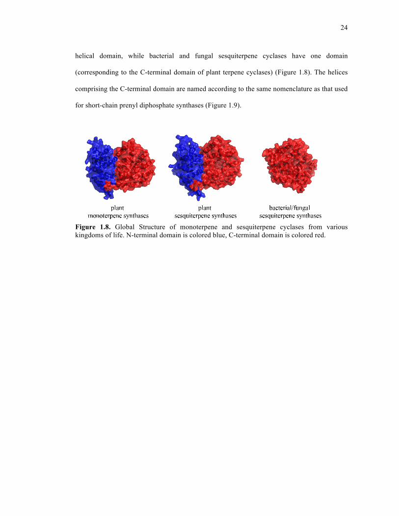
(corresponding to the C-terminal domain of plant terpene cyclases) (Figure 1.8). The helices
comprising the C-terminal domain are named according to the same nomenclature as that used
for short-chain prenyl diphosphate synthases (Figure 1.9).
Figure 1.8. Global Structure of monoterpene and sesquiterpene cyclases from various kingdoms of life. N-terminal domain is colored blue, C-terminal domain is colored red.
25
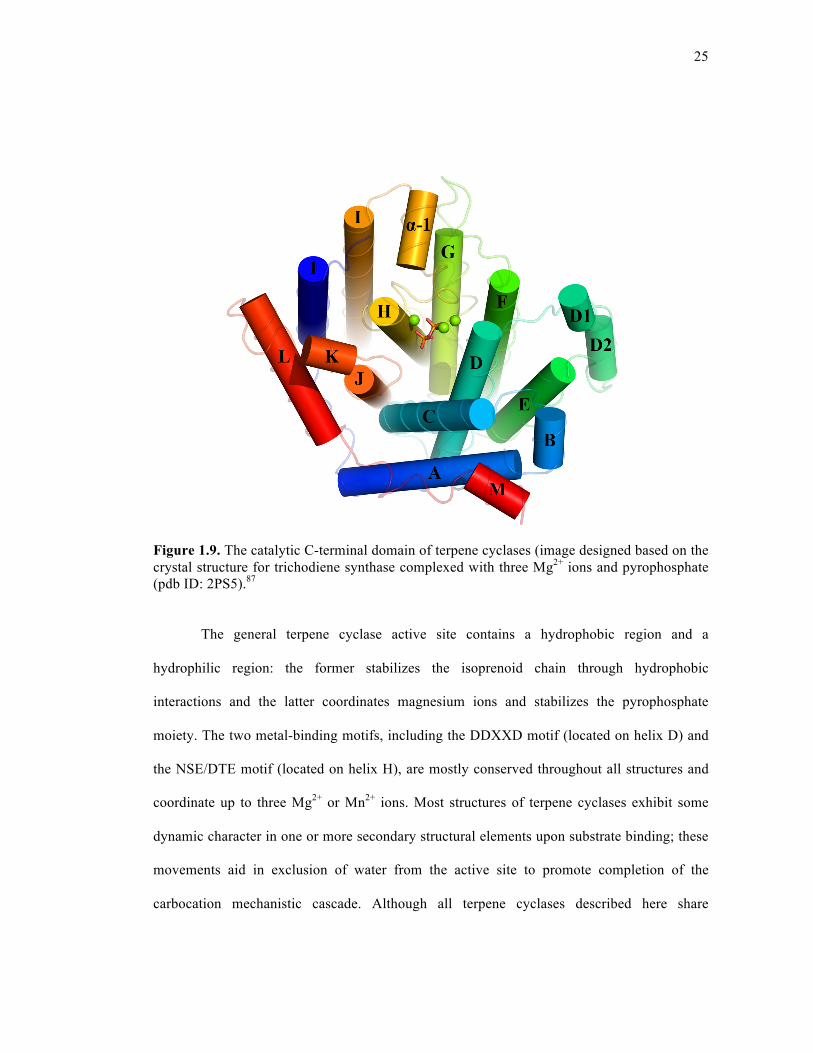
Figure 1.9. The catalytic C-terminal domain of terpene cyclases (image designed based on the crystal structure for trichodiene synthase complexed with three Mg2+ ions and pyrophosphate (pdb ID: 2PS5).87
The general terpene cyclase active site contains a hydrophobic region and a
hydrophilic region: the former stabilizes the isoprenoid chain through hydrophobic
interactions and the latter coordinates magnesium ions and stabilizes the pyrophosphate
moiety. The two metal-binding motifs, including the DDXXD motif (located on helix D) and
the NSE/DTE motif (located on helix H), are mostly conserved throughout all structures and
coordinate up to three Mg2+ or Mn2+ ions. Most structures of terpene cyclases exhibit some
dynamic character in one or more secondary structural elements upon substrate binding; these
movements aid in exclusion of water from the active site to promote completion of the
carbocation mechanistic cascade. Although all terpene cyclases described here share
26
considerable structural homology, there are several noteworthy structural differences between
monoterpene and sesquiterpene cyclases, between sesquiterpene cyclases from different
kingdoms of life, and between individual cyclases. These differences are outlined below.
1.4.3 Divalent metal ion coordination
In general, most terpene cyclases require three divalent metal ions to assist in
pyrophosphate ionization and subsequent catalysis. In the first published crystal structure of a
terpene cyclase, 5-epi-aristolochene synthase (TEAS), MgA2+ and MgB
2+ bind in the
unliganded enzyme and MgC2+ additionally binds in the presence of the substrate analog,
farnesyl hydroxyphosphonate (FHP)75. The first two Mg2+ ions coordinate with octahedral
geometry to the DDXXD motif and NSE/DTE motif, respectively; the third Mg2+ ion binds in
close proximity to MgA2+ and, in the presence of FHP, also coordinates with octahedral
geometry to the DDXXD motif, the phosphate moiety, and several water molecules75 (Figure
1.10).
27
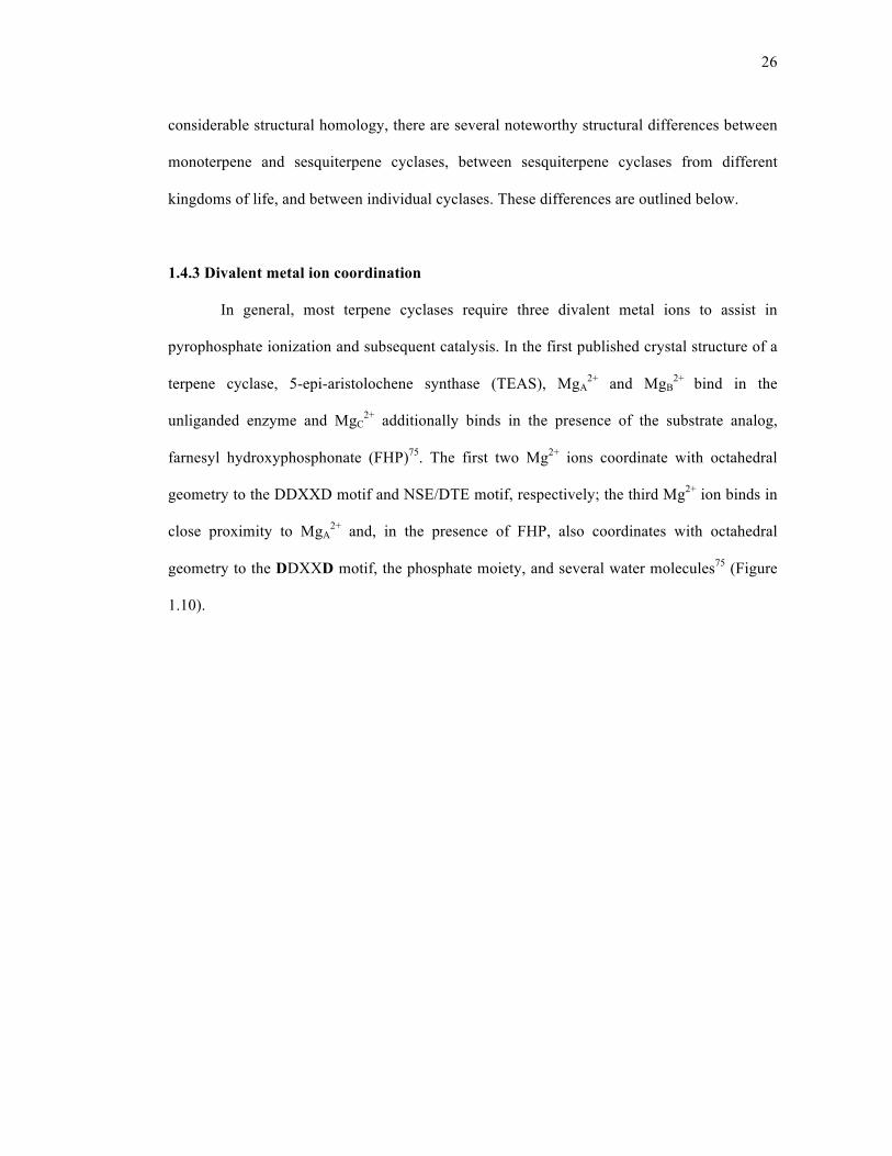
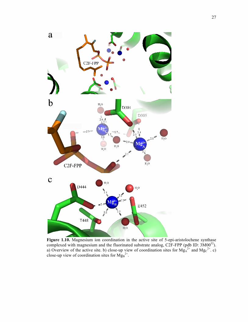
Figure 1.10. Magnesium ion coordination in the active site of 5-epi-aristolochene synthase complexed with magnesium and the fluorinated substrate analog, C2F-FPP (pdb ID: 3M0052). a) Overview of the active site. b) close-up view of coordination sites for MgA
2+ and MgC2+. c)
close-up view of coordination sites for MgB2+.
28
This example is one of many variations on what is observed for divalent metal ion
coordination in the active site of a terpene cyclase. For example, in the unliganded structure of
(+)-bornyl diphosphate synthase, only one magnesium ion is coordinated to the DDXXD
motif and the other two are missing, whereas the substrate-analog bound structure shows three
Mg2+ ions in locations that are consistent with what is observed for TEAS77. In comparing
various crystal structures of aristolochene synthase from A. terreus, monomer D shows either
one or two Mg2+ ions bound (either MgB2+ or MgB
2+ and MgC2+) in the presence of
pyrophosphate or substrate analog; however, the other three monomers show only substrate
analog with no accompanying divalent metal ion coordination83. Such monomeric differences
are thought to represent snapshots of various phases of the terpene cyclase reaction; however,
these results also highlight that MgB2+ plays a very important role in properly orienting the
pyrophosphate moiety of the substrate for catalysis83. Fungal trichodiene synthase and
bacterial epi-isozizaene synthase both coordinate three divalent magnesium ions, however in
contrast to structures of plant terpene cyclases which coordinate MgA2+ and MgC
2+ with the
first and last aspartic acid in the DDXXD motif, these two enzymes only coordinate with the
first aspartic acid82, 85. Additionally, the second aspartic acid of the motif plays a role in the
hydrogen-bonding network between the substrate and surrounding residues, and mutation at
this position causes significant loss of activity85, 88. Notably, bacterial and fungal terpene
cyclases usually contain an NSE motif (instead of a DTE motif as seen in most plant terpene
cyclases); thus, divalent metal ion coordination in terpene cyclases appears to have evolved
slightly differently in plants compared to fungi and bacteria. The most interesting example of
metal ion coordination is in δ-cadinene synthase. This sesquiterpene cyclase contains the
conventional DDXXD motif that binds MgA2+ and MgC
2+, however it is missing the highly
conserved NSE/DTE motif and instead contains another DDXXD/E motif that coordinates the

29
MgB2+ ion81. Both δ-selinene synthase and γ-humulene synthase also contain this additional
DDXXD motif57. This second motif corresponds to SARM in short-chain prenyl diphosphate
synthases.
1.4.4. Ligand-induced structural changes
In general, upon substrate (or pyrophosphate) binding, terpene cyclases close their
active sites to accommodate the ligand, to exclude water, and to initiate pyrophosphate
ionization.82 Plant terpene cyclases adopt more subtle changes upon ligand binding compared
to fungal and bacterial terpene cyclases81, 82, 85 For example, superposition of apo and ligand-
bound structures of 5-epi-aristolochene synthase from tobacco (TEAS) generates a root mean
square deviation (rmsd) for Cα atoms of 0.43Å75 while a similar superposition in fungal
trichodiene synthase generates an rmsd for Cα atoms of 1.4Å.82 Some plant terpene cyclase
structures show ordering of the following motifs when complexed with ligand: the A-C loop,
the J-K loop, part of helix H, and the amino-terminus75, 77, 79. Others, such as δ-cadinene
synthase from cotton, do not demonstrate any such conformational changes81; In contrast,
fungal and bacterial crystal structures show a large degree of movement in several or all of the
following motifs: helices 1, D, H, J, K, L, and loops 1-A, D-D1, F-G, H-α1, J-K, and K-L.82, 85,
89 Fungal and bacterial structures may undergo such drastic conformational changes to
compensate for the fact that they lack the amino-terminal domain that the plant enzymes have
to protect the active site from highly reactive water.81 Additionally, fungal and bacterial
terpene cyclases usually produce one specific product compared to those of plant origin,
suggesting that they may adopt a more rigid active site contour on which to template the
substrate83.
30
1.4.5. Substrate analogs
Ongoing efforts are aimed towards complexation of terpene cyclases with substrate-
like and reaction-like mimics and/or inhibitors to gain insight on each terpene cyclase reaction
mechanism. Complexes of monoterpene cyclases with GPP, linalyl diphosphate (the
isomerized version of GPP), and various carbocation mimics have been reported. In the case
of (+)-bornyl diphosphate synthase, a variety of aza analogs were synthesized and complexed
with the enzyme to mimic the carbocation intermediates generated throughout the reaction;
these mimics were somewhat successful, although the geometry at the nitrogen in the aza
analog is different than that of the planar carbocation center. A similar result is observed in
epi-isozizaene synthase complexed with the benzyl triethylammonium cation (BTAC), which
is meant to mimic the bisabolyl cation (the first cation formed in the mechanism) but also has
different geometry than the naturally occurring carbocation intermediate. Fluorinated substrate
analogs, including 2-fluoro-GPP (2F-GPP), 2-fluoro-FPP (2F-FPP), and 12,13-fluoro-FPP
(difluoro-FPP, or DF-FPP) are most commonly used as substrate mimics for terpene
cyclases52, 77, 89. These analogs are usually non-hydrolyzable due to the presence of the
fluorine atom, which withdraws negative charge from the proximal carbon-carbon double
bond via the inductive effect, rendering (in most cases) inability for pyrophosphate ionization
and electrophilic cyclization. Aristolochene synthase is the exception since it is able to
hydrolyze 2F-FPP into a stable intermediate, 2-fluorogermacrene A (but cannot complete the
reaction to generate aristolochene)89. In some cases, the electron density for the isoprenoid tail
of the substrate or substrate analog is less clearly defined, which is most likely a reflection of a
more dynamic substrate. For example, the electron density for the isoprenoid moiety of nearly
any given FPP analog is much more clearly defined in the active site of aristolochene synthase
compared to TEAS,75, 89, 90 which is perhaps correlated to the fact that the former produces
31
aristolochene exclusively91, while the latter produces a variety of minor products in addition to
5-epi-aristolochene.55
1.5. Emergence of terpene synthases from primary metabolism
There are several theories on the evolution of terpene cyclases. Based on intron/exon
organization in plant angiosperms and gymnosperms, one theory suggests that the ancestral
terpene synthase was a diterpene synthase of primary metabolism (such as KS or CDS) that
underwent gene duplication and divergent evolution to create the present day plant terpene
cyclases.64 Due to lack of sequence similarity, large differences in intron/exon organization,
and large phylogenetic distances between clades, microbial and plant terpene synthases were
thought to have undergone convergent evolution.64 More recently, however, a theory that
incorporates a hierarchy of levels of evolution involving triterpene synthases, bacterial
diterpene synthases, and eventually plant diterpene synthases, has come into view.92 This
theory, based on structural, functional, and sequence comparisons, suggests that the first
bacterial class I diterpene cyclases were created from the ancient triterpene synthase
(containing the DXDD motif and performing the proton-initiated reaction), while the first
bacterial class II diterpene cyclase was created from an ancestor of the class II terpene cyclase
fold (containing the DDXXD motif and performing the ionization-dependent reaction). Class I
and class II diterpene cyclase domains then fused together to create modern day plant
diterpene cyclases, which eventually, through the loss of several exons, evolved into present
day plant monoterpene and sesquiterpene synthases.92 This theory speculates that all terpene
cyclases were derived from bacterial ancestors, and that bacteria eventually transferred these
genes to plants.92
32
1.6. Conclusions
Isoprenoid biosynthesis constitutes a network of biosynthetic pathways that spans
primary and secondary metabolism. In primary metabolism, the MVA pathway and DXP
pathway produce the two essential building blocks for biosynthesis of all downstream
isoprenoids: IPP and DMAPP. The MVA and DXP pathways are not as well understood as
once thought, especially in archaea where some enzymes in the mevalonate pathway have still
not been identified. Work discussed in chapters 5 and 6 focuses on resolving such issues by
using the crystal structure of an archaeal kinase as a starting point towards the discovery of
MVA pathway alternatives both within and outside of this domain of life.
The short-chain prenyl diphosphate synthases represent a family of enzymes that
bridges the primary and secondary metabolic pathways of isoprenoid biosynthesis. Using the
two essential IPP and DMAPP building blocks, these enzymes synthesize GPP, FPP, and
GGPP, which are then substrates for all downstream primary and secondary metabolic
enzymes in this pathway, including the monoterpene, sesquiterpene, and diterpene synthases
of secondary metabolism, respectively.
In the case of all terpene cyclases, the idea that such product diversity can be created
from one substrate is fascinating and has been explored here in three different ways. Chapter 1
addresses how a chemical profile can change throughout the landscape of sequence space that
exists between two sesquiterpene cyclases. Chapter 2 analyzes both structural and functional
effects associated with both substrate and product promiscuity among wild type TEAS and a
promiscuous mutant. Chapter 3 discusses how an extensive mutational analysis at an amino
terminal motif in patchoulol synthase (PAS) has demonstrated its importance in maintaining a
chemically complex product profile.

33
REFERENCES
1. Novakova, Z.; Surin, S.; Blasko, J.; Majernik, A.; Smigan, P., Membrane proteins and squalene-hydrosqualene profile in methanoarchaeon Methanothermobacter thermautotrophicus resistant to N,N'-dicyclohexylcarbodiimide. Folia microbiologica 2008, 53 (3), 237-240.
2. Ourisson, G.; Rohmer, M.; Poralla, K., Prokaryotic hopanoids and other polyterpenoid
sterol surrogates. Annual Review of Microbiology 1987, 41, 301-333. 3. Eichler, J.; Adams, M. W., Posttranslational protein modification in Archaea.
Microbiology and molecular biology reviews : MMBR 2005, 69 (3), 393-425. 4. Bartley, G. E.; Scolnik, P. A., Plant carotenoids: pigments for photoprotection, visual
attraction, and human health. Plant Cell 1995, 7 (7), 1027-38. 5. Trumpower, B. L., New concepts on the role of ubiquinone in the mitochondrial
respiratory chain. J Bioenerg Biomembr 1981, 13 (1-2), 1-24. 6. Hedden, P.; Kamiya, Y., GIBBERELLIN BIOSYNTHESIS: Enzymes, Genes and
Their Regulation. Annu Rev Plant Physiol Plant Mol Biol 1997, 48, 431-460. 7. Arigoni, D.; Sagner, S.; Latzel, C.; Eisenreich, W.; Bacher, A.; Zenk, M. H.,
Terpenoid biosynthesis from 1-deoxy-D-xylulose in higher plants by intramolecular skeletal rearrangement. Proc Natl Acad Sci U S A 1997, 94 (20), 10600-5.
8. Eisenreich, W.; Schwarz, M.; Cartayrade, A.; Arigoni, D.; Zenk, M. H.; Bacher, A.,
The deoxyxylulose phosphate pathway of terpenoid biosynthesis in plants and microorganisms. Chemistry & biology 1998, 5 (9), R221-33.
9. Rohmer, M., The discovery of a mevalonate-independent pathway for isoprenoid
biosynthesis in bacteria, algae and higher plants. Natural product reports 1999, 16 (5), 565-574.
10. Sprenger, G. A.; Schorken, U.; Wiegert, T.; Grolle, S.; de Graaf, A. A.; Taylor, S. V.;
Begley, T. P.; Bringer-Meyer, S.; Sahm, H., Identification of a thiamin-dependent synthase in Escherichia coli required for the formation of the 1-deoxy-D-xylulose 5-phosphate precursor to isoprenoids, thiamin, and pyridoxol. Proc Natl Acad Sci U S A 1997, 94 (24), 12857-62.
11. Rohdich, F.; Zepeck, F.; Adam, P.; Hecht, S.; Kaiser, J.; Laupitz, R.; Grawert, T.;
Amslinger, S.; Eisenreich, W.; Bacher, A.; Arigoni, D., The deoxyxylulose phosphate pathway of isoprenoid biosynthesis: studies on the mechanisms of the reactions catalyzed by IspG and IspH protein. Proc Natl Acad Sci U S A 2003, 100 (4), 1586-91.
34
12. Nyland, R. L., 2nd; Xiao, Y.; Liu, P.; Freel Meyers, C. L., IspG converts an epoxide substrate analogue to (E)-4-hydroxy-3-methylbut-2-enyl diphosphate: implications for IspG catalysis in isoprenoid biosynthesis. J Am Chem Soc 2009, 131 (49), 17734-5.
13. Grawert, T.; Span, I.; Eisenreich, W.; Rohdich, F.; Eppinger, J.; Bacher, A.; Groll, M., Probing the reaction mechanism of IspH protein by x-ray structure analysis. Proc Natl Acad Sci U S A 2010, 107 (3), 1077-81.
14. Brown, M. S.; Dana, S. E.; Goldstein, J. L., Regulation of 3-hydroxy-3-methylglutaryl
coenzyme A reductase activity in human fibroblasts by lipoproteins. Proc Natl Acad Sci U S A 1973, 70 (7), 2162-6.
15. Furberg, C. D.; Adams, H. P., Jr.; Applegate, W. B.; Byington, R. P.; Espeland, M.
A.; Hartwell, T.; Hunninghake, D. B.; Lefkowitz, D. S.; Probstfield, J.; Riley, W. A.; et al., Effect of lovastatin on early carotid atherosclerosis and cardiovascular events. Asymptomatic Carotid Artery Progression Study (ACAPS) Research Group. Circulation 1994, 90 (4), 1679-87.
16. Sapir-Mir, M.; Mett, A.; Belausov, E.; Tal-Meshulam, S.; Frydman, A.; Gidoni, D.;
Eyal, Y., Peroxisomal localization of Arabidopsis isopentenyl diphosphate isomerases suggests that part of the plant isoprenoid mevalonic acid pathway is compartmentalized to peroxisomes. Plant Physiol 2008, 148 (3), 1219-28.
17. Carrero-Lerida, J.; Perez-Moreno, G.; Castillo-Acosta, V. M.; Ruiz-Perez, L. M.;
Gonzalez-Pacanowska, D., Intracellular location of the early steps of the isoprenoid biosynthetic pathway in the trypanosomatids Leishmania major and Trypanosoma brucei. Int J Parasitol 2009, 39 (3), 307-14.
18. Hartman, I. Z.; Liu, P.; Zehmer, J. K.; Luby-Phelps, K.; Jo, Y.; Anderson, R. G.;
DeBose-Boyd, R. A., Sterol-induced dislocation of 3-hydroxy-3-methylglutaryl coenzyme A reductase from endoplasmic reticulum membranes into the cytosol through a subcellular compartment resembling lipid droplets. J Biol Chem 2010, 285 (25), 19288-98.
19. Smit, A.; Mushegian, A., Biosynthesis of isoprenoids via mevalonate in Archaea: the
lost pathway. Genome research 2000, 10 (10), 1468-1484. 20. Grochowski, L. L.; Xu, H.; White, R. H., Methanocaldococcus jannaschii uses a
modified mevalonate pathway for biosynthesis of isopentenyl diphosphate. Journal of Bacteriology 2006, 188 (9), 3192-3198.
21. Ogura, K.; Koyama, T., Enzymatic Aspects of Isoprenoid Chain Elongation. Chem
Rev 1998, 98 (4), 1263-1276. 22. Burke, C. C.; Wildung, M. R.; Croteau, R., Geranyl diphosphate synthase: cloning,
expression, and characterization of this prenyltransferase as a heterodimer. Proc Natl Acad Sci U S A 1999, 96 (23), 13062-7.
35
23. Tarshis, L. C.; Yan, M.; Poulter, C. D.; Sacchettini, J. C., Crystal structure of recombinant farnesyl diphosphate synthase at 2.6-A resolution. Biochemistry 1994, 33 (36), 10871-7.
24. Tarshis, L. C.; Proteau, P. J.; Kellogg, B. A.; Sacchettini, J. C.; Poulter, C. D.,
Regulation of product chain length by isoprenyl diphosphate synthases. Proc Natl Acad Sci U S A 1996, 93 (26), 15018-23.
25. Wang, K.; Ohnuma, S., Chain-length determination mechanism of isoprenyl
diphosphate synthases and implications for molecular evolution. Trends Biochem Sci 1999, 24 (11), 445-51.
26. Hosfield, D. J.; Zhang, Y.; Dougan, D. R.; Broun, A.; Tari, L. W.; Swanson, R. V.;
Finn, J., Structural basis for bisphosphonate-mediated inhibition of isoprenoid biosynthesis. J Biol Chem 2004, 279 (10), 8526-9.
27. Ohnuma, S.; Narita, K.; Nakazawa, T.; Ishida, C.; Takeuchi, Y.; Ohto, C.; Nishino, T.,
A role of the amino acid residue located on the fifth position before the first aspartate-rich motif of farnesyl diphosphate synthase on determination of the final product. J Biol Chem 1996, 271 (48), 30748-54.
28. Lee, P. C.; Petri, R.; Mijts, B. N.; Watts, K. T.; Schmidt-Dannert, C., Directed
evolution of Escherichia coli farnesyl diphosphate synthase (IspA) reveals novel structural determinants of chain length specificity. Metab Eng 2005, 7 (1), 18-26.
29. Ohnuma, S.; Hirooka, K.; Ohto, C.; Nishino, T., Conversion from archaeal
geranylgeranyl diphosphate synthase to farnesyl diphosphate synthase. Two amino acids before the first aspartate-rich motif solely determine eukaryotic farnesyl diphosphate synthase activity. J Biol Chem 1997, 272 (8), 5192-8.
30. Gershenzon, J.; Dudareva, N., The function of terpene natural products in the natural
world. Nature chemical biology 2007, 3 (7), 408-414. 31. Pichersky, E.; Lewinsohn, E.; Croteau, R., Purification and characterization of S-
linalool synthase, an enzyme involved in the production of floral scent in Clarkia breweri. Arch Biochem Biophys 1995, 316 (2), 803-7.
32. Kollner, T. G.; Held, M.; Lenk, C.; Hiltpold, I.; Turlings, T. C.; Gershenzon, J.;
Degenhardt, J., A maize (E)-beta-caryophyllene synthase implicated in indirect defense responses against herbivores is not expressed in most American maize varieties. Plant Cell 2008, 20 (2), 482-94.
33. Hezari, M.; Lewis, N. G.; Croteau, R., Purification and characterization of taxa-
4(5),11(12)-diene synthase from Pacific yew (Taxus brevifolia) that catalyzes the first committed step of taxol biosynthesis. Archives of Biochemistry and Biophysics 1995, 322 (2), 437-444.
36
34. Bouwmeester, H. J.; Wallaart, T. E.; Janssen, M. H.; van Loo, B.; Jansen, B. J.; Posthumus, M. A.; Schmidt, C. O.; De Kraker, J. W.; Konig, W. A.; Franssen, M. C., Amorpha-4,11-diene synthase catalyses the first probable step in artemisinin biosynthesis. Phytochemistry 1999, 52 (5), 843-54.
35. Kirby, J.; Keasling, J. D., Biosynthesis of plant isoprenoids: perspectives for microbial
engineering. Annu Rev Plant Biol 2009, 60, 335-55. 36. Asadollahi, M. A.; Maury, J.; Moller, K.; Nielsen, K. F.; Schalk, M.; Clark, A.;
Nielsen, J., Production of plant sesquiterpenes in Saccharomyces cerevisiae: effect of ERG9 repression on sesquiterpene biosynthesis. Biotechnol Bioeng 2008, 99 (3), 666-77.
37. Ohto, C.; Muramatsu, M.; Obata, S.; Sakuradani, E.; Shimizu, S., Overexpression of
the gene encoding HMG-CoA reductase in Saccharomyces cerevisiae for production of prenyl alcohols. Appl Microbiol Biotechnol 2009, 82 (5), 837-45.
38. Morrone, D.; Lowry, L.; Determan, M. K.; Hershey, D. M.; Xu, M.; Peters, R. J.,
Increasing diterpene yield with a modular metabolic engineering system in E. coli: comparison of MEV and MEP isoprenoid precursor pathway engineering. Appl Microbiol Biotechnol 2010, 85 (6), 1893-906.
39. Martin, V. J.; Pitera, D. J.; Withers, S. T.; Newman, J. D.; Keasling, J. D.,
Engineering a mevalonate pathway in Escherichia coli for production of terpenoids. Nature biotechnology 2003, 21 (7), 796-802.
40. Pitera, D. J.; Paddon, C. J.; Newman, J. D.; Keasling, J. D., Balancing a heterologous
mevalonate pathway for improved isoprenoid production in Escherichia coli. Metabolic engineering 2007, 9 (2), 193-207.
41. Fu, Z.; Voynova, N. E.; Herdendorf, T. J.; Miziorko, H. M.; Kim, J. J., Biochemical
and structural basis for feedback inhibition of mevalonate kinase and isoprenoid metabolism. Biochemistry 2008, 47 (12), 3715-24.
42. Bohlmann, J.; Meyer-Gauen, G.; Croteau, R., Plant terpenoid synthases: molecular
biology and phylogenetic analysis. Proc. Natl. Acad. Sci. USA 1998, 95, 4126-4133. 43. Cane, D. E.; Xue, Q.; Fitzsimons, B. C., Trichodiene synthase. Probing the role of the
highly conserved aspartate-rich region by site-directed mutagenesis. Biochemistry 1996, 35 (38), 12369-12376.
44. Christianson, D. W., Structural biology and chemistry of the terpenoid cyclases.
Chemical reviews 2006, 106 (8), 3412-3442. 45. McGarvey, D. J.; Croteau, R., Terpenoid metabolism. Plant Cell 1995, 7 (7), 1015-26. 46. Cane, D. E.; Kang, I., Aristolochene synthase: purification, molecular cloning, high-
level expression in Escherichia coli, and characterization of the Aspergillus terreus cyclase. Archives of Biochemistry and Biophysics 2000, 376 (2), 354-364.
37
47. Williams, D. C.; McGarvey, D. J.; Katahira, E. J.; Croteau, R., Truncation of
limonene synthase preprotein provides a fully active 'pseudomature' form of this monoterpene cyclase and reveals the function of the amino-terminal arginine pair. Biochemistry 1998, 37 (35), 12213-12220.
48. Rajaonarivony, J. I.; Gershenzon, J.; Croteau, R., Characterization and mechanism of
(4S)-limonene synthase, a monoterpene cyclase from the glandular trichomes of peppermint (Mentha x piperita). Arch Biochem Biophys 1992, 296 (1), 49-57.
49. Degenhardt, J.; Kollner, T. G.; Gershenzon, J., Monoterpene and sesquiterpene
synthases and the origin of terpene skeletal diversity in plants. Phytochemistry 2009, 70 (15-16), 1621-37.
50. Schilmiller, A. L.; Schauvinhold, I.; Larson, M.; Xu, R.; Charbonneau, A. L.;
Schmidt, A.; Wilkerson, C.; Last, R. L.; Pichersky, E., Monoterpenes in the glandular trichomes of tomato are synthesized from a neryl diphosphate precursor rather than geranyl diphosphate. Proc Natl Acad Sci U S A 2009, 106 (26), 10865-70.
51. Sallaud, C.; Rontein, D.; Onillon, S.; Jabes, F.; Duffe, P.; Giacalone, C.; Thoraval, S.;
Escoffier, C.; Herbette, G.; Leonhardt, N.; Causse, M.; Tissier, A., A novel pathway for sesquiterpene biosynthesis from Z,Z-farnesyl pyrophosphate in the wild tomato Solanum habrochaites. Plant Cell 2009, 21 (1), 301-17.
52. Noel, J. P.; Dellas, N.; Faraldos, J. A.; Zhao, M.; Hess, B. A., Jr.; Smentek, L.;
Coates, R. M.; O'Maille, P. E., Structural elucidation of cisoid and transoid cyclization pathways of a sesquiterpene synthase using 2-fluorofarnesyl diphosphates. ACS chemical biology 2010, 5 (4), 377-392.
53. Picaud, S.; Olofsson, L.; Brodelius, M.; Brodelius, P. E., Expression, purification, and
characterization of recombinant amorpha-4,11-diene synthase from Artemisia annua L. Arch Biochem Biophys 2005, 436 (2), 215-26.
54. Thulasiram, H. V.; Poulter, C. D., Farnesyl diphosphate synthase: The art of
compromise between substrate selectivity and stereoselectivity. Journal of the American Chemical Society 2006, 128 (49), 15819-15823.
55. O'Maille, P. E.; Chappell, J.; Noel, J. P., Biosynthetic potential of sesquiterpene
synthases: Alternative products of tobacco 5-epi-aristolochene synthase. Archives of Biochemistry and Biophysics 2006, 448 (1-2), 73-82.
56. Cane, D. E., How to evolve a silk purse from a sow's ear. Nat Chem Biol 2006, 2 (4),
179-80. 57. Little, D. B.; Croteau, R. B., Alteration of product formation by directed mutagenesis
and truncation of the multiple-product sesquiterpene synthases delta-selinene synthase and gamma-humulene synthase. Archives of Biochemistry and Biophysics 2002, 402 (1), 120-135.
38
58. Deguerry, F.; Pastore, L.; Wu, S.; Clark, A.; Chappell, J.; Schalk, M., The diverse
sesquiterpene profile of patchouli, Pogostemon cablin, is correlated with a limited number of sesquiterpene synthases. Archives of Biochemistry and Biophysics 2006, 454 (2), 123-136.
59. Yoshikuni, Y.; Keasling, J. D., Pathway engineering by designed divergent evolution.
Curr Opin Chem Biol 2007, 11 (2), 233-9. 60. O'Maille, P. E.; Malone, A.; Dellas, N.; Andes Hess, B., Jr.; Smentek, L.; Sheehan, I.;
Greenhagen, B. T.; Chappell, J.; Manning, G.; Noel, J. P., Quantitative exploration of the catalytic landscape separating divergent plant sesquiterpene synthases. Nature chemical biology 2008, 4 (10), 617-623.
61. Yoshikuni, Y.; Ferrin, T. E.; Keasling, J. D., Designed divergent evolution of enzyme
function. Nature 2006, 440 (7087), 1078-1082. 62. Greenhagen, B. T.; O'Maille, P. E.; Noel, J. P.; Chappell, J., Identifying and
manipulating structural determinates linking catalytic specificities in terpene synthases. Proceedings of the National Academy of Sciences of the United States of America 2006, 103 (26), 9826-9831.
63. Lin, X.; Hezari, M.; Koepp, A. E.; Floss, H. G.; Croteau, R., Mechanism of taxadiene
synthase, a diterpene cyclase that catalyzes the first step of taxol biosynthesis in Pacific yew. Biochemistry 1996, 35 (9), 2968-77.
64. Trapp, S. C.; Croteau, R. B., Genomic organization of plant terpene synthases and
molecular evolutionary implications. Genetics 2001, 158 (2), 811-832. 65. Sun, T. P.; Kamiya, Y., The Arabidopsis GA1 locus encodes the cyclase ent-kaurene
synthetase A of gibberellin biosynthesis. Plant Cell 1994, 6 (10), 1509-18. 66. Morrone, D.; Chambers, J.; Lowry, L.; Kim, G.; Anterola, A.; Bender, K.; Peters, R.
J., Gibberellin biosynthesis in bacteria: separate ent-copalyl diphosphate and ent-kaurene synthases in Bradyrhizobium japonicum. FEBS Lett 2009, 583 (2), 475-80.
67. Saito, T.; Abe, H.; Yamane, H.; Sakurai, A.; Murofushi, N.; Takio, K.; Takahashi, N.;
Kamiya, Y., Purification and Properties of ent-Kaurene Synthase B from Immature Seeds of Pumpkin. Plant Physiol 1995, 109 (4), 1239-1245.
68. Hayashi, K.; Kawaide, H.; Notomi, M.; Sakigi, Y.; Matsuo, A.; Nozaki, H.,
Identification and functional analysis of bifunctional ent-kaurene synthase from the moss Physcomitrella patens. FEBS Lett 2006, 580 (26), 6175-81.
69. Toyomasu, T.; Kawaide, H.; Ishizaki, A.; Shinoda, S.; Otsuka, M.; Mitsuhashi, W.;
Sassa, T., Cloning of a full-length cDNA encoding ent-kaurene synthase from Gibberella fujikuroi: functional analysis of a bifunctional diterpene cyclase. Bioscience, biotechnology, and biochemistry 2000, 64 (3), 660-664.

39
70. Vogel, B. S.; Wildung, M. R.; Vogel, G.; Croteau, R., Abietadiene synthase from
grand fir (Abies grandis). cDNA isolation, characterization, and bacterial expression of a bifunctional diterpene cyclase involved in resin acid biosynthesis. J Biol Chem 1996, 271 (38), 23262-8.
71. Peters, R. J.; Ravn, M. M.; Coates, R. M.; Croteau, R. B., Bifunctional abietadiene
synthase: free diffusive transfer of the (+)-copalyl diphosphate intermediate between two distinct active sites. J Am Chem Soc 2001, 123 (37), 8974-8.
72. Peters, R. J.; Croteau, R. B., Abietadiene synthase catalysis: conserved residues
involved in protonation-initiated cyclization of geranylgeranyl diphosphate to (+)-copalyl diphosphate. Biochemistry 2002, 41 (6), 1836-42.
73. Xu, M.; Hillwig, M. L.; Prisic, S.; Coates, R. M.; Peters, R. J., Functional
identification of rice syn-copalyl diphosphate synthase and its role in initiating biosynthesis of diterpenoid phytoalexin/allelopathic natural products. Plant J 2004, 39 (3), 309-18.
74. Peters, R. J.; Carter, O. A.; Zhang, Y.; Matthews, B. W.; Croteau, R. B., Bifunctional
abietadiene synthase: mutual structural dependence of the active sites for protonation-initiated and ionization-initiated cyclizations. Biochemistry 2003, 42 (9), 2700-7.
75. Starks, C. M.; Back, K.; Chappell, J.; Noel, J. P., Structural basis for cyclic terpene
biosynthesis by tobacco 5-epi-aristolochene synthase. Science 1997, 277 (5333), 1815-1820.
76. Wendt, K. U.; Schulz, G. E., Isoprenoid biosynthesis: manifold chemistry catalyzed
by similar enzymes. Structure (London, England : 1993) 1998, 6 (2), 127-133. 77. Whittington, D. A.; Wise, M. L.; Urbansky, M.; Coates, R. M.; Croteau, R. B.;
Christianson, D. W., Bornyl diphosphate synthase: structure and strategy for carbocation manipulation by a terpenoid cyclase. Proceedings of the National Academy of Sciences of the United States of America 2002, 99 (24), 15375-15380.
78. Lesburg, C. A.; Zhai, G.; Cane, D. E.; Christianson, D. W., Crystal structure of
pentalenene synthase: mechanistic insights on terpenoid cyclization reactions in biology. Science (New York, N.Y.) 1997, 277 (5333), 1820-1824.
79. Hyatt, D. C.; Youn, B.; Zhao, Y.; Santhamma, B.; Coates, R. M.; Croteau, R. B.;
Kang, C., Structure of limonene synthase, a simple model for terpenoid cyclase catalysis. Proceedings of the National Academy of Sciences of the United States of America 2007, 104 (13), 5360-5365.
80. Kampranis, S. C.; Ioannidis, D.; Purvis, A.; Mahrez, W.; Ninga, E.; Katerelos, N. A.;
Anssour, S.; Dunwell, J. M.; Degenhardt, J.; Makris, A. M.; Goodenough, P. W.; Johnson, C. B., Rational conversion of substrate and product specificity in a Salvia
40
monoterpene synthase: structural insights into the evolution of terpene synthase function. The Plant Cell 2007, 19 (6), 1994-2005.
81. Gennadios, H. A.; Gonzalez, V.; Di Costanzo, L.; Li, A.; Yu, F.; Miller, D. J.;
Allemann, R. K.; Christianson, D. W., Crystal structure of (+)-delta-cadinene synthase from Gossypium arboreum and evolutionary divergence of metal binding motifs for catalysis. Biochemistry 2009, 48 (26), 6175-83.
82. Rynkiewicz, M. J.; Cane, D. E.; Christianson, D. W., Structure of trichodiene synthase
from Fusarium sporotrichioides provides mechanistic inferences on the terpene cyclization cascade. Proceedings of the National Academy of Sciences of the United States of America 2001, 98 (24), 13543-13548.
83. Shishova, E. Y.; Di Costanzo, L.; Cane, D. E.; Christianson, D. W., X-ray crystal
structure of aristolochene synthase from Aspergillus terreus and evolution of templates for the cyclization of farnesyl diphosphate. Biochemistry 2007, 46 (7), 1941-1951.
84. Caruthers, J. M.; Kang, I.; Rynkiewicz, M. J.; Cane, D. E.; Christianson, D. W.,
Crystal structure determination of aristolochene synthase from the blue cheese mold, Penicillium roqueforti. The Journal of biological chemistry 2000, 275 (33), 25533-25539.
85. Aaron, J. A.; Lin, X.; Cane, D. E.; Christianson, D. W., Structure of epi-isozizaene
synthase from Streptomyces coelicolor A3(2), a platform for new terpenoid cyclization templates. Biochemistry 2010, 49 (8), 1787-97.
86. Reardon, D.; Farber, G. K., The structure and evolution of alpha/beta barrel proteins.
FASEB J 1995, 9 (7), 497-503. 87. Vedula, L. S.; Jiang, J.; Zakharian, T.; Cane, D. E.; Christianson, D. W., Structural
and mechanistic analysis of trichodiene synthase using site-directed mutagenesis: probing the catalytic function of tyrosine-295 and the asparagine-225/serine-229/glutamate-233-Mg2+B motif. Arch Biochem Biophys 2008, 469 (2), 184-94.
88. Lin, X.; Cane, D. E., Biosynthesis of the sesquiterpene antibiotic albaflavenone in
Streptomyces coelicolor. Mechanism and stereochemistry of the enzymatic formation of epi-isozizaene. J Am Chem Soc 2009, 131 (18), 6332-3.
89. Shishova, E. Y.; Yu, F.; Miller, D. J.; Faraldos, J. A.; Zhao, Y.; Coates, R. M.;
Allemann, R. K.; Cane, D. E.; Christianson, D. W., X-ray crystallographic studies of substrate binding to aristolochene synthase suggest a metal ion binding sequence for catalysis. J Biol Chem 2008, 283 (22), 15431-9.
90. Shishova, E. Y.; Di Costanzo, L.; Cane, D. E.; Christianson, D. W., X-ray crystal
structure of aristolochene synthase from Aspergillus terreus and evolution of templates for the cyclization of farnesyl diphosphate. Biochemistry 2007, 46 (7), 1941-51.

41
91. Felicetti, B.; Cane, D. E., Aristolochene synthase: mechanistic analysis of active site
residues by site-directed mutagenesis. J Am Chem Soc 2004, 126 (23), 7212-21. 92. Cao, R.; Zhang, Y.; Mann, F. M.; Huang, C.; Mukkamala, D.; Hudock, M. P.; Mead,
M. E.; Prisic, S.; Wang, K.; Lin, F. Y.; Chang, T. K.; Peters, R. J.; Oldfield, E., Diterpene cyclases and the nature of the isoprene fold. Proteins 2010, 78 (11), 2417-32.
42
Chapter 2
Quantitative Exploration of the Catalytic Landscape Separating Divergent Plant
Sesquiterpene Synthases

43
2.1. ABSTRACT
Throughout molecular evolution, organisms create assorted chemicals in response to
varying ecological niches. Catalytic landscapes underlie metabolic evolution, wherein
mutational steps alter the biosynthetic properties of enzymes. We report the first systematic
quantitative characterization of a catalytic landscape underlying the evolution of sesquiterpene
chemical diversity. Based on our previous discovery of a set of 9 naturally occurring amino
acid substitutions that functionally inter-converted orthologous sesquiterpene synthases from
Nicotiana tabacum and Hyoscyamus muticus, we created a library of all possible residue
combinations (29 = 512) in the N. tabacum parent. The product spectra of 418 active enzymes
to reveal a rugged landscape where several minimal combinations of the 9 mutations encode
convergent solutions to the inter-conversions of parental activities. Quantitative comparisons
indicate context dependence for mutational effects - epistasis - in product specificity and
promiscuity. These results provide a measure of the mutational accessibility of phenotypic
variability among a diverging lineage of terpene synthases.
2.2. INTRODUCTION
The acquisition of innovative metabolic activities is a major force shaping
evolutionary change, but one that is poorly understood. Metabolic adaptability is particularly
crucial for a plant’s capacity to synthesize specialized chemicals affording protection against
microbial pathogens1-3, elicitation of symbiotic relationships4, attractive5 and repellent6
activities and physiological responses to local environments (as reviewed7). Understanding the
evolution of metabolic function at the molecular level requires knowledge of the distribution
of enzymatic properties through accessible mutational changes, and hence defining the
catalytic landscape. Currently, there is no reported measurement of the catalytic landscapes
44
underlying secondary (specialized) metabolism. The physicochemical constraints relating
sequence variation and metabolic output raise important fundamental questions concerning
catalytic complexity and natural selection. For instance, how does a particular biosynthetic
property like catalytic efficiency or substrate/product specificity vary amongst extant and
probable ancestral sequences? Moreover, how likely is the emergence of new product
specificities in a family of diverging biosynthetic enzymes?
To experimentally approach these questions in the current work, we relied upon i.) a
model system composed of a pair of closely-related secondary metabolic enzymes, ii.) a
simplified set of naturally occurring mutations which interconvert a defined catalytic property
that is functionally divergent between the parental sequences, iii.) structure-based
combinatorial protein engineering (SCOPE)8 to provide a means of creating an enzyme
lineage representing putative evolutionary intermediates in one parental background, and iv.) a
gas chromatography-mass spectrometry (GC-MS) method9 for measuring the catalytic
properties (recording the chemical readout) of the enzyme library. Therefore, quantitative
comparison of catalytic properties of enzymes across these simplified and probable lineages
provides a direct measure of functional variation likely correlated with phenotypic variation in
the defense response of parental species. Moreover, this comprehensive study explores the
mutational accessibility of alternative biosynthetic properties over this experimentally defined
region of sequence space.
Terpene synthases of secondary metabolism are a diverse enzyme family responsible
for the biosynthesis of complex chemicals. Tobacco 5-epi-aristolochene synthase (TEAS) and
henbane premnaspirodiene synthase (HPS) are closely related (75% amino acid identity)
enzymes yet cyclize ionized farnesyl diphosphate (FPP, 1) to form 5-epi-aristolochene (5-EA,
2) and premnaspirodiene (PSD, 3), respectively. These structurally distinct terpene
45
hydrocarbons are precursors of antifungal phytoalexins in solanaceous plants10, 11.
Mechanistically, TEAS and HPS share a common carbocation intermediate during an
electrophilic cyclization reaction, but differ in directing either a methyl or methylene
migration, respectively (Figure 1 panel a). Density functional theory calculations indicate both
of these alkyl shifts to be endothermic (~3 kcal/mol), with the methylene shift's transition state
of lower energy (Figure 2.1).
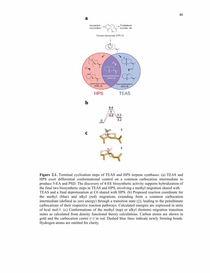
46
Figure 2.1. Terminal cyclization steps of TEAS and HPS terpene synthases. (a) TEAS and HPS exert differential conformational control on a common carbocation intermediate to produce 5-EA and PSD. The discovery of 4-EE biosynthetic activity supports hybridization of the final two biosynthetic steps in TEAS and HPS, involving a methyl migration shared with TEAS and a final deprotonation at C6 shared with HPS. (b) Proposed reaction coordinate for the methyl (blue) and alkyl (red) migrations extending from a common carbocation intermediate (defined as zero energy) through a transition state (‡), leading to the penultimate carbocations of their respective reaction pathways. Calculated energies are expressed in units of kcal mol-1. (c) Conformations of the methyl (top) or alkyl (bottom) migration transition states as calculated from density functional theory calculations. Carbon atoms are shown in gold and the carbocation center (+) in red. Dashed blue lines indicate newly forming bonds. Hydrogen atoms are omitted for clarity.
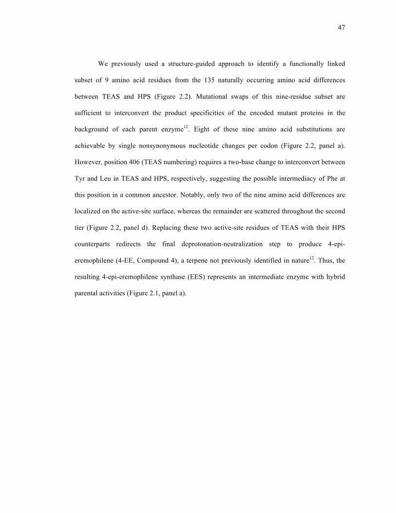
47
We previously used a structure-guided approach to identify a functionally linked
subset of 9 amino acid residues from the 135 naturally occurring amino acid differences
between TEAS and HPS (Figure 2.2). Mutational swaps of this nine-residue subset are
sufficient to interconvert the product specificities of the encoded mutant proteins in the
background of each parent enzyme12. Eight of these nine amino acid substitutions are
achievable by single nonsynonymous nucleotide changes per codon (Figure 2.2, panel a).
However, position 406 (TEAS numbering) requires a two-base change to interconvert between
Tyr and Leu in TEAS and HPS, respectively, suggesting the possible intermediacy of Phe at
this position in a common ancestor. Notably, only two of the nine amino acid differences are
localized on the active-site surface, whereas the remainder are scattered throughout the second
tier (Figure 2.2, panel d). Replacing these two active-site residues of TEAS with their HPS
counterparts redirects the final deprotonation-neutralization step to produce 4-epi-
eremophilene (4-EE, Compound 4), a terpene not previously identified in nature12. Thus, the
resulting 4-epi-eremophilene synthase (EES) represents an intermediate enzyme with hybrid
parental activities (Figure 2.1, panel a).
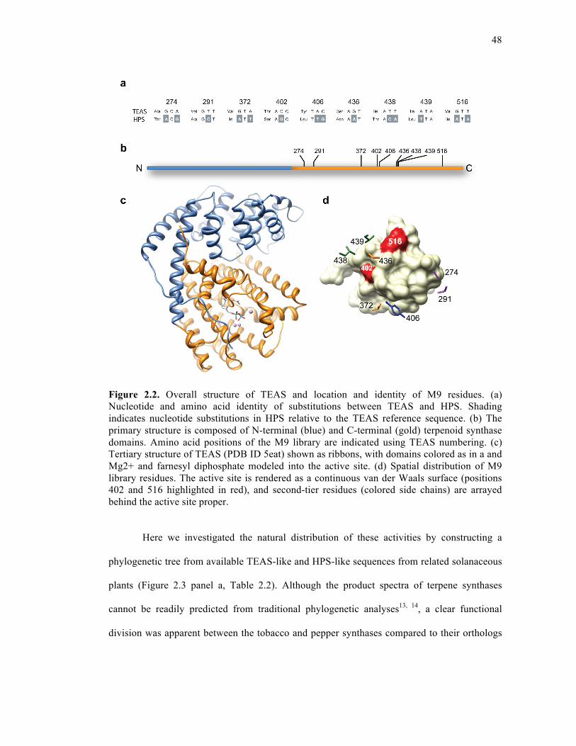
48
Figure 2.2. Overall structure of TEAS and location and identity of M9 residues. (a) Nucleotide and amino acid identity of substitutions between TEAS and HPS. Shading indicates nucleotide substitutions in HPS relative to the TEAS reference sequence. (b) The primary structure is composed of N-terminal (blue) and C-terminal (gold) terpenoid synthase domains. Amino acid positions of the M9 library are indicated using TEAS numbering. (c) Tertiary structure of TEAS (PDB ID 5eat) shown as ribbons, with domains colored as in a and Mg2+ and farnesyl diphosphate modeled into the active site. (d) Spatial distribution of M9 library residues. The active site is rendered as a continuous van der Waals surface (positions 402 and 516 highlighted in red), and second-tier residues (colored side chains) are arrayed behind the active site proper.
Here we investigated the natural distribution of these activities by constructing a
phylogenetic tree from available TEAS-like and HPS-like sequences from related solanaceous
plants (Figure 2.3 panel a, Table 2.2). Although the product spectra of terpene synthases
cannot be readily predicted from traditional phylogenetic analyses13, 14, a clear functional
division was apparent between the tobacco and pepper synthases compared to their orthologs

49
in tomato, potato and henbane. This division was also apparent at the level of our nine-residue
subset, with the exception of the Capsicum annuum synthase (Figure 2.3 panel b). This TEAS-
like enzyme differs from both TEAS- and HPS-like groups at three positions and, most
notably, contains a threonine at position 438 like HPS, suggesting that the first mutational
steps in the TEAS-HPS divergence occurred at these positions. Evaluating the functional
divergence of TEAS and HPS within the context of these nine amino acid substitutions
provides a simplified experimental system to address the broader issue of how prevalent—and
hence evolvable—these parental and alternative biosynthetic activities are throughout the
intervening lineages connecting these extant enzymes. Measuring the distribution of
biosynthetic activities over this sequence space defines a functionally relevant portion of the
overall catalytic landscape and provides a window into the complex functional terrain
underlying the evolution of these enzymes. Although variation at other positions may have
contributed to the functional divergence of TEAS and HPS in meaningful ways, this focused
set of functionally important residues makes it experimentally tractable to quantitatively
characterize a catalytic landscape of secondary metabolism to biochemical resolution.
50
Figure 2.3. Phylogenetic distribution of solanaceous TEAS- and HPS-like terpene synthases. (a) An unrooted phylogenetic tree of 5-EA and PSD terpene synthases created from available sequences (Table 2.2). Branches are colored according to established or putative functions as TEAS-like (blue) or HPS-like (red). (b) Sequence alignment of the M9 residue positions of the sequences in a, with HPS-like residues shaded in gray. Boxes mark residues of C. annuum that differ from both TEAS and HPS. Of note, the previous taxonomic classification Lycopersicon esculentum is used here, consistent with the database entries for their respective proteins. However, the most recent taxonomic nomenclature has been changed to Solanum esculentum.
2.3. RESULTS AND DISCUSSION
2.3.1. Creation and characterization of the M9 lineage
Using SCOPE, we constructed a gene library encoding all permutations of nine amino
acid substitutions in TEAS (29 = 512 combinations) previously shown to functionally
interconvert TEAS and HPS15. The resulting library, termed the M9 lineage, represents the
nodes along all possible mutational pathways (9! = 362,880) between wild-type TEAS and the
nine mutant HPS-like forms (TEAS M9). The combinatorial exploration of variation at these
diverging positions using SCOPE therefore captures a portion of the functionally relevant
genetic variation leading to the current extant sequences. We cloned and identified mutant
genes by sequencing, resulting in 432 unique combinations (Table 2.3). We then expressed

51
and purified individual mutants from Escherichia coli, leading to the recovery of 418 active
proteins. We developed an in vitro biochemical assay for increased sample throughput and
analysis of terpene synthases using GC-MS9, which provided quantified chemical fingerprints
and catalytic activities of the M9 library proteins (Tables 2.4 and 2.5).
To quantitatively assess product specificity, the catalytic property defined here as the
relative proportion of products, we calculated the product percentages of each mutant from
their respective total ion chromatograms. PSD, 5-EA and 4-EE were the dominant products
observed across a wide spectrum of mutants, accompanied by a collection of minor products
that were grouped together and treated as a single product class for simplicity. This
assumption may introduce error, as the ionization efficiencies of these minor components are
as yet unknown; however, their inclusion contributes to the measure of product specificity and
promiscuity.
A three-dimensional scatter plot shows how the product specificities of mutants
distribute throughout chemical space (Figure 2.4 panel a). The three dominant products (5-EA,
4-EE and PSD) define a two-dimensional triangular plane, and the collective minor products
contribute the third dimension of the tetrahedron. Mutants with varying degrees of catalytic
promiscuity radiate uniformly from a cluster of TEAS-like activities, together forming a
continuum with more HPS-like mutants. By contrast, EES-like enzymes are rare, appearing as
a sparsely populated subgroup. Subdividing the scatter plot into three smaller tetrahedrons of
equal volume geometrically defines product specificity as >50% 5-EA, 4-EE or PSD, whereas
the central volume represents promiscuous activities (Figure 2.4 panel b). The majority of
mutants were promiscuous (51%), showing expanded product distributions and upregulation
of other TEAS minor products, predominantly germacrene A (Compound 5)16, a neutral
intermediate along the TEAS and HPS cyclization pathways and the major product of a
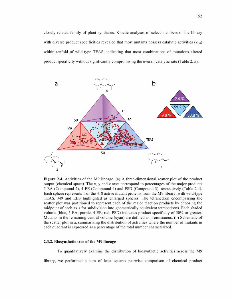
52
closely related family of plant synthases. Kinetic analyses of select members of the library
with diverse product specificities revealed that most mutants possess catalytic activities (kcat)
within tenfold of wild-type TEAS, indicating that most combinations of mutations altered
product specificity without significantly compromising the overall catalytic rate (Table 2. 5).
Figure 2.4. Activities of the M9 lineage. (a) A three-dimensional scatter plot of the product output (chemical space). The x, y and z axes correspond to percentages of the major products 5-EA (Compound 2), 4-EE (Compound 4) and PSD (Compound 3), respectively (Table 2.4). Each sphere represents 1 of the 418 active mutant proteins from the M9 library, with wild-type TEAS, M9 and EES highlighted as enlarged spheres. The tetrahedron encompassing the scatter plot was partitioned to represent each of the major reaction products by choosing the midpoint of each axis for subdivision into geometrically equivalent tetrahedrons. Each shaded volume (blue, 5-EA; purple, 4-EE; red, PSD) indicates product specificity of 50% or greater. Mutants in the remaining central volume (cyan) are defined as promiscuous. (b) Schematic of the scatter plot in a, summarizing the distribution of activities where the number of mutants in each quadrant is expressed as a percentage of the total number characterized.
2.3.2. Biosynthetic tree of the M9 lineage
To quantitatively examine the distribution of biosynthetic activities across the M9
library, we performed a sum of least squares pairwise comparison of chemical product
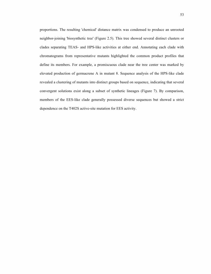
53
proportions. The resulting 'chemical' distance matrix was condensed to produce an unrooted
neighbor-joining 'biosynthetic tree' (Figure 2.5). This tree showed several distinct clusters or
clades separating TEAS- and HPS-like activities at either end. Annotating each clade with
chromatograms from representative mutants highlighted the common product profiles that
define its members. For example, a promiscuous clade near the tree center was marked by
elevated production of germacrene A in mutant 8. Sequence analysis of the HPS-like clade
revealed a clustering of mutants into distinct groups based on sequence, indicating that several
convergent solutions exist along a subset of synthetic lineages (Figure 7). By comparison,
members of the EES-like clade generally possessed diverse sequences but showed a strict
dependence on the T402S active-site mutation for EES activity.
54
Figure 2.5. Biosynthetic tree of the M9 library. A similarity-based cluster diagram was constructed to quantitatively organize the M9 library according to terpene product spectra from the pairwise alignment of product proportions for each of the 418 active mutants (described in Methods). Clades are colored according to the major reaction product (defined in Figure 2.4 panel a), with representative chromatograms identified and numbered branching off each major clade. Product peaks in the chromatograms are colored blue for 5-EA (Compound 2), purple for 4-EE (Compound 4) and red for PSD (Compound 3).
55
2.3.3. Chemical distances of mutational effects
Values from the chemical distance matrix are a measure of changes in product spectra
between mutants and hence provide a quantitative basis to assess the influence of each
mutation on product outcome. To determine whether one of the nine positions is most crucial
for controlling product specificity, we considered the effect of mutating a particular position in
the background of all other possible combinations of mutations. We calculated the average
chemical distance of each of the nine positions in this manner and found them to be
comparable, each having a large s.d. of 50% or greater than each residue's average distance
(Table 2.6). This result indicated that no single position is more important than another,
suggesting that a position's influence on the control of product specificity is context
dependent.
2.3.4. Quantifying mutational context
Given the importance of context, we investigated the accessibility of alternative
product specificities in various mutant backgrounds. To quantitatively examine context
effects, we tabulated chemical distances for a subset of 236 mutants for which there were
complete data for all single mutational neighbors (permutations that differed at only one
position). For example, all TEAS single mutants were characterized and represented the
mutational neighbors that were a single, coding mutational step away from wild-type TEAS.
However, some permutations of the 512 combinations were not identified by sequencing or
did not produce recombinant protein; these were absent from the final dataset and hence
omitted from this analysis.
The average interneighbor distance (AID) was calculated for each mutant; specific
examples show how this index relates to chemical space (scatter plot) and sequence space
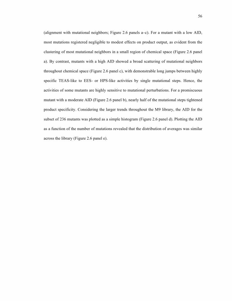
56
(alignment with mutational neighbors; Figure 2.6 panels a–c). For a mutant with a low AID,
most mutations registered negligible to modest effects on product output, as evident from the
clustering of most mutational neighbors in a small region of chemical space (Figure 2.6 panel
a). By contrast, mutants with a high AID showed a broad scattering of mutational neighbors
throughout chemical space (Figure 2.6 panel c), with demonstrable long jumps between highly
specific TEAS-like to EES- or HPS-like activities by single mutational steps. Hence, the
activities of some mutants are highly sensitive to mutational perturbations. For a promiscuous
mutant with a moderate AID (Figure 2.6 panel b), nearly half of the mutational steps tightened
product specificity. Considering the larger trends throughout the M9 library, the AID for the
subset of 236 mutants was plotted as a simple histogram (Figure 2.6 panel d). Plotting the AID
as a function of the number of mutations revealed that the distribution of averages was similar
across the library (Figure 2.6 panel e).
57
Figure 2.6. AID in chemical and sequence space. (a–c) A representative mutant (unlabeled red sphere) is shown in chemical space, along with all nine possible single-mutant neighbors (numbered green spheres) to show short (a), medium (b) and high (c) AID. Sequences of each representative mutant are referenced across the top of the three alignment tables, with mutational neighbors and distances listed below. Each mutated position is boxed, and residues of HPS origin are indicated with shading. (d) AID for a subset of 236 mutants was plotted as a simple histogram, where the shoulders and apex of the distribution are labeled 'a', 'b' and 'c' to correspond to representative mutants above. (e) The distribution of AID as a function of the number of accumulated HPS substitutions was plotted, where M1 refers to all single mutants, M2 to all double mutants, and so on.
2.3.5. Discussion
The emerging picture from these experiments is a rugged landscape in which
alternative catalytic specificities are mutationally accessible, requiring as little as a single base
change in the coding gene. Single mutations on average exert moderate effects, relaxing
product specificity while upregulating 5-EA, 4-EE, PSD or other TEAS minor products,
consistent with postulates that specific activities arise from catalytically promiscuous
58
ancestors11-19. Most mutations are additive, but rapid or punctuated changes in product output
are not rare. In fact, such hot spots (AID > 50) account for 7% of the mutants analyzed thus
far, indicating considerable biosynthetic potential for rapid evolutionary jumps throughout the
M9 lineage. This rapid adaptability may be unique to terpene cyclases, stemming in part from
the subtle energetic differences between competing cyclization pathways (Figure 2.1 panel b)
that ultimately govern product specificity. By implication, TEAS-HPS predecessors had the
potential for frequent shifts between PSD, 4-EE and 5-EA biosynthesis to generate rapidly
changing chemical repertoires throughout evolution.
Although both additive and punctuated specificity changes have been observed in
terpene cyclases20-22, this is the first effort to quantitatively measure the frequency and
distribution of these enzymatic properties over a catalytic landscape. To quantitatively
describe this landscape, it was essential to use a simple Euclidean distance metric, a chemical
distance that is generally applicable to mapping how any experimentally defined enzymatic
property is distributed throughout sequence space. In the current work, this metric provided a
means to quantitatively compare product spectra of terpene synthases, assess the effects of
mutations in different backgrounds—particularly mutational neighbors—and construct a
biosynthetic tree to quantitatively organize the M9 enzyme lineage by functional relatedness.
Structural and phylogenetic information has been invaluable in guiding mutagenesis
experiments leading to the successful interconversion of terpene cyclase substrate23 and
product specificities12, 23, 24. In the absence of phylogenetically derived models, applying
saturation mutagenesis to the active site of a notably promiscuous terpene cyclase, γ-humulene
synthase, has made considerable engineering advances to generate specific activities21. By
contrast, the work here explores phylogenetically relevant biosynthetic interrelationships that
extend product specificity control beyond the active site. Characterizing the reciprocal M9
59
lineages in HPS will be of great future interest to address the contribution of alternative
protein backgrounds to the product specificity landscape.
Only recently have efforts been made to characterize the underlying adaptive
landscapes of molecular evolution25-28 or to trace the evolutionary origins of the four
fundamental isoprenoid-based coupling reactions29. Our study provides the first experimental
measure of the complex functional terrain evident in secondary metabolism from the
construction and biochemical characterization of intervening lineages between a pair of extant
and diverging enzymes. Although it is tempting to speculate that 4-EE was the dominant
product of a TEAS-HPS common ancestor on the basis of its hybrid mechanistic origins, this
activity represents less than 3% of the total library. Also, greater product specificity for PSD is
achievable with fewer than nine mutations. For example, an M8 (mutant 226, Table 2.4, Table
2.7) produces 81% PSD, versus 72% by M9. However, the native henbane enzyme produces
97% PSD, indicating that additional mutations beyond the nine considered here contribute to
this high degree of product specificity. The facile phenotypic change from minimal sequence
changes uncovered by our work suggests that it is extremely difficult to make accurate
assignments of ancestral function in this pervasive class of secondary metabolic enzymes. This
result has broader implications for reconstructing ancestral proteins and ascribing ancient
functions; one must consider the extent of phenotypic variation among a population of
putative intermediates encompassed by a 'probabilistic guess' of the most likely ancestor.
Connecting catalytic landscapes of secondary metabolism to fitness landscapes of
organisms remains an enormous future challenge. Antibiotic resistance or primary metabolic
functions in microbes have direct survival consequences easily measured in laboratory
evolution experiments, but the fitness contributions of secondary metabolism, which are of
particular relevance to speciation in complex organisms, are intrinsically more difficult to
60
study. This arises in part from the myriad roles of secondary metabolites in the greater
chemical ecology of host organisms. In the current work, relating the in vitro results to in vivo
functions involves several caveats; numerous factors including transcription, translation and
solubility surely contribute to enzyme evolution, and possible effects of the cellular
environment on enzymatic activity must also be considered. There is precedence, however, for
the correspondence of in vitro and in vivo product profiles of terpene cyclases24, so the
observed plasticity of terpene cyclase enzymatic function from in vitro biochemical
measurements is likely to approximate the activities of these enzymes in their native
environment. More extensive sequencing efforts and biochemical annotation of terpene
synthases from the larger family of solanaceous plants will both address the degree to which
mutant combinations of the M9 lineage reflect the actual evolutionary lineages and provide
valuable insights toward understanding the role of secondary metabolism in shaping the
evolution of the Solanaceae.
The observation that HPS-like activity is achievable by many combinations of
mutations lying outside the active site (Tables 2.7, Table 2.8) highlights the crucial importance
of epistasis. This phenomenon has been documented and described in other enzyme systems25-
27 and is manifested here as a highly interdependent network of interactions in the protein that
ultimately controls product specificity. These functionally crucial yet distal mutations are not
surprising, given the effects of other distant mutations on protein and enzyme function30, 31.
Modulation of isoprenoid cyclization by discrete ensembles of peripherally distributed
residues is suggestive of energetic networks spread throughout the protein structure32, which
may allow greater adaptive capabilities. As recently noted, the interface of enzyme
adaptability with intrinsic and induced substrate reactivity underlies the emergence of cyclic
diversity in secondary metabolism33. Our quantitative exploration of the catalytic landscape of
61
the M9 lineage provides a first glimpse into the functional effects of quantum evolutionary
change on specialized biosynthesis.
2.4. METHODS
2.4.1. Library construction
SCOPE mutant gene synthesis was done using published methods15. A library
encoding 512 mutants comprising all permutations of the original TEAS M9 mutants12 was
produced, with 432 unique combinations identified by DNA sequencing (Table 2.3). The
library was created as gene sets consisting of a series of discrete mixtures to reduce
oversampling. Mutant TEAS genes were subcloned into pH9GW, an in-house expression
vector encoding nine N-terminal histidine residues, using the Gateway system (Invitrogen)
according to the manufacturer's instructions.
2.4.2. Biosynthetic tree construction
Terpene products from GC-MS analyses were quantified by integration of product
peak areas and transformed into percentages, where 5-EA, 4-EE, PSD and all remaining
products represented four groups adding up to 100% (Table 2.4). A distance (d) between every
pair of mutants was calculated by the sum of least squares:
d = sqrt [ (w1 – w2)2 + (x1 – x2)2 + (y1 – y2)2 + (z1 – z2)2]
where product profile 1 has coordinates w1, x1, y1 and z1, and product profile2 has
coordinates w2, x2, y2 and z2. The variables w, x, y and z correspond to 5-EA, 4-EE, PSD and
the sum of remaining products, respectively. A large n x n matrix was dimensionally reduced
into a standard phylogenetic tree to show which mutants cluster together in space. An
unrooted N-J tree was produced using MEGA 3.1 software34.
62
2.4.3. Sequencing
Plasmid DNA was prepared by the Microarray Core Facility at The Salk Institute, and
DNA sequencing was done by Eton Biosciences.
2.4.4. Vial assay characterization
In vitro assays of purified recombinant enzyme were conducted in duplicate according
to a previously published method9. Products were quantified by integration of peak areas from
the gas chromatography trace using Agilent ChemStation software and expressed as a
percentage of the total products. Notably, germacrene A was detected as the thermally
rearranged product β-elemene. Authentic standards of 5-EA, 4-EE and PSD were used for
instrument calibration and absolute product quantitation for kinetic measurements
(Supplementary Methods online).
2.4.5. Protein expression
pH9GW expression vectors were transformed into E. coli BL21(λDE3) and plated on
LB growth media with 50 µg/ml kanamycin selection. Colonies were transferred to 1 ml liquid
media (LB with kanamycin) in 96-well plates followed by 16 hrs growth with shaking at 37˚C
at 275 rpm. Cultures were diluted 10-fold into 5 ml of TB growth media with kanamycin in
24-well round bottom plates covered with micro-porous tape, followed by growth with
shaking at 37˚C at 275 rpm until cultures reached OD600 ≥ 1.5. Protein expression was
induced by addition of IPTG to 0.1 mM followed by growth with shaking at 20˚C at 275 rpm
for 5 hrs. Cells were harvested by centrifugation and cell pellets were frozen at -20˚C.
63
2.4.6. Purification of library proteins
Pellets from 5 ml expression cultures were re-suspended by adding 0.8 ml of lysis
buffer (50 mM Tris-HCl, 500 mM NaCl, 20 mM imidazole, 10% glycerol (v/v), 10 mM β-
mercaptoethanol, and 1% (v/v) Tween-20, pH 8) containing 1 mg/ml lysozyme and 1 mM
EDTA directly to frozen pellets followed by shaking at room temperature (25˚C) at 350 rpm
for 30 minutes. Next, 10 µl of benzonase solution (850 mM MgCl2 and 3.78 U/ul benzonase
(Novagen) was added followed by additional shaking at 350 rpm for 15 min. Lysates were
passed through a Whatman unifilter 96-well plate and collected in another Whatman plate
containing 100 µl bed-volume of superflow Ni-NTA resin (QIAgen), preequilibrated with
wash buffer using a vacuum manifold. Each well was washed with 2 ml lysis buffer, followed
by 1.5 ml wash buffer (lysis buffer lacking Tween- 20). Resin was air dried prior to addition
of 150 µl elution buffer (wash buffer containing 250 mM imidazole), incubated for 10 min,
followed by centrifugation to recover eluted protein. Protein recovery (~0.25 µg per 5 mL
culture) and purity (approximately 95%) were verified by SDS-PAGE analysis and UV at an
absorbance value of 280nm. An equal volume of 100% glycerol was added to eluted samples
followed by long term storage at -20˚C.
2.4.7. Kinetic Measurements
Enzyme kinetics was performed using the vial assay9 under the following modified
conditions. Reactions were composed on a 500 µl scale using a 3- component buffer system35
(25 mM 2- (N-Morpholino) ethanesulfonic acid (MES), 50 mM Tris, and 25 mM 3-
(Cyclohexylamino) propanesulfonic acid (CAPS)) at pH = 7.0 with 10 mM MgCl2 and a fixed
substrate concentration of 300 uM farnesyl pyrophosphate (FPP). For enzyme quantitation,
proteins samples were denatured in 6M guanidinium-HCl prior to measuring UV absorbance
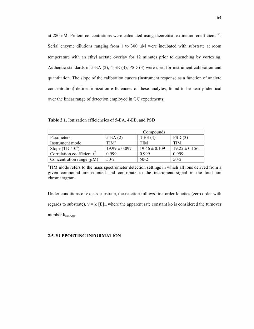
64
at 280 nM. Protein concentrations were calculated using theoretical extinction coefficients36.
Serial enzyme dilutions ranging from 1 to 300 µM were incubated with substrate at room
temperature with an ethyl acetate overlay for 12 minutes prior to quenching by vortexing.
Authentic standards of 5-EA (2), 4-EE (4), PSD (3) were used for instrument calibration and
quantitation. The slope of the calibration curves (instrument response as a function of analyte
concentration) defines ionization efficiencies of these analytes, found to be nearly identical
over the linear range of detection employed in GC experiments:
Table 2.1. Ionization efficiencies of 5-EA, 4-EE, and PSD
aTIM mode refers to the mass spectrometer detection settings in which all ions derived from a given compound are counted and contribute to the instrument signal in the total ion chromatogram.
Under conditions of excess substrate, the reaction follows first order kinetics (zero order with
regards to substrate), v = ko[E]o, where the apparent rate constant ko is considered the turnover
number kcat,App.
2.5. SUPPORTING INFORMATION
Compounds Parameters 5-EA (2) 4-EE (4) PSD (3) Instrument mode TIMa TIM TIM Slope (TIC/105) 19.99 ± 0.097 19.46 ± 0.109 19.25 ± 0.156 Correlation coefficient r2 0.999 0.999 0.999 Concentration range (µM) 50-2 50-2 50-2
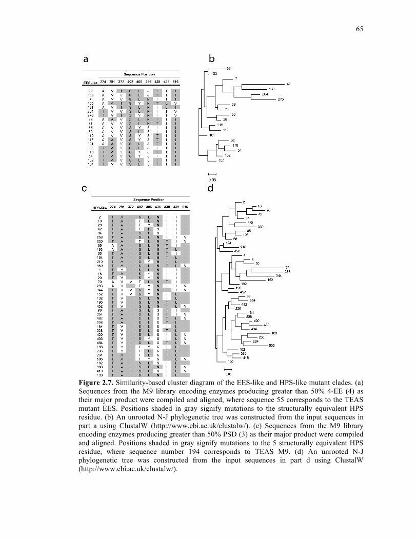
65
Figure 2.7. Similarity-based cluster diagram of the EES-like and HPS-like mutant clades. (a) Sequences from the M9 library encoding enzymes producing greater than 50% 4-EE (4) as their major product were compiled and aligned, where sequence 55 corresponds to the TEAS mutant EES. Positions shaded in gray signify mutations to the structurally equivalent HPS residue. (b) An unrooted N-J phylogenetic tree was constructed from the input sequences in part a using ClustalW (http://www.ebi.ac.uk/clustalw/). (c) Sequences from the M9 library encoding enzymes producing greater than 50% PSD (3) as their major product were compiled and aligned. Positions shaded in gray signify mutations to the 5 structurally equivalent HPS residue, where sequence number 194 corresponds to TEAS M9. (d) An unrooted N-J phylogenetic tree was constructed from the input sequences in part d using ClustalW (http://www.ebi.ac.uk/clustalw/).
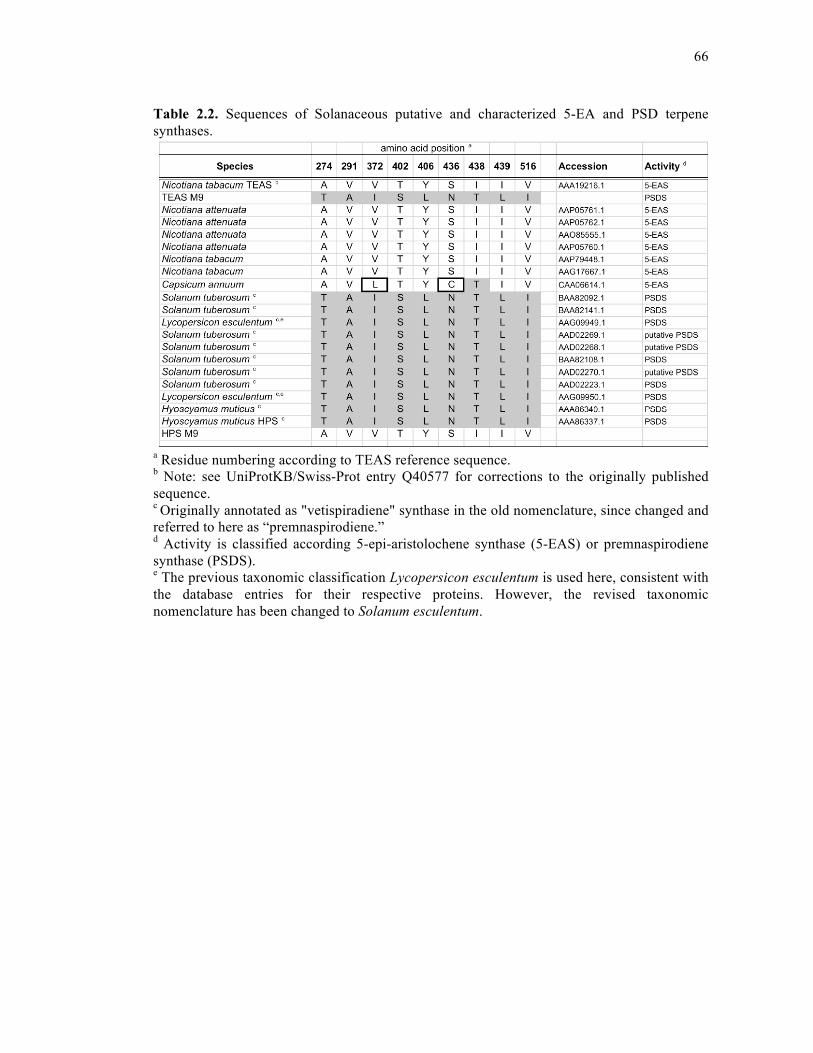
66
Table 2.2. Sequences of Solanaceous putative and characterized 5-EA and PSD terpene synthases.
a Residue numbering according to TEAS reference sequence. b Note: see UniProtKB/Swiss-Prot entry Q40577 for corrections to the originally published sequence. c Originally annotated as "vetispiradiene" synthase in the old nomenclature, since changed and referred to here as “premnaspirodiene.” d Activity is classified according 5-epi-aristolochene synthase (5-EAS) or premnaspirodiene synthase (PSDS). e The previous taxonomic classification Lycopersicon esculentum is used here, consistent with the database entries for their respective proteins. However, the revised taxonomic nomenclature has been changed to Solanum esculentum.

67
Table 2.3. SCOPE library construction statistics
M9 Library Statistics Library complexity 512 Clones sequenced 3,047 Fold oversampling 5.9 Unique clones identified 432 Additional mutations: silent 52 frame-shift 91 point 65 total 208 mutation rate 6.80%
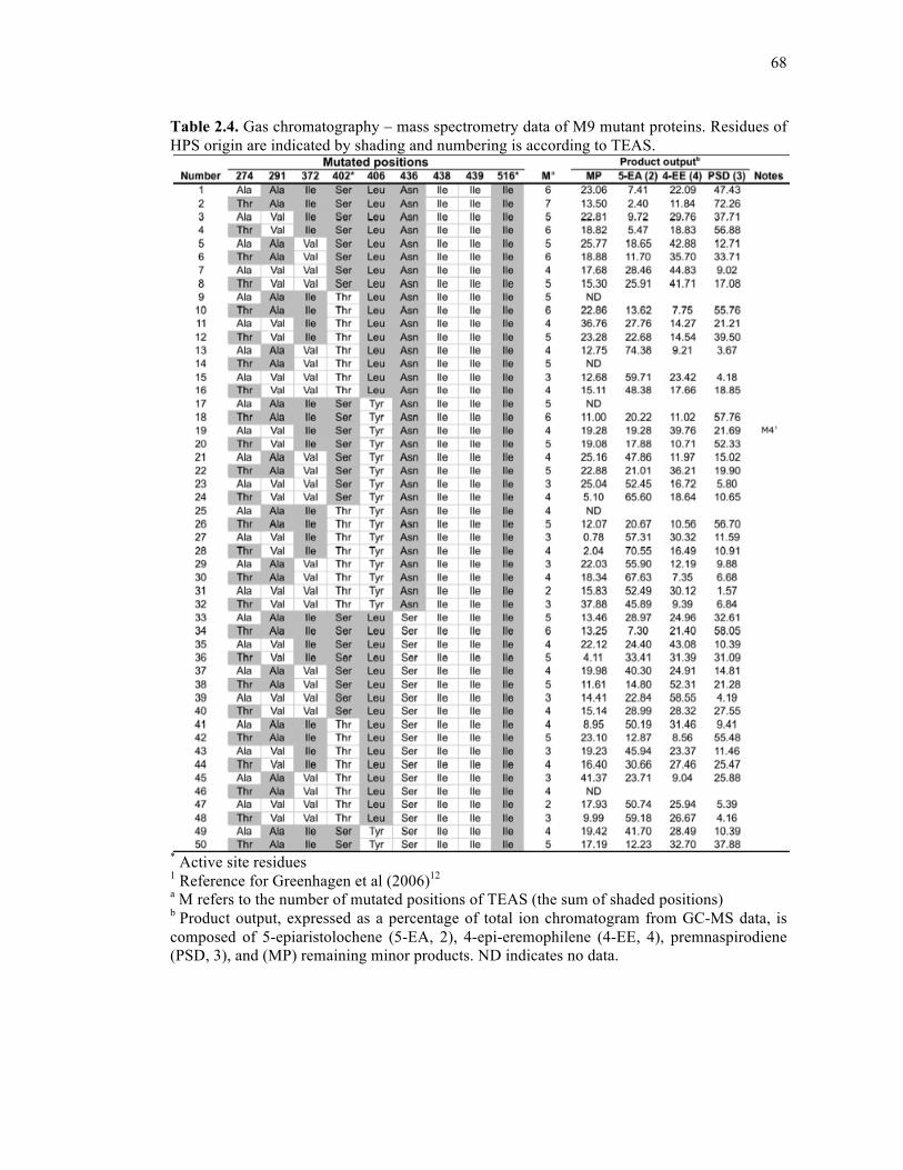
68
Table 2.4. Gas chromatography – mass spectrometry data of M9 mutant proteins. Residues of HPS origin are indicated by shading and numbering is according to TEAS.
* Active site residues 1 Reference for Greenhagen et al (2006)12 a M refers to the number of mutated positions of TEAS (the sum of shaded positions) b Product output, expressed as a percentage of total ion chromatogram from GC-MS data, is composed of 5-epiaristolochene (5-EA, 2), 4-epi-eremophilene (4-EE, 4), premnaspirodiene (PSD, 3), and (MP) remaining minor products. ND indicates no data.
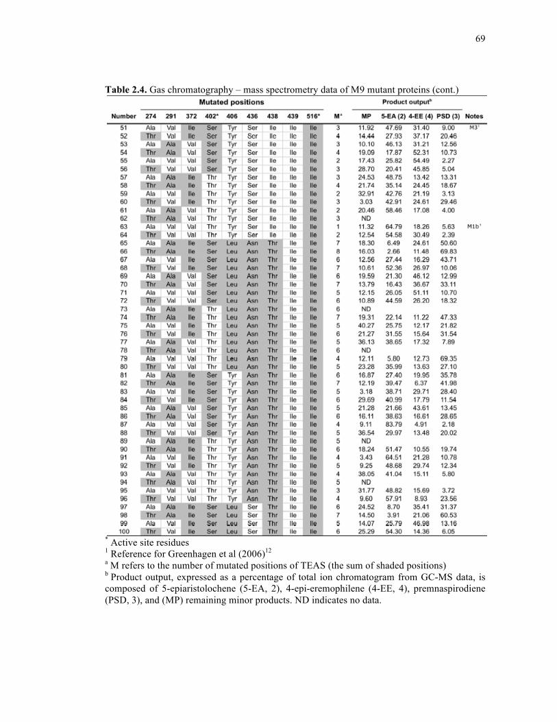
69
Table 2.4. Gas chromatography – mass spectrometry data of M9 mutant proteins (cont.)
* Active site residues 1 Reference for Greenhagen et al (2006)12 a M refers to the number of mutated positions of TEAS (the sum of shaded positions) b Product output, expressed as a percentage of total ion chromatogram from GC-MS data, is composed of 5-epiaristolochene (5-EA, 2), 4-epi-eremophilene (4-EE, 4), premnaspirodiene (PSD, 3), and (MP) remaining minor products. ND indicates no data.
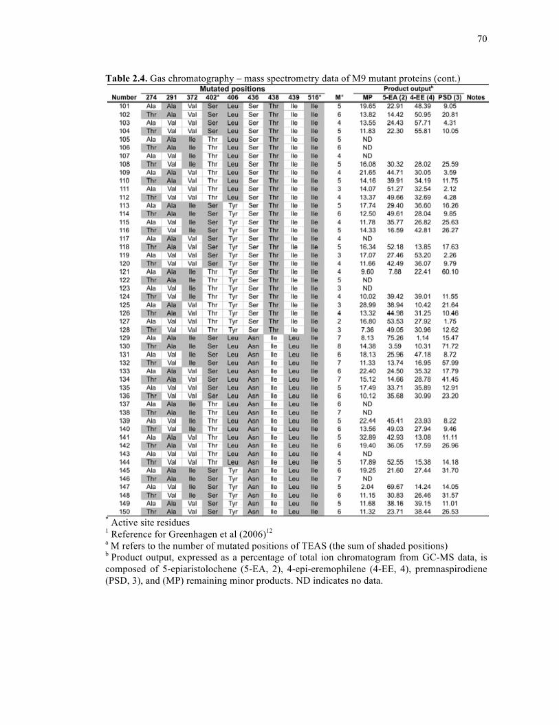
70
Table 2.4. Gas chromatography – mass spectrometry data of M9 mutant proteins (cont.)
* Active site residues 1 Reference for Greenhagen et al (2006)12 a M refers to the number of mutated positions of TEAS (the sum of shaded positions) b Product output, expressed as a percentage of total ion chromatogram from GC-MS data, is composed of 5-epiaristolochene (5-EA, 2), 4-epi-eremophilene (4-EE, 4), premnaspirodiene (PSD, 3), and (MP) remaining minor products. ND indicates no data.
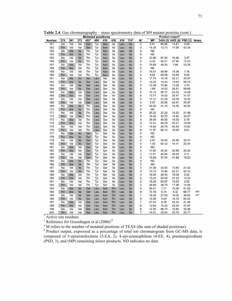
71
Table 2.4. Gas chromatography – mass spectrometry data of M9 mutant proteins (cont.)
* Active site residues 1 Reference for Greenhagen et al (2006)12 a M refers to the number of mutated positions of TEAS (the sum of shaded positions) b Product output, expressed as a percentage of total ion chromatogram from GC-MS data, is composed of 5-epiaristolochene (5-EA, 2), 4-epi-eremophilene (4-EE, 4), premnaspirodiene (PSD, 3), and (MP) remaining minor products. ND indicates no data.
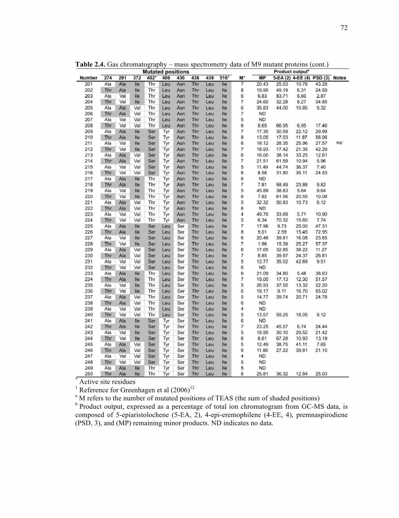
72
Table 2.4. Gas chromatography – mass spectrometry data of M9 mutant proteins (cont.)
* Active site residues 1 Reference for Greenhagen et al (2006)12 a M refers to the number of mutated positions of TEAS (the sum of shaded positions) b Product output, expressed as a percentage of total ion chromatogram from GC-MS data, is composed of 5-epiaristolochene (5-EA, 2), 4-epi-eremophilene (4-EE, 4), premnaspirodiene (PSD, 3), and (MP) remaining minor products. ND indicates no data.
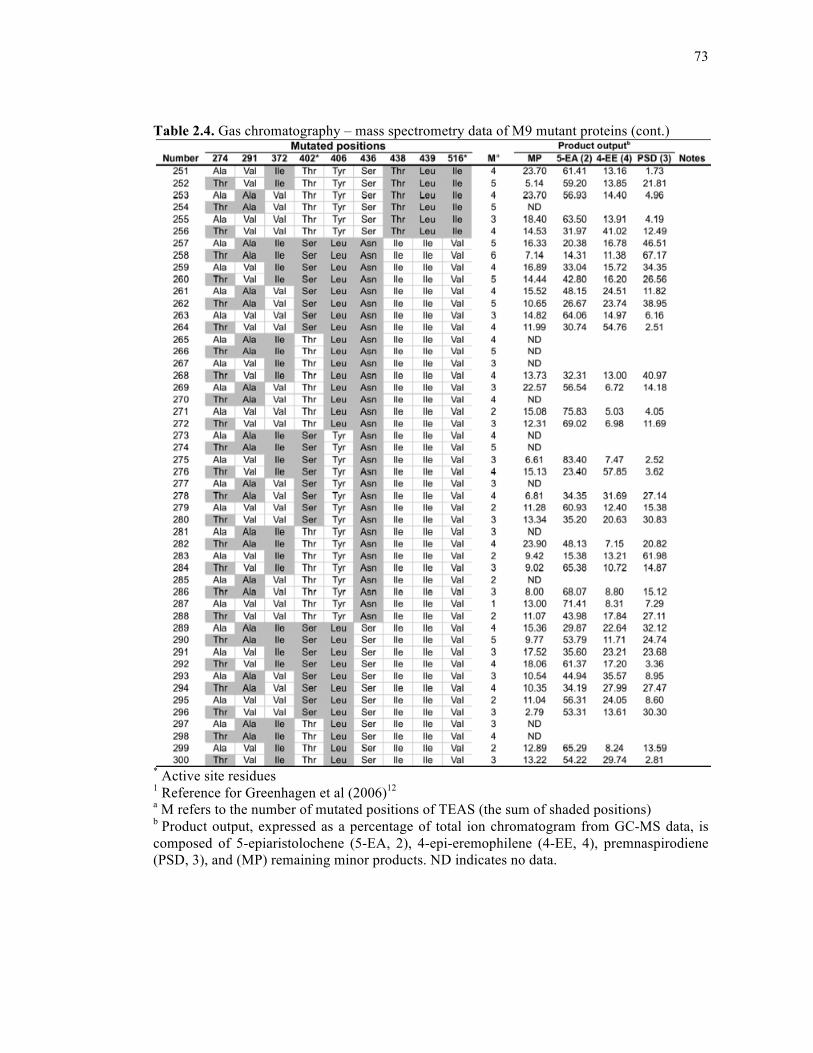
73
Table 2.4. Gas chromatography – mass spectrometry data of M9 mutant proteins (cont.)
* Active site residues 1 Reference for Greenhagen et al (2006)12 a M refers to the number of mutated positions of TEAS (the sum of shaded positions) b Product output, expressed as a percentage of total ion chromatogram from GC-MS data, is composed of 5-epiaristolochene (5-EA, 2), 4-epi-eremophilene (4-EE, 4), premnaspirodiene (PSD, 3), and (MP) remaining minor products. ND indicates no data.
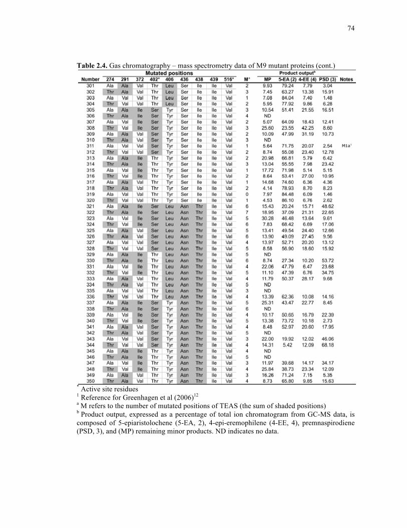
74
Table 2.4. Gas chromatography – mass spectrometry data of M9 mutant proteins (cont.)
* Active site residues 1 Reference for Greenhagen et al (2006)12 a M refers to the number of mutated positions of TEAS (the sum of shaded positions) b Product output, expressed as a percentage of total ion chromatogram from GC-MS data, is composed of 5-epiaristolochene (5-EA, 2), 4-epi-eremophilene (4-EE, 4), premnaspirodiene (PSD, 3), and (MP) remaining minor products. ND indicates no data.
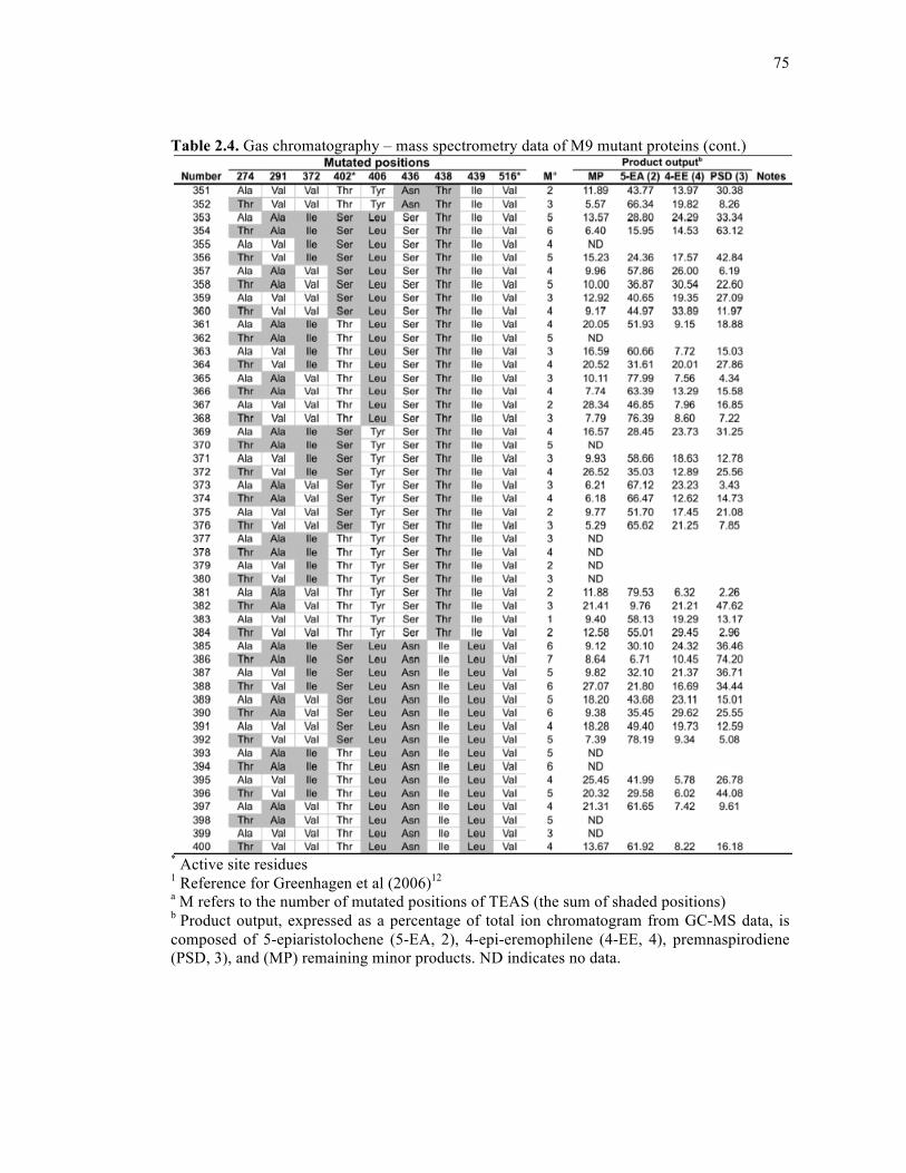
75
Table 2.4. Gas chromatography – mass spectrometry data of M9 mutant proteins (cont.)
* Active site residues 1 Reference for Greenhagen et al (2006)12 a M refers to the number of mutated positions of TEAS (the sum of shaded positions) b Product output, expressed as a percentage of total ion chromatogram from GC-MS data, is composed of 5-epiaristolochene (5-EA, 2), 4-epi-eremophilene (4-EE, 4), premnaspirodiene (PSD, 3), and (MP) remaining minor products. ND indicates no data.

76
Table 2.4. Gas chromatography – mass spectrometry data of M9 mutant proteins (cont.)
* Active site residues 1 Reference for Greenhagen et al (2006)12 a M refers to the number of mutated positions of TEAS (the sum of shaded positions) b Product output, expressed as a percentage of total ion chromatogram from GC-MS data, is composed of 5-epiaristolochene (5-EA, 2), 4-epi-eremophilene (4-EE, 4), premnaspirodiene (PSD, 3), and (MP) remaining minor products. ND indicates no data.
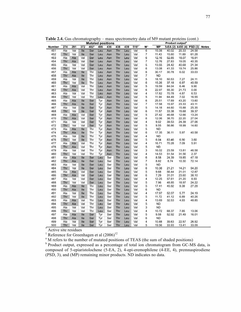
77
Table 2.4. Gas chromatography – mass spectrometry data of M9 mutant proteins (cont.)
* Active site residues 1 Reference for Greenhagen et al (2006)12 a M refers to the number of mutated positions of TEAS (the sum of shaded positions) b Product output, expressed as a percentage of total ion chromatogram from GC-MS data, is composed of 5-epiaristolochene (5-EA, 2), 4-epi-eremophilene (4-EE, 4), premnaspirodiene (PSD, 3), and (MP) remaining minor products. ND indicates no data.
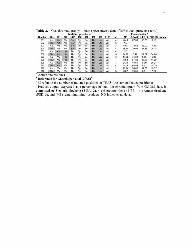
78
Table 2.4. Gas chromatography – mass spectrometry data of M9 mutant proteins (cont.)
* Active site residues 1 Reference for Greenhagen et al (2006)12 a M refers to the number of mutated positions of TEAS (the sum of shaded positions) b Product output, expressed as a percentage of total ion chromatogram from GC-MS data, is composed of 5-epiaristolochene (5-EA, 2), 4-epi-eremophilene (4-EE, 4), premnaspirodiene (PSD, 3), and (MP) remaining minor products. ND indicates no data.
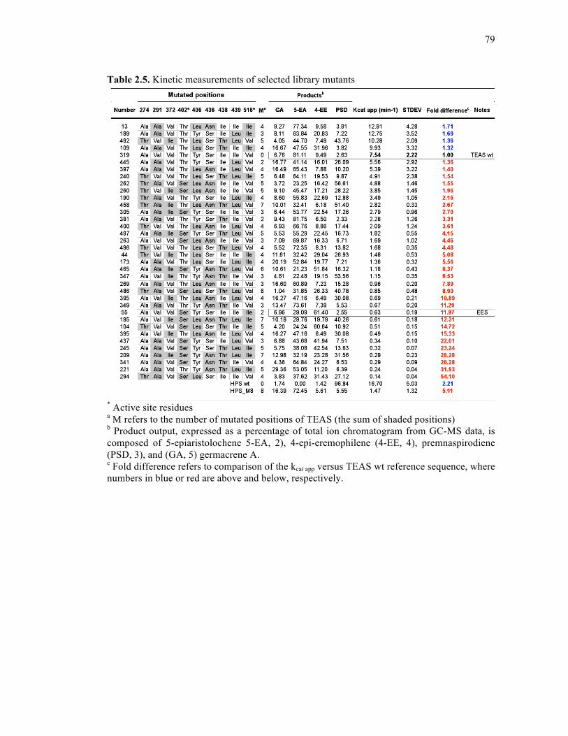
79
Table 2.5. Kinetic measurements of selected library mutants
* Active site residues a M refers to the number of mutated positions of TEAS (the sum of shaded positions) b Product output, expressed as a percentage of total ion chromatogram from GC-MS data, is composed of 5-epiaristolochene 5-EA, 2), 4-epi-eremophilene (4-EE, 4), premnaspirodiene (PSD, 3), and (GA, 5) germacrene A. c Fold difference refers to comparison of the kcat app versus TEAS wt reference sequence, where numbers in blue or red are above and below, respectively.
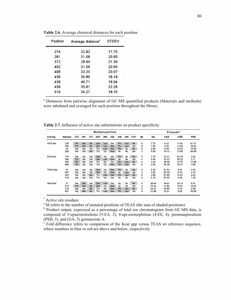
80
Table 2.6. Average chemical distances for each position
a Distances from pairwise alignment of GC-MS quantified products (Materials and methods) were tabulated and averaged for each position throughout the library. Table 2.7. Influence of active site substitutions on product specificity
* Active site residues a M refers to the number of mutated positions of TEAS (the sum of shaded positions) b Product output, expressed as a percentage of total ion chromatogram from GC-MS data, is composed of 5-epiaristolochene (5-EA, 2), 4-epi-eremophilene (4-EE, 4), premnaspirodiene (PSD, 3), and (GA, 5) germacrene A. c Fold difference refers to comparison of the Kcat app versus TEAS wt reference sequence, where numbers in blue or red are above and below, respectively.
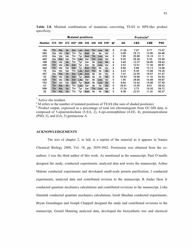
81
Table 2.8. Minimal combinations of mutations converting TEAS to HPS-like product specificity.
* Active site residues a M refers to the number of mutated positions of TEAS (the sum of shaded positions) b Product output, expressed as a percentage of total ion chromatogram from GC-MS data, is composed of 5-epiaristolochene (5-EA, 2), 4-epi-eremophilene (4-EE, 4), premnaspirodiene (PSD, 3), and (GA, 5) germacrene A. ACKNOWLEDGEMENTS
The text of chapter 2, in full, is a reprint of the material as it appears in Nature
Chemical Biology 2008, Vol. 18, pp. 3039-3042. Permission was obtained from the co-
authors. I was the third author of this work. As mentioned in the manuscript, Paul O’maille
designed the study, conducted experiments, analyzed data and wrote the manuscript, Arthur
Malone conducted experiments and developed small-scale protein purification, I conducted
experiments, analyzed data and contributed revision to the manuscript, B Andes Hess Jr
conducted quantum mechanics calculations and contributed revisions to the manuscript, Lidia
Smentek conducted quantum mechanics calculations, Iseult Sheehan conducted experiments,
Bryan Greenhagen and Joseph Chappell designed the study and contributed revisions to the
manuscript, Gerard Manning analyzed data, developed the biosynthetic tree and chemical
82
distance analysis, and contributed revisions to the manuscript, and Joseph P. Noel designed
the study, analyzed the data and wrote the manuscript. This research was performed under the
supervision of Joseph P. Noel.
REFERENCES
1. Grayer, R. J.; Kokubun, T., Plant-fungal interactions: the search for phytoalexins and other antifungal compounds from higher plants. Phytochemistry 2001, 56, 253-263.
2. Pedras, M. S.; Okanga, F. I.; Zaharia, I. L.; Khan, A. Q., Phytoalexins from crucifers:
synthesis, biosynthesis, and biotransformation. Phytochemistry 2000, 53, 161-176. 3. Harborne, J. B., The comparative biochemistry of phytoalexin induction in plants.
Biochem. Syst. Ecol. 1999, 27, 335-367. 4. Akiyama, K.; Matsuzaki, K.; Hayashi, H., Plant sesquiterpenes induce hyphal
branching in arbuscular mycorrhizal fungi. Nature 2005, 435, 824-827. 5. Mumm, R.; Hilker, M., The significance of background odour for an egg parasitoid to
detect plants with host eggs. Chem. Senses 2005, 30, 337-343. 6. Feeny, P., Herbivores: Their Interactions with Secondary Plant Metabolites. Academic
Press: 1992. 7. Gershenzon, J.; Dudareva, N., The function of terpene natural products in the natural
world. Nature chemical biology 2007, 3 (7), 408-414. 8. O'Maille, P. E.; Bakhtina, M.; Tsai, M. D., Structure-based combinatorial protein
engineering (SCOPE). Journal of Molecular Biology 2002, 321 (4), 677-691. 9. O'Maille, P. E.; Chappell, J.; Noel, J. P., A single-vial analytical and quantitative gas
chromatography-mass spectrometry assay for terpene synthases. Anal. Biochem. 2004, 335, 210-217.
10. Back, K.; He, S.; Kim, K. U.; Shin, D. H., Cloning and bacterial expression of
sesquiterpene cyclase, a key branch point enzyme for the synthesis of sesquiterpenoid phytoalexin capsidiol in UV-challenged leaves of Capsicum annuum. Plant Cell Physiol. 1998, 39, 899-904.
11. Facchini, P. J.; Chappell, J., Gene family for an elicitor-induced sesquiterpene cyclase
in tobacco. Proc. Natl. Acad. Sci. USA 1992, 89, 11088-11092. 12. Greenhagen, B. T.; O'Maille, P. E.; Noel, J. P.; Chappell, J., Identifying and
manipulating structural determinates linking catalytic specificities in terpene
83
synthases. Proceedings of the National Academy of Sciences of the United States of America 2006, 103 (26), 9826-9831.
13. Dudareva, N., (E)-beta-ocimene and myrcene synthase genes of floral scent
biosynthesis in snapdragon: function and expression of three terpene synthase genes of a new terpene synthase subfamily. Plant Cell 2003, 15, 1227-1241.
14. Bohlmann, J.; Meyer-Gauen, G.; Croteau, R., Plant terpenoid synthases: molecular
biology and phylogenetic analysis. Proc. Natl. Acad. Sci. USA 1998, 95, 4126-4133. 15. O'Maille, P. E.; Tsai, M. D.; Greenhagen, B. T.; Chappell, J.; Noel, J. P., Gene library
synthesis by structure-based combinatorial protein engineering. Methods Enzymol. 2004, 388, 75-91.
16. O'Maille, P. E.; Chappell, J.; Noel, J. P., Biosynthetic potential of sesquiterpene
synthases: Alternative products of tobacco 5-epi-aristolochene synthase. Archives of Biochemistry and Biophysics 2006, 448 (1-2), 73-82.
17. Copley, S. D., Enzymes with extra talents: moonlighting functions and catalytic
promiscuity. Curr. Opin. Chem. Biol. 2003, 7, 265-272. 18. Jensen, R. A., Enzyme recruitment in evolution of new function. Annu. Rev.
Microbiol. 1976, 30, 409-425. 19. O'Brien, P. J.; Herschlag, D., Catalytic promiscuity and the evolution of new
enzymatic activities. Chem. Biol. 1999, 6, R91-R105. 20. Wilderman, P. R.; Peters, R. J., A single residue switch converts abietadiene synthase
into a pimaradiene specific cyclase. J. Am. Chem. Soc. 2007, 129, 15736-15737. 21. Yoshikuni, Y.; Ferrin, T. E.; Keasling, J. D., Designed divergent evolution of enzyme
function. Nature 2006, 440 (7087), 1078-1082. 22. Hyatt, D. C.; Croteau, R., Mutational analysis of a monoterpene synthase reaction:
altered catalysis through directed mutagenesis of (-)-pinene synthase from Abies grandis. Arch. Biochem. Biophys. 2005, 439, 222-233.
23. Kampranis, S. C.; Ioannidis, D.; Purvis, A.; Mahrez, W.; Ninga, E.; Katerelos, N. A.;
Anssour, S.; Dunwell, J. M.; Degenhardt, J.; Makris, A. M.; Goodenough, P. W.; Johnson, C. B., Rational conversion of substrate and product specificity in a Salvia monoterpene synthase: structural insights into the evolution of terpene synthase function. The Plant Cell 2007, 19 (6), 1994-2005.
24. Kollner, T. G.; Schnee, C.; Gershenzon, J.; Degenhardt, J., The variability of
sesquiterpenes emitted from two Zea mays cultivars is controlled by allelic variation of two terpene synthase genes encoding stereoselective multiple product enzymes. Plant Cell 2004, 16, 1115-1131.

84
25. Weinreich, D. M.; Delaney, N. F.; Depristo, M. A.; Hartl, D. L., Darwinian evolution can follow only very few mutational paths to fitter proteins. Science 2006, 312, 111-114.
26. Ortlund, E. A.; Bridgham, J. T.; Redinbo, M. R.; Thornton, J. W., Crystal structure of
an ancient protein: evolution by conformational epistasis. Science 2007, 317, 1544-1548.
27. Bershtein, S.; Segal, M.; Bekerman, R.; Tokuriki, N.; Tawfik, D. S., Robustness-
epistasis link shapes the fitness landscape of a randomly drifting protein. Nature 2006, 444, 929-932.
28. Miller, S. P.; Lunzer, M.; Dean, A. M., Direct demonstration of an adaptive constraint.
Science 2006, 314, 458-461. 29. Thulasiram, H. V.; Erickson, H. K.; Poulter, C. D., Chimeras of two isoprenoid
synthases catalyze all four coupling reactions in isoprenoid biosynthesis. Science 2007, 316, 73-76.
30. Agarwal, P. K.; Billeter, S. R.; Rajagopalan, P. T.; Benkovic, S. J.; Hammes-Schiffer,
S., Network of coupled promoting motions in enzyme catalysis. Proc. Natl. Acad. Sci. USA 2002, 99, 2794-2799.
31. Rajagopalan, P. T.; Lutz, S.; Benkovic, S. J., Coupling interactions of distal residues
enhance dihydrofolate reductase catalysis: mutational effects on hydride transfer rates. Biochemistry 2002, 41, 12618-12628.
32. Lockless, S. W.; Ranganathan, R., Evolutionarily conserved pathways of energetic
connectivity in protein families. Science 1999, 286, 295-299. 33. Austin, M. B.; O'Maille, P. E.; Noel, J. P., Evolving biosynthetic tangos negotiate
mechanistic landscapes. Nat. Chem. Biol. 2008, 4, 217-222. 34. Tamura, K.; Dudley, J.; Nei, M.; Kumar, S., MEGA4: Molecular Evolutionary
Genetics Analysis (MEGA) software version 4.0. Mol. Biol. Evol. 2007, 24, 1596-1599.
35. Ellis, K. J.; Morrison, J. F., Buffers of constant ionic strength for studying pH-
dependent processes. Methods Enzymol 1982, 87, 405-26. 36. Gill, S. C.; von Hippel, P. H., Calculation of protein extinction coefficients from
amino acid sequence data. Anal Biochem 1989, 182 (2), 319-26.
85
Chapter 3
Structural Elucidation of Cisoid and Transoid Cyclization Pathways of a Sesquiterpene
Synthase Using 2-Fluorofarnesyl Diphosphates
86
3.1. ABSTRACT
Sesquiterpene skeletal complexity in nature originates from the enzyme-catalyzed
ionization of (trans,trans)-farnesyl diphosphate (FPP) (1a) and subsequent cyclization along
either 2,3-transoid or 2,3-cisoid farnesyl cation pathways. Tobacco 5-epi-aristolochene
synthase (TEAS), a transoid synthase, produces cisoid products as a component of its minor
product spectrum. To investigate the cryptic cisoid cyclization pathway in TEAS, we
employed (cis,trans)-FPP (1b) as an alternative substrate. Strikingly, TEAS was catalytically
robust in the enzymatic conversion of (cis,trans)-FPP (1b) to exclusively (≥99.5%) cisoid
products. Further, crystallographic characterization of wild-type TEAS and a catalytically
promiscuous mutant (M4 TEAS) with 2-fluoro analogues of both all-trans FPP (1a) and
(cis,trans)-FPP (1b) revealed binding modes consistent with preorganization of the farnesyl
chain. These results provide a structural glimpse into both cisoid and transoid cyclization
pathways efficiently templated by a single enzyme active site, consistent with the recently
elucidated stereochemistry of the cisoid products. Further, computational studies using density
functional theory calculations reveal concerted, highly asynchronous cyclization pathways
leading to the major cisoid cyclization products. The implications of these discoveries for
expanded sesquiterpene diversity in nature are discussed.
3.2. INTRODUCTION
Terpenes comprise the most structurally diverse class of natural products, playing
essential ecological roles by mediating communication between plants and insects, by
providing antimicrobial defenses for plants, and likely acting in additional undefined
capacities (as reviewed previously1. Terpenoids originate from primary isoprenoid
metabolism, wherein iterative condensation of 5-carbon isoprene units (isopentenyl
87
diphosphate and dimethylallyl diphosphate) catalyzed by prenyltransferases produce
polyisoprenoid diphosphate substrates of varying lengths (for a review, see Liang et al 20022).
Terpene synthases, in turn, often referred to as cyclases given the cyclic nature of many of
their products, transform the polyisoprenoid diphosphate substrates, e.g., geranyl diphosphate
C10, farnesyl diphosphate C15, or geranylgeranyl diphosphate C20, into structurally diverse
mono-, sesqui-, and diterpene products, respectively.
The structural complexity of these molecules underlies their diverse biological
activities. Ruzicka formulated the biogenetic isoprene rule, which predicted the formation of
sesquiterpenes arising from the head-to-tail connection of three isoprene units (5 carbons
each) where the skeletal complexity can be formally deduced from farnesol3. Cane later
developed a general stereochemical model for sesquiterpene biogenesis involving the
idealized fold of the farnesyl chain in the active site posing the reacting carbons to direct a
sequence of electrophilic cyclizations and rearrangements following pyrophosphate
loss/ionization4. Moreover, a limited number of conformations of the farnesyl substrate give
rise to much greater product diversity. Product specificity or in many cases product diversity
arises from a limited number of farnesyl chain conformations, wherein the reacting double
bonds reside mutually perpendicular to a common plane. Thus, there is a direct
correspondence between the absolute stereochemical configuration of the sesquiterpene
product and the inferred conformation of the precursor4. A central challenge for the structural
enzymologist is to define how individual terpene synthases statically or dynamically
discriminate between alternative polyprenyl cation conformational modes or selectively favor
particular conformations to shepherd reactive intermediates along distinct cyclization
cascades.
88
Structural biology provides a framework for addressing the evolutionary origins of
complex terpenoid metabolites and their biosynthetic pathways. Terpene synthases comprise a
structurally conserved enzyme family, which adopt a common α-helical architecture termed
the class I terpenoid cyclase fold, first revealed from the crystal structures of tobacco 5-epi-
aristolochene synthase (TEAS) from Nicotiana tobaccum5 and pentalenene synthase from
Streptomyces UC53196. The lyase function of these enzymes stems from two conserved metal
binding motifs: the “aspartate-rich” DDxxD motif that coordinates two Mg2+ ions and the
“NSE/DTE” motif that coordinates a third Mg2+. These static X-ray crystallographic studies
show that the binding of (Mg2+)3-PPi stabilizes the active site in a closed conformation that is
sequestered from bulk solvent7. In addition to multiple divalent cation coordination bonds, the
PPi anion accepts hydrogen bonds from conserved basic residues when bound to the closed
synthase conformation, while a hydrophobic pocket, lined by a number of aromatic residues,
cradles the farnesyl chain and most likely templates the cyclization reaction by enforcing
particular substrate conformations and stabilizing carbocations through π-stacking
interactions.
While the wealth of structural diversity among terpene hydrocarbons arises from
bifurcations along multistep cyclization pathways, divergence at the earliest mechanistic step
defines two major classes of terpene synthases and hence distinct product families. The
“transoid” synthases ionize (trans,trans)-farnesyl diphosphate (FPP) (1a) to generate the 2,3-
transoid farnesyl cation (trans along the C2−C3 bond) followed by initial C1 attack on the
distal C10−C11 double bond, prior to further downstream cyclizations (Figure 3.1). By
contrast, the “cisoid” synthases conduct an initial C2−C3 double bond isomerization prior to
cyclization, wherein the nascent farnesyl cation is recaptured at C3 by pyrophosphate to form
the neutral, enzyme-bound (3R)- or (3S)-nerolidyl diphosphate (NPP), thus allowing rotation
89
about the C2−C3 bond from trans to cis. Reionization of NPP generates the 2,3-cisoid farnesyl
cation, which undergoes further cyclization via initial C1 attack either on the proximal C6−C7
or on the distal C10−C11 double bonds prior to further transformations. This reaction
mechanism has been invoked to account for the formation of β-macrocarpene8, amorpha-4,11-
diene9, trichodiene10, and cedranes such as isocedrol11. Moreover, the biosynthesis of epi-
isozizaene was recently described in connection with the isolation and functional
characterization of epi-isozizaene synthase from Streptomyces coelicolor12. The cisoid
mechanistic class in sesquiterpene cyclases is akin to the majority of monoterpene cyclases
that proceed via the cis (neryl) allylic cation to form the corresponding cyclic monoterpene
products (for a review, see Davis et al 200013).

90
Figure 3.1. Mechanism of TEAS-catalyzed cyclization of (cis,trans)-FPP to (+)-2-epi-prezizaene (2). a) The structure and semisystematic nomenclature for isomers of the FPP substrate and fluorinated analogues used in this study are indicated. b) Based on biochemical and stereochemical information governing the nature of the cisoid products, a cyclization mechanism is proposed to account for all identified products along this multistep pathway.18 c) The configuration of the terminal isopropenyl tail of the (7R)-β-bisabolyl cation relates to the final cyclization products of the cisoid pathway.

91
Though most sesquiterpene synthases can be classified as belonging to either the
cisoid or transoid classes, some display cryptic activities associated with the other pathway.
Notably, TEAS catalyzes the formation of (+)-5-epi-aristolochene (1), the first committed step
in the biosynthesis of the phytoalexin capsidiol, the principal component of tobacco’s
antifungal chemical defense14. Aside from its major product, TEAS generates an additional 24
minor products, some of which are derived from the cisoid cyclization pathway15. The
structural variations of these cisoid products result from a multistep mechanism of cyclizations
and rearrangements, suggesting that TEAS templates the cisoid cation pathway with fidelity
and enables the formation of a distinct set of complex skeletal structures. These unexpected
observations give rise to several confounding questions. Does a single parental fold of the
farnesyl chain give rise to products along both the cisoid and transoid pathways in TEAS? On
a structural level, how are both pathways templated within a single active site? How can the
cryptic cisoid pathway in TEAS become activated? Does this “vestigial” activity portend an
unanticipated new function for TEAS in tobacco?
To address these questions, we investigated the cisoid cyclization recently discovered
in TEAS using synthetically derived (cis,trans)-FPP (1b), a geometrical isomer of the native
all-trans substrate (Figure 3.1, panel a). [The descriptors “cis” and “trans” in (cis,trans)-FPP
and the fluoroFPP isomers refer to the longest carbon chain about the 2,3 and 6,7 double
bonds, respectively, as defined in Figure 3.1, panel a. For more formalized nomenclature, see
Fox et al 200116 and Rigaudy et al 197917] Remarkably, TEAS efficiently converts this
alternative substrate into predominantly (+)-2-epi-prezizaene (2), a novel sesquiterpene
hydrocarbon related to the naturally occurring alcohol jinkohol18, along with other cisoid
cation-derived products. Large-scale enzyme reactions produced sufficient amounts of
92
hydrocarbon products for stereochemical elucidation and positive identification of nine
compounds18.
In the present investigation, crystallographic analyses of wild-type TEAS and a
previously reported promiscuous mutant (TEAS M4)19, with unreactive 2-fluoro analogues (2a
and 2b) of (trans,trans)- and (cis,trans)-FPP (1a and 1b, respectively) revealed catalytically
relevant binding modes and distinct farnesyl chain topologies that are consistent with
preorganization by the active-site for cisoid or transoid cyclization, and hence, the predicted
stereochemical course of the reaction. Further, key transition state geometries calculated using
density functional theory revealed concerted, highly asynchronous cyclization pathways.
Thus, combining biochemical, computational, and crystallographic analyses with the recently
elucidated stereochemistry of the cisoid products, we pictorially reconstruct herein the TEAS-
catalyzed transoid and cisoid cyclization pathways. Further, comparison of wild-type and
mutant TEAS-analogue complexes provides structural snapshots and insights into product
specificity/diversity reflected in the preorganization of the farnesyl chain along 2 major
cyclization pathways.
3.3. RESULTS AND DISCUSSION
3.3.1. TEAS-Directed Cisoid Cyclization with (cis,trans)-FPP
To investigate the cryptic cyclization activity in TEAS, we synthesized the 2,3-cis
geometrical isomer of farnesyl diphosphate (cis,trans)-FPP (1b)18. This substrate analogue is
effectively “preisomerized”, and hence its ionization by TEAS would be expected to generate
the cisoid farnesyl cation, which in turn should feed directly into the cisoid cyclization
pathway (Figure 3.1, panel b). Indeed, our pilot experiments revealed that TEAS generated a
near exclusive spectrum of cisoid products when incubated with (cis,trans)-FPP (1b) as
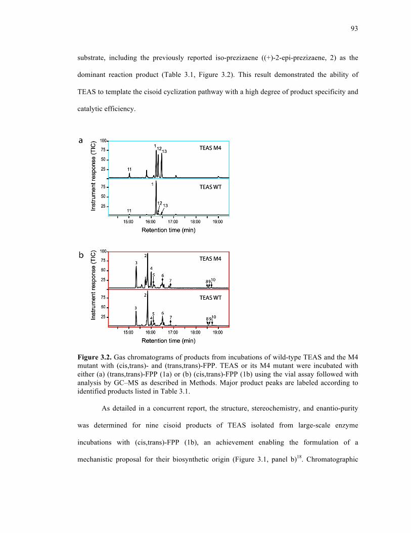
93
substrate, including the previously reported iso-prezizaene ((+)-2-epi-prezizaene, 2) as the
dominant reaction product (Table 3.1, Figure 3.2). This result demonstrated the ability of
TEAS to template the cisoid cyclization pathway with a high degree of product specificity and
catalytic efficiency.
Figure 3.2. Gas chromatograms of products from incubations of wild-type TEAS and the M4 mutant with (cis,trans)- and (trans,trans)-FPP. TEAS or its M4 mutant were incubated with either (a) (trans,trans)-FPP (1a) or (b) (cis,trans)-FPP (1b) using the vial assay followed with analysis by GC–MS as described in Methods. Major product peaks are labeled according to identified products listed in Table 3.1. As detailed in a concurrent report, the structure, stereochemistry, and enantio-purity
was determined for nine cisoid products of TEAS isolated from large-scale enzyme
incubations with (cis,trans)-FPP (1b), an achievement enabling the formulation of a
mechanistic proposal for their biosynthetic origin (Figure 3.1, panel b)18. Chromatographic

94
separations or enrichments of five hydrocarbon and three alcohol fractions, together with
comparative NMR spectral data, chiral GC analyses, optical rotation measurements, and
chemical correlations, allowed assignment of structures for (+)-2-epi-prezizaene (2), (−)-α-
cedrene (3), α-acoradiene (4), 4-epi-α-acoradiene (5), (−)-β-curcumene (6), nerolidol (7), the
α-bisabalol epimers (8 and 9), and 2,3-cis-farnesol (10) shown in Figure 3.1, panel b and listed
in Table 3.1. Importantly, knowledge of the relative and absolute stereochemistry of the cisoid
hydrocarbon products provided vital information for guiding computational studies and
accurately describing the stereochemical course of the TEAS cisoid cyclization reaction
mechanism (Figure 3.1, panel b and below).
To further characterize the product specificity of wild-type TEAS and a previously
described promiscuous mutant (A274T V372I Y406L V516I) referred to here as M4 TEAS19,
we performed GC−MS analyses via the vial assay20 to examine product mass spectra derived
from native (1a) and (cis,trans)-FPP (1b) substrates (Figure 3.2, Table 3.1). TEAS generated
18 distinct terpene products from the 2-cis substrate, including (+)-2-epi-prezizaene (2), which
constitutes nearly half the product spectrum (46% by TIC). With the native all-trans FPP (1a)
substrate, M4 TEAS exhibits relaxed product specificity in the terminal steps of the transoid
cyclization pathway, producing roughly equal amounts of 5-epi-aristolochene (1), 4-epi-
eremophilene (12), and premnaspirodiene (13). With (cis,trans)-FPP (1b), M4 TEAS produced
the same repertoire of hydrocarbons as the wild-type enzyme, again displaying relaxed
product specificity with only a third of the turnovers producing (+)-2-epi-prezizaene (2). This
result indicates that the catalytic promiscuity of M4 TEAS extends to the cisoid pathway.
To assess the efficiency of the enzymatic conversion of (cis,trans)-FPP (1b) to (+)-2-
epi-prezizaene (2) by wild-type TEAS and M4 TEAS, we conducted steady-state kinetic
experiments (Table 3.2). The experiments using (trans,trans)-FPP (1a) reveal that both
95
enzymes display comparable catalytic efficiencies (kcat/Km), with the higher Km of M4 TEAS
(13.3 µM) offset by a higher overall turnover number (9.4 min−1). Both enzymes utilize
(cis,trans)-FPP (1b) with similar catalytic efficiencies, with the promiscuous TEAS M4
possessing a Km (7.9 µM) lower than that of wild-type but also a slower overall turnover (4.6
min−1), the reverse of the trend observed for (trans,trans)-FPP (1a). Additionally, enzymatic
activities were assessed using the 2-fluoro analogues of (trans,trans)-FPP (2a) and (cis,trans)-
FPP (2b) (Figure 3.1, panel a). Wild-type TEAS and several of its catalytically active mutants
fail to turn over either (2-trans,6-trans)-2-fluorofarnesyl diphosphate (trans-2F-FPP, 2a) or (2-
cis,6-trans)-2-fluorofarnesyl diphosphate (cis-2F-FPP, 2b) after 24-h incubation periods. The
lack of measurable catalytic activity when using the fluoro-substituted FPPs is most likely due
to a strong electron-withdrawing inductive effect due to the presence of the fluoro substituent
at position 2 that prevents ionization and pyrophosphate loss. By contrast, the fungal
sesquiterpene cyclase aristolochene synthase converted trans-2F-FPP (2a) cleanly to 2-
fluorogermacrene A after extended incubation times21, and trichodiene synthase produced
several fluorinated sesquiterpene hydrocarbons of unknown structure22. Although unreactive
fluoro analogues have been useful for crystallography with limonene synthase23 and more
recently 2F-FPP complexes with aristolochene synthase24, the inertness of the 2-fluoro-FPPs
proved useful for our crystallographic experiments.
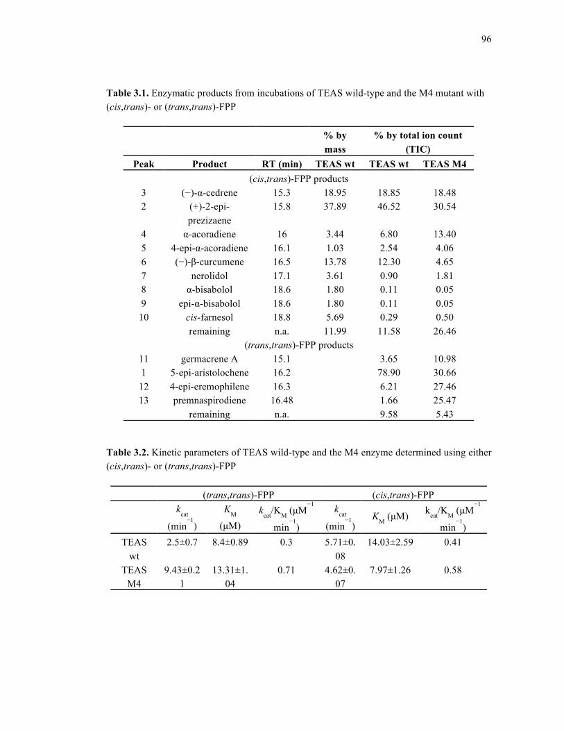
96
Table 3.1. Enzymatic products from incubations of TEAS wild-type and the M4 mutant with (cis,trans)- or (trans,trans)-FPP
Table 3.2. Kinetic parameters of TEAS wild-type and the M4 enzyme determined using either (cis,trans)- or (trans,trans)-FPP
% by mass
% by total ion count (TIC)
Peak Product RT (min) TEAS wt TEAS wt TEAS M4 (cis,trans)-FPP products
3 (−)-α-cedrene 15.3 18.95 18.85 18.48 2 (+)-2-epi-
prezizaene 15.8 37.89 46.52 30.54
4 α-acoradiene 16 3.44 6.80 13.40 5 4-epi-α-acoradiene 16.1 1.03 2.54 4.06 6 (−)-β-curcumene 16.5 13.78 12.30 4.65 7 nerolidol 17.1 3.61 0.90 1.81 8 α-bisabolol 18.6 1.80 0.11 0.05 9 epi-α-bisabolol 18.6 1.80 0.11 0.05
10 cis-farnesol 18.8 5.69 0.29 0.50 remaining n.a. 11.99 11.58 26.46
(trans,trans)-FPP products 11 germacrene A 15.1 3.65 10.98 1 5-epi-aristolochene 16.2 78.90 30.66
12 4-epi-eremophilene 16.3 6.21 27.46 13 premnaspirodiene 16.48 1.66 25.47
remaining n.a. 9.58 5.43
(trans,trans)-FPP (cis,trans)-FPP
kcat
(min−1
)
KM
(µM) kcat/KM (µM
−1
min−1
)
kcat
(min−1
) KM (µM) kcat/KM (µM
−1
min−1
) TEAS
wt 2.5±0.7 8.4±0.89 0.3 5.71±0.
08 14.03±2.59 0.41
TEAS M4
9.43±0.21
13.31±1.04
0.71 4.62±0.07
7.97±1.26 0.58
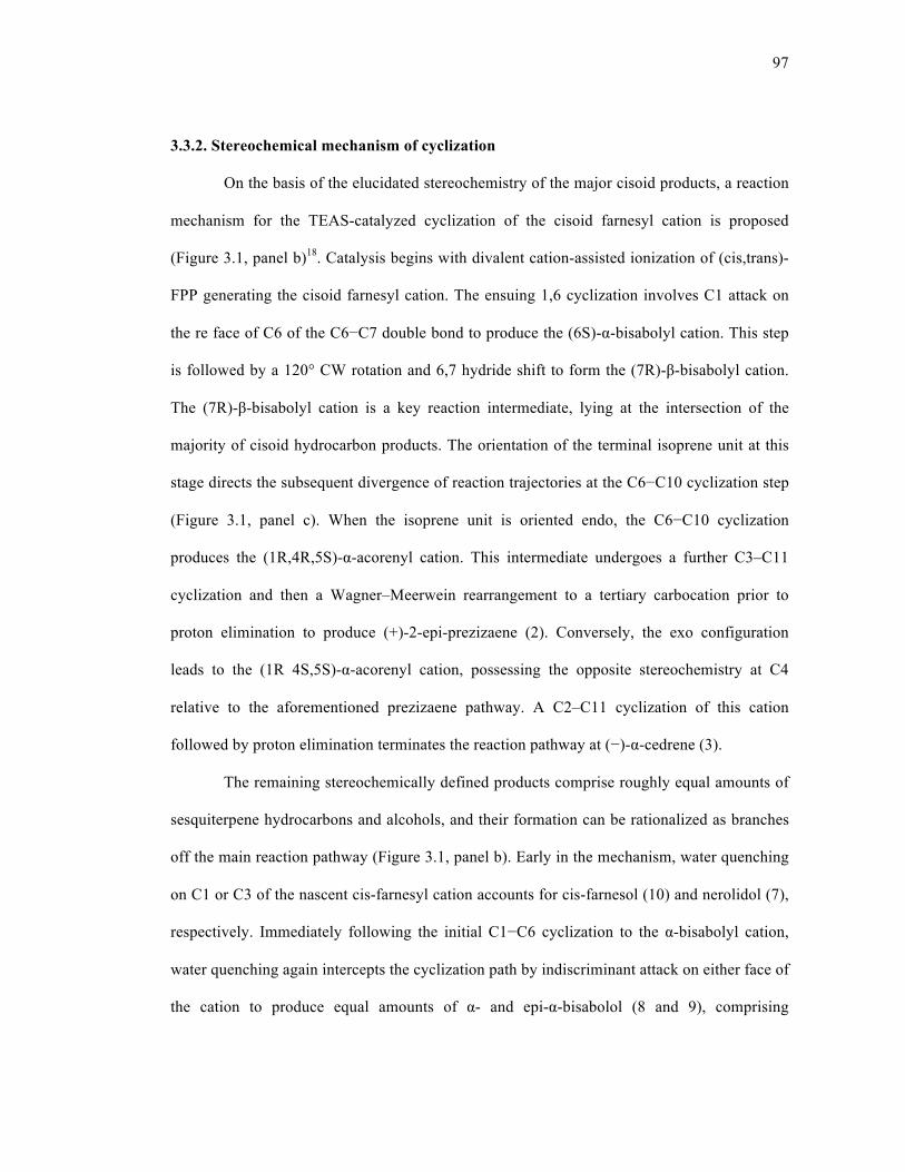
97
3.3.2. Stereochemical mechanism of cyclization
On the basis of the elucidated stereochemistry of the major cisoid products, a reaction
mechanism for the TEAS-catalyzed cyclization of the cisoid farnesyl cation is proposed
(Figure 3.1, panel b)18. Catalysis begins with divalent cation-assisted ionization of (cis,trans)-
FPP generating the cisoid farnesyl cation. The ensuing 1,6 cyclization involves C1 attack on
the re face of C6 of the C6−C7 double bond to produce the (6S)-α-bisabolyl cation. This step
is followed by a 120° CW rotation and 6,7 hydride shift to form the (7R)-β-bisabolyl cation.
The (7R)-β-bisabolyl cation is a key reaction intermediate, lying at the intersection of the
majority of cisoid hydrocarbon products. The orientation of the terminal isoprene unit at this
stage directs the subsequent divergence of reaction trajectories at the C6−C10 cyclization step
(Figure 3.1, panel c). When the isoprene unit is oriented endo, the C6−C10 cyclization
produces the (1R,4R,5S)-α-acorenyl cation. This intermediate undergoes a further C3–C11
cyclization and then a Wagner–Meerwein rearrangement to a tertiary carbocation prior to
proton elimination to produce (+)-2-epi-prezizaene (2). Conversely, the exo configuration
leads to the (1R 4S,5S)-α-acorenyl cation, possessing the opposite stereochemistry at C4
relative to the aforementioned prezizaene pathway. A C2–C11 cyclization of this cation
followed by proton elimination terminates the reaction pathway at (−)-α-cedrene (3).
The remaining stereochemically defined products comprise roughly equal amounts of
sesquiterpene hydrocarbons and alcohols, and their formation can be rationalized as branches
off the main reaction pathway (Figure 3.1, panel b). Early in the mechanism, water quenching
on C1 or C3 of the nascent cis-farnesyl cation accounts for cis-farnesol (10) and nerolidol (7),
respectively. Immediately following the initial C1−C6 cyclization to the α-bisabolyl cation,
water quenching again intercepts the cyclization path by indiscriminant attack on either face of
the cation to produce equal amounts of α- and epi-α-bisabolol (8 and 9), comprising
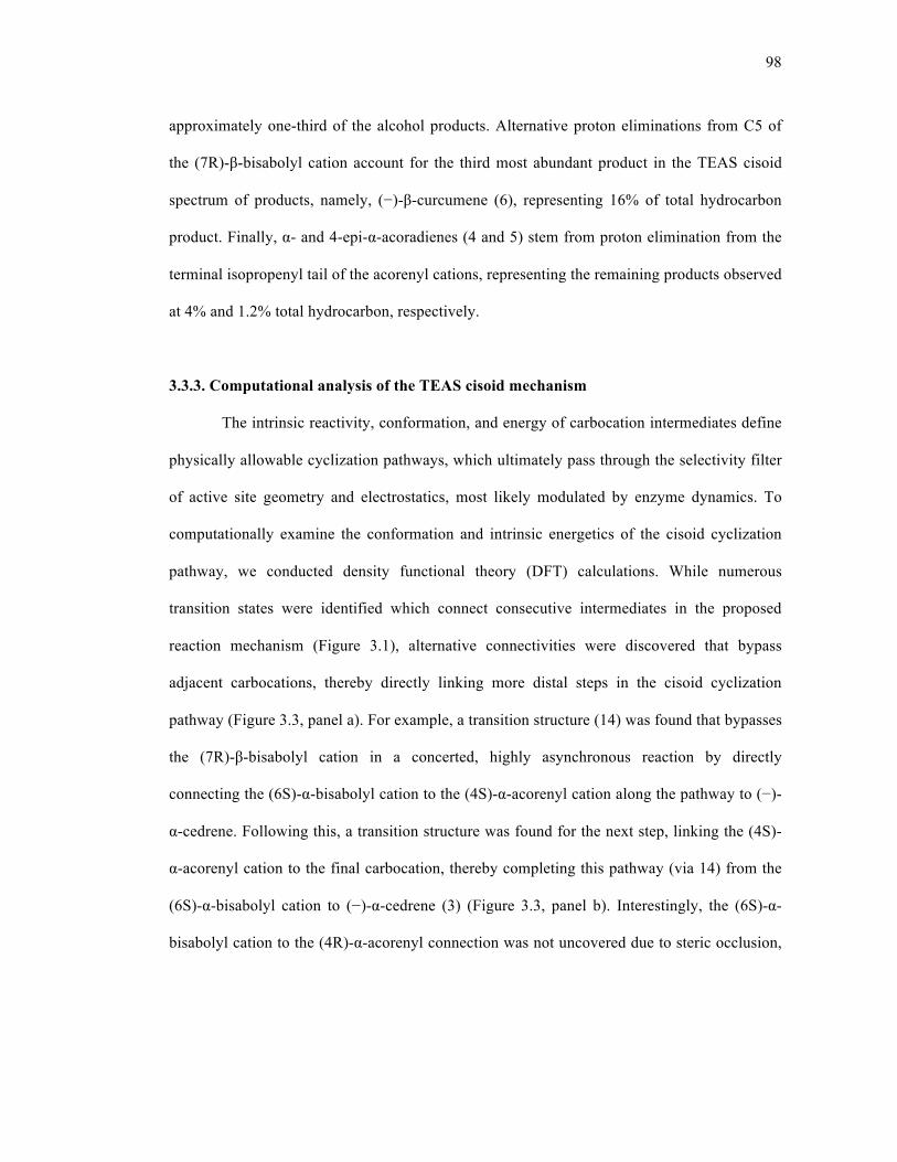
98
approximately one-third of the alcohol products. Alternative proton eliminations from C5 of
the (7R)-β-bisabolyl cation account for the third most abundant product in the TEAS cisoid
spectrum of products, namely, (−)-β-curcumene (6), representing 16% of total hydrocarbon
product. Finally, α- and 4-epi-α-acoradienes (4 and 5) stem from proton elimination from the
terminal isopropenyl tail of the acorenyl cations, representing the remaining products observed
at 4% and 1.2% total hydrocarbon, respectively.
3.3.3. Computational analysis of the TEAS cisoid mechanism
The intrinsic reactivity, conformation, and energy of carbocation intermediates define
physically allowable cyclization pathways, which ultimately pass through the selectivity filter
of active site geometry and electrostatics, most likely modulated by enzyme dynamics. To
computationally examine the conformation and intrinsic energetics of the cisoid cyclization
pathway, we conducted density functional theory (DFT) calculations. While numerous
transition states were identified which connect consecutive intermediates in the proposed
reaction mechanism (Figure 3.1), alternative connectivities were discovered that bypass
adjacent carbocations, thereby directly linking more distal steps in the cisoid cyclization
pathway (Figure 3.3, panel a). For example, a transition structure (14) was found that bypasses
the (7R)-β-bisabolyl cation in a concerted, highly asynchronous reaction by directly
connecting the (6S)-α-bisabolyl cation to the (4S)-α-acorenyl cation along the pathway to (−)-
α-cedrene. Following this, a transition structure was found for the next step, linking the (4S)-
α-acorenyl cation to the final carbocation, thereby completing this pathway (via 14) from the
(6S)-α-bisabolyl cation to (−)-α-cedrene (3) (Figure 3.3, panel b). Interestingly, the (6S)-α-
bisabolyl cation to the (4R)-α-acorenyl connection was not uncovered due to steric occlusion,

99
indicating the importance of the (7R)-β-bisabolyl cation along the pathway to (+)-2-epi-
prezizaene (2).
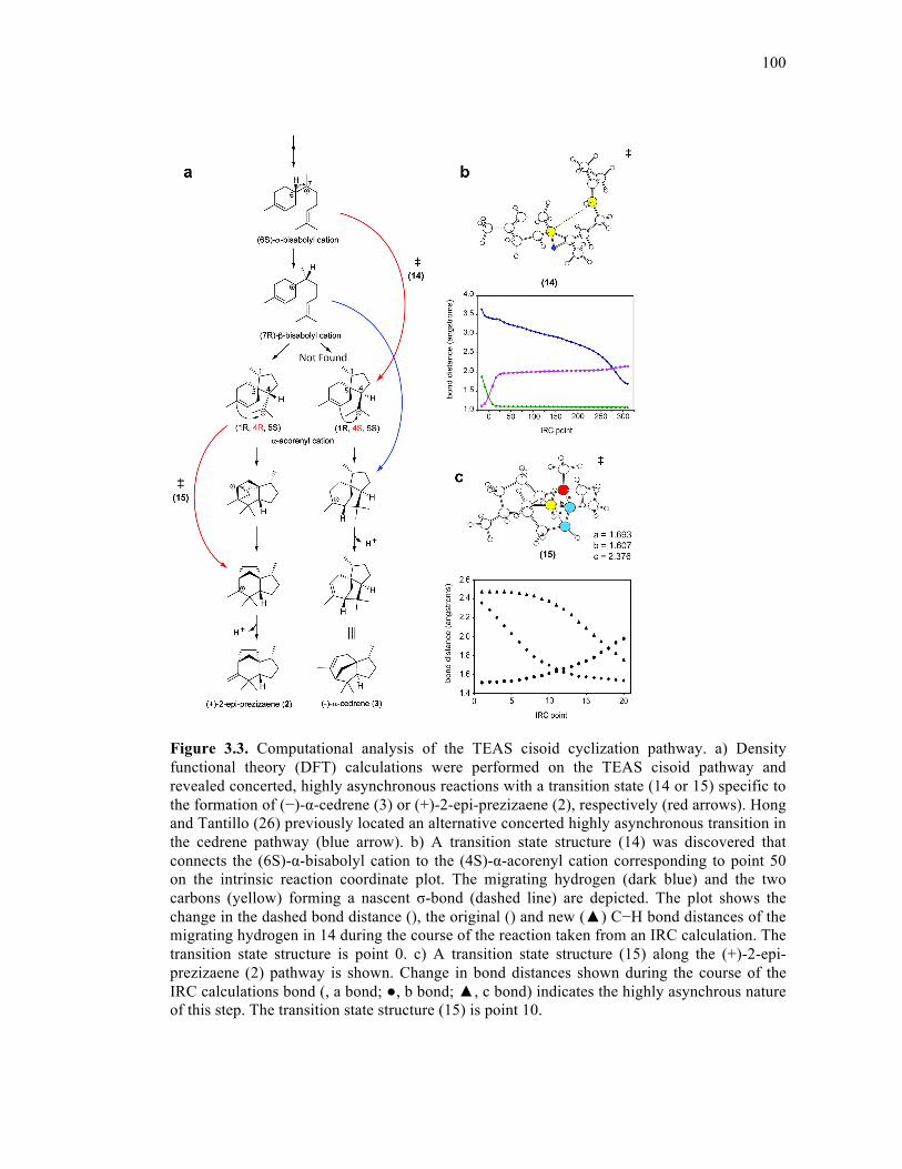
100
Figure 3.3. Computational analysis of the TEAS cisoid cyclization pathway. a) Density functional theory (DFT) calculations were performed on the TEAS cisoid pathway and revealed concerted, highly asynchronous reactions with a transition state (14 or 15) specific to the formation of (−)-α-cedrene (3) or (+)-2-epi-prezizaene (2), respectively (red arrows). Hong and Tantillo (26) previously located an alternative concerted highly asynchronous transition in the cedrene pathway (blue arrow). b) A transition state structure (14) was discovered that connects the (6S)-α-bisabolyl cation to the (4S)-α-acorenyl cation corresponding to point 50 on the intrinsic reaction coordinate plot. The migrating hydrogen (dark blue) and the two carbons (yellow) forming a nascent σ-bond (dashed line) are depicted. The plot shows the change in the dashed bond distance (), the original () and new (▲) C−H bond distances of the migrating hydrogen in 14 during the course of the reaction taken from an IRC calculation. The transition state structure is point 0. c) A transition state structure (15) along the (+)-2-epi-prezizaene (2) pathway is shown. Change in bond distances shown during the course of the IRC calculations bond (, a bond; ●, b bond; ▲, c bond) indicates the highly asynchrous nature of this step. The transition state structure (15) is point 10.
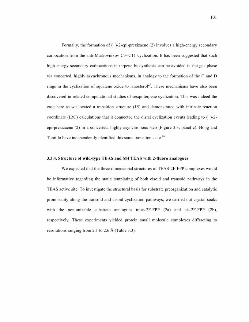
101
Formally, the formation of (+)-2-epi-prezizaene (2) involves a high-energy secondary
carbocation from the anti-Markovnikov C3−C11 cyclization. It has been suggested that such
high-energy secondary carbocations in terpene biosynthesis can be avoided in the gas phase
via concerted, highly asynchronous mechanisms, in analogy to the formation of the C and D
rings in the cyclization of squalene oxide to lanosterol25. These mechanisms have also been
discovered in related computational studies of sesquiterpene cyclization. This was indeed the
case here as we located a transition structure (15) and demonstrated with intrinsic reaction
coordinate (IRC) calculations that it connected the distal cyclization events leading to (+)-2-
epi-prezizaene (2) in a concerted, highly asynchronous step (Figure 3.3, panel c). Hong and
Tantillo have independently identified this same transition state.26
3.3.4. Structure of wild-type TEAS and M4 TEAS with 2-fluoro analogues
We expected that the three-dimensional structures of TEAS-2F-FPP complexes would
be informative regarding the static templating of both cisoid and transoid pathways in the
TEAS active site. To investigate the structural basis for substrate preorganization and catalytic
promiscuity along the transoid and cisoid cyclization pathways, we carried out crystal soaks
with the nonionizable substrate analogues trans-2F-FPP (2a) and cis-2F-FPP (2b),
respectively. These experiments yielded protein−small molecule complexes diffracting to
resolutions ranging from 2.1 to 2.6 Å (Table 3.3).
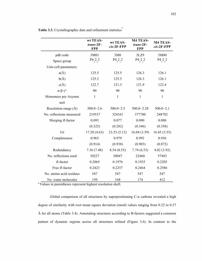
102
Table 3.3. Crystallographic data and refinement statisticsa
wt TEAS-trans-2F-
FPP
wt TEAS-cis-2F-FPP
M4 TEAS-trans-2F-
FPP
M4 TEAS-cis-2F-FPP
pdb code 3M01 3M0 3LZ9 3M00
Space group P412
12 P4
12
12 P4
12
12 P4
12
12
Unit-cell parameters:
a(Å) 125.5 125.5 126.3 126.1
b(Å) 125.5 125.5 126.3 126.1
c(Å) 122.7 121.3 121.9 122.4
α-β-γ° 90 90 90 90
Monomers per Asymm
unit
1 1 1 1
Resolution range (Å) 500.0−2.6 500.0−2.5 500.0−2.28 500.0−2.1
No. reflections measured 219537 324161 377780 248702
Merging R-factor 0.093
(0.325)
0.077
(0.282)
0.090
(0.346)
0.080
(0.358)
I/σ 17.28 (4.63) 23.53 (5.12) 16.04 (3.59) 16.45 (3.55)
Completeness 0.963
(0.914)
0.979
(0.938)
0.992
(0.903)
0.956
(0.872)
Redundancy 7.36 (7.48) 8.54 (8.55) 7.74 (6.53) 4.02 (3.92)
No. reflections used 30227 30047 22460 57483
R-factor 0.2065 0.1976 0.1935 0.2205
Free R-factor 0.2423 0.2257 0.2464 0.2586
No. amino acid residues 547 547 547 547
No. water molecules 150 168 174 412 a Values in parentheses represent highest resolution shell.
Global comparison of all structures by superpositioning C-α carbons revealed a high
degree of similarity with root mean square deviation (rmsd) values ranging from 0.22 to 0.37
Å for all atoms (Table 3.4). Annotating structures according to B-factors suggested a common
pattern of dynamic regions across all structures refined (Figure 3.6). In contrast to the
103
originally published TEAS·farnesyl hydroxyphosphonate (FHP) structure, all complexes
described here exhibit disorder in a portion of the J−K catalytic loop, a region encompassing
amino acids 521−533 that completes the enclosure of the active site during catalysis5. Several
residues were excised from both the wild-type and M4 TEAS models during refinement due to
a lack of clearly observable electron density and the attendant poor refinement of these regions
(Figure 3.7). As previously noted, the mutations in the M4 TEAS protein reside either in the
active site (V516I) or distribute more peripherally around the active site surface (A274T,
V372I, and Y406L) with distances from the active site center ranging from 7 to 14 Å (Figure
3.8). While each mutated side chain was readily discernible in the electron density, no
significant backbone distortions were evident, strongly hinting at dynamic, not static,
modulation of the active site contour for templating transformations of farnesyl cations in
TEAS. However, the V516I mutation directly affects the active site contour with implications
for substrate binding as discussed below.
Observable electron density is present in the active site regions for all complexes, and
the positions of ligand-binding residues were clearly established with the exception of Y527
on the J−K loop. In all the structures, contiguous electron density stretches from the DDxxD
motif through the diphosphate moiety into the NSE/DTE motif enshrouding the catalytically
essential Mg2+ ions (Figure 3.4, panel c). Although three Mg2+ ions are visible in each
complex, a complete octahedral coordination sphere of waters is only discernible in the
highest resolution M4 TEAS-cis-2F-FPP complex. Electron density surrounding the
diphosphate appendage is the most prominent feature in the calculated electron density
(without ligands modeled) with large σ values in the SIGMAA-weighted 2Fo − Fc electron
density maps (Figure 3.4, panel b). Clear electron density extends from the diphosphate
through the first isoprene unit containing the fluoro substituent in all complexes but trails off
104
through the center of the chain and picks up again at the distal isoprene unit (Figure 3.4, panel
e). Despite the waning electron density for the distal isoprene units, the farnesyl chain clearly
curls into a U-shape in all complexes, particularly at lower σ where continuous density is
apparent in the wild-type complexes (Figure 3.4, panel e). Taken together, these complexes
display near complete occupancy (based upon the unmistakable diphosphate and first isoprene
unit electron density), and aside from an incomplete J−K loop, Mg2+ ions and ligands are
bound with the farnesyl chain folded in a manner consistent with the formation of major
cyclization products along both the transoid and cisoid mechanistic pathways.
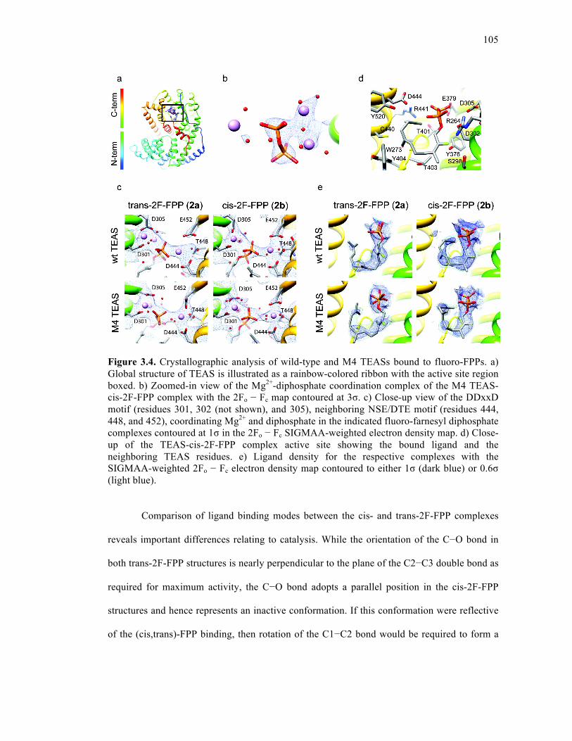
105
Figure 3.4. Crystallographic analysis of wild-type and M4 TEASs bound to fluoro-FPPs. a) Global structure of TEAS is illustrated as a rainbow-colored ribbon with the active site region boxed. b) Zoomed-in view of the Mg2+-diphosphate coordination complex of the M4 TEAS-cis-2F-FPP complex with the 2Fo − Fc map contoured at 3σ. c) Close-up view of the DDxxD motif (residues 301, 302 (not shown), and 305), neighboring NSE/DTE motif (residues 444, 448, and 452), coordinating Mg2+ and diphosphate in the indicated fluoro-farnesyl diphosphate complexes contoured at 1σ in the 2Fo − Fc SIGMAA-weighted electron density map. d) Close-up of the TEAS-cis-2F-FPP complex active site showing the bound ligand and the neighboring TEAS residues. e) Ligand density for the respective complexes with the SIGMAA-weighted 2Fo − Fc electron density map contoured to either 1σ (dark blue) or 0.6σ (light blue).
Comparison of ligand binding modes between the cis- and trans-2F-FPP complexes
reveals important differences relating to catalysis. While the orientation of the C−O bond in
both trans-2F-FPP structures is nearly perpendicular to the plane of the C2−C3 double bond as
required for maximum activity, the C−O bond adopts a parallel position in the cis-2F-FPP
structures and hence represents an inactive conformation. If this conformation were reflective
of the (cis,trans)-FPP binding, then rotation of the C1−C2 bond would be required to form a
106
catalytically active complex. This inactive conformation may be promoted by the 2-fluoro
moieties in cis-2F-FPP (2b) through its electrostatic interaction with Arg 264 residing 3 Å
away (Figure 3.4, panel d).
3.3.5. Spatial reconstruction of cisoid and transoid reaction pathways in TEAS
Multiple substrate binding modes discerned during building and refinement for the
extended farnesyl chain could potentially satisfy, and likely contribute to, the observed
electron densities (Figure 3.9). Despite these ambiguities, the general topology of the farnesyl
chain is clear and consistent with the anticipated parental fold inferred from the elucidated
stereochemistry of the final products. Importantly, electron density for the cis-2F-FPP
complexes reveals that the terminal isoprene unit curls into a helical (endo) fold in accordance
with the anticipated conformation (Figure 3.1, panel c). This orients the plane of the C10–C11
double bond parallel to a potentially attacking carbocation at C1. In contrast, the plane of the
C10–C11 double bond of the terminal isoprene unit is perpendicular to a nascent C1
carbocation in the trans-2F-FPP complexes, in accord with an initial C1–C10 cyclization
along the transoid pathway.
To spatially reconstruct the two major cyclization pathways in TEAS, we manually
docked transition state structures and models of the major products into the respective active
sites of wild-type trans-2F-FPP and cis-2F-FPP complexes (Figure 3.5, panel a). Restraining
the placement of products/intermediates such that cyclization to a specific product most likely
proceeds with the minimal amount of conformational distortion for the nascent farnesyl cation
en route to the final products results in a mechanistically plausible transition state geometry en
route to the observed dominant product.

107
Superposition of the transition state structure (15) on the farnesyl chain indicates that
substantial contraction of the substrate must occur to produce the compact (+)-2-epi-
prezizaene (2) final product. The crucial elements of preorganization are the juxtaposition of
C1 and C6 together with the endo orientation of the farnesyl tail, both consistent with the
observed electron density of the TEAS·cis-2F-FPP complex. Therefore, the static picture
drawn from these observations reveals a catalytically relevant substrate binding conformation
and substrate/intermediate preorganization very early along the cisoid pathway catalyzed by
TEAS. Based on this model, the pyrophosphate ion would reside close by but suitably
sequestered by neighboring interactions to stabilize the developing positive charge of the
secondary carbocation while limiting recapture probability prior to the final proton elimination
yielding (+)-2-epi-prezizaene (2).
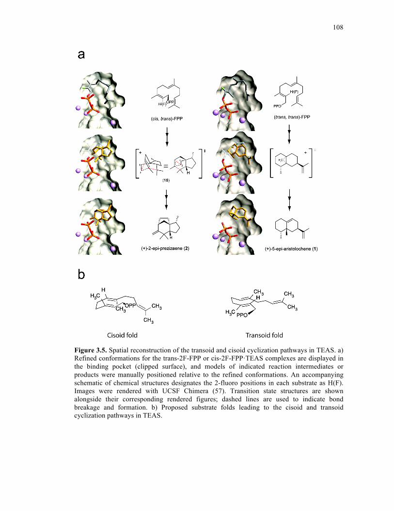
108
Figure 3.5. Spatial reconstruction of the transoid and cisoid cyclization pathways in TEAS. a) Refined conformations for the trans-2F-FPP or cis-2F-FPP·TEAS complexes are displayed in the binding pocket (clipped surface), and models of indicated reaction intermediates or products were manually positioned relative to the refined conformations. An accompanying schematic of chemical structures designates the 2-fluoro positions in each substrate as H(F). Images were rendered with UCSF Chimera (57). Transition state structures are shown alongside their corresponding rendered figures; dashed lines are used to indicate bond breakage and formation. b) Proposed substrate folds leading to the cisoid and transoid cyclization pathways in TEAS.
109
For the transoid pathway, the key transition state structure leading to the major
product (+)-5-epi-aristolochene (1) involves a methyl migration atop the decalin ring system
of the eremophilyl carbocation (Figure 3.5, panel a, right) as previously reported19. To achieve
this energetically favorable alkyl migration, substrate folding must preorganize an initial
electrophilic attack of C1 on C10 of the distal double bond. This requires substantial
movement of the chain following ionization, as these atoms are 5 Å apart in the ground state
complexes. However, judging by the degree of overlap between the transition state model and
the farnesyl chain, this motion can be accommodated largely within the first isoprene unit with
minimal conformational adjustments of the more distal isoprene units. Therefore, this
conformation of the farnesyl chain is consistent with TEAS preorganizing FPP (or more likely
the resultant acyclic farnesyl cation) for cyclization to 5-epi-aristolochene (1), in contrast to
the original TEAS·FHP complex(5). The phosphate moiety of FHP overlaps with the β-
phosphates of 2F-FPPs, although the farnesyl chain is more extended and folds in essentially
the opposite direction (Figure 3.9).
3.3.6. Cisoid cyclase activities with (trans,trans)-FPP
On the basis of our spatial reconstruction of the cisoid cyclization pathway, we
propose a model describing the “cisoid fold” of (trans,trans)-FPP (1a) that is representative of
the preorganization of the farnesyl chain leading to its conversion to cisoid-cation-derived
hydrocarbons. Accordingly, an alternative, catalytically productive binding mode of FPP is
populated in which the farnesyl chain curls into its typical U-shaped topology, but with the
first two isoprenoid units inverted relative to the “transoid fold” configuration (Figure 3.5,
panel b). The cisoid binding mode therefore possesses the DU configuration, opposite to that
described for germacrene A27, and importantly, with the distal isoprenoid unit curled below
110
this plane into an alternative binding pocket formed by T401, T402, C440, R441, D444, and
the diphosphate moiety (Figure 3.4, panel a). We posit that the positioning and anchoring of
the terminal isoprenoid unit is an essential stereochemical feature for triggering ionization,
perhaps through both steric and electronic effects. Upon ionization, a kinetically slow initial
isomerization occurs as the C10–C11 double bond is rotated out of position for electrophilic
attack by C1; in turn, a re face capture by the pyrophosphate ion on C3 of the nascent farnesyl
cation generates the neutral (3S)-NPP intermediate. Rotation around the C2–C3 bond followed
by reionization generates the 2,3-cis-farnesyl cation and entry into the cisoid cyclization
pathway.
3.3.7. Structural picture of catalytic promiscuity
The ligand–protein structures of a promiscuous TEAS mutant offer a glimpse into the
structural underpinnings of product specificity or lack thereof. To discern the structural basis
for product specificity in both cisoid and transoid cyclization pathways, we conducted a
comparative analysis of wild-type TEAS and M4 mutant structures with particular attention
focused on the active site contour and farnesyl chain binding modes. The most obvious surface
distortion, whether statically or dynamically derived, is contributed by the V516I mutation,
which introduces a methyl group into the active site cavity (Figure 3.10). While no drastic
distortion is evident in the comparative models of the farnesyl chain, the electron density for
the farnesyl chain in the M4 TEAS structures is discontinuous, indicative of increased
dynamic motion and/or local disorder (Figure 3.4, panel e). Interestingly, only the M4 TEAS-
cis-2F-FPP complex exhibits a significant shift in the position of Y520, which additionally
alters the active site surface features. The ligand-dependency for the Y520 shift may reflect
the interaction between the farnesyl chain, wherein the central isoprene unit is inverted
111
relative to the corresponding wild type complex, and active site residues in defining the
preorganized binding state. Despite the higher resolution of the M4 structures, the density for
the ligand is discontinuous, even when the electron density maps are viewed at low σ, in
contrast to the wild-type structures (Figure 3.4, panel e). It is probable that a more dynamic
farnesyl chain in M4-TEAS-cis/trans-2F-FPP structures explains the lack of electron density
for the entire farnesyl chain consistent with this M4 TEAS’s promiscuous catalytic activity
along both pathways.
3.3.8. Conclusions
(cis,trans)-FPP proved effective in directing reactions along the cisoid cyclization
pathway in TEAS. The isolation and stereochemical elucidation of the products lead to
formulation of reasonable reaction pathways to the cisoid-derived sesquiterpene skeletons18.
Traditionally, chemical tools have been a vital part of defining the stereochemical course of
terpene biosynthesis, as elegantly exemplified by the use of (1R)-[1-3H]- and (1S)-[1-3H]-
geranyl diphosphate to provide direct experimental confirmation that cyclization along the
cisoid pathway results in net retention of configuration at C1 of the substrate28. Fluoro
isoprenoid diphosphate substrate analogues also have been instrumental in elucidating
mechanistic aspects of terpene biosynthesis, most notably through the interception of reaction
intermediates such as 6-fluorogermacrene A29 and 7-fluoroverticillenes30 in TEAS and
taxadiene synthase enzymes, respectively. Most recently, 2F-FPP and 12,13-difluorofarnesyl
diphosphate (DF-FPP) were instrumental in deciphering the probable order of metal-ion
binding and conformational changes required for catalysis by aristolochene synthase from
Aspergillus terrus24.
112
To date, crystallographic analyses of terpene cyclases have yielded important insights
into how these enzymes function on the atomic scale. Most notably, anchoring the
diphosphate moiety of the substrate and metal coordination by the DDxxD and NSE/DTE
motifs shown in crystal structures paints a picture of the fundamental role of these events in
terpene synthase catalysis. Structures containing inorganic pyrophosphate or a substrate
analogue bound in the active site display ordering of various loops proximal to the active site,
consistent with a closed protein conformation that shields reactive carbocation intermediates
from solvent5, 23, 31. Further, alterations in pyrophosphate binding are thought to aid in the
modulation of prenyl chain orientation within the active site and most likely modulate the fate
of the early intermediates along prescribed mechanistic pathways32. All structures reported in
the current study contain the full complement of Mg2+ ions coordinating the diphosphate of the
fluoro-farnesyl analogues. However, despite this clear coordination geometry, elements of the
J−K loop remain disordered in both wild-type and mutant structures bound to diphosphate
containing ligands with Y527 electron density missing from the active site. This lack of
observable density for Y527 stands in contrast to the original TEAS·FHP complex where this
residue is clearly discernible (Figure 3.7).
These observations hint at a greater role of dynamics in terpene chain cyclization than
evident from the early structural work based upon static crystal structures. The wild-type
ligand complexes in the current study revealed density for the farnesyl chain folded in a
manner consistent with catalysis, and this can be interpreted in light of the established
stereochemistry for all TEAS products. Moreover, these static observations enabled the
positing of 2 distinct parental folds each of which gives rise to either cisoid or transoid
cyclization pathways for TEAS (Figure 3.5, panel b). The structure of a complex of limonene
synthase with 2-fluorolinalyl diphosphate captured another instance where the isoprenoid
113
chain conformation is consistent with the geometry of the final product23. However, it has
been noted that most reported crystal structures of terpene synthases complexed with
isoprenoid substrate analogues, including 2F-FPP used here, reveal isoprenoid tail
conformations that are not catalytically relevant5, 23, 24, 31. Considering the ideal case, where
there is unambiguous density for every atom of the farnesyl chain, a central challenge in the
field remains to resolve structural features responsible for product specificity or lack thereof
from a static picture alone, given the degeneracy of possible products arising from a single
parental substrate fold. Future progress toward defining the origins of sesquiterpene skeletal
complexity will undoubtedly benefit from integrating dynamic information from NMR and
time-resolved fluorescence (in progress) with computational approaches and protein
crystallography to develop a much clearer and time-resolved biophysical picture of terpene
synthase directed cyclization.
What possible relevance does the cryptic cisoid cyclization pathway of TEAS have in
the natural world? Although (cis,trans)-FPP has not been identified as a metabolite in tobacco
or related Solanaceous plants, a (cis,trans)-farnesyl diphosphate synthase has been identified
in Mycobacterium tuberculosis involved in bacterial cell wall synthesis33-34, suggesting the
potential relevance of this compound in other biological systems. Moreover, while often
observed, the biological significance of small amounts (3–14% of total product) of (cis,trans)-
FPP formation by FPP synthases35 has been ignored to date. Is it possible then that TEAS
possesses a “moonlighting” role in vivo by gathering up what we would normally consider
biosynthetic “waste” and recycling it into a bioactive product? While TEAS produces cisoid
terpenes in vitro, the presence of these metabolites has yet to be confirmed in planta.
Nonetheless, TEAS clearly possesses an efficient catalytic potential to access presently
unanticipated in vivo chemical diversity from lengthy branches of the cisoid reaction pathway,
114
a property that may have been naturally selected for and that can also be immediately
exploited for biotechnological applications starting with (cis,trans)-FPP.
3.4. METHODS
3.4.1. Organic synthesis
(cis,trans)-FPP was available from the concurrent investigation18. (trans, trans)- and
(cis,trans)-2-FluoroFPPs were accessed from the corresponding 2-fluorofarnesol isomers30 by
conversion to the respective 2-fluorofarnesyl chlorides and SN2 displacements with
(nBu)4N/diphosphate (HOPP)36 with complete retention of the 2,3-double bond configurations
by means of procedures similar to those reported previously (see Supporting Information)30.
(cis,trans)-2-FluoroFPP has not been previously described in the literature. Characterization
data for ammonium salt of 2b are as follows: white solid (51 mg, 68%); 1H NMR (CD3OD,
400 MHz) δ 5.17–5.11 (m, 1H, vinyl H), 5.11–5.05 (m, 1H, vinyl H), 4.59 (dd, 2H, J = 23.3,
5.4 Hz, CH2OPP), 2.16–2.11 (m, 4H, CH2), 2.09–2.04 (m, 2H, CH2), 2.00–1.95 (m, 2H, CH2),
1.68 (d, 3H, J = 3.5 Hz, CH3), 1.66 (q, 3H, J = 1.2 Hz, CH3), 1.61 (d, 3H, J = 1.2 Hz, CH3),
1.60 (br d, 3H, J = 0.6 Hz, CH3); 31P NMR (CD3OD, 162 MHz) δ −7.99 (br d, J = 14.9 Hz),
−9.30 (br d, J = 14.1 Hz); 19F NMR (CD3OD, 376 MHz) δ −118.9 (td, J = 23.2, 3.5 Hz).
3.4.2. Protein expression and purification
pH9GW expression vectors (an in-house Gateway destination vector) were
transformed into E. coli BL21(λDE3) and plated on LB agar containing 50 µg/mL kanamycin
for selection. Colonies were transferred to 100 mL of liquid media (LB with kanamycin)
followed by 16-h growth with shaking at 37°C at 275 rpm. Cultures were diluted 50-fold into
1 L of Terrific Broth with kanamycin, followed by growth with shaking at 37°C at 275 rpm
115
until cultures reached OD600 ≥ 1.5. Protein expression was induced by addition of isopropyl
β-D-thiogalactoside (IPTG) to 0.1 mM followed by growth with shaking at 20°C at 275 rpm
for 5 h. Cells were harvested by centrifugation and cell pellets frozen at -20°C. Frozen pellets
were re-suspended in lysis buffer (50 mM Tris-HCl, pH 8.0, 500 mM NaCl, 20 mM
imidazole, pH 8.0, 10% [v/v] glycerol, 10 mM β- mercaptoethanol, and 1% [v/v] Tween-20)
containing 1 mg/mL lysozyme followed by stirring at 4°C for 1 h. After sonication and
centrifugation, the clarified supernatant was passed over a column of Ni2+–NTA resin
(Qiagen), washed with 10 bed volumes of lysis buffer and 10 bed volumes of wash buffer (50
mM Tris–HCl, pH 8.0, 500 mM NaCl, 20 mM imidazole, pH 8.0, 20 mM β-mercaptoethanol,
and 10% [v/v] glycerol), and the His-tagged protein was eluted with elution buffer (50 mM
Tris–HCl, pH 8.0, 500 mM NaCl, 250 mM imidazole, pH 8.0, 20 mM β- mercaptoethanol,
and 10% [v/v] glycerol). N-terminal His-tags were removed via proteolysis with thrombin as
follows: thrombin was added to a ratio of 1:1,000 [w/w] directly to the eluted protein fraction
and dialyzed against two changes of buffer (50 mM Tris–HCl, pH 8.0, 100 mM NaCl, and 10
mM β-mercaptoethanol) over 24 h at 4 °C. Following digestion, samples were passed over a
column containing 0.5 mL benzamidine sepharose to remove thrombin and 0.5 mL Ni2+-
NTA resin to capture undigested protein. The resulting protein solutions were collected and
concentrated to approximately 10 mg/mL or greater by centrifugation using 30,000 Da
molecular weight cut-off concentrators (Millipore, Bedford, MA). Concentrated samples were
injected onto a Sephacryl S-200 column equilibrated with buffer (25 mM Tris–HCl, pH 8.0,
50 mM NaCl and 1 mM DTT). Fractions corresponding to digested protein were verified by
SDS-PAGE, pooled and concentrated (as described above) to approximately 20 mg/mL and
aliquoted for freezing at -80°C. Samples were judged to be ~99% pure by Coomassie stained
SDS-PAGE gels.
116
3.4.3. Kinetic measurement
Kinetic characterization of purified wild-type and M4 mutant TEASs were conducted
as previously described20. Briefly, 500-µL scale reactions using a 3-component buffer system
(25 mM 2-(N-morpholino)ethanesulfonic acid (MES), 50 mM Tris, and 25 mM 3-
(cyclohexylamino)propanesulfonic acid (CAPS) at pH 7.0 with 10 mM MgCl2) were
conducted in triplicate at room temperature (25°C) with 15 nM protein and variable
concentrations of (cis,trans)-FPP. Reaction products were analyzed using a Hewlett–Packard
6890 gas chromatograph (GC) coupled to a 5973 mass selective detector (MSD) equipped
with an HP- 5MS capillary column (0.25 mm i.d. 30 m with 0.25 µm film thickness) (Agilent
Technologies). Product quantification was performed using SIM mode, set to detect ions with
m/z = 91, 133, and 189. The GC was operated at a He flow rate of 2 mL/min, and the MSD
was operated at 70 eV. Split-less injections (2 µL) were performed with an inlet temp of
250°C, a temp that drives the Cope rearrangement of germacrene A (11) to completion. The
GC was programmed with an initial oven temp of 50°C (5-min hold), which was then
increased 10°C/min up to 180°C (4-min hold), followed by a 100°C/min ramp until 240°C (1-
min hold). A solvent delay of 8.5 min was allowed prior to the acquisition of the MS data. (+)-
2-Epi-prezizaene (2) was quantified by integration of peak areas using Enhanced Chemstation
(version B.01.00, Agilent Technologies).
The GC–MS instrument was calibrated with an authentic (+)-2-epi-prezizaene
standard16. Corrected velocity data (Table 3.1) were fitted to the Michaelis–Menten equation
using GraphPad Prism (version 4.00 for Windows, GraphPad Software).
117
3.4.4. Protein crystallization and data collection
Crystallization of purified proteins was conducted using hanging drops over a 0.5-mL
reservoir (15% w/v PEG8K, 200 mM Mg(OAc)2, 100 mM 3-(N-morpholino)-2-
hydroxypropanesulfonic acid (MOPSO)-Na+, pH 7.0). Crystal soaks were conducted overnight
in reservoir solution containing 10 mM fluoro-farnesyl diphosphate ligand. Crystals were
frozen in soak solution also containing 20% v/v ethylene glycol as cryoprotectant. Data
collection was performed at the Stanford Synchrotron Radiation Laboratory (SSRL) beamline
1–5 for wild-type TEAS-2F-FPP and cis-2F-FPP complexes, while data for the M4 TEAS
mutant structures were collected at the Advanced Light Source (ALS) beamline 8.2.1. All data
were processed using XDS software37. The initial crystallographic structure solutions were
obtained through molecular replacement analyses using the TEAS-FHP complex (PDB id
5eat) as the search model with Molrep in Collaborative Computational Project No. 4
(CCP4)38. Model building was performed using COOT39 and rounds of refinement were
conducted using Crystallography NMR System (CNS)40. To refine the position of the farnesyl
chain, the first isoprene unit containing the 2-flouro group was built and refined, followed by
sequential addition, building and refinement of remaining isoprene units. For the wild type
cis-2F-FPP complex, additional multi-conformer refinement was undertaken using the
program RefMac41-48 in CCP438. The fold inferred from the known stereochemistry of the final
products was built into the density followed by a final round of refinement using CNS to
produce the current models of the farnesyl chain (Figure 3.10).
3.4.5. Computational methods
All calculations were performed with GAUSSIAN 98W and GAUSSIAN 09W49 and
the density functional method using B3LYP, Becke’s three-parameter hybrid method50 with
118
the Lee–Yang–Parr correlation functional51 and the 6-31G* basis set52. All stationary points
were confirmed with second derivative calculations. Energies reported here include zero-point
energy corrections calculated with unscaled B3LYP/6-31G* frequencies obtained analytically
with G98W. Intrinsic reaction coordinate calculations53-54 were used to determine reaction
pathways. Single point mpw1pw91/6-311+G(2d,p)//B3LYP/6-31G* calculations as
recommended by Matsuda55 were carried out for all stationary points reported.56
3.4.6. Product elucidation
A preparative-scale incubation was carried out using 127 mg (310 mmol) of
(cis,trans)-FPP and a total of 18 mg of recombinant wild-type TEAS in order to accumulate
sufficient material for chromatographic fractionations, NMR analyses, and optical rotation
measurements of the major products as described18.
3.5. SUPPORTING INFORMATION
3.5.1. Preparation and Characterization of (2-cis, 6-trans)-2-Fluorofarnesyl Diphosphate
General Aspects:
1H and 13C NMR spectra were recorded in CDCl3 (1H, 7.26; 13C, 77.0) or CD3OD [1H,
3.31 (quintet); 13C, 49.2 (septet)] with U400 and U500 spectrometers in SCS NMR
Spectroscopy Facility at the University of Illinois. Chemical shifts are in ppm and coupling
constants are in Hertz. The abbreviation ‘app’ is used to describe the apparent multiplicity of
the peak and may or may not be a valid first-order analysis.
All chemical reactions were performed in flame-dried glassware under nitrogen. THF
and Et2O were dried and distilled from Na/benzophenone; benzene and CH2Cl2 were dried and
distilled from CaH2. Hexane and ethyl acetate were freshly distilled from CaH2. DMF,
119
acetonitrile, and CDCl3 were dried over molecular sieves (4 Ǻ) prior use. TLC analyses were
performed on silica gel 60 F254 precoated-plates 250 µm. All retention factors (Rf) are on
silica gel TLC plates until otherwise noted. TLC visualizations were performed with 5%
phosphomolybdic acid (0.2 M in 2.5% concd. H2SO4/EtOH (v/v)), I2 vapor, or UV light.
Commercial reagents were used without further purification unless specifically noted. Column
chromatography was performed according to Still’s procedure58 using 100-700 times excess
32- 64 µm grade silica gel. Products separated by chromatography are specified in elution
order.
(2Z, 6E)- 1-Chloro-3,7,11-trimethylundeca-2,6,10-triene ((2-cis, 6-trans)-2-
Fluorofarnesyl Chloride)
(2-cis, 6-trans)-2-Fluorofarnesol30 was converted to the allylic chloride under Meyers’
conditions59 as previously describe for (2-trans, 6-trans)-2-fluorofarnesol.24 Reaction of the
alcohol (44 mg, 0.18 mmol) with LiCl (77 mg, 1.8 mmol), s-collidine (222 mg, 1.8 mmol),
andMsCl (67 mg, 0.54 mmol) in dry DMF provided the chloride as a yellow oil (47 mg, 99%).
The chloride was converted to the diphosphate directly without purification. Product
characterization data: TLC Rf 0.83 (15% EtOAc in hexane); 1H NMR (CDCl3, 400 MHz) δ
5.09 (m, 2H, vinyl H), 4.18 (dd, 2H, J = 22.5, 0.5 Hz, CH2Cl), 1.95-2.18 (m, 8H, 4CH2), 1.72
( app d, 3H, Japp = 3.5 Hz, CH3), 1.68 (d, 3H, J = 1.0 Hz, CH3), 1.60 (s, 6H, 2CH3); 19F
NMR (CDCl3, 376 MHz) δ –116.7 (td, J = 23.2, 2.8 Hz).
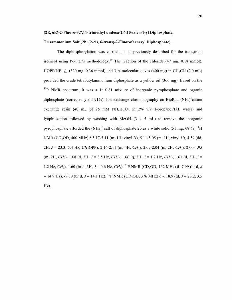
120
(2E, 6E)-2-Fluoro-3,7,11-trimethyl undeca-2,6,10-trien-1-yl Diphosphate,
Trisammonium Salt (2b, (2-cis, 6-trans)-2-Fluorofarnesyl Diphosphate).
The diphosphorylation was carried out as previously described for the trans,trans
isomer4 using Poulter’s methodology.60 The reaction of the chloride (47 mg, 0.18 mmol),
HOPP(NBu4)3 (320 mg, 0.36 mmol) and 3 Å molecular sieves (400 mg) in CH3CN (2.0 mL)
provided the crude tetrabutylammonium diphosphate as a yellow oil (366 mg). Based on the
31P NMR spectrum, it was a 1: 0.81 mixture of inorganic pyrophosphate and organic
diphosphate (corrected yield 91%). Ion exchange chromatography on BioRad (NH4)+cation
exchange resin (40 mL of 25 mM NH4HCO3 in 2% v/v 1-propanol/D.I. water) and
lyophilization followed by washing with MeOH (3 x 5 mL) to remove the inorganic
pyrophosphate afforded the (NH4)+ salt of diphosphate 2b as a white solid (51 mg, 68 %): 1H
NMR (CD3OD, 400 MHz) δ 5.17-5.11 (m, 1H, vinyl H), 5.11-5.05 (m, 1H, vinyl H), 4.59 (dd,
2H, J = 23.3, 5.4 Hz, CH2OPP), 2.16-2.11 (m, 4H, CH2), 2.09-2.04 (m, 2H, CH2), 2.00-1.95
(m, 2H, CH2), 1.68 (d, 3H, J = 3.5 Hz, CH3), 1.66 (q, 3H, J = 1.2 Hz, CH3), 1.61 (d, 3H, J =
1.2 Hz, CH3), 1.60 (br d, 3H, J = 0.6 Hz, CH3); 31P NMR (CD3OD, 162 MHz) δ -7.99 (br d, J
= 14.9 Hz), -9.30 (br d, J = 14.1 Hz); 19F NMR (CD3OD, 376 MHz) δ -118.9 (td, J = 23.2, 3.5
Hz).
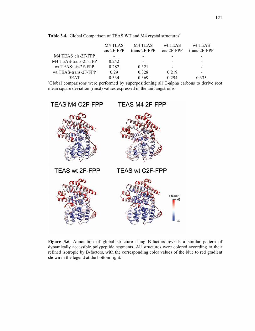
121
Table 3.4. Global Comparison of TEAS WT and M4 crystal structuresa
aGlobal comparisons were performed by superpositioning all C-alpha carbons to derive root mean square deviation (rmsd) values expressed in the unit angstroms.
Figure 3.6. Annotation of global structure using B-factors reveals a similar pattern of dynamically accessible polypeptide segments. All structures were colored according to their refined isotropic by B-factors, with the corresponding color values of the blue to red gradient shown in the legend at the bottom right.
M4 TEAS cis-2F-FPP
M4 TEAS trans-2F-FPP
wt TEAS cis-2F-FPP
wt TEAS trans-2F-FPP
M4 TEAS·cis-2F-FPP - - - - M4 TEAS·trans-2F-FPP 0.242 - - -
wt TEAS·cis-2F-FPP 0.282 0.321 - - wt TEAS-trans-2F-FPP 0.29 0.328 0.219 -
5EAT 0.334 0.369 0.294 0.335

122
Figure 3.7. Disorder in the J-K loop of experimental crystal structures. An active site model for the wild-type TEAS trans-2F-FPP is shown as a van der Waals surface clipped to reveal the bound substrate analogue and helices J and K with the intervening loops. All experimental structures are overlaid on the original TEAS-FHP structure (pdb id 5eat) shown in a grey semitransparent trace. Each structure is colored as indicated in the legend below, with the omitted J-K loop regions highlighted in grey.
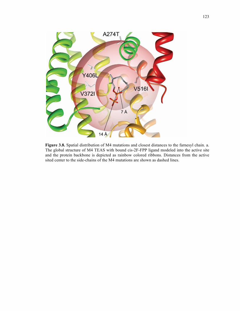
123
Figure 3.8. Spatial distribution of M4 mutations and closest distances to the farnesyl chain. a. The global structure of M4 TEAS with bound cis-2F-FPP ligand modeled into the active site and the protein backbone is depicted as rainbow colored ribbons. Distances from the active sited center to the side-chains of the M4 mutations are shown as dashed lines.
124
Figure 3.9. Farnesyl chain topology of wild-type TEAS from fluorofarnesyl analogues. a. Observable electron density from the wild-type complex with cis-2F-FPP reveals a U-shaped curl (left panel) possibly contributed to by four distinct binding modes of the farnesyl chain (right panel). b. Calculated electron density contoured at 1σ in the SIGMAA-weighted 2Fo-Fc map with the modeled trans-2F-FPP shown with a plane passing through the U-shape curl of the farnesyl chain (left panel). An overlay of trans-2F-FPP (silver chain) with farnesylhydroxy phosphonate (FHP, white chain) in the calculated electron density for the trans-2F-FPP ligand from the left panel.
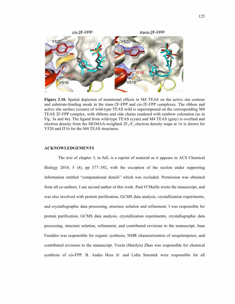
125
Figure 3.10. Spatial depiction of mutational effects in M4 TEAS on the active site contour and substrate-binding mode in the trans-2F-FPP and cis-2F-FPP complexes. The ribbon and active site surface (cream) of wild-type TEAS wild is superimposed on the corresponding M4 TEAS 2F-FPP complex, with ribbons and side chains rendered with rainbow coloration (as in Fig. 3a and 4a). The ligand from wild-type TEAS (cyan) and M4 TEAS (gray) is overlaid and electron density from the SIGMAA-weighted 2Fo-Fc electron density maps at 1σ is shown for Y520 and I516 for the M4 TEAS structures.
ACKNOWLEDGEMENTS
The text of chapter 3, in full, is a reprint of material as it appears in ACS Chemical
Biology 2010, 5 (4), pp 377–392, with the exception of the section under supporting
information entitled “computational details” which was excluded. Permission was obtained
from all co-authors. I am second author of this work. Paul O’Maille wrote the manuscript, and
was also involved with protein purification, GCMS data analysis, crystallization experiments,
and crystallographic data processing, structure solution and refinement. I was responsible for
protein purification, GCMS data analysis, crystallization experiments, crystallographic data
processing, structure solution, refinement, and contributed revisions to the manuscript. Juan
Faraldos was responsible for organic synthesis, NMR characterization of sesquiterpenes, and
contributed revisions to the manuscript. Yuxin (Marilyn) Zhao was responsible for chemical
synthesis of cis-FPP. B. Andes Hess Jr. and Lidia Smentek were responsible for all
126
computational studies. The research included in the manuscript was performed under the
supervision of Robert Coates and Joseph P. Noel (who also contributed revisions and helped
write the manuscript).
REFERENCES 1. Gershenzon, J. and Dudareva, N. (2007) The function of terpene natural products in
the natural world Nat. Chem. Biol. 3, 408– 414. 2. Liang, P., Ko, T., and Wang, A. (2002) Structure, mechanism and function of
prenyltransferases Eur. J. Biochem. 269, 3339– 3354. 3. Ruzicka, L., Eschenmoser, A., and Heusser, H. (1953) The isoprene rule and the
biogenesis of terpenic compounds Experentia 9, 357– 367. 4. Cane, D. (1985) Isoprenoid biosynthesis. Stereochemistry of the cyclization of allylic
pyrophosphates Acc. Chem. Res. 18, 220– 226. 5. Starks, C., Back, K., Chappell, J., and Noel, J. (1997) Structural basis for cyclic
terpene biosynthesis by tobacco 5-epi-aristolochene synthase Science 277, 1815– 1820.
6. Lesburg, C., Zhai, G., Cane, D., and Christianson, D. (1997) Crystal structure of
pentalenene synthase: mechanistic insights on terpenoid cyclization reactions in biology Science 277, 1820– 1824.
7. Shishova, E., Di Costanzo, L., Cane, D., and Christianson, D. (2007) X-ray crystal
structure of aristolochene synthase from Aspergillus terreus and evolution of templates for the cyclization of farnesyl diphosphate Biochemistry 46, 1941– 1951.
8. Kllner, T., Schnee, C., Li, S., Svatos, A., Schneider, B., Gershenzon, J., and
Degenhardt, J. (2008) Protonation of a neutral (S)-β-bisabolene intermediate is involved in (S)-β-macrocarpene formation by the maize sesquiterpene synthases TPS6 and TPS11 J. Biol. Chem. 283, 20779– 20788.
9. Picaud, S., Mercke, P., He, X., Sterner, O., Brodelius, M., Cane, D., and Brodelius, P.
(2006) Amorpha-4,11-diene synthase: mechanism and stereochemistry of the enzymatic cyclization of farnesyl diphosphate Arch. Biochem. Biophys. 448, 150– 155.
10. Cane, D. and Ha, H. (1988) Trichodiene biosynthesis and the role of nerolidyl
pyrophosphate in the enzymatic cyclization of farnesyl pyrophosphate J. Am. Chem. Soc. 110, 6865– 6870.

127
11. Mercke, P., Crock, J., Croteau, R., and Brodelius, P. (1999) Cloning, expression, and characterization of epi-cedrol synthase, a sesquiterpene cyclase from Artemisia annua L Arch. Biochem. Biophys. 369, 213– 222.
12. Lin, X. and Cane, D. (2009) Biosynthesis of the sesquiterpene antibiotic
albaflavenone in Streptomyces coelicolor. Mechanism and stereochemistry of the enzymatic formation of epi-isozizaene J. Am. Chem. Soc. 131, 6332– 6333.
13. Davis, E. M. and Croteau, R. (2000) Cyclization enzymes in the biosynthesis of
monoterpenes, sesquiterpenes, and diterpenes Top. Curr. Chem. 209, 53– 95. 14. Gordon, M., Stoessl, A., and Stothers, J. (1973) Post-infectional inhibitors from
plants. 4. Structure of capsidiol - antifungal sesquiterpene from sweet peppers Can. J. Chem. 51, 748– 752.
15. O’Maille, P. E., Chappell, J., and Noel, J. (2006) Biosynthetic potential of
sesquiterpene synthases: alternative products of tobacco 5-epi-aristolochene synthase Arch. Biochem. Biophys. 448, 73– 82.
16. Fox, R. B. and Powell, W. H. (2001) Nomenclature of Organic Compounds, 2nd ed.,
pp 306− 308, American Chemical Society and Oxford University Press, Oxford. 17. Rigaudy, J. and Klesney, S. P. (1979) IUPAC Nomenclature of Organic Chemistry:
Sections A, B, C, D, E, F and H, pp 475− 477, Pergamon Press, Oxford. 18. Faraldos, J. A., O’Maille, P. E., Dellas, N., Noel, J., and Coates, R. M. (2009)
Bisabolyl-derived sesquiterpenes from tobacco 5-epi-aristolochene synthase-catalyzed cyclization of (2Z, 6E)-farnesyl diphosphate. J. Am. Chem. Soc., accepted for publication.
19. O’Maille, P. E., Malone, A., Dellas, N., Andes Hess, B. J., Smentek, L., Sheehan, I.,
Greenhagen, B., Chappell, J., Manning, G., and Noel, J. (2008) Quantitative exploration of the catalytic landscape separating divergent plant sesquiterpene synthases Nat. Chem. Biol. 4, 617– 623.
20. O’Maille, P. E., Chappell, J., and Noel, J. (2004) A single-vial analytical and
quantitative gas chromatography-mass spectrometry assay for terpene synthases Anal. Biochem. 335, 210– 217.
21. Miller, D., Yu, F., and Allemann, R. (2007) Aristolochene synthase-catalyzed
cyclization of 2-fluorofarnesyl-diphosphate to 2-fluorogermacrene A ChemBiochem 8, 1819– 1825.
22. Vedula, L., Zhao, Y., Coates, R., Koyama, T., Cane, D., and Christianson, D. (2007)
Exploring biosynthetic diversity with trichodiene synthase Arch. Biochem. Biophys. 466, 260– 266.
128
23. Hyatt, D., Youn, B., Zhao, Y., Santhamma, B., Coates, R., Croteau, R., and Kang, C. (2007) Structure of limonene synthase, a simple model for terpenoid cyclase catalysis Proc. Natl. Acad. Sci. U.S.A. 104, 5360– 5365.
24. Shishova, E., Yu, F., Miller, D. J., Faraldos, J., Zhao, Y., Coates, R., Allemann, R.,
Cane, D., and Christianson, D. (2008) X-ray crystallographic studies of substrate binding to aristolochene synthase suggest a metal binding sequence for catalysis J. Biol. Chem. 283, 15431– 15439.
25. Hess, B. (2002) Concomitant C-ring expansion and D-ring formation in lanosterol
biosynthesis from squalene without violation of Markovnikov’s rule J. Am. Chem. Soc. 124, 10286– 10287.
26. Hong, Y. and Tantillo, D. (2009) Consequences of conformational preorganization in
sesquiterpene biosynthesis: theoretical studies on the formation of the bisabolene, curcumene, acoradiene, zizaene, cedrene, duprezianene, and sesquithuriferol sesquiterpenes J. Am. Chem. Soc. 131, 7999– 8015.
27. Faraldos, J. A., Wu, S., Chappell, J., and Coates, R. M. (2007) Conformational
analysis of (+)-germacrene A by variable-temperature NMR and NOE spectroscopy Tetrahedron 63, 7733– 7742.
28. Croteau, R., Felton, N., and Wheeler, C. (1985) Stereochemistry at C-1 of geranyl
pyrophosphate and neryl pyrophosphate in the cyclization to (−)-bornyl pyrophosphate J. Biol. Chem. 260, 5956– 5962.
29. Faraldos, J. A., Zhao, Y., O’Maille, P. E., Noel, J., and Coates, R. M. (2007)
Interception of the enzymatic conversion of farnesyl diphosphate to 5-epi-aristolochene by using a fluoro substrate analogue: 1-fluorogermacrene A from (2E,6Z)-6-fluorofarnesyl diphosphate ChemBioChem 8, 1826– 1833.
30. Jin, Y. H., Williams, D., Croteau, R., and Coates, R. M. (2005) Taxadiene synthase-
catalyzed cyclization of 6-fluorogeranylgeranyl diphosphate to 7-fluoroverticillenes J. Am. Chem. Soc. 127, 7834– 7842.
31. Whittington, D., Wise, M., Urbansky, M., Coates, R., Croteau, R., and Christianson,
D. (2002) Bornyl diphosphate synthase: structure and strategy for carbocation manipulation by a terpenoid cyclase Proc. Natl. Acad. Sci. U.S.A. 99, 15375– 15380.
32. Vedula, L., Cane, D., and Christianson, D. (2005) Role of arginine-304 in the
diphosphate-triggered active site closure mechanism of trichodiene synthase Biochemistry 44, 12719– 12727.
33. Schulbach, M., Brennan, P., and Crick, D. (2000) Identification of a short (C15) chain
Z-isoprenyl diphosphate synthase and a homologous long (C50) chain isoprenyl diphosphate synthase in Mycobacterium tuberculosis J. Biol. Chem. 275, 22876– 22881.
129
34. Schulbach, M., Mahapatra, S., Macchia, M., Barontini, S., Papi, C., Minutolo, F., Bertini, S., Brennan, P., and Crick, D. (2001) Purification, enzymatic characterization, and inhibition of the Z-farnesyl diphosphate synthase from Mycobacterium tuberculosis J. Biol. Chem. 276, 11624– 11630.
35. Thulasiram, H. and Poulter, C. D. (2006) Farnesyl diphosphate synthase: the art of
compromise between substrate selectivity and stereoselectivity J. Am. Chem. Soc. 128, 15819– 15823.
36. Woodside, A., Huang, Z., and Poulter, C. D. (1993) Trisammonium geranyl
diphosphate, in Organic Synthesis, Collect. Vol. 8, pp 616− 620, Wiley, New York. 37. Kabsch, W. (1993) Automated processing of rotation diffraction data from crystals of
initially unknown symmetry and cell constants J. Appl. Crystallogr. 26, 795– 800. 38. (1994) The CCP4 suite: programs for protein crystallography, Acta Crystallogr. D 50,
760– 763. 39. Emsley, P. and Cowton, K. (2004) Coot: model-building tools for molecular graphics
Acta Crystallogr. D 60, 2126– 2132. 40. Brunger, A., Adams, P., Clore, G., Delano, W., Gros, P., Grosse-Kunstleve, R. W.,
Jiang, J., Kuszewski, J., Nilges, M., Pannu, N., Read, R., Rice, L., Simonson, T., and Warren, G. (1998) Crystallography & NMR system: A new software suite for macromolecular structure determination Acta Crystallogr. D 54, 905– 921.
41. Murshudov, G., Vagin, A., Lebedev, A., Wilson, K. S., and Dodson, E. J. (1999)
Efficient anisotropic refinement of macromolecular structures using FFT Acta Crystallog.r D 55, 247– 255.
42. Murshudov, G., Vagin, A., and Dodson, E. J. (1997) Refinement of macromolecular
structures by the maximum-likelihood method Acta Crystallogr. D 53, 240– 255. 43. Pannu, N., Murshudov, G., Dodson, E., and Read, R. (1998) Incorporation of prior
phase information strengthens maximum-likelihood structure refinement Acta Crystallogr. D 54, 1285– 1294.
44. Skubak, P., Murshudov, G., and Pannu, N. (2004) Direct incorporation of
experimental phase information in model refinement Acta Crystallogr. D 60, 2196– 2201.
45. Steiner, R., Lebedev, A., and Murshudov, G. (2003) Fisher’s information in
maximum-likelihood macromolecular crystallographic refinement Acta Crystallogr. D 59, 2114– 2124.
46. Vagin, A., Steiner, R., Lebedev, A., Potterton, L., Mcnicholas, S., Long, F., and
Murshudov, G. (2004) REFMAC5 dictionary: organization of prior chemical knowledge and guidelines for its use Acta Crystallogr. D 60, 2184– 2195.
130
47. Winn, M., Isupov, M., and Murshudov, G. (2001) Use of TLS parameters to model
anisotropic displacements in macromolecular refinement Acta Crystallogr. D 57, 122– 133.
48. Winn, M., Murshudov, G., and Papiz, M. (2003) Macromolecular TLS refinement in
REFMAC at moderate resolutions Acta Crystallogr. D 374, 300– 321. 49. Frisch, M. et al. (1998) Gaussian, Inc., Pittsburgh, PA. 50. Becke, A. (1993) Density-functional thermochemistry 3. The role of exact exchange J.
Chem. Phys. 98, 5648-5652 51. Lee, C., Yang, W., and Parr, R. (1988) Development of the Colle-Salvetti correlation-
energy formula into a functional of the electron density Phys. Rev. B 37, 785. 52. Hariharan, P. and Pople, J. (1973) The influence of polarization functions on
molecular orbital hydrogenation energies Theor. Chim. Acta 28, 213. 53. Gonzalez, C. and Schlegel, H. (1989) An improved algorithm for reaction path
following J. Chem. Phys. 90, 2154. 54. Gonzalez, C. and Schlegel, H. (1990) Reaction path following in mass-weighted
internal coordinates J. Phys. Chem. 94, 5523. 55. Matsuda, S. and Wilson, W. (2006) Mechanistic insights into triterpene synthesis from
quantum mechanical calculations. Detection of systematic errors in B3LYP cyclization energies Org. Biomol. Chem. 4, 530.
56. Adamo, C. and Barone, V. (1998) Exchange functionals with improved long-range
behavior and adiabatic connection methods without adjustable parameters: The mPW and mPW1PW models J. Chem. Phys. 108, 664.
57. Pettersen, E. F., Goddard, T. D., Huang, C. C., Couch, G. S., Greenblatt, D. M.,
Meng, E. C., and Ferrin, T. E. (2004) UCSF Chimera—a visualization system for exploratory research and analysis J. Comput. Chem. 25, 1605– 1612.
58. Still, W. C., Kahn, M., and Mitra, A. (1978) Rapid chromatographic technique for
preparative separations with moderate resolution, Journal of Organic Chemistry 43, 2923-2925.
59. Collington, E. W., and Meyers, A. I. (1971) Facile and specific conversion of allylic alcohols to allylic chlorides without rearrangement Journal of Organic Chemistry 36, 3044-&.
60. Woodside, A. B., Zheng, H., and Poulter, C. D. (1988) Trisammonium geranyl diphosphate, Organic Syntheses 66, 211-219.
131
Chapter 4
A Conserved Amino Terminal Motif in Patchouli Alcohol Synthase
Controls Product Distribution
132
4.1. ABSTRACT
Terpene cyclases are a class of enzymes in specialized metabolism that utilize the
universal building blocks isopentenyl diphosphate and dimethylallyl diphosphate of primary
metabolism to synthesize of broad array of downstream isoprenoid products. Terpene
synthases cyclize C10, C15, or C20 isoprenoid diphosphates into one or more terpenes products.
Here, we demonstrate the importance of a pseudo-conserved Arg-Pro (RP) motif at the amino
terminal regions of a selection of both (product) diverse and more specific sesquiterpene
cyclases including patchoulol synthase (PAS), 5-epi aristolochene synthase (TEAS), amorpha-
4,11-diene synthase (ADS), and premnaspirodiene synthase (HPS). The corresponding motif
in monoterpene cyclases (an Arg pair termed the RR motif) has been proposed to be involved
with the isomerization event in monoterpene cyclases, although a clearly defined role has not
yet been articulated. We find that mutation of the RP motif in PAS causes extreme product
profile shifts to mechanistically simpler products upon mutation to any residue at Arg or to
bulkier or more flexible residues at Pro, suggesting a newfound role for the RP motif in
modulating product profile. However, TEAS, ADS, and HPS show only slight changes in
product profile upon mutation. Additionally, mutational studies of a conserved salt bridge
interaction between Arg of the RP motif and a C-terminal Glu provide additional evidence to
suggest that certain plant terpene cyclases may “cap” their C-terminal active sites with their
N-terminal domains, thereby aiding in the exclusion of water from the terpene cyclase active
site, which houses highly reactive carbocation intermediates throughout the reaction. These
results suggest that the RP motif has varied importance in product profile control among
sesquiterpene cyclases and contributes to active site capping to facilitate the terpene cyclase
reaction.
133
4.2. INTRODUCTION
Terpene cyclases (synthases) encompass a family of enzymes serving critical roles in
the secondary metabolism and chemical ecology of plants, bacteria, fungi and marine
organisms1. These enzymes participate in isoprenoid biosynthesis, catalyzing the conversion
of geranyl diphosphate (GPP), farnesyl diphosphate (FPP), or geranygeranyl diphosphate
(GGPP) into monoterpenes, sesquiterpenes, or diterpenes, respectively. In plants, terpene
cyclases contain both an N-terminal and C-terminal domain. The C-terminal domain, which
encompasses the last two-thirds of the protein, catalyzes ionization-initiated cyclization
reactions. This domain contains two magnesium binding motifs (DDXXD and NSE/DTE),
functioning to position the diphosphate moiety of the substrate2 and also a sphere of
hydrophobic residues to aid in the stabilization of carbocationic intermediates that the enzyme
manipulates throughout the reaction.
The function of the N-terminal domain in most plant terpene cyclases remains
unknown. Published crystal structures of plant monoterpene and sesquiterpene cyclases reveal
that the N-terminal domain structurally resembles the active site region in glycosyl hydrolases,
although does not exhibit a similar functionality3. A function for the amino-terminal region
has however been assigned in certain plant diterpene cyclases. For example, in both
abietadiene synthase4 and ent-kaurene synthase5, 6, the N-terminal domain participates in a
proton-initiated reaction to cyclize GGPP into the intermediate copalyl diphosphate before
proceeding on to each C-terminal ionization-dependent product. Evidence supporting the idea
that these diterpene cyclases resemble ancestral terpene cyclases suggests that this domain
may be vestigial in the more recent cyclases.7
Although the N-terminal domain has not been clearly assigned a functional role in
plant monoterpene or sesquiterpene cyclases, it does appear to serve an indirect role in
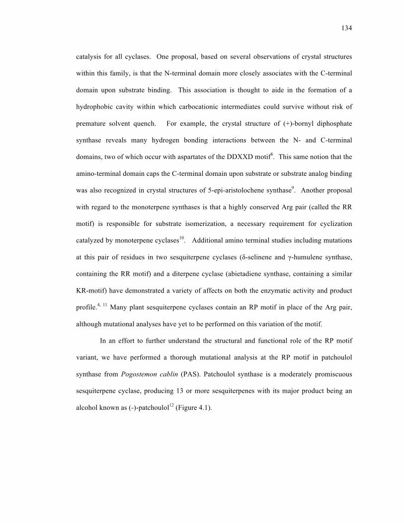
134
catalysis for all cyclases. One proposal, based on several observations of crystal structures
within this family, is that the N-terminal domain more closely associates with the C-terminal
domain upon substrate binding. This association is thought to aide in the formation of a
hydrophobic cavity within which carbocationic intermediates could survive without risk of
premature solvent quench. For example, the crystal structure of (+)-bornyl diphosphate
synthase reveals many hydrogen bonding interactions between the N- and C-terminal
domains, two of which occur with aspartates of the DDXXD motif8. This same notion that the
amino-terminal domain caps the C-terminal domain upon substrate or substrate analog binding
was also recognized in crystal structures of 5-epi-aristolochene synthase9. Another proposal
with regard to the monoterpene synthases is that a highly conserved Arg pair (called the RR
motif) is responsible for substrate isomerization, a necessary requirement for cyclization
catalyzed by monoterpene cyclases10. Additional amino terminal studies including mutations
at this pair of residues in two sesquiterpene cyclases (δ-selinene and γ-humulene synthase,
containing the RR motif) and a diterpene cyclase (abietadiene synthase, containing a similar
KR-motif) have demonstrated a variety of affects on both the enzymatic activity and product
profile.4, 11 Many plant sesquiterpene cyclases contain an RP motif in place of the Arg pair,
although mutational analyses have yet to be performed on this variation of the motif.
In an effort to further understand the structural and functional role of the RP motif
variant, we have performed a thorough mutational analysis at the RP motif in patchoulol
synthase from Pogostemon cablin (PAS). Patchoulol synthase is a moderately promiscuous
sesquiterpene cyclase, producing 13 or more sesquiterpenes with its major product being an
alcohol known as (-)-patchoulol12 (Figure 4.1).
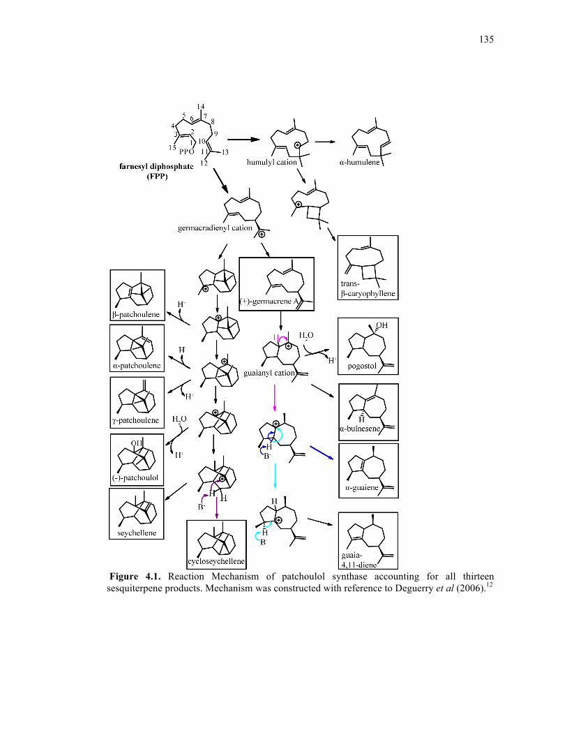
135
Figure 4.1. Reaction Mechanism of patchoulol synthase accounting for all thirteen sesquiterpene products. Mechanism was constructed with reference to Deguerry et al (2006).12
136
Given that the RP motif is somewhat conserved among the plant sesquiterpene
cyclases, we have also mutated these positions in three other sesquiterpene synthases (5-epi-
aristolochene synthase from Nicotiana tabacum: TEAS; amorpha-4,11-diene synthase from
Artemisia annua: ADS; premnaspirodiene synthase from Hyoscyamus muticus: HPS) in an
attempt to define a broader role for this variant of the motif. Recently published crystal
structures of certain TEAS mutants13 reveal important interactions between the RP motif and
other regions of the protein, which are assumed to be essential for normal RP motif function.
This analysis reveals a specific and cooperative structural and functional role for the RP motif
in PAS, and also demonstrates that the motif has varying degrees of importance in other plant
sesquiterpene cyclases.
4.3. RESULTS AND DISCUSSION
4.3.1. RP motif in PAS
Wild type PAS utilizes FPP to synthesize 13 identifiable products that can be grouped
into four categories according to their mechanistic complexity: 1) Mechanistically simple
products, such as β-elemene (a cope-rearrangement of germacrene A14) β-caryophyllene, and
α-humulene. These molecules have carbocation precursors that are easily created through
cyclization followed by only one or two rearrangements before the final elimination step. 2)
Mechanistically intermediate products derived from the guaianyl cation, including pogostol, α-
guaiene, α-bulnesene, and guaia-4,11-diene. The sesquiterpene α-bulnesene is considered the
simplest of this group because its formation requires no further hydride rearrangements. 3)
Mechanistically intermediate products derived from the patchoulene cation, including α-, β-,
and γ-patchoulene. 4) Mechanistically complex products such as seychellene,
137
cycloseychellene and (-)-patchoulol, which are derived from the most intricate series of
catalytic events (Figure 4.1).
In order to initially assess the effect of amino-terminal truncation on PAS product
profile, a series of amino terminal deletions were constructed, including N6, N10, N15, N16,
and N17, which represent 5, 9, 14, 15, and 16 amino acids deleted from the amino terminus,
respectively (Figure 4.2). Of these constructs, only N17 significantly alters the product profile
and is unable to produce the majority of products including (-)-patchoulol. Notably, N15,
which is two amino acids longer than N17, completely restores the product distribution
(Figure 4.3, Table 4.1, Table 4.2). The two amino acids that appear to play a role in this
restoration are Arg15-Pro16 (termed the RP motif).
Figure 4.2. Truncation mutant constructs in patchoulol synthase. The lower sequence represents the full PAS sequence, shown above this in the box the amino terminal sequences of each construct; truncated portions of each construct are shown in gray.
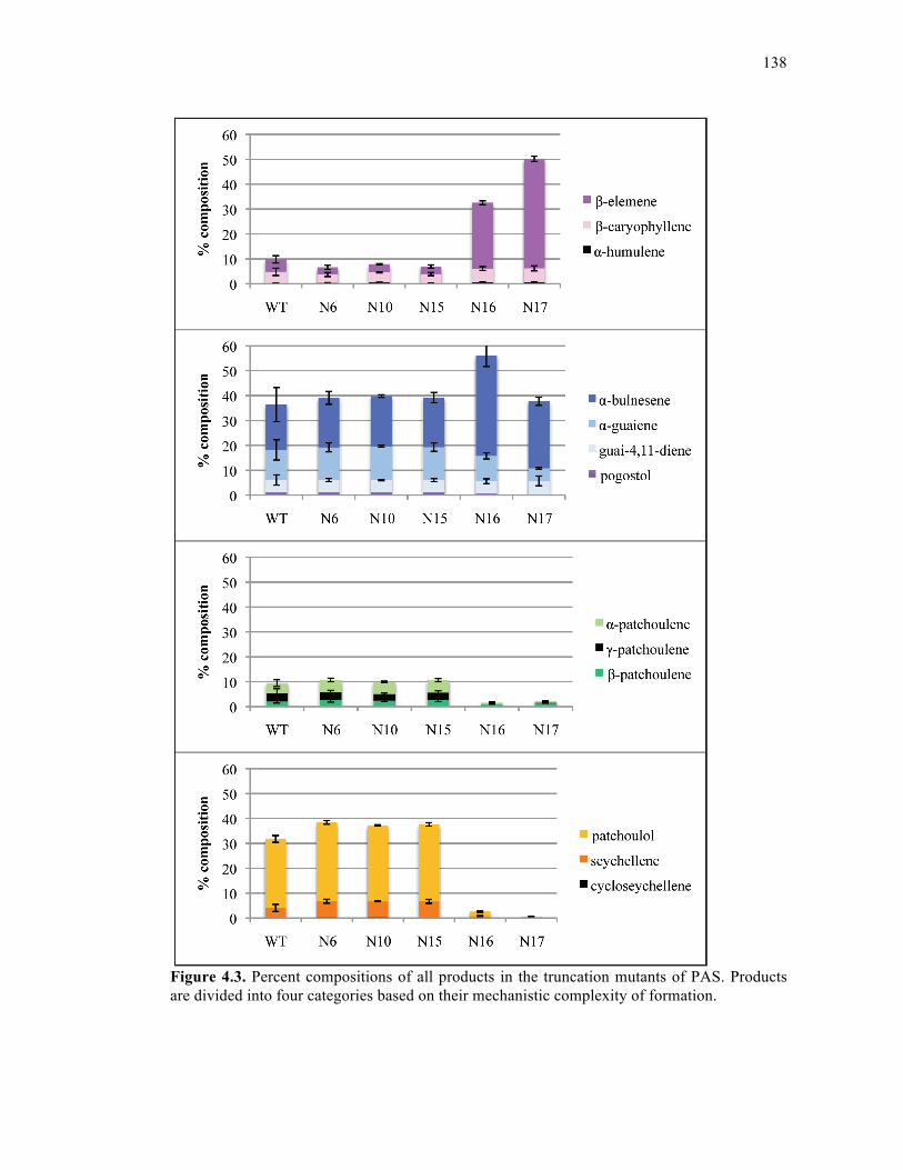
138
Figure 4.3. Percent compositions of all products in the truncation mutants of PAS. Products are divided into four categories based on their mechanistic complexity of formation.
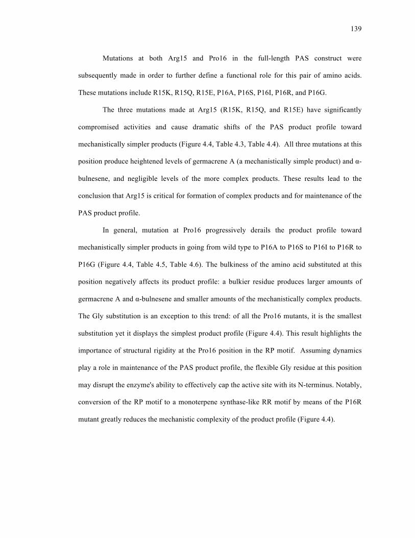
139
Mutations at both Arg15 and Pro16 in the full-length PAS construct were
subsequently made in order to further define a functional role for this pair of amino acids.
These mutations include R15K, R15Q, R15E, P16A, P16S, P16I, P16R, and P16G.
The three mutations made at Arg15 (R15K, R15Q, and R15E) have significantly
compromised activities and cause dramatic shifts of the PAS product profile toward
mechanistically simpler products (Figure 4.4, Table 4.3, Table 4.4). All three mutations at this
position produce heightened levels of germacrene A (a mechanistically simple product) and α-
bulnesene, and negligible levels of the more complex products. These results lead to the
conclusion that Arg15 is critical for formation of complex products and for maintenance of the
PAS product profile.
In general, mutation at Pro16 progressively derails the product profile toward
mechanistically simpler products in going from wild type to P16A to P16S to P16I to P16R to
P16G (Figure 4.4, Table 4.5, Table 4.6). The bulkiness of the amino acid substituted at this
position negatively affects its product profile: a bulkier residue produces larger amounts of
germacrene A and α-bulnesene and smaller amounts of the mechanistically complex products.
The Gly substitution is an exception to this trend: of all the Pro16 mutants, it is the smallest
substitution yet it displays the simplest product profile (Figure 4.4). This result highlights the
importance of structural rigidity at the Pro16 position in the RP motif. Assuming dynamics
play a role in maintenance of the PAS product profile, the flexible Gly residue at this position
may disrupt the enzyme's ability to effectively cap the active site with its N-terminus. Notably,
conversion of the RP motif to a monoterpene synthase-like RR motif by means of the P16R
mutant greatly reduces the mechanistic complexity of the product profile (Figure 4.4).
140
Figure 4.4. Percent compositions of all products in PAS RP motif mutants. Products are divided into four categories based on their mechanistic complexity of formation.
141
4.3.2. RP motif mutants in other sesquiterpene cyclases
Three other sesquiterpene cyclases were mutated at their RP motifs, including
premnaspirodiene synthase from Hyoscyamus muticus (HPS), amorpha-4,11-diene synthase
from Artemisia annua (ADS), and 5-epi-aristolochene synthase from Nicotiana tabacum
(TEAS). These cyclases are highly specific, producing one major product and many minor
products at very low, sometimes undetectable levels15, 16. Initially, all three enzymes were
engineered to transform their RP motif into an RR motif with a single point mutation at the
Pro position. HPS P18R and ADS P11R maintain wild type levels of their major products,
with values of premnaspirodiene at 93.7(±0.5)% and 93.4(±0.3)% for HPS WT and HPS
P11R, respectively, and values of amorpha-4,11-diene at 84.3(±3.2)% and 85.0(±1.6) for ADS
WT and ADS P11R, respectively (Table 4.11, Table 4.12, Table 4.13, Table 4.14). There are
no accompanying variations in percent composition of minor products for either enzyme.
TEAS P16R, however, does show slight changes in its product profile compared to wild type,
but only with respect to two of its products: in going from wild type to P16R, the %
composition of 5-epi-aristolochene decreases from 82.6(±0.4)% to 79.6(±0.5)%, while the %
composition of germacrene-A increases from 1.7(±0.1)% to 4.0(±0.2)% (Figure 4.7, Table 4.9,
Table 4.10). These product profile changes observed for the TEAS mutant are easily
accounted for compared to product profile changes for the PAS mutants discussed above.
Crystal structure data from TEAS enabled the identification of another feature that
may be important for amino-terminal capping. The original crystal structure of wild type
TEAS was solved in 1997, however the first 16 amino acids are missing from the PDB
coordinates, likely due to a dynamic amino-terminus9. A more recently published crystal
structure of wild type TEAS complexed with the non-hydrolyzable FPP analog trans,2-fluoro-
farnesyl diphosphate (2F-FPP) shows several more residues built into the N-terminus,
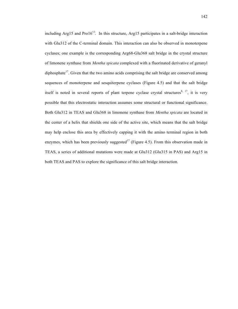
142
including Arg15 and Pro1613. In this structure, Arg15 participates in a salt-bridge interaction
with Glu312 of the C-terminal domain. This interaction can also be observed in monoterpene
cyclases; one example is the corresponding Arg68-Glu368 salt bridge in the crystal structure
of limonene synthase from Mentha spicata complexed with a fluorinated derivative of geranyl
diphosphate17. Given that the two amino acids comprising the salt bridge are conserved among
sequences of monoterpene and sesquiterpene cyclases (Figure 4.5) and that the salt bridge
itself is noted in several reports of plant terpene cyclase crystal structures8, 17, it is very
possible that this electrostatic interaction assumes some structural or functional significance.
Both Glu312 in TEAS and Glu368 in limonene synthase from Mentha spicata are located in
the center of a helix that shields one side of the active site, which means that the salt bridge
may help enclose this area by effectively capping it with the amino terminal region in both
enzymes, which has been previously suggested17 (Figure 4.5). From this observation made in
TEAS, a series of additional mutations were made at Glu312 (Glu315 in PAS) and Arg15 in
both TEAS and PAS to explore the significance of this salt bridge interaction.
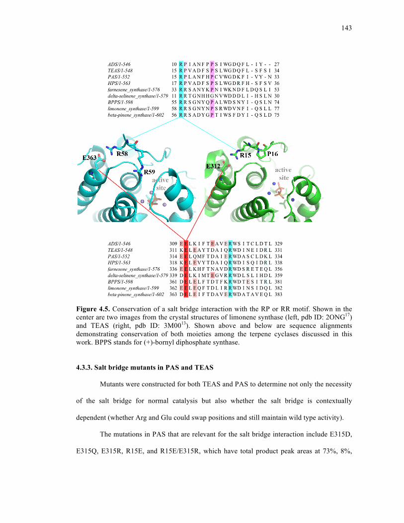
143
Figure 4.5. Conservation of a salt bridge interaction with the RP or RR motif. Shown in the center are two images from the crystal structures of limonene synthase (left, pdb ID: 2ONG17) and TEAS (right, pdb ID: 3M0013). Shown above and below are sequence alignments demonstrating conservation of both moieties among the terpene cyclases discussed in this work. BPPS stands for (+)-bornyl diphosphate synthase.
4.3.3. Salt bridge mutants in PAS and TEAS
Mutants were constructed for both TEAS and PAS to determine not only the necessity
of the salt bridge for normal catalysis but also whether the salt bridge is contextually
dependent (whether Arg and Glu could swap positions and still maintain wild type activity).
The mutations in PAS that are relevant for the salt bridge interaction include E315D,
E315Q, E315R, R15E, and R15E/E315R, which have total product peak areas at 73%, 8%,
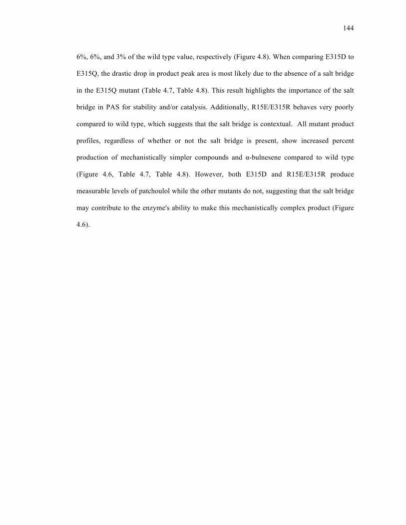
144
6%, 6%, and 3% of the wild type value, respectively (Figure 4.8). When comparing E315D to
E315Q, the drastic drop in product peak area is most likely due to the absence of a salt bridge
in the E315Q mutant (Table 4.7, Table 4.8). This result highlights the importance of the salt
bridge in PAS for stability and/or catalysis. Additionally, R15E/E315R behaves very poorly
compared to wild type, which suggests that the salt bridge is contextual. All mutant product
profiles, regardless of whether or not the salt bridge is present, show increased percent
production of mechanistically simpler compounds and α-bulnesene compared to wild type
(Figure 4.6, Table 4.7, Table 4.8). However, both E315D and R15E/E315R produce
measurable levels of patchoulol while the other mutants do not, suggesting that the salt bridge
may contribute to the enzyme's ability to make this mechanistically complex product (Figure
4.6).
145
Figure 4.6. Percent compositions of all products in PAS salt bridge mutants. Products are divided into four categories based on their mechanistic complexity of formation.
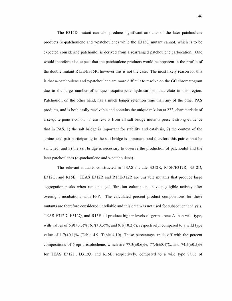
146
The E315D mutant can also produce significant amounts of the later patchoulene
products (α-patchoulene and γ-patchoulene) while the E315Q mutant cannot, which is to be
expected considering patchoulol is derived from a rearranged patchoulene carbocation. One
would therefore also expect that the patchoulene products would be apparent in the profile of
the double mutant R15E/E315R, however this is not the case. The most likely reason for this
is that α-patchoulene and γ-patchoulene are more difficult to resolve on the GC chromatogram
due to the large number of unique sesquiterpene hydrocarbons that elute in this region.
Patchoulol, on the other hand, has a much longer retention time than any of the other PAS
products, and is both easily resolvable and contains the unique m/z ion at 222, characteristic of
a sesquiterpene alcohol. These results from all salt bridge mutants present strong evidence
that in PAS, 1) the salt bridge is important for stability and catalysis, 2) the context of the
amino acid pair participating in the salt bridge is important, and therefore this pair cannot be
switched, and 3) the salt bridge is necessary to observe the production of patchoulol and the
later patchoulenes (α-patchoulene and γ-patchoulene).
The relevant mutants constructed in TEAS include E312R, R15E/E312R, E312D,
E312Q, and R15E. TEAS E312R and R15E/312R are unstable mutants that produce large
aggregation peaks when run on a gel filtration column and have negligible activity after
overnight incubations with FPP. The calculated percent product compositions for these
mutants are therefore considered unreliable and this data was not used for subsequent analysis.
TEAS E312D, E312Q, and R15E all produce higher levels of germacrene A than wild type,
with values of 6.9(±0.3)%, 6.7(±0.3)%, and 9.1(±0.2)%, respectively, compared to a wild type
value of 1.7(±0.1)% (Table 4.9, Table 4.10). These percentages trade off with the percent
compositions of 5-epi-aristolochene, which are 77.3(±0.6)%, 77.4(±0.4)%, and 74.5(±0.5)%
for TEAS E312D, D312Q, and R15E, respectively, compared to a wild type value of
147
82.6(±0.4) (Figure 4.7, Table 4.9, Table 4.10). Minimal and often insignificant variations are
observed between mutants and wild type for all remaining detectable minor products. In
addition to having matching product profiles, TEAS E312D and E312Q have total product
peak areas at 89% of the wild type value (Figure 4.8). It is therefore clear that unlike in PAS,
the absence of this salt bridge in TEAS does not affect product profile or activity. However,
the fact that both E312R and R15E/E312R are highly unstable and virtually inactive is an
indication that the context of the salt bridge is highly important.
Figure 4.7. Percent compositions of all products in TEAS mutants
148
4.3.4. Conclusions
From these results, it is clear the RP motif is crucial for normal activity in PAS. Any
mutation at Arg15 and progressively bulkier substitutions at Pro16 are detrimental toward
production of mechanistically complex PAS products. One of the reasons that Arg15 is
necessary for the maintenance of wild type activity is most likely due to an electrostatic
interaction that exists between this residue and Glu315, as also seen in crystal structures of
wild type TEAS complexed with fluoro-FPP analogs13, a crystal structure of limonene
synthase complexed with a fluoro-GPP analog17, and a crystal structure of (+)-bornyl
diphosphate synthase complexed with pyrophosphate8. Bulkier mutations at Pro16 produce
product profiles that are increasingly abundant in mechanistically simpler products such as
germacrene A and α-bulnesene, with the exception of P16G, which displays one of the
simplest product profiles. The behavior of the PAS P16G mutant suggests that the restricted
conformational space explored by Pro residues is important in an otherwise flexible amino
terminal segment. Therefore, in PAS, Arg15 and Pro16 cooperate to achieve dynamic
regulation of the amino-terminal region; the structural rigidity of Pro16 aides in the
positioning of Arg15 such that it can interact with a residue in the C-terminal domain of the
protein, thereby capping the active site with the amino-terminal region. This capping
mechanism probably helps shield reactive carbocation intermediates present in the active site
from bulk solvent throughout the course of the reaction. The rigidity of Pro16 in PAS is
analogous to the structural stabilization provided by an additional salt bridge observed in (+)-
bornyl diphosphate synthase between the second Arg in the RR motif (R56) and Asp355.8
There are undoubtedly other amino-terminal residues involved in hydrogen bonding,
van der Waals, and electrostatic interactions that contribute to amino-terminal "active site
capping" in plant terpene cyclases. However, the observation that these two residues can
149
restore the wild type product profile in going from the N17 truncation mutant to N15 is strong
evidence for the importance of these specific residues in the amino-terminal region of PAS.
Of the three other sesquiterpenes that have been mutated at their RP motifs (TEAS,
HPS, and ADS), only TEAS shows a mutant product profile that differs from wild type.
Unlike PAS, product fluctuations observed in the TEAS product profile are quite easily
accounted for: in TEAS P16R and in all active salt bridge mutants (TEAS E312D, TEAS
E312Q, and TEAS R15E), a loss in the major product 5-epi-aristolochene directly corresponds
to a gain in the mechanistically simple product germacrene A; these losses and gains are at
most 7-8%. In contrast, PAS P16R loses almost 20% patchoulol and gains 17% α-bulnesene
compared to wild type, in addition to loss and gain of other products. The fact that PAS P16R
appears to have a much more dramatic affect on the wild type product profile compared to
TEAS P16R suggests that perhaps enzyme promiscuity and RP motif functionality are
somehow correlated. This hypothesis also makes sense with the results observed for the two
other relatively non-promiscuous enzymes HPS and ADS, where mutant product profiles
either did not deviate from wild type or exhibit undetectable product derailment. However, in
previous work by Little et al (2002) on two promiscuous sesquiterpene cyclases, δ-selinene
synthase shows dramatic changes in product profile when mutated in this region while γ-
humulene synthase does not11. This result suggests that the level of promiscuity does not
necessarily correlate with RP motif functional trends.
In comparison to PAS, mutation of the salt bridge in TEAS does not appear to be as
important for maintenance of the product profile. This was expected to a certain extent due to
the fact that the TEAS RP motif mutants also do not alter the product profiles to a large
degree. However, Glu312 in TEAS does appear to be important for enzyme stability and
activity, given the E312R behaves poorly and the double mutant R1E/E312R (that reverses the

150
salt bridge) is very unstable and almost completely inactive. This result suggests that the salt
bridge between the Glu and Arg is indeed contextually dependent. For all active salt bridge
mutants, losses in the percentage of 5-epi-aristolochene correspond almost exactly to gains in
% production of germacrene A. This result may be obvious considering germacrene A is the
second most produced sesquiterpene in the TEAS profile. However, it means that mutations
in the RP motif or the salt bridge in TEAS result in derailment towards one very simple
monocyclic product, indicating premature carbocation release from the active site.
In PAS, given that the P16G mutant and mutants at the Arg15 position cause dramatic
product profile shifts, it appears that an increase in flexibility within this region has a similar
effect as a disruption in the salt bridge interaction. It is therefore likely that the dynamics of
this amino-terminal region of PAS are in part controlled by Pro16, which offers the structural
rigidity required to position Arg15 appropriately for the salt bridge interaction with Glu315.
Variations of the RR motif appear to exist in terpene cyclases that are thought to be the
ancestors of this family of enzymes. For example, abietadiene synthase, a diterpene cyclases
from Artemisia annua, contains a KR-motif that is located in a similar position as the RR
motif in monoterpene cyclases. When both residues are mutated to Ala, this enzyme can no
longer efficiently perform the ionization-dependent reaction, although there is no report of
product derailment18. The fact that this motif is present in a cyclase that is thought to be
ancestral to present day terpene cyclases is an indication of its rooted importance within the
family. The problem is indeed more complex than it looks, because some diterpene cyclases,
such as levopimaradiene synthase from Ginkgo biloba, do not contain this motif.
4.4. METHODS
4.4.1. Mutant Construction, Overexpression, and purification
151
All truncation mutants and point mutations were constructed using the QuikChange
protocol with PfuTurbo® DNA Polymerase (Stratagene) together with a 7 min PCR extension
time. The plasmid pHis9Gateway (a pet28-based gateway vector containing an N-temrinal
nine-histidine tag) containing the mutated terpene cyclase gene insert was transformed into E.
coli Bl21(DE3) competent cells (Novagen). One colony was grown in LB media (75 ml)
overnight at 37°C, 25 ml of the overnight culture was transferred into one liter of TB media
and grown at 37°C until an OD600 of 1.2. Isopropyl-β-D-thiogalactoside (0.2 mM final
concentration) was then added, cells were shaken for 5-6 hours at 22°C, harvested by
centrifugation and lysed using lysis buffer (50 mM Tris-HCl, pH 8.0, 500 mM NaCl, 20 mM
imidazole, 1% (v/v) Tween-20, 10% (v/v) glycerol, 10 mM 2-mercaptoethanol) containing
lysozyme (1 mg ml-1). The lysate was stirred at 4°C for 1 hr, sonicated, and centrifuged at
21,000 rpm for 45 min at 4°C. The supernatant was loaded onto a column containing Ni-NTA
agarose S3 resin (Qiagen), washed with lysis buffer and wash buffer (lysis buffer without
Tween- 20), and then eluted with elution buffer (wash buffer containing 250 mM imidazole).
The protein was digested with thrombin overnight during dialysis in buffer (50 mM Tris-HCl,
pH 8.0, 100 mM NaCl) containing 10 mM 2-mercaptoethanol. The dialyzed solution was
passed through a column containing Benzamidine Sepharose 4 Fast Flow (high sub) (GE
Healthcare) and Ni-NTA agarose. The eluant concentrated and passed through a HiLoad™
16/16 Superdex™ 200 prep grade (GE Healthcare) gel filtration column using dialysis buffer
containing 2 mM DTT. IPK fractions were combined, concentrated, and frozen at –80°C.
4.4.2. Specific activity measurements and product profile quantification by GC-MS
All wild type and mutant terpene cyclases samples were prepared in triplicate using
the vial assay protocol.19 1µM enzyme was injected into a 500µl aqueous layer containing
152
10mM MgCl2, 100µM FPP, and a 3-component buffer system (25 mM 2-(N-
morpholino)ethanesulfonic acid (MES), 50 mM Tris, and 25 mM 3-
(cyclohexylamino)propanesulfonic acid (CAPS) at pH 7.0). The aqueous layer was
immediately overlayed with an equal volume of ethyl acetate and the reactions were incubated
overnight, vortexed, and then run on the Gas Chromatograph Mass Spectrometer (GCMS)
using a previous established protocol13.
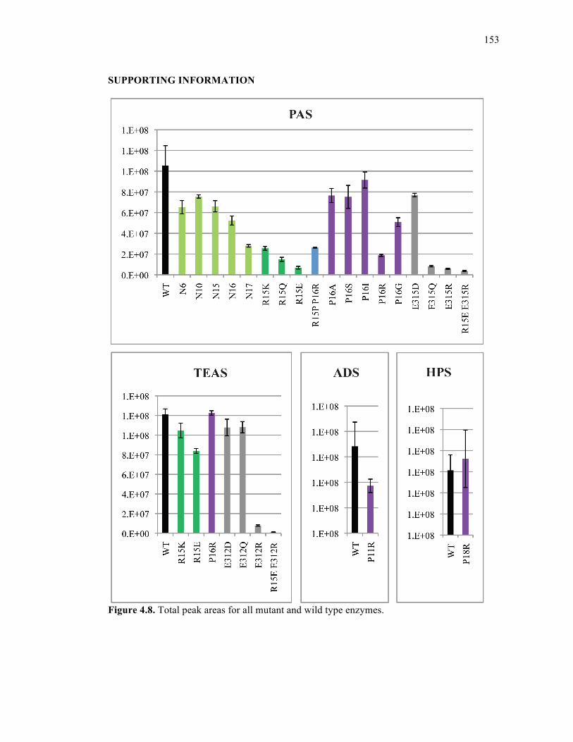
153
SUPPORTING INFORMATION
Figure 4.8. Total peak areas for all mutant and wild type enzymes.
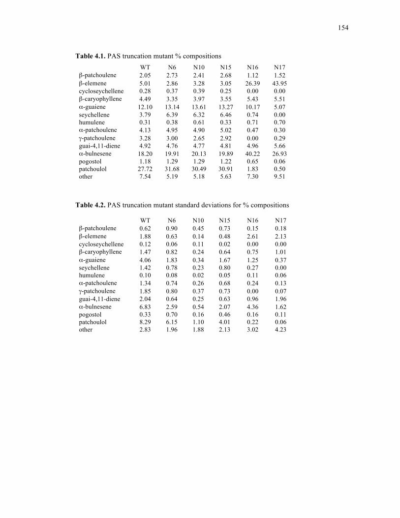
154
Table 4.1. PAS truncation mutant % compositions
Table 4.2. PAS truncation mutant standard deviations for % compositions
WT N6 N10 N15 N16 N17 β-patchoulene 2.05 2.73 2.41 2.68 1.12 1.52 β-elemene 5.01 2.86 3.28 3.05 26.39 43.95 cycloseychellene 0.28 0.37 0.39 0.25 0.00 0.00 β-caryophyllene 4.49 3.35 3.97 3.55 5.43 5.51 α-guaiene 12.10 13.14 13.61 13.27 10.17 5.07 seychellene 3.79 6.39 6.32 6.46 0.74 0.00 humulene 0.31 0.38 0.61 0.33 0.71 0.70 α-patchoulene 4.13 4.95 4.90 5.02 0.47 0.30 γ-patchoulene 3.28 3.00 2.65 2.92 0.00 0.29 guai-4,11-diene 4.92 4.76 4.77 4.81 4.96 5.66 α-bulnesene 18.20 19.91 20.13 19.89 40.22 26.93 pogostol 1.18 1.29 1.29 1.22 0.65 0.06 patchoulol 27.72 31.68 30.49 30.91 1.83 0.50 other 7.54 5.19 5.18 5.63 7.30 9.51
WT N6 N10 N15 N16 N17 β-patchoulene 0.62 0.90 0.45 0.73 0.15 0.18 β-elemene 1.88 0.63 0.14 0.48 2.61 2.13 cycloseychellene 0.12 0.06 0.11 0.02 0.00 0.00 β-caryophyllene 1.47 0.82 0.24 0.64 0.75 1.01 α-guaiene 4.06 1.83 0.34 1.67 1.25 0.37 seychellene 1.42 0.78 0.23 0.80 0.27 0.00 humulene 0.10 0.08 0.02 0.05 0.11 0.06 α-patchoulene 1.34 0.74 0.26 0.68 0.24 0.13 γ-patchoulene 1.85 0.80 0.37 0.73 0.00 0.07 guai-4,11-diene 2.04 0.64 0.25 0.63 0.96 1.96 α-bulnesene 6.83 2.59 0.54 2.07 4.36 1.62 pogostol 0.33 0.70 0.16 0.46 0.16 0.11 patchoulol 8.29 6.15 1.10 4.01 0.22 0.06 other 2.83 1.96 1.88 2.13 3.02 4.23
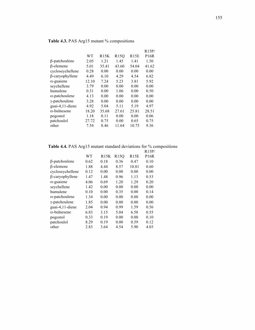
155
Table 4.3. PAS Arg15 mutant % compositions
Table 4.4. PAS Arg15 mutant standard deviations for % compositions
WT R15K R15Q R15E R15P/ P16R
β-patchoulene 2.05 1.21 1.45 1.41 1.50 β-elemene 5.01 35.41 43.60 54.04 41.62 cycloseychellene 0.28 0.00 0.00 0.00 0.00 β-caryophyllene 4.49 6.10 4.29 4.54 6.82 α-guaiene 12.10 7.24 5.23 3.81 5.92 seychellene 3.79 0.00 0.00 0.00 0.00 humulene 0.31 0.00 1.06 0.00 0.50 α-patchoulene 4.13 0.00 0.00 0.00 0.00 γ-patchoulene 3.28 0.00 0.00 0.00 0.00 guai-4,11-diene 4.92 5.04 5.11 5.19 4.97 α-bulnesene 18.20 35.68 27.61 25.81 28.51 pogostol 1.18 0.11 0.00 0.00 0.06 patchoulol 27.72 0.75 0.00 0.65 0.75 other 7.54 8.46 11.64 10.75 9.36
WT R15K R15Q R15E R15P/ P16R
β-patchoulene 0.62 0.18 0.36 0.47 0.10 β-elemene 1.88 4.44 8.57 10.81 0.60 cycloseychellene 0.12 0.00 0.00 0.00 0.00 β-caryophyllene 1.47 1.48 0.96 1.13 0.53 α-guaiene 4.06 0.69 1.20 1.29 0.20 seychellene 1.42 0.00 0.00 0.00 0.00 humulene 0.10 0.00 0.35 0.00 0.14 α-patchoulene 1.34 0.00 0.00 0.00 0.00 γ-patchoulene 1.85 0.00 0.00 0.00 0.00 guai-4,11-diene 2.04 0.94 0.99 1.59 0.56 α-bulnesene 6.83 3.15 5.04 6.58 0.55 pogostol 0.33 0.19 0.00 0.00 0.10 patchoulol 8.29 0.19 0.00 0.59 0.12 other 2.83 3.64 4.54 5.90 4.03
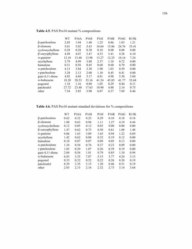
156
Table 4.5. PAS Pro16 mutant % compositions
Table 4.6. PAS Pro16 mutant standard deviations for % compositions
WT P16A P16S P16I P16R P16G R15K β-patchoulene 2.05 1.94 1.40 1.25 0.86 1.03 1.21 β-elemene 5.01 5.02 5.43 10.64 15.86 24.76 35.41 cycloseychellene 0.28 0.28 0.30 0.18 0.00 0.00 0.00 β-caryophyllene 4.49 4.07 3.47 4.53 5.41 4.28 6.10 α-guaiene 12.10 13.48 13.98 13.27 12.25 10.16 7.24 seychellene 3.79 4.99 3.88 2.37 1.18 0.72 0.00 humulene 0.31 0.56 0.45 0.68 0.60 0.70 0.00 α-patchoulene 4.13 3.84 3.30 1.90 1.03 0.59 0.00 γ-patchoulene 3.28 2.13 2.08 1.10 0.45 0.41 0.00 guai-4,11-diene 4.92 4.68 5.17 4.81 4.98 5.30 5.04 α-bulnesene 18.20 28.53 35.16 41.24 45.85 41.77 35.68 pogostol 1.18 1.16 0.88 1.05 0.29 0.44 0.11 patchoulol 27.72 23.48 17.63 10.90 4.88 2.14 0.75 other 7.54 5.85 5.90 6.07 6.37 7.69 8.46
WT P16A P16S P16I P16R P16G R15K β-patchoulene 0.62 0.32 0.23 0.29 0.10 0.18 0.18 β-elemene 1.88 0.63 0.96 1.11 1.27 4.19 4.44 cycloseychellene 0.12 0.05 0.12 0.03 0.00 0.00 0.00 β-caryophyllene 1.47 0.62 0.73 0.50 0.81 1.08 1.48 α-guaiene 4.06 1.63 3.09 1.65 0.94 1.22 0.69 seychellene 1.42 0.62 0.88 0.32 0.19 0.12 0.00 humulene 0.10 0.07 0.07 0.09 0.05 0.13 0.00 α-patchoulene 1.34 0.54 0.76 0.27 0.23 0.09 0.00 γ-patchoulene 1.85 0.29 1.07 0.26 0.39 0.19 0.00 guai-4,11-diene 2.04 0.56 1.81 0.79 0.85 1.10 0.94 α-bulnesene 6.83 3.35 7.87 5.15 3.77 4.24 3.15 pogostol 0.33 0.32 0.52 0.22 0.26 0.30 0.19 patchoulol 8.29 3.35 3.15 1.20 0.46 0.51 0.19 other 2.83 2.15 2.16 2.32 2.73 3.14 3.64

157
Table 4.7. PAS salt bridge mutant % compositions
Table 4.8. PAS salt bridge mutant standard deviations of % compositions
WT E315D E315Q E315R
R15E/ E315R
β-patchoulene 2.05 1.08 1.35 2.14 2.01 β-elemene 5.01 19.86 16.25 51.21 51.14 cycloseychellene 0.28 0.15 0.00 0.00 0.00 β-caryophyllene 4.49 4.33 1.97 1.38 1.87 α-guaiene 12.10 10.81 13.96 0.46 0.00 seychellene 3.79 1.29 0.00 0.00 0.00 humulene 0.31 0.77 0.00 0.00 0.00 α-patchoulene 4.13 0.95 0.00 0.00 0.00 γ-patchoulene 3.28 0.72 0.00 0.00 0.00 guai-4,11-diene 4.92 4.54 6.54 6.20 5.45 α-bulnesene 18.20 42.64 44.61 24.97 21.71 pogostol 1.18 0.79 0.22 0.00 0.00 patchoulol 27.72 5.45 1.60 0.00 2.74 other 7.54 6.62 13.50 13.63 15.07
WT E315D E315Q E315R
R15E/ E315R
β-patchoulene 0.62 0.05 0.18 0.68 0.43 β-elemene 1.88 0.59 1.77 5.36 12.29 cycloseychellene 0.12 0.00 0.00 0.00 0.00 β-caryophyllene 1.47 0.18 0.54 0.19 0.59 α-guaiene 4.06 0.35 2.50 0.07 0.00 seychellene 1.42 0.06 0.00 0.00 0.00 humulene 0.10 0.03 0.00 0.00 0.00 α-patchoulene 1.34 0.07 0.00 0.00 0.00 γ-patchoulene 1.85 0.08 0.00 0.00 0.00 guai-4,11-diene 2.04 0.65 2.41 1.44 2.11 α-bulnesene 6.83 1.70 15.58 2.14 2.84 pogostol 0.33 0.18 0.39 0.00 0.00 patchoulol 8.29 0.36 2.13 0.00 0.67 other 2.83 2.64 7.09 7.89 9.14
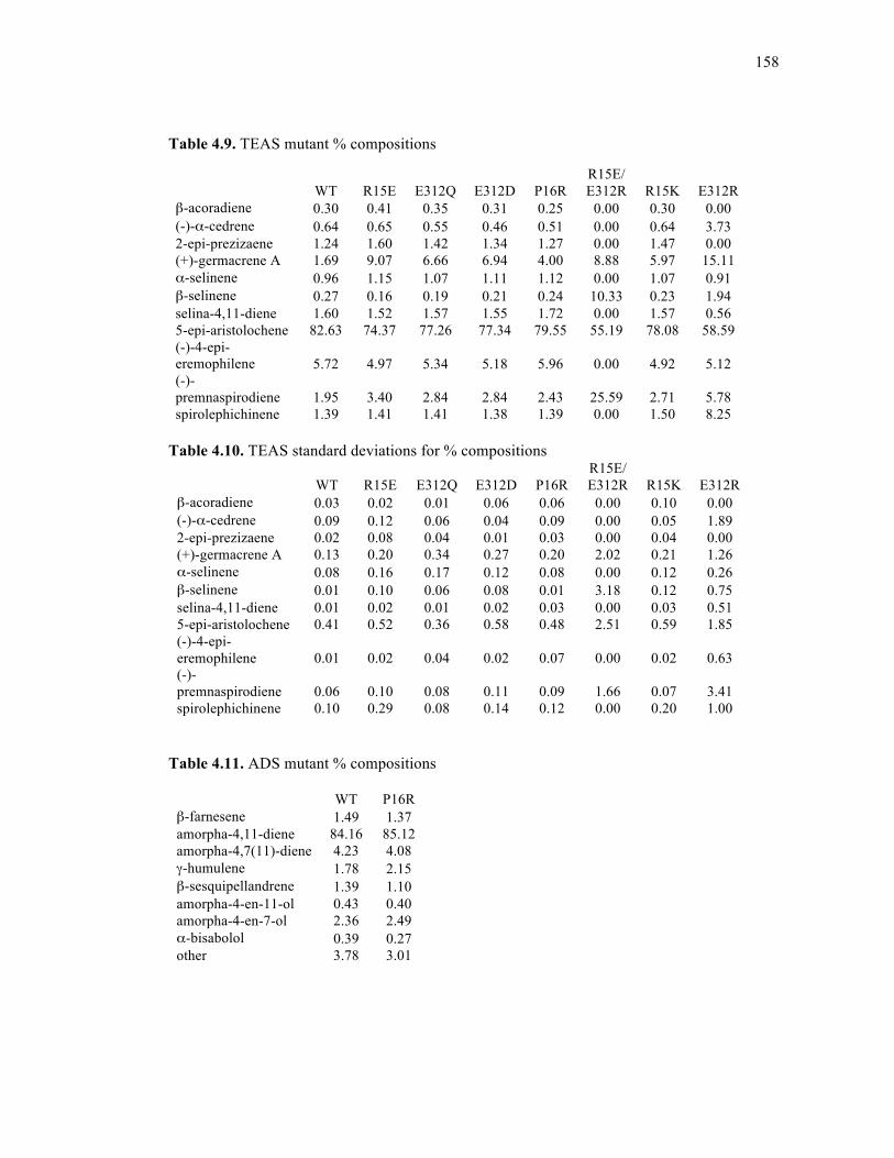
158
Table 4.9. TEAS mutant % compositions
Table 4.10. TEAS standard deviations for % compositions
Table 4.11. ADS mutant % compositions
WT P16R β-farnesene 1.49 1.37 amorpha-4,11-diene 84.16 85.12 amorpha-4,7(11)-diene 4.23 4.08 γ-humulene 1.78 2.15 β-sesquipellandrene 1.39 1.10 amorpha-4-en-11-ol 0.43 0.40 amorpha-4-en-7-ol 2.36 2.49 α-bisabolol 0.39 0.27 other 3.78 3.01
WT R15E E312Q E312D P16R R15E/ E312R R15K E312R
β-acoradiene 0.30 0.41 0.35 0.31 0.25 0.00 0.30 0.00 (-)-α-cedrene 0.64 0.65 0.55 0.46 0.51 0.00 0.64 3.73 2-epi-prezizaene 1.24 1.60 1.42 1.34 1.27 0.00 1.47 0.00 (+)-germacrene A 1.69 9.07 6.66 6.94 4.00 8.88 5.97 15.11 α-selinene 0.96 1.15 1.07 1.11 1.12 0.00 1.07 0.91 β-selinene 0.27 0.16 0.19 0.21 0.24 10.33 0.23 1.94 selina-4,11-diene 1.60 1.52 1.57 1.55 1.72 0.00 1.57 0.56 5-epi-aristolochene 82.63 74.37 77.26 77.34 79.55 55.19 78.08 58.59 (-)-4-epi-eremophilene 5.72 4.97 5.34 5.18 5.96 0.00 4.92 5.12 (-)-premnaspirodiene 1.95 3.40 2.84 2.84 2.43 25.59 2.71 5.78 spirolephichinene 1.39 1.41 1.41 1.38 1.39 0.00 1.50 8.25
WT R15E E312Q E312D P16R R15E/ E312R R15K E312R
β-acoradiene 0.03 0.02 0.01 0.06 0.06 0.00 0.10 0.00 (-)-α-cedrene 0.09 0.12 0.06 0.04 0.09 0.00 0.05 1.89 2-epi-prezizaene 0.02 0.08 0.04 0.01 0.03 0.00 0.04 0.00 (+)-germacrene A 0.13 0.20 0.34 0.27 0.20 2.02 0.21 1.26 α-selinene 0.08 0.16 0.17 0.12 0.08 0.00 0.12 0.26 β-selinene 0.01 0.10 0.06 0.08 0.01 3.18 0.12 0.75 selina-4,11-diene 0.01 0.02 0.01 0.02 0.03 0.00 0.03 0.51 5-epi-aristolochene 0.41 0.52 0.36 0.58 0.48 2.51 0.59 1.85 (-)-4-epi-eremophilene 0.01 0.02 0.04 0.02 0.07 0.00 0.02 0.63 (-)-premnaspirodiene 0.06 0.10 0.08 0.11 0.09 1.66 0.07 3.41 spirolephichinene 0.10 0.29 0.08 0.14 0.12 0.00 0.20 1.00
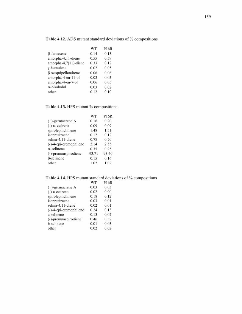
159
Table 4.12. ADS mutant standard deviations of % compositions
WT P16R β-farnesene 0.14 0.13 amorpha-4,11-diene 0.55 0.59 amorpha-4,7(11)-diene 0.33 0.12 γ-humulene 0.02 0.05 β-sesquipellandrene 0.06 0.06 amorpha-4-en-11-ol 0.03 0.03 amorpha-4-en-7-ol 0.06 0.05 α-bisabolol 0.03 0.02 other 0.12 0.10
Table 4.13. HPS mutant % compositions
WT P16R (+)-germacrene A 0.16 0.20 (-)-α-cedrene 0.09 0.09 spirolephichinene 1.48 1.51 isoprezizaene 0.12 0.12 selina-4,11-diene 0.78 0.70 (-)-4-epi-eremophilene 2.14 2.55 α-selinene 0.35 0.25 (-)-premnaspirodiene 93.71 93.40 β-selinene 0.15 0.16 other 1.02 1.02
Table 4.14. HPS mutant standard deviations of % compositions
WT P16R (+)-germacrene A 0.03 0.03 (-)-a-cedrene 0.02 0.00 spirolephichinene 0.18 0.12 isoprezizaene 0.03 0.01 selina-4,11-diene 0.02 0.01 (-)-4-epi-eremophilene 0.24 0.13 a-selinene 0.13 0.02 (-)-premnaspirodiene 0.46 0.32 b-selinene 0.01 0.03 other 0.02 0.02
160
ACKNOWLEDGEMENTS
The text of chapter 4, in part, is currently being prepared for submission for
publication of the material. Dellas, Nikki; Noel, Joseph P. I am the first author of this material.
All experiments were performed under the supervision of Joseph P. Noel.
REFERENCES
1. Gershenzon, J.; Dudareva, N., The function of terpene natural products in the natural world. Nature chemical biology 2007, 3 (7), 408-414.
2. Christianson, D. W., Structural biology and chemistry of the terpenoid cyclases.
Chemical reviews 2006, 106 (8), 3412-3442. 3. Wendt, K. U.; Schulz, G. E., Isoprenoid biosynthesis: manifold chemistry catalyzed
by similar enzymes. Structure (London, England : 1993) 1998, 6 (2), 127-133. 4. Peters, R. J.; Flory, J. E.; Jetter, R.; Ravn, M. M.; Lee, H. J.; Coates, R. M.; Croteau,
R. B., Abietadiene synthase from grand fir (Abies grandis): characterization and mechanism of action of the "pseudomature" recombinant enzyme. Biochemistry (John Wiley & Sons) 2000, 39 (50), 15592-15602.
5. Kawaide, H.; Sassa, T.; Kamiya, Y., Functional analysis of the two interacting cyclase
domains in ent-kaurene synthase from the fungus Phaeosphaeria sp. L487 and a comparison with cyclases from higher plants. The Journal of biological chemistry 2000, 275 (4), 2276-2280.
6. Toyomasu, T.; Kawaide, H.; Ishizaki, A.; Shinoda, S.; Otsuka, M.; Mitsuhashi, W.;
Sassa, T., Cloning of a full-length cDNA encoding ent-kaurene synthase from Gibberella fujikuroi: functional analysis of a bifunctional diterpene cyclase. Bioscience, biotechnology, and biochemistry 2000, 64 (3), 660-664.
7. Trapp, S. C.; Croteau, R. B., Genomic organization of plant terpene synthases and
molecular evolutionary implications. Genetics 2001, 158 (2), 811-832. 8. Whittington, D. A.; Wise, M. L.; Urbansky, M.; Coates, R. M.; Croteau, R. B.;
Christianson, D. W., Bornyl diphosphate synthase: structure and strategy for carbocation manipulation by a terpenoid cyclase. Proceedings of the National Academy of Sciences of the United States of America 2002, 99 (24), 15375-15380.
9. Starks, C. M.; Back, K.; Chappell, J.; Noel, J. P., Structural basis for cyclic terpene
biosynthesis by tobacco 5-epi-aristolochene synthase. Science 1997, 277 (5333), 1815-1820.
161
10. Williams, D. C.; McGarvey, D. J.; Katahira, E. J.; Croteau, R., Truncation of limonene synthase preprotein provides a fully active 'pseudomature' form of this monoterpene cyclase and reveals the function of the amino-terminal arginine pair. Biochemistry 1998, 37 (35), 12213-12220.
11. Little, D. B.; Croteau, R. B., Alteration of product formation by directed mutagenesis
and truncation of the multiple-product sesquiterpene synthases delta-selinene synthase and gamma-humulene synthase. Archives of Biochemistry and Biophysics 2002, 402 (1), 120-135.
12. Deguerry, F.; Pastore, L.; Wu, S.; Clark, A.; Chappell, J.; Schalk, M., The diverse
sesquiterpene profile of patchouli, Pogostemon cablin, is correlated with a limited number of sesquiterpene synthases. Archives of Biochemistry and Biophysics 2006, 454 (2), 123-136.
13. Noel, J. P.; Dellas, N.; Faraldos, J. A.; Zhao, M.; Hess, B. A., Jr.; Smentek, L.;
Coates, R. M.; O'Maille, P. E., Structural elucidation of cisoid and transoid cyclization pathways of a sesquiterpene synthase using 2-fluorofarnesyl diphosphates. ACS chemical biology 2010, 5 (4), 377-392.
14. Prosser, I.; Phillips, A. L.; Gittings, S.; Lewis, M. J.; Hooper, A. M.; Pickett, J. A.;
Beale, M. H., (+)-(10R)-Germacrene A synthase from goldenrod, Solidago canadensis; cDNA isolation, bacterial expression and functional analysis. Phytochemistry 2002, 60 (7), 691-702.
15. O'Maille, P. E.; Chappell, J.; Noel, J. P., Biosynthetic potential of sesquiterpene
synthases: Alternative products of tobacco 5-epi-aristolochene synthase. Archives of Biochemistry and Biophysics 2006, 448 (1-2), 73-82.
16. Mercke, P.; Bengtsson, M.; Bouwmeester, H. J.; Posthumus, M. A.; Brodelius, P. E.,
Molecular cloning, expression, and characterization of amorpha-4,11-diene synthase, a key enzyme of artemisinin biosynthesis in Artemisia annua L. Archives of Biochemistry and Biophysics 2000, 381 (2), 173-180.
17. Hyatt, D. C.; Youn, B.; Zhao, Y.; Santhamma, B.; Coates, R. M.; Croteau, R. B.;
Kang, C., Structure of limonene synthase, a simple model for terpenoid cyclase catalysis. Proceedings of the National Academy of Sciences of the United States of America 2007, 104 (13), 5360-5365.
18. Peters, R. J.; Carter, O. A.; Zhang, Y.; Matthews, B. W.; Croteau, R. B., Bifunctional
abietadiene synthase: mutual structural dependence of the active sites for protonation-initiated and ionization-initiated cyclizations. Biochemistry 2003, 42 (9), 2700-7.
19. O'Maille, P. E.; Chappell, J.; Noel, J. P., A single-vial analytical and quantitative gas
chromatography-mass spectrometry assay for terpene synthases. Analytical Biochemistry 2004, 335 (2), 210-217.
162
Chapter 5
Mutation of Archaeal Isopentenyl Phosphate Kinase Highlights Mechanism and Guides Phosphorylation of Additional Isoprenoid
Monophosphates
163
5.1. ABSTRACT
The biosynthesis of isopentenyl diphosphate (IPP) from either the mevalonate (MVA)
or the 1-deoxy-d-xylulose 5-phosphate (DXP) pathway provides the key metabolite for
primary and secondary isoprenoid biosynthesis. Isoprenoid metabolism plays crucial roles in
membrane stability, steroid biosynthesis, vitamin production, protein localization, defense and
communication, photoprotection, sugar transport, and glycoprotein biosynthesis. Recently, an
alternative branch of the MVA pathway was discovered in the archaeon Methanocaldococcus
jannaschii involving a small molecule kinase, isopentenyl phosphate kinase (IPK). IPK
belongs to the amino acid kinase (AAK) superfamily. In vitro, IPK phosphorylates isopentenyl
monophosphate (IP) in an ATP and Mg2+-dependent reaction producing IPP. Here, we
describe crystal structures of IPK from M. jannaschii refined to nominal resolutions of 2.0−2.8
Å. Notably, an active site histidine residue (His60) forms a hydrogen bond with the terminal
phosphate of both substrate and product. This His residue serves as a marker for a subset of
the AAK family that catalyzes phosphorylation of phosphate or phosphonate functional
groups; the larger family includes carboxyl-directed kinases, which lack this active site
residue. Using steady-state kinetic analysis of H60A, H60N, and H60Q mutants, the
protonated form of the Nε2 nitrogen of His60 was shown to be essential for catalysis, most
likely through hydrogen bond stabilization of the transition state accompanying
transphosphorylation. Moreover, the structures served as the starting point for the engineering
of IPK mutants capable of the chemoenzymatic synthesis of longer chain isoprenoid
diphosphates from monophosphate precursors.
164
5.2. INTRODUCTION
Isopentenyl diphosphate (IPP) and its isomeric partner dimethylallyl diphosphate
(DMAPP) are precursors for a diverse collection of primary and secondary isoprenoid
metabolites in all organisms. Following its formation, successive units of IPP are used
together either with DMAPP, formed by the action of types I or II IPP isomerases, or with the
IPP extended isoprenoid diphosphate chain, to biosynthesize C10, C15, or C20 oligoprenyl
diphosphates known as geranyl diphosphate (GPP), farnesyl diphosphate (FPP), and
geranylgeranyl diphosphate (GGPP), respectively, as well as larger isoprenoid diphosphates.
In plants and some microorganisms, GPP, FPP, and GGPP also serve as starting materials for
the biosynthesis of a large class of specialized and often cyclic terpene hydrocarbons1. FPP is
the most ubiquitous of the three isoprenoid diphosphate building blocks, as it resides at the
juncture of bifurcating branches of the general isoprenoid biosynthetic pathway leading to
both primary and secondary metabolites. Squalene, hopanoids, and steroids, serve as critical
components of cellular membranes and, in the case of steroids, also serve as transcription
modulators through nuclear hormone receptor engagement2, 3. Moreover, dolichols play
essential roles in N-glycosylation and membrane anchorage of sugars in eukaryotes and
archaea4. The 20-carbon GGPP molecule functions as the precursor to all carotenoids, the
latter of which provides photoprotection in plants, fungi, algae, bacteria, and some archaea5, 6.
Interestingly, GGPP also is a precursor to the isoprenoid-derived hydrocarbon moiety of lipids
that is present exclusively in archaea (see Koga et al (2007) for a review on archaeal lipids7).
Over the last two decades, two distinct pathways have been characterized that biosynthesize
IPP and DMAPP, namely the mevalonate (MVA) pathway and the more recently discovered
1-deoxy-d-xylulose 5-phosphate (DXP) pathway8. The MVA pathway is utilized by animals,
plants (cytosol), fungi, and certain bacteria, while the DXP pathway resides in plant plastids, a
165
number of eubacteria, cyanobacteria, and certain parasitic organisms9. In archaea, orthologs
for almost all of the genes encoding the MVA pathway are present except for two;
interestingly, the last two genes encoding phosphomevalonate kinase and
diphosphomevalonate decarboxylase appear to be missing from the genomes of almost all
archaea. For this reason, the isoprenoid pathway in archaea is referred to as “The Lost
Pathway”10. In 2006, Grochowski et al. discovered an enzyme and its associated gene in the
archaeon Methanocaldococcus jannaschii that belongs to the larger family of amino acid
kinases (AAK) but catalyzes the ATP-dependent phosphorylation of IP, thereby producing
IPP11. This enzyme, named isopentenyl phosphate kinase (IPK), appears to be a starting point
for the functional reconstruction of The Lost Pathway, representative of a completely
unexpected biosynthetic variation of the MVA pathway.
IPK shares significant sequence homology with proteins in the AAK superfamily
(Pf000696, Figure 5.1). Members of this family employ Mg2+−ATP to catalyze
phosphorylation of carboxylate, carbamate, phosphonate, or phosphate functional groups.
Here, crystal structures of IPK from M. jannaschii are presented in the ‘apo’ form and in
complex with substrate (IP) and product (IPP). These structures allow for rational mutagenesis
and biochemical analyses of residue(s) near the phosphate moiety and the isopentenyl tail of
IP, respectively. Mutation of a residue near the phosphate of IP demonstrates a key role for
His-directed hydrogen bonding in the phosphorylation of phosphate or phosphonate groups.
Mutation of residues near the isoprenyl moiety of IP establishes IPK as a starting point for
engineering the phosphorylation of alternative phosphate/phosphonate bearing small
molecules, including geranyl monophosphate (GP) and farnesyl monophosphate (FP). This
sets the stage for a multitude of applications in chemoenzymatic syntheses including
diphosphate analog synthesis, low cost radio-labeling of isoprenoid diphosphates with 32P or

166
35S containing β-phosphates, and the possible in vivo recycling of isoprenoid monophosphates
formed upon FPP up-regulation and degradation in heterologous hosts.
Figure 5.1. The amino acid kinase (AAK) family members. Isopentenyl phosphate kinase (IPK) reaction depicted across the top. Representative family members displayed from left to right: carbamate kinase (CK), aspartokinase (AK), glutamate-5-kinase (G5K), N-acetyl-l-glutamate kinase (NAGK), fosfomycin resistance kinase (FomA), and uridine monophosphate kinase (UMPK). The percent sequence identities relative to IPK are listed above each enzyme. Reactions shaded green utilize a phosphate or phosphonate phosphoryl acceptor, while the reactions shaded red utilize carbamate or carboxylate groups as phosphate acceptors.
5.3. RESULTS AND DISCUSSION
5.3.1. Three-dimensional architecture
IPK represents the newest member of the AAK superfamily to be structurally
determined. The overall fold, commonly referred to as the open αβα sandwich, was first
discovered in carbamate kinase from E. faecalis12. IPK is architecturally most similar to
fosfomycin resistance kinase (FomA) from S. wedmorensis with a root-mean-square deviation
(rmsd) of 2.0 Å for superimposed backbone atoms (NH−Cα−C) and a sequence identity of
22%. However, it shares the highest sequence identity, 25%, with uridine monophosphate
167
kinase (UMPK) from A. fulgidus. Two subdivisions of the AAK superfamily exist, referred to
here as the phosphate and carboxylate subdivisions, respectively. Enzymes in the phosphate
subdivision, including IPK, FomA, and UMPK, catalyze phosphorylation of a phosphate or
phosphonate moiety. Enzymes in the carboxylate subdivision, including carbamate kinase, N-
acetylglutamate kinase (NAGK), aspartokinase, and glutamate-5-kinase, catalyze
phosphorylation of a carbamate or carboxylate group (Figure 5.1).
Like all other AAKs, IPK adopts a dimeric quaternary structure, and each monomer
folds into structurally distinct N- and C-terminal domains (Figure 5.2). The N-terminal
domain, spanning residues 1−171, binds the nucleophilic phosphate group (IP in IPK). The C-
terminal domain, spanning residues 171−260, coordinates a Mg2+ ion and binds the phosphate
donor ATP. Although all attempts to crystallize M. jannaschii IPK with ATP, ADP,
AMPPNP, and a variety of other analogs have thus far been unsuccessful, the location of the
nucleotide-binding site is structurally conserved among all family members, affording a
reasonable model for ATP binding. Each monomer of IPK contains 16 β-strands, 8 α-helices,
and 1 310 helix. The core of the open αβα sandwich, represented by 8 β-strands, β14, β16, β15,
β11, β1, β2, β8, and β5, resides between 4 α-helices on one side, αF, αH, αE, and αD, and 3 on
the other, αG, αA, and αC (Figure 5.2). Four β-hairpins, one α-helix, and one 310 helix (η1)
decorate the periphery of the central β-sandwich. Three of the hairpins, β3−β4, β6−β7, and
β9−β10, lie in the N-terminal domain and surround the back wall and one side of the IP
binding pocket; the αB helix covers the remaining side of the isopentenyl-binding surface. The
fourth β-hairpin, β12−β13, located within the C-terminal domain, resides in close proximity to
the expected location for the adenine ring of ATP. Finally, the 310 helix links one end of the
central β5 strand and the β6−β7 hairpin. Although the β1−αA junction is depicted as a loop in
Figure 5.2, it also adopts a helical structure in some cases.
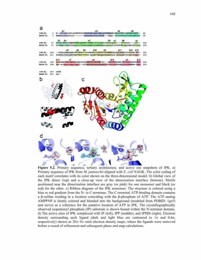
168
Figure 5.2. Primary sequence, tertiary architecture, and active site snapshots of IPK. a) Primary sequence of IPK from M. jannaschii aligned with E. coli NAGK. The color coding of each motif correlates with its color shown on the three-dimensional model. b) Global view of the IPK dimer (top) and a close-up view of the dimerization interface (bottom). Motifs positioned near the dimerization interface are gray (or pink) for one monomer and black (or red) for the other. c) Ribbon diagram of the IPK monomer. The structure is colored using a blue to red gradient from the N- to C-terminus. The C-terminal ATP-binding domain contains a β-sulfate residing in a location coinciding with the β-phosphate of ATP. The ATP analog AMPPNP is faintly colored and blended into the background (modeled from PDBID: 1gs5) and serves as a reference for the putative location of ATP in IPK. The crystallographically observed isopentenyl phosphate (IP) substrate is shown bound within the N-terminal domain. d) The active sites of IPK complexed with IP (left), IPP (middle), and IPPβS (right). Electron density surrounding each ligand (dark and light blue are contoured to 1σ and 0.6σ, respectively) shown as 2Fo−Fc omit electron density maps, where the ligands were removed before a round of refinement and subsequent phase and map calculations.
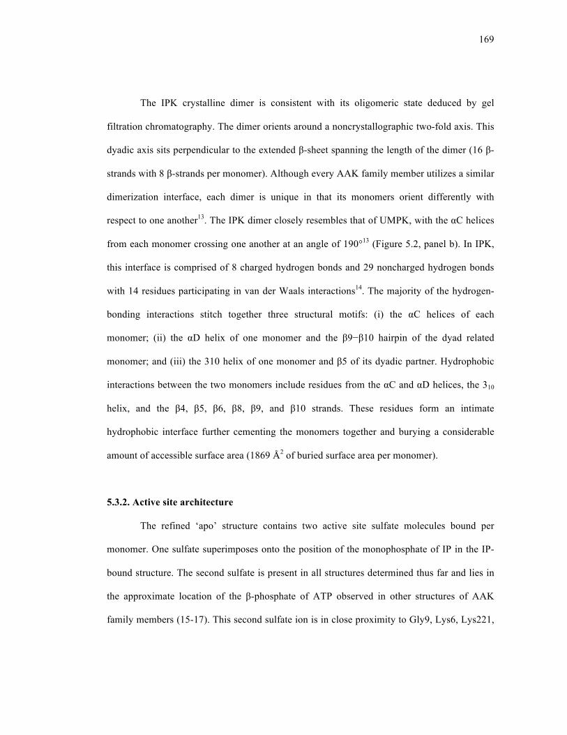
169
The IPK crystalline dimer is consistent with its oligomeric state deduced by gel
filtration chromatography. The dimer orients around a noncrystallographic two-fold axis. This
dyadic axis sits perpendicular to the extended β-sheet spanning the length of the dimer (16 β-
strands with 8 β-strands per monomer). Although every AAK family member utilizes a similar
dimerization interface, each dimer is unique in that its monomers orient differently with
respect to one another13. The IPK dimer closely resembles that of UMPK, with the αC helices
from each monomer crossing one another at an angle of 190°13 (Figure 5.2, panel b). In IPK,
this interface is comprised of 8 charged hydrogen bonds and 29 noncharged hydrogen bonds
with 14 residues participating in van der Waals interactions14. The majority of the hydrogen-
bonding interactions stitch together three structural motifs: (i) the αC helices of each
monomer; (ii) the αD helix of one monomer and the β9−β10 hairpin of the dyad related
monomer; and (iii) the 310 helix of one monomer and β5 of its dyadic partner. Hydrophobic
interactions between the two monomers include residues from the αC and αD helices, the 310
helix, and the β4, β5, β6, β8, β9, and β10 strands. These residues form an intimate
hydrophobic interface further cementing the monomers together and burying a considerable
amount of accessible surface area (1869 Å2 of buried surface area per monomer).
5.3.2. Active site architecture
The refined ‘apo’ structure contains two active site sulfate molecules bound per
monomer. One sulfate superimposes onto the position of the monophosphate of IP in the IP-
bound structure. The second sulfate is present in all structures determined thus far and lies in
the approximate location of the β-phosphate of ATP observed in other structures of AAK
family members (15-17). This second sulfate ion is in close proximity to Gly9, Lys6, Lys221,
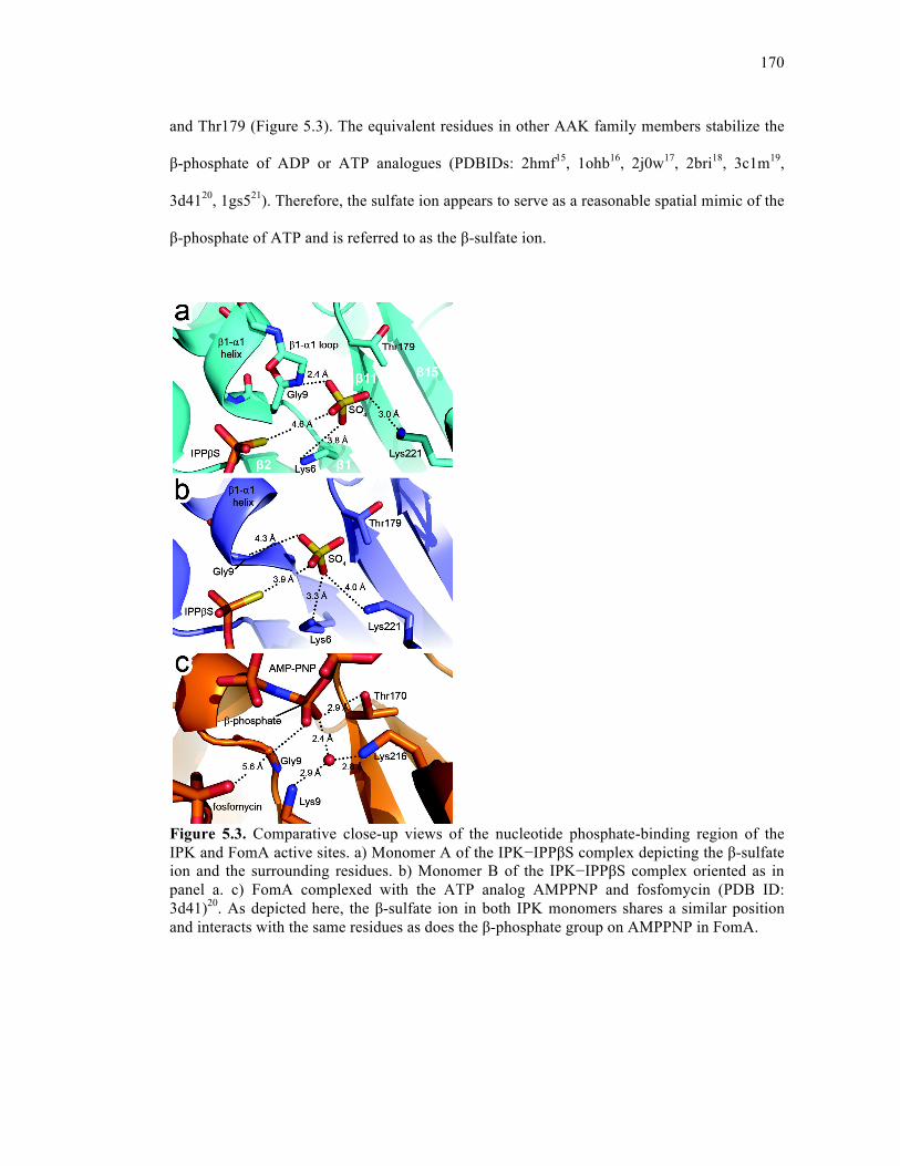
170
and Thr179 (Figure 5.3). The equivalent residues in other AAK family members stabilize the
β-phosphate of ADP or ATP analogues (PDBIDs: 2hmf15, 1ohb16, 2j0w17, 2bri18, 3c1m19,
3d4120, 1gs521). Therefore, the sulfate ion appears to serve as a reasonable spatial mimic of the
β-phosphate of ATP and is referred to as the β-sulfate ion.
Figure 5.3. Comparative close-up views of the nucleotide phosphate-binding region of the IPK and FomA active sites. a) Monomer A of the IPK−IPPβS complex depicting the β-sulfate ion and the surrounding residues. b) Monomer B of the IPK−IPPβS complex oriented as in panel a. c) FomA complexed with the ATP analog AMPPNP and fosfomycin (PDB ID: 3d41)20. As depicted here, the β-sulfate ion in both IPK monomers shares a similar position and interacts with the same residues as does the β-phosphate group on AMPPNP in FomA.

171
The structures of IPK in complex with IP and IPP define the secondary structural
elements comprising the IP-binding pocket and include the β2−αB glycine-rich loop, the αB
helix, the β3−β4 hairpin, the β4−αC loop, the N-terminal section of the αC helix, and the
β9−β10 hairpin (Figure 5.4, panel a). The β2−αB loop is one of two conserved glycine-rich
loops present throughout the AAK family. It is thought to contribute to charge neutralization
in the transition state during phosphoryl transfer16, 20. Notably, the orientation of the αB helix
is conserved only in the phosphate division of the AAK superfamily (including IPK, FomA,
and UMPK). In FomA, the αB helix orders when substrate is present but is otherwise
disordered20. In contrast, the αB helix in IPK is ordered in both ‘apo’- and IP-bound structures.
Of even more limited familial distribution, the β3−β4 hairpin is present only in IPK and
NAGK. In NAG-bound NAGK structures, the hairpin often exists in a closed conformation21,
22; in contrast, all structures of IPK reveal the motif in an open conformation. Regardless, the
hairpin may play a role in shielding the substrate-binding pocket from the surrounding solvent
in both enzymes.
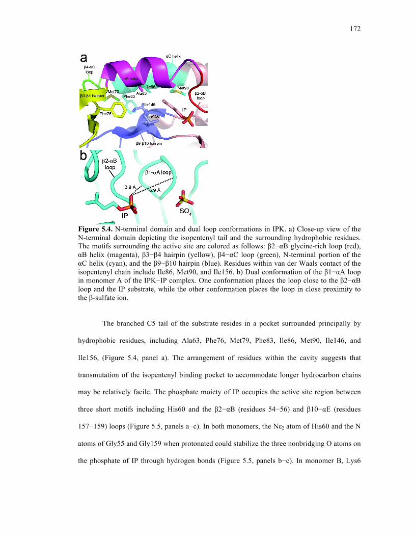
172
Figure 5.4. N-terminal domain and dual loop conformations in IPK. a) Close-up view of the N-terminal domain depicting the isopentenyl tail and the surrounding hydrophobic residues. The motifs surrounding the active site are colored as follows: β2−αB glycine-rich loop (red), αB helix (magenta), β3−β4 hairpin (yellow), β4−αC loop (green), N-terminal portion of the αC helix (cyan), and the β9−β10 hairpin (blue). Residues within van der Waals contact of the isopentenyl chain include Ile86, Met90, and Ile156. b) Dual conformation of the β1−αA loop in monomer A of the IPK−IP complex. One conformation places the loop close to the β2−αB loop and the IP substrate, while the other conformation places the loop in close proximity to the β-sulfate ion.
The branched C5 tail of the substrate resides in a pocket surrounded principally by
hydrophobic residues, including Ala63, Phe76, Met79, Phe83, Ile86, Met90, Ile146, and
Ile156, (Figure 5.4, panel a). The arrangement of residues within the cavity suggests that
transmutation of the isopentenyl binding pocket to accommodate longer hydrocarbon chains
may be relatively facile. The phosphate moiety of IP occupies the active site region between
three short motifs including His60 and the β2−αB (residues 54−56) and β10−αE (residues
157−159) loops (Figure 5.5, panels a−c). In both monomers, the Nε2 atom of His60 and the N
atoms of Gly55 and Gly159 when protonated could stabilize the three nonbridging O atoms on
the phosphate of IP through hydrogen bonds (Figure 5.5, panels b−c). In monomer B, Lys6
173
also interacts with one of these nonbridging O-atoms indirectly through hydrogen-bonding
interactions with an intervening water molecule; in monomer A, there is no water molecule to
facilitate this interaction.
Figure 5.5. IPK in complex with IP and IPP. a) Tertiary structure superposition of monomers A and B of the IPK−IP complex. The rmsd between the two monomers is 1.31 Å. b−c) Close-up views of residues proximal to and hydrogen bonding with the α-phosphate of IP in monomers A (panel b) and B (panel c). In monomer B, a water molecule bridges the side-chain amino group of Lys6 and a nonbridging oxygen atom of the IP phosphate. d) Tertiary structure superposition of monomers A and B of the IPK−IPP complex. The rmsd between the two monomers is 1.39 Å. e−f) Views of the multiple conformers of IPP (labeled as IPP-a and IPP-b) in both monomers A (panel e) and B (panel f).

174
In monomer A, a loop at the β1−αA junction (Gly8−Leu12), residing near the active
site, can adopt two distinct substrate-binding conformations based upon refinement of
alternative conformations and the observed electron density. In one conformation, the loop lies
near the active site β-sulfate ion, while in the other, the loop lies closer to the β2−αB loop
(Figure 5.4, panel b). None of the residues in this loop participate in hydrogen-bonding
interactions with IP; however, the dual binding conformations are not observed for the ‘apo’
structure, suggesting that loop movement is partially dependent on the presence of substrate.
In monomer B, the loop adopts one binding conformation roughly equidistant between the two
modes present in monomer A.
5.3.3. Multiple conformations of IPP in a single active site
The crystal structure of IPK with its product bound reveals that IPP adopts two
distinct conformers designated conformers A and B (Figure 5.5, panels d−f). These
conformers are similar except for the orientation of the β-phosphate group and the adjacent
bridging O atom. In conformer A, these two moieties sit closer to the β10−αE loop, while in
conformer B, they reside closer to the β2−αB loop (Figure 5.5, panels e−f). In both
conformers, a nonbridging O atom of the β-phosphate group hydrogen bonds to the protonated
Nε2 atom of His60. A superposition of the two monomers (Figure 5.5, panel d), reveals that
His60 sits in a different location in each monomer, which may reflect conformations of this
residue that are dynamically accessible as the phosphorylation reaction proceeds.
In monomer A only, a water molecule rests between a nonbridging O atom from the
α-phosphate of IPP and the carboxylate moiety of Asp160 (Figure 5.5, panel e). This water
molecule is also found in substrate-bound structures of FomA kinase (PDBID 3d41)20, E. coli
NAGK (pdb ID 1gs5)21, P. furiosus UMPK (pdb ID 2bmu)18, and E. coli UMPK (pdb ID

175
2bne)23, and it is stabilized in a similar fashion in each of these structures. Asp160 of IPK is
highly conserved among the AAK family and has been suggested to function as an active site
base and a central organizing residue20, 24.
As discussed previously, the β1−αA loop occupies two conformations in monomer A
of the IPK−IP complex: one that places it in close proximity to the β2−αB loop, and another
that interacts with the β-sulfate ion (Figure 5.4, panel b). In monomer A of the IPK−IPP
complex, the latter conformation is the major binding mode observed. The former
conformation can also be seen in the electron density, although this binding mode is so subtle
that it did not refine well and was not built into the final structure. This minor binding mode
may, however, hold some significance, as it places Gly9 of the β1−αA loop in close proximity
to the β-phosphate group in conformer B of IPP. The β1−αA loop is often reported to interact
with the β- and γ-phosphate groups from ATP analogs; however, there are also examples of
this loop interacting with the β-phosphate of the product (in UMPK from E. coli)16, 23.
The catalytically relevant conformer for IPP is most likely conformer B. This
hypothesis is supported by three pieces of information: (i) The β1−αA loop, which is thought
to play a key role during phosphoryl transfer, accesses a minor binding mode that is in close
proximity to the β-phosphate of conformer B16; (ii) A superposition of UDP-bound UMPK
from E. coli and IPP-bound IPK demonstrates that the phosphate moiety of UDP
superimposes with conformer B of IPP23; and (iii) The ATPγS/IP/Mg2+ complex structure
(discussed below) exhibits clear electron density for a IPPβS molecule bound in a single
conformation that superimposes with conformer B of IPP from the IPP-bound structure.
Conformer A of IPP may, therefore, represent a post-reaction enzyme−product (EP) complex.
176
5.3.4. Product-bound active site containing IPPβS
When a crystal of IPK was soaked in a stabilizing solution containing IP, Mg2+, and
ATPγS, the product IPPβS was observed bound in the active site. This product resembles IPP
except one of the nonbridging O atoms on the β-phosphate is clearly replaced with an S atom,
as evidenced by the additional electron density associated with the β-thiophosphate. Notably,
no electron density for the second product ADP is seen. This is the only structure determined
where both substrates, IP and ATPγS, were soaked into the crystal resulting in a catalyzed
reaction in the crystal lattice. Interestingly, this structure reveals only one binding mode for
IPPβS consistent with the orientation of conformer B in the IPP-bound structure. In both
monomers, Gly159 from the β10−αE loop stabilizes the α-phosphate group of IPPβS; the β-
thiophosphate group remains in close proximity to His60. However, in monomer B (compared
to monomer A), the substrate migrates to a position that shifts away from this residue and
resides closer to the β-sulfate ion (Figure 5.3). The intermediate location of the β-phosphate
group in monomer B coupled with the inferred heightened dynamics of certain loops within
this monomer suggests that the monomer B structure depicts an earlier phase in the
transphosphorylation reaction compared to monomer A.
5.3.5. His60 plays a key role in binding and catalysis
From the results discussed above, it is evident that His60 plays an important role in
both substrate and product sequestration. This binding role is accomplished through a
hydrogen-bonding interaction between the protonated Nε2 atom of His60 and a nonbridging O
atom from the terminal phosphate group on either the substrate (IP) or the product (IPP).
His60 was mutated to Asn, Gln, and Ala; the Asn and Gln mutations are isosteric with the
protonated Nε1 and Nε2 groups on His, respectively. The three mutants were assayed at 25 °C
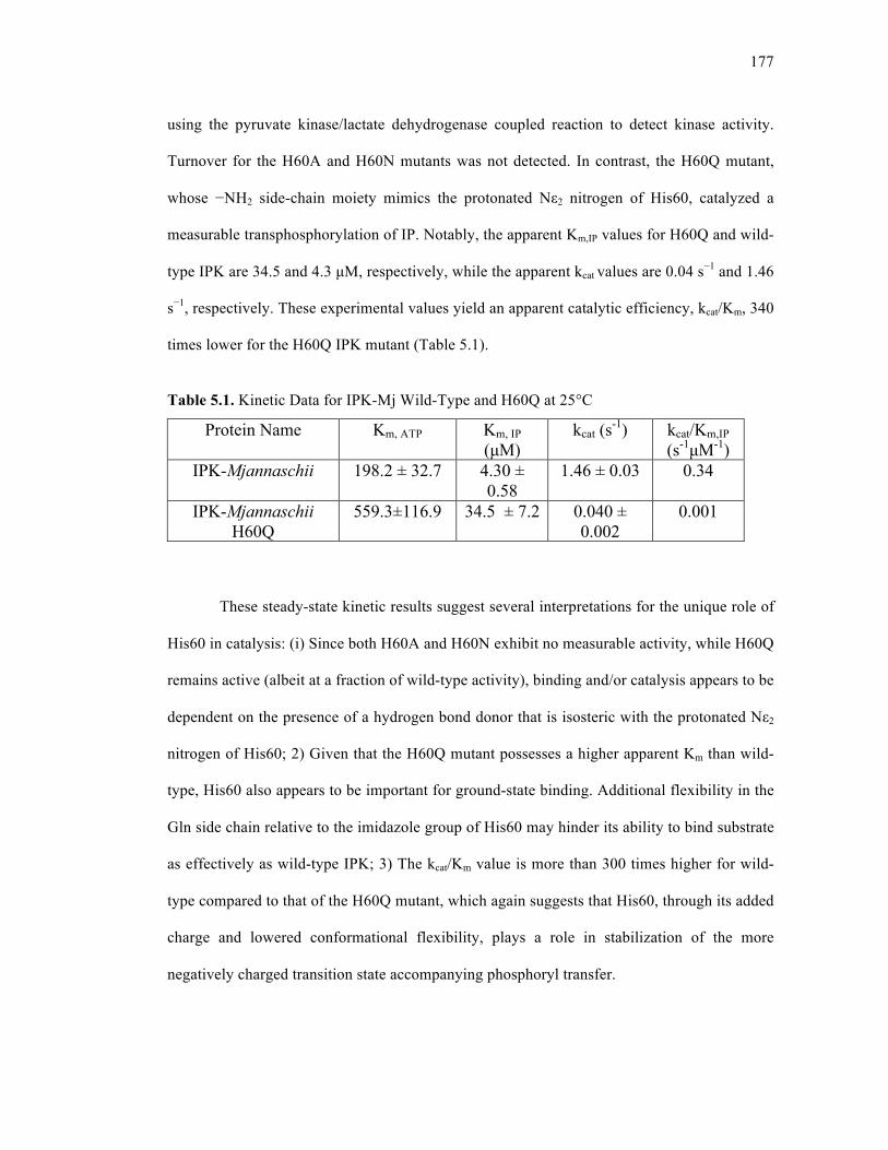
177
using the pyruvate kinase/lactate dehydrogenase coupled reaction to detect kinase activity.
Turnover for the H60A and H60N mutants was not detected. In contrast, the H60Q mutant,
whose −NH2 side-chain moiety mimics the protonated Nε2 nitrogen of His60, catalyzed a
measurable transphosphorylation of IP. Notably, the apparent Km,IP values for H60Q and wild-
type IPK are 34.5 and 4.3 µM, respectively, while the apparent kcat values are 0.04 s−1 and 1.46
s−1, respectively. These experimental values yield an apparent catalytic efficiency, kcat/Km, 340
times lower for the H60Q IPK mutant (Table 5.1).
Table 5.1. Kinetic Data for IPK-Mj Wild-Type and H60Q at 25°C
These steady-state kinetic results suggest several interpretations for the unique role of
His60 in catalysis: (i) Since both H60A and H60N exhibit no measurable activity, while H60Q
remains active (albeit at a fraction of wild-type activity), binding and/or catalysis appears to be
dependent on the presence of a hydrogen bond donor that is isosteric with the protonated Nε2
nitrogen of His60; 2) Given that the H60Q mutant possesses a higher apparent Km than wild-
type, His60 also appears to be important for ground-state binding. Additional flexibility in the
Gln side chain relative to the imidazole group of His60 may hinder its ability to bind substrate
as effectively as wild-type IPK; 3) The kcat/Km value is more than 300 times higher for wild-
type compared to that of the H60Q mutant, which again suggests that His60, through its added
charge and lowered conformational flexibility, plays a role in stabilization of the more
negatively charged transition state accompanying phosphoryl transfer.
Protein Name Km, ATP Km, IP (µM)
kcat (s-1) kcat/Km,IP (s-1µM-1)
IPK-Mjannaschii 198.2 ± 32.7 4.30 ± 0.58
1.46 ± 0.03 0.34
IPK-Mjannaschii H60Q
559.3±116.9 34.5 ± 7.2 0.040 ± 0.002
0.001
178
Through comparison of the solved IPK structures, it is evident that His60 shifts from
stabilizing the α-phosphate on the substrate IP to stabilizing the β-phosphate on the product
IPP. In FomA, His58 (the equivalent residue to His60 of IPK) indirectly stabilizes the
substrate through an intervening water molecule that is within hydrogen-bonding distance of
both His58 and fosfomycin20. In UMPKs, an arginine residue that aligns with His60 appears to
serve two roles: (i) in bacterial UMPKs, this Arg interacts with GTP, which is an allosteric
activator for all bacterial UMPKs25; and (ii) in both bacterial and archaeal UMPKs, this Arg
stabilizes the phosphate intermediate throughout the course of the reaction23, 26. In
comparisons of crystal structures of E. coli UMPK, Arg62 points in opposite directions to
fulfill these two roles25, indicating that the length and the conformational freedom of this
residue are important for its functional versatility. It is therefore not surprising that a mutation
to histidine at this equivalent position results in loss of GTP activation, thereby reducing the
enzyme’s catalytic activity27.
The phosphate division of the AAK family encompasses the three enzymes discussed
above, which are the only currently known family members that contain a residue aligning
with His60 of IPK; the four family members in the carboxylate division do not possess a
residue or a motif that structurally aligns with this region of IPK. Regardless of other roles
that this residue may play, His60 in IPK, His58 in FomA, and the aligning arginine in all
UMPKs most likely play a role in substrate/product binding and/or transition-state
stabilization.
5.3.6. IPK mutants can phosphorylate oligoprenyl monophosphates
As mentioned previously, the tail of the IP substrate is sequestered within a
hydrophobic binding pocket (Figure 5.4, panel a). At the back of the pocket, several residues,
179
Ile86, Ile146, and Ile156, can be mutated to smaller amino acids to accommodate the binding
of ligands with extended carbon chains (such as GP and FP). With this idea in mind, several
point mutants were generated including I86A, I86G, I146A, I146G, and I156A, and most of
these were able to convert FP to FPP while the wild-type enzyme lacked an equivalent activity
(Figure 5.6). These observations support the idea that mutation to smaller residues widens the
cavity to allow for the binding of an extended isoprenoid tail, while mutation to bulkier
residues hinders this ability. It was found that several double and triple mutant combinations
displayed improved FP to FPP conversion by an order of magnitude compared to the single
mutants, providing evidence that a deeper or larger cavity is more effective for FP binding and
catalysis (Figure 5.6). It is also reassuring that these mutations are contextually dependent,
meaning that mutations at the very back of the active site (at position 83) are not effective
unless they are present with mutations closer to the front of the active site (at positions 86,
146, or 156).
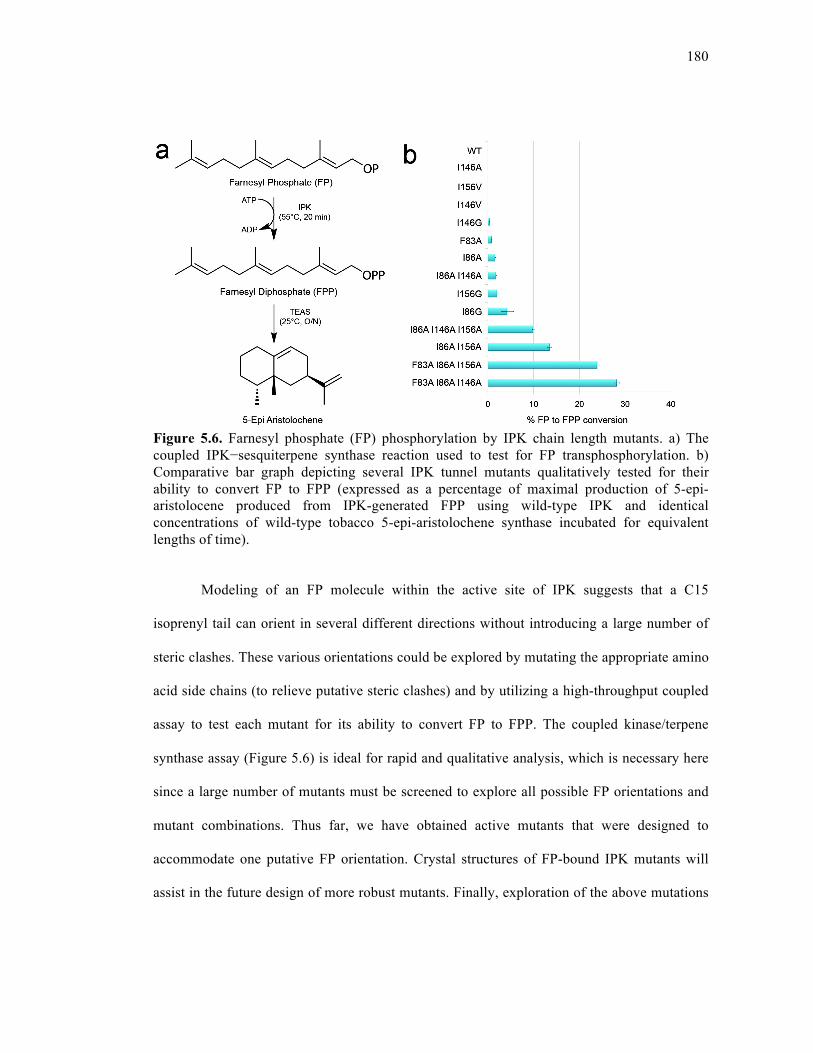
180
Figure 5.6. Farnesyl phosphate (FP) phosphorylation by IPK chain length mutants. a) The coupled IPK−sesquiterpene synthase reaction used to test for FP transphosphorylation. b) Comparative bar graph depicting several IPK tunnel mutants qualitatively tested for their ability to convert FP to FPP (expressed as a percentage of maximal production of 5-epi-aristolocene produced from IPK-generated FPP using wild-type IPK and identical concentrations of wild-type tobacco 5-epi-aristolochene synthase incubated for equivalent lengths of time).
Modeling of an FP molecule within the active site of IPK suggests that a C15
isoprenyl tail can orient in several different directions without introducing a large number of
steric clashes. These various orientations could be explored by mutating the appropriate amino
acid side chains (to relieve putative steric clashes) and by utilizing a high-throughput coupled
assay to test each mutant for its ability to convert FP to FPP. The coupled kinase/terpene
synthase assay (Figure 5.6) is ideal for rapid and qualitative analysis, which is necessary here
since a large number of mutants must be screened to explore all possible FP orientations and
mutant combinations. Thus far, we have obtained active mutants that were designed to
accommodate one putative FP orientation. Crystal structures of FP-bound IPK mutants will
assist in the future design of more robust mutants. Finally, exploration of the above mutations
181
in the context of a mesophilic ortholog should afford improvement in kinetic activity for new
phosphate-bearing substrates; a more accurate picture of dynamic behavior can be painted for
a mesophilic IPK being studied at ambient temperature rather than thermophilic IPK (used
here) being studied at ambient temperature.
5.3.7. Conclusions
Isopentenyl phosphate kinase (IPK) from M. jannaschii is the newest member of the
large amino acid kinase (AAK) family to be structurally characterized. The phosphate division
of the AAK family is comprised of three proteins [IPK, fosfomycin resistance kinase (FomA),
and uridine monophosphate kinase (UMPK)] that exclusively align with one another along
their αB helices. More importantly, they all contain a superimposable residue at position 60 (in
IPK) that indirectly or directly stabilizes the terminal phosphate group of the substrate or
product. Using the His60 marker, we have been able to identify putative IPK homologues
from a number of phylogenetically diverse eukarya. Work to be presented elsewhere is
focused on in vitro and in vivo analyses of these latter proteins, given that, in most cases, the
organisms in question also contain the full complement of predicted mevalonate (MVA)
pathway enzymes.
Finally, we have shown that our initial goal to rationally engineer IPK to accept longer
chain substrates is possible. These mutants along with an expanded mutational analysis in
IPKs from mesophilic hosts can serve a variety of applications. For example, they can be used
to recycle isoprenyl monophosphates, which are thought to be one possible in vivo byproduct
of farnesyl diphosphate (FPP) degradation (through the action of an alkaline phosphatase)28, 29.
This recycling mechanism would be useful in an in vivo system designed to overproduce
isoprenoids (such as terpenes) by means of the MVA pathway in a fungal or bacterial host.
182
The IPK chain-length mutants would also be useful in the chemo-enzymatic synthesis of
radio-labeled geranyl diphosphate (GPP) or FPP as well as a variety of other analogs including
fluorescently tagged isoprenyl tails30, 31. We have demonstrated that IPK can be rationally
engineered to accept and phosphorylate oligoprenyl monophosphate substrates, such as FP.
Future work with IPK from M. jannaschii (and with orthologous IPKs) will focus on
redesigning the enzyme(s) such that they can bind and more efficiently turn over a variety of
bulky GP and FP analogs.
5.4. METHODS
5.4.1. Activity assays and steady-state kinetic analyses
All specific activity and kinetic measurements were performed using the pyruvate
kinase−lactate dehydrogenase coupled assay32. The reaction in 200 µL includes 7 U of
pyruvate kinase, 10 U of lactate dehydrogenase, 2 mM of phosphoenolpyruvate, 0.16 mM of
NADH, 50 mM of Tris−HCl, pH of 8.0, 100 mM of KCl, 8 mM of MgCl2, and varying
concentrations of ATP or IP (purchased from Larodan Fine Chemicals and Isoprenoids, LLC).
When IP was varied, the concentration of ATP was fixed at 4 mM. When ATP was varied, the
concentration of IP was fixed at 100 µM for wild-type IPK and 500 µM for the H60Q mutant.
The reaction was initiated by the addition of IPK (0.15 µg mL−1 final concentration) and
followed by observing the depletion of NADH at 340 nm, expressed as Δ(AU340)/Δt and
converted to Δ(ADP)/Δt. These values were plotted against substrate concentration in
GraphPad Prism (Version 5.01 for Windows) to compute the kinetic parameters kcat and Km,
using the “nonlinear regression enzyme kinetic analysis” option.

183
5.4.2. Kinase/terpene synthase coupled Assay for chain-length mutants
The coupled assay consists of two steps: the kinase reaction followed by the terpene
synthase reaction (Figure 5.6). The 50 µL of kinase reaction includes 4 mM of ATP, 8 mM of
MgCl2, 1 mM of FP (or GP), and 50 mM of Tris−HCl, pH of 8.0. The reaction was initiated
by the injection of IPK (1 µM of the final concentration), incubated at 55 °C for 20 min, and
then cooled on ice for 10 min. The subsequent 500 µL of terpene cyclase reaction includes 10
µL of the aforementioned IPK reaction, 8 mM of MgCl2, 30 µg of tobacco 5-epi aristolochene
synthase (TEAS), and 50 mM of Tris−HCl, pH 8.0. Each sample was overlaid with an equal
volume of ethyl acetate, incubated at RT overnight, and vortexed for terpene extraction and
subsequent injection onto the GC-MS (Hewlett-Packard, 6890/5973 system) equipped with a
HP-5MS column (0.25 mm × 30 m × 0.25 µm). The method employed is similar to that
reported in O’Maille et al.33 with an injection temperature reduced from 250 to 200 °C.
5.4.3. Structure solution and refinement
Data were processed and scaled with XDS34. The reduced single anomalous
diffraction (SAD) data from the IPK ‘apo’ crystal treated with ethyl mercuric phosphate was
used in SOLVE35 to locate and refine the positions of two Hg atoms per asymmetric unit,
followed by phasing (mean figure of merit: 0.33). The program RESOLVE35 was then
employed to build 424 out of 520 residues into the SAD-derived model. Model building and
phase improvement were accomplished using ARP/wARP36, 37. The refined model was used as
the starting model for the structure determination of IPK in complex with IP, IPP, and IPPβS.
Simulated annealing and rigid-body refinements were performed in CNS version 1.2 for each
structure before additional rounds of refinement in CNS version 1.238 and CCP436. COOT was
used for map-model visualization and manual model building39. Areaimol was used for a

184
calculation of the buried surface area per monomer36. PROCHECK was used to assess the
quality of all models40. The data and refinement statistics are listed in Table 5.2.
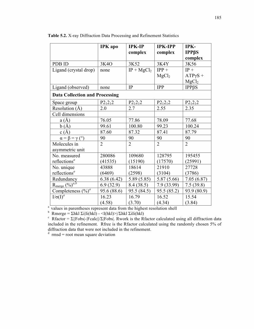
185
Table 5.2. X-ray Diffraction Data Processing and Refinement Statistics
IPK apo IPK-IP complex
IPK-IPP complex
IPK-IPPβS complex
PDB ID 3K4O 3K52 3K4Y 3K56 Ligand (crystal drop) none IP + MgCl2 IPP +
MgCl2 IP + ATPγS + MgCl2
Ligand (observed) none IP IPP IPPβS Data Collection and Processing Space group P21212 P21212 P21212 P21212 Resolution (Å) 2.0 2.7 2.55 2.35 Cell dimensions a (Å) 76.05 77.86 78.09 77.68 b (Å) 99.61 100.80 99.23 100.24 c (Å) 87.60 87.32 87.41 87.79 α = β = γ (°) 90 90 90 90 Molecules in asymmetric unit
2 2 2 2
No. measured reflectionsa
280086 (41535)
109680 (15190)
128795 (17570)
195455 (25991)
No. unique reflectionsa
43888 (6469)
18614 (2598)
21910 (3104)
27728 (3786)
Redundancy 6.38 (6.42) 5.89 (5.85) 5.87 (5.66) 7.05 (6.87) Rmerge (%)a,b 6.9 (32.9) 8.4 (38.5) 7.9 (33.99) 7.5 (39.8) Completeness (%)a 95.6 (88.6) 95.5 (84.5) 95.5 (85.2) 93.9 (80.9) I/σ(I)a 16.23
(4.58) 16.79 (3.70)
16.52 (4.34)
15.54 (3.84)
a values in parentheses represent data from the highest resolution shell b Rmerge = Σhkl Σi|Ii(hkl) - <I(hkl)>|/Σhkl ΣiIi(hkl) c Rfactor = Σ||Fobs|-|Fcalc||/Σ|Fobs|. Rwork is the Rfactor calculated using all diffraction data included in the refinement. Rfree is the Rfactor calculated using the randomly chosen 5% of diffraction data that were not included in the refinement. d rmsd = root mean square deviation
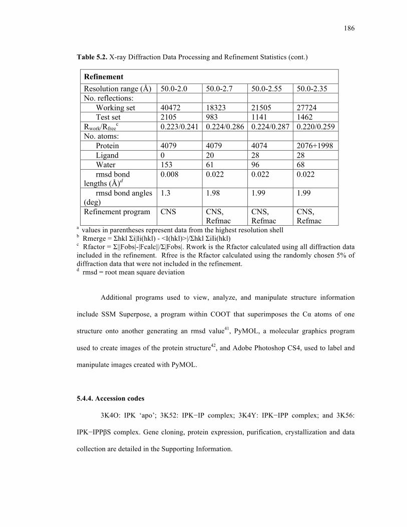
186
Table 5.2. X-ray Diffraction Data Processing and Refinement Statistics (cont.)
Refinement Resolution range (Å) 50.0-2.0 50.0-2.7 50.0-2.55 50.0-2.35 No. reflections: Working set 40472 18323 21505 27724 Test set 2105 983 1141 1462 Rwork/Rfree
c 0.223/0.241 0.224/0.286 0.224/0.287 0.220/0.259 No. atoms: Protein 4079 4079 4074 2076+1998 Ligand 0 20 28 28 Water 153 61 96 68 rmsd bond lengths (Å)d
0.008 0.022 0.022 0.022
rmsd bond angles (deg)
1.3 1.98 1.99 1.99
Refinement program CNS CNS, Refmac
CNS, Refmac
CNS, Refmac
a values in parentheses represent data from the highest resolution shell b Rmerge = Σhkl Σi|Ii(hkl) - <I(hkl)>|/Σhkl ΣiIi(hkl) c Rfactor = Σ||Fobs|-|Fcalc||/Σ|Fobs|. Rwork is the Rfactor calculated using all diffraction data included in the refinement. Rfree is the Rfactor calculated using the randomly chosen 5% of diffraction data that were not included in the refinement. d rmsd = root mean square deviation
Additional programs used to view, analyze, and manipulate structure information
include SSM Superpose, a program within COOT that superimposes the Cα atoms of one
structure onto another generating an rmsd value41, PyMOL, a molecular graphics program
used to create images of the protein structure42, and Adobe Photoshop CS4, used to label and
manipulate images created with PyMOL.
5.4.4. Accession codes
3K4O: IPK ‘apo’; 3K52: IPK−IP complex; 3K4Y: IPK−IPP complex; and 3K56:
IPK−IPPβS complex. Gene cloning, protein expression, purification, crystallization and data
collection are detailed in the Supporting Information.
187
5.5. SUPPORTING INFORMATION
5.5.1. Cloning of IPK genes and mutant construction
The IPK gene MJ004411 was amplified from Methanocaldococcus jannaschii genomic
DNA (ATCC® 43067D-5™) by PCR. An IPK homolog from Methanococcus maripaludis
was also amplified from genomic DNA (ATCC® BAA-1333D-5™) by PCR. Both genes were
amplified using Phusion™ High-Fidelity DNA polymerase (New England Biolabs, Inc)
employing a 60°C annealing temperature and a 30 s PCR extension time. The PCR products
were digested with NcoI and XhoI (New England Biolabs, Inc), purified, and ligated into a
NcoI/XhoI digested pHIS8 vector (a modified version of pET28a(+) containing an N-terminal
8-histidine tag) using T4 DNA ligase (New England Biolabs, Inc). All mutations in IPK were
made using the QuikChange protocol with PfuTurbo® DNA Polymerase (Stratagene) together
with a 6.5 min PCR extension time. The primer pairs used in all PCR reactions are listed in
Table 5.3.
5.5.2. Protein expression and purification
The plasmid containing the IPK gene (IPKpHIS8) was transformed into E. coli
Bl21(DE3) competent cells (Novagen). One colony was grown in LB media (75 ml) overnight
at 37°C, 25 ml of the overnight culture was transferred to one liter of TB media and grown at
37°C until an OD600 of 1.2. Isopropyl-β-D-thiogalactoside (0.2 mM final concentration) was
then added, cells were shaken overnight at 37°C for approximately 12–14 hr, harvested by
centrifugation and lysed using lysis buffer (50 mM Tris-HCl, pH 8.0, 500 mM NaCl, 20 mM
imidazole, 1% (v/v) Tween-20, 10% (v/v) glycerol, 10 mM 2-mercaptoethanol) containing
lysozyme (1 mg ml-1). The lysate was stirred at 4°C for 1 hr, sonicated, and centrifuged at
21,000 rpm for 45 min at 4°C. The supernatant was loaded onto a column containing Ni-NTA
188
agarose S3 resin (Qiagen), washed with lysis buffer and wash buffer (lysis buffer without
Tween- 20), and then eluted with elution buffer (wash buffer containing 250 mM imidazole).
The protein was digested with thrombin overnight during dialysis in buffer (50 mM Tris-HCl,
pH 8.0, 100 mM NaCl) containing 10 mM 2-mercaptoethanol. The dialyzed solution was
passed through a column containing Benzamidine Sepharose 4 Fast Flow (high sub) (GE
Healthcare) and Ni-NTA agarose. The eluant was heated at 80°C for 10 min to precipitate
contaminating proteins and the supernatant passed through a HiLoad™ 16/16 Superdex™ 200
prep grade (GE Healthcare) gel filtration column using dialysis buffer containing 2 mM DTT.
IPK fractions were combined, concentrated to approximately 15 mg ml-1, and frozen at –80°C.
5.5.3. Crystallization and data collection
Crystals of IPK were grown by hanging-drop vapor-diffusion using a 2 µl drop
containing 1 µl of IPK (15 mg ml-1) and 1 µl of reservoir. Crystallization conditions were
obtained using the Hampton Crystal Screen I (Hampton Research) and optimized to improve
crystal morphology and size. IPK crystals formed large plates over a reservoir containing 1.5–
1.6 M ammonium sulfate at 298 K. The plates were visible after 1–2 days and reached
maximum size after 1 week. Crystal soaks were set up with heavy atoms (0.1–0.5 mM ethyl
mercuric phosphate) or ligands (1 mM IP, 5 mM IPP or 1 mMIP / 5 mM ATPγS) in 2.0 M
ammonium sulfate. After 1–2 days, crystals were placed in a cryo-protectant (2.0 M
ammonium sulfate and 20% (v/v) ethylene glycol) for 10–30 sec then flash frozen in liquid
nitrogen. X-ray data were collected at 110 K on ALS beamlines 8.2.1 and 8.2.2 (Lawrence
Berkeley National Laboratory, Berkeley, CA) using an ADSC Q315 CCD detector. All x-ray
diffraction data including single anomalous diffraction (SAD) data were collected at λ=1.0 Å.
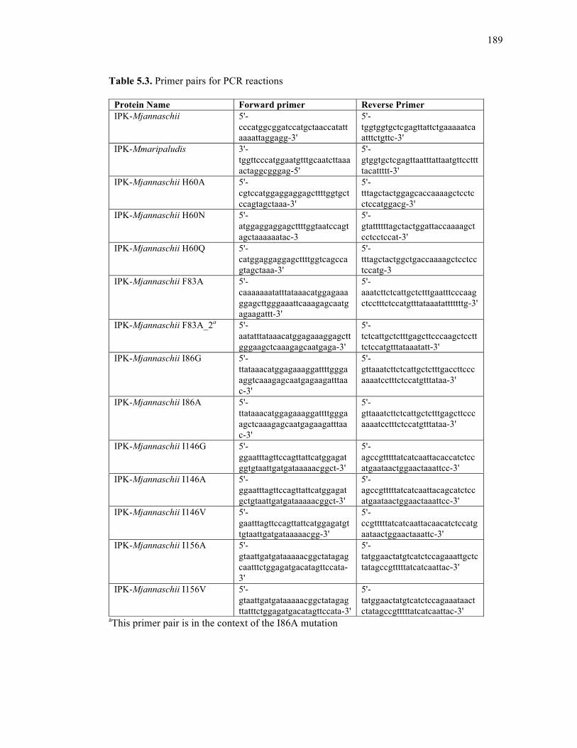
189
Table 5.3. Primer pairs for PCR reactions
aThis primer pair is in the context of the I86A mutation
Protein Name Forward primer Reverse Primer IPK-Mjannaschii 5'-
cccatggcggatccatgctaaccatattaaaattaggagg-3'
5'-tggtggtgctcgagttattctgaaaaatcaatttctgttc-3'
IPK-Mmaripaludis 3'-tggttcccatggaatgtttgcaatcttaaaactaggcgggag-5'
5'-gtggtgctcgagttaatttattaatgttccttttacattttt-3'
IPK-Mjannaschii H60A 5'-cgtccatggaggaggagcttttggtgctccagtagctaaa-3'
5'-tttagctactggagcaccaaaagctcctcctccatggacg-3'
IPK-Mjannaschii H60N 5'- atggaggaggagcttttggtaatccagtagctaaaaaatac-3
5'- gtattttttagctactggattaccaaaagctcctcctccat-3'
IPK-Mjannaschii H60Q 5'- catggaggaggagcttttggtcagccagtagctaaa-3'
5'- tttagctactggctgaccaaaagctcctcctccatg-3
IPK-Mjannaschii F83A 5'-caaaaaaatatttataaacatggagaaaggagcttgggaaattcaaagagcaatgagaagattt-3'
5'-aaatcttctcattgctctttgaatttcccaagctcctttctccatgtttataaatatttttttg-3'
IPK-Mjannaschii F83A_2a 5'-aatatttataaacatggagaaaggagcttgggaagctcaaagagcaatgaga-3'
5'-tctcattgctctttgagcttcccaagctcctttctccatgtttataaatatt-3'
IPK-Mjannaschii I86G 5'-ttataaacatggagaaaggattttgggaaggtcaaagagcaatgagaagatttaac-3'
5'-gttaaatcttctcattgctctttgaccttcccaaaatcctttctccatgtttataa-3'
IPK-Mjannaschii I86A 5'-ttataaacatggagaaaggattttgggaagctcaaagagcaatgagaagatttaac-3'
5'-gttaaatcttctcattgctctttgagcttcccaaaatcctttctccatgtttataa-3'
IPK-Mjannaschii I146G 5'-ggaatttagttccagttattcatggagatggtgtaattgatgataaaaacggct-3'
5'-agccgtttttatcatcaattacaccatctccatgaataactggaactaaattcc-3'
IPK-Mjannaschii I146A 5'-ggaatttagttccagttattcatggagatgctgtaattgatgataaaaacggct-3'
5'-agccgtttttatcatcaattacagcatctccatgaataactggaactaaattcc-3'
IPK-Mjannaschii I146V 5'-gaatttagttccagttattcatggagatgttgtaattgatgataaaaacgg-3'
5'-ccgtttttatcatcaattacaacatctccatgaataactggaactaaattc-3'
IPK-Mjannaschii I156A 5'-gtaattgatgataaaaacggctatagagcaatttctggagatgacatagttccata-3'
5'-tatggaactatgtcatctccagaaattgctctatagccgtttttatcatcaattac-3'
IPK-Mjannaschii I156V 5'-gtaattgatgataaaaacggctatagagttatttctggagatgacatagttccata-3'
5'-tatggaactatgtcatctccagaaataactctatagccgtttttatcatcaattac-3'
190
ACKNOWLEDGEMENTS
The text of chapter 5, in full, is a reprint of the material as it appears in ACS Chemical
Biology 2010, 5(6), pp 589-601. I am the primary author of this paper. The research was
performed under the supervision of Joseph P. Noel.
REFERENCES
1. Gershenzon, J.; Dudareva, N., The function of terpene natural products in the natural world. Nature chemical biology 2007, 3 (7), 408-414.
2. Novakova, Z.; Surin, S.; Blasko, J.; Majernik, A.; Smigan, P., Membrane proteins and
squalene-hydrosqualene profile in methanoarchaeon Methanothermobacter thermautotrophicus resistant to N,N'-dicyclohexylcarbodiimide. Folia microbiologica 2008, 53 (3), 237-240.
3. Ourisson, G.; Rohmer, M.; Poralla, K., Prokaryotic hopanoids and other polyterpenoid
sterol surrogates. Annual Review of Microbiology 1987, 41, 301-333. 4. Eichler, J.; Adams, M. W., Posttranslational protein modification in Archaea.
Microbiology and molecular biology reviews : MMBR 2005, 69 (3), 393-425. 5. Sieiro, C.; Poza, M.; de Miguel, T.; Villa, T. G., Genetic basis of microbial
carotenogenesis. International microbiology : the official journal of the Spanish Society for Microbiology 2003, 6 (1), 11-16.
6. Hemmi, H.; Ikejiri, S.; Nakayama, T.; Nishino, T., Fusion-type lycopene beta-cyclase
from a thermoacidophilic archaeon Sulfolobus solfataricus. Biochemical and biophysical research communications 2003, 305 (3), 586-591.
7. Koga, Y.; Morii, H., Biosynthesis of ether-type polar lipids in archaea and
evolutionary considerations. Microbiology and molecular biology reviews : MMBR 2007, 71 (1), 97-120.
8. Rohmer, M., The discovery of a mevalonate-independent pathway for isoprenoid
biosynthesis in bacteria, algae and higher plants. Natural product reports 1999, 16 (5), 565-574.
9. Lange, B. M.; Rujan, T.; Martin, W.; Croteau, R., Isoprenoid biosynthesis: the
evolution of two ancient and distinct pathways across genomes. Proceedings of the National Academy of Sciences of the United States of America 2000, 97 (24), 13172-13177.
10. Smit, A.; Mushegian, A., Biosynthesis of isoprenoids via mevalonate in Archaea: the
lost pathway. Genome research 2000, 10 (10), 1468-1484.
191
11. Grochowski, L. L.; Xu, H.; White, R. H., Methanocaldococcus jannaschii uses a
modified mevalonate pathway for biosynthesis of isopentenyl diphosphate. Journal of Bacteriology 2006, 188 (9), 3192-3198.
12. Marina, A.; Alzari, P. M.; Bravo, J.; Uriarte, M.; Barcelona, B.; Fita, I.; Rubio, V.,
Carbamate kinase: New structural machinery for making carbamoyl phosphate, the common precursor of pyrimidines and arginine. Protein science : a publication of the Protein Society 1999, 8 (4), 934-940.
13. Marco-Marin, C.; Gil-Ortiz, F.; Perez-Arellano, I.; Cervera, J.; Fita, I.; Rubio, V., A
novel two-domain architecture within the amino acid kinase enzyme family revealed by the crystal structure of Escherichia coli glutamate 5-kinase. Journal of Molecular Biology 2007, 367 (5), 1431-1446.
14. Krissinel, E.; Henrick, K., Inference of macromolecular assemblies from crystalline
state. Journal of Molecular Biology 2007, 372 (3), 774-797. 15. Faehnle, C. R.; Liu, X.; Pavlovsky, A.; Viola, R. E., The initial step in the archaeal
aspartate biosynthetic pathway catalyzed by a monofunctional aspartokinase. Acta crystallographica.Section F, Structural biology and crystallization communications 2006, 62 (Pt 10), 962-966.
16. Gil-Ortiz, F.; Ramon-Maiques, S.; Fita, I.; Rubio, V., The course of phosphorus in the
reaction of N-acetyl-L-glutamate kinase, determined from the structures of crystalline complexes, including a complex with an AlF(4)(-) transition state mimic. Journal of Molecular Biology 2003, 331 (1), 231-244.
17. Kotaka, M.; Ren, J.; Lockyer, M.; Hawkins, A. R.; Stammers, D. K., Structures of R-
and T-state Escherichia coli aspartokinase III. Mechanisms of the allosteric transition and inhibition by lysine. The Journal of biological chemistry 2006, 281 (42), 31544-31552.
18. Marco-Marin, C.; Gil-Ortiz, F.; Rubio, V., The crystal structure of Pyrococcus
furiosus UMP kinase provides insight into catalysis and regulation in microbial pyrimidine nucleotide biosynthesis. Journal of Molecular Biology 2005, 352 (2), 438-454.
19. Liu, X.; Pavlovsky, A. G.; Viola, R. E., The structural basis for allosteric inhibition of
a threonine-sensitive aspartokinase. The Journal of biological chemistry 2008, 283 (23), 16216-16225.
20. Pakhomova, S.; Bartlett, S. G.; Augustus, A.; Kuzuyama, T.; Newcomer, M. E.,
Crystal structure of fosfomycin resistance kinase FomA from Streptomyces wedmorensis. The Journal of biological chemistry 2008, 283 (42), 28518-28526.
21. Ramon-Maiques, S.; Marina, A.; Gil-Ortiz, F.; Fita, I.; Rubio, V., Structure of
acetylglutamate kinase, a key enzyme for arginine biosynthesis and a prototype for the
192
amino acid kinase enzyme family, during catalysis. Structure (London, England : 1993) 2002, 10 (3), 329-342.
22. Ramon-Maiques, S.; Fernandez-Murga, M. L.; Gil-Ortiz, F.; Vagin, A.; Fita, I.;
Rubio, V., Structural bases of feed-back control of arginine biosynthesis, revealed by the structures of two hexameric N-acetylglutamate kinases, from Thermotoga maritima and Pseudomonas aeruginosa. Journal of Molecular Biology 2006, 356 (3), 695-713.
23. Briozzo, P.; Evrin, C.; Meyer, P.; Assairi, L.; Joly, N.; Barzu, O.; Gilles, A. M.,
Structure of Escherichia coli UMP kinase differs from that of other nucleoside monophosphate kinases and sheds new light on enzyme regulation. The Journal of biological chemistry 2005, 280 (27), 25533-25540.
24. Marco-Marin, C.; Ramon-Maiques, S.; Tavarez, S.; Rubio, V., Site-directed
mutagenesis of Escherichia coli acetylglutamate kinase and aspartokinase III probes the catalytic and substrate-binding mechanisms of these amino acid kinase family enzymes and allows three-dimensional modelling of aspartokinase. Journal of Molecular Biology 2003, 334 (3), 459-476.
25. Meyer, P.; Evrin, C.; Briozzo, P.; Joly, N.; Barzu, O.; Gilles, A. M., Structural and
functional characterization of Escherichia coli UMP kinase in complex with its allosteric regulator GTP. The Journal of biological chemistry 2008, 283 (51), 36011-36018.
26. Jensen, K. S.; Johansson, E.; Jensen, K. F., Structural and enzymatic investigation of
the Sulfolobus solfataricus uridylate kinase shows competitive UTP inhibition and the lack of GTP stimulation. Biochemistry 2007, 46 (10), 2745-2757.
27. Bucurenci, N.; Serina, L.; Zaharia, C.; Landais, S.; Danchin, A.; Barzu, O., Mutational
analysis of UMP kinase from Escherichia coli. Journal of Bacteriology 1998, 180 (3), 473-477.
28. Song, L., A soluble form of phosphatase in Saccharomyces cerevisiae capable of
converting farnesyl diphosphate into E,E-farnesol. Applied Biochemistry and Biotechnology 2006, 128 (2), 149-158.
29. Coleman, J. E., Structure and mechanism of alkaline phosphatase. Annual Review of
Biophysics and Biomolecular Structure 1992, 21, 441-483. 30. Rose, M. W.; Rose, N. D.; Boggs, J.; Lenevich, S.; Xu, J.; Barany, G.; Distefano, M.
D., Evaluation of geranylazide and farnesylazide diphosphate for incorporation of prenylazides into a CAAX box-containing peptide using protein farnesyltransferase. The journal of peptide research : official journal of the American Peptide Society 2005, 65 (6), 529-537.
31. Hovlid, M. L.; Edelstein, R. L.; Henry, O.; Ochocki, J.; DeGraw, A.; Lenevich, S.;
Talbot, T.; Young, V. G.; Hruza, A. W.; Lopez-Gallego, F.; Labello, N. P.; Strickland,
193
C. L.; Schmidt-Dannert, C.; Distefano, M. D., Synthesis, properties, and applications of diazotrifluropropanoyl-containing photoactive analogs of farnesyl diphosphate containing modified linkages for enhanced stability. Chemical biology & drug design 2010, 75 (1), 51-67.
32. Lindsley, J. E., Use of a real-time, coupled assay to measure the ATPase activity of
DNA topoisomerase II. Methods in molecular biology (Clifton, N.J.) 2001, 95, 57-64. 33. O'Maille, P. E.; Tsai, M. D.; Greenhagen, B. T.; Chappell, J.; Noel, J. P., Gene library
synthesis by structure-based combinatorial protein engineering. Methods Enzymol. 2004, 388, 75-91.
34. Kabsch, W., Automatic processing of rotation diffraction data from crystals of
initially unknown symmetry and cell constants. Journal of Applied Crystallography 1993, 26 (6), 795-800.
35. Terwilliger, T., SOLVE and RESOLVE: automated structure solution, density
modification and model building. Journal of synchrotron radiation 2004, 11 (Pt 1), 49-52.
36. Collaborative Computational Project, N., The CCP4 suite: programs for protein
crystallography. Acta crystallographica.Section D, Biological crystallography 1994, 50 (Pt 5), 760-763.
37. Perrakis, A.; Morris, R.; Lamzin, V. S., Automated protein model building combined
with iterative structure refinement. Nature structural biology 1999, 6 (5), 458-463. 38. Brunger, A. T., Version 1.2 of the Crystallography and NMR system. Nature
protocols 2007, 2 (11), 2728-2733. 39. Brunger, A. T.; Adams, P. D.; Clore, G. M.; DeLano, W. L.; Gros, P.; Grosse-
Kunstleve, R. W.; Jiang, J. S.; Kuszewski, J.; Nilges, M.; Pannu, N. S.; Read, R. J.; Rice, L. M.; Simonson, T.; Warren, G. L., Crystallography & NMR system: A new software suite for macromolecular structure determination. Acta crystallographica.Section D, Biological crystallography 1998, 54 (Pt 5), 905-921.
40. Laskowski, R. A.; MacArthur, M. W.; Moss, D. S.; Thornton, J. M., PROCHECK: a
program to check the stereochemical quality of protein structures. Journal of Applied Crystallography 1993, 26, 283-291.
41. Krissinel, E.; Henrick, K., Secondary-structure matching (SSM), a new tool for fast
protein structure alignment in three dimensions. Acta crystallographica.Section D, Biological crystallography 2004, 60 (Pt 12 Pt 1), 2256-2268.
42. DeLano, W. L., The PyMOL Molecular Graphics System. DeLano Scientific, Palo
Alto, CA, USA, 2002.
194
Chapter 6
Isopentenyl Phosphate Kinase Homologs Outside of Archaea Suggest a Bifurcating
Mevalonate Pathway in a Diversity of Eukaryotes
195
6.1. ABSTRACT
Archaea encode a variant of the canonical mevalonate pathway, using isopentenyl
phosphate kinase (IPK) as part of a two-enzyme substitution in the final steps of isopentenyl
diphosphate (IPP) biosynthesis. We found IPK homologs intermittently distributed in most
major eukaryotic lineages. These homologs retain IPK activity, suggesting that many
eukaryotes possess a bifurcating mevalonate pathway.
6.2. INTRODUCTION
IPP and its isomer, dimethylallyl diphosphate (DMAPP), are essential precursors to all
isoprenoids including steroids, terpenoids, carotenoids, and numerous primary and secondary
metabolites. IPP biosynthesis occurs through the classical mevalonate pathway (MVA) in
eukaryotes and some bacteria or through the 1-deoxy-D-xylulose 5-phosphate (DXP) pathway
in plastid-bearing eukaryotes and bacteria. Recent work suggests that archaea use a variant of
the MVA pathway (referred to as a modified or alternative MVA pathway)1-3. Archaea have
genes for all but the last two enzymes of the classical MVA pathway, phosphomevalonate
kinase (PMK) and diphosphomevalonate decarboxylase (DPM-DC) (Figure 6.1, Table 6.2).1
Grochowski et al. characterized an isopentenyl phosphate kinase (IPK) in the thermophilic
archaeon Methanocaldococcus jannaschii, which catalyzes the ATP-dependent
phosphorylation of isopentenyl phosphate (IP) to IPP2. These authors proposed the alternative
MVA pathway, which reverses the last two steps of the classical pathway by using an
(unknown) phosphomevalonate decarboxylase followed by IPK. Very recent work has
reviewed the phylogeny of the MVA pathway across all three domains of life3; work here
focuses on the characterization of IPKs in both Archaea and Eukarya.
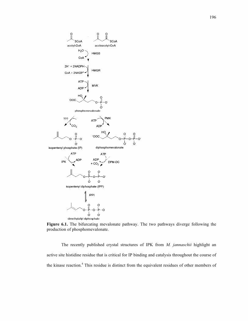
196
Figure 6.1. The bifurcating mevalonate pathway. The two pathways diverge following the production of phosphomevalonate.
The recently published crystal structures of IPK from M. jannaschii highlight an
active site histidine residue that is critical for IP binding and catalysis throughout the course of
the kinase reaction.4 This residue is distinct from the equivalent residues of other members of
197
the amino acid kinase (AAK) superfamily to which IPK belongs, and it can therefore be used
as a marker to identify putative IPK homologs.
6.3. RESULTS AND DISCUSSION
6.3.1. Phylogenetic diversity of IPK
We used overall sequence conservation coupled with the characteristic histidine to
explore the phylogenetic diversity of IPK. Psi-blast and profile HMMs were used to detect
IPK homologs in public protein, EST, and genome databases; other AAK profiles were used
to distinguish ambiguous homologs. We find IPK in almost all archaea, a small cluster of
GNS bacteria, and in an exceptionally sporadic distribution across most major eukaryotic
lineages (Figure 6.2).
Within animals, the gene appears to have been independently lost many times in
evolution (Figure 6.2, Supporting Information). Such scattered distribution suggests an
unprecedented degree of gene loss or an equally unusual degree of horizontal gene transfer.
For example, IPK is absent from choanoflagellates and sponges, but found in early branching
animals such as Trichoplax and corals. It is found in a shark (C. milii) but not teleost fish, in
an amphibian (the newt N. viridescens) but not in frogs, and in a lizard (A. carolinensis) and a
snake (P. olfersii), but not in any bird or mammal. IPK is absent from most fungi; by contrast,
it is found in every sequenced green plant genome. Bacterial IPK is restricted to a small
cluster of 5 genomes within the class and phylum Chloroflexi (green non-sulfur bacteria).
Bacteria also contain the closest structural homolog of IPK, fomA from S. wedmorensis,
which phosphorylates and inactivates the antibiotic fosfomycin5.
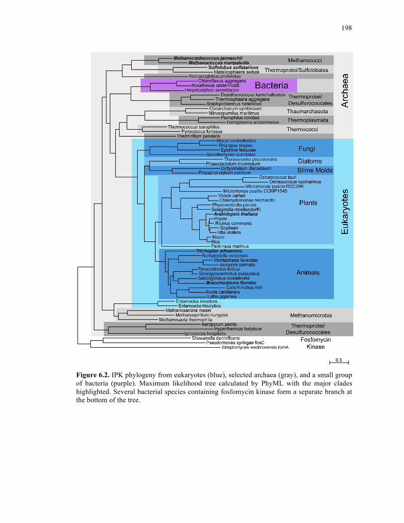
198
Figure 6.2. IPK phylogeny from eukaryotes (blue), selected archaea (gray), and a small group of bacteria (purple). Maximum likelihood tree calculated by PhyML with the major clades highlighted. Several bacterial species containing fosfomycin kinase form a separate branch at the bottom of the tree.

199
6.3.2. Catalytic activity of IPK homologs
We tested five homlogs for catalytic activity, including the characterized IPK from M.
jannaschii2, 4 two other archaea (Methanococcus maripaludis and Sulfolobus solfataricus) and
three eukaryotes: Trichoplax adhaerens (early-branching metazoan), Branchiostoma floridae
(chordate), and Arabidopsis thaliana (plant). Remarkably, all six IPK homologs catalyzed the
phosphorylation of IP to IPP; kinetic constants are reported in Table 6.1.
Table 6.1. Kinetic constants for characterized IPKs
Km, IP (µM) kcat (s−1) Ki, IP kcat/Km,IP (s−1µM−1)
Goodness of fit (R2)a
M. jannaschii 4.3 (±0.6)b 1.46 (±0.03) -c 0.34 (±0.05)
0.90
M. maripaludis 21.4 (±4.3) 15.2 (±1.4) 877 (±550) 0.71 (±0.16)
0.99
S. solfataricus 23.6 (±4.8) 0.91 (±0.05) - 0.04 (±0.01)
0.92
B. floridae 13.3 (±2.0) 27.2 (±1.2) 2820 (±1700) 2.05 (±0.32) 0.98 A. thaliana 0.79 (±0.35) 1.9 (±0.2) 522 (±381) 2.4 (±1.1) 0.95
T. adhaerens 3.1 (±1.6) 2.4 (±0.2) - 0.77 (0.40)
0.79
a R2 = 1.0 – (SSreg/SStot), where SSreg = sum of squares value, SStot = sum of squares of the distances between each point and a horizontal line passing through the average of all y values b Values in parentheses represent standard error (or propagation of error) for each kinetic constant c Ki constant was not calculated
Although most archaea lack the last enzymes of the classical MVA pathway, the order
Sulfolobales contains all of them in addition to IPK (Table 6.2). IPK from S. solfataricus has a
much lower catalytic efficiency than the other IPKs tested (Table 6.1) and may therefore be
losing function. This agrees with the observation that IPK has persisted across most eukaryotic
lineages, but has been lost during many rare evolutionary events, probably due to partial
redundancy with the MVA pathway. Green plants are the exception, in which IPK may have

200
gained an indispensable function. Subcellular compartmentalization is a precedent of plant
isoprenoid biosynthesis6, however no localization signals could be found in plant IPKs.
6.3.3. Role for IPK in other kingdoms of life
The unusual phylogeny of IPK and its membership in a family of kinases that
phosphorylate such a broad range of substrates leave open the possibility that IPK may play a
different physiological role, such as participating in the phosphorylation of an IP-like substrate
or recycling IP that may accumulate in vivo as a consequence of phosphatase-dependent IPP
degradation. Although certain archaeal IPKs demonstrate some ability to phosphorylate other
isoprenoid substrates (such as dimethylallyl phosphate and geranyl phosphate), they prefer IP7.
Failure to date to identify the decarboxylase required to complete the alternative pathway is
also reason to speculate on the role of IPK. The M. jannaschii gene MJ0403 which has
sequence similarity to iron-binding dioxygenases has been proposed to serve this function2,
however attempts to show biochemical activity have not been successful, and the search for
other possible decarboxylase candidates is under way.
6.3.4. Conclusions
Remote homology techniques and an active site histidine residue were used to
successfully find and characterize IPK homologs among eukaryotes and some bacteria. In
contrast to previous research which has only briefly described the existence of eukaryotic
IPKs3, 7, this detailed report includes a thorough phylogenetic analysis of this gene across all
domains of life, and the first experimental evidence for the existence of IPK outside of
archaea. The presence of active eukaryotic IPKs supports the idea that a bifurcating pathway
may exist in plants, some animals, and several fungi and protists. Future work will involve the

201
complete biochemical characterization of both the classical and alternative MVA pathways
(including the decarboxylase which has thus far been uncharacterized) in a given organism.
6.4. METHODS
6.4.1. Cloning of IPK homologs
IPK homologs from Archaea (M. jannaschii, M. maripaludis C5, S. solfataricus P2)
were cloned from genomic DNA from American Type Cell Cultures (ATCC) as previously
described for M. jannaschii1 into a pET28a(+) vector containing an N-terminal 8-histidine tag
using PCR primers as follows:
S. solfataricus P2 forward: 5'-tggttcCCATGGAttggaaatggatatgggatctgaattg-3'
S. solfataricus P2 reverse: 5 -gtggtgCTCGAGtcaggcattcggattacctcttactaaa-3'
M. maripaludis C5 forward: 5'-tggttcCCATGGaatgtttgcaatcttaaaactaggcgggag-3'
M. maripaludis C5 reverse: 5'-gtggtgCTCGAGttaatttattaatgttccttttacattttt-3'
The three IPK homologs from Eukarya (A. thaliana, T. adhaerens, B. floridae) were
ordered as synthetic genes from Genscript (Piscataway, NJ, USA) and sub-cloned using
Gateway technology from Invitrogen (San Diego, CA, USA) into pHIS9GW, an in-house
vector modified to contain a 9-histidine tag.
6.4.2. Protein expression and purification
All proteins were expressed according to a previously described procedure with
several modifications4. While all E. coli Bl21 (DE3) cells expressing archaeal proteins were
induced with 0.2mM IPTG overnight at 37°C, all cells expressing eukaryotic proteins were
induced with 1.0mM IPTG for five hours at 22°C. All proteins were purified similarly and as
202
previously described, however none of the proteins other than M. jannaschii were incubated at
80°C.
6.4.3. Steady-state kinetic analysis
Kinetic measurements were performed on IPK from M. maripaludis, S. solfataricus,
and B. floridae using a coupled pyruvate kinase –lactate dehydrogenase assay as previously
described for IPK from M. jannaschii that employs varying IP concentrations ranging from
2µM-1mM1. Steady-State kinetic curves were fitted using Prism (GraphPad Software Inc.,
San Diego, CA, USA) to compute Km, kcat, and where appropriate, Ki, IP. Activity
measurements were performed for T. adhaerens and A. thaliana using the coupled assay at
four different IP concentrations (2µM, 10µM, 50µM, and 100µM) in triplicate.
6.4.4. Bioinformatics
Public protein, cDNA, EST and genomic databases were searched for IPK homologs,
using individual IPK protein sequences, and profile Hidden Markov models built from several
individual IPK clades. Genes were predicted from genomic sequence using Genewise8 and
TimeLogic® GeneDetective™ (Active Motif Inc., Carlsbad, CA) programs, with manual
editing. Protein sequences were aligned with Muscle9, and edited with ClustalX10 and in
JalView11. Figure 6.2 was created using PhyML11 using the SPR model and rooted with
fosfomycin kinase sequences. Manual editing was used to merge EST sequences and gene
predictions, correct frameshifts and fuse one gene split across two contigs. Discrepancies
between individual ESTs were resolved to maximize sequence similarity to close homologs.
203
6.4.5. Phylogenetic Distribution of IPK
Archaea. IPK found in all but three of the 74 complete archaeal genomes found in the
Integrated Microbial Genomes (IMG) database as of Mar 8, 20107. Exceptions are both S.
acidocaldarius and S. tokodaii, and Nanoarchaeum equitans, a symbiont archaeon with a
reduced genome.
Bacteria. Clear IPK homologs found only in all five sequenced genomes of the class
Chloroflexi, but not within other classes of the phylum Chloroflexi. Divergent homologs found
in Streptomyces wedmorensis, Streptomyces fradiae and one strain of Pseudomonas syringae
(all probably fosfomycin kinases), and Shewanella denitrificans. The P. syringae gene is
found only in a contig from strain PB-5123, and not several other sequenced strains. The
sequence contains a frameshift within the ORF and lacks the H60 residue, both of which may
be sequencing errors.
Eukaryotes. Searches were made of the non-redundant amino acid (NRAA) Genbank
database8, the database of expressed sequence tags (dbEST)12, and a wide variety of genome
databases, including those at Ensembl (www.ensembl.org)13, Joint Genome Institute (JGI,
genome.jgi-psf.org/), Baylor College of Medicine (www.hgsc.bcm.tmc.edu), Sanger Institute
(www.genedb.org/) and the Broad Institute (www.broadinstitute.org). Searches were with a
series of IPK homologs (blastp against predicted peptides, tblastn against genome) and using a
hidden Markov model profile searched against the genome, using Gene Detective.
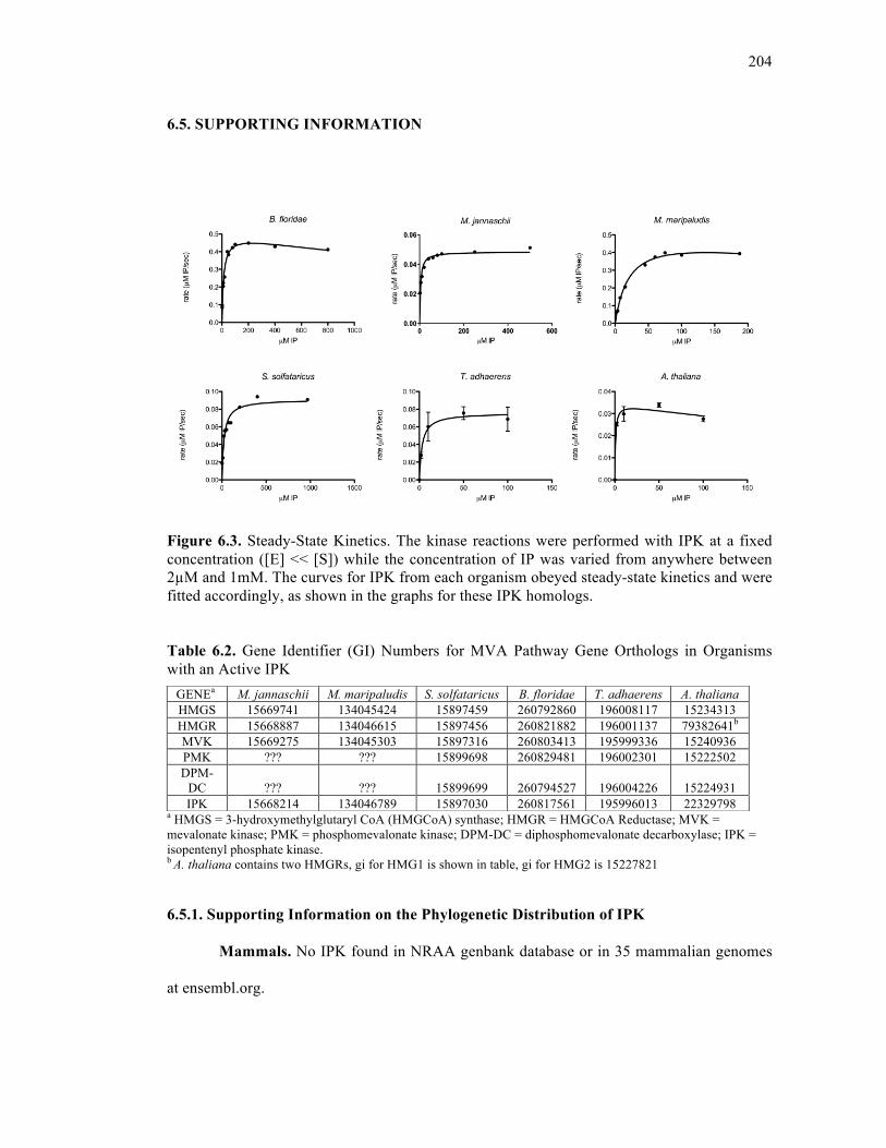
204
6.5. SUPPORTING INFORMATION
Figure 6.3. Steady-State Kinetics. The kinase reactions were performed with IPK at a fixed concentration ([E] << [S]) while the concentration of IP was varied from anywhere between 2µM and 1mM. The curves for IPK from each organism obeyed steady-state kinetics and were fitted accordingly, as shown in the graphs for these IPK homologs. Table 6.2. Gene Identifier (GI) Numbers for MVA Pathway Gene Orthologs in Organisms with an Active IPK
a HMGS = 3-hydroxymethylglutaryl CoA (HMGCoA) synthase; HMGR = HMGCoA Reductase; MVK = mevalonate kinase; PMK = phosphomevalonate kinase; DPM-DC = diphosphomevalonate decarboxylase; IPK = isopentenyl phosphate kinase. b A. thaliana contains two HMGRs, gi for HMG1 is shown in table, gi for HMG2 is 15227821 6.5.1. Supporting Information on the Phylogenetic Distribution of IPK
Mammals. No IPK found in NRAA genbank database or in 35 mammalian genomes
at ensembl.org.
GENEa M. jannaschii M. maripaludis S. solfataricus B. floridae T. adhaerens A. thaliana HMGS 15669741 134045424 15897459 260792860 196008117 15234313 HMGR 15668887 134046615 15897456 260821882 196001137 79382641b MVK 15669275 134045303 15897316 260803413 195999336 15240936 PMK ??? ??? 15899698 260829481 196002301 15222502 DPM-
DC ??? ??? 15899699 260794527 196004226 15224931 IPK 15668214 134046789 15897030 260817561 195996013 22329798
205
Birds and Reptiles. IPK found in the genome of Anolis carolensis (anole lizard) and
in an EST from a colubrid snake (Philodryas olfersii). No IPK in the genomes of chicken,
zebra finch or turkey.
Amphibians. IPK found in one EST from a newt (Notophthalmus viridescens), but
not seen in the genome of Xenopus tropicalis.
Teleost fish. No IPK found in genomes or proteomes of 5 fish (Danio rerio, Oryzias
latipes, Takifugu rubripes, Tetraodon nigrovidris, or Gasterosteus aculeatus). A partial IPK
was assembled from the draft genome of the elephant shark, Callorhinchus milii.
Invertebrate chordates. IPK was found as a predicted gene in the lancelet,
Branchiostoma floridae, and in the hemichordate acorn worm, Saccoglossus kowalevskii. No
IPK was found in Ciona intestinalis or Ciona savignyi.
Echinoderms (Deuterostomes). An almost-complete IPK was assembled from ESTs
of the sea star, Paracentrotus lividus, and an almost complete prediction was made from the
related sea urchin Strongylocentrotus purpuratus.
Arthropods. No IPK was found in the genomes of insects: 12 Drosophila sp., Apis
mellifera, Aedes aegypti, Anopheles gambiae, Culex quinquefasciatus or Pediculus humanus
or the arachnid, Ixodes scapularis. IPK was found in the EST from one crustacean, the lobster
Homarus americanus, but not in the genome of another, Daphnia pulex.
Other Bilaterians. IPK was found in Annelids, in single ESTs from the leech, Hirudo
medicinalis and the earthworm, Eisenia fetida, but was not found in the draft genome of the
polychaete worm, Capitella teleta or the leech, Helobdella robusta. IPK was also found in the
molluscs, in the form of a full length gene prediction from the limpet, Lottia gigantea, an EST
from Aplysia kurodai and a partial prediction in the genome of Aplysia californica. IPK was

206
not found in the platyhelminthes genomes, Schistosoma mansoni or S. japonicum, or in 6
nematode genomes.
Cnidarians. ESTs for IPK were found in two corals (Acropora palmata, Montastraea
faveolata) and an IPK was predicted in the sea anemone Nematostella vectensis, but could not
be found in the Hydra magnapilliata genome.
Other early metazoans. An IPK was predicted in Trichoplax adherens, none could
be found in the sponge Amphimedon queenslandica.
Pre-metazoans (within Holozoa). No IPK was found in the genomes of Monosiga
brevicollis, Salpingoeca rosetta or Capsaspora owczarzaki.
Fungi. IPK was found in one of two chytrid genomes (Spizellomyces punctatus), and
in the early branching fungi Rhizopus oryzae and Mucor circinelloides. No IPK was found in
34 other fungal genomes, though a complete IPK was surprisingly assembled from an EST
library taken from Epichloe festucae, a member of the Pezizomycotina. Since several related
genomes have been sequenced, this may be a case of horizontal transfer (or even EST library
contamination).
Green plants. IPK is found in all green plant genomes surveyed, including flowering
plants, the lycophyte (Selaginella moellendorffii), the moss (Physcomitrella patens), the two
Ostreococcus genomes (O. tauri, O. lucimarinus), two strains of Micromonas pusilla, and
both Chlamydomonas reinhardtii and Volvox carteri. There may be a duplication of IPK in
Physcomitrella, but the two sequences in the genome are very close and might be allelic
variants.
Amoebozoa. IPK is found in the slime molds Dictyostelium discoideum and
Polysphondylium pallidum, and also in three species of Entamoeba (E. hislolytica, E.
207
invadens, E. dispar), though these three are more similar to IPK from a clade of archaea than
to eukaryotes.
Alveolata. IPK is also found in Perkinsus marinus, but not in Apicomplexa (any of 6
Plasmodium genomes, or two Cryptosporidium genomes), or in any of 3 ciliates (Tetrahymena
thermophila, Paramecium tetraurelia, Ichthyophthirius multifilis).
Diatoms. IPK is found in both Thalassiosira pseudonana and Phaeodactylum
tricornutum.
Kinetoplastida. No IPK is found in 4 species of Trypanosoma or 4 species of
Leishmania.
Excavates. No IPK was found in three assemblages of Giardia lamblia or in
Trichomonas vaginalis.
Others. IPK was found in ESTs from Malawimonas jakobiformis, and from the
dinoflagellate Alexandrium tamarense, but not in the genome of Naegleria gruberi
(Heterolobosea)
6.5.2. Ultra-conserved residues
All IPK sequences conserved the following 11 residues, or were missing these regions due to
truncation (number by M. jannaschii): K6, G8, G9, K15, G54, H60, P140, G144, D213, T215,
G216. K221 is conserved in all but one possible gene prediction error, and G253-T254 in the
C-terminus may also be invariant, but mis-predicted in several sequences.
6.5.3. Additional sequences
Additional and fragmentary sequences omitted from the alignment file (omitted for
being redundant or partial, degrading the tree).
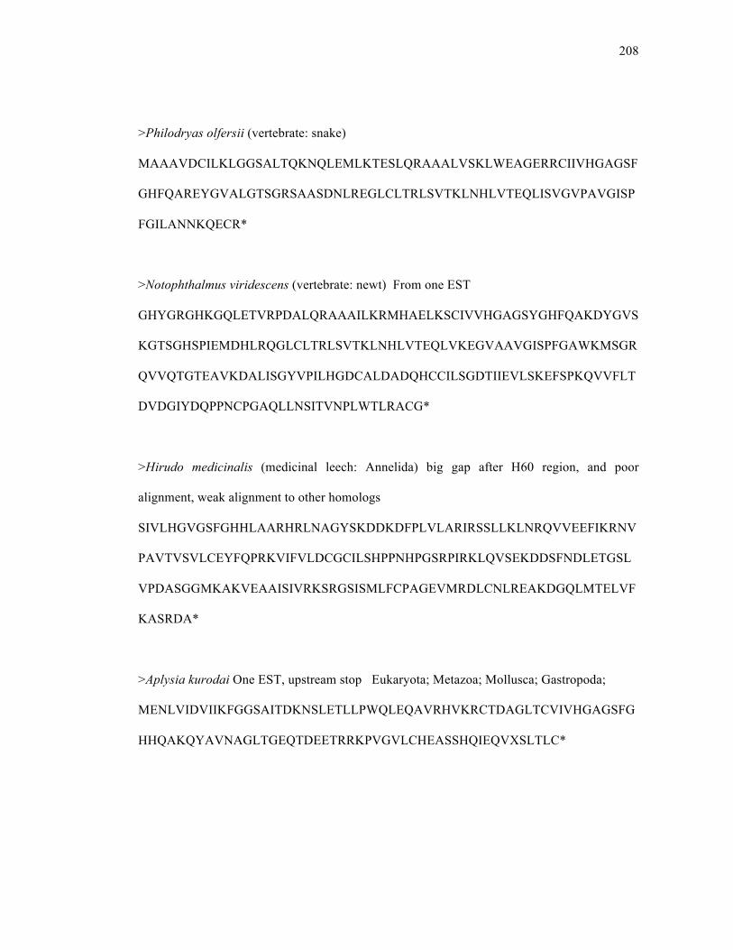
208
>Philodryas olfersii (vertebrate: snake)
MAAAVDCILKLGGSALTQKNQLEMLKTESLQRAAALVSKLWEAGERRCIIVHGAGSF
GHFQAREYGVALGTSGRSAASDNLREGLCLTRLSVTKLNHLVTEQLISVGVPAVGISP
FGILANNKQECR*
>Notophthalmus viridescens (vertebrate: newt) From one EST
GHYGRGHKGQLETVRPDALQRAAAILKRMHAELKSCIVVHGAGSYGHFQAKDYGVS
KGTSGHSPIEMDHLRQGLCLTRLSVTKLNHLVTEQLVKEGVAAVGISPFGAWKMSGR
QVVQTGTEAVKDALISGYVPILHGDCALDADQHCCILSGDTIIEVLSKEFSPKQVVFLT
DVDGIYDQPPNCPGAQLLNSITVNPLWTLRACG*
>Hirudo medicinalis (medicinal leech: Annelida) big gap after H60 region, and poor
alignment, weak alignment to other homologs
SIVLHGVGSFGHHLAARHRLNAGYSKDDKDFPLVLARIRSSLLKLNRQVVEEFIKRNV
PAVTVSVLCEYFQPRKVIFVLDCGCILSHPPNHPGSRPIRKLQVSEKDDSFNDLETGSL
VPDASGGMKAKVEAAISIVRKSRGSISMLFCPAGEVMRDLCNLREAKDGQLMTELVF
KASRDA*
>Aplysia kurodai One EST, upstream stop Eukaryota; Metazoa; Mollusca; Gastropoda;
MENLVIDVIIKFGGSAITDKNSLETLLPWQLEQAVRHVKRCTDAGLTCVIVHGAGSFG
HHQAKQYAVNAGLTGEQTDEETRRKPVGVLCHEASSHQIEQVXSLTLC*
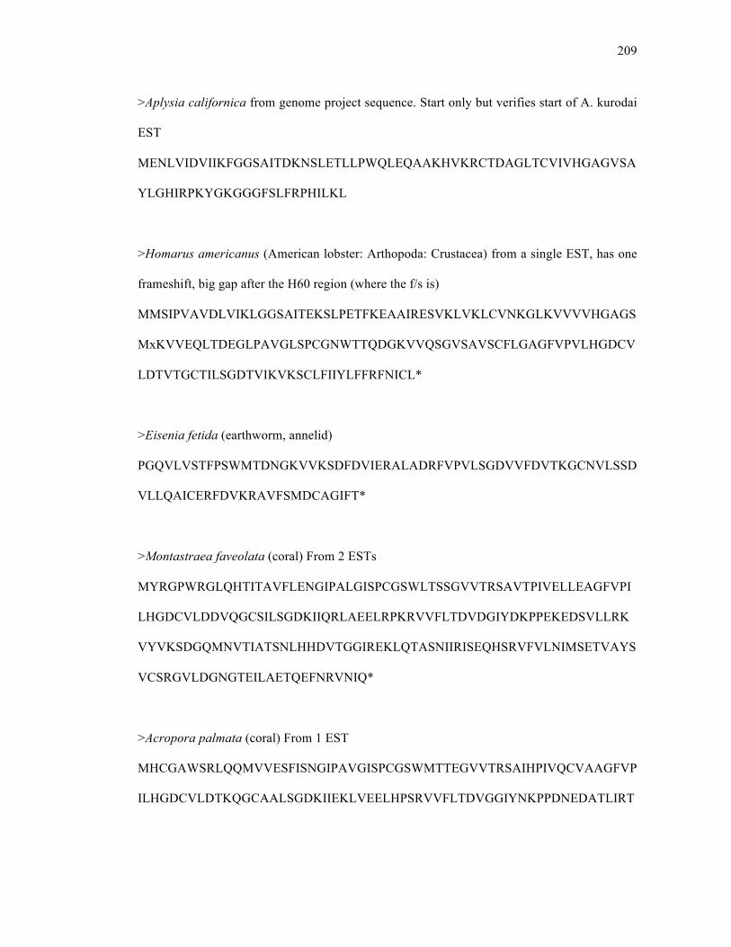
209
>Aplysia californica from genome project sequence. Start only but verifies start of A. kurodai
EST
MENLVIDVIIKFGGSAITDKNSLETLLPWQLEQAAKHVKRCTDAGLTCVIVHGAGVSA
YLGHIRPKYGKGGGFSLFRPHILKL
>Homarus americanus (American lobster: Arthopoda: Crustacea) from a single EST, has one
frameshift, big gap after the H60 region (where the f/s is)
MMSIPVAVDLVIKLGGSAITEKSLPETFKEAAIRESVKLVKLCVNKGLKVVVVHGAGS
MxKVVEQLTDEGLPAVGLSPCGNWTTQDGKVVQSGVSAVSCFLGAGFVPVLHGDCV
LDTVTGCTILSGDTVIKVKSCLFIIYLFFRFNICL*
>Eisenia fetida (earthworm, annelid)
PGQVLVSTFPSWMTDNGKVVKSDFDVIERALADRFVPVLSGDVVFDVTKGCNVLSSD
VLLQAICERFDVKRAVFSMDCAGIFT*
>Montastraea faveolata (coral) From 2 ESTs
MYRGPWRGLQHTITAVFLENGIPALGISPCGSWLTSSGVVTRSAVTPIVELLEAGFVPI
LHGDCVLDDVQGCSILSGDKIIQRLAEELRPKRVVFLTDVDGIYDKPPEKEDSVLLRK
VYVKSDGQMNVTIATSNLHHDVTGGIREKLQTASNIIRISEQHSRVFVLNIMSETVAYS
VCSRGVLDGNGTEILAETQEFNRVNIQ*
>Acropora palmata (coral) From 1 EST
MHCGAWSRLQQMVVESFISNGIPAVGISPCGSWMTTEGVVTRSAIHPIVQCVAAGFVP
ILHGDCVLDTKQGCAALSGDKIIEKLVEELHPSRVVFLTDVGGIYNKPPDNEDATLIRT
210
VFVNPSGKMSVAIATSVLTHDVTGGVCEKLRTASNIVLISGGKTRVFVANVMAEANV
YS
>Nicotiana tabacum (plant) From 2 ESTs, C-terminal open
MEQNKASAPVPPTKRVRCIVKLGGAAITCKNKLETIDEENLTEVSSQLRQALIPNSDSA
KILGMDWSKRPGQSEPPSFVDEFSDQPVADSESFIVVHGAGSFGHFQASKSGVHKGGL
SRPLVKAGFVATRISVTSLNLEIVRALAREGIPSIGMSPFSCGWSTCQRNMTEADISMVI
KAIDAGFIPVLHGDAVLDTLQECTILSGDVIIRHLAAELKPEFVVFLTDVLGVYDRPPV
EPGAVLIREIAVREDGSWSVVKPRLEDTSKPVEFTVAAHDTTGGMVTKITEAAMIAKL
GIDVYITKAGTDHSVKAPP
>Petunia axillaris (plant) from 2 ESTs X's added based on alignment to other plants
MEQKATTVATKRVRCIVKLGGAAITCKNKLETIHEDNLRQVSSQLRQVLIPDSASAKV
LGMDWSKTPGHSEAPSIVDDFSYQPVATSETFIVVHGAGSFGHFQASKSSVHKGGLSR
PLVKAGFVATRISVTSLNLEIVRALAREGIPSIGMSPFSCGWSTCERNMTEADTSMVIK
ALDAGFIPVLHGDAVLDTLQDCTILSGDVIIRHXXXXXXXXXXXXXXXXXXXXXSST
SGPGAVLIREIAVREDGSWSVVKPRLEDASKPVEFTVAAHDTTGGMVTKITEAAMIA
KLGIDVYITKAGTDHSVKALSGILQGGIPDDWLGTAIRYMS*
>Alexandrium tamarense (dinoflagellate) from two ESTs
VLDAAQGAAVLSGDVWMVELCKELKAKSAVFVTDVDGVFTRPPWEEGAELVREILV
DTKTGELELPGVSMSAASHDVTGGLKAELESAAEVLVRAPSVQAVYIVRAGSEGAAQ
ALRGEAPLRGTTLRRKPRD*
211
>Malawimonas jakobiformis (Malawimonadidae) from one EST
MVIIVKFGGSALTDKASFETLRSDALNRCSVAVSQALAAGHRVIIVHGAGSFGHHQA
KRFALSAGLLSHSAAAGSTVTVSAETERAWSAIAPHEQQLGLAHCRASVQRLNAHVV
HSLLRRNVPAVTMSAFPNWFTDGKRLVSDIVPPVLAALERGLVPVLHGDVVMDHAQ
GITVLSGDVIWRCFAVRCRRRRSAR
>Entamoeba dispar (Apicomplexan)
MNSIPNLIILKIGGSYLTEKNRVDGPPVLENIHVFSKCLAQFIHSHPKQPIILAHGAGSFG
HVPAAKYHLAEGFHKTGVIECEMAMQELSSVIVNSLIKEGVSAIPFHPFNFVVTENKRI
VDMYLQPLQMMINQGIIPVVHGDIAMDIIQGSCILSADQLVPELAIRFGCSRIGFICNTP
VLNDKGEVIPLINEQNYDSIKKFLHGCKGVDVTGGMAGKISELMIAAKKHMIQSYVF
EGTKECLELFLEGNDVGTKVCQ*
>Emiliania huxleyi (Haptophyceae)
MLITRHAAILAAGLLLLTPVAGAFLFLGRRLRRRALRSARCTLVVKLGGSAVTDKTCF
ETVRVAALRETACALSRSPLLAGTVLVHGAGSFGHFHAREHGVSRGTAHSAFSWRGF
ALTRSSVTRLNGIVLTALLEQGIAACGLPPFPRWVLCGGALTDTDEPLGEVRSLLSRG
VVPVLHGDAVFDEARGAAILSGDTLVEELRRLPPSARPAPSSLTDVAGIFHRPPGEDG
AALLRRIVVGPSGEVVDLPQMRTAAHDVTGGVAAERSAPAALHAFPSLIPAALLVCR
LRRS*
>Chloroflexus aurantiacus J-10-fl
MYTFIKFGGSVITDKTGREAADLVVIERLAHAVAEARAADPNLALVLGHGSGSFGHH
YAARYGVHRGIPLSADHTGFALTAAAALRLNRIVVDTLLAAQVPAVSFQPSASLQST
212
NGQIITWETAPIAEALQRRLVPVIHGDVAFDTAQGTAIISTEALLSFLALRSPLQPRRIIL
VGEAAVYTADPHRDPTAQPIPLINQENIAQVLVMTGGSRAADVTGGMRSKIELMWHL
IERLPELEVTLIGPDPALLTAALLGQSLAMGTVIKRW*
>Roseiflexus sp. RS-1
MIVFIKFGGSVITDKQQQERADIDTIRQLAEELRQALDAARDLCVIVGHGSGSFGHVY
AQRYGIHRGLAPDDDWMGFALTSGAALRLNRIVVDELLAAGIPALALQPSTTLLARG
GRLVHWETGSLERALERRMVPVIHGDVAFDDVQGSAIISTEQLLAHLATLPTLRPARI
VLVGEAGVYTADPRINPQAERIARIDRRNIANVLAGAGGSHGVDVTGGMRSKVELM
WQLVQTVPGLQVYLIGPKPGSLKRALLGDDTVEGTVIVGG*
>Streptomyces fradiae (fomA homolog)
MTPDFLAIKVGGSLFSRKDEPGSLDDDAVTRFARNFARLAETYRGRMVLISGGGAFG
HGAIRDHDTAHAFSLAGLTEATFEVKKRWAEKLRQIGVDAFPLQLAAMCTLRDGTPQ
LRSEVLRSVLDHGVLPVLAGDALFDEHGKLWAFSSDRVPEVLLPMVEGRLRVVTLTD
VDGIVTDGAGGDAILPEIDARSPQQAYAALWGSSEWDATGAMHTKLDALVTCARRG
AECFIMRGRPDSDLEFLTAPFSSWPAHVRSTRITTTASV*
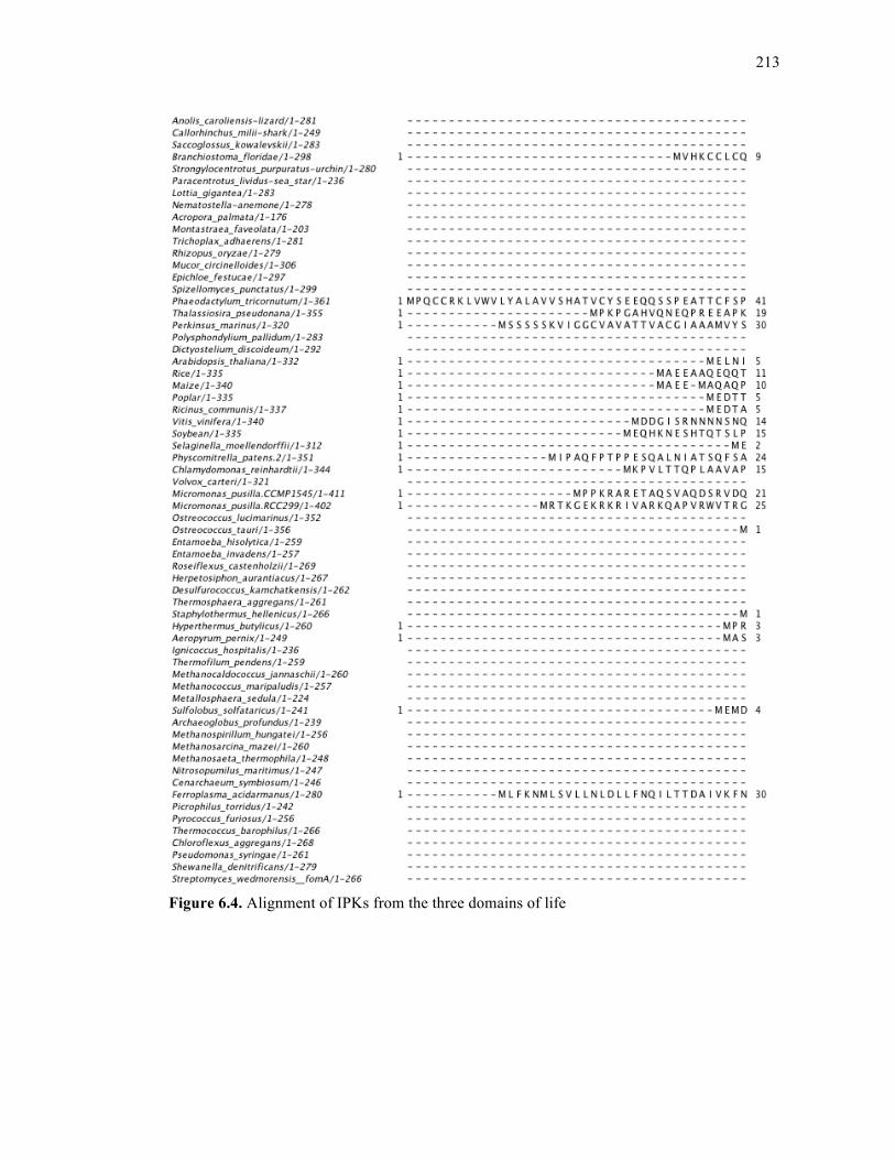
213
Figure 6.4. Alignment of IPKs from the three domains of life
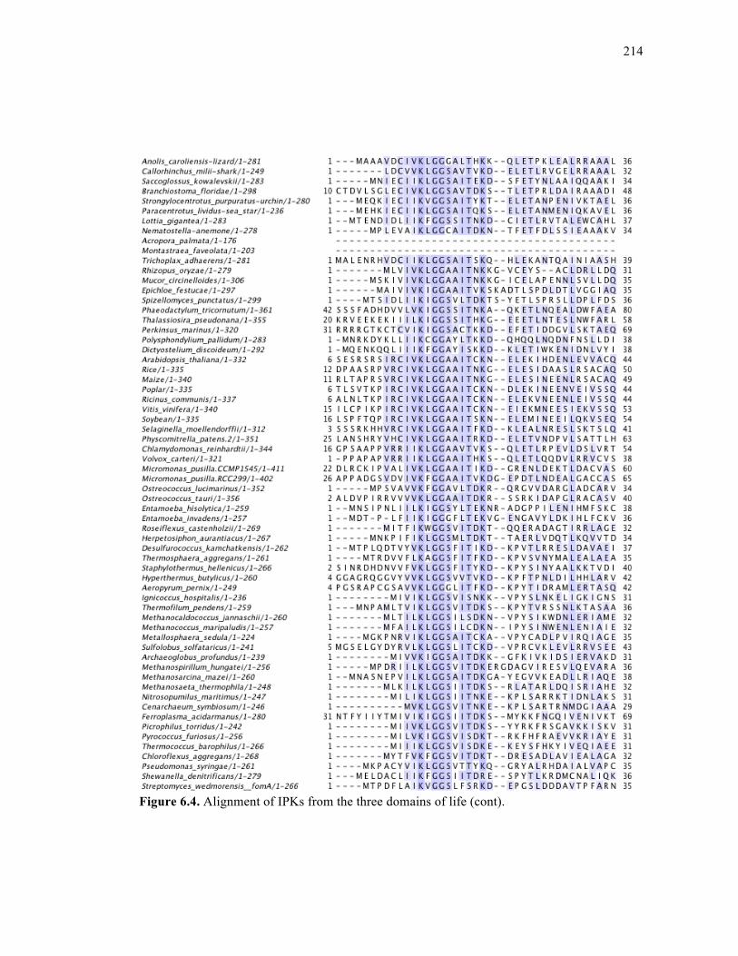
214
Figure 6.4. Alignment of IPKs from the three domains of life (cont).
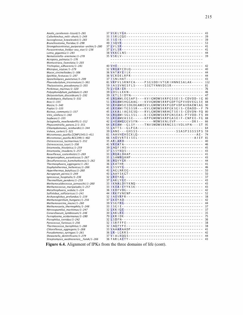
215
Figure 6.4. Alignment of IPKs from the three domains of life (cont).
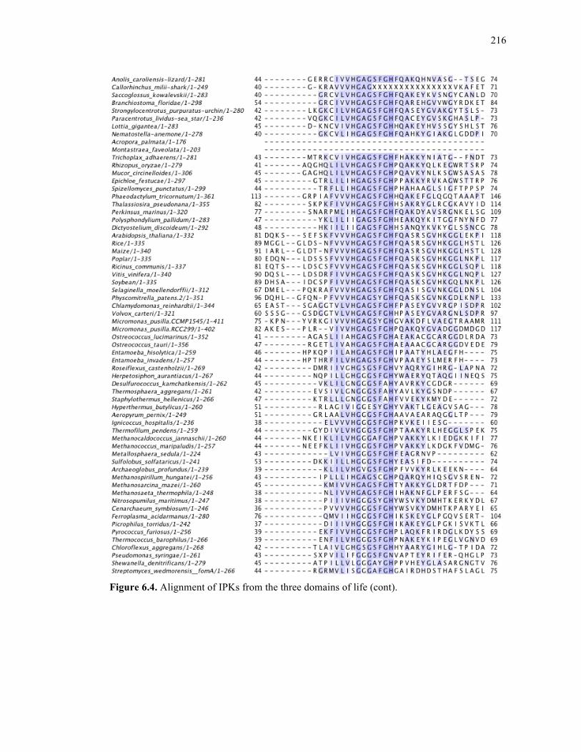
216
Figure 6.4. Alignment of IPKs from the three domains of life (cont).
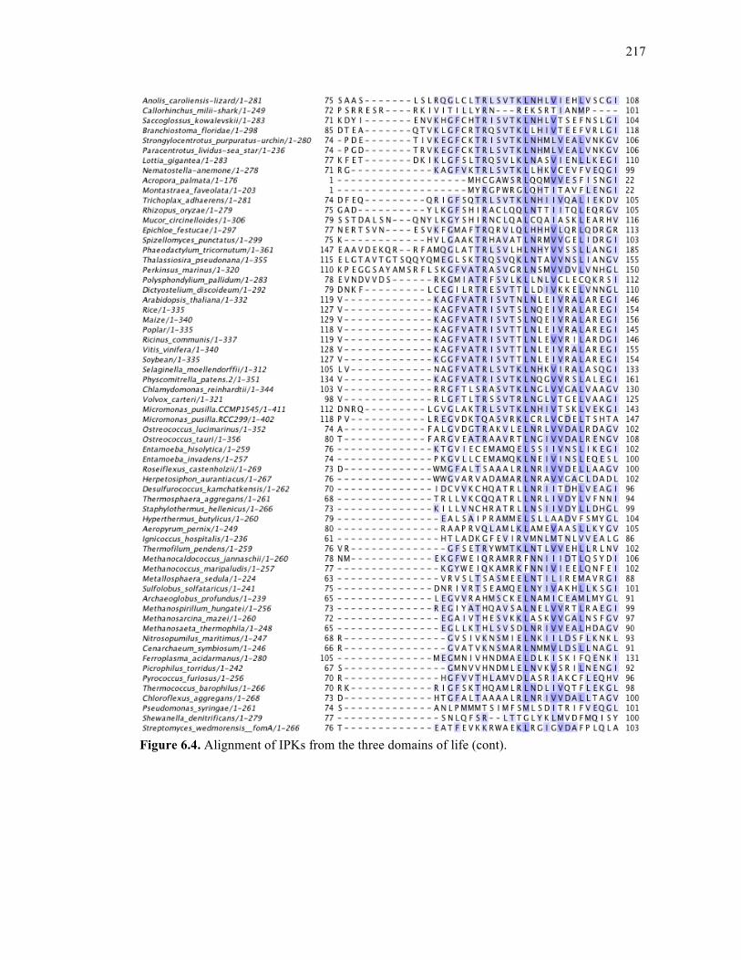
217
Figure 6.4. Alignment of IPKs from the three domains of life (cont).
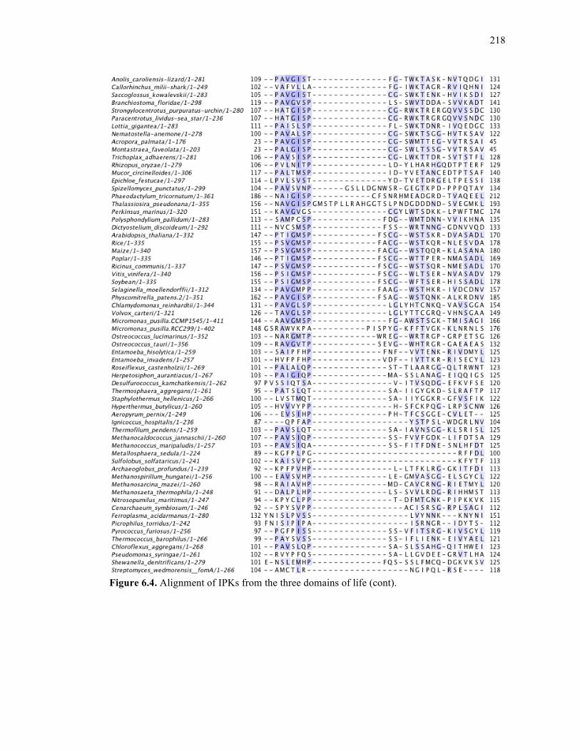
218
Figure 6.4. Alignment of IPKs from the three domains of life (cont).
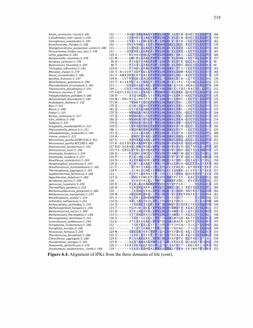
219
Figure 6.4. Alignment of IPKs from the three domains of life (cont).

220
Figure 6.4. Alignment of IPKs from the three domains of life (cont).
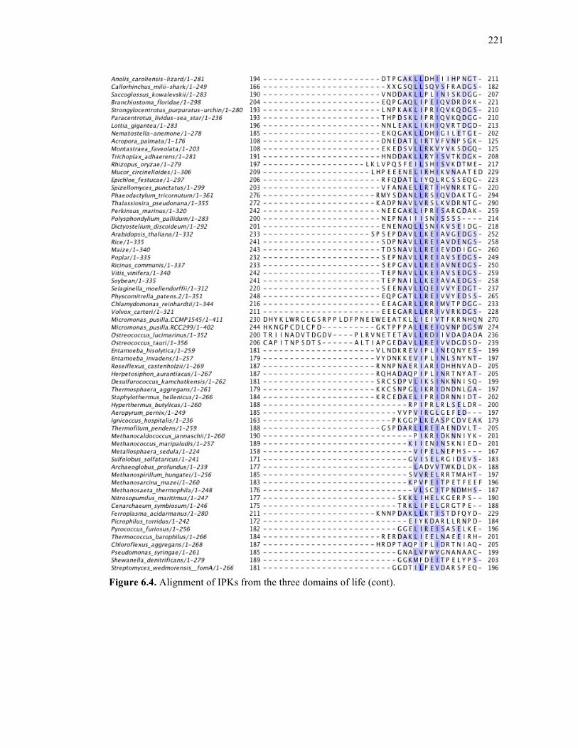
221
Figure 6.4. Alignment of IPKs from the three domains of life (cont).
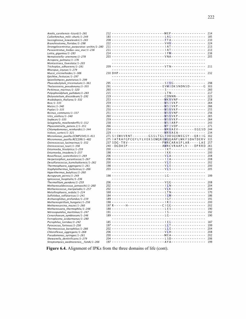
222
Figure 6.4. Alignment of IPKs from the three domains of life (cont).
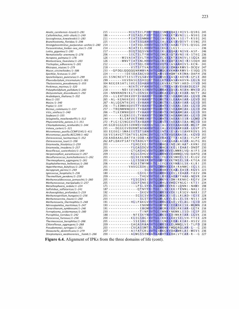
223
Figure 6.4. Alignment of IPKs from the three domains of life (cont).
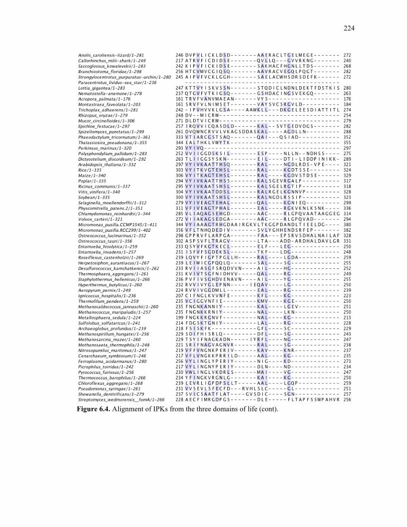
224
Figure 6.4. Alignment of IPKs from the three domains of life (cont).

225
Figure 6.4. Alignment of IPKs from the three domains of life (cont).
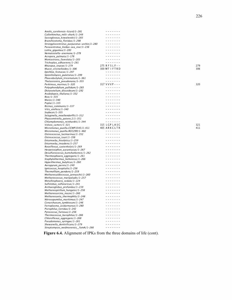
226
Figure 6.4. Alignment of IPKs from the three domains of life (cont).
227
ACKNOWLEDGEMENTS
The text of chapter 6, in part, has been submitted for publication of the material as it
may appear in Chemical Communications, 2010, Dellas, Nikki; Manning, Gerard, Noel,
Joseph P. I am the first author of this paper. Gerard Manning and Joseph P. Noel are the
corresponding authors. I was responsible for all gene cloning, enzyme expression, purification,
and kinetic characterization of IPK and its homologs. Gerard Manning was responsible for the
bioinformatic and phylogenetic analysis of IPK and its homologs. All experiments were
performed under the supervision of Joseph P. Noel.
REFERENCES 1. Smit, A.; Mushegian, A., Biosynthesis of isoprenoids via mevalonate in Archaea: the
lost pathway. Genome research 2000, 10 (10), 1468-1484. 2. Grochowski, L. L.; Xu, H.; White, R. H., Methanocaldococcus jannaschii uses a
modified mevalonate pathway for biosynthesis of isopentenyl diphosphate. Journal of Bacteriology 2006, 188 (9), 3192-3198.
3. Lombard, J.; Moreira, D., Origins and early evolution of the mevalonate pathway of
isoprenoid biosynthesis in the three domains of life. Mol Biol Evol. 4. Dellas, N.; Noel, J. P., Mutation of archaeal isopentenyl phosphate kinase highlights
mechanism and guides phosphorylation of additional isoprenoid monophosphates. ACS Chem Biol 2010, 5 (6), 589-601.
5. Pakhomova, S.; Bartlett, S. G.; Augustus, A.; Kuzuyama, T.; Newcomer, M. E.,
Crystal structure of fosfomycin resistance kinase FomA from Streptomyces wedmorensis. The Journal of biological chemistry 2008, 283 (42), 28518-28526.
6. Nagegowda, D. A., Plant volatile terpenoid metabolism: biosynthetic genes,
transcriptional regulation and subcellular compartmentation. FEBS Lett 2010, 584 (14), 2965-73.
7. Chen, M.; Poulter, C. D., Characterization of thermophilic archaeal isopentenyl
phosphate kinases. Biochemistry 2010, 49 (1), 207-17. 8. Birney, E.; Clamp, M.; Durbin, R., GeneWise and Genomewise. Genome Res 2004,
14 (5), 988-95.
228
9. Edgar, R. C., MUSCLE: multiple sequence alignment with high accuracy and high throughput. Nucleic Acids Res 2004, 32 (5), 1792-7.
10. Larkin, M. A.; Blackshields, G.; Brown, N. P.; Chenna, R.; McGettigan, P. A.;
McWilliam, H.; Valentin, F.; Wallace, I. M.; Wilm, A.; Lopez, R.; Thompson, J. D.; Gibson, T. J.; Higgins, D. G., Clustal W and Clustal X version 2.0. Bioinformatics 2007, 23 (21), 2947-8.
11. Waterhouse, A. M.; Procter, J. B.; Martin, D. M.; Clamp, M.; Barton, G. J., Jalview
Version 2--a multiple sequence alignment editor and analysis workbench. Bioinformatics 2009, 25 (9), 1189-91.
12. Boguski, M. S.; Lowe, T. M.; Tolstoshev, C. M., dbEST--database for "expressed
sequence tags". Nat Genet 1993, 4 (4), 332-3. 13. Birney, E.; Andrews, D.; Bevan, P.; Caccamo, M.; Cameron, G.; Chen, Y.; Clarke, L.;
Coates, G.; Cox, T.; Cuff, J.; Curwen, V.; Cutts, T.; Down, T.; Durbin, R.; Eyras, E.; Fernandez-Suarez, X. M.; Gane, P.; Gibbins, B.; Gilbert, J.; Hammond, M.; Hotz, H.; Iyer, V.; Kahari, A.; Jekosch, K.; Kasprzyk, A.; Keefe, D.; Keenan, S.; Lehvaslaiho, H.; McVicker, G.; Melsopp, C.; Meidl, P.; Mongin, E.; Pettett, R.; Potter, S.; Proctor, G.; Rae, M.; Searle, S.; Slater, G.; Smedley, D.; Smith, J.; Spooner, W.; Stabenau, A.; Stalker, J.; Storey, R.; Ureta-Vidal, A.; Woodwark, C.; Clamp, M.; Hubbard, T., Ensembl 2004. Nucleic Acids Res 2004, 32 (Database issue), D468-70.

229
Chapter 7
Conclusions
230
7.1. Overview
Isoprenoid biosynthesis constitutes an immensely diverse, highly branched network of
pathways that spans both primary and secondary (specialized) metabolism in all organisms.
The scope of this work includes two types of enzymes: terpene cyclases of secondary
metabolism and isopentenyl phosphate kinase of primary metabolism.
Terpene cyclases are a fascinating class of enzymes that, based on structure, function,
and some sequence motif conservation, are thought to have evolved from short-chain prenyl
diphosphates of primary metabolism, which are responsible for the biosynthesis of GPP, FPP,
or GGPP molecules that monoterpene, sesquiterpene, or diterpene synthases utilize as
substrates for their electrophilic cyclization reactions, respectively. From a global perspective,
this work demonstrates the adaptability of sesquiterpene cyclases to mutation without
significant loss of function, but instead a gain of product promiscuity.1 This research also
shows how both substrate and product promiscuity are important from a structural perspective
in terms of both substrate orientation and dynamics of the isoprenoid tail within the active
site.2
Isopentenyl monophosphate kinase was originally thought to be solely important for
archaeal isoprenoid biosynthesis.3, 4 However structural and functional studies described here
have allowed for the identification of a uniquely important residue within the enzyme active
site.5 This residue behaves as a marker to locate IPK homologs from other kingdoms, and
successful characterization of these homologs proves that they are indeed true IPKs. These
results imply the potential existence of a branched mevalonate pathway in Archaea and
Eukarya.
231
7.2. Terpene synthases of specialized metabolism
Terpene cyclases constitute a class of enzymes that biosynthesize a chemically diverse
profile of compounds known as terpenes. In this work, the study of several sesquiterpene
synthases, including TEAS, HPS, and PAS, describes experimental findings associated with
the structural, functional, and chemical properties of these enzymes and their small molecule
products, which are all derived from the common substrate, FPP.
TEAS, whose major product is 5-epi aristolochene (5EA), can be converted to an
HPS-like enzyme (termed “TEAS M9”) that produces premnaspirodiene (PSD) as its major
product by mutation of nine amino acids that are located in and around the TEAS active site.6
On the initial mutational pathway towards M9, TEAS mutants display significant upregulation
of a minor product, 4-epi eremophilene (4EE); the mechanism for its formation represents a
hybrid of the 5EA mechanism and the PSD mechanism.6 In order to fully characterize the
catalytic landscape of the enzymes spanning the sequence space between TEAS and M9, a
mutant library including all possible combinations of these nine mutations was created, as
described in Chapter 2.1 Although the catalytic landscape shows, on average, that the pathway
towards the upregulation of another major product requires navigation through a promiscuous
terrain, certain mutants bypass this terrain, demonstrating “jumps” to other products upon
mutation of only one or two residues. From an evolutionary standpoint, these results cannot
address the question of ancestry, that is, which enzyme (TEAS or HPS) came before the other.
No individual mutations are found to control product specificity in one direction or the other;
for example, no single mutation always upregulates a specific product, regardless of context.
Instead, these mutations are very context dependent; that is, one amino acid mutation can
contribute differently to phenotype in the context of its local environment. However, these
results do, in part, support the theory that terpene cyclases were derived from a promiscuous
232
ancestor. In a selection of mutants, the observation of drastic product shifts accompanying
single amino acid changes indicates that these cyclases have the ability to rapidly evolve a
significantly different chemical profile with only a small change in sequence space. This
ability could be a reflection of a sessile organism’s approach toward environmental
adaptability.
More recent discoveries suggest that not only product promiscuity, but also substrate
promiscuity may play a role in controlling these interesting and often chemically complex
terpene cyclase product profiles.2 For example, the observation that FPP synthases can
produce a certain percentage of cis-FPP in addition to its major product, trans-FPP, indicates
the availability of an additional substrate for sesquiterpene cyclases.7 Chapter 3 addresses this
complex question in both a structural and a functional sense. Surprisingly, both trans-FPP and
cis-FPP are substrates for TEAS, and each generates a trans-derived or cis-derived product
spectrum including unique major products, 5EA and (+)-2-epi-prezizaene, respectively.2
Functional comparisons between TEAS wt and TEAS M4 (a product-promiscuous mutant
from the M9 library that produces equal amounts of 5EA, 4EE, and PSD) reveal that TEAS
M4 is also more promiscuous than wild type when using cis-FPP as a substrate; this indicates
that the level of product promiscuity, at least in this case, is independent of a cis- or trans-
derived substrate. Comparisons of crystal structures of TEAS wt and TEAS M4 in complex
with non-hydrolyzable substrate analogs 2F-FPP and cis-2F-FPP demonstrate that product
promiscuity is directly related to dynamics of the isoprenoid chain in the active site. For
example, the structures of TEAS M4 in complex with either 2F-FPP or cis-2F-FPP show
significantly less electron density for the isoprenyl tail of the ligand compared with both
TEAS wt structures.2
233
The product promiscuity of another terpene cyclase, patchoulol synthase (PAS) can
also be altered in a rather surprisingly way: through mutations in an amino terminal region.
The amino-terminal region is not thought to have a direct role in the terpene cyclase catalytic
reaction. It is, however, thought to aid in active site capping to prevent premature release of a
carbocation intermediates during the course of the reaction.8-11 Mutation of both promiscuous
and nonpromiscuous sesquiterpene cyclases reveals that certain cyclases, such as PAS, exhibit
drastic product profile changes upon mutation at the RP motif in its amino terminal region,
while other less promiscuous sesquiterpene cyclases exhibit little to no change. Although
previous work speculates a general role for the amino-terminal region of these proteins,8-11 this
work articulates a direct role for this region, involving the RP motif. The series of mutations
performed at both Arg and Pro of this motif in PAS suggest that while the Arg provides an
anchor for the N-terminal tail through a salt bridge interaction with a C-terminal residue, the
Pro provides the structural rigidity necessary to complete this task.
7.3. IPK of primary metabolism
7.3.1. Overview
Isopentenyl phosphate kinase (IPK) is an enzyme initially characterized from the
thermophilic archaeon M. jannaschii that phosphorylates isopentenyl monophosphate (IP) to
isopentenyl diphosphate (IPP).4 IPP is one of two building blocks for all downstream
isoprenoids, and it is therefore essential that its mechanism(s) of formation are understood
among all three domains of life. IPK in particular was originally thought to be an enzyme
exclusive to archaea, representing one of two enzymes required to complete the missing steps
of the MVA pathway in this domain of life.3, 4 In the classic mevalonate pathway, the last two
steps leading to the production of IPP include genes encoding PMK and DPM-DC that

234
perform phosphorylation and decarboxylation of phosphomevalonate and
diphosphomevalonate, respectively. In archaea, based on lack of evidence for these two
orthologs and also the partial identification of an alternative route for the production of IPP,
the reaction is thought to proceed in the reverse order, involving a decarboxylase followed by
a kinase.4 The kinase step is performed by IPK, and its structural and functional
characterization, as discussed in chapter 5, allows for: 1) engineering of a deeper active site
cavity for successful turnover of longer chained isoprenoid phosphates, and 2) the
identification of an active site histidine residue that is unique to this member of the family and
can therefore be used as a marker to identify IPK homologs from other kingdoms of life.5
7.3.2. Applications for IPK chain-length mutants
The goals behind engineering IPK to accept longer chained isoprenoid phosphates are
two-fold: 1) to design a synthetic metabolic pathway, and 2) to synthesize isoprenoid
diphosphate analogs. Over the past decade, there have been immense efforts on the front of
MVA pathway upregulation, which has been accomplished through heterologous expression
of MVA pathway enzymes in E. coli or upregulation in S. cerevisiae.12-15 These efforts are
geared towards the production of large quantities of terpenes, carotenoids, and other secondary
metabolites that have, or can easily be derivatized into, compounds that have biological
activity and medicinal value. Upregulation or overexpression of this pathway causes problems
associated with metabolic flux,14 production of unwanted byproducts (such as farnesol),12 and
feedback inhibition (such FPP inhibition of mevalonate kinase).16 As discussed in chapter 5,
IPK has been engineered to bind and turn over FP to FPP, which is extremely valuable
towards the design of a much simpler synthetic metabolic pathway that would not suffer from
any of the problems discussed above. For example, the overproduction of any given
235
sesquiterpene includes only three enzymatic steps: 1) phosphorylation of an inexpensive
substrate such as farnesol (or an ester of farnesol) to FP, 2) phosphorylation of FP to FPP
performed by the IPK chain-length mutant, and 3) cyclization by a sesquiterpene cyclase to
the final sesquiterpene product. Although steps two and three are characterized, future work
will address the design of a kinase that can phosphorylate isoprenoid alcohols of varying
lengths.
Examples of chemoenzymatic synthesis of isoprenoid diphosphate analogs include the
synthesis of fluorescent derivatives17 and radiolabeled derivatives (through reaction of the
substrate with radiolabeled ATP or ATP-γS), which would be useful for following in vivo
prenylation or any other primary or secondary metabolic process involving isoprenoids.
7.3.3. Implications for active eukaryotic IPKs
The identification of His60 as a critical residue for binding and catalysis in IPK has
been monumental in the location of IPK homologs in other kingdoms of life, as discussed in
chapter 6.5 This identification and successful characterization of eukaryotic IPKs has
implications for the presence of an alternative MVA pathway in organisms that already have a
fully functioning classic MVA pathway. Although IPK has a spotty distribution throughout
the animal kingdom, the presence of IPK homologs in all green plants is an indication of its
marked importance within this kingdom of life. In contrast to other kingdoms of life,
isoprenoid biosynthesis within the plant kingdom is already very complex and includes both
the DXP pathway, which operates in plant plastids, and the MVA pathway, which operates in
the cytosol and/or other organelles.18-20 Since compartmentalization is already a feature of
isoprenoid biosynthesis in plants, it is tempting to speculate that a branched MVA pathway
may allow for even further compartmentalization of certain enzymes within this pathway.
236
Since the DXP pathway and MVA pathway play different roles in plant isoprenoid
biosynthesis (for example, GPP and GGPP are synthesized from the DXP pathway while FPP
is synthesized from the MVA pathway), it is also possible that the branching mevalonate
pathway directs the biosynthesis of specific primary or secondary metabolites, exerting yet
another dimension of control over isoprenoid biosynthesis in the plant kingdom.
Ultimately, these hypotheses remain speculative until the missing piece to the
alternative mevalonate pathway has been identified and characterized. This missing piece is
the decarboxylase that catalyzes the first step after bifurcation from the classical pathway: the
step that converts phosphomevalonate to isopentenyl monophosphate. One gene candidate that
has been proposed to catalyze this reaction is the gene MJ0403 from M. jannaschii,4 which is
a putative dioxygenase that has sequence homology to LigAB and MEMO (mediator of
ErbB2-driven cell motility). LigAB is a ring-cleaving extradiol dioxygenase that binds non-
heme Fe2+ and plays a role in lignin degradation,21 while MEMO is a human protein with
homology to dioxygenases but no known catalytic function, and no experimental evidence
demonstrating ability to bind a metal ion.22 Although we have cloned and purified the MJ0403
putative decarboxylase, we have not been able to establish assay conditions that demonstrate
successful turnover of phosphomevalonate to IP. Although efforts on assay optimization
(including variation of metal ion type, metal ion concentration, and presence or absence of
potential co-factors) are ongoing, the search for other decarboxylase candidates is under way.
REFERENCES
1. O'Maille, P. E.; Malone, A.; Dellas, N.; Andes Hess, B., Jr.; Smentek, L.; Sheehan, I.; Greenhagen, B. T.; Chappell, J.; Manning, G.; Noel, J. P., Quantitative exploration of the catalytic landscape separating divergent plant sesquiterpene synthases. Nature chemical biology 2008, 4 (10), 617-623.
237
2. Noel, J. P.; Dellas, N.; Faraldos, J. A.; Zhao, M.; Hess, B. A., Jr.; Smentek, L.; Coates, R. M.; O'Maille, P. E., Structural elucidation of cisoid and transoid cyclization pathways of a sesquiterpene synthase using 2-fluorofarnesyl diphosphates. ACS chemical biology 2010, 5 (4), 377-392.
3. Smit, A.; Mushegian, A., Biosynthesis of isoprenoids via mevalonate in Archaea: the
lost pathway. Genome research 2000, 10 (10), 1468-1484. 4. Grochowski, L. L.; Xu, H.; White, R. H., Methanocaldococcus jannaschii uses a
modified mevalonate pathway for biosynthesis of isopentenyl diphosphate. Journal of Bacteriology 2006, 188 (9), 3192-3198.
5. Dellas, N.; Noel, J. P., Mutation of archaeal isopentenyl phosphate kinase highlights
mechanism and guides phosphorylation of additional isoprenoid monophosphates. ACS Chem Biol 2010, 5 (6), 589-601.
6. Greenhagen, B. T.; O'Maille, P. E.; Noel, J. P.; Chappell, J., Identifying and
manipulating structural determinates linking catalytic specificities in terpene synthases. Proceedings of the National Academy of Sciences of the United States of America 2006, 103 (26), 9826-9831.
7. Thulasiram, H. V.; Poulter, C. D., Farnesyl diphosphate synthase: the art of
compromise between substrate selectivity and stereoselectivity. J Am Chem Soc 2006, 128 (49), 15819-23.
8. Whittington, D. A.; Wise, M. L.; Urbansky, M.; Coates, R. M.; Croteau, R. B.;
Christianson, D. W., Bornyl diphosphate synthase: structure and strategy for carbocation manipulation by a terpenoid cyclase. Proceedings of the National Academy of Sciences of the United States of America 2002, 99 (24), 15375-15380.
9. Starks, C. M.; Back, K.; Chappell, J.; Noel, J. P., Structural basis for cyclic terpene
biosynthesis by tobacco 5-epi-aristolochene synthase. Science 1997, 277 (5333), 1815-1820.
10. Hyatt, D. C.; Youn, B.; Zhao, Y.; Santhamma, B.; Coates, R. M.; Croteau, R. B.;
Kang, C., Structure of limonene synthase, a simple model for terpenoid cyclase catalysis. Proceedings of the National Academy of Sciences of the United States of America 2007, 104 (13), 5360-5365.
11. Little, D. B.; Croteau, R. B., Alteration of product formation by directed mutagenesis
and truncation of the multiple-product sesquiterpene synthases delta-selinene synthase and gamma-humulene synthase. Archives of Biochemistry and Biophysics 2002, 402 (1), 120-135.
12. Asadollahi, M. A.; Maury, J.; Moller, K.; Nielsen, K. F.; Schalk, M.; Clark, A.;
Nielsen, J., Production of plant sesquiterpenes in Saccharomyces cerevisiae: effect of ERG9 repression on sesquiterpene biosynthesis. Biotechnol Bioeng 2008, 99 (3), 666-77.

238
13. Ohto, C.; Muramatsu, M.; Obata, S.; Sakuradani, E.; Shimizu, S., Overexpression of
the gene encoding HMG-CoA reductase in Saccharomyces cerevisiae for production of prenyl alcohols. Appl Microbiol Biotechnol 2009, 82 (5), 837-45.
14. Martin, V. J.; Pitera, D. J.; Withers, S. T.; Newman, J. D.; Keasling, J. D.,
Engineering a mevalonate pathway in Escherichia coli for production of terpenoids. Nature biotechnology 2003, 21 (7), 796-802.
15. Pitera, D. J.; Paddon, C. J.; Newman, J. D.; Keasling, J. D., Balancing a heterologous
mevalonate pathway for improved isoprenoid production in Escherichia coli. Metabolic engineering 2007, 9 (2), 193-207.
16. Fu, Z.; Voynova, N. E.; Herdendorf, T. J.; Miziorko, H. M.; Kim, J. J., Biochemical
and structural basis for feedback inhibition of mevalonate kinase and isoprenoid metabolism. Biochemistry 2008, 47 (12), 3715-24.
17. Hovlid, M. L.; Edelstein, R. L.; Henry, O.; Ochocki, J.; DeGraw, A.; Lenevich, S.;
Talbot, T.; Young, V. G.; Hruza, A. W.; Lopez-Gallego, F.; Labello, N. P.; Strickland, C. L.; Schmidt-Dannert, C.; Distefano, M. D., Synthesis, properties, and applications of diazotrifluropropanoyl-containing photoactive analogs of farnesyl diphosphate containing modified linkages for enhanced stability. Chemical biology & drug design 2010, 75 (1), 51-67.
18. Sapir-Mir, M.; Mett, A.; Belausov, E.; Tal-Meshulam, S.; Frydman, A.; Gidoni, D.;
Eyal, Y., Peroxisomal localization of Arabidopsis isopentenyl diphosphate isomerases suggests that part of the plant isoprenoid mevalonic acid pathway is compartmentalized to peroxisomes. Plant Physiol 2008, 148 (3), 1219-28.
19. Carrero-Lerida, J.; Perez-Moreno, G.; Castillo-Acosta, V. M.; Ruiz-Perez, L. M.;
Gonzalez-Pacanowska, D., Intracellular location of the early steps of the isoprenoid biosynthetic pathway in the trypanosomatids Leishmania major and Trypanosoma brucei. Int J Parasitol 2009, 39 (3), 307-14.
20. Hartman, I. Z.; Liu, P.; Zehmer, J. K.; Luby-Phelps, K.; Jo, Y.; Anderson, R. G.;
DeBose-Boyd, R. A., Sterol-induced dislocation of 3-hydroxy-3-methylglutaryl coenzyme A reductase from endoplasmic reticulum membranes into the cytosol through a subcellular compartment resembling lipid droplets. J Biol Chem 2010, 285 (25), 19288-98.
21. Sugimoto, K.; Senda, T.; Aoshima, H.; Masai, E.; Fukuda, M.; Mitsui, Y., Crystal
structure of an aromatic ring opening dioxygenase LigAB, a protocatechuate 4,5-dioxygenase, under aerobic conditions. Structure 1999, 7 (8), 953-65.
22. Qiu, C.; Lienhard, S.; Hynes, N. E.; Badache, A.; Leahy, D. J., Memo is homologous
to nonheme iron dioxygenases and binds an ErbB2-derived phosphopeptide in its vestigial active site. J Biol Chem 2008, 283 (5), 2734-40.
Related Documents