Accepted by W. Sterrer: 21 Nov. 2013; published: 6 Jan. 2014 ZOOTAXA ISSN 1175-5326 (print edition) ISSN 1175-5334 (online edition) Copyright © 2014 Magnolia Press Zootaxa 3753 (2): 177–186 www.mapress.com/zootaxa/ Article 177 http://dx.doi.org/10.11646/zootaxa.3753.2.7 http://zoobank.org/urn:lsid:zoobank.org:pub:74D353B7-4D92-4674-938C-B7A46BD5E831 A new species of Supramontana Carbayo & Leal-Zanchet (Platyhelminthes, Continenticola, Geoplanidae) from the Interior Atlantic Forest LISANDRO NEGRETE 1, 2 , ANA MARIA LEAL-ZANCHET 3 & FRANCISCO BRUSA 1,2,4 1 División Zoología Invertebrados, Facultad de Ciencias Naturales y Museo, Universidad Nacional de La Plata, Paseo del Bosque s/n, La Plata, Argentina 2 CONICET 3 Instituto de Pesquisas de Planárias, Universidade do Vale do Rio dos Sinos, 93022-000 São Leopoldo, Rio Grande do Sul, Brazil 4 Corresponding author. E-mail: [email protected] Abstract Supramontana argentina sp. nov. (Platyhelminthes, Continenticola, Geoplanidae) from north-eastern Argentina is herein described. The new species differs from Supramontana irritata Carbayo & Leal-Zanchet, 2003 from Brazil, the only spe- cies of this genus so far described, by external and internal morphological characters. Supramontana argentina sp. nov. is characterized by having a colour pattern with a yellowish median band, thin para-median black stripes, and two dark grey lateral bands on the dorsal surface. The most outstanding features of the internal morphology are a ventral cephalic retractor muscle almost circular in cross section, prostatic vesicle extrabulbar, tubular and very long, and penis papilla con- ical and blunt with a sinuous ejaculatory duct. Key words: triclads, land planarian, Geoplaninae, Argentina, Neotropical Region Introduction The taxonomy of land planarians (Geoplanidae) is mainly based on a combination of external morphological features and internal anatomical characters, mostly of the copulatory apparatus, which are revealed by histological techniques (Winsor 1998). Some aspects of the internal morphology, especially of the cephalic region, have not been considered by some researchers, and are therefore unknown in many species (Carbayo & Leal-Zanchet 2003). The importance of studying these structures for taxonomy purposes was previously suggested by CG Froehlich (1955) and EM Froehlich (1978). The taxon is not sufficiently known, thus current classification is in continuous change. The genus Supramontana Carbayo & Leal-Zanchet, 2003 (Geoplaninae) was erected to include one species of land planarian with a certain cephalic muscular specialization. This flatworm species possesses a cephalic retractor muscle consisting of fibres of the ventral cutaneous longitudinal muscular layer sunk into the parenchyma (Carbayo & Leal-Zanchet 2003). The existence of a retractor muscle in the cephalic region has been reported in other Geoplaninae genera. However, Supramontana is distinguished from the rest of the current Geoplaninae by a combination of characters of the internal morphology, such as ventral longitudinal cutaneous musculature in two layers, sub-neural parenchymatic muscle layer along the body, presence of a (permanent) penis papilla, and common glandular ovovitelline duct horizontal and dorsal to the female atrium. This combination of characters precluded the authors from placing the species in any of the known genera of Geoplaninae. In this paper we describe a new species of Supramontana that inhabits the southern portion of the Interior Atlantic Forest in north- eastern Argentina.

Welcome message from author
This document is posted to help you gain knowledge. Please leave a comment to let me know what you think about it! Share it to your friends and learn new things together.
Transcript

ZOOTAXA
ISSN 1175-5326 (print edition)
ISSN 1175-5334 (online edition)Copyright © 2014 Magnolia Press
Zootaxa 3753 (2): 177–186
www.mapress.com/zootaxa/Article
http://dx.doi.org/10.11646/zootaxa.3753.2.7
http://zoobank.org/urn:lsid:zoobank.org:pub:74D353B7-4D92-4674-938C-B7A46BD5E831
A new species of Supramontana Carbayo & Leal-Zanchet (Platyhelminthes,
Continenticola, Geoplanidae) from the Interior Atlantic Forest
LISANDRO NEGRETE1, 2
, ANA MARIA LEAL-ZANCHET3 & FRANCISCO BRUSA
1,2,4
1
División Zoología Invertebrados, Facultad de Ciencias Naturales y Museo, Universidad Nacional de La Plata, Paseo del Bosque s/n,
La Plata, Argentina2
CONICET3
Instituto de Pesquisas de Planárias, Universidade do Vale do Rio dos Sinos, 93022-000 São Leopoldo, Rio Grande do Sul, Brazil4
Corresponding author. E-mail: [email protected]
Abstract
Supramontana argentina sp. nov. (Platyhelminthes, Continenticola, Geoplanidae) from north-eastern Argentina is herein
described. The new species differs from Supramontana irritata Carbayo & Leal-Zanchet, 2003 from Brazil, the only spe-
cies of this genus so far described, by external and internal morphological characters. Supramontana argentina sp. nov.
is characterized by having a colour pattern with a yellowish median band, thin para-median black stripes, and two dark
grey lateral bands on the dorsal surface. The most outstanding features of the internal morphology are a ventral cephalic
retractor muscle almost circular in cross section, prostatic vesicle extrabulbar, tubular and very long, and penis papilla con-
ical and blunt with a sinuous ejaculatory duct.
Key words: triclads, land planarian, Geoplaninae, Argentina, Neotropical Region
Introduction
The taxonomy of land planarians (Geoplanidae) is mainly based on a combination of external morphological
features and internal anatomical characters, mostly of the copulatory apparatus, which are revealed by histological
techniques (Winsor 1998). Some aspects of the internal morphology, especially of the cephalic region, have not
been considered by some researchers, and are therefore unknown in many species (Carbayo & Leal-Zanchet 2003).
The importance of studying these structures for taxonomy purposes was previously suggested by CG Froehlich
(1955) and EM Froehlich (1978). The taxon is not sufficiently known, thus current classification is in continuous
change.
The genus Supramontana Carbayo & Leal-Zanchet, 2003 (Geoplaninae) was erected to include one species of
land planarian with a certain cephalic muscular specialization. This flatworm species possesses a cephalic retractor
muscle consisting of fibres of the ventral cutaneous longitudinal muscular layer sunk into the parenchyma
(Carbayo & Leal-Zanchet 2003). The existence of a retractor muscle in the cephalic region has been reported in
other Geoplaninae genera. However, Supramontana is distinguished from the rest of the current Geoplaninae by a
combination of characters of the internal morphology, such as ventral longitudinal cutaneous musculature in two
layers, sub-neural parenchymatic muscle layer along the body, presence of a (permanent) penis papilla, and
common glandular ovovitelline duct horizontal and dorsal to the female atrium. This combination of characters
precluded the authors from placing the species in any of the known genera of Geoplaninae. In this paper we
describe a new species of Supramontana that inhabits the southern portion of the Interior Atlantic Forest in north-
eastern Argentina.
Accepted by W. Sterrer: 21 Nov. 2013; published: 6 Jan. 2014 177

Material and methods
Land planarians were collected from 2008 to 2013 in the Interior Atlantic Forest ecosystem in north-eastern
Argentina: Reserva de Vida Silvestre Urugua-í (RVSU) (25o59’ S, 54o05’ W), Campo Anexo Manuel Belgrano
(CAMB) in San Antonio County (26o02’ S, 53o47’ W) and Iguazú National Park (INP) (25o39’ S, 54o27’ W), in
Misiones province. The RVSU and INP are within the Paranaense Forest, which is characterized by a
semideciduous seasonal forest, and CAMB belongs to the Araucaria Moist Forest, which is dominated by Paraná
pine Araucaria angustifolia and tree ferns (Giraudo et al. 2003).
The specimens were manually collected during the day beneath fallen logs, and during the night, when
planarians crawl above the soil. The animals were photographed in vivo and their external morphology and colour
pattern were recorded. The animals were euthanized in boiling water, fixed in 10% formaldehyde and conserved in
70% ethanol. Body fragments corresponding to the anterior body region, pre-pharyngeal region, pharynx and
copulatory apparatus were dehydrated in an ascending series of ethanol and embedded in Paraplast®. Sagittal and
transverse serial sections (6-8 μm thick) of the anterior body region, pre-pharyngeal region, pharynx and
copulatory apparatus were stained with Masson’s trichrome and hematoxylin–eosin methods (Romeis 1989). The
cutaneous musculature height to body height ratio (cutaneous muscular index, CMI) was calculated according to
CG Froehlich (1955) from transverse sections at the pre-pharyngeal level. The parenchymatic musculature height
to body height ratio (parenchymatic muscular index, PMI) was also calculated in the same histological
preparations. The copulatory apparatus of the specimens was reconstructed for descriptive and identification
purposes.
The holotype and paratypes were deposited in the Invertebrate Collection at Museo de La Plata (MLP),
Argentina.
Taxonomy
Family Geoplanidae Stimpson, 1857
Subfamily Geoplaninae Stimpson, 1857
Genus Supramontana Carbayo & Leal-Zanchet, 2003
Supramontana argentina sp. nov.
Type series. Holotype: MLP 6699. Argentina, Misiones, INP, 13.IV.2013, L. Negrete, coll.; anterior region 1:
transverse sections on 17 slides; anterior region 2: sagittal sections on 30 slides; pre-pharyngeal region: transverse
sections on 6 slides; pharynx: sagittal sections on 18 slides; copulatory apparatus: sagittal sections on 35 slides.
Paratypes: MLP 6698. Argentina, Misiones, RVSU, 05.V.2008, L. Negrete, coll.; anterior and pre-pharyngeal
regions: transverse sections on 17 slides; pharynx and copulatory apparatus: sagittal sections on 7 slides. MLP
6476. Argentina, Misiones, RVSU, 21.VIII.2009, L. Negrete, coll.; anterior region: sagittal sections on 14 slides;
pre-pharyngeal region: transverse sections on 3 slides; pharynx and copulatory apparatus: sagittal sections on 28
slides. MLP 6475. Argentina, Misiones, CAMB, 20.III.2010, L. Negrete, coll.; anterior region: sagittal sections on
15 slides; pre-pharyngeal region: transverse sections on 3 slides; pharynx and copulatory apparatus: sagittal
sections on 30 slides.
Diagnosis. Species of Supramontana with dorsal surface with a yellowish median band, bordered by two black
para-median stripes, and two dark grey lateral bands; ventral surface whitish; ventral cephalic retractor muscle
almost circular in cross section; prostatic vesicle extrabulbar, tubular and very long; penis papilla conical and blunt
with a sinuous ejaculatory duct.
Type locality. Iguazú National Park, in native subtropical forest.
Habitat. The flatworms were found on the ground under fallen logs and when they were crawling at night, in
environments with native vegetation.
Etymology. The specific name honours Argentina, the country where the species occurs.
NEGRETE ET AL.178 · Zootaxa 3753 (2) © 2014 Magnolia Press
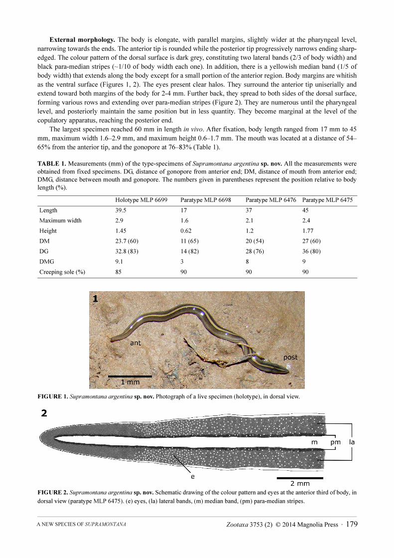
External morphology. The body is elongate, with parallel margins, slightly wider at the pharyngeal level,
narrowing towards the ends. The anterior tip is rounded while the posterior tip progressively narrows ending sharp-
edged. The colour pattern of the dorsal surface is dark grey, constituting two lateral bands (2/3 of body width) and
black para-median stripes (~1/10 of body width each one). In addition, there is a yellowish median band (1/5 of
body width) that extends along the body except for a small portion of the anterior region. Body margins are whitish
as the ventral surface (Figures 1, 2). The eyes present clear halos. They surround the anterior tip uniserially and
extend toward both margins of the body for 2-4 mm. Further back, they spread to both sides of the dorsal surface,
forming various rows and extending over para-median stripes (Figure 2). They are numerous until the pharyngeal
level, and posteriorly maintain the same position but in less quantity. They become marginal at the level of the
copulatory apparatus, reaching the posterior end.
The largest specimen reached 60 mm in length in vivo. After fixation, body length ranged from 17 mm to 45
mm, maximum width 1.6–2.9 mm, and maximum height 0.6–1.7 mm. The mouth was located at a distance of 54–
65% from the anterior tip, and the gonopore at 76–83% (Table 1).
TABLE 1. Measurements (mm) of the type-specimens of Supramontana argentina sp. nov. All the measurements were
obtained from fixed specimens. DG, distance of gonopore from anterior end; DM, distance of mouth from anterior end;
DMG, distance between mouth and gonopore. The numbers given in parentheses represent the position relative to body
length (%).
FIGURE 1. Supramontana argentina sp. nov. Photograph of a live specimen (holotype), in dorsal view.
FIGURE 2. Supramontana argentina sp. nov. Schematic drawing of the colour pattern and eyes at the anterior third of body, in
dorsal view (paratype MLP 6475). (e) eyes, (la) lateral bands, (m) median band, (pm) para-median stripes.
Holotype MLP 6699 Paratype MLP 6698 Paratype MLP 6476 Paratype MLP 6475
Length 39.5 17 37 45
Maximum width 2.9 1.6 2.1 2.4
Height 1.45 0.62 1.2 1.77
DM 23.7 (60) 11 (65) 20 (54) 27 (60)
DG 32.8 (83) 14 (82) 28 (76) 36 (80)
DMG 9.1 3 8 9
Creeping sole (%) 85 90 90 90
Zootaxa 3753 (2) © 2014 Magnolia Press · 179A NEW SPECIES OF SUPRAMONTANA

Internal morphology. Cephalic region. The position and arrangement of the cutaneous muscle layers differ
from those in the pre-pharyngeal region (see below) in that the ventral longitudinal cutaneous muscle layer is sunk
into the parenchyma and forms a retractor muscle that is almost circular in cross section (Figure 3). This retractor
muscle diminishes in thickness towards the anterior end until disappear next to the tip. Backward it becomes
progressively wider and finally form two layers of longitudinal muscles that occupy ventrally the whole width of
body, separated each other by a nerve plexus (Figures 4, 5). Circular, oblique and dorsal longitudinal layers of the
cutaneous musculature are thinner than in the pre-pharyngeal region. The parenchymatic musculature is arranged
as supra-intestinal and sub-intestinal transverse layers, thinner as the intestine becomes less apparent towards the
tip. Dorso-ventral fibres are scarce. There is a sub-neural transverse parenchymatic muscle layer, located just
below the nerve plate and above the retractor muscle (Figure 3). Sensory pits, as a simple invagination of ventral
epidermis (25–45 µm deep), surround the cephalic region (Figure 3).
FIGURES 3–5. Supramontana argentina sp. nov. (3) Schematic drawing of a transverse section of the cephalic region
(holotype); (4) schematic drawing of a transverse section of the pre-pharyngeal region (paratype MLP 6476); (5) half of a
transversal section of the pre-pharyngeal region (paratype MLP 6476). (cn) cutaneous nerve net, (cs) creeping sole, (dep) dorsal
epidermis, (dl) dorsal cutaneous longitudinal musculature, (dp) dorsal parenchymatic musculature, (dvp) dorso-ventral
parenchymatic musculature, (e) eye, (gs) glandular secretion, (i) intestine, (n) nervous plate, (od) ovovitelline duct, (r) retractor
muscle, (sbp) sub-intestinal parenchymatic musculature, (sd) sperm duct, (se) sensory pit, (snp) sub-neural parenchymatic
musculature, (spp) supra-intestinal parenchymatic musculature, (t) testes, (vit) vitellaria, (vl) ventral cutaneous longitudinal
musculature.
Numerous rhabditogen cells with xanthophil secretion (rhammites), cells with coarse granular erythrophil
secretion (~2.5 μm), and cells with strong cyanophil, coarse granular secretion (~2 μm) open through anterior tip
and dorsal epidermis. The narrow creeping sole—between 14% and 51% of body width on first 0.7 mm of body
length—receives abundant coarse granular strong cyanophil secretion (~2.5 μm). In addition, less frequent
rhabditogen cells (small rhabdites) and erythrophil cells with fine granular secretion (about 1 μm) open through the
creeping sole. The remaining ventral surface presents abundant rhabditogen cells with xanthophil secretion
NEGRETE ET AL.180 · Zootaxa 3753 (2) © 2014 Magnolia Press

(rhammites), numerous cells with strong cyanophil, coarse granular secretion (~2 μm), and less frequent cells with
ill-defined, coarse granular secretion (~3 μm).
Epidermis and musculature at pre-pharyngeal region. Five types of secretory cells open through dorsal
epidermis (15–25 μm high) and body margins: (1) numerous cells with coarse granular erythrophil secretion (~3
μm), (2) abundant rhabditogen cells with xanthophil secretion (rhammites), and less frequent (3) erythrophil cells
with fine granular secretion (<1 μm), (4) weakly cyanophil cells with amorphous secretion and (5) cells with strong
cyanophil, coarse granular secretion (~2 μm). There is no glandular margin (Figures 4, 5). Ventral epidermis (22.5–
30 μm high) is ciliated on creeping sole, which is approximately 85-90% of body width (Figure 4). Four types of
glands discharge their secretion through the creeping sole: abundant cells with coarse granular, strong cyanophil
secretion (~3 μm), rhabditogen cells (with small rhabdites) and erythrophil cells of two types, one with fine (<1
μm) and other with coarse (~2 μm) granular secretion.
The cutaneous musculature consists of three layers: a thin external subepithelial layer of circular muscles,
followed by a diagonal layer with decussate fibres, and a thicker longitudinal layer arranged in bundles. Ventrally,
the longitudinal layer is thicker than dorsally, being subdivided into two portions, one just below the diagonal
fibres, and the innermost one sunk into the parenchyma (Figures 4, 5, Table 2). The CMI varies between 14% and
21% (Table 2).
TABLE 2. Thickness of cutaneous (CM) and parenchymatic (PM) musculatures (µm), and CMI and PMI indices at pre-
pharyngeal region of type-specimens of Supramontana argentina sp. nov.
The parenchymatic musculature is composed by four layers: a dorsal subcutaneous with oblique fibres, a
supra-intestinal and a sub-intestinal transverse layers, and a sub-neural transverse layer (Figures 4, 5). Also, dorso-
ventral fibres are arranged among intestinal branches. The thickness of the parenchymatic musculature represents
6–11% of body height (Table 2).
Digestive system. The pharynx (1.4–2.75 mm in length) is bell-shaped, with the dorsal insertion at mouth level
or a bit posteriorly displaced (Figure 6). The mouth is situated in the anterior third of the pharyngeal pouch (1.45–
3.75 mm in length). The epithelial lining of the outer surface of the pharynx is cuboidal and ciliated. The outer
pharyngeal musculature is arranged in two layers: a thin longitudinal subepithelial layer (2.5–5 μm thick) followed
by a subjacent circular layer (25–27.5 μm thick). The epithelium of the pharyngeal lumen is columnar, ciliated, and
Holotype
MLP 6699
Paratype
MLP 6698
Paratype
MLP 6476
Paratype
MLP 6475
CM dorsal
Circular 2.5 2.5 5 5
Diagonal 10 10 12.5 15
Longitudinal 65 45 56 65
Total 77.5 57.5 73.5 85
CM ventral
Circular 2.5 2.5 5 5
Diagonal 15 10 12.5 10
Longitudinal (external) 75 25 50 50
Longitudinal (internal) 100 50–75 100 90
Total 192.5 87.5–112.5 167.5 155
CMI (%) 19 18–21 20 14
PM dorsal 50 20 35 37.5–50
PM supra-intestinal 15–35 10 35 50–62.5
PM sub-intestinal 20–40 12.5 25 25
PM sub-neural 5–10 10 15–25 50
PMI (%) 6–9 8 9–10 9–11
Zootaxa 3753 (2) © 2014 Magnolia Press · 181A NEW SPECIES OF SUPRAMONTANA

strongly erythrophil. The inner pharyngeal musculature consists of a circular subepithelial layer (25–50 μm thick)
followed by a subjacent longitudinal one (10–37.5 μm thick). Six secretory cell types, three of them with cell necks
close to the epithelium of the pharyngeal lumen, open through this epithelium: cells with fine granular erythrophil
secretion (<1 μm); cells with fine granular cyanophil secretion (1 μm), and cells with fine granular weak cyanophil
secretion (1 μm). Other three types of glands show cell necks close to the outer epithelium of the pharynx, opening
through the pharyngeal tip: cells with cyanophil amorphous secretion; cells with coarse granular erythrophil
secretion (~3 μm); and cells with fine granular weak erythrophil secretion (<1 μm). The oesophagus is absent
(Figure 6).
FIGURE 6. Supramontana argentina sp. nov. (holotype). Sagittal section of the pharynx. (di) dorsal insertion, (i) intestine,
(mo) mouth, (ph) pharynx, (php) pharyngeal pouch, (pl) pharyngeal lumen, (t) testes, (vi) ventral insertion, (vit) vitellaria. The
arrowhead indicates the outer pharyngeal musculature and the arrow indicates the inner pharyngeal musculature.
Male reproductive system. The testes are arranged in two or three rows on each side of the body, dorsal to the
intestinal branches and just below the supra-intestinal parenchymatic muscle layer (Figures 4, 5). They are pre-
pharyngeal, appearing behind the ovaries and extending to near the level of the ventral pharyngeal insertion. They
are located at a distance between 18–23% and 48–61% of the body length from anterior end (Table 3). Testes are
rounded or ovoid, occupying 9–23% of the height of body (Table 3). The sperm ducts are located above the sub-
intestinal parenchymatic muscle layer or among its fibres, being dorsal to the ovovitelline ducts, laterally displaced
(Figures 4, 5). The sperm ducts expand distally with their lumen full of spermatozoa. They bend toward the dorsum
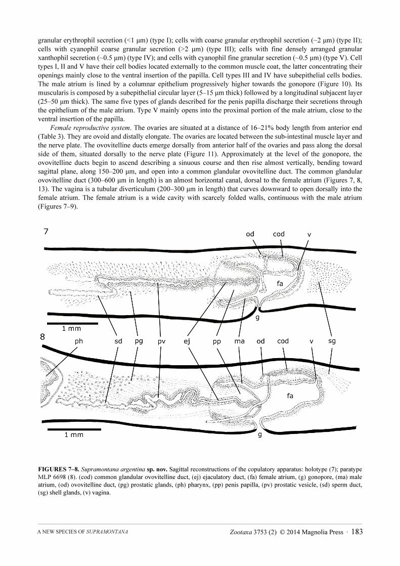
and slightly backward and open laterally into the proximal portion of the prostatic vesicle (Figures 7, 8). The
extrabulbar prostatic vesicle is a long unpaired tube. Its proximal portion is almost straight and dilated; the rest of
the prostatic vesicle is tortuous. The ejaculatory duct runs almost centrally in sinuous way through the penis
papilla. The latter occupies the entire length of the male atrium, being conical and blunt in shape, with its insertions
at the same transverse level (Figures 7–10). The walls of the male atrium are not folded.
The prostatic vesicle is lined with ciliated columnar epithelium followed by a muscular layer with intermingled
circular and less abundant longitudinal fibres (10–30 μm thick) (Figures 7–9). The cell necks of numerous
secretory cells with fine granular erythrophil secretion (<1 μm) and less abundant cyanophil cells with coarse
granular secretion (~2 μm) open through the epithelium of the prostatic vesicle. The cell bodies of these glands are
found in the parenchyma, mainly around the prostatic vesicle (Figures 7–9). The ejaculatory duct is lined with
ciliated columnar epithelium, followed by a thin muscle layer of circular fibres mixed with some longitudinal fibres
(5–15 μm thick). The ejaculatory duct receives abundant secretion from cells with coarse granular cyanophil
secretion (~2 μm) and less abundant cells with fine granular erythrophil secretion (<1 μm). The epithelium of the
penis papilla is columnar, progressively lower towards the tip, and the musculature is arranged in a circular
subepithelial layer (5–15 μm thick) followed by a longitudinal one (2.5 μm thick). Five types of secretory cells run
longitudinally in the papilla, with numerous openings through its lining epithelium: abundant cells with fine
NEGRETE ET AL.182 · Zootaxa 3753 (2) © 2014 Magnolia Press
granular erythrophil secretion (<1 μm) (type I); cells with coarse granular erythrophil secretion (~2 μm) (type II);
cells with cyanophil coarse granular secretion (>2 μm) (type III); cells with fine densely arranged granular
xanthophil secretion (~0.5 μm) (type IV); and cells with cyanophil fine granular secretion (~0.5 μm) (type V). Cell
types I, II and V have their cell bodies located externally to the common muscle coat, the latter concentrating their
openings mainly close to the ventral insertion of the papilla. Cell types III and IV have subepithelial cells bodies.
The male atrium is lined by a columnar epithelium progressively higher towards the gonopore (Figure 10). Its
muscularis is composed by a subepithelial circular layer (5–15 μm thick) followed by a longitudinal subjacent layer
(25–50 μm thick). The same five types of glands described for the penis papilla discharge their secretions through
the epithelium of the male atrium. Type V mainly opens into the proximal portion of the male atrium, close to the
ventral insertion of the papilla.
Female reproductive system. The ovaries are situated at a distance of 16–21% body length from anterior end
(Table 3). They are ovoid and distally elongate. The ovaries are located between the sub-intestinal muscle layer and
the nerve plate. The ovovitelline ducts emerge dorsally from anterior half of the ovaries and pass along the dorsal
side of them, situated dorsally to the nerve plate (Figure 11). Approximately at the level of the gonopore, the
ovovitelline ducts begin to ascend describing a sinuous course and then rise almost vertically, bending toward
sagittal plane, along 150–200 μm, and open into a common glandular ovovitelline duct. The common glandular
ovovitelline duct (300–600 μm in length) is an almost horizontal canal, dorsal to the female atrium (Figures 7, 8,
13). The vagina is a tubular diverticulum (200–300 μm in length) that curves downward to open dorsally into the
female atrium. The female atrium is a wide cavity with scarcely folded walls, continuous with the male atrium
(Figures 7–9).
FIGURES 7–8. Supramontana argentina sp. nov. Sagittal reconstructions of the copulatory apparatus: holotype (7); paratype
MLP 6698 (8). (cod) common glandular ovovitelline duct, (ej) ejaculatory duct, (fa) female atrium, (g) gonopore, (ma) male
atrium, (od) ovovitelline duct, (pg) prostatic glands, (ph) pharynx, (pp) penis papilla, (pv) prostatic vesicle, (sd) sperm duct,
(sg) shell glands, (v) vagina.
Zootaxa 3753 (2) © 2014 Magnolia Press · 183A NEW SPECIES OF SUPRAMONTANA
TABLE 3. Measurements (mm) of reproductive organs of type-specimens of Supramontana argentina sp. nov. LCGD,
length of common glandular ovovitelline duct; LFA, length of female atrium; LMA, length of male atrium; LPP, length
of penis papilla; LPV, length of prostatic vesicle; LV, length of vagina; T:BH, ratio of the height of testes to the height of
the body. The numbers given in parentheses represent the position relative to body length (%).
The epithelium lining the ovovitelline ducts is ciliated and cuboidal. Cells with fine granular erythrophil
secretion and scarce shell glands open into the vertical portions of the ovovitelline ducts, while the sagittal distal
tracts receive abundant secretion from the shell glands (Figure 12). The common glandular ovovitelline duct is
lined with a ciliated columnar epithelium (Figure 13), receiving abundant secretion of two types of shell glands:
xanthophil coarse granular secretion (~2–3 μm) and cyanophil coarse granular secretion (~3–4 μm). Both glands
have their cell bodies in the parenchyma, posteriorly to the copulatory apparatus (Figure 9). The vagina is lined
with a ciliated columnar epithelium and the musculature is composed by circular subepithelial layer (5–15 μm
thick) and a longitudinal subjacent layer (10–15 μm thick). The epithelium of the vagina receives few secretory
cells of three types, similar to types I, III and IV of the penis papilla and male atrium. The female atrium is lined by
a non-ciliated pluriestratified epithelium and the muscularis is arranged in two layers, circular subepithelial (10–25
μm thick) and longitudinal subjacent (10–20 μm thick). Four types of secretory cells, similar to types I, II, III and
IV of the penis papilla and male atrium, open into female atrium. The common muscular coat is mainly composed
by longitudinal fibres, thicker dorsally (~50 μm thick) than ventrally (~25 μm thick), besides some oblique and
circular fibres.
Vitellaria are well developed and are dispersed throughout the body. In the anterior and pre-pharyngeal regions
vitellaria extend dorsally, ventrally and between intestine branches (Figures 4–6), being abundant behind the
copulatory apparatus.
Discussion
The external and internal characters of the new species herein described strongly support its inclusion into the
genus Supramontana Carbayo & Leal-Zanchet, 2003.
Regarding the external morphology, when Supramontana argentina sp. nov. is crawling, the anterior end has
an inverted ‘U’ shape in cross section, as described for the type-species by Carbayo & Leal-Zanchet (2003), like a
gable roof. In relation to the internal characters, the new species presents, as the type-species of Supramontana, a
mid-ventral retractor muscle in the cephalic region composed by fibres of the ventral cutaneous longitudinal
muscular layer sunk into the parenchyma. Also, there is a sub-neural transverse parenchymatic muscular layer
along the entire body (Carbayo & Leal-Zanchet 2003). Other features of S. argentina sp. nov. that agree with
characteristics of the type-species of the genus are the absence of a glandular margin, the presence of a permanent
penis papilla (protrusible penis), a male atrium without folded walls, and a long and horizontal common glandular
ovovitelline duct dorsal to the female atrium.
Holotype
MLP 6699
Paratype
MLP 6698
Paratype
MLP 6476
Paratype
MLP 6475
Anteriormost testes 9 (23) 3.9 (23) 6.5 (18) –
Posteriormost testes 22.7 (57) 10.4 (61) 17.9 (48) 26 (58)
T:BH (%) 15–22 15–18 9–23 12
LPV 2.25 0.9 1.8 2.2
LPP 0.8 0.5 0.38 0.48
LMA 0.85 0.6 0.7 0.6
Location of ovaries 8.1 (20) 3.6 (21) 6 (16) –
LCGD 0.4 0.3 0.6 0.3
LV 0.3 0.2 0.25 0.25
LFA 0.6 0.5 1.1 0.9
NEGRETE ET AL.184 · Zootaxa 3753 (2) © 2014 Magnolia Press

FIGURES 9–13. Supramontana argentina sp. nov. (holotype). Sagittal sections: (9) copulatory apparatus; (10) penis papilla;
(11) ovary; (12–13) details of the female reproductive system. (cod) common glandular ovovitelline duct, (ej) ejaculatory duct,
(fa) female atrium, (g) gonopore, (i) intestine, (ma) male atrium, (od) ovovitelline duct, (ov) ovary, (pg) prostatic glands, (pp)
penis papilla, (pv) prostatic vesicle, (sg) shell glands, (v) vagina, (vit) vitellaria, (vl) ventral cutaneous longitudinal
musculature.
Zootaxa 3753 (2) © 2014 Magnolia Press · 185A NEW SPECIES OF SUPRAMONTANA

Supramontana argentina sp. nov. differs externally from the type-species of the genus by its dorsal colour
pattern. Supramontana irritata possesses a pale yellowish background with dark brown spots dispersed onto the
dorsum which forms a distinct narrow median stripe and three pairs of narrow stripes, varying in intensity among
specimens (Carbayo & Leal-Zanchet 2003). The new species shows a colour pattern with a striking yellow median
band and one pair of black stripes on a dark grey background.
Regarding the internal morphology, both species have an extrabulbar and unpaired prostatic vesicle, with its
proximal region rather straight and dilated, and a sinuous distal portion. In S. irritata the ejaculatory duct is a
straight canal without openings of secretory cells in its epithelium, but in S. argentina sp. nov. this duct runs
sinuously through the penis papilla and receives abundant secretion. The penis papilla is similarly shaped in both
species, but it is pointed in S. irritata and blunt in S. argentina sp. nov. Also, the male atrium is only partially
occupied by the papilla in S. irritata, whereas the papilla occupies the whole cavity of the male atrium in S.
argentina sp. nov. The female reproductive system of S. argentina sp. nov. is similar to that of S. irritata, except
for the common glandular ovovitelline duct, which is longer in S. irritata (~1 mm) than in S. argentina sp. nov.
(about half of this length).
The finding of a second species of Supramontana consolidates the identity of this genus, reinforces the
diagnosis of the genus provided by Carbayo & Leal-Zanchet (2003), and extends its distribution range along the
Atlantic Forest.
Acknowledgements
The authors are grateful to Fundación Vida Silvestre Argentina and Ministerio de Ecología y Recursos Naturales
Renovables from Misiones province for permission to conduct sampling at RVSU, and especially to the park
managers Ariel Tombo and Laura Aréjola for support during fieldwork. We are thankful to the Administración de
Parques Nacionales and INTA San Antonio for permission to conduct sampling at CAMB and INP. We thank
Marcelo Kostlin for his support during fieldwork at INP. We also thank Idea Wild for providing optic equipment.
This work was partially financed by CONICET (Consejo Nacional de Investigaciones Científicas y Técnicas),
Agencia Nacional de Promoción Científica y Tecnológica (FONCyT) PICT 2007-01287, Ministerio de Ciencia,
Tecnología e Innovación Productiva, Argentina and Coordenação de Aperfeiçoamento de Pessoal de Nível
Superior, Brazil (MINCYT/CAPES 202/2012) and UNLP (Universidad Nacional de La Plata) 11/N600. Dr. Hugh
Jones, Dr. Fernando Carbayo and an anonymous reviewer are acknowledged for their constructive suggestions.
References
Carbayo, F. & Leal-Zanchet, A.M. (2003) Two new genera of geoplaninid land planarians (Platyhelminthes: Tricladida:
Terricola) of Brazil in the light of cephalic specialisations. Invertebrate Systematics, 17, 449–468.
http://dx.doi.org/10.1071/it01035
Di Bitetti, M.S., Placci, G. & Dietz, L.A. (2003) Una Visión de Biodiversidad para la Ecorregión del Bosque Atlántico del Alto
Paraná: Diseño de un Paisaje para la Conservación de la Biodiversidad y prioridades para las acciones de conservación.
World Wildlife Foundation, Washington DC, 154 pp.
Froehlich, C.G. (1955) Sôbre morfologia e taxonomia das Geoplanidae. Boletins da Faculdade de Filosofia, Ciências e Letras
da Universidade de São Paulo, Série Zoologia, 19, 195–279.
Froehlich, E.M. (1978) On a collection of Chilean landplanarians. Boletim de Zoologia da Universidade de São Paulo, 3, 7–80.
Galindo-Leal, C. & Câmara, G. (2003) Atlantic Forest Hotspot Status: An Overview. In: Galindo-Leal, C. & Câmara, G. (Eds.),
The Atlantic Forest of South America: biodiversity status, threats, and outlook. Island Press, Washington DC, pp. 3–11.
Giraudo, A.R., Povedano, H., Belgrano, M.J., Krauczuk, E., Pardiñas, U., Miquelarena, A., Ligier, D., Baldo, D. & Castelino,
M. (2003) Biodiversity Status of the Interior Atlantic Forest of Argentina. In: Galindo-Leal, C. & Câmara, G. (Eds.), The
Atlantic Forest of South America: biodiversity status, threats, and outlook. Island Press, Washington DC, pp. 160–180.
Romeis, B. (1989) Mikroskopische Technik. Urban und Schwarzenberg, München, 697 pp.
Winsor, L. (1998) Aspects of taxonomy and functional histology in terrestrial flatworms (Tricladida: Terricola). Pedobiologia,
42, 412–432.
NEGRETE ET AL.186 · Zootaxa 3753 (2) © 2014 Magnolia Press
Related Documents