Thyroid Hormone-Dependent Regulation of T1 -Tubulin during Brain Development Petra I. Lorenzo,* Catherine Me ´ nard, † Freda D. Miller, † and Juan Bernal* ,1 *Instituto de Investigaciones Biome ´dicas “Alberto Sols,” CSIC, 28029 Madrid, Spain; and † Centre for Neuronal Survival, Montreal Neurological Institute, McGill University, Montreal, Quebec, Canada Thyroid hormone (T3) is essential for brain development and most of its actions are exerted at the gene expression level after interaction with nuclear receptors. In particular, genes encoding cytoskeletal proteins are influenced by the thyroidal status. Thyroid hormone is involved in the normal downregulation of the T1 -tubulin gene during postnatal growth. The action of T3 on T1 tubulin expres- sion is complex and is exerted at least at two levels. In cultured cells, T3 induces a transient and fast decrease of T1 mRNA concentration. This effect is enhanced when transcription is blocked by actinomycin D, suggesting that T3 increases mRNA degradation. In transgenic ani- mals T3 affects the expression of -galactosidase under control of the T1 promoter in the same way as the en- dogenous gene, supporting an effect mediated through the T1 promoter. However, the T1 promoter is not reg- ulated by T3 in transfected cells and, therefore, the effects of the hormone in vivo are likely to be indirect. It is con- cluded that regulation of T1 -tubulin by thyroid hormone is the result of multiple influences including effects on mRNA half life and indirect effects at the promoter level. INTRODUCTION Thyroid hormone (TH) is an important regulator of physiological and developmental processes and plays a crucial role in maturation of the CNS. TH deficiency during critical periods of development produces per- manent and severe alterations in the structure and func- tion of the nervous system, including delays of neuro- nal differentiation with abnormal morphology of dendrites and disturbed outgrowth of neuronal pro- cesses (Bernal and Nunez, 1995). Both neurons and oligodendrocytes are cell targets of TH during CNS maturation. These cell types contain developmentally regulated TH receptors and express genes that are reg- ulated in vivo and in vitro by the hormone (Bernal and Nunez, 1995; Mun ˜ oz and Bernal, 1997). Some of these genes are related to key events occurring during neurite outgrowth, such as those encoding cytoskeletal pro- teins. Alterations of microtubule assembly have been described in a form of congenital hypothyroidism in mice (hyt/hyt mice) and also take place after induction of neonatal hypothyroidism in previously normal ani- mals (Biesiada et al., 1996; Nunez et al., 1991). These alterations in microtubule assembly and stabilization are partly mediated through TH-dependent, regulated expression of microtubule-associated proteins (MAPs) (Aniello et al., 1991a; Benjamin et al., 1988) and specific tubulin isoforms (Aniello et al., 1991b; Figueiredo et al., 1993; Lewis et al., 1985). Several studies suggest that TH deficiency maintains an immature composition of mi- crotubules by delaying the developmental program of expression of several tubulin and MAP genes (Aniello et al., 1991a,b; Charrie ´re-Bertrand and Nunez, 1992; Nunez et al., 1991, 1992). In this context, TH is an epigenetic signal required to achieve the construction of a normal neuronal network during development. In- deed TH has been shown to play a role in tubulin gene expression, in particular -tubulin, during brain devel- opment. Two -tubulin genes are expressed in the embryonic rat nervous system and are named T1 and T26 (Miller et al., 1987), homologous to mouse M1 and M2, re- spectively (Lewis et al., 1985). Whereas T26 is constitu- tively expressed in neurons and in nonneuronal cells, T1 is neuron-specific and its expression correlates with neuronal growth (Knoops and Octave, 1997; Miller et al., 1987). Expression of T1 -tubulin is high during 1 To whom correspondence and reprint requests should be ad- dressed at the Instituto Alberto Sols, Arturo Duperier 4, 28029 Ma- drid, Spain. Fax: 34-91-585-4587. E-mail: [email protected]. Molecular and Cellular Neuroscience 19, 333–343 (2002) doi:10.1006/mcne.2001.1087, available online at http://www.idealibrary.com on MCN 1044-7431/02 $35.00 © 2002 Elsevier Science (USA) All rights reserved. 333

Welcome message from author
This document is posted to help you gain knowledge. Please leave a comment to let me know what you think about it! Share it to your friends and learn new things together.
Transcript
Molecular and Cellular Neuroscience 19, 333–343 (2002)
doi:10.1006/mcne.2001.1087, available online at http://www.idealibrary.com on MCN
Thyroid Hormone-Dependent Regulation of T�1�-Tubulin during Brain DevelopmentPetra I. Lorenzo,* Catherine Menard,† Freda D. Miller,†
and Juan Bernal* ,1
*Instituto de Investigaciones Biomedicas “Alberto Sols,” CSIC, 28029 Madrid, Spain; and†Centre for Neuronal Survival, Montreal Neurological Institute, McGill University,Montreal, Quebec, Canada
Thyroid hormone (T3) is essential for brain developmentand most of its actions are exerted at the gene expressionlevel after interaction with nuclear receptors. In particular,genes encoding cytoskeletal proteins are influenced bythe thyroidal status. Thyroid hormone is involved in thenormal downregulation of the T�1 �-tubulin gene duringpostnatal growth. The action of T3 on T�1 tubulin expres-sion is complex and is exerted at least at two levels. Incultured cells, T3 induces a transient and fast decrease ofT�1 mRNA concentration. This effect is enhanced whentranscription is blocked by actinomycin D, suggestingthat T3 increases mRNA degradation. In transgenic ani-mals T3 affects the expression of �-galactosidase undercontrol of the T�1 promoter in the same way as the en-dogenous gene, supporting an effect mediated throughthe T�1 promoter. However, the T�1 promoter is not reg-ulated by T3 in transfected cells and, therefore, the effectsof the hormone in vivo are likely to be indirect. It is con-cluded that regulation of T�1 �-tubulin by thyroid hormoneis the result of multiple influences including effects onmRNA half life and indirect effects at the promoter level.
INTRODUCTION
Thyroid hormone (TH) is an important regulator ofphysiological and developmental processes and plays acrucial role in maturation of the CNS. TH deficiencyduring critical periods of development produces per-manent and severe alterations in the structure and func-tion of the nervous system, including delays of neuro-nal differentiation with abnormal morphology ofdendrites and disturbed outgrowth of neuronal pro-cesses (Bernal and Nunez, 1995). Both neurons and
1 To whom correspondence and reprint requests should be ad-dressed at the Instituto Alberto Sols, Arturo Duperier 4, 28029 Ma-
drid, Spain. Fax: �34-91-585-4587. E-mail: [email protected].1044-7431/02 $35.00© 2002 Elsevier Science (USA)All rights reserved.
oligodendrocytes are cell targets of TH during CNSmaturation. These cell types contain developmentallyregulated TH receptors and express genes that are reg-ulated in vivo and in vitro by the hormone (Bernal andNunez, 1995; Munoz and Bernal, 1997). Some of thesegenes are related to key events occurring during neuriteoutgrowth, such as those encoding cytoskeletal pro-teins. Alterations of microtubule assembly have beendescribed in a form of congenital hypothyroidism inmice (hyt/hyt mice) and also take place after inductionof neonatal hypothyroidism in previously normal ani-mals (Biesiada et al., 1996; Nunez et al., 1991). Thesealterations in microtubule assembly and stabilizationare partly mediated through TH-dependent, regulatedexpression of microtubule-associated proteins (MAPs)(Aniello et al., 1991a; Benjamin et al., 1988) and specifictubulin isoforms (Aniello et al., 1991b; Figueiredo et al.,1993; Lewis et al., 1985). Several studies suggest that THdeficiency maintains an immature composition of mi-crotubules by delaying the developmental program ofexpression of several tubulin and MAP genes (Anielloet al., 1991a,b; Charriere-Bertrand and Nunez, 1992;Nunez et al., 1991, 1992). In this context, TH is anepigenetic signal required to achieve the construction ofa normal neuronal network during development. In-deed TH has been shown to play a role in tubulin geneexpression, in particular �-tubulin, during brain devel-opment.
Two �-tubulin genes are expressed in the embryonicrat nervous system and are named T�1 and T26 (Milleret al., 1987), homologous to mouse M�1 and M�2, re-spectively (Lewis et al., 1985). Whereas T26 is constitu-tively expressed in neurons and in nonneuronal cells,T�1 is neuron-specific and its expression correlates withneuronal growth (Knoops and Octave, 1997; Miller et
al., 1987). Expression of T�1 �-tubulin is high during333
developmental growth and is subsequently downregu-lated around the time of target contact and neuronalmaturation (Gloster et al., 1999; Mathew and Miller,1990; Miller et al., 1987; Wu et al., 1997). T�1 increases inresponse to different extrinsic cues that induce thegrowth of mature neurons (Geddes et al., 1990; Ma et al.,1992; Mathew and Miller, 1990, 1993; Miller et al., 1989;Tetzlaff et al., 1991). In this context it has been proposedthat T�1 �-tubulin induction reflects a generalized neu-ronal growth response that, once initiated, is similar indeveloping and in mature neurons (Geddes et al., 1990;Mathew and Miller, 1990).
In hypothyroid animals T�1 �-tubulin expression isincreased in cerebellum and decreased in cerebralhemispheres (Aniello et al., 1991b; Figueiredo et al.,1993; Nunez et al., 1991). The information provided bythese studies, however, is limited because the analysisof T�1 �-tubulin expression was done by Northernblotting, thus overlooking any regional-specific varia-tion of T�1 �-tubulin regulation. In addition the ques-tion about the mode of action of thyroid hormone onthe regulation of T�1 �-tubulin expression was notaddressed. Given the importance of T�1 �-tubulin inneuronal growth and regeneration, we have reevalu-ated its regulation by TH both in vivo and in vitro. Invivo, studies were aimed at the analysis of the temporaland regional patterns of TH regulation. In vitro studieswere done with the goal of elucidating the molecularmechanisms of regulation by thyroid hormone.
RESULTS
T�-1 �-Tubulin Expression in Vivo
We used a specific labeled riboprobe from the 3�untranslated region of the �-tubulin mRNA to analyzethe expression of the T�1 isoform in normal and hypo-thyroid rats during postnatal development. Coronalslices from both groups of animals were used for in situhybridization. The patterns of expression in the cere-brum and in the cerebellum are illustrated in Figs. 1 and2, respectively.
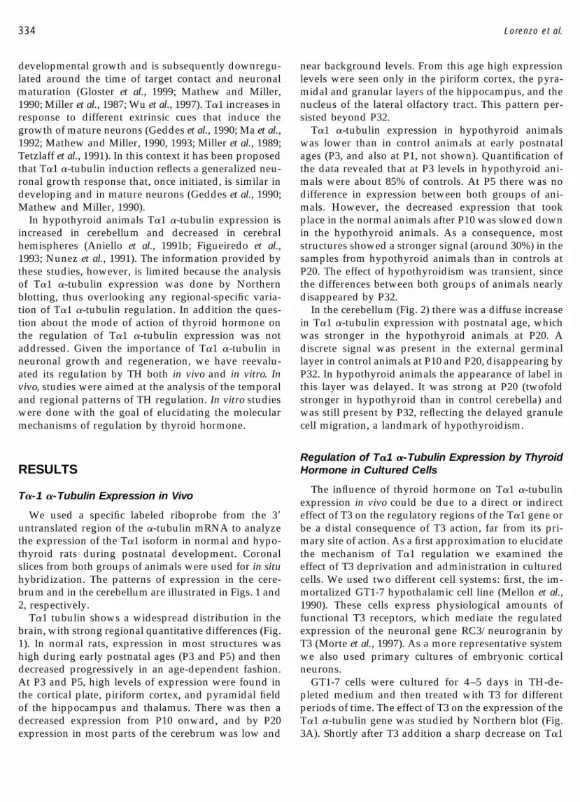
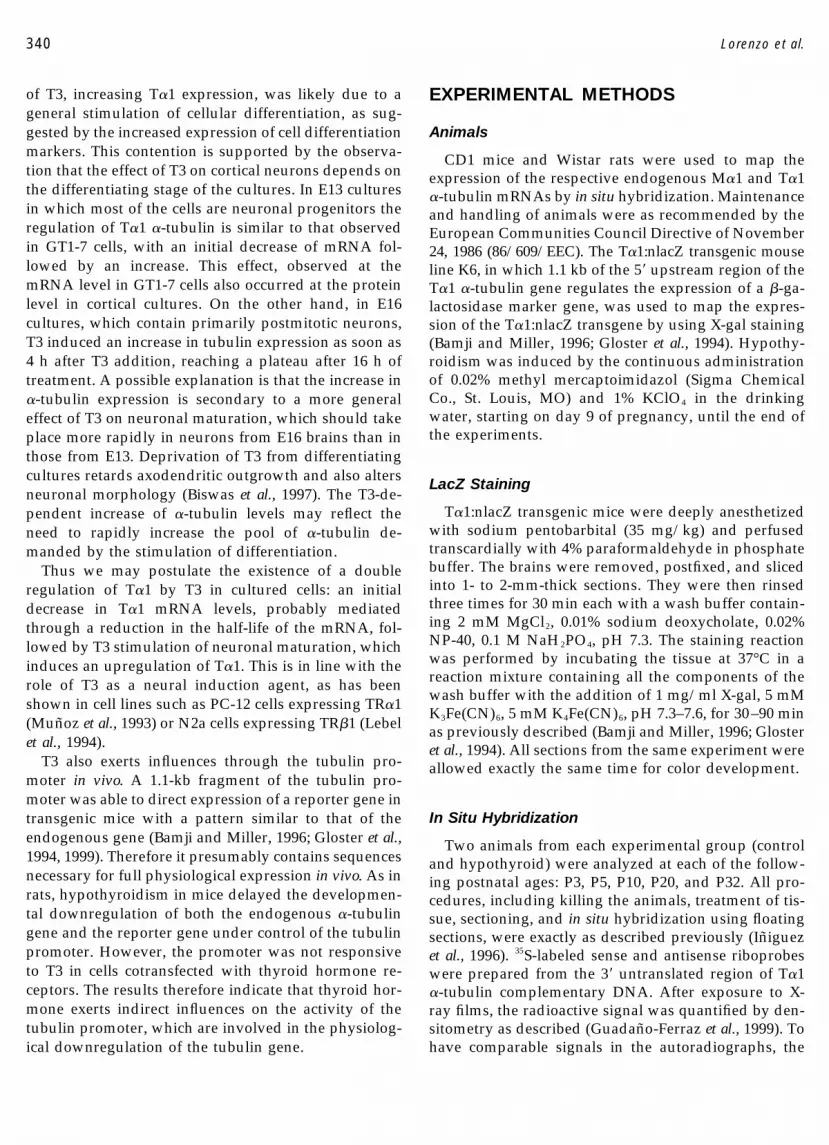
T�1 tubulin shows a widespread distribution in thebrain, with strong regional quantitative differences (Fig.1). In normal rats, expression in most structures washigh during early postnatal ages (P3 and P5) and thendecreased progressively in an age-dependent fashion.At P3 and P5, high levels of expression were found inthe cortical plate, piriform cortex, and pyramidal fieldof the hippocampus and thalamus. There was then adecreased expression from P10 onward, and by P20expression in most parts of the cerebrum was low and
near background levels. From this age high expressionlevels were seen only in the piriform cortex, the pyra-midal and granular layers of the hippocampus, and thenucleus of the lateral olfactory tract. This pattern per-sisted beyond P32.
T�1 �-tubulin expression in hypothyroid animalswas lower than in control animals at early postnatalages (P3, and also at P1, not shown). Quantification ofthe data revealed that at P3 levels in hypothyroid ani-mals were about 85% of controls. At P5 there was nodifference in expression between both groups of ani-mals. However, the decreased expression that tookplace in the normal animals after P10 was slowed downin the hypothyroid animals. As a consequence, moststructures showed a stronger signal (around 30%) in thesamples from hypothyroid animals than in controls atP20. The effect of hypothyroidism was transient, sincethe differences between both groups of animals nearlydisappeared by P32.
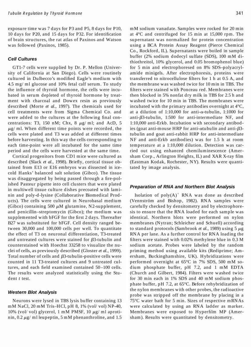
In the cerebellum (Fig. 2) there was a diffuse increasein T�1 �-tubulin expression with postnatal age, whichwas stronger in the hypothyroid animals at P20. Adiscrete signal was present in the external germinallayer in control animals at P10 and P20, disappearing byP32. In hypothyroid animals the appearance of label inthis layer was delayed. It was strong at P20 (twofoldstronger in hypothyroid than in control cerebella) andwas still present by P32, reflecting the delayed granulecell migration, a landmark of hypothyroidism.
Regulation of T�1 �-Tubulin Expression by ThyroidHormone in Cultured Cells
The influence of thyroid hormone on T�1 �-tubulinexpression in vivo could be due to a direct or indirecteffect of T3 on the regulatory regions of the T�1 gene orbe a distal consequence of T3 action, far from its pri-mary site of action. As a first approximation to elucidatethe mechanism of T�1 regulation we examined theeffect of T3 deprivation and administration in culturedcells. We used two different cell systems: first, the im-mortalized GT1-7 hypothalamic cell line (Mellon et al.,1990). These cells express physiological amounts offunctional T3 receptors, which mediate the regulatedexpression of the neuronal gene RC3/neurogranin byT3 (Morte et al., 1997). As a more representative systemwe also used primary cultures of embryonic corticalneurons.
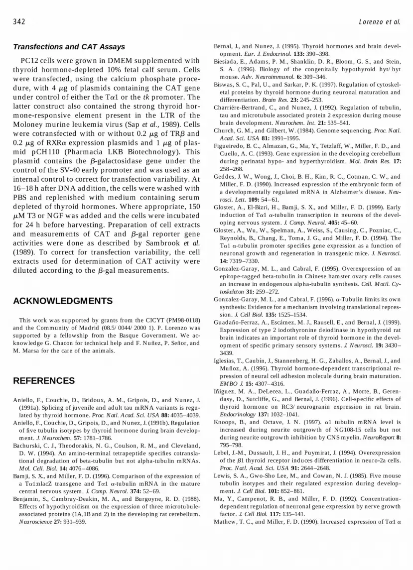
GT1-7 cells were cultured for 4–5 days in TH-de-pleted medium and then treated with T3 for differentperiods of time. The effect of T3 on the expression of theT�1 �-tubulin gene was studied by Northern blot (Fig.3A). Shortly after T3 addition a sharp decrease on T�1
334 Lorenzo et al.
mRNA levels was observed (49 � 11%, mean � SD ofthree experiments). This initial effect of T3 was tran-sient, and from 8 to 24 h T�1 mRNA concentrationsincreased, finally reaching control levels. Expression ofa neuronal differentiation marker, NSE, was alsochecked in the same cultures. T3 induced a late increaseof NSE mRNA concentration after 16 h of treatment, inparallel to the increase in T�1 �-tubulin. A similar studycarried out with primary cell cultures obtained fromE13 mouse cerebral cortex showed the same initial T3-induced inhibition of T�1 expression (Fig. 3B) and asubsequent increase by 16 h.
The effect of T3 on the levels of T�1 �-tubulin proteinwas also studied in primary cultures of cortical cellsobtained from either E13 or E16 mice, using an anti-tubulin antibody. Since T�1 �-tubulin mRNA com-prises greater than 95% of the total population of �-tu-bulin mRNA in the embryonic nervous system (Glosteret al., 1994; Miller et al., 1987), we may assume that mostof the �-tubulin detected by us in the embryonic cortical
cultures was T�1. Tubulin content was measured byWestern blotting of cell extracts using Ponceau redstaining as a control for protein loading (not shown)and the levels of Erkb by immunoblotting. The result ofthe Western blots is shown in Fig. 4A. The quantifica-tion of the bands is shown on the right-hand side of thefigure. In E13 cultures �-tubulin concentration de-creased after 4 h of T3 addition and increased thereaf-ter, in agreement with mRNA data. The levels of �3-tubulin and intermediate NF were also measured. Bothproteins experienced an initial increase within 8 h of T3addition, which was followed by a more sustained in-creased after 16–24 h. The significance of the transientdecrease observed for intermediate NF at 16 h is notclear at present. The late increase of both proteins par-alleled that of T�1 �-tubulin, suggesting that T3 stimu-lated the differentiation of these cells.
To assess in more detail the effect of T3 on neuronaldifferentiation, the E13 cultures were immunostainedfor �3-tubulin 24 h after T3 addition. The cultures were
FIG. 1. Pattern of expression of T�1 �-tubulin mRNA in rat brain. In situ hybridization analysis of T�1 �-tubulin mRNA at different timesduring postnatal brain development (P3 to P32) in normal control (C, top) and hypothyroid (H, bottom) rats. Coronal slices from the differentages and thyroid conditions were processed simultaneously. Exposure time was 7 days for P3 and P5, 8 days for P10, 10 days for P20, and 15days for P32. In normal rats there was a decreased signal after P10, which in hypothyroid rats takes place after P20. Abbreviations: Pir, piriformcortex; TH, thalamus; DG, dentate gyrus; CA1, CA3, pyramidal layer of hippocampus; Ctx, cortex; Nlot, nucleus of the lateral olfactory tract.
335Tubulin Regulation by Thyroid Hormone
also counterstained with Hoechst 33258 to visualize thenuclei of all cells. Data from different T3-treated (n �11) or untreated cultures (n � 9) showed that T3 didnot affect the proportion of differentiated cells, i.e.,those expressing �3-tubulin in relation to the total num-ber of cells (31.6 � 11.8% in the untreated culturesversus 27.8 � 5.9% in the T3-treated cultures). How-ever, addition of T3 increased significantly the propor-tion of �3-tubulin-positive cells that presented neuritesof a length greater than one cell diameter (26.4 � 6.7%in the T3-treated cultures versus 17.6 � 6.5% in theuntreated cultures, P � 0.016). Therefore, addition ofT3 to cultures of cells derived from E13 cultures, whichinitially contain mainly neuronal progenitors, was as-sociated with an enhanced neuronal differentiation. Toassess the effect of T3 in a more differentiated culturewe used cells derived from E16 brains (Fig. 4B): in thesecells T3 induced an increased tubulin content a fewhours after hormone addition without the initial de-crease observed in undifferentiated cultures. �3-Tubu-lin and intermediate NF content were not modified byT3 treatment in these cultures (not shown).
To analyze whether the short-term inhibition of T�1tubulin expression by T3 was due to inhibition of tran-scription or increased mRNA degradation, we treatedthe GT1-7 cells with T3 in the presence or absence ofactinomycin D (AcD), an inhibitor of transcription, orcycloheximide (Chx), an inhibitor of translation, as pre-viously described (Morte et al., 1997). In the presence ofAcD (Fig. 5A) T�1 mRNA was decreased by 40% at 6 hcompared with the untreated control cells, suggesting a
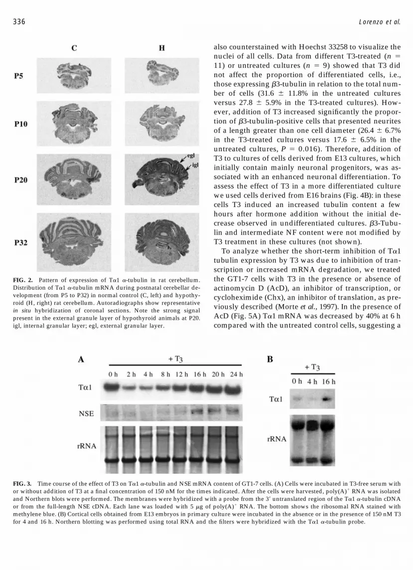
FIG. 2. Pattern of expression of T�1 �-tubulin in rat cerebellum.Distribution of T�1 �-tubulin mRNA during postnatal cerebellar de-velopment (from P5 to P32) in normal control (C, left) and hypothy-roid (H, right) rat cerebellum. Autoradiographs show representativein situ hybridization of coronal sections. Note the strong signalpresent in the external granule layer of hypothyroid animals at P20.igl, internal granular layer; egl, external granular layer.
FIG. 3. Time course of the effect of T3 on T�1 �-tubulin and NSE mRNA content of GT1-7 cells. (A) Cells were incubated in T3-free serum withor without addition of T3 at a final concentration of 150 nM for the times indicated. After the cells were harvested, poly(A)� RNA was isolatedand Northern blots were performed. The membranes were hybridized with a probe from the 3� untranslated region of the T�1 �-tubulin cDNAor from the full-length NSE cDNA. Each lane was loaded with 5 �g of poly(A)� RNA. The bottom shows the ribosomal RNA stained withmethylene blue. (B) Cortical cells obtained from E13 embryos in primary culture were incubated in the absence or in the presence of 150 nM T3for 4 and 16 h. Northern blotting was performed using total RNA and the filters were hybridized with the T�1 �-tubulin probe.
336 Lorenzo et al.
half-life for the mRNA of around 8–9 h, compatiblewith other estimates (Bachurski et al., 1994). T3 aloneinduced a similar T�1 mRNA decrease. In the presenceof both AcD and T3, the decrease of T�1 mRNA wasmore pronounced than with either agent alone, suggest-ing a destabilization of T�1 mRNA in addition to ablocked transcription. That the concentration of T�1mRNA is also regulated at the level of mRNA stabili-zation is supported by the effect of Chx (Fig. 5B). In itspresence T�1 mRNA experienced a sharp decrease(more than 60% at 6 h), suggesting that Chx preventedthe synthesis of short-lived proteins involved in T�1mRNA stability. The effect of Chx was more pro-nounced than that of T3 alone, and addition of both Chxand T3 did not decrease T�1 mRNA any further.
Role of the T�1 Tubulin Promoter in T3 Action
The T�1 tubulin promoter has been isolated and char-acterized (Gloster et al., 1994). In transgenic mice, a
DNA fragment containing the proximal T�1 promoterand the upstream region up to �1.1 kb from the start oftranscription was able to direct �-gal expression in amanner closely resembling that of endogenous T�1 tu-bulin (Bamji and Miller, 1996; Gloster et al., 1994, 1999;Miller et al., 1996; Wu et al., 1997). Since this DNAfragment apparently contains most of the regulatorysites for physiological T�1 expression we analyzed therole of the promoter in the effect of thyroid hormone onT�1 tubulin expression both in vitro and in vivo. First wechecked the possibility that the promoter is directlyresponsive to T3. A construct containing the CAT geneunder control of the proximal T�1 promoter describedabove was transfected in GT1-7 cells that express phys-iological amounts of functional T3 receptors. We didnot observe any effect of T3 treatment (not shown). Theexperiments were then performed using PC12 cells, acell line in which addition of NGF induces upregulationof T�1 tubulin mRNA together with neurite outgrowth.
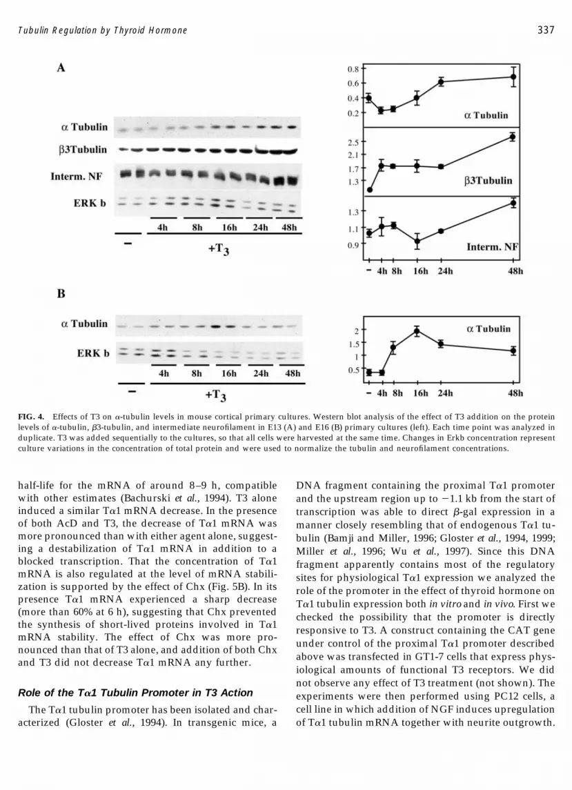
FIG. 4. Effects of T3 on �-tubulin levels in mouse cortical primary cultures. Western blot analysis of the effect of T3 addition on the proteinlevels of �-tubulin, �3-tubulin, and intermediate neurofilament in E13 (A) and E16 (B) primary cultures (left). Each time point was analyzed induplicate. T3 was added sequentially to the cultures, so that all cells were harvested at the same time. Changes in Erkb concentration representculture variations in the concentration of total protein and were used to normalize the tubulin and neurofilament concentrations.
337Tubulin Regulation by Thyroid Hormone
The cells therefore allowed for the control of the respon-siveness of the transfected T�1 promoter to NGF. PC12cells were transfected with the T�1 promoter togetherwith expression vectors encoding T3 receptors (Fig.6A). Addition of T3 did not result in a stimulation ofCAT activity. As controls, addition of NGF was able toinduce the reporter gene (Fig. 6A) and T3 was able toactivate the thymidine kinase (tk) promoter through aTRE (Fig. 6B). It can be concluded therefore that T3 doesnot influence directly the activity of the T�1 �-tubulinpromoter.
T3, however, could exert indirect effects in vivo on theT�-1 �-tubulin promoter as part of a distal response toa primary action of T3 elsewhere. This possibility waschecked using the transgenic mouse line K6, in whichthe �-gal gene is under control of the T�1 promoter. Inthese mice, expression of T�1:nlacZ is closely similar tothat of the endogenous gene, suggesting that the 1.1-kbpromoter fragment contains all sequences necessary forthe regulated expression of T�1 tubulin (Bamji andMiller, 1996). The mice were made hypothyroid using acombination of two antithyroid drugs, as described un-der Experimental Methods. The reduced weight of theanimals and the strong alterations in Purkinje cell or-ganization (not shown) confirmed the effectiveness ofthe treatment.
Expression of the transgene was analyzed by histo-chemistry for �-galactosidase and that of the endoge-nous gene by in situ hybridization, and the results are
illustrated in Fig. 7. In Fig. 7A, it is shown that �-galstaining was stronger in the slices from hypothyroidmice at P15. These differences were not observed at P20and P35 (not shown). In Figs. 7B and 7C, expression ofthe endogenous tubulin (M�1 tubulin) mRNA isshown. There was a stronger signal in the hypothyroidbrains at P15 than in the control animals and the effectof hypothyroidism was not observed at P35. Thereforethe effects of hypothyroidism on the endogenous tubu-lin gene in mice were similar to those observed in ratsand also similar to the effects on the promoter-drivenreporter gene, suggesting that the effect of thyroid hor-mone is exerted, at least partially, through the �-tubulinpromoter.
DISCUSSION
In this work we have studied the role of thyroidhormone on the expression of the �1 tubulin isoform
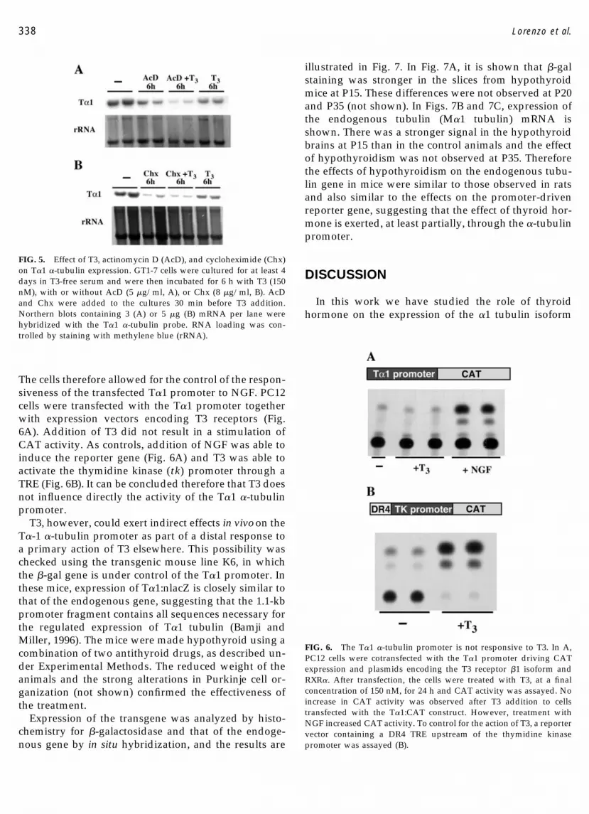
FIG. 5. Effect of T3, actinomycin D (AcD), and cycloheximide (Chx)on T�1 �-tubulin expression. GT1-7 cells were cultured for at least 4days in T3-free serum and were then incubated for 6 h with T3 (150nM), with or without AcD (5 �g/ml, A), or Chx (8 �g/ml, B). AcDand Chx were added to the cultures 30 min before T3 addition.Northern blots containing 3 (A) or 5 �g (B) mRNA per lane werehybridized with the T�1 �-tubulin probe. RNA loading was con-trolled by staining with methylene blue (rRNA).
FIG. 6. The T�1 �-tubulin promoter is not responsive to T3. In A,PC12 cells were cotransfected with the T�1 promoter driving CATexpression and plasmids encoding the T3 receptor �1 isoform andRXR�. After transfection, the cells were treated with T3, at a finalconcentration of 150 nM, for 24 h and CAT activity was assayed. Noincrease in CAT activity was observed after T3 addition to cellstransfected with the T�1:CAT construct. However, treatment withNGF increased CAT activity. To control for the action of T3, a reportervector containing a DR4 TRE upstream of the thymidine kinasepromoter was assayed (B).
338 Lorenzo et al.
(T�1 or M�1 �-tubulin) in the rodent brain duringpostnatal development. Although T�1 �-tubulin is ex-pressed in other tissues such as lung or testis (Lewis etal., 1985), in the brain it is neuron-specific and stronglydependent on the stage of neuronal growth and differ-entiation (Mathew and Miller, 1990, 1993; Miller et al.,1987, 1989, 1991; Wu et al., 1997). Although widelyexpressed, T�1 mRNA levels vary considerably amongdifferent regions during brain maturation (Miller et al.,1987). We found that during early postnatal develop-ment expression is high in neocortex, piriform cortex,hippocampus, thalamus, and granular layers of the cer-ebellum, in agreement with data from Miller et al. (1987)for P3 animals. As development proceeds, T�1 mRNAlevels decrease as maturational processes progress ineach region (Paxinos, 1985). The main consequence ofhypothyroidism was to delay this progressive decreaseof T�1 mRNA with age, leading to differences betweennormal and hypothyroid rats that were more pro-nounced around P20 in most brain regions. These re-sults generally agree with previous data from otherauthors using Northern blotting analysis of �-tubulinexpression (Aniello et al., 1991b; Figueiredo et al., 1993).
The mechanism of thyroid hormone action on T�1�-tubulin expression is complex. The previously de-scribed, putative thyroid hormone receptor-responsiveelement in the �-tubulin gene (Iglesias et al., 1996) hasbeen found by us to be nonfunctional (unpublishedresults from our laboratory). Therefore, we have lookedfor alternative mechanisms of T3 action by analyzingtubulin expression in cultured cells and the role of thetubulin promoter in transgenic mice. For the analysis invitro we used the neuronal cell line GT1-7, expressingphysiological amounts of functional T3 receptors(Morte et al., 1997). In these cells T3 exerts a fast andtransient decrease of tubulin mRNA, followed by arecovery to control levels. This effect was also observedin primary cultures isolated from E13 embryonic brains.In both cases the recovery of tubulin levels was parallelto an increased differentiation of the cells induced by T3as shown by induction of differentiation markers, suchas NSE in the GT1-7 cells, or �3-tubulin and intermedi-ate neurofilament in cortical neurons.
The initial effect of T3 on T�1 �-tubulin mRNA levelsappears to be due to increased mRNA degradation,perhaps through inhibition of mRNA-stabilizing pro-teins. The experiments involving Chx suggest that theT�1 �-tubulin mRNA levels are regulated, at least par-tially, by stabilizing proteins. As a matter of fact, theregulation of tubulin mRNA levels occurs primarily atthe level of mRNA stability, which is coupled to tubulintranslation (Gonzalez-Garay and Cabral, 1995, 1996;Theodorakis and Cleveland, 1992). The secondary effect
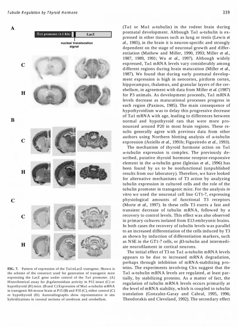
FIG. 7. Pattern of expression of the T�1:nLacZ transgene. Shown isthe scheme of the construct used for generation of transgenic miceexpressing the LacZ gene under control of the T�1 promoter. (A)Histochemical assay for �-galactosidase activity in P15 intact (C) orhypothyroid (H) mice. (B and C) Expression of M�1 �-tubulin mRNAin transgenic K6 mouse brain at P15 (B) and P35 (C), either control (C)or hypothyroid (H). Autoradiographs show representative in situhybridizations in coronal sections of cerebrum and cerebellum.
339Tubulin Regulation by Thyroid Hormone
of T3, increasing T�1 expression, was likely due to ageneral stimulation of cellular differentiation, as sug-gested by the increased expression of cell differentiationmarkers. This contention is supported by the observa-tion that the effect of T3 on cortical neurons depends onthe differentiating stage of the cultures. In E13 culturesin which most of the cells are neuronal progenitors theregulation of T�1 �-tubulin is similar to that observedin GT1-7 cells, with an initial decrease of mRNA fol-lowed by an increase. This effect, observed at themRNA level in GT1-7 cells also occurred at the proteinlevel in cortical cultures. On the other hand, in E16cultures, which contain primarily postmitotic neurons,T3 induced an increase in tubulin expression as soon as4 h after T3 addition, reaching a plateau after 16 h oftreatment. A possible explanation is that the increase in�-tubulin expression is secondary to a more generaleffect of T3 on neuronal maturation, which should takeplace more rapidly in neurons from E16 brains than inthose from E13. Deprivation of T3 from differentiatingcultures retards axodendritic outgrowth and also altersneuronal morphology (Biswas et al., 1997). The T3-de-pendent increase of �-tubulin levels may reflect theneed to rapidly increase the pool of �-tubulin de-manded by the stimulation of differentiation.
Thus we may postulate the existence of a doubleregulation of T�1 by T3 in cultured cells: an initialdecrease in T�1 mRNA levels, probably mediatedthrough a reduction in the half-life of the mRNA, fol-lowed by T3 stimulation of neuronal maturation, whichinduces an upregulation of T�1. This is in line with therole of T3 as a neural induction agent, as has beenshown in cell lines such as PC-12 cells expressing TR�1(Munoz et al., 1993) or N2a cells expressing TR�1 (Lebelet al., 1994).
T3 also exerts influences through the tubulin pro-moter in vivo. A 1.1-kb fragment of the tubulin pro-moter was able to direct expression of a reporter gene intransgenic mice with a pattern similar to that of theendogenous gene (Bamji and Miller, 1996; Gloster et al.,1994, 1999). Therefore it presumably contains sequencesnecessary for full physiological expression in vivo. As inrats, hypothyroidism in mice delayed the developmen-tal downregulation of both the endogenous �-tubulingene and the reporter gene under control of the tubulinpromoter. However, the promoter was not responsiveto T3 in cells cotransfected with thyroid hormone re-ceptors. The results therefore indicate that thyroid hor-mone exerts indirect influences on the activity of thetubulin promoter, which are involved in the physiolog-ical downregulation of the tubulin gene.
EXPERIMENTAL METHODS
Animals
CD1 mice and Wistar rats were used to map theexpression of the respective endogenous M�1 and T�1�-tubulin mRNAs by in situ hybridization. Maintenanceand handling of animals were as recommended by theEuropean Communities Council Directive of November24, 1986 (86/609/EEC). The T�1:nlacZ transgenic mouseline K6, in which 1.1 kb of the 5� upstream region of theT�1 �-tubulin gene regulates the expression of a �-ga-lactosidase marker gene, was used to map the expres-sion of the T�1:nlacZ transgene by using X-gal staining(Bamji and Miller, 1996; Gloster et al., 1994). Hypothy-roidism was induced by the continuous administrationof 0.02% methyl mercaptoimidazol (Sigma ChemicalCo., St. Louis, MO) and 1% KClO4 in the drinkingwater, starting on day 9 of pregnancy, until the end ofthe experiments.
LacZ Staining
T�1:nlacZ transgenic mice were deeply anesthetizedwith sodium pentobarbital (35 mg/kg) and perfusedtranscardially with 4% paraformaldehyde in phosphatebuffer. The brains were removed, postfixed, and slicedinto 1- to 2-mm-thick sections. They were then rinsedthree times for 30 min each with a wash buffer contain-ing 2 mM MgCl2, 0.01% sodium deoxycholate, 0.02%NP-40, 0.1 M NaH2PO4, pH 7.3. The staining reactionwas performed by incubating the tissue at 37°C in areaction mixture containing all the components of thewash buffer with the addition of 1 mg/ml X-gal, 5 mMK3Fe(CN)6, 5 mM K4Fe(CN)6, pH 7.3–7.6, for 30–90 minas previously described (Bamji and Miller, 1996; Glosteret al., 1994). All sections from the same experiment wereallowed exactly the same time for color development.
In Situ Hybridization
Two animals from each experimental group (controland hypothyroid) were analyzed at each of the follow-ing postnatal ages: P3, P5, P10, P20, and P32. All pro-cedures, including killing the animals, treatment of tis-sue, sectioning, and in situ hybridization using floatingsections, were exactly as described previously (Iniguezet al., 1996). 35S-labeled sense and antisense riboprobeswere prepared from the 3� untranslated region of T�1�-tubulin complementary DNA. After exposure to X-ray films, the radioactive signal was quantified by den-sitometry as described (Guadano-Ferraz et al., 1999). Tohave comparable signals in the autoradiographs, the
340 Lorenzo et al.
exposure time was 7 days for P3 and P5, 8 days for P10,10 days for P20, and 15 days for P32. For identificationof brain structures, the rat atlas of Paxinos and Watsonwas followed (Paxinos, 1985).
Cell Cultures
GT1-7 cells were supplied by Dr. P. Mellon (Univer-sity of California at San Diego). Cells were routinelycultured in Dulbecco’s modified Eagle’s medium with4.5 mg/ml glucose and 10% fetal calf serum. To studythe influence of thyroid hormone, the cells were incu-bated in serum depleted of thyroid hormone by treat-ment with charcoal and Dowex resin as previouslydescribed (Morte et al., 1997). The chemicals used forcell treatments were from Sigma Chemical Co. andwere added to the cultures at the following final con-centrations: T3, 150 nM; Chx, 8 �g/ml; and AcD, 5�g/ml. When different time points were recorded, thecells were plated and T3 was added at different timesbefore harvesting. In this way the cells corresponding toeach time-point were all incubated for the same timeperiod and the cells were harvested at the same time.
Cortical progenitors from CD1 mice were cultured asdescribed (Slack et al., 1998). Briefly, cortical tissue ob-tained from E13 or E16 embryos was dissected in ice-cold Hanks’ balanced salt solution (Gibco). The tissuewas disaggregated by being passed through a fire-pol-ished Pasteur pipette into cell clusters that were platedin multiwell tissue culture dishes precoated with lami-nin and poly-d-lysine (Collaborative Biomedical Prod-ucts). The cells were cultured in Neurobasal medium(Gibco) containing 500 �M glutamine, N2-supplement,and penicillin–streptomycin (Gibco); the medium wassupplemented with bFGF for the first 2 days. ThereafterB27 was substituted for bFGF. Cell density ranged be-tween 30,000 and 100,000 cells per well. To quantitatethe effect of T3 on neuronal differentiation, T3-treatedand untreated cultures were stained for �3-tubulin andcounterstained with Hoechst 33258 to visualize the nu-clei of cells, as previously described (Gloster et al., 1999).Total number of cells and �3-tubulin-positive cells werecounted in 11 T3-treated cultures and 9 untreated cul-tures, and each field examined contained 50–100 cells.The results were analyzed statistically using the Stu-dent t test.
Western Blot Analysis
Neurons were lysed in TBS lysis buffer containing 13mM NaCl, 20 mM Tris–HCl, pH 8, 1% (vol/vol) NP-40,10% (vol/vol) glycerol, 1 mM PMSF, 10 �g/ml aproti-nin, 0.2 �g/ml leupeptin, 5 mM phenanthroline, and 1.5
mM sodium vanadate. Samples were rocked for 20 minat 4°C and centrifuged for 15 min at 15,000 rpm. Thesupernatant was normalized for protein concentrationusing a BCA Protein Assay Reagent (Pierce ChemicalCo., Rockford, IL). Supernatants were boiled in samplebuffer (2% sodium dodecyl sulfate (SDS), 100 mM di-thiothreitol, 10% glycerol, and 0.05 bromophenol blue)for 5 min and electrophoresed on 8% SDS–polyacryl-amide minigels. After electrophoresis, proteins weretransferred to nitrocellulose filters for 1 h at 0.5 A, andthe membrane was washed twice for 10 min in TBS. Thefilters were stained with Ponceau red. Membranes werethen blocked in 5% nonfat dry milk in TBS for 2.5 h andwashed twice for 10 min in TBS. The membranes wereincubated with the primary antibodies overnight at 4°C,at a dilution of 1:10,000 for anti-�-tubulin, 1:1000 foranti-�3-tubulin, 1:500 for anti-intermediate NF, and1:10,000 anti-Erkb. Incubation with secondary antibod-ies (goat anti-mouse HRP for anti-�tubulin and anti-�3-tubulin and goat anti-rabbit HRP for anti-intermediateNF and anti-Erkb antibodies) was for 1.5 h at roomtemperature at a 1:10,000 dilution. Detection was car-ried out using enhanced chemiluminescence (Amer-sham Corp., Arlington Heights, IL) and XAR X-ray film(Eastman Kodak, Rochester, NY). Results were quanti-tated by image analysis.
Preparation of RNA and Northern Blot Analysis
Isolation of poly(A)� RNA was done as described(Vennstrom and Bishop, 1982). RNA samples werecarefully checked by densitometry and by electrophore-sis to ensure that the RNA loaded for each sample wasidentical. Northern blots were performed on nylonmembranes (Nytran; Schleicher and Schuell) accordingto standard protocols (Sambrook et al., 1989) using 5 �gRNA per lane. As a further control for RNA loading thefilters were stained with 0.02% methylene blue in 0.3 Msodium acetate. Probes were labeled by the randompriming method using available kits (Rediprime; Am-ersham, Buckinghamshire, UK). Hybridizations wereperformed overnight at 65°C in 7% SDS, 500 mM so-dium phosphate buffer, pH 7.2, and 1 mM EDTA(Church and Gilbert, 1984). Filters were washed twicefor 30 min each in 1% SDS and 40 mM sodium phos-phate buffer, pH 7.2, at 65°C. Before rehybridization ofthe nylon membranes with other probes, the radioactiveprobe was stripped off the membrane by placing in a75°C water bath for 5 min. Sizes of respective mRNAswere calculated by using an RNA ladder as marker.Membranes were exposed to Hyperfilm MP (Amer-sham). Results were quantitated by densitometry.
341Tubulin Regulation by Thyroid Hormone
Transfections and CAT Assays
PC12 cells were grown in DMEM supplemented withthyroid hormone-depleted 10% fetal calf serum. Cellswere transfected, using the calcium phosphate proce-dure, with 4 �g of plasmids containing the CAT geneunder control of either the T�1 or the tk promoter. Thelatter construct also contained the strong thyroid hor-mone-responsive element present in the LTR of theMoloney murine leukemia virus (Sap et al., 1989). Cellswere cotransfected with or without 0.2 �g of TR� and0.2 �g of RXR� expression plasmids and 1 �g of plas-mid pCH110 (Pharmacia LKB Biotechnology). Thisplasmid contains the �-galactosidase gene under thecontrol of the SV-40 early promoter and was used as aninternal control to correct for transfection variability. At16–18 h after DNA addition, the cells were washed withPBS and replenished with medium containing serumdepleted of thyroid hormones. Where appropriate, 150�M T3 or NGF was added and the cells were incubatedfor 24 h before harvesting. Preparation of cell extractsand measurements of CAT and �-gal reporter geneactivities were done as described by Sambrook et al.(1989). To correct for transfection variability, the cellextracts used for determination of CAT activity werediluted according to the �-gal measurements.
ACKNOWLEDGMENTS
This work was supported by grants from the CICYT (PM98-0118)and the Community of Madrid (08.5/0044/2000 1). P. Lorenzo wassupported by a fellowship from the Basque Government. We ac-knowledge G. Chacon for technical help and F. Nunez, P. Senor, andM. Marsa for the care of the animals.
REFERENCES
Aniello, F., Couchie, D., Bridoux, A. M., Gripois, D., and Nunez, J.(1991a). Splicing of juvenile and adult tau mRNA variants is regu-lated by thyroid hormone. Proc. Natl. Acad. Sci. USA 88: 4035–4039.
Aniello, F., Couchie, D., Gripois, D., and Nunez, J. (1991b). Regulationof five tubulin isotypes by thyroid hormone during brain develop-ment. J. Neurochem. 57: 1781–1786.
Bachurski, C. J., Theodorakis, N. G., Coulson, R. M., and Cleveland,D. W. (1994). An amino-terminal tetrapeptide specifies cotransla-tional degradation of beta-tubulin but not alpha-tubulin mRNAs.Mol. Cell. Biol. 14: 4076–4086.
Bamji, S. X., and Miller, F. D. (1996). Comparison of the expression ofa T�1:nlacZ transgene and T�1 �-tubulin mRNA in the maturecentral nervous system. J. Comp. Neurol. 374: 52–69.
Benjamin, S., Cambray-Deakin, M. A., and Burgoyne, R. D. (1988).Effects of hypothyroidism on the expression of three microtubule-associated proteins (1A,1B and 2) in the developing rat cerebellum.Neuroscience 27: 931–939.
Bernal, J., and Nunez, J. (1995). Thyroid hormones and brain devel-opment. Eur. J. Endocrinol. 133: 390–398.
Biesiada, E., Adams, P. M., Shanklin, D. R., Bloom, G. S., and Stein,S. A. (1996). Biology of the congenitally hypothyroid hyt/hytmouse. Adv. Neuroimmunol. 6: 309–346.
Biswas, S. C., Pal, U., and Sarkar, P. K. (1997). Regulation of cytoskel-etal proteins by thyroid hormone during neuronal maturation anddifferentiation. Brain Res. 23: 245–253.
Charriere-Bertrand, C., and Nunez, J. (1992). Regulation of tubulin,tau and microtubule associated protein 2 expression during mousebrain development. Neurochem. Int. 21: 535–541.
Church, G. M., and Gilbert, W. (1984). Genome sequencing. Proc. Natl.Acad. Sci. USA 81: 1991–1995.
Figueiredo, B. C., Almazan, G., Ma, Y., Tetzlaff, W., Miller, F. D., andCuello, A. C. (1993). Gene expression in the developing cerebellumduring perinatal hypo- and hyperthyroidism. Mol. Brain Res. 17:258–268.
Geddes, J. W., Wong, J., Choi, B. H., Kim, R. C., Cotman, C. W., andMiller, F. D. (1990). Increased expression of the embryonic form ofa developmentally regulated mRNA in Alzheimer’s disease. Neu-rosci. Lett. 109: 54–61.
Gloster, A., El-Bizri, H., Bamji, S. X., and Miller, F. D. (1999). Earlyinduction of T�1 �-tubulin transcription in neurons of the devel-oping nervous system. J. Comp. Neurol. 405: 45–60.
Gloster, A., Wu, W., Spelman, A., Weiss, S., Causing, C., Pozniac, C.,Reynolds, B., Chang, E., Toma, J. G., and Miller, F. D. (1994). TheT�1 �-tubulin promoter specifies gene expression as a function ofneuronal growth and regeneration in transgenic mice. J. Neurosci.14: 7319–7330.
Gonzalez-Garay, M. L., and Cabral, F. (1995). Overexpression of anepitope-tagged beta-tubulin in Chinese hamster ovary cells causesan increase in endogenous alpha-tubulin synthesis. Cell. Motil. Cy-toskeleton 31: 259–272.
Gonzalez-Garay, M. L., and Cabral, F. (1996). �-Tubulin limits its ownsynthesis: Evidence for a mechanism involving translational repres-sion. J. Cell Biol. 135: 1525–1534.
Guadano-Ferraz, A., Escamez, M. J., Rausell, E., and Bernal, J. (1999).Expression of type 2 iodothyronine deiodinase in hypothyroid ratbrain indicates an important role of thyroid hormone in the devel-opment of specific primary sensory systems. J. Neurosci. 19: 3430–3439.
Iglesias, T., Caubın, J., Stannenberg, H. G., Zaballos, A., Bernal, J., andMunoz, A. (1996). Thyroid hormone-dependent transcriptional re-pression of neural cell adhesion molecule during brain maturation.EMBO J. 15: 4307–4316.
Iniguez, M. A., DeLecea, L., Guadano-Ferraz, A., Morte, B., Geren-dasy, D., Sutcliffe, G., and Bernal, J. (1996). Cell-specific effects ofthyroid hormone on RC3/neurogranin expression in rat brain.Endocrinology 137: 1032–1041.
Knoops, B., and Octave, J. N. (1997). �1 tubulin mRNA level isincreased during neurite outgrowth of NG108-15 cells but notduring neurite outgrowth inhibition by CNS myelin. NeuroReport 8:795–798.
Lebel, J.-M., Dussault, J. H., and Puymirat, J. (1994). Overexpressionof the �1 thyroid receptor induces differentiation in neuro-2a cells.Proc. Natl. Acad. Sci. USA 91: 2644–2648.
Lewis, S. A., Gwo-Sho Lee, M., and Cowan, N. J. (1985). Five mousetubulin isotypes and their regulated expression during develop-ment. J. Cell Biol. 101: 852–861.
Ma, Y., Campenot, R. B., and Miller, F. D. (1992). Concentration-dependent regulation of neuronal gene expression by nerve growthfactor. J. Cell Biol. 117: 135–141.
Mathew, T. C., and Miller, F. D. (1990). Increased expression of T�1 �
342 Lorenzo et al.
tubulin mRNA during collateral and NGF-induced sprouting ofsympathetic neurons. Dev. Biol. 141: 84–92.
Mathew, T. C., and Miller, F. D. (1993). Induction of t�1 �-tubulinmRNA during neuronal regeneration is a function of the amount ofaxon lost. Dev. Biol. 158: 467–474.
Mellon, P. L., Windle, J. J., Goldsmith, P. C., Padula, C. A., andWeiner, R. I. (1990). Immortalization of hypothalamic GnRH neu-rons by genetically targeted tumorigenesis. Neuron 5: 1–10.
Miller, F. D., Mathew, T. C., and Toma, J. G. (1991). Regulation ofnerve growth factor receptor gene expression by nerve growthfactor in the developing peripheral nervous system. J. Cell Biol. 112:303–312.
Miller, F. D., Naus, C. C. G., Durand, M., Bloom, F. E., and Milner, R.(1987). Isotypes of �-tubulin are differentially regulated duringneuronal maturation. J. Cell Biol. 105: 3065–3073.
Miller, F. D., Rogers, D., Bamji, S. X., Slack, R. S., and Gloster, A.(1996). Analysis and manipulation of neuronal gene expressionusing the T�1 �-tubulin promoter. Semin. Neurosci. 8: 117–124.
Miller, F. D., Tetzlaff, W., Bisby, M. A., Fawcett, J. W., and Milner, R.(1989). Rapid induction of the major embryonic a tubulin mRNA,Ta1, during nerve regeneration in adult rats. J. Neurosci. 9: 1452–1463.
Morte, B., Iniguez, M. A., Lorenzo, P. I., and Bernal, J. (1997). Thyroidhormone-regulated expression of RC3/neurogranin in the immor-talized hypothalamic cell line GT1-7. J. Neurochem. 69: 902–909.
Munoz, A., and Bernal, J. (1997). Biological activities of thyroid hor-mone receptors. Eur. J. Endocrinol. 137: 433–445.
Munoz, A., Wrighton, C., Seliger, B., Bernal, J., and Beug, H. (1993).Thyroid hormone receptor/c-erbA: Control of commitment anddifferentiation in the neuronal/chromaffin progenitor line PC12.J. Cell Biol. 121: 423–438.
Nunez, J., Couchie, D., Aniello, F., and Bridoux, A. M. (1991). Regu-lation by thyroid hormone of microtubule assembly and neuronaldifferentiation. Neurochem. Res. 16: 975–982.
Nunez, J., Couchie, D., Aniello, F., and Bridoux, A. M. (1992). Thyroidhormone effect on neuronal differentiation during brain develop-ment. Acta Med. Austriaca 19: 36–39.
Paxinos, G. (1985). The Rat Nervous System. Academic Press, Sydney.Sambrook, J., Fritsch, E. F., and Maniatis, T. (1989). Molecular Cloning:
A Laboratory Manual. Cold Spring Harbor Laboratory Press, ColdSpring Harbor, NY.
Sap, J., Munoz, A., Schmitt, J., Stunnenberg, H., and Vennstrom, B.(1989). Repression of transcription mediated at a thyroid hormoneresponse element by the v-erbA oncogene product. Nature 340:242–244.
Slack, R. S., El-Bizri, H., Wong, J., Belliveau, D. J., and Miller, F. D.(1998). A critical temporal requirement for the retinoblastoma pro-tein family during neuronal determination. J. Cell Biol. 140: 1497–1509.
Tetzlaff, W., Alexander, S. W., Miller, F. D., and Bisby, M. A. (1991).Response of facial and rubrospinal neurons to axotomy: Changes inmRNA expression for cytoskeletal proteins and GAP-43. J. Neurosci.11: 2528–2544.
Theodorakis, N. G., and Cleveland, D. W. (1992). Physical evidencefor cotranslational regulation of beta-tubulin mRNA degradation.Mol. Cell. Biol. 12: 791–799.
Vennstrom, B., and Bishop, J. M. (1982). Isolation and characterizationof chicken DNA homologous to the two putative oncogenes ofavian erythroblastosis virus. Cell 28: 135–143.
Wu, W., Gloster, A., and Miller, F. D. (1997). Transcriptional repres-sion of the growth-associated T�1 �-tubulin gene by target contact.J. Neurosci. Res. 48: 477–487.
Received July 31, 2001Revised November 22, 2001Accepted December 6, 2001
343Tubulin Regulation by Thyroid Hormone
Related Documents


![Atypical Thyroid Function Tests, Thyroid Hormone ... · Atypical Thyroid Function Tests, Thyroid Hormone Resistance [Atipik Tiroid Fonksiyon Testleri: Tiroid Hormon Direnci] Soner](https://static.cupdf.com/doc/110x72/5c83755009d3f2be2a8b56f6/atypical-thyroid-function-tests-thyroid-hormone-atypical-thyroid-function.jpg)