79 Three Dimensional Assessment of soft tissue and Airway changes after Rapid Maxillary Expansion in Cleft Palate Patients using Cone Beam CT Ahmed M. Elbahar * , Wael M. Refai, Mohamed E. Ibrahim, Mohamed G. Elshal Orthodontic Department, Faculty of Dentistry, Minia University, Egypt [email protected] Abstract: Objective: Measuring air way and soft tissue changes in cleft palate patients after rapid maxillary arch expansion by bonded hyrax appliance using cone-beam computed tomography. Material and methods: RME for 10 cleft patients ranged from 8-12 years old then pre and post expansion CBCT image was taken to them and analyzed by on demand 3D app software (Cybermed, South Korea) was utilized for linear and angular measurement. Results: Significant increase in nasal width, air way width, no change in air way depth and enhance soft tissue measurement. Conclusion: Enhanced air way and soft tissue measurements after RME on cleft patients. [Ahmed M. Elbahar, Wael M. Refai, Mohamed E. Ibrahim, Mohamed G. Elshal. Three Dimensional Assessment of soft tissue and Airway changes after Rapid Maxillary Expansion in Cleft Palate Patients using Cone Beam CT. Life Sci J 2019;16(11):79-87]. ISSN: 1097-8135 (Print) / ISSN: 2372-613X (Online). http://www.lifesciencesite.com . 8. doi:10.7537/marslsj161119.08 . Key Words: RME, Cleft patients and Air way. 1. Introduction: Cleft lip and palate (CLP) is the most common human congenital malformation affecting the facial region. Cleft lip and palate occurs at the time of early embryogenesis from a failure in fusion of medial nasal and maxillary processes that result in orofacialclefting involving the upper lip, alveolus and/or primary palate. The most widely adopted management strategy includes the surgical reconnection of the cleft anatomical structures followed by their development to gain proper appearance, occlusion, and speech. Maxillary growth in operated CLP patients is often decreased in the 3 dimensions. The maxillary growth deficiency affects the dental arches relationship on the vertical, sagittal, and transverse planes, frequently resulting in anterior and/or posterior crossbite occurring in the early dentition. (3) Early correction of posterior crossbite is considered necessary for children with cleft palate to relocate the permanent tooth follicles in a more favorable position, and improve the potential for normal development of occlusion. (44) Rapid maxillary expansion (RME) is an orthodontic procedure that aims tocorrect the maxillary arch constriction by transversal separation of maxillary halves. The aim of maxillary expansion is not only to treat the posterior crossbite, but also to align the maxillary segment. This procedure increases the alveolar cleft width and creates room for bone graft placement. CBCT images are valuable for determining the volume of the alveolar defect. From all previously mentioned, the skeletal and dental changes that occur due to RME in cleft patients proved to be a point of worthy investigation. Accordingly this study will be conducted to highlight this aim. (27) Materials and Methods: This study performed on 10 unilateral cleft lip and palate patients (UCLP) in the mixed dentition with cleft lip and palate surgical repair performed at early childhood with constricted maxillary arch. The patients’ age’s ranged from 8-12 years old. They were collected from the clinic of the department of orthodontics faculty of dentistry Minia University. Patients were treated by RME as a part of their comprehensive treatment using the bonded hyrax appliance. Patient Age from 8-12 years. Patient in mixed dentition with constricted maxillary arch. Patient performed lip and palatal repair in early childhood. Posterior cross bite is present. Free from any systemic diseases. No previous orthodontic or orthopedic treatment. For each patient these records were obtained. Standardized extraoral and intraoral photographs. Standardized dental casts. Panorama and lateral cephalometric radiographs. CBCT before and after expansion (after 6 months of treatment). Palatal arch was constructed to the patients for retention. Each patient was treated by RME utilizing bonded hyrax appliance. Activation of appliance One quarter turns twice per day for each patient. The expansion continue until over correction was obtained to the extent that palatal cusps of the maxillary first molar touches the buccal cusps of the opposing teeth then the appliance

Welcome message from author
This document is posted to help you gain knowledge. Please leave a comment to let me know what you think about it! Share it to your friends and learn new things together.
Transcript
79
Three Dimensional Assessment of soft tissue and Airway changes after Rapid Maxillary Expansion in Cleft
Palate Patients using Cone Beam CT
Ahmed M. Elbahar*, Wael M. Refai, Mohamed E. Ibrahim, Mohamed G. Elshal
Orthodontic Department, Faculty of Dentistry, Minia University, Egypt [email protected]
Abstract: Objective: Measuring air way and soft tissue changes in cleft palate patients after rapid maxillary arch expansion by bonded hyrax appliance using cone-beam computed tomography. Material and methods: RME for 10 cleft patients ranged from 8-12 years old then pre and post expansion CBCT image was taken to them and analyzed by on demand 3D app software (Cybermed, South Korea) was utilized for linear and angular measurement. Results: Significant increase in nasal width, air way width, no change in air way depth and enhance soft tissue measurement. Conclusion: Enhanced air way and soft tissue measurements after RME on cleft patients. [Ahmed M. Elbahar, Wael M. Refai, Mohamed E. Ibrahim, Mohamed G. Elshal. Three Dimensional Assessment of soft tissue and Airway changes after Rapid Maxillary Expansion in Cleft Palate Patients using Cone Beam CT. Life Sci J 2019;16(11):79-87]. ISSN: 1097-8135 (Print) / ISSN: 2372-613X (Online). http://www.lifesciencesite.com. 8. doi:10.7537/marslsj161119.08. Key Words: RME, Cleft patients and Air way. 1. Introduction:
Cleft lip and palate (CLP) is the most common human congenital malformation affecting the facial region. Cleft lip and palate occurs at the time of early embryogenesis from a failure in fusion of medial nasal and maxillary processes that result in orofacialclefting involving the upper lip, alveolus and/or primary palate. The most widely adopted management strategy includes the surgical reconnection of the cleft anatomical structures followed by their development to gain proper appearance, occlusion, and speech. Maxillary growth in operated CLP patients is often decreased in the 3 dimensions. The maxillary growth deficiency affects the dental arches relationship on the vertical, sagittal, and transverse planes, frequently resulting in anterior and/or posterior crossbite occurring in the early dentition. (3)
Early correction of posterior crossbite is considered necessary for children with cleft palate to relocate the permanent tooth follicles in a more favorable position, and improve the potential for normal development of occlusion. (44)
Rapid maxillary expansion (RME) is an orthodontic procedure that aims tocorrect the maxillary arch constriction by transversal separation of maxillary halves. The aim of maxillary expansion is not only to treat the posterior crossbite, but also to align the maxillary segment. This procedure increases the alveolar cleft width and creates room for bone graft placement. CBCT images are valuable for determining the volume of the alveolar defect. From all previously mentioned, the skeletal and dental
changes that occur due to RME in cleft patients proved to be a point of worthy investigation. Accordingly this study will be conducted to highlight this aim. (27)
Materials and Methods:
This study performed on 10 unilateral cleft lip and palate patients (UCLP) in the mixed dentition with cleft lip and palate surgical repair performed at early childhood with constricted maxillary arch. The patients’ age’s ranged from 8-12 years old. They were collected from the clinic of the department of orthodontics faculty of dentistry Minia University. Patients were treated by RME as a part of their comprehensive treatment using the bonded hyrax appliance. Patient Age from 8-12 years. Patient in mixed dentition with constricted maxillary arch. Patient performed lip and palatal repair in early childhood. Posterior cross bite is present. Free from any systemic diseases. No previous orthodontic or orthopedic treatment.
For each patient these records were obtained. Standardized extraoral and intraoral photographs. Standardized dental casts. Panorama and lateral cephalometric radiographs. CBCT before and after expansion (after 6 months of treatment). Palatal arch was constructed to the patients for retention. Each patient was treated by RME utilizing bonded hyrax appliance. Activation of appliance One quarter turns twice per day for each patient. The expansion continue until over correction was obtained to the extent that palatal cusps of the maxillary first molar touches the buccal cusps of the opposing teeth then the appliance

Life Science Journal 2019;16(11) http://www.lifesciencesite.com LSJ
80
was left in situ as a retainer for six months and then the appliance was removed.
All patients under this study were submitted for CBCT scanning for angular and linear evaluation. After acquisition, data were exported and transferred
in DICOM format and downloaded via a Compact Disk (CD) to a personal computer for analysis, where, Ondemand 3D App software (Cybermed, South Korea) was utilized for linear and aList of Landmarks.
a- Frontal view
b- Lateral view
Fig 1 extra oral photographs before expansion
Upper occlusal view
Lower occlusal view
Frontal view
Right side view

Life Science Journal 2019;16(11) http://www.lifesciencesite.com LSJ
81
Left side view
Fig2. Pre-expansion intra-oral photographs
Fig.3 Frontal view with hyrax
Lateral view
Frontal view
Fig 4 Extra-oral photographs after expansion
Upper occlusal view
Lower occlusal view
Life Science Journal 2019;16(11) http://www.lifesciencesite.com LSJ
82
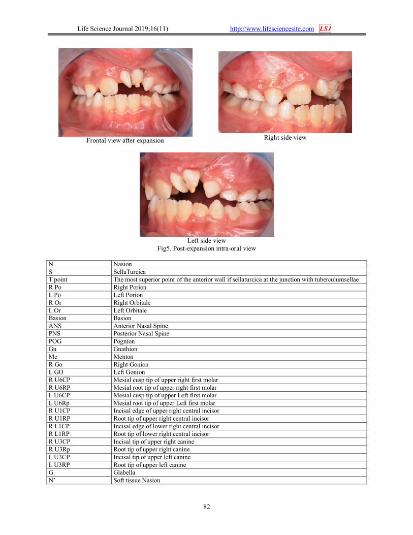
Frontal view after expansion
Right side view
Left side view
Fig5. Post-expansion intra-oral view
N Nasion S SellaTurcica T point The most superior point of the anterior wall if sellaturcica at the junction with tuberculumsellae R Po Right Porion L Po Left Porion R Or Right Orbitale L Or Left Orbitale Basion Basion ANS Anterior Nasal Spine PNS Posterior Nasal Spine POG Pognion Gn Gnathion Me Menton R Go Right Gonion L GO Left Gonion R U6CP Mesial cusp tip of upper right first molar R U6RP Mesial root tip of upper right first molar L U6CP Mesial cusp tip of upper Left first molar L U6Rp Mesial root tip of upper Left first molar R U1CP Incisal edge of upper right central incisor R U1RP Root tip of upper right central incisor R L1CP Incisal edge of lower right central incisor R L1RP Root tip of lower right central incisor R U3CP Incisal tip of upper right canine R U3Rp Root tip of upper right canine L U3CP Incisal tip of upper left canine L U3RP Root tip of upper left canine G Glabella N’ Soft tissue Nasion
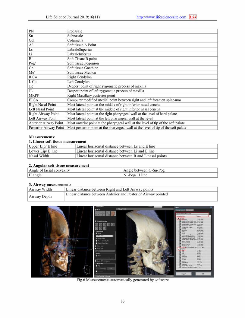
Life Science Journal 2019;16(11) http://www.lifesciencesite.com LSJ
83
PN Pronasale Sn Subnasale Col Columella A’ Soft tissue A Point Ls LabraleSuperius Li LabraleInferius B’ Soft Tissue B point Pog’ Soft tissue Pogonion Gn’ Soft tissue Gnathion Me’ Soft tissue Menton R Co Right Condylon L Co Left Condylon JR Deepest point of right zygomatic process of maxilla JL Deepest point of left zygomatic process of maxilla MRPP Right Maxillary posterior point ELSA Computer modified medial point between right and left foramen spinosum Right Nasal Point Most lateral point at the middle of right inferior nasal concha Left Nasal Point Most lateral point at the middle of right inferior nasal concha Right Airway Point Most lateral point at the right pharyngeal wall at the level of hard palate Left Airway Point Most lateral point at the left pharyngeal wall at the level Anterior Airway Point Most anterior point at the pharyngeal wall at the level of tip of the soft palate Posterior Airway Point Most posterior point at the pharyngeal wall at the level of tip of the soft palate Measurements: 1. Linear soft tissue measurement Upper Lip/ E line Linear horizontal distance between Ls and E line Lower Lip/ E line Linear horizontal distance between Li and E line Nasal Width Linear horizontal distance between R and L nasal points 2. Angular soft tissue measurement Angle of facial convexity Angle between G-Sn-Pog H angle N’-Pog/ H line 3. Airway measurements Airway Width Linear distance between Right and Left Airway points
Airway Depth Linear distance between Anterior and Posterior Airway pointed
Fig.6 Measurements automatically generated by software
Life Science Journal 2019;16(11) http://www.lifesciencesite.com LSJ
84
Statistical method: The collected data were coded, tabulated, and
statistically analyzed using SPSS program and graphical presentation was done using Microsoft excel version 2016.
Descriptive statistics were done for parametric quantitative data by mean ± standard deviation and for non-parametric quantitative data by median and interquartile range (IQR), while they were done for categorical data by number and percentage.
Determination of the normality of data was done by Shapiro-Wilk test.
Analyses were done for parametric quantitative data using Paired Samples T test between pre and post treatment.
Analyses were done for non-parametric quantitative data using Wilcoxon Signed rank test between pre and post treatment.
The level of significance was taken at (P value < 0.05) Results:
Data from this study were gathered, tabulated and then statistically analyzed.
Results are presented under the following heading:
1. Soft tissue measurement analysis: a. Soft tissue angular measures:
i. H- angle ii. G-Sn-Pog
b. Soft tissue linear measures: i. Upper lip/ E line
ii. Lower lip / E line. 2. Nasal measurement analysis: 3. Air way measurements
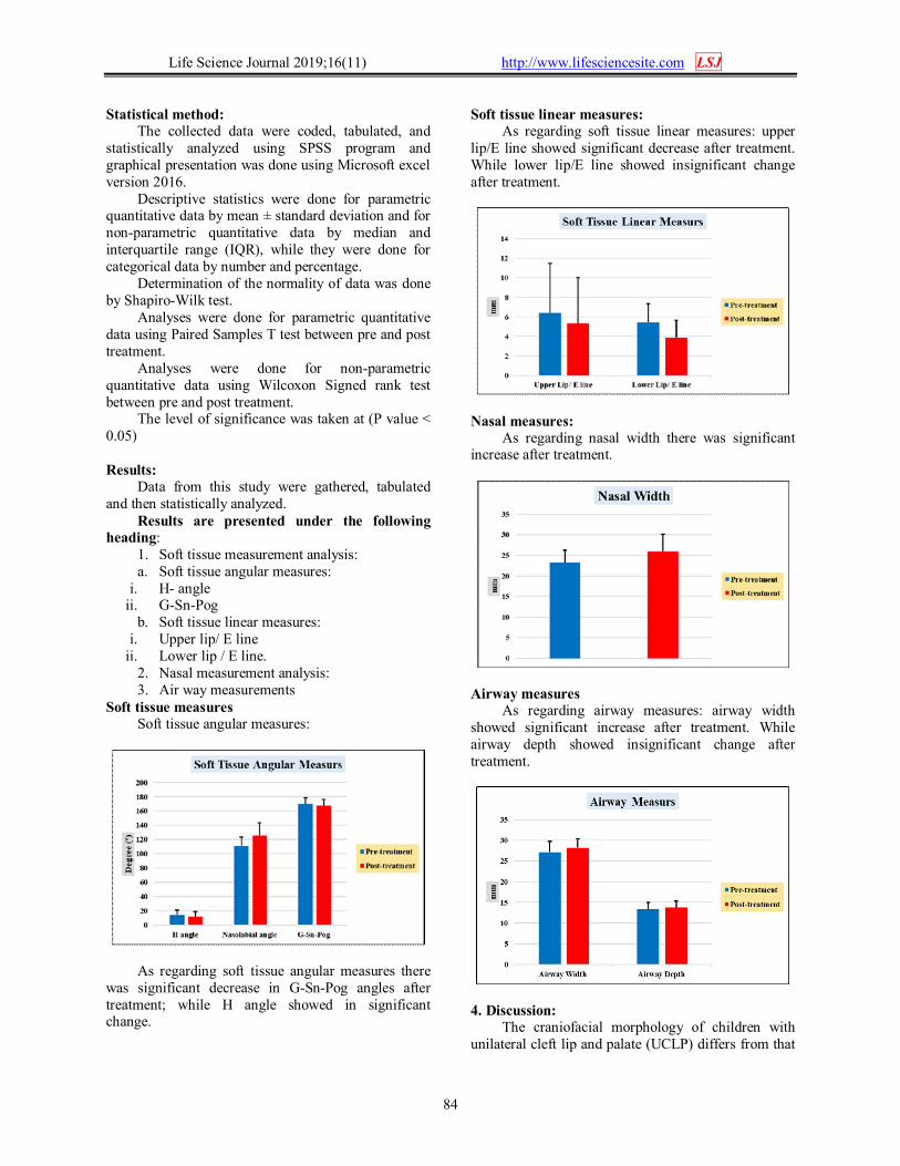
Soft tissue measures Soft tissue angular measures:
As regarding soft tissue angular measures there
was significant decrease in G-Sn-Pog angles after treatment; while H angle showed in significant change.
Soft tissue linear measures: As regarding soft tissue linear measures: upper
lip/E line showed significant decrease after treatment. While lower lip/E line showed insignificant change after treatment.
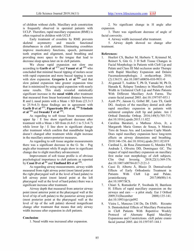
Nasal measures:
As regarding nasal width there was significant increase after treatment.
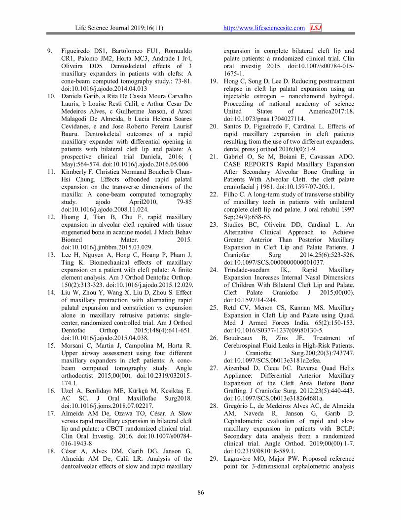
Airway measures
As regarding airway measures: airway width showed significant increase after treatment. While airway depth showed insignificant change after treatment.
4. Discussion: The craniofacial morphology of children with
unilateral cleft lip and palate (UCLP) differs from that
Life Science Journal 2019;16(11) http://www.lifesciencesite.com LSJ
85
of children without clefts. Maxillary arch constriction is frequently observed in operated patients with UCLP. Therefore, rapid maxillary expansion (RME) is often required in children with UCLP.
Early treatment of crossbite by RME prevents skeletal asymmetry and muscle functional disturbances in cleft patients. Eliminating crossbites improve masticatory functions, speech, permanent teeth eruption and alignment, nasal breathing and providing more space to the tongue that lead to decrease sleep apnea later on in cleft patients.
We chose rapid expansion not slow one according to Garib et al (17) and Vasant et al (25) who concluded that minimal buccal tipping of molars occur with rapid expansion and more buccal tipping is seen with slow expansion. Gregorio L et al (28) said that slow palatal expansion take greater expansion time that is minimized by using rapid expansion with nearly same results. This study revealed statistically significant increase in the nasal width after treatment that measured from linear horizontal distance between R and L nasal points with a Mean ± SD from (23.3±3 to 25.9±4.3) these findings are in agreement with Garib D et al (17), Figuerirado DS et al, (10) Trindade et al (24) and Iwasaki T et al (35).
As regarding to soft tissue linear measurement upper lip / E line show significant decrease after treatment with a Mean ± SD from (6.4±5.1 to 5.3±4.7) while lower lip / E line show in significant change after treatment which confirm that mandibular length doesn’t changed after treatment while slight increase in the maxillary antero-posterior measures.
As regarding to soft tissue angular measurements there was a significant decrease in the G- Sn – Pog angle after treatment while H angle show in significant change due to slight maxillary advancement.
Improvement of soft tissue profile is of obvious psychological importance to cleft patients as reported by Casai D et al (6) and Tindlund RS et al (33).
As regarding airway measurements. airway width measured from right airway point (most lateral point at the right pharyngeal wall at the level of hard palate) to left airway point (most lateral point at the left pharyngeal wall at the level of hard palate) there was a significant increase after treatment.
Airway depth that measured from anterior airway point (most anterior point at the pharyngeal wall at the level of tip of soft palate) to the posterior airway point (most posterior point at the pharyngeal wall at the level of tip of the soft palate) showed insignificant change after treatment that means that only airway width increase after expansion in cleft patients. Conclusion:
1. Nasal width was increased after expansion.
2. No significant change in H angle after expansion.
3. There was significant decrease of angle of facial convexity.
4. Airway width increased after treatment. 5. Airway depth showed no change after
treatment. References: 1. Hoefert CS, Bacher M, Herberts T, Krimmel M,
Reinert S, Göz G. 3 D Soft Tissue Changes in Facial Morphology in Patients with Cleft Lip and Palate and Class III Mal occlusion under Therapy with Rapid Maxillary Expansion and Delaire Facemaskmorphologie. J. orofacorthop. 2010; (2):136151. doi:10.1007/s00056-010-9931-0
2. Al-gunaid T, Asahito T, Ph D, Yamaki M, Ph D, Hanada K. Relapse Tendency in Maxillary Arch Width in Unilateral Cleft Lip and Palate Patients With Different Maxillary Arch Forms. The cranio facial journal 2003. doi:10.1597/07-053.1
3. Ayub PV, Janson G, Gribel BF, Lara TS, Garib DG. Analysis of the maxillary dental arch after rapid maxillary expansion in patients with unilateral complete cleft lip and palate. Am J Orthod Dentofac Orthop. 2016;149(5):705-715. doi:10.1016/j.ajodo.2015.11.022
4. Carolina Baratieri, a Matheus Alves, Jr, a Margareth Maria Gomes de Souza, b Monica Tirre de Souza Ara. and Lucianne Cople Maiab. Does rapid maxillary expansion have long-term effects on airway dimensions and breathing. 2010:146-156. doi:10.1016/j.ajodo.2011.02.019
5. Cardinal L, da Rosa Zimermann G, Mendes FM, Andrade I, Oliveira DD, Dominguez GC. The impact of rapid maxillary expansion on maxillary first molar root morphology of cleft subjects. Clin Oral Investig. 2018;22(1):369-376. doi:10.1007/s00784-017-2121-3
6. Cassi D, Alberto Ã, Blasio D. Dentoalveolar Effects of Early Orthodontic Treatment in Patients With Cleft Lip and Palate. jcraniofacsurg 2017;00(00):1-6. doi:10.1097/SCS.
7. Chiari S, Romsdorfer P, Swoboda H, Bantleon H. Effects of rapid maxillary expansion on the airways and ears — a pilot study. Eur J Orthod 2009;31(December 2008):135-141. doi:10.1093/ejo/cjn092
8. Vieira L, Menezes LM De, De EMS, Rizzatto S. Dentoskeletal Effects of Maxillary Protraction in Cleft Patients With Repetitive Weekly Protocol of Alternate Rapid Maxillary Expansions and Constrictions. cleft palate cranio facial journal 2005. doi:10.1597/07-144.1
Life Science Journal 2019;16(11) http://www.lifesciencesite.com LSJ
86
9. Figueiredo DS1, Bartolomeo FU1, Romualdo CR1, Palomo JM2, Horta MC3, Andrade I Jr4, Oliveira DD5. Dentoskeletal effects of 3 maxillary expanders in patients with clefts: A cone-beam computed tomography study.: 73-81. doi:10.1016/j.ajodo.2014.04.013
10. Daniela Garib, a Rita De Cassia Moura Carvalho Lauris, b Louise Resti Calil, c Arthur Cesar De Medeiros Alves, c Guilherme Janson, d Araci Malagodi De Almeida, b Lucia Helena Soares Cevidanes, e and Jose Roberto Pereira Laurisf Bauru. Dentoskeletal outcomes of a rapid maxillary expander with differential opening in patients with bilateral cleft lip and palate: A prospective clinical trial Daniela, 2016; ( May):564-574. doi:10.1016/j.ajodo.2016.05.006
11. Kimberly F. Christiea Normand Boucherb Chun-Hsi Chung. Effects ofbonded rapid palatal expansion on the transverse dimensions of the maxilla: A cone-beam computed tomography study. ajodo April2010, 79-85 doi:10.1016/j.ajodo.2008.11.024.
12. Huang J, Tian B, Chu F. rapid maxillary expansion in alveolar cleft repaired with tissue engeneried bone in acanine model. J Mech Behav Biomed Mater. 2015. doi:10.1016/j.jmbbm.2015.03.029.
13. Lee H, Nguyen A, Hong C, Hoang P, Pham J, Ting K. Biomechanical effects of maxillary expansion on a patient with cleft palate: A finite element analysis. Am J Orthod Dentofac Orthop. 150(2):313-323. doi:10.1016/j.ajodo.2015.12.029.
14. Liu W, Zhou Y, Wang X, Liu D, Zhou S. Effect of maxillary protraction with alternating rapid palatal expansion and constriction vs expansion alone in maxillary retrusive patients: single-center, randomized controlled trial. Am J Orthod Dentofac Orthop. 2015;148(4):641-651. doi:10.1016/j.ajodo.2015.04.038.
15. Morsani C, Martin J, Campolina M, Horta R. Upper airway assessment using four different maxillary expanders in cleft patients: A cone-beam computed tomography study. Angle orthodontist 2015;00(00). doi:10.2319/032015-174.1.
16. Uzel A, Benlidayı ME, Kürkçü M, Kesiktaş E. AC SC. J Oral Maxillofac Surg2018. doi:10.1016/j.joms.2018.07.02217.
17. Almeida AM De, Ozawa TO, César. A Slow versus rapid maxillary expansion in bilateral cleft lip and palate: a CBCT randomized clinical trial. Clin Oral Investig. 2016. doi:10.1007/s00784-016-1943-8
18. César A, Alves DM, Garib DG, Janson G, Almeida AM De, Calil LR. Analysis of the dentoalveolar effects of slow and rapid maxillary
expansion in complete bilateral cleft lip and palate patients: a randomized clinical trial. Clin oral investig 2015. doi:10.1007/s00784-015-1675-1.
19. Hong C, Song D, Lee D. Reducing posttreatment relapse in cleft lip palatal expansion using an injectable estrogen – nanodiamond hydrogel. Proceeding of national academy of science United States of America2017:18. doi:10.1073/pnas.1704027114.
20. Santos D, Figueiredo F, Cardinal L. Effects of rapid maxillary expansion in cleft patients resulting from the use of two different expanders. dental press j orthod 2016;0(0):1-9.
21. Gabriel O, Sc M, Boiani E, Cavassan ADO. CASE REPORTS Rapid Maxillary Expansion After Secondary Alveolar Bone Grafting in Patients With Alveolar Cleft. the cleft palate craniofacial j 1961. doi:10.1597/07-205.1.
22. Filho C. A long-term study of transverse stability of maxillary teeth in patients with unilateral complete cleft lip and palate. J oral rehabil 1997 Sep;24(9):658-65.
23. Studies BC, Oliveira DD, Cardinal L. An Alternative Clinical Approach to Achieve Greater Anterior Than Posterior Maxillary Expansion in Cleft Lip and Palate Patients. J Craniofac Surg 2014;25(6):523-526. doi:10.1097/SCS.0000000000001037.
24. Trindade-suedam IK,. Rapid Maxillary Expansion Increases Internal Nasal Dimensions of Children With Bilateral Cleft Lip and Palate. Cleft Palate Craniofac J 2015;00(00). doi:10.1597/14-244.
25. Retd CV, Menon CS, Kannan MS. Maxillary Expansion in Cleft Lip and Palate using Quad. Med J Armed Forces India. 65(2):150-153. doi:10.1016/S0377-1237(09)80130-5.
26. Boudreaux B, Zins JE. Treatment of Cerebrospinal Fluid Leaks in High-Risk Patients. J Craniofac Surg.200;20(3):743747. doi:10.1097/SCS.0b013e3181a2efea.
27. Aizenbud D, Ciceu ÞC. Reverse Quad Helix Appliance: Differential Anterior Maxillary Expansion of the Cleft Area Before Bone Grafting. J Craniofac Surg. 2012;23(5):440-443. doi:10.1097/SCS.0b013e318264681a.
28. Gregório L, de Medeiros Alves AC, de Almeida AM, Naveda R, Janson G, Garib D. Cephalometric evaluation of rapid and slow maxillary expansion in patients with BCLP: Secondary data analysis from a randomized clinical trial. Angle Orthod. 2019;00(00):1-7. doi:10.2319/081018-589.1.
29. Lagravère MO, Major PW. Proposed reference point for 3-dimensional cephalometric analysis
Life Science Journal 2019;16(11) http://www.lifesciencesite.com LSJ
87
with cone-beam computerized tomography. Am J Orthod Dentofac Orthop. 2005; 128(5):657-660. doi:10.1016/j.ajodo.2005.07.003.
30. Cevidanes L, Oliveira AEF, Motta A, Phillips C, Burke B, Tyndall D. Head orientation in CBCT-generated cephalograms. Angle Orthod. 2009;79(5):971-977. doi:10.2319/090208-460.1
31. De Clerck HJ, Cornelis MA, Cevidanes LH, Heymann GC, Tulloch CJF. Orthopedic Traction of the Maxilla With Miniplates: A New Perspective for Treatment of Midface Deficiency. J Oral Maxillofac Surg. 2009; 67(10):2123-2129. doi:10.1016/j.joms.2009.03.007.
32. Sgouros S, Natarajan K, Hockly A. Skull base growth in childhood. Pediatr Neurosurg. 1999; 31(5):259-268.
33. Rolf Tindlund, per Rygh. Different Effects On Facial Morphology In Unilateral And Bilateral Cleft Lip And Palate Patients. the cleft palate-cranio facial journal 1993 30 (2)- 208-221.
34. Waitzman AA, Posnick JC, Armstrong DC, Pron GE. Craniofacial skeletal measurements based on computed tomography: Part II. Normal values and growth trends. Cleft Palate-Craniofacial J. 1992;29(2):118-128. doi:10.1597/1545-1569(1992)029<0118: CSMBOC>2.3. CO;2.
35. Iwasaki T, Yanagisawa-Minami A, Suga H, et al. Rapid maxillary expansion effects of nasal airway in children with cleft lip and palate using computational fluid dynamics. Orthod Craniofacial Res. 2019:0-3. doi:10.1111/ocr.12311.
36. Perillo L, Vitale M, d’Apuzzo F, Isola G, Nucera R, Matarese G. Interdisciplinary approach for a patient with unilateral cleft lip and palate. Am J
Orthod Dentofac Orthop. 2018; 153(6):883-894. doi:10.1016/j.ajodo.2016.12.035.
37. Carpentier S, Gastel J Van. Evaluation of Transverse Maxillary Expansion after a Segmental Posterior Subapical Maxillary Osteotomy in Cleft Lip and Palate Patients with Severe Collapse of the Lateral Maxillary Segments. Cleft Palate Craniofac J.2014; 51(November):651-657. doi: 10.1597/113-232.
38. Rocha MO, Oliveira DD, Costa FO, Pires LR, Diniz AR, Soares RV. Plaque index and gingival index during rapid maxillary expansion of patients with unilateral cleft lip and palate. Dental Press J Orthod 2017; 22(6).
39. Delaire J, Verdon P, Lumineau JP. Some results of extra-oral tractions with front-chin rest in the orthodontic treatment of class 3 maxillomandibular malformations and of bony sequelae of cleft lip and palate. Rev Stomatol Chir Maxillofac j 1972; 73:633–642
40. Tindlund RS, Rygh P. Soft-tissue profile changes during widening and protraction of the maxilla in patients with cleft lip and palate compared with normal growth and development. Cleft Palate Craniofacial J 1993;30:454–568.
41. Di Blasio A, Mandelli G, Generali I. Facial aesthetic and childhood. Eu J Paediatr Dent 2009; 10:131–134.
42. Melson B. The cranial base. Acta Odontol Scand 1974:32: suppl 62.
43. Di Blasio A, Mandelli G, Generali I. Facial aesthetic and childhood. Eu J Paediatr Dent 2009; 10:131–134.
44. William C. Berlocher, Brett H, Mueller, Tinanoff N. The effect of maxillary palatal expansion on the primary dental arch circumference. The American Academy of Pedodontics. 1980; 2, (1).
10/22/2019
Related Documents