Gregory H. Altman Helen H. Lu Rebecca L. Horan Tara Calabro Daniel Ryder David L. Kaplan 1 Tufts University, Department of Chemical & Biological Engineering, Bioengineering Center, 4 Colby Street, Medford, MA 02155 Peter Stark Harvard Medical School, Center for Technology & Engineering, Boston, MA Ivan Martin University Hospital Basel, Department of Surgery, Research Division, 4031 Basel, Switzerland John C. Richmond New England Medical Center, Department of Orthopaedics, Boston, MA 02111 Gordana Vunjak-Novakovic Massachusetts Institute of Technology, Division of Health Sciences and Technology, Cambridge, MA 02139 Advanced Bioreactor with Controlled Application of Multi-Dimensional Strain For Tissue Engineering Advanced bioreactors are essential for meeting the complex requirements of in vitro engineering functional skeletal tissues. To address this need, we have developed a com- puter controlled bench-top bioreactor system with capability to apply complex concurrent mechanical strains to three-dimensional matrices independently housed in 24 reactor vessels, in conjunction with enhanced environmental and fluidic control. We demonstrate the potential of this new system to address needs in tissue engineering, specifically toward the development of a tissue engineered anterior cruciate ligament from human bone- marrow stromal cells (hBMSC), where complex mechanical and biochemical environment control is essential to tissue function. Well-controlled mechanical strains (resolution of ,0.1 m m for translational and ,0.1° for rotational strain) and dissolved oxygen tension (between 0% – 95%61%! could be applied to the developing tissue, while maintaining temperature at 371/ 20.2° C about developing tissue over prolonged periods of opera- tion. A total of 48 reactor vessels containing cell culture medium and silk fiber matrices were run for up to 21 days under 90° rotational and 2 mm translational deformations at 0.0167 Hz with only one succumbing to contamination due to a leak at an medium outlet port. Twenty-four silk fiber matrices seeded with human bone marrow stromal cells (hBMSCs) housed within reactor vessels were maintained at constant temperature ~ 371/ 20.2° C! , pH ~ 7.41/ 20.02! , and pO 2 ~ 201/ 20.5%! over 14 days in culture. The system supported cell spreading and growth on the silk fiber matrices based on SEM characterization, as well as the differentiation of the cells into ligament-like cells and tissue (Altman et al., 2001). @DOI: 10.1115/1.1519280# Introduction Tissue engineering can potentially address tissue and organ fail- ure by providing functional tissue constructs grown in vitro that have a capacity to continue to develop in vivo and integrate with the host tissues. In addition, engineered tissues can serve as physi- ologically relevant models for quantitative in vitro studies of bio- logical mechanisms inherent in tissue development. The clinical and scientific utility of tissue engineering critically depends on our abilities to ~a! direct cells to form specialized tissues, ~b! provide and characterize the specific regulatory signals ~biochemi- cal, physical! that are known to modulate cell function and tissue development, and ~c! precisely control the environmental condi- tions ~e.g. pH, temperature! and mass transport of chemical spe- cies ~e.g. nutrients, oxygen, metabolites, growth factors! to and from the cells. To meet these requirements and advance the utility of tissue engineering, improved bioreactor systems are needed. Bioreactors are a key component of tissue engineering, provid- ing a controlled environment to direct cellular responses toward specific tissue structures. Ideally, a tissue engineering bioreactor must provide: ~1! an appropriate environment ~e.g., temperature, humidity!, the maintenance of biochemical conditions ~e.g., pH, pO2, concentrations of nutrients and growth factors! to support cell proliferation and/or differentiation into functional tissues, ~2! sufficient metabolite transport to and from the developing tissue, and ~3! structurally defined support for cell attachment and tissue formation. Mechanical stress plays a significant role in tissue formation and repair in vivo. Recently, more focus has been given to the utilization of mechanical signals in vitro, either in the form of shear stress generated by fluid flow, hydrodynamic pressure or as direct mechanical stress applied to the scaffold upon which cells were growing. Constant strain applied to fibroblasts seeded on collagen gels, induced fibroblast elongation and alignment of the cells and enhanced the functional assembly of the extracellular matrix ~ECM!@1–3#. 2D cyclic mechanical strain promoted 1 Corresponding author: David L. Kaplan, Tufts University, Department of Chemi- cal & Biological Engineering, 4 Colby Street, Medford, MA 02155, phone: 617-627- 3251, fax: 617-627-3231, email: [email protected] Contributed by the Bioengineering Division for publication in the JOURNAL OF BIOMECHANICAL ENGINEERING. Manuscript received October 2001; revised manu- script received, July 2002. Associate Editor: M. Toner. 742 Õ Vol. 124, DECEMBER 2002 Copyright © 2002 by ASME Transactions of the ASME

Welcome message from author
This document is posted to help you gain knowledge. Please leave a comment to let me know what you think about it! Share it to your friends and learn new things together.
Transcript

vitroa com-rrentactorstrateowardbone-mentn ofn
ingra-icess atoutletcellsrature
SEMand
Gregory H. Altman
Helen H. Lu
Rebecca L. Horan
Tara Calabro
Daniel Ryder
David L. Kaplan1
Tufts University,Department of Chemical & Biological
Engineering,Bioengineering Center,
4 Colby Street,Medford, MA 02155
Peter StarkHarvard Medical School,
Center for Technology & Engineering,Boston, MA
Ivan MartinUniversity Hospital Basel,
Department of Surgery,Research Division,
4031 Basel, Switzerland
John C. RichmondNew England Medical Center,Department of Orthopaedics,
Boston, MA 02111
Gordana Vunjak-NovakovicMassachusetts Institute of Technology,
Division of Health Sciences and Technology,Cambridge, MA 02139
Advanced Bioreactor withControlled Application ofMulti-Dimensional Strain ForTissue EngineeringAdvanced bioreactors are essential for meeting the complex requirements of inengineering functional skeletal tissues. To address this need, we have developedputer controlled bench-top bioreactor system with capability to apply complex concumechanical strains to three-dimensional matrices independently housed in 24 revessels, in conjunction with enhanced environmental and fluidic control. We demonthe potential of this new system to address needs in tissue engineering, specifically tthe development of a tissue engineered anterior cruciate ligament from humanmarrow stromal cells (hBMSC), where complex mechanical and biochemical environcontrol is essential to tissue function. Well-controlled mechanical strains (resolutio,0.1mm for translational and,0.1° for rotational strain) and dissolved oxygen tensio(between 0%–95%61%! could be applied to the developing tissue, while maintaintemperature at 371/20.2°C about developing tissue over prolonged periods of opetion. A total of 48 reactor vessels containing cell culture medium and silk fiber matrwere run for up to 21 days under 90° rotational and 2 mm translational deformation0.0167 Hz with only one succumbing to contamination due to a leak at an mediumport. Twenty-four silk fiber matrices seeded with human bone marrow stromal(hBMSCs) housed within reactor vessels were maintained at constant tempe~371/20.2°C!, pH ~7.41/20.02!, and pO2 ~201/20.5%! over 14 days in culture.The system supported cell spreading and growth on the silk fiber matrices based oncharacterization, as well as the differentiation of the cells into ligament-like cellstissue (Altman et al., 2001).@DOI: 10.1115/1.1519280#
io
u
e
tilityd.
vid-ardctor
,
ue,ue
ionhe
r asellson
theulard
m2
Introduction
Tissue engineering can potentially address tissue and organure by providing functional tissue constructs grownin vitro thathave a capacity to continue to developin vivo and integrate withthe host tissues. In addition, engineered tissues can serve as pologically relevant models for quantitativein vitro studies of bio-logical mechanisms inherent in tissue development. The clinand scientific utility of tissue engineering critically dependsour abilities to ~a! direct cells to form specialized tissues,~b!provide and characterize the specific regulatory signals~biochemi-cal, physical! that are known to modulate cell function and tissdevelopment, and~c! precisely control the environmental condtions ~e.g. pH, temperature! and mass transport of chemical spcies ~e.g. nutrients, oxygen, metabolites, growth factors! to and
1Corresponding author: David L. Kaplan, Tufts University, Department of Checal & Biological Engineering, 4 Colby Street, Medford, MA 02155, phone: 617-63251, fax: 617-627-3231, email: [email protected]
Contributed by the Bioengineering Division for publication in the JOURNAL OFBIOMECHANICAL ENGINEERING. Manuscript received October 2001; revised manscript received, July 2002. Associate Editor: M. Toner.
742 Õ Vol. 124, DECEMBER 2002 Copyright
fail-
hysi-
caln
ei--
from the cells. To meet these requirements and advance the uof tissue engineering, improved bioreactor systems are neede
Bioreactors are a key component of tissue engineering, proing a controlled environment to direct cellular responses towspecific tissue structures. Ideally, a tissue engineering bioreamust provide:~1! an appropriate environment~e.g., temperaturehumidity!, the maintenance of biochemical conditions~e.g., pH,pO2, concentrations of nutrients and growth factors! to supportcell proliferation and/or differentiation into functional tissues,~2!sufficient metabolite transport to and from the developing tissand~3! structurally defined support for cell attachment and tissformation.
Mechanical stress plays a significant role in tissue formatand repairin vivo. Recently, more focus has been given to tutilization of mechanical signalsin vitro, either in the form ofshear stress generated by fluid flow, hydrodynamic pressure odirect mechanical stress applied to the scaffold upon which cwere growing. Constant strain applied to fibroblasts seededcollagen gels, induced fibroblast elongation and alignment ofcells and enhanced the functional assembly of the extracellmatrix ~ECM! @1–3#. 2D cyclic mechanical strain promote
i-7-
u-
© 2002 by ASME Transactions of the ASME

e
o
,
s
t
cd
s-e
n
n
etncr
T
a
e
f
rolnges-ni--
or-4
tc-ate-
ed-arerts
smooth muscle cell proliferation, and ECM synthesis@4# and col-lagen I synthesis by anterior cruciate ligament~ACL! fibroblasts@5#. Uniaxial cyclic loading via a piston was used to generatcyclic traction reactor for the long-term culture of fibroblastscollagen gels@6#. 3D dynamic shear and compression bioreacthave been used to stimulate explants of cartilage or bone@7#. 3Dreactors with pulsatile flow have been used to induce smomuscle cell alignment for blood vessel engineering@8#. Dynami-cally fluctuating hydrodynamic shear and pressure during bioretor cultivation of engineered cartilage@9–13#, cardiac muscle@14–16#, and blood vessels@17# resulted in improved structuraand functional properties of the engineered tissues. Howeverexisting in vitro systems utilizing mechanical strain are, at beonly crude approximations of the complexin vivo environments.This gap betweenin vitro and in vivo conditions was the impetufor the development of a new bioreactor that would better simlate some of the physiological conditionsin vitro and permit thesystematic study of cell growth and differentiation into functiontissue structures.
The ACL was selected as a model tissue due to the complein mechanical forces applied under normal physiological contions @18# as well as the diverse biochemical environment ofknee joint@19#. Thus, the ACL provides a suitable target againwhich the efficacy of the new bioreactor design can be judgedaddition, there is a clinical need for improved ACL tissue replaments due to donor site morbidity, lengthy rehabilitation perioand increased risk of tendonitis.
A number of studies used ligament fibroblasts in bioreactorgenerate ligament-like tissues@2,5#. Recent findings of the potential of adult stem cells to lead to a variety of differentiated ctypes @20,21# suggest that this cell type will provide importannew options to forming biologically and functionally relevaligament tissuesin vitro. However, this goal can only be achieveif suitable environmental signals, both mechanical and biochecal, can be provided to the cells to direct their differentiation paImportantly, none of the known biochemical regulatory factohas been shown to promote adult stem cell differentiation iligament-like cellsin vitro. Previously, we successfully demonstrated that human bone marrow stromal cells~hBMSCs!, in theabsence of specific ligament growth and regulatory factors, codifferentiate into ligament-like cells through the applicationphysiologically relevant cyclic multi-dimensional mechanicstrain @22#. Based on those findings, a novel bioreactor systwas developed that could impart sophisticated mechanical siging, designed to mimic some aspects of the actualin vivo envi-ronment, to the growing tissue under well-controlled environmtal conditions. In this paper we describe the design and validaof a bench-top bioreactor system that provides enhanced coof the biochemical environment and complex mechanical forapplied to the cell-seeded scaffold for ligament tissue engineeThe system has been characterized and its utility demonstrateexperiments using hBMSCs grown on silk fiber matrices.
System OverviewThe system encompasses two independently controlled bior
tors that share a common environmental control chamber.system, shown in Fig. 1 A&B, is comprised of the followincomponents/subsystems:~a! the reactor vessels in which the mtrix is resident,~b! an environmental chamber to control gas echange via the growth medium,~c! a multi-channel peristalticpump to recirculate the growth medium,~d! a motion control sub-system to subject the matrix to mechanical stimuli and~e! a ther-mal control subsystem to maintain appropriate tissue growth tperatures. Each reactor vessel has its own independent looavoid cross contamination and allow change in biochemicaltors from one vessel to another.
Journal of Biomechanical Engineering
ainrs
oth
ac-
lthe
st,
u-
al
xitydi-hest. Ine-s,
to
llttdmi-th.rsto
-
uldofalemnal-
n-iontroles
ing.d in
eac-he
g-x-
m-p toac-
Reactor VesselThe bioreactor provides independent but concurrent cont
over translational and rotational strains imparted to the growitissue housed within the reactor vessel. The co-axial reactor vsels are designed to provide two degrees of freedom for mechacal deformation of the growing ligaments. Each vessel is comprised of top and bottom mounts, a vessel tube, and two anchshafts~Fig. 2A!. Custom-designed parts are machined from 30stainless-steel~S.S.!. Off of the shelf items are: 18-8 S.S. sescrews, polycarbonate tubing, nylon or kynar luer lock connetions, bronze hubs and rubber Buna-O O-rings. The polycarbontubing, which comprises the vessel wall, is optically clear allowing real time observation. The inside diameter of the tube is fixat 1.91 cm (0.759) while the length of the tube is sized to accommodate the length of the matrix or tissue desired. Vesselsdesigned to allow for quick assembly/disassembly and all pacan be steam or gas sterilized.
Fig. 1 A. Schematic illustrating the bioreactor system. B.Functioning bioreactor system including „a… peristaltic pump,„b… environmental gas chamber and „c… the two bioreactorscontaining 24 vessels.
DECEMBER 2002, Vol. 124 Õ 743
744 Õ Vol. 124
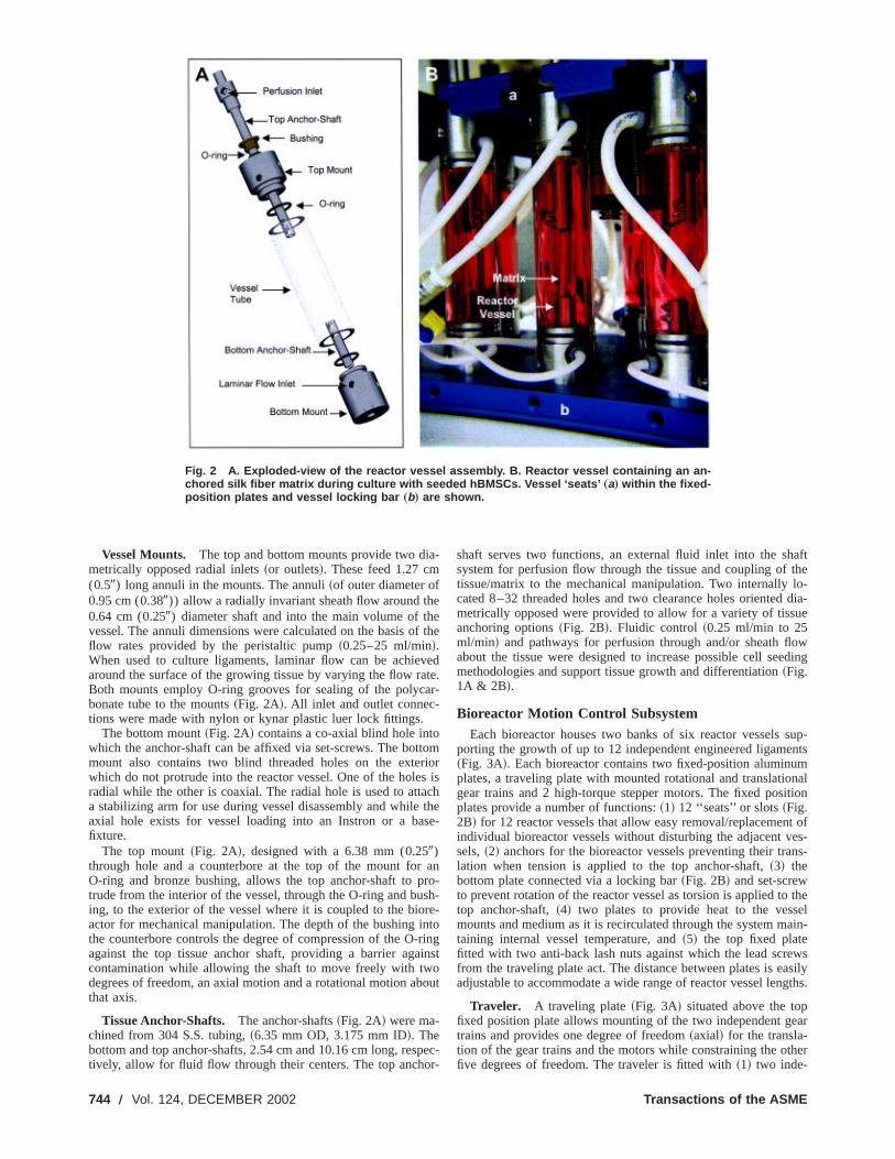
Fig. 2 A. Exploded-view of the reactor vessel assembly. B. Reactor vessel containing an an-chored silk fiber matrix during culture with seeded hBMSCs. Vessel ‘seats’ „a… within the fixed-position plates and vessel locking bar „b… are shown.
a
f
v
a
a
n
aftthelo-dia-
ue
owding
sup-ntsmnaltion
t ofes-ns-
theelain-
wssily
gths.
ear
her
Vessel Mounts. The top and bottom mounts provide two dimetrically opposed radial inlets~or outlets!. These feed 1.27 cm(0.59) long annuli in the mounts. The annuli~of outer diameter of0.95 cm (0.389)) allow a radially invariant sheath flow around th0.64 cm (0.259) diameter shaft and into the main volume of thvessel. The annuli dimensions were calculated on the basis oflow rates provided by the peristaltic pump~0.25–25 ml/min!.When used to culture ligaments, laminar flow can be achiearound the surface of the growing tissue by varying the flow raBoth mounts employ O-ring grooves for sealing of the polycbonate tube to the mounts~Fig. 2A!. All inlet and outlet connec-tions were made with nylon or kynar plastic luer lock fittings.
The bottom mount~Fig. 2A! contains a co-axial blind hole intowhich the anchor-shaft can be affixed via set-screws. The botmount also contains two blind threaded holes on the extewhich do not protrude into the reactor vessel. One of the holeradial while the other is coaxial. The radial hole is used to atta stabilizing arm for use during vessel disassembly and whileaxial hole exists for vessel loading into an Instron or a bafixture.
The top mount~Fig. 2A!, designed with a 6.38 mm (0.259)through hole and a counterbore at the top of the mount forO-ring and bronze bushing, allows the top anchor-shaft to ptrude from the interior of the vessel, through the O-ring and buing, to the exterior of the vessel where it is coupled to the bioactor for mechanical manipulation. The depth of the bushing ithe counterbore controls the degree of compression of the O-against the top tissue anchor shaft, providing a barrier agacontamination while allowing the shaft to move freely with twdegrees of freedom, an axial motion and a rotational motion abthat axis.
Tissue Anchor-Shafts. The anchor-shafts~Fig. 2A! were ma-chined from 304 S.S. tubing,~6.35 mm OD, 3.175 mm ID!. Thebottom and top anchor-shafts, 2.54 cm and 10.16 cm long, restively, allow for fluid flow through their centers. The top ancho
, DECEMBER 2002
-
eethe
edte.r-
tomriors ischthese-
anro-sh-re-to
ringinstoout
pec-r-
shaft serves two functions, an external fluid inlet into the shsystem for perfusion flow through the tissue and coupling oftissue/matrix to the mechanical manipulation. Two internallycated 8–32 threaded holes and two clearance holes orientedmetrically opposed were provided to allow for a variety of tissanchoring options~Fig. 2B!. Fluidic control ~0.25 ml/min to 25ml/min! and pathways for perfusion through and/or sheath flabout the tissue were designed to increase possible cell seemethodologies and support tissue growth and differentiation~Fig.1A & 2B!.
Bioreactor Motion Control SubsystemEach bioreactor houses two banks of six reactor vessels
porting the growth of up to 12 independent engineered ligame~Fig. 3A!. Each bioreactor contains two fixed-position aluminuplates, a traveling plate with mounted rotational and translatiogear trains and 2 high-torque stepper motors. The fixed posiplates provide a number of functions:~1! 12 ‘‘seats’’ or slots~Fig.2B! for 12 reactor vessels that allow easy removal/replacemenindividual bioreactor vessels without disturbing the adjacent vsels,~2! anchors for the bioreactor vessels preventing their tralation when tension is applied to the top anchor-shaft,~3! thebottom plate connected via a locking bar~Fig. 2B! and set-screwto prevent rotation of the reactor vessel as torsion is applied totop anchor-shaft,~4! two plates to provide heat to the vessmounts and medium as it is recirculated through the system mtaining internal vessel temperature, and~5! the top fixed platefitted with two anti-back lash nuts against which the lead screfrom the traveling plate act. The distance between plates is eaadjustable to accommodate a wide range of reactor vessel len
Traveler. A traveling plate~Fig. 3A! situated above the topfixed position plate allows mounting of the two independent gtrains and provides one degree of freedom~axial! for the transla-tion of the gear trains and the motors while constraining the otfive degrees of freedom. The traveler is fitted with~1! two inde-
Transactions of the ASME

Journal of Biom
Fig. 3 A. System bioreactors „a… with 24 loaded reactor vessels „b…. The traveler „c… housingboth rotational „in view … and translational gear trains, the high torque stepper motors „d…, andthe linear bearing „e… are shown. B. Chart illustrating empirical translational and rotationaldisplacement data for a programmed 10 cycle 2 mm and 90° regime at 0.0167 Hz.
r
ia
lo
.d
x
sla-led
tomVi-ro-eartop
48.8ard
ticoint,ry-n ofa-
y
wall
pendent gear trains,~2! 4 self-aligning recirculating ball lineabearings for traveler translation, and~3! a motor mount plate al-lowing both stepper motors to engage their specific gear train
Drive Mechanisms. The gear trains, when driven by thespecific stepper motors, independently control rotationaltranslational deformation. The rotational gear train includes35.08 cm diameter spur gears~72 pitch! in a 1:4 ratio with thestepper motor. Each gear is aligned co-axially with the reacvessel seated below and sits on top of a bronze thrust bearingattaches to a 6.35 cm (2.59) long precision ground 0.635 cm(0.2509) 316 S.S. rod. The rod extends from the gear, throughbearing in the traveler, and protrudes 19 from the bottom surfaceof the traveler towards the reactor vessels. The rod is held in pvia a 6.35 mm (0.2509) long collar pressed against the bottomthe thrust bearing. The remaining 1.91 cm (0.759) of the rod isused to attach the anchor-shaft extending up from the reactorsel via a coupling. The coupling provides an independent wayattaching/detaching the reactor vessel from the system duringcourse of an experiment
Translational deformation is produced via two precision 1cm (0.59) diameter 2.54 mm (0.19) pitch stainless steel leascrews reacting against the anti-backlash nuts affixed to thefixed-position plate. The lead screws are driven through areduction by a stepper motor. The stepper is capable of microsping ~51,200 microsteps/revolution!. The combination of the mo-tor’s resolution and gear reduction results in sub-micron atranslation. When actuated, the entire mass of the traveler incing the gear trains for both rotation and translation as well asmotors and the associated hardware~bearings, bushings, etc.!move relative to the top fixed plate and, hence, relative to
echanical Engineering
s.
rnd12
torand
the
acef
ves-ofthe
27
top1:3tep-
iallud-the
the
bioreactor vessels’ bottom tissue anchors producing axial trantion of the upper tissue anchor. This method allows for uncoupaxial and rotational displacements.
Motion Control Software. LPT Indexer motion control soft-ware, high torque stepper motors~50 lbs-in torque! with accom-panying micro-step drivers~51,200 steps/rev! and power supplywere purchased from Servo-Systems Co., Montville, NJ. Cussoftware to engage the LPT Indexer software was written insual Basic V. 6.0 to control the stepper motors. The software pvides precise independent control over the rotational and linmovements. Rates for linear and rotational movement of thetissue anchors range from zero to 6.5 inches/sec andrevolutions/sec. The software allows the user to input the forwand return rotational and linear rates, total excursion~rotationaland linear!, provides for an intermediate period of rest or stamode at the extreme point, a rest or static mode at the home pand the number of repetitions. Several different cycles with vaing strain regimes can be programmed and run for the duratioa experiment. A common loading regime for the culture of ligments is shown in Fig. 3B.
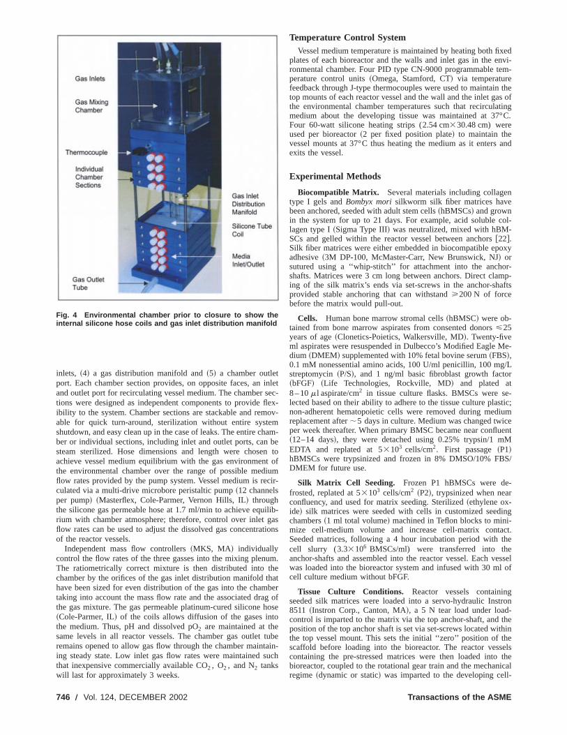
Environmental Chamber and Gas ExchangeA multi-component environmental chamber~Fig. 4! was built
to provide precise pH and pO2 control to all 24 reactor vessels bsilicone tubing gas exchangers~a coil of 1.83 m tubing, 1.588 mm(1/169) I.D., 3.175 mm (1/89) O.D., for each vessel!. Environ-mental chamber components include~1! 12 individual chambersections designed to house two coils per section,~2! an additionalchamber to house two thermocouples to measure chamberand internal temperature,~3! a gas mixing plenum with 3 gas
DECEMBER 2002, Vol. 124 Õ 745
n
o
a
xednvi-m-
thes ofting°C.
and
n
ol-
oxy
r-mp-fts
Me-
/Lr
e-stic;iumeentM
S/
-
ing
ct.
eessell of
gtron
thethineelstheical
l-
inlets, ~4! a gas distribution manifold and~5! a chamber outletport. Each chamber section provides, on opposite faces, anand outlet port for recirculating vessel medium. The chamber stions were designed as independent components to provideibility to the system. Chamber sections are stackable and remable for quick turn-around, sterilization without entire systeshutdown, and easy clean up in the case of leaks. The entire chber or individual sections, including inlet and outlet ports, cansteam sterilized. Hose dimensions and length were choseachieve vessel medium equilibrium with the gas environmentthe environmental chamber over the range of possible medflow rates provided by the pump system. Vessel medium is reculated via a multi-drive microbore peristaltic pump~12 channelsper pump! ~Masterflex, Cole-Parmer, Vernon Hills, IL! throughthe silicone gas permeable hose at 1.7 ml/min to achieve equrium with chamber atmosphere; therefore, control over inlet gflow rates can be used to adjust the dissolved gas concentraof the reactor vessels.
Independent mass flow controllers~MKS, MA! individuallycontrol the flow rates of the three gasses into the mixing plenuThe ratiometrically correct mixture is then distributed into thchamber by the orifices of the gas inlet distribution manifold thhave been sized for even distribution of the gas into the chamtaking into account the mass flow rate and the associated drathe gas mixture. The gas permeable platinum-cured silicone h~Cole-Parmer, IL! of the coils allows diffusion of the gases intthe medium. Thus, pH and dissolved pO2 are maintained at thesame levels in all reactor vessels. The chamber gas outletremains opened to allow gas flow through the chamber mainting steady state. Low inlet gas flow rates were maintained sthat inexpensive commercially available CO2 , O2 , and N2 tankswill last for approximately 3 weeks.
Fig. 4 Environmental chamber prior to closure to show theinternal silicone hose coils and gas inlet distribution manifold
746 Õ Vol. 124, DECEMBER 2002
inletec-flex-ov-mam-be
toof
iumcir-
ilib-as
tions
m.eatberg ofose
tubein-
uch
Temperature Control SystemVessel medium temperature is maintained by heating both fi
plates of each bioreactor and the walls and inlet gas in the eronmental chamber. Four PID type CN-9000 programmable teperature control units~Omega, Stamford, CT! via temperaturefeedback through J-type thermocouples were used to maintaintop mounts of each reactor vessel and the wall and the inlet gathe environmental chamber temperatures such that recirculamedium about the developing tissue was maintained at 37Four 60-watt silicone heating strips (2.54 cm330.48 cm) wereused per bioreactor~2 per fixed position plate! to maintain thevessel mounts at 37°C thus heating the medium as it entersexits the vessel.
Experimental Methods
Biocompatible Matrix. Several materials including collagetype I gels andBombyx morisilkworm silk fiber matrices havebeen anchored, seeded with adult stem cells~hBMSCs! and grownin the system for up to 21 days. For example, acid soluble clagen type I~Sigma Type III! was neutralized, mixed with hBM-SCs and gelled within the reactor vessel between anchors@22#.Silk fiber matrices were either embedded in biocompatible epadhesive~3M DP-100, McMaster-Carr, New Brunswick, NJ! orsutured using a ‘‘whip-stitch’’ for attachment into the anchoshafts. Matrices were 3 cm long between anchors. Direct claing of the silk matrix’s ends via set-screws in the anchor-shaprovided stable anchoring that can withstand>200 N of forcebefore the matrix would pull-out.
Cells. Human bone marrow stromal cells~hBMSC! were ob-tained from bone marrow aspirates from consented donors<25years of age~Clonetics-Poietics, Walkersville, MD!. Twenty-fiveml aspirates were resuspended in Dulbecco’s Modified Eagledium ~DMEM! supplemented with 10% fetal bovine serum~FBS!,0.1 mM nonessential amino acids, 100 U/ml penicillin, 100 mgstreptomycin~P/S!, and 1 ng/ml basic fibroblast growth facto~bFGF! ~Life Technologies, Rockville, MD! and plated at8 – 10m l aspirate/cm2 in tissue culture flasks. BMSCs were slected based on their ability to adhere to the tissue culture planon-adherent hematopoietic cells were removed during medreplacement after;5 days in culture. Medium was changed twicper week thereafter. When primary BMSC became near conflu~12–14 days!, they were detached using 0.25% trypsin/1 mEDTA and replated at 53103 cells/cm2. First passage~P1!hBMSCs were trypsinized and frozen in 8% DMSO/10% FBDMEM for future use.
Silk Matrix Cell Seeding. Frozen P1 hBMSCs were defrosted, replated at 53103 cells/cm2 ~P2!, trypsinized when nearconfluency, and used for matrix seeding. Sterilized~ethylene ox-ide! silk matrices were seeded with cells in customized seedchambers~1 ml total volume! machined in Teflon blocks to mini-mize cell-medium volume and increase cell-matrix contaSeeded matrices, following a 4 hour incubation period with thecell slurry (3.33106 BMSCs/ml) were transferred into thanchor-shafts and assembled into the reactor vessel. Each vwas loaded into the bioreactor system and infused with 30 mcell culture medium without bFGF.
Tissue Culture Conditions. Reactor vessels containinseeded silk matrices were loaded into a servo-hydraulic Ins8511 ~Instron Corp., Canton, MA!, a 5 N tear load under load-control is imparted to the matrix via the top anchor-shaft, andposition of the top anchor shaft is set via set-screws located withe top vessel mount. This sets the initial ‘‘zero’’ position of thscaffold before loading into the bioreactor. The reactor vesscontaining the pre-stressed matrices were then loaded intobioreactor, coupled to the rotational gear train and the mechanregime ~dynamic or static! was imparted to the developing cel
Transactions of the ASME
n
e
oa
m
e
T
u
a
i
oct
si
a
s
te
7
thefor
-lateghplestterL
tinghadll-
lyts.
eac-sent
op-hasing
sels;n atingselslly
ved
ideedache toear
r ofeen
lash
to
ercali-
onsern-than
on-s ofhethe
ndndels,theuntthethin
ub-nds
scaffold complex. Reactor vessel medium was changed bawise ~50%/change! twice a week. pH was maintained at 7.4 adissolved oxygen at 20% pO2 .
Experimental Analysis
Bioreactor Motion Control Subsystem Characterization.As is the case in a fully utilized bioreactor, reactor vessels ocpied all available seats during linear and angular displacemcharacterization. Translational hysteresis was measured usingnier calipers with resolution of 25.4mm clamped to the bottomfixed plate of the bioreactor. Caliper jaws were situated betwthe anchor shafts and displacement between the top and boanchor shafts for vessels seated in the third and sixth positionthe bioreactor was measured for a 2 mm cyclic translation at0.0167 Hz. A spring mounted between the traveling plate andcaliper jaw was used to ensure continuous measurement throout the cyclic regime by keeping the caliper jaw in constant ctact with the top anchor shaft. Initial displacement between shwas set to 3 cm, the caliper zeroed and displacement at the inhome and extreme positions measured and recorded for 10 cyTemporal changes relative to both linear and rotational displaments were measured using a stopwatch to verify programcyclic frequency.
Rotational hysteresis was measured on the axis of the biortor vessel furthest~along the gear train! from the driving axis ofthe stepper motor used to control rotational displacement.bioreactor in this position suffers from the most hysteresis as hteretical error compiles in a linear~gear train! system. The hyster-esis was measured using a mirror and a coherent light so~laser!. The central shaft of the reactor vessel seated in the sposition was machined to allow placement of the reflective plof the first surface mirror to be collinear with the axis. The las~HeNe 3mW! was placed such that the plane of the incident areflected light to/from the mirror remained normal to the reflectsurface of the mirror and intersecting the axis of the bioreacvessel. Hysteresis was measured by causing, through the mcontrol software and stepper motor, cyclic rotational displament. This resulted in a linear translation of the reflected spoa diffuse reflecting screen~e.g., white wall! approximately 3meters from the reactor vessel and mirror; the angular measment system resulted in 0.0268° resolution. The displacementhe reflected spot was marked relative to the initial position~zerodisplacement!. The locations of the spot at the extreme pointrotation and following the return to the initial condition were alrecorded and repeated for 9 additional cycles. In order to achdesired resolution, available room space limited rotational dplacement to 30°. Two sets of measurements (N510 cycles perset! were performed for 20° and 30° of rotation and maximudeviations calculated.
Reactor Vessel Medium Temperature Characterization.Extensive testing was performed to evaluate the temperature athe developing tissue in the reactor vessel. Feedback tempercontrol of the top anchor mount and maintenance of environmtal chamber wall temperature at 37°C was used to control medtemperature around the 3 cm long silk fiber matrix over 21 dayculture. A submersible miniature thermocouple~Omega, CT! cali-brated at 37°C by the manufacturer~resolution1/20.2°C) wasused to measure temperature about the mid-section of the mas well as the area where the top and bottom anchor shafts anthe matrix. Three ports were introduced to the reactor vesselwall at desired locations along the length of the matrix for instion of the thermocouple during the 21-day experiment. Measuments were taken at least twice a day and deviations from 3recorded.
pH and Dissolved pO2 Measurements. Medium pH and dis-solved O2 were measured off-line using a blood gas analyzer~In-strumentation Laboratory 1610, Lexington, MA!. Following 50%
Journal of Biomechanical Engineering
tch-d
cu-entver-
enttoms of
topugh-n-ftsitialcles.ce-ed
ac-
heys-
rceixthneerndngtortione-on
ure-t of
ofoeveis-
m
boutture
en-ium
in
atrixchoruber-re-°C
medium exchange, 15 ml of vessel medium extracted fromreactor vessel into a 20 ml syringe was immediately assayedpH and pO2 .
Scanning Electron Microscopy. The matrices were harvested at timed intervals, washed with 0.2 M sodium cacodybuffer, fixed overnight in Karnovsky fixative, dehydrated throuan ethanol series and left to dry in Freon overnight. The samwere sputter-coated with Au using a Polaron SC502 SpuCoater~Fison Instruments!, and imaged at 15 keV with a JEOJXA 840 Scanning Electron Microscope.
Results and DiscussionA new bench-top bioreactor system~Figs. 1A, B! was designed,
constructed and utilized to support tissue engineering starfrom isolated cells and 3D matrices. The bioreactor systemability to apply independent multi-dimensional complex and wecontrolled mechanical strains~at ,0.1mm for translational and,0.1° for rotational strain! to developing tissue, and to preciseand accurately control the biochemical and fluidic environmenThe bench-top system can accommodate up to 24 individual rtor vessels~Fig. 2A! in two independently controlled bioreactorto permit the concurrent study of varied strain rates and percstrain in order to identify optimal parameters for tissue develment without the needed for an incubator. Enhanced flexibilitybeen achieved through the modular bench-top design allowconcurrent but independent operation of up to 24 reactor vesindividual reactor vessels can be added, replaced or withdrawany time during the course of an experiment without disturbsystem function. Matrices can be anchored into the reactor vesto support the culture of developing tissue within a mechanicadynamic environment. Cell seeding options have been improthrough enhanced fluidic control~0.25–25 ml/min! utilizing per-fusion through and/or sheath flow around the matrix~Fig. 1A!.
Reactor vessels, via the top mount O-ring and bushing, prova barrier to contamination while allowing the tissue culturwithin the vessel to be exposed to multi-dimensional strains. Ebioreactor can apply a unique programmed mechanical regimits 12 housed vessels, i.e., forward and return rotational and linrates, total excursion~rotational and linear!, intermediate staticmode at the extreme point and/or the home point, the numbecycle repetitions for a specific regime, and the duration betwdifferent regimes can be controlled~Fig. 3B!. An unlimited num-ber of regimes could be programmed. The use of anti-backnuts and micro-step drivers combined with 1:4~angular! and 1:3~translation! gear reductions minimized linear system hysteresiswithin the resolution of our measuring equipment~vernier caliperswith resolution of 25.4mm! and technique. Linear hysteresis ov10 cycles was measured to be less than the resolution of theper’s at less than 25mm or 1.25% for 2mm of travel~Fig. 3B!.Deviations between the third and sixth bioreactor seat positifrom the programmed theoretical displacements were not discable as well. Rotational hysteresis was measured to be less0.2° ~0.36%! for a angular displacement of 30°~Fig. 3B! and wasless than 0.1° for a 20° displacement over 10 cycles. It is reasable to expect when extrapolating to 90° of rotation, hysteresisubstantially less than 1% of total displacement will result. Taccuracy of the programmed cyclic frequency derived frommotion control software was verified to within1/20.5 sec or theresolution of the stopwatch and timer.
A total of 48 reactor vessels containing cell culture medium asilk matrices were run for up to 21 days under 90° rotational a2 mm translational deformations at 0.0167 Hz. Of the 48 vessonly one became contaminated during culture due to a leak onoutlet port. Feedback temperature control of the top anchor moto 37°C and the maintenance of wall temperature at 37°C inenvironmental chamber resulted in constant temperature withe resolution of the measuring thermocouple (1/20.2°C) aboutthe 3 cm long silk matrix housed in the reactor vessel. A smerged wire thermocouple place in the middle and at the two e
DECEMBER 2002, Vol. 124 Õ 747
n
n
x-or-thedi-alem-s,la-allhy-hysi-op
f ah-
ure.or-d tollynal
t by:D
ath
andol
op-e
rteda
ipu-ys-on-seselse-e--ingg
ical
of the matrix anchored in the reactor vessel demonstratedconstant temperature was maintained at 371/20.2°C (mean1/2standard deviation! about the silk matrix over the 21 days ooperation.
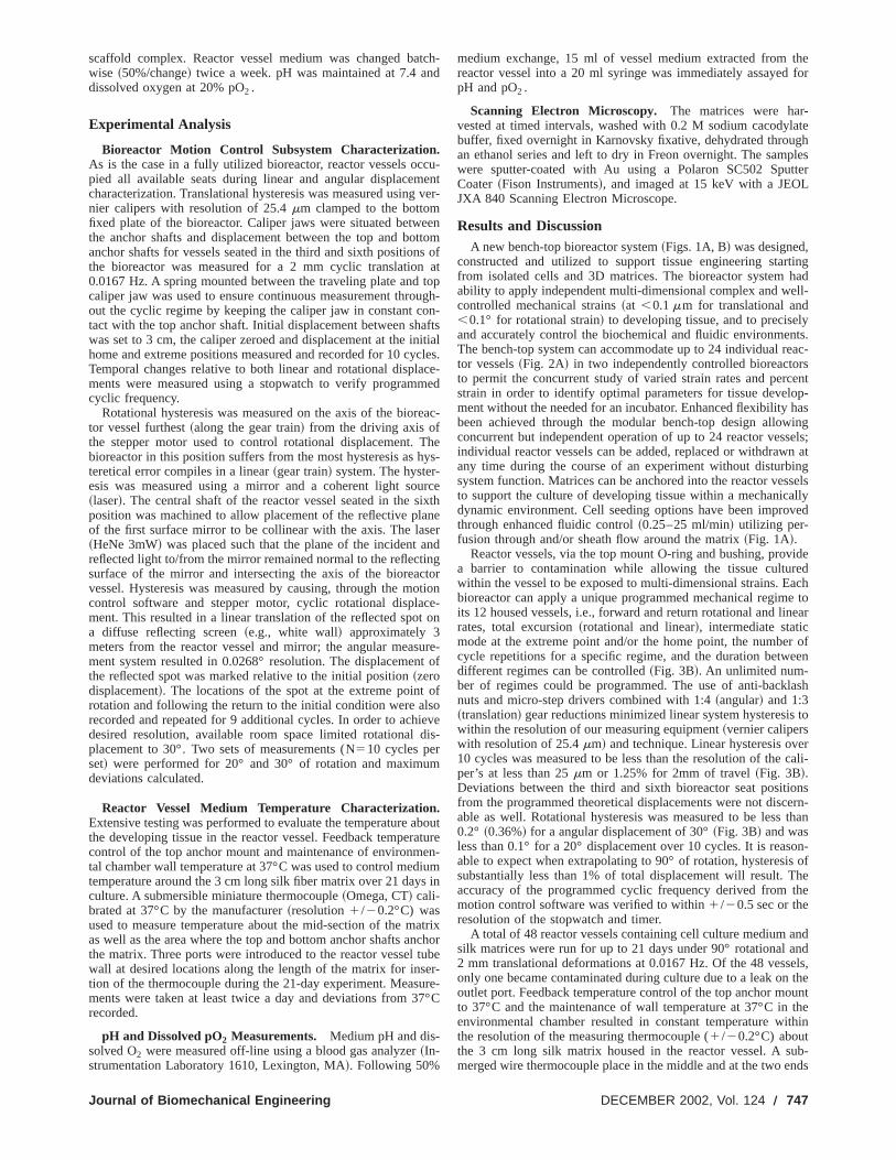
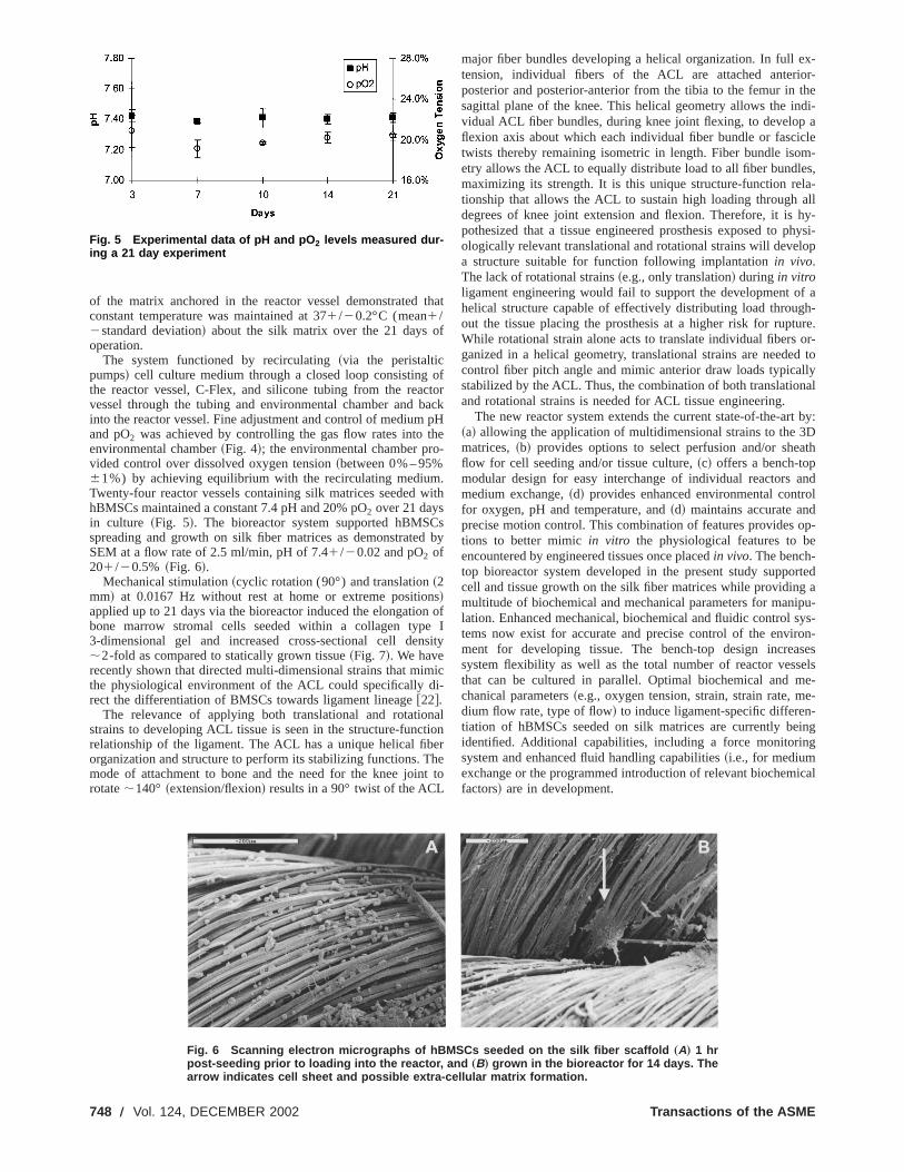
The system functioned by recirculating~via the peristalticpumps! cell culture medium through a closed loop consistingthe reactor vessel, C-Flex, and silicone tubing from the reacvessel through the tubing and environmental chamber and binto the reactor vessel. Fine adjustment and control of mediumand pO2 was achieved by controlling the gas flow rates into tenvironmental chamber~Fig. 4!; the environmental chamber provided control over dissolved oxygen tension~between 0% – 95%61%) by achieving equilibrium with the recirculating mediumTwenty-four reactor vessels containing silk matrices seeded whBMSCs maintained a constant 7.4 pH and 20% pO2 over 21 daysin culture ~Fig. 5!. The bioreactor system supported hBMSCspreading and growth on silk fiber matrices as demonstratedSEM at a flow rate of 2.5 ml/min, pH of 7.41/20.02 and pO2 of201/20.5% ~Fig. 6!.
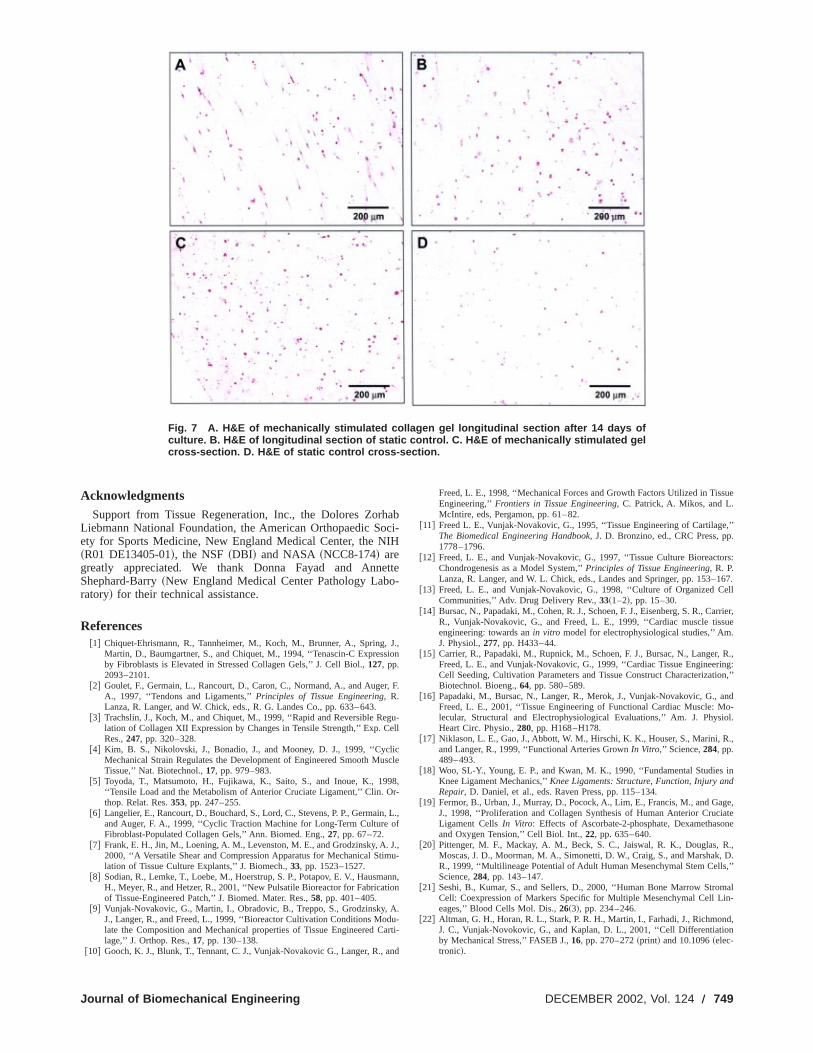
Mechanical stimulation~cyclic rotation (90°) and translation~2mm! at 0.0167 Hz without rest at home or extreme position!applied up to 21 days via the bioreactor induced the elongatiobone marrow stromal cells seeded within a collagen type3-dimensional gel and increased cross-sectional cell den;2-fold as compared to statically grown tissue~Fig. 7!. We haverecently shown that directed multi-dimensional strains that mimthe physiological environment of the ACL could specifically drect the differentiation of BMSCs towards ligament lineage@22#.
The relevance of applying both translational and rotatiostrains to developing ACL tissue is seen in the structure-functrelationship of the ligament. The ACL has a unique helical fiborganization and structure to perform its stabilizing functions. Tmode of attachment to bone and the need for the knee joinrotate;140° ~extension/flexion! results in a 90° twist of the ACL
Fig. 5 Experimental data of pH and pO 2 levels measured dur-ing a 21 day experiment
748 Õ Vol. 124, DECEMBER 2002
that
f
oftorackpH
he-
.ith
sby
sofI
sity
ici-
alionerhet to
major fiber bundles developing a helical organization. In full etension, individual fibers of the ACL are attached anteriposterior and posterior-anterior from the tibia to the femur insagittal plane of the knee. This helical geometry allows the invidual ACL fiber bundles, during knee joint flexing, to developflexion axis about which each individual fiber bundle or fascictwists thereby remaining isometric in length. Fiber bundle isoetry allows the ACL to equally distribute load to all fiber bundlemaximizing its strength. It is this unique structure-function retionship that allows the ACL to sustain high loading throughdegrees of knee joint extension and flexion. Therefore, it ispothesized that a tissue engineered prosthesis exposed to pologically relevant translational and rotational strains will devela structure suitable for function following implantationin vivo.The lack of rotational strains~e.g., only translation! during in vitroligament engineering would fail to support the development ohelical structure capable of effectively distributing load througout the tissue placing the prosthesis at a higher risk for ruptWhile rotational strain alone acts to translate individual fibersganized in a helical geometry, translational strains are needecontrol fiber pitch angle and mimic anterior draw loads typicastabilized by the ACL. Thus, the combination of both translatioand rotational strains is needed for ACL tissue engineering.
The new reactor system extends the current state-of-the-ar~a! allowing the application of multidimensional strains to the 3matrices,~b! provides options to select perfusion and/or sheflow for cell seeding and/or tissue culture,~c! offers a bench-topmodular design for easy interchange of individual reactorsmedium exchange,~d! provides enhanced environmental contrfor oxygen, pH and temperature, and~d! maintains accurate andprecise motion control. This combination of features providestions to better mimicin vitro the physiological features to bencountered by engineered tissues once placedin vivo. The bench-top bioreactor system developed in the present study suppocell and tissue growth on the silk fiber matrices while providingmultitude of biochemical and mechanical parameters for manlation. Enhanced mechanical, biochemical and fluidic control stems now exist for accurate and precise control of the envirment for developing tissue. The bench-top design increasystem flexibility as well as the total number of reactor vessthat can be cultured in parallel. Optimal biochemical and mchanical parameters~e.g., oxygen tension, strain, strain rate, mdium flow rate, type of flow! to induce ligament-specific differentiation of hBMSCs seeded on silk matrices are currently beidentified. Additional capabilities, including a force monitorinsystem and enhanced fluid handling capabilities~i.e., for mediumexchange or the programmed introduction of relevant biochemfactors! are in development.
Fig. 6 Scanning electron micrographs of hBMSCs seeded on the silk fiber scaffold „A… 1 hrpost-seeding prior to loading into the reactor, and „B… grown in the bioreactor for 14 days. Thearrow indicates cell sheet and possible extra-cellular matrix formation.
Transactions of the ASME
Journal of Biom
Fig. 7 A. H&E of mechanically stimulated collagen gel longitudinal section after 14 days ofculture. B. H&E of longitudinal section of static control. C. H&E of mechanically stimulated gelcross-section. D. H&E of static control cross-section.
h
eo
r
g
li
9r
o
Jm
ai
Ad
sue
,’’.
rs:
167.ell
rrier,ue
.
, R.,ng:ion,’’
ndo-
ol.
.,
ind
ge,atene
R.,D.,’’
aln-
d,n
AcknowledgmentsSupport from Tissue Regeneration, Inc., the Dolores Zor
Liebmann National Foundation, the American Orthopaedic Soety for Sports Medicine, New England Medical Center, the N~R01 DE13405-01!, the NSF~DBI! and NASA ~NCC8-174! aregreatly appreciated. We thank Donna Fayad and AnnShephard-Barry~New England Medical Center Pathology Labratory! for their technical assistance.
References@1# Chiquet-Ehrismann, R., Tannheimer, M., Koch, M., Brunner, A., Spring,
Martin, D., Baumgartner, S., and Chiquet, M., 1994, ‘‘Tenascin-C Expressby Fibroblasts is Elevated in Stressed Collagen Gels,’’ J. Cell Biol.,127, pp.2093–2101.
@2# Goulet, F., Germain, L., Rancourt, D., Caron, C., Normand, A., and AugeA., 1997, ‘‘Tendons and Ligaments,’’Principles of Tissue Engineering, R.Lanza, R. Langer, and W. Chick, eds., R. G. Landes Co., pp. 633–643.
@3# Trachslin, J., Koch, M., and Chiquet, M., 1999, ‘‘Rapid and Reversible Relation of Collagen XII Expression by Changes in Tensile Strength,’’ Exp. CRes.,247, pp. 320–328.
@4# Kim, B. S., Nikolovski, J., Bonadio, J., and Mooney, D. J., 1999, ‘‘CycMechanical Strain Regulates the Development of Engineered Smooth MuTissue,’’ Nat. Biotechnol.,17, pp. 979–983.
@5# Toyoda, T., Matsumoto, H., Fujikawa, K., Saito, S., and Inoue, K., 19‘‘Tensile Load and the Metabolism of Anterior Cruciate Ligament,’’ Clin. Othop. Relat. Res.353, pp. 247–255.
@6# Langelier, E., Rancourt, D., Bouchard, S., Lord, C., Stevens, P. P., Germainand Auger, F. A., 1999, ‘‘Cyclic Traction Machine for Long-Term CultureFibroblast-Populated Collagen Gels,’’ Ann. Biomed. Eng.,27, pp. 67–72.
@7# Frank, E. H., Jin, M., Loening, A. M., Levenston, M. E., and Grodzinsky, A.2000, ‘‘A Versatile Shear and Compression Apparatus for Mechanical Stilation of Tissue Culture Explants,’’ J. Biomech.,33, pp. 1523–1527.
@8# Sodian, R., Lemke, T., Loebe, M., Hoerstrup, S. P., Potapov, E. V., HausmH., Meyer, R., and Hetzer, R., 2001, ‘‘New Pulsatile Bioreactor for Fabricatof Tissue-Engineered Patch,’’ J. Biomed. Mater. Res.,58, pp. 401–405.
@9# Vunjak-Novakovic, G., Martin, I., Obradovic, B., Treppo, S., Grodzinsky,J., Langer, R., and Freed, L., 1999, ‘‘Bioreactor Cultivation Conditions Molate the Composition and Mechanical properties of Tissue Engineered Clage,’’ J. Orthop. Res.,17, pp. 130–138.
@10# Gooch, K. J., Blunk, T., Tennant, C. J., Vunjak-Novakovic G., Langer, R., a
echanical Engineering
abci-
IH
tte-
J.,ion
, F.
u-ell
cscle
8,-
, L.,f
.,u-
nn,on
.u-arti-
nd
Freed, L. E., 1998, ‘‘Mechanical Forces and Growth Factors Utilized in TisEngineering,’’Frontiers in Tissue Engineering, C. Patrick, A. Mikos, and L.McIntire, eds, Pergamon, pp. 61–82.
@11# Freed L. E., Vunjak-Novakovic, G., 1995, ‘‘Tissue Engineering of CartilageThe Biomedical Engineering Handbook, J. D. Bronzino, ed., CRC Press, pp1778–1796.
@12# Freed, L. E., and Vunjak-Novakovic, G., 1997, ‘‘Tissue Culture BioreactoChondrogenesis as a Model System,’’Principles of Tissue Engineering, R. P.Lanza, R. Langer, and W. L. Chick, eds., Landes and Springer, pp. 153–
@13# Freed, L. E., and Vunjak-Novakovic, G., 1998, ‘‘Culture of Organized CCommunities,’’ Adv. Drug Delivery Rev.,33~1–2!, pp. 15–30.
@14# Bursac, N., Papadaki, M., Cohen, R. J., Schoen, F. J., Eisenberg, S. R., CaR., Vunjak-Novakovic, G., and Freed, L. E., 1999, ‘‘Cardiac muscle tissengineering: towards anin vitro model for electrophysiological studies,’’ AmJ. Physiol.,277, pp. H433–44.
@15# Carrier, R., Papadaki, M., Rupnick, M., Schoen, F. J., Bursac, N., LangerFreed, L. E., and Vunjak-Novakovic, G., 1999, ‘‘Cardiac Tissue EngineeriCell Seeding, Cultivation Parameters and Tissue Construct CharacterizatBiotechnol. Bioeng.,64, pp. 580–589.
@16# Papadaki, M., Bursac, N., Langer, R., Merok, J., Vunjak-Novakovic, G., aFreed, L. E., 2001, ‘‘Tissue Engineering of Functional Cardiac Muscle: Mlecular, Structural and Electrophysiological Evaluations,’’ Am. J. PhysiHeart Circ. Physio.,280, pp. H168–H178.
@17# Niklason, L. E., Gao, J., Abbott, W. M., Hirschi, K. K., Houser, S., Marini, Rand Langer, R., 1999, ‘‘Functional Arteries GrownIn Vitro,’’ Science,284, pp.489–493.
@18# Woo, SL-Y., Young, E. P., and Kwan, M. K., 1990, ‘‘Fundamental StudiesKnee Ligament Mechanics,’’Knee Ligaments: Structure, Function, Injury anRepair, D. Daniel, et al., eds. Raven Press, pp. 115–134.
@19# Fermor, B., Urban, J., Murray, D., Pocock, A., Lim, E., Francis, M., and GaJ., 1998, ‘‘Proliferation and Collagen Synthesis of Human Anterior CruciLigament CellsIn Vitro: Effects of Ascorbate-2-phosphate, Dexamethasoand Oxygen Tension,’’ Cell Biol. Int.,22, pp. 635–640.
@20# Pittenger, M. F., Mackay, A. M., Beck, S. C., Jaiswal, R. K., Douglas,Moscas, J. D., Moorman, M. A., Simonetti, D. W., Craig, S., and Marshak,R., 1999, ‘‘Multilineage Potential of Adult Human Mesenchymal Stem CellsScience,284, pp. 143–147.
@21# Seshi, B., Kumar, S., and Sellers, D., 2000, ‘‘Human Bone Marrow StromCell: Coexpression of Markers Specific for Multiple Mesenchymal Cell Lieages,’’ Blood Cells Mol. Dis.,26~3!, pp. 234–246.
@22# Altman, G. H., Horan, R. L., Stark, P. R. H., Martin, I., Farhadi, J., RichmonJ. C., Vunjak-Novokovic, G., and Kaplan, D. L., 2001, ‘‘Cell Differentiatioby Mechanical Stress,’’ FASEB J.,16, pp. 270–272~print! and 10.1096~elec-tronic!.
DECEMBER 2002, Vol. 124 Õ 749
Related Documents


![Computational technologies in tissue engineeringcell/scaffold cultures in Tissue Engineering applications [12]. The main objective is to design an optimal bioreactor system for optimal](https://static.cupdf.com/doc/110x72/5f9136f9c4be4300fb3b3c19/computational-technologies-in-tissue-engineering-cellscaffold-cultures-in-tissue.jpg)