Thiourea-induced thyroid hormone depletion impairs testicular recrudescence in the air-breathing catfish, Clarias gariepinus I. Swapna a , M. Rajasekhar a , A. Supriya b , K. Raghuveer a , G. Sreenivasulu a , M.K. Rasheeda a , K.C. Majumdar c , H. Kagawa d , H. Tanaka e , A. Dutta-Gupta a , B. Senthilkumaran a, ⁎ a Department of Animal Sciences, School of Life Sciences, University of Hyderabad, Hyderabad 500 046, India b School of Life Sciences, University of Hyderabad, Hyderabad 500 046, India c Center for Cellular and Molecular Biology, Uppal Road, Hyderabad 500 007, India d Laboratory of Fish Reproduction, Department of Biological Production and Environmental Science, Faculty of Agriculture, Miyazaki University, Miyazaki 889-2192, Japan e National Research Institute of Aquaculture, Fisheries Research Agency, Nansei, Mie 516-0193, Japan Received 29 September 2005; received in revised form 7 January 2006; accepted 8 January 2006 Available online 29 March 2006 Abstract We used thiourea-induced thyroid hormone depletion as a strategy to understand the influence of thyroid hormones on testicular recrudescence of the air-breathing catfish, Clarias gariepinus. Treatment with 0.03% thiourea via immersion for 21days induced hypothyroidism (thyroid hormone depletion) as evidenced by significantly reduced serum T 3 levels. Thiourea-treated males had narrowed seminiferous lobules with fewer spermatozoa in testis, very little or no secretory fluid, reduced protein and sialic acid levels in seminal vesicles when compared to controls. The histological changes were accompanied by reduction in serum and tissue levels of testosterone (T) and 11-ketotestosterone (11-KT), a potent male specific androgen in fish. Qualitative changes in the localization of catfish gonadotropin-releasing hormone (cfGnRH) and luteinizing hormone (LH, heterologous system) revealed a reduction in the distribution of immunoreactive neuronal cells and fibers in thyroid depleted fish. Interestingly, thiourea-withdrawal group showed physiological and histological signs of recovery after 21days such as reappearance of spermatozoa and partial restoration of 11-KT and T levels. These data demonstrate that thyroid hormones play a significant role in testicular function of catfish. The mechanism of action includes modulating sex steroids either directly or through the hypothalamo (GnRH)-hypophyseal (LH) axis. © 2006 Elsevier Inc. All rights reserved. Keywords: 11-ketotestosterone; Hypothyroidism; T 3 ; Teleosts; Testis; Testosterone; Thiourea 1. Introduction Thyroid hormones are essential endocrine mediators exerting tissue and development specific effects in almost all vertebrates studied to date (King and May, 1984; Buchholz et al., 2003). Studies in mammals demonstrated a critical role for thyroid hormones in reproduction and embryogenesis (Stradtman, 1993; Jannini et al., 1995; Teerds et al., 1998) in addition to its inputs in cellular metabolism. Thyroid abnormalities are associated with irregularities in reproductive cycles and delayed onset of puberty (Jannini et al., 1995; Maran, 2003). The role of thyroid on ovarian function is well known (Stradtman, 1993), while similar studies on male reproductive system present several conflicting observations. Weiss and Burns (1988) indicate little or no effect on testicular histology, spermatogen- esis and serum testosterone (T) levels while Crissman et al. (2000) reported a decrease in serum T levels and sperm motility with no marked reduction in fertility. T 3 affects Sertoli cell proliferation and differentiation, the onset of the formation of adult-type Leydig cell population and their function during the neonatal–prepubertal period in rats (Teerds et al., 1998; Ariyaratne et al., 2000). Neonatal hypothyroidism in rats increases adult testis size and capacity for sperm production Comparative Biochemistry and Physiology, Part A 144 (2006) 1 – 10 www.elsevier.com/locate/cbpa ⁎ Corresponding author. Tel.: +91 40 23134562; fax: +91 40 23010120. E-mail address: [email protected] (B. Senthilkumaran). 1095-6433/$ - see front matter © 2006 Elsevier Inc. All rights reserved. doi:10.1016/j.cbpa.2006.01.017

Welcome message from author
This document is posted to help you gain knowledge. Please leave a comment to let me know what you think about it! Share it to your friends and learn new things together.
Transcript
logy, Part A 144 (2006) 1–10www.elsevier.com/locate/cbpa
Comparative Biochemistry and Physio
Thiourea-induced thyroid hormone depletion impairs testicular recrudescencein the air-breathing catfish, Clarias gariepinus
I. Swapna a, M. Rajasekhar a, A. Supriya b, K. Raghuveer a, G. Sreenivasulu a, M.K. Rasheeda a,K.C. Majumdar c, H. Kagawa d, H. Tanaka e, A. Dutta-Gupta a, B. Senthilkumaran a,⁎
a Department of Animal Sciences, School of Life Sciences, University of Hyderabad, Hyderabad 500 046, Indiab School of Life Sciences, University of Hyderabad, Hyderabad 500 046, India
c Center for Cellular and Molecular Biology, Uppal Road, Hyderabad 500 007, Indiad Laboratory of Fish Reproduction, Department of Biological Production and Environmental Science, Faculty of Agriculture, Miyazaki University,
Miyazaki 889-2192, Japane National Research Institute of Aquaculture, Fisheries Research Agency, Nansei, Mie 516-0193, Japan
Received 29 September 2005; received in revised form 7 January 2006; accepted 8 January 2006Available online 29 March 2006
Abstract
We used thiourea-induced thyroid hormone depletion as a strategy to understand the influence of thyroid hormones on testicular recrudescenceof the air-breathing catfish, Clarias gariepinus. Treatment with 0.03% thiourea via immersion for 21days induced hypothyroidism (thyroidhormone depletion) as evidenced by significantly reduced serum T3 levels. Thiourea-treated males had narrowed seminiferous lobules with fewerspermatozoa in testis, very little or no secretory fluid, reduced protein and sialic acid levels in seminal vesicles when compared to controls. Thehistological changes were accompanied by reduction in serum and tissue levels of testosterone (T) and 11-ketotestosterone (11-KT), a potent malespecific androgen in fish. Qualitative changes in the localization of catfish gonadotropin-releasing hormone (cfGnRH) and luteinizing hormone(LH, heterologous system) revealed a reduction in the distribution of immunoreactive neuronal cells and fibers in thyroid depleted fish.Interestingly, thiourea-withdrawal group showed physiological and histological signs of recovery after 21days such as reappearance ofspermatozoa and partial restoration of 11-KT and T levels. These data demonstrate that thyroid hormones play a significant role in testicularfunction of catfish. The mechanism of action includes modulating sex steroids either directly or through the hypothalamo (GnRH)-hypophyseal(LH) axis.© 2006 Elsevier Inc. All rights reserved.
Keywords: 11-ketotestosterone; Hypothyroidism; T3; Teleosts; Testis; Testosterone; Thiourea
1. Introduction
Thyroid hormones are essential endocrine mediators exertingtissue and development specific effects in almost all vertebratesstudied to date (King and May, 1984; Buchholz et al., 2003).Studies in mammals demonstrated a critical role for thyroidhormones in reproduction and embryogenesis (Stradtman,1993; Jannini et al., 1995; Teerds et al., 1998) in addition toits inputs in cellular metabolism. Thyroid abnormalities areassociated with irregularities in reproductive cycles and delayed
⁎ Corresponding author. Tel.: +91 40 23134562; fax: +91 40 23010120.E-mail address: [email protected] (B. Senthilkumaran).
1095-6433/$ - see front matter © 2006 Elsevier Inc. All rights reserved.doi:10.1016/j.cbpa.2006.01.017
onset of puberty (Jannini et al., 1995; Maran, 2003). The role ofthyroid on ovarian function is well known (Stradtman, 1993),while similar studies on male reproductive system presentseveral conflicting observations. Weiss and Burns (1988)indicate little or no effect on testicular histology, spermatogen-esis and serum testosterone (T) levels while Crissman et al.(2000) reported a decrease in serum T levels and sperm motilitywith no marked reduction in fertility. T3 affects Sertoli cellproliferation and differentiation, the onset of the formation ofadult-type Leydig cell population and their function during theneonatal–prepubertal period in rats (Teerds et al., 1998;Ariyaratne et al., 2000). Neonatal hypothyroidism in ratsincreases adult testis size and capacity for sperm production
2 I. Swapna et al. / Comparative Biochemistry and Physiology, Part A 144 (2006) 1–10
while neonatal hyperthyroidism shows an opposite effect. Thetiming of hypothyroidism around puberty affects testicularfunction (Anasti et al., 1995). Hypothyroidism also affectsbiochemical constituents and functions of male accessoryreproductive glands such as prostate and seminal vesicles inadult rodents (Buitrago and Diez, 1987; Senthilkumaran et al.,1991; Maran, 2003).
Studies in fish contributed relevant information to under-stand the role of thyroid in metamorphosis (de Jesus et al.,1991), embryogenesis (Liu and Chan, 2002) and reproduction(Reine and Leatherland, 2003). Teleosts do not posses acompact thyroid gland as do other vertebrates; rather the thyroidfollicles are diffusely dispersed. The reproductive state duringthe course of the study assumes significance as thyroidhormones may exert a stage dependent effect. Fish have welldefined seasonal reproductive cycles associated with a definitepattern of changes in thyroid hormone levels (Cyr et al., 1988).Much of the earlier work in fish with regard to thyroid hormoneon male reproduction was done in the spawning phase and/orconcentrated only on few histological morphometric changes ininterstitial cells (Misra and Panday, 1985). The role of thyroidhormones in fish reproduction generated new insight after therecent cloning of thyroid stimulating hormone (TSH) receptorfrom testis of the African catfish, Clarias gariepinus (Vischerand Bogerd, 2003), gonad of the striped bass, Morone saxatilis(Kumar et al., 2000) and the demonstration that the goitrogen 6-n-propyl-2-thiouracil given during testis development increasesSertoli and germ cell numbers per cyst in the Nile tilapiaOreochromis niloticus (Matta et al., 2002). Recent evidencesalso indicated the association of thyroid with gonadotropin-releasing hormone (GnRH) and catecholaminergic systems infish (Parhar et al., 2000; Chaube and Joy, 2003), raising thepossibility for thyroid hormone action on gonadal function viahypothalamo–hypophyseal axis.
The present study sought to investigate the effect of thyroidhormone depletion on the male reproductive system of the air-breathing catfish during testicular recrudescence (stagecorresponding to renewed spermatogenesis), with specialemphasis on changes in testicular and ‘seminal vesicle’histology, serum and tissue levels of T and 11-ketotestosterone(11-KT). Qualitative changes in the localization of gonadotro-pin-releasing hormone (catfish form; cfGnRH) and luteinizinghormone (LH; heterologous system) were also probed.Thiourea, a well-documented thyroid hormone depletingcompound was used to induce hypothyroidism. It has beenshown to act via inhibition of thyroid peroxidase (Davidson etal., 1979).
2. Materials and methods
Adult (early recrudescence/early preparatory phase) air-breathing catfish C. gariepinus (200–250g) caught from local(Hyderabad, India) fresh water ponds were purchased fromfishermen at the end of January 2004 for experimentation. Malefish were selected and acclimated for 3weeks by maintaining inaquarium tanks filled with filtered tap water under normalphotoperiod and ambient temperature (26±2°C). Fish were fed
minced goat liver ad libitum during acclimation and experi-mentation. Commonly referred to as the African catfish, thisspecies is abundantly available in ponds and lakes in and aroundHyderabad, and in some rivers of Northern India. However, notmuch information is available with regard to the seasonalcycling patterns of this species in the South of India. Ourpreliminary observations revealed that the local populationexhibited a seasonal cycling pattern. Four broadly distinguish-able phases, namely, early preparatory, prespawning, spawningphases and postspawning were seen in both sexes (Lehri, 1967;van Oordt et al., 1987) as opposed to the laboratory maintainedfish kept under constant photoperiod and temperature condi-tions that are ready for spawning throughout the year (van Oordtet al., 1987). Wild caught fish are annual breeders and thespawning phase lasts for about 3months from June to Augustcoinciding with the south west monsoon in the region.However, sometimes the spawning season could extend up tolate October. The preparatory phase lasts from mid-February toMay and fish caught around late November to January areusually in resting to early preparatory phase.
2.1. Thiourea treatment
Laboratory acclimated fish were divided into three groups of10 fish each. The first group of fish (Group I) were sacrificed toexamine status of testis prior to the start of treatment withthiourea. Unlike mammals, in fish the thyroid gland is not asingle encapsulated organ, rather the thyroid follicles arediffusely dispersed within the connective tissue on the surfaceof the ventral aorta and other sub-pharyngeal and para-pharyngeal areas. This particular feature makes it difficult toinduce thyroid hormone depletion in fish by routine thyroid-ectomy. Hence, the chosen route for thyroid hormone depletionin fish is by using thiourea, one of the known thyroid hormonedepleting compounds. After pilot studies, thyroid hormonedepletion was induced by adding thiourea (SRL, Mumbai,India) to the final concentration of 0.03% (Misra and Panday,1985; Chaube and Joy, 2003) to well-aerated aquarium tanksholding the catfish for 21days. Group II fish were maintained infiltered water as parallel control. Group III fish were maintainedin filtered water containing thiourea, which was replenishedonce a day. Five fish from Group III were not sacrificed after21days of thiourea treatment. They were maintained for anadditional 21day period in absence of thiourea and thensacrificed to study effects of withdrawal of thiourea.
2.2. Sample collection and processing
After the duration of 21days, fish from both groups wereweighed and blood was obtained by caudal puncture. Serumwas obtained by centrifuging the blood after clotting at 1500 ×gand stored at −80°C. Testes and seminal vesicles were dissectedout, weighed. 500mg was fixed in Bouin's for histologicalexamination, and the remaining tissue was snap frozen in liquidnitrogen till processing for analysis of protein, sialic acid, 11-KT and T levels. Tissue processing for all biochemicalestimations was done at 4°C. Testis and seminal vesicular
3I. Swapna et al. / Comparative Biochemistry and Physiology, Part A 144 (2006) 1–10
tissues were homogenized in 0.1M phosphate buffered saline(PBS) and the resulting homogenate (10%) was lyophilized andstored briefly in −80°C.
For histological examination, tissues were fixed in freshlyprepared Bouin's fluid for 1–2h at room temperature, thenprocessed with graded alcohol series and embedded in paraplast(Kendall, Mansfield, MA, USA). Sections of 6μm thicknesswere cut with a rotatory microtome (Leitz, Wetzlar, Germany)and stained with hematoxylin-eosin. Microscopic examinationsand photography was done with a Nikon EFD-3 microscopefitted with a Nikon NFX-35 automatic camera (Nikon, Tokyo,Japan). Higher magnification images were obtained using aLeica microscope (Buffalo, NY, USA) fitted with a Kodak (DX7630) digital camera. Cells in different stages of spermatogen-esis (spermatogonia, spermatocytes, spermatids/spermatozoa),were counted from twenty different fields in various regions oftestis. The percentage of three different stages was thencalculated for each field against the total number of cellspresent in that field; the mean of twenty fields was calculated foreach category. Since primary and secondary spermatocytescould not be distinguished, they were grouped as spermato-cytes. Likewise spermatids and mature spermatozoa weregrouped as spermatozoa.
2.3. Estimation of proteins and sialic acid
Estimation of protein was done by the method of Lowry et al.(1951). Tissue sialic acid levels were determined as per themethod described in Senthilkumaran and Joy (1993). In briefthe tissue was precipitated with 5% trichloroacetic acid. Afterovernight drying in a desiccator, the precipitated portion washydrolyzed in 1ml of 0.1N H2SO4 for 1h at 80°C to liberate thebound sialic acid. The mixture was centrifuged at 1000 ×g and0.3ml of the supernatant was oxidized with sodium metaper-iodate in concentrated phosphoric acid. The periodate-oxidationproduct was coupled with thiobarbituric acid; the resultingchromophores were extracted into cyclohexane and measured at532 and 562nm in a spectrophotometer.
2.4. Enzyme immunoassay (EIA) for T3, T and 11-KT
Serum T3 levels were assayed using specific T3 EIA (MedixBiochemica, Kauniainen, Finland) kit. The intra- and inter assaycoefficients of variation [n=5, mean±SEM] were 1.34±0.13%and 2.78±0.15% for T3. The sensitivity for T3 was 0.2ng/mlusing the Medix Biochemica kit.
Serum and tissue 11-KT and T levels were estimated usingspecific EIA (Cayman, Ann Arbor, MI, USA) kits usingmanufacturer's protocol. Intra- and inter assay variations were
Table 1Percentage distribution of cells in different stages of spermatogenesis as observed in
January (first week) March (first
Spermatogonia 3.98±0.69 3.48±0.39Spermatocytes (primary/secondary) 31.56±1.07 24.39±1.24Spermatids/spermatozoa 65.89±2.23 75.64±2.69
within the limits specified in the manufacturer's protocol. Tantisera of Cayman kit cross reacts with 5α-Dihydrotestoster-one (5α-DHT; 27%), 5β-Dihydrotestosterone (5β-DHT; 19%),Androstenedione (4%), 11-KT (2%), 5-Androstenedione(0.51%), Epi-testosterone (0.2%), Androsterone (0.05%),Androsterone sulfate (0.04%), Testosterone sulfate (0.03%),Dihydroepiandrostenedione sulfate (0.02%). T and 11-KT arethe major androgens in catfish (Cavaco et al., 2001). Presence of5α-DHT and 5β-DHT have not been earlier reported in catfish(Cavaco et al., 2001; Cavaco, 2005). 11-KT antisera supplied inthe Cayman EIA kit showed cross reactivity with 4-Androsten-11β,17β-diol-3-one (0.01%), T (b0.01%), 5α-Androstan-17β-ol-3-one (b0.01%), 5α-Androsten-3β-diol (b0.01%). A recov-ery of 93–95% was obtained by the extraction proceduredescribed in the manufacturer's manual for T and 11-KT(Cayman).The dilutions of series of serum samples and/or tissueextracts from catfish with differing hormone concentrationswere linear with standards of T and 11-KT, respectively. Intra-and inter assay coefficients of variation [n=5, mean±SEM]were 1.02±0.04% and 1.81±0.15% for T and 0.81±0.09% and1.32±0.11% for 11-KT. The sensitivity was 6pg/ml using theCayman T kit and 1.3pg/mL with the Cayman 11-KT kit.
2.5. Immunocytochemistry for cfGnRH amdLH
Immunocytochemistry (ICC) for cfGnRH was carried out asper the following procedure. The sections were deparaffinisedand rehydrated in a gradient of decreasing concentrations ofalcohol and finally in PBS containing 1% Tween-20 (pH7.6).The sections were then treated with normal goat serum (1 :10dilution). Primary antibodies against cfGnRH at 1 :2000dilution were applied to the sections and incubated at 4°C for48h in a humid chamber. The sections were then washed withPBS thrice at 5min interval and incubated at room temperaturefor 1h with biotin labeled anti-rabbit IgG. The unboundantibody was washed away with PBS and the sections wereincubated at room temperature for 30min with streptavidinhorseradish peroxidase conjugate. The sections were thenwashed with phosphate buffer (PB) and then incubated with0.05% 3,3′diaminobenzidine tetrahydrochloride with 0.01%H2O2 in 0.05M Tris–HCl (pH7.6) for 10min in a dark chamberat room temperature. The slides were then rinsed with PB,dehydrated through graded alcohol series, cleared in xylene andmounted in DPX. All the chemicals and second antibodies usedfor ICC were part of the kit components supplied by BangaloreGENEI Pvt. Ltd. (Bangalore, India).
Similar ICC procedure was followed for LH using theantiserum (pmGTH-II) raised against red seabream (Pagrusmajor) LH. The antiserum was found to work efficiently at
testis during various intervals in 2004 expressed as mean±SEM (n=5)
week) May (first week) July (first week) Late November
0.123±0.01 Not detectable 11.7±1.021.59±0.09 0.249±0.008 58.8±3.5297.95±4.51 99.4±0.5 29.41±4.21
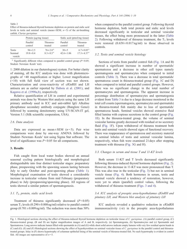
Table 2Effect of thiourea-induced thyroid hormone depletion on protein and sialic acidlevels in testis and seminal vesicle (mean±SEM; n=5) of the air-breathingcatfish, Clarias gariepinus
Protein (μg/mg tissue) Sialic acid (pmol/mg tissue)
Parallelcontrol
Thiourea-treated
Parallelcontrol
Thiourea-treated
Testis 94±2.5 76±2.0 a 30±5 6.7±0.05 a
Seminalvesicle
19.8±0.4 8.3±0.5 a 42.6±1.06 17.3±4.37 a
a Significantly different when compared to parallel control group (Pb0.05;Student–Newman–Keuls' test).
4 I. Swapna et al. / Comparative Biochemistry and Physiology, Part A 144 (2006) 1–10
1 :2000 dilution in our (heterologous) system. For better clarityof staining, all the ICC analysis was done with photomicro-graphs of ×40 magnification or higher. Lower magnification(×10) with full field view of section was not shown.Characterization and cross-reactivity of cfGnRH and LHantisera are as earlier reported by Dubois et al. (2001), andKagawa et al. (1998a,b), respectively.
Relative quantification of LH in control and experimentalanimals was done by Western blot analysis using the same LHprimary antibody used in ICC and anti-rabbit IgG Alkalinephosphatase secondary antibody conjugate (Banglore Genei)followed by scanning densitometry using UV-SCAN-IT gel,Version 5.1 (Silk scientific cooperation, USA).
2.6. Data analysis
Data are expressed as mean±SEM (n=5). Pair wisecomparisons were done by one-way ANOVA followed byStudent–Newman–Keuls' test using Sigma Stat software. Thelevel of significance was Pb0.05 for all comparisons.
3. Results
Fish caught from local water bodies showed an annualseasonal cycling pattern histologically and morphologicaldistinguishable into four distinct testicular stages: preparatoryphase, prespawning with the spawning season lasting betweenJuly to early October and post-spawning phase (Table 1).Morphological examination of testis showed a considerableincrease in testicular volume from mid February (preparatoryphase) to July (prespawning/spawning phase). All regions oftestis showed a similar pattern of spermatogenesis.
3.1. T3, protein, sialic acid levels
Treatment of thiourea significantly decreased (Pb0.05)serum T3 levels (0.290±0.008ng/ml) relative to parallel controlgroup (0.701±0.009ng/ml). The reduction was more than 50%
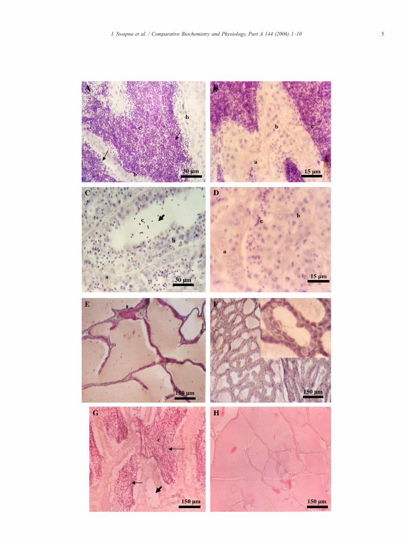
Fig. 1. Histological sections showing the effect of thiourea-induced thyroid hormonthiourea-treated group, (B and D) are higher magnification images of A and B,Spermatozoa. Long arrows indicate lumen filled with spermatids in (A) and (G) while(C) and (G). (E) and (F) Histological sections showing the effect of hypothyroidismtreated groups. Inlay in (F) shows hypertrophy of columnar epithelial lining of the sefish. All images are representative of n=5.
when compared to the parallel control group. Following thyroidhormone depletion, both total protein and sialic acid levelsdecreased significantly in testicular and seminal vesiculartissues, the effect being more pronounced in the latter (Table2). Following withdrawal of thiourea treatment, the T3 levelswere restored (0.658±0.017ng/ml) to those observed incontrols.
3.2. Testis and seminal vesicle histology
Sections of testis from parallel control fish (Fig. 1A and B)showed a significant increase in number of spermatids/spermatozoa and a decrease in other cell types namelyspermatogonia and spermatocytes when compared to initialcontrols (Table 3). There was a decrease in total spermatids/spermatozoa count in thiourea-treated group (Fig. 1C and D)when compared to initial and parallel control groups. However,there was no significant change in the total number ofspermatocytes and spermatogonia. The apparent increase inpercentage distribution of spermatocytes and spermatogoniaevident from Table 3 is due to a large decrease monitored in thetotal cell counts (spermatogonia, spermatocytes and spermatids)in thiourea-treated fish mainly due to loss of spermatids/spermatozoa heads. Seminal vesicles showed extensive fluidfilled lumina with copious secretions in the control group (Fig.1E). In the thiourea-treated group, the volume of seminalvesicular lumina greatly decreased with residual or no secretoryfluid (Fig. 1F). Upon withdrawal of thiourea treatment bothtestis and seminal vesicle showed signs of functional recovery.There was reappearance of spermatozoa and secretory materialin seminal lobules of testis and seminal vesicular lumina,respectively, when fish were examined 21days after stoppingtreatment with thiourea (Fig. 3G and H).
3.3. Changes in serum and tissue T and 11-KT levels
Both serum 11-KT and T levels decreased significantlyfollowing thiourea-induced thyroid hormone depletion (Fig. 2).However, the decrease in 11-KT was more pronounced than T.This was also true in the testicular (Fig. 3) but not in seminalvesicle tissue (Fig. 4). Both hormones in serum, testis andseminal vesicle showed a tendency of restoration, however,were yet to attain (parallel) control values, following thewithdrawal of thiourea treatment (Figs. 3 and 4).
3.4. ICC analysis of preoptic area-hypothalamus cfGnRH andpituitary LH, and Western blot analysis of pituitary LH
ICC analysis revealed a qualitative reduction in cfGnRHimmunoreactivity (-ir) in the preoptic area-hypothalamus
e depletion on testicular tissue of C. gariepinus. (A) parallel control group, (C)respectively. (a) Spermatogonia, (b) Spermatocytes and (c) Spermatids andshort solid arrows indicate empty lumens with very few residual sperm heads inon seminal vesicular tissue of C. gariepinus in the parallel control and thiourea-minal vesicle of thiourea-treated fish. No such hypertrophy is evident in control
5I. Swapna et al. / Comparative Biochemistry and Physiology, Part A 144 (2006) 1–10
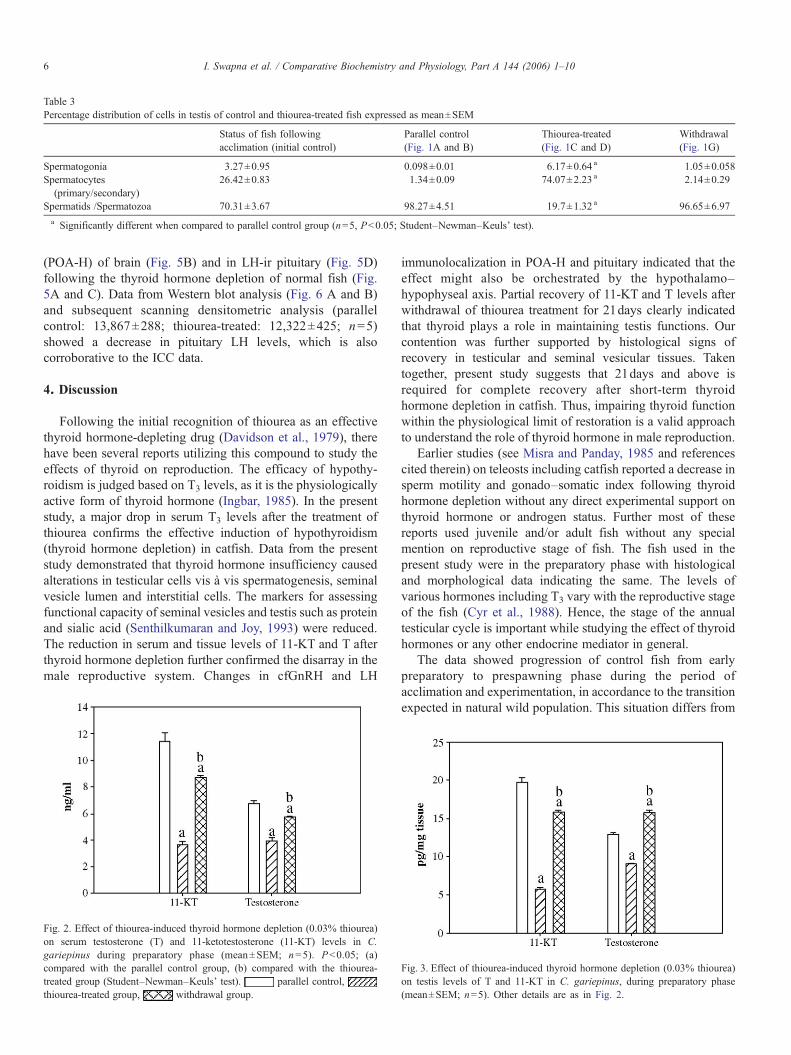
Table 3Percentage distribution of cells in testis of control and thiourea-treated fish expressed as mean±SEM
Status of fish followingacclimation (initial control)
Parallel control(Fig. 1A and B)
Thiourea-treated(Fig. 1C and D)
Withdrawal(Fig. 1G)
Spermatogonia 3.27±0.95 0.098±0.01 6.17±0.64 a 1.05±0.058Spermatocytes(primary/secondary)
26.42±0.83 1.34±0.09 74.07±2.23 a 2.14±0.29
Spermatids /Spermatozoa 70.31±3.67 98.27±4.51 19.7±1.32 a 96.65±6.97a Significantly different when compared to parallel control group (n=5, Pb0.05; Student–Newman–Keuls' test).
6 I. Swapna et al. / Comparative Biochemistry and Physiology, Part A 144 (2006) 1–10
(POA-H) of brain (Fig. 5B) and in LH-ir pituitary (Fig. 5D)following the thyroid hormone depletion of normal fish (Fig.5A and C). Data from Western blot analysis (Fig. 6 A and B)and subsequent scanning densitometric analysis (parallelcontrol: 13,867±288; thiourea-treated: 12,322±425; n=5)showed a decrease in pituitary LH levels, which is alsocorroborative to the ICC data.
4. Discussion
Following the initial recognition of thiourea as an effectivethyroid hormone-depleting drug (Davidson et al., 1979), therehave been several reports utilizing this compound to study theeffects of thyroid on reproduction. The efficacy of hypothy-roidism is judged based on T3 levels, as it is the physiologicallyactive form of thyroid hormone (Ingbar, 1985). In the presentstudy, a major drop in serum T3 levels after the treatment ofthiourea confirms the effective induction of hypothyroidism(thyroid hormone depletion) in catfish. Data from the presentstudy demonstrated that thyroid hormone insufficiency causedalterations in testicular cells vis à vis spermatogenesis, seminalvesicle lumen and interstitial cells. The markers for assessingfunctional capacity of seminal vesicles and testis such as proteinand sialic acid (Senthilkumaran and Joy, 1993) were reduced.The reduction in serum and tissue levels of 11-KT and T afterthyroid hormone depletion further confirmed the disarray in themale reproductive system. Changes in cfGnRH and LH
Fig. 2. Effect of thiourea-induced thyroid hormone depletion (0.03% thiourea)on serum testosterone (T) and 11-ketotestosterone (11-KT) levels in C.gariepinus during preparatory phase (mean±SEM; n=5). Pb0.05; (a)compared with the parallel control group, (b) compared with the thiourea-treated group (Student–Newman–Keuls' test). parallel control,thiourea-treated group, withdrawal group.
immunolocalization in POA-H and pituitary indicated that theeffect might also be orchestrated by the hypothalamo–hypophyseal axis. Partial recovery of 11-KT and T levels afterwithdrawal of thiourea treatment for 21days clearly indicatedthat thyroid plays a role in maintaining testis functions. Ourcontention was further supported by histological signs ofrecovery in testicular and seminal vesicular tissues. Takentogether, present study suggests that 21days and above isrequired for complete recovery after short-term thyroidhormone depletion in catfish. Thus, impairing thyroid functionwithin the physiological limit of restoration is a valid approachto understand the role of thyroid hormone in male reproduction.
Earlier studies (see Misra and Panday, 1985 and referencescited therein) on teleosts including catfish reported a decrease insperm motility and gonado–somatic index following thyroidhormone depletion without any direct experimental support onthyroid hormone or androgen status. Further most of thesereports used juvenile and/or adult fish without any specialmention on reproductive stage of fish. The fish used in thepresent study were in the preparatory phase with histologicaland morphological data indicating the same. The levels ofvarious hormones including T3 vary with the reproductive stageof the fish (Cyr et al., 1988). Hence, the stage of the annualtesticular cycle is important while studying the effect of thyroidhormones or any other endocrine mediator in general.
The data showed progression of control fish from earlypreparatory to prespawning phase during the period ofacclimation and experimentation, in accordance to the transitionexpected in natural wild population. This situation differs from
Fig. 3. Effect of thiourea-induced thyroid hormone depletion (0.03% thiourea)on testis levels of T and 11-KT in C. gariepinus, during preparatory phase(mean±SEM; n=5). Other details are as in Fig. 2.
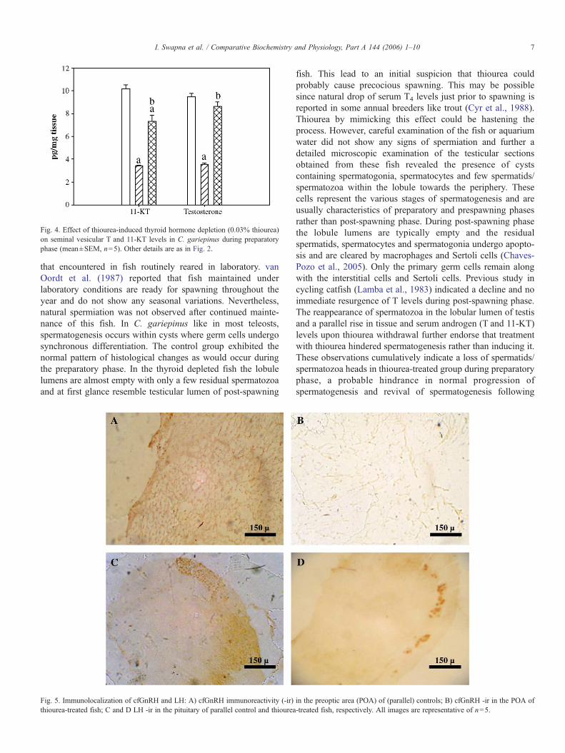
Fig. 4. Effect of thiourea-induced thyroid hormone depletion (0.03% thiourea)on seminal vesicular T and 11-KT levels in C. gariepinus during preparatoryphase (mean±SEM, n=5). Other details are as in Fig. 2.
7I. Swapna et al. / Comparative Biochemistry and Physiology, Part A 144 (2006) 1–10
that encountered in fish routinely reared in laboratory. vanOordt et al. (1987) reported that fish maintained underlaboratory conditions are ready for spawning throughout theyear and do not show any seasonal variations. Nevertheless,natural spermiation was not observed after continued mainte-nance of this fish. In C. gariepinus like in most teleosts,spermatogenesis occurs within cysts where germ cells undergosynchronous differentiation. The control group exhibited thenormal pattern of histological changes as would occur duringthe preparatory phase. In the thyroid depleted fish the lobulelumens are almost empty with only a few residual spermatozoaand at first glance resemble testicular lumen of post-spawning
Fig. 5. Immunolocalization of cfGnRH and LH: A) cfGnRH immunoreactivity (-ir)thiourea-treated fish; C and D LH -ir in the pituitary of parallel control and thiourea
fish. This lead to an initial suspicion that thiourea couldprobably cause precocious spawning. This may be possiblesince natural drop of serum T4 levels just prior to spawning isreported in some annual breeders like trout (Cyr et al., 1988).Thiourea by mimicking this effect could be hastening theprocess. However, careful examination of the fish or aquariumwater did not show any signs of spermiation and further adetailed microscopic examination of the testicular sectionsobtained from these fish revealed the presence of cystscontaining spermatogonia, spermatocytes and few spermatids/spermatozoa within the lobule towards the periphery. Thesecells represent the various stages of spermatogenesis and areusually characteristics of preparatory and prespawning phasesrather than post-spawning phase. During post-spawning phasethe lobule lumens are typically empty and the residualspermatids, spermatocytes and spermatogonia undergo apopto-sis and are cleared by macrophages and Sertoli cells (Chaves-Pozo et al., 2005). Only the primary germ cells remain alongwith the interstitial cells and Sertoli cells. Previous study incycling catfish (Lamba et al., 1983) indicated a decline and noimmediate resurgence of T levels during post-spawning phase.The reappearance of spermatozoa in the lobular lumen of testisand a parallel rise in tissue and serum androgen (T and 11-KT)levels upon thiourea withdrawal further endorse that treatmentwith thiourea hindered spermatogenesis rather than inducing it.These observations cumulatively indicate a loss of spermatids/spermatozoa heads in thiourea-treated group during preparatoryphase, a probable hindrance in normal progression ofspermatogenesis and revival of spermatogenesis following
in the preoptic area (POA) of (parallel) controls; B) cfGnRH -ir in the POA of-treated fish, respectively. All images are representative of n=5.
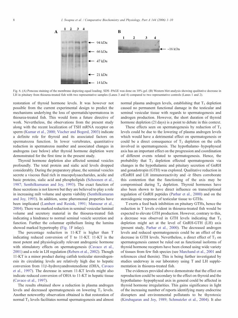
Fig. 6. (A) Ponceau staining of the membrane depicting equal loading. SDS–PAGE was done on 10% gel. (B) Western blot analysis showing qualitative decrease inLH in pituitary from thiourea-treated fish with two representative samples (Lanes 3 and 4) compared to two representative controls (Lanes 1 and 2).
8 I. Swapna et al. / Comparative Biochemistry and Physiology, Part A 144 (2006) 1–10
restoration of thyroid hormone levels. It was however notpossible from the current experimental design to predict themechanisms underlying the loss of spermatids/spermatozoa inthiourea-treated fish. This would form a future directive ofwork. Nevertheless, the observations from the present studyalong with the recent localization of TSH mRNA receptor onsperm (Kumar et al., 2000; Vischer and Bogerd, 2003) indicatea definite role for thyroid and its associated factors onspermatozoa function. In lower vertebrates, quantitativereduction in spermatozoa number and associated changes inandrogens (see below) after thyroid hormone depletion weredemonstrated for the first time in the present study.
Thyroid hormone depletion also affected seminal vesiclesprofoundly. The total protein and sialic acid levels droppedconsiderably. During the preparatory phase, the seminal vesiclessecrete a viscous fluid rich in mucopolysaccharides, acidic andbasic proteins, sialic acid and phospholipids (Schoonen et al.,1987; Senthilkumaran and Joy, 1993). The exact function ofthese secretions is not known but they are believed to play a rolein increasing milt volume and sperm viability (Senthilkumaranand Joy, 1993). In addition, some pheromonal properties havebeen implicated (Lambert and Resink, 1991; Mansour et al.,2004). There was marked reduction in seminal vesicular luminalvolume and secretory material in the thiourea-treated fishindicating a hindrance to normal seminal vesicle secretion andfunction. Further the columnar epithelium lining the luminashowed marked hypertrophy (Fig. 1F inlay).
The percentage reduction in 11-KT is higher than Tindicating reduced conversion of T to 11-KT. 11-KT is themost potent and physiologically relevant androgenic hormonewith stimulatory effects on spermatogenesis (Cavaco et al.,2001) and a role in LH regulation (Rebers et al., 2002). Though11-KT is a minor product during catfish testicular steroidogen-esis its circulating levels are relatively high due to hepaticconversion from 11β-hydroxyandrostenedione (OHA, Cavacoet al., 1997). The decrease in serum 11-KT levels might alsoindicate reduced conversion of OHA to 11-KT in hepatic tissue(Cavaco et al., 1997).
The results obtained show a reduction in plasma androgenlevels and decreased spermatogenesis on lowering T3 levels.Another noteworthy observation obtained is that restoration ofnormal T3 levels facilitates normal spermatogenesis and almost
normal plasma androgen levels, establishing that T3 depletioncaused no permanent functional damage in the testicular andseminal vesicular tissue with regards to spermatogenesis andandrogen production. However, the short duration of thyroidhormone depletion (21days) is a point to debate in this context.
These effects seen on spermatogenesis by reduction of T3
levels could be due to the lowering of plasma androgen levelswhich would have a detrimental effect on spermatogenesis orcould be a direct consequence of T3 depletion on the cellsinvolved in spermatogenesis. The hypothalamo–hypophysealaxis has an important effect on the progression and coordinationof different events related to spermatogenesis. Hence, theprobability that T3 depletion effected spermatogenesis viachanges in the hypothalamic and pituitary secretion of GnRHand gonadotropin (GTH) was explored. Qualitative reduction incfGnRH and LH immunoreactivity and -ir fibers corroboratethis contention that the functioning of the axis may becompromised during T4 depletion. Thyroid hormones havealso been shown to have direct influence on transcriptionalregulation of GnRH peptides (Parhar et al., 2000) and on thesteroidogenic response of testicular tissue to GTHs.
T exerts a feed back inhibition on pituitary GTHs, hence thereduction in T levels evident in thiourea-treated fish would beexpected to elevate GTH production. However, contrary to this,a decrease was observed in GTH levels indicating that T3
depletion might act at the level of GnRH-GTH (LH) axis(present study, Parhar et al., 2000). The decreased androgenlevels and reduced spermatogenesis could be an effect of thedecrease in GTH levels. Nevertheless, a direct effect of T3 onspermatogenesis cannot be ruled out as functional isoforms ofthyroid hormone receptors have been cloned using wide varietyof tissues from few fish species (see Marchand et al., 2001 andreferences cited therein). This is being further investigated bystudies underway in our laboratory using T and LH supple-mentation in thiourea-treated fish.
The evidences provided above demonstrate that the effect onreproduction could be secondary to the effect on thyroid and thehypothalamo–hypophyseal axis in general could be affected inthyroid hormone irregularities. This gains significance in lightof the increasing number of reports identifying many endocrinedisrupters and environmental pollutants to be thyrotoxic(Kirubagaran and Joy, 1989; Schmutzler et al., 2004). It also
9I. Swapna et al. / Comparative Biochemistry and Physiology, Part A 144 (2006) 1–10
ascertains the need for caution before claiming that a compoundhas direct effect on gonadal development as the effect could bemediated through disruption of thyroid or any other endocrinefunction.
This study, therefore, establishes the requirement of thyroidhormones for progression of testicular recrudescence and malesex steroid production. Furthermore, our study suggests aplausible role for cfGnRH, LH in this process. Additionalresearch in this direction using specific hormone supplemen-tation would help to identify the exact sites of thyroid hormoneaction in the hypothalamo–hypophyseal–gonadal axis.
Acknowledgements
The financial support in part by ‘seed-money grant’ awardedto BS by Dr. Kota Harinarayana, the Vice-Chancellor ofUniversity of Hyderabad and the Grants-in-Aid from Depart-ment of Biotechnology (DBT; BT/PR4567/AAQ/03/219/2003)and University Grants Commission [UGC; F.30-195/2004(SR)]are acknowledged. The authors gratefully acknowledge Dr.Rüdiger Schulz (University of Utrecht, Faculty of Biology, TheNetherlands) for providing us cfGnRH antisera and also forvaluable discussions and critical reading of the manuscript. Thiswork was also supported by Council of Industrial and ScientificResearch Junior/Senior Research Fellowships to IS, GS andMKR, and a DBTstudent scholarship to AS. The authors deeplyappreciate all the four anonymous reviewers for their invaluablecomments to improve the first version of this manuscript.
References
Anasti, J.N., Flack, M.R., Froehlich, J., Nelson, L.M., Nisula, B.C., 1995. Apotential novel mechanism for precocious puberty in juvenile hypothyroid-ism. J. Clin. Endocrinol. Metab. 80, 276–279.
Ariyaratne, H.B.S., Mason, J.I., Handagama, S.M.L.C.M., 2000. Effects ofthyroid and luteinizing hormones on the onset of precursor celldifferentiation into Leydig progenitor cells in the prepubertal rat testis.Biol. Reprod. 63, 898–904.
Buchholz, D.R., Hsia, S.C., Fu, L., Shi, Y.B., 2003. A dominant-negativethyroid hormone receptor blocks amphibian metamorphosis by retainingcorepressors at target genes. Mol. Cell. Biol. 23, 6750–6758.
Buitrago, J.M.G., Diez, L.C.G., 1987. Serum hormones and seminal parametersin males with thyroid disturbance. Andrologia 19, 37–41.
Cavaco, J.E., 2005. Sex steroids and spermatogenesis in the African catfish(Clarias gariepinus). Arch. Androl. 51, 99–107.
Cavaco, J.E.B., Vischer, H.F., Lambert, J.G.D., Goos, H.J.Th., Schulz, R.W.,1997. Mismatch between patterns of circulating and testicular androgens inadult African catfish, Clarias gariepinus. Fish Physiol. Biochem. 17,155–162.
Cavaco, J.E., Bogerd, J., Goos, H., Schulz, R.W., 2001. Testosterone inhibits 11-ketotestosterone-induced spermatogenesis in African catfish (Clariasgariepinus). Biol. Reprod. 65, 1807–1812.
Chaube, R., Joy, K.P., 2003. Thyroid hormone modulation of brain in vivotyrosine hydroxylase activity and kinetics in the female catfish, Hetero-pneustes fossilis. J. Endocrinol. 179, 205–215.
Chaves-Pozo, E., Mulero, V., Meseguer, J., Ayala, A.G., 2005. An overview ofcell renewal in the testis throughout the reproductive cycle of a seasonalbreeding teleost, the gilthead seabream (Sparus aurata L). Biol. Reprod. 72,593–601.
Crissman, J.W., Cooke, P.S., Hess, R.A., Marty, M.S., Liberacki, A.B., 2000.Postulated human sperm count decline may involve historic elimination of
juvenile iodine deficiency: a new hypothesis with experimental evidence inthe rat. Toxicol. Sci. 53, 400–410.
Cyr, D.G., Bromage, N.R., Duston, J., Eales, J.G., 1988. Seasonal patterns inserum levels of thyroid hormones and sex steroids in relation to photoperiod-induced changes in spawning time in rainbow trout, Salmo gairdneri. Gen.Comp. Endocrinol. 69, 217–225.
Davidson, B., Soodak, M., Strout, H.V., Neary, J.T., Nakamura, C., Maloof, F.,1979. Thiourea and cyanamide as inhibitors of thyroid peroxidase: the roleof iodide. Endocrinology 104, 919–924.
de Jesus, E.G., Hirano, T., Inui, Y., 1991. Changes in cortisol and thyroidhormone concentrations during early development and metamorphosis in theJapanese flounder, Paralichthys olivaceus. Gen. Comp. Endocrinol. 82,369–376.
Dubois, E.A., Slob, S., Zandbergen, M.A., Peute, J., Goos, H.J., 2001. Gonadalsteroids and the maturation of the species-specific gonadotropin-releasinghormone system in brain and pituitary of the male African catfish (Clariasgariepinus). Comp. Biochem. Physiol. B 129, 381–387.
Ingbar, S.H., 1985. The thyroid gland. In: Wilson, J.D., Foster, D.W. (Eds.),Williams Textbook of Endocrinology. W. B. Saunders Company, WestWashington square, Philadelphia, USA, pp. 682–815. Section 5.
Jannini, E.A., Ulisse, S., D'Armiento, M., 1995. Thyroid hormone and malegonadal function. Endocr. Rev. 16, 443–459.
Kagawa, H., Kawazoe, I., Tanaka, H., Okuzawa, K., 1998a. Immunocytochem-ical identification of two distinct gonadotropic cells (GTH I and GTH II) inthe pituitary of bluefin tuna, Thunnus thynnus. Gen. Comp. Endocrinol. 110,11–18.
Kagawa, H., Tanaka, H., Okuzawa, K., Kobayashi, M., 1998b. GTH II but notGTH I induces final maturation and the development of maturationalcompetence of oocytes of red seabream in vitro. Gen. Comp. Endocrinol.112, 80–88.
King, D.B., May, J.D., 1984. Thyroidal influence on body growth. J. Exp. Zool.232, 453–460.
Kirubagaran, R., Joy, K.P., 1989. Toxic effects of mercurials on thyroidfunction of the catfish, Clarias batrachus (L). Ecotoxicol. Environ. Saf. 17,265–271.
Kumar, S.R., Shigeho, I., Katherine, K., Swanson, P., Dittman, A., Alok, D.,Zohar, Y., Trant, J.M., 2000. Cloning and functional expression of athyrotropin receptor from the gonads of a vertebrate (bony fish): potentialthyroid-independent role for thyrotropin in reproduction. Mol. Cell.Endocrinol. 167, 1–9.
Lamba, V.J., Goswami, S.V., Sundararaj, B.I., 1983. Circannual and circadianvariations in plasma levels of steroids (cortisol, estradiol-17β, estrone, andtestosterone) correlated with the annual gonadal cycle in the catfish,Heteropneustes fossilis (Bloch). Gen. Comp. Endocrinol. 50, 205–225.
Lambert, J.G., Resink, J.W., 1991. Steroid glucuronides as male pheromones inthe reproduction of the African catfish Clarias gariepinus—a brief review.J. Steroid Biochem. Mol. Biol. 40, 549–556.
Lehri, C.K., 1967. The annual cycle in the testis of the catfish: Clarias batrachusL. Acta Anat. (Basel) 67, 135–154.
Liu, Y.W., Chan, W.K., 2002. Thyroid hormones are important for embryonic tolarval transitory phase in zebrafish. Differentiation 70, 36–45.
Lowry, O.H., Rossenberg, N.J., Fany, A.L., Randall, R.J., 1951. Proteinmeasurement with Folin phenol reagent. J. Biol. Chem. 193, 265–274.
Mansour, N., Franz, L., Robert, A.P., 2004. Seminal vesicle secretion of Africancatfish, its composition, its behaviour in water and saline solutions and itsinfluence on gamete fertilizability. J. Exp. Zool. 301, 745–755.
Maran, R.R.M., 2003. Thyroid hormones: their role in testicular steroidogenesis.Arch. Androl. 49, 375–388.
Marchand, O., Safi, R., Escriva, H., Van Rompaey, E., Prunet, P., Laudet, V.,2001. Molecular cloning and characterization of thyroid hormone receptorsin teleost fish. J. Mol. Endocrinol. 26, 51–65.
Matta, S.L.P., Vilela, D.A.R., Godinho, H.P., França, L.R., 2002. The goitrogen6-n-propyl-2-thiouracil (ptu) given during testis development increasesSertoli and germ cell numbers per cyst in fish: the tilapia (Oreochromisniloticus) model. Endocrinology 143, 970–978.
Misra, M., Panday, K., 1985. Effect of thiourea on the testicular steroidogenesisand secondary sexual characters of a tropical freshwater fish,Mystus vittatus(Bloch). Ann. Endocrinol. (Paris) 46, 421–425.
10 I. Swapna et al. / Comparative Biochemistry and Physiology, Part A 144 (2006) 1–10
Parhar, I.S., Soga, T., Sakuma, Y., 2000. Thyroid hormone and estrogen regulatebrain region-specific messenger ribonucleic acids encoding three gonado-tropin-releasing hormone genes in sexually immature male fish, Oreochro-mis niloticus. Endocrinology 141, 1618–1626.
Rebers, F.E.M., Hassing, G.A.M., van Dijk, W., van Straaten, E., Goos, H.J.,Schulz, R.W., 2002. Gonadotropin-releasing hormone does not directlystimulate luteinizing hormone biosynthesis in male African catfish. Biol.Reprod. 66, 1604–1611.
Reine, J.C., Leatherland, J.F., 2003. Trafficking of l-triiodothyronine betweenovarian fluid and oocytes of rainbow trout (Oncorhynchus mykiss). Comp.Biochem. Physiol. B 136, 267–274.
Schmutzler, C., Hamann, I., Hofmann, P.J., Kovacs, G., Stemmler, L., Mentrup,B., Schomburg, L., Ambrugger, P., Gruters, A., Seidlova-Wuttke, D., Jarry,H., Wuttke, W., Kohrle, J., 2004. Endocrine active compounds affectthyrotropin and thyroid hormone levels in serum as well as endpoints ofthyroid hormone action in liver, heart and kidney. Toxicology 205, 95–102.
Schoonen, W.G., Granneman, J.C., Lambert, J.G., Viveen, W.J., van Oordt, P.G., 1987. Quantitative studies of steroid bioconversions in the seminalvesicles of spawning male African catfish, Clarias gariepinus (Burchell),under natural conditions, and of non-spawning catfish under natural and fishfarm conditions. Comp. Biochem. Physiol. B 87, 687–695.
Senthilkumaran, B., Joy, K.P., 1993. Annual cyclic and castration andcyproterone acetate-induced, changes in sialic acid content of the seminal
vesicle of the catfish, Heteropneustes fossilis (Bloch). Fish Physiol.Biochem. 10, 425–430.
Senthilkumaran, B., Aruldhas, M.M., Udhayakumar, R.C.R., Govindarajulu, P.G., 1991. Influence of thyroidectomy on seminal vesicular glycosidases ofyoung albino rats. Ind. J. Exp. Biol. 29, 205–207.
Stradtman, E.W., 1993. Thyroid dysfunction and ovulatory disorders. In: Carr,B.R., Blackwell, R.E. (Eds.), Textbook of Reproductive Medicine. CTAppleton, Norwalk, pp. 297–321.
Teerds, K.J., de Rooij, D.G., de Jong, F.H., van Haaster, L.H., 1998.Development of the adult-type Leydig cell population in the rat is affectedby neonatal thyroid hormone levels. Biol. Reprod. 59, 344–350.
van Oordt, P.G.W.J., Peute, J., Van den Hurk, R., Viveen, W.J.A.R., 1987.Annual correlative changes in gonads and pitutary gonadotrophs of feralcatfish, Clarias gariepinus. Aquaculture 63, 27–41.
Vischer, H.F., Bogerd, J., 2003. Cloning and functional characterization of atesticular TSH receptor cDNA from the African catfish (Clarias gariepinus).J. Mol. Endocrinol. 30, 227–238.
Weiss, S.R., Burns, J.M., 1988. The effect of acute treatment with twogoitrogens on plasma thyroid hormones, testosterone and testicularmorphology in adult male rats. Comp. Biochem. Physiol. A 90, 449–452.
Related Documents