Molecular Immunology 47 (2010) 1923–1930 Contents lists available at ScienceDirect Molecular Immunology journal homepage: www.elsevier.com/locate/molimm Susceptibility of mice genetically deficient in SP-A or SP-D gene to Invasive Pulmonary Aspergillosis Taruna Madan a,b , Kenneth B.M. Reid c , Howard Clark c,d , Mamta Singh a , Annapurna Nayak e , P. Usha Sarma a,f , Samuel Hawgood g , Uday Kishore e,∗ a Institute of Genomics and Integrative Biology, Council for Scientific and Industrial Research, Delhi University Campus, Mall Road, Delhi 110007, India b Department of Innate Immunity, National Institute for Research in Reproductive Health, Mumbai 400012, India c Medical Research Council Immunochemistry Unit, Department of Biochemistry, University of Oxford, South Parks Road, Oxford OX1 3QU, UK d University of Southampton, MP 803 Level F, South Block, Southampton General Hospital, Southampton SO16 6YD, UK e Centre for Infection, Immunity & Disease Mechanisms, Biosciences, School of Health Sciences and Social Care, Brunel University, West London UB8 3PH, UK f Department of Plant Pathology, Indian Agricultural Research Institute, Pusa Road, Delhi 110012, India g Department of Pediatrics and Cardiovascular Research Institute, University of California San Francisco, San Francisco, CA 94118-1245, USA article info Article history: Received 26 January 2010 Accepted 25 February 2010 Available online 21 April 2010 Keywords: Fungal Infection Gene deficient mice Lung Surfactant protein A. fumigatus abstract Pulmonary surfactant proteins, SP-A and SP-D, are carbohydrate pattern recognition molecules of innate immunity, which significantly enhance phagocytosis and killing of Aspergillus fumigatus, a pathogenic fungus, by neutrophils and macrophages. The present study examined the susceptibility of immuno- suppressed SP-A gene deficient (SP-A −/− ) or SP-D gene deficient (SP-D −/− ) mice to A. fumigatus conidia challenge compared to wild-type (WT) mice. A. fumigatus-challenged SP-A −/− (SP-A −/− IPA) mice showed less mortality (40%) than the WT-IPA mice (100%) and increased mortality (60%) following administration of SP-A with decreased TNF- and IFN- to IL-4 ratio than SP-A −/− IPA mice. The SP-D −/− IPA mice (57.14%) showed similar mortality as WT-IPA mice (60%). However, the SP-D −/− IPA mice (42.86% mortality on day 2) died earlier than the WT-IPA mice (20% mortality on day 2), showed a higher hyphal density and tissue injury in lungs. Treatment with SP-D or a recombinant fragment of human SP-D rhSP-D reduced the mortality to 50% and 33%, respectively, concomitant with higher IFN- to IL-4 ratios in treated SP-D −/− mice, compared to untreated control group. The results showed that SP-D gene deficient mice are more susceptible to IPA while SP-A gene deficient mice acquire resistance to IPA. © 2010 Elsevier Ltd. All rights reserved. 1. Introduction The hydrophilic lung surfactant proteins, SP-A and SP-D, are car- bohydrate pattern recognition molecules of innate immunity. The structure of SP-A and SP-D molecules comprise of an N-terminal triple helical collagen region and homotrimeric ligand-recognition domain called a C-type lectin or carbohydrate recognition domain (CRD). These CRD interact with carbohydrate pattern structures on the surfaces of pathogenic organisms such as viruses, bacte- ria, and fungi, and cause agglutination and direct inhibition of Abbreviations: SP-A, human surfactant protein A; SP-D, human surfactant pro- tein D; rhSP-D, a recombinant fragment of human surfactant protein D, composed of homotrimeric neck and C-type lectin domains; A. fumigatus, Aspergillus fumiga- tus; ABPA, allergic bronchopulmonary aspergillosis; WT, wild-type; SP-A −/ − , mice genetically deficient in the SP-A gene; SP-D −/ − , mice genetically deficient in SP-D gene; HRP, horseradish peroxidase; OPD, o-phenylenediamine; AP, alkaline phos- phatase; BALF, bronchoalveolar lavage fluid. ∗ Corresponding author. Tel.: +44 1895 266362; fax: +44 1895274348. E-mail addresses: [email protected], [email protected] (U. Kishore). growth, together with enhanced phagocytosis through neutrophils and macrophages (Kuroki et al., 2007). When challenged with pathogens, allergens, apoptotic cells or necrotic cells, SP-A and SP-D are known to interact with phagocytic cells and enhance their chemotactic, phagocytic, antigen presentation and oxidative properties (Kishore et al., 2005). Thus, the pattern recognition of pathogens via CRDs and subsequent engagement of collagen region with immune cells via collectin receptor enhances phagocytosis and killing of pathogens (Kishore et al., 2006). Aspergillus species are one of the prominent causes of fungal respiratory infections worldwide. Aspergillus infection of immuno- suppressed hosts results in Invasive aspergillosis, characterized by fungal invasion and high mortality rates. In earlier studies, both human SP-A and SP-D bound and agglutinated Aspergillus fumi- gatus conidia and these interactions enhanced phagocytosis and killing of germinating conidia by human neutrophils and alveo- lar macrophages (Madan et al., 1997; Allen et al., 2001). Intranasal administration of an anti-fungal drug—Amphotericin B (AmB), SP-D and rhSP-D (a recombinant fragment of human SP-D compris- ing neck and carbohydrate recognition domains of human SP-D) to an immunosuppressed murine model of Invasive Pulmonary 0161-5890/$ – see front matter © 2010 Elsevier Ltd. All rights reserved. doi:10.1016/j.molimm.2010.02.027

Welcome message from author
This document is posted to help you gain knowledge. Please leave a comment to let me know what you think about it! Share it to your friends and learn new things together.
Transcript
SP
TAa
b
c
d
e
f
g
a
ARAA
KFIGLSA
1
bstd(or
totggp
(
0d
Molecular Immunology 47 (2010) 1923–1930
Contents lists available at ScienceDirect
Molecular Immunology
journa l homepage: www.e lsev ier .com/ locate /mol imm
usceptibility of mice genetically deficient in SP-A or SP-D gene to Invasiveulmonary Aspergillosis
aruna Madana,b, Kenneth B.M. Reidc, Howard Clarkc,d, Mamta Singha,nnapurna Nayake, P. Usha Sarmaa,f, Samuel Hawgoodg, Uday Kishoree,∗
Institute of Genomics and Integrative Biology, Council for Scientific and Industrial Research, Delhi University Campus, Mall Road, Delhi 110007, IndiaDepartment of Innate Immunity, National Institute for Research in Reproductive Health, Mumbai 400012, IndiaMedical Research Council Immunochemistry Unit, Department of Biochemistry, University of Oxford, South Parks Road, Oxford OX1 3QU, UKUniversity of Southampton, MP 803 Level F, South Block, Southampton General Hospital, Southampton SO16 6YD, UKCentre for Infection, Immunity & Disease Mechanisms, Biosciences, School of Health Sciences and Social Care, Brunel University, West London UB8 3PH, UKDepartment of Plant Pathology, Indian Agricultural Research Institute, Pusa Road, Delhi 110012, IndiaDepartment of Pediatrics and Cardiovascular Research Institute, University of California San Francisco, San Francisco, CA 94118-1245, USA
r t i c l e i n f o
rticle history:eceived 26 January 2010ccepted 25 February 2010vailable online 21 April 2010
eywords:ungal
a b s t r a c t
Pulmonary surfactant proteins, SP-A and SP-D, are carbohydrate pattern recognition molecules of innateimmunity, which significantly enhance phagocytosis and killing of Aspergillus fumigatus, a pathogenicfungus, by neutrophils and macrophages. The present study examined the susceptibility of immuno-suppressed SP-A gene deficient (SP-A−/−) or SP-D gene deficient (SP-D−/−) mice to A. fumigatus conidiachallenge compared to wild-type (WT) mice. A. fumigatus-challenged SP-A−/− (SP-A−/− IPA) mice showedless mortality (40%) than the WT-IPA mice (100%) and increased mortality (60%) following administration
−/− −/−
nfectionene deficient miceungurfactant protein. fumigatusof SP-A with decreased TNF-� and IFN-� to IL-4 ratio than SP-A IPA mice. The SP-D IPA mice (57.14%)showed similar mortality as WT-IPA mice (60%). However, the SP-D −/− IPA mice (42.86% mortality onday 2) died earlier than the WT-IPA mice (20% mortality on day 2), showed a higher hyphal density andtissue injury in lungs. Treatment with SP-D or a recombinant fragment of human SP-D rhSP-D reduced themortality to 50% and 33%, respectively, concomitant with higher IFN-� to IL-4 ratios in treated SP-D−/−
ted cP-A g
mice, compared to untreasusceptible to IPA while S
. Introduction
The hydrophilic lung surfactant proteins, SP-A and SP-D, are car-ohydrate pattern recognition molecules of innate immunity. Thetructure of SP-A and SP-D molecules comprise of an N-terminalriple helical collagen region and homotrimeric ligand-recognition
omain called a C-type lectin or carbohydrate recognition domainCRD). These CRD interact with carbohydrate pattern structuresn the surfaces of pathogenic organisms such as viruses, bacte-ia, and fungi, and cause agglutination and direct inhibition ofAbbreviations: SP-A, human surfactant protein A; SP-D, human surfactant pro-ein D; rhSP-D, a recombinant fragment of human surfactant protein D, composedf homotrimeric neck and C-type lectin domains; A. fumigatus, Aspergillus fumiga-us; ABPA, allergic bronchopulmonary aspergillosis; WT, wild-type; SP-A− /− , miceenetically deficient in the SP-A gene; SP-D− /− , mice genetically deficient in SP-Dene; HRP, horseradish peroxidase; OPD, o-phenylenediamine; AP, alkaline phos-hatase; BALF, bronchoalveolar lavage fluid.∗ Corresponding author. Tel.: +44 1895 266362; fax: +44 1895274348.
E-mail addresses: [email protected], [email protected]. Kishore).
161-5890/$ – see front matter © 2010 Elsevier Ltd. All rights reserved.oi:10.1016/j.molimm.2010.02.027
ontrol group. The results showed that SP-D gene deficient mice are moreene deficient mice acquire resistance to IPA.
© 2010 Elsevier Ltd. All rights reserved.
growth, together with enhanced phagocytosis through neutrophilsand macrophages (Kuroki et al., 2007). When challenged withpathogens, allergens, apoptotic cells or necrotic cells, SP-A andSP-D are known to interact with phagocytic cells and enhancetheir chemotactic, phagocytic, antigen presentation and oxidativeproperties (Kishore et al., 2005). Thus, the pattern recognition ofpathogens via CRDs and subsequent engagement of collagen regionwith immune cells via collectin receptor enhances phagocytosisand killing of pathogens (Kishore et al., 2006).
Aspergillus species are one of the prominent causes of fungalrespiratory infections worldwide. Aspergillus infection of immuno-suppressed hosts results in Invasive aspergillosis, characterized byfungal invasion and high mortality rates. In earlier studies, bothhuman SP-A and SP-D bound and agglutinated Aspergillus fumi-gatus conidia and these interactions enhanced phagocytosis andkilling of germinating conidia by human neutrophils and alveo-
lar macrophages (Madan et al., 1997; Allen et al., 2001). Intranasaladministration of an anti-fungal drug—Amphotericin B (AmB), SP-Dand rhSP-D (a recombinant fragment of human SP-D compris-ing neck and carbohydrate recognition domains of human SP-D)to an immunosuppressed murine model of Invasive Pulmonary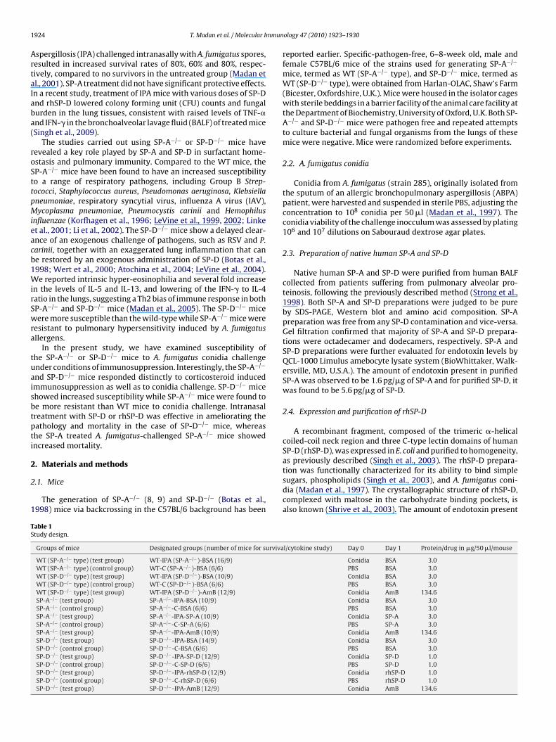
1 mmun
ArtaIaba(
roSttpMieacb1WirSwra
tuaisbtpti
2
2
1
TS
924 T. Madan et al. / Molecular I
spergillosis (IPA) challenged intranasally with A. fumigatus spores,esulted in increased survival rates of 80%, 60% and 80%, respec-ively, compared to no survivors in the untreated group (Madan etl., 2001). SP-A treatment did not have significant protective effects.n a recent study, treatment of IPA mice with various doses of SP-Dnd rhSP-D lowered colony forming unit (CFU) counts and fungalurden in the lung tissues, consistent with raised levels of TNF-�nd IFN-� in the bronchoalveolar lavage fluid (BALF) of treated miceSingh et al., 2009).
The studies carried out using SP-A−/− or SP-D−/− mice haveevealed a key role played by SP-A and SP-D in surfactant home-stasis and pulmonary immunity. Compared to the WT mice, theP-A−/− mice have been found to have an increased susceptibilityo a range of respiratory pathogens, including Group B Strep-ococci, Staphylococcus aureus, Pseudomonas aeruginosa, Klebsiellaneumoniae, respiratory syncytial virus, influenza A virus (IAV),ycoplasma pneumoniae, Pneumocystis carinii and Hemophilus
nfluenzae (Korfhagen et al., 1996; LeVine et al., 1999, 2002; Linket al., 2001; Li et al., 2002). The SP-D−/− mice show a delayed clear-nce of an exogenous challenge of pathogens, such as RSV and P.arinii, together with an exaggerated lung inflammation that cane restored by an exogenous administration of SP-D (Botas et al.,998; Wert et al., 2000; Atochina et al., 2004; LeVine et al., 2004).e reported intrinsic hyper-eosinophilia and several fold increase
n the levels of IL-5 and IL-13, and lowering of the IFN-� to IL-4atio in the lungs, suggesting a Th2 bias of immune response in bothP-A−/− and SP-D−/− mice (Madan et al., 2005). The SP-D−/− miceere more susceptible than the wild-type while SP-A−/− mice were
esistant to pulmonary hypersensitivity induced by A. fumigatusllergens.
In the present study, we have examined susceptibility ofhe SP-A−/− or SP-D−/− mice to A. fumigatus conidia challengender conditions of immunosuppression. Interestingly, the SP-A−/−
nd SP-D−/− mice responded distinctly to corticosteroid inducedmmunosuppression as well as to conidia challenge. SP-D−/− micehowed increased susceptibility while SP-A−/− mice were found toe more resistant than WT mice to conidia challenge. Intranasalreatment with SP-D or rhSP-D was effective in ameliorating theathology and mortality in the case of SP-D−/− mice, whereashe SP-A treated A. fumigatus-challenged SP-A−/− mice showedncreased mortality.
. Materials and methods
.1. Mice
The generation of SP-A−/− (8, 9) and SP-D−/− (Botas et al.,998) mice via backcrossing in the C57BL/6 background has been
able 1tudy design.
Groups of mice Designated groups (number of mice for surviva
WT (SP-A−/− type) (test group) WT-IPA (SP-A−/−)-BSA (16/9)WT (SP-A−/− type) (control group) WT-C (SP-A−/−)-BSA (6/6)WT (SP-D−/− type) (test group) WT-IPA (SP-D−/−)-BSA (10/9)WT (SP-D−/− type) (control group) WT-C (SP-D−/−)-BSA (6/6)WT (SP-D−/− type) (test group) WT-IPA (SP-D−/−)-AmB (12/9)SP-A−/− (test group) SP-A−/−-IPA-BSA (10/9)SP-A−/− (control group) SP-A−/−-C-BSA (6/6)SP-A−/− (test group) SP-A−/−-IPA-SP-A (10/9)SP-A−/− (control group) SP-A−/−-C-SP-A (6/6)SP-A−/− (test group) SP-A−/−-IPA-AmB (10/9)SP-D−/− (test group) SP-D−/−-IPA-BSA (14/9)SP-D−/− (control group) SP-D−/−-C-BSA (6/6)SP-D−/− (test group) SP-D−/−-IPA-SP-D (12/9)SP-D−/− (control group) SP-D−/−-C-SP-D (6/6)SP-D−/− (test group) SP-D−/−-IPA-rhSP-D (12/9)SP-D−/− (control group) SP-D−/−-C-rhSP-D (6/6)SP-D−/− (test group) SP-D−/−-IPA-AmB (12/9)
ology 47 (2010) 1923–1930
reported earlier. Specific-pathogen-free, 6–8-week old, male andfemale C57BL/6 mice of the strains used for generating SP-A−/−
mice, termed as WT (SP-A−/− type), and SP-D−/− mice, termed asWT (SP-D−/− type), were obtained from Harlan-OLAC, Shaw’s Farm(Bicester, Oxfordshire, U.K.). Mice were housed in the isolator cageswith sterile beddings in a barrier facility of the animal care facility atthe Department of Biochemistry, University of Oxford, U.K. Both SP-A−/− and SP-D−/− mice were pathogen free and repeated attemptsto culture bacterial and fungal organisms from the lungs of thesemice were negative. Mice were randomized before experiments.
2.2. A. fumigatus conidia
Conidia from A. fumigatus (strain 285), originally isolated fromthe sputum of an allergic bronchopulmonary aspergillosis (ABPA)patient, were harvested and suspended in sterile PBS, adjusting theconcentration to 108 conidia per 50 �l (Madan et al., 1997). Theconidia viability of the challenge inocculum was assessed by plating106 and 107 dilutions on Sabouraud dextrose agar plates.
2.3. Preparation of native human SP-A and SP-D
Native human SP-A and SP-D were purified from human BALFcollected from patients suffering from pulmonary alveolar pro-teinosis, following the previously described method (Strong et al.,1998). Both SP-A and SP-D preparations were judged to be pureby SDS-PAGE, Western blot and amino acid composition. SP-Apreparation was free from any SP-D contamination and vice-versa.Gel filtration confirmed that majority of SP-A and SP-D prepara-tions were octadecamer and dodecamers, respectively. SP-A andSP-D preparations were further evaluated for endotoxin levels byQCL-1000 Limulus amebocyte lysate system (BioWhittaker, Walk-ersville, MD, U.S.A.). The amount of endotoxin present in purifiedSP-A was observed to be 1.6 pg/�g of SP-A and for purified SP-D, itwas found to be 5.6 pg/�g of SP-D.
2.4. Expression and purification of rhSP-D
A recombinant fragment, composed of the trimeric �-helicalcoiled-coil neck region and three C-type lectin domains of humanSP-D (rhSP-D), was expressed in E. coli and purified to homogeneity,as previously described (Singh et al., 2003). The rhSP-D prepara-
tion was functionally characterized for its ability to bind simplesugars, phospholipids (Singh et al., 2003), and A. fumigatus coni-dia (Madan et al., 1997). The crystallographic structure of rhSP-D,complexed with maltose in the carbohydrate binding pockets, isalso known (Shrive et al., 2003). The amount of endotoxin presentl/cytokine study) Day 0 Day 1 Protein/drug in �g/50 �l/mouse
Conidia BSA 3.0PBS BSA 3.0Conidia BSA 3.0PBS BSA 3.0Conidia AmB 134.6Conidia BSA 3.0PBS BSA 3.0Conidia SP-A 3.0PBS SP-A 3.0Conidia AmB 134.6Conidia BSA 3.0PBS BSA 3.0Conidia SP-D 1.0PBS SP-D 1.0Conidia rhSP-D 1.0PBS rhSP-D 1.0Conidia AmB 134.6

mmun
ip
2
Ft
2
((auaDaTrtThasaprg
2
tu
2
eerhdTtcaf
2
ctgfs1Cde
one-population ANOVA test using MicroCal Origin Version 3.0statistical package (MicroCal Software Inc., Northampton, MA,U.S.A.). Cytokine data were compared using unpaired two-tailedMann–Whitney (nonparametric) test. Statistical analysis for the
T. Madan et al. / Molecular I
n the rhSP-D preparations was found to be 4 pg/�g of purifiedrotein.
.5. Preparation of AmB
A 4.16 mg/ml solution of AmB (Deoxycholate amphotericin B,ungizone, E.R. Squibb, Hounslow, UK) was prepared and dilutedo 2.692 mg/ml using sterile PBS immediately prior to use.
.6. Immunosuppression and challenge with A. fumigatus conidia
The murine model of IPA was generated as previously describedMadan et al., 2001). The lung cytokine profile of naïve, controlimmunosuppressed) and A. fumigatus-challenged (IPA) mice ofll the three genotypes viz. SP-A−/−, SP-D−/− and WT were eval-ated. In order to evaluate the susceptibility of SP-A−/−, SP-D−/−
nd WT mice to IPA. Effect of various treatments such as SP-A, SP-, BSA (as a control protein) and AmB (as a positive control) werelso assessed. The experimental design has been summarized inable 1. The study was reviewed and approved by the Institutionaleview committee. The experiments were conducted in accordanceo the rules laid down within appropriate Home Office license.he rationale for the amount of SP-A and SP-D used for therapy,as been reported earlier (Madan et al., 2005). The lung histologynd cytokine studies were performed in experiments separate fromurvival studies. Since various Control groups of mice had shownlterations in cytokine profiles on administration of test or controlrotein, the study has used the ratios of a test group with theirespective control for comparative evaluation between differentroups.
.7. Survival rate analysis
The various mice groups were monitored for the survival for upo 15 days. The survival rate was analyzed by Kaplan Meier testsing JMP software 3.2.1 (SAS Institute Inc., North Carolina, U.S.A.).
.8. Colony forming units (CFU)
Lungs were aseptically removed from the mice sacrificed fromach group on day 2, 4 and 8 following the conidia challenge. Anqual amount by weight of lung tissue (∼10 mg) comprising of fourandomly selected representative samples from the four lobes, wasomogenized using a mechanical homogenizer for 1 min with a 1:5ilution (w/v) in sterile PBS, for cytokine studies and CFU analysis.he lung suspension was serially diluted 2-fold and incubated inriplicate on SDA plates at 37 ◦C. A. fumigatus colonies were thenounted three times after 24 h incubation. The values representverage ± standard deviation of 12 data points (3 from each dilutionrom 2 animals of each group).
.9. Cytokine levels in lung suspension
A portion of the lung cell suspension obtained above wasentrifuged and resuspended in RPMI-1640 culture medium con-aining 10% (v/v) heat inactivated bovine serum and 10 �g/mlentamicin (2 × 10⁶ cells/well). Cells were allowed to proliferateor 72 h in 5% (v/v) CO2 incubator. The supernatants from the lung
uspension were assayed for the cytokines such as IL-4, IL-5, IL-0, IL-12, IL-13, TNF-� and IFN-� using the kits from Endogen,ambridge, MA, U.S.A. The values represent average ± standardeviation of 6 data points (3 from each animal from 2 animals ofach group).ology 47 (2010) 1923–1930 1925
2.10. Histological examination of the lung sections
For histopathology, 4 randomly selected representative speci-mens were taken from all the four lung lobes of the sacrificed mice,fixed in 10% (v/v) formaldehyde, and stored at 4 ◦C. The tissue sec-tions were made using a microtome, then stained with H&E/GMS,and examined at a magnification of 40× and 400×. Each picture is arepresentative of 8 sections (4 each from 2 animals of each group).
2.11. Statistical analysis
CFU data were expressed as mean ± SD and compared by
Fig. 1. Mortality in various groups of mice. Mice were immunosuppressed withcorticosteroid prior to an intranasal challenge with A. fumigatus conidia. (A) SP-A gene deficient mice (SP-A−/−) mice and (B) SP-D gene deficient (SP-D−/−) micecompared with their respective wild-type (WT) mice. The bar graphs depict thelung CFU counts isolated from the above-mentioned groups on day 2 of the study.
1926 T. Madan et al. / Molecular Immunology 47 (2010) 1923–1930
Table 2Ratio of cytokine levels of lung suspensions of A. fumigatus challenged, control and naïve SP-A−/− and SP-D−/− mice groups to their respective groups of WT mice on day 0.
IFN-� TNF-� IL-4 IL-5 IL-10 IL-12 IL-13
SP-A−/−-IPA-BSA 1.00 1.12 −1.68* 1.87* −1.21 1.00 2.05*
SP-A−/−-control-BSA −1.06 −2.67* −2.88* −13.4* −3.58* 1.29 −1.28SP-A−/−-naïve −1.92* 1.41 1.00 3.93* 1.13 −1.24 13.1*
SP-D−/−-IPA-BSA 1.52* 1.48 1.00 1.00 1.39 1.00 1.68*
SP-D−/−-control-BSA 1.12 −1.19 2.04* 1.00 −3.73* −1.22 1.55*
SP-D−/−-naïve −2.7* −1.23 1.00 4.68* 1.01 −1.26 11.6*
E ls of e± cientf spect
Ito
3
3f
Ir(�tT
(
FSdE
ach value represents a mean of nine readings (triplicate values from three anima5%. The negative sign indicates a decrease in the level of cytokine in the gene defi
or SP-A−/− and SP-D−/− mice are from WT (SP-A−/− type) and WT (SP-D−/− type), re* p < 0.05.
FN-� and IL-4 ratios were carried out using two by two table (aest for bivariate analysis) The analysis was considered significantnly when p < 0.05 after Bonferroni correction.
. Results
.1. Survival of WT, SP-A−/− and SP-D−/− mice following A.umigatus challenge
The two wild-type groups WT-IPA-BSA (SP-A−/− type) and WT-PA-BSA (SP-D−/− type) showed mortality rates of 100% and 60%,espectively. The SP-A−/−-IPA-BSA mice had a lower mortality40%), 3.95-fold less CFU count and a significant increase in IFN-
to IL-4 ratio (1.69-fold) when subjected to IPA compared tohe WT-IPA-BSA (SP-A−/− type) mice (100%) (p < 0.05) (Fig. 1A,able 2).
The SP-D−/−-IPA-BSA mice (57.14%) showed similar mortalityp = 0.85) and IFN-� to IL-4 ratio (1.3-fold increase), but 3.53-
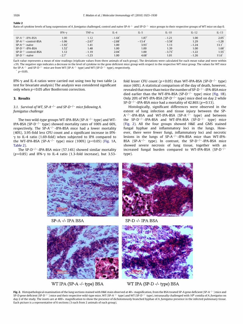
ig. 2. Histopathological examination of the lung sections stained with H&E stain observeP-D gene deficient (SP-D−/−) mice and their respective wild-type mice, WT (SP-A−/− typeay 2 of the study. The insets are at 400× magnification to show the presence of dichotomach picture is a representative of 6 sections (3 each from 2 animals of each group).
ach group). The deviations were calculated for each mean value and were withinmice group with respect to the respective WT mice group. The values for WT miceively.
fold lesser CFU count (p < 0.05) than WT-IPA-BSA (SP-D−/− type)mice (60%). A statistical comparison of the day of death, however,revealed that more than twice the number of SP-D−/−-IPA-BSA micedied earlier than the WT-IPA-BSA (SP-D−/− type) mice (Fig. 1B).Only 20% of WT-IPA-BSA (SP-D−/− type) mice died on day 2 whileSP-D−/−-IPA-BSA mice had a mortality of 42.86% (p = 0.13).
Histologically, significant differences were observed in theextent of lung infection and tissue injury between the SP-A−/−-IPA-BSA and WT-IPA-BSA (SP-A−/− type) and betweenthe SP-D−/−-IPA-BSA and WT-IPA-BSA (SP-D−/− type) mice(Fig. 2). All the four groups showed H&E and GMS stainedfungal hyphae and inflammatory loci in the lungs. How-ever, there were fewer fungi, inflammatory loci and necrotic
lesions in the lungs of SP-A−/−-IPA-BSA mice than WT-IPA-BSA (SP-A−/− type). In contrast, the SP-D−/−-IPA-BSA miceshowed severe necrosis of lung tissue, together with anincreased fungal burden compared to WT-IPA-BSA (SP-D−/−type).
d at 40× magnification, from the BSA treated SP-A gene deficient (SP-A−/−) mice and) and WT (SP-D−/− type), intranasally challenged with 108 conidia of A. fumigatus on
ously branched hyphae of A. fumigatus presence in the infected pulmonary tissue.
T. Madan et al. / Molecular Immun
Table 3Ratio of IFN-� to IL-4 levels on day 2, 4 and 8 and percent survival in various groupsof mice.
Group IFN-�/IL-4 % survival
Day 2 Day 4 Day 8
WT-IPA (SP-A−/−)-BSA 2.65 4.72 – 0WT-C (SP-A−/−)-BSA 1.77 4.54 –WT-IPA (SP-D−/−)-BSA 1.83 4.8 4.56 40WT-C (SP-D−/−)-BSA 2.43 5.67 0.086WT-IPA (SP-D−/−)-AmB 4.33 6.06 3.84 75SP-A−/−-IPA-BSA 4.5 2.62 3.57 60SP-A−/−-C-BSA 4.8 a 0.048SP-A−/−-IPA-SP-A 4.77 2.21 2.5 40SP-A−/−-C-SP-A 2.52 3.7 0.081SP-A−/−-IPA-AmB 4.87 4.41 5.43 80SP-D−/−-IPA-BSA 2.79 1.93 3.67 42.86SP-D−/−-C-BSA 1.34 3.54 3.25SP-D−/−-IPA-SP-D 3.36 3.81 3.73 50SP-D−/−-C-SP-D 2.29 4.96 3.11SP-D−/−-IPA-rhSP-D 3.74 7.49 5.67 67SP-D−/−-C-rhSP-D 1.9 3.68 3.96SP-D−/−-IPA-AmB 2.77 6.26 4.8 83.33
(
3c
3
lId
mice resulted in a 2.4-fold increase in survival (75%) (p = 0.21) in
FAr
–) No mice survived.a IFN-� levels were not in the detectable range in the SP-A−/−-C-BSA mice.
.2. Treatment of WT, SP-A−/− and SP-D−/− mice followinghallenge with conidia
.2.1. Administration of SP-A to SP-A−/−-IPA mice
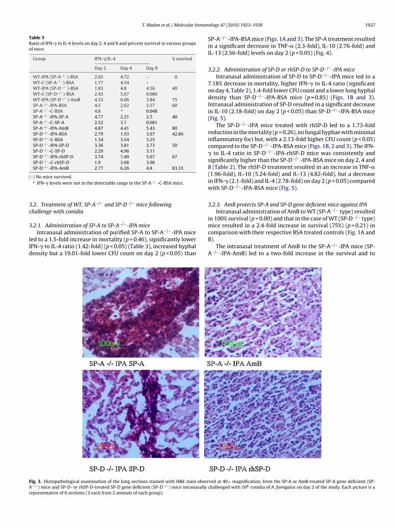
Intranasal administration of purified SP-A to SP-A−/−-IPA miceed to a 1.5-fold increase in mortality (p = 0.46), significantly lowerFN-� to IL-4 ratio (1.42-fold) (p < 0.05) (Table 3), increased hyphalensity but a 19.01-fold lower CFU count on day 2 (p < 0.05) than
ig. 3. Histopathological examination of the lung sections stained with H&E stain obser−/−) mice and SP-D- or rhSP-D-treated SP-D gene deficient (SP-D−/−) mice intranasally cepresentative of 6 sections (3 each from 2 animals of each group).
ology 47 (2010) 1923–1930 1927
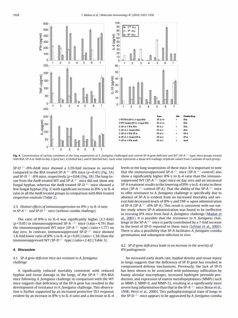
SP-A−/−-IPA-BSA mice (Figs. 1A and 3). The SP-A treatment resultedin a significant decrease in TNF-� (2.3-fold), IL-10 (2.76-fold) andIL-13 (2.56-fold) levels on day 2 (p < 0.05) (Fig. 4).
3.2.2. Administration of SP-D or rhSP-D to SP-D−/−-IPA miceIntranasal administration of SP-D to SP-D−/−-IPA mice led to a
7.18% decrease in mortality, higher IFN-� to IL-4 ratio (significanton day 4, Table 2), 1.4-fold lower CFU count and a lower lung hyphaldensity than SP-D−/−-IPA-BSA mice (p = 0.85) (Figs. 1B and 3).Intranasal administration of SP-D resulted in a significant decreasein IL-10 (2.18-fold) on day 2 (p < 0.05) than SP-D−/−-IPA-BSA mice(Fig. 5).
The SP-D−/−-IPA mice treated with rhSP-D led to a 1.73-foldreduction in the mortality (p = 0.26), no fungal hyphae with minimalinflammatory foci but, with a 2.13-fold higher CFU count (p < 0.05)compared to the SP-D−/−-IPA-BSA mice (Figs. 1B, 2 and 3). The IFN-� to IL-4 ratio in SP-D−/−-IPA-rhSP-D mice was consistently andsignificantly higher than the SP-D−/−-IPA-BSA mice on day 2, 4 and8 (Table 2). The rhSP-D treatment resulted in an increase in TNF-�(1.96-fold), IL-10 (5.24-fold) and IL-13 (4.82-fold), but a decreasein IFN-� (2.1-fold) and IL-4 (2.78-fold) on day 2 (p < 0.05) comparedwith SP-D−/−-IPA-BSA mice (Fig. 5).
3.2.3. AmB protects SP-A and SP-D gene deficient mice against IPAIntranasal administration of AmB to WT (SP-A−/− type) resulted
in 100% survival (p = 0.00) and that in the case of WT (SP-D−/− type)
comparison with their respective BSA treated controls (Fig. 1A andB).
The intranasal treatment of AmB to the SP-A−/−-IPA mice (SP-A−/−-IPA-AmB) led to a two-fold increase in the survival and to
ved at 40× magnification, from the SP-A or AmB-treated SP-A gene deficient (SP-hallenged with 108 conidia of A. fumigatus on day 2 of the study. Each picture is a
1928 T. Madan et al. / Molecular Immunology 47 (2010) 1923–1930
F challew valu
Scasffrr
3i
(td1i
4
4c
hmmdte
ig. 4. Concentration of various cytokines in the lung suspensions of A. fumigatusith BSA, SP-A or AmB on day 2 (grey bar), 4 (dotted bar), and 8 (hatched bar). Each
P-D−/−-IPA-AmB mice showed a 3.59-fold increase in survivalompared to the BSA treated SP-A−/−-IPA mice (p = 0.43) (Fig. 1A)nd SP-D−/−-IPA mice, respectively (p = 0.04) (Fig. 1B). The lung tis-ue from the AmB treated WT and SP-A−/− mice did not show anyungal hyphae, whereas the AmB treated SP-D−/− mice showed aew fungal hyphae (Fig. 3) with significant increase in IFN-� to IL-4atio in all the AmB treated groups in comparison with BSA treatedespective controls (Table 2).
.3. Distinct effects of immunosuppression on IFN-� to IL-4 ration SP-A−/− and SP-D−/− mice (without conidia challenge)
The ratio of IFN-� to IL-4 was significantly higher (2.7-fold)p < 0.05) in immunosuppressed SP-A −/− mice (ratio = 4.79) thanhe immunosuppressed WT mice (SP-A−/− type) (ratio = 1.77) onay zero. In contrast, immunosuppressed SP-D−/− mice showed.8-fold lower ratio of IFN-� to IL-4 (p < 0.05) (ratio = 1.34) than the
mmunosuppressed WT (SP-D−/− type) (ratio = 2.42) (Table 3).
. Discussion
.1. SP-A gene deficient mice are resistant to A. fumigatushallenge
A significantly reduced mortality consistent with reducedyphae and tissue damage in the lungs, of the SP-A−/−-IPA-BSA
ice following A. fumigatus challenge in comparison with the WTice suggests that deficiency of the SP-A gene has resulted in theevelopment of resistance to A. fumigatus challenge. This observa-ion is further supported by an increased protective Th1 responsevident by an increase in IFN-� to IL-4 ratio and a decrease in IL-4
nged and control-SP-A gene deficient and WT (SP-A−/− type) mice groups treatede represents a mean of 6 readings (triplicate values from 2 animals of each group).
levels in the lung suspensions of these mice. It is important to notethat the immunosuppressed SP-A−/− mice (SP-A−/−-control) alsoshow a significantly higher IFN-� to IL-4 ratio than the immuno-suppressed WT (SP-A−/− type) mice on day zero and an intranasalSP-A treatment results in the lowering of IFN-� to IL-4 ratio in thesemice (SP-A−/−-control-SP-A). That the ability of the SP-A−/− miceto offer resistance to A. fumigatus challenge is specifically due toabsence of SP-A is evident from an increased mortality and sev-eral fold decreased levels of IFN-� and TNF-� upon administrationof SP-A (SP-A−/−-IPA-SP-A). This result is consistent with our ear-lier study where SP-A administration was found to be ineffectivein rescuing IPA mice from fatal A. fumigatus challenge (Madan etal., 2001). It is possible that the resistance to A. fumigatus chal-lenge in the SP-A−/− mice is partly contributed by a 7-fold increasein the level of SP-D reported in these mice (LeVine et al., 2002).There is also a possibility that SP-A facilitates A. fumigates conidiagermination and subsequent infection in vivo.
4.2. SP-D gene deficiency leads to an increase in the severity ofIPA pathogenesis
An increased early death rate, hyphal density and tissue injuryin lungs suggests that the deficiency of SP-D gene has resulted incompromised defense mechanisms. Previously, the lack of SP-Dhas been shown to be associated with pulmonary infiltration byfoamy alveolar macrophages, increased hydrogen peroxide pro-
duction, and expression of matrix metallopeptidases (MMPs) suchas MMP-2, MMP-9, and MMP-12, resulting in a significantly moresevere lung inflammation than that in the SP-A−/− mice (Botas et al.,1998; Wert et al., 2000). This pathophysiological state of lungs inthe SP-D−/− mice appears to be aggravated by A. fumigatus conidia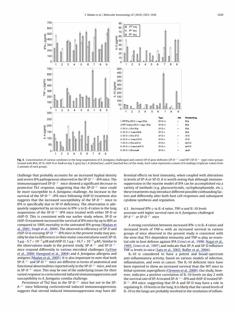
T. Madan et al. / Molecular Immunology 47 (2010) 1923–1930 1929
F hallent ched2
caipbssIqsrrcars3tmeaSfivs
As
ig. 5. Concentration of various cytokines in the lung suspensions of A. fumigatus created with BSA, SP-D, rhSP-D or AmB on day 2 (grey bar), 4 (dotted bar), and 8 (hatanimals of each group).
hallenge that probably accounts for an increased hyphal densitynd severe IPA pathogenesis observed in the SP-D−/−-IPA mice. Themmunosuppressed SP-D−/− mice showed a significant decrease inrotective Th1 response, suggesting that the SP-D−/− mice coulde more susceptible to A. fumigatus challenge. An increase in theurvival of the SP-D−/−-IPA mice following rhSP-D treatment alsouggests that the increased susceptibility of the SP-D−/− mice toPA is specifically due to SP-D deficiency. The observation is ade-uately supported by an increase in IFN-� to IL-4 ratios in the lunguspensions of the SP-D−/−-IPA mice treated with either SP-D orhSP-D. This is consistent with our earlier study where, SP-D orhSP-D treatment increased the survival of IPA mice by up to 80% asompared to 100% mortality in the untreated IPA group (Madan etl., 2001; Singh et al., 2009). The observed in efficiency of SP-D andhSP-D in rescuing SP-D−/−-IPA mice in the present study may pos-ibly be due to differences in their molar concentrations used (SP-D,�g – 5.7 × 10⁻³ �M and rhSP-D, 1 �g – 16.7 × 10⁻³ �M). Similar to
he observations made in the present study, SP-A−/− and SP-D−/−
ice respond differently to various microbial challenges (LeVinet al., 2000; Hawgood et al., 2004) and A. fumigatus allergens andntigens (Madan et al., 2005). It is also important to note that bothP-A−/− and SP-D−/− mice are different in terms of anatomical andunctional abnormalities reported only in the SP-D−/− mice, and notn SP-A−/− mice. This may be one of the underlying issues for their
aried response to corticosteroid induced immunosuppression andusceptibility to A. fumigatus conidia challenge.Persistence of Th2 bias in the SP-D−/− mice but not in the SP-−/− mice following corticosteroid induced immunosuppression,uggests that steroid induced immunosuppression may have dif-
ged and control-SP-D gene deficient (SP-D−/−) and WT (SP-D−/− type) mice groupsbar) of the study. Each value represents a mean of 6 readings (triplicate values from
ferential effects on host immunity, when coupled with alterationsin levels of SP-A or SP-D. It is worth noting that although immuno-suppression in the murine model of IPA can be accomplished via avariety of methods (e.g. glucocorticoids, cyclophosphamide, etc.),these treatments may introduce different possible confounding fac-tors and differently alter both host cell responses and subsequentcytokine synthesis and regulation.
4.3. Increased IFN-� to IL-4 ratios, TNF-a and IL-10 levelsassociate with higher survival rates in A. fumigatus-challengedSP-A−/− or SP-D−/− mice
A strong correlation between increased IFN-� to IL-4 ratios andincreased levels of TNF-� with an increased survival in variousgroups of mice observed in the present study is consistent withthe view that Th1-dependent immunity and TNF-� play an essen-tial role in host defense against IPA (Cenci et al., 1998; Nagai et al.,1995; Cenci et al., 1997) and indicate that SP-A and SP-D influenceTNF-� levels in mice (Sato et al., 2002; Bufler et al., 2004).
IL-10 is considered to have a potent and broad-spectrumanti-inflammatory activity, based on various models of infection,inflammation, and even in cancer. The IL-10 deficient mice havebeen reported to show an increased survival than the WT mice tolethal systemic aspergillosis (Clemons et al., 2000). Our study, how-
ever, indicates a positive correlation of IL-10 levels on day 2 withthe survival rate of SP-A treated SP-A−/−-IPA and rhSP-D treated SP-D−/−-IPA mice, suggesting that SP-A and SP-D may have a role inregulating IL-10 levels in the lung. It is likely that the raised levels ofIL-10 in the lungs are probably involved in the resolution of inflam-
1 mmun
mAih(aS
i3swoo
mwthlci
A
tTt
R
A
A
B
B
C
C
C
H
930 T. Madan et al. / Molecular I
ation following early episode of lung infiltration in response to. fumigatus challenge. A recent study showed that IL-10 secret-
ng, regulatory T cells are essential to completely suppress airwayyperreactivity in an antigen dependent murine model of asthmaPresser et al., 2008). Our in vitro data using human dendritic cellsnd T-cells also appear to suggest modulation of IL-10 by SP-A andP-D (Kishore et al., unpublished).
Nine out of 11 groups of mice in the present study showed a pos-tive correlation of lung CFU counts on day 2 with mortality on day, while two groups namely SP-D−/−-IPA-AmB and SP-D−/−-rhSP-Dhowed a negative correlation with lung CFU counts in comparisonith SP-D−/−-IPA-BSA. We are unable to explain the higher CFUs
n day 2 in these groups wherein there was no mortality on day 3r on any subsequent days in comparison with SP-D−/−-IPA-BSA.
In conclusion, the present study reports that deficiency of SP-Day predispose the host to a severe form of invasive aspergillosisith a rapid and fatal outcome, Both SP-A and SP-D have impor-
ant roles in the regulation of Th1-Th2 homoeostasis and therebyost defense status against A. fumigatus in the lungs. Their absence
eading to varied susceptibility to A. fumigatus challenge in corti-osteroid immunosuppressed mice highlights their balancing actsn host defense against A. fumigatus.
cknowledgments
This work was supported by the Council for Scientific and Indus-rial Research (T.M. and P.U.S.), the Department of Science andechnology, India (M.S.), the Medical Research Council (K.B.R., H.C.)he European Commission (K.B.R., U.K.) and BRIEF award to U.K.
eferences
llen, M.J., Voelker, D.R., Mason, R.J., 2001. Interactions of surfactant proteins A andD with Saccharomyces cerevisiae and Aspergillus fumigatus. Infect. Immun. 69,2037–2044.
tochina, E.N., Gow, A.J., Beck, J.M., et al., 2004. Delayed clearance of Pneumocystiscarinii infection, increased inflammation, and altered nitric oxide metabolismin lungs of surfactant protein-D knockout mice. J. Infect. Dis. 189, 1528–1539.
otas, C., Poulain, F., Akiyama, J., et al., 1998. Altered surfactant homeostasis andalveolar type II cell morphology in mice lacking surfactant protein D. Proc. Natl.Acad. Sci. U.S.A. 95, 11869–11874.
ufler, P., Schikor, D., Schmidt, B., Griese, M., 2004. Cytokine stimulation by Pseu-domonas aeruginosa—strain variation and modulation by pulmonary surfactant.Exp. Lung Res. 30, 163–179.
enci, E., Perito, S., Enssle, K.H., Mosci, P., Latge, J.P., Romani, L., Bistoni, F., 1997.Th1 and Th2 cytokines in mice with invasive aspergillosis. Infect. Immun. 65,564–570.
enci, E., Mencacci, A., Fe d’Ostiani, C., et al., 1998. Cytokine- and T helper-dependentlung mucosal immunity in mice with invasive pulmonary aspergillosis. J. Infect.Dis. 178, 1750–1760.
lemons, K.V., Grunig, G., Sobel, R.A., Mirels, L.F., Rennick, D.M., Stevens, D.A., 2000.Role of IL-10 in invasive aspergillosis: increased resistance of IL-10 gene knock-out mice to lethal systemic aspergillosis. Clin. Exp. Immunol. 122, 186–191.
awgood, S., Brown, C., Edmondson, J., Stumbaugh, A., Allen, L., Goerke, J., Clark,H., Poulain, F., 2004. Pulmonary collectins modulate strain-specific influenza Avirus infection and host responses. J. Virol. 78, 8565–8572.
ology 47 (2010) 1923–1930
Kuroki, Y., Takahashi, M., Nishitani, C., 2007. Pulmonary collectins in innate immu-nity of the lung. Cell Microbiol. 9, 1871–1879.
Kishore, U., Bernal, A.L., Kamran, M.F., et al., 2005. Surfactant proteins SP-A andSP-D in human health and disease. Arch. Immunol. Ther. Exp. (Warsz) 53,399–417.
Kishore, U., Greenhough, T.J., Waters, P., et al., 2006. Surfactant proteinsSP-A and SP-D: structure, function and receptors. Mol. Immunol. 43,1293–1315.
Korfhagen, T.R., Bruno, M.D., Ross, G.F., et al., 1996. Altered surfactant functionand structure in SP-A gene targeted mice. Proc. Natl. Acad. Sci. U.S.A. 93,9594–9599.
LeVine, A.M., Gwozdz, J., Stark, J., Bruno, M., Whitsett, J.A., Korfhagen, T.R., 1999.Surfactant protein A enhances respiratory syncytial virus clearance in vivo. J.Clin. Invest. 103, 1015–1021.
Linke, M.J., Harris, C.E., Korfhagen, T.R., et al., 2001. Immunosuppressed surfactantprotein A-deficient mice have increased susceptibility to Pneumocystis cariniiinfection. J. Infect. Dis. 183, 943–952.
LeVine, A.M., Hartshorn, K., Elliott, J., Whitsett, J.A., Korfhagen, T.R., 2002. Absence ofSP-A modulates innate and adaptive defense responses to pulmonary influenzainfection. Am. J. Physiol. Lung Cell Mol. Physiol. 282, L563–L572.
LeVine, A.M., Whitsett, J.A., Gwozdz, J.A., Richardson, T.R., Fisher, J.H., Burhans, M.H.,Korfhagen, T.R., 2000. Distinct effects of surfactant protein A or D deficiencyduring bacterial infection on the lung. J. Immunol. 165, 3934–3940.
LeVine, A.M., Elliott, J., Whitsett, J.A., Srikiatkhachorn, A., Crouch, E.C., DeSilva, N.,Korfhagen, T.R., 2004. Surfactant protein D enhances phagocytosis and pul-monary clearance of respiratory syncytial virus. Am. J. Respir. Cell Mol. Biol.31, 193–199.
Li, G., Siddiqui, J., Hendry, M., et al., 2002. Surfactant protein A-deficient mice displayan exaggerated early inflammatory response to a �-resistant strain of influenzaA virus. Am. J. Respir. Cell Mol. Biol. 26, 277–282.
Madan, T., Eggleton, P., Kishore, U., Strong, P., Aggrawal, S.S., Sarma, P.U., Reid, K.B.M.,1997. Binding of pulmonary surfactant proteins A and D to Aspergillus fumigatusconidia enhances phagocytosis and killing by human neutrophils and alveolarmacrophages. Infect. Immun. 65, 3171–3179.
Madan, T., Kishore, U., Singh, M., Strong, P., Hussain, E.M., Reid, K.B.M., Sarma, P.U.,2001. Protective role of lung surfactant protein D in a murine model of invasivepulmonary aspergillosis. Infect. Immun. 69, 2728–2731.
Madan, T., Reid, K.B.M., Singh, M., Sarma, P.U., Kishore, U., 2005. Susceptibility of micegenetically deficient in the Surfactant Protein (SP)-A or SP-D gene to pulmonaryhypersensitivity induced by antigens and allergens of Aspergillus fumigatus. J.Immunol. 174, 6943–6954.
Nagai, H., Guo, J., Choi, H., Kurup, V.P., 1995. Interferon-� and tumor necrosis factor-�protect mice from invasive aspergillosis. J. Infect. Dis. 172, 1554–1560.
Presser, K., Schwinge, D., Wegmann, M., et al., 2008. Coexpression of TGF-beta1 andIL-10 enables regulatory T cells to completely suppress airway hyperreactivity.J. Immunol. 181, 7751–7758.
Sato, K., Tomioka, H., Shimizu, T., Gonda, T., Ota, F., Sano, C., 2002. Type II alveolarcells play roles in macrophage-mediated host innate resistance to pulmonarymycobacterial infections by producing pro-inflammatory cytokines. J. Infect. Dis.185, 1139–1147.
Shrive, A.K., Tharia, H.A., Strong, P., et al., 2003. High-resolution structural insightsinto ligand binding and immune cell recognition by human lung surfactantprotein D. J. Mol. Biol. 331, 509–523.
Singh, M., Madan, T., Waters, P., et al., 2009. Therapeutic effects of recombinant formsof full-length and truncated human surfactant protein D in a murine model ofinvasive pulmonary aspergillosis. Mol. Immunol. 46 (11–12), 2363–2369.
Singh, M., Madan, T., Waters, P., Parida, S.K., Sarma, U., Kishore, U., 2003. Protectiveeffects of a recombinant fragment of human surfactant protein D in a murinemodel of pulmonary hypersensitivity induced by dust mite allergens. Immunol.Lett. 86 (3), 299–307.
Strong, P., Kishore, U., Morgan, C., Lopez Bernal, A., Singh, M., Reid, K.B.M., 1998. A
novel method of purifying lung surfactant proteins A and D from the lung lavageof alveolar proteinosis patients and from pooled amniotic fluid. J. Immunol.Methods 220, 139–149.Wert, S.E., Yoshida, M., LeVine, A.M., et al., 2000. Increased metalloproteinaseactivity, oxidant production, and emphysema in surfactant protein D gene-inactivated mice. Proc. Natl. Acad. Sci. U.S.A. 97, 5972–5977.
Related Documents