PowerPoint ® Lecture Slides prepared by Karen Dunbar Kareiva Ivy Tech Community College © Annie Leibovitz/Contact Press Images Chapter 15 Part B The Endocrine System © 2017 Pearson Education, Inc.

Welcome message from author
This document is posted to help you gain knowledge. Please leave a comment to let me know what you think about it! Share it to your friends and learn new things together.
Transcript
PowerPoint® Lecture Slides
prepared by
Karen Dunbar Kareiva
Ivy Tech Community College© Annie Leibovitz/Contact Press Images
Chapter 15 Part B
The
Endocrine
System
© 2017 Pearson Education, Inc.

15.7 Thyroid Gland
Location and Structure
• Butterfly-shaped gland in anterior neck on the
trachea, just inferior to larynx, that consists of:
• Isthmus: median mass connecting two lateral
lobes
• Follicles: hollow sphere of epithelial follicular
cells that produce glycoprotein thyroglobulin
© 2017 Pearson Education, Inc.

Location and Structure (cont.)
• Colloid: fluid of follicle lumen containing
thyroglobulin plus iodine and is precursor to
thyroid hormone
• Parafollicular cells: produce hormone
calcitonin
© 2017 Pearson Education, Inc.

Figure 15.8a The thyroid gland.
© 2017 Pearson Education, Inc.
Hyoid bone
Thyroid
cartilage
Common
carotid
artery
Inferior
thyroid
artery
Trachea
Aorta
Epiglottis
Superior
thyroid
artery
Isthmus of
thyroid gland
Left subclavian
artery
Left lateral
lobe of
thyroid
gland
Gross anatomy of the thyroid gland,
anterior view

Figure 15.8b The thyroid gland.
© 2017 Pearson Education, Inc.
Photomicrograph of thyroid gland
follicles (315×)
Parafollicular cells
(secrete calcitonin)
Follicular cells
(secrete thyroid
hormone)
Colloid-filled
follicles

Thyroid Hormone (TH)
• Body’s major metabolic hormone
• Found in two forms
– T4 (thyroxine): major form that consists of two
tyrosine molecules with four bound iodine atoms
– T3 (triiodothyronine): form that has two
tyrosines with three bound iodine atoms
• Must be converted to T4 at tissue level
• Both are iodine-containing amine hormones
© 2017 Pearson Education, Inc.

Thyroid Hormone (TH) (cont.)
• TH affects virtually every cell in body
• Enters target cell and binds to intracellular
receptors within nucleus
– Triggers transcription of various metabolic genes
• Effects of thyroid hormone include:
– Increases basal metabolic rate and heat
production
• Referred to as calorigenic effect
© 2017 Pearson Education, Inc.

Thyroid Hormone (TH) (cont.)
– Regulates tissue growth and development
• Critical for normal skeletal and nervous system
development and reproductive capabilities
– Maintains blood pressure
• Increases adrenergic receptors in blood vessels
© 2017 Pearson Education, Inc.

Thyroid Hormone (TH)
• Synthesis
– Thyroid gland stores hormone extracellularly in
follicle lumen until triggered by TSH to release
– Seven steps involved in synthesis of TH:
1. Thyroglobulin is synthesized and discharged into
follicle lumen
2. Iodide is trapped: iodide ions (I–) are actively taken
into cell and released into lumen
3. Iodide oxidized: electrons are removed, converting it
to iodine (I2)
© 2017 Pearson Education, Inc.

Thyroid Hormone (TH) (cont.)
• Synthesis (cont.)
4. Iodine is attached to tyrosine: mediated by
peroxidase enzymes
– Monoiodotyrosine (MIT): formed if only one iodine
attaches
– Diiodotyrosine (DIT): formed if two iodines attach
5. Iodinated tyrosines link together to form T3 and T4
– If one MIT and one DIT link, T3 is formed
– If two DITs link, T4 is formed
© 2017 Pearson Education, Inc.
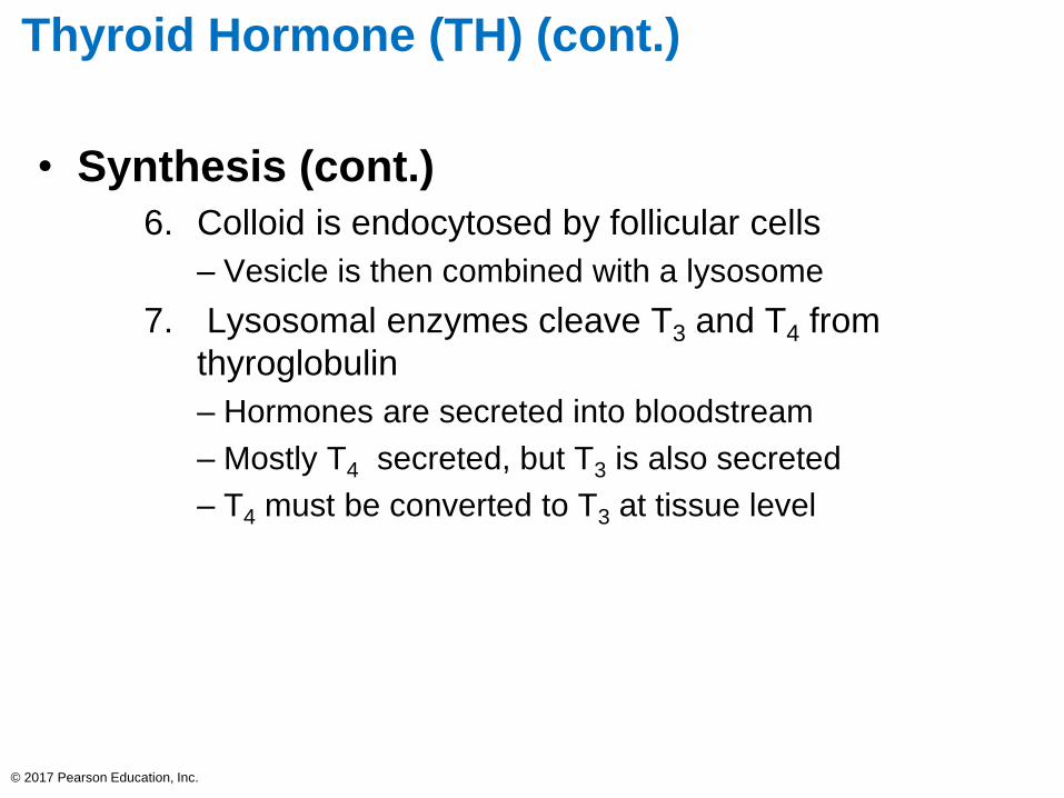
Thyroid Hormone (TH) (cont.)
• Synthesis (cont.)
6. Colloid is endocytosed by follicular cells
– Vesicle is then combined with a lysosome
7. Lysosomal enzymes cleave T3 and T4 from
thyroglobulin
– Hormones are secreted into bloodstream
– Mostly T4 secreted, but T3 is also secreted
– T4 must be converted to T3 at tissue level
© 2017 Pearson Education, Inc.

Figure 15.9 Synthesis of thyroid hormone.
© 2017 Pearson Education, Inc.
Capillary
Rough
ER
Golgi
apparatus
Iodide (I )
Thyroid follicular cells
Colloid
Colloid in
lumen of
follicle
Thyroglobulin is synthesized and
discharged into the follicle lumen.
1
Slide 2

Figure 15.9 Synthesis of thyroid hormone.
© 2017 Pearson Education, Inc.
Capillary
Rough
ER
Golgi
apparatus
Iodide (I )
Thyroid follicular cells
Colloid
lodide (I ) is trapped
(actively transported in).
Colloid in
lumen of
follicle
Thyroglobulin is synthesized and
discharged into the follicle lumen.
1
2
Slide 3

Figure 15.9 Synthesis of thyroid hormone.
© 2017 Pearson Education, Inc.
Capillary
Rough
ER
Golgi
apparatus
Iodide (I )
Thyroid follicular cells
Colloid
Iodine
Iodide
is oxidized
to iodine.
lodide (I ) is trapped
(actively transported in).
Colloid in
lumen of
follicle
Thyroglobulin is synthesized and
discharged into the follicle lumen.
1
3
2
Slide 4
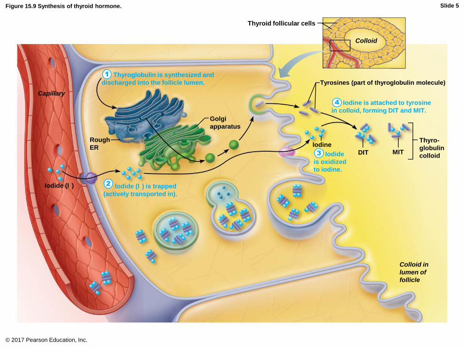
Figure 15.9 Synthesis of thyroid hormone.
© 2017 Pearson Education, Inc.
Capillary
Rough
ER
Golgi
apparatus
Iodide (I )
Thyroid follicular cells
Colloid
Tyrosines (part of thyroglobulin molecule)
Thyro-
globulin
colloidMITDIT
Iodine
Iodine is attached to tyrosine
in colloid, forming DIT and MIT.
Iodide
is oxidized
to iodine.
lodide (I ) is trapped
(actively transported in).
Colloid in
lumen of
follicle
Thyroglobulin is synthesized and
discharged into the follicle lumen.
1
4
3
2
Slide 5

Figure 15.9 Synthesis of thyroid hormone.
© 2017 Pearson Education, Inc.
Capillary
Rough
ER
Golgi
apparatus
Iodide (I )
Thyroid follicular cells
Colloid
Tyrosines (part of thyroglobulin molecule)
Thyro-
globulin
colloidMITDIT
Iodine
Iodine is attached to tyrosine
in colloid, forming DIT and MIT.
Iodide
is oxidized
to iodine.
Iodinated tyrosines are
linked together to form T3
and T4.
lodide (I ) is trapped
(actively transported in).
Colloid in
lumen of
follicle
T4
T3
Thyroglobulin is synthesized and
discharged into the follicle lumen.
1
4
3
52
Slide 6

Figure 15.9 Synthesis of thyroid hormone.
© 2017 Pearson Education, Inc.
Capillary
Rough
ER
Golgi
apparatus
Iodide (I )
Lysosome
Thyroid follicular cells
Colloid
Tyrosines (part of thyroglobulin molecule)
Thyro-
globulin
colloidMITDIT
Iodine
Iodine is attached to tyrosine
in colloid, forming DIT and MIT.
Iodide
is oxidized
to iodine.
Iodinated tyrosines are
linked together to form T3
and T4.
lodide (I ) is trapped
(actively transported in).
Colloid in
lumen of
follicle
T4
T3
Thyroglobulin is synthesized and
discharged into the follicle lumen.
1
4
3
5
Thyroglobulin colloid is
endocytosed and combined
with a lysosome.
6
2
Slide 7

Figure 15.9 Synthesis of thyroid hormone.
© 2017 Pearson Education, Inc.
Capillary
Rough
ER
Golgi
apparatus
Iodide (I )
Lysosome
Thyroid follicular cells
Colloid
Tyrosines (part of thyroglobulin molecule)
Thyro-
globulin
colloidMITDIT
Iodine
Iodine is attached to tyrosine
in colloid, forming DIT and MIT.
Iodide
is oxidized
to iodine.
Iodinated tyrosines are
linked together to form T3
and T4.
lodide (I ) is trapped
(actively transported in).
Colloid in
lumen of
follicle
T4
T4
T3
T3
T4
T3
To peripheral tissues
Thyroglobulin is synthesized and
discharged into the follicle lumen.
1
4
3
5
Thyroglobulin colloid is
endocytosed and combined
with a lysosome.
6
Lysosomal enzymes
cleave T4 and T3 from
thyroglobulin and hormones
diffuse into bloodstream.
7
2
Slide 8

Thyroid Hormone (TH) (cont.)
• Transport and regulation
– T4 and T3 transported by thyroxine-binding
globulins (TBGs)
• Both bind to target receptors, but T3 is 10 times more
active than T4
• Peripheral tissues have enzyme needed to convert T4
to T3
– Enzyme removes one iodine
© 2017 Pearson Education, Inc.

Thyroid Hormone (TH) (cont.)
• Transport and regulation (cont.)
– TH release is regulated by negative feedback
• Falling TH levels stimulate release of thyroid-
stimulating hormone (TSH)
– Rising TH levels provide negative feedback inhibition
on TSH
– TSH can also be inhibited by GHIH, dopamine, and
increased levels of cortisol and iodide
• Hypothalamic thyrotropin-releasing hormone (TRH)
can overcome negative feedback during pregnancy or
exposure to cold, especially in infants
© 2017 Pearson Education, Inc.

Figure 15.7 Regulation of thyroid hormone secretion.
© 2017 Pearson Education, Inc.
Hypothalamus
Anterior pituitary
Thyroid gland
Thyroid
hormones
Target cells
TRH
TSH
Stimulates
Inhibits

Table 15.3 Major Effects of Thyroid Hormone (T4 and T3) in the Body
© 2017 Pearson Education, Inc.

Clinical – Homeostatic Imbalance 15.4
• Hyposecretion of TH in adults can lead to
myxedema
– Symptoms include low metabolic rate, thick
and/or dry skin, puffy eyes, feeling chilled,
constipation, edema, mental sluggishness,
lethargy
– If due to lack of iodine, a goiter may develop
• Lack of iodine decreases TH levels, which triggers
increased TSH secretion, triggering thyroid to
synthesize more and more unusable thyroglobulin
• Thyroid enlarges
© 2017 Pearson Education, Inc.

Clinical – Homeostatic Imbalance 15.4
• Hyposecretion in infants leads to cretinism
– Symptoms include intellectual disabilities, short
and disproportionately sized body, thick tongue
and neck
© 2017 Pearson Education, Inc.

Figure 15.10a Thyroid disorders.
© 2017 Pearson Education, Inc.
An enlarged thyroid
(goiter); due to iodinedeficiency

Clinical – Homeostatic Imbalance 15.4
• Hypersecretion of TH: most common type is
Graves’ disease
– Autoimmune disease: body makes abnormal
antibodies directed against thyroid follicular cells
– Antibodies mimic TSH, stimulating TH release
– Symptoms include elevated metabolic rate,
sweating, rapid and irregular heartbeats,
nervousness, and weight loss despite adequate
food
• Exophthalmos may result: eyes protrude as tissue
behind eyes becomes edematous and fibrous
© 2017 Pearson Education, Inc.

Clinical – Homeostatic Imbalance 15.4
• Hypersecretion of TH: most common type is
Graves’ disease (cont.)
– Treatments include surgical removal of thyroid or
radioactive iodine to destroy active thyroid cells
© 2017 Pearson Education, Inc.

Figure 15.10b Thyroid disorders.
© 2017 Pearson Education, Inc.
Bulging eyes (exophthalmos) of
Graves’ disease

Calcitonin
• Produced by parafollicular (C) cells in
response to high Ca2+ levels
• Antagonist to parathyroid hormone (PTH)
• No known physiological role in humans at
normal physiological levels, but at higher-than-
normal doses:
– Inhibits osteoclast activity and prevents release
of Ca2+ from bone matrix
– Stimulates Ca2+ uptake and incorporation into
bone matrix
© 2017 Pearson Education, Inc.

Figure 15.8b The thyroid gland.
© 2017 Pearson Education, Inc.
Photomicrograph of thyroid gland
follicles (315×)
Parafollicular cells
(secrete calcitonin)
Follicular cells
(secrete thyroid
hormone)
Colloid-filled
follicles

15.8 Parathyroid Gland
• Four to eight tiny yellow-brown glands
embedded in posterior aspect of thyroid
• Contain oxyphil cells (function not clear) and
parathyroid cells that secrete parathyroid
hormone (PTH), or parathormone
• PTH is most important hormone in Ca2+
homeostasis
– Secreted in response to low blood levels of Ca2+
– Inhibited by rising levels of Ca2+
• Target organs are skeleton, kidneys, and
intestine© 2017 Pearson Education, Inc.

Figure 15.11 The parathyroid glands.
© 2017 Pearson Education, Inc.
Pharynx(posterioraspect)
Thyroid
gland
Esophagus
Trachea
Parathyroid
glands
Capillary
Parathyroid
cells(secreteparathyroidhormone)
Oxyphil
cells

15.8 Parathyroid Gland
• Functions to:
– Stimulate osteoclasts to digest bone matrix and
release Ca2+ to blood
– Enhances reabsorption of Ca2+ and secretion of
phosphate (PO43-) by kidneys
– Promotes activation of vitamin D by kidneys,
which leads to increased absorption of Ca2+ by
intestinal mucosa
© 2017 Pearson Education, Inc.
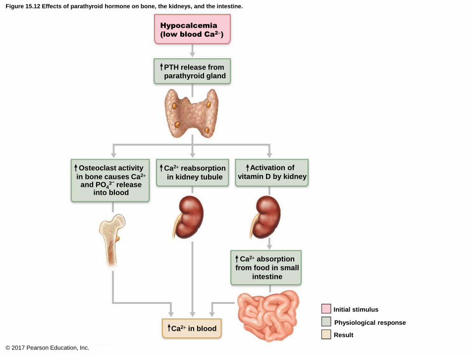
Figure 15.12 Effects of parathyroid hormone on bone, the kidneys, and the intestine.
© 2017 Pearson Education, Inc.
Hypocalcemia
(low blood Ca2+)
PTH release fromparathyroid gland
Osteoclast activity
in bone causes Ca2+
and PO43–
releaseinto blood
Ca2+ reabsorption
in kidney tubule
Activation of
vitamin D by kidney
Ca2+ absorption
from food in small
intestine
Ca2+ in blood
Initial stimulus
Physiological response
Result

Clinical – Homeostatic Imbalance 15.5
• Hyperparathyroidism due to parathyroid gland
tumor
– Calcium leaches from bones, causing them to
soften and deform
– Elevated Ca2+ depresses nervous system and
contributes to formation of kidney stones
– Osteitis fibrosa cystica: severe form resulting in
easily fractured bones
© 2017 Pearson Education, Inc.

Clinical – Homeostatic Imbalance 15.5
• Hypoparathyroidism following gland trauma or
removal can cause hypocalcemia
– Results in tetany, respiratory paralysis, and
death
© 2017 Pearson Education, Inc.

15.9 Adrenal Gland
• Paired, pyramid-shaped organs atop kidneys
– Also referred to as suprarenal glands
• Structurally and functionally it is two glands in
one
– Adrenal cortex: three layers of glandular tissue
that synthesize and secrete several different
hormones
– Adrenal medulla: nervous tissue that is part of
sympathetic nervous system
© 2017 Pearson Education, Inc.

Adrenal Cortex
• This area of adrenal gland produces over 24
different hormones collectively called
corticosteroids
• Steroid hormones are not stored in cells
– Rate of release depends on rate of synthesis
• Three layers of cortical cells produce the
different corticosteroids
– Zona glomerulosa—Mineralocorticoids
– Zona fasciculata—Glucocorticoids
– Zona reticularis—Gonadocorticoids
© 2017 Pearson Education, Inc.

Figure 15.13 Microscopic structure of the adrenal gland.
© 2017 Pearson Education, Inc.
Adrenal gland
• Medulla
• Cortex
Kidney
Co
rte
xM
ed
ulla
Capsule
Zonaglomerulosa
Zonafasciculata
Zona
reticularis
Adrenalmedulla
Drawing of the
histology of theadrenal cortex anda portion of theadrenal medulla
Photomicrograph
(115×)
Hormonessecreted
Aldosterone
Cortisolandandrogens
Epinephrine andnorepinephrine

Adrenal Cortex (cont.)
• Mineralocorticoids
– Regulate electrolyte concentrations (primarily
Na+ and K+) in ECF
• Importance of Na+: affects ECF volume, blood volume,
blood pressure, and levels of other ions (K+, H+,
HCO3− and Cl−)
• Importance of K+: sets resting membrane potential of
cells
– Aldosterone: most potent mineralocorticoid
• Stimulates Na+ reabsorption by kidneys
– Results in increased blood volume and blood pressure
• Stimulates K+ elimination by kidneys
© 2017 Pearson Education, Inc.

Adrenal Cortex (cont.)
• Mineralocorticoids (cont.)
– Effects of aldosterone are short lived
– Stimulates synthesis and activation of Na+-K+
ATPase transport pumps
• Pump exchanges Na+ for K+
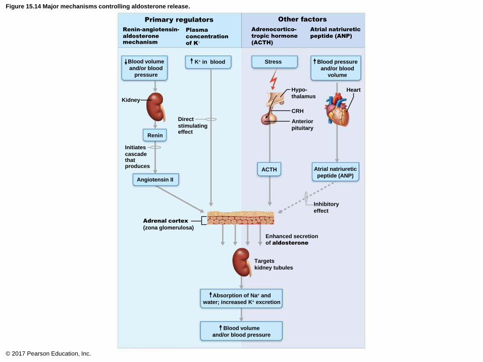
– Factors that regulate aldosterone secretion:
• Renin-angiotensin-aldosterone mechanism
• Plasma concentration of K+
• ACTH
• Atrial natriuretic peptide
© 2017 Pearson Education, Inc.

Adrenal Cortex (cont.)
• Mineralocorticoids (cont.)
– Renin-angiotensin-aldosterone mechanism
1. Decreased blood pressure stimulates special cells in
kidneys
2. These cells release renin into blood
3. Renin cleaves off part of plasma protein,
angiotensinogen, that triggers enzyme cascade,
resulting in conversion to angiotensin II
– Angiotensin II is a potent stimulator of aldosterone
release
© 2017 Pearson Education, Inc.

Adrenal Cortex (cont.)
• Mineralocorticoids (cont.)
– Plasma concentration of K+
• Increased K+ directly influences zona glomerulosa
cells to release aldosterone
– Increased K+ directly stimulates aldosterone release;
low levels inhibit it
– ACTH
• Can cause small increases of aldosterone during
periods of increased stress
– Atrial natriuretic peptide (ANP)
• Secreted by heart in response to high blood pressure
• Blocks renin and aldosterone secretion to decrease
blood pressure© 2017 Pearson Education, Inc.
Direct
stimulatingeffect
Initiates
cascadethatproduces
Figure 15.14 Major mechanisms controlling aldosterone release.
© 2017 Pearson Education, Inc.
Blood volume
and/or blood
pressure
Kidney
K+ in blood
Renin-angiotensin-
aldosteronemechanism
Plasma
concentration
of K+
Primary regulators Other factors
Adrenocortico-
tropic hormone
(ACTH)
Atrial natriuretic
peptide (ANP)
Renin
Stress
Angiotensin II
Blood pressure
and/or blood
volume
Hypo-
thalamusHeart
CRH
Anterior
pituitary
ACTH Atrial natriuretic
peptide (ANP)
Inhibitory
effect
Enhanced secretion
of aldosterone
Targets
kidney tubules
Absorption of Na+ and
water; increased K+ excretion
Blood volume
and/or blood pressure
Adrenal cortex
(zona glomerulosa)

Clinical – Homeostatic Imbalance 15.6
• Aldosteronism: hypersecretion usually due to
adrenal tumors
• Results in two major problems:
1. Hypertension and edema due to excessive Na+
2. Excretion of K+, leading to abnormal
nonresponsive neurons and muscle
© 2017 Pearson Education, Inc.

Adrenal Cortex (cont.)
• Glucocorticoids
– Influence metabolism of most cells and help us
resist stressors
– Keep blood glucose levels relatively constant
– Maintain blood pressure by increasing action of
vasoconstrictors
– Glucocorticoid hormones include:
• Cortisol (hydrocortisone); only glucocorticoid in
significant amounts in humans
• Cortisone
• Corticosterone
© 2017 Pearson Education, Inc.

Adrenal Cortex (cont.)
– Regulation of secretion
• Cortisol is released in response to ACTH
– ACTH released in response to corticotropin-releasing
hormone (CRH)
– CRH released in response to low cortisol levels
– Increased cortisol levels inhibit ACTH and CRH through
negative feedback
• Cortisol secretion cycles are governed by patterns of
eating and activity
• Acute stress (infection, physical or emotional trauma)
interrupts cortisol rhythm
• CNS can override cortisol inhibition of ACTH and
CRH, leading to more cortisol secretion
© 2017 Pearson Education, Inc.

Adrenal Cortex (cont.)
– Actions
• Cortisol causes increase in blood levels of glucose,
fatty acids, and amino acids
• Prime metabolic effect is gluconeogenesis, formation
of glucose from fats and proteins
– Encourages cells to use fatty acids for fuel so glucose
is “saved” for brain
• Other function is to enhance vasoconstriction
– Causes rise in blood pressure to quickly distribute
nutrients to cells
© 2017 Pearson Education, Inc.

Adrenal Cortex (cont.)
– Actions (cont.)
• Excessive levels of glucocorticoids:
– Depress cartilage and bone formation
– Inhibit inflammation by decreasing release of
inflammatory chemicals
– Depress immune system
– Disrupt normal cardiovascular, neural, and
gastrointestinal functions
• Glucocorticoid drugs can control symptoms of many
inflammatory diseases (arthritis, allergies) but can also
cause undesirable effects
© 2017 Pearson Education, Inc.

Clinical – Homeostatic Imbalance 15.7
• Hypersecretion—Cushing’s syndrome/disease
– Depresses cartilage/bone formation and immune
system; inhibits inflammation; disrupts neural,
cardiovascular, and gastrointestinal function
– Causes: tumor on pituitary, lungs, pancreas,
kidney, or adrenal cortex; overuse of
corticosteroids
– Cushingoid signs: “moon” face and “buffalo hump”
– Treatment: removal of tumor, discontinuation of
drugs
© 2017 Pearson Education, Inc.

Clinical – Homeostatic Imbalance 15.7
• Hyposecretion—Addison’s disease
– Also involves deficits in mineralocorticoids
– Decrease in glucose and Na+ levels
– Weight loss, severe dehydration, and
hypotension
– Treatment: corticosteroid replacement therapy
© 2017 Pearson Education, Inc.

Figure 15.15 The effects of excess glucocorticoid.
© 2017 Pearson Education, Inc.
Patient before onset Same patient with Cushing’s
syndrome. The white arrow shows the characteristic “buffalo hump” of fat on theupper back.

Adrenal Cortex (cont.)
• Gonadocorticoids (adrenal sex hormone)
– Weak androgens (male sex hormones) converted
to testosterone in tissue cells, some to estrogens
• Example: androstenedione and
dehydroepiandrosterone (DHEA)
– May contribute to:
• Onset of puberty and appearance of secondary sex
characteristics
• Sex drive in women
• Source of estrogens in postmenopausal women
© 2017 Pearson Education, Inc.

Clinical – Homeostatic Imbalance 15.8
• Hypersecretion
– Adrenogenital syndrome (masculinization)
– Not noticeable in adult males
• Already masculinized with testosterone, so no effect
– Females and prepubertal males
• Boys: reproductive organs mature; secondary sex
characteristics emerge early
• Females: beard, masculine pattern of body hair;
clitoris resembles small penis
© 2017 Pearson Education, Inc.

Adrenal Medulla
• Medullary chromaffin cells synthesize
catecholamines epinephrine (80%) and
norepinephrine (20%)
• Effects of catecholamines:
– Vasoconstriction
– Increased heart rate
– Increased blood glucose levels
– Blood diverted to brain, heart, and skeletal
muscle
© 2017 Pearson Education, Inc.

Adrenal Medulla (cont.)
• Both hormones have basically same effects,
but:
– Epinephrine is more a stimulator of metabolic
activities
• Example: bronchial dilation, and blood flow to skeletal
muscles and heart
– Norepinephrine has more of an influence on
peripheral vasoconstriction and blood pressure
• Responses to stressors are brief, unlike adrenal
cortical hormones
© 2017 Pearson Education, Inc.

Figure 15.16 Stress and the adrenal gland.
Short-term stress Prolonged stress
Nerve impulses
Spinal cord
Preganglionic
sympatheticfibers
Adrenal medulla
(secretes amino acid–based hormones)
Norepinephrine
and epinephrine (catecholamines)
Short-term stress response
• Heart rate increases
• Blood pressure increases
• Bronchioles dilate• Liver converts glycogen to glucose and releases
glucose to blood• Blood flow changes, reducing digestive system activity
and urine output• Metabolic rate increases
Stress
Hypothalamus
CRH (corticotropin-
releasing hormone)
Corticotropic cells
of anterior pituitary
To target via blood
Adrenal cortex(secretes steroidhormones)
ACTH
Mineralocorticoids Glucocorticoids
Long-term stress response
• Kidneys retain
sodium and water• Blood volume and
blood pressurerise
• Proteins and fats converted
to glucose or broken down
for energy• Blood glucose increases• Immune system
supressed
© 2017 Pearson Education, Inc.

Clinical – Homeostatic Imbalance 15.9
• Hyposecretion
– Epinephrine and norepinephrine are not
essential to life; therefore there are no problems
associated with hyposecretion
• Hypersecretion
– Leads to symptoms of uncontrolled sympathetic
nervous system, such as:
• Hyperglycemia, increased metabolic rate, rapid
heartbeat, palpitations, hypertension, intense
nervousness, and sweating
– Can be due to pheochromocytoma, tumor of
medullary chromaffin cells© 2017 Pearson Education, Inc.

Table 15.4 Adrenal Gland Hormones: Summary of Regulation and Effects
© 2017 Pearson Education, Inc.

15.10 Pineal Gland
• Small gland hanging from roof of third ventricle
• Pinealocytes secrete melatonin, derived from
serotonin
• Melatonin may affect:
– Timing of sexual maturation and puberty
– Day/night cycles
– Physiological processes that show rhythmic
variations (body temperature, sleep, appetite)
– Production of antioxidant and detoxification
molecules in cells
© 2017 Pearson Education, Inc.

Figure 15.1 Location of selected endocrine organs of the body.
© 2017 Pearson Education, Inc.
Pineal gland
Hypothalamus
Pituitary gland
Thyroid gland
Parathyroid glands
(on dorsal aspect of thyroid gland)
Thymus
Adrenal glands
Pancreas
Gonads
• Ovary (female)
• Testis (male)
15.11 Other Endocrine Organs
Pancreas
• Triangular gland located partially behind stomach
• Has both exocrine and endocrine cells
– Acinar cells (exocrine) produce enzyme-rich
juice for digestion
– Pancreatic islets (islets of Langerhans) contain
endocrine cells
• Alpha () cells produce glucagon (hyperglycemic
hormone)
• Beta () cells produce insulin (hypoglycemic
hormone)
© 2017 Pearson Education, Inc.

Figure 15.17 Photomicrograph of differentially stained pancreatic tissue.
© 2017 Pearson Education, Inc.
Pancreatic islet
• (Glucagon-
producing)cells
• (Insulin-
producing) cells
Pancreatic acinar
cells (exocrine)

15.11 Other Endocrine Organs
• Glucagon
– Extremely potent hyperglycemic agent
• Triggered by decreased blood glucose levels, rising
amino acid levels, or sympathetic nervous system
– Raises blood glucose levels by targeting liver to:
• Break down glycogen into glucose
– Glycogenolysis
• Synthesize glucose from lactic acid and other
noncarbohydrates
– Gluconeogenesis
• Release glucose into blood
© 2017 Pearson Education, Inc.

15.11 Other Endocrine Organs
• Insulin
– Secreted when blood glucose levels increase
– Synthesized as proinsulin that is then modified
– Insulin lowers blood glucose levels in three
ways:
• Enhances membrane transport of glucose into fat and
muscle cells
• Inhibits breakdown of glycogen to glucose
• Inhibits conversion of amino acids or fats to glucose
© 2017 Pearson Education, Inc.

15.11 Other Endocrine Organs
• Insulin (cont.)
– Not needed for glucose uptake in liver, kidney, or
brain
– Plays a role in neuronal development, learning,
and memory
– Binding to tyrosine kinase enzyme receptor
triggers cell to increase glucose uptake
– Insulin also triggers cells to:
• Catalyze oxidation of glucose for ATP production: first
priority
• Polymerize glucose to form glycogen
• Convert glucose to fat (particularly in adipose tissue)© 2017 Pearson Education, Inc.

15.11 Other Endocrine Organs
• Insulin (cont.)
– Factors that influence insulin release
• Elevated blood glucose levels: primary stimulus
• Rising blood levels of amino acids and fatty acids
• Release of acetylcholine by parasympathetic nerve
fibers
• Hormones glucagon, epinephrine, growth hormone,
thyroxine, glucocorticoids
• Somatostatin and sympathetic nervous system inhibit
insulin release
© 2017 Pearson Education, Inc.

Figure 15.18 Insulin and glucagon from the pancreas regulate blood glucose levels.
© 2017 Pearson Education, Inc.
Stimulates glucose
uptake by cells
Tissue cellsInsulin
Stimulates
glycogen
formation
Blood
glucosefalls tonormalrange.
Pancreas Glucose Glycogen
Liver
StimulusBlood
glucose level BALANCE:
Blood
glucose
rises tonormalrange.
StimulusBlood
glucose level
Pancreas
Glucose Glycogen
LiverStimulates
glycogen
breakdown
Glucagon

Clinical – Homeostatic Imbalance 15.10
• Diabetes mellitus (DM) can be due to:
– Hyposecretion of insulin: Type 1
– Hypoactivity of insulin: Type 2
– When blood glucose levels remain high, person
feels nauseated, leading to sympathetic
response
• Fight-or-flight response acts to further increase blood
glucose levels
– Glycosuria: excess glucose is spilled into urine
© 2017 Pearson Education, Inc.

Clinical – Homeostatic Imbalance 15.10
• Three cardinal signs of DM:
– Polyuria: huge urine output
• Glucose acts as osmotic diuretic
– Polydipsia: excessive thirst
• From water loss due to polyuria
– Polyphagia: excessive hunger and food
consumption
• Cells cannot take up glucose and are “starving”
© 2017 Pearson Education, Inc.

Clinical – Homeostatic Imbalance 15.10
– When sugars cannot be used as fuel, as in DM,
fats are used, causing lipidemia: high levels of
fatty acids in blood
– Fatty acid metabolism results in formation of
ketones (ketone bodies)
– Ketones are acidic, and their build-up in blood
can cause ketoacidosis
• Also causes ketonuria: ketone bodies in urine
– Untreated ketoacidosis causes hyperpnea,
disrupted heart activity and O2 transport, and
severe depression of nervous system that can
possibly lead to coma and death © 2017 Pearson Education, Inc.

Clinical – Homeostatic Imbalance 15.10
• Hyperinsulinism
– Excessive insulin secretion
– Causes hypoglycemia: low blood glucose levels
– Symptoms: anxiety, nervousness, disorientation,
unconsciousness, even death
– Treatment: sugar ingestion
© 2017 Pearson Education, Inc.

Blood pH due to ketone
bodies (ketoacidosis)
Figure 15.19 Consequences of insulin deficit (diabetes mellitus).
© 2017 Pearson Education, Inc.
Insulin
All tissues Liver breaks down
glycogen to glucose(gluconeogenesis)
Skeletal muscle breaks
down proteins
Adipocytes break down fat (lipolysis)
Liver converts fats to ketone bodies
Liver converts amino
acids to glucose
Glucose uptake
(and usage)
Blood glucose
(hyperglycemia)
Blood
Urine Glucose in urine (glycosuria)
Glucose “pulls” water
into kidney tubules
Osmotic diuresis
Signs and symptomsPolyuria
( Urine output)
Dehydration
Polydipsia
( Water intake)
Ketones in urine (ketonuria)
Ketones “pull” cations
into kidney tubules
Loss of Na+, K+, H+ in urine
Polyphagia
( Appetite)
• Heart rhythm
abnormalities
• Nausea, vomiting,
abdominal pain
• Central nervous
system depression,
coma
• Acetone breath
• Rate and depth
of breathing

BioFlix Video: Homeostasis
© 2017 Pearson Education, Inc.

The Gonads and Placenta
• Gonads produce same steroid sex hormones as
those of adrenal cortex, just lesser amounts
• Ovaries produce estrogens and progesterone
– Estrogen
• Maturation of reproductive organs
• Appearance of secondary sexual characteristics
• With progesterone, causes breast development and
cyclic changes in uterine mucosa
© 2017 Pearson Education, Inc.

The Gonads and Placenta (cont.)
• Testes produce testosterone
– Initiates maturation of male reproductive organs
– Causes appearance of male secondary sexual
characteristics and sex drive
– Necessary for normal sperm production
– Maintains reproductive organs in functional state
• Placenta secretes estrogens, progesterone,
and human chorionic gonadotropin (hCG)
© 2017 Pearson Education, Inc.
Hormone Secretion by Other Organs
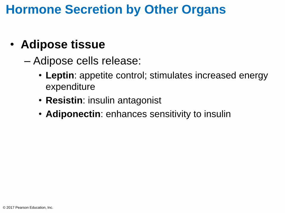
• Adipose tissue
– Adipose cells release:
• Leptin: appetite control; stimulates increased energy
expenditure
• Resistin: insulin antagonist
• Adiponectin: enhances sensitivity to insulin
© 2017 Pearson Education, Inc.

Hormone Secretion by Other Organs (cont.)
• Gastrointestinal tract
– Enteroendocrine cells secrete these hormones:
• Gastrin stimulates release of HCl
• Ghrelin from stomach stimulates food intake
• Secretin stimulates liver and pancreas
• Cholecystokinin (CCK) activates pancreas,
gallbladder, and hepatopancreatic sphincter
• Incretins enhance insulin release and inhibit
glucagon
© 2017 Pearson Education, Inc.

Hormone Secretion by Other Organs (cont.)
• Heart
– Atrial natriuretic peptide (ANP) decreases
blood Na+ concentration, therefore blood
pressure and blood volume
• Kidneys
– Erythropoietin signals production of red blood
cells
– Renin initiates the renin-angiotensin-aldosterone
mechanism
© 2017 Pearson Education, Inc.

Hormone Secretion by Other Organs (cont.)
• Skeleton
– Osteoblasts in bone secrete osteocalcin
• Prods pancreas to secrete more insulin; restricts fat
storage; improves glucose handling; reduces body fat
• Activated by insulin
• Low levels of osteocalcin are present in type 2
diabetes: perhaps increasing levels may be new
treatment
© 2017 Pearson Education, Inc.

Hormone Secretion by Other Organs (cont.)
• Skin
– Cholecalciferol, precursor of vitamin D
– Calcitriol: active form of vitamin D that helps
absorb calcium from intestine
– Also modulates immunity, decreases
inflammation, and may act as anticancer agent
© 2017 Pearson Education, Inc.

Hormone Secretion by Other Organs (cont.)
• Thymus
– Large in infants and children; shrinks with age
– Thymulin, thymopoietins, and thymosins may
be involved in normal development of T
lymphocytes in immune response
• Classified as hormones but act as paracrines
© 2017 Pearson Education, Inc.
Related Documents


![Adrenal Imaging - University of Floridaxray.ufl.edu/files/2010/02/Adrenal-Imaging.pdfadrenal glands [3], and a metastasis might ... CT, adrenal imaging, adrenal lymphoma imaging, adrenal](https://static.cupdf.com/doc/110x72/5b26814c7f8b9a8c0f8b4820/adrenal-imaging-university-of-glands-3-and-a-metastasis-might-ct-adrenal.jpg)