The effects of neurotensin and epidermal growth factor upon ERK-, Akt- and EGFR-phosphorylation in cells from the pancreatic adenocarcinoma cell line Panc-1 Ulrich Mathias Eide 11th semester assignment at the Faculty of Medicine University of Oslo 15th October 2011 Supervisor: Professor Dagny L. Sandnes

Welcome message from author
This document is posted to help you gain knowledge. Please leave a comment to let me know what you think about it! Share it to your friends and learn new things together.
Transcript
The effects of neurotensin and epidermal growth factor upon ERK-, Akt- and EGFR-phosphorylation in cells from the pancreatic
adenocarcinoma cell line Panc-1
Ulrich Mathias Eide
11th semester assignment at the Faculty of Medicine
University of Oslo
15th October 2011
Supervisor: Professor Dagny L. Sandnes

ABSTRACTIn this paper we have used western blotting to study the phosphorylation of the intracellular enzymes ERK (MAPK) and Akt as well as the EGF receptor in response to stimulation with neurotensin (NT) and epidermal growth factor (EGF). Both these substances activate signaling cascades frequently overactive in cancer. Cells from the Panc-1 cell line, which is derived from a human ductal pancreatic carcinoma, have been used in our experiments. Previous studies have investigated ERK activation in these cells, but little is reported about Akt (PKB) phosphorylation in response to NT stimulation. We wanted to see whether or not NT induced transactivation of the EGF receptor and we also studied the effect of preincubation with different inhibitors in order to elucidate by which mechanisms EGF- and NT-induced phosphorylation occur. Finally, we stimulated our cells with thapsigargin or tetradecanoyl phorbol acetate (TPA) to investigate the effect of increased intracellular calcium and activation of protein kinase C (PKC), respectively. Results: EGF and NT stimulated ERK- and Akt-phosphorylation in a dose dependent fashion. EGF also stimulated EGFR phosphorylation dose dependently. We detected no NT-induced transactivation of EGFR. NT- and EGF-induced Akt activation was not mediated through PKC or increased intracellular calcium. NT-induced ERK activation seemed to be PKC-dependent, whereas EGF- induced ERK activation was reduced by 50% when inhibiting PKC.
INTRODUCTIONBackgroundPancreatic cancer, of which 90% are ductal adenocarcinomas, has a 5 year survival rate of 5% when all stages are included [1][2]. This, together with its estimated incidence of about 36.000 new cases pr year in the US only, makes it the fourth leading cause of cancer death in both men and women [2]. With these grave statistics at hand, the hope is that novel therapeutic strategies can arise from a better understanding of the signaling pathways that stimulate the lethal proliferation and migration of ductal pancreatic cancer cells [1][3]. Unfortunately, the last 20 years of research in the field has not contributed to any significant improvement in clinical treatment or early detection of pancreatic cancer, although the knowledge of cellular mechanisms present in this cancer undoubtedly has increased [2][4] .
Receptor tyrosine kinasesReceptor tyrosine kinases (RTKs) are transmembrane glycoproteins with a cytoplasmatic domain that harbors tyrosine kinase activity. Binding of appropriate ligands, such as EGF, in the extracellular domain allows EGFR dimerization, which induces cross-phosphorylation and thereby activation. Activated RTKs recruit different protein complexes to the membrane which initiates different signaling cascades. Three of these will briefly be mentioned (Fig. 1):
Recruited phosphoinositide 3-kinase (PI3K) phosphorylates phosphatidylinositol-(4,5)-bisphosphate (PIP2) producing phosphatidylinositol-(3,4,5)-trisphosphate (PIP3). The available PIP3
acts as a binding site for Akt, which upon binding reveals its two amino residues for phosphorylation. Once phosphorylated, Akt acts as a kinase for many downstream proteins and is thereby involved in regulating protein synthesis, cell survival, proliferation and metabolism.[5][6]. A major regulator of this signaling pathway is PTEN, a phosphatase that reduces the available PIP3
by converting it back to PIP2. Not surprisingly, loss of PTEN function contributes to cancer development [6].
The Ras/Raf/MEK/ERK pathway is another downstream pathway of RTK activation. Activated RTKs recruit the Grb2-son of sevenless (SOS) complex, which loads membrane bound Ras with
1
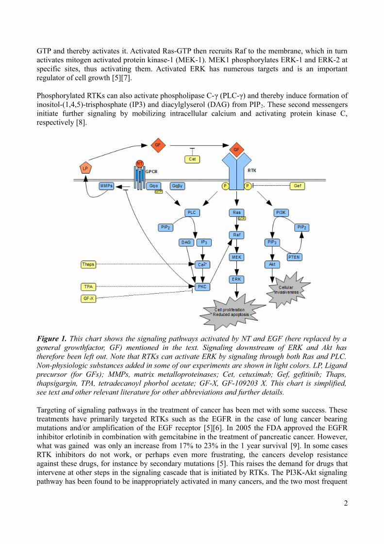
GTP and thereby activates it. Activated Ras-GTP then recruits Raf to the membrane, which in turn activates mitogen activated protein kinase-1 (MEK-1). MEK1 phosphorylates ERK-1 and ERK-2 at specific sites, thus activating them. Activated ERK has numerous targets and is an important regulator of cell growth [5][7].
Phosphorylated RTKs can also activate phospholipase C-γ (PLC-γ) and thereby induce formation of inositol-(1,4,5)-trisphosphate (IP3) and diacylglyserol (DAG) from PIP2. These second messengers initiate further signaling by mobilizing intracellular calcium and activating protein kinase C, respectively [8].
Targeting of signaling pathways in the treatment of cancer has been met with some success. These treatments have primarily targeted RTKs such as the EGFR in the case of lung cancer bearing mutations and/or amplification of the EGF receptor [5][6]. In 2005 the FDA approved the EGFR inhibitor erlotinib in combination with gemcitabine in the treatment of pancreatic cancer. However, what was gained was only an increase from 17% to 23% in the 1 year survival [9]. In some cases RTK inhibitors do not work, or perhaps even more frustrating, the cancers develop resistance against these drugs, for instance by secondary mutations [5]. This raises the demand for drugs that intervene at other steps in the signaling cascade that is initiated by RTKs. The PI3K-Akt signaling pathway has been found to be inappropriately activated in many cancers, and the two most frequent
2
Figure 1. This chart shows the signaling pathways activated by NT and EGF (here replaced by a general growthfactor, GF) mentioned in the text. Signaling downstream of ERK and Akt has therefore been left out. Note that RTKs can activate ERK by signaling through both Ras and PLC. Non-physiologic substances added in some of our experiments are shown in light colors. LP, Ligand precursor (for GFs); MMPs, matrix metalloproteinases; Cet, cetuximab; Gef, gefitinib; Thaps, thapsigargin, TPA, tetradecanoyl phorbol acetate; GF-X, GF-109203 X. This chart is simplified, see text and other relevant literature for other abbreviations and further details.

reasons for this is in fact RTK activation and somatic mutations in specific components of this signaling pathway [5][6]. It has even been demonstrated that for a RTK inhibitor to be effective in treatment, it must down-regulate PI3K signaling [6]. This has spurred the development of numerous drugs targeting components of the PI3K pathway. As for our Panc-1 cells, these cells have been shown to harbour elevated expression of Akt2 and KRas [10]. The former is a part of the PI3K signaling pathway, and a promoter of cellular invasiveness and mesenchymal characteristics, whereas the latter is part of the important Ras/Raf/MEK/ERK pathway shown to promote proliferation and avoidance of apoptosis when activated [5][6][11]. Activated KRas is among the most common genetic alterations in pancreatic cancer, but all attempts to directly block mutated KRas function have failed so far [4][12]. The mutated KRas does not seem to be sufficient to promote constitutive activation of the ERK pathway in Panc-1 cells, although there seems to be some disagreement concerning this [3][13].
Epidermal Growth FactorEGF is a 6 kDa protein discovered in 1960. It is a signaling molecule that stimulates the growth of epithelial tissues during development and throughout life. Among many other human epithelial cancers, pancreatic cancer cells have been reported to overexpress certain members of the EGFR family, and even secrete EGFR ligands including TGF-ɑ and EGF itself [1][13]. This implies that there might be an autocrine loop of selfstimulation causing and maintaining a malignant phenotype [1]. Indeed, one study found a direct linear correlation between sensitivity to an EGF inhibitor (gefitinib) and TGF-ɑ production when looking at 9 different human pancreatic cancer cell lines [13].
G protein-coupled receptorsG protein-coupled receptors (GPCRs) are structurally characterised by their 7 transmembrane alpha helices. They relay a wide range of signals from a large number of agonists, and these receptors are in fact the largest group of cell surface proteins involved in signal transduction [14][15]. This position in physiology has made GCPRs the target of more than half of all currently available drugs [15]. Heterotrimeric G proteins consist of α, β and γ subunits. Upon binding of an extracellular agonist, the GPCRs α-helices move, causing a conformational change in the cytoplasmatic domain of the receptor. This leads to the exchange of GDP for GTP in the α subunit of the G protein. This exchange causes conformational changes in the G protein, activating the Gα-GTP subunit and revealing effector interaction sites in the βγ heterodimer. These activated subunits can then relay signals in different ways. Inactivation happens partially by GTP hydrolysis by the Gα subunit and partially by acute homologus desensitization initiated by GPCR kinases (GRKs) [14][15]. G proteins are classified into four groups according to their specific α subunit: G s, Gi Gq and G12 [14] [15]. In general, the Gq family controls the activity of phosphatidylinositol-specific phospholipases such as phospholipase C-β (PLC-β), whereas the PLC-γ is regulated by tyrosine kinases, although there probably is some overlap [8]. Activated PLC catalyzes the hydrolysis of PIP2 to produce the two second messengers IP3 and DAG which, as previously mentioned, mobilize intracellular Ca2+
and protein kinase C (PKC) respectively (Fig. 1) [3]. NT is one of many GPCR agonists capable of activating Gq in this manner [16].
TransactivationAn important signaling pathway present in many cells is GPCR-induced phosphorylation of the EGFR receptor (transactivation), which causes Ras-dependent ERK activation [14]. In many cases this transactivation is mediated by the release of ligand precursors mediated by metalloproteinases that are activated by GPCRs [14]. This was also demonstrated in Panc-1 cells where it also turned
3

out that transactivation is strongly inhibited by PKC, thereby explaining earlier failed attempts to provide evidence of this mechanism (Fig. 1) [3][17].
NeurotensinNT, a 13 amino acid peptide discovered in 1973, acts as a neurotransmitter in the central nervous system and as an endocrine agent in the periphery, being especially abundant in the gastrointestinal tract [18]. NT acts through binding to GPCRs which activate PKC [3]. PKC can then activate ERK through the Ras/Raf/MEK/ERK pathway, although there also seems to exist PKC-independent ERK activation pathways depending on cellular context [14]. Although NT has numerous physiological effects on multiple organs, one major reason for its attention in the scientific community is its ability to act as a potent cellular mitogen for multiple cell types [1][14][18]. Even more interesting is the frequent expression of NT and its receptor in malignant cells. Autoradiography of frozen sections of human pancreatic cancer specimens revealed NT binding sites in about 75% of the cases, compared to no NT receptors in chronic pancreatitis or normal pancreatic tissue [1]. This study included pancreatic acini, ducts and islets. Another study, using immunohistochemistry to look at pancreatic duct specimens only, reported similar figures. In this case 80% of the cancer specimens expressed both NT and its receptor, compared to 20% with weak expression of NT and no expression of the receptor in the controls [19]. It has also been shown that the expression of mRNA for the NT receptor subtype 1 is markedly increased in ductal pancreatic cancer cells compared to normal controls [1]. Finally, it has been found that neuroendocrine cells in intrapancreatic ganglia in both malignant and healthy tissue can produce NT and thus stimulate the overabundant NT receptors present in ductal pancreatic cancer cells [1]. Considering these differences between normal and malignant tissue, the potential value of knowing the NT signaling mechanisms becomes obvious in future attempts to design new drugs against pancreatic cancer.
CHEMICALSDulbecco’s modified Eagle’s medium, penicillin and streptomycin were from Gibco (Grand Island, NY). Neurotensin, 12-O-tetradecanoylphorbol-13-acetate (TPA), thapsigargin, epidermal growth factor (EGF) and Ponceu S solution were obtained from Sigma-Aldrich (St.Louis, MO). [2-[1-(3-dimetylaminopropyl)-1H-indol-3-yl]-maleimide] (GF109203X)) was purchased from Calbiochem (San Diego, CA). 4-Quinazolinamine, N-(3-chloro-4-fluorophenyl)-7-methoxy-6-[3-4-morpholin)propoxy] (gefitinib) was a gift from Astra Zeneca (LondonCheshire, UK), and cetuximab was kindly provided by Merck KgaA (Darmstadt, Germany). Antibodies against phosphorylated Akt (Ser473), dually phosphorylated ERK (Thr202/Tyr204) and phospho-EGF receptor (Tyr1173) were obtained from Cell Signaling Technology (Boston, MA). Anti-ERK antibodies was obtained from Upstate (Billerica, MA). Secondary antibodies were purchased from Bio-Rad Laboratories (Hercules, CA). All other chemicals were of analytical quality. Stock solutions of test compounds were prepared in DMSO (TPA, thapsigargin, gefitinib) or 0.9 % NaCl (neurotensin, GF109203X ). EGF was dissolved in 4 mM HCl. Cetuximab was dissolved in phosphate-buffered saline (PBS). When solutions containing DMSO were used, the final concentration of DMSO was kept as low as possible.
CELL CULTUREPancreatic adenocarcinoma cell line Panc-1 was obtained from ATCC (Manassas, VA). The cells were maintained in Dulbecco’s modified Eagle’s medium containing 4.5 g/l glucose supplemented with 10 % fetal bovine serum, penicillin (67 μg/ml), streptomycin (100 μg/ml) and 4 mM glutamine. Cells were plated onto Costar plastic culture wells (Corning Life Sciences, Acton, MA)
4
at a density of 25 000 cells/cm2. The cultures were kept in 95% air/5% CO2 at 37°C. After 24 hours the medium was replaced with serum-free medium and the cells were cultured for another 24 hours before stimulation with agonists. Cells were stimulated with agonists for 5 minutes, unless otherwise stated. For stimulation, the EGF concentrations ranged from 5 to 10 nM and the NT concentrations ranged from 100 to 1000 nM, unless otherwise stated. When inhibitors were present, these were added 30 minutes before agonist stimulation. The medium was then discarded, the cell layer was washed with 0.9% NaCl, and Laemmli buffer was added. The cells were scraped off with a cell lifter and transferred to Eppendorff tubes. Samples were boiled for 10 minutes.
IMMUNOBLOTTINGAliquots with ~15 000 cells (total cell lysate prepared in Laemmli buffer as described above) were electrophoresed on 10 % (w/v) polyacrylamide gels. This was followed by protein electrotransfer to nitrocellulose membranes. The membranes were stained with Ponceu S solution in order to evaluate protein transfer and guide the cutting of the membrane into three separate parts. These parts were then rinsed with water and incubated for 1 hour in a dry milk blocking solution before immunoblotting with antibodies against phospho-Akt, phospho-ERK1/2 and phospho-EGFR. Immunoreactive bands were visualized with enhanced chemiluminescence using LumiGLO (KPL Protein Research Products, Gaithersburg, MD). Membranes were then stripped by placing them for 5 minutes in 0,5 M NaOH. After rinsing them thoroughly with water, blocking, immunoblotting and visualization were repeated, using antibodies against total ERK or GAPDH in the immunoblotting process.
QUANTATIONChemiluminescence was detected using a CCD camera (SensiCam) from Ultra-Violet Products. The dynamic range was set using in-house software (LabWorks version 4.6.00.0), images copied into a Microsoft word 97-2003 file, and then copied into ImageJ version 1.42q. Quantitation of the bands was made using a fixed width sampling tool. No background-correction was made in ImageJ, as it has been reported to worsen the results [20]. All densitometry results were obtained from analyzing the integral intensity of the middle third of the band, unless otherwise stated. When using LabWorks for quantitation we measured IOD with a fixed lane width as well. In our dose response experiments we arbitrarily set 100% intensity (or max signal) as the intensity from the bands of samples stimulated with the highest NT or EGF concentration. For the inhibition of EGF-induced phosphorylation by gefitinib, 100% is set as the intensity recorded without adding gefitinib. All error bars and uncertainties are presented as ±1 standard deviation.
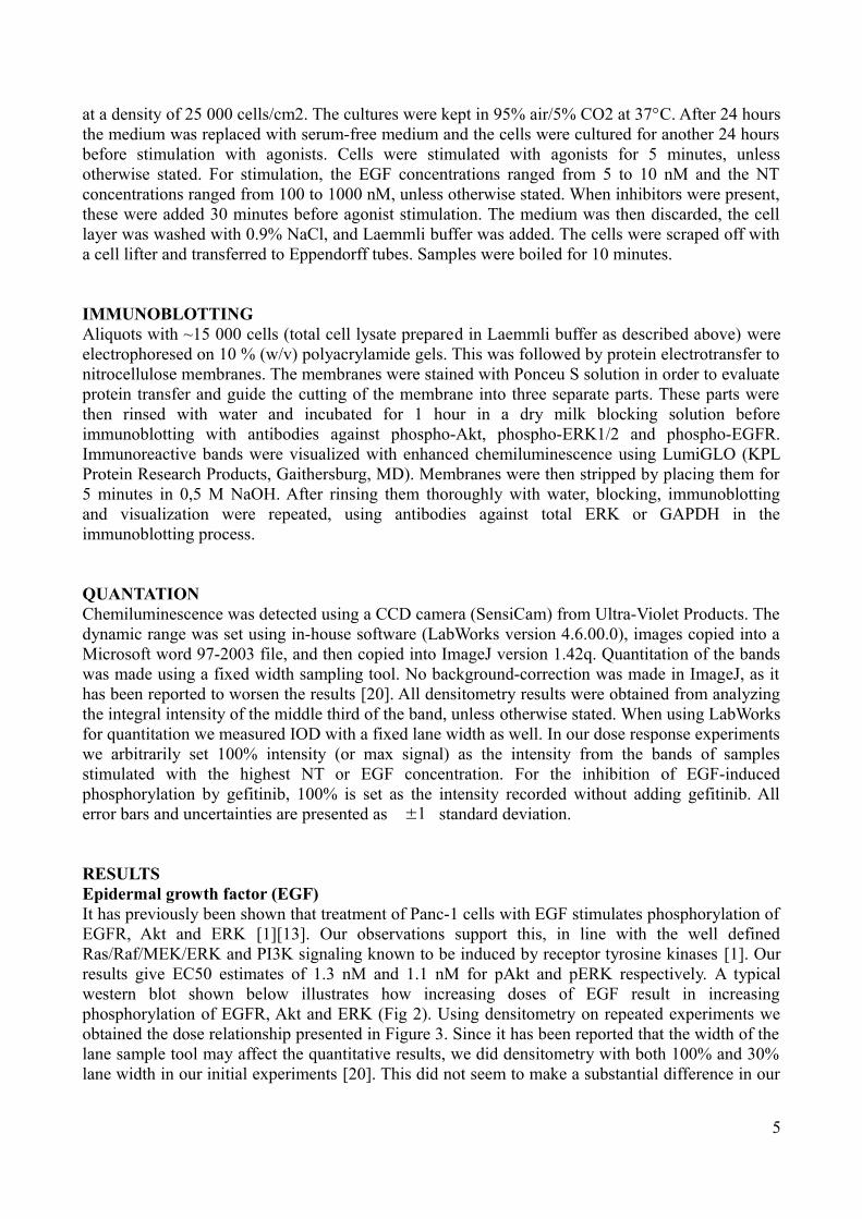
RESULTSEpidermal growth factor (EGF)It has previously been shown that treatment of Panc-1 cells with EGF stimulates phosphorylation of EGFR, Akt and ERK [1][13]. Our observations support this, in line with the well defined Ras/Raf/MEK/ERK and PI3K signaling known to be induced by receptor tyrosine kinases [1]. Our results give EC50 estimates of 1.3 nM and 1.1 nM for pAkt and pERK respectively. A typical western blot shown below illustrates how increasing doses of EGF result in increasing phosphorylation of EGFR, Akt and ERK (Fig 2). Using densitometry on repeated experiments we obtained the dose relationship presented in Figure 3. Since it has been reported that the width of the lane sample tool may affect the quantitative results, we did densitometry with both 100% and 30% lane width in our initial experiments [20]. This did not seem to make a substantial difference in our
5
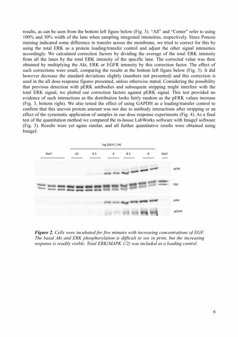
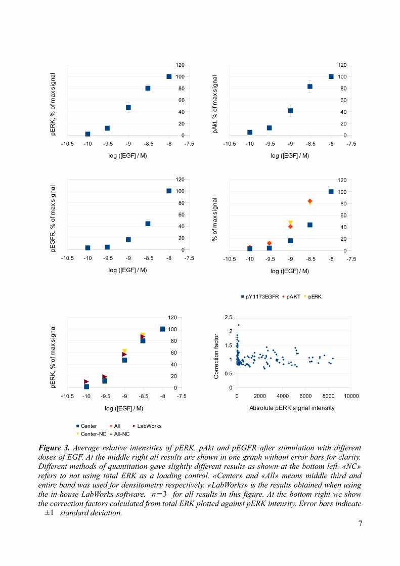
results, as can be seen from the bottom left figure below (Fig. 3). “All” and “Center” refer to using 100% and 30% width of the lane when sampling integrated intensities, respectively. Since Ponceu staining indicated some difference in transfer across the membrane, we tried to correct for this by using the total ERK as a protein loading/transfer control and adjust the other signal intensities accordingly. We calculated correction factors by dividing the average of the total ERK intensity from all the lanes by the total ERK intensity of the specific lane. The corrected value was then obtained by multiplying the Akt, ERK or EGFR intensity by this correction factor. The effect of such corrections were small, comparing the results at the bottom left figure below (Fig. 3). It did however decrease the standard deviations slightly (numbers not presented) and this correction is used in the all dose-response figures presented, unless otherwise stated. Considering the possibility that previous detection with pERK antibodies and subsequent stripping might interfere with the total ERK signal, we plotted our correction factors against pERK signal. This test provided no evidence of such interactions as the distribution looks fairly random as the pERK values increase (Fig. 3, bottom right). We also tested the effect of using GAPDH as a loading/transfer control to confirm that this uneven protein amount was not due to antibody interactions after stripping or an effect of the systematic application of samples in our dose response experiments (Fig. 4). As a final test of the quantitation method we compared the in-house LabWorks software with ImageJ software (Fig. 3). Results were yet again similar, and all further quantitative results were obtained using ImageJ.
6
Figure 2. Cells were incubated for five minutes with increasing concentrations of EGF. The basal Akt and ERK phosphorylation is difficult to see in print, but the increasing response is readily visible. Total ERK(MAPK 1/2) was included as a loading control.
pERK
ERK
pAkt
pEGFR
NaCl -10 -9.5 -9 -8.5 -8 NaCl
log ([EGF] / M)
7
Figure 3. Average relative intensities of pERK, pAkt and pEGFR after stimulation with different doses of EGF. At the middle right all results are shown in one graph without error bars for clarity. Different methods of quantitation gave slightly different results as shown at the bottom left. «NC» refers to not using total ERK as a loading control. «Center» and «All» means middle third and entire band was used for densitometry respectively. «LabWorks» is the results obtained when using the in-house LabWorks software. n=3 for all results in this figure. At the bottom right we show the correction factors calculated from total ERK plotted against pERK intensity. Error bars indicate ±1 standard deviation.
-10.5 -10 -9.5 -9 -8.5 -8 -7.50
20
40
60
80
100
120
log ([EGF] / M)
pER
K, %
of m
ax s
igna
l
-10.5 -10 -9.5 -9 -8.5 -8 -7.50
20
40
60
80
100
120
Center All LabWorksCenter-NC All-NC
log ([EGF] / M)
pER
K, %
of m
ax s
igna
l
-10.5 -10 -9.5 -9 -8.5 -8 -7.50
20
40
60
80
100
120
log ([EGF] / M)
pAkt
, % o
f max
sig
nal
-10.5 -10 -9.5 -9 -8.5 -8 -7.50
20
40
60
80
100
120
log ([EGF] / M)
pEG
FR, %
of m
ax s
igna
l
-10.5 -10 -9.5 -9 -8.5 -8 -7.50
20
40
60
80
100
120
pY1173EGFR pAKT pERK
log ([EGF] / M)
% o
f max
sig
nal
0 2000 4000 6000 8000 100000
0.5
1
1.5
2
2.5
Absolute pERK signal intensity
Cor
rect
ion
fact
or
8
Figure 4. There seemed to be a slight difference in transfer efficiency across the membrane, with the center lanes getting better transfer. To the left we show the correction factors estimated in our EGF dose response curves using total ERK as a tool to estimate total protein amount, n=3 . To the right we show correction factors estimated from different blots using GAPDH instead of total ERK, n=6 . These blots were not from dose-response experiments. The results are similar, making this effect unlikely to be due to antibody interactions after stripping or an effect of the systematically increasing EGF doses in the case of the EGF dose response curves. Error bars indicate ±1 standard deviation.
1 2 3 4 5 6 7 8 9 10 11 12 13 140
0.4
0.8
1.2
1.6
Lane number
Aver
age
ER
K c
orre
ctio
n fa
ctor
1 2 3 4 5 6 7 8 9 10 11 12 13 140
0.4
0.8
1.2
1.6
Lane Number
Aver
age
GAP
DH
cor
rect
ion
fact
or

Neurotensin (NT)It has previously been shown that NT induces a rapid and dose-dependent ERK phosphorylation in Panc-1 cells [21]. This activation was later shown to be PKC-dependent, and not only did NT induce phosphorylation, but it also induced translocation of the enzyme to the nucleus [3]. Our results indicate a dose-dependent ERK response with an estimated EC50 of 1.8 nM. A typical western blot is shown below (Fig. 5). Although the standard deviations from the densitometry indicate no significant difference from 5-1000 nM, there seems to be a steadily increasing response from 1-100 nM as illustrated in the averaged results (Fig. 6). There was some variation in behavior reaching 100 nM, the peak at 10 nM in the presented blot was not typical. Although not as convincing as for ERK, Akt phosphorylation does seem to be dose-dependent as well, with a stable response in the range 10-1000 nM NT (Fig. 6). The estimated EC50 of 0.65 nM is of questionable value as the basal phosphorylation is about 40% of what NT maximally induced. We did not observe any EGFR phosphorylation.
9
Figure 6. NT seems to induce ERK and Akt phosphorylation in a dose-dependent fashion. Samples without added NT is arbitarily set at 0.01 nM for comparison to the basal activity. The data point at 0.3 nM NT was obtained from one single experiment. Error bars indicate ±1 standard deviation,
n=4 .
Figure 5. Cells were incubated for five minutes with increasing concentrations of NT. NT stimulated ERK and Akt phosphorylation. No bands were identified for pEGFR and this blot is therefore not shown. The ERK phosphorylation is convincingly dose dependent whereas the same can not be said for Akt. This blot shows a somewhat untypical reduction in phosphorylation at 100 and 1000 nM NT.
pERK
ERK
pAkt
NaCl -9 -9.5 -8 -7 -6
log ([NT] / M)
-12 -11 -10 -9 -8 -7 -6 -50
20
40
60
80
100
120
log ([NT] / M)
pER
K, %
of m
ax s
igna
l
-12 -11 -10 -9 -8 -7 -6 -50
20
40
60
80
100
120
log ([NT] / M)
pAkt
, % o
f max
sig
nal

Effect of inhibitorsThe effect of three different inhibiting substances were examined in our experiments, one PKC inhibitor, GF 109203X (from now on referred to as GF), and two EGFR antagonists cetuximab and gefitinib (gef) [22][23]. Cetuximab and gefitinib work by preventing ligand binding, and inhibiting the receptor tyrosine kinase by competitively replacing ATP, respectively [23]. GF inhibits PKC by competitively replacing ATP [22]. All inhibitors were added 30 minutes before agonist stimulation.
Effect of inhibitors combined with EGFPreincubation with 3.5 μM GF consistently reduced the EGF-induced ERK phosphorylation in our experiments (Fig. 7). The effect upon Akt and EGFR was somewhat less obvious. We calculated intensity ratios by dividing the band intensity from samples preincubated with GF (+GF) by the band intensity from samples not preincubated with GF (-GF). For pAkt the intensity ratio +GF/-GF was 1.3±0.5 whereas for pERK the result was 0.5±0.1 . In other words, there was no reliable effect upon pAkt, but a marked attenuation of pERK. For EGFR the ratio was 1.0±0.8 . There seemed to be a small effect on basal pAkt on adding GF, but the results varied and quantitation revealed an insignificant +GF/-GF ratio for the controls of 1.6±0.8 .
10
Figure 7. EGF-induced ERK phosphorylation was slightly inhibited by 30 minutes of preincubation with 3.5 μM GF. Although not readily visible in this figure, the quantitation of this blot actually gives an intensity ratio for +GF/-GF of 0.75 when not corrected for GAPDH, and 0.66 when corrected. Akt phosphorylation seemed to be slightly increased, whereas no conclusion could be drawn from the EGFR blotts. Since EGF induces such strong Akt phosphorylation it was necessary to overexpose blotts where both EGF and NT effects were studied to see the effect of NT upon Akt. The two pAkt blots shown at the bottom are in fact the same but with different amounts of exposure.
pERK
NaCl EGF NT NaCl EGF NT
NaCl GF
GAPDH
pAkt
pAkt(2)
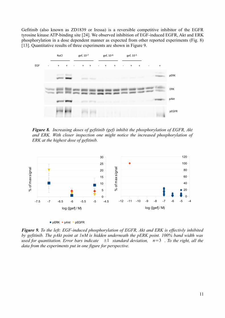
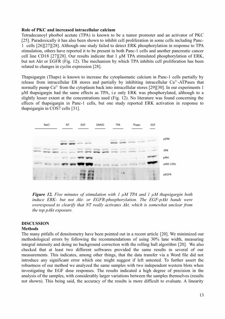
Gefitinib (also known as ZD1839 or Iressa) is a reversible competitive inhibitor of the EGFR tyrosine kinase ATP-binding site [24]. We observed inhibition of EGF-induced EGFR, Akt and ERK phosphorylation in a dose dependent manner as expected from other reported experiments (Fig. 8) [13]. Quantitative results of three experiments are shown in Figure 9.
11
Figure 8. Increasing doses of gefitinib (gef) inhibit the phosphorylation of EGFR, Akt and ERK. With closer inspection one might notice the increased phosphorylation of ERK at the highest dose of gefitinib.
pERK
ERK
pAkt
pEGFR
NaCl gef, 10-7 gef, 10-6 gef, 10-5
EGF - - - - -+ + + + + + + + +
Figure 9. To the left: EGF-induced phosphorylation of EGFR, Akt and ERK is effectivly inhibited by gefitinib. The pAkt point at 1nM is hidden underneath the pERK point. 100% band width was used for quantitation. Error bars indicate ±1 standard deviation, n=3 . To the right, all the data from the experiments put in one figure for perspective.
-12 -11 -10 -9 -8 -7 -6 -5 -40
20
40
60
80
100
120
log ([gef] / M)
% o
f max
sig
nal
-7.5 -7 -6.5 -6 -5.5 -5 -4.50
5
10
15
20
25
30
pERK pAkt pEGFR
log ([gef] / M)
% o
f max
sig
nal

30 minutes of preincubation with 25 μg/mL cetuximab completely abrogated the EGF-induced phosphorylation of EGFR, Akt and ERK in the Panc-1 cells (Fig. 10).
Effect of inhibitors combined with NTPretreatment with 3.5 μM GF almost nullified NT-induced ERK phosphorylation (Fig. 7). This was to be expected, as there already is convincing evidence that NT-induced ERK phosphorylation is strongly PKC-dependent in Panc-1 cells [3]. GF had no major effect on Akt activation, although densitometry revealed a small difference with the intensity ratio for +GF/-GF being 0.93±0.03 .
Pretreatment with 10 μM gefitinib did not affect NT-induced ERK phosphorylation, whereas Akt phosphorylation seemed to be decreased to basal levels (Fig. 11).
Pretreatment with 25 μg/mL cetuximab insignificantly attenuated ERK phosphorylation, +Cet/-Cet ratio 0.8±0.2 , but did not affect Akt phosphorylation (Fig. 10).
12
Figure 10. Thirty minutes of preincubation with 25 μg/mL cetuximab abrogates EGF-induced ERK, Akt and EGFR phosphorylation. It also attenuates NT-induced ERK phosphorylation.
NaCl EGF NT NaCl EGF NT
NaCl Cetuximab
pERK
GAPDH
pAkt
pEGFR
Figure 11. Pretreatment with 10 μM gefitinib for 30 minutes has no effect upon NT-induced ERK phosphorylation but does reduce Akt phosphorylation to a basal level. Upon closer inspection one might notice the slightly increased ERK phosphorylation in the NaCl samples pretreated with gefininib.
NaCl NaCl
gef gef
pERK
NT NT
pAkt
GAPDH
NaCl
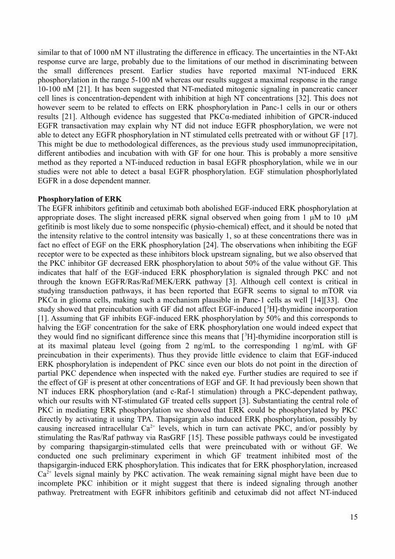
Role of PKC and increased intracellular calciumTetradecanoyl phorbol acetate (TPA) is known to be a tumor promoter and an activator of PKC [25]. Paradoxically it has also been shown to inhibit cell proliferation in some cells including Panc-1 cells [26][27][28]. Although one study failed to detect ERK phosphorylation in response to TPA stimulation, others have reported it to be present in both Panc-1 cells and another pancreatic cancer cell line CD18 [27][28]. Our results indicate that 1 μM TPA stimulated phosphorylation of ERK, but not Akt or EGFR (Fig. 12). The mechanism by which TPA inhibits cell proliferation has been related to changes in cyclin expression [28].
Thapsigargin (Thaps) is known to increase the cytoplasmatic calcium in Panc-1 cells partially by release from intracellular ER stores and partially by inhibiting intracellular Ca2+-ATPases that normally pump Ca2+ from the cytoplasm back into intracellular stores [29][30]. In our experiments 1 μM thapsigargin had the same effects as TPA, i.e only ERK was phosphorylated, although to a slightly lesser extent at the concentrations used (Fig. 12). No literature was found concerning the effects of thapsigargin in Panc-1 cells, but one study reported ERK activation in response to thapsigargin in COS7 cells [31].
DISCUSSIONMethodsThe many pitfalls of densitometry have been pointed out in a recent article [20]. We minimized our methodological errors by following the recommendations of using 30% lane width, measuring integral intensity and doing no background correction with the rolling ball algorithm [20]. We also checked that at least two different softwares provided the same results in several of our measurements. This indicates, among other things, that the data transfer via a Word file did not introduce any significant error which one might suggest if left untested. To further assert the robustness of our method we analyzed the same samples with two independent western blots when investigating the EGF dose responses. The results indicated a high degree of precision in the analysis of the samples, with considerably larger variations between the samples themselves (results not shown). This being said, the accuracy of the results is more difficult to evaluate. A linearity
13
Figure 12. Five minutes of stimulation with 1 μM TPA and 1 μM thapsigargin both induce ERK- but not Akt- or EGFR-phosphorylation. The EGF-pAkt bands were overexposed to clearify that NT really activates Akt, which is somewhat unclear from the top pAkt exposure.
NaCl NT EGF DMSO TPA Thaps
pERK
ERK
pAkt
pAkt-120s
pEGFR
EGF

between chemiluminescence signal and amount of phosphorylation is something that has been assumed, but is not necessarily true. In determining the dose response curves the membranes were exposed once. It might have been better to expose for long enough time to get readily visible bands at the lowest concentrations, decrease the exposure time when studying the higher concentration and incrementally calculate intensity ratios. The attempted correction for total protein used in the dose-response figures might be claimed improper because of a possible interaction between phospho-ERK and total-ERK antibodies. Such an interaction could produce a systematically decreased or increased total-ERK signal depending on the degree of phosphorylation of ERK, but this did not seem to be the case as illustrated by the distribution of points in the bottom right figure of Figure 3. Finally, we observed the same systematic trend of stronger chemiluminescence signals towards the middle of the blots when measuring GAPDH as we did when measuring total ERK (Fig. 4). The effect might stem from the transferring process as the Ponceu coloring indicated, or it might be caused by other effects such as the angle between the detector and the membrane, or the binding of antibodies.
In our intensity ratio calculations we compared bands in which the samples had been either preincubated with an inhibitor I (+I) or not preincubated with I (-I) before stimulation with NT or EGF, providing an intensity ratio +I/-I. This method does not take into account that the fact that I might have some effect on the intensity by itself and not only by modulating the EGF/NT effect. Such an effect would have been seen in the controls receiving I only, and where suspicion was raised we calculated intensity ratios for the control samples as well. The perhaps most obvious way of comparing intensities, dividing stimulated- by control-intensity, was not a viable option as the controls mostly gave weak signals, and the ratios were thus very sensitive to small changes. This was especially the case when analyzing pERK signals, and keeping it simple we applied the same method when analyzing pAkt signals. The times we did calculate intensity ratios for the controls the large standard deviations rendered the possible trend uninformative. Correction using GAPDH or total ERK did not affect these results considerably. We did however correct our values as our blots were not designed to evaluate quantitative differences. This meant that +I and -I lanes often were separated by some other lanes which could alter the results if not corrected (Fig. 4).
Dose response curvesOur results have shown that both EGF and NT can activate Akt and ERK in a dose-dependent manner. In the case of EGF stimulation, Akt and ERK responses were quite similar, while EGFR phosphorylation required higher amounts of EGF to reach the same degree of relative phosphorylation. This might be interpreted as a high sensitivity/quick response of ERK and Akt phosphorylation in response to the upstream EGFR activation. Although a study measuring incorporation of [3H]-thymidine into DNA showed a plateau in incorporation when the EGF concentration reached about 1 ng/mL, the phosphorylation shows a continuous increasing response up to 10 ng/mL [1]. This indicates that at maximal stimulation of DNA synthesis through ERK/Akt/EGFR signaling, there is still a quite substantial amount of unphosphorylated ERK/Akt/EGFR left. Since we did not seem to reach maximal phosphorylation of the EGFR in our EGF experiments we did not estimate an EC50 value in this case. For ERK the response to NT and EGF both gave strong increases in phosphorylation at concentrations of 10 nM for both substances. EGF was slightly more potent with an EC50 of 1.1 nM compared to 1.8 nM for NT. For Akt however, the response to NT was much weaker than the response to EGF. The increase in signal when stimulated with maximal doses of NT was in fact only about twice the basal signal whereas the increase in signal when stimulated with maximal doses of EGF was about 20 times. These figures do of course depend on the doses of NT and EGF used, but it does not look as if even further increased NT doses would give any increased response. At 0.5 nM EGF, the Akt phosphorylation is
14
similar to that of 1000 nM NT illustrating the difference in efficacy. The uncertainties in the NT-Akt response curve are large, probably due to the limitations of our method in discriminating between the small differences present. Earlier studies have reported maximal NT-induced ERK phosphorylation in the range 5-100 nM whereas our results suggest a maximal response in the range 10-100 nM [21]. It has been suggested that NT-mediated mitogenic signaling in pancreatic cancer cell lines is concentration-dependent with inhibition at high NT concentrations [32]. This does not however seem to be related to effects on ERK phosphorylation in Panc-1 cells in our or others results [21]. Although evidence has suggested that PKCα-mediated inhibition of GPCR-induced EGFR transactivation may explain why NT did not induce EGFR phosphorylation, we were not able to detect any EGFR phosphorylation in NT stimulated cells pretreated with or without GF [17]. This might be due to methodological differences, as the previous study used immunoprecipitation, different antibodies and incubation with with GF for one hour. This is probably a more sensitive method as they reported a NT-induced reduction in basal EGFR phosphorylation, while we in our studies were not able to detect a basal EGFR phosphorylation. EGF stimulation phosphorlylated EGFR in a dose dependent manner.
Phosphorylation of ERKThe EGFR inhibitors gefitinib and cetuximab both abolished EGF-induced ERK phosphorylation at appropriate doses. The slight increased pERK signal observed when going from 1 μM to 10 μM gefitinib is most likely due to some nonspecific (physio-chemical) effect, and it should be noted that the intensity relative to the control intensity was basically 1, so at these concentrations there was in fact no effect of EGF on the ERK phosphorylation [24]. The observations when inhibiting the EGF receptor were to be expected as these inhibitors block upstream signaling, but we also observed that the PKC inhibitor GF decreased ERK phosphorylation to about 50% of the value without GF. This indicates that half of the EGF-induced ERK phosphorylation is signaled through PKC and not through the known EGFR/Ras/Raf/MEK/ERK pathway [3]. Although cell context is critical in studying transduction pathways, it has been reported that EGFR seems to signal to mTOR via PKCα in glioma cells, making such a mechanism plausible in Panc-1 cells as well [14][33]. One study showed that preincubation with GF did not affect EGF-induced [3H]-thymidine incorporation [1]. Assuming that GF inhibits EGF-induced ERK phosphorylation by 50% and this corresponds to halving the EGF concentration for the sake of ERK phosphorylation one would indeed expect that they would find no significant difference since this means that [3H]-thymidine incorporation still is at its maximal plateau level (going from 2 ng/mL to the corresponding 1 ng/mL with GF preincubation in their experiments). Thus they provide little evidence to claim that EGF-induced ERK phosphorylation is independent of PKC since even our blots do not point in the direction of partial PKC dependence when inspected with the naked eye. Further studies are required to see if the effect of GF is present at other concentrations of EGF and GF. It had previously been shown that NT induces ERK phosphorylation (and c-Raf-1 stimulation) through a PKC-dependent pathway, which our results with NT-stimulated GF treated cells support [3]. Substantiating the central role of PKC in mediating ERK phosphorylation we showed that ERK could be phosphorylated by PKC directly by activating it using TPA. Thapsigargin also induced ERK phosphorylation, possibly by causing increased intracellular Ca2+ levels, which in turn can activate PKC, and/or possibly by stimulating the Ras/Raf pathway via RasGRF [15]. These possible pathways could be investigated by comparing thapsigargin-stimulated cells that were preincubated with or without GF. We conducted one such preliminary experiment in which GF treatment inhibited most of the thapsigargin-induced ERK phosphorylation. This indicates that for ERK phosphorylation, increased Ca2+ levels signal mainly by PKC activation. The weak remaining signal might have been due to incomplete PKC inhibition or it might suggest that there is indeed signaling through another pathway. Pretreatment with EGFR inhibitors gefitinib and cetuximab did not affect NT-induced
15
ERK phosphorylation significantly, as could be expected.
Phosphorylation of AktThe average Akt phosphorylation increased slightly in response to PKC inhibition in our experiments. This response was very small compared to the EGF-induced Akt phosphorylation, but increased the basal Akt phosphorylation from about 40 % to about 70 % of max NT-induced effect. This observation may be explained by reduced inhibitory feedback from PKC on EGFR or some other RTK [14]. It may also be caused by contamination from neighboring wells containing large amounts of pAkt. Such an explanation would be likely if this was only observed in EGF experiments, but it seemed to be present in the experiments with NT as well, rendering this hypothesis less likely. Regardless of the cause, the uncertainties are too large for anything but speculation. The effect of Akt phosphorylation in response to inhibition of PKC by GF was small and uncertain for NT and EGF stimulation respectively. Gefitinib and cetuximab inhibited EGF- induced Akt activation as could be expected and cetuximab had no convincing effect upon NT stimulated Akt activation. However, the fact that gefitinib seemed to inhibit NT-induced Akt phosporylation was unexpected. This could suggest that the EGF receptor is involved in mediating the NT-induced Akt phosphorylation for instance by transactivation, although we found no phosphorylation of the EGF receptor on pY1173. However this might as well be a nonspecific effect of the large concentration of gefitinib we used, which also affected (increased) ERK phosphorylation at 10 μM. It is noteworthy that Akt was not activated in the experiments with TPA and thapsigargin , thus indicating that Akt activation is initiated upstream of calcium release and PKC activation. In the case of EGF stimulation this can be explained by EGFRs activating PI3K, but for NT stimulation there is no well established link between GPCRs and PI3K activation, although it has recently been demonstrated that neurotensin-stimulated Akt phosphorylation is abolished by pretreating the cells with TGX-221, an inhibitor of PI3Kβ [34].
Phosphorylation of EGFRAs previously mentioned we were only able to observe EGFR phosphorylation in our experiments with EGF. We observed no apparent NT-induced transactivation upon treatment with the PKC inhibitor GF. The EGFR receptor was not phosphorylated after treatment with TPA or thapsigargin, but if our method was sensitive enough we might have expected to see some reduction in the reported basal EGFR phosphorylation when activating PKC using TPA [17].
CONCLUSIONSWe have found evidence indicating that EGF-induced ERK activation is mediated equally through a Ca2+/PKC-dependent pathway, and a non PKC-dependent pathway. We may speculate that these pathways are parts of the PLC-γ/DAG+IP3/Ca2+/PKC/ERK- and the Ras/Raf/Mek/ERK-cascades respectively. For NT-induced ERK activation our evidence suggests a PKC dependent pathway only. Activation of Akt is initiated upstream of increased intracellular calcium and activation of PKC.
ACKNOWLEDGMENTSThanks to Dagny Lise Sandnes, Eva Østby Magnussen and Monica Aasrum for their help and guidance throughout this project.
16
REFERENCES1: Kristina Kisfalvi, Sushovan Guha, and Enrique Rozengurt, Neurotensin and EGF induce synergistic stimulation of DNA synthesis by increasing the duration of ERK signaling in ductal pancreatic cancer cells, Journal of Cellular Physiology, 2005 202:880-8902: Jemal A. Siegel R et al, Cancer statistics, 2010, Ca Cancer J Clin, 2010 60:277-3003: Sushovan Guha, J. Adrian Lunn, Chintda Santiskulvong, and Enrique Rozengurt, Neurotensin stimulates protein kinase C-dependent mitogenic signaling in human pancreatic carcinoma cell line PANC-1, Cancer Research, 2003 63:2379-23874: Vanja Vaccaro et al., Emerging pathways and future targets for the molecular therapy of pancreatic cancer, Expert Opin Ther Targets [Epub ahead of print], doi: 10.1517/14728222.2011.607438, 2011 :5: Engelman JA., Targeting PI3K signaling in cancer: opportunities, challenges and limitations, Nat Rev Cancer, 2009 9:550-5626: Liu P., Cheng H., Roberts TM and Zhao JJ., Targeting the phosphoinositide 3-kinase pathway in cancer, Nat Rev Drug Discov., 2009 8:627-6447: Steelman LS, Chappell WH, Abrams SL et al, Roles of the Raf/MEK/ERK and PI3K/PTEN/Akt/mTOR pathways in controlling growth and sensitivity to therapy-implications for cancer and aging, Aging, 2011 3:192-2228: Bunney TD. and Katan M., PLC regulation: emerging pictures for molecular mechanisms, Trends in Biochemical Sciences, 2011 36:88-969: Moore MJ, Goldstein D et al., Erlotinib plus gemcitabine compared with gemcitabine alone in patients with advanced pancreatic cancer: a phase III trial of the National Cancer Institute of Canada Clinical Trials Group, J Clin Oncol, 2007 25:1960-610: Shi XH, Liang ZY, Ren XY and Liu TH, Combined silencing of K-ras and Akt2 oncogenes achieves synergistic effects in inhibiting pancreatic cancer cell growth in vitro and in vivo, Cancer Gene Therapy, 2009 16:227-23611: Michl P, Downward J., Mechanisms of disease: PI3K/AKT signaling in gastrointestinal cancers, Gastroenterol, 2005 43:1133-113912: Almoguera C., Shibata D. et al, Most human carcinomas of the exocrine pancreas contain mutant c-K-ras genes., Cell, 1988 53:549-5413: Maria S. Pino, Mrissa Shrader et al., Transforming growth factor ɑ expression drives constitutive epidermal growth factor receptor pathway activation and sensitivity to gefitinib (Iressa) in human pancreatic cancer cell lines, Cancer Res 2006, 2006 66:3802-381214: Enrique Rozengurt, Mitogenic signaling pathways induced by G protein-coupled receptors, Journal of Cellular Physiology, 2007 213:589-60215: Gutkind J., Regulation of mitogen-activated protein kinase signaling networks by G-protein-coupled receptors, Sci STKE, 2000 40:16: Rozengurt E., Guha S, Sinnett-Smith J., Gastrointestinal pepide signaling in health and disease, Eur J Surg Suppl., 2002 :23-3817: Chintda Santiskulvong, Enrique Rozengurt, Protein kinase Ca mediated feedback inhibition of EGF receptor transactivation induced by Gq -coupled receptor agonists, Cellular Signaling, 2007 19:1348-1357
17

18: Evers BM., Neurotensin and growth of normal and neoplastic tissues, Peptides, 2006 10:2424-3319: Mustain WC., Rychahou PG, Evers BM., The role of neurotensin in physiologic and pathologic processes, Curr Opin Endocrinol Diabetes Obes., 2011 18:75-8220: Gassmann M., Grenacher b., Rohde B. and Vogel J., Quantifying western blots: Pitfalls of densitometry, Electrophoresis, 2009 30:1845-185521: Ryder et al, G protein-coupled receptor signaling in human ductal pancreatic cancer cells: neurotensin responsiveness and mitogenic stumulation., J. Cell Physiol, 2001 186:53-6422: Toullec D., Pianetti P et al, The bisindolylmaleimide GF 109203X is a potent and selective inhibitor of protein kinase C*, The Journal of Biological Chemistry, 1991 226:15771-1578123: Mendelsohn J, Baselga J., Epidermal growth factor receptor targeting in cancer, Semin Oncol, 2006 33:369-8524: Brehmer D., Greff Z, Godi K, et al., Cellular targets of gefitinib, Cancer Res, 2005 65:379-9225: Matsubara N., Mizukawa K., et al, Differing roles of protein kinase C on the signal transduction of tumor necrosis factor-alpha and -beta on PANC-1 cells: in vitro autoradiographic investigation, Cell Signal., 1993 5:811-81626: Wu Wen-Sheng and Huang Jun-Ming, Activation of protein kinase C alpha is required for TPA-triggered ERK(MAPK) signaling and growth inhibition of human hepatoma cell HepG2, Journal of Biomedical Science, 2005 12:289-29627: Avila GE, Zheng Xi et al, Inhibitory effects of 12-o-tetradecanoylphorbol-13-acetate alone or in combination with all-trans retinoic acid on the growth of cultured human pancreas cancer cells and pancreas tumor xenografts in immunodeficient mice, The Journal of Pharmacology and Experimental Therapeutics, 2005 315:170-18728: Salabat MR, Ding XZ et al, On the mechanisms of 12-o-tetradecanoylphorbol-13-acetate induced growth arrest in pancreatic cancer cells, Pancreas, 2006 33:148-15529: Wu L., Katz S., Brown GR., Inositol 1,4,5-trisphosphate-, GTP-, arachidonic acid - and thapsigargin-mediated intracellular calcium movement in PANC-1 microsomes., Cell Calcium, 1994 15:228-24030: Lytton J, Westlin M, Hanley MR, Thapsigargin inhibits the sarcoplasmic or endoplasmic reticulum Ca-ATPase family of calcium pumps., J Biol Chem, 1991 226:17067-7131: Kamiya T, Obara A, Hara H, Inagaki N, Adachi T, ER stress inducer, thapsigargin, decreases extracellular-superoxide dismutase through MEK/ERK signalling cascades in COS7 cells., Free Radic Res, 2011 45:692-832: Ishizuka J, Townsend CM Jr, Thompson JC. , Neurotensin regulates growth of human pancreatic cancer, Ann. Surgery, 1993 217:439-44533: Fan Q, Cheng C, Knight ZA, Haas-Kogan D et al., EGFR signals to mTOR through PKC and independently of Akt in glioma, Science Signaling, 2009 2:34: Müller, K., Tveteraas IH. et al, Role of protein kinase C and epidermal growth factor receptor signalling in growth stimulation by neurotensin in colon carcinoma cells, BMC Cancer, accepted for publication, 2011 :
APPENDIX
18
19

20

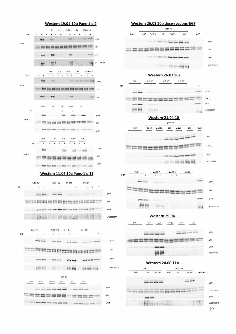
Note: these pictures are compressed pictures of the blots used for quantitation, and data may therefore have been lost.
21
Related Documents