Thalamic Strokes that Severely Impair Arousal Extend into the Brainstem Joseph Hindman B.S. 1 *, Mark D. Bowren M.A. 2 *, Joel Bruss B.S. 3 , Brad Wright MD 4 , Joel C. Geerling MD PhD 3 **, Aaron D. Boes MD PhD 5 ** 1 University of Iowa Carver College of Medicine; University of Iowa Hospitals and Clinics 2 Department of Psychological and Brain Sciences, University of Iowa 3 Department of Neurology, University of Iowa Hospitals and Clinics 4 Department of Radiology, University of Utah Health 5 Deptartments of Pediatrics, Neurology & Psychiatry, University of Iowa Hospitals and Clinics *Co-first authors **Co-last authors Correspondence to: [email protected] or [email protected] Title/Running head: Thalamic lesions & arousal This article is protected by copyright. All rights reserved. Accepted Article Thi s article has been accepted for publication and undergone full peer review but has not been through the copyediting, typesetting, pagination and proofreading process, which may lead to differences between this version and the Version of Record. Please cite this article as doi: 10.1002/ana.25377

Welcome message from author
This document is posted to help you gain knowledge. Please leave a comment to let me know what you think about it! Share it to your friends and learn new things together.
Transcript

Thalamic Strokes that Severely Impair Arousal Extend into the Brainstem
Joseph Hindman B.S.1*, Mark D. Bowren M.A.
2*, Joel Bruss B.S.
3, Brad Wright MD
4,
Joel C. Geerling MD PhD3**, Aaron D. Boes MD PhD
5**
1University of Iowa Carver College of Medicine; University of Iowa Hospitals and Clinics
2Department of Psychological and Brain Sciences, University of Iowa 3Department of Neurology, University of Iowa Hospitals and Clinics
4Department of Radiology, University of Utah Health
5Deptartments of Pediatrics, Neurology & Psychiatry, University of Iowa Hospitals and Clinics
*Co-first authors
**Co-last authors
Correspondence to: [email protected] or [email protected]
Title/Running head: Thalamic lesions & arousal
This article is protected by copyright. All rights reserved.
Acc
epte
d A
rticl
e
Thi s article has been accepted for publication and undergone full peer review but has not
been through the copyediting, typesetting, pagination and proofreading process, which may lead to differences between this version and the Version of Record. Please cite this article as doi: 10.1002/ana.25377

Hindman, Bowren et al 2
Abstract:
In this study we evaluate the role of the thalamus in the neural circuitry of arousal. Level of
consciousness within the first 12 hours of a thalamic stroke is assessed with lesion symptom
mapping. Impaired arousal correlates with lesions in the paramedian posterior thalamus near the
centromedian and parafascicular nuclei, posterior hypothalamus, and midbrain tegmentum. All
patients with severely impaired arousal (coma, stupor) had lesion extension into the midbrain
and/or pontine tegmentum, whereas purely thalamic lesions did not severely impair arousal.
These results are consistent with growing evidence that pathways most critical for human arousal
lay outside the thalamus.
This article is protected by copyright. All rights reserved.
Acc
epte
d A
rticl
e

Hindman, Bowren et al 3
Introduction
Arousal is a critical feature of consciousness, yet the neural circuitry that maintains it is poorly
understood. Early models of an ascending reticular activating system (ARAS) originating from
diffuse neurons in the brainstem reticular formation have been revised to include discrete
brainstem regions and individual nuclei that are preferentially involved in maintaining a state of
wakefulness1-5
.
Classically, ARAS projections were thought to synapse in the “non-specific” nuclei of the
midline and intralaminar thalamus, which project diffusely to the cerebral cortex5. It soon
became dogmatic in the field that the thalamus was the critical relay of the human arousal
system6, 7
. Yet the assumption that critical, arousal-promoting projections from the brainstem
must synapse in the thalamus has been challenged by a growing body of research in experimental
animals. For example, extensive ablation or inhibition of thalamic neurons fails to alter the
normal pattern and amount of waking and sleeping behavior, and the only apparent EEG
abnormality that results is a loss of spindle activity during sleep8-10
.
Outside the thalamus, neurons in the posterior hypothalamus and basal forebrain appear more
important than the thalamus for linking brainstem arousal-promoting projections to the cerebral
cortex. Ablating the posterior hypothalamus or inhibiting corticopetal neurons in the basal
forebrain nuclei produces coma, whereas exciting these regions is potently wake-promoting3, 9, 11-
13.
This article is protected by copyright. All rights reserved.
Acc
epte
d A
rticl
e

Hindman, Bowren et al 4
Unlike these advances in understanding the ascending arousal system in experimental animals,
we lack detailed information about the importance of the thalamus for human arousal. A study of
atrophy patterns in a large cohort of patients with disorders of consciousness observed no
association between thalamic atrophy and wakefulness, but this study could not determine
whether the thalamus is necessary for maintaining a basic level of arousal14
. As such, the
question of whether the human thalamus is necessary for maintaining a basic level of arousal
remains unanswered. Answering this question is more than just an academic concern; it has
direct, therapeutic implications for patient care. Based on the idea that the ascending arousal
system relays in the thalamus, patients with disorders of consciousness have had electrodes
implanted into their thalami by several groups of investigators, with inconsistent results15
. It is
imperative that we learn more about the importance of the human thalamus and adjoining brain
regions for maintaining arousal because this information holds the potential to improve
therapeutic targeting for neuromodulation in coma and other disorders of consciousness.
To help inform this debate, we identify patients with ischemic lesions involving the thalamus to
address whether the thalamus itself is responsible for the reduced level of consciousness in
patients with artery of Percheron and related stroke syndromes involving the thalamus. We
hypothesize that severe impairments in arousal (coma, stupor) do not occur with focal lesions
limited to the thalamus but may be seen when thalamic lesions extend into the brainstem.
This article is protected by copyright. All rights reserved.
Acc
epte
d A
rticl
e

Hindman, Bowren et al 5
Methods
For this retrospective study, we identified patients by searching the electronic medical records of
the University of Iowa Hospitals and Clinics using the key terms “thalamic infarct”, “cerebral
infarct”, “coma”, “artery of Percheron”, “basilar tip infarct”, and “paramedian infarct” between
2000 and 2017. We reviewed clinical records to determine whether patients had corresponding
magnetic resonance imaging (MRI) with abnormal diffusion signal in the thalamus. Exclusion
criteria included presence of sepsis, alcohol or drug overdose, seizure, or trauma. This search
yielded 23 patients (seventeen men, ages 63.30 + 14.25). An additional ten patients (five men,
ages 50.30 + 16.23) were added from a previously published study on artery of Percheron
infarcts conducted by the University of Utah Hospital16
. These cases were analyzed using the
same protocols as those from Iowa. In total, 33 patients were included in the analysis. The
previously published study did not perform lesion-symptom mapping and their results do not
overlap with the current analysis. This study was approved by the Institutional Review Board of
the University of Iowa.
The level of arousal displayed by each patient during the first twelve hours after the onset of
symptoms was extrapolated from review of the electronic medical record based primarily on
neurological exam findings. Level of arousal was rated on a 6-point scale (coma, 1; stupor, 2;
obtunded, 3; somnolent, 4; lethargic, 5; awake, 6, terms defined previously17
) by two
neurologists blinded to patient imaging (J.C.G. and A.D.B).
This article is protected by copyright. All rights reserved.
Acc
epte
d A
rticl
e

Hindman, Bowren et al 6
The location of the stroke was identified using the clinical MR images. Lesion location was
reproduced onto a 3D template brain (MNI152 atlas, 1 mm x 1 mm x 1 mm) using FSL
(http://fsl.fmrib.ox.ac.uk/fsldownloads/) as performed previously1, and the accuracy of each
lesion was reviewed independently by two neurologists blinded to clinical outcome (J.C.G. and
A.D.B.).
Lesion-symptom mapping (LSM) was used to conduct a statistical test to identify regions that,
when lesioned, were associated with greater impairment in arousal. LSM was performed using
LESYMAP, a lesion-symptom mapping package in R that employs sparse canonical correlation
analysis to find a pattern of normalized voxel weights that is most strongly correlated with
impairment in arousal. The statistical significance of the map of voxel weights was determined
through a 4-fold cross-validation correlation between predicted and observed arousal scores. This
approach has several advantages compared to mass univariate (i.e., “voxel-wise”) lesion-
symptom mapping18
. The statistical significance of the overall model was determined, and each
voxel received a weight between 0 and 1, with higher values associated with greater impairment
in arousal. The resulting voxel weights were color-coded and overlaid onto the template brain for
display. The statistical analysis was limited to voxels in which three or more patients had
damage, excluding areas with sparse coverage with less than 10% of the cohort, as performed
previously18
.
Results
This article is protected by copyright. All rights reserved.
Acc
epte
d A
rticl
e

Hindman, Bowren et al 7
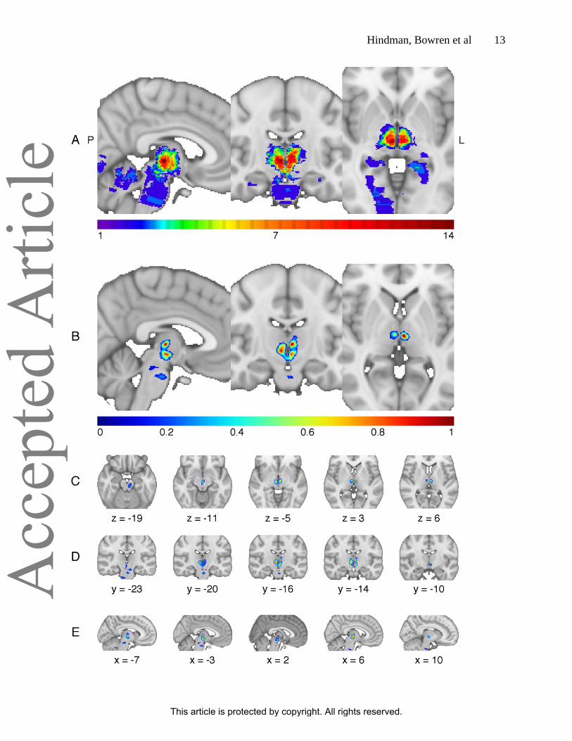
The lesion overlap (Fig 1A) demonstrates coverage of the thalamus and limited coverage of the
brainstem and other structures supplied by the posterior circulation. The overall LSM is
significant (cross-validated correlation = 0.59, p < .001). Regions associated with impaired
arousal extend from the bilateral ventromedial thalamus in the region of the centromedian and
parafascicular nuclei (Fig. 1B-E) with a peak at MNI coordinate: -3, -16, 3. The four patients
with severe impairments in arousal (2 coma, 2 stupor) all had infarcts extensively involving the
brainstem in addition to the thalamus (Fig 2). In contrast, 12 patients with thalamic lesions with
minimal to no extension into the brainstem were awake on presentation with no impairment of
consciousness. The remaining 17 patients with intermediate extension into the midbrain had
partial impairments in level of arousal (obtunded, somnolent, or lethargic).
Conclusions
This study provides a formal analysis of human thalamic lesions attempting to localize sub-
regions that may impair arousal. Our analysis identifies an association between impaired arousal
and a bilateral region of the ventromedial thalamus near the centromedian and parafascicular
nuclei, extending into posterior hypothalamus and midbrain. The association of this thalamic
region with impaired arousal must be interpreted cautiously because every patient with a
thalamic lesion causing a severe impairment in arousal had extension into the brainstem, whereas
no patients with lesions entirely limited to the thalamus demonstrated severe impairment in
arousal. These results are consistent with evidence from experimental animals that the thalamus
is not critical for arousal8-10
, and suggest that historically the thalamus was implicated due to its
shared vascular supply with brainstem structures that are critical for arousal19
. These findings are
This article is protected by copyright. All rights reserved.
Acc
epte
d A
rticl
e

Hindman, Bowren et al 8
consistent with prior research highlighting the role of the brainstem and posterior hypothalamus
as critical regions in the arousal pathway1-4
, dating back to Moruzzi and Magoun‟s demonstration
that the brainstem and caudal diencephalon show the lowest stimulation threshold for cortical
arousal5. Similarly, our findings are consistent with prior work linking dorsolateral pontine
tegmentum lesions to coma in humans1, 4
and coma-like outcomes in animals9. An alternative
explanation for our findings would be that lesions must damage both the thalamus and the
brainstem to severely impair arousal, but this is not consistent with prior evidence that ischemic
strokes and other lesions limited to the brainstem cause coma1, 4
.
This analysis is limited to 33 patients and we cannot exclude the possibility that a purely
thalamic lesion can severely impair consciousness, though we did not observe such a case. We
also note that most if not all cases from the literature describing „thalamic coma‟ have
radiographic evidence of lesions extending into the hypothalamus and brainstem, have exam
findings not consistent with the diagnosis of coma, or have clinical signs like ophthalmoplegia
that signify damage to the pons or midbrain20-22
. Our findings are also limited in that we focus on
level of arousal in the first 12 hours and do not evaluate hypersomnia or circadian rhythm
disruptions that have been reported with diencephalic lesions.
In conclusion, the current results are consistent with studies in experimental animals that
challenge the long-held view that the thalamus is a critical node in the arousal pathway8-10, 13
.
Future work with larger sample sizes will be needed to confirm and extend these findings, both
to better understand the neural circuitry of arousal and to inform targeted neuromodulation
approaches for disorders of consciousness.
This article is protected by copyright. All rights reserved.
Acc
epte
d A
rticl
e

Hindman, Bowren et al 9
Acknowledgements: We thank A. Andres Capizzano, MD for assistance acquiring imaging
data.
This study was supported by Carver College of Medicine Research Fellowship (J.H.) A.D.B. was
supported by NIH/NINDS grant K12HD027748-24, K12NS098482-01. J.C.G. was supported by
K08-NS099425.
Author contributions: ADB and JCG contributed to the conception and design of the study and
oversaw all aspects; MDB and JH contributed to the acquisition and analysis of data; BW
provided imaging data; MDB performed the lesion mapping and created figures; JH contributed
to drafting the manuscript; all authors contributed to editing the manuscript.
Potential Conflicts of Interest: The authors have no relevant financial disclosures. ADB
participated in a data safety and monitoring board for Ekso Bionics.
References:
1. Fischer DB, Boes AD, Demertzi A, et al. A human brain network derived from coma-
causing brainstem lesions. Neurology 2016;87:2427-2434.
This article is protected by copyright. All rights reserved.
Acc
epte
d A
rticl
e

Hindman, Bowren et al 10
2. Eban-Rothschild A, Rothschild G, Giardino WJ, Jones JR, de Lecea L. VTA
dopaminergic neurons regulate ethologically relevant sleep-wake behaviors. Nat Neurosci
2016;19:1356-1366.
3. Pedersen NP, Ferrari L, Venner A, et al. Supramammillary glutamate neurons are a key
node of the arousal system. Nat Commun 2017;8:1405.
4. Parvizi J, Damasio AR. Neuroanatomical correlates of brainstem coma. Brain
2003;126:1524-1536.
5. Moruzzi G, Magoun HW. Brainstem reticular formation and activation of the EEG.
Electroencephalogr Clin Neurophysiol 1949;1:455-473.
6. Steriade M. Ascending control of thalamic and cortical responsiveness. Int Rev
Neurobiol 1970;12:87-144.
7. Papez JW. Central reticular path to intralaminar and reticular nuclei of thalamus for
activating EEG related to consciousness. Electroencephalogr Clin Neurophysiol 1956;8:117-128.
8. Villablanca J, Salinas-Zeballos ME. Sleep-wakefulness, EEG and behavioral studies of
chronic cats without the thalamus: The "athalamic" cat. Arch Ital Biol 1972;110:383–411.
9. Fuller PM, Sherman D, Pedersen NP, Saper CB, Lu J. Reassessment of the structural
basis of the ascending arousal system. J Comp Neurol 2011;519:933-956.
10. Buzsaki G, Bickford RG, Ponomareff G, Thal LJ, Mandel R, Gage FH. Nucleus basalis
and thalamic control of neocortical activity in the freely moving rat. J Neurosci 1988;8:4007–
4026.
11. Saper CB, Fuller PM. Wake-sleep circuitry: an overview. Curr Opin Neurobiol
2017;44:186-192.
This article is protected by copyright. All rights reserved.
Acc
epte
d A
rticl
e

Hindman, Bowren et al 11
12. Xu M, Chung S, Zhang S, et al. Basal forebrain circuit for sleep-wake control. Nat
Neurosci 2015;18:1641-1647.
13. Anaclet C, Pedersen NP, Ferrari LL, et al. Basal forebrain control of wakefulness and
cortical rhythms. Nat Commun 2015;6:8744.
14. Lutkenhoff ES, Chiang J, Tshibanda L, et al. Thalamic and extrathalamic mechanisms of
consciousness after severe brain injury. Ann Neurol 2015;78:68-76.
15. Vanhoecke J, Hariz M. Deep brain stimulation for disorders of consciousness: Systematic
review of cases and ethics. Brain Stimul 2017;10:1013-1023.
16. Lazzaro NA, Wright B, Castillo M, et al. Artery of percheron infarction: imaging patterns
and clinical spectrum. AJNR Am J Neuroradiol 2010;31:1283-1289.
17. Posner JB, Saper CB, Schiff N, Plum F. Plum and Posner‟s Diagnosis of Stupor and
Coma., 4 ed. New York, NY: Oxford University Press, 2007.
18. Pustina D, Avants B, Faseyitan OK, Medaglia JD, Coslett HB. Improved accuracy of
lesion to symptom mapping with multivariate sparse canonical correlations. Neuropsychologia
2018;115:154-166.
19. Schmahmann JD. Vascular syndromes of the thalamus. Stroke 2003;34:2264-2278.
20. Bogousslavsky J, Regli F, Uske A. Thalamic infarcts: clinical syndromes, etiology, and
prognosis. Neurology 1988;38:837-848.
21. Zappella N, Merceron S, Nifle C, et al. Artery of Percheron infarction as an unusual
cause of coma: three cases and literature review. Neurocrit Care 2014;20:494-501.
22. Rivera-Lara L, Henninger N. Delayed sudden coma due to artery of percheron infarction.
Arch Neurol 2011;68:386-387.
This article is protected by copyright. All rights reserved.
Acc
epte
d A
rticl
e

Hindman, Bowren et al 12
Figure Legends:
Figure 1: (A) Overlap of all 33 lesions in this study. All involve the thalamus, and some extend
into the brainstem, cerebellum, or cortical regions supplied by the posterior cerebral artery. The
maximum lesion overlap was 14/33 at MNI coordinate 5, -17, -2. (B) Lesion-Symptom Mapping
(LSM) Result. The voxel weights derived from the LSM analysis are shown centered on the voxel
with the greatest weight (-3, -16, 3). The color-coded scale represents the normalized voxel
weights derived from the LSM analysis. Greater weights are associated with a more severe
impairment in arousal. Representative slices of the Lesion-Symptom Mapping result are depicted
in (C) axial, (D) coronal, and (E) sagittal views.
Figure 2: Lesions in patients presenting with coma (A & B) or stupor (C & D). In each, bilateral
thalamic infarcts extended into the posterior hypothalamus and midbrain (A – D) and in two, the
pons (A & B). No patients with a lesion restricted to the thalamus had a severe impairment in
arousal (coma or stupor).
This article is protected by copyright. All rights reserved.
Acc
epte
d A
rticl
e
Hindman, Bowren et al 13
This article is protected by copyright. All rights reserved.
Acc
epte
d A
rticl
e

Hindman, Bowren et al 14
This article is protected by copyright. All rights reserved.
Acc
epte
d A
rticl
e
Related Documents