ARTICLE OPEN Targeting SYK of monocyte-derived macrophages regulates liver fibrosis via crosstalking with Erk/Hif1α and remodeling liver inflammatory environment Xuejiao Chen 1,2,3,5 , Ziyi Wang 1,2,3,5 , Sheng Han 1,2,3,5 , Zeng Wang 1,2,3 , Yu Zhang 1,2,3 , Xiangdong Li 1,2,3 , Nan Xia 1,2,3 , Wenjie Yu 1,2,3 , Chenyang Jia 4 , Yong Ni 4 ✉ and Liyong Pu 1,2,3 ✉ © The Author(s) 2021 Liver fibrosis is a danger signal indicating a huge risk of liver cancer occurrence, but there is still no effective clinical means to regulate the progress of liver fibrosis. Although a variety of drugs targeting SYK have been developed for tumors and autoimmune diseases, the mechanism and specific efficacy of SYK’s role in liver fibrosis are not yet clear. Our studies based on chronic CCL4, bile duct ligation, and subacute TAA mouse models show that SYK in monocyte-derived macrophages (MoMFs) is fully dependent on phosphorylation of Erk to up-regulate the expression of Hif1α, thereby forming the crosstalk with SYK to drive liver fibrosis progress. We have evaluated the ability of the small molecule SYK inhibitor GS9973 in a variety of models. Contrary to previous impressions, high-frequency administration of GS9973 will aggravate CCL4-induced liver fibrosis, which is especially unsuitable for patients with cholestasis whose clinical features are bile duct obstruction. In addition, we found that inhibition of MoMFs SYK impairs the expression of CXCL1, on one hand, it reduces the recruitment of CD11bhiLy6Chi inflammatory cells, and on the other hand, it promotes the phenotype cross-dress process of pro-resolution MoMFs, thereby remodeling the chronic inflammatory environment of the fibrotic liver. Our further findings indicate that on the basis of the administration of CCR2/CCR5 dual inhibitor Cenicriviroc, further inhibiting MoMFs SYK may give patients with fibrosis additional benefits. Cell Death and Disease (2021)12:1123 ; https://doi.org/10.1038/s41419-021-04403-2 INTRODUCTION Liver fibrosis is a chronic liver disease characterized by chronic tissue damage and the accumulation of extracellular protein matrix [1]. The main causes of liver fibrosis are viral hepatitis, alcoholic liver disease, and non-alcoholic fatty liver. Repeated chronic damage to the liver stimulates hepatic stellate cells (HSC) to synthesize and accumulate excessive extracellular matrix [2]. Liver fibrosis progresses to liver cirrhosis and thus becomes an important risk factor for the progression of liver cancer. The diagnosis methods of liver fibrosis include invasive and non- invasive, but pathological biopsy is still the gold standard. Ishak score (Ishak0–6) is commonly used clinically to classify the severity of liver fibrosis [3]. Spleen tyrosine kinase (SYK) is a 72 kDa non-receptor tyrosine kinase that plays a multiple roles in human immune response [4]. Existing studies have shown that SYK has expressed in other tissue cells [5] and plays a wide range of physiological and pathological functions [6], which is suitable for the liver [5]. Studies have shown that the use of SYK kinase inhibitors to block SYK phosphorylation can effectively reduce the development of various liver diseases, including liver fibrosis, alcoholic liver disease, non-alcoholic fatty liver, hepatocellular carcinoma [5, 7]. Antagonistic SYK can inhibit liver fibrosis by inhibiting HSC activation, and the expression of SYK reflects the inducing stage of fibrosis to a certain extent. It is already known that the myeloid SYK inhibitor (Piceatannol, PRT06260) can effectively reduce liver fibrosis, hepatocyte damage and inflammation. GS9973 is a new type, selective SYK inhibitor has been shown to have anti-fibrosis activity in vivo [8]. The Erk signaling pathway is one of the keys to regulate the main phenotypic response of fibroblasts. Erk1/2 may be activated by MERTK from the macrophages surface, and TGF-β transcription factor regulatory protein-1 (AP-1) can be activated by Erk1/2, in addition, Erk1/2 can activate TGFβ1 through the GAS pathway [9]. The Erk pathway acts actively role in driving liver fibrosis by targeting HSC [10]. HiF1α is closely related to the development of hepatic fibrosis, and liver fibrosis caused by sinus morphological imbalances and functional blocks cause hepatocytes to hypoxia, which in turn causes degradation of HIF1α to be inhibited, thereby increasing expressions [11]. HiF1α lacks fibrosis of mice liver that relieves biliary tube ligation [12]. The expression level of liver α-SMA and COL1A1/2 of BDL mice after myeloid knockout HiF1α or HIF1β is Received: 28 May 2021 Revised: 24 October 2021 Accepted: 12 November 2021 1 Hepatobiliary Center, The First Affiliated Hospital of Nanjing Medical University, Nanjing, China. 2 Key Laboratory of Liver Transplantation, Chinese Academy of Medical Sciences, Nanjing, China. 3 NHC Key Laboratory of Living Donor Liver Transplantation (Nanjing Medical University), Nanjing, Jiangsu Province, China. 4 Department of Hepatopancreatobiliary Surgery, Shenzhen Second People’s Hospital, The First Affiliated Hospital of Shenzhen University, Shenzhen, Guangdong, China. 5 These authors contributed equally: Xuejiao Chen, Ziyi Wang, Sheng Han. ✉ email: [email protected]; [email protected] Edited by Professor Anastasis Stephanou www.nature.com/cddis Official journal of CDDpress 1234567890();,:

Welcome message from author
This document is posted to help you gain knowledge. Please leave a comment to let me know what you think about it! Share it to your friends and learn new things together.
Transcript
ARTICLE OPEN
Targeting SYK of monocyte-derived macrophages regulatesliver fibrosis via crosstalking with Erk/Hif1α and remodelingliver inflammatory environmentXuejiao Chen1,2,3,5, Ziyi Wang1,2,3,5, Sheng Han1,2,3,5, Zeng Wang1,2,3, Yu Zhang1,2,3, Xiangdong Li1,2,3, Nan Xia1,2,3, Wenjie Yu1,2,3,Chenyang Jia4, Yong Ni 4✉ and Liyong Pu 1,2,3✉
© The Author(s) 2021
Liver fibrosis is a danger signal indicating a huge risk of liver cancer occurrence, but there is still no effective clinical means toregulate the progress of liver fibrosis. Although a variety of drugs targeting SYK have been developed for tumors and autoimmunediseases, the mechanism and specific efficacy of SYK’s role in liver fibrosis are not yet clear. Our studies based on chronic CCL4, bileduct ligation, and subacute TAA mouse models show that SYK in monocyte-derived macrophages (MoMFs) is fully dependent onphosphorylation of Erk to up-regulate the expression of Hif1α, thereby forming the crosstalk with SYK to drive liver fibrosis progress.We have evaluated the ability of the small molecule SYK inhibitor GS9973 in a variety of models. Contrary to previous impressions,high-frequency administration of GS9973 will aggravate CCL4-induced liver fibrosis, which is especially unsuitable for patients withcholestasis whose clinical features are bile duct obstruction. In addition, we found that inhibition of MoMFs SYK impairs theexpression of CXCL1, on one hand, it reduces the recruitment of CD11bhiLy6Chi inflammatory cells, and on the other hand, itpromotes the phenotype cross-dress process of pro-resolution MoMFs, thereby remodeling the chronic inflammatory environmentof the fibrotic liver. Our further findings indicate that on the basis of the administration of CCR2/CCR5 dual inhibitor Cenicriviroc,further inhibiting MoMFs SYK may give patients with fibrosis additional benefits.
Cell Death and Disease (2021) 12:1123 ; https://doi.org/10.1038/s41419-021-04403-2
INTRODUCTIONLiver fibrosis is a chronic liver disease characterized by chronictissue damage and the accumulation of extracellular proteinmatrix [1]. The main causes of liver fibrosis are viral hepatitis,alcoholic liver disease, and non-alcoholic fatty liver. Repeatedchronic damage to the liver stimulates hepatic stellate cells (HSC)to synthesize and accumulate excessive extracellular matrix [2].Liver fibrosis progresses to liver cirrhosis and thus becomes animportant risk factor for the progression of liver cancer. Thediagnosis methods of liver fibrosis include invasive and non-invasive, but pathological biopsy is still the gold standard. Ishakscore (Ishak0–6) is commonly used clinically to classify the severityof liver fibrosis [3].Spleen tyrosine kinase (SYK) is a 72 kDa non-receptor tyrosine
kinase that plays a multiple roles in human immune response [4].Existing studies have shown that SYK has expressed in other tissuecells [5] and plays a wide range of physiological and pathologicalfunctions [6], which is suitable for the liver [5]. Studies have shownthat the use of SYK kinase inhibitors to block SYK phosphorylationcan effectively reduce the development of various liver diseases,including liver fibrosis, alcoholic liver disease, non-alcoholic fatty
liver, hepatocellular carcinoma [5, 7]. Antagonistic SYK can inhibitliver fibrosis by inhibiting HSC activation, and the expression ofSYK reflects the inducing stage of fibrosis to a certain extent. It isalready known that the myeloid SYK inhibitor (Piceatannol,PRT06260) can effectively reduce liver fibrosis, hepatocyte damageand inflammation. GS9973 is a new type, selective SYK inhibitorhas been shown to have anti-fibrosis activity in vivo [8].The Erk signaling pathway is one of the keys to regulate the
main phenotypic response of fibroblasts. Erk1/2 may be activatedby MERTK from the macrophages surface, and TGF-β transcriptionfactor regulatory protein-1 (AP-1) can be activated by Erk1/2, inaddition, Erk1/2 can activate TGFβ1 through the GAS pathway [9].The Erk pathway acts actively role in driving liver fibrosis bytargeting HSC [10].HiF1α is closely related to the development of hepatic fibrosis,
and liver fibrosis caused by sinus morphological imbalances andfunctional blocks cause hepatocytes to hypoxia, which in turncauses degradation of HIF1α to be inhibited, thereby increasingexpressions [11]. HiF1α lacks fibrosis of mice liver that relievesbiliary tube ligation [12]. The expression level of liver α-SMA andCOL1A1/2 of BDL mice after myeloid knockout HiF1α or HIF1β is
Received: 28 May 2021 Revised: 24 October 2021 Accepted: 12 November 2021
1Hepatobiliary Center, The First Affiliated Hospital of Nanjing Medical University, Nanjing, China. 2Key Laboratory of Liver Transplantation, Chinese Academy of Medical Sciences,Nanjing, China. 3NHC Key Laboratory of Living Donor Liver Transplantation (Nanjing Medical University), Nanjing, Jiangsu Province, China. 4Department ofHepatopancreatobiliary Surgery, Shenzhen Second People’s Hospital, The First Affiliated Hospital of Shenzhen University, Shenzhen, Guangdong, China. 5These authorscontributed equally: Xuejiao Chen, Ziyi Wang, Sheng Han. ✉email: [email protected]; [email protected] by Professor Anastasis Stephanou
www.nature.com/cddis
Official journal of CDDpress
1234567890();,:
significantly reduced compared with WT mice [13]. In addition,HIF1α is detected in liver macrophages in patients with cirrhosis.Erk1/2 has proven to be important signals for regulating HIF1α.Over-expression Erk1 significantly enhanced the activity of HIF1αunder hypoxia [14], HIF1α and Erk expression increased with CCL4-induced rat liver fibrosis [15].Despite the effectiveness of targeting myeloid SYK in mouse
fibrosis, our research highlights the effectiveness of SYK in mono-derived macrophages. On one aspect, Knocked down the SYK inMoMFs impairs the expression of CXCL1 and increases the raisingof LY6Chi inflammatory cells. On the other aspect, SYK crossedwith the Erk/HIF1α axis and plays a crucial effect in the progress ofdriving fibrosis. In addition to clarifying the possible mechanism ofSYK driving liver fibrosis in MoMFs, we also evaluated theeffectiveness of GS9973, and the results were not ideal. It isworth noting that we have proved that target MoMFs SYK toenhance CCR2/CCR5 dual inhibitors Cernicriviroc inhibit theprogress of fibrosis.
METHODS AND MATERIALSPatient samplesCollected liver samples from patients undergoing liver resection at theHepatobiliary Center of the First Affiliated Hospital of Nanjing MedicalUniversity, and collected and analyzed patient samples at different stagesof liver fibrosis according to the Ishak classification. All patients whodonated samples for this project signed informed consent, and allexperiments were approved by the Ethics Committee of Nanjing MedicalUniversity.
Murine liver fibrosis modelsCCL4 induced mouse fibrosis model: 6–8 weeks old C57/BL6 male micepurchased from Vital River Laboratory Animal Technology Co. Ltd (Beijing,China) were injected intraperitoneally with CCL4 (0.6 ul/g body weight)(CCL4: olive oil= 1:3) every 3 days, continuously administered for4–8 weeks. Inhibitor GS9973 (20 mg/kg) was dissolved in DMSO, preparedas a working solution with olive oil, and injected every 2 days. One dayafter the last administration of CCL4, the mice were sacrificed and the liverand serum were harvested.Bile duct ligation (BDL) model: Adult mice were anesthetized with
isoflurane inhalation. After opening the abdominal cavity, the hilar of theliver was exposed, and the common bile duct was ligated with 4–0 vicrylrapide. The sham operation group received the same procedure except forthe ligation of the common bile duct. The mice were sacrificed 2–4 weeksafter the operation and the liver and serum were obtained (at least 5 mice/group). All mice were numbered by one experimenter and treated blindlyor operated on by another experimenter.
TAA-induced liver fibrosis models6–8 weeks wild-type C57/BL6 male mice were given a single sublethal dose(100mg/kg, ip) injection of hepatotoxin thioacetamide(TAA) intraperito-neally. The mice were sacrificed 1d, 3d, and 7d after injection, and liverswere obtained simultaneously. For the siSYK-mannosepolymer treatmentgroup, mice were injected into the tail vein 4 h before TAA injection.
Knockdown of SYK in vivo/vitroIn vivo: The SYK siRNA (20mg/kg, Santa Cruze, California, USA) wascoupled with a mannose-conjugated polymer (polyplus transfection,Illkich, France), and injected into the tail vein of mice (200mg/kg, twice)4 h before the induction of inflammation to Delivery to liver CD206himacrophages.In vitro: The shSYK lentivirus (Genepharm, Shanghai, China) was
synthesized to transfect BMDMs collected from WT B6 mice. BMDM wastransfected 1 day before stimulating with LPS.
Isolation and culture of liver macrophagesPreheated PBS was used to perfuse the liver (5 ml/min). After the liverbecame completely white, the liver was perfused with type IV collagenase(Sigma Aldrich, 2 ml/min) dissolved in HBSS. After removing thegallbladder, the liver was harvested, and the digested cells were filtered
with a 70 um cell strainer (Fisherbrand) to prepare a single-cell suspension.After centrifugation, discard the hepatic parenchymal cells, add 50% and25% Percoll, centrifuge at 890 g for 15min, aspirate the cells in the middlelayer and continue to culture after centrifugation to obtain adherentmacrophages.
Extraction of bone marrow-derived macrophages (BMDM)Bone marrow cells were isolated from the femur and tibia of mice. Filterthrough a 70 um filter and use red blood cell lysate to remove red bloodcells. Cells were then cultured in DMEM supplemented with 10% FBS and20% L929-conditioned medium, and the culture medium was replacedafter 7 days for subsequent experiments.
Western blotAfter lysing the tissues and cells with RIPA, add the loading buffer waterbath. After SDS-PAGE gel electrophoresis, transfer to PVDF membrane. Usefor SYK (Cell Signaling Technology, 13198), p-SYK (Bioss, bs-3434R), Erk1+Erk2 (abcam, ab184699), p-Erk1+ p-Erk2 (abcam, ab214036), HIF1α (CellSignaling Technology, 36169), TGF-β (abcam, ab215715), α-SMA (CellSignaling Technology, 19245), β-actin (Cell Signaling Technology, 4970)antibody for detection, using chemiluminescence-based method for colordevelopment.
qPCRAfter extracting total RNA from tissues or cells, it is reverse transcribed intocDNA. SYBR Green fluorescent dye was used for real-time fluorescencequantitative polymerase chain reaction, all expression level and results ofthe target gene are standardized for the GAPDH expression.
Determination of serum ALT, ASTThe measured biochemical markers of liver function included alanineaminotransferase (ALT), aspartate aminotransferase (AST). Whole bloodsamples were collected from mice’s intravalains placed at 4 °C overnight,and then 3000 rpm for 15min. Using the corresponding kit (S03030,S03040) to export the results after the automatic biochemical analyzer(Rayto Life and Analytical Sciences Co., Ltd. Chemray 240).
Immunohistochemistry and immunofluorescenceIHC. Fix liver tissue samples in 4% paraformaldehyde, embed in paraffin,slice into 4um-thick sections and then perform (hematoxylin-eosin) H & Estaining, Sirius red staining, and masson staining. Refer to the ishak scoringsystem for blind evaluation of fibrosis samples.
Immunofluorescence. The fixed tissue sections are placed in EDTA antigenretrieval buffer (PH= 9.0), and then boiled in the microwave for repair.Block BSA for 30min, add the corresponding primary antibodies: p-SYK(Bioss, bs-3434R), p-Erk1+ p-Erk2(abcam, ab214036), α-SMA(Cell SignalingTechnology,19245), and incubate overnight. After washing with PBS, addthe secondary antibody of the corresponding species. After incubating inthe dark for 50min, nuclei were stained. observe the slides under aconfocal fluorescence microscope (NIKON ECLIPSE C1) and images werecollected.
Flow cytometryThe obtained macrophage lysed red blood cells were added to cellstaining buffer (biolegend, 420201) to make a single-cell suspension, andFc receptor blocker was added to reduce non-specific staining. For mouseCD11b(Biolegend,101206) and Ly6C(Biolegend,128012), F4/80(Biole-gend,123110), they were processed with a flow cytometer (Beckman,Gallios) after incubation. The data were analyzed with Flow Jo.
Statistical analysisThe data are expressed as mean ± SD, and the statistical differencebetween subgroups is determined by GraphPad Prism software. Student’s ttest (and Nonparametric test), one-way ANOVA were used to compare thedifferences of each group. A P value less than 0.05 is consideredstatistically significant. One experimenter numbered patient and mouseliver tissue before the experiment and randomly assigned it to anotherexperimenter for blind evaluation. Results were analyzed by at least twoother qualified experimenters.
X. Chen et al.
2
Cell Death and Disease (2021) 12:1123
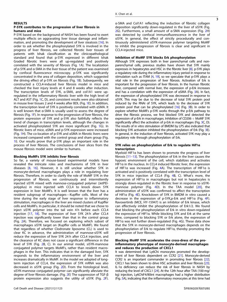
RESULTSP-SYK contributes to the progression of liver fibrosis inhumans and miceP-SYK based on the background of NASH has been found to exertmultiple effects on aggravating liver tissue damage and inflam-mation, and promoting the development of liver steatosis [16]. Inorder to ask whether the phosphorylated SYK is involved in theprogress of liver fibrosis, we collected fibrotic liver tissues ofpatients with Ishak classification as the clinicopathologicalstandard and found that p-SYK expressed in different IshakGraded fibrotic livers were all up-regulated and positivelycorrelated with the severity of fibrosis (Fig. 1A). The localizationof p-SYK and α-SMA in the liver tissue of the patient was examinedby confocal fluorescence microscopy. p-SYK was significantlyconcentrated in the area of collagen deposition, which suggestedthe driving effect of p-SYK on fibrosis (Fig. 1B). Subsequently, weconstructed a CCL4-induced liver fibrosis model in mice andchecked the liver injury levels at 4 and 8 weeks after induction.The transcription levels of SYK, α-SMA, and col1A1 were up-regulated in the inflammatory fibrotic liver with the high level ofALT and AST (Fig. 1C, D), and consistent results were also obtainedin mouse liver tissues 2 and 4 weeks after BDL (Fig. 1E). In addition,the transcription level of SYK is positively correlated with αSMA. Itis well known that α-SMA is usually used to assess the degree offibrosis (Fig. 1F). In response to the progression of liver fibrosis, theprotein expression of SYK and p-SYK also faithfully reflects thetrend of changes in transcription levels, although the changes inactivated phosphorylated SYK are more significant (Fig. 1G). Infibrotic livers of mice, αSMA and p-SYK expression were increased(Fig. 1H). The co-location of p-SYK and αSMA in fibrotic livers wereincreased compared with the control group and sham group (Fig.1I). Our results prove that p-SYK plays an important role in theprocess of liver fibrosis. The conclusions of liver slices from themouse fibrosis model were similar to humans.
Blocking MoMFs SYK inhibits liver fibrosisSo far, a variety of mouse-based experimental models haverevealed the intricate roles and mechanisms of SYK in liverdiseases [8, 16]. However, it is still unclear whether SYK inmonocyte-derived macrophages plays a role in regulating liverfibrosis. Therefore, in order to clarify the role of MoMF SYK in theprogression of fibrosis, we continuously and intermittentlyinjected siSYK mixed with mannose-conjugated polymer (JetPEI-polyplus) in mice injected with CCL4 to knock down SYKexpression in liver MoMFs. It is well known that the liver has aresident subgroup of macrophages—Kupffer cells. Most of thetime during the early stage of liver response to inflammatorystimulation, macrophages in the liver are mixed clusters of Kupffercells and MoMFs. In particular, it should be noted that we chose toinject siSYK polymer into the tail vein 4 h before each CCL4injection [17, 18]. The expression of liver SYK 24 h after CCL4injection was significantly lower than that in the control group(Fig. 2A). Therefore, we focused on exploring which subset ofmacrophages siSYK target on (Kupffer cells or MoMF). We foundthat regardless of whether Clodronate liposome (CL) is used toclear KC in advance, the administration of mannose-siSYK willreduce the expression of liver SYK 24 h after CCL4 injection, andthe clearance of KC does not cause a significant difference in thelevel of SYK (Fig. 2B, C). In our animal model, siSYK-mannoseconjugated polymer targets MoMFs, rather than resident macro-phages in the liver. This indicates that in the damaged liver, SYKresponds to the inflammatory environment of the liver andincreases dramatically in MoMF. In the model we adopted of long-term injection of CCL4, the expression of SYK mRNA was alsodecreased in siSYK group (Fig. 2D). From the histological level,siSYK-mannose conjugated polymer can significantly alleviate thedegree of liver fibrosis damage. (Fig. 2E) The suppression of TGF-βprotein expression also suggests the utility of siSYK (Fig. 2F).
α-SMA and Col1A1 reflecting the induction of fibrotic collagendeposition significantly down-regulated in the liver of siSYK (Fig.2G). Furthermore, a small amount of α-SMA expression (Fig. 2H)was detected by confocal immunofluorescence in the liver ofsiSYK. In general, the effect of strictly procedurally and con-tinuously administered siSYK-mannose polymer targeting MoMFto inhibit the progression of fibrosis is clear and significant inCCL4-injected mice.
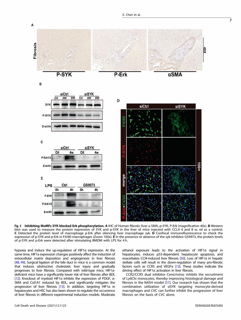
Inhibition of MoMF SYK blocks Erk phosphorylationAlthough SYK expresses both in liver parenchymal cells and non-parenchymal cells, previous studies have shown that SYK mainlyexpresses in hepatocytes and HSC in the fibrotic liver [8]. p-SYK playsa regulatory role during the inflammatory injury period in response tostimulation such as ITAM [4, 19], so we speculate that p-SYK plays avital role in the progression of liver fibrosis. Activation of Erk isimportant for the progression of liver fibrosis. In the human fibroticliver, compared with normal liver, the expression of p-Erk increasesand has a correlation with the expression of αSMA (Fig. 3A). In fact,the expression of phosphorylated SYK was decreased in response tosiSYK. This may be due to the decrease of total SYK protein poolinduced by the RNAi of SYK, which leads to the decrease of SYKprotein pool that can be phosphorylated [16] (Fig. 3B). In order toexplore whether MoMFs p-SYK works through the p-Erk pathway todrive the fibrosis process, we first blocked SYK and detected theexpression of p-Erk in macrophages. Inhibition of CD206+MoMF SYKsignificantly affect the activation of p-Erk in macrophages (Fig. 3C, D).The results of in vitro stimulation of BMDM by LPS also indicated thatblocking SYK activation inhibited the phosphorylation of Erk (Fig. 3E).In general, in the induction of liver fibrosis, activated SYK may play aregulatory role through phosphorylation of Erk.
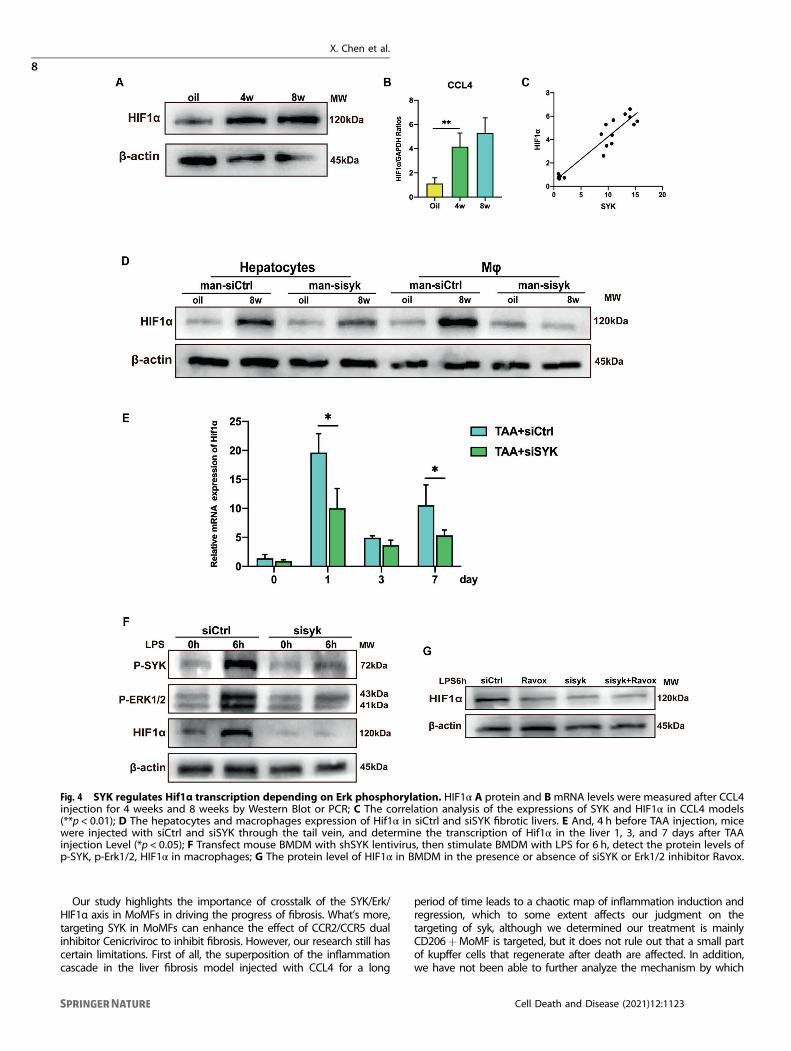
SYK relies on phosphorylation of Erk to regulate Hif1αtranscriptionMyeloid HIF1α has been shown to promote the progress of liverfibrosis [11–13]. The phosphorylation of Erk in the liver causes thehypoxic environment of the cell, which stabilizes and activatesHIF1α in the nucleus. In CCL4 induced fibrotic liver, the expressionof HIF1α was increased (Fig. 4A). The transcription of HIF1α isactivated and is positively correlated with the transcription level ofSYK in mice injection of CCL4 (Fig. 4B, C). What’s more, theexpression of HIF1α in macrophages but not in hepatocytes isstrongly down-regulated in the fibrotic liver in response to siSYK-mannose polymer (Fig. 4D). In the TAA model [20], theadministration of siSYK was confirmed to affect the transcriptionof Hif1α (Fig. 4E). Knockdown of SYK in BMDM in vitro caused adecrease in the expression of p-SYK,p-Erk and HIF1α (Fig. 4F).Ravoxertinib (MCE, HY-15947) is an inhibitor of Erk kinase, whichcan effectively inhibit the phosphorylation of Erk1/2. We foundthat blocking the phosphorylation of Erk in vitro down-regulatedthe expression of HIF1α. While blocking SYK and Erk at the sametime, compared to blocking SYK or Erk alone, the expression ofHIF1α was not further down-regulated (Fig. 4G). These data showthat the SYK in monocyte-derived macrophages depends on thephosphorylation of Erk to regulate HIF1α, thereby promoting theprogression of liver fibrosis.
Blocking MoMF SYK accelerates the cross-dress of the pro-inflammatory phenotype of monocyte-derived macrophagesand reduces the production of CXCL1It was determined that Ly6Chi monocytes promoted the develop-ment of liver fibrosis dependent on CCR2 [21]. Monocyte-derivedCCR2 is an important commander in promoting liver fibrosis [22].CXCL1 has been shown to drive HSC activation and liver fibrosis [23].IL-1α deficiency can reduce the risk of liver fibrosis in NASH byreducing the level of CXCL1 [24]. At the 12th hour after TAA (100mg/kg) injection, Ly6ChiF4/80int macrophages had a higher distribution(Fig. 5A), indicating that the inflammatory monocytes at this time had
X. Chen et al.
3
Cell Death and Disease (2021) 12:1123
partially transformed into anti-inflammatory resolution macrophages.The recruitment of Ly6ChiF4/80low inflammatory monocytes and theproduction of Ly6ChiF4/80int transitional macrophages graduallydecreased, while the pro-resolution Ly6ClowF4/80hi MoMF whichoriginated from the cross-dress of pro-inflammatory phenotype was
gradually increasing. MoMF obtains the expression of CD206 throughcross-dress to mediate inflammation repair, which is also the keymechanism for the regulation of siSYK targeting MoMF expressingCD206. In a mouse model with a single sublethal dose of TAA,blocking MoMF SYK can reduce the induction and deposition of
X. Chen et al.
4
Cell Death and Disease (2021) 12:1123
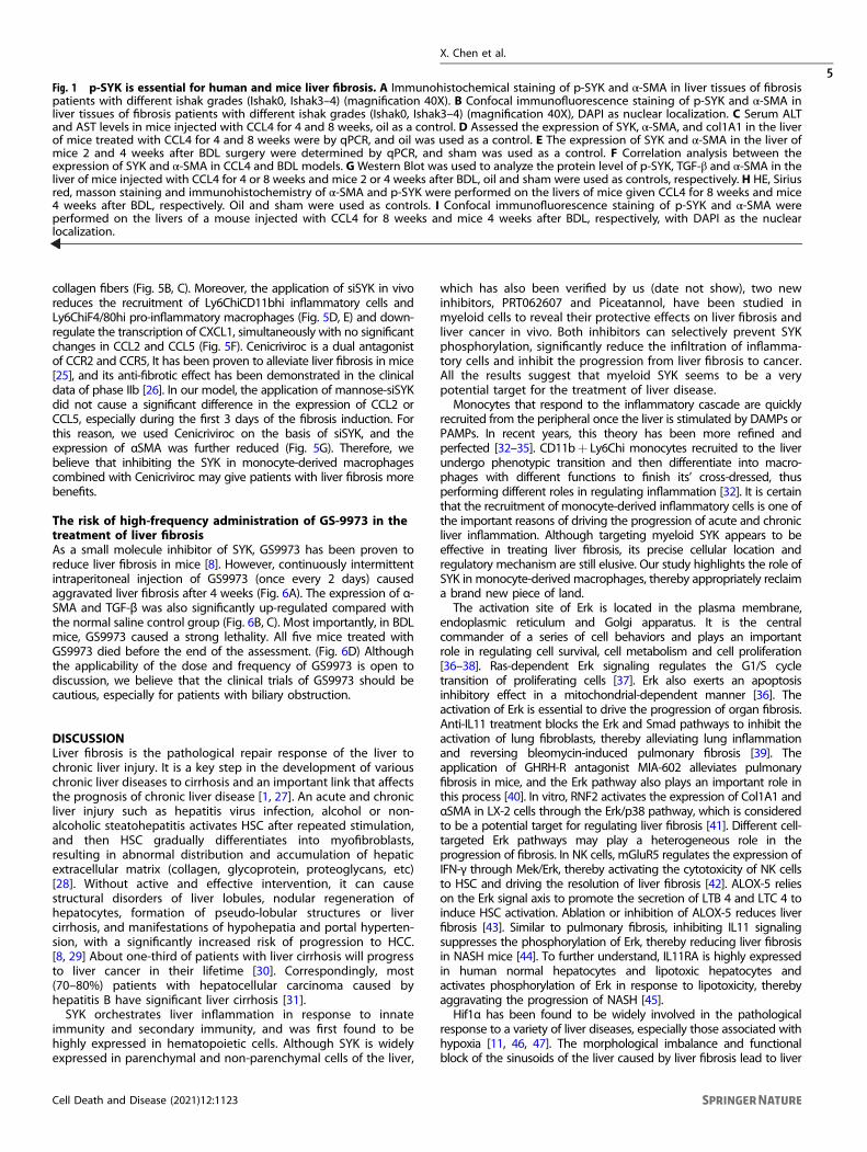
collagen fibers (Fig. 5B, C). Moreover, the application of siSYK in vivoreduces the recruitment of Ly6ChiCD11bhi inflammatory cells andLy6ChiF4/80hi pro-inflammatory macrophages (Fig. 5D, E) and down-regulate the transcription of CXCL1, simultaneously with no significantchanges in CCL2 and CCL5 (Fig. 5F). Cenicriviroc is a dual antagonistof CCR2 and CCR5, It has been proven to alleviate liver fibrosis in mice[25], and its anti-fibrotic effect has been demonstrated in the clinicaldata of phase IIb [26]. In our model, the application of mannose-siSYKdid not cause a significant difference in the expression of CCL2 orCCL5, especially during the first 3 days of the fibrosis induction. Forthis reason, we used Cenicriviroc on the basis of siSYK, and theexpression of αSMA was further reduced (Fig. 5G). Therefore, webelieve that inhibiting the SYK in monocyte-derived macrophagescombined with Cenicriviroc may give patients with liver fibrosis morebenefits.
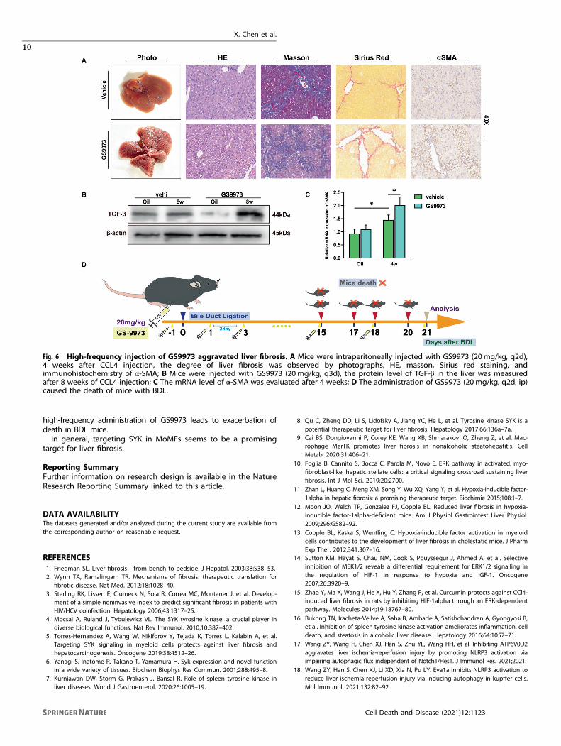
The risk of high-frequency administration of GS-9973 in thetreatment of liver fibrosisAs a small molecule inhibitor of SYK, GS9973 has been proven toreduce liver fibrosis in mice [8]. However, continuously intermittentintraperitoneal injection of GS9973 (once every 2 days) causedaggravated liver fibrosis after 4 weeks (Fig. 6A). The expression of α-SMA and TGF-β was also significantly up-regulated compared withthe normal saline control group (Fig. 6B, C). Most importantly, in BDLmice, GS9973 caused a strong lethality. All five mice treated withGS9973 died before the end of the assessment. (Fig. 6D) Althoughthe applicability of the dose and frequency of GS9973 is open todiscussion, we believe that the clinical trials of GS9973 should becautious, especially for patients with biliary obstruction.
DISCUSSIONLiver fibrosis is the pathological repair response of the liver tochronic liver injury. It is a key step in the development of variouschronic liver diseases to cirrhosis and an important link that affectsthe prognosis of chronic liver disease [1, 27]. An acute and chronicliver injury such as hepatitis virus infection, alcohol or non-alcoholic steatohepatitis activates HSC after repeated stimulation,and then HSC gradually differentiates into myofibroblasts,resulting in abnormal distribution and accumulation of hepaticextracellular matrix (collagen, glycoprotein, proteoglycans, etc)[28]. Without active and effective intervention, it can causestructural disorders of liver lobules, nodular regeneration ofhepatocytes, formation of pseudo-lobular structures or livercirrhosis, and manifestations of hypohepatia and portal hyperten-sion, with a significantly increased risk of progression to HCC.[8, 29] About one-third of patients with liver cirrhosis will progressto liver cancer in their lifetime [30]. Correspondingly, most(70–80%) patients with hepatocellular carcinoma caused byhepatitis B have significant liver cirrhosis [31].SYK orchestrates liver inflammation in response to innate
immunity and secondary immunity, and was first found to behighly expressed in hematopoietic cells. Although SYK is widelyexpressed in parenchymal and non-parenchymal cells of the liver,
which has also been verified by us (date not show), two newinhibitors, PRT062607 and Piceatannol, have been studied inmyeloid cells to reveal their protective effects on liver fibrosis andliver cancer in vivo. Both inhibitors can selectively prevent SYKphosphorylation, significantly reduce the infiltration of inflamma-tory cells and inhibit the progression from liver fibrosis to cancer.All the results suggest that myeloid SYK seems to be a verypotential target for the treatment of liver disease.Monocytes that respond to the inflammatory cascade are quickly
recruited from the peripheral once the liver is stimulated by DAMPs orPAMPs. In recent years, this theory has been more refined andperfected [32–35]. CD11b+ Ly6Chi monocytes recruited to the liverundergo phenotypic transition and then differentiate into macro-phages with different functions to finish its’ cross-dressed, thusperforming different roles in regulating inflammation [32]. It is certainthat the recruitment of monocyte-derived inflammatory cells is one ofthe important reasons of driving the progression of acute and chronicliver inflammation. Although targeting myeloid SYK appears to beeffective in treating liver fibrosis, its precise cellular location andregulatory mechanism are still elusive. Our study highlights the role ofSYK in monocyte-derived macrophages, thereby appropriately reclaima brand new piece of land.The activation site of Erk is located in the plasma membrane,
endoplasmic reticulum and Golgi apparatus. It is the centralcommander of a series of cell behaviors and plays an importantrole in regulating cell survival, cell metabolism and cell proliferation[36–38]. Ras-dependent Erk signaling regulates the G1/S cycletransition of proliferating cells [37]. Erk also exerts an apoptosisinhibitory effect in a mitochondrial-dependent manner [36]. Theactivation of Erk is essential to drive the progression of organ fibrosis.Anti-IL11 treatment blocks the Erk and Smad pathways to inhibit theactivation of lung fibroblasts, thereby alleviating lung inflammationand reversing bleomycin-induced pulmonary fibrosis [39]. Theapplication of GHRH-R antagonist MIA-602 alleviates pulmonaryfibrosis in mice, and the Erk pathway also plays an important role inthis process [40]. In vitro, RNF2 activates the expression of Col1A1 andαSMA in LX-2 cells through the Erk/p38 pathway, which is consideredto be a potential target for regulating liver fibrosis [41]. Different cell-targeted Erk pathways may play a heterogeneous role in theprogression of fibrosis. In NK cells, mGluR5 regulates the expression ofIFN-γ through Mek/Erk, thereby activating the cytotoxicity of NK cellsto HSC and driving the resolution of liver fibrosis [42]. ALOX-5 relieson the Erk signal axis to promote the secretion of LTB 4 and LTC 4 toinduce HSC activation. Ablation or inhibition of ALOX-5 reduces liverfibrosis [43]. Similar to pulmonary fibrosis, inhibiting IL11 signalingsuppresses the phosphorylation of Erk, thereby reducing liver fibrosisin NASH mice [44]. To further understand, IL11RA is highly expressedin human normal hepatocytes and lipotoxic hepatocytes andactivates phosphorylation of Erk in response to lipotoxicity, therebyaggravating the progression of NASH [45].Hif1α has been found to be widely involved in the pathological
response to a variety of liver diseases, especially those associated withhypoxia [11, 46, 47]. The morphological imbalance and functionalblock of the sinusoids of the liver caused by liver fibrosis lead to liver
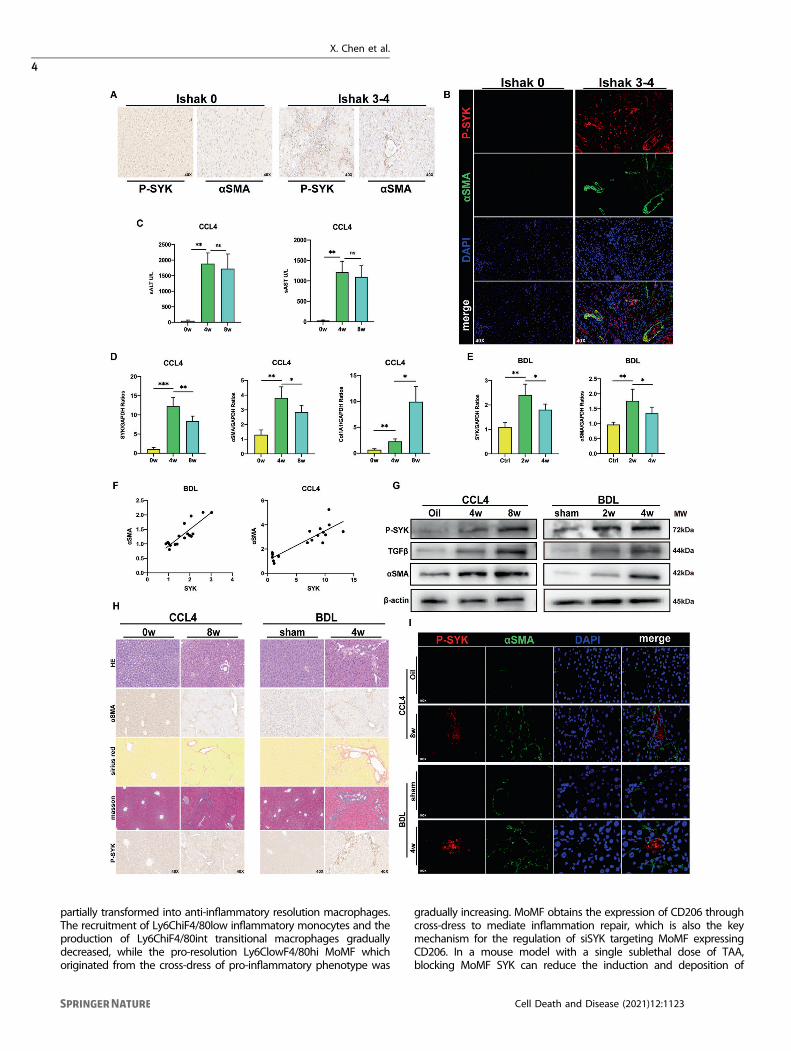
Fig. 1 p-SYK is essential for human and mice liver fibrosis. A Immunohistochemical staining of p-SYK and α-SMA in liver tissues of fibrosispatients with different ishak grades (Ishak0, Ishak3–4) (magnification 40X). B Confocal immunofluorescence staining of p-SYK and α-SMA inliver tissues of fibrosis patients with different ishak grades (Ishak0, Ishak3–4) (magnification 40X), DAPI as nuclear localization. C Serum ALTand AST levels in mice injected with CCL4 for 4 and 8 weeks, oil as a control. D Assessed the expression of SYK, α-SMA, and col1A1 in the liverof mice treated with CCL4 for 4 and 8 weeks were by qPCR, and oil was used as a control. E The expression of SYK and α-SMA in the liver ofmice 2 and 4 weeks after BDL surgery were determined by qPCR, and sham was used as a control. F Correlation analysis between theexpression of SYK and α-SMA in CCL4 and BDL models. GWestern Blot was used to analyze the protein level of p-SYK, TGF-β and α-SMA in theliver of mice injected with CCL4 for 4 or 8 weeks and mice 2 or 4 weeks after BDL, oil and sham were used as controls, respectively. H HE, Siriusred, masson staining and immunohistochemistry of α-SMA and p-SYK were performed on the livers of mice given CCL4 for 8 weeks and mice4 weeks after BDL, respectively. Oil and sham were used as controls. I Confocal immunofluorescence staining of p-SYK and α-SMA wereperformed on the livers of a mouse injected with CCL4 for 8 weeks and mice 4 weeks after BDL, respectively, with DAPI as the nuclearlocalization.
X. Chen et al.
5
Cell Death and Disease (2021) 12:1123
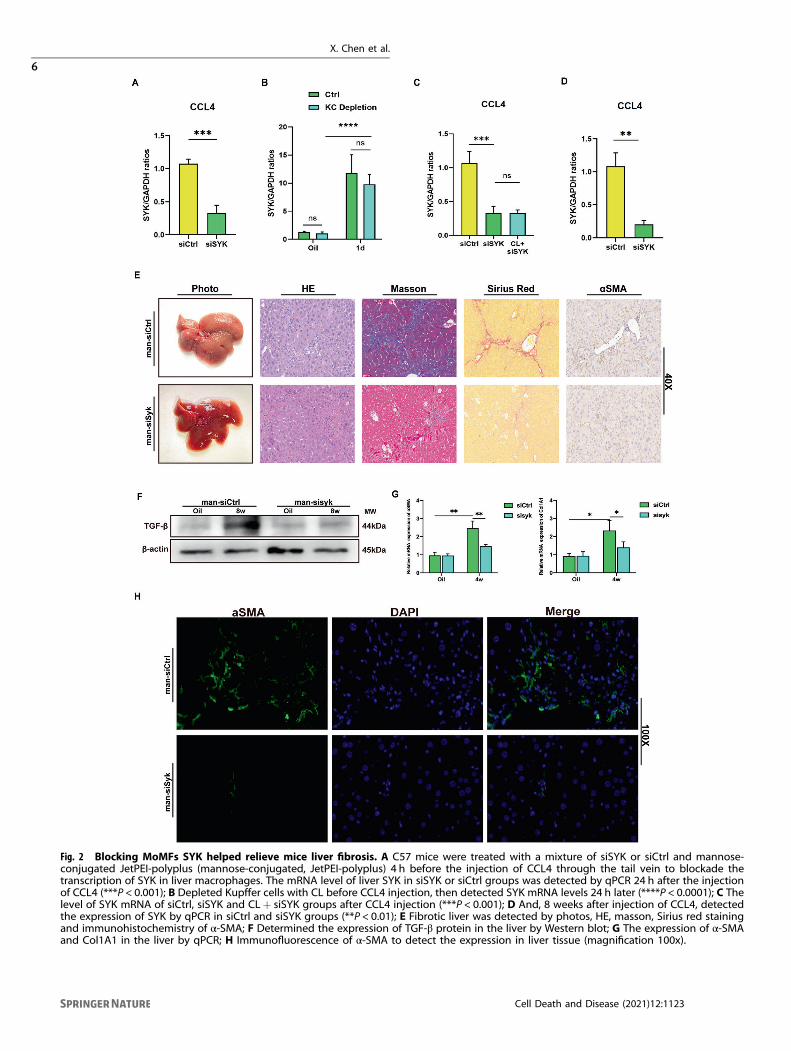
Fig. 2 Blocking MoMFs SYK helped relieve mice liver fibrosis. A C57 mice were treated with a mixture of siSYK or siCtrl and mannose-conjugated JetPEI-polyplus (mannose-conjugated, JetPEI-polyplus) 4 h before the injection of CCL4 through the tail vein to blockade thetranscription of SYK in liver macrophages. The mRNA level of liver SYK in siSYK or siCtrl groups was detected by qPCR 24 h after the injectionof CCL4 (***P < 0.001); B Depleted Kupffer cells with CL before CCL4 injection, then detected SYK mRNA levels 24 h later (****P < 0.0001); C Thelevel of SYK mRNA of siCtrl, siSYK and CL+ siSYK groups after CCL4 injection (***P < 0.001); D And, 8 weeks after injection of CCL4, detectedthe expression of SYK by qPCR in siCtrl and siSYK groups (**P < 0.01); E Fibrotic liver was detected by photos, HE, masson, Sirius red stainingand immunohistochemistry of α-SMA; F Determined the expression of TGF-β protein in the liver by Western blot; G The expression of α-SMAand Col1A1 in the liver by qPCR; H Immunofluorescence of α-SMA to detect the expression in liver tissue (magnification 100x).
X. Chen et al.
6
Cell Death and Disease (2021) 12:1123
hypoxia and induce the up-regulation of HIF1α expression. At thesame time, HIF1a expression changes positively affect the induction ofextracellular matrix deposition and angiogenesis in liver fibrosis[48, 49]. Surgical ligation of the bile duct in mice is a common modelthat induces obstructive cholestatic liver injury and graduallyprogresses to liver fibrosis. Compared with wild-type mice, HIF1α-deficient mice have a significantly lower risk of liver fibrosis after BDL[12]. Knockout of myeloid HIF1α inhibits the expression of PDGF, α-SMA and Col1A1 induced by BDL, and significantly mitigates theprogression of liver fibrosis [13]. In addition, targeting HIF1α inhepatocytes and HSC has also been shown to regulate the occurrenceof liver fibrosis in different experimental induction models. Moderate
ethanol exposure leads to the activation of HIF1α signal inhepatocytes, induces p53-dependent hepatocyte apoptosis, andexacerbates CCl4-induced liver fibrosis [50]. Loss of HIF1α in hepaticstellate cells will result in the down-regulation of many pro-fibroticfactors such as CCR5 and VEGFα [13]. These studies indicate thedriving effect of HIF1α activation in liver fibrosis.CCR2/CCR5 dual inhibitor Cenicriviroc inhibits the recruitment
of Ly6Chi monocytes, thereby improving histological damage andfibrosis in the NASH model [51]. Our research has shown that thecombination utilization of siSYK targeting monocyte-derivedmacrophages and CVC can further inhibit the progression of liverfibrosis on the basis of CVC alone.
Fig. 3 Inhibiting MoMFs SYK blocked Erk phosphorylation. A IHC of Human fibrotic liver α-SMA, p-SYK, P-Erk (magnification 40x). B Westernblot was used to measure the protein expression of SYK and p-SYK in the liver of mice injected with CCL4 4 and 8 w, oil as a control.C Detected the protein level of macrophage p-Erk after silencing liver macrophage syk. D Confocal immunofluorescence to check theexpression of p-SYK and p-Erk in F4/80 macrophages (Zoom 100x). E In the presence or absence of the syk inhibitor GS9973, the protein levelsof p-SYK and p-Erk were detected after stimulating BMDM with LPS for 4 h.
X. Chen et al.
7
Cell Death and Disease (2021) 12:1123
Our study highlights the importance of crosstalk of the SYK/Erk/HIF1α axis in MoMFs in driving the progress of fibrosis. What’s more,targeting SYK in MoMFs can enhance the effect of CCR2/CCR5 dualinhibitor Cenicriviroc to inhibit fibrosis. However, our research still hascertain limitations. First of all, the superposition of the inflammationcascade in the liver fibrosis model injected with CCL4 for a long
period of time leads to a chaotic map of inflammation induction andregression, which to some extent affects our judgment on thetargeting of syk, although we determined our treatment is mainlyCD206+MoMF is targeted, but it does not rule out that a small partof kupffer cells that regenerate after death are affected. In addition,we have not been able to further analyze the mechanism by which
Fig. 4 SYK regulates Hif1α transcription depending on Erk phosphorylation. HIF1α A protein and BmRNA levels were measured after CCL4injection for 4 weeks and 8 weeks by Western Blot or PCR; C The correlation analysis of the expressions of SYK and HIF1α in CCL4 models(**p < 0.01); D The hepatocytes and macrophages expression of Hif1α in siCtrl and siSYK fibrotic livers. E And, 4 h before TAA injection, micewere injected with siCtrl and siSYK through the tail vein, and determine the transcription of Hif1α in the liver 1, 3, and 7 days after TAAinjection Level (*p < 0.05); F Transfect mouse BMDM with shSYK lentivirus, then stimulate BMDM with LPS for 6 h, detect the protein levels ofp-SYK, p-Erk1/2, HIF1α in macrophages; G The protein level of HIF1α in BMDM in the presence or absence of siSYK or Erk1/2 inhibitor Ravox.
X. Chen et al.
8
Cell Death and Disease (2021) 12:1123
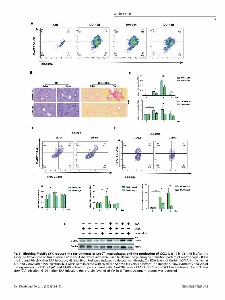
Fig. 5 Blocking MoMFs SYK reduced the recruitments of Ly6Chi macrophages and the production of CXCL1. A 12 h, 24 h, 48 h after thesubacute lethal dose of TAA in mice, F4/80 and Ly6C expression were used to define the phenotypic transition pattern of macrophages. B Onthe 3rd and 7th day after TAA injection, HE and Sirius Red were induced to detect liver fibrosis. C mRNA levels of Col1A1, αSMA, in the liver at1, 3, and 7 days after TAA injection; D, EMice were injected with siCtrl or siSYK via tail vein 4 h before TAA injection. Flow cytometry analysis ofthe expression of CD11b, Ly6C and F4/80 in liver nonparenchymal cells. F mRNA levels of CCL5, CCL2, and CXCL1 in the liver at 1 and 3 daysafter TAA injection; G 24 h after TAA injection, the protein level of αSMA in different treatment groups was detected.
X. Chen et al.
9
Cell Death and Disease (2021) 12:1123
high-frequency administration of GS9973 leads to exacerbation ofdeath in BDL mice.In general, targeting SYK in MoMFs seems to be a promising
target for liver fibrosis.
Reporting SummaryFurther information on research design is available in the NatureResearch Reporting Summary linked to this article.
DATA AVAILABILITYThe datasets generated and/or analyzed during the current study are available fromthe corresponding author on reasonable request.
REFERENCES1. Friedman SL. Liver fibrosis—from bench to bedside. J Hepatol. 2003;38:S38–53.2. Wynn TA, Ramalingam TR. Mechanisms of fibrosis: therapeutic translation for
fibrotic disease. Nat Med. 2012;18:1028–40.3. Sterling RK, Lissen E, Clumeck N, Sola R, Correa MC, Montaner J, et al. Develop-
ment of a simple noninvasive index to predict significant fibrosis in patients withHIV/HCV coinfection. Hepatology 2006;43:1317–25.
4. Mocsai A, Ruland J, Tybulewicz VL. The SYK tyrosine kinase: a crucial player indiverse biological functions. Nat Rev Immunol. 2010;10:387–402.
5. Torres-Hernandez A, Wang W, Nikiforov Y, Tejada K, Torres L, Kalabin A, et al.Targeting SYK signaling in myeloid cells protects against liver fibrosis andhepatocarcinogenesis. Oncogene 2019;38:4512–26.
6. Yanagi S, Inatome R, Takano T, Yamamura H. Syk expression and novel functionin a wide variety of tissues. Biochem Biophys Res Commun. 2001;288:495–8.
7. Kurniawan DW, Storm G, Prakash J, Bansal R. Role of spleen tyrosine kinase inliver diseases. World J Gastroenterol. 2020;26:1005–19.
8. Qu C, Zheng DD, Li S, Lidofsky A, Jiang YC, He L, et al. Tyrosine kinase SYK is apotential therapeutic target for liver fibrosis. Hepatology 2017;66:136a–7a.
9. Cai BS, Dongiovanni P, Corey KE, Wang XB, Shmarakov IO, Zheng Z, et al. Mac-rophage MerTK promotes liver fibrosis in nonalcoholic steatohepatitis. CellMetab. 2020;31:406–21.
10. Foglia B, Cannito S, Bocca C, Parola M, Novo E. ERK pathway in activated, myo-fibroblast-like, hepatic stellate cells: a critical signaling crossroad sustaining liverfibrosis. Int J Mol Sci. 2019;20:2700.
11. Zhan L, Huang C, Meng XM, Song Y, Wu XQ, Yang Y, et al. Hypoxia-inducible factor-1alpha in hepatic fibrosis: a promising therapeutic target. Biochimie 2015;108:1–7.
12. Moon JO, Welch TP, Gonzalez FJ, Copple BL. Reduced liver fibrosis in hypoxia-inducible factor-1alpha-deficient mice. Am J Physiol Gastrointest Liver Physiol.2009;296:G582–92.
13. Copple BL, Kaska S, Wentling C. Hypoxia-inducible factor activation in myeloidcells contributes to the development of liver fibrosis in cholestatic mice. J PharmExp Ther. 2012;341:307–16.
14. Sutton KM, Hayat S, Chau NM, Cook S, Pouyssegur J, Ahmed A, et al. Selectiveinhibition of MEK1/2 reveals a differential requirement for ERK1/2 signalling inthe regulation of HIF-1 in response to hypoxia and IGF-1. Oncogene2007;26:3920–9.
15. Zhao Y, Ma X, Wang J, He X, Hu Y, Zhang P, et al. Curcumin protects against CCl4-induced liver fibrosis in rats by inhibiting HIF-1alpha through an ERK-dependentpathway. Molecules 2014;19:18767–80.
16. Bukong TN, Iracheta-Vellve A, Saha B, Ambade A, Satishchandran A, Gyongyosi B,et al. Inhibition of spleen tyrosine kinase activation ameliorates inflammation, celldeath, and steatosis in alcoholic liver disease. Hepatology 2016;64:1057–71.
17. Wang ZY, Wang H, Chen XJ, Han S, Zhu YL, Wang HH, et al. Inhibiting ATP6V0D2aggravates liver ischemia-reperfusion injury by promoting NLRP3 activation viaimpairing autophagic flux independent of Notch1/Hes1. J Immunol Res. 2021;2021.
18. Wang ZY, Han S, Chen XJ, Li XD, Xia N, Pu LY. Eva1a inhibits NLRP3 activation toreduce liver ischemia-reperfusion injury via inducing autophagy in kupffer cells.Mol Immunol. 2021;132:82–92.
Fig. 6 High-frequency injection of GS9973 aggravated liver fibrosis. A Mice were intraperitoneally injected with GS9973 (20mg/kg, q2d),4 weeks after CCL4 injection, the degree of liver fibrosis was observed by photographs, HE, masson, Sirius red staining, andimmunohistochemistry of α-SMA; B Mice were injected with GS9973 (20mg/kg, q3d), the protein level of TGF-β in the liver was measuredafter 8 weeks of CCL4 injection; C The mRNA level of α-SMA was evaluated after 4 weeks; D The administration of GS9973 (20mg/kg, q2d, ip)caused the death of mice with BDL.
X. Chen et al.
10
Cell Death and Disease (2021) 12:1123
19. Poole A, Gibbins JM, Turner M, van Vugt MJ, van de Winkel JG, Saito T, et al. TheFc receptor gamma-chain and the tyrosine kinase Syk are essential for activationof mouse platelets by collagen. EMBO J. 1997;16:2333–41.
20. An P, Wei LL, Zhao S, Sverdlov DY, Vaid KA, Miyamoto M, et al. Hepatocytemitochondria-derived danger signals directly activate hepatic stellate cells anddrive progression of liver fibrosis. Nat Commun. 2020;11:2362.
21. Karlmark KR, Weiskirchen R, Zimmermann HW, Gasssler N, Ginhoux F, Weber C,et al. Hepatic recruitment of the inflammatory Gr1(+) monocyte subset uponliver injury promotes hepatic fibrosis. Hepatology 2009;50:261–74.
22. Seki E, De Minicis S, Inokuchi S, Taura K, Miyai K, Van Rooijen N, et al. CCR2promotes hepatic fibrosis in mice. Hepatology 2009;50:185–97.
23. Nischalke HD, Berger C, Lutz P, Langhans B, Wolter F, Eisenhardt M, et al. Influ-ence of the CXCL1 rs4074 A allele on alcohol induced cirrhosis and HCC inpatients of European Descent. Plos ONE. 2013;8:e80848.
24. Lu PH, Yu MC, Wei MJ, Kuo KL. The therapeutic strategies for uremic toxinscontrol in chronic kidney disease. Toxins. 2021;13:573.
25. Lefebvre E, Moyle G, Reshef R, Richman LP, Thompson M, Hong F, et al. Anti-fibrotic effects of the dual CCR2/CCR5 antagonist cenicriviroc in animal models ofliver and kidney fibrosis. PLoS One. 2016;11:e0158156.
26. Ratziu V, Sanyal A, Harrison SA, Wong VW, Francque S, Goodman Z, et al. Cen-icriviroc treatment for adults with nonalcoholic steatohepatitis and fibrosis: finalanalysis of the Phase 2b CENTAUR study. Hepatology 2020;72:892–905.
27. Hernandez-Gea V, Friedman SL. Pathogenesis of liver fibrosis. Annu Rev Pathol.2011;6:425–56.
28. Mederacke I, Hsu CC, Troeger JS, Huebener P, Mu XR, Dapito DH, et al. Fatetracing reveals hepatic stellate cells as dominant contributors to liver fibrosisindependent of its aetiology. Nat Commun. 2013;4:1–11.
29. McGlynn KA, Petrick JL, London WT. Global epidemiology of hepatocellular carcinomaan emphasis on demographic and regional variability. Clin Liver Dis. 2015;19:223–38.
30. Baglieri J, Brenner DA, Kisseleva T. The role of fibrosis and liver-associated fibroblastsin the pathogenesis of hepatocellular carcinoma. Int J Mol Sci. 2019;20:1723.
31. El-Serag HB. Hepatocellular carcinoma. N. Engl J Med. 2011;365:1118–27.32. Davies LC, Rosas M, Jenkins SJ, Liao CT, Scurr MJ, Brombacher F, et al. Distinct
bone marrow-derived and tissue-resident macrophage lineages proliferate at keystages during inflammation. Nat Commun. 2013;4:1–10.
33. Holt MP, Cheng LL, Ju C. Identification and characterization of infiltrating mac-rophages in acetaminophen-induced liver injury. J Leukoc Biol. 2008;84:1410–21.
34. Dal-Secco D, Wang J, Zeng ZT, Kolaczkowska E, Wong CHY, Petri B, et al. Adynamic spectrum of monocytes arising from the in situ reprogramming of CCR2(+) monocytes at a site of sterile injury. J Exp Med. 2015;212:447–56.
35. Zigmond E, Samia-Grinberg S, Pasmanik-Chor M, Brazowski E, Shibolet O, Halpern Z,et al. Infiltrating monocyte-derived macrophages and resident kupffer cells displaydifferent ontogeny and functions in acute liver injury. J Immunol. 2014;193:344–53.
36. Berra E, Diaz-Meco MT, Moscat J. The activation of p38 and apoptosis by theinhibition of Erk is antagonized by the phosphoinositide 3-kinase/Akt pathway. JBiol Chem. 1998;273:10792–7.
37. Voisin L, Saba-El-Leil MK, Julien C, Fremin C, Meloche S. Genetic demonstration ofa redundant role of extracellular signal-regulated kinase 1 (ERK1) and ERK2mitogen-activated protein kinases in promoting fibroblast proliferation. Mol CellBiol. 2010;30:2918–32.
38. Harding A, Tian T, Westbury E, Frische E, Hancock JF. Subcellular localizationdetermines MAP kinase signal output. Curr Biol. 2005;15:869–73.
39. Ng B, Dong JR, D’Agostino G, Viswanathan S, Widjaja AA, Lim WW, et al. Interleukin-11is a therapeutic target in idiopathic pulmonary fibrosis. Sci Transl Med. 2019;11.
40. Zhang CX, Cai RZ, Lazerson A, Delcroix G, Wangpaichitr M, Mirsaeidi M, et al.Growth hormone-releasing hormone receptor antagonist modulates lunginflammation and fibrosis due to bleomycin. Lung 2019;197:541–9.
41. Yan Q, Pan LX, Qi SL, Liu F, Wang Z, Qian C, et al. RNF2 mediates hepatic stellatecells activation by regulating ERK/p38 signaling pathway in LX-2 cells. Front Celland Dev Biol. 2021;9:494.
42. Choi WM, Ryu T, Lee JH, Shim YR, Kim MH, Kim HH, et al. Metabotropic glutamatereceptor 5 in natural killer cells attenuates liver fibrosis by exerting cytotoxicity toactivated stellate cells. Hepatology 2021;74:2170–85.
43. Pu SY, Li YP, Liu QH, Zhang X, Chen L, Li R, et al. Inhibition of 5-lipoxygenase inhepatic stellate cells alleviates liver fibrosis. Front Pharmacol. 2021;12:119.
44. Widjaja AA, Singh BK, Adami E, Viswanathan S, Dong JR, D’Agostino GA, et al.Inhibiting interleukin 11 signaling reduces hepatocyte death and liver fibrosis,inflammation, and steatosis in mouse models of nonalcoholic steatohepatitis.Gastroenterology 2019;157:777–92.
45. Dong JR, Viswanathan S, Adami E, Singh BK, Chothani SP, Ng B, et al. Hepatocyte-specific IL11 cis-signaling drives lipotoxicity and underlies the transition fromNAFLD to NASH. Nat Commun. 2021;12:1–15.
46. Chiu DKC, Tse APW, Xu IMJ, Di Cui J, Lai RKH, Li LL, et al. Hypoxia inducible factorHIF-1 promotes myeloid-derived suppressor cells accumulation through ENTPD2/CD39L1 in hepatocellular carcinoma. Nat Commun. 2017;8.
47. Han J, He YP, Zhao H, Xu XW. Hypoxia inducible factor-1 promotes liver fibrosis innonalcoholic fatty liver disease by activating PTEN/p65 signaling pathway. J CellBiochem. 2019;120:14735–44.
48. Chang E, Yang JW, Nagavarapu U, Herron GS. Aging and survival of cutaneousmicrovasculature. J Investigative Dermatol. 2002;118:752–8.
49. Onori P, Morini S, Franchitto A, Sferra R, Alvaro D, Gaudio E. Hepatic microvascularfeatures in experimental cirrhosis: a structural and morphometrical study in CCl4-treated rats. J Hepatol. 2000;33:555–63.
50. Roychowdhury S, Chiang DJ, McMullen MR, Nagy LE. Moderate, chronic ethanolfeeding exacerbates carbon-tetrachloride-induced hepatic fibrosis viahepatocyte-specific hypoxia inducible factor 1alpha. Pharm Res Perspect. 2014;2:e00061.
51. Krenkel O, Puengel T, Govaere O, Abdallah AT, Mossanen JC, Kohlhepp M, et al.Therapeutic inhibition of inflammatory monocyte recruitment reduces steato-hepatitis and liver fibrosis. Hepatology 2018;67:1270–83.
ACKNOWLEDGEMENTSThis study was supported by the National Natural Science Foundation of China(81870443, 81273261) and the Shenzhen Foundation of Science and Technology(grant number JCYJ20170817172116272), Sanming Project of Medicine in Shenzhen(SZSM201812079).
AUTHOR CONTRIBUTIONSXC, ZW, and SH carried out the experiments; ZW, XL, YZ analyzed the data; NX, WY, CJassembled the figures; XC and ZW drafted the manuscript; LP conceived the ideasand supervised the study, YN supervised the study and revised the manuscript; Allauthors approved the final version of the manuscript.
CONFLICT OF INTERESTAll the authors declared no competing interests.
ETHICS APPROVALThe human liver tissues collection and its related experiment were performed withthe approval of the Medical Ethics Committee of the First Affiliated Hospital ofNanjing Medical University (2018-0020) and conformed to the declaration of Helsinki.All mouse experiments were approved by the Experimental Animal Ethics Committeeof Nanjing Medical University (2011057) and in compliance with institutionalregulations.
ADDITIONAL INFORMATIONSupplementary information The online version contains supplementary materialavailable at https://doi.org/10.1038/s41419-021-04403-2.
Correspondence and requests for materials should be addressed to Yong Ni orLiyong Pu.
Reprints and permission information is available at http://www.nature.com/reprints
Publisher’s note Springer Nature remains neutral with regard to jurisdictional claimsin published maps and institutional affiliations.
Open Access This article is licensed under a Creative CommonsAttribution 4.0 International License, which permits use, sharing,
adaptation, distribution and reproduction in anymedium or format, as long as you giveappropriate credit to the original author(s) and the source, provide a link to the CreativeCommons license, and indicate if changes were made. The images or other third partymaterial in this article are included in the article’s Creative Commons license, unlessindicated otherwise in a credit line to the material. If material is not included in thearticle’s Creative Commons license and your intended use is not permitted by statutoryregulation or exceeds the permitted use, you will need to obtain permission directlyfrom the copyright holder. To view a copy of this license, visit http://creativecommons.org/licenses/by/4.0/.
© The Author(s) 2021
X. Chen et al.
11
Cell Death and Disease (2021) 12:1123
Related Documents